High-resolution metagenomics targets specific functional types in complex microbial communities
28
eScholarship provides open access, scholarly publishing services to the University of California and delivers a dynamic research platform to scholars worldwide. Lawrence Berkeley National Laboratory Lawrence Berkeley National Laboratory Title: High-resolution metagenomics targets major functional types in complex microbial communities Author: Kalyuzhnaya, Marina G. Publication Date: 08-25-2009 Publication Info: Lawrence Berkeley National Laboratory Permalink: http://escholarship.org/uc/item/5t53v3t3
Transcript of High-resolution metagenomics targets specific functional types in complex microbial communities

eScholarship provides open access, scholarly publishingservices to the University of California and delivers a dynamicresearch platform to scholars worldwide.
Lawrence Berkeley National LaboratoryLawrence Berkeley National Laboratory
Title:High-resolution metagenomics targets major functional types in complex microbial communities
Author:Kalyuzhnaya, Marina G.
Publication Date:08-25-2009
Publication Info:Lawrence Berkeley National Laboratory
Permalink:http://escholarship.org/uc/item/5t53v3t3
High-resolution metagenomics targets major functional types in complex microbial
communities
Marina G. Kalyuzhnaya1, Alla Lapidus3, Natalia Ivanova3, Alex C. Copeland3,
Alice C. McHardy4#, Ernest Szeto5, Asaf Salamov3, Igor V. Grigoriev3, Dominic Suciu6,
Samuel R. Levine2, Victor M. Markowitz5, Isidore Rigoutsos4, Susannah G. Tringe3,
David C. Bruce7, Paul M. Richardson3, Mary. E. Lidstrom1,2 & Ludmila Chistoserdova2
Departments of 1Microbiology and 2Chemical Engineering, University of Washington,
Seattle, WA 98195; 3Production Genomics Facility, DOE Joint Genome Institute, 2800
Mitchell Drive, Bldg 400, Walnut Creek, CA 94596; 4Bioinformatics and Pattern
Discovery Group, IBM Thomas J. Watson Research Center, 1101 Kitchawan Road,
Yorktown Heights, NY 10598; 5Biological Data Management and Technology Center,
Lawrence Berkeley National Laboratory, 1 Cyclotron Road, Mail Stop 50A-1148,
Berkeley CA 94720; 6Combimatrix Corporation, 6500 Harbour Heights Pkwy, Mukilteo,
WA 98275; 7DOE Joint Genome Institute, Los Alamos National Laboratory, Los
Alamos, NM.
#Current address: Computational Genomics and Epidemiology Group, Max-Planck
Institute for Computer Science, Campus E1 4, 66123 Saarbruecken, Germany

Most microbes in the biosphere remain uncultured and unknown1. Whole
genome shotgun (WGS) sequencing of environmental DNA (metagenomics) allows
glimpses into genetic and metabolic potentials of natural microbial communities2-4.
However, in communities of high complexity metagenomics fail to link specific
microbes to specific ecological functions. To overcome this limitation, we selectively
targeted populations involved in oxidizing single-carbon (C1) compounds in Lake
Washington (Seattle, USA) by labeling their DNA via stable isotope probing (SIP),
followed by WGS sequencing. Metagenome analysis demonstrated specific sequence
enrichments in response to different C1 substrates, highlighting ecological roles of
individual phylotypes. We further demonstrated the utility of our approach by
extracting a nearly complete genome of a novel methylotroph Methylotenera mobilis,
reconstructing its metabolism and conducting genome-wide analyses. This approach
allowing high-resolution genomic analysis of ecologically relevant species has the
potential to be applied to a wide variety of ecosystems.
2

Methylotrophy, metabolism of organic compounds containing no carbon-carbon
bonds (C1 compounds), such as methane, methanol and methylated amines, is an
important part of the global carbon cycle on Earth5,6. Identities of methylotrophs
involved in utilization of specific C1 substrates in a variety of environments have been
previously assessed via both culture reliant7 and culture independent methods8. The
former provide important models for understanding the specific biochemical pathways
enabling methylotrophy, and the latter provide insights into species richness within
specific functional groups. However, while genomic data for some model methylotrophs
are now available9-11, these may not represent major players in specific functional guilds.
At the same time, current methods for environmental detection provide little insight into
the genomic structure of uncultivated methylotrophs.
Metagenomics or environmental genomics has recently become a powerful tool
for collecting information on microbial communities, bypassing cultivation of individual
species1-4. However, traditional metagenomic sequencing usually involves high cost and
effort. Therefore only limited information can be gathered about highly complex natural
communities, such as the ones inhabiting soils and lake sediments. As a proof of
principle, we here utilized a strategy for targeting specific functional types within a
community, via substrate-specific labeling of their DNA using Stable Isotope Probing
(DNA-SIP) 12. Focusing the sequencing effort on the labeled fraction of community DNA
should result in higher sequence coverage for ecologically relevant species within a
metagenome, directly linking them to an ecological function.
As our test model, we selected populations of microbes involved in
methylotrophy in the sediment of a freshwater lake, Lake Washington in Seattle, WA,
3

USA, an environment known for high rates of methane consumption13. Sediment samples
from Lake Washington were exposed separately to 13C-labeled methane, methanol,
methylamine, formaldehyde, and formate, to target populations actively utilizing each of
the C1 compounds. Total DNA was extracted from each microcosm, and the 13C-labeled
fractions were separated from unlabeled DNA by isopycnic centrifugation (Fig. S1). 13C-
labeled DNA was used to construct five separate shotgun libraries, and these were
sequenced at the JGI-PGF. 26 to 59 million base pairs (Mb) of sequence were produced
from each microcosm, totaling 255 Mb. Sequences were assembled, automatically
annotated, and loaded into the JGI’s IMG/M system (Table 1 and Supplementary
Methods), followed by manual analysis. Sequence coverage and degree of assembly
depended on the sequencing effort applied and on the species richness and evenness of
the enriched communities. Based on analysis of 16S rRNA gene sequences, community
complexity was significantly reduced in microcosms exposed to each of the C1 substrates
compared to the complexity of the non-enriched community that we conservatively
estimate to be over 5,000 species (Fig. 1, Table S1 and Supplementary Methods), and
shifted toward specific functional guilds that included both bona fide methylotrophs
(Methylobacter tundripaludum, Methylomonas sp., Methylotenera mobilis,
Methyloversatilis universalis, Ralstonia eutropha) and organisms only distantly related to
any cultivated species, implicating the latter in environmental cycling of C1 compounds.
The closest relatives of these included Verrucomicrobia, Nitrospirae, Planctomycetes,
Acidobacteria, Cyanobacteria, and Proteobacteria. It is possible that some of these were
not labeled by the primary substrate but by a labeled by-product, such as CO2, as a result
of cross-feeding. The 16S rRNA data were supported by data on phylogenetic profiling of
4

each metagenomic dataset, based on top BLAST hit distribution patterns (not shown).
From these analyses, the methylamine microcosm was one of the least complex in terms
of species richness (Table S1) and most enriched in genes diagnostic for C1 transforming
capability (Table S2). It was dominated by a group of closely related strains identified as
M. mobilis, represented by a novel obligate methylamine utilizer recently isolated from
Lake Washington14. Based on 16S rRNA gene sequence coverage (up to 20X, Table S1),
complete or nearly complete genomes of a few M. mobilis strains were predicted to be
encoded in the methylamine microcosm metagenome. From traditional laboratory
enrichments, M. mobilis does not appear to be a “weed” organism, as it is readily out-
competed by other methylamine-utilizers (Table S3). However the incubation conditions
used in this study must have favored M. mobilis, which appears to comprise less than
0.4% of the total bacterial population, based on random sequencing of amplified 16S
rRNA genes15. A composite genome of M. mobilis totaling slightly over 11 Mb was
extracted from the methylamine microcosm metagenome using the recently described
compositional binning method, PhyloPythia16 (genome statistics are shown in Tables 1
and S4). The quality of binning and the recovery of complete or almost complete
genomes were validated by hybridizing DNA of a laboratory-cultivated M. mobilis to a
custom DNA micoarray based on this composite genome (Supplementary Methods). We
also validated genome completeness by examining the presence of various metabolic and
housekeeping genes (Tables S5 and S6). In terms of central metabolism, we identified a
complete set of genes for specific pathways enabling methylamine utilization in M.
mobilis. Multiple copies for each gene were identified (3 to 15, Table S5), consistent with
the composite genome being representative of a few closely related strains. In terms of
5

the housekeeping functions, completeness of the genome was demonstrated by the
presence of 181 tRNA genes corresponding to 36 tRNA acceptors for recognizing all 20
amino acids (not shown), and of a complete set of aminoacyl-tRNA transferases (Table
S6). Standard sets of genes for DNA replication, transcription and translation were
identified, and complete pathways were reconstructed for biosynthesis of all the amino
acids and nucleotides and all the essential vitamins (Table S6).
We reconstructed the metabolism of M. mobilis and conducted genome-wide
comparisons with the genome of Methylobacillus flagellatus, a methylotroph closely
related to M. mobilis, of a similar genome size11,14 (Figures 2 and S2 and Tables S4-6).
M. mobilis from Lake Washington (Table S1) and M. flagellatus are 93 to 95% similar at
the 16S rRNA gene sequence level and share most of the pathways enabling
methylotrophy. However, they were found to be quite different in their genomic content,
gene synteny and gene conservation. Reciprocal BLAST analyses revealed that only 57%
of the proteins translated from the M. flagellatus chromosome had homologs in M.
mobilis at a 50% cut-off, and only 62% of the proteins translated from the composite
genome of M. mobilis had homologs in M. flagellatus. Focusing on some of the highly
conserved genomic regions encoding methylotrophy functions, we uncovered examples
of non-homologous replacements in common biochemical pathways as well as examples
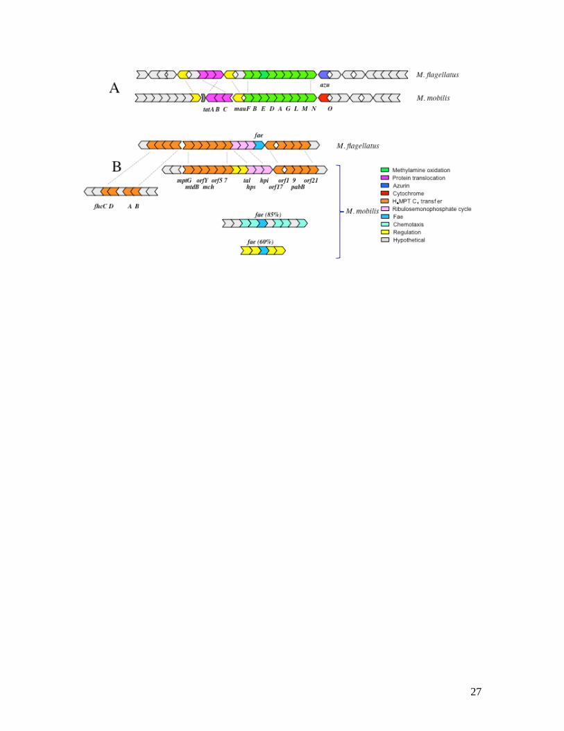
of homolog recruitment into novel/secondary functions. Two of the notable examples are
illustrated in Fig. S3. (A) A gene for azurin, a specific electron acceptor from
methylamine dehydrogenase (MADH) in M. flagellatus11 is missing from the MADH
gene cluster (and elsewhere in the composite genome) in M. mobilis. Instead, it is
replaced by a cytochrome (c551/552) gene that is so far unique to M. mobilis, demonstrating
6

that in two closely related organisms, different strategies are employed for one of the key
energy-generating pathways. (B) From a highly conserved gene cluster encoding
reactions of tetrahydromethanopterin (H4MPT)-linked formaldehyde oxidation, the fae
gene is missing in M. mobilis. In its place, two novel genes are present, encoding a sensor
histidine kinase and a response regulator. A homolog closely related to fae from M.
flagellatus (85% amino acid identity) is instead part of a gene cluster predicted to be
involved in chemotaxis, while a second, less similar homolog (60% amino acid identity),
representing a novel phylogenetic subtype of fae (Fig. 3), is part of a predicted regulatory
gene cluster. This conspicuous gene clustering suggests that Fae (Formaldehyde
Activating Enzyme), whose known enzymatic function is to bind formaldehyde and
convert it into methylene-H4MPT17, must have a second function, possibly as a sensor
component of regulatory and/or signal transduction systems. This hypothesis is supported
by experimental removal of the ‘chemotaxis’ gene cluster shown in Fig. 3, which did not
affect chemotaxis of M. mobilis toward methylamine (not shown). Presence of fae
homologs in genomes that do not encode H4MPT-dependent C1 transfer functions also
supports this hypothesis (Supplementary Methods).
Global genome-genome comparisons between M. mobilis and M. flagellatus
revealed that the conserved parts of the genomes encode central metabolism and
housekeeping functions (methylotrophy, energy transduction, replication, transcription,
translation, amino acid and vitamin biosynthesis), while the variable parts of the genomes
encode auxiliary functions (transport, regulation, electron transfer, CRISPR, prophage,
non-essential biochemical pathways). We were able to precisely map 63 indels of more
than two genes on the chromosome of M. flagellatus, totaling approximately 1,070 kb,
7

not present in the composite genome of M. mobilis (Table S7). The number and the size
of indels could not be estimated with such precision for M. mobilis because of the
composite nature of its genome (Table S8), but we were able to calculate that
approximately 600 kb of sequences per genome were unique, when the genome size was
estimated at approximately 2.5 Mb. One notable element missing from the composite
genome of M. mobilis was the methanol dehydrogenase-encoding gene cluster thought to
be highly conserved in most methylotrophs7. Conversely, some enzymes and pathways
not present in M. flagellatus were identified in M. mobilis, such as the methylcitric acid
cycle (Fig. S4). Comparisons of energy-generating electron transfer pathways encoded in
the two genomes showed little overlap (Table S9), suggesting adaptation to significantly
different life styles. For example, the presence of genes for the denitrification pathway
suggested a propensity for M. mobilis to thrive in microaerobic environments, which was
subsequently proven in experiments with cultivated M. mobilis (not shown), while M.
flagellatus is known to be a strict aerobe11. The predicted denitrification capability of M.
mobilis also suggests that C1 and nitrogen cycling in Lake Washington sediment may be
significantly interlinked.
Sequences of M. mobilis were also present in the metagenomes of microcosms
incubated with methane, methanol, and formaldehyde (Tables S1 and S10), possibly as a
result of cross-feeding on labeled formaldehyde that is an intermediate in the oxidation of
methane and methanol. To test whether M. mobilis strains labeled by these substrates
were metabolically different from M. mobilis strains in the methylamine microcosm, we
conducted substrate-specific genome-genome comparisons, interrogating each dataset
separately and all three datasets at once (to increase sequence coverage) with the M.
8
mobilis composite genome. In this way we detected a number of genes that were not
present in the combined dataset for methane, methanol and formaldehyde microcosms,
but were unique to the methylamine microcosm. Remarkably, the entire gene cluster
encoding methylamine oxidation (mauFBEAGLMNO) was missing from the former,
suggesting that methylamine oxidizing capability is “an acquired taste” and not an
attribute of M. mobilis as a species and suggesting alternative primary substrates for some
M. mobilis strains. In contrast, hits were found for the entire set of genes involved in the
methylcitric acid cycle, pointing to its potential role as a central metabolic pathway (not
shown). One proposed function for this cycle could be in utilizing propionate that is a
product of de-methylation of a compound(s) typical of aquatic environments (such as
dimethylsulfoniopropionate18). Another gene with a predicted function that was unique to
the methylamine microcosm M. mobilis was the novel, divergent fae (Fig. 3 and S3),
suggesting this novel fae and the surrounding genes may have a specialized function in
the metabolism of methylamine. Conversely, specific metabolic traits were detected in
methane and methanol microcosm M. mobilis that were not present in the methylamine
microcosm strains. A remarkable feature of M. mobilis from the methanol microcosm
was the presence of RuBisCO genes, suggesting these strains may be capable of
autotrophy. M. mobilis from the methane microcosm featured nitrogenase genes,
suggesting that some M. mobilis strains may be active in nitrogen fixation. We recently
isolated a number of M. mobilis strains that reveal nutritional properties
matching those predicted from the metagenomes. We are planning to completely
sequence the genomes of three of these strains and compare them to each other
and to the metagenome.
9

In addition to the M. mobilis composite genome, highly covered bacteriophage
genomes were recovered from the methylamine sample. One of these (37 kb) was
homologous to the genome of the Bordetella phage BPP119 (not shown), while others
(approximately 10 kb) were distantly related to the genome of a marine bacteriophage
PM2, the only member of the Corticoviridae family20, and to a prophage found in the
genome of M. flagellatus11 (Fig. S5). Two of the contigs of the latter type were found to
contain overlapping sequences at the ends. These were trimmed and joined at the ends to
produce circular phage chromosome sequences. The presence of phage chromosomes in
the methylamine microcosm metagenome indicates that free-living phages were
propagating during the microcosm incubation with 13C-methylamine. M. mobilis is the
most likely host for these phages, due to its dominance in the labeled microcosm
community. However, the phage sequences were missing from the methane, methanol
and formaldehyde microcosms, indicating a specific association between phage and
methylamine-utilizing M. mobilis. This was supported by the presence of a conspicuous
gene cluster, also unique to the methylamine-utilizing M. mobilis, which encodes pilus
assembly and secretion functions (cpaABCEFtadBC; Fig. S6). This pilus is a possible
candidate for a specific phage receptor. In addition to these, a number of other candidate
phage receptors were unique to the methylamine M. mobilis (a biopolymer transporter, a
major facilitator). The connection between methylamine metabolism and phage
association is very intriguing. One hypothetical scenario could be imagined in which a
specific transporter for methylamine also serves as a specific phage receptor. However,
this hypothesis will need to be tested experimentally.
10

We were also able to analyze other, less covered genomes by supplementing the
PhyloPythia binning with protein recruitment using related genome sequences as a
reference4. From comparisons with the Methylococcus capsulatus genome9, we estimated
that a large portion of the (composite) genome of M. tundripaludum was present in the
methane microcosm dataset (not shown). We conducted metabolic reconstruction for this
organism (Fig. S7) and mapped indels on the chromosome of M. capsulatus (not shown).
Trends similar to the ones noted for gene conservation between M. mobilis and M.
flagellatus were observed: the core parts of the genomes of M. tundripaludum and M.
capsulatus, encoding central metabolism and house keeping genes, were conserved,
while parts of the genomes encoding auxiliary functions were not. Notable omissions
from the M. tundripalidum genome were gene clusters encoding the soluble methane
monooxygenase, RuBisCO, and enzymes of the serine cycle. These genomic features
agree with physiological analysis of the cultivated M. tundripaludum strain21. In a similar
fashion, a large portion of a R. eutropha genome was recovered from the formate
microcosm metagenome. It was highly similar to the published genome of Strain H-1622,
encoding all the core functions and only missing genes for a few auxiliary pathways, such
as CO dehydrogenase and polysaccharide biosynthesis. It also appeared to lack the
megaplasmid found in Strain H-16 (data not shown). Partial genomes were obtained for
uncultivated representatives of Burkholderiaceae, Comamonadaceae, Rhodocyclaceae,
and Actinobacteria, the groups that include methylotrophic representatives (Table S11).
Besides the bona fide methylotrophs, our functional enrichment approach
suggested that phyla not traditionally classified as methylotrophs may be involved in C1
transformations, such as Verrucomicrobia, Nitrospirae and Planctomycetes. The lower
11

coverage of these strains may reflect either slower rates of metabolism or sub-optimal
incubation conditions. Acidophilic methane-oxidizing Verrucomicrobia have been
described recently23-25. However, based on 16S rRNA and functional gene comparisons,
Verrucomicrobia uncovered in this study are only distantly related to these organisms
(<90% 16S rRNA identity). We analyzed the datasets containing Verrucomicrobia
phylogenetic markers (methane, methanol and formaldehyde microcosms) for the
presence of specific functional genes potentially enabling methylotrophy in these species
and identified a conspicuous gene (mtaB) that was present in one or more copies in each
dataset, predicted to encode a methanol:corrinoid methyltransferase. This enzyme has
been characterized in methylotrophic archaea26 and suggested to be involved in methanol
utilization by Clostridia27. However, MtaB sequences from the Lake Washington
metagenome were most similar to a homolog from the only publicly available
Verrucomicrobia genome, that of Opitutaceae bacterium TAV2 (Table S12), thus
implicating this bacterium as well as the Verrucomicrobia detected in this study in
methanol utilization. While no methylotrophic Planctomycetes or Nitrospirae have been
obtained at the moment in pure cultures, these organisms are often detected in
environments with high rates of C1 metabolism28.
This work is a proof of principle study demonstrating that the metagenomics
approach can enable detailed analysis of the genomes of environmentally relevant
microbes, by-passing pure culture isolation, even if the species in question comprise a
minor fraction in a highly complex microbial community. A specific enrichment step,
such as SIP employed here, is key to increasing the resolution of metagenomics, by
focusing the sequencing effort on specific functional types. We have presented here a
12

detailed analysis of the genome of a novel methylotroph, M. mobilis that comprises less
than 0.4% of the total bacterial population in Lake Washington sediment. We also
demonstrated the utility of SIP-enabled metagenomics in uncovering specific bacterium-
phage relationships, suggesting the existence of complex population dynamics involving
multiple strains of M. mobilis and multiple strains of novel corticoviruses. The existence
of such dynamics, likely involving competition for a nutrient (methylamine or a different
methylated compound), in turn highlights the potential environmental importance of C1
compounds as components of global carbon cycling. A genome of an uncultivated M.
tundripaludum was also analyzed in detail, expanding the current genomic knowledge of
methane utilizers. In addition, we identified Verrucomicrobia only distantly related to
recently described methanotrophic isolates, suggesting that methylotrophy may be a
common attribute of this phylum. Overall, this study uncovered the existence of dynamic
and diverse populations responding to C1 substrates, pointing toward the existence of a
complex, multi-tired microbial food web involved in environmental C1 cycling in Lake
Washington sediment and likely in other freshwater lake sediments.
In conclusion, the variation on the standard metagenomics approach described
here, employing function-specific enrichment, allows high-resolution genomic analysis of
major functional types and has the potential to be used in a wide variety of ecosystems
with a wide variety of labeled substrates, as well as in combination with other types of
enrichment.
Methods
13

Sample collection, stable isotope probing and DNA extraction. Sediment samples
were collected on May 15, 2005, from a 63 m deep station in Lake Washington, Seattle,
Washington (47038.075’ N, 122015.993’ W) using a box core that allowed collection of
undisturbed sediment. Samples were transported to the laboratory on ice and immediately
used to set up microcosms. Each microcosms contained 10 ml sediment from the
oxygenated top 1 cm layer, 90 ml Lake Washington water, and one of the following 13C
substrates: methane (50% of air), methanol (10 mM), methylamine (10 mM),
formaldehyde (1 mM), or formate (10 mM). All substrates were 99 atom % 13C and were
purchased from Sigma-Aldrich, with the exception of [13C] methanol, which was
provided by the National Stable Isotope Resource at Los Alamos National Laboratory.
The samples were incubated for 3-5 (methylamine and methane), 5-7 (methanol) or 10-14
(formaldehyde and formate) days at room temperature, with shaking. It has been
previously demonstrated that SIP incubations at the in situ temperature (8oC) resulted in
similar community structures while longer incubation times were required29. DNA was
extracted and purified and subjected to density gradient ultracentrifugation as previously
described12, 29, with slight modifications (Supplementary Methods). 13C-DNA fractions
were visualized in UV (Fig. S1) and collected using 19-gauge needles.
DNA sequencing and assembly. Five shotgun libraries were constructed, one from each
microcosm, in the pUC18 vector (1-3 kb inserts). The libraries were sequenced with
BigDye Terminators v3.1 and resolved with ABI PRISM 3730 (ABI) sequencers. A total
of 344,832 reads comprising 255.08 megabases (Mb) of Phred Q20 sequence were
generated. Sequences were screened for vector contaminations and quality trimmed using
LUCY30 and assembled, both en masse and by sample, using the PGA assembler.
14
Assembly statistics are shown in Table 1. These draft quality assemblies were manually
validated and used for all downstream analyses.
Compositional binning. Assembled metagenomic fragments were binned (classified)
using PhyloPythia, a phylogenetic classifier that uses a multi-class Support Vector
machine (SVM) for the composition-based assignment of fragments at different
taxonomic ranks, essentially as previously described16. Generic models for the ranks of
domain, phylum and class were combined with models for the dominating clades in the
sample. The generic models represent all clades covered by three or more species at the
corresponding ranks among the sequenced microbial isolates. At the rank of family, a
sample-specific model was created with classes for the clades Methylococcaceae,
Burkholderiaceae, Rhodocyclaceae, Methylophilaceae and Comamonadaceae and a class
‘other’. A sample-specific model for the dominant sample populations was created with
classes for the Methylotenera and Methylobacter populations and a class ‘other’. The
sample-specific population model was trained on 138 kb and 141 kb of contigs for the
Methylotenera and Methylobacter populations, respectively, that were identified based on
phylogenetic marker genes, as well as sequenced isolates for the class ‘other’. The
family-level model was trained using the sample-specific data and additional sequenced
isolates available for the corresponding clades. For each model, five sample-specific
multi-class SVMs were created using fragments of lengths of 3, 5, 10, 15 and 50 kb,
respectively. All input sequences were extended by their reverse complement prior to
computation of the compositional feature vectors. The parameters w and l were both set
to 5 for the sample-specific models. The final classifier consisting of the sample-specific
and generic clade models was applied to assign all fragments >1 kb of the samples. In
15

case of conflicting assignments, preference was given to assignments of the sample-
specific models. Data were incorporated into the Integrated Microbial Genomes with
Microbiome Samples (IMG/M) system (http://img.jgi.doe.gov/m). This whole-genome
shotgun project has been deposited at DDBJ/EMBL/GenBank under accession
number XXX.
Species richness estimation.16S rRNA gene fragments were amplified from sediment
DNA using the EUB27f/1496R primer set following by cloning into the pCR2.1 vector
(Invitrogen), as recommended by the manufacturer. Inserts of 859 randomly selected
clones were subjected to restriction fragment length polymorphism (RFLP) analysis, after
digestion with AluI (Fermentas). The GeneTools imaging software (ProcessGelFiles4.m)
was used to compare the restriction patterns. AluI restriction fragments resulting from
pCR2.1 were used as internal locators to adjust the positions of the insert fragments.
Different restriction patterns were clustered by likeness using agglomerative clustering
(Matlab, Mathworks). Clones predicted to be identical by these analyses were sequenced
in order to verify the efficiency of the analysis, and in each case the identity was proven.
Nine groups were identified containing two identical sequences, three groups containing
three identical sequences, and one group containing four identical sequences. Chao
nonparametric richness estimators were implemented to estimate species richness using
the computational tool EstimateS (version 8, http://purl.oclc.org/estimates), resulting in
the lowest richness estimate of 5,430.
Protein recruitment. Protein recruitment was carried out essentially as previously
described4 except for protein sequences rather than DNA sequences were used. The
Phylogenetic Profiler tool that is part of the IMG/M package was used. In the case of M.
tundripaludum/ M. capsulatus pair, cut-offs of 60% to 80% were used, based on 89% 16S
16

rRNA gene similarity between the two strains. In the case of R. eutropha/ R. eutropha H-
16 pair (99% 16S rRNA gene similarity), a cut-off of 90% was used.
References
1. The New Science of Metagenomics: Revealing the Secrets of Our Microbial Planet.
The National Academies Press (2007).
2. Tyson, G.W. et al. Community structure and metabolism through reconstruction of
microbial genomes from the environment. Nature 428:37-43 (2004).
3. Tringe, S.G. et al. Comparative metagenomics of microbial communities. Science
308:554-557 (2005).
4. Rusch, D.B. et al. The Sorcerer II Global Ocean Sampling Expedition: Northwest
Atlantic through Eastern Tropical Pacific. PLoS Biol. 5:e77 (2007).
5. Hanson, R.S. & Hanson, T.E. Methanotrophic bacteria. Microbiol Rev. 60:439-471
(1996).
6. Guenter, A. The contribution of reactive carbon emissions from vegetation to the
carbon balance of terrestrial ecosystems. Chemosphere 49:837-844 (2002).
17

7. Lidstrom, M.E. Aerobic methylotrophic procaryotes. In: A. Balows, H.G.
Truper, M. Dworkin, W. Harder and K.-H. Schleifer (ed.) The Prokaryotes. Springer
Verlag, New York, NY (2006).
8. McDonald, I.R., L. Bodrossy, Y. Chen & Murrell, J.C. Molecular ecology techniques
for the study of aerobic methanotrophs. Appl Environ Microbiol. 74:1305-1315 (2008).
9. Ward, N. et al. Genomic insights into methanotrophy: the complete genome sequence
of Methylococcus capsulatus (Bath). PLoS Biol. 2:e303 (2004).
10. Kane, S.R. et al. Whole-Genome Analysis of Methyl tert-Butyl Ether (MTBE)-
Degrading Beta-Proteobacterium Methylibium petroleiphilum PM1. J Bacteriol. 189:
1931-1945 (2007).
11. Chistoserdova, L. et al. The genome of Methylobacillus flagellatus, the molecular
basis for obligate methylotrophy, and the polyphyletic origin of methylotrophy. J.
Bacteriol. 189:4020-4027 (2007).
12. Radajewski, S., Ineson, P., Parekh, N.R. & Murrell, J.C. Stable-isotope probing as a
tool in microbial ecology. Nature 403:646-649 (2000).
18

13. Auman, A.J., Stolyar, S., Costello, A.M. & Lidstrom, M.E. Molecular
characterization of methanotrophic isolates from freshwater lake sediment. Appl Environ
Microbiol. 66:5259-66 (2000).
14. Kalyuzhnaya, M.G., Bowerman, S., Lara, J.C., Lidstrom, M.E. & Chistoserdova, L.
Methylotenera mobilis gen. nov., sp. nov, an obligately methylamine-utilizing bacterium
within the family Methylophilaceae. Int J Syst Evol Microbiol. 56:2819-2823 (2006).
15. Kalyuzhnya, M.G., Lidstrom, M.E. & Chistosedova, L. Real-time detection of
actively metabolizing microbes via redox sensing as applied to methylotroph populations
in Lake Washington. ISME J. (2008, In Press).
16. McHardy, A.C., Garcia Martin H., Tsirigos, A. Hugenholtz P. & Rigoutsos I.
Accurate phylogenetic classification of variable-length DNA fragments. Nat Methods
4:63-72 (2007).
17. Vorholt, J.A., Marx, C.J., Lidstrom, M.E. & Thauer, R.K. Novel formaldehyde-
activating enzyme in Methylobacterium extorquens AM1 required for growth on
methanol. J Bacteriol. 182:6645-6650 (2000).
18. Ginsburg, B. et al. DMS formation by dimethylsulfoniopropionate route in
freshwater. Environ Sci Technol. 32:2130-2136 (1998).
19. Liu, M. et al. Genomic and genetic analysis of Bordetella bacteriophages encoding
19

reverse transcriptase-mediated tropism-switching cassettes. J Bacteriol. 186:1503-1517
(2004).
20. Krupovic M. et al. Genome characterization of lipid-containing marine bacteriophage
PM2 by transposon insertion mutagenesis. J Virol. 80:9270-9278 (2006).
21. Wartiainen, I., Hestnes, A.G., McDonald, I.R. & Svening, M.M. Methylobacter
tundripaludum sp. nov., a methane-oxidizing bacterium from Arctic wetland soil on the
Svalbard islands, Norway (78° N). Int J Syst Evol Microbiol. 56:109-113 (2006).
22. Pohlmann, A. et al. Genome sequence of the bioplastic-producing “Knallgas”
bacterium Ralstonia eutopha H16. Nat Biotechnol. 24:1257-1262 (2006).
23. Islam, T., Jensen, S., Reigstad, L.J., Larsen, & Ø, Birkeland, N.-K. Methane
oxidation at 55°C and pH 2 by a thermoacidophilic bacterium belonging to the
Verrucomicrobia phylum. PNAS 105:300-304 (2008).
24. Dunfield, P.F. et al. Methane oxidation by an extremely acidophilic bacterium of the
phylum Verrucomicrobia. Nature 450: 879-82 (2007).
25. Pol, A. et al. Methanotrophy below pH 1 by a new Verrucomicrobia species. Nature
450:874-878 (2007).
20

26. Sauer, K. and Thauer, R.K. Methanol:coenzyme M methyltransferase from
Methanosarcina barkeri. Zinc dependence and thermodynamics of the
methanol:cob(I)alamin methyltransferase reaction. Eur J Biochem. 249:280-285 (1997).
27. Das, A. et al. Characterization of a corrinoid protein involved in the C1 metabolism
of strict anaerobic bacterium Moorella thermoacetica. Protteins: Struct Funct Bioinform.
67:167-176 (2007).
28. Lösekann, T. et al. Diversity and abundance of aerobic and anaerobic methane
oxidizers at the Haakon Mosby mud volcano, Barents Sea. Appl Environ Microbiol.
73:3348-3362 (2007).
29. Nercessian, O., Noyes, E., Kalyuzhnaya, M.G., Lidstrom, M.E. & Chistoserdova, L.
Bacterial populations active in metabolism of C1 compounds in the sediment of Lake
Washington, a freshwater lake. Appl Environ Microbiol. 71:6885-6899 (2005).
30. Chou, H.H. & Holmes, M.H. DNA sequence quality trimming and vector removal.
Bioinformatics 17:1093-1104 (2001).
21

Acknowledgements This research was supported by the National Science Foundation as
part of the Microbial Observatories program (MCB-0604269). This work was performed,
in part, under the auspices of the U.S. Department of Energy’s Office of Science
Biological and Environmental Research Program, and by the University of California,
Lawrence Livermore National Laboratory under Contract No. W-7405-Eng-48,
Lawrence Berkeley National Laboratory under contract No. DE-AC02-05CH11231, and
Los Alamos National Laboratory under contract No. DE-AC02-06NA25396. The
sequencing for the project was provided through the US Department of Energy (DOE)
Community Sequencing Program (http://www.jgi.doe.gov/CSP/index.html).
Author Contributions
M.G.K., M.E.L. and L.C. conceived the project. M.E.L. and L.C. coordinated project
execution. M.G.K. collected samples, performed SIP, purified DNA for sequencing and
performed microarray hybridizations. D.B. and P.M.R. oversaw library construction and
sequencing. S.G.T. oversaw sequence assembly and analysis. A.C.C. and A.L. carried out
assemblies. A.S. and I.V.G. conducted gene prediction and annotation. A.C.M. and I.R.
carried out binning. E.S. and V.M.M. carried out data processing and loading into
IMG/M. L.C. and N.I. carried out metabolic reconstruction. S.R.L. and M.G.K.
performed species richness estimates. D.S. carried our microarray design. M.G.K.,
M.E.L. and L.C. wrote the initial draft of the paper, all other authors contributed.
22

Table 1. Summary sequencing and assembly and gene prediction statistics
_________________________________________________________________________________________________________________
Methane Methanol Methylamine Formaldehyde Formate Combined Methylotenera
_________________________________________________________________________________________________________________
Assembly statistics
Number of reads 71,808 67,200 83,712 80,640 41,472 344,832 NA
Average read length (bp) 792 797 709 712 638 741 NA
Trimmed read length (Mbp) 56.85 53.53 59.34 58.91 26.45 255.08 NA
Non-redundant sequence (bp) 52.16 50.25 37.23 57.62 17.57 211.47 11.16
Percent of reads in contigs 10.2 10.0 55.5 7.3 34.3 27.6 100
Total contigs (>2 kb) 2,797 2,871 7,558 2,583 3,618 25,877 4,078
Total singlets 59,417 56,408 29,217 69,104 18,857 215,581 0
Average sequence coverage (x) 1.6 1.6 1.9 1.7 1.9 1.7 2.1
Highest sequence coverage (x) 7.0 4.8 20.4 6.4 4.7 23.1 20.4
Average size of contigs (bp) 1,418 1,288 2,065 1,166 1,265 1,593 2,736
Largest contig (bp) 6,174 5,913 20,771 4,714 6,276 22,407 15,820
GC content (%) 58.9 59.5 53.0 57.9 65.8 58.3 46.2
Gene predictions
Protein coding genes 81,076 77,229 54,340 89,729 28,700 321,503 12,719
Genes in COGs 43,456 40,773 33,643 46,032 17,112 174,344 10,082
Genes in Pfams 28,090 26,494 23,586 29,375 10,585 115,228 8,543
Predicted enzymes 3,089 3,047 5,005 3,065 1,417 16,780 3,264
Number of 16S rRNA genes 12 12 10 18 5 61 3
Number of tRNA genes 405 412 376 504 121 1,728 181
__________________________________________________________________________________________________________________
23

Figure legends
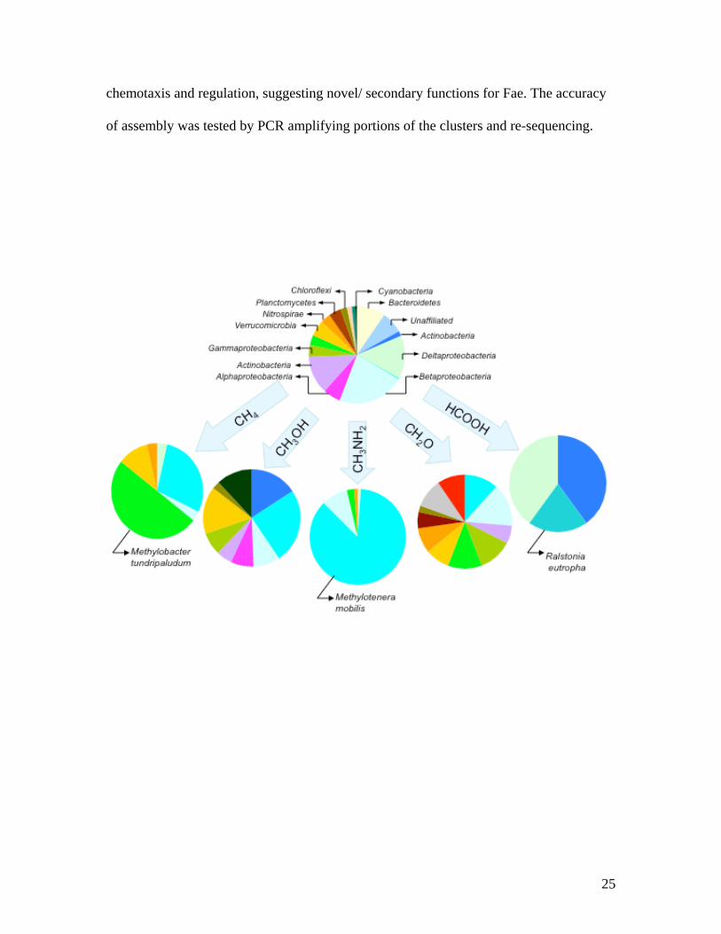
Figure 1. Taxonomic distribution of 16S rRNA gene sequences from metagenomes. The
sum of coverage scores for each phylum (Table S1) were used for metagenomes
generated in this work, and data from5 were used for the non-enriched community.
Similar taxonomic distributions were observed when PCR-amplified libraries generated
for each microcosm were analyzed as in5 (data not shown). ◊Ralstonia eutropha; ◊
Methylotenera mobilis; ◊ other Betaproteobacteria; ◊Methylobacter tundripaludum; ◊
other Gammaproteobacteria; ◊ Alphaproteobacteria; ◊ Deltaproteobacteria; ◊
Actinobacteria; ◊ Acidobacteria; ◊ Archaea; ◊ Bacteroidetes; ◊ Chloroflexi; ◊
Cyanobacteria; ◊ Firmicutes; ◊ Gemmatimonadetes; ◊ Planctomycetes; ◊
Verrucomicrobia; ◊ Unclassified bacteria; ◊ Chloroplasts.
Figure 2. Metabolic features of M. mobilis compared to metabolic features of M.
flagellatus as deduced from genomic comparisons. Major metabolic pathways and energy
generating systems are shown. Similar shapes indicate similar functions, different colors
indicate lack of homology at the protein level.
Figure 3. Comparison of gene clusters involved in methylotrophy in M. mobilis and M.
flagellatus. A. In the methylamine oxidation gene cluster, the gene for azurin, an electron
acceptor from methylamine dehydrogenase in M. flagellatus is replaced by a gene
encoding cytochrome C551/552 suggesting a functional replacement. B. Two fae genes in
M. mobilis are parts of gene clusters predicted to be involved, respectively in sensing/
24
chemotaxis and regulation, suggesting novel/ secondary functions for Fae. The accuracy
of assembly was tested by PCR amplifying portions of the clusters and re-sequencing.
25

26
27