herpetological review - Academic Colleges and Departments
184
-
Upload
khangminh22 -
Category
Documents
-
view
0 -
download
0
Transcript of herpetological review - Academic Colleges and Departments


SOCIETY FOR THE STUDY OF AMPHIBIANS AND REPTILES www.ssarherps.org
The Society for the Study of Amphibians and Reptiles, the largest international herpetological society, is a not-for-profit organization established to advance research, conservation, and education concerning amphibians and reptiles. Founded in 1958, SSAR is widely recognized today as having the most diverse society-sponsored program of services and publications for herpetologists. Membership is open to anyone with an interest in herpetology—professionals and serious amateurs alike—who wish to join with us to advance the goals of the Society.
All members of the SSAR are entitled to vote by mail ballot for Society officers, which allows overseas members
to participate in determining the Society's activi-ties; also, many international members attend the annual meetings and serve on editorial boards and committees.
All members and institutions receive the Soci-ety’s primary technical publication, the Journal of Herpetology, and its bulletin, Herpetological Review;
both are published four times per year. Members also receive pre-publication discounts on other Society publications, which are advertised in Herpetological Review.
To join SSAR or to renew your membership, please visit the secure online ZenScientist website via this link:
http://www.ssarherps.org/pages/membership.php
Future Annual Meetings2014 — Chattanooga, Tennessee, 30 July–3 August (JMIH with ASIH, HL, and AES)2015 — Lawrence, Kansas 30 July–3 August (SSAR with PARC and KHS)
SSAR OFFICERS (2013) President ROBERT D. ALDRIDGE Saint Louis University [email protected]
President-elect AARON M. BAUER Villanova University [email protected]
Secretary MARION R. PREEST The Claremont Colleges [email protected]
Treasurer ANN PATERSON Williams Baptist College [email protected]
Publications Secretary BRECK BARTHOLOMEW Salt Lake City, Utah [email protected]
Immediate Past President JOSEPH R. MENDELSON, III Zoo Atlanta [email protected]
Directors FRANK BURBRINK (2016) College of Staten Island, USA ALISON CREE (2016) University of Otago, New Zealand TIFFANY DOAN (2014) ' State College of Florida, Manatee-Sarasota, USA LISA HAZARD (2016) Montclair State University, USA TRAVIS LaDUC (2014) University of Texas at Austin,USA JENNIFER PRAMUK (2014) Woodland Park Zoo, Seattle, USA CAROL SPENCER (2014) University of California, Berkeley, USA GREGORY WATKINS-COLWELL (2016) Yale University Museum of Natural History, USA
Trustee GEORGE R. PISANI University of Kansas
SSAR EDITORS Journal of Herpetology ERIN MUTHS, Co-Editor U.S. Geological Survey, Fort Collins
GAD PERRY, Co-Editor Texas Tech University
Contributions to Herpetology KRAIG ADLER, Editor Cornell University
Facsimile Reprints in Herpetology AARON M. BAUER, Editor Villanova University
Herpetological Circulars JOHN J. MORIARTY, Editor Plymouth, Minnesota
Catalogue of American Amphibians and Reptiles TRAVIS LADUC, Co-Editor University of Texas at Austin
CHRISTOPHER J. BELL, Co-Editor University of Texas at Austin
Herpetological Conservation JOSEPH C. MITCHELL, Editor Mitchell Ecological Research Services
EditorROBERT W. HANSEN16333 Deer Path LaneClovis, California 93619-9735 [email protected]
Associate EditorsMICHAEL F. BENARDCase Western Reserve University, USA
JESSE L. BRUNNERWashington State University, USA
FÉLIX B. CRUZINIBIOMA, Río Negro, Argentina
RAUL DIAZLa Sierra University, California, USA
ROBERT E. ESPINOZACalifornia State University, Northridge, USA
SCOTT M. BOBACKDickinson College, Carlisle, Pennsylvannia, USA
GUNTHER KÖHLERForschungsinstitut und Naturmuseum Senckenberg, Germany
PETER V. LINDEMANEdinboro University, USA
DEANNA H. OLSONUSDA Forestry Science Lab, Corvallis, Oregon, USA
JODI ROWLEYAustralian Museum, Australia
DANIEL SAENZUSDA Forest Service, Nacogdoches, Texas, USA
Index EditorRUTHE SMITH
Section EditorsBook Reviews
AARON M. BAUERVillanova University, [email protected]
Current ResearchBECK A. WEHRLEUniversity of California, Irvine, [email protected]
BEN LOWEUniversity of Minnesota, [email protected]
ConservationPRIYA NANJAPPAAssociation of Fish & Wildlife Agencies,[email protected]
Geographic DistributionINDRANEIL DASUniversiti Malaysia Sarawak, [email protected]
JERRY D. JOHNSONThe University of Texas at El Paso, [email protected]
ALAN M. RICHMONDUniversity of Massachusetts, [email protected]
GUSTAVO J. SCROCCHIFundación Miguel Lillo, [email protected]
DAVID C. BLACKBURNCalifornia Academy of Sciences, [email protected]
STEPHEN RICHARDSSouth Australia Museum, [email protected]
Zoo ViewJAMES B. MURPHYSmithsonian National Zoological Park, [email protected]
NomenclatureJAY M. SAVAGESan Diego State University, California, USA [email protected]
HerpetocultureROBERT HILLZoo Atlanta, [email protected]
WULF SCHLEIPMeckenheim, [email protected]
Natural History NotesJAMES H. HARDINGMichigan State University, [email protected]
SEAN P. GRAHAMPennsylvania State University, [email protected]
JACKSON D. SHEDDTNC Dye Creek Preserve, California, [email protected]
JOHN D. WILLSONUniversity of Arkansas, Fayetteville, [email protected]
DAVID A. STEENAuburn University, Auburn, Alabama, [email protected]
Copy EditorsDREW R. DAVISUniversity of South Dakota, USA
KYLE MILLER HESEDUniversity of Maryland, USA
DANIEL PORTIK University of California, Berkeley, USA
ELIZABETH TIMPEUniversity of Connecticut, USA
HERPETOLOGICAL REVIEWTHE QUARTERLY BULLETIN OF THE
SOCIETY FOR THE STUDY OF AMPHIBIANS AND REPTILES
© 2013 Society for the Study of Amphibians and Reptiles

Herpetological Review 44(4), 2013
533533
2013 Annual Meeting, Albuquerque, New Mexico
The 56th Annual Meeting of SSAR took place from 10–15 July 2013 at the Albuquerque Convention Center in Albuquerque, New Mexico, USA (Fig. 1). Organizing societies this year were So-ciety for the Study of Amphibians and Reptiles (along with the International Society for the History and Bibliography of Her-petology), American Elasmobranch Society (celebrating its 29th annual meeting), American Society of Ichthyologists and Herpe-tologists (celebrating its 93rd annual meeting) and The Herpetol-ogists’ League (celebrating its 71st annual meeting). The meeting was hosted by the University of New Mexico and the Museum of Southwestern Biology. The local hosts were Tom Turner, Lex Snyder, Steve Ross, Tom Giermakowski, Steven Platania, Norman
Mercado-Silva, and Mason Ryan. They were assisted by a small army of volunteers including Meghan Balk, Olivia Chavez, Tracy Diver, Elizabeth Gallagher, Levi Gray, Victoria Hansen, Angela Hung, Ian Latella, Lorraine McInnes, Tyler Pilger, Jolene Rearick, Mason Ryan, Brad Truett, and Rhiannon West. Thanks to K-State University Division of Continuing Education. We thank all of the above for a fun and successful meeting.
Nearly 900 herpetologists and ichthyologists attended the meeting. This number includes 417 students and 303 members of SSAR. The meeting included 45 oral sessions and five sympo-sia. Symposia topics included “Detectability and Studying Rare Species: When Cryptic Natural Histories Defy both Conventional
ABOUT OUR COVER: Lanthanotus borneensisLanthanotus borneensis, the Bor-
nean Earless Monitor, surely among herpetology’s “holy grails,” is a 200-mm (SVL) varanoid lizard whose common name reflects the lack of an ear open-ing. This poorly known, enigmatic spe-cies is considered to be the sole living representative of the family Lantha-notidae. The only other taxon allocated to the Lanthanotidae is Cherminotus longifrons, from the Upper Cretaceous in the Gobi Desert, Mongolia (Borsuk-
Białynicka 1984. Palaeontol. Polonica 46:5–105), although this taxonomic allocation has been challenged (Gao and Norell 2000. Bull. Amer. Mus. Nat. Hist. 249:1–118).
The name Lanthanotus is derived from Greek, meaning “hid-den ears,” but a shallow tympanic cavity is present. Males show blunt, rectangular jaws, while females have relatively pointed jaws. The dorsal surface is brownish-orange, sometimes with a dark vertebral stripe, and the venter is yellow with brownish or-ange and ochre mottling; the tongue is pink.
The Lanthanotidae has been assigned to the anguinomor-phan lizards (McDowell and Bogert 1954. Bull. Am. Mus. Nat. Hist. 105:1–142), but the relationships among these families have been the subject of debate. Lee’s cladistic analysis (1997. Phil. Trans. Royal Soc. London B 352:53–91), based on osteologi-cal characters, reveals that the lanthanotids and varanids are the closest relatives of snakes. An early molecular study of Fuller et al. (1998. Mol. Phylogen. Evol. 9:294–307), using 12S rRNA se-quences, showed surprisingly low sequence divergence between these two groups, and a sister-taxa relationship was revealed by Ast (2001. Cladistics 17:211–226). More recent molecular stud-ies of global squamates using nuclear genes confirm the posi-tion of the Lanthanotidae within the Anguinomorpha, with a sister relationship with the Varanidae; these two lineages, along with Shinisauridae, are sister to the Helodermatidae and other members of the Anguinomorpha (Wiens et al. 2012. Biol. Letters 8:1043–1046; Pyron et al. 2013. BMC Evol. Biol. 13:93). The most recent common ancestor node for the family corresponds to the
Mid-Cretaceous, 108 mya (Douglas et al. 2010. Mol. Phylogen. Evol. 55:153–167).
Described by Fritz Steindachner (1877. Denkschr. Kais. Akad. Wiss., Wien 38:93–96) based on a specimen now in the Vienna Museum of Natural History (Naturhistorisches Museum Wien 16365) from Sarawak, Lanthanotus borneensis is known to in-habit lowland forests and low hills of northwestern Borneo, in-cluding the states of Sarawak (Malaysia) and Kalimantan Barat (Indonesia), and is seldom encountered. Consequently, little is known of its field biology, and this subfossorial and aquatic spe-cies feeds on earthworms and crustaceans, and probably fish. Nocturnal, its daytime retreats include burrows up to about 30 cm, under vegetation, rocks, and litter of rocky stream banks. Semi-torpid by day, at night it forages on land and in water. Typi-cally sluggish, it can flatten its body when touched, and other be-haviors associated with threat response includes struggling, def-ecating, hissing, and biting. Skin is shed in a single piece, similar to that in some other anguinomorphan squamates and snakes. Mating has been observed in early February, females producing 2–5 oval, leathery-shelled eggs that measure ca. 30 mm in length.
Our cover image was captured by Indraneil Das in western Sarawak, East Malaysia (Borneo), using a Nikon D600 and AF-S VR Micro-Nikkor 105mm f/2.8G IF-ED lens. Shutter speed set at 1/60 sec, f/18, using an off-camera Nikon SB-910 flash unit, at-tached to a Lastolite Ezybox Hotshoe.
Das is Professor at the Institute of Biodiversity and Environmental Conservation, Universiti Malaysia Sarawak. His current research cen-ters on ecology and conservation of amphibians and reptiles, especially the role of fragmentation and other landscape change, and the system-atics and natural history of these species, especially in tropical Asia. His work on Lanthonotus was sup-ported by a Mohammed bin Zayed Species Conservation Grant.
SSAR BUSINESSPH
OTO
BY
PUI Y
ON
G M
IN

Herpetological Review 44(4), 2013
534
and Progressive Statistics,” “Fishes and Morphology Today,” and “Fish Out of Water: Evolutionary and Ecological Issues in the Conservation of Fishes in Water-Altered Environments.” SSAR sponsored a symposium entitled “Impact of Energy De-velopment on Amphibians and Reptiles in North America.” This timely topic attracted a diversity of speakers (n = 16) and a large audience (Fig. 2). We thank Jeff Lovich and Priya Nanjappa for co-organizing this symposium.
Following the usual pre-conference day of Board and Ex-ecutive Committee meetings, the JMIH began officially with the Plenary Session on July 11th. Local Host Committee Chair, Tom Turner, welcomed us warmly to Albuquerque and thanked the other members of the local committee and the many donors who contributed to the 2013 JMIH. He then introduced Ray Pow-ell (New Mexico Commissioner of Public Lands and a veterinar-ian) who spoke about the importance of scientists engaging ac-tively with government and other elected officials. He noted that these officials don’t know who we are or what we have to offer, hence the need for us to approach them, to communicate with them, and to “make them look good!”
Following Commissioner Powell’s talk, various awards were presented. Bill Matthews presented the Robert H. Gibbs Award
to Lynn Parenti who noted that this was the 37th ASIH/JMIH meeting in a row that she has attended. The Henry S. Fitch Award for Excellence in Herpetology was presented in absentia to Roy McDiarmid by Henry Mushinsky. The Robert K. Johnson Ward for Excellence in Service to ASIH was presented in absentia to Pat Gregory by Al Savitzky. Steve Mullin announced Craig Guyer as the winner of the 2013 SSAR/HL/ASIH Meritorious Teaching Award in Herpetology. The PARC Alison Haskell Award for Excel-lence in Herpetological Conservation was given to Charlie Paint-er and the first Joseph Nelson Award for Lifetime Achievement in Ichthyology was presented to Gerald Smith.
Tom Turner then introduced the Keynote Speaker, William deBuys, a writer and conservation biologist, who entitled his talk “Hotter and Drier: Life in the 21st Century Southwest.” The speak-er particularly focused on rainfall and water availability, but also spoke about models and observations, record climactic events in the past few years (e.g. according to records, New Mexico has just experienced its driest 24 months and its hottest month in July of 2012), increases in CO
2 concentrations, the impacts of increased
energy on the Earth, expansion of Hadley cells, and the recession of sea ice. An interesting point the speaker made was that, un-like other major natural disasters (e.g., tornados and hurricanes),
Fig. 1. Welcome poster (Native American pottery designs from south-western Pueblo group) designed by David Dennis and given by SSAR to all who attended the annual meeting. SSAR thanks James and Sar-ah Christiansen for their sponsorship.
Fig. 2. Participants in the SSAR-sponsored symposium “Impact of En-ergy Development on Amphibians and Reptiles in North America.” L to R: Steve Spear, Denim Jochimsen, Doug Keinath, Priya Nanjappa, Wendy Palen, Barry Sinervo, Jeff Lovich, Lee Fitzgerald, Jen Williams, Jessica Homyack, Jackie Guzy.
PHO
TO B
Y R.
DU
RTSC
HE
Fig. 3. Some of us escaped from the meeting and the heat for a few hours. Here, Lynn Haugen, Marcus Simons, and Julian Lee enjoy the view from the top of the aerial tramway on Sandia Peak.
PHO
TO B
Y M
. PRE
EST

Herpetological Review 44(4), 2013
535535
which tend to bring people together, drought divides people, of-ten bitterly. He finished by noting that, just like the Mount Gra-ham Ground Squirrel, humans also live on a habitat island (in our case, the Earth) and commented that he sees much beauty on Earth in the diversity of organisms and their adaptations, espe-cially in the desert southwest and that as long as there is beauty, there is something to protect. The talk was a fitting way to open a meeting in such a beautiful location (Fig. 3).
Gregor Caillet, the AES plenary speaker, then spoke on “Un-ravelling the Secret Lives of Sharks and Rays” and David Sever, the Herpetologists’ League Distinguished Herpetologist gave a presentation on his career as a “HistoHerpetologist.” Dave noted that he started his herp collection at the age of 14 (much of which is now at University of Michigan) and joined SSAR, ASIH, and HL at the age of 15. He then commented on his interest in skin glands in salamanders—a field in which he found his “special niche.” His take home message was “if you love research, you’ll find the time and the funds to do it.”
The ASIH Past Presidential Address officially titled “Physi-ological Ecology: Past, Present, and Future” was then given by Steve Beaupre. The actual title Steve admitted was “Cool Stuff I’ve Learned about Rattlesnakes.” He decided to become a her-petologist at age 7 and confessed that rattlesnakes had become an obsession. A key aspect of his research is trying to understand the effects of the laws of thermodynamics on organisms, and in his career he has addressed questions on metabolism, bioen-ergetics, heat budgets, mass budgets, and behavior. He spoke about his work in Big Bend, Texas on two populations of Rock Rattlesnakes, in which he looked at the effects of elevation, tem-perature, and rainfall on bioenergetics. One of the key things he learned to appreciate was how flexible these snakes are in their ability to cope with the environment. Steve spoke about his postdoctoral research in which he particularly focused on ter-restrial ectothermic vertebrates as low energy systems and be-gan connecting energetic costs with behaviors. Steve’s research at the University of Arkansas has included long-term studies
of Timber Rattlesnakes, focusing on “boom and bust” years for snake populations. He noted dramatic changes in field meta-bolic rates, growth rates, and reproductive activity over the years and referred to the snakes as classic bet-hedgers, able to tolerate extreme starvation, but with the capacity for rapid physiological upregulation in acorn (and thus small mammal) mast years.
Following the Plenary Session, we congregated on steps in the large Civic Plaza opposite the Convention Center for the group photo (Fig. 4).
Social and Professional Events
Past-President Joe Mendelson, III (Zoo Atlanta) gave the 2013 SSAR President’s Travelogue (“The Ballad of Gringo Per-dido: The Art of Getting Lost”) on the evening of July 10th (Fig. 5). Joe spoke of the role of serendipity and the art of noticing things in his career as a herpetologist. He noted the importance of access to natural history collections and living specimens when trying to answer questions such as “where is it? where is it not? what is it? what does it do?” Joe noted his predilection for turning a hotel room into a lab and a truck into a home and commented that “finding new species never gets old.” Based on many years’ worth of field work, Joe made the following astute recommendations: put energy not into not getting lost, but into getting “unlost,” always check out field sites in the day time, just stop talking after the third day on a long field trip, and if you get sick or stranded in the field, just don’t care and then it’s not a problem!
One hundred twelve students attended the SSAR/HL Student Reception late afternoon on July 11th. Thanks to the donors of door prizes and the 31 professionals who attended. The Student Reception was followed by the Joint Meeting Reception at the Al-buquerque Museum of Art and History. We enjoyed southwest-ern food and wandering through the exhibits.
Fig. 4. Waterfall in the Civic Plaza opposite the Convention Center where the group photo was taken. On the evening of July 12th, a showcase concert and competition of Mariachi Spectacular de Albuquerque was held here.
PHO
TO B
Y R.
W. H
AN
SEN
Herpetological Review 44(4), 2013
536
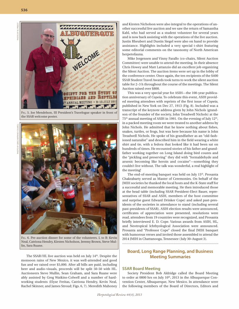
The SSAR/HL live auction was held on July 14th. Despite the monsoon rains of New Mexico, it was well-attended and good fun and we raised over $5,000. After all bills are paid, including beer and audio-visuals, proceeds will be split 50-50 with HL. Auctioneers Steve Mullin, Sean Graham, and Sara Ruane were ably assisted by Greg Watkins-Colwell and a number of hard-working students (Elyse Freitas, Catriona Hendry, Kevin Neal, Rachel Skinner, and James Stroud; Figs. 6, 7). Meredith Mahoney
and Kirsten Nicholson were also integral to the operations of an-other successful live auction and we saw the return of Samantha Kahl, who had served as a student volunteer for several years and is now back assisting with the operations of the live auction. Justin Rheubert and Dustin Siegel were also on hand to provide assistance. Highlights included a very special t-shirt featuring some editorial comments on the taxonomy of North American herpetofauna.
Mike Jorgensen and Vinny Farallo (co-chairs, Silent Auction Committee) were unable to attend the meeting. In their absence Chris Howey and Matt Lattanzio did an excellent job organizing the Silent Auction. The auction items were set up in the lobby of the conference center. Once again, the ten recipients of the $400 SSAR Student Travel Awards took turns to work the silent auction table for 2–3 h throughout the course of the meetings. The Silent Auction raised over $800.
This was a very special year for ASIH—the 100-year publica-tion anniversary of Copeia. To celebrate this event, ASIH provid-ed meeting attendees with reprints of the first issue of Copeia, published in New York on Dec 27, 1913 (Fig. 8). Included was a transcript of the keynote address given by John Nichols (grand-son of the founder of the society, John Treadwell Nichols) at the 75th annual meeting of ASIH in 1991. On the evening of July 12th, in a packed meeting room we were treated to another address by John Nichols. He admitted that he knew nothing about fishes, snakes, turtles, or frogs, but was here because his name is John Treadwell Nichols. He spoke of his grandfather as an “old-fash-ioned naturalist” and described him in the field wearing a white shirt and tie, with a fedora that looked like it had been sat on hundreds of times. He recounted stories of his father and grand-father working together on Long Island doing bird counts and the “pickling and preserving” they did with “formaldehyde and arsenic becoming like heroin and cocaine”—something they couldn’t live without. The talk was wonderful, a real highlight of the meeting! The end-of-meeting banquet was held on July 15th. Prosanta Chakrabarty served as Master of Ceremonies. On behalf of the JMIH societies he thanked the local hosts and the K-State staff for a successful and memorable meeting. He then introduced those at the head table (including SSAR President-Elect Bauer, repre-sentatives of SSAR and ASIH, members of the host committee and surprise guest Edward Drinker Cope) and asked past-pres-idents of the societies in attendance to stand (including several past-presidents of SSAR). ASIH election results were announced, certificates of appreciation were presented, resolutions were read, attendees from 19 countries were recognized, and Prosanta briefly interviewed E. D. Cope. Various awards from ASIH, HL, and Neotropical Ichthyological Association were announced. Prosanta and “Professor Cope” closed the final JMIH banquet with humorous verses and invited those assembled to attend the 2014 JMIH in Chattanooga, Tennessee (July 30–August 3).
Board, Long Range Planning, and Business Meeting Summaries
SSAR Board MeetingSociety President Bob Aldridge called the Board Meeting
to order at 0800 hrs on July 10th, 2013 in the Albuquerque Con-vention Center, Albuquerque, New Mexico. In attendance were the following members of the Board of Directors, Editors and
Fig. 5. Joe Mendelson, III President’s Travelogue speaker in front of the SSAR welcome poster.
PHO
TO B
Y M
. PRE
EST
Fig. 6. Pre-auction dinner for some of the volunteers. L to R: Kevin Neal, Catriona Hendry, Kirsten Nicholson, Jeremy Brown, Steve Mul-lin, Sara Ruane.
PHO
TO B
Y G
REG
WAT
KIN
S-CO
LWEL
L

Herpetological Review 44(4), 2013
537
Committee Chairs: Kraig Adler (Editor, Contributions to Herpe-tology; Chair, Long Range Planning Committee), Bob Aldridge (President), Breck Bartholomew (Publications Secretary), Aaron Bauer (President-Elect; Editor, Facsimile Reprints in Herpetol-ogy), Rafe Brown (Chair, Seibert Award Committee; Chair, Local Meeting Committee, 2015), Alison Cree (Board Member, non-US 2016), Brian Crother (Chair, Standard English and Scien-tific Names Committee), Raul Diaz (Webmaster), Tiffany Doan (Board Member, Reg. 2014), Richard Durtsche (Coordinator, Symposium Committee), Marina Gerson (co-Chair Membership Committee), Robert Hansen (Editor, Herpetological Review), Travis LaDuc (Board Member, at-large 2014; co-Editor CAAR), Lisa Hazard (Board Member, Reg. 2016), Joseph Mendelson, III (Immediate Past-President), Kirsten Nicholson (Past-Treasur-er), Ann Paterson (Treasurer; co-Chair, Membership Commit-tee), Gad Perry (co-Editor, Journal of Herpetology), Marion Pre-est (Secretary), Al Savitzky (SSAR Representative to AIBS and BioOne), Greg Watkins-Colwell (Board Member, Reg, 2016; Orga-nizer, SSAR/HL Live Auction), and Dawn Wilson (Chair, Student Participation Committee). Additional society members present included Jim Christensen, Vic Hutchison, Samantha Kahl, Henry Mushinsky, Justin Rheubert, and Tony Wilmes.
Introductions were made and those present were reminded of the open meeting regarding the activities of the Long Range Planning Committee to be held on July 13th. Minutes of the 2012 Board of Directors Meeting (Vancouver, British Columbia, Can-ada) were approved.
Officers’ ReportsPresident Bob Aldridge reported that he and Past-President
Joe Mendelson sent a letter to 492 lapsed SSAR members in Dec 2012 encouraging them to renew their membership. Bob plans to resend a letter in December of this year. He signed a letter written by the members of the Conservation Committee to the Honorable Puerto Rico Planning Board encouraging them to dis-allow any development activities that could even remotely lead to the loss, or “incidental take” of individuals of the threatened Puerto Rican Crested Toad (Peltophryne lemur). Additionally he wrote “An Open Letter to the WikiHerps Community from the Officers and Editors of SSAR,” stating that it is a copyright viola-tion to post PDF versions of Herp. Review. Bob thanks Kraig Adler for composing the initial draft of the letter and Bob Hansen and Breck Bartholomew for advice. The letter was posted by Joe Men-delson on April 10, 2013.
In the June issue of Herp. Review, an open letter from Presi-dent Aldridge was published in which he introduced himself and stressed the importance of membership in our Society. President Aldridge signed an “Invitation to Membership” letter that was distributed along with an issue of Herp. Review to attendees at the 1st North Carolina Congress of Herpetology, held at the North Carolina Zoo, Asheboro, NC. SSAR was a sponsor of this meeting and also donated books for their auction.
President Aldridge reappointed Kraig Adler Chair of the Long Range Planning Committee and invited Past-President Mendel-son to present the 2013 Travelogue at the JMIH. Bob met with the JMIH MMPC in Albuquerque in April to plan the 2013 JMIH. The MMPC arranged times and places for business meetings, sym-posia, oral presentations, social events, etc.
Immediate Past-President Mendelson reported a quiet final few months in his Presidency. He wrote a final Presidential es-say that was published in Herp. Review that focused on outreach education. Joe solicited Carol Spencer to lead the SSAR Website
Fig. 8. First page of article by H. W. Fowler (Philadelphia, PA) in Issue 1 of Copeia (1913).
Fig. 7. Rachel Skinner and Kevin Neal preparing for the live auction.
PHO
TO B
Y G
REG
WAT
KIN
S-CO
LWEL
L

Herpetological Review 44(4), 2013
538
Committee and was involved in Travis LaDuc and Chris Bell tak-ing over as co-Editors of CAAR. He signed the online publishing agreement with Allen Press that was approved by the Board in 2012 and a contract with EBSCO to expand electronic publishing with them to include Herp. Review.
Ann Paterson and Kirsten Nicholson (the Current- and Past-Treasurers respectively) reported that SSAR experienced another sound financial year during 2012 and say that the future is look-ing bright in the recovering economy. Overall, SSAR underspent the 2012 budget (by $42,516) which is good news given recent changes to journal formats. However, some points of discussion are the transition to stricter policies regarding notification/pay-ment of page charges, and a possible increased push towards more active acquisition of paid advertisements in Herpetologi-cal Review. Membership numbers are down slightly, but hardly noticeable. Royalty income has remained fairly steady, which is somewhat unexpected, as we had been predicting a steady in-crease in payments. We also received several generous donations for special projects, in particular from Ronald Javitch and George Zug, and several others in honor of Andy Price. Our investment portfolio enjoyed substantial growth during 2012 (up $85,000 from the previous year), and 2013 is showing continued growth as well.
The Society’s investments increased during 2012, in keep-ing with trends in the global market. The total market value as of December 31st, 2012 was $645,096. By June, 2013 we had in-creased further to $666,022. Examination of the performance of our funds shows that their percent yields have increased over 2012 and 2013.
The transition to our new Treasurer, Ann Paterson, began early in 2013 and Kirsten has been assisting greatly with this change. Ann is working on changing some investments and is trying to get our taxes finalized earlier in the year.
The approved 2012 budget was balanced. Although some expenses went over budget, most were under budget. CAAR was nearly double the expected cost, but was catching up with several accounts. The appearance of the Publications Secretary (Breck Bartholomew) being over budget is erroneous because that office has taken over the handling of memberships from Al-len Press, so what was a separate expense paid out to Allen Press is now subsumed within Breck’s expenses. The 2011 JMIH meet-ing was well over budget and SSAR was hit with a very large fee as a consequence. Last year’s meeting fees however were paid for by the WCH and we were spared that expense. Both JHerp and Herp. Review were under budget. There was no return on the Auctions again this year (2012), as we voted to donate the money to the WCH. At the 2013 meetings, Kirsten and Ann hope to dis-cuss details and problems regarding the efficient running of the auctions, as the cost of running them severely cuts into making profits to fund student travel awards.
Joe Mendelson commented on SSAR being under budget and asked whether we needed to spend more money. As a not-for-profit organization, there are IRS regulations in regards to this. Breck Bartholomew noted that many of the funds raised in the past year are due to one-off donations and that SSAR can’t count on receiving them in the future.
Secretary Marion Preest summarized the 2012 World Con-gress of Herpetology for publication in Herp. Review. She kept track of changes in personnel and regularly updated SSAR let-terhead, informed the Editors of JHerp. and Herp. Review of these changes, and provided various updates to Webmaster Raul Diaz. She wrote letters to student winners of awards (e.g., Kennedy,
GIH, etc) and prepared announcements for publication in Herp. Review. The Secretary compiled the 2013 Annual Report and prepared agendas for the Board and Business Meetings for the 2013 JMIH. She was again involved in helping to organize a re-ception for student members of SSAR and HL to be held at the JMIH. Marion corresponded with the Board regarding various issues that needed a vote, e.g., a proposal regarding remainder-ing some SSAR publications, symposium proposals, etc. She circulated documents that needed to be signed by members of the Board (e.g., Conflict of Interest), confirmed eligibility for par-ticipation in various student activities (e.g., Travel Awards, Poster Awards, Seibert Awards, etc.), and dealt with many emails from SSAR members and the general public.
Breck Bartholomew reported that total income from the Pub-lications Office in 2012 was $124,714.01. He is requesting a bud-get for 2014 that includes funds to cover rent, utilities, insurance etc. for the office, as Breck is closing down his business Biblio-mania (which has, for many years, covered these costs for the Society). Breck has contacted book dealers and authors regard-ing remaindering books to try and deal with excess inventory. Breck suggested that we think of other publication avenues (e.g., iTunes, Ebooks, etc.)
George Pisani reported that he again filed SSAR’s annual cor-porate report with the Office of the Kansas Secretary of State.
Editors’ ReportsCo-Editors of the Catalogue of American Amphibians and
Reptiles, Chris Bell and Travis LaDuc took over the reins of CAAR at the beginning of 2013. They wish to thank Bob Powell for com-pleting and publishing the last of the accounts that were sub-mitted to Andy Price. Chris and Travis assumed all of the edito-rial responsibilities in an effort to maintain the high standards of the series, particularly in the formatting and completeness of the literature citations, personally vetting every citation just as Andy Price and Bob Powell did before them. They have revised the CAAR instructions to authors and, as of May 31, 2013, have received six account submissions and corresponded with several potential authors on an additional 10–15 accounts. They estab-lished a new “dibs” list for taxa reserved by authors.
During the transition period, there were questions about the suitability and logistics behind revising previously published spe-cies accounts. Chris and Travis found precedence for this in the history of the series. Five accounts were revised over the 50-year history of the series, and each revision was published under a new account number, with no specific notations indicating the previous CAAR account number and no secondary authorship granted to the author(s) of a previous account. The online system makes it easy to cross-link the new account to the previous one.
Publication of the series is now electronic, and accounts are issued immediately upon acceptance and may be downloaded on demand from the CAAR website. The co-Editors hope to bring the number of annually published accounts to a minimum of 15. Because publishing and all correspondence are now elec-tronic, costs for editing the series are extremely low. Chris and Travis wish to thank Breck Bartholomew who was instrumental in scanning and posting all previous accounts to the web, where they are now available to members (http://www.zenscientist.com/index.php/filedrawer/Open-Access-Journals/caar/).
The lack of an ISSN number for the CAAR series has now been rectified. Regulations require separate ISSN for the print and online versions. They are ISSN 2325-4882 (print) and ISSN 2325-5021 (online).

Herpetological Review 44(4), 2013
539
Contributions to Herpetology Editor Kraig Adler reported that “Contributions to the History of Herpetology,” vol. 3, by Kraig Adler, John S. Applegarth, and Ronald Altig was published in 2012. A paper-bound version was presented to all delegates at the WCH. Costs were subsidized with a $5,000 donation plus a further $10,000 for the other congress giveaways. “A Contribution to the Herpetology of Northern Pakistan,” by Rafaqat Masroor was also published in 2012. “Contributions to the History of Herpetology,” vol. 1, by Kraig Adler, John S. Applegarth, and Ronald Altig will be reprinted (with annotations and corrections to volumes 1, 2, and 3) in 2014 (making all three volumes available at the same time). “Herpetology at the University of Kansas,” by William E. Duellman, will be issued in 2015 prior to SSAR’s meeting at KU. This is a de-tailed history of one of the most distinguished herpetological re-search and graduate education programs, beginning with Edward H. Taylor, the first curator, and concluding with the current era of herpetologists. It will include a description of Henry Fitch’s long-term program at the Natural History Reservation, biographies of all herpetological graduates of the KU program, numerous photo-graphs, and summaries of major expeditions, meetings, and other activities. “A Guide to the Snakes of the Philippines” by Rafe Brown, Alan Leviton, Maren Gaulke, and Arvin Diesmos, “Field Guide to Amphibians and Reptiles of the West Indies” by S. Blair Hedges, and “Lizards of Southern Africa” edited by William R. Branch and Aaron Bauer are also planned beyond 2013.
Editor Aaron Bauer (Facsimile Reprints in Herpetology) re-ported that the monumental “Erpétologie Générale ou Histoire Naturelle Complète des Reptiles” (1834–1854) by Duméril, Bi-bron & Duméril was published in December 2012 with Kraig Adler as guest editor. This work of 7000+ pages, including 240 plates, significant new material by Roger Bour, and a compre-hensive index, was produced in a run of 400 sets. The cost of this work was generously supported by Ronald Javitch, and SSAR funding was spread out over a two-year period. Two works nearing completion have been offered to the facsimile series. Both are collected works with substantial new material. “The Collected Herpetological Works of Giorgio Jan” has an introduc-tion, annotated bibliography, and extensive systematic com-ments by Roy McDiarmid and Jay Savage. The collected snake publications of Jan were published in 1857–1882 in French, Italian, and German. The plan is to publish the facsimiles four pages to one page in order to accommodate 500+ pages of new material by McDiarmid and Savage. “The Collected Herpeto-logical Works of J. V. Barboza du Bocage (1823–1907)” has an in-troduction, annotated bibliography, and data on types by Luis Ceríaco. Bocage was the most important Portuguese vertebrate zoologist of the 19th Century and published extensively on all groups, mostly based on collections from the far-flung Portu-guese colonies of the day (Angola, Mozambique, Cape Verde, Timor, Guinea, São Tomé, Macau). He also published on Por-tuguese mainland taxa and other areas, notably New Caledo-nia. In all, he authored 69 herpetological papers between 1863 and 1905, nearly all in Portuguese or French. Most are relatively short (<20 pages), but his magnum opus: Herpétologie d’Angola et du Congo (1895), was 223 pages long, with 19 plates. The to-tal pagination of Bocage’s herpetological works is 759 pp. The facsimile will include approximately 100 pages of new material dealing with Bocage’s life and works, his complete bibliogra-phy, and short accounts for each of the 104 species of amphib-ians and reptiles he described. Ceríaco anticipates an interest by the Portuguese Academy of Sciences in supporting the work through co-publishing.
John Moriarty (Editor, Herpetological Circulars) reported that Herpetological Circulars 41 (A Guide to Tissue Collection, Preser-vation, and Management for Reptiles and Amphibians) is in final review and is expected to be published in November 2013.
Editor of Herpetological Conservation, Joe Mitchell, reported that no inquiries for publication were received in 2012 and he plans to advertise for submissions soon. His criteria are that the topic be broadly applicable to herp conservation and manage-ment, that the geographic coverage not be limited to one coun-try but, preferentially, global in scope or at least regional, and the work makes an important contribution that will be cited ex-tensively. The Board discussed the fact that Herp. Conservation volumes don’t need to be, and in fact haven’t always been, large tomes and encourages Joe to think of smaller projects (in addi-tion to large ones).
Robert Hansen, editor of Herpetological Review, reports con-tinued smooth operation. Volume 43 consisted of 632 pages and Volume 44 is projected to run ~700 pages. Personnel changes include the resignations of Brad Lock, Michael Grace, and Em-ily Taylor and the addition of Robert Hill, Scott Boback, David Blackburn, Stephen Richards, and David Steen. Over two years ago, Denise Wilson Mayer, a volunteer at Zoo Atlanta, undertook the daunting task of indexing all back issues of Herp. Review. She made excellent progress before moving on to other pursuits. Meanwhile, Ruthe Smith, associated with the SSAR Publications Office as a part-time employee, has been indexing each new is-sue. To date, all issues of Herp. Review have been indexed, with the master file currently in EndNote format maintained by Breck Bartholomew. The next step is to make this index file publicly available through the SSAR web page. The Board returned to the issue of Herp. Review being made available through BioOne (part of the impetus for indexing the journal). Hansen will contact BioOne to see where we stand.
Co-Editors of Journal of Herpetology, Erin Muths and Gad Perry, report that former Editor Geoff Smith remains an Associ-ate Editor (AE) for special projects and Paul Andreadis remains the Index Editor. The AE roster at the end of 2012 contained six new names and many continuing ones: Paul Bartelt (US), Phil Bishop (New Zealand), Xavier Bonnet (France), Rafe Brown (US), Tracey Brown (new, US), Russ Burke (US), Steve Corn (US), Tif-fany Doan (US), Jennifer Gillette (New Zealand), Evan Grant (new, US), Brian Greene (US), James Harris (Portugal), Tibor Hartel (new, Romania), Toby Hibbitts (US), Hinrich Kaiser (new, US), Nancy Karraker (newly in the US), Edgar Lehr (US), Marc Mazerolle (Canada), Frank Mazzotti (US), Don Miles (US), John Rowe (US), Christopher Salice (new, US), Glenn Shea (new, Aus-tralia), Stephen Tilley (US), Tony Tucker (new, Australia), James Watling (US), and Eric Wild (Brazil); both Editors also function as AEs. Ximena Bernal (US), Nancy FitzSimmons (Australia), Wal-ter Meshaka (US), and Phil Smith (US) ended their tenure as AEs in 2012. Expanding the AE base, especially by adding additional colleagues from other countries, remains a challenge. Erin and Gad are especially interested in expanding representation from Brazil and Asia.
Overall, new submissions declined slightly in 2012. Manu-scripts from 35 countries were submitted in 2012. As in previous years, the US remains the leading source of manuscripts (~43% of all submissions) and Brazil remains the second highest source (~14% of submissions). Despite availability of the free editing service from SSAR, language remains a concern for many manu-scripts from non-English-speaking countries. The larger number of such manuscripts places an added burden on the editorial

Herpetological Review 44(4), 2013
540
process, particularly at the final editing stages. According to the AP “Member Submissions Report,” 35% of all submissions in 2012 and 2011 had one or more authors who are SSAR members, compared to 35% in 2010, 38% in 2009, 39% in 2008, and 38% in 2007 and 2006. Submissions so far for 2013 (117 as of June 15th) are lower than the number submitted during the same period in 2012 (172). The amount of time needed to reach initial and final decisions remains similar or is slightly improved compared to the past few years, but the wait between acceptance of a pa-per and publication remained approximately 12 months in 2012. Gad and Erin have taken several steps to address this, includ-ing instructing AEs to reject a greater proportion of submissions and publishing an especially large December issue in 2012 and March issue in 2013. These measures have helped us catch up with the buildup of manuscripts and reduce the wait from ac-ceptance to publication to approximately 9 months.
The Journal now has an official website (http://journalof-herpetology.org), maintained by Allen Press. This site offers ac-cess to information needed by authors, articles published in the journal (free to SSAR members), pre-publication versions of up-coming papers, and links to SSAR. The site also allows us to post “online only” supporting material such as video, sound files, and raw data.
Another recent change is associated with new technology at AP, allowing us to publish all-color at no extra cost. As currently published articles were accepted before this change, the transi-tion to more color will be ongoing. However, authors are being contacted and offered an opportunity to replace b&w figures with color ones.
The co-Editors have used Editorial Board members this year for several quick turnaround reviews and in addressing an issue of plagiarism. This issue was identified by a reviewer, and report-ed by the AE to the Editors. They checked the data and used the expertise of the Editorial Board to vet their response to the au-thors. After ample opportunity to rebut and defend their manu-script, the paper was rejected and all authors were informed that manuscripts by them would not be accepted by the Journal of Herpetology for two years. The information was also shared with the editors of other herpetological journals, as part of a multi-society effort to reduce ethical problems within the profession.
The new page charge policy is now in place: If at least one author is an SSAR member in good standing, page charges are not levied. However, if funding allows, members are encouraged to assist in the production of the journal by contributing to page charges. Page charges for non-members are $150 per page. If there are unusual circumstances, a waiver may be requested by contacting Breck Bartholomew.
Obtaining qualified AEs and reviewers remains a huge chal-lenge and Erin and Gad expect this problem to get worse, given the increasing shift of academic institutions to a for-profit mana-gerial style.
The co-Editors raised four issues for the Board to consider:• Replacement of Editors.—Erin and Gad are asking to con-
clude their tenure as co-Editors for the Journal of Herpetology on January 1, 2014, following four years of service. They pro-pose to remain actively involved in the publication of the jour-nal through July 2014 to facilitate transition to a new Editor or co-Editors. They will both serve on the Editorial Board and Erin Muths will remain as an AE.
• Electronic publishing.—Establishment of the new journal’s website is an important development and Erin and Gad are pleased that the Society is looking at professionalizing its Web
presence. They believe that there is a need for careful consid-eration of the not-far future move to an all-digital journal, and to funding models that would support the Journal and society when that happens.
• Co-publishing proposal.—In 2011, the Board rejected the AP co-publishing proposal. Erin and Gad feel that this option should be reconsidered in 2014 after the new editor(s) meet with Allen Press for updated details.
• Reward for AE service.—The co-Editors would appreciate the Board reconsidering membership as token recognition to be granted only to those AEs who have served for over a year and completed their reviews in a timely manner.
In their requested budget for 2014 ($128,150), the co-editors have added the cost for maintenance of the Journal website by AP. They expect to publish a similar number of papers in 2014 as in 2013, and anticipate an increase in mailing costs, but expect no change in editorial office costs.
Standing Committee Chair ReportsThe Conservation Committee consists of Ross Alford, April
Barreca, Jennifer Germano, John Jensen, Nancy Karraker, Karen Lips, Gad Perry, Stephen Richter, and Betsie Rothermel (Chair). Two new graduate student members (Nicole Angeli and Cecilia Langhorne) joined the committee in 2012. In July 2012, the Cen-ter for Biological Diversity petitioned U.S. Fish & Wildlife Service (FWS) to list 53 species of reptiles and amphibians under the En-dangered Species Act. The consensus of the Conservation Com-mittee was to not take any action at that time, but rather to wait and submit comments on particular species if/when there is a full status review by FWS. In August 2012, SSAR signed on to a let-ter sent to the U.S. Senate Interior and Environment Appropria-tions Subcommittee opposing drastic cuts in FY13 funding for the State and Tribal Wildlife Grants Program. In April 2013, they sent a letter to the Southeast region of U.S. Fish & Wildlife Service regarding habitat protection for the Puerto Rican Crested Toad (Peltophryne lemur). Specifically, they urged the agency not to add this species to the list of species covered under an incidental take permit for a proposed wind energy facility.
Joe Beatty, Chair of the Dean Metter Award Committee, re-ceived 39 proposals this year and Mitch Tucker, a Ph.D. student at University of Missouri, was selected as the winner. Mitch’s pro-posal was entitled, ”Behavioral consequences of polyploidy in Gray Treefrogs, Hyla chrysoscelis.” A key component of the pro-posal hinges upon being able to generate autopolyploid frogs in the lab, which Tucker is able to do with a “cold-shock” treatment developed by him and his supervisor, Carl Gerhardt. The com-mittee thought Tucker’s proposal was especially innovative and experimental and takes an approach involving fieldwork as well as laboratory work to provide insight to a particularly interesting biological system.
The 21st annual Seibert Awards competition (Rafe Brown, Chair) was run at the 55th Annual Meeting of SSAR in Vancouver. There were 53 eligible presentations. The Seibert Award winners for 2012 were: Systematics/Evolution: Sara Ruane (College of Staten Island), “Speciation in the milksnake (Lampropeltis tri-angulum).” Ecology: Thomas Luhring (University of Missouri), “Islands in the sun: nutrient cycling in isolated systems is medi-ated by canopy cover, predation, and the complex life-histories of their transient tenants.” Physiology/Morphology: Rory Telem-eco (Iowa State University), “Effects of temperature during de-velopment on the offspring phenotype of a facultative thermo-regulator, the Southern Alligator Lizard (Elgaria multicarinata:

Herpetological Review 44(4), 2013
541
Anguidae).” Conservation: Shawn McCracken (Texas State Uni-versity, San Marcos), “Living on the edge: oil road effects on the occupancy and abundance of anurans inhabiting an upper canopy tank bromeliad (Aechmea zebrina) in lowland rainforest of the Yasuni Biosphere Reserve, Amazonian Ecuador.” All win-ners received a check for US $200 and a book from University of California Press. Honorable mentions were: Systematics/Evolu-tion: Hilton Oyamaguchi (University of California, Los Angeles), “Divergence along a Brazilian rainforest-savanna gradient and the role in generating diversity.” Ecology: Julia Riley (Laurentian University), “Should I stay or should I go? Influence of environ-mental factors on Chrysemys picta hatchling overwintering strat-egy.” Physiology/Morphology: Gareth Hopkins (Utah State Uni-versity) “Embryonic survival in salt among Rough-skinned Newt (Taricha granulosa) families.”
No report was received from the Herpetology Education Committee (Hinrich Kaiser, Chair).
The winning paper for the 2012 Kennedy Award (Lynnette Sievert, Committee Chair) is by Alexa K. Fritzsche (coauthored with Stacey L. Weiss), “Effect of signaler body size on the re-sponse of male striped plateau lizards. Journal of Herpetology 46:79–84.” Alexa will receive a check for $200 or $400 equivalent in SSAR publications.
Kraig Adler, Chair of the Long Range Planning Committee (LRPC), reported that Carol Spencer (newly appointed Chair of the Website Committee) has been added to the LRPC. Follow-ing a very active year after being revived in late 2010, the com-mittee as a whole was quiescent for much of 2012 and 2013. Kraig however was busy coordinating SSAR’s contributions to the World Congress in Vancouver, e.g., a giveaway poster fea-turing an original drawing by David Dennis, a giveaway issue of Herp. Review, a membership brochure, a giveaway book and two booklets (“Contributions to the History of Herpetology, Vol. 3” by K. Adler, J. S. Applegarth, and R. Altig; and two booklets on the scientific and standard vernacular names of amphibians and reptiles in North America north of Mexico, in separate English [B. Crother, editor] and French [D. M. Green, editor] versions), etc. Kraig wishes to particularly thank Breck Bartholomew, Bob Han-sen, John Moriarty, and Kirsten Nicholson for their assistance. SSAR was recognized by WCH as the sole Gold-Level Sponsor for our many contributions. Kraig was also involved with Rafe Brown and others in the KU local committee with early planning for the 2015 meeting. He helped them prepare the first ad for the meeting, which appeared in the December 2012 Herp. Review, and approached PARC (Partners in Amphibian and Reptile Con-servation) to join us at KU as our co-sponsor.
Chair of the Meeting Management and Planning Commit-tee, Rafe Brown, presented information about progress to date in planning for the 2015 meeting at University of Kansas. The local committee consists of Rafe Brown (Chair), Richard Glor, William Duellman, Linda Trueb, and George Pisani. Organizing societ-ies are SSAR, PARC, and Kansas Herpetological Society. Tenta-tive dates are July 29th to Aug 3rd (includes a ”zero” day at the beginning for the SSAR Board Meeting). There will likely be two plenary lectures, a number of symposia, a student workshop, a live animal display, and tours of the Museum and the Henry Fitch Natural History Reservation. Social events will include those we have come to expect at JMIH, some new events, and other old favorites that have been revived, e.g., the President’s Travelogue, the student reception, a social specifically to recog-nize senior herpetologists, the revised (now digital) audiovisual shows of David Dennis and Eric Juterbock (“Amphibians of the
Appalachians,” “Herps of the American West,” “Herpetologists Then and Now”), live and silent auctions, a herp art exhibit, the herp quiz, a picnic, etc. Projected prices for registration and ac-commodation are very reasonable (Registration: $280 regular members, $140 students and retired members, Accommodation: $34–$150/night)
In the past year the Membership Committee (Ann Paterson and Marina Gerson, co-Chairs) was less active than it has been previously while it was waiting to see the results of the 2011 pro-posals. The Committee has been more closely following mem-bership data to examine patterns and develop procedures for more closely monitoring and analyzing these patterns, develop-ing proposals for possible correspondence with new members and with members who fail to renew, continuing to compile a list of other herpetological societies, and responding to concerns and comments from members and non-members as needed. The detailed proposals for possible correspondence with mem-bers are available upon request. Ann Paterson will be discussing them with Bob Aldridge to coordinate with other activities.
Ann and Marina now receive monthly membership data. They remarked on the continued decrease in membership. They noted that more people are taking advantage of the online and senior membership categories, but that there are declines in oth-er categories. The recent dues increase explains the increase in membership income and possibly also the decrease in member-ship numbers. It is hoped that access to membership data will allow the committee to examine the effect of various activities (e.g., mailings) on membership trends. Breck commented that the membership office sees an increase in renewals and in new memberships around the time Roger Conant GIH proposals, travel award applications, etc., are due.
The Board discussed the idea of making student dues avail-able to individuals for 1–2 years after they are no longer students as a way of encouraging continued membership. This would not require creating another category of membership (of which we already have many) and would need to be advertised. As there is every year, there was discussion of why someone should become a member of SSAR in the first place and why they should renew their membership annually. There is concern over the “revolving door” of members, i.e., individuals (usually students) are mem-bers for a few years and then fail to renew, only to be replaced equally temporarily by other young members. The importance of reaching out to undergraduates was emphasized. Someone remarked that there are professional herpetologists who them-selves are not members of the society even though they come to the meetings, send their students to the meetings, publish in our journals, etc. The importance of professionals setting examples for their students and encouraging, or even requiring, them to become members and attend meetings was stressed. A comment was made that we need to focus on our core mission and current demographic and make the Society work in whatever form it ex-ists, i.e., we can’t force people to become members. Along the same lines, Al Savitzky asked us to think about whether we are currently operating under a model that is not sustainable in the long term.
Kris Kaiser (Chair, Mentorship Committee) reported that, in its first year, the Mentorship Program paired 14 students with mentors. Follow-up surveys showed a general satisfaction with the program, and gave useful feedback for year two of the program. The program this year was advertised in Herp. Re-view, on the SSAR website, and on the JMIH registration page under “Graduate Student Information,” and applications were

Herpetological Review 44(4), 2013
542
accepted for both mentors and mentees. The committee is con-sidering working with the MMPC to make this part of the actual meeting registration next year to increase visibility.
The Nominations Committee (Greg Watkins-Colwell, Chair) compiled a list of nominees for the positions of President-Elect, Treasurer, Regular Board Member (three needed), and a non-US Board Member. Marion Preest was willing to run again for the position of Secretary. Voting was carried out electronically with photographs of the candidates being made available. Upon com-pletion of the 2012 election, Greg Watkins-Colwell resigned his post as Nominations Chair, to be replaced by Robert Espinoza. The nominees:
President-Elect.—Aaron Bauer, Geoff SmithTreasurer.—Ann PatersonSecretary.—Marion PreestBoard Member (Regular).—Frank Burbrink, Richard Durtsche, Marina Gerson, Lisa Hazard, Greg Watkins-ColwellBoard Member (non-US).—Alison Cree, Carlos Navas, Hi-detoshi Ota, Wolfgang Wüster
Rob Denton (Resolutions Chair) presented resolutions at the SSAR Business Meeting in Vancouver in 2012. These have been published in Herp. Review.
Josh Kapfer (Chair, Roger Conant Grants-in-Herpetology Committee) received 80 proposals. Most applications were in the “Laboratory Research” category. The winners each received US $500 and they are:
Conservation.—Todd Pierson, University of GeorgiaEducation.—Tiffany Vanderwerf, Buffalo ZooThe Andrew H. Price Field Research Grant in Herpetology.— Mark Oliva, California State University-NorthridgeLaboratory Research.—Schyler Nunziata, University of Ken-tuckyTravel.—Justin Lawrence, The University of MississippiInternational.—Jessica Hacking, Flinders University, Australia
Josh reported that, as approved by the Board in 2012, he now requests that a signed waiver of liability (available on the GIH website) be included with all grant applications. Josh contin-ues to have difficulties with reviewers submitting assessments on time. As approved by the Board, the Field Research category has been renamed the Andrew H. Price Field Research Grant in Herpetology. It will continue with this name for as long as funds permit.
Brian Crother, Chair, Standard English and Scientific Names Committee, reported that the 7th edition was released last year at the WCH, and work is yet to start on the next edition. The 7th edi-tion has not yet been posted on the SSAR website, but the CNAH and the Reptile Database sites have the list up. The list is also be-ing used by the iNaturalist site and the names are being used for the development of a Global BioBlitz.
Erin Muths was the lead organizer of a lunchtime student workshop sponsored by SSAR and HL at the WCH in Vancou-ver. The workshop was titled “How to Turn Your Thesis or Dis-sertation Chapter into a Publishable Manuscript & Participate in the Peer-Review Process.” Editors or Associate Editors from six herpetological journals from the U.S. and Europe participated in the panel presentation. The workshop and panel discussion was designed for graduate students interested in turning their graduate work into publishable manuscripts and learning about the publication process and details on incorporating peer review into their professional career. The goals of the workshop were
to provide an understanding of the mechanics of submitting a manuscript and successfully seeing it through the publication process. Topics included transforming the thesis into a manu-script, de-mystifying the review process, interpreting reviews, responding to reviewers, and participating in the peer review process.
Over 80 students attended, the question and answer session was useful, and feedback during the rest of the WCH was posi-tive. Discussion by the editors after the workshop included de-cisions to post the PowerPoint presentation on the websites of HL and SSAR and the European societies (this is pending) and to consider presenting the workshop every 3–4 years. Dawn Wilson (Chair, Student Participation Committee) indicated that she will ask for ideas about future workshops from students. A sugges-tion was made that we record and upload the workshops on the new SSAR website.
The second annual SSAR Student Poster Awards (Tiffany Doan, Chair) were presented during the Annual Meeting of the SSAR at the WCH in 2012. There were 40 eligible posters, twice the number of the inaugural year. In recognition of outstanding student poster presentations at the annual meeting, the follow-ing awards (a check for US $100 and a book from University of California Press) were made: Evolution, Genetics, & Systematics: Luke Frishkoff, Stanford University, “Body size evolution and the colonization dynamics of a radiating Caribbean frog lineage (Eleutherodactylus).” Conservation & Management: Julia Riley, Laurentian University, To conserve and protect: evaluation of a turtle conservation tool, nest caging, ecology, natural history, distribution, & behavior; Matthew Lattanzio, Ohio University, “Habitat use, phenotypic variation, and grazing: The responses of tree lizard populations to altered landscapes.” Physiology & Morphology: Nicole Christie, Sonoma State University, “Effects of nest temperature variation on viability and sex determination of western pond turtles.”
Chuck Crumly worked for a number of years for University of California Press and generously donated UC Press books to the winners of the Seibert Awards, and more recently to winners of the poster awards. Chuck has left UC press and there was dis-cussion of where we might obtain books for paper and poster winners. We could use SSAR books, or the chairs of the Seibert and Poster Awards committees could contact book publishers to see if any are willing to donate books. A point was made that the books needed to be equally attractive to US and non-US mem-bers of SSAR, i.e., not necessarily with a strong North American focus.
In 2012, Mike Jorgensen and Vinny Farallo (Co-Chairs, Student Travel Awards Committee) received 41 applicants for the travel award, about equal to the number in previous years. The ten re-cipients of the US $400 SSAR Student Travel Award took turns to work the silent auction table for 2–3 h over the course of the WCH. Location of the auction was in a low foot-traffic area at the WCH and probably contributed to the difference in total earnings from the previous year in Minneapolis. Since the WCH, it was proposed that the silent and live auctions be coordinated by a single com-mittee in order to streamline the donations to each.
Carol Spencer (Chair), Joe Mendelson, Raul Diaz, Vincent Farallo, Michelle Koo, and Todd Pierson serve on the recently re-vived Website Subcommittee. In 2011 the Board designated US $10,000 for a one-time complete overhaul of the Society’s web-site, with US $3000 per annum for upkeep and renovations, if needed. This group has met several times in 2013 (both in person and via Skype). Three potential website designers/programmers
Herpetological Review 44(4), 2013
543
have been asked to submit bids for redesigning the SSAR website using content and renovation suggestions from the subcommit-tee and the LRPC. The subcommittee welcomes feedback and suggestions from any member of SSAR.
Coordinators’ ReportsElection Officer, Dan Noble, reported that most ballots were
submitted electronically in 2012 and the following individuals were elected:
President-Elect – Aaron BauerTreasurer – Ann PatersonSecretary – Marion PreestBoard Member (Regular) – Frank Burbrink, Lisa Hazard, Greg Watkins- ColwellBoard Member (non-US) – Alison Cree
The Live Auction Committee coordinator is Greg Watkins-Colwell, who reported that the proceeds from the 2012 SSAR/HL Live Auction ($4072) benefited the WCH. Efforts were made to include as many different auctioneers (with different auctioning styles) as possible and to rely upon the help of students who had not assisted with the auction in the past. Following the auction, Kirsten Nicholson, Meredith Mahoney, and Gregory Watkins-Colwell immediately began discussions of how to improve upon the live auction model.
Representative to AIBS and BioOne, Al Savitzky, reported that BioOne, a nonprofit electronic publishing consortium, contin-ues to provide online publishing services for the society and to return both royalties and profit-sharing proceeds to its member publishers. Al has moved away from the mid-Atlantic region, and no longer attends the annual BioOne Publishers and Part-ners Meeting. He suggested that the society may wish to identify someone closer to Washington, DC, to serve as its representa-tive to BioOne, although, given that most of the presentations at those meetings are archived on the BioOne website, this may not be a major concern to the society.
According to a 2012 year-end summary provided in the April 2013 newsletter, BioOne ended its 11th year online with over 100,000 articles and 900,000 pages in its collection. Those papers have attracted over 18.9 million hits. These represent contribu-tions from 129 publishers, producing 171 titles. Significantly, 42% of BioOne.1 subscribers and 56% of BioOne.2 subscribers are lo-cated outside of North America. Furthermore, BioOne makes its content available free to 2500 institutions in developing coun-tries. Revenue sharing in 2012 increased by 6.3% over the pre-vious year. Presentations at the 2013 Publishers and Partners Meeting included a discussion of the changing state of academic libraries in China; a description of BioOne’s first self-published title (Elementa: Science of the Anthropocene, a fully open-access, online journal due to appear this July); an extensive presentation on citation metrics (especially the h statistic); and a presentation on alternative search strategies for online exploration of the liter-ature (including the beta version of Microsoft Academic Search).
The American Institute of Biological Sciences (AIBS) is an umbrella organization that serves about 160 member societies and organizations (MSOs), most of them in the broad areas of organismal biology and ecology. The most recent AIBS Council meeting, in December 2012, focused on the history and future of scholarly societies in the biological sciences, including a pre-sentation by organizational consultant Mary Byers. As part of its strategic planning process, the AIBS Board of Directors has reviewed two books by Harrison Coerver and Mary Byers, Race
for Relevance: 5 Radical Changes for Associations and Road to Relevance: 5 Strategies for the “New Normal.” Coerver and Byers maintain that factors such as a tighter economy, technological advances, and a generational shift in membership have left tra-ditional nonprofit professional associations struggling to sur-vive. Among the threats to their survival are the failure of organi-zations to focus on their core strengths (as opposed to constantly trying out new services); failure to confront and resolve redun-dancies among organizations or chapters; and either inertia or pressure from the “old guard” to maintain the status quo or to anticipate failure from changes “so they can advocate a return to the traditions of the past [Road to Relevance, p. 143].” Confront-ing its own decline in individual membership, AIBS has trans-formed its governance structure and focused its mission over the past few years through intensive strategic planning and in-trospection. If the threats to organizational survival that Coerver and Byers perceive are true for many scientific organizations, as they appear to be, the three North American herpetological soci-eties should be aware of these trends.
As part of its own strategic planning process, AIBS surveyed representatives of its MSOs concerning trends in their various societies and organizations, and the results have now been made available to the broader community. They received 110 respons-es from society officers, and the resulting report is available for free viewing and download at http://www.jstor.org/stable/full/10.1525/bio.2012.62.4.3. Similarly, an extensive survey was sent to over 15,000 individual biologists and a detailed report, based on over 4000 responses, is available at http://www.access.aibs.org/page/Index/?. That report includes a wealth of informa-tion on attitudes toward professional societies at different stages of individuals’ scientific careers, from undergraduates to emeriti. Careful attention to the results of that survey would better in-form decisions by the Board of our society concerning the inter-ests and priorities of our members.
AIBS continues to focus much of its service to MSOs in two areas, education and public policy. AIBS has been especially ac-tive as an advocate for organismal biology, biological collections, and ecology at the national level. AIBS has also taken a major role in development of an Implementation Plan for a Network Integrated Biocollections Alliance; supporting sound scientific integrity policies at the National Park Service and the National Science Foundation; and opposing the federal budget sequestra-tion due to its impacts on scientific funding. Individual society members are encouraged to register with the AIBS Legislative Action Center (http://capwiz.com/aibs/home/), which provides timely notices of significant policy issues and makes it easy to respond with messages to one’s senators and representatives.
This year, SSAR Symposium Coordinator Richard Durtsche received one symposium proposal: “Infrared Imaging: Cellular/Molecular Mechanisms, Behavioral Biology and Ecology of Pit Vipers, Pythons and Boas” (Michael S. Grace, lead organizer). This symposium will bring together a diverse group of biologists from a variety of fields to discuss the current state of knowledge of, and research on, infrared imaging systems and their roles in the lives of snakes. The goals also include an analysis of the next important steps in research of snake thermal biology, the ecological implications of invasions of exotic species possessing novel “weaponry,” and a look at the technological advancements that may derive from a thorough understanding of the mecha-nisms of snake infrared imaging. The symposium proposal was sent out for external review and received comments from two reviewers. The reviews and proposal were submitted to the SSAR

Herpetological Review 44(4), 2013
544
Board for consideration of sponsorship and the Board approved the proposal.
New BusinessPresident Aldridge informed the Board that President Men-
delson signed a five-year contract with K-State Conference Ser-vices in 2012. Contained in this contract is a provision allowing any of the participating societies to withdraw from a particular meeting by providing a three-year notice.
There was discussion of “WikiHerps” posting full copies of SSAR publications, e.g., copies of Herp. Review have appeared on-line within 1–2 days of issuance. The site also provides links to book-length publications. Although the Society wants to encour-age access to scholarly publications and is well aware that ease of access varies worldwide, these actions clearly represent a viola-tion of copyright and they have important implications for SSAR. Various officers and editors were involved in recent months in determining an appropriate way of dealing with this issue. On the morning of the Board meeting, President Aldridge uploaded an open letter addressed to the administrators of WikiHerps in which he outlined how the Society operates (i.e., we are a volun-teer organization that is primarily member-supported) and the potential effects of the actions of WikiHerps on our membership and our long-term survival as a Society. We were pleased that WikiHerps responded positively to this letter and the Society is in ongoing discussions with WikiHerps.
Greg Watkins-Colwell and Kirsten Nicholson presented vari-ous ideas regarding changes to the Live and Silent Auctions. We now have a single committee overseeing both auctions. This will help to ensure that items are funneled towards the appropriate event. Discussion turned to the cost of running the Live Auc-tion (this is significant and cuts into the contribution to the Stu-dent Travel Awards budget) and ways of lowering expenses, e.g., implementing a cover charge, finding a local brewery willing to donate beer or a sponsor willing to cover costs, holding the auc-tion at a nearby bar or restaurant, etc. It was emphasized that attendees needed to be reminded that, in addition to being a fun social event that can encourage membership and involvement in SSAR, the main point of the Live Auction is to raise funds to support student members.
A suggestion was received from the Membership Office that, on an annual basis, we acknowledge and publicly thank Life and Sustaining members of the Society by publishing a list of their names in Herp. Review. This was last done in 2004. The Secretary will contact Life and Sustaining members to request permission to publish their names and will ensure that this list appears an-nually.
Dick Durtsche requested that, if, in a given year, SSAR spon-sors just one symposium at the Annual Meeting, the Board con-sider allocating the entire symposium budget (currently $3,000) rather than the normal $1,500. The Board approved this request. Gad Perry suggested that participants in SSAR-sponsored sym-posia consider contributing a special section to J. Herp.
The Board then turned to the 2014 budget. A doubling of funds allocated to the Student Poster Awards and the Roger Conant Grants in Herpetology Awards, a 50% increase in funds for the Student Travel Awards, and a request from the Student Mentorship Committee to support a lunch meeting for mentors and mentees were approved. A balanced budget of $320,000 for 2014 was approved by the Board. The meeting was adjourned at 1339 h.
LRPC Meeting
An open meeting of the Long Range Planning Committee (LRPC) occurred on July 13th. LRPC Chair Kraig Adler called the meeting to order. In attendance were the following members of SSAR: Kraig Adler, Bob Aldridge, Ann Paterson, Aaron Bauer, Rafe Brown, Raul Diaz, Linda Ford, Bob Hansen, Kris Kaiser, Joe Mendelson, Gad Perry, Marion Preest, Dawn Wilson. Kraig dis-tributed an agenda and introduced members of the LRPC and other officers present. Four main topics were discussed.
SSAR Website.—Joe Mendelson and Raul Diaz (members of the Website Subcommittee) reviewed the status of the website update. The Society needs a revamped website that is easily updatable with lots of imagery and capable of webcasting. We need to avoid redundancy between the ZenScientist and SSAR websites, while still making it easy to bounce from one site to the other. Ideally officers and committee chairs would be able to update their own material (e.g., the Seibert Chair could post information about the award winners). Some training would likely be necessary. Joe and Raul recommended that we pro-vide the webmaster with a list of “trigger dates” on which he/she would contact the appropriate person for a particular up-date. A suggestion was made that portraits and short bios of “key” people in the Society be posted. There was discussion of posting the budget and IRS tax forms to make the finances of the Society as transparent as possible and also to make it clear that we are a volunteer society (i.e., none of the officers, edi-tors, committee chairs, etc., makes any salary from SSAR) and that more than half of our annual budget relies on member dues. There was also some discussion of making at least part of the website available in a language other than English (e.g., Spanish, Portuguese), and of having a non-graphics version available for those with limited internet access. Kraig request-ed that the subcommittee keep members of the LRPC updated as proposals for redesigning the website are received and as various decisions are being made.
SSAR Publications Office.—There was discussion of trying to reduce our stock of old journals and books that are no longer selling. The Society has sent some copies to various places, e.g., to individuals teaching herpetology classes accompanied by in-vitations to join SSAR. It is not clear whether this has resulted in any new members. We now order fewer extra copies of our pub-lications. This reduces our holdings, but we still need to ensure that we have sufficient extra copies to provide a full year’s worth of journals as we recruit new members. There was a suggestion that we provide some incentive to try to avoid the necessity of over-printing, e.g., if someone does not join or renew by a certain date, they would only be able to receive electronic versions of our journals.
In light of the closure of his business Bibliomania, Breck Bar-tholomew has offered to step down as Publications Secretary. The Society has received an offer from another vendor to take over. Kraig Adler reviewed the services that Breck has provided SSAR for many years. After some discussion (including mention of Breck’s generosity, loyalty, and long-standing service to the Society and the excellent job his office does of handling mem-bership issues), the LRPC voted unanimously to recommend to President Aldridge that we remain with Breck.
Herpetological Review 44(4), 2013
545545
New Member-, Publicity-, and Income-Generating Ideas.—Various ideas for increasing membership, publicity, and income were discussed. One idea was to publish anthologies of all of the Herp. Review articles about, for example, Ambystoma, or larger groups (frogs, salamanders). Bob Hansen pointed out that this would not be an easy task because, even though all issues of Herp. Review are now electronic, they are in PDF form, which makes searching them difficult. Bob also did not think these kinds of anthologies would generate much income. A sugges-tion was made that regional aggregations of articles (e.g., all of the Herp. Review articles about species in a particular US region) might be popular.
There was ongoing discussion of having Herp. Review picked up by BioOne or JSTOR. “Repackaging” Herp. Review (e.g., mov-ing the “news” section to the end or placing it on the website) may make it more attractive to these outlets. Ann, Bob Hansen, and Breck will contact BioOne to ask them about the criteria they use when deciding whether to accept a given publication.
Increasing SSAR’s Reach Overseas.—A number of suggestions regarding raising SSAR’s profile internationally were discussed. For example, the Society should be keeping track of when non-US herp societies meet and send membership information for distribution. Aaron Bauer suggested that we also send informa-tion about articles of relevance to members of these societies (e.g., send a list of articles from Herp. Review and J. Herp about herps in Africa to the meeting of the Herpetological Association of Africa) to make them aware of the fact that SSAR truly is an international society.
We need to do a better job of making overseas students (both our members and members of other herp societies) more aware of our travel and research awards. A suggestion was made that we use the additional funds just allocated to the Student Travel budget to specifically target overseas members. It would be use-ful to get feedback on the perceptions towards SSAR of overseas herpetologists who are not members of the society (i.e., are we relevant to them and if we are not perceived in that way, what can we do to change that perception?). We need to promote the new policy of $0 page charges for members, as well as the under-utilized pre-submission manuscript review service.
There was a suggestion that we target large Herp Expos, e.g., in Anaheim, Chicago, Arlington, etc. Some of these expos will give the society access to a free, 10 x 10 ft draped booth in which we could stock membership brochures and publications.
A local member could be persuaded to give an evening lecture or show one of the updated SSAR Dennis and Juterbock audiovisual shows at one of these expos. Some SSAR members currently sponsor (or have sponsored in the past) individuals who either cannot afford membership dues or they pay a one-off membership as a way of encourag-ing individuals to become long-time members. We could think about formalizing a program that matches non-members with potential sponsors. Alternatively, we could have a checkbox on the membership renewal form in which a member indicates if they wish to contribute to support a new member or if they wish to contribute to the meeting registration costs of a current over-seas member. Ann agreed to follow up on these suggestions.
SSAR Business Meeting
The Annual SSAR Business Meeting was called to order by President Aldridge (1800 h, July 13th). Seventy-nine members of SSAR were present. Officers, Editors, and Committee Chairs in-troduced themselves to the other attendees and gave brief sum-maries of their Annual Reports and relevant information from the Board Meeting. Henri Seibert and Student Poster Awards were presented by Rafe Brown and Tiffany Doan, respectively. The meeting was adjourned at 1906 h.
List of Abbreviations UsedAIBS - American Institute of Biological SciencesAP - Allen PressCAAR - Catalogue of American Amphibians and ReptilesEBSCO - Elton Bryson Stephens CompanyIRS – U.S. Internal Revenue ServiceJMIH - Joint Meeting of Ichthyologists and HerpetologistsLRPC - Long Range Planning CommitteeMMPC - Meeting Management and Planning CommitteeMSO - Member Societies and OrganizationsPARC - Partners in Amphibian and Reptile ConservationWCH - World Congress of Herpetology
SSAR Life and Sustaining Member Recognition
SSAR wishes to publicly acknowledge the following Life and Sustaining Members. We would also like to thank those members who support the Society at these levels, but wished to remain anonymous. Anyone interested in becoming a Life or Sustaining Member, please contact Breck Bartholomew at the Membership Office ([email protected]).
Life Members.—Kraig Adler, Kevin Arbuckle, Teresa Avila Pires, Mark Bailey, Breck Bartholomew, Thomas Beauvais, Brian Bastarache, Karen Bjorndahl, David Blackburn, I. Brisbin, Ed-mund Brodie, Rafe Brown, Stephen Busack, Donald Buth, John Cadle, David Canatella, William Carter, Jean-Phillippe Chippaux, Wen-Hao Chou, James Christiansen, Justin Congdon, Merel Cox, Ronald Crombie, Stephen Darbyshire, Kevin de Queiroz, Rafael
de Sá, William Degenhardt, David Dennis, Michael Dix, Wei-Guo Du, Arthur Echternacht, Stephen Edwards, Robert Feuer, Clyde Fisher, Lee Fitzgerald, Neil Ford, Michael Forstner, M.J. Fou-quette, Ruben Franco, Thomas Fritts, Andrew Gardner, Freder-ick Gehlback, Alberto Gonzalez-Romero, Eli Greenbaum, Celio Haddad, Stephen Hammack, David Hardy, Larc Hayes, Harold Heatwole, Fred Hendricks, Ronald Heyer, Tsutomu Hikida, Wal-ter Hödl, Rebecca Homan, Kim Howell, Andrew Huck, James Huheey, M. Hunter, Herman in den Bosch, John Iverson, Mor-ris Jackson, Dora Jacobs, Ronald Javitch, Mark Jennings, Eiiti Kasuya, J.P. Kennedy, Douglas Kibbe, Dirk Kreulen, William La-mar, Macreay Landy, Robin Lawson, Adam Leache, Carl Lieb, James List, James MacMahon, Anne Maglia, Eugene Majerow-icz, Roy McDiarmid, Wolfgang Michl, Nicholas Millichamp, Jo-seph Mitchell, Stephen Mullin, Hidetoshi Ota, Margalee Patton, Eric Pianka, George Pisani, Dwight Platt, Harvey Pough, Robert
—Respectfully submitted by MARION PREEST, SSAR Secretary(with assistance gratefully acknowledged from
Aaron Bauer and Greg Watkins-Colwell)

Herpetological Review 44(4), 2013
546
Powell, Thomson Reuters, Yukio Sakisaka, Frederick Schlauch, Norman Scott, Richard Scott, Diane Secoy, Frank Slavens, Ian Straughan, Douglas Taylor, Robert Thomas, Stephen Tilley, Linda Trueb, Thomas Uzzell, Eddie van Dijk, Frank Velte, Bonnie Wal-ters, Joseph Ward, Gregory Watkins-Colwell, Robert Webb, Yehu-dah Werner, Chris Williams, Kelly Zamudio, Robert Zappalorti.
Sustaining Members.—Jae Abel, Diane Barber, Catherine Bevier, Peter Bloom, Russell Burke, Oswald Burkhard, Marlin Corn, Maureen Donnelly, Todd Esque, Chris Feldman, Byron Fie-gel, Nirvana Filoramo, Thierry Fretey, Jerry Johnson, J. E. Juter-bock, Teresa Mayfield, Peter Mayne, Michael McCoid, Robert McKeever, Francis Mondrosch, Eric Munscher, J. Martha Patricia Ramirez Pinilla, David Wales, Kirwin Werner.
SSAR Student Poster Awards
The SSAR Poster Awards will provide recognition for the best student posters presented at the annual meeting of the SSAR. To be eligible, the presented poster must be the result of research conducted by the individual making the presentation. The re-search must have been conducted while the student was en-rolled in either an undergraduate or graduate degree program. Students entering the competition must be members of SSAR. The presentations will be judged by the SSAR student poster prize committee. One SSAR Poster Award of U.S. $200 (plus a book gift from an academic press that supports the award) may be given in each of the following four categories:
• Evolution, Genetics, & Systematics• Ecology, Natural History, Distribution, & Behavior• Physiology & Morphology• Conservation & Management
The SSAR Poster Award will be judged by the following criteria:• Quality of Research• Quality of the Visual Display of the Research• Professionalism and Confidence of the Presenter
Students may win the award only one time. Please indi-cate the appropriate category for which you are applying on the abstract submission form. Contact Tiffany Doan at [email protected] or 860-839-2944 for further information.
Recommendations to Students Entering the SSAR Poster Competition
The SSAR Poster Award was presented for the first time at the 2011 Joint Meeting. The SSAR Board of Directors wishes to recognize that poster presentations are an important part of the annual Joint Meeting event and that they are an important com-ponent of the graduate and undergraduate experience. The pre-sentation of some research projects is better suited to a poster session, which allows much greater communication between the presenter and the audience. The following recommendations are intended to aid students in improving the quality of their poster presentations. The criteria presented below are applicable to any poster given at a scientific meeting.
Preparing the PresentationA. The Poster• More so than an oral presentation, a poster needs to visually
draw in the audience. When hundreds of posters are up at the same time, one’s poster needs to stand out to be noticed.
• Put the poster up as early as is allowed by the conference cen-ter. That way you will have as many viewers as possible.
• Organization is key—a cluttered poster that is hard to read will not keep the attention of the audience.
• Use large fonts: at least 28–36 size font for the smallest words; approximately 90 size for the title.
• Use sans serif fonts such as Arial, Helvetica, and Calibri, which are easy to read from a distance.
• Use as many figures as possible, including graphs and photo-graphs, to display your data.
• Use color to your advantage, but do not make it overwhelm-ing. No one wants to look at a black-and-white poster, but too much color can be distracting.
• Do not include paragraphs on your poster (no one wants to stand there and read them). Bullet points or short sentences are much easier to read.
B. Oral Presentation• Prepare a short explanation to your poster so that viewers do
not have to read everything.• Anticipate questions that the audience might have.• Be prepared to discuss further research that you may later per-
form based on the results from the project about which you are presenting.
• Remember that other scientists have stopped at your poster because they are interested in your research. Relax and enjoy the experience of “talking shop” with other scientists who what similar interests.
Describing Your ResearchA. Design• Present your research in a larger context by addressing previ-
ous studies (choose 1 or 2 to make your point). Relate your data to the knowledge of your field.
• Clearly state your hypothesis(es) and/or objectives up front.• Flag techniques that are original to your study (i.e., this study
is the first to address this question or to use this technique). B. Summary of Results• Present your results clearly. Tables with N and P values are fine
(but not necessary) if they are readable. Always indicate your sample sizes.
• Indicate the importance of your results. How do they relate to previous studies? Biology in general?
C. Importance of Study• An abstract, summary, or conclusions section of your poster is
helpful in tying together many results.• State the importance of your study and how your research has
added to knowledge of the subject presented. Are your results generally applicable to larger biological questions?
Herpetological Review 44(4), 2013
547
Kentucky Collections Consolidated
Thomas More College has donated the previously orphaned amphibian and reptile collections from the Baker-Hunt Foun-dation Museum (Covington, Kentucky aka The Williams Collec-tion), the University of Kentucky (the Roger Barbour collection), and the University of Louisville (the Burt L. Monroe collection) to the Cincinnati Museum Center. Further information on these collections may be obtained from John Ferner ([email protected]) and Herman Mays ([email protected]).
Internship at Smithsonian’s National Zoo
ogical Park
An unpaid internship position is available at the Smith-sonian’s National Zoological Park (NZP) in the herpetology department. NZP exhibits living animal and plant collections and conducts research in conservation biology and reproduc-tive sciences as well as providing educational and recreational environments for the visiting public. The diverse living am-phibian and reptile collection is housed in one of the most beautiful reptile buildings in the world, built in 1931. The NZP mission is to celebrate, study, and protect the diversity of animals and their habitats. NZP operates a public zoo of 167 acres in Washington, D.C. and is home to a diverse animal collection, ranging from frogs to elephants. NZP also man-ages the Smithsonian Conservation Biology Institute (SCBI) with a Smithsonian George Mason University campus on 3150 acres at Front Royal, Virginia. Research programs in ani-mal health, behavior, medicine, and reproduction takes place in NZP, SCBI, and in field locations throughout the world. For additional information, including qualifications, job de-scription, and application procedures, please contact: Dr. James B. Murphy, Curator at [email protected], (tel. 202-633-3251). In-formation about the Zoo’s program can be found by consulting Murphy and Xanten (2007. Seventy five years of herpetology at the Smithsonian’s National Zoological Park. Herpetol. Rev. 38:262–273).
Partners in Amphibian and Reptile Conservation Receives Award
At the annual meeting of The Wildlife Society in Milwaukee, Wisconsin, in October 2013, Partners in Amphibian and Rep-tile Conservation (PARC) received The Wildlife Society’s Group Achievement Award, which recognizes an organization’s out-standing wildlife achievement. PARC joins other imminent con-servation organizations, including the Wildlife Management In-stitute, the Nature Conservancy, the [International] Association of Fish and Wildlife Agencies, and many other productive and re-spected wildlife conservation groups as a recipient of this award. Read more about the award and past winners here: http://www.wildlife.org/who-we-are/awards/group-achievement. Special thanks to Dr. Jessica Homyack, current SE PARC Se-nior Co-chair and active member of The Wildlife Society, who submitted the nomination with support from several active PARC members, including those who are also members of The Wildlife Society. This award also reflects the tireless efforts of members and product/project development volunteers through the USA who engage through Regional Working Groups and a growing number of State Chapters.
Amphibian & Reptile Conservation Announces New Issue and Website
Amphibian & Reptile Conservation (ISSN: 1083-446X; eISSN: 1525-9153) (ARC) is an open-access international journal that publishes original peer-reviewed research, reviews, short com-munications, new species descriptions, country overviews, and more, that focus on a variety of topics relating to the biology, conservation, natural history, and diversity of amphibians and reptiles. ARC is now published semi-annually and each issue typically focuses on one country or subject but not exclusively, with general issues being produced as well. The most recent is-sue (September 2013) focuses on Mexico and includes seven pa-pers. An upcoming issue on the country of Colombia is being de-veloped for 2014. Potential author(s) are encouraged to submit papers directly to the editor at: [email protected] and/or for further discussion and consideration.
The NEW journal website is now located at: amphibian-rep-tile-conservation.org and is the official journal destination for news, updates, current and past papers published in the journal, and more. All current and previously published papers of ARC have been added to ZenScientist.com and can be downloaded for free there as well as on the official ARC website (amphibian-reptile-conservation.org):
http://www.zenscientist.com/index.php/filedrawer/Open-Ac-cess-Journals/Amphibian--and--Reptile-Conservation/a
NEWSNOTES

Herpetological Review 44(4), 2013
548548
Sustainable Amphibian Conservation of the Americas Symposium
The first meeting of the Sustainable Amphibian Con-servation of the Americas Symposium (SACAS) occurred in Siquirres, Costa Rica on 30 July‒—5 August 2013. The confer-ence was a first of its kind for bringing together academicians, zoo professionals, amphibian enthusiasts, and conservation-ists in a forum where they could share expertise from a wide array of backgrounds. The five-day event was an exciting mixture of invited lectures, roundtable discussions, and field trips to nearby natural areas containing high amphibian di-versity and endemism, including the Costa Rican Amphibian Research Center (CRARC). Local expeditions yielded excep-tional opportunities for the participants to catch glimpses of and photograph gorgeous salamanders and frogs such as the critically endangered Lemur Leaf Frog (Agalychnis lemur; Fig. 1). The distinguished speakers included specialists from ex situ-focused amphibian conservation projects in Colombia (Tesoros de Colombia), Costa Rica (Costa Rican Amphibian
MEETINGS
Fig. 1. A Critically Endangered Lemur Leaf Frog (Agalychnis lemur) from the Costa Rican Amphibian Research Center reserve, one of the many species of amphibians and reptiles seen by participants during the field trip outings in reserves around Siquirres and at La Selva.
PHO
TO B
Y BR
IAN
KU
BICK
I
Fig. 2. Participants of the Sustainable Amphibian Conservation in the Americas Symposium held 30 July—5 August 2013 in Siquirres, Costa Rica. Back row (L to R): Brian Kubicki, Jan Post, Eric Andersen, Erick Berlin, Richard Revis, Federico Bolaños, Justin Yaeger, Erin Kelley, Devin Edmonds, Jason Brown, Colin Clark, Henry Krull, Brent Brock, Chris Sherman, Lucio Sepe, Ga-briel Schmied, Jan Dohnal, Steven David Johnson; middle row (L to R): Holly Schroeder, Aura Reyes, Jennifer Pramuk, Matthew Mirabello, Sara Erickson, Ariel Horner, Lola Guarderas, Michael Wallitis, Ivan Lozano Ortega, Christin Schwarz; front row (L to R): Aaron Tuggle, Ed Smith, Lincoln Erickson, Ron Skylstad, Teresa Sepe, Dave Huth, Roberto Ibañez. Not pictured: Tim Herman, Aura Kubicki, Stefan Lötters, Mark Pepper, and Evan Twomey.
PHO
TO B
Y A
URA
REY
ES

Herpetological Review 44(4), 2013
549
Research Center), Ecuador (Wikiri), Peru (Understory Enter-prises), and Madagascar (Association Mitsinjo). One of the more interesting themes of this meeting was the concept of “conservation through bio commerce” where revenue gener-ated from surplus captive bred offspring is helping to support in country amphibian conservation efforts. More specifically, several of the speakers highlighted during the conference are maintaining colonies of threatened amphibians and devel-oping partnerships with federal governments. Profits from the sale of certified captive bred offspring to the internation-al commercial trade are returned to the country of origin to support habitat protection, education, research, and other conservation efforts. These captive breeding programs also can help assure the survival of focal amphibian species in ar-eas where emerging threats such as amphibian chytrid fun-gus (Bd) are present or where other threats such as rampant habitat alteration cannot be mitigated in a timely fashion. By flooding the market with certified captive bred stock, these projects also might reduce illegal collection of wild amphib-ians common in the pet trade. For example, all commercial trade of the endangered Golden Poison Frog (Phyllobates terribilis) endemic to Colombia currently is sustained with illegally collected animals. Bio commerce and captive breed-ing could discourage wildlife smuggling and encourage legal purchase of this charismatic species. Additional speakers from the United States representing Toledo Zoo, Woodland Park Zoo, and Tree Walkers International presented lectures on amphibian-focused ex situ recovery projects, reintroduc-tion programs, and education efforts. Lectures by accom-plished researchers from Germany, Guatemala, Mexico (in-cluding the keynote speaker Federico Bolaños), and United
States provided overviews of cutting-edge research being performed on the evolution, taxonomy, and natural history of dendrobatids and other amphibian taxa.
This symposium also was intended as a fundraising event from which proceeds will be disbursed among five ex situ-focused amphibian conservation projects. The last evening of the meeting included a live auction of donated items as well as sales of a t-shirt designed for the event. Throughout the week, the well-organized field trips offered tours of local reserves (e.g., Cahuita National Park, La Selva Biological Sta-tion), a botanical garden, an indigenous-run chocolate farm, as well as daytime and lively nighttime treks at the CRARC by renowned glass frog expert Brian Kubicki.
In all, more than 40 participants representing a dozen countries, all with an interest in neotropical amphibian con-servation and biology, were in attendance. This symposium’s success is the result of its thoughtful development and or-ganization by Michael Wallitis and Richard Revis from Black Jungle Terrarium Supply who have had a life-long interest in dendrobatid and other tropical species and their conserva-tion. Additional kudos to go Erik Anderson and Holly Schro-eder who organized the symposium’s successful fundraising efforts. Arguably, SACAS’s most valuable outcome was the sharing of information that occurred throughout the week among its participants representing a wide diversity of ex-pertise (Fig. 2).
Meetings Calendar
Meeting announcement information should be sent directly to the Editor ([email protected]) well in advance of the event.
9–10 January 2014—California / Nevada Amphibian Population Task Force Annual Meeting, Beatty, Nevada, USA. Information: http://www.canvamphibs.com/
21–23 February 2014—39th Annual Desert Tortoise Council Sym-posium, Ontario, California, USA. Information: http://www.de-serttortoise.org/symposium/index.html
18–20 May 2014—6th Conference on the Biology of Plethodontid Salamanders, University of Tulsa, Tulsa, Oklahoma, USA. Infor-mation: http://plethodontides2014.weebly.com
4–7 June 2014—Biology of the Pitvipers 2, Tulsa, Oklahoma, USA. Information: http://www.biologyofthepitvipers.com
16–20 July 2014—International Herpetological Symposium, Riverside, California, USA. Information: https://www.facebook.com/InternationalHerpetologicalSymposium
30 July–3 August 2014—Joint Meeting of Ichthyologists and Her-petologists (SSAR, HL, ASIH), Chattanooga, Tennessee, USA. In-formation: http://www.dce.k-state.edu/conf/jointmeeting/
—JENNIFER PRAMUK, Curator, Woodland Park Zoo, 601 N. 59th Street,
Seattle, Washington 98103, USA

Herpetological Review 44(4), 2013
550550
The purpose of Current Research is to present brief summa-ries and citations for selected papers from journals other than those published by the American Society of Ichthyologists and Herpetologists, The Herpetologists’ League, and the Society for the Study of Amphibians and Reptiles. Limited space prohibits comprehensive coverage of the literature, but an effort will be made to cover a variety of taxa and topics. To ensure that the cov-erage is as broad and current as possible, authors are invited to send reprints to the Current Research section editors, Beck Weh-rle or Ben Lowe; e-mail addresses may be found on the inside front cover.
Two Turtle Genomes Shed Light on the Evolution and Development of
Turtle-Specific Morphology
Turtles have a unique body plan compared to other verte-brates, complicating morphology-based attempts to place tur-tles in the vertebrate phylogeny and shed light on their morpho-logical evolution. To better understand the evolutionary history of turtle armor, modified scapulae, and lack of temporal fenestra and to elucidate the lineage’s phylogenetic placement, the au-thors conducted a phylogenetic analysis with new genomic data. The authors used shotgun sequencing to generate genomic se-quence data for a soft-shell turtle (Pelodiscus sinensis) and the Green Sea Turtle (Chelonia mydas). From these data, a series of sequence contigs were formed through de novo assembly aver-aging in excess of 3.3 megabases in length (N50), and resulting in genomes 2.2 gigabases in size. A concatenated dataset of 1113 homologous genes was generated that included both turtle spe-cies, as well as ten other vertebrates including birds, crocodyl-ians, a lizard, mammals, an amphibian, and a fish. Several phylo-genetic applications were then employed to infer the phlyogenic relationships and divergence times of these taxa. These phylo-genetic analyses placed turtles as the sister taxon to crocodyl-ians and birds, consistent with other recent genomic studies. Furthermore, these analyses revealed the turtle-archosaur split to have occurred sometime between 248 and 268 million years ago, coinciding with the earliest hypothesized stem turtle, Eu-notosaurus, dated to roughly 260 million years. Interestingly, the olfactory receptor gene family has undergone substantial diver-sification in turtles, with the majority of the expansion occurring after the divergence of P. sinensis and C. mydas. Other genes con-trolling hunger and taste perception appear to have been lost in these turtles. The authors identified suites of genes that showed accelerated evolution, suggesting that these increased rates are tied to morphogenesis. One gene showing a highly accelerated rate of evolution in these turtles is Mgs3t, which has been report-ed in other taxa to reduce oxidative stress, possibly contributing to the decreased senescence and longevity observed in turtles. The authors also collected and sequenced RNA samples from turtle and chicken at different developmental stages. They found that these animals fit the “Hourglass Model” of vertebrate de-velopment, where developmental differentiation (and therefore gene expression) between two organisms is greatest early and late in development and reduced at mid-development, when the general vertebrate body plan is developing. Sure enough, despite
exhibiting a radically divergent body plan, they found weak but significant support for turtle mid-development gene expression being similar to that of birds relative to early and late develop-ment. Furthermore, they found 233 genes with increased ex-pression in turtles after the split from their sister group, many of which are likely involved with ossification and extracellular matrix regulation, including the formation of the turtle-specific carapacial ridge. These findings serve to broaden our under-standing of amniote relationships and morphological evolution, as well as evolution within testudines.
Wang, Z., n. irie, and colleagues. 2013. The draft genomes of soft-shell turtle and Green Sea Turtle yield insights into the development and evolution of the turtle-specific body plan. Nature Genetics 45:701–706.
Correspondence to: NAOKI IRIE, RIKEN Center for Developmental Biology, Kobe, Japan; e-mail: [email protected]
New Frog Pheromone Found
Males of most frog species bear nuptial pads, swollen regions on the hand that develop during the mating season. The sexu-ally dimorphic and seasonal nature of these structures strongly supports the hypothesis that they have a reproductive func-tion. It has historically been thought these structures function to aid in gripping females during amplexus, necessary either because females are slippery or other males are intent on dis-lodging these competitors. The authors of this paper propose a new theory of function of nuptial pad function: pheromone pro-duction and delivery. The presence of glandular tissue in anuran nuptial pads has been noted previously, the product of which has been presumed to provide additional grip during amplexus. To investigate these structures further, the authors subjected nuptial pad tissue from breeding Rana temporaria to micro-CT scanning and histological staining. Furthermore, to understand their biochemical makeup, RNA transcripts were taken from the nuptial pad of a breeding frog. These transcripts were reverse-transcribed, cloned, and sequenced, as well as subjected to 3’ RACE-PCR (a similar method that selectively amplifies the 3’ side of transcripts that share a specific motif). Finally, high-perfor-mance liquid chromatography (HPLC) and mass spectrometry was performed on samples from nuptial pads from breeding and non-breeding frogs to characterize and quantify the constituent molecules. They found that the nuptial pads possess two distinct dermal gland types, each with pores exiting on the skin surface. The first gland type is a large gland with a thin epithelial layer and large lumen and the second is smaller with a thick epithelial layer and limited lumen. The transcriptome analyses discovered that the most abundant transcript type coded for five variants of a single protein, which the authors christened “amplexin.” A search for similar known proteins (BLAST) found it most closely matched the salamander pheromone Plethodon Modulating Factor (PMF), strongly suggesting a pheromone function for am-plexin. Furthermore, this finding may indicate a pheromone class that evolved in the branch leading to Lissamphibia (assuming lissamphibian monophyly). The HPLC and mass spectrometry
CURRENT RESEARCH

Herpetological Review 44(4), 2013
551
analyses showed that amplexin concentration spiked during the breeding season, and amplexins were completely absent out-side the breeding season. As female frogs frequently exhibit skin abrasions in the pectoral region subsequent to ovipositing, the authors posit that nuptial pad bearing frogs may be delivering pheromones transdermally, as some plethodontid salamanders do. If so, this represents the first evidence of frog pheromones that are passed directly from male to female, rather than through the environment.
Willaert, B., i. Van Bocxlaer, and colleagues. 2013. Frog nuptial pads secrete mating season-specific proteins related to salaman-der pheromones. Journal of Experimental Biology (in press) doi:10.1242/ jeb.086363.
Correspondence to: INES VAN BOCXLAER, Vrije Universiteit Brussel, Biol-ogy Department, Amphibian Evolution Lab, Pleinlaan 2, B-1050 Brussels, Belgium; e-mail: [email protected]
Temperature Affects the Growth of Chytrid Fungus
Many pathogens are affected by environmental condi-tions such as temperature, precipitation, and humidity. Thus, anthropogenic changes in the environment can spark disease outbreaks. Chytrid fungus (Batrachochytrium dendrobatidis or Bd) is recognized as one of the leading causes of amphibian declines worldwide, with infections presenting as more intense and more common at cooler times of year and at high eleva-tions. It has been proposed that Bd strains can adapt to their local environments. Understanding the properties of Bd across the narrow thermal range in which it can grow optimally can help predict effects on amphibian populations as climates change. Three strains of Bd from widely different locales in Aus-tralia were isolated from a Green-eyed Treefrog (Litoria serrate, Queensland), a Booroolong Frog (L. booroolongensis, New South Wales), and a Whistling Treefrog (L. ewingii, Tasmania) tadpole. The strains were grown in vitro at ten different constant tem-peratures (13–28°C) to examine the effects of temperature on growth and to determine the thermal tolerances of the fungus. The density of each isolate at each temperature was measured every 24 h over 15 days using spectrophotometry, at which point the density reached a plateau. The authors measured reproduc-tive output and growth over 3–4 days by sacrificing a replicate colony and determining the zoospore concentration in 30µl of sample. Temperature most affected Bd growth during the loga-rithmic growth phase. During this phase, the Queensland strain (QLD) grew the fastest from 15°–25°C, the New South Wales strain (NSW) grew fastest from 19°–23°C, and the Tasmania strain (TAS) grew the fastest from 19°–25°C. By growth plateau, NAW and TAS had reached the highest Bd density at 13°C. The QLD isolate grew the slowest at 26° and 27°C, whereas the TAS strain showed moderate growth and NSW strain did not grow at these temperatures. No strains grew at 28°C. Time to zoospore release was not different by strain, but increased at warmer temperatures, while zoospore activity decreased with warmer temperatures. This study better elucidates the stark effects that temperature can have on Bd virulence and shows evidence for thermal adapta-tion in this fungus. This increased understanding of Bd ecology
can be used to predict effects on amphibian populations with changing environmental conditions.
steVenson, l. a., r. a. alFord, s. c. Bell, e. a. roZnik, l. Berger, d. Pike. 2013. Variation in thermal performance of a widespread pathogen, the Amphibian Chytrid Fungus Batrachochytrium dendrobatidis. PLoS ONE 8:e73830.
Correspondence to: LISA STEVENSON, School of Marine and Tropical Bi-ology, James Cook University, Townsville, Queensland, Australia; email: [email protected]
Latitudinal Patterns of Amphibian Diversity Evaluated with Phylogenetics
It has long been noted that across the tree of life, organismal group diversity tends to increase as distance from the equator decreases—and amphibians are no exception to this phenom-enon. Several hypotheses have been erected to explain the high species richness in the tropics, including more niches, long-term climate stability, elevated primary productivity, longer regional presence, elevated speciation rates, decreased extinction rates, and elevated and/or greater opportunity for immigration. The authors of this paper made use of a previously published, time-calibrated phylogeny of 2871 amphibians (~40% of extant spe-cies) in an effort to test some of these hypotheses. They employed methods that can simultaneously estimate rates of speciation, extinction, and other processes across a phylogeny. These analy-ses take advantage of the fact that lineages with identical net-diversification rates (speciation minus extinction) but different extinction/speciation ratios will exhibit differently structured phylogenies. Specifically, lineages with higher ratios will appear to have undergone a recent burst of diversification, as extinc-tion has a disproportionate effect on older branches. Methods aiming to estimate these rates solely from information on extant taxa are somewhat controversial, as they assume that rates have remained constant through time, but simulations have shown that model performance improves with sample size and cover-age. The authors also compiled geographic range data for 6117 species. From these, the distributional center was calculated and an ecological value was assigned (the first axis of a principal component analysis of 21 climatic and ecological variables, PC1, which was closely correlated with latitude). To investigate inter-region dispersal, they compiled a dataset wherein 6576 species were assigned to one or more of twelve global ecoregions. Suf-fice it to say there is inadequate space here to describe all that was tested in this paper, however the results of these analyses are fascinating. The hypothesis that high amphibian diversity in the tropics can be explained solely by there being more adjacent land in the tropics and thus more opportunities for immigration was rejected. They did indeed find higher speciation rates and near zero extinction rates in the tropics, and moderate extinction rates in the temperate zone. Despite these dynamics, dispersal rates into the tropics from temperate regions were found to be five times higher than movement in the other direction. Clade richness was positively correlated with area occupied and nega-tively correlated with latitude and PC1. Also, lineages with low stem- to crown-age ratios are relatively species poor, having suf-fered disproportionate levels of extinction. Some findings were at odds with recent, family-level studies of amphibian diversity,

Herpetological Review 44(4), 2013
552
such as the amount of time regions have been occupied having no correlation with richness, and increased niche evolution not conferring increased speciation rates. The authors conclude that increased speciation, low extinction, and lopsided dispersal rates explain the high diversity of the tropics, despite longer lineage occupation times in temperate regions. The authors suggest that constrictions of habitable area and drops in temperature experi-enced during glacial cycles could account for increased extinc-tion rates in temperate regions.
Pyron, r. a., and J. J. Wiens. 2013. Large-scale phylogenetic analyses reveal the causes of high tropical amphibian diversity. Proceed-ings of the Royal Society B 280:20131622.
Correspondence to: R. ALEXANDER PYRON, Department of Biological Sciences, The George Washington University, 2023 G Street NW, Washing-ton, DC 20052, USA; e-mail: [email protected].
Turtle Embryos Move Within Eggs for Thermoregulation
Behavioral thermoregulation is common in post-hatchling ectotherms and allows them to maintain their preferred body temperatures, increasing fitness through optimized processes from locomotion to digestion to reproductive output. It was thought that only post-hatching developmental stages used this strategy until a recent study on demonstrating that embryos of the Chinese soft-shelled turtle (Pelodiscus sinensis) can move to warmer parts of the egg. Yet it was not clear if these movements were due to embryo behavior, or to fluid dynamics within the egg. Additionally, it was not known if the embryos could move to cooler areas within the egg when temperatures rose above their optimal temperatures. The authors incubated 125 eggs from the Chinese Three-keeled Pond Turtle (Chinemys reeversii) at tem-peratures ranging from 26°C to 33°C, as this species is has low hatching success above 32°C. Eggs were assigned to one of five temperature treatments, either being held constant at 26°C, dor-sally heated to 29°C, or laterally heated to 29°, 30°, or 33°C. Eggs were candled and the midpoint of the embryo was marked on the egg and re-measured a week later. The authors found that the thermal treatments affected the position of the embryos. Embry-os moved towards areas where the egg surface temperature was 29°–30°C, yet moved away from the too hot 33°C areas. To test if this was in fact due to behavior, at ten days, the halfway point of incubation, half the embryos were euthanized by injection and again the embryo position was remeasured a week later. Dead embryos did not move towards the heat source. These results support that embryonic turtles can indeed behaviorally thermo-regulate, moving to parts of the egg with preferred temperatures. This may be especially important in turtles and other animals with temperature dependent sex determination as it could be a mechanism under which sex can be influenced by embryonic behavior.
Zhao, B., t. li, r. shine, W. du. 2013. Turtle embryos move to optimal thermal environments within the egg. Biology Letters 9:20130337.
Correspondence to: WEI-GUO DU, Key Laboratory of Animal Ecology and Conservation Biology, Institute of Zoology, Chinese Academy of Sciences, Beijing 100101, China; email: [email protected]
Frogs Without Middle Ears Use Mouths to Hear The middle ear is an adaptation that allows land dwelling animals to hear in air, yet some frogs that lack this structure still can hear and produce auditory signals directed at conspecifics. The mechanisms allowing sound to be transferred to the inner ear without a tympanic middle ear are unknown. It has been hypothesized that sound could be transferred via the lungs, the opercular system, and/or through bone conduction in these “ear-less” frogs. The authors recorded calls from male Sechellophryne gardineri (Sooglossidae), a diminutive frog from the Seychelles islands and one of the world’s smallest tetrapods. They analyzed call characteristics and performed playback experiments with males in the field to evaluate intensity of responses. Males did not modulate the frequency or amplitude of their calls. For ev-ery experimental playback, S. gardineri called back, suggesting hearing. Three-dimensional imaging of mass density was gen-erated via holotomography for four earless frogs (all four extant Sooglossidae species including S. gardineri), as well as four eared frogs from the Caribbean. Morphology of the skull and distance between the inner ear and air were measured for the frogs and sound resonance frequencies were calculated for these struc-tures. S. gardineri were found to have no tympanum, middle ear cavity, or a columella (a middle ear bone present in most living amphibians and reptiles). The authors identified a number of morphological characteristics likely tied to hearing, such as high density, thin tissues around the inner ear, low mineralization of the skull, and overall high ossification. Since the volume of the lungs was smaller than that of the mouth, the pulmonary sys-tem is an unlikely route of sound transmission. Additionally, the lungs had a higher resonance frequency than those that made up the calls of the frogs. The resonance frequencies of bone struc-tures was found to weakly match to the frequencies of the calls produced by S. gardineri, whereas the resonant frequency of the oral cavity closely matched that of the dominant call frequency. Thus, vibration of the oral cavity appears to be the mechanism to bring sound to the inner ear of these earless frogs.
Boistel, r., t. auBin, P. cloetens, F. Peyrin, t. scotti, P. herZog, J. ger-lach, n. Pollet, and J.-F. auBry. 2013. How minute sooglossid frogs hear without a middle ear. Proceedings of the National Academy of Sciences 110:15360-15364.
Correspondence to: RENAUD BOISTEL, Centre de Neurosciences Paris-Sud (CNPS), Centre National de la Recherche Scientifique (CNRS) Unité Mixte de Recherche (UMR) 8195, Université Paris XI, 91405 Orsay, France; e-mail: [email protected]
Stable Isotopes Reveal Tadpole Niche Not Filled by Insect Grazers Following Amphibian Decline
With biodiversity losses, it is important to understand how a whole ecosystem will be affected. Catastrophic amphib-ian declines in the Neotropics have been hypothesized to have widespread consequences on stream ecosystems. Using stable isotope analyses, the authors identify community responses to tadpole declines in the Río Maria, a headwater stream in central Panama. In 2006, more than 20 species of tadpoles were pres-ent in the stream, yet over the subsequent two years, tadpole

Herpetological Review 44(4), 2013
553
biomass dropped 98%. The authors predicted that insect grazers would move in to the tadpoles’ dietary niches, specifically that of the dominant tadpole grazer, Lithobates warszewitschii (Ra-nidae). By measuring the biomass and characterizing the diet of L. warszewitschii pre-decline and the four most common in-sect grazers (Stenonema sp., Thraulodes sp., Psephenus sp., and Petrophila sp.) both pre- and post-decline, they were able to compare trophic niches. Insects were collected in the dry sea-sons 2006 and 2008, identified to genus, and sorted into feeding classes. Organic material was collected from stream rocks during the same periods, whereas L. warszewitschii tadpoles were only collected in 2006. Each type of sample was dried at 50°C, ground up, and subjected to ‒15N and ‒13C analysis. For each sample, N iso-tope ratio was plotted against C isotope ratio, and the 2-dimen-tional mean and spread were identified for the tadpoles and each insect species pre- and post-frog decline. The guts were removed from five of each insect grazer per collection year, as were foregut segments from five L. warszewitschii tadpoles collected in 2006, and their contents were determined. The authors found that in-sect biomass in the stream did not change from 2006 to 2008, though the biomass of organic material on rocks more than doubled post-tadpole decline. Despite no change in organic material isotope ratios, three of the four insect taxa shifted their isotopic niche in the direction of L. warszewitschii subsequent to the decline. Yet the carbon and nitrogen isotopic niches of the grazing insects did not overlap with that of the tadpoles either pre-or post-decline. L. warszewitschii were found to primarily eat non-algal biofilm, and two insect taxa consumed a greater proportion of non-algal biofilm post-decline. The increase in organic material in the stream coupled with the lack of insect biomass increase post-tadpole decline supports the conclusion that despite shifts in diet, the insects did not move into the open tadpole dietary niche. This is one of the first studies to use stable isotopes to quantify effects of biodiversity loss in an ecosystem, showing that tadpole loss does indeed affect the nutrients avail-able in the system.
Barnum, t. r., P. VerBurg, s. s. kilham, m. r. Whiles, k. r. liPs, c. colón-gaud, and c. m. Pringle. 2013. Use of stable isotope ratios to char-acterize potential shifts in the isotopic niches of grazing insects following an amphibian decline in a Neotropical stream. Journal of Tropical Ecology 29:291–299.
Correspondence to: THOMAS BARNUM, Odum School of Ecology, Univer-sity of Georgia, Athens, Georgia, USA; e-mail: [email protected]
Sea Snake Disappearances from a Biodiversity Hotspot
Biodiversity declines are widespread across vertebrates, of-ten tied to habitat destruction and over-exploitation. Yet in some cases, the causes of population declines are unknown, posing a great challenge to conservation efforts. Many snake declines in the last decade have occurred in protected areas, with evidence for factors leading to reduced population carrying capacity. Sea snakes have historically been found with great species diversity and individual abundances in the Timor Sea off the coast of Aus-tralia. The remarkable diversity and abundance of sea snakes at one site in particular, Ashmore Reef, has been known since the early twentieth century, and snake surveys have been conducted there regularly since the early 1970s. However, recent evidence of
snake declines at Ashmore Reef have surfaced, though no cause has been identified. To assess the status of sea snake populations in this hotspot, a combination of 11 survey efforts conducted between 1973 and 2010 were compiled from previous studies or conducted by the authors to estimate abundances and spe-cies diversity. The authors collected data on habitat complexity of the reef and estimated benthic cover using transects. In 1973 and 1994, more than 40 sea snakes were found per day. By 2002, only 21 snakes were found per day, decreasing to averages of 1–7 snakes found daily in subsequent years, despite increases in re-searcher sampling efforts. Additionally, there were concomitant decreases in sea snake species found, dropping from 9 species per day found in 1973, to 5 species in 2002, and 1–2 species in subsequent years, with some species disappearing from the site entirely. Habitat areas used by sea snakes also began shrinking after 2002. From their 2010 surveys, the authors found signifi-cant differences in coral cover and habitat complexity in differ-ent portions of the reef. Since 1983, Ashmore Reef had been a protected area, suggesting that habitat protection alone is not sufficient to curb these sea snake disappearances. Though the causes of these declines are unknown, the authors propose and discuss the following mechanisms of decline: habitat loss, prey loss, poaching and incidental capture, disease, invasive species, pollution, and recruitment failure. However, the authors did not find evidence to support any of these hypotheses, but do identify areas for further study, specifically work on reproductive con-dition of seas snakes from this area. They conclude that these enigmatic declines are likely due to multiple environmental and biological factors and that more research on other sea snake populations may yield clues to how best to manage these declin-ing populations.
lukoschek, V., m. Beger, d. ceccarelli, Z. richards, and m. Pratchett. 2013. Enigmatic declines of Australia’s sea snakes from a biodiver-sity hotspot. Biological Conservation 166:191–202.
Correspondence to: VIMOKSALEHI LUKOSCHEK, Australian Research Council Centre of Excellence for Coral Reef Studies, Room 120, DB032, James Cook University, Townsville, QLD 4811, Australia; e-mail: [email protected]

Herpetological Review 44(4), 2013
554554
Herpetological Review, 2013, 44(4), 554–556.© 2013 by Society for the Study of Amphibians and Reptiles
Robert R. Capranica (1931–2012) and the Science of Anuran Communication
Field herpetologists—and field biologists generally—have been entertained, soothed, and sometimes even seduced by the nocturnal serenades of frogs and toads. A harbinger of spring in the temperate zone and of the rainy season in the tropics, those who have not experienced a frog chorus have missed out on one of Nature’s great spectacles. Aristophanes, the comic playright of ancient Athens, was the first to describe a frog’s mating call in written form (in 405 BCE). It wasn’t until 1905, however, that Robert M. Yerkes, a psychologist then at Harvard University, showed conclusively that frogs could actually hear the calls! Two Cornell University ornithologists, Arthur A. Allen and Peter Paul Kellogg, made the first sound recordings of frog calls (on the optical sound track of movie film in 1935). The original use of “sonagraphs” (sound spectrograms) to visualize frog calls was by W. Frank Blair of the University of Texas at Austin (1954), and Bernard S. Martof, then at the University of Georgia, was the first to conduct playback experiments with tape-recorded calls (1958). During this same period, Charles M. Bogert, of the American Museum of Natural History, provided a functional classification of frog vocalizations and addressed many issues about the biological significance of frog calls including their role as isolating mechanisms in repro-duction. It could be attested, however, that the subject of anuran communication did not truly become a rigorous experimental science until the research performed by a young electrical engi-neer–turned-neuroscientist was initiated in the early 1960s. Rob-ert R. Capranica, one of the founders of neuroethology (a branch of biology that seeks to understand the neural basis of natural animal behaviors), began his research career in the Department of Sensory and Perceptual Processes at Bell Telephone Laborato-ries in Murray Hill, New Jersey, and then, in 1969, moved to the Department of Neurobiology and Behavior at Cornell University in central New York State. It was Capranica who pioneered the use of quantitative behavioral analyses to determine key features in a frog’s complex vocal signal and later combined these studies with electrophysiological analyses of the frog’s auditory system,
thus firmly establishing the connection between brain and be-havior. He set the stage for numerous investigations that linked hearing and decoding mechanisms in frogs with both their eco-logical setting and their evolutionary diversification. Capranica forever changed the standards for bioacoustics research, and he trained and inspired several generations of biologists and en-gineers to study animal communication from this same broad biological perspective. In doing so he made frog communication one of the best-studied phenomena in biological science. Robert Rudy Capranica, who died last year a few weeks shy of his 81st birthday, was born in Los Angeles, California, on 29 May 1931, and raised in southern California. His formal education was entirely in electrical engineering (University of California at Berkeley, B.S. 1958; New York University, M.S. 1960; Massachu-setts Institute of Technology, Sc.D. 1964). Since 1958 he had been working at the famed Bell labs where they had supported his re-search on the auditory system of frogs, which formed the basis
OBITUARIES
KRAIG ADLERCornell University, Department of Neurobiology and BehaviorIthaca, New York 14853-2702, USAe-mail: [email protected] M. NARINSUniversity of California at Los Angeles, Departments of Integrative Biology and Physiology and of Ecology and Evolutionary BiologyLos Angeles, California 90095-1606, USAe-mail: [email protected] J. RYANUniversity of Texas at AustinDepartment of Integrative BiologyAustin, Texas 78712-8058, USAe-mail: [email protected]

Herpetological Review 44(4), 2013
555
for his doctoral dissertation. What he produced has been re-ferred to as “. . . arguably the most elegant dissertation research ever completed in animal bioacoustics.” This was published as a book by MIT Press in 1965—entitled “The Evoked Vocal Re-sponse of the Bullfrog [Lithobates catesbeianus]. A Study of Com-munication by Sound”—and is now a classic in the field. In it he pioneered the use of synthetic calls for playback experiments, showed that the mating call of the bullfrog was species-specific, and systematically demonstrated that the two principal compo-nents of the bullfrog’s mating call (its high- and low-frequency energy bands) must be present simultaneously to evoke a vocal response from an isolated male. It was a first attempt to answer the query, “What does the frog’s ear tell the frog’s brain?” and he spent the rest of his career attempting to answer this fundamen-tal question. Although Capranica devoted much of his time studying the function of calls of various species in the field and the lab, his ultimate goal was to understand how acoustic signals are pro-cessed. Prior to his work, it was thought that the decoding of sen-sory information was done solely in the brain and that the ear was simply a passive conduit for information. He demonstrated that much of the signal processing was, in fact, done within the inner ear organs themselves, an example of what is called “pe-ripheral processing.” Capranica and his colleagues also followed this message to the brain and determined how information is processed through a series of brain nuclei, culminating in a “mating call detector” in the frog’s thalamus.
After arriving at Cornell, Capranica the engineer also became an accomplished field naturalist. His first overseas field trip was to Puerto Rico in 1973, together with his wife (Patricia), a new postdoc from Murray Littlejohn’s lab in Australia (Jasper Loftus-Hills), and one of his first graduate students (Peter Narins). He was involved in all aspects of the field work, experimental de-sign, and testing. Narins was searching for an appropriate spe-cies to study for his dissertation research and thought that the ubiquitous and abundant Puerto Rican Coqui (Eleutherodac-tylus coqui) would be a likely candidate. He wanted a frog that produced a call with a sequence of simple notes to replicate in the time domain what Capranica had studied with bullfrogs in the frequency domain, in order to determine if the normal note sequence was required for a natural behavior. The coqui frog seemed to be ideal because males produce a two-note call (“co-qui”); each note is spectrally simple and easily synthesized (a Cornell graduate student, Bruce Land, built the first portable “co-qui” synthesizer in 1972); and this species is large enough to permit electrophysiological recordings from its auditory system. On this first trip to Puerto Rico, a series of acoustic playback experiments with male coquis revealed that the natural (actually, a synthetic version of the natural) two-note call evoked a char-acteristic one-note (“co” only) response from males in the field nearly half the time it was played. Moreover, both the sequence-reversed call (“qui-co”) and the single note (“co”) were equally effective at evoking the one-note response, but the “qui” note alone was almost completely ineffective. Capranica’s enthusi-asm for these field experiments was infectious and inspiring to everyone around him. Capranica financially supported field studies by his own stu-dents and postdocs, and also by those working in other Cornell labs. Loftus-Hills was not directly involved in these Puerto Rican experiments, but on this same trip he discovered a spectacular new species of coqui calling from water-filled bromeliads 10 m above the ground, on the basis of its previously unrecognized call. After his untimely death in 1974, the frog was named for him by George Drewry and Kirkland Jones in Journal of Herpetology (1976). This species—the Golden Coqui (Eleutherodactylus jas-peri)—was the first and is still the only ovoviviparous frog known from the Western Hemisphere. Narins returned to Puerto Rico in 1974 to continue his field studies, accompanied by a fellow graduate student in another lab, Kent Wells, who was thus intro-duced to Neotropical frogs and their behavioral ecology. Many in his own field of neuroethology were unaware of Cap-ranica’s influence on studies of animal speciation. Species-rec-ognition studies in frogs offered important proof that behavioral isolation could drive speciation. Blair, Littlejohn, and Carl Ger-hardt, Capranica’s first postdoc, all made seminal contributions to this field by showing how the mating call and its recognition resulted in species-specific mating preferences by females. All of these results were consistent with Capranica’s studies of how the auditory system, inner ear, and brain bias the perception of fe-males to find calls of their own species more attractive. Capranica also teamed up with the noted Israeli biologist, Eviatar Nevo, to extend the concept of species recognition to that of dialect recognition. Combining ecological surveys of the calls of cricket frogs (Acris) throughout much of the United States with detailed studies of auditory neurophysiology, Capranica and his colleagues showed how variation in calls and the neural mecha-nisms that decode them can co-vary from one population to the next even within one species. Although he was trained as an elec-trical engineer and practiced as a neuroethologist, he knew how

Herpetological Review 44(4), 2013
556
important sexual communication was to evolutionary biology. As such, even the evolutionary biologists who spent time at his bench, such as Michael Ryan, were made to feel welcome. Capranica’s lab at Cornell was home to an interdisciplinary hive of graduate and postdoctoral students drawn from engi-neering, psychology, neuroscience, and animal behavior. They studied communication and the evolution of sound processing in a diverse array of anuran taxa including Ascaphus and Xeno-pus and in representatives of many families of advanced frogs. They also studied the lateral-line sensory organs in Xenopus. Among the young scientists he mentored were his own gradu-ate students (including Karen Mudry Avil, Christine Boake, Eliot Brenowitz, Avis Cohen, Martha Constantine-Paton, Frank Dodd [his only student who worked with reptiles—on the vocalizations of geckos], Albert Feng, Peter Narins, John Paton, Gary Rose, Da-vid Yager, and Harold Zakon), his postdocs (Deana Bodnar, Carl Gerhardt, Jasper Loftus-Hills, Michael Murray, Andrea Megela Simmons, Walter Wilczynski, and Nigel Woolf), and rotational students from other labs (Stephen Nowicki and Michael Ryan). In the lab, Capranica worked closely with his long-time tech-nician, Anne Moffat, and together they carried out long elec-trophysiological experiments in which they would record from single fibers in the auditory systems of frogs. Their studies were usually done on Fridays, a day that was held sacred and thus un-available for student lab research, but the students would often hang around the lab on those days simply to watch the master at work. The experiments would begin in the morning when the animals were anesthetized, the surgery was done, and the frogs were then placed in the soundproof chamber and the electrode was poised to enter the auditory nerve. The recordings were done in the afternoon but often extended beyond dinner and into the late hours of the evening. Capranica also served his discipline as a highly dedicated associate editor for section A (sensory, neural, and behavioral physiology) of the Journal of Comparative Physiology (1974–1986) and thereby helped to raise its standards to become the leading journal for neuroethology. In attracting manuscripts
from around the world, he would often re-write entire papers into proper English in order to help make neuroethology a more international discipline. In the early 1980s, together with a small group of Europeans and Americans, he founded the Interna-tional Society for Neuroethology, which now awards the Cap-ranica Prize each year for the best paper published in the field of neuroethology. His well-known sense of humor and infectious laughter made him a popular teacher, lecturer, and friend. Only on one occasion did Capranica attend a herpetological meeting. At the SSAR meeting in 1976, held at Miami University in Ohio, he participated in a symposium, entitled “Reproductive Biology of Amphibians” (published the next year under the edi-torship of Douglas Taylor and Sheldon Guttman). After listening to a speaker remark how he tested the toxicity of skin secretions of frogs—by requiring his entire class of students to lick them—Capranica exclaimed, “You herpetologists really are crazy.” One might infer from this comment that he did not consider himself to be a card-carrying herpetologist, but his research neverthe-less is counted among the most important bodies of work ever conducted with amphibians. Capranica’s major research accomplishments include: 1) multiple studies showing that the auditory systems of different species of frogs have different spectral sensitivities; 2) the dis-covery of acoustic dialects in frogs and the existence of distinct call types in allopatric populations; 3) the introduction of the concept of the “matched filter” to provide an underlying basis for the co-evolution of sender and receiver; and 4) studies on the remarkable temporal sensitivity found in the cells of the anuran central nervous system that are often closely matched to adver-tisement call features. Together with his Cornell colleague, Watt Webb, an engineering physicist, he applied the then-novel tech-nique of laser Doppler vibrometry to measure the nano-scale vi-brations in the eardrums of frogs and other animals. LDV is now a standard technique in the auditory mechanics community. Robert Capranica died on 11 May 2012 in Tucson, Arizona, where he had retired in 1993. He is survived by his wife, Patricia Alna Capranica (née Mullen).

Herpetological Review 44(4), 2013
ARTICLES 557
Herpetological Review, 2013, 44(4), 557–561.© 2013 by Society for the Study of Amphibians and Reptiles
Caretta caretta in Tunisia: Natural History and Report of a New Regular Nesting Area Five sea turtle species have been recorded in the Mediterra-nean Basin (Delaugerre 1988; Rivera et al. 2011); however, only three species occur regularly. The Leatherback (Dermochelys coriacea) regularly enters the Mediterranean for foraging, with no documentation of nesting activity to date (Casale et al. 2003). The Green Turtle (Chelonia mydas) breeds only in the eastern Mediterranean, with the main nesting populations occurring in Turkey, Cyprus, and Syria (Kasparek et al. 2001; Rees et al. 2008). The Loggerhead (Caretta caretta) is the most common species, widely distributed in the Mediterranean Sea, with an average of 7200 nests documented annually (Casale and Margaritoulis 2010). However, nesting is almost entirely confined to the east-ern Mediterranean Basin, with the main nesting sites being con-centrated in Greece, Cyprus, Turkey, and Libya (Casale and Mar-garitoulis 2010). Minimal to moderate nesting has been recorded in Egypt, the Gaza strip, Lebanon, Israel, Italy, Syria, and Tuni-sia (Campbell et al. 2001; Margaritoulis et al. 2003; Casale and Margaritoulis 2010). Recently, sporadic nesting has also been recorded in the western Mediterranean, namely in Spain, Cor-sica, Sicily, and along the Tyrrhenian coast of Italy (Delaugerre and Cesarini 2004; Casale et al. 2012). The Loggerhead is the only species of sea turtle known to nest on Tunisian beaches (Laurent et al. 1990). Since the initiation of monitoring of sea turtle activity in the Mediterranean (nesting, foraging, bycatch, and live/dead strandings), it was evident that Tunisian beaches represent the westernmost nesting grounds, albeit minor, for Loggerheads in the southern Mediterranean (Laurent et al. 1990), as there are no records of regular Loggerhead nesting activity in Algeria or Mo-rocco (Casale and Margaritoulis 2010). However, the full distri-bution of nesting activity in Tunisia remains poorly documented. Many potential nesting sites have not been monitored since the first mission of WWF in Tunisia (Laurent et al. 1990) and only one monitoring program has been implemented since 1997, on the Kuriat Islands, which are considered to be the only regular Log-gerhead nesting site in Tunisia (Casale and Margaritoulis 2010). Tunisia has many other potentially suitable sites for Loggerhead nesting, with nesting activity being occasionally reported on var-ious beaches by local inhabitants (Laurent et al. 1990). In this paper we aim to increase available information about Loggerhead nesting activity in Tunisia. At a national scale, we as-similated all available records from the published literature of Loggerhead records and nesting events to determine this spe-cies’ distribution in Tunisia since the 18th century. At a local scale,
we report the nesting activity of Loggerheads at Essir beach in the city of Chebba (Mahdia Governorate). We describe the char-acteristics of the beach and identify possible threats to nesting activity. Methods.—Tunisia has approximately 1250 km of coastline, of which 400 km is sandy beaches. A third of the coastline is lo-cated in the western basin of the Mediterranean Sea and is main-ly characterised by a rocky zone with intermittent small beach-es. The remaining coastline extends along the eastern basin of the Mediterranean and is mainly characterized by large sandy beaches. Our observations took place on the beaches of Chebba, which is located on the eastern coast of Tunisia (35°14‒N, 11°9‒E). Chebba is situated 60 km N of the city of Sfax and 30 km S of the city of Mahdia (locality 11 in Fig. 1). The Chebba coast extends 29 km and includes several small islands. Almost all of the Cheb-ba shoreline is rocky, with dead sea grass (Posidonia oceanica) deposited on submerged rocks; however, there are two sandy beaches: Sidi Massouad (length 100 m and mean width 25 m) and Essir (length 400 m and mean width 35 m). The Kuriat Is-lands represent the closest known Loggerhead nesting sites and are located about 65 km N of Chebba (locality 10 in Fig.1). Our investigations were based on several lines of evidence, including 1) information received from people frequenting the two beaches at night; 2) records of recently hatched turtles being captured in fishing nets close to the beach; and 3) direct obser-vations made by us from 2003–2013. We did not dig to confirm the presence of potential nests. We used only track morphology to infer whether a clutch had been laid or not and made direct observations of hatchling turtles on the beach. Results.—The first reports of the presence of Loggerheads in the Gulf of Gabes were made in 1889 (Servonet 1889). Subse-quently, the presence of Loggerheads was mentioned by Olivier (1896) in the region of Tunis and Bizerte, Mayet (1903) and Seurat
ARTICLESARTICLES
JIHÈNE BEN HASSINE*Faculty of Sciences of Tunis, Department of Biology, University of Tunis-El Manar, 2092 Tunis, TunisiaDANIEL ESCORIZAInstitute of Aquatic Ecology and Department of Environmental Science. University of Girona, Campus Montilivi, Faculty of Sciences, 1 7071 Girona, Spain
*Corresponding author; e-mail: [email protected]

Herpetological Review 44(4), 2013
558 ARTICLES
(1934) in the Gulf of Gabes, Mosauer (1934) in Sfax, and Blanc (1908, 1935) and Domergue (1959) along the entire coast of Tuni-sia. According to these authors, Loggerheads were very common throughout Tunisia. Other publications have reported the pres-ence of large numbers of sea turtles, which were probably Log-gerheads; according to Bouchon-Brandely and Berthoule (1890) and Charcot (1924), sea turtles were very common in the areas of Sousse and Banc des Esquerquis (northeastern Bizerte), with fishing boats potentially landing five or six turtles a day. There is a paucity of published information about Logger-head nesting activity in Tunisia. According to Blanc (1935), Log-gerheads lay eggs on the islands, islets, and deserted beaches of Tunisia. Knoepffler (1962) and Parent (1981) mentioned that Loggerheads nest on “petite Syrie” (Gulf of Gabes) and the beach of Bougrara. However, Argano (1979) stated that the eastern coast of Tunisia represented the most important region in North Africa for Loggerhead nesting activity, considering the immense range of uninhabited beaches. In the past, intense and widespread nesting has been ob-served along the Tunisian coastline; however, nesting numbers subsequently collapsed because of habitat degradation and in-tense fishing activity (Bradai et al. 2008). According to Laurent et al. (1990), Schleich et al. (1996), Bradai (2000), Chaeib et al. (2011), and the testimonies of the fishermen and local habit-ants, there is evidence of past nesting activity on the beaches of Salakta, the beaches between Chebba and Gdhabna, Eddouwira beach, the beaches between Mahdia-Hiboun (locality 5, Fig. 1) and Ras Dimas (locality 4, Fig. 1), the beaches of Ghannouche (Gabes; locality 7, Fig. 1), the beaches between El Bibane, Zarzis, and the Libian border (locality 9, Fig. 1), the beaches of Nabeul (locality 2, Fig.1), the beaches south of Hammamet (locality 3, Fig. 1), and the beaches of Kerkennah Island (locality 6, Fig. 1) and Sidi Mehrez (Jerba Island; locality 8, Fig. 1). Only Groom-bridge (1990) reported Loggerhead nesting activity in northern Tunisia, at Cape Serrat (locality 1, Fig. 1). The identity of nest-ing sea turtles in Tunisia as Loggerheads was first confirmed be-tween 1987 and 1988 by Laurent L. and A. Jeudy de Grissac (Lau-rent et al. 1993; Fig. 1), on a beach situated between Ras Dimas and Mahdia (25 km south off Monastir), the Kuriat Islands (15 km off the coast near Monastir), and at Sidi Massaoud beach in Chebba (Ellouze 1996; Fig. 1). The southern part of the Gulf of Gabes (Tunisia) is consid-ered to be an important overwintering and foraging region for juvenile and adult Loggerheads in the Mediterranean (Laurent and Lescure 1994; Margaritoulis et al. 2003). However, nesting activity has not been reported on the beaches between Sfax and the border of Libya (RACSPA 2001). The Kuriat Islands are two small islands (Little Kuriat and Great Kuriat) off the east coast of Tunisia that represent the only known stable and regularly used nesting site for Loggerheads in Tunisia (Casale and Margaritoulis 2010). However, only a small number of nests are deposited each year, with annual numbers ranging from 3–15 (Jribi et al. 2009). Since 1993, nesting activity was not recorded in Hiboun or between Mahdia and Ras Dimas beaches (Ellouze 1996). In addition, nesting has not been report-ed on Sidi Massaoud beach since 1997 and this nesting area was considered to be used only irregularly (Casale and Margaritoulis 2010). After the initial record of Loggerhead nesting activity on Sidi Massaoud beach in Chebba (Fig. 2) in 1994 (two nests) and in 1995 (one nest; Ellouze 1996), follow-up surveys during 1996–2000 recorded no further nesting activity. This lack of nesting
Fig. 1. Distribution of nesting sites of Caretta caretta in Tunisia. Black circles: literature sites. Red rectangles: documented nesting sites. 1) Cape Serrat; 2) Nabeul; 3) Hammamet; 4) Ras Dimas; 5) Mahdia; 6) Kerkennah Island; 7) Ghannouche (Gabes); 8) Sidi Mehrez (Jerba Is-land); 9) El bibane to Libyan borders; 10) Kuriat Island; 11) Chebba.
Fig. 2. Nesting area in Chebba. A) Essir beach; B) Sidi Massouad beach.

Herpetological Review 44(4), 2013
ARTICLES 559
was possibly due the physical artificial alteration of this beach (Bradai et al. 2008; pers. observ.). However, during the summer of 2003, vacationers found hatchlings on the road close to Essir beach (probably due to artificial lighting behind the beach diso-rienting the hatchlings and drawing them inland). Since then and until summer 2013, hatchling turtles have been recorded emerging from the sand of Essir beach every summer (from mid-August to the mid- September) or have been caught in fishing nets close to the coast (Fig. 3). The crawl tracks of nesting females were recorded on Essir beach from late June to July in 2005 and 2008. Recently, female Loggerheads have possibly shifted nest-ing locations from Sidi Massaoud beach to Essir beach, about 1.24 km further north (Fig. 2). This change in nest-site fidelity is probably a consequence of disturbance affecting Sidi Massouad beach, where a large section of beach area has been destroyed (Fig. 4). Sidi Massouad beach decreased from 150 m in length and 50 m in width at the central point during 1994 (Ellouze 1996) to 100 m in length and 25 m in width at the central point during 2013 (pers. observ.; Fig. 4A). Although Essir beach is also sub-ject to intensive human recreational use during summer, in July 2013, a total of 97 hatchling turtles emerged from two nests at Essir Beach (Fig. 2). Many of them where found lost on the road paralleling the beach by local people. However, in September 2013 hatchling turtles were found for the first time since 1997 at Sidi Massouad beach, possibly in relation to a reduction of hu-man disturbance. Discussion.—Our observations, in combination with the lit-erature review of turtle nesting activity, indicate that the number of Loggerhead nesting beaches in Tunisia may be underestimat-ed and that Chebba beaches represents the southernmost regu-lar nesting site for Loggerheads in Tunisia. In summer 2013, a dead adult female was found at Eddouwira beach. This suggests that nesting activity may also exist between this unfrequented beach and Ghabna beaches. Our observations indicated that the nesting period on Chebba beach is limited to the last two weeks of June and the first two weeks of July, which is similar to that recorded at almost all the Mediterranean nesting sites (Margari-toulis et al. 2003). According to Ellouze (1996), the hatching suc-cess (percentage of eggs hatched) and hatchling emergence suc-cess (percentage of hatchling emerged) on Chebba beach were 81% and 74%, respectively, indicating that this beach is suitable for incubating Loggerhead nests. On the Kuriat Islands, which is currently the only location in Tunisia where Loggerhead nesting beaches have been monitored and protected, the mean hatching success and hatchling emergence success were 73% and 70%, re-spectively (Jribi et al. 2006). The sea turtle nesting season in Tunisia coincides with the tourist high season, both occurring during the summer months of June to August. Certain tourism activities may prevent turtles from nesting. The Loggerhead sea turtle is subject to several threats in the area of Chebba beach, both on the beach and in the sea. On Chebba beach, camping and sun-umbrellas risk dam-aging nests through piercing the eggs and potentially lowering incubation temperatures (Fig. 4B). When incubation tempera-tures drop below 24°C, embryonic development stops irrevers-ibly (Yntema and Mrosovsky 1982). In addition, the lowering of incubation temperatures might alter hatchling sex ratios, as more males are produced at temperatures below 29°C (Kaska et al. 1998). The artificial lights adjacent to or directly behind the beach (harbor and corniche) have been shown to disorient hatchlings, which are attracted to light, consequently increas-ing the time that hatchlings spend on the beach, increasing their
vulnerability to desiccation or predation by cats and sea birds that frequent Chebba beach (Lorne and Salmon 2007). The me-chanical cleaning of Essir beach (to remove rubbish and sea grass) and the use of four-wheel drive cars on the beach obliterate turtle tracks and nests, which confounds monitoring routines and more importantly may destroy nests (through compacting the sand and crushing eggs). The physical alteration of the Essir nesting beach (due to the placement of sun-umbrellas, sunbeds,
Fig. 3. Emergent hatchling turtle on Essir Beach, Chebba (17 August 2013).
PHO
TO B
Y JI
HÈN
E BE
N H
ASS
INE
Fig. 4. Anthropogenic activity in the nesting area of Caretta caretta in Chebba. A) Sidi Massouad beach; B) Essir beach.
PHO
TO B
Y JI
HÈN
E BE
N H
ASS
INE

Herpetological Review 44(4), 2013
560 ARTICLES
boardwalk, two wooden cabins, and beachfront construction) and reduction in nesting habitat through building permanent structures on the beach of Sidi Massouad may prevent female turtles from reaching a suitable nesting location on the beach, leading to a direct impact on hatchling emergence and hatching success. Evidence of this threat was observed in September 2010, when more than 100 hatchlings were found dead, trapped under a boardwalk that is installed every summer in the first week of July. Furthermore, human use of the beach at night risks disturb-ing female turtles and may prevent them from emerging onto the beach to nest. In Tunisia, in addition to mortality rates caused by fishing methods (Jribi et al. 2007), marine turtles may, on occa-sion, be intentionally killed by some fishermen in Chebba. At present, national marine turtle conservation strategies in Tunisia are primarily focused on reducing the at-sea mortality of adult and juvenile Loggerheads (Jribi et al. 2010; Casale et al. 2010) and on protecting the nesting sites on the Kuriat Islands. Beyond the Kuriat Islands, the protection of nests on beaches along the Tunisian coastline would contribute appreciably to-ward improving Loggerhead numbers and, possibly, genetic diversity. Therefore, large-scale national surveys are required to determine whether other previously described areas with low levels of Loggerhead nesting activity, such as Chebba, might also have undetected nesting activity. To support the conservation of Loggerhead sea turtles, we strongly recommend the introduc-tion of a large-scale conservation approach that reduces at-sea mortality in parallel with protecting known and suspected nest-ing areas, through a nationwide replicated survey of all potential nesting sites.
Acknowledgments.—We are very grateful to two anonymous re-viewers for their substantial contribution to the improvement of this article. We thank R. Ben Hassine for field assistance and S. Nouira, S. Karra, and M. N. Bradai for providing literature.
literature cited
argano, R. 1979. Preliminary report on western Mediterranean sea turtles. Annual report on WWF project 1474, Rome.
Blanc, M. 1908. Sur les reptiles de Provence. Feuill. Jeun. Nat. 34:1–192.
———. 1935. Faune Tunisienne 3. Ed. Ronéo, Tunis. 280 pp. Bouchon-Brandely, G., and A. Berthoule. 1891. Les pêches mariti-
mes en Algérie et en Tunisie. Rapport au Ministre de la Marine. Baudoin, Paris.
Bradai, m. n. 2000. Les tortues marines en Tunisie. Etat de connais-sances et recommandation de conservation. Unpublished doc-toral dissertation, University of Sfax.
Bradai, m. n., s. karaa, i. JriBi, a. elouaer, o. chaieB, h. elhili, l. Ben naceur, and k. maatoug. 2008. Rapport relatif au réseau national d’échouages des tortues marines et des cétacés: Bilan 2006–2007. INSTM.
camPBell, a., m., clarke, s. ghoneim, W. s. hameid, c. simms, and c. ed-Wards. 2001. On status and conservation of marine turtles along the Egyptian Mediterranean Sea coast: results of the Darwin Ini-tiative Sea Turtle Conservation Project 1998–2000. Zool. Middle East 24:19–29.
casale, P., m. aFFronte, g. insacco, d. Freggi, c. Vallini, P. P. d’astore, r. Basso, g. Paolillo, g. aBBate, and r. argano. 2010. Sea turtle strand-ings reveal high anthropogenic mortality in Italian waters. Aquat. Conserv. 20: 611–620.
———, and d. margaritoulis. 2010. Sea Turtles in the Mediterranean: Distribution, Threats and Conservation Priorities. IUCN-MTSG, Gland. 294 pp.
———, P. nicolosi, d. Freggi, m. turchetto, and r. argano. 2003. Leatherback turtles (Dermochelys coriacea) in Italy and in the Mediterranean Basin. Herpetol. J. 13:135–139.
———, g. Palilla, a. salemi, a. naPoli, m. PrinZi, l. genco, d. BonaViri, a. mastrogiacomo, m. oliVerio, and m. l. ValVo. 2012. Exceptional sea turtle nest records in 2011 suggest an underestimated nesting potential in Sicily (Italy). Acta Herpetol. 7: 181–188.
chaieB, o., a. el ouaer, F. maFFucci, m. n. Bradai, F. BentiVegna, k. said, and n. chatti. 2011. Genetic survey of loggerhead turtle Caretta caretta nesting population in Tunisia. Mar. Biodivers. Rec. 3:e20.
charcot, m. J. B. 1924. Rapport préliminaire sur la champagne du “Pourquoi pas” en 1923. N°1884. Imprimerie Nationale, Paris.
delaugerre, m. 1988. Statut des tortues marines de la Corse et de la Méditerranée. Vie Milieu 37:243–264.
———, and c. cesarini. 2004. Confirmed nesting of the loggerhead turtle in Corsica. Mar. Turt. Newsl. 104:12.
domergue, c. a. 1959. Liste des Batraciens, Chéloniens et Sauriens de Tunisie et d’Afrique du Nord. Bull. Soc. Sci. Nat. Tunisie 9–10:75–80.
ellouZe, g. 1996. Contribution à l’étude de la reproduction de la tortue Caouanne Caretta carettta Linnaeus, 1758. Unpublished diplôme d’études approfondies d’écologie animale dissertation. University of Tunis El-Manar, Tunis.
groomBridge, B. 1990. Marine Turtles in the Mediterranean: Distribu-tion, Population Status, Conservation. Council of Europe, Nature and Environment Series, Strasbourg. 98 pp.
JriBi, i., m. n. Bradai, and a. Bouain. 2006. Loggerhead turtle nesting activity in Kuriat Islands (Tunisia): assessment of nine years moni-toring. Mar. Turt. Newsl. 112:12–13.
———, ———, and ———. 2007. Impact of trawl fishery on marine turtles in the Gulf of Gabès (Tunisia). Herpetol. J. 17:110–114.
———, ———, and ———. 2009. Nesting activity of loggerhead turtle Caretta caretta in Tunisia. In A. Demetropoulos, and O. Türkozan (eds.), Proceedings, Second Mediterranean Conference on Marine Turtles, pp. 69–99. Barcelona Convention – Bern Convention – Bonn Convention (CMS), Kemer.
———, k. echWikhi, a. Bouain, and m. n. Bradai. 2010. Interactions des tortues marines avec les engins de pêche dans le golfe de Gabès (Tunisie). Rapp. Comm. Int. Mer Mèdit. 39:550.
kaska, y., J. r. doWnie, r. tiPPett, and r. W. Furness. 1998. Natural tem-perature regimes for loggerhead and green turtle nests in the east-ern Mediterranean. Can. J. Zool. 76:723–729.
kasParek, m., B. J. godley, and a. c. Broderick. 2001. Nesting of the green turtle, Chelonia mydas, in the Mediterranean: a review of status and conservation needs. Zool. Middle East 24:45–74.
knoePFFler, l. P. 1962. Une curieuse anomalie de la carapace chez Ca-retta caretta L. Vie Milieu 13:237–331.
laurent, l., and J. lescure. 1994. L’hivernage des tortues caouannes Caretta caretta (L.) dans le sud tunisien. Rev. Ecol. (Terre Vie). 49:63–86.
———, s. nouira, a., and m. n. Bradai. 1990. Les tortues marines de Tunisie: premières données. Bull. Soc. Herpetol. Fr. 53:1–17.
———, ———, ———, and J. lescure. 1993. Les Tortues Marines en Tunisie, Algérie et Maroc Méditerranéen. WWF, projet 3937, Rap-port Scientifique Final. 45 pp.
lorne, J. k., and m. salmon. 2007. Effects of exposure to artificial light-ing on orientation of hatchling sea turtles on the beach and in the ocean. Endang. Species Res. 3:23–30.
margaritoulis, d., r. argano, i. Baran, F. BentiVegna, m. n. Bradai, J. a. camiñas, P. casale, g. de metrio, a. demetroPoulos, g. gerosa, B. J. godley, d. a. haddoud, J. houghton, l. laurent, and B. laZar. 2003. Loggerhead turtles in the Mediterranean Sea: present knowledge and conservation perspectives. In A. B. Bolten and B. Witherington (eds.), Loggerhead Sea Turtles, pp. 175–198. Smithsonian Institu-tion Press, Washington, D.C.
mayet, y. 1903. Catalogue Raisonné des Reptiles et Batraciens de la Tunisie. Exploration Scientifique de la Tunisie: Zoologie Reptiles et Batraciens, Paris. 32 pp.

Herpetological Review 44(4), 2013
ARTICLES 561
mosauer, W. 1934. The reptiles and amphibians of Tunisia. Publ. Univ. California Biol. Sci. 1:49–64.
oliVier, e. 1896. Matériaux pour la faune de Tunisie. Catalogue des Reptiles. Rev. Sci. Bourbonnais centre France 9:117–128.
Parent, g. h. 1981. Quelques observations écologiques sur l’herpéto-faune de l’île de Djerba. Nat. Belg. 62:122–150.
racsPa (regional actiVity center For sPecially Protected areas). 2001. Action Plan for the Conservation of Mediterranean Marine Turtles. UNEP-MAP-RACSPA, Tunis.
rees, a. F., a. saad, and m. Jony. 2008. Discovery of a regionally impor-tant green turtle Chelonia mydas rookery in Syria. Oryx 42:456–459.
riVera, x., d. escoriZa, J. margaleF, o. arriBas, and s. carranZa. 2011. Amfibis i Rèptils de Catalunya, Pais Valencià i Balears. Lynx, Bar-celona. 274 pp.
schleich, h. h., W. kastle, and k. kaBisch. 1996. Amphibians and Rep-tiles of North Africa. Koeltz Scientific. Books, Koenigstein. 626 pp.
serVonet, J. 1889. Les pêches dans le golfe de Gabès. Rev. Marit. Co-lon. 101:142–161.
seurat, l. c. 1934. Formations littorales et estuaires de la Syrte mi-neure (golfe de Gabès). Bull. Sta. Océanogr. Salambô 32:8–54.
yntema, c. l., and n. mrosoVsky. 1982. Critical periods and pivotal temperatures for sexual differentiation in loggerhead sea turtles. Can. J. Zool. 60:1012–1016.
Herpetological Review, 2013, 44(4), 561–569.© 2013 by Society for the Study of Amphibians and Reptiles
Inferring Sea Turtle Recapture Rates Using Photographic Identification The ability to consistently identify individuals over time is essential when studying population parameters such as growth rates, age structure, migratory movements, survivorship, resi-dency, and population size (Stevick et al. 2001; Williams et al. 2002; McMahon et al. 2007). Individual recognition often is ac-complished by capturing animals and applying a unique marker such as artificial tags or brands that allow for identification upon recapture (McMahon et al. 2007). Such marking techniques are successfully used by many capture-mark-recapture (CMR) stud-ies; however, marked animals may experience adverse physio-logical or behavioral effects as a result of the capture process and marking procedure and markings may cause pain or infection, reduce survival and reproduction, or increase risks of entangle-ment (see Walker et al. 2012 for a review). Additionally, the loss of identification markers can compromise estimates of demo-graphic parameters (Arnason and Mills 1981; McDonald et al. 2003). Identifying individuals by their natural markings serves as an alternative to physically marking animals and is a technique that has been used in long-term studies of a variety of terrestrial and aquatic species (see Morrison et al. 2011 for a review). The pro-cess, known as photographic identification (hereafter referred to as photo-ID), involves photographing distinctive characteristics (i.e., spot, pigmentation, or scale patterns, dorsal fin shape and scarring) of an individual to determine if it is a new capture or a recapture (Wursig and Jefferson 1990; Kelly 2001; Frisch and Hobbs 2007; Speed et al. 2007). Photo-ID techniques have been used to examine a wide range of population parameters, some of which include the effect of human interactions on animal be-havior (Constantine 2001; Samuels and Bejder 2004), estimates of survival (Langtimm et al. 2004), population size (Karanth and Nichols 1998; McClintock et al. 2013), growth rates (Graham and Roberts 2007), the prevalence of disease (Thompson and Ham-mond 1992; Bennet et al. 2000), foraging behavior and group dy-namics (Hoelzel 1993), and site fidelity and movement patterns (Karlsson et al. 2005; Baird et al. 2008). Most studies of sea turtle populations use external (e.g., plastic or metal Inconel) and/or internal (e.g., Passive Inte-grated Transponder [PIT]) tags to identify individuals (Balazs 1999). These tagging methods have greatly improved our under-standing of sea turtle movements, reproductive biology, strand-ings, growth rates, and residency (Balazs 1999); however, errors
associated with tag loss have been identified (Balazs 1982; Mros-ovsky and Shettleworth 1982; Bjorndal et al. 1996; Bellini et al. 2001; Rivalan et al. 2005; Reisser et al. 2008; Braun McNeill et al. 2013), as have complications with the tagging, handling, and capture processes (Nichols and Seminoff 1998; Broderick and Godley 1999; Wyneken et al. 2010). Photo-ID is becoming an in-creasingly popular tool for the study of sea turtles because of its nonintrusive nature and the low-cost of digital technology, and it is a technique that has been used to examine a variety of demo-graphic characteristics and conservation issues related to turtle populations (McDonald and Dutton 1996; Bennett et al. 2000; Bounantony 2008; Pauwels et al. 2008; Reisser et al. 2008; Scho-field et al. 2008; Jean et al. 2010; Lloyd et al. 2012; Goncalves and Loureiro 2013). For instance, photo-ID has been used to avoid the repeat satellite tagging of individual sea turtles (Schofield et al. 2013a,b), revise remigration estimates (McDonald and Dut-ton 1996), identify the number of individuals potentially injured by boat strikes (Schofield et al. 2013a), examine the regression of fibropapillomas (Bennett et al. 2000), assess behavioral inter-actions of conspecifics (Schofield et al. 2007), estimate tag loss (Reisser et al. 2008), and determine operational population sex ratios (Schofield et al. 2009; Hays et al. 2010) and breeding peri-odicity (Hays et al. 2010). Beginning in 1989, the National Marine Fisheries Service (NMFS) instituted an in-water CMR project using traditional tagging methods (double Inconel tagging since 1989 and PIT tagging since 1995) to identify individual loggerhead (Caretta caretta), green (Chelonia mydas), and Kemp’s ridley (Lepidochel-ys kempii) sea turtles in the estuarine waters of North Carolina (Epperly et al. 1995). However, beginning in 2008, permitting re-strictions prevented the application of tags to turtles captured during a fishery-dependent catch-per-unit-effort (CPUE) study
APRIL GOODMAN HALL*National Marine Fisheries Service, 101 Pivers Island RoadBeaufort, North Carolina 28516, USAJOANNE BRAUN McNEILLNational Marine Fisheries Service, 101 Pivers Island RoadBeaufort, North Carolina 28516, USAe-mail: [email protected]
*Corresponding author; e-mail: [email protected]

Herpetological Review 44(4), 2013
562 ARTICLES
designed to estimate the local abundance of turtles. As a re-sult, we began using photo-ID, thereby allowing us to continue identifying recaptured turtles and thus improve our estimate of unique individuals captured. In this study, we (1) describe the photo-ID technique, (2) compare our 2008 and 2009 estimates of turtle recapture rates using photo-ID with those determined prior to 2008 using Inconel and PIT tags, (3) estimate the number of turtles that may have been misidentified as unique individu-als because no photograph was taken or because of poor image quality, and (4) discuss the application of photo-ID as a tool to examine sea turtle abundance. To the best of our knowledge, this is the first study to estimate sea turtle recapture rates during a CPUE study exclusively using photo-ID as a means of identifica-tion. Materials and Methods.―—The study site included eastern Pamlico, Core, and Back Sounds, which are part of the Pamlico-Albemarle Estuarine Complex (PAEC; Fig. 1). This system con-sists of shallow, productive, lagoonal-type ecosystems (Roelofs and Bumpus 1953; Copeland and Gray 1991) that are bounded on the east side by barrier islands, commonly known as the Out-er Banks. The PAEC is an important developmental and foraging habitat for juvenile Loggerhead, Green, and Kemp’s Ridley tur-tles from April through December (Epperly et al. 1995; Epperly et al. 2007). Sea turtles were captured in commercial pound nets from early autumn (mid-September) to early winter (mid-December) in 2008 and 2009 (see Epperly et al. 2007 for a detailed descrip-tion of the pound net fishery and capture methods). Fishers were chosen at random each week from the State’s pound net registra-tion files. Four observers attempted to sample three fishing trips each on a weekly basis. One observer worked with fishers from Hatteras Island, one with fishers from Ocracoke Island, and two with fishers from Carteret County. Upon capture, turtles were scanned for PIT tags and their flippers were examined for Inconel tags. An Olympus Stylus 850 SW (8.0 megapixel) digital camera was used to photograph the carapace (Fig. 2) as well as the left and right lateral (Fig. 3) and dorsal views (Fig. 4) of the head of each turtle. Photographs of the carapace and dorsal view of the head were taken at a distance of 0.5 to 1 m from the turtle, and the left and right lateral views of
the head were photographed 15 to 30 cm from the turtle. Upon returning from the field, turtles were assigned unique identifi-cation numbers which were saved with their corresponding im-ages. The best-quality (e.g., in focus, good lighting, no or very low glare, proper angle of the turtle to the camera) image of each photographic view was cropped to isolate the desired view using
Fig. 1. Study site within the Pamlico-Albemarle Estuarine Complex, North Carolina, USA. Sea turtle capture locations are represented by dark gray circles.
Fig. 2. Photographic view of the carapace that was used to identify re-captured turtles. Images 6415a and 6415b are of the same individual on two different survey days (see capture dates on images). Image 5810 is of a different individual, and is provided to show the differ-ence in scute characteristics. Unique scute colorations that were used to identify 6415 as a recapture are surrounded by white circles. The shape of scute outlines (traced in black and located to the right of each image) varies among individuals and also was used to iden-tify recaptures.
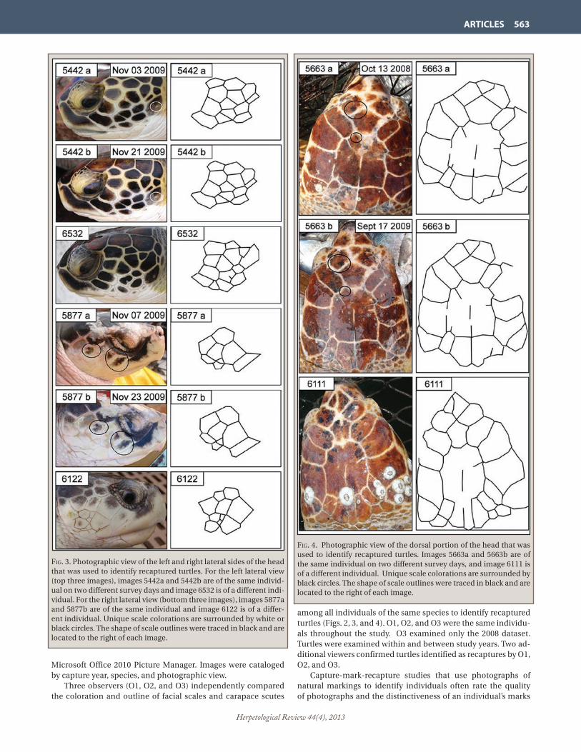
Herpetological Review 44(4), 2013
ARTICLES 563
Microsoft Office 2010 Picture Manager. Images were cataloged by capture year, species, and photographic view. Three observers (O1, O2, and O3) independently compared the coloration and outline of facial scales and carapace scutes
among all individuals of the same species to identify recaptured turtles (Figs. 2, 3, and 4). O1, O2, and O3 were the same individu-als throughout the study. O3 examined only the 2008 dataset. Turtles were examined within and between study years. Two ad-ditional viewers confirmed turtles identified as recaptures by O1, O2, and O3. Capture-mark-recapture studies that use photographs of natural markings to identify individuals often rate the quality of photographs and the distinctiveness of an individual’s marks
Fig. 4. Photographic view of the dorsal portion of the head that was used to identify recaptured turtles. Images 5663a and 5663b are of the same individual on two different survey days, and image 6111 is of a different individual. Unique scale colorations are surrounded by black circles. The shape of scale outlines were traced in black and are located to the right of each image.
Fig. 3. Photographic view of the left and right lateral sides of the head that was used to identify recaptured turtles. For the left lateral view (top three images), images 5442a and 5442b are of the same individ-ual on two different survey days and image 6532 is of a different indi-vidual. For the right lateral view (bottom three images), images 5877a and 5877b are of the same individual and image 6122 is of a differ-ent individual. Unique scale colorations are surrounded by white or black circles. The shape of scale outlines were traced in black and are located to the right of each image.

Herpetological Review 44(4), 2013
564 ARTICLES
because these characteristics influence the probability of identi-fying a previously photographed individual (Friday et al. 2000). We rated the quality (Q) of photographs (Table 1), but did not as-sign a distinctiveness rating because the distinct scale and scute patterns of individual sea turtles allow for their identification as long as image quality is good. We modeled our rating system for image quality after Urian et al. (1999) and Friday et al. (2000). We compared our 2008 and 2009 sea turtle recapture rates to recapture rates of sea turtles sampled in the PAEC during autumn and early winter of 1995–1997 and 2001–2003 (Epperly et al. 2007) and 2007 (NMFS Southeast Fisheries Science Cen-ter [SEFSC], Beaufort, North Carolina [NC], unpublished data) to see if our estimates of recapture rates were within the range of these previous studies. All studies were conducted using the same sampling protocols, with the exception that Inconel and PIT tags could be applied to turtles prior to 2008. Therefore, our 2008 and 2009 estimates of sea turtle catch rates were derived from photo-ID, while catch rates prior to 2008 were obtained using Inconel and PIT tags. Similar to Epperly et al. (2007), we estimated the total number of unique individuals captured an-nually for each species by subtracting the number of within-year recaptured turtles from the total number of turtles captured. In-dividuals identified as being recaptured twice on one day were not considered to be recaptures. One assumption of CMR studies is that all previously marked animals are identified as such upon recapture (Stevick et al. 2001). When identifying individuals by their natural markings, errors can occur if an observer incorrectly concludes that two captures of the same individual represent two different indi-viduals (a false negative) or if captures of different individuals are identified as a single individual (a false positive; Stevick et al. 2001). To ensure that false positives were not included in our analysis, all turtles identified as recaptures were confirmed by O1, O2, and O3, as well as by two additional viewers. To address false negatives, we assumed that the unique scale and scute pat-tern of turtles would allow for individual identification if images were of good (Q2) or excellent (Q3) quality. However, the iden-tification of individuals without images (Q0) would not be pos-sible, and individuals with poor-quality images (Q1) may only be identified some of the time, thus resulting in an inflated estimate
of unique individuals. To account for the number of turtles that may have been misidentified as unique individuals, we applied the following equation to the estimated total number of unique individuals for each species in 2008 and 2009:
X= (Q0+Q1) – Q1*
Where X is the number of turtles possibly misidentified as unique individuals, Q0 is the number of individuals without photographs, Q1 is the number of individuals with poor-quality
taBle 1. Image quality (Q) rating system for sea turtles captured by pound net and digitally photographed in the Pamlico-Albemarle Es-tuarine Complex of North Carolina, USA, during autumn and early winter of 2008 and 2009.
Rating Criteria Q0 Image unavailable. Turtle escaped from the net before a
photograph was taken.
Q1 Poor-quality image. Image out of focus; strong glare; pho-tographic view is angled or a portion of the desired view is missing; epibiota concealed scute or scale patterns.
Q2 Good-quality image. May be slightly out of focus; some glare; photographic view is slightly angled or a small por-tion of the desired view is missing; epibiota may be present, but does not significiantly conceal scute or scale patterns.
Q3 Excellent-quality image. Image is clear; no/very low glare; desired photographic view is directly facing the camera; epibiota may be present, but does not conceal scute or scale patterns.
taBle 2. Comparison of photo-ID derived recapture rates for sea tur-tles in the Pamlico-Albemarle Estuarine Complex, North Carolina, USA, during 2008 and 2009, with inconel and PIT tag derived turtle recapture rates from previous studiesa,b conducted in the same re-gion. Photo-ID recapture rates during 2008 and 2009 are highlighted in gray. In the column ‘recaptures within year,’ the percentage of re-captures within each year for each species is in parenthesis. For each species in 2008 and 2009, the percentage in parenthesis in the col-umn ‘turtles possibly misidentified as unique individuals,’ refers to the percentage of turtles captured that may have been misidentifed as unique individuals because no image was available to identify them by, or image quality was poor.
Year Total Recaptures Estimated total Turtles possibly captured within year unique misidentified as individuals unique individuals
Caretta caretta
1995a 111 9 (8%) 102 —
1996a 97 5 (5%) 92 —‒
1997a 156 3 (2%) 153 ‒—
2001a 258 33 (13%) 225 ‒—
2002a 207 16 (8%) 191 ‒—
2003a 158 11 (7%) 147 ‒—
2007b 81 2 (2%) 79 ‒—
2008 175 18 (10%) 157 13 (7%)
2009 201 25 (12%) 176 12 (6%)
Chelonia mydas
1995a 42 1 (2%) 41 ‒—
1996a 33 0 (0%) 33 ‒—
1997a 31 1 (3%) 30 ‒—
2001a 67 4 (6%) 63 ‒—
2002a 44 0 (0%) 44 ‒—
2003a 29 0 (0%) 29 ‒—
2007b 142 1 (<1%) 141 ‒—
2008 105 8 (8%) 97 8 (8%)
2009 151 4 (3%) 147 3 (2%)
Lepidochelys kempii
1995a 1 0 (0%) 1 ‒—
1996a 4 1 (25%) 3 ‒—
1997a 10 0 (0%) 10 ‒—
2001a 30 1 (3%) 29 ‒—
2002a 18 0 (0%) 18 ‒—
2003a 7 0 (0%) 7 ‒—
2007b 9 0 (0%) 9 ‒—
2008 31 1 (3%) 30 6 (19%)
2009 41 0 (0%) 41 4 (10%)a aEpperly et al. 2007 bNMFS Beaufort Lab unpublished data

Herpetological Review 44(4), 2013
ARTICLES 565
photographs, and Q1* is the number of individuals with poor-quality photographs that were identified as recaptures. Results.―—A total of 376 Loggerheads, 256 Greens, and 72 Kemp’s Ridleys were captured in 2008 and 2009 (Table 2). Of these, 43 Loggerheads, 12 Greens, and 1 Kemp’s Ridley were iden-tified as recaptures using photo-ID. The identification of three of these recaptures was confirmed by Inconel and/or PIT tags ap-plied prior to 2008. When examining recaptures among years, no Green or Kemp’s Ridley turtles captured in 2008 were identified as recaptures in 2009, and eight Loggerhead turtles captured in 2008 were identified as recaptures in 2009. Time intervals between ini-tial capture and recapture ranged from 1 to 386 days. Observer O1 identified a total of 27 recaptures for 2008, 29 recaptures for 2009, and 8 recaptures when comparing 2008 and 2009 captures. All turtles identified as recaptures by O2 and O3 also were identified as recaptures by O1. Observer O2 identified 20 recaptures for 2008, 27 recaptures for 2009, and 8 recaptures when comparing 2008 and 2009 captures. Observer O3 identi-fied 21 recaptures for 2008. No observers identified a capture as a false positive recapture. The two additional viewers concurred that all turtles identified as recaptures by the observers were re-captures. The percentage of turtles that we identified as recaptures in 2008 and 2009 for each species (Loggerheads ranged from 10–12%, Greens from 3–8%, and Kemp’s Ridleys from 0–3%) was comparable to recapture estimates from the 1995–1997, 2001–2003, and 2007 studies (Loggerheads ranged from 2–13%, Greens from 0–6%, and Kemp’s Ridleys from 0–25%; Epperly et al. 2007; NMFS SEFSC Beaufort, North Carolina, unpubl. data; Table 2). The percentage of turtles possibly misidentified as unique in-dividuals in 2008 and 2009 ranged from 6–7% for Loggerheads, 2–8% for Greens, and 10–19% for Kemp’s Ridleys. Of the four views photographed in 2008, the dorsal view of the head contained the most Q2 and Q3 images (89%), followed by the carapace (77%), and right (73%) and left (69%) lateral sides of the head. Because the dorsal view of the head was of-ten the best-quality image, and since we found that examining all photographic views was very time consuming (on average, it took 10–15 minutes to compare one image to 200 other images), the dorsal head view was the only photographic view examined in 2009 and when comparing individuals among years; however, the other three views were examined if the dorsal view was of poor quality (<Q2). Image quality of the dorsal head views im-proved in 2009 (94% of images were ≥Q2; Table 3).
In some cases, recaptured individuals could be identified from poor-quality (Q1) images; 12% of recaptures in 2008 and 8% in 2009 were identified from Q1 images (Table 3). We also found that distinguishing features such as scars, sloughing of the scales or scutes, skin colorations, and barnacle patterns on the cara-pace and head could aid with the identification of individuals (Figs. 2–5), and that these characteristics were especially help-ful when viewing images of poor quality (Fig. 5). However, cau-tion was needed when viewing barnacle patterns because some barnacle formations grew quickly, while others moved in a short (within weeks) amount of time (Fig. 6). Discussion.‒—Many studies of sea turtle populations rely solely on the application of identifying markers to recognize individuals; however, the usefulness of photo-ID as a tool to study sea turtle populations is increasingly becoming apparent. Photo-ID served as a valuable resource to our study by provid-ing us with an alternate method for identifying individual turtles when traditional tagging methods could not be used. As a result, we were able to identify recaptured turtles during our study, thereby allowing for an improved estimate of unique individu-als captured. The information derived from these data will allow for evaluations of the status of sea turtle populations in this re-gion, which is imperative for the development of conservation policies. These findings also further support the application of photo-ID as a means to monitor sea turtle populations and the lessons we learned can serve to aid others who are considering applying the technique to their research program. In-water studies using both photo-ID and Inconel/and or PIT tags to monitor sea turtle populations have indicated that individual turtles can be consistently identified upon recapture by the unique variations of their facial scales (Reisser et al. 2008; Schofield et al. 2008). Because it was not possible to confirm the identity of many of the individuals we identified as recaptures using photo-ID (since so few recaptured turtles had been previ-ously tagged), we compared our photo-ID recapture rates with those derived from Inconel and/or PIT tags during previous studies (Epperly et al. 2007, NMFS SEFSC Beaufort, North Caro-lina, unpubl. data) within the survey region. While we found that our recapture rates were within the range of those established during these studies, the limited timeframe of our study and the differing methods used to identify recaptures limits our ability to make a detailed comparison. Furthermore, comparison of the two identification methods is complicated by the likelihood that photo-ID recapture rates may be greater than those associated
taBle 3. Number of sea turtles captured and subsequently identified as a recaptures in 2008 and 2009, binned by the quality rating of the pho-tograph of the captured turtle. All photographs were of the dorsal head view. NA= not applicable, and refers to inability of observers to identify recaptured turtles if image quality was 0. The percentage of turtles identified as a recapture within each quality rating bin is in parentheses. Year Species Quality 0 Quality 1 Quality 2 Quality 3 Total Recaptures Total Recaptures Total Recaptures Total Recaptures captured captured captured captured 2008 Caretta caretta 2 NA 16 5 28 7 129 21 Chelonia mydas 0 NA 9 1 17 2 79 13 Lepidochelys kempii 0 NA 6 0 7 0 18 2 Total: 2 (1%) NA 31 (10%) 6 (12%) 52 (17%) 9 (18%) 226 (72%) 36 (70%)
2009 Caretta caretta 0 NA 15 3 33 7 153 36 Chelonia mydas 0 NA 4 1 21 0 126 6 Lepidochelys kempii 0 NA 4 0 8 0 29 0 Total: 0 (0%) NA 23 (6%) 4 (8%) 62 (16%) 7 (13%) 308 (78%) 42 (79%)

Herpetological Review 44(4), 2013
566 ARTICLES
solely with traditional tagging methods because turtles that have lost external tags or suffered PIT tag malfunctions can still be identified. However, given previous validation of the photo-ID technique for sea turtle identification (Reisser et al. 2008; Scho-field et al. 2008), and because the majority of our images were of good or excellent quality (Table 3), we believe our estimate of unique individuals is an accurate representation of the sea turtle population within the estuary at this time of year. Our estimate of turtles that may have been misidentified as unique individuals because no image was available to identify
them, or the image was of poor quality, further improves the ac-curacy of our estimate of unique individuals captured in 2008 and 2009. It is also probable that this estimate considerably over-estimates the number of turtles that may have been misidenti-fied as unique individuals, because it is unlikely that all individu-als without images or with poor-quality images were recaptures. Finally, our estimate of the percentage of turtles that may have been misidentified as unique individuals was greatest for Kemp’s Ridleys. This is likely because the facial scale outlines of Kemp’s Ridleys are often less well defined by color variations between the scales, resulting in a monotonous appearance (e.g., images 5877a,b, and 6122 in Fig. 3). Thus, image quality was especially important for identifying individual Kemp’s Ridleys and images that were even only slightly blurry were likely to receive a lower quality rating compared to similar images of another species. Long-term field studies that monitor changes in demograph-ic characteristics (i.e., survival, abundance, growth, and recruit-ment) of a population over time are invaluable, especially for long-lived species with complex life histories such as sea turtles (Epperly et al. 2007). However, accurate estimates of popula-tion parameters collected in long-term studies require that in-dividuals in a population be consistently identified upon each encounter (Wursig and Jefferson 1990). To date, little is known of the stability of the shape of sea turtle facial scales and carapace scutes throughout their lifespan. Due to the short length of our study, the longest period of time over which we identified a re-captured turtle using photo-ID was a little over a year; however, longer time intervals have been reported. For instance, Reisser et al. (2008), Jean et al. (2010), and Goncalves and Loureiro (2013) identified individual cheloniid turtles by their lateral facial scales over a period just short of three years, four years, and six years, respectively. These studies, along with ours, indicate that pho-to-ID can serve as an accurate, low-cost, and non-invasive tool for identifying individual sea turtles from year to year; however, future studies are needed to clarify the persistence of scute and scale shapes over longer periods (e.g., decades). Incorrectly identifying an individual as a recapture, or fail-ing to identify an individual as a recapture, can lead to biases in estimates of population parameters (Stevick et al. 2001). Upon reviewing the turtles identified as recaptures by each observer in 2008, we did not document the occurrence of false positive er-rors, but O2 and O3 did not identify a number of recaptures. It is likely that some of the recaptured turtles that were not identified by O2 and O3 were overlooked because of poor image quality. Ad-ditionally, consistently identifying individuals requires a certain amount of familiarity with the key characteristics that should be examined when viewing images. Consequently, O1 and O2 dis-cussed the characteristics that they focused on when examining images and this discussion, along with an improvement in im-age quality, resulted in fewer false negative errors in 2009 and when comparing the 2008 and 2009 captures. This improvement highlights the importance of ensuring that all observers are ad-equately trained, as well as the value of good-quality images. Estimates of population parameters also may be influenced by tag loss because an individual that has lost all tags is likely to be misidentified as a new individual upon subsequent en-counter (Seber 1982; Nichols and Hines 1993; Rivalan et al. 2005; Braun McNeill et al. 2013). Such misidentifications have the potential to negatively impact management decisions that are based on models that evaluate the status of populations. As a result, some CMR studies of sea turtles have attempted to es-timate tag loss rates so a correction can be applied to estimates
Fig. 5. Poor-quality images (5476a) could occasionally be used to identify recaptured turtles (5476b), especially if unique scale char-acteristics and/or barnacle patterns (circled) were present on the turtle.
Fig. 6. Rapid growth of barnacles on the head of a loggerhead sea turtle between its initial capture (6422a) and recapture (6422b; 38 days), and movement of barnacles (circled in white) on the carapace of a green turtle between its initial capture (5144a) and recapture (5144b; 16 days).

Herpetological Review 44(4), 2013
ARTICLES 567
of population parameters (Van Dam and Diez 1999; Rivalan et al. 2005; Reisser et al. 2008; Braun McNeill et al. 2013). Photo-ID holds great potential as a tool that can be used to estimate tag loss rates of sea turtle populations. This was demonstrated by Reisser et al. (2008), who used photo-ID to estimate Inconel tag loss in juvenile Green and Hawksbill turtles caught at Arvoredo Island, southern Brazil. They found a high rate of tag loss among turtles at their study site, prompting them to suggest that other CMR studies consider integrating photo-ID into their research programs given its potential for increasing the accuracy of data collected. We found that all four photographic views could be used to identify turtles, but we primarily used the dorsal view because it was typically the easiest to obtain, and therefore, often the best-quality view. This was because turtles were photographed on small vessels with little space to maneuver, making it diffi-cult to position the camera parallel with the side of the turtle’s head to obtain a clear, lateral view. To take lateral photos, ob-servers needed to kneel on the deck next to turtles, or hold the camera parallel to the turtle’s head and take the image without looking through the view finder, often resulting in blurry im-ages. Additionally, for larger turtles such as Loggerheads, ob-servers frequently had difficulty raising the camera to sufficient height so that the field view encompassed the entire carapace. Furthermore, the carapace of some turtles, particularly Logger-heads, was also often covered in epibionts, thereby obstructing the viewing of scutes. As a result of these difficulties, when de-signing a photo-ID study, we suggest that researchers consider the conditions in which they will be acquiring images and the size class of the species they will be photographing to determine which photographic view is optimal for their study. For example, lateral views of the head may be easier to acquire when photo-graphing turtles while diving, but overhead images of the head may be preferable when space is limited or turtles are too large to manipulate easily. Although we could identify some recaptured turtles from poor-quality images, the importance of good-quality images cannot be overstated (Hammond et al. 1990; Whitehead 1990; Friday et al. 2000; Kelly 2001). We found that it was often diffi-cult to distinguish among individuals using poor-quality images because the outlines of their scutes and scales were usually ob-scured. Additionally, characteristics such as scars, scute and scale colorations, and epibionts often helped with the identification of turtles from poor-quality images; however, these characteristics are likely to change over time. Therefore, while such character-istics may help with identification within a season, identifying turtles from poor-quality images among years is likely to be more challenging. As the number of photos in an image library increases, it be-comes more time consuming to manually compare images to one another (Arzoumanian et al. 2005). One option for decreas-ing the time it takes to compare turtle images is to divide indi-viduals into subgroups based on scale pattern characteristics (Schofield et al. 2008) so observers only have to match images to a smaller subgroup rather than all previous captures (Bradfield 2004). Jean et al. (2010) also devised a method for cataloging in-dividual turtles based on the location and shape of the scales on the lateral portion of the head, resulting in a unique code that allows for the quick identification of individuals. We chose not to use such identification methods, mainly because determining which group to place individuals in could be complicated by a number of factors (i.e., epibiont coverage, sloughing of scales or
poorly defined scale boundaries, unfavorable pose of the turtle, and poor image quality), and we did not want to risk misidentify-ing an individual because it was grouped incorrectly. In addition, we did not consider using subgroups to aid with identification until after all photographs were taken, and thus, the scale pat-tern characteristics that may have been chosen to group images were not always well represented. However, subgrouping and cataloging individuals is likely to prove beneficial to many pho-to-ID studies of sea turtles, and we recommend that the scale pattern characteristics that will be used to group images be well defined prior to the start of a project. The identification of individual sea turtles by their natural markings served as a valuable means for examining sea turtle recapture rates during our fishery-dependent CPUE study and we suggest that other researchers consider integrating photo-ID into their CPUE and CMR studies. When used in lieu of tradi-tional tagging methods, photo-ID can serve as a reliable, non-intrusive, and low-cost method for identifying individual turtles within a study site. However, identifying individuals using only photo-ID can be time consuming, and a turtle identified sole-ly by photo-ID would not be identified as a recaptured animal when encountered by other researchers. The development of computer-assisted image matching software for cheloniid tur-tles would not only reduce the time it takes to identify individu-als, but also may allow for the creation of a global database that could be used to identify individuals and track their movements (Bounantony 2008; Pauwels et al. 2008). When combined with traditional tagging methods, photo-ID can increase the accuracy of data collected by CMR studies by allowing for an estimate of tag loss (Reisser et al. 2008). Additionally, photo-ID databases collected by research groups can be used to examine a wide range of demographic characteristics (i.e., behavior, remigration estimates, sex ratios, breeding periodicity; McDonald and Dut-ton 1996; Schofield et al. 2007; Schofield et al. 2009; Hays et al. 2010), health (e.g., the prevalence of disease; Bennett et al. 2000), and conservation and management issues (e.g., incidence of boat strikes; Schofield et al. 2013a).
Acknowledgments.—We thank Alex Houston, Ann Pierce, and Billy Shaw for taking the images that were used to identify turtles, Amanda Schworm for viewing images as one of the three observers, and Larisa Avens and Lisa R. Goshe for reviewing the images of tur-tles that were identified as recaptures. We also appreciate the coop-eration of the commercial pound net fishers who participated in this project. Larisa Avens, Alex Chester, Annie Gorgone, Lisa R. Goshe, Aleta Hohn, Patti Marraro, and three anonymous reviewers provided helpful comments that improved this manuscript. This research was conducted under the authority of U.S. Endangered Species Act Sec-tion 10(a)(1)(A) scientific research permits from the U.S. Fish and Wildlife Service (#TE-676379) and the NMFS (#1260 and 1551). The NMFS does not approve, recommend, or endorse any product or material mentioned in this publication.
literature cited
arnason, a. n., and k. h. mills. 1981. Bias and loss of precision due to tag loss in Jolly-Seber estimates for mark-recapture experiments. Can. J. Fish. Aquat. Sci. 38:1077–1095.
arZoumanian, Z., J. holmBerg, and B. norman. 2005. An astronomical pattern-matching algorithm for computer-aided identification of whale sharks (Rhincodon typus). J. Appl. Ecol. 42:999–1011.
Baird, r. W., a. m. gorgone, d. J. mcsWeeney, d. l. WeBster, d. r. salden, m. h. deakos, a. d. ligon, g. s. schorr, J. BarloW, and s. d. mahaFFy.

Herpetological Review 44(4), 2013
568 ARTICLES
2008. False killer whales (Pseudorca crassidens) around the main Hawaiian Islands: Long-term site fidelity, inter-island movements, and association patterns. Mar. Mammal Sci. 24:591–612.
BalaZs, g. h. 1982. Factors affecting the retention of metal tags on sea turtles. Mar. Turtle Newsl. 20:11–14.
———. 1999. Factors to consider in the tagging of sea turtles. In K. L. Eckert, K. A. Bjorndal, F. A. Abreu-Grobois, and M. Donnelly (eds.), Research and Management Techniques for the Conservation of Sea Turtles, pp. 101–109. IUCN/SSC Mar. Turt. Spec. Group Publ. No. 4.
Bellini, c., m. h. godFrey, and t. m. sanches. 2001. Metal tag loss in wild juvenile hawksbill sea turtles (Eretmochelys imbricata). Her-petol. Rev. 32:172–174.
Bennett, P., u. keuPer-Bennett, and g. h. BalaZs. 2000. Photographic evidence for the regression of fibropapilloma afflicting green tur-tles at Honokawai, Maui, in the Hawaiian Islands. In H. Kalb, and T. Wibbels (eds.), Proceedings of the 19th Annual Symposium on Sea Turtle Biology and Conservation, pp. 37–39. U.S. Dept. Com-merce NOAA Tech. Memo., South Padre Island, Texas.
BJorndal, k. a., a. B. Bolten, c. J. lagueux, and a. chaVes. 1996. Prob-ability of tag loss in green turtles nesting at Tortuguero, Costa Rica. J. Herpetol. 30:567–571.
Bounantony, d. 2008. An analysis of utilizing the leatherback’s pi-neal spot for photo-identification. M.S. thesis. Nicholas School of the Environment and Earth Sciences, Duke University, Durham, North Carolina, 50 pp.
BradField, k. s. 2004. Photographic identification of individual Archey’s frogs, Leiopelma archeyi, from natural markings, p. 36. DOC Science Internal Series 191, Department of Conservation, Wellington.
Braun mcneill, J., a. m. schueller, l. aVens, a. goodman hall, l. r. goshe, and s. P. ePPerly. 2013. Estimates of tag loss for loggerhead (Caretta caretta) sea turtles from the western North Atlantic. Her-petol. Rev. 44:221–226.
Broderick, a. c., and B. J. godley. 1999. Effect of tagging marine tur-tles on nesting behavior and reproductive success. Anim. Behav. 58:587–591.
constantine, r. 2001. Increased avoidance of swimmers by wild bot-tlenose dolphins (Tursiops truncatus) due to long-term exposure to swim-with-dolphin tourism. Mar. Mamm. Sci. 17:689–702.
coPeland, B. J., and J. gray. 1991. Status and Trends Report of the Al-bemarle-Pamlico Estuary. In J. Steal (ed.), Albemarle-Pamlico Es-tuarine Study Report 90-01, pp.17–56. North Carolina Department of Environment, Health, and Natural Resources, Raleigh, North Carolina.
ePPerly, s., J. Braun, and a. VeishloW. 1995. Sea turtles in North Caro-lina waters. Conserv. Biol. 9:384–394.
ePPerly, s. P., J. Braun-mcneill, and P. m. richards. 2007. Trends in catch rates of sea turtles in North Carolina, USA. Endang. Spec. Res. 3:283–293.
Friday, n., t. d. smith, P. t. steVick, and J. allen. 2000. Measurement of photographic quality and individual distinctiveness for the photo-graphic identification of humpback whales, Megaptera novaean-gliae. Mar. Mamm. Sci. 16:355–374.
Frisch, a. J., and J. a. hoBBs. 2007. Photographic identification based on unique, polymorphic colour patterns: a novel method for tracking a marine crustacean. J. Exp. Mar. Biol. Ecol. 351:294–299.
goncalVes, B. t., and n. s. loureiro. 2013. Advances in sea turtle pho-to-identification. A case study carried out at the marine biological reserve of Arvoredo, Brazil. In T. Tucker, L. Belskis, A. Panagopou-lou, A. Rees, M. Frick, K. Williams, R. LeRoux, and K. Stewart (eds.), Proceedings of the 33rd Annual Symposium on Sea Turtle Biol-ogy and Conservation, p. 225. U.S. Dept. Commerce NOAA Tech. Memo., Baltimore, Maryland.
graham, r. t., and c. m. roBerts. 2007. Assessing the size, growth rate and structure of a seasonal population of whale sharks (Rhin-codon typus Smith 1828) using conventional tagging and photo identification. Fish. Res. 84:71–80.
hays, g. c., s. Fossette, k. a. katselidis, g. schoField, and m. B. graVe-nor. 2010. Breeding periodicity for male sea turtles: good news for conservation in the face of climate change. Conserv. Biol. 24:1636–1643.
hoelZel, a. r. 1993. Foraging behaviour and social group dynamics in Puget Sound killer whales. Anim. Behav. 45:581–591.
hammond, P. s., s. a. miZroch, and g. P. donoVan. 1990. Individual rec-ognition of cetaceans: use of photo-identification and other tech-niques to estimate population parameters. Rep. Int. Whal. Comm., Special Issue 12.
Jean, c., s. ciccione, e. talma, k. Ballorain, and J. BourJea. 2010. Photo-identification method for green and hawksbill turtles- first results from Reunion. Ind. Ocean Turtle Newsl. 11:8–13.
karanth, k. u., and J. d. nichols. 1998. Estimation of tiger densities in India using photographic captures and recaptures. Ecology 79:2852–2862.
karlsson, o., l. hiBy, t. lundBerg, m. Jüssi, i. Jüssi, and B. helander. 2005. Photo-identification, site fidelity, and movement of female gray seals (Halichoerus grypus) between haul-outs in the Baltic Sea. Ambio 34:628–634.
kelly, m. J. 2001. Computer-aided photograph matching in studies using individual identification: an example from Serengeti chee-tahs. J. Mamm. 82:440–449.
langtimm, c. a., c. a. Beck, h. h. edWards, k. J. Fick-child, B. B. ack-erman, s. l. Barton, and W. c. hartley. 2004. Survival estimates for Florida manatees from the photo-identification of individuals. Mar. Mamm. Sci. 20:438–463.
lloyd, J. r., m. a. maldonado, and r. staFFord. 2012. Methods of de-veloping user-friendly keys to identify green sea turtles (Chelo-nia mydas L.) from photographs. Int. J. Zool. 2012:7. 317568. doi 10.1155/2012/317568.
mcclintock, B. t., P. B. conn, r. s. alonso, and k. r. crooks. 2013. In-tegrated modeling of bilateral photo-identification data in mark-recapture analyses. Ecology 94:1464–1471.
mcdonald, d. l., and P. h. dutton. 1996. Use of PIT tags and photo-identification to revise remigration estimates of leatherback tur-tles (Dermochelys coriacea) nesting in St. Croix, U.S. Virgin Islands, 1979-1995. Chelonian Conserv. Biol. 2:148–152.
mcdonald, t. l., s. c. amstruP, and B. F. J. manly. 2003. Tag loss can bias Jolly-Seber capture-recapture estimates. Wildlife Soc. Bull. 31:814–822.
mcmahon, c. r., c. J. BradshaW, and g. c. hays. 2007. Applying the heat to research techniques for species conservation. Conserv. Biol. 21:271–3.
morrison, t. a., J. yoshiZaki, J. d. nichols, and d. t. Bolger. 2011. Estimating survival in photographic capture-recapture studies: overcoming misidentification error. Meth. Ecol. Evol. 2:454–463.
mrosoVsky, n., and s. J. shettleWorth. 1982. What double tagging stud-ies can tell us. Mar. Turtle Newsl. 22:11–15.
nichols, J. d., and J. e. hines. 1993. Survival rate estimation in the presence of tag loss using joint analysis of capture-recapture and resighting data. In J. D. Lebreton and P. M. North (eds.), Marked In-dividuals in the Study of Bird Populations, pp. 229–243. Birkhauser Verlag, Basel, Switzerland.
nichols, W. J., and J. a. seminoFF. 1998. Plastic “rototags” may be linked to sea turtle by-catch. Mar. Turtle Newsl. 19:20–21.
PauWels, e. J., P. m. de ZeeuW, and d. m. Bounantony. 2008. Leather-backs matching by automated image recognition. Lect. Notes Comput. Sci. 5077:417–425.
reisser, J., m. Proietti, P. kinas, and i. saZima. 2008. Photographic iden-tification of sea turtles: method description and validation, with an estimation of tag loss. Endang. Spec. Res. 5:73–82.
riValan, P., m. h. godFrey, a. c. PreVot-Julliard, and m. girondot. 2005. Maximum likelihood estimates of tag loss in leatherback sea tur-tles. J. Wildl. Manage. 69:540–548.
roeloFs, e. W., and d. F. BumPus. 1953. The hydrography of Pamlico Sound. Bull. Mar. Sci. Gulf Carib. 3:181–205.

Herpetological Review 44(4), 2013
ARTICLES 569
samuels, a., and l. BeJder. 2004. Chronic interaction between humans and free-ranging bottlenose dolphins near Panama City Beach, Florida, USA. J. Cetacean Res. Manage. 6:69–77.
schoField, g., k. a. katselidis, P. dimoPoulos, and J. d. Pantis. 2008. Investigating the viability of photo-identification as an objective tool to study endangered sea turtle populations. J. Exp. Mar. Biol. Ecol. 360:103–108.
———, ———, J. d. Pantis, P. dimoPoulos, and g. c. hays. 2007. Fe-male-female aggression: structure of interaction and outcome in loggerhead sea turtles. Mar. Ecol. Prog. Ser. 336:267–274.
———, m. k. s. lilley, c. m. BishoP, P. BroWn, k. a. katselidis, P. dimo-Poulos, J. d. Pantis, and g. c. hays. 2009. Conservation hotspots: intense space use by breeding male and female loggerheads at the Mediterranean’s largest rookery. Endang. Spec. Res. 10:191–202.
———, r. scott, a. dimadi, s. Fossette, k. a. katselidis, d. koutsouBa, m. k. s. lilley, J. d. Pantis, a. d. karagouni, and g. c. hays. 2013a. Evidence-based marine protected area planning for a highly mo-bile endangered marine vertebrate. Biol. Conserv. 161:101–109.
———, a. dimadi, s. Fossette, k. a. katselidis, d. koutsouBas, m. k. s. lilley, a. luckman, J. d. Pantis, a. d. karagouni, and g. c. hays. 2013b. Satellite tracking large numbers of individuals to infer population level dispersal and core areas for the protection of an endangered species. Divers. Distrib. 1–11.
seBer, g. a. F. 1982. The Estimation of Animal Abundance and Related Parameters. Macmillan, New York. 654 pp.
sPeed, c. W., m. g. meekan, and c. J. a. BradshaW. 2007. Spot the match —wildlife photo-identification using information theory. Front. Zool. 4. doi 10.1186/1742-9994-4-2.
steVick, P. t., P. J. PalsBøll, t. d. smith, m. V. BraVington, and P. s. ham-mond. 2001. Errors in identification using natural markings: rates, sources, and effects on capture-recapture estimates of abundance. Can. J. Fish. Aquat. Sci. 58:1861–1870.
thomPson, P. m., and P. s. hammond. 1992. The use of photography to monitor dermal disease in wild bottlenose dolphins (Tursiops truncatus). Ambio 21:135–137.
urian, g. t., a. a. hohn, and l. J. hansen. 1999. Status of the photo-identification catalog of coastal bottlenose dolphins of the west-ern North Atlantic: report of a workshop of catalog contributors. U.S. Dept. Commerce NOAA Tech. Memo. NMFS-SEFSC-425.
Van dam, r. P., and c. e. dieZ. 1999. Differential tag retention in Carib-bean hawksbill turtles. Chelonian Conserv. Biol. 3:225–229.
Walker, k. a., a. W. trites, m. haulena, and d. m. Weary. 2012. A review of the effects of different marking and tagging techniques on ma-rine mammals. Wildl. Res. 39:15–30.
Whitehead, h. 1990. Computer assisted individual identification of sperm whale flukes. Rep. Int. Whal. Comm., Special Issue 12:71–77.
Williams, B. k., J. d. nichols, and m. J. conroy. 2002. Analysis and Management of Animal Populations: Modeling, Estimation, and Decision Making. Academic Press, San Diego, 817 pp.
Wursig, B., and t. a. JeFFerson. 1990. Methods of photo-identification for small cetaceans. Rep. Int. Whal. Comm., Special Issue 12:43–52.
Wyneken, J., s. P. ePPerly, B. higgins, e. mcmichael, c. merigo, and J. P. Flanagan. 2010. PIT tag migration in sea turtle flippers. Herpetol. Rev. 41:448–454.
Herpetological Review, 2013, 44(4), 569–572.© 2013 by Society for the Study of Amphibians and Reptiles
Limestone Cave as a Cradle of the Ryukyu Ground Gecko, Goniurosaurus kuroiwae
Goniurosaurus kuroiwae is a eublepharid gecko distrib-uted in the central part of the Ryukyu Archipelago, Japan (Ota 1989). Among the members of the genus Goniurosaurus, G. kuroiwae has been relatively well studied for its reproduc-tive ecology (Tanaka and Nishihira 1987, 1989). According to these authors, gravid females possessing two eggs per clutch appear from late May to late August, and the ratio of gravid females to total females exceeds 80% at the peak. Neverthe-less, the oviposition site of this gecko in the wild is unknown. Because G. kuroiwae and its close relatives face the risk of ex-tinction and are designated as endangered on the IUCN Red List (Ota 2010), knowledge regarding oviposition and incuba-tion sites in the wild is important for the planning of a habitat preservation program.
In November 2007 and 2012, we found a total of three pairs of unidentified squamate eggshells in a limestone cave in the southern part of Okinawajima Island. There are 24 na-tive and introduced squamates recorded from this island: seven gekkotans, two iguanians, four scincomorphans, and 11 ophidians (Maenosono and Toda 2007). We presumed the eggshells to belong to G. kuroiwae because of their small size, parchment-shelled eggs, and local abundance of this gecko around the caves. To confirm this, we attempted to deter-mine sequences of the residual-DNA on the eggshells and compared them with available data for other reptile taxa. We also measured several environmental parameters at and around the oviposition sites.
materials and methods
Study site.—All eggshells were found in a limestone cave located in the southern part of Okinawajima Island (26°08‒N, 127°45‒E; datum WGS84); further details concerning the location are not provided in order to restrict illegal hunting by pet dealers, which is one of the serious threats to G. kuroiwae (Kanari and Xu 2012; Ota 2010). The cave opens southward at the midpoint of a cliff. A passage having a width for the entry of only one person runs northward, curving east at 3–4 m from the entrance, and reaches a round hall (approximately 5 m in width and depth, 3 m in height). There are many small pockets on the walls, and all eggshells were found on the surface of sediments deposited in-side these pockets. The hall has another exit at the opposite end of the entrance, but it was not possible to examine the cave any further because it was too narrow (Fig. 1a).
TAKAKI KURITA*Graduate School of Engineering and Science, University of the Ryukyus, Nishihara, Okinawa 903-0213, JapanREMI KAWAMURAMaehara 2-6-1-403, Ginowan, Okinawa 901-2215, JapanMAMORU TODATropical Biosphere Research Center, University of the Ryukyus, Nishihara, Okinawa 903-0213, Japan
*Corresponding author; e-mail: [email protected]
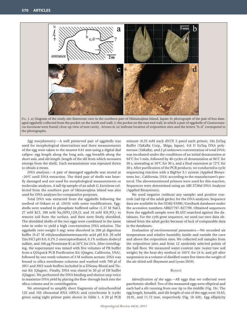
Herpetological Review 44(4), 2013
570 ARTICLES
Egg morphometry.—A well preserved pair of eggshells was used for morphological observations and three measurements of the egg were taken to the nearest 0.01 mm using a digital dial caliper: egg length along the long axis, egg breadth along the short axis, and slit length (length of the slit from which neonates emerge from the shell). Each measurement was repeated thrice to obtain a mean.
DNA analyses.—A pair of damaged eggshells was stored at −20°C until DNA extraction. The third pair of shells was heav-ily damaged and not used for morphological measurements or molecular analyses. A tail tip sample of an adult G. kuroiwae col-lected from the southern part of Okinawajima Island was also used for DNA analyses for comparative purposes.
Total DNA was extracted from the eggshells following the method of Oskam et al. (2010) with some modifications. Egg-shells were washed in phosphate buffered saline (1.37 M NaCl, 27 mM KCl, 100 mM Na
2HPO
4‒12H
2O, and 18 mM KH
2PO
4) to
remove soil from the surface, and then were finely shredded. The shredded shells of the two eggs were combined in the same tube in order to yield a high concentration DNA solution. The eggshells (wet-weight 5 mg) were dissolved in 200 µl digestion buffer (0.47 M ethylenediaminetetraacetic acid pH 8.0, 20 mM Tris/HCl pH 8.0, 0.2% 2-mercaptoethanol, 0.1% sodium dodecyl sulfate, and 100 μg Proteinase K) at 56°C for 24 h. After centrifug-ing, the supernatant was mixed with five volumes of PB buffer from a QIAquick PCR Purification Kit (Qiagen, California, USA), followed by one tenth volumes of 3 M sodium acetate. DNA was bound to silica membrane columns and washed with 700 µl of AW1 and AW2 wash buffers included in a DNeasy Blood and Tis-sue Kit (Qiagen). Finally, DNA was eluted in 50 µl of EB buffer (Qiagen). We performed the DNA binding and elution step twice to maximize DNA yield by placing the flow-through back into the silica column and re-centrifugation.
We attempted to amplify short fragments of mitochondrial 12S and 16S ribosomal RNA (rRNA) and cytochrome b (cytb) genes using eight primer pairs shown in Table 1. A 20 µl PCR
mixture (0.25 mM each dNTP, 5 pmol each primer, 10x ExTaq Buffer [TaKaRa Corp., Shiga, Japan], 0.8 U ExTaq DNA poly-merase [TaKaRa], and 2 µl unknown concentration of total DNA) was incubated under the conditions of an initial denaturation at 94°C for 3 min, followed by 40 cycles of denaturation at 94°C for 30 s, annealing at 50°C for 30 s, and a final extension at 72°C for 30 s. After purification of the PCR products, we conducted a cycle sequencing reaction with a BigDye 3.1 system (Applied Biosys-tems Inc., California, USA) according to the manufacturer’s pro-tocol. The abovementioned primers were used for this reaction. Sequences were determined using an ABI 3730xl DNA Analyzer (Applied Biosystems).
We used negative (without any sample) and positive con-trols (tail tip of the adult gecko) for the DNA analyses. Sequence data are available in the DDBJ/EMBL/GenBank databases under the accession numbers AB827797–827799. Obtained sequences from the eggshell sample were BLAST-searched against the da-tabases. For the cytb gene sequence, we used our own data ob-tained from the adult gecko because of lack of comparable data in the databases.
Evaluation of environmental parameters.—We recorded air temperature and relative humidity inside and outside the cave and above the oviposition sites. We collected soil samples from the oviposition sites and from 12 randomly selected points of the hall floor. We measured water-content rate (water/raw soil weight) by the heat-dry method at 105°C for 24 h, and pH after suspension in a volume of distilled water five times the weight of the air-dried soil (Rayment and Lyons 2010).
results
Identification of the eggs.—All eggs that we collected were parchment-shelled. Two of the measured eggs were elliptical and each had a slit running from one tip to the middle (Fig. 1b). The egg length, breadth, and slit length of one of the eggs were 19.63, 10.91, and 11.72 mm, respectively (Fig. 1b left). Egg ellipticity
Fig. 1. a) Diagram of the study-site limestone cave in the southern part of Okinawajima Island, Japan; b) photograph of the pair of less dam-aged eggshells collected from the pocket on the north end wall; c) the pocket on the east end wall, in which a pair of eggshells of Goniurosau-rus kuroiwae were found; close up view of nest cavity . Arrows in (a) indicate location of oviposition sites and the letters “b–d” correspond to the photographs.

Herpetological Review 44(4), 2013
ARTICLES 571
(egg breadth/egg length) was 0.60. The egg length and slit length of the other deformed egg was 19.34 and 12.52 mm, respectively (Fig. 1b right).
DNA fragments of expected lengths were successfully ampli-fied from the eggshell extract by four of the eight primer pairs; of which three were successfully sequenced yielding 211 bp se-quences of 12S rRNA (167 and 70 bp with 26 bp overlap) and 193 bp of cytb genes. Some amplification signals were detected in three out of the eight negative controls on agarose gel, but these products were not the target because of their unexpected longer lengths (>500 bp; Table 1).
A BLAST search revealed that the sequence obtained from the 12S rRNA gene matched (100%) with that of G. kuroiwae from the southern part of Okinawajima Island (Accession No. AB028750), followed by the congeneric species from the Ryukyus (96–99%) and a Chinese congener (89%). In a BLAST search with our cytb gene sequence as a query, we got hits for homologous genes of some anurans and chiropterans but with very low lev-els of matching (79–82%). In contrast, the cytb sequence was well-matched with that of G. kuroiwae determined in this study (99%), with two synonymous nucleotide substitutions in the third codon positions.
Environmental parameters.—In November 2012, daytime air temperature and relative humidity were 30.1°C and 79% at the entrance of the cave, and 23.6°C and 99% in the hall, respec-tively. Indirect sunlight scarcely reached the passage, and none reached the hall. There were percolating water drips falling from stalactites in the cave and no other surface water was present. The floor of the cave was extensively covered with flowstones and cohesive brownish soil. In the hall, patchy black sediments originating from bat guano were accumulated. Two of the 12 soil samples were taken from such bat guano-rich sites. Water-content rate and pH of the sediments in the hall ranged between 32.0–69.1% and 3.8–8.5, respectively.
Three pairs of eggshells were found in three different small pockets on the wall of the hall. These pockets were located
0.20–1.45 m above the hall floor. One pocket was at the north end and the other two pockets were at the east end (Fig. 1a). The north pocket and one of the east pockets were horizontally con-cave (Fig. 1c, d); the other east pocket was vertically concave and covered with a fallen piece of broken stalactite. Air temperature measured in front of the pockets was 21.7–22.3°C and relative humidity was 99%. A layer of small granular soil, approximate-ly 30–100 mm in thickness, was deposited at the bottom of the pockets, but bat guano was absent. Water-content rate and pH of the soil that was used as the egg laying substrate were 33.5–46.5% and 8.1–8.2, respectively. All eggshells were exposed on the sur-face of the soil. The outer eggshell surfaces were entirely covered with soil particles, but no soil was found on the inner surfaces.
discussion
The results of the genetic analyses revealed that the eggshells found in the limestone cave were of G. kuroiwae. Morphological features of the eggs were also consistent with captive breeding records of closely-related congeners, i.e., parchment shell and size (20 × 12 mm; Henkel and Schmidt 1995). Neonates appeared to have hatched from the eggs, confirming that environmental conditions in the cave were appropriate for incubation. There-fore, we conclude that G. kuroiwae uses this limestone cave as an oviposition site.
Why is this limestone cave a suitable oviposition and incuba-tion site for G. kuroiwae? One conceivable factor is the stability of hydric conditions; parchment-shelled eggs require a certain amount of ambient air and substrate moisture level to avoid ei-ther drying out or becoming moldy (Henkel and Schmidt 1995; Werner 1972). The extreme high humidity recorded in the cave (99%) protects the substrate and eggs from dehydration. In addi-tion, eggs in the cave are also protected from exposure to rains. Thermal conditions during the incubation period critically affect embryonic development and performance of hatchlings in many reptile species (see Du and Shine 2010 and references therein). In this respect, caves are thermally quite stable (Culver and Pipan 2009), and this may be important for this gecko.
Environmental conditions in caves are more uniform than the outside, but are still variable in a fine spatial scale. In fact, our soil samples had highly variable water-content rates and pH, representing extreme water retention and acidification in some samples most probably due to water drips from stalactites and bat excrement (Culver and Pipan 2009; Ferreira et al. 2007). Such excess water could damage eggs (Henkel and Schmidt 1995), and soil acidification can negatively affect embryonic development in some lizards with parchment-shelled eggs (Marco et al. 2005). However, it appears that the pockets on the walls offer some de-gree of protection from organic substances and water.
Although we lack quantitative data, soil compaction appar-ently differed between the oviposition sites and the hall floor. The soil particles were granular and loose in the egg laying pock-ets, but were tight and cohesive on the floor. In captivity, some eublepharid geckos are known to make a pit in humid ground to lay eggs and cover them with soil after oviposition (Henkel and Schmidt 1995). We observed that the outer surfaces of eggshells were entirely coated with soil particles, strongly suggesting that G. kuroiwae also deposit their eggs in soil and this necessitates loose soil at oviposition sites.
Acknowledgments.—The Okinawa Prefectural Board of Educa-tion provided permission to handle the protected geckos. We thank
taBle 1. The eight primer pairs designed for amplification of short fragments of mitochondrial DNA. The filled circles indicate success-ful amplification and sequencing of the target fragments, and the open circles indicate that fragments were amplified but failed to se-quence. Size is the expected length of the PCR products.
Primer Sequence (5’ to 3’) Locus Size
FGss1F CAACCTAGAGGAGCCTGTCC 12S 152 bp
FGss1R CACCTTGACCTGACGTTTTT • FGss2F CCCTACTATGCTTAGCCATTAACC 12S 191 bp
FGss2R TGACGACGGCGGTATATAGG ‒•FGls1F GACGAGAAGACCCTGTGGAG 16S 240 bp
FGls1R AAGAGGATTGCGCTGTTATC
FGls2F GGATAACAGCGCAATCCTCT 16S 118 bp
FGls2R AATCGTTGAACAAACGAACC ‒o
FGcb1F AAAACAACATGAAACACAGGAGT cytb 120 bp
FGcb1R AAGGTTTGTGATTACGGTTGC
FGcb2F TATCATTCTGAGGCGCAACC cytb 211 bp
FGcb2R GGATTGTTTGAGCCTGTTTCA
FGcb3F CACACATTTGCCGAGATGTA cytb 233 bp
FGcb3R GGTTGCGCCTCAGAATGATA ‒ •FGcb4F CCAGAAAACTTCATTCCAGCA cytb 210 bp
FGcb4R TTGGGATAAGGGTCGAAATG

Herpetological Review 44(4), 2013
572 ARTICLES
M. Tokuzato for his great efforts to help us to get the permission. This research was partially supported by the Zoshinkai Fund for Protection of Endangered Animals and by the Grant from the Japan Society for the Promotion of Science (JSPS KAKENHI Grant number 22510244).
literature cited
culVer, d. c., and t. PiPan. 2009. The Biology of Caves and Other Sub-terranean Habitats. Oxford Univ. Press, New York. 254 pp.
du, W. g., and r. shine. 2010. Why do the eggs of lizards (Bassiana duperreyi: Scincidae) hatch sooner if incubated at fluctuating rather than constant temperatures? Biol. J. Linn. Soc. 101:642–650.
Ferreira, r. l., x. Prous, and r. P. martins. 2007. Structure of bat guano communities in a dry Brazilian cave. Trop. Zool. 20:55–74.
henkel, F. W., and W. schmidt. 1995. Geckoes: Biology, Husbandry, and Reproduction. Krieger Publ. Co., Malabar, Florida. 237 pp.
kanari, k., and l. xu. 2012. Trade in Japanese endemic reptiles in China and recommendations for species conservation. TRAFFIC. <www.traffic.org> Downloaded on 4 April 2013.
maenosono, t., and m. toda. 2007. Distributions of amphibians and terrestrial reptiles in the Ryukyu Archipelago: a review of pub-lished records. Akamata 18:28–46. (in Japanese)
marco, a., m. l. lóPeZ-Vicente, and V. PéreZ-mellado. 2005. Soil acidi-fication negatively affects embryonic development of flexible-shelled lizard eggs. Herpetol. J. 15:107–111.
oskam, c. l, J. haile, e. mclay, P. rigBy, m. e. allentoFt, m. e. olsen, c. Bengtsson, g. h. miller, J. schWenninger, c. JacomB, r. Walter, a. Baynes, J. dortch, m. Parker-Pearson, m. t. P. gilBert, r. n. hold-aWay, e. WillersleV, and m. Bunce. 2010. Fossil avian eggshell pre-serves ancient DNA. Proc. R. Soc. B 277:1991–2000.
ota, h. 1989. A review of the geckos (Lacertilia: Reptilia) of the Ryukyu Archipelago and Taiwan. In M Matsui, T. Hikida, and R. C. Goris (eds.), Current Herpetology in East Asia: Proceedings of the Second Japan-China Herpetological Symposium, pp. 222–261. The Herpetological Society of Japan, Kyoto.
———. 2010. Goniurosaurus kuroiwae. In IUCN 2012. IUCN Red List of Threatened Species Version 2012.2 <www.iucnredlist.org> Downloaded on 8 January 2013.
rayment, g. e., and d. J. lyons. 2010. Soil Chemical Methods: Austral-asia. CSIRO Publ., Collingwood, Victoria. 520 pp.
tanaka, s., and m. nishihira. 1987. A field study of seasonal, daily, and diel activity patterns of Eublepharis kuroiwae kuroiwae. Herpeto-logica 43:482–489.
———, and ———. 1989. Growth and reproduction of the gekkonid lizard Eublepharis kuroiwae kuroiwae. In M. Matsui, T. Hikida, and R. C. Goris (eds.), Current Herpetology in East Asia: Proceed-ings of the Second Japan-China Herpetological Symposium, pp. 349–357. The Herpetological Society of Japan, Kyoto.
Werner, y. l. 1972. Observations on eggs of eublepharid lizards, with comments on the evolution of the Gekkonoidea. Zool. Meded. 47:211–224.
Herpetological Review, 2013, 44(4), 572–579.© 2013 by Society for the Study of Amphibians and Reptiles
Parasitism of Reptiles by the Blacklegged Tick (Ixodes scapularis) and Western Blacklegged Tick (Ixodes pacificus) with New Records of I. scapularis from Arkansas and Oklahoma Lizards: Implications for Lyme Disease Epidemiology
Worldwide, at least 116 species of ticks are known to para-sitize reptiles (Barnard and Durden 2000). For some of these tick species, including members of the Holarctic Ixodes ricinus complex (including I. pacificus, I. persulcatus, I. ricinus, and I. scapularis), the immature stages (larvae and nymphs) often feed on reptiles whereas adults mainly feed on larger mammals, in-cluding humans (Clark et al. 2013; Keirans et al. 1999). The afore-mentioned four species in this complex have significant medical and veterinary importance as zoonotic vectors of the causative agents of Lyme disease (= Lyme borreliosis), human babesiosis,
human granulocytic anaplasmosis, and/or tick-borne encepha-litis in the northern hemisphere (Nicholson et al. 2009).
Lyme disease, caused by infection with Borrelia burgdorferi sensu lato spirochete bacteria, is an emerging or persistent zoo-nosis in much of the Northern Hemisphere (Oliver 1996; Scott et al. 2012). Presently, at least 18 distinct genomospecies are rec-ognized within B. burgdorferi sensu lato and three of these, B. burgdorferi sensu stricto, B. afzelii, and B. garinii, are known to cause disease in humans (Nicholson et al. 2009; Scott et al. 2012). Ticks belonging to the I. ricinus complex are the principal vec-tors with different tick species being primary vectors in different regions (Oliver 1996). In North America, the Blacklegged Tick, I. scapularis Say, is the main vector in eastern states and prov-inces whereas the Western Blacklegged Tick, I. pacificus Cooley and Kohls, is the principal vector in far western states and a Ca-nadian province (Burgdorfer et al. 1985; Clover and Lane 1995; Eisen et al. 2004a, b; Keirans et al. 1996; Lane and Lavoie 1988; Lane and Loye 1989). Certain species of rodents, and to a lesser extent birds, are enzootic reservoir hosts of B. burgdorferi sensu lato (Castro and Wright 2007; Clark et al. 2005; Clover and Lane 1995; Durden and Oliver 1999; Lane and Lavoie 1988; Scott et al. 2012). However, immature stages (larvae and nymphs) of both I. scapularis and I. pacificus not only feed on mammals and birds, but also on certain species of reptiles, especially lizards (Clark et al. 2005; Durden et al. 2002; Giery and Ostfeld 2007; Lumbad
CHRIS T. McALLISTER*Science and Mathematics Division, Eastern Oklahoma State College, Idabel, Oklahoma 74745, USA; e-mail: [email protected] A. DURDENDepartment of Biology, Georgia Southern University, Statesboro, Georgia 30458, USAMATTHEW B. CONNIORHealth and Natural Sciences, South Arkansas Community College, El Dorado, Arkansas 71730, USA HENRY W. ROBISONDepartment of Biology, Southern Arkansas University, Magnolia, Arkansas 71754, USA
*Corresponding author

Herpetological Review 44(4), 2013
ARTICLES 573
et al. 2011; Swanson and Norris 2007; Talleklint-Eisen and Eisen 1999). In fact, in some regions, up to 98% of nymphal I. pacificus in a given habitat feed on lizards (Casher et al. 2002). If these liz-ards are incompetent reservoir hosts of strains of B. burgdorferi that cause human disease, feeding of immature I. scapularis on lizards would serve to decrease the infection prevalence in tick populations, thereby creating a “dilution effect” (Schmidt and Ostfeld 2001) and an example of biodiversity providing “ecosys-tem services” to public health (Ogden and Tsao 2009).
Some researchers have reported that certain lizards may be reservoir hosts of B. burgdorferi in North America (Clark et al. 2005; Giery and Ostfled 2007; Levin et al. 1996; Swanson and Norris 2007). Similarly, in Eurasia and North Africa, where im-mature stages of I. ricinus frequently parasitize lizards (Bauwens 1983; Dsolui et al. 2006; Keirans et al. 1999; Matchuschka et al. 1991), some of these reptiles have been reported to be reservoir hosts of B. burgdorferi sensu lato, notably of the genomospecies B. lusitaniae (Amore et al. 2007; Dsouli et al. 2006; Majláthová et al. 2006), which is not a human pathogen. Conversely, other researchers have shown that other species of lizards are refrac-tory to infection by B. burgdorferi and have borreliacidal factors in their blood that effectively kill spirochetes (Kuo et al. 2000; Lane and Quistad 1998; Lane et al. 2006; Wright et al. 1998). In this article, we report new lizard hosts of I. scapularis in Arkansas and Oklahoma, list the known reptilian hosts of both I. scapularis and I. pacificus by state/province in North America, record tick stage(s) collected from each host species, and briefly consider the potential role of lizards in Lyme disease epidemiology.
Methods.—Between 2009–2013, 65 lizards (juveniles and adults) were collected by hand from various localities in upland (Ouachita and Ozark plateaus) and Gulf Coastal Plain habitat in 12 Arkansas counties (Baxter, Bradley, Calhoun, Faulkner, Greene, Izard, Madison, Newton, Ouachita, Polk, Searcy, and Union), as follows: 11 Green Anoles (Anolis carolinensis), eight Prairie Racerunners, (Aspidoscelis sexlineata viridis), four South-ern Coal Skinks (Plestiodon anthracinus pluvialis), 20 Five-lined Skinks (Plestiodon fasciatus), one Broad-headed Skink (Plesti-odon laticeps), and four Prairie Lizards (Sceloporus consobrinus); 12 P. fasciatus and five S. consobrinus were also collected from
McCurtain Co., Oklahoma. Following the guidelines for the hu-mane treatment of research animals (HACC 2004) specimens were either examined live and released or euthanized with an intraperitoneal injection of sodium pentobarbital and their bod-ies examined for arthropod parasites. When ticks were found they were removed with tweezers and placed in vials containing 70% ethanol and shipped to one of us (LAD) for identification. Voucher tick specimens are deposited in the U.S. National Tick Collection, Statesboro, Georgia, under accession numbers RML 124935 (from P. laticeps), RML 124936–124937 (from P. fasciatus), and RML 124938 (from S. consobrinus). Host vouchers are de-posited in the Arkansas State University Herpetological Collec-tion (ASUMZ), State University, Arkansas. Previously published records of I. scapularis and I. pacificus parasitizing reptiles in the U.S. and Canada were compiled through extensive literature searches.
Fig. 1. Thirteen counties in Arkansas and one in southeastern Oklahoma where lizards were collected showing those infested with ticks (solid circle in county) and those without ticks (open circle in county).
Fig. 2. A nymphal Ixodes scapularis embedded under scales of dermis in Plestiodon fasciatus from McCurtain County, Oklahoma.
Fig. 3. Sixteen states where larvae and nymphs of Ixodes scapularis (solid dots) and Ixodes pacificus (open dots) have been reported from various lizard hosts. The latter tick is also reported from lizard hosts in British Columbia, Canada (not shown). Star = new records for I. scapularis from lizards.

Herpetological Review 44(4), 2013
574 ARTICLES
ta
Ble
1. R
eco
rded
rep
tili
an h
ost
s o
f Bla
ckle
gged
Tic
ks (
Ixod
es s
cap
ula
ris)
an
d W
este
rn B
lack
legg
ed T
icks
(Ix
odes
pac
ific
us)
in N
ort
h A
mer
ica.
Ho
st fa
mil
y Sc
ien
tifi
c n
ame
Co
mm
on
nam
e T
ick
spec
ies
Tic
k st
ages
co
llect
ed1
Stat
e/P
rovi
nce
rec
ord
s an
d c
itat
ion
s
An
guid
ae
Op
his
auru
s at
ten
uat
us
Sl
end
er G
lass
Liz
ard
Ix
odes
sca
pu
lari
s L,
N
(NC
) Le
vin
e et
al.
(199
7)
Un
spec
ifie
d (
AL,
FL,
GA
an
d/o
r SC
) O
live
r et
al.
(199
3)
Op
his
auru
s co
mp
ress
us
Isla
nd
Gla
ss L
izar
d
L,
N
(GA
) D
urd
en &
Oli
ver
(199
9)
Un
spec
ifie
d (
AL,
FL,
GA
, an
d/o
r SC
) O
live
r et
al.
(199
3)
Op
his
auru
s m
imic
us
Mim
ic G
lass
Liz
ard
L U
nsp
ecif
ied
(A
L, F
L, G
A, a
nd
/or
SC)
Oli
ver
et a
l. (1
993)
O
ph
isau
rus
ven
tral
is
Eas
tern
Gla
ss L
izar
d
L,
N
(FL)
Cla
rk e
t al.
(200
5)
(GA
) D
urd
en e
t al.
(200
2)
(NC
) A
pp
erso
n e
t al.
(199
3), L
evin
e et
al.
(199
7)
(SC
) C
lark
et a
l. (2
005)
(T
X)
Ead
s (1
949)
U
nsp
ecif
ied
(A
L, F
L, G
A, a
nd
/or
SC)
Oli
ver
et a
l. (1
993)
O
ph
isau
rus
sp.
un
det
erm
ined
sp
ecie
s
L, N
U
nsp
ecif
ied
, Kei
ran
s et
al.
(199
6)D
acty
loid
ae
An
olis
car
olin
ensi
s G
reen
An
ole
L (A
R)
pre
sen
t rep
ort
(F
L) R
oge
rs (
1953
)
(NC
) Le
vin
e et
al.
(199
7)
(SC
) R
eeve
s et
al.
(200
2)P
hry
no
som
atid
ae
Scel
opor
us
con
sobr
inu
s P
rair
ie L
izar
d
N
(A
R)
Lan
cast
er (
1973
), p
rese
nt r
epo
rt
(T
X)
Bre
nn
an (
1945
)
Scel
opor
us
oliv
aceu
s Te
xas
Spin
y Li
zard
L, N
(T
X)
Bre
nn
an (
1945
)
Scel
opor
us
un
du
latu
s E
aste
rn F
ence
Liz
ard
L, N
(F
L) H
ixso
n (
1941
)
(MD
) Sw
anso
n &
No
rris
(20
07)
(M
D/N
Y)
Gie
ry &
Ost
feld
(20
07)
(M
O)
Ko
llars
et a
l. (1
999)
U
nsp
ecif
ied
, Bis
ho
pp
& T
rem
ble
y (1
945)
Scin
cid
ae
Ple
stio
don
egr
egiu
s M
ole
Ski
nk
N
U
nsp
ecif
ied
(A
L, F
L, G
A, o
r SC
) (O
live
r et
al.
1993
)
Ple
stio
don
fasc
iatu
s F
ive-
Lin
ed S
kin
k
L, N
(A
R)
pre
sen
t rep
ort
(M
D)
Swan
son
& N
orr
is (
2007
)
(MD
/NY
) G
iery
& O
stfe
ld (
2007
)
(MO
) K
olla
rs e
t al.
(199
9)
(NC
) Le
vin
e et
al.
(199
7)
Un
spec
ifie
d (
AL,
FL,
GA
, an
d/o
r SC
) O
live
r et
al.
(199
3)
Ple
stio
don
inex
pec
tatu
s So
uth
east
ern
Fiv
e-Li
ned
Ski
nk
L,
N
(FL)
Cla
rk e
t al.
(200
5)
(GA
) D
urd
en &
Oli
ver
(199
9), D
urd
en e
t al.
(200
2)
(NC
) A
pp
erso
n e
t al.
(199
3), L
evin
e et
al.
(199
7)
(SC
) C
lark
et a
l. (2
005)
U
nsp
ecif
ied
(A
L, F
L, G
A, a
nd
/or
SC)
Oli
ver
et a
l. (1
993)
P
lest
iod
on la
tice
ps
Bro
adh
ead
Ski
nk
L,
N
(AR
) La
nca
ster
(19
73),
pre
sen
t rep
ort
(F
L) H
ixso
n (
1941
), R
oge
rs (
1953
) C
lark
et a
l. (2
005)
(G
A)
Kei
ran
s et
al.
(199
6), L
aven
der
& O
live
r (1
996)
,
Du
rden
& O
live
r (1
999)
, Du
rden
et a
l. (2
002)
(N
C)
Ap
per
son
et a
l. (1
993)
, Lev
ine
et a
l. (1
997)
(M
D)
Swan
son
& N
orr
is (
2007
(M
D/N
Y)
Gie
ry &
Ost
feld
(20
07)
(S
C)
Cla
rk e
t al.
(200
5)
Un
spec
ifie
d (
AL,
FL,
GA
, an
d/o
r SC
) O
live
r et
al.
(199
3)
Un
spec
ifie
d, B
ish
op
p &
Tre
mb
ley
(194
5)
Sc
ince
lla
late
rali
s G
rou
nd
Ski
nk
L,
N
(FL)
Ro
gers
(19
53),
Kei
ran
s et
al.
(199
6)
(GA
) La
ven
der
& O
live
r (1
996)
(N
C)
Ap
per
son
et a
l. (1
993)
, Kei
ran
s et
al.
(199
6)
(TX
) B
ren
nan
194
5)
Herpetological Review 44(4), 2013
ARTICLES 575
ta
Ble
1. C
on
tin
ued
.
Ho
st fa
mil
y Sc
ien
tifi
c n
ame
Co
mm
on
nam
e T
ick
spec
ies
Tic
k st
ages
co
llect
ed1
Stat
e/P
rovi
nce
rec
ord
s an
d c
itat
ion
s
“S
kin
k”
un
det
erm
ined
sp
ecie
s
L, N
U
nsp
ecif
ied
, Bis
ho
pp
& T
rem
ble
y (1
945)
,
Kei
ran
s et
al.
(199
6)Te
iid
ae
Asp
idos
celi
s se
xlin
eata
Si
x-Li
ned
Rac
eru
nn
er
L,
N
(NC
) Le
vin
e et
al.
(199
7)E
myd
idae
Te
rrap
ene
caro
lin
a E
aste
rn B
ox T
urt
le
N
(T
N)
Ree
ves
et a
l. (2
007)
An
guid
ae
Elg
aria
c. c
oeru
lea
San
Fra
nci
sco
Alli
gato
r Li
zard
Ix
odes
pac
ific
us
L, N
(C
A)
Jelli
son
(19
34),
Mo
hr
(196
4),
A
rth
ur
& S
now
(19
68),
Fu
rman
& L
oo
mis
(19
84),
Eis
en e
t al.
(200
4a),
Cas
tro
& W
righ
t (20
07)
E
lgar
ia c
oeru
lea
pri
nci
pis
N
ort
hw
este
rn A
lliga
tor
Liza
rd
L,
N
(BC
) G
regs
on
(19
56),
Art
hu
r &
Sn
ow (
1968
),
A
rnas
on
(19
92)
E
lgar
ia m
. mu
ltic
arin
ata
Cal
ifo
rnia
Alli
gato
r Li
zard
L, N
(C
A)
Co
ole
y &
Ko
hls
(19
45),
Ho
lden
ried
et a
l. (1
951)
,
Art
hu
r &
Sn
ow (
1968
), F
urm
an &
Lo
om
is (
1984
),
B
enn
ett (
1987
), W
ebb
et a
l. (1
990)
,
Talle
klin
t-E
isen
an
d E
isen
(19
99),
Wri
ght e
t al.
(200
3a, b
),
Eis
en e
t al.
(200
4a),
Cas
tro
& W
righ
t (20
07)
(O
R)
Art
hu
r &
Sn
ow (
1968
)
(WA
) A
rth
ur
& S
now
(19
68)
Elg
aria
mu
ltic
arin
ata
scin
cica
ud
a O
rego
n A
lliga
tor
Liza
rd
L,
N
(CA
) Je
lliso
n (
1934
), C
oo
ley
& K
oh
ls (
1945
),
A
rth
ur
& S
now
(19
68),
Fu
rman
& L
oo
mis
(19
84),
Wri
ght e
t al.
1998
)
Elg
aria
sp.
u
nd
eter
min
ed s
pec
ies
L,
N, A
(C
A)
Mo
hr
et a
l. (1
964)
, Fu
rman
& L
oo
mis
(19
84),
W
ebb
et a
l. (1
990)
, Cas
tro
& W
righ
t (20
07)
Ph
ryn
oso
mat
idae
Sc
elop
oru
s g.
gra
cios
us
No
rth
ern
Sag
ebru
sh L
izar
d
L,
N
(CA
) A
rth
ur
& S
now
(19
68),
Fu
rman
& L
oo
mis
(19
84),
W
ebb
et a
l. (1
990)
, Go
ldb
erg
& B
urs
ey (
1991
),
W
righ
t et a
l. (2
003a
, b),
Cas
tro
& W
righ
t (20
07)
Sc
elop
oru
s oc
cid
enta
lis
Wes
tern
Fen
ce L
izar
d
L,
N
(CA
) Je
lliso
n (
1934
), C
oo
ley
& K
oh
ls (
1945
),
Ho
lden
ried
et a
l. (1
951)
, Mo
hr
et a
l. (1
964)
,
Art
hu
r &
Sn
ow (
1968
), F
urm
an &
Lo
om
is (
1984
),
M
anw
eile
r et
al.
(199
0, 1
992)
, Web
b e
t al.
(199
0),
Ta
llekl
int-
Eis
en &
Eis
en (
1999
), E
isen
et a
l. (2
001,
200
4a),
Sch
all e
t al.
(200
0), C
ash
er e
t al.
(200
2),
W
righ
t et a
l. (2
003a
, b),
Cas
tro
& W
righ
t (20
07),
Lum
bad
et a
l. (2
011)
, an
d o
ther
s
(OR
) A
rth
ur
& S
now
(19
68)
U
nsp
ecif
ied
, Bis
ho
pp
& T
rem
ble
y (1
945)
Sc
elop
oru
s u
nd
ula
tus
Eas
tern
Fen
ce L
izar
d²
U
(A
Z)
Ols
on
et a
l. (1
992)
Sc
elop
oru
s sp
. u
nd
eter
min
ed s
pec
ies
L,
N
(CA
) Fu
rman
& L
oo
mis
(19
84)
Uta
sta
nsb
uri
ana
Sid
e-B
lotc
hed
Liz
ard
L, N
(C
A)
Ben
net
t (19
87),
Web
b e
t al.
(199
0),
G
old
ber
g &
Bu
rsey
(19
91),
Cas
tro
& W
righ
t (20
07)
Scin
cid
ae
Ple
stio
don
gil
bert
i G
ilb
ert’s
Ski
nk
L,
N
(CA
) C
astr
o &
Wri
ght (
2007
)
Ple
stio
don
ski
lton
ian
us
W
este
rn S
kin
k
L, N
(C
A)
Ben
net
t (19
87),
Cas
tro
& W
righ
t (20
07)
Teii
dae
A
spid
osce
lis
tess
elat
a C
om
mo
n C
hec
kere
d W
hip
tail
3
N
(CA
) H
old
enri
ed e
t al.
(195
1)
Asp
idos
celi
s ti
gris
W
este
rn W
hip
tail
L, N
(C
A)
Furm
an &
Lo
om
is (
1984
), C
astr
o &
Wri
ght (
2007
)
Asp
idos
celi
s sp
. u
nd
eter
min
ed s
pec
ies
L,
N
(CA
) C
oo
ley
& K
oh
ls (
1945
), A
rth
ur
& S
now
(19
68)
Un
know
n fa
mil
y “C
oas
t Liz
ard
” u
nd
eter
min
ed s
pec
ies
U
(B
C)
Art
hu
r &
Sn
ow (
1968
)
“Liz
ard
s”
un
det
erm
ined
sp
ecie
s
U
(CA
) C
oo
ley
& K
oh
ls (
1945
), A
rth
ur
& S
now
(19
68),
Fu
rman
& L
oo
mis
(19
84),
Web
b e
t al.
(199
0)
(OR
) C
oo
ley
& K
oh
ls (
1945
), A
rth
ur
& S
now
(19
68)
Co
lub
rid
ae
Th
amn
oph
is o
rdin
oid
es
No
rth
wes
tern
Gar
ter
Snak
e
U
(OR
) A
rth
ur
& S
now
(19
68)
1 L =
Lar
va(e
), N
= N
ymp
h(s
), A
= A
du
lt(s
), U
= U
nd
eter
min
ed (
no
t sta
ted
in o
rigi
nal
pap
er).
2 T
his
ho
st a
sso
ciat
ion
was
rep
ort
ed fo
r M
oh
ave
Co
un
ty, A
rizo
na;
how
ever
, S. u
nd
ula
tus
do
es n
ot o
ccu
r in
Ari
zon
a.
3 Th
is h
ost
ass
oci
atio
n w
as r
epo
rted
for
Ala
med
a C
ou
nty
, Cal
ifo
rnia
; how
ever
, A. t
esse
lata
do
es n
ot o
ccu
r in
Cal
ifo
rnia
.

Herpetological Review 44(4), 2013
576 ARTICLES
Results.—Forty-four ticks were found on 11 of 65 (17%) liz-ards; a total of 13 larval and 31 nymphal specimens were col-lected from lizards from six of 13 (46%) counties of Arkansas and a single county in Oklahoma (Fig. 1). All ticks were subsequently identified as I. scapularis. A single P. laticeps collected on 1 May 2012 from Union Co. harbored seven nymphs and 12 larvae; eight (60%) P. fasciatus collected on 18 March 2012 from Marion Co., 26 April 2012, 1 May 2012, 17 July 2012, 10 September 2012, and 19 May 2013 from Union Co., 19 August 2012 from Calhoun Co. and 29 March 2013 from Searcy Co. had one, one, one, six, two, two (plus one larva), one, and three nymphal specimens, respec-tively; a single (25%) S. consobrinus collected on 27 April 2012 in Izard Co. had two nymphs; and one (9%) A. carolinensis collected on 24 February 2013 from Union Co. had three nymphs. A single (8%) P. fasciatus collected on 25 May 2013 from McCurtain Co., Oklahoma had two nymphs, one of which is shown in Fig. 2.
Table 1 lists the recorded reptilian hosts by state/province of I. scapularis and I. pacificus and includes references and stag-es (larvae, nymphs, and/or adults) for each host species. Fig. 3 shows the 16 states from which these two tick species have cur-rently been reported from reptilian hosts.
Discussion.—Ixodes scapularis has an extensive Nearctic range and has been reported previously from parts of southeast-ern Canada, across the eastern USA, and from the Mexican state of Coahuila (Keirans et al. 1996). In the USA, it is known from Florida to Maine in the east to central Texas and north to the Da-kotas in the west (Keirans et al. 1996). There are also records of immature stages attached to migrating birds from other regions (Scott et al. 2012). Lancaster (1973) mentions that nymphs of I. scapularis have been found on the “common fence lizard” and “blue-tailed skink” in Arkansas but does not give a citation for this statement or list specific lizard species in his table of hosts for this tick. This species is the most common Ixodes in Arkan-sas (Lancaster 1973), where adults parasitize larger mammals (mainly deer and livestock) during the cooler months of the year while immature stages occur on a variety of smaller mammals, birds, and reptiles. However, we document the first definitive liz-ard host records for I. scapularis in Arkansas and Oklahoma, and the first voucher specimens reported from S. consobrinus (for-merly S. undulatus hyacinthinus).
Various lizard species have been reported as hosts of lar-vae and nymphs of I. scapularis in the eastern U.S. including Alabama (Oliver et al. 1993), Florida (Clark et al. 2005; Oliver et al. 1993; Rogers 1953), Georgia (Durden et al. 2002; Keirans et al. 1996; Oliver at al. 1993), Maryland (Giery and Ostfeld 2007; Swanson and Norris 2007), Missouri (Kollars et al. 1999), New York (Giery and Ostfeld 2007), North Carolina (Apperson et al. 1993; Levine et al. 1997), South Carolina (Clark et al. 2005; Oliver et al. 1993), and Texas (Brennan 1945; Eads 1949) (Table 1, Fig. 1). There is also a record of nymphal I. scapularis parasitizing an Eastern Box Turtle (Terrapene carolina carolina) in Tennessee (Reeves et al. 2007). However, little is known about I. scapularis infesting lizards west of the Mississippi River, particularly in Ar-kansas and Oklahoma.
In the far western United States and parts of far western Can-ada, larvae and nymphs of I. pacificus parasitize various species of lizards (Table 1). Most of these records are from California (Ar-thur and Snow 1968; Casher et al. 2002; Castro and Wright 2007; Eisen et al. 2001, 2004a, b; Goldberg and Bursey 1991; Lane and Loye 1989; Lumbad et al. 2011; Manweiler et al. 1990, 1992; Schall et al. 2000; Talleklint-Eisen and Eisen 1999; Webb et al. 1990; Wright et al. 1998), but there are additional records from Arizona
(Olson et al. 1992), Oregon (Arthur and Snow 1968; Cooley and Kohls 1945; Eads 1949), Washington (Arthur and Snow 1968), and British Columbia, Canada (Arnason 1992; Arthur and Snow 1968; Gregson 1956) (Table 1, Fig. 1). There are also records of adult I. pacificus from undetermined alligator lizard species (Elgaria spp.) in California (Cooley and Kohls 1945; Mohr et al. 1964) (Ta-ble 1). Immature stages of I. pacificus presumably also parasitize lizards in other states where this tick is known to occur, namely Idaho, Nevada, and Utah.
Lizards can be relatively important hosts of immature I. scap-ularis and I. pacificus. For example, Durden and Oliver (1999) recorded 373 larvae and 21 nymphs of I. scapularis on a single P. laticeps in coastal Georgia, and Durden et al. (2002) reported a peak monthly average of 96.0 I. scapularis immatures/skink (P. laticeps and P. inexpectatus combined) during May 1994 in the same region. At the same site, Cotton Mice (Peromyscus gossy-pinus) were parasitized by a peak monthly average of 2.9 im-mature I. scapularis (during June 1994) with a maximum of 31 larvae on a single mouse (Durden and Oliver 1999). In one study in California, the Western Fence Lizard (Sceloporus occidentalis) accounted for 78% of the larval feedings and 98% of nymphal feedings for the I. pacificus population (Casher et al. 2002). These data suggest that in some regions up to 10x the number of im-mature I. scapularis and I. pacificus feed on lizards compared to the number that feed on other hosts (mammals and birds) in the same habitat. However, these numbers should be viewed with caution because infestation rates are clearly dependent on varia-tion in host communities and because host body temperatures affect engorgement times of attached ticks. We did not record body temperature of our lizards when captured, but others have found that under laboratory conditions larvae and nymphs of I. scapularis remained attached to lizards for about 3x as long on skinks (Plestiodon spp.) as on P. gossypinus (Durden and Oli-ver 1999). Similarly, Goldberg and Bursey (1991) reported that, at 24°C, larvae and nymphs of I. pacificus attached to Northern Sagebrush Lizards (Sceloporus graciosus graciosus) for approxi-mately eight days and to Common Side-blotched Lizards (Uta stansburiana) for 16 days.
Galbe and Oliver (1992) successfully fed five separate co-horts of I. scapularis larvae on the same individual Plestiodon laticeps and noted no reduction in tick engorgement prevalence or weights; additionally, antibodies against I. scapularis salivary gland antigens were not present in the blood of these lizards. Be-cause of these phenomena, lizards could represent an important component in the ecology and epidemiology of any pathogens transmitted by these two tick species, in particular B. burgdorferi, the causative agent in Lyme disease. Because I. scapularis and I. pacificus immatures exhibit little host preference when offered laboratory mice, birds (baby chicks), or lizards as hosts (James and Oliver 1990) (larvae of both species showed a slight prefer-ence for mice, and nymphs preferred lizards and mice equally over chicks), the larger numbers of these ticks found on lizards in many regions is presumably related to the greater accessibil-ity or availability of lizards. Slowak and Lane (2011) also tested the host preference of immature stages of I. pacificus under laboratory conditions but they used more natural hosts in their experiments: Deer Mouse (Peromyscus maniculatus), California Kangaroo Rat (Dipodomys californicus), California Towhee (Pipi-lio crissalis), and Western Fence Lizard. They demonstrated that both larvae and nymphs of I. pacificus preferred Western Fence Lizards as hosts but that rodents were also frequently parasitized and towhees were rarely parasitized.

Herpetological Review 44(4), 2013
ARTICLES 577
The exact role lizard hosts play in Lyme disease ecology and epidemiology depends to a large extent on whether lizards are reservoir-competent or reservoir-incompetent hosts for geno-mospecies of B. burgdorferi that can cause human disease and on the proportion of immature ticks that feed on lizards com-pared to alternative hosts in a given region. Early interpretations, sometimes supported by circumstantial data, suggested that liz-ards are refractory to infection with B. burgdorferi which would mean that any immatures of I. scapularis and I. pacificus feeding on lizards would not become infected with spirochetes (Durden and Oliver 1999). This would lead to a dilution effect with fewer infected ticks and fewer cases of Lyme disease in areas where most immature ticks feed on lizards rather than on reservoir–competent rodents. This seems to correlate well with the fewer cases of Lyme disease contracted in the southeastern United States where lizards are abundant and appear to feed most of the immature I. scapularis in the tick population (Durden and Oliver 1999; Oliver 1996). Conversely, in the northeastern and upper midwestern states, where Lyme disease is hyperendemic, lizard biodiversity and abundance is lower (compared to that in the southeastern USA) and most of the immature I. scapularis feed on reservoir-competent rodents (Oliver 1996).
Despite this hypothesis, Levin et al. (1996) reported pro-longed (up to 5 wk) laboratory infections of both P. inexpectatus and A. carolinensis by B. burgdorferi sensu stricto. However, as more lizard species have been tested for their ability to main-tain infections of B. burgdorferi-group spirochetes, the picture has become more complicated. For example, although Clark et al. (2005) used polymerase chain reaction amplification of spe-cific primers to demonstrate the presence of B. burgdorferi in the blood of 86 of 160 (54%) lizards from Florida and South Caro-lina (representing nine species and six genera of lizards), other researchers have found other lizard species to be refractory to infection by B. burgdorferi sensu lato. Lane (1990) was unable to experimentally infect Western Fence Lizards with B. burgdorferi although one individual lizard apparently maintained a transi-tory spirochetemia for two days. It was later discovered that S. occidentalis has a borreliacidal factor in its blood that kills both B. burgdorferi (Lane and Quistad 1998) and the closely related B. burgdorferi-group spirochete Borrelia bissettii (Lane et al. 2006). Wright et al. (1998) similarly showed that Southern Alligator Liz-ard (Elgaria multicarinata) blood contains a borreliacidal factor. The borreliacidal factors in the blood of both S. occidentalis and E. multicarinata were shown to be proteins comprising the alter-native complement pathway (Kuo et al. 2000).
In southern Maryland, Swanson and Norris (2007) detected B. burgdorferi markers in eight of 29 (28%) field-caught lizards (S. undulatus and Plestiodon spp.). Also, the role of lizards in the ecology of Lyme disease was examined in two endemic zones in the northeastern USA (one in New York and one in Maryland) by Giery and Ostfeld (2007). Based on very low infection preva-lences (0–2%) in xenodiagnostic larval I. scapularis fed on field-caught P. fasciatus or S. undulatus, they devised a model to pre-dict the ability of different vertebrate host species to influence the prevalence of B. burgdorferi in the tick population, which is an important component of human Lyme disease risk. They con-sidered P. fasciatus to be reservoir-incompetent for B. burgdorferi and S. undulatus to be partially reservoir-competent. Combin-ing these data with population densities of the two lizard species and their average tick burdens, they concluded that P. fasciatus is a dilution host for Lyme disease risk in the areas studied, mean-ing that its presence in the ecosystem reduces the prevalence of
B. burgdorferi-infected ticks and therefore the risk of B. burgdor-feri transmission. Similarly, Salkeld and Lane (2010) modeled B. burgdorferi transmission and maintenance in hosts and vectors in oak woodlands in northwestern California and demonstrated that Western Fence Lizards are important hosts for feeding im-mature I. pacificus but reduce Lyme disease transmission risk (nymphal infection prevalence). The hypothesis that Western Fence Lizards serve as dilution hosts and reduce Lyme disease risk by diverting immature I. pacificus ticks away from B. burg-dorferi-competent rodents in California was tested experimen-tally by Swei et al. (2011) who removed lizards from field plots and then tracked tick populations in those plots. Following lizard removal, many larval I. pacificus failed to find alternate hosts, re-sulting in lower nymphal densities the following year. Although lizard removal did not result in increased infection prevalences in I. pacificus nymphs, it did result in fewer infected nymphs and, therefore, decreased Lyme disease risk to humans. Nevertheless, Swei et al. (2011) argued that incompetent reservoirs (Western Fence Lizards, in this case) may actually increase disease risk by promoting higher vector density and therefore, a larger overall density of infected vectors.
Based on previous reports, the competence of lizards as res-ervoir hosts for B. burgdorferi sensu lato differs widely depend-ing on the lizard species being studied. Some lizard hosts of Lyme disease tick vectors are reported to be reservoir-competent and appear to perpetuate the transmission of the spirochete whereas others are reservoir-incompetent and evidently serve as dilu-tion hosts by reducing the number of ticks feeding on reservoir-competent rodents, effectively diminishing Lyme disease risk in those regions. Regardless, lizards appear to play a fundamental role in maintaining populations of I. scapularis and I. pacificus in parts of North America although tick burdens differ in different regions and are dependent on local vertebrate host communi-ties (Apperson et al. 1993; Casher et al. 2002; Durden and Oliver 1999; Eisen et al. 2004a, b). Future studies to assess or re-assess the reservoir competence of different lizard species to strains of B. burgdorferi that cause human disease would shed additional light on their role as either dilution or (possibly) amplification hosts for Lyme borreliosis (see Ogden and Tsao 2009). We en-courage others to report additional state and county records of lizard infestation by Ixodes spp. to more clearly illustrate tick-lizard associations, an important component in understanding the spread of Lyme disease.
Acknowledgments.—We thank the Arkansas Game and Fish Commission (AG&F) and Oklahoma Department of Wildlife Conser-vation (ODWC) for Scientific Collecting Permits issued to CTM (Nos. AG&F 030420131 and ODWC 5246, respectively) and MBC (AG&F No. 102220121), and S. E. Trauth (ASUMZ) for expert curatorial as-sistance.
literature cited
amore, g., l. tomassone, e. grego, c. ragagl, l. Bertolotti, P. neBBia, s. rosati, and a. mannelli. 2007. Borrelia lusitaniae in immature Ixodes ricinus (Acari: Ixodidae) feeding on common wall lizards in Tuscany, central Italy. J. Med. Entomol. 44:303–307.
aPPerson, c. s., J. F. leVine, t. l. eVans, a. BrasWell, and J. heller. 1993. Relative utilization of reptiles and rodents as hosts by immature Ixodes scapularis (Acari: Ixodidae) in the coastal plain of North Carolina, USA. Exp. Appl. Acarol. 17:719–731.
arnason, c. s. 1992. Biology of the western black-legged tick, Ixodes pacificus (Cooley and Kohls, 1943): a potential vector of Lyme

Herpetological Review 44(4), 2013
578 ARTICLES
disease in south coastal British Columbia. M.S. Thesis, Simon Fra-ser University, Burnaby, BC. 64 pp.
arthur, d. r., and k. r. snoW. 1968. Ixodes pacificus Cooley & Kohls, 1943: its life history and occurrence. Parasitology 58:893–906.
Barnard, s. m., and l. a. durden. 2000. a Veterinary Guide to the Para-sites of Reptiles. Volume 2. Arthropods (excluding Mites). Krieger Publ. Co., Malabar, Florida. 288 pp.
BauWens, d., h. struBosch, and a. h. P. stumPel. 1983. The lizards Lac-erta agilis and L. vivipara as hosts to larvae and nymphs of the tick Ixodes ricinus. Holarctic Ecol. 6:32–40.
Bennett, s. g. 1987. Medically important and other acarines on verte-brates from Santa Catalina Island, California. Bull. Soc. Vect. Ecol. 12:534–538.
BishoPP, F. c., and h. l. tremBley. 1945. Distribution and hosts of cer-tain North American ticks. J. Parasitol. 31:1–54.
Brennan, J. m. 1945. Field investigations pertinent to Bullis fever. Texas Rep. Biol. Med. 3:112–121.
BurgdorFer, W., r. s. lane, a. g. BarBour, r. a. gresBrink, and J. r. an-derson. 1985. The western black-legged tick, Ixodes pacificus: a vector of Borrelia burgdorferi. Am. J. Trop. Med. Hyg. 34:925–930.
casher, l., r. s. lane, r. Barrett, and l. eisen. 2002. Relative impor-tance of lizards and mammals as hosts for ixodid ticks in northern California. Exp. Appl. Acarol. 26:127–143.
castro, m. B., and s. a. Wright. 2007. Vertebrate hosts of Ixodes pacifi-cus (Acari: Ixodidae) in California. J. Vect. Ecol. 32:140–149.
clark, k., a. hendricks, and d. Burge. 2005. Molecular identification and analysis of Borrelia burgdorferi sensu lato in lizards in the southeastern United States. Appl. Environ. Microbiol. 71:2616–2625.
———, B. leydet, and s. hartman. 2013. Lyme borreliosis in human patients in Florida and Georgia, USA. Int. J. Med. Sci. 10:915–931.
cloVer, J. r., and r. s. lane. 1995. Evidence implicating nymphal Ixo-des pacificus (Acari: Ixodidae) in the epidemiology of Lyme disease in California. Am. J. Trop. med. Hyg. 53:237–240.
cooley, r. a., and g. m. kohls. 1945. The genus Ixodes in North Amer-ica. Natl. Inst. Health Bull. No. 184. 246 pp.
dsouli, n., h. younsi-kaBachii, d. Postic, s. nouira, l. gern, and a. Bouattour. 2006. Reservoir role of lizard Psammodromus algirus in transmission cycle of Borrelia burgdorferi sensu lato (Spirochaeta-ceae) in Tunisia. J. Med. Entomol. 43:737–742.
durden, l. a., and J. h. oliVer, Jr. 1999. Ecology of Ixodes scapularis and Lyme disease in coastal Georgia. In G. R. Needham, R. Mitch-ell, D. J. Horn, and W. C. Welbourn (eds.), Acarology IX, volume 2, Symposia, pp. 379–383. Ohio Biological Survey, Columbus, Ohio.
———, ———, c. W. Banks, and g. n. Vogel. 2002. Parasitism of liz-ards by immature stages of the blacklegged tick, Ixodes scapularis (Acar: Ixodidae). Exp. Appl. Acarol. 26:257–266.
eads, r. B. 1949. Notes on Ixodes scapularis Say with an additional lizard host. Entomol. News 60:238–240.
eisen, l., r. J. eisen, and r. s. lane. 2004a. The roles of birds, lizards, and rodents as hosts for the western black-legged tick Ixodes paci-ficus. J. Vect. Ecol. 29:295–308.
———, ———, and ———. 2001. Prevalence and abundance of Ixo-des pacificus immatures (Acari: Ixodidae) infesting western fence lizards (Sceloporus occidentalis) in northern California: temporal trends and environmental correlates. J. Parasitol. 87:1301–1307.
eisen, r. J., l. eisen, and r. s. lane. 2004b. Habitat-related variation in infestation of lizards and rodents with Ixodes ticks in dense woodlands in Mendocino County, California. Exp. Appl. Acarol. 33:215–233.
Furman, d. P., and e. c. loomis. 1984. The ticks of California (Acari: Ixodida). Bull. California Insect Surv. 25:1–239.
galBe, J., and J. h. oliVer, Jr. 1992. Immune response of lizards and rodents to larval Ixodes scapularis (Acari: Ixodidae). J. Med. Ento-mol. 29:774–783.
giery, s. t., and r. s. ostFeld. 2007. The role of lizards in the ecology of Lyme disease in two endemic zones of the northeastern United States. J. Parasitol. 93:511–517.
goldBerg, s. r., and c. r. Bursey. 1991. Duration of attachment by mites and ticks on the iguanid lizards Sceloporus graciosus and Uta stansburiana. J. Wildl. Dis. 27:719–722.
gregson, J. d. 1956. The Ixodoidea of Canada. Canada Dept. Agric., Ottawa, Ontario, Publ. 930:1–92.
herPetological animal care use committee (hacc) oF the american so-ciety oF ichthyologists and herPetologists. 2004. Guidelines for use of live amphibians and reptiles in field and laboratory research. Second ed. Available: http://www.research.fsu.edu/acuc/poli-cies_Guidelines/ASIH_HACC_GuidelinesAmphibians.pdf
hixson, h. 1941. a suicide host. Florida Entomol. 24:34.holdenreid, r., F. c. eVans, and d. s. longanecker. 1951. Host-parasite-
disease relationships in a mammalian community in the central coast range of California. Ecol. Monogr. 21:1–18.
hui, l. t., m. B. castro, J. cloVer, m. a. thomPson, and s. husted. 2004. Summary of Ixodes pacificus surveillance and testing for Borrelia burgdorferi in California. Proc. Pap. Mosq. Vector Contr. Assoc. Ca-lif. 72:77–83.
James, a. m., and J. h. oliVer, Jr. 1990. Feeding and host preference of immature Ixodes dammini, I. scapularis, and I. pacificus (Acari: Ixodidae). J. Med. Entomol. 27:324–330.
Jellison, W. l. 1934. The parasitism of lizards by Ixodes ricinus califor-nicus. J. Parasitol. 20:243.
keirans, J. e., h. J. hutcheson, l. a. durden, and J. s. h. klomPen. 1996. Ixodes (Ixodes) scapularis (Acari: Ixodidae): redescription of all ac-tive stages, distribution, hosts, geographical variation, and medi-cal and veterinary importance. J. Med. Entomol. 33:297–318.
———, g. r. needham, and J. h. oliVer, Jr. 1999. The Ixodes ricinus complex worldwide: diagnosis of the species in the complex, hosts and distribution. In G. R. Needham, R. Mitchell, D. J. Horn, and W. C. Welbourn (eds.), Acarology IX: Symposia, pp. 341–347. Ohio Biological Survey, Columbus.
kollars, t. m., J. h. oliVer, Jr., P. g. kollars, and l. a. durden. 1999. Seasonal activity and host associations of Ixodes scapularis (Acari: Ixodidae) in southeastern Missouri. J. Med. Entomol. 36:720–726.
kuo, m. m., r. s. lane, and P. c. gicias. 2000. A comparative study of mammalian and reptilian alternative pathway of complement-mediated killing of the Lyme disease spirochete (Borrelia burgdor-feri). J. Parasitol. 86:1223–1228.
lancaster, J. l., Jr. 1973. A guide to ticks of Arkansas. Agric. Exp. Stat. Univ. Ark. Bull. No. 779. 39 pp.
lane, r. s. 1990. Susceptibility of the western fence lizard (Sceloporus occidentalis) to the Lyme borreliosis spirochete (Borrelia burgdor-feri). Am. J. Trop. Med. Hyg. 42:75–82.
———, and P. e. laVoie. 1988. lyme borreliosis in California: acaro-logical, clinical and epidemiological studies. Ann. New York Acad. Sci. 539:192–203.
———, and J. e. loye. 1989. Lyme disease in California: interrelation-ships of Ixodes pacificus (Acari: Ixodidae), the western fence lizard (Sceloporus occidentalis) and Borrelia burgdorferi. J. Med. Ento-mol. 26:272–278.
———, and g. B. Quistad. 1998. Borreliacidal factor in the blood of the western fence lizard (Sceloporus occidentalis). J. Parasitol. 84:29–34.
———, J. mun, l. eisen, and r. J. eisen. 2006. Refractoriness of the western fence lizard (Sceloporus occidentalis) to the Lyme disease group spirochete Borrelia bissettii. J. Parasitol. 92:691–696.
laVender, d. r., and J. h. oliVer, Jr. 1996. Ticks (Acari: Ixodidae) in Bulloch County, Georgia. J. Med. Entomol. 33: 224–231.
leVin, m., J. e. leVine, s. yang, P. hoWard, and c. s. aPPerson. 1996. Res-ervoir competence of the southeastern five-lined skink (Eumeces inexpectatus) and the green anole (Anolis carolinensis) for Borrelia burgdorferi. Am. J. Trop. Med. Hyg. 54:92–97.
leVine, J. F., c. s. aPPerson, P. hoWard, m. WashBurn, and a. l. BrasWell. 1997. Lizards as hosts for immature Ixodes scapularis (Acari: Ixodi-dae) in North Carolina. J. Med. Entomol. 34:594–598.
lumBad, a. s., l. k. VredeVoe, and e. n. taylor. 2011. Season and sex of host affect intensities of ectoparasites in western fence lizards

Herpetological Review 44(4), 2013
ARTICLES 579
(Sceloporus occidentalis) on the central coast of California. South-west. Nat. 56:369–377.
Majláthová, v., I. Majláth, M. DerDáková, B. vIchová, anD B. Pet’ko. 2006. Borrelia lusitaniae and green lizards (Lacerta viridis), Karst region, Slovakia. Emerg. Infect. Dis. 12:1895–1901.
manWeiler, s. a., r. s. lane, W. m. Block, and m. l. morrison. 1990. Sur-vey of birds and lizards (Acari) and spirochetal infection in north-ern California. J. Med. Entomol. 27: 1011–1015.
———, ———, and c. h. temPelis. 1992. The western fence lizard Sce-loporus occidentalis: evidence of field exposure to Borrelia burg-dorferi in relation to infestation by Ixodes pacificus (Acari: Ixodi-dae). Am. J. Trop. Med. Hyg. 47:328–336.
matchuschka, F.-r., k. musgraVe, d. richter, and a. sPielman. 1991. Hosts on which nymphal Ixodes ricinus most abundantly feed. Am. J. Trop. Med. Hyg. 44:100–107.
mohr, c. o., d. e. Beck, and e. P. Brinton. 1964. Observations on host-parasite relationships and seasonal history of ticks in San Mateo County, California. Great Basin Nat. 24:1–6.
nicholson, W. l., d. e. sonenshine, r. s. lane, and g. uileBerg. 2009. ticks (Ixodoidea). In G. R. Mullen and L. A. Durden (eds.), Medi-cal and Veterinary Entomology, pp. 493–542. Elsevier, Amsterdam.
ogden, n. h., and J. i. tsao. 2009. Biodiversity and Lyme disease: dilu-tion or amplification? Epidemics 1:196–206.
oliVer, J. h., Jr. 1996. Lyme borreliosis in the southern United States: a review. J. Parasitol. 82:926–935.
———, g. a. cummins, and m. s. Joiner. 1993. Immature Ixodes scapu-laris (Acari: Ixodidae) parasitizing lizards from the southeastern U.S.A. J. Parasitol. 79:684–689.
olson, c. a., e. W. cuPP, s. luckhart, J. m. c. riBeiro, and c. leVy. 1992. Occurrence of Ixodes pacificus (Parasitiformes: Ixodidae) in Ari-zona. J. Med. Entomol. 29:1060–1062.
reeVes, W. k., l. a. durden, c. m. ritZi, k. r. Beckham, P. e. suPer, and B. m. o’connor. 2007. Ectoparasites and other ectosymbiotic arthro-pods of vertebrates in the Great Smoky Mountains National Park, USA. Zootaxa 1392:31–68.
———, ———, and W. Wills. 2002. New records of ticks (Acari: Ar-gasidae, Ixodidae) from South Carolina. J. Agric. Urban Entomol. 19:197–204.
rogers, a. J. 1953. A study of the ixodid ticks of northern Florida, in-cluding the biology and life history of Ixodes scapularis Say (Ix-odidae: Acarina). Ph.D. dissertation, Univ. Maryland, College Park, Maryland. 191 pp.
salkeld, d. J., and r. s. lane. 2010. community ecology and disease risk: lizards, squirrels, and the Lyme disease spirochete in Califor-nia, USA. Ecology 91:293–298.
schall, J. J., h. r. PrendeVille, and k. a. hanley. 2000. Prevalence of the tick, Ixodes pacificus, on western fence lizards, Sceloporus occiden-talis: trends by gender, size, season, site, and mite infestation. J. Herpetol. 34:160–163.
schmidt, k. a., and r. s. ostFeld. 2001. Biodiversity and the dilution effect in disease ecology. Ecology 82:609–619.
scott, J. d., J. F. anderson, and l. a. durden. 2012. Widespread disper-sal of Borrelia burgdorferi-infected ticks collected from songbirds across Canada. J. Parasitol. 98:49–59.
sloWik, t. J., and r. s. lane. 2009. Feeding preferences of the immature stages of three western North American ticks (Acari) for avian, rep-tilian, or rodent hosts. J. Med. Entomol. 46:115–122.
sWanson, k. i., and d. e. norris. 2007. Detection of Borrelia burgdorferi DNA in lizards from southern Maryland. Vector–Borne Zoonot. Dis. 7:42–49.
sWei, a., r. s. ostFeld, r. s. lane, and c. J. Briggs. 2011. impact of ex-perimental removal of lizards on Lyme disease risk. Proc. roy. Soc. B: Biol. Sci. 278:2970–2978.
talleklint–eisen, l., and r. J. eisen. 1999. Abundance of ticks (Acari: Ixodidae) infesting the western fence lizard, Sceloporus occi-dentdalis, in relation to environmental factors. Exp. Appl. Acarol. 23:731–740.
WeBB, J. P., Jr., s. g. Bennett, and g. l. chalet. 1990. The larval ticks of the genus Ixodes Latreille (Acari: Ixodidae) of California. Bull. Soc. Vect. Ecol. 15:73–124.
Wright, s. a., r. s. lane, and J. r. cloVer. 1998. Infestation of the southern alligator lizard (Squamata: Anguidae) by Ixodes pacificus (Acari: Ixodidae) and its susceptibility to Borrelia burgdorferi. J. Med. Entomol. 35:1044–1049.
———, d. a. lemanager, r. l. mcBride, and d. a. BroWn. 2003b. The western black-legged tick, Ixodes pacificus, in the Sutter Buttes. J. Vector Ecol. 28:171–174.
———, ———, d. WoodWard, n. anderson, and m. thomPson. 2003a. A survey of Ixodes pacificus on bird and lizard hosts in northern Cali-fornia. Proc. Pap. Mosq. Vect. Contr. Assoc. California 71:58–63.
———, ———, J. r. tucker, m. V. armiJos, and s. a. yamamoto. 2006. An avian contribution to the presence of Ixodes pacificus (Acari: Ix-odidae) and Borrelia burgdorferi on the Sutter Buttes of California. J. Med. Entomol. 368−374.
Herpetological Review, 2013, 44(4), 579–582.© 2013 by Society for the Study of Amphibians and Reptiles
Ontogenetic Shift in Habitat Use in the Western Fence Lizard (Sceloporus occidentalis) in Northern California
Ontogenetic shifts in habitat use are obvious for many amphibian species where aquatic larvae shift to terrestrial adults. In lizards, where ontogenetic habitat shifts may not be nearly as dramatic as those that accompany amphibian metamorphosis, there is still evidence for important habi-tat changes during development. Two general patterns have been observed in lizards. Habitat shifts can occur either 1) horizontally, where ontogeny is associated with different habitat types across a landscape or 2) vertically, where a spe-cies remains in the same habitat type but shifts its distribu-tion altitudinally along a substrate (e.g., rocks or trees).
Horizontal shifts have been observed in several lizard spe-cies. In Anolis aeneus, Stamps (1983) found that juveniles hatch in shady areas, migrate to clearings, and then move back into
shady habitat as adults. Hirth (1963) observed that Basiliscus plumifrons juveniles are most commonly found on beaches
MAX. R. LAMBERT*School of Forestry and Environmental Studies, Yale University, 370 Prospect St., New Haven, Connecticut 06511, USACATHERINE M. YASUDADepartment of Biological Science, California State University, Chico, California 95929-0040, USABRIAN D. TODDDepartment of Wildlife, Fish, and Conservation Biology, University of California, Davis, California 95616, USA
*Corresponding author; e-mail: [email protected]

Herpetological Review 44(4), 2013
580 ARTICLES
whereas adults of the species are found almost exclusively in adjacent hedges. Baharav (1975) found that young Phrynosoma solare remained adjacent to ant nests whereas adult horned lizards used habitat along ant trails, farther from ant mounds and closer to large rocks used for basking. Another horizontal-type ontogenetic habitat shift has been observed in the agamid, Amphibolurus ornatus, where adults are predominantly found on rock outcroppings whereas juveniles are concentrated at the base of boulders adjacent to these outcroppings (Bradshaw 1971).
Vertical habitat shifts are documented in Iguana iguana (Henderson 1974), Anolis cristatellus (Kiester et al. 1975), and Chamaeleo chamaeleon (Keren-Rotem et al. 2006), where adults are found higher in trees whereas juveniles use the base of trees or remain on the ground. Interestingly, Varanus komodoensis exhibit a similar, albeit reversed, pattern where hatchlings are arboreal whereas older animals are terrestrial (Imansyah et al. 2008). In Africa, Pseudocordylus capensis exhibits a vertically-stratified distribution where adult males are highest on rocks, adult females are at intermediate heights, and juveniles are found at the base of rocks (Eifler et al. 2007). Simon and Mid-dendorf (1976) found that Sceloporus jarrovi shifts from using smaller, lower branches as juveniles to larger, higher branches as adults. Furthermore, although the territories of all S. jarrovi age classes spatially overlap, there is a temporal habitat shift where adults use the habitat in the mornings whereas juveniles do so in the afternoon (Simon and Middendorf 1976).
Stamps (1983) outlined six non-mutually exclusive factors that might explain ontogenetic habitat shifts: spatial distribu-tion of food, suitable perches, intraspecific competition with another age class, interspecific competition, thermoregula-tory constraints, and predation by conspecifics or other spe-cies. Predation pressure best supported the horizontal habitat shift seen in A. aeneus (Stamps 1983) and the vertical shift seen in C. chamaeleon (Keren-Rotem et al. 2006). There is also sup-port for intraspecific competition (Bradshaw 1971; Schall 1974), food (Baharav 1975; Henderson 1974), and perches (Simon and Middendorf 1976) as explanations for certain ontogenetic habi-tat shifts in lizards. Although habitat use by different lizard age classes can be potentially complex, understanding habitat use during ontogeny is integral for studying population dynamics and processes (Lecchini and Galzhin 2005).
Our goal in the present study is to determine whether there was a difference in habitat use between two age-classes of West-ern Fence Lizards (Sceloporus occidentalis), young-of-the-year (hereafter juveniles) and adults. We used area- and time-con-strained searches to measure adult and juvenile lizard densi-ties in riparian cobble bars as well as upland, grassland habitats in northern California. In our study, riparian cobble bars and grassland habitats are particularly interesting because of their spatial juxtaposition. These cobble bars extend short distances (~2–20 m) from rivers and transition sharply into grassland habi-tat. The close proximity of these different environs implies that lizards should be able to readily cross between cobble bars and grassland. Given this, we were interested in whether there were differences in habitat use between age classes across relatively short distances. Because of the previously documented riverine subsidies (foreign nutrient inputs from areas of higher produc-tivity into areas of low productivity, Sabo and Power 2002), and thermoregulatory or predatory refuge advantages (Sabo 2003) provided by riparian cobbles, we predicted that juvenile S. occi-dentalis would appear disproportionately more often in riparian
cobble bars relative to adult lizards. We further predicted that adult lizards would be more prevalent in upland habitat farther from the stream edge where they could potentially capitalize on different thermoregulatory substrate and prey resources. Al-though S. occidentalis is widespread and is heavily studied, to our knowledge there has been no evaluation of an ontogenetic habitat shift in this species. Here, we provide the first evidence of differential habitat use between juvenile and adult S. occidenta-lis. Because habitat management is key in wildlife conservation, understanding age-specific habitat use is important for manage-ment.
Materials and Methods.—Our study took place adjacent to the confluence of the South Fork Eel River and Fox Creek in the Angelo Coast Range Reserve (Mendocino Co., California, USA) during September 2010. We focused on a riparian cobble bar and its adjacent upland, grassland habitat (Fig. 1). For each habitat type (riparian and upland), we selected two habitat patches that were neighboring each other, resulting in four adjacent survey sites.
For surveys, we selected two riparian and two upland sites and conducted a half-hour time-constrained search at each lo-cation. We surveyed one pair of riparian and upland sites over two consecutive days and then surveyed the other two sites (one upland and one riparian) on the following two days. We confined our surveys visually using physiognomic delineations between the surveyed habitat and adjacent habitats (i.e., hard transitions from cobble to grassland, dirt paths, trees, etc.). The two upland habitat patches were 2,042.5 m2 and 409 m2, while the two ripar-ian sites were 441 m2 and 387.2 m2 in size. Because the home range size of S. occidentalis is approximately 19–50 m2 for juve-niles and 51–73 m2 for adults (Davis and Ford 1983), and because all sites were less than 10 m apart, we pooled the two replicate sites for both habitat types for statistical analysis, assuming some movement of lizards among habitat patches. In order to avoid straying into adjacent habitat we surveyed approximate-ly 1 m inside of edges. Each time we spotted a lizard we would pause the search effort and capture the lizard. We captured adult lizards using a noose made from a fly-rod and dental floss and captured juveniles either by noose or a dip net. Although liz-ards seldom evaded capture, if a lizard escaped, we restarted the search effort. The same two surveyors were responsible for sighting and capturing all lizards. One surveyor was responsible for net captures while the other was responsible for noose cap-tures. On the ventral side of each lizard, we used a felt-tipped pen to write a unique three-digit number and then measured its snout-vent length (SVL). As in Lambert et al. (2012), S. occiden-talis exhibited a bimodal distribution of SVLs in which juveniles all had an SVL < 4 cm and adults had SVLs > 5 cm, with no indi-viduals between these two size classes. After we processed and released lizards, we continued the search where we had stopped previously. On the following day, we resurveyed the same areas but in reverse order so that each location was surveyed once in the morning and once in the evening. This is important because Simon and Middendorf (1976) found that S. jarrovi adults and juveniles temporally segregate in the same habitat, so it is neces-sary to survey both in the morning and also in the afternoon to adequately assess the number of lizards present. To avoid dou-ble-counting individuals, we marked all new individuals cap-tured on the second day of surveys.
We used a Chi-square test to determine whether densities of lizards in the two age classes were distributed non-randomly in the two habitats. We measured the area of each survey site and

Herpetological Review 44(4), 2013
ARTICLES 581
calculated lizard densities as lizards/ha. Chi-square tests require integers so we rounded all densities to the nearest integer for analysis.
To characterize the habitats, we randomly oriented two 20-m line-transects that were parallel and spaced 20 m apart in each surveyed area. Every meter, we noted the substrate under the transect line. We defined cobbles as any rock < 0.5 m in length, boulders as any rock > 1 m long, and also identified the domi-nant substrate such as dry grass or logs.
Results.—Including search time as well as lizard process-ing time, we spent 73–170 minutes (mean = 109.5 min, SD ± 42.4 min) and 65–148 minutes (mean = 109.75 min, SD ± 35.7 min) surveying riparian and grassland habitats respectively. In total, we caught 9 adults and 18 juveniles in the riparian habitats and 16 adults and 9 juveniles in the upland habitats. The density of adults was 108.7 lizards/ha in riparian areas and 65.3 lizards/ha in upland habitat. The density of juve-niles was 217.3 lizards/ha in riparian areas and 36.7 lizards/ha in upland habitat (Fig. 2). A Chi-square test on the density of lizards indicated a significantly non-random distribution of age classes among two habitat types, such that juveniles were more abundant in riparian areas whereas adults were more abundant in upland areas (p < 0.001). Because we marked each animal, we were able to confirm that no ani-mals moved between any of the four study sites.
Our transects indicated that riparian habitat was com-posed of 48% cobble, 18% sand, 16% live grass, 9% bare ground, and < 3% each of boulders, logs, water, bedrock, and dry grass. Upland habitat was composed of 68% dry grass, 13% cobbles, 6% boulders, and 4% thistle, and < 3% each of logs, live grass, bare ground, manzanita (Arctostaphylos spp.), and oak (Quercus spp.).
Discussion.—Our data provide evidence for an ontoge-netic habitat shift in S. occidentalis. Similar to Sabo and Pow-er (2002), we found the densities of lizards in general were higher in riparian cobble bars; however, the distributional pattern we observed at least qualitatively shows that the relative proportions of each class was reversed between the two habitat types. Specifically, the number of juveniles was roughly twice that of adults in riparian cobble bars whereas the opposite was true in upland grassland habitat. Although we are unable to explicitly calculate detection probabilities, we are confident that the ability to detect both age classes was relatively equal in both habitat types. On the cobble bars, where juveniles were most prevalent, adults rarely fled un-der rocks and were readily visible. In the uplands, the grasses were relatively sparse and so juveniles tended to be easy to see. Because of this, we do not believe that our data were dramatically affected by differences in our ability to detect a particular age class in the two habitats.
Interestingly, the total number of lizards caught was simi-lar between habitats, but the total densities of lizards were much lower in the upland habitat relative to the riparian habitat. This is likely because upland habitat was composed mainly of dry grass and was limited in the number of basking and shelter structures like boulders and logs. S. occidentalis in the upland habitat were found almost entirely on large basking substrates (logs and boulders), indicating that the species only uses a small fraction of the available habitat. Although the riparian habitats were also limited, and more so, in large basking objects, the main habitat characteristic were cobbles, which can act as refuges, thermoregulatory
substrate, and high ground for juvenile lizards. Cobbles are also homogenously distributed in our riparian sites and therefore likely host more lizards for a given unit of area.
Prior work by Sabo and Power (2002) and Sabo (2003) in the same riparian cobble bar system provides inference as to the mechanisms behind this ontogenetic habitat shift. In relation to the hypotheses proposed by Stamps (1983), there is support for food availability, thermal requirements, predation, and appropriate perches. Sabo and Power (2002) found that riverine subsidies in northern California increase growth rates of subadult S. occidentalis, suggesting juvenile S. occidentalis may be capitalizing on the abundance of in-vertebrates along cobble bars to accelerate growth when young. Sabo (2003) found that cobbles, relative to other cov-er like wood or burrows, remained within a more thermally optimal zone for S. occidentalis and that gravid female S. occidentalis in cobble bars almost exclusively used cobbles for overnight refuge, whereas males were less selective in cobble bars, and upland females relied more on wood and burrows while gravid. Together with our findings, this in-dicates that cobble bars may provide more thermally ideal habitat for gravid lizards to lay eggs, and that juvenile lizards remain on cobble bars after hatching rather than dispers-ing as other species of lizards often do (Schall 1974, Stamps 1983). Sabo (2003) also reported predation by garter snakes (Thamnophis elegans) on adult female, but not adult male, S.
Fig. 1. A map showing approximate locations of the riparian (solid line) and upland grassland (dashed line) patches. The study site was along the Eel River at the Angelo Coast Range Reserve, Mendocino, California, USA.
Fig. 2. The densities of adult and juvenile lizards between riparian and upland habitats.

Herpetological Review 44(4), 2013
582 ARTICLES
occidentalis, and that females choose cobbles that are slight-ly smaller than those most ideal thermally, suggesting that S. occidentalis can preferentially select certain cobbles that are less accessible to larger predators. It is thus possible that juvenile lizards may have more refuge from predators in a ri-parian cobble bar than in upland areas. Furthermore, larger basking surfaces were more prevalent in the upland habitat relative to riparian habitat, so there may have been inappro-priate perches for adult lizards to bask upon in the cobble bars, especially given that it is common for adult lizards to bask higher up than juveniles (Bradshaw 1971; Keren-Rotem et al. 2006; Kiester et al. 1975). Alternatively, we suggest that adults may defend perches from younger lizards, thus con-fining juveniles to riparian areas (sensu Schall 1974, Schoen-er 1975).
Sceloporus occidentalis is widely distributed in the west-ern United States and occurs in an array of habitats that vary significantly from the riparian cobble bar and upland habitats we studied in northern California (Stebbins 2003). Asbury and Adolph (2007) found that S. occidentalis from geographically separated and distinct habitats exhibit a high degree of behavioral plasticity and tend to choose similar basking structures when placed in similar environments. We recommend future investigation as to whether S. occidenta-lis exhibits ontogenetic habitat shifts across a wider array of habitat types and, if they do, which factors (e.g., food or ther-moregulation) drive this habitat shift. From an evolutionary ecology perspective, this could be particularly interesting in novel, urbanized settings where S. occidentalis not only oc-curs (Asbury and Adolph 2007), but where its physiology and ecology might have changed over time, potentially in favor of greater juvenile growth and survival.
Given how widespread, common, and heavily studied S. occidentalis is, it surprises us that an ontogenetic habitat shift has not yet been reported for this species. If these habi-tat shifts occur commonly in wildlife, as the literature indi-cates for lizards, then understanding such patterns may be important for managing the appropriate habitat features for species of conservation concern. We believe this study high-lights the importance of studying the habitat use of different age classes across species.
Acknowledgments.—Our work followed UC Davis IACUC proto-col UCD#15331 and California Department of Fish and Wildlife per-mit CDFW SCP #11197 to BDT. We thank Peter Steel (Manager of the Angelo Coast Range Reserve) and the University of California Natural Reserve System. We also thank Doug Kelt, Melia Nafus, Robert Walsh, and the rest of the 2010 UC Davis WFC 101 Field Course for support during field work.
literature cited
asBury, d. a., and s. c. adolPh. 2007. Behavioural plasticity in an eco-logical generalist: microhabitat use by western fence lizards. Evol. Ecol. Res. 9:801–815.
BaharaV, d. 1975. Movement of the horned lizard Phrynosoma solare. Copeia 1975:649–657.
BradshaW, s. d. 1971. Growth and mortality in a field population of Amphibolurus lizards exposed to seasonal cold and aridity. J. Zool., London 165:1–25.
daVis, J. 1967. Growth and size of the western fence lizard (Sceloporus occidentalis). Copeia 1967:721–731.
———, and r. g. Ford. 1983. Home range in the western fence lizard (Sceloporus occidentalis occidentalis). Copeia 1983:933–940.
eiFler, d. a., m. a. eiFler, and e. n. eiFler. 2007. Habitat use and move-ment patterns in the graceful crag lizard, Pseudocordylus capensis. Afr. Zool. 42:152–157.
henderson, r. W. 1974. Aspects of the ecology of the juvenile common iguana (Iguana iguana). Herpetologica 30:327–332.
hirth, h. F. 1963. The ecology of two lizards on a tropical beach. Ecol. Monogr. 33:83–112.
imansyah, m. J., t. s. JessoP, c. cioFi, and Z. akBar. 2008. Ontogenetic differences in the spatial ecology of immature Komodo dragons. J. Zool. 274:107–115.
keren-rotem, t., a. Bouskila, and e. geFFen. 2006. Ontogenetic habitat shift and risk of cannibalism in the common chameleon (Chamae-leo chamaeleon). Behav. Ecol. Sociobol. 59:723–731.
kiester, a. r., g. c. gorman, and d. c. arroyo. 1975. Habitat selection behavior of three species of Anolis lizards. Ecology 56:220–225.
lamBert, m. r., c. m. yasuda, and B. d. todd. 2012. Evaluation of a photographic technique for estimating body size in lizards from a distance. Herpetol. Con. Biol. 7:83–88.
lecchini, d., and r. galZin. 2005. Spatial repartition and ontogenetic shifts in habitat use by coral reef fishes (Moorea, French Polyne-sia). Mar. Biol. 147:47–58.
moermond, t. c. 1979. Habitat constraints on the behavior, morphol-ogy, and community structure of Anolis lizards. Ecology 60:152–164.
saBo, J. l. 2003. Hot rocks or no hot rocks: overnight retreat availabil-ity and selection by a diurnal lizard. Oecologia 136:329–335.
———, and m. e. PoWer. 2002. River-watershed exchange. Effects of riverine subsidies on riparian lizards and their terrestrial prey. Ecology 83:1860–1869.
schall, J. J. 1974. Population structure of the Aruban whiptail liz-ard, Cnemidophorus arubensis, in varied habitats. Herpetologica 30:38–44.
schoener, t. W. 1975. Presence and absence of habitat shift in some widespread lizard species. Ecol. Monogr. 45:233–258.
simon, c. a., and g. a. middendorF. 1976. Resource partitioning by an iguanid lizard: temporal and microhabitat aspects. Ecology 57:1317–1320.
stamPs, J. a. 1983. The relationship between ontogenetic habitat shifts, competition and predator avoidance in a juvenile lizard (Anolis aeneus). Behav. Ecol. Sociobiol. 12:19–33.
steBBins, r. c. 2003. Amphibians and Reptiles of Western North Amer-ica. 3rd ed. Houghton Mifflin Co, New York City, New York. 560 pp.

Herpetological Review 44(4), 2013
CONSERVATION 583
Herpetological Review, 2013, 44(4), 583–590.© 2013 by Society for the Study of Amphibians and Reptiles
Status and Conservation of a Gondwana Legacy: Bullock’s False Toad, Telmatobufo bullocki (Amphibia: Anura: Calyptocephalellidae)
Lowland temperate forests often suffer from anthropo-genic influences owing to their productive soils and ease of accessibility (Pérez et al. 2009). In fact, extensive alteration of Chile’s lowland temperate forests has occurred for over four centuries (Armesto et al. 1994; Donoso and Lara 1996; Pérez et al. 2009). The fragmented forests that remain in Chile are sandwiched between the Andes Mountains to the east, the Pa-cific Ocean to the west, and the Atacama Desert to the north. A narrow strip of southern Chile and adjacent Argentina house all that remains of the temperate humid forests of the region (Aravena et al. 2002). These forests are biologically unique owing to isolation since the Tertiary Period and they host sig-nificant numbers of endemic plants and animals (Aravena et al. 2002; Armesto et al. 1996; Arroyo et al. 1996; Villagrán and Hinojosa 1997). South Chile’s Nothofagus forests play a key role in creating the microhabitats used by some of the region’s largely endemic amphibian fauna (Cuevas and Cifuentes 2009). There are minimally 37 anuran species unique to these forests, constituting no less than 85% of the regional amphib-ian fauna including three endemic genera (Calyptocephalella, Insuetophrynus, Telmatobufo) and an endemic family, the Ca-lyptocephalellidae (Ortiz and Díaz-Páez 2006; Díaz-Páez et al. 2008; Vidal et al. 2008). The genera Alsodes, Atelognathus, Ba-trachyla, Chaltenobatrachus, Eupsophus, Hylorina, and Rhi-noderma are unique to the region, some of which are found in both Chile and adjacent Argentina.
An amphibian found only in these southern forests is Bull-ock’s False Toad, Telmatobufo bullocki (Figs. 1–3). It is a relict species restricted to Nothofagus forest fragments in south Chile (Correa et al. 2006; Donoso et al. 2010; Núñez and Formas 2000; Sánchez 2010). This frog is infrequently observed as it spends much of its time deep in forest litter. Because much of its habitat has been converted through silviculture and agriculture, and be-cause so few specimens have been seen, the species is now listed by the IUCN as critically endangered (IUCN 2012).
TelmaTobufo taxonomy
Philippi (1899) described Bufo venustus from four specimens collected in the Andean foothills of southern Chile. Schmidt, in 1952, erected the genus Telmatobufo. Formas and Veloso re-assigned Bufo venustus to Telmatobufo venustus in 1982. The
genus contains two (possibly three) other species: T. australis (Formas 1972), T. bullocki (Schmidt 1952), and, questionably,
CONSERVATIONCONSERVATION
DANTÉ B. FENOLIO*Department of Conservation Research, Atlanta Botanical Garden, 1345 Piedmont Rd. NE, Atlanta, Georgia 30309, USAVIRGINIA MORENO-PUIGEcology, Conservation & Behavior Group, Institute of Natural and Mathematical Sciences, Massey University, Auckland, New Zealande-mail: [email protected] G. LEVYDepartment of Population Health and Pathobiology, North Carolina State University College of Veterinary Medicine, 4700 Hillsborough Street, Raleigh, North Carolina 27606, USAe-mail: [email protected]É J. NÚÑEZInstituto de Ciencias Marinas y Limnológicas, Universidad Austral de Chile, Casilla 567, Valdivia, Chilee-mail: [email protected] W. LAMARSchool of Sciences and Mathematics, The University of Texas at Tyler, 3900 University Blvd., Tyler, Texas 75799, USAe-mail: [email protected] O. FABRYZoológico Nacional, Parque Metropolitano de Santiago, Pío Nono 450, Recoleta, Santiago, Código Postal 8420541, Chilee-mail: [email protected] S. TIRADOZoológico Nacional, Parque Metropolitano de Santiago, Pío Nono 450, Recoleta, Santiago, Código Postal 8420541, Chilee-mail: [email protected] L. CRUMPDepartment of Biology, Utah State University, Logan, Utah 84322-5305, USA e-mail: [email protected] ANDRÉS CHARRIER**Centro de Estudios Avanzados de Ecología y Biodiversidad, Departamento de Ecología, Facultad de Ciéncias Biológicas P. Universidad Católica de Chile, Santiago, Chile
*Current address: Department of Conservation and Research, San Antonio Zoo, 3903 N. St. Mary’s St., San Antonio, Texas 78212, USACorresponding author; e-mail: [email protected]**Current address: Departamento de Ecología, Facultad de Ciéncias Biológicas P. Universidad Católica de Chile, IEB, Santiago, Chile e-mail: [email protected]

Herpetological Review 44(4), 2013
584 CONSERVATION
T. ignotus (Cuevas 2010). Formas et al. (2001) discussed taxo-nomic relationships among the then-recognized three species of Telmatobufo. Hypothesized phylogenetic relationships be-tween Telmatobufo and Calyptocephalella (= Caudiverbera) have been proposed (e.g., Correa et al. 2006; Formas and Espinoza 1975; Frost et al. 2006; Lynch 1978; Núñez et al. 2000). Correa et al. (2006) indicated that the closest living lineage of frogs to Telmatobufo and Caudiverbera (now Calyptocephalella) (tribe Calyptocephalellini) are Australian myobatrachids and limno-dynastids. The Calyptocephalellini may be ancient; Núñez and Formas (2000) indicated that the lineage that led to the Calypto-cephalellini diverged around 35 million years ago (mya) and that the lineage of Telmatobufo emerged approximately 20–25 mya. San Mauro et al. (2005) and Donoso et al. (2010) suggested that the Calyptocephalellidae may represent a relict lineage of Gond-wanian origin.
TelmaTobufo is a reclusiVe taxon noW only Found in Forest Fragments
Frogs of the genus Telmatobufo are seldom encountered. Cuevas (2010) noted that “Since the description of this genus (Schmidt 1952), specimens of only 20 adults of the three species
have been collected. These have been from Cabrerías, Llanca-hue, Cerro Püschel (T. australis); Parque Nacional Nahuelbuta, Rucapehuen (T. bullocki); and Altos de Vilches (T. venustus).” As another example, adult T. venustus was recorded for the first time in 100 years in 1999 (Stuart et al. 2008; Veloso et al. 2004). The lar-va was not described until 1983 (Díaz et al. 1983), 84 years after the species’ description. Veloso (2006) reported that only adult specimens have been observed. Fenolio et al. (2011) reported that the only probable remaining locality for T. venustus is Altos de Vilches. During our recent (2011) surveys, we found several specimens of the less frequently encountered Telmatobufo bull-ocki in Nothofagus forests of the Butamalal River drainage sys-tem and in two more sites in the Nahuelbuta Mountains, which we report here. The tadpole of this species was not described until 1988 (Formas, 1988), 36 years after the description of the species. The numbers of specimens that have been found in this study likely exceed the total of all Telmatobufo in museum col-lections. However, T. bullocki remains one of the least-known amphibians in the world, and there are few images of the species available. Exceptions to this can be found at http://ramonreyes-carrasco.blogspot.com/, at http://www.arkive.org/telmatobufo/telmatobufo-bullocki, and in Rabanal and Nuñez (2009) and Diez et al. (2011).
Fig. 1. A female Bullock’s False Toad, Telmatobufo bullocki, found buried 0.7 m in forest loam beneath a log.
Fig. 2. A female Bullock’s False Toad, Telmatobufo bullocki; this is a robust species with the ability to withstand strong currents of icy water as well as range into surrounding forest under moderately dry conditions.
Fig. 3. A male Bullock’s False Toad, Telmatobufo bullocki. Despite its large size and imposing appearance, this remains one of the least-known amphibians in the world.
COLO
R RE
PRO
DU
CTI
ON
SU
PPO
RTED
BY
THE
THO
MA
S BE
AU
VAIS
FU
ND

Herpetological Review 44(4), 2013
CONSERVATION 585
distriBution and haBitat oF TelmaTobufo bullocki
All known Telmatobufo are endemic to southern Chile. Bull-ock’s False Toad, Telmatobufo bullocki, is found in coastal range Nothofagus forest between 35.9794°S and 38.0000°S and between 10 and 800 m elev. (Donoso et al. 2010; Escobar et al. 2005; For-mas et al. 2001). Most localities are just south of the Río Bío Bío but several new sites north of the river have also been reported (Donoso et al. 2010; Escobar et al. 2005). Smith-Ramírez (2004) cited a forest fragment (Los Queules National Reserve) as hous-ing the northernmost population, also reported in Donoso et al. (2010). Formas et al. (2001) recognized nine localities but Veloso et al. (2004) argued that the species is known from fewer than five localities. The distribution of Telmatobufo bullocki has been reviewed and/or commented upon by Cei (1962), Péfaur (1971), Smith-Ramírez (2004), Escobar et al. (2005), and Donoso et al. (2010); Donoso et al. (2010) included a range map. The only two protected areas in which the species has been found are Nahuel-buta National Park and Los Queules National Reserve (Donoso et al. 2010; Escobar et al. 2005). A fourth Telmatobufo species, T. ignotus (Cuevas, 2010), has been described from Los Queules National Reserve. Donoso et al. (2010) claim genetic confirma-tion of their specimens from the same site as T. bullocki, but this has been questioned by Cuevas (2010), who further suggested that the material found by Escobar might also prove to be T. ig-notus. So, the possibilities are that 1) T. bullocki exists in the Cor-dillera de la Costa where it 2) may or may not be syntopic with T. ignotus; or that 3) T. ignotus is conspecific with T. bullocki. We find the evidence in favor of recognizing T. ignotus as a distinct species unconvincing and thus regard the status of Telmatobufo in the Cordillera de la Costa as unresolved. Bullock’s False Toad is found near coastal range mountain streams (Donoso et al. 2010),
yet the frogs can move considerable distances from drainage sys-tems. Donoso et al. (2010) found individuals over 300 m from the nearest stream, and we have found them in excess of this dis-tance. Bullock’s False Toads appear to spend much of their non-reproductive time buried beneath forest debris. Favored micro-habitats include areas deep below logs and rocks in Nothofagus forest (Formas et al. 2001; Rabanal and Nuñez 2009). Escobar et al. (2005) found an individual in a pine plantation, Pinus radiata, 90 meters from native forest.
conserVation threats
All members of the family Calyptocephalellidae are threat-ened with extinction (IUCN 2012). Coastal range Nothofagus forest is declining in quality and quantity (Wolodarsky-Franke and Díaz Herrera 2011; IUCN 2012). Habitat loss, particularly conversion of Nothofagus forests to pine and eucalyptus, is af-fecting Telmatobufo (Cuevas 2010; Fenolio et al. 2011; Rabanal and Núñez 2009; Stuart et al. 2008; Veloso et al. 2004). Remain-ing habitat has been reduced to isolated forest fragments sur-rounded by pine plantations and agriculture (Donoso et al. 2010; Echeverría et al. 2006; Sánchez 2010). Survey work is needed to determine the population status of all species (Stuart et al. 2008; Veloso et al. 2004) but we know that Bullock’s False Toad now inhabits less than 500 km2, which is spread across several for-est fragments (IUCN 2009). Any use of heavy machinery in na-tive forests, or even in silviculture areas, could disturb terrestrial habitat as these amphibians burrow in forest litter. Cuevas and Cifuentes (2009) argued that the life history of frogs like Telmato-bufo, having a stream-adapted tadpole with specific habitat re-quirements, puts them at risk owing to anthropogenic environ-mental changes in south Chile. Heavy sediments and siltation of breeding streams from human activities in the areas where Telmatobufo are found could adversely affect the larval stages of these ancient amphibians (Sánchez 2010). Roads (their con-struction and use) and timber harvesting have been document-ed as a source of fine sediments washing into aquatic habitats and degrading them relative to the needs of amphibians living there (Ashton et al. 2006; Trombulak and Frissell 2000; Welsh and Oliver 1998). Hydrologic alterations (such as “run-of-the-river” hydroelectric plants) or impoundments could alter larval habi-tat. Hydroelectric plants in Nahuelbuta are already in place and new facilities are being constructed (e.g., on the Río Picoiquen and the Río Cayucupil, respectively). Forest fire comprises an-other significant threat to T. venustus (Veloso 2006), particularly in light of the recent conflagrations in south Chile.
There are additional threats to consider. Emergent infectious amphibian disease (amphibian chytrid fungus, Batrachochy-trium dendrobatidis, or Bd) was first reported in Chile from an invasive frog, Xenopus laevis (Solís et al. 2010), and subsequently in native amphibian species from south Chile (Bourke et al. 2010, 2011). Emergent infectious disease has been implicated in the rapid decline of amphibian populations around the world (e.g., Berger et al. 1998; Daszak et al. 1999, 2000, 2003; Rachowicz et al. 2006). Epidemics may pose a greater extinction risk to species with reduced distributions.
Introduced and invasive species could serve as an additional threat to Telmatobufo. There is no question that invasive spe-cies have damaged native Chilean fauna (De Buen 1959; Jaksic 1998; Jaksic and Fuentes 1991; Jaksic et al. 2002; Lobos and Jak-sic 2004; Lobos and Measey 2002; Lobos et al. 1999, 2005). The rapid expansion of salmonid fishes, post-introduction, has been
Fig. 4. False Toads of the genus Telmatobufo are reclusive for much of their lives. When they are active, either during nocturnal foraging or during reproductive periods, they often inhabit moist drainages and spray zones.

Herpetological Review 44(4), 2013
586 CONSERVATION
well-documented, as has been the ensuing threat to native wild-life (Soto et al. 2006). We surveyed streams for three species of Telmatobufo and suggest that there may be a negative correlation between the presence of introduced trout (Brown Trout, Salmo trutta, and Rainbow Trout, Oncorhynchus mykiss) and Telmato-bufo tadpoles (Fenolio et al. 2011). Field observations by one of the authors (JJN) indicate that when Brown Trout (Salmo trutta) are present, Telmatobufo tadpoles are not found; conversely, when Telmatobufo tadpoles are present in a given stream, there is an increased biodiversity in invertebrates and freshwater fish (i.e., the ecosystem is intact). Observations reported in Soto-Azat and Valenzuela-Sánchez (2012) support this. During some of our surveys of Telmatobufo breeding streams, we found that where trout are absent (one stream for T. venustus, one stream for T. bullocki, and one stream for T. australis), tadpoles existed. Tad-poles were not found in many streams where trout were pres-ent (> 12 streams sampled). During surveys in Altos de Lircay National Reserve, we encountered adult T. venustus along three streams but found tadpoles in only two of them. Rainbow Trout are common in the stream where we have been unable to locate tadpoles despite intensive searches. A local park ranger and a former landowner indicated that the trout were introduced to the first stream in the mid-1960s. However, one of us (VM) has found trout coexisting with T. bullocki tadpoles in many of the streams in Nahuelbuta. Observations of trout with tadpoles do not necessarily demonstrate that a negative interaction is absent and might represent a stage in the process. Trout could prey on or compete with the tadpoles; introduced fish have been impli-cated in other montane stream amphibian declines (e.g., Adams 1999; Collins and Storfer 2003; Kats and Ferrer 2003).
our disease surVeys
Bullock’s False Toad was located in a forest fragment along the Río Butamalal in January of 2011 during a frog survey orga-nized by Felipe Rabanal and including Tomas Rivas, Edgardo Flores, Ramon Reyes, and Virginia Moreno-Puig. They found two individuals (Rabanal and Moreno-Puig, in press; http://elal-madecayucupil.blogspot.co.nz/2011/01/excursion-nocturna-en-butamalal.html). Subsequently, a group of conservationists found the species there during a field trip after an amphibian conservation symposium. We surveyed the forest fragment sev-eral times in December of 2011 and October of 2012. Over 50 person-hours were spent searching the area for T. bullocki. Ow-ing to concerns about illegal collecting, the exact location is not included here. We located seven adults, all buried beneath forest debris. More adult specimens were encountered at a second and third site in the Nahuelbuta Mountain Range, during Febuary, October, and November 2012, pursuant to a dissertation project (Moreno-Puig, in prep.). Skin swabs from all individuals were sent to the lab of MGL. The results for amphibian chytrid fungus, Batrachochytrium dendrobatidis (Bd), are reported in Table 1.
Methods for detecting Bd were as follows. Frogs were handled using a new set of vinyl gloves per specimen. Frogs were individ-ually swabbed using sterile polyester-tipped applicators (Puritan Medical, Guilford, Maine, USA), which were gently wiped on the gular region, the flanks, the bottoms of the hands and feet, and along the ventral surface for 12 passes per body region. When dry, the tips of the swabs were transferred to individual sterile tubes and stored at 4°C until extracted and assayed. The tip of the polyester applicator was transferred to a 2 mL cryovial; 100 µl of Prepman Ultra (Applied Biosystems, Foster City, California,
taBle 1. Results of disease surveillance for amphibian chytrid fungus, Batrachochytrium dendrobatidis or Bd, in 45 wild Bullock’s False Toads, Telmatobufo bullocki. Exact locality data / GPS coordinates not pro-vided to ensure the safety of the focal populations. NMR = Nahuelbuta Mountain Range. No Bd was detected at NMR site No1, a 24% infection rate of Bd was recorded among sampled individuals at NMR site No2, and a 50% infection rate of Bd was recorded among sampled individu-als at NMR site No3 (the exceptionally small sample size of NMR site No3 needs to be taken into consideration).
Locality Individual Date Result number
NMR, site No1 Individual No.1 December 2011 Bd negative
NMR, site No1 Individual No.2 December 2011 Bd negative
NMR, site No1 Individual No.3 December 2011 Bd negative
NMR, site No1 Individual No.4 December 2011 Bd negative
NMR, site No1 Individual No.5 December 2011 Bd negative
NMR, site No1 Individual No.6 October 2012 Bd negative
NMR, site No1 Individual No.7 October 2012 Bd negative
NMR, site No2 Individual No.8 February 2012 Bd positive
NMR, site No2 Individual No.9 February 2012 Bd positive
NMR, site No2 Individual No.10 February 2012 Bd positive
NMR, site No2 Individual No.11 February 2012 Bd positive
NMR, site No2 Individual No.12 February 2012 Bd negative
NMR, site No2 Individual No.13 February 2012 Bd positive
NMR, site No3 Individual No.14 February 2012 Bd negative
NMR, site No3 Individual No.15 February 2012 Bd positive
NMR, site No2 Individual No.16 October 2012 Bd negative
NMR, site No2 Individual No.17 October 2012 Bd positive
NMR, site No2 Individual No.18 October 2012 Bd negative
NMR, site No2 Individual No.19 October 2012 Bd positive
NMR, site No2 Individual No.20 October 2012 Bd negative
NMR, site No2 Individual No.21 October 2012 Bd negative
NMR, site No2 Individual No.22 October 2012 Bd negative
NMR, site No2 Individual No.23 October 2012 Bd negative
NMR, site No2 Individual No.24 November 2012 Bd negative
NMR, site No2 Individual No.25 November 2012 Bd negative
NMR, site No2 Individual No.26 November 2012 Bd negative
NMR, site No2 Individual No.27 November 2012 Bd positive
NMR, site No2 Individual No.28 November 2012 Bd negative
NMR, site No2 Individual No.29 November 2012 Bd negative
NMR, site No2 Individual No.30 November 2012 Bd negative
NMR, site No2 Individual No.31 November 2012 Bd negative
NMR, site No2 Individual No.32 November 2012 Bd negative
NMR, site No2 Individual No.33 November 2012 Bd negative
NMR, site No2 Individual No.34 November 2012 Bd negative
NMR, site No2 Individual No.35 November 2012 Bd negative
NMR, site No2 Individual No.36 November 2012 Bd negative
NMR, site No2 Individual No.37 November 2012 Bd negative
NMR, site No2 Individual No.38 November 2012 Bd negative
NMR, site No2 Individual No.39 November 2012 Bd negative
NMR, site No2 Individual No.40 November 2012 Bd negative
NMR, site No2 Individual No.41 November 2012 Bd negative
NMR, site No2 Individual No.42 November 2012 Bd negative
NMR, site No2 Individual No.43 November 2012 Bd negative
NMR, site No2 Individual No.44 November 2012 Bd negative
NMR, site No2 Individual No.45 November 2012 Bd negative

Herpetological Review 44(4), 2013
CONSERVATION 587
USA) was added followed by 50 mg 0.5-mm glass beads for ex-traction. The tubes were vortexed and homogenized for 1 min in a Mini BeadBeate (Biospec Products), then placed in boiling water for 10 min, cooled for 1 min, centrifuged at 14,000 rpm for 3 min in a Marathon 16KM centrifuge (Fisher Scientific) after which the liquid was transferred using a micropipette to clean 0.6-ml microcentrifuge tubes. The recovered supernatant was then stored at 4°C until assayed using the quantitative real-time PCR protocol of Boyle et al. (2004) using 1 µl of the extract as the DNA template.
conserVation actions
An unpublished conservation assessment exists for the Bull-ock’s False Toad (Sánchez 2010). Subsequently, Soto-Azat et al. (2012) mirrored many of the recommendations made by Sán-chez (2010). Although we do not agree with all of the conclusions made by Sánchez (2010), the assessment is a good starting point. The document calls for several actions: 1) historical sites should be searched for the frog during all times of the year; 2) vegeta-tion type should be recorded across the traditional range of the species to identify specific plant communities that might be im-portant to help identify habitat in unsurveyed areas; 3) study the condition of the water across the range of the species to identify critical hydrology for larvae; 4) evaluate the best uses of public outreach; 5) assess the impact of exotic wood culture in the re-gion; 6) evaluate all other amphibian species across the range of T. bullocki; 7) tally area resource extraction; and 8) implement environmental education across the range of the frog. Sánchez (2010) calls for an order of operations that focuses on current threats once vegetation, water, and resource extraction analyses are performed. We respectfully disagree owing to the urgency of the situation with the few remaining populations of this species. Most forest fragments where this species remains are unprotect-ed and could vanish before the proposed analyses are complete. All of the proposed analyses could be performed after critical habitat is secured and without impact on remaining popula-tions. Sánchez and colleagues found no specimens of T. bullocki, and their report notes the difficulty of finding specimens and the paucity of basic information regarding biology and ecology. That study does not suggest captive reproduction as a conserva-tion tool (presumably because they could not find frogs) despite support for assurance colonies by Conservation International, Amphibian Ark, and in published documents such as the Am-phibian Conservation Action Plan. Despite their stated order of operations, Sánchez (2010) concludes by arguing for protecting remaining forest fragments as the main thrust of a conservation effort; we concur. Finally, the report notes considerable local interest in the conservation of the species during conservation workshops. A recommendation from the report calls for estab-lishing T. bullocki as a flagship species for the conservation of the region. Sánchez (2010) includes several valuable conserva-tion suggestions. We base our proposals on our field experiences, from suggestions in the literature, and from recommendations in other conservation assessments. Our recommendations also presume the ability to find live specimens and in doing so, draw from suggestions proposed in the Amphibian Conservation Ac-tion Plan (Gascone et al. 2007).
The presence of Bd in two of three focal populations of T. bullocki is alarming (Table 1); however, no mortality has been recorded. With frogs persisting at the infected sites, and in light of an infection rate of 24% and 50% of surveyed individuals at the
two sites where Bd has been detected, we suspect that T. bullocki might be a carrier of Bd. Continued disease surveys are under way. We caution conservation authorities against any early dis-missal of the threat of Bd to this species based on our findings; the disease has swept through amphibian populations in Gua-temala and Panama attesting to the potential speed and lethal-ity of the pathogen (e.g., Mendelson et al. 2004). Our results re-quire several years of verification before we can comment with authority as to the effects of Bd on T. bullocki. More intensive disease surveys across south Chile should delineate the range of the pathogen, the susceptible species, and the speed at which it is spreading.
Chilean Native Forest Law (Ministerio de Agricultura) pro-tects vegetation surrounding drainage systems, thereby indi-rectly protecting amphibian species living adjacent to streams. With T. bullocki inhabiting sites as far away as 300 m or more from the water during their non-reproductive season, they are susceptible to commercial tree plantation activities, including the use of heavy machinery which compresses forest litter (habi-tat for adult animals). Silviculture activities that might threaten this species include application of herbicides and fertilizers and mechanical disturbance during harvesting and thinning opera-tions (Donoso et al. 2010). Wider forest buffers around streams are needed to retain gallery forest that is critical to the non-re-productive habitat of Bullock’s False Toad (Donoso et al. 2010).
Determining where populations of this species can be found is of critical importance (Sánchez 2010; Rabanal and Moreno-Puig, in prep.). Once occupied areas are identified, an intensive effort needs to be made to safeguard these forest fragments. It is not enough to know where remaining populations are; we must elevate the forest fragments to preserves because few popula-tions are contained presently within protected lands.
Once populations are identified, long-term monitoring pro-grams must be implemented so that we can better define the needs of the species and have an understanding of their popu-lation ecology. These efforts should be based on standardized field methods, which need to be developed based on the eco-logical knowledge of the species. The “rarity” of T. bullocki is at least partly due to inappropriate field techniques. The detection probability of the species is low, but it is also a function of other variables (observer, weather, time of the year, etc.) and this could be confounded with low abundance (or absence in some cases). Long-term monitoring of wild populations should be systematic and carefully planned. One of the authors (VM) is collecting data for her dissertation which includes habitat use and movement patterns, impact of land-use (plantations) on the species, and conservation genetics (population structure, genetic diversity, and gene flow).
Any proposed reduction or impoundment of the flow of lar-val streams and rivers needs to be assessed for potential impact on larvae. How are “run-of-the-river” hydroelectric plants affect-ing larvae? When drainage systems are reduced to the “environ-mental flow” (defined as 10% of the yearly average flow), how does that impact T. bullocki, especially the larvae?
Systematic studies need to be conducted to determine the impact of introduced salmonid fishes on the larvae of Telmato-bufo. If introduced salmonid fishes are preying on or competing with Telmatobufo larvae, the interaction could remove recruit-ment from many, if not most, of the drainage systems across the range of all known species. If the fish are feeding on the tadpoles, salmonid abatement programs must be implemented to elimi-nate the predator from critical larval habitats. The resistance of

Herpetological Review 44(4), 2013
588 CONSERVATION
government and local authorities owing to tourism dollars gen-erated via fishing could be considerable.
Based on the habitat loss and subsequent population frag-mentation that all Telmatobufo species have suffered, we pro-pose that ex-situ assurance colonies of Telmatobufo, including T. bullocki, be established immediately. The National Zoo of Chile in Santiago, the San Antonio Zoo, and the Atlanta Botanical Gar-den have a six-year-old partnership that developed facilities to support a captive assurance colony of Darwin’s Frogs, Rhino-derma darwinii, at the National Zoo of Chile. That facility is now operational, amphibian keepers have been trained and are in place, and the program has reproducing groups of R. darwinii. The National Zoo of Chile in Santiago and the San Antonio Zoo are looking to expand their program to support other threatened amphibians of Chile. Facilities exist and permits have been ob-tained for the creation of assurance colonies of three species of Telmatobufo (T. venustus, T. bullocki, and T. australis). The goal is to establish the assurance colonies before wild populations drop to numbers so low that assurance colonies cannot be cre-ated. Another goal includes the development of captive care and reproduction protocols, which will be published to share meth-odology with all interested stakeholders. Amphibian specialists from the San Antonio Zoo’s conservation program, with experi-ence in captive breeding of endangered amphibians, will sup-port the effort at the National Zoo of Chile in the same way that the zoo was supported while the Darwin’s Frog facility was cre-ated and populated with groups of frogs. Fundraising is a joint venture. Although breeding protocols may be developed outside of Chile, involved stakeholders believe that retaining the assur-ance colonies within Chile encourages local enthusiasm for the conservation of native and endangered amphibians, resulting in a project that is likely to endure.
Finally, public education and outreach is a critical compo-nent of any conservation effort, particularly coming from zoos, aquariums, botanical gardens, and natural history museums (Miller et al. 2004). Our project has developed a bilingual web-site to promote conservation and contact with the public (www.savedarwinsfrogs.org). We have developed interpretive signage at the National Zoo of Chile around one of the amphibian breed-ing labs to involve the public in the project. Sánchez (2010) pro-posed promoting T. bullocki as a regional flagship species for conservation in the coastal range of south Chile. The suggestion is valuable based on local public interest in the species and its conservation; further development of the idea within regional conservation circles should be supported.
Ensuring survival of the ancient Calyptocephalellidae re-quires an expansion of existing partnerships to involve the zoo-logical community, amphibian conservation-oriented programs (such as those at the San Antonio Zoo), the Chilean Forestry industry, and governmental wildlife authorities and respective agencies. Only with support from all pertinent parties can work toward a comprehensive conservation program occur. Such a program could be modeled after a collaborative effort like Part-ners for Amphibian and Reptile Conservation (PARC; see Gib-bons 2005; http://www.parcplace.org/).
Acknowledgments.—We are grateful to M. Suyemoto for expert technical assistance with PCR analysis, and Karen Gore for assistance with Bd samples and management of data. The following provided funding assistance: the Shared Earth Foundation, the Association of Zoos and Aquariums Conservation Endowment Fund (grant No. 08-809), the European Association of Zoos and Aquariums’ Amphibian
Campaign and EAZA members for funding our grant proposal, “The Conservation of Chile’s Imperiled Amphibians”—as well as addi-tional EAZA support for VM, Tree Walkers International, the Sophie Danforth Conservation Biology Fund, the George and Mary Rabb Foundation, the Chicago Board of Trade Endangered Species Fund, Cisco Systems, the Chicago Herpetological Society, the Mohamed bin Zayed Species Conservation Fund, the Rufford Small Grants Foundation, and the Sedgwick County Zoo. Becas Chile (CONICYT) provided a scholarship to VM for her work on this species. The fol-lowing provided permits: Forestal Arauco S.A., Forestal Mininco, SAG (Servicio Agricola Ganadero) Permit Nos. 0373, 2355, 5604, 6977. We thank anonymous reviewers for comments and corrections. Klaus Busse helped throughout our project. ZooMed Laboratories pro-vided generous assistance. Robert Hill and Mark Mandica offered suggestions as we developed the conservation breeding centers. Os-valdo Cabezas, amphibian keeper at the National Zoo of Chile, has been instrumental in keeping our animals happy and healthy. Jaime Garcia and Ricardo León were helpful in implementation of the con-servation breeding labs. T. Elgueta, C. Aguayo, and E. Flores provided field assistance.
literature cited
adams, m. J. 1999. Correlated factors in amphibian decline: exotic species and habitat change in western Washington. J. Wildl. Man-age. 63(4):162–1171.
araVena, J. c., m. r. carmona, c. a. PéreZ, and J. J. armesto. 2002. Changes in tree species richness, stand structure and soil proper-ties in a successional chronosequence in northern Chiloé Island, Chile. Rev. Chilena Hist. Nat. 75:339–360.
arMesto, j. j., c. Donoso, anD c. vIllagrán. 1994. Desde la era glacial a la industrial: La historia del bosque templado Chileno. Ambiente y Desarrollo 10:64–71.
arroyo, m. t. k., l. caViares, a. PeñaloZa, m. riVeros, and a. m. Faggi. 1996. Relaciones fitogeográficas y patrones regionales de riqueza de especies en la flora del bosque lluvioso templado de Sudaméri-ca. In J. J. Armesto, C. Villagrán, and M. T. K. Arroyo (eds.), Ecología de los Bosques Nativos de Chile, pp. 71–99. Editorial Universitaria, Santiago, Chile.
ashton, d. t., s. B. marks, and h. h. Welsh Jr. 2006. Evidence of con-tinued effects from timber harvesting in redwood forests of north-western California. Forest Ecol. Manage. 122(1–3):183–193.
Berger, l., r. sPeare, P. dasZak, d. e. green, a. a. cunningham, c. l. goggin, r. slocomBe, m. a. ragan, a. d. hyatt, k. r. mcdonald, h. B. hines, k. r. liPs, g. marantelli, and h. Parkes. 1998. Chytridio-mycosis causes amphibian mortality associated with population declines in the rain forests of Australia and Central America. Proc. Nat. Acad. Sci. 95(15):9031–9036.
Bourke, J., F. mutschmann, t. ohst, P. ulmer, a. gutsche, k. Busse, h. Werning, and W. Boehme. 2010. Batrachochytrium dendrobatidis in Darwin’s frog Rhinoderma spp. in Chile. Dis. Aq. Org. 92:217–221.
–––––, t. ohst, y. gräser, W. Böhme, and J. Plötner. 2011. New records of Batrachochytrium dendrobatidis in Chilean frogs. Dis. Aq. Org. 95:259–261.
Boyle, d. g., d. B. Boyle, V. olsen, J. a. t. morgan, and a. d. hyatt. 2004. Rapid quantitative detection of chytridiomycosis (Batrachochy-trium dendrobatidis) in amphibian samples using real-time PCR assay. Dis. Aq. Org. 60:141–148.
cei, J. 1962. Batracios de Chile. Universidad de Chile, Santiago, Chile. 128 pp.
collins, J. P., and a. storFer. 2003. Global amphibian declines: sorting the hypotheses. Divers. Distrib. 9(2):89–98.
correa, c., a. Veloso, P. iturra, and m. a. méndeZ. 2006. Phylogenetic relationships of Chilean leptodactylids: a molecular approach based on mitochondrial genes 12S and 16S. Rev. Chilena Hist. Nat. 79:435–450.

Herpetological Review 44(4), 2013
CONSERVATION 589
cueVas, c. c. 2010. A new species of Telmatobufo (Schmidt 1852) (An-ura: Calyptocephalellidae) from a remnant of the Maulino Forest, Central Chile. Gayana 74(2):102–112.
———, and s. l. ciFuentes. 2009. Frogs and life strategies: an ap-proach to evaluate forest ecosystem in southern Chile. In C. Oyar-zún and Y. Staelens (eds.), Ecological Advances on Chilean Tem-perate Rainforests, pp. 17–30. Academia Press, Gent, Belgium.
dasZak, P., l. Berger, a. a. cunningham, a. d. hyatt, d. e. green, and r. sPeare. 1999. Emerging infectious diseases and amphibian popu-lation declines. Emerging Infectious Diseases 5:735–748.
———, a. a. cunningham, and a. d. hyatt. 2000. Emerging infectious diseases of wildlife-threats to biodiversity and human health. Sci-ence 287:443–449.
———, ———, and ———. 2003. Infectious disease and amphibian population declines. Divers. Distrib. 9(2):141–150.
de Buen, F. 1959. Los peces exoticos en las aguas dulces de Chile. In-vestigaciones Zoologicas Chilenas 5:103–137.
díaZ, n., m. sallaBerry, and h. núñeZ. 1983. The tadpole of Telmato-bufo venustus (Anura: Leptodactylidae) with a consideration of generic relationships. Herpetologica 39(2):111–113.
Díaz-Páez, h., j. j. núñez, h. núñez, anD j. c. ortIz. 2008. Estado de conservación de anfibios y reptiles. In M. A. Vidal and A. Labra (eds.), Herpetología de Chile, pp. 233–267. Science Verlag, Edicio-nes, Santiago, Chile.
dieZ, J. c., s. iPPi, a. charrier, and c. garín. 2011, Guía de Campo de los Vertebrados Terrestres Fauna de los Bosques Templados de Chile. CORMA, Concepción, Chile. 262 pp.
donoso, d. s., and a. lara. 1996. Utilización de los bosques nativos en Chile: pasado presente, y future. In J. J. Armesto, C. Villagrán, and M. T. Arroyo (eds.), Ecologiá de los Bosques Nativos de Chile, pp. 71–99. Editorial Universitaria, Santiago, Chile.
–––––, c. correa, P. henríQueZ, n. F. lagos, and m. a. méndeZ. 2010. Amphibia, Anura, Calyptocephalellidae, Telmatobufo bullocki Schmidt, 1952: Distribution extensión, habitat use and geographic distribution map. Check List 6(2):298–300.
echeVerría, c., d. coomes, J. salas, and J. m. rey-Benayas. 2006. Rapid deforestation and fragmentation of Chilean temperate forests. Biol. Conserv. 130:481–494.
escoBar, m. a. h., c. F. estades, m. Falcy, and m. a. VukasoVic. 2005. Geographic distribution: Telmatobufo bullocki. Herpetol. Rev. 36(1):77.
Fenolio, d. B., a. charrier, m. g. leVy, m. o. FaBry, m. s. tirado, m. l. crumP, W. W. lamar, and P. calderón. 2011. A review of the Chile Mountains false toad Telmatobufo venustus (Amphibia: Anura: Calyptocephalellidae), with comments on its conservation status. Herpetol. Rev. 42(4):514–519.
Formas, J. r. 1972. New observations of Telmatobufo australis. J. Her-petol. 66(1):1–3.
–––––, 1988. The tadpole of Telmatobufo bullocki (Anura: Leptodac-tylidae). Herpetologica 44(4):458–460.
–––––, and n. d. esPinoZa. 1975. Karyological relationships of Telmato-bufo (Anura: Leptodactylidae). Herpetologica 31:429–432.
–––––, J. J. núñeZ, and l. m. BrieVa. 2001. Osteología, taxonomía y re-laciones filogenéticás de las ranas del género Telmatobufo (Lepto-dactylidae). Rev. Chilena Hist. Nat. 74:365–387.
Frost, d. r., t. grant, J. FaiVoVich, r. h. Bain, a. haas, c. F. B. haddad, r. o. de sa, a. channing, m. Wilkinson, s. c. donnellan, c. J. raxWorthy, J. a. camPBell, B. l. Blotto, P. moler, r. c. dreWes, r. a. nussBaum, J. d. lynch, d. m. green, and W. c. Wheeler. 2006. The amphibian tree of life. Bull. Am. Mus. Nat. Hist. 297:1–370.
gascone, c., J. P. collins, r. d. moore, d. r. church, J. e. mckay, and J. r. mendelson iii (eds.). 2007. Amphibian Conservation Action Plan. IUCN/SSC Amphibian Specialist Group. Gland, Switzerland and Cambridge, UK. 64 pp.
giBBons, W. 2005. Embracing human diversity in conservation. In M. J. Lannoo (ed.), Amphibian Declines, the Conservation of United States Species, pp. 15–16. University of California Press, Berkeley, California.
iucn. 2009. IUCN Red List of Threatened Species. Version 2009.2. Electronic database accessible at www.iucnredlist.org. Accessed 13 December 2009.
———. 2012. IUCN Red List of Threatened Species. Version 2012.2. www.iucnredlist.org
Jaksic, F. m. 1998. Vertebrate invaders and their ecological impacts in Chile. Biodivers.
conserV. 7:1427–1445.–––––, and e. r. Fuentes. 1991. Ecology of a successful invader: the
European rabbit in Central Chile. In R. H. Groves and F. di Cas-tri (eds.), Biogeography of Mediterranean Invasions, pp. 273–283. Cambridge University Press, Cambridge, UK.
–––––, J. a. iriarte, J. e. JiméneZ, and d. r. martíneZ. 2002. Invaders without frontiers: cross-border invasions of exotic mammals. Biol. Invas. 4:157–173.
kats, l. B., and r. P. Ferrer. 2003. Alien predators and amphibian de-clines: review of two decades of science and the transition to con-servation. Divers. Distrib. 9(2)99–110.
loBos, g., P. cattan, and m. lóPeZ. 1999. Antecedentes de la ecología trófica del sapo Africano Xenopus laevis en la zona central de Chile. Bol. Mus. Nac. Hist. Nat. 48:7–18.
–––––, and F. m. Jaksic. 2004. The ongoing invasion of African clawed frogs (Xenopus laevis) in Chile: causes of concern. Biodiv. Cons. 14:429–439.
–––––, and g. J. measey. 2002. Invasive populations of Xenopus laevis (Daudin) in Chile. Herpetol. J. 12:163–168.
–––––, m. Ferres, and r. e. Palma. 2005. Presencia de los géneros inva-sores Mus y Rattus en áreas naturales de Chile: un riesgo ambien-tal y epidemiológico. Rev. Chilena Hist. Nat. 78:113–124.
lynch, J. d. 1978. A re-assessment of the telmatobiine leptodactylid frogs of Patagonia. Occas. Pap. Mus. Nat. Hist., Univ. Kansas 72:1–57.
mendelson iii, J. r., e. d. Brodie Jr., J. h. malone, m. e. aceVedo, m. a. Baker, n. J. smatresk, and J. a. camPBell. 2004. Factors associated with the catastrophic decline of a cloudforest frog fauna in Guate-mala. Rev. Biol. Trop. 52(4):991–1000.
miller, B., W. conWay, r. P. reading, c. Wemmer, d. Wildt, d. kleiman, s. monFort, a. raBinoWitZ, B. armstrong, and m. hutchins. 2004. Eval-uating the conservation missions of zoos, aquariums, botanical gardens, and natural history museums. Conserv. Biol. 18(1):86–93.
núñeZ, J., and J. r. Formas. 2000. Evolutionary history of the Chilean frog genus Telmatobufo (Leptodactylidae): an immunological ap-proach. Amphibia-Reptilia 21(3):351–356.
ortIz, j. c., anD h. Díaz-Páez. 2006. Estado de conocimiento de los anfibios de Chile. Gayana 70(1):114–121.
PéFaur, J. 1971. Nota sobre Telmatobufo bullocki Schmidt (Anura, Leptodactylidae). Bol. Mus. Nac. Hist. Nat. 32:215–225.
PéreZ, c. a., m. r. carmona, J. c. araVena, J. m. Fariña, and J. J. armesto. 2009. Land cover change from primary to secondary lowland for-est: effects on tree species composition and C/N ratio of litter and soil. In N. Verhoest, P. Boeckx, C. Oyarzún, and R. Godoy (eds.), Ecological Advances on Chilean Temperate Rainforests, pp. 5–16. Academia Press, Gent, Belgium.
PhiliPPi, r. a. 1899. Descripciones breves de dos nuevas especies de sapos (Bufo). An. Univ. Chile 104:723–725.
raBanal g., F. e., and J. J. nuñeZ n. 2009. Anfibios de los Bosques Tem-plados de Chile. Universidad Austral de Chile, Valdivia, Chile. 206 pp.
rachoWicZ l. J. , r. a. knaPP, J. a. t. morgan, m. J. stice, V. t. Vreden-Burg, J. m. Parker, and c. J. Briggs. 2006. Emerging infectious dis-ease as a proximate cause of amphibian mass mortality. Ecology 87(7):1671–1683.
san mauro, d., m. Vences, m. alcoBendas, r. Zardoya, and a. meyer. 2005. Initial diversification of living amphibians predated the breakup of Pangaea. Amer. Nat. 165(5):590–599.
sánchez, P. 2010. Recopilación científica y técnica y elaboración de una propuesta de plan de conservación para Telmatobufo bullocki Schmidt 1952, en torno al Área de Influencia Directa del Parque

Herpetological Review 44(4), 2013
590 CONSERVATION
Nacional Nahuelbuta, Región de la Araucanía, Chile. Temuco, Chile: Universidad Católica de Temuco. Report No. 1285-19-L109. Available from: Universidad Católica de Temuco, Chile. 118 pp.
schmidt, k. P. 1952. A new leptodactylid frog from Chile. Field. Zool. 34:11–15.
smith-ramíreZ, c. 2004. The Chilean coastal range: a vanishing center of biodiversity and endemism in South American temperate rain-forests. Biodiv. Conserv. 13: 373–393.
solís, r., g. loBos, s. F. Walker, m. Fisher, and J. Bosch. 2010. Presence of Batrachochytrium dendrobatidis in feral populations of Xeno-pus laevis in Chile. Biol. Inv. 12:1641–1646.
soto, D., I. arIsMenDI, j. gonzález, j. sanzana, F. jara, c. jara, e. guzMan, and a. lara. 2006. Southern Chile, trout and salmon country: inva-sion patterns and threats for native species. Rev. Chilena Hist. Nat. 79:97–117.
soto-azat, c., anD a. valenzuela-sánchez (eDs.). 2012. Conservación de Anfibios de Chile. Universidad Nacional Andrés Bello, Santiago, Chile. 100 pp.
–––––, c. cuevas, e. Flores, anD a. valenzuela-sánchez. 2012. Conser-vación de Telmatobufo bullocki (Sapo de Bullock) y su habitat en los bosques degradados de Nahuelbuta. In C. Soto-Azat, and A. Valenzuela-Sánchez (eds.), Conservación de Anfibios de Chile, pp. 70–75. Universidad Nacional Andrés Bello, Santiago, Chile.
stuart, s., m. hoFFmann, J. chanson, n. cox, r. Berridge, P. ramandi, and B. young. 2008. Threatened Amphibians of the World. Lynx
Editions, Barcelona, Spain; IUCN, Gland, Switzerland; and Con-servation International, Arlington, Virginia.
tromBulak, s. c., and c. a. Frissell. 2000. Review of ecological effects of roads on terrestrial and aquatic communities. Conserv. Biol. 14(1):18–30.
Veloso, a. 2006. Batracios de las cuencas hidrográficas de Chile: ori-gen, diversidad y estado de conservación. In I. Vila, A. Veloso, R. Schlatter, and C. Ramírez (eds.), Macrófitas y Vertebrados de los sistemas límnicos de Chile, pp. 103–140. Editorial Universitaria, Santiago, Chile.
–––––, h. núñeZ, and r. Formas. 2004. Telmatobufo bullocki. In IUCN Red List of Threatened Species. Version 2009.1. Electronic data-base accessible at www.iucnredlist.org. Accessed on 16 August 2009.
Vidal, m. a., m. iturra-cid and J. c. ortiZ. 2008. Clasificación de anfi-bios y reptiles. In M. A. Vidal and A. Labra (eds.), Herpetología de Chile, pp. 79–106. Science Verlag, Santiago, Chile.
vIllagrán, c., anD l. F. hInojosa. 1997. Historia de los bosques del sur de Sudamérica, II: análisis fitogeográfico. Rev. Chilena Hist. Nat. 70:241–267.
Welsh, h. h., Jr., and l. m. oliVer. 1998. Stream amphibians as indica-tors of ecosystem stress: a case study from California’s redwoods. Ecol. App. 8(4):1118–1132.
Wolodarsky-Franke, a., and s. díaZ herrera. 2011. Cordillera de Na-huelbuta. Reserva Mundial de Biodiversidad. WWF, Valdivia, Chile.
Herpetological Review, 2013, 44(4), 590–594.© 2013 by Society for the Study of Amphibians and Reptiles
Deconstructing a Southern Portuguese Monster: The Effects of a Children’s Story on Children’s Perceptions of Geckos Amphibians and reptiles carry the unfortunate burden of be-ing feared and hated throughout the world. As a result, herpe-tologists and others dealing directly with herpetofaunal conser-vation are often faced with widespread public misinformation and, in many areas, folklore-based misconceptions that depict amphibians and reptiles as evil, dangerous, or disease-carrying animals. These situations constitute a serious conservation problem, as they often result in a lack of support for conserva-tion campaigns, a general disregard for these animals in environ-mental impact assessments, and even episodes of direct perse-cution and killing of animals (Ceríaco 2012). In the reptile world, snakes are by far the most feared and misunderstood animals globally. This may be explained by cultural interpretations and snake-related folklore in most human societies, but is also due in part to an apparent genetic aversion that primates have towards snakes (Ohman and Mineka 2003; Ohman and Soares 1994). However, snakes are not the only herpetofaunal group hated and feared by humans. Other cases are quite specific to particular
regions and historic sociocultural settings, deriving from folk-lore and cultural-specific interpretations (Bauer and Russell 1987; Ceríaco et al. 2011). These situations are often unknown to the majority of investigators and conservationists, contributing to a poor understanding of this phenomenon at a global level. Additionally, these specific situations are rarely brought to the attention of institutions and international agencies dedicated to the conservation of biodiversity (Mascia et al. 2003). Therefore, the importance of bridging the gap between conservation biol-ogy and the study of folklore is far more pressing than might have been imagined some years ago (Mascia et al. 2003). Folklore can be loosely defined as a series of legends, music, oral history, proverbs, taboos, jokes, popular beliefs, and cus-toms that are the traditions of a given culture, sub-culture, or group that have been passed from person to person or genera-tion to generation by oral transmission or imitation (Medin and Atran 1999). Recently, Ceríaco et al. (2011) investigated folklore regarding geckos in southern Portugal and identified several resulting conservation issues. Both gecko species occurring in mainland Portugal, the Mediterranean House Gecko (Hemidac-tylus turcicus) and the Common House Gecko (Tarentola mau-ritanica), are depicted as venomous, “sticky,” and “disgusting” carriers of a dermatological disease, the “cobro,” which affects humans through direct or indirect contact by this animal (Cería-co et al. 2011). This idea is quite prevalent throughout south-ern Portugal, but also extends through Arab or Arab-influenced countries such as Morocco, Egypt, and Pakistan, suggesting a common folkloric origin. However, this concept is nearly absent in other European countries where both species occur but his-torical Arabic influence has been less substantial (Ceríaco et al. 2011). This widespread folklore directly contributes to the killing
LUIS MIGUEL PIRES CERÍACO*Centro de Estudos de História e Filosofia da Ciência, Universidade de Évora, Palácio do Vimioso, Largo Marques de Marialva, 8, 7000-809 Évora, PortugalandMuseu Nacional de História Natural e da Ciência, Rua da Escola Politécnica, 58, 1250 Lisboa, PortugalMARIANA PIMENTEL MARQUESDepartamento de Biologia, Universidade de Évora, Herdade da Mitra, 7000 Évora, Portugale-mail: [email protected]
*Corresponding author; e-mail: [email protected]

Herpetological Review 44(4), 2013
CONSERVATION 591
of many geckos, since both species are often found among hu-man settlements (Cabral et al. 2006; Ceríaco et al. 2011; Mateus and Jacinto 2010). Although only H. turcicus is classified as Vul-nerable by the Portuguese Red List of Vertebrates (Cabral et al. 2006), continuous persecution could contribute to the reduc-tion or even extinction of local gecko populations (Ceríaco et al. 2011). This maltreatment is also ethically condemnable and for-bidden by the national legislation (Cabral et al. 2006). This situ-ation, although long since established, has not been identified or addressed until very recently, and has been virtually absent from environmental education actions that have taken place in Portugal. In the last decade, several environmental education and awareness operations have been globally implemented in an at-tempt to alter these misconceptions and negative attitudes that humans have towards reptiles and amphibians. For example, ef-forts have been made to understand children’s relationships with snakes in order to promote a positive and more comprehensive understanding of their ecology and biology (Ballouard 2012; Gangloff 2011; Gomez et al. 2004; Prokop et al. 2009; Tomazic 2011; Wojnowski 2009). Such undertakings are critical not only due to the direct influence they have on public knowledge and attitudes towards these animals, but also because they demon-strate that simple education methodologies are capable of sig-nificantly improving public opinion. Since 2010 we have been promoting a volunteer project provocatively called Salvem as Os-gas! (Save the Geckos!). This project, funded and managed by the University of Évora’s Counsel of Biology Students (Conselho de Estudantes de Biologia de Évora; CEBE), is intended to study the bio-ecological aspects of the two aforementioned gecko species occurring in southern Portugal (Ceríaco et al., in prep.) in order to document and understand the folklore and traditional eco-logical knowledge of southern Portuguese people towards these animals (Ceríaco et al. 2011). Additionally, the project promotes several environmental education activities to amiably present these animals to local populations. In this paper we present one specific case study in gecko environmental education imple-mented through this project involving kindergarten (ages 4–5 years) and elementary school children (ages 5–8 years). Methods.—From May 2011 to August 2012, we conducted visits to a total of four kindergartens and two primary schools in Évora and one primary school in Beja, southern Portugal, to present a children’s story about the friendship between a little boy and a gecko. This story was developed by the authors, ful-ly illustrated with cartoons by a colleague, and published as a children’s book (CEBE 2011, Box 1). Specific topics about gecko biology and ecology were addressed in the story, including food habits, diversity, habitat, anatomical characteristics, and ecosys-tem importance. Simultaneously, folklore-driven ideas, like sup-posed toxicity and disease-carrying characteristics, were refuted. These topics, both integrated into the story, were selected in or-der to encompass the greatest number of accurate and informa-tive gecko characteristics, while still catering to the interests of the children. To present this story, children were gathered into a reading location, where either an author (MM) or undergraduate volun-teer read the story while the illustrations were being projected on the wall of the classroom (Fig. 1). After the story, we showed several slides presenting both cartoons and real pictures of the two relevant gecko species to better explain their morphological differences, ecology, and biology. All of this was followed by an informal chat with the children, during which they were invited
to present their ideas, doubts, and personal experiences with the animals. We established a data collection plan with the educators and teachers in order to quantify children’s opinions, ideas, and attitudes both before and after the story session. A total of 114 students participated in this survey, with similar proportions of males and females (61 girls and 53 boys), with a median age of 7 years (minimum age = 5 years, maximum age = 8). Data were col-lected at two separate times, with the initial session being con-ducted one week before the presentation of the story, and the lat-ter session being conducted after the presentation, in the same day. For these sessions, children were asked to make a drawing of geckos and provide up to five adjectives that best described these animals in their opinion. Before and after results were then compared using a chi-squared test with a level of significance ―= 0.05 (Maroco 2007). For this we compared the numbers of adjec-tives used before and after the session, as well as the presence of the different topics and characteristics in the drawings made before the session and those in the drawing made after it. Results and discussion.—Throughout these sessions a total of 15 adjectives were used by the children to describe geckos, with
Fig. 1. General aspect of one of the storytelling sessions in Évora, Por-tugal, June 2011.
PHO
TO B
Y LU
IS C
ERÍA
CO
Fig. 2. Some drawings made by schoolchildren after one of the ses-sions, Évora, Portugal.
PHO
TO B
Y LU
IS C
ERÍA
CO

Herpetological Review 44(4), 2013
592 CONSERVATION
3 of the adjectives only being used prior to the story reading, and 2 others being only used after the session (Table 1). Statis-tically significant changes were detected in almost every adjec-tive given by the children. Statistically significant increases (p < 0.0001) were found in children’s use of the adjectives “friendly,” “protector,” “beautiful,” “cute,” and “useful.” A statistically signif-icant decrease (p < 0.0001) was found in the use of the adjectives “bad,” “venomous,” “scary,” “dangerous,” “sticky,” “slimy,” “ugly,” and “disgusting.” The adjectives “small” (p < 0.2893) and “quick” (p < 0.6276) did not have a statistically significant change, al-though their use decreased after the session (Table 1). Some of the children did not understand the original instruc-tions for drawing and deviated considerably from the purpose of the investigation. This resulted in the depiction of subjects clearly unrelated to geckos, such as TV shows, cartoons, etc. The possibility that some children didn’t know how to draw a gecko, and therefore drew different objects instead, was also consid-ered after discussion with educators and the children. Since we
did not have criteria to separate the initial drawings that inten-tionally deviated from the instructions from those that deviated due to lack of knowledge, we decided to remove both from the analysis. It is important to note that in order to prevent discrimi-nation of any of the children, all participated in both drawing sessions, even if only a part could be used in the analysis. With these samples removed, a total of 45 drawing pairs (one made before the story sessions, the other made after it), were catego-rized for statistical analysis. We evaluated five different charac-teristics in both drawings (Fig. 2). Three of those characteristics were direct interpretations of the drawing, which we assumed reflected the most evident characteristics of the drawing, such as, “Morphological characters of the animal” (e.g., presence of distinctive traits such as coloration, skin patterns, claws), “Eco-logical aspects of the animal” (e.g., presence of the animal near lamps, feeding habits, habitat), and “Presence of humans in the drawing” (existence or not of human representations near the animal representation). Two were indirect interpretations of the drawing, which reflect surrounding details, such as “Presence of ‘happy’ features” (e.g., the animal or the human was smil-ing, a smiling sun/moon, flowers), and “Presence of ‘negative’ features” (e.g., the animal had an aggressive stance, presence of harmful objects, darker colorations). Statistically significant changes, using a chi-squared test, were detected in most of the drawing characteristics under analysis. Statistically significant increases were found in the characteristics “Ecological aspects of the animal” (p < 0.0001), “Presence of humans in the drawing” (p < 0.0001), and “Presence of ‘happy’ features” (p < 0.0001), while statistically significant decreases were found in “Presence of ‘negative’ features” (p < 0.0001). The characteristics “Morphological characters of the an-imal” (p < 0.3556) did not have a statistically significant change (Table 2). These results demonstrate that the use of a children’s story, where bio-ecological aspects of geckos were presented, is an ef-fective way to considerably improve children’s opinion about these animals. Subsequent assessments could address whether this improvement would become permanent or long-lasting. It is interesting to note that the majority of the children, all of a very young age, already possessed a very strong and misguided opin-ion about these animals, suggesting that such ideas are orally transmitted between generations (see Ceríaco et al. 2011). The majority of children perceived geckos as “venomous” and “dan-gerous” and very few recognized any utility or beauty in the ani-mals, reflecting the folklore-driven image of geckos held by the general southern Portuguese population. These ideas, influenced by parents and grandparents and absorbed at quite a young age, undermine the future relations between these children and the animals. Additionally, the drawing results suggest that the chil-dren’s view of these animals is both incomplete and populated by negative feelings and attitudes. Many of the children possessed a poor understanding of these reptiles, lacking the majority of typi-cal characteristics, but depicted the animal with very aggressive strokes, exaggerated dangerousness (e.g., big and sharp teeth, large claws), or dangerous objects (e.g., poison bottles). Conservation biology is a multidisciplinary science, inte-grating ideas, methodologies, knowledge, and techniques from a broad range of biological and ecological fields, in addition to fields traditionally associated with the humanities (e.g., eth-nography, cultural anthropology; Primack 2006). Therefore, a multidisciplinary approach is of dual importance, both because of the diversity of approaches necessary to solve such complex
taBle 1. Number of times that adjectives were used by children to de-scribe geckos in pre- and post-session inquiries. Statistically signifi-cant changes (p < 0.0001) denoted with an asterisk.
Adjective # of uses # of uses Change before session after session
Bad 56 0 -56*
Venomous 86 0 -86*
Scary 67 2 -65*
Dangerous 90 0 -90*
Sticky 74 5 -69*
Slimy 80 16 -64*
Small 33 24 -9
Ugly 77 20 -57*
Disgusting 88 22 -66*
Friendly 0 90 +90*
Protector 2 96 +94*
Beautiful 4 76 +72*
Cute 1 70 +69*
Quick 10 7 -3
Useful 0 80 +80*
taBle 2. Statistically significant changes (p < 0.0001) denoted with an asterisk.
Characteristics # of instances # of instances Change before session after session
Morphological characters of the animal 33 42 +9
Ecological aspects of the animal 2 40 +38*
Presence of humans in the drawing 0 45 +45*
Presence of “happy” features 5 37 +32*
Presence of “negative” features 40 0 -40*

Herpetological Review 44(4), 2013
CONSERVATION 593
My Little Gecko Friend! The story behind the experimental children’s story
“A Minha Amiga Osguita! A verdadeira história de uma amizade entre um menino e uma osga (My Gecko friend! The true story of a friendship between a boy and a gecko)” was designed to tell a story about the impor-tance of geckos and their harmlessness to small children. The book tells the story of a little boy who meets a gecko, depicted as a H. turcicus, in his room. The boy was scared when he saw it, due to the stories and ideas that he had heard about the animal presenting it as poisonous and disease-carrying. After the initial shock and surpassing his fears, both became friends and the gecko promised to keep the boy safe from mosquito bites. The friend-ship grew and the gecko eventually presented the little boy to his family where the gecko parents were depicted as H. turcicus and his “cousins” as T. mauritanica, in an attempt to present the diversity of species occurring in Portugal. Things were good until the day the little boy’s
mother found the gecko in the boy’s room while he was in school and tried to kill it. Scared, the gecko ran and told all the other geckos what happened. Due to the inci-dent the geckos decided to go on strike, eating no more mosquitoes to show to the humans how life would be without them around. After some days of the strike, most of the human population in the town was full of bites and a town hall meeting was called to deal with the plague of mosquitoes. There, the little boy presented his gecko friend and pointed to the geckos as being the responsible for his protection against mosquitoes bites. He also ex-plained that these animals are not dangerous. From that day forward all the people started to respect geckos and stopped the persecution. The last pages of the book pres-ent a biologically accurate cartoon representing both species, showing the general morphological differences between them, as well as a summary of the project.
Fig. 3. Illustrations of the children’s book, presenting different parts of the story where bio-ecological information or attitudes to-ward the geckos were implicit.
Herpetological Review 44(4), 2013
594 CONSERVATION
problems and because many conservation issues deal with an-thropogenic influence as it applies to global biodiversity (Mascia et al. 2003). In the case of human attitudes towards animals, there are many different issues that clearly affect these relations which are currently being studied under the discipline of ethnozoology (Alves 2012). Despite their relevance, ethnozoological studies and data are still quite limited and mostly absent from conserva-tion action plans. Without a fully documented picture about how people understand certain animals, any attempt to conserve or protect them may remain limited and poorly received by local people. Even documentation and knowledge of these relations is singlehandedly insufficient for resolving this persistent prob-lem. Subsequent actions are needed to tackle the issue, with en-vironmental education often understood as a more effective and pedagogic approach than laws and punitive actions (Ardoin and Heimlich 2013). We argue that these environmental education ac-tions, in the form of children’s stories, hands-on activities, field trips, etc., will be far more influential if they address specific con-cerns about discrete species, while still incorporating the cultural and ethnobiological background of the target groups. As stated above, these school-based sessions were part of a larger project in which other approaches were implement-ed. Flyers and small descriptive pamphlets were distributed throughout the region, two newspapers articles were published (one in a nationally circulated paper and the other in a regional newspaper), a Facebook page was created, and participation in open-access scientific meetings was conducted. After each of the school-based sessions, a book, sticker, and information pamphlet aimed at parental education were offered to the chil-dren. This was in attempt to pass the message on to the parents by the children themselves. These types of indirect, private ap-proaches, in combination with the other forms of public aware-ness described above, can facilitate the dissemination of the message, and may contribute to a more successful acceptance of these new ideas by the adults (Vaughan et al. 1999). This type of intergenerational transfer of knowledge and ideas has already been demonstrated in other environmental education activities, such as those reported by Vaughan et al. (1999), and has proved successful not only within households, but also throughout the community. Our strategy may have reversed the transfer of mis-information and folkloric concepts of geckos by leading children to deliver real bio-ecological information, such as food habits, diversity, and ecological importance, to their parents and grand-parents. Considering the results obtained from these sessions, as well as the spontaneous and authentic reactions of children, we are compelled to conclude that the maintenance of these kinds of activities is an effective way to combat misconceptions, while at the same time alerting children to local biodiversity.
Acknowledgments.—We thank all the children for their atten-tion and enthusiasm shown during the sessions. Also thanks to all the teachers and kindergarten educators who kindly permitted us to conduct this investigation in their classes. We want also to thank Margarida Pereira and Natália Madeira for their help during the ses-sions and for their continual support in all aspects of the project. We are especially grateful to Madalena Marques for her help with the creative writing of the story and to Clara Ferreira for the illustrations she made for the book. Conselho de Estudantes de Biologia de Évora (CEBE), a local biology students’ association, funded and managed the “Salvem as Osgas!” (Save the Geckos!) Project. We thank Aaron Bauer and Arianna L. Kuhn for the English revision of the text and Eric Gangloff and an anonymous reviewer who provided important comments and critiques that helped improve the manuscript.
literature cited
alVes, r. r. n. 2012. Relationships between fauna and people and the role of ethnozoology in animal conservation. Ethnobiol. Conserv. 1:2.
ardoin, n. m., and J. e. heimlich. 2013. Views from the field: conser-vation educators’ and practitioners’ perceptions of education as a strategy for achieving conservation outcomes. J. Environ. Educ. 44(2):97–115.
Ballouard, J-m., g. ProVost, d. Barré, and x. Bonnet. 2012. Influence of a field trip on the attitude of schoolchildren toward unpopular organisms: an experience with snakes. J. Herpetol. 46(3):423–428.
Bauer, a. m., and a. P. russel. 1987. Hoplodactylus delcourti (Reptilia: Gekkonidae) and the Kawekaweau of Maori Folklore. J. Ethnobiol. 7(1):83–91.
caBral, m. J., J. almeida, P. r. almeida, t. dellinger, n. Ferrand de al-meida, m. e. oliVeira, J. m Palmeirim, a. i. QueiroZ, l. rogado, and m. santos-reis (eds.). 2006. Livro Vermelho dos Vertebrados de Portu-gal. Instituto da Conservação da Natureza/Assírio & Alvim, Lisboa. 660 pp.
ceríaco, l. 2012. Human attitudes towards herpetofauna: The influ-ence of folklore and negative values on the conservation of am-phibians and reptiles in Portugal. J. Ethnobiol. Ethnomed. 8:8.
———, m. marQues, n. madeira, c. Vila-Viçosa, and P. mendes. 2011. Folklore and traditional ecological knowledge of geckos in south-ern Portugal: implications for conservation and science. J. Ethno-biol. Ethnomed. 7:26.
ceBe. 2011. A minha amiga Osguita! A verdadeira história de uma amizade entre um menino e uma osga![My Gecko friend! The true story of a friendship between a boy and a gecko!] Bubok Publish-ing, Évora. 46 pp.
gangloFF, e. 2011. Urban students and urban serpents: the effect of hands-on learning in student perception of snakes. Herpetol. Rev. 42(3):349–351.
gomeZ, l., k. W. larsen, and P. Walton. 2004. “Snake talks” in the classroom: do they influence children’s attitudes? Herpetol. Rev. 35:338–341.
maroco, J. 2007. Análise Estatística com utilização de SPSS. Edições Sílabo, Lisboa. 824 pp.
mascia, m. B., J. P. Brosius, t. a. doBson, B. c. ForBes, l. horoWitZ, m. a. mckean, and n. J. turner. 2003. Conservation and the social sci-ences. Cons. Biol. 17: 649–650.
mateus, o. and J. Jacinto. 2010. Hemidactylus turcicus. In A. Loureiro, N. Ferrand de Almeida, M. A. Carretero, and O. S. Paulo (eds.), At-las dos Anfíbios e Répteis de Portugal, pp. 130–131. Esfera do Caos Editores, Lisboa.
medin, d. l., and s. atran (eds.). 1999. Folkbiology. MIT Press, Cam-bridge, Massachusetts. 514 pp.
ohman, a., and s. mineka. 2003. The malicious serpent: snakes as a prototypical stimulus for an evolved module of fear. Curr. Dir. Psy-chol . Sci. 12:5–9.
———, and J. J. F. soares. 1994. Unconscious anxiety: phobic respons-es to masked stimuli. J. Abnorm. Psychol. 103:231–240.
Primack, r. B. 2006. Essentials of Conservation Biology. Sinauer As-sociates, Sunderland, Massachusetts. 601 pp.
ProkoP, P., m. oZel, and m. usak. 2009. Cross-cultural comparison of student attitudes toward snakes. Soc. Anim. 17:224–240.
tomaZic, i. 2011. Pre-service biology teachers’ and primary school students’ attitudes toward and knowledge about snakes. Eurasia J. Math. Sci. Technol. 7:161–171.
Vaughan, c., J. gack, h. soloraZano, and r. ray. 1999. The effect of en-vironmental education on schoolchildren, their parents and com-munity members: A study of intergenerational and intercommu-nity learning. J. Environ. Educ. 34(3):12–21.
WoJnoWski, d. 2009. Kenyan teachers’ conceptions of snakes: herpe-tologists as teacher mentors. Herpetol. Rev. 40:158–162.

Herpetological Review 44(4), 2013
TECHNIQUES 595
Herpetological Review, 2013, 44(4), 595–600.© 2013 by Society for the Study of Amphibians and Reptiles
New Experimental Method for Semen Extraction in Freshwater Turtles
Tortoises and freshwater turtles are one of the most endan-gered vertebrate groups, with nearly 50% of species facing the threat of extinction (Ihlow et al. 2012). Globally, they face many threats, including habitat loss and degradation, commercial harvest for food and the pet industry, road mortality, increased predation on all age classes, and the effects of pollution and cli-mate change (Buhlmann et al. 2009). Certain life-history charac-teristics such as high egg mortality and delayed sexual maturity make populations of many turtle species especially vulnerable to the effects of human encroachment such that any threat can significantly reduce recruitment and survival and thus popula-tion viability (Litzgus 2006). In Nova Scotia, Canada, popula-tion declines have been observed for all four native species of freshwater turtles and accordingly, three of the four species are protected at the federal or provincial level (COSEWIC 2007, 2008; Herman et al. 1995). Reproductive dynamics of male Emydoidea blandingii and Glyptemys insculpta have not been investigated (Ernst and Lovich 2009), and previous research has suggested that in Nova Scotia, only a small proportion (5% or less) of the adult male E. blandingii are contributing to the gene pool (Beck-ett 2006). Research into the male contribution to the gene pool of each species will improve our knowledge of turtle reproduc-tive ecology and aid in effective conservation and recovery plan-ning to preserve these small and declining populations, based on sound scientific research.
As part of the study of male turtle reproductive dynamics, sperm samples may need to be collected and evaluated. These samples are key for investigating sperm quality and quantity and give insightful information into the reproductive stability of a turtle population. For example, to investigate the cause of the apparent low male mating success in Blanding’s Turtle, sperm samples must be obtained.
To date, there are three recognized methods for sperm collec-tion from turtles. Two of the methods are lethal and the other is highly invasive. Oviduct flushing has been used to collect sperm from inseminated female turtles. After the turtle is euthanized, typically by an intraperitoneal overdose of sodium pentobarbi-tal, the oviduct is removed (Gist et al. 1990). The aliquot can then be observed under a microscope for presence and abundance of sperm. Although this technique has led to significant insights into sperm storage in female turtles, there are several downfalls. Aside from being lethal, this technique does not allow for identifica-tion of spermatozoa from individual males and is thus less useful when looking at reproductive deficits from a male perspective. All three species in the present study (Chrysemys picta, Emydoidea
blandingii, and Glyptemys insculpta) are known to be promiscu-ous and/or have sperm storage capacity (Banning Anthonysamy 2012; Jenkins and Babbitt 2003; Kauffman 1992; Pearse et al. 2001), which would not allow us to isolate the actual contributing male.
Removal of the epididymis from a male turtle is another le-thal method for collecting spermatozoa. Once the individual is euthanized, the organ can be removed for specific investigation (Gist and Congdon 1998; Gist et al. 2000, 2001). This technique has allowed researchers to investigate the viability and motility of turtle spermatozoa as well as the function this reproductive organ has in sperm storage (Gist et al. 2000); however, this tech-nique is not ideal for use on threatened or endangered species.
Electroejaculation is another technique for sperm collec-tion. Despite being non-lethal, it is highly invasive. Developed by Wood et al. (1982), electroejaculation is commonly used with domestic animals and has been successfully performed on non-anaesthetized turtles as well (Gist et al. 1990). However, according to Gist et al. (1990), it is unreliable while being both invasive and time consuming. Tanasanti et al. (2007) concurred with Wood et al. (1982) that turtles should be sedated before this technique is performed and other less invasive methods should be investigated first.
Because of the listed status of Nova Scotia species, the meth-ods found in the literature were deemed inadequate; two require sacrifice, and electroejaculation is invasive and marginally suc-cessful. Therefore, a new technique was needed. The aim of this project was to develop and validate a new experimental method that uses vibration on the plastron and carapace to induce erec-tion and ejaculation in male turtles. This method was tested on the three emydid species found in Nova Scotia to determine its success rate. If proven successful, this new technique for sperm collection could replace the more invasive or lethal techniques that presently exist and thus enable further research in popula-tion genetics and conservation of small and declining popula-tions of threatened and endangered species.
Materials and Methods.—Surveys for G. insculpta took place at a site known for having the highest local Wood Turtle density
JOSÉ LEFEBVRE*SHERRI-ANNE CARTER STEPHEN W. MOCKFORDDepartment of Biology, Acadia University, 33 Westwood Ave, Wolfville, Nova Scotia, Canada, B4P 2R6.
*Corresponding author; e-mail: [email protected]
TECHNIQUESTECHNIQUES
Herpetological Review 44(4), 2013
596 TECHNIQUES
in the Mosquodoboit watershed (Holman 2009). The sampling area was an abandoned undulating agricultural field bordering the Little River (44.9979°N, 63.1694°W; Fig. 1). The field consisted of common forbs such as goldenrod (Solidago sp.), with dense growth of alder (Alnus sp.) adjacent to the river. All G. insculpta were found within this site between 9 and 29 June 2011.
Emydoidea blandingii and C. picta were sampled from Mc-Gowan Lake (West Side Bog, 44.4366°N, 65.0751°W), Kejimkujik National Park (Grafton Lake, 44.3856°N, 65.1895°W and Heber Meadow-cove 2, 44.4084°N, 65.2636°W), and Twin Lakes (Jenny’s Pond, 44.4833°N, 65.2449°W; Fig. 1). All sampling sites had ex-tensive cover of Sphagnum sp. and had been previously noted to have extensive turtle activity (feeding, breeding, basking, and nesting; McNeil 2002). Sampling for both species took place pe-riodically between 3 June and 17 August 2011.
All G. insculpta were collected by hand during visual sur-veys of the study site. Turtles were inspected for notches in the carapace indicating previous capture. Age and morphological measurements were recorded following standard protocols. Sex was determined by examining secondary sexual characteristics (Ream and Ream 1966).
Emydoidea blandingii and C. picta were captured either by hand or by trapping. Hoop traps 1 m in diameter with 3-cm mesh and 30-cm mouths were baited with sardines in soybean oil, set in groups of five, and checked daily for four days (20 trap-nights). Trap locations were recorded in UTM coordinates using a Garmin GPS. All E. blandingii captured were examined for carapacial notches indicating previous capture; individual C.
picta were not marked and measurements were not recorded. Age and morphological measurements were recorded follow-ing standard protocols. Sex for both species was determined by examining secondary sexual characteristics (Ream and Ream 1966).
The trial began with placement of the male turtle in an up-right vertical position. A vibrator (either the NMC® 5" Multi-speed, Pearlescence, Silky-Smooth plastic vibrator or a generic 2.5" battery-operated silver-bullet-design vibrator; Fig. 2), set to the highest frequency, was then firmly placed against the cara-pace of the turtle. Generally, the vibrator used was determined by the size of the turtle and whether the vibrator could fit be-tween the carapace and plastron at the anterior end of the turtle.
The vibrator was then moved along the carapace in linear and circular motions (varying). If no erection was produced af-ter covering the whole carapace, the vibrator was then placed against the plastron and the same motions were repeated. If still no erection was induced, the vibrator was first placed firmly on
Fig. 2. A) Silver bullet-design vibrator; B) Five-inch (12.7-cm) plastic vibrator. The design was chosen depending on the size of the indi-vidual turtle.
Fig. 1. Locations of spermatozoa sample collection from Chrysemys picta, Glyptemys insculpta, and Emydoidea blandingii in Nova Sco-tia, Canada.

Herpetological Review 44(4), 2013
TECHNIQUES 597
the head of the turtle and then on the lateral region between the hind leg and tail.
If, at any point, the turtle appeared distressed (e.g., hid in shell and pulled tail or legs up and away from the vibrator), the researcher moved the vibrator to a new location. If an erection was induced, the researcher continued to vibrate that area and placed a Fisherbrand® 50-mL disposable centrifuge tube over the erect phallus to collect ejaculate. All trials were timed in sec-onds and are reported in minutes.
If no ejaculation was induced after a minimum of four min (maximum of eight min), the trial was terminated. The turtle was then allowed to roam freely in a contained area for up to 15 min before a second trial, with a maximum of 12 min, began. A second attempt was made only if a complete ejaculation did not occur in the first attempt.
For each species, vibration of all areas of the carapace and plastron was attempted on all turtles and some turtles appeared to have particular regions that induced an erection. These induc-tion regions varied over all areas of the carapace and plastron. After running trials on ten C. picta, it was found that placing the vibrator directly on their head caused them to expose their phal-luses (if only for several seconds). Stimulation of C. picta thereaf-ter focused more time on the head of the turtle and less near the posterior end. Furthermore, all E. blandingii exposed their phal-lus (at least a little) upon being placed in the vertical position prior to the use of the vibrator, but no instance of ejaculation oc-curred with this type of manipulation in any of the three species.
All ejaculate samples collected were refrigerated within 5 h of collection. A trial was deemed successful when the presence
of spermatozoa was observed after inspection under a micro-scope. No previously sampled individual was subjected to the technique for more than two attempts.
To determine if there was a statistically significant difference in the number of successful attempts between each species, four Chi-square tests were performed manually (one three-way comparsion, and three pair-wise comparisons). All other statisti-cal analysis was conducted in R version 2.8.1 (R Development Core Team 2008). Four Fisher’s exact tests were performed to confirm the results of the Chi-square tests (reducing concerns of inflation of results due to small sample sizes). Two ANOVAs were performed with time as the dependent variable and species as a covariate. Subsequent pairwise comparisons between each species were completed using the glht function in package mult-comp in R to run Tukey’s tests with Holm’s adjustment, using the family-wise error rate set at a‒ = 0.05 (Holm 1977, 1979). Welch two-sample t-tests (Welch 1947) were used to compare times be-tween successful and unsuccessful trials in G. insculpta and C. picta, with ‒a = 0.05.
Results.—Fifty-nine male turtles were used for the vibrational sperm extraction technique between 9 June and 17 August 2011. Ejaculate samples were obtained from 18 males in total, with samples being obtained from all three species (Table 1).
The technique was very successful in obtaining semen sam-ples from E. blandingii (100%), but success rates were low for the other two species (12% for G. insculpta and 13% for C. picta). Chi-square comparison revealed a significant difference in the number of successful trials among the three species (‒c2 = 122.30, df = 2, P < 0.001), with significant differences between E. blandin-gii and G. insculpta (‒c2 = 69.66, df = 1, P < 0.001) and E. blandingii and C. picta (‒c2 = 66.27, df = 1, P < 0.001), but not between G. insculpta and C. picta (‒c2 < 0.01, df = 1, P > 0.05). Fisher’s exact tests confirmed the Chi-square results, with significant differ-ences in the number of successful trials among the three species (P < 0.001) as well as between E. blandingii and G. insculpta (P < 0.001) and between E. blandingii and C. picta (P < 0.001), but not between G. insculpta and C. picta (P = 1).
Comparison of the mean total trial duration for each species revealed that G. insculpta had, on average, the longest trial peri-od and C. picta had the shortest (Table 2). There was a significant difference in trial duration among the three species (ANOVA; F = 34.69, df = 2, P < 0.001). Subsequent pairwise comparisons of the duration of all trials showed significant differences in trial duration between E. blandingii and C. picta (df = 1, t = −6.07, P < 0.001) and between G. insculpta and C. picta (df = 1, t = 7.36, P < 0.001), but not between E. blandingii and G. insculpta (df = 1, t = 0.43, P = 0.67). As for all trials, comparison of only successful tri-als demonstrated that it took on average the longest time to col-lect a sample from G. insculpta and the shortest time to collect a sample from C. picta (Table 3). A significant difference existed in the duration of successful trials among the three species (df = 2, F = 4.28, P = 0.034), with a significant difference existing in successful attempts bewteen E. blandingii and C. picta (df = 1, t = −2.47, P < 0.001) and between G. insculpta and C. picta (df = 1, t = 2.59, P < 0.001) but not between G. insculpta and E. blandingii (df = 1, t = 1.07, P = 0.30). No significant difference was found in the duration of attempts between successful and unsuccessful attempts for C. picta (t = −0.06, df = 28, P = 0.95). Similarly, no significant difference was found between successful and unsuc-cessful attempts for G. insculpta (t = −0.72, df = 15, P = 0.59)
Discussion.—A new, non-invasive sperm extraction tech-nique was tested on the three emydid species found in Nova
taBle 1. The number of males sampled with semen extraction tech-nique from the three emydid species (Chrysemys picta, Glyptemys insculpta, and Emydoidea blandingii) in Nova Scotia. Species Males Successful Mean attempts sampled trials (max. 2)
C. picta 30 4 2.0
G. insculpta 17 2 2.0
E. blandingii 12 12 1.3
taBle 2. Mean duration of all attempts made on each species (Chrys-emys picta, Glyptemys insculpta, and Emydoidea blandingii).
Species Average time Standard deviation (mins) (mins)
E. blandingii 13.0 3.83
C. picta 7.7 1.64
G. insculpta 13.4 2.81
taBle 3. Mean duration of successful attempts and their standard de-viations for each species (Chrysemys picta, Glyptemys insculpta, and Emydoidea blandingii). Species Average time Standard deviation (mins) (mins)
E. blandingii 13.0 3.83
C. picta 7.8 1.71
G. insculpta 16.0 5.65

Herpetological Review 44(4), 2013
598 TECHNIQUES
Scotia. The technique was significantly more successful on E. blandingii (100% success rate) than on the other two species; however, samples were obtained from all three.
Despite small sample sizes, the results are encouraging, particularly in the case of E. blandingii. The majority of the in-dividuals sampled from each species came from single isolated populations. Sampling primarily from these specific populations could have had several effects on the success rates determined in this study (Ream and Ream 1966). First, these populations vary in their human contact. All individual E. blandingii sampled had previously been handled and belonged to populations subjected to extensive surveying in the past. While being handled, specifi-cally the notching of the carapace which causes a small vibra-tion, E. blandingii males typically exposed their phalluses, thus prompting the idea of using a vibrator to induce ejaculation (J. Lefebvre, unpubl. data). Consequently, these turtles, having past human contact, could have been more relaxed during applica-tion of the technique than either G. insculpta or C. picta, which had not been sampled as extensively. Nonetheless, E. blandingii are known for their calm disposition (Lefebvre 2009; S. Mockford, pers. obs. 2011), which may also make them good candidates for this technique.
The majority of the G. insculpta sampled also had been previ-ously handled. Although the implications of prior handling for this technique are not known, at least one male sampled earlier in the summer exposed his phallus immediately when captured, a behavior not previously noted in this population (W. Holman, pers. comm. 2011). This anecdote suggests that the technique shows potential with G. insculpta.
Reptiles in captivity are known to be sensitive to stress from handling, and sex hormones and overall reproductive activity can be greatly impacted by stress (Mahmoud and Licht 1997). However, the overall effects of stress on turtle arousal remain unclear. Also, stress hormones such as corticosterone have been shown to increase most significantly within the first 30 minutes of handling free-living Red-eared Slider turtles (Trachemys scrip-ta elegans; Cash et al. 1997). Snapping Turtles (Chelydra serpen-tina) subjected to captivity and blood sampling have been found to have significant declines in testosterone (Mahmoud and Li-cht 1997). Higher testosterone levels have been associated with spermatogenesis and mating activity in several species of turtles (Wibbels et al. 1987).
Conversely, some studies have indicated that exposing an erect phallus may actually be a defense mechanism for stressed turtles (McArthur 2008). Therefore, the stressful effects of han-dling may have decreased the sexual arousal of some of the male turtles sampled, but possibly to a lesser extent for turtles who had previously been handled. Or, the most stressed turtles may have actually been the ones to expose their phallus more quickly. The male E. blandingii sampled for this study all exposed their phalluses almost immediately upon being held vertically. This behavior has been noted quite commonly in this population and does not appear to be due to stress or aggression in this species (J. Lefebvre, unpubl. data).
The time of year could explain the differences in successful attempts between species. The sampling all took place within the summer months, the middle of the active season for these emydids. All three species are known to mate within these months; however, copulation is known to peak prior to and fol-lowing overwintering for E. blandingii (Edge et al. 2010; Newton and Herman 2009), and Walde et al. (2003) reported that the ma-jority of mating events of G. insculpta were observed in the fall,
almost half in October. Previous studies using electroejaculation found significant differences in the success rate of sperm ob-tained depending on the time of year in C. picta (Gist et al. 1990). Males may be more receptive to the technique if it is performed during their natural peak mating season. As Gist et al. (1990) suggested, the ability of the epididymis to transfer sperm to the sperm duct may vary temporally. This might explain the low suc-cess rate for this species; spermatogenesis might not have been completed. This might also be the case with C. picta, for which spermatogenesis reaches its peak in September (Ernst 1971; Gist et al. 1990; Hugues 2011; Mitchell 1988; Moll 1979).
Another possible explanation for the wide range of success rates found in this study could be physiological and anatomical differences among the species sampled. What little comparative research has been done has found significant variance in the male reproductive structures (Zug 1966). The general male turtle reproductive cycle has been studied in only a few species (Gib-bons 1968; Moll 1979). Even less research has focused on com-paring reproductive physiological and anatomical differences between species. It is not uncommon to observe domesticated turtles exposing their phalluses in the absence of sexual arousal or stress, although the cause remains largely unknown (McAr-thur 2008). Consequently, it is possible that anatomical differ-ences may be the key to explaining some of the variation in suc-cess rates among species.
Continued testing of this technique may improve the sampling procedures used in this study and reveal variations in the process that increase sampling success for C. picta and G. insculpta. Turtles should be marked to avoid multiple sampling of individuals and sampling should be done throughout the active season, from several different populations with comparable human contact histories.
Male C. picta ejaculated after significantly shorter durations than the other two species. Within species, there was no signifi-cant difference in time between successful and unsuccessful tri-als, thus duration of the trial did not appear to play a critical role in success rates.
These results may be due to the timing methods used in this study. Setting a minimum range of duration instead of a precise time made timing results unspecific. In the future, a more quan-titative scale to measure the distress of the turtle should be used. Mating behavior for turtles is a multi-stage, lengthy affair (Baker and Gilligham 1983; Ernst 1971; Kauffman 1992). Likewise, ex-actly when the trial was considered “complete” was difficult to define. We normally terminated a “successful” attempt when no more ejaculate fluid was being obtained or the phallus would no longer stay erect. However, it is difficult to determine whether the individual turtle had actually completed his ejaculation or simply grew stressed, timid, or uninterested. Kauffman (1992) observed that up to 85% of mating events did not result in sperm transfer. As of yet, no study has been able to determine the exact stimulus for complete ejaculation in any turtle species (Wynek-en et al. 2008).
The few previous investigations into turtle ejaculations have described the action as a continuous flow of fluid over various lengths of time (Wyneken et al. 2008). It is difficult to assess the duration of copulation or ejaculation in the species studied, as most reproduction takes place under water (COSEWIC 2007; Ernst and Lovich 2009; Gilhen 1984). Studies to date have found that E. blandingii copulate on average for 23 minutes, G. insculp-ta between 22 and 33 minutes, and C. picta between 5 and 15 minutes (Ernst and Lovich 2009). Therefore, it is conceivable that

Herpetological Review 44(4), 2013
TECHNIQUES 599
samples collected in this study were not complete ejaculates and sampling should have continued for longer durations despite the turtles’ apparent responsive behaviors.
By measuring the amount of seminal fluid collected, we could have improved our confidence in the termination of the trials. Ejaculate amounts in past studies have varied in volume from 1 mL (Olive Ridley Turtles, Lepidochelys olivacea; Tanasanti et al. 2007) to 100 mL (Green Turtles, Chelonia mydas; Wood et al. 1982). Although these previous studies focus on species much larger than our three emydids, by at least knowing an expected range of semen volume for turtles in general, attempted trials made in this study could have been terminated on a more objec-tive scale. Kimskulvech and Suttiyotin (2012) studied the Black Marsh Turtle (Siebenrockiella crassicollis), which is similar to emydids in size, and could not collect sufficient volume to assess quantity and quality. This basic information warrants further study based on the volume of ejaculate fluid obtained.
Conclusions.—With freshwater turtles being on the decline worldwide, it is becoming increasingly difficult to justify killing adult turtles, even for the sake of research. The present study designed and tested a new sperm extraction technique for male turtles that is minimally invasive and non-lethal. Although there was significant variation in success among species, this should not discourage the further application of this technique—other well-documented, non-lethal sperm extraction techniques also have a large range of variation in success.
The simple practicality of this technique also gives it an ad-vantage over other sperm extraction techniques. With minimal equipment needed and the fact that it can be performed in situ, thereby reducing stress and handling time, it is hard to argue against its use. Encouraging results, especially for the endan-gered E. blandingii, should make it the primary option for sperm extraction in turtles, particularly when sperm is needed from threatened or endangered species.
Acknowledgments.—All protocols and procedures were ap-proved by the Acadia University Animal Care Committee, and proper permits were acquired from the Nova Scotia Department of Natural Resources and Parks Canada. We thank J. A. McNeil for trapping and processing of Blanding’s Turtles and Painted Turtles. We also thank W. Holman, B. and H. Carter, and M. Smith for help in the capture and processing of Wood Turtles. This work would not have been pos-sible without the financial support from NSERC, Acadia University, and the Canadian Wildlife Federation.
literature cited
Baker, P. J., and J. c. gillingham. 1983. An analysis of courtship be-havior in Blanding’s turtles, Emydoidea blandingii. Herpetologica 39:166– 173.
Banning anthonysamy, W. J. 2012. Spatial ecology, habitat use, genetic diversity, and reproductive success: measures of connectivity of a sympatric freshwater turtle assemblage in a fragmented land-scape. Ph.D. Dissertation, University of Illinois, Urbana, Illinois. 200 pp.
Beckett, J. m. 2006. Genetic analysis of multiple paternity and male reproductive success in a small peripheral population of Bland-ing’s turtle, Emydoidea blandingii. Masters Thesis, Acadia Univer-sity, Wolfville, Nova Scotia. 52 pp.
Buhlmann, k. a., t. s. B. akre, J. B. iVerson, d. k. russell, a. mittermeier, a. georges, a. g. J. rhodin, P. P. Van diJk, and J. W. giBBons. 2009. A global analysis of tortoise and freshwater turtle distributions with identification of priority conservation areas. Chelon. Conserv. Biol. 8:116–149.
cash, W. B., r. l. holBerton, and s. s. knight. 1997. Corticosterone secretion in response to capture and handling in free-living red-eared slider turtles. Gen. Comp. Endocrinol. 108:427–433.
coseWic. 2007. COSEWIC assessment and update status report of the wood turtle Glyptemys insculpta in Canada. Committee on the Status of Endangered Wildlife in Canada, Environment Canada, Ottawa. 42 pp.
———. 2008. COSEWIC assessment and status report on the snap-ping turtle Chelydra serpentina in Canada. Committee on the Status of Endangered Wildlife in Canada, Environment Canada, Ottawa. 47 pp.
edge, c. B., B. d. steinBerg, r. J. Brooks, and J. d. litZgus. 2010. Habitat selection by Blanding’s turtles (Emydoidea blandingii) in a rela-tively pristine landscape. Ecoscience 17:90–99.
ernst, c. h. 1971. Sexual cycles and maturity of the turtle Chrysemys picta. Biol. Bull. 140:191–200.
———, and J. e. loVich. 2009. Turtles of the United States and Canada. The Johns Hopkins University Press, Baltimore, Maryland. 827 pp.
giBBons, J. W. 1968. Reproductive potential, activity and cycles in the painted turtle, Chrysemys picta. Ecology 49:399–409.
gilhen, J. 1984. Amphibians and Reptiles of Nova Scotia. Nova Scotia Museum, Halifax, Nova Scotia. 162 pp.
gist, d. h, J. a. michaelson, and J. m. Jones. 1990. Autumn mating in the painted turtle, Chrysemys picta. Herpetologica 46:331–336.
———, and J. d. congdon. 1998. Oviductal sperm storage as a repro-ductive tactic of turtles. J. Exp. Zool. 282:526–534.
———, t. W. turner, and J. d. congdon. 2000. Chemical and thermal effects on the viability and motility of spermatozoa from the turtle epididymis. J. Reprod. Fert. 119:271–277.
———, s. m. daWes, t. W. turner, s. sheldon, and J. d. congdon. 2001 Sperm storage in turtles: a male perspective. J. Exp. Zool. 292:180–186.
herman, t. B., t. d. PoWer, and B. r. eaton. 1995. COSEWIC report: sta-tus of Blanding’s turtles (Emydoidea blandingii) in Nova Scotia, Canada. Can. Field-Nat. 109:182–191.
holm, s. 1977. Sequentially rejective multiple test procedures. Statis-tical Research Report 1977–1. Institute of Mathematics and Statis-tics, University of Um‒a, Sweden.
———. 1979. A simple sequentially rejective multiple test procedure. Scandinav. J. Stats. 6:65–70.
holman, W. g. 2009. The nesting ecology of the wood turtle, Glypte-mys insculpta, at the northeastern limit of its range. Bachelors Honors Thesis, Acadia University, Wolfville, Nova Scotia. 36 pp.
hugues, e. J. 2011. The effects of sex ratio on male reproductive suc-cess in painted turtles (Chrysemys picta). PhD Dissertation, Uni-versity of Guelph, Guelph, Ontario. 125 pp.
ihloW, F., J. damBach, J. o. engler, m. Flecks, t. hartmann, s. nekum, h. raJaei, and d. rödder. 2012. On the brink of extinction? How climate change may affect global chelonian species richness and distribution. Global Change Biol. 18:1520–1530.
Jenkins, r. J., and k. J. BaBBitt. 2003. Emydoidea blandingii (Blanding’s turtle): mating behavior. Herpetol. Rev. 34:361–362.
kauFFman, J. h. 1992. The social behavior of wood turtles, Clemmys insculpta, in Pennsylvania. Herpetol. Monogr. 6:1–25.
kimskulVech, s., and P. suttiyotin. 2012. Stimulation patterns for erec-tion and ejaculation using electroejaculator in black marsh tur-tles. Thai J. Vet. Med. 42:325–331.
leFeBVre, J. 2009. Marrying conservation and exploitation: combining integrated resource management plans (IRMs) and the Blanding’s turtle (Emydoidea blandingii) to conserve and manage wetland ecosystems in Nova Scotia. Masters Thesis, Acadia University, Wolfville, Nova Scotia. 86 pp.
litZgus, J. d. 2006. Sex differences in longevity of the spotted turtle (Clemmys guttata). Copeia 2006:281–288.
mahmoud, i. y., and P. licht. 1997. Seasonal changes in gonadal activ-ity and the effects of stress reproductive hormones in the common snapping turtle, Chelydra serpentina. Gen. Comp. Endocrinol. 107:359–372.

Herpetological Review 44(4), 2013
600 TECHNIQUES
mcarthur, s. 2008. Medicine and Surgery of Tortoises and Turtles. Wiley Blackwell, Oxford, UK. 600 pp.
mcneil, J. 2002. Distribution, movements, morphology and repro-duction in a population of Blanding’s turtle (Emydoidea bland-ingii) in an unprotected landscape in southwestern Nova Scotia. Masters Thesis, Acadia University, Wolfville, Nova Scotia. 236 pp.
mitchell, J. c. 1988. Population ecology and life histories of the fresh-water turtles Chrysemys picta and Sternotherus odoratus in an ur-ban lake. Herpetol. Monogr. 2:40–61.
moll, e. o. 1979. Reproductive cycles and adaptations. In M. Har-less and H. Morlock (eds.), Turtles: Perspectives and Research, pp. 305–331. John Wiley and Sons, New York.
neWton, e. J., and t. B. herman. 2009. Habitat, movement and behav-iour of overwintering Blanding’s turtles (Emydoidea blandingii) in Nova Scotia. Can. J. Zool. 87:299–309.
Pearse, d. e., F. J. JanZen, and J. c. aVise. 2001. Genetic markers sub-stantiate long-term storage and utilization of sperm by female painted turtles. Heredity 86:378–384.
r deVeloPment core team. 2008. R: A Language and Environment for Statistical Computing. R Foundation for Statistical Computing. Vi-enna, Austria.
ream, c., and r. ream. 1966. The influence of sampling methods on the estimation of population structure in painted turtles. Am. Midl. Nat. 75:325–338.
tanasanti, m., c. suJaritthanyatrakul, k. dhanarun, k. sahatrakul, P. sakorncharoun, s. manaWatthana, P. sanyathitiseree, and k. sirina-rumittr. 2007. Electroejaculation and semen evaluation in olive ridley turtle (Lepidochelys olivacea) and hawksbill turtle (Eretmo-chelys imbricata). In N. Arai (ed.), Thailand Proceedings of the 4th International Symposium SEASTAR2000 and Asian Bio-logging Science, pp. 29–32. Kyoto University Press, Kyoto, Japan.
Walde, a. d., J. r. Bider, c. daigle, d. masse, J. c. Bourgeios, J. Jutras, and r. d. titman. 2003. Ecological aspects of a wood turtle, Glypte-mys insculpta, population at the northern limit of its range in Que-bec. Can. Field-Nat. 117:377–388.
Welch, B. l. 1947. The generalization of “Students” problem when several population variances are involved. Biometrika 34:28–35.
WiBBels, t., d. W. oWens, and m. amoss. 1987. Seasonal changes in serum testosterone of loggerhead sea turtles captured along the Atlantic coast of the United States. In W. N. Witzel (ed.), Ecology of East Florida Sea Turtles, pp. 59–64. United States Department of Commerce, Washington, D.C.
Wood, F., c. PlatZ, k. critchley, and J. Wood. 1982. Semen collection by electroejaculation of the green turtle, Chelonia mydas. Brit. J. Herpetol. 6:200–202.
Wyneken, J., m. godFrey, and V. Bels. 2008. Biology of Turtles. Taylor and Francis CRC Press, Boca Raton, Florida. 389 pp.
Zug, g. r. 1966. Penile morphology and the relationships of cryptodi-ran turtles. Occ. Pap. Mus. Zool. Univ. Michigan 647:1–24.
Herpetological Review, 2013, 44(4), 600–603.© 2013 by Society for the Study of Amphibians and Reptiles
The Use of Photo Identification on Dickerson’s Collared Lizard (Crotaphytus dickersonae)
Identification of individuals is important for studying the behavior and population demographics of animals. To mark individuals, several techniques can be used (reviewed in Silvy et al. 2005). In studies focusing on lizards the indi-viduals often are marked with a painted number or symbol to allow for identification (e.g., Haenel et al. 2003; Husak et al. 2006; Kwiatkowski and Sullivan 2002). This technique is relatively non-invasive and allows identification from a dis-tance. However, it is not permanent since lizards shed their skin. Permanent marking techniques that can be used in liz-ards are PIT tags (Godfrey et al. 2006; Rutherford and Gregory 2003), sewing beads in the base of the tail (Fisher and Muth 1989; Halloy and Robles 2002), branding (Hitchmough et al. 2012; Stamps 1977) and, most commonly, toe-clipping (e.g., Allsop et al. 2006; Calsbeek and Cox 2010; Chapple and Keogh 2005; Haenel et al. 2003; Sinervo et al. 2001). In toe-clipping a unique combination of digits is removed from the lizard. The advantages of this technique are that it is permanent, easy and cheap, and lizards can be easily identified and marked
in the field (Ferner 1979), although not from a large dis-tance. The main disadvantage of toe-clipping is its invasive nature: toe-clipping may cause inflammation and infection (e.g., Golay and Durrer 1994; Lemckert 1996; Narayan et al. 2011). In addition, negative effects have been reported on mobility, survival and on the probability of recapture of the toe-clipped animal in some species (e.g., Bloch and Irschick 2005; Davis and Ovaska 2001; McCarthy et al. 2009; Schmidt and Schwarzkopf 2010), but not in others (see review in Perry et al. 2011). Natural toe loss is common in some lizard species (e.g., Freeman 1997; Hudson 1996; Kwiatkowski and Sullivan 2002; Morrison et al. 2002). In these species toe-clipping can have fewer negative effects (Hudson 1996), but makes toe-clipping less reliable as an identification technique because in some cases it cannot be determined if the toes were lost naturally or through clipping. More importantly toe-clipping can cause pain and stress according to the definition given by the International Association for the Study of Pain (IASP 1979), and might be considered unethical and should be dis-couraged (FDA 1989; May 2004).
An alternative to toe-clipping could be the use of photo identification. In many animal species individuals exhibit unique visual morphological characteristics, and photo identification is a non-invasive identification technique that relies on these natural markings. Photo identification is well-documented in studies on marine mammals, which have dis-tinctive features, such as scars or the shape of the dorsal fin, allowing identification of individuals (Würsig and Jefferson 1990). On the first encounter, photos are made of distinctive markings of the animal and these photos are stored in a digi-tal library. When the animal is seen again, subsequent photos
LIEKE FABER*Department of Biology, Utrecht University, Padualaan 8, 3584 CH, Utrecht, The NetherlandsMELISSA PLASMAN Instituto de Ecología, Universidad Nacional Autónoma de México, A.P. 70-275, Ciudad Universitaria, Mexico D.F., 04510, MexicoMARIE JOSÉ DUCHATEAUDepartment of Biology, Utrecht University, Padualaan 8, 3584 CH, Utrecht, The Netherlands
*Corresponding author; e-mail: [email protected]

Herpetological Review 44(4), 2013
TECHNIQUES 601
will be taken to allow cross-matching with the photos in the digital library (Speed et al. 2007).
In herpetology, photo identification has been used in several studies on amphibians, using differences in individual color pat-terns for identification (e.g., Gamble et al. 2008; Kopp-Hamber-gher 1998; Kurashina et al. 2003; Streich et al. 1997; Winkler and Heunisch 1997). Surprisingly, the use of photo identification in studies on lizards is low. Differences in ventral scales have suc-cessfully been used for identification of Podarcis muralis and La-certa bilineata (Sacchi et al. 2010). However, since ventral scales are normally not visible, lizards have to be caught and handled each time. Capture and handling is a potential stressor for lizards (e.g., Moore et al. 1991; Cabanac and Cabanac 2000), so identi-fication by other features would be preferable when handling of the lizard is not needed.
The use of photo identification has not been documented in lizards of the family Crotaphytidae, although all species have a pattern of reticulations and/or spots (McGuire 1996). Toe-clip-ping is the most commonly used permanent marking technique in the lizards of this family (e.g., Baird and Curtis 2010; Husak et al. 2006; Husak et al. 2009; Lappin and Husak 2005; Moehn 1976; Plasman et al. 2007).
In our study on Crotaphytus dickersonae, we wanted to inves-tigate whether photo identification can be used as a permanent identification technique and therefore serve as an alternative to toe-clipping. Crotaphytus dickersonae is a rock-dwelling species that can be found on the rocky hills of Isla Tiburón in the Sea of Cortez and the adjacent mountainous coastline of western So-nora, Mexico (McGuire 1996). These lizards have a natural pat-tern of dorsal and lateral white dots (McGuire 1996). We studied whether this pattern is unique and permanent for each lizard. For this purpose, we took photos of each lizard we were able to catch (N = 38: female = 22, male = 16), the first time they were caught (May–July 2011). Capture of the lizards was necessary for another study on this species involving body measurements. The photos were taken with a Panasonic® Lumix DMC-FS6 digital camera of both lateral sides of the lizards. The photos were print-ed (10 cm x 15 cm) and were carried in the field so individuals could be identified at posterior sightings. In addition, we tem-porarily marked each lizard with four dots on the dorsal base of the tail in a unique combination of green, red and yellow paint (non-toxic acrylic paint). This served as a way to verify identifica-tions of the lizards based on the photos that were taken, and also allowed identification from a distance.
The 38 photographed lizards all had a unique dorsal and lateral pattern of white dots (Fig. 1). We were successfully able to identify each individual we re-encountered in the field (N = 27) by comparing it with the printed photos in the field. We were able to identify the lizards based on their pattern of white dots from a distance of approximately 3–5 m, which is well outside the range at which these animals allow a human to approach be-fore they flee (Plasman et al. 2007). Photographs of lizards that were recaptured a year after this study (N = 7) showed no change in pattern of white spots (Fig. 2).
The identification of the 38 individuals was done by compar-ing the pictures visually. A larger sample size however, would require software for pattern recognizing algorithms to match images, such as I3S (Van Tienhoven et al. 2007). Pattern recogni-tion software is available and is used for salamanders (Gamble et al. 2008), frogs (Voros et al. 2007), cheetahs (Kelly 2001), sharks (Arzoumanian et al. 2005; Van Tienhoven et al. 2007), and whales (Hiby and Lovell 2001).
We conclude that photo identification can be used as an al-ternative to the commonly used toe-clipping as a permanent identification technique in C. dickersonae. This method could be used for other species as well that have natural skin markings. Photo identification is a cheap, easy, and non-invasive identifi-cation technique that alleviates some of the potentially negative effects of toe-clipping.
Acknowledgments.—We thank G. E. Castro and A. B. Fleishman for help with fieldwork, Prescott College for facilitating lodging, and A. B. Fleishman for helpful comments on an earlier draft of this manuscript. We thank L. R. Torres for arranging the capture-release permits for C. dickersonae, which were obtained from the Secretaria del Medio Ambiente y Recursos Naturales (SEMARNAT).
literature cited
allsoP, d. J., d. a. Warner, t. langkilde, W. du, and r. shine. 2006. Do operational sex ratios influence sex allocation in viviparous liz-ards with temperature-dependent sex determination? J. Evol. Biol. 19:1175–1182.
arZoumanian, Z., J. holmBerg, and B. norman. 2005. An astronomical pattern-matching algorithm for computer-aided identification of whale sharks Rhincodon typus. J. Appl. Ecol. 42:999–1011.
Baird, t. a., and J. l. curtis. 2010. Context-dependent acquisition of territories by male collared lizards: the role of mortality. Behav. Ecol. 21:753–758.
Bloch, n., and d. J. irschick. 2005. Toe-clipping dramatically reduces clinging performance in a pad-bearing lizard (Anolis carolinensis). J. Herpetol. 39:288–293.
caBanac, a., and m. caBanac. 2000. Heart rate response to gentle han-dling of frog and lizard. Behav. Process. 52:89–95.
Fig. 1. Photos of the pattern of white dots of four different Crotaphy-tus dickersonae lizards (A + B: females; C + D: males). Differences can be seen in the shape, position, and number of the dots.
Fig. 2. Photos of a male Crotaphytus dickersonae, from May 2011 (A) and May 2012 (B), showing no change in the pattern of white dots.
COLO
R RE
PRO
DU
CTI
ON
SU
PPO
RTED
BY
THE
THO
MA
S BE
AU
VAIS
FU
ND

Herpetological Review 44(4), 2013
602 TECHNIQUES
calsBeek, r., and r. m. cox. 2010. Experimentally assessing the rela-tive importance of predation and competition as agents of selec-tion. Nature 465:613–616.
chaPPle, d. g., and J. s. keogh. 2005. Complex mating system and dispersal patterns in a social lizard, Egernia whitii. Mol. Ecol. 14:1215–1227.
daVis, t. m., and k. oVaska. 2001. Individual recognition of amphib-ians: Effects of toeclipping and fluorescent tagging on salamander Plethodon vehiculum. J. Herpetol. 35:217–225.
Fda [Federal drug administration]. 1989. Good laboratory practice regulations; Minot amendment. Fed. Reg. 54(75), 20 April.
Ferner, J. W. 1979. A review of marking techniques for amphibians and reptiles. Society for the Study of Amphibians and Reptiles. Herpetol. Circ. 9:1–41.
Fisher, m., and a. muth. 1989. A technique for permanently marking lizards. Herpetol. Rev. 20:45–46.
Freeman, a. B. 1997. Comparative ecology of two Oligosoma skinks in coastal Canterbury: A contrast with Central Otago. New Zealand J. Ecol. 21:153–160.
gamBle, l., s. raVela, and k. mcgarigal. 2008. Multi-scale features for identifying individuals in large biological databases: an applica-tion of pattern recognition technology to the marbled salamander Ambystoma opacum. J. Appl. Ecol. 45:170–180.
godFrey, s. s., c. m. Bull, k. murray, and m. g. gardner. 2006. Trans-mission mode and distribution of parasites among groups of the social lizard Egernia stokesii. Parasitol. Res. 99:223–230.
golay, n., and h. durrer. 1994. Inflammation due to toe-clipping in Natterjack toads (Bufo calamita). Amphibia-Reptilia 15:81–83.
haenel, g. J., l. c. smith, and h. B. John-alder. 2003. Home-range anal-ysis in Sceloporus undulatus (eastern fence lizard). I. Spacing pat-terns and the context of territorial behavior. Copeia 2003(1):99–112.
halloy, m., and c. roBles. 2002. Spatial distribution in a neotropical lizard, Liolaemus quilmes (Liolaemidae): site fidelity and overlap-ping among males and females. Bull. Maryland Herpetol. Soc. 38:118–129.
hiBy, l., and P. loVell. 2001. A note on an automated system for matching the callosity patterns on aerial photographs of southern right whales. J. Cet. Res. Manag. 2:291–295.
hitchmough, r., k. neilson, k. goddard, m. goold, B. gartrell, s. cock-Burn, and n. ling. 2012. Assesment of microbranding as an alterna-tive technique for long-term identification of New Zealand lizards. New Zealand J. Ecol. 36:151–156.
hudson, s. 1996. Natural toe loss in southeastern Australian skinks: implications for marking lizards by toe-clipping. J. Herpetol. 30:106–110.
husak, J. F., s. F. Fox, m. B. loVern, and r. a. Van den Bussche. 2006. Fast-er lizards sire more offspring: sexual selection on whole-animal performance. Evolution 60:2122–2130.
———, a. k. laPPin, and r. a. Van den Bussche. 2009. The fitness advan-tage of a high-performance weapon. Biol. J. Linn. Soc. 96:840–845.
iasP. 1979. Report of International Association for the study of pain subcommittee on taxonomy. Pain 6:249–252.
kelly, m. J. 2001. Computer-aided photograph matching in studies using individual identification: an example from Serengeti chee-tahs. J. Mammal. 82:440–449.
koPP-hamBerger, m. 1998. Eine Methode zur individuellen Erken-nung von Feuersalamandern (Salamandra salamandra terrestris) anhand des Zeichnungsmusters. Salamandra 3:239–244.
kurashina, n., t. utsunomiya, y. utsunomiya, s. okada, and i. okochi. 2003. Estimating the population size of an endangered population of Rana porosa brevipoda (Amphibia: Ranidae) from photographic identification. Herpetol. Rev. 34:348–349.
kWiatkoWski, m. a., and B. k. sulliVan. 2002. Mating system structure and population density in a polygynous lizard, Sauromalus obe-sus (= ater). Behav. Ecol. 13:201–208.
laPPin, a. k., and J. F. husak. 2005. Weapon performance, not size, de-termines mating success and potential reproductive output in the collared lizard (Crotaphytus collaris). Am. Nat. 166:426–436.
lemckert, F. 1996. Effects of toe-clipping on the survival and behav-iour of the Australian frog Crinia signifera. Amphibia-Reptilia 17:187–290.
may, r. m. 2004. Ethics and amphibians. Nature 431:403.mccarthy, m. a., W. F. Weller, and k. m. Parris. 2009. Effects of toe
clipping on survival, recapture, and return rates of Jefferson sala-manders (Ambystoma jeffersonianum) in Ontario, Canada. J. Her-petol. 43:394–401.
mcguire, J. a. 1996. Phylogenetic systematic of crotaphytid lizards (Reptilia: Iguana: Crotaphytidae). Bull. Carnegie Mus. Nat. Hist. 32:1–143.
moehn, l. d. 1976. The effect of sunlight on a despotism in the desert collared lizard, Crotaphytus insularis (Reptilia, Lacertilia, Iguani-dae). J. Herpetol. 10:259–261.
moore, m. c., c. W. thomPson, and c. a. marler. 1991. Reciprocal changes in corticosterone and testosterone levels following acute and chronic handling stress in the tree lizard, Urosaurus ornatus. Gen. Comp. Endocr. 81:217–226.
morrison, s. F., J. scott keogh, and J. a. W. scott. 2002. Molecular de-termination of paternity in a natural population of the multiply mating polygynous lizard Eulamprus heatwolei. Mol. Ecol. 11:535–545.
narayan, e. J., F. c. molinia, c. kindermann, J. F. cockrem, and J-m. hero. 2011. Urinary corticosterone responses to capture and toe-clip-ping in the cane toad (Rhinella marina) indicate that toe-clipping is a stressor for amphibians. Gen. Comp. Endocr. 174:238–245.
Perry, g., m. c. Wallace, d. Perry, h. curZer, and P. muhlBerger. 2011. Toe clipping of amphibians and reptiles: science, ethics, and the law. J. Herpetol. 45:547–555.
Plasman, m., m. J. h. m. duchateau, and J. m. macedonia. 2007. Anti-predation behaviour of Dickerson’s collared lizard, Crotaphytus dickersonae. Anim. Biol. 57:231–246.
rutherFord, P. l., and P. t. gregory. 2003. Habitat use and movement pattern of northern alligator lizards (Elgaria coerulea) and western skinks (Eumeces skiltoniamus) in southeastern British Columbia. J. Herpetol. 37:98–106.
sacchi, r., s. scali, d. Pellitteri-rosa, F. PuPin, a. gentilli, s. tettamanti, l. caVigioli, l. racina, V. maiocchi, P. galeotti, and m. Fasola. 2010. Photographic identification in reptiles: a matter of scales. Am-phibia-Reptilia 31:489–502.
schmidt, k., and l. schWarZkoPF. 2010. Visible implant elastomer tag-ging and toe-clipping: effects of marking on locomotor perfor-mance of frogs and skinks. Herpetol. J. 20:99–105.
silVy, n. J., r. r. loPeZ, and m. J. Peterson. 2005. Wildlife marking techniques. In C. E. Braun (ed.), Techniques for Wildlife Investiga-tions and Management, pp. 339–376. 6th ed. The Wildlife Society, Bethesda, Maryland.
sinerVo, B., c. Bleay, and c. adamoPoulou. 2001. Social causes of cor-relational selection and the resolution of a heritable throat color polymorphism in a lizard. Evolution 55:2040–2052.
sPeed, c. W., m. g. meekan, and c. J. a. BradshaW. 2007. Spot the match—wildlife photo-identification using information theory. Front. Zool. 4: doi:10.1186/1742-9994-4-2.
stamPs, J. a. 1977. The relationship between resource competition, risk, and aggression in a tropical territorial lizard. Ecology 58:349–358.
streich W. J., h. Beckmann, n. schneeWeiss, and k. JeWgenoW. 1997. Computergestützte Bildanalyse von Fleckenmustern der Rotbau-chunke (Bombina bombina). Mertensiella 7:93–102.
Van tienhoVen, a. m., J. e. den hartog, and r. a. reiJns. 2007. A com-puter-aided program for pattern-matching of natural marks on the spotted raggedtooth shark Carcharias taurus. J. Appl. Ecol. 44:273–280.
Voros, J., F. sZalay, and l. BarBaras. 2007. A new method for quantita-tive pattern analysis applied to two European Bombina species. Herpetol. J. 17:97–103.
Winkler c., and g. heunisch. 1997. Photographical methods for in-dividual identification of Alpine newts (Triturus alpestris) and

Herpetological Review 44(4), 2013
TECHNIQUES 603
Herpetological Review, 2013, 44(4), 603–604.© 2013 by Society for the Study of Amphibians and Reptiles
A Simple and Effective Technique for Demonstrating Post-prandial Metabolic Response of Snakes in the Classroom
The rapid up-regulation of snake metabolic rate follow-ing feeding has been well-documented (e.g., McCue 2006; Secor 2009; Wang et al. 2006). Although snakes are not the only animals to exhibit an increased post-prandial metabolic rate (McCue 2006; Secor 2009; Wang et al. 2006), also known as specific dynamic action, they do show an unusually large increase due in part to their low standard metabolic rate (Se-cor 2009). In fact, the Burmese Python (Python molurus), has been proposed as a model organism for studying vertebrate physiological regulation, including specific dynamic action (Secor and Diamond 1998). Because snakes, in general, ex-hibit dramatic metabolic responses to feeding (Secor and Diamond 2000) and many teaching faculty include the topic of snake metabolic up-regulation in courses on vertebrate bi-ology, vertebrate physiology, and herpetology, snakes make particularly compelling subjects for classroom demonstra-tion and experimentation. Here, a simple and inexpensive method for measuring metabolic rates in snakes to demon-strate metabolic up-regulation in the classroom is proposed.
Research questions related to metabolism typically rely on expensive respirometers wherein relative gas concentra-tions are measured using electronic gas analyzers (for a com-prehensive overview of respirometry techniques, see Lighton 2008). While essential for obtaining research-quality data, this type of analysis is not required for classroom demon-stration. Additionally, this type of equipment is not always available outside of research laboratories; consequently, undergraduate access to these instruments is typically quite limited. A simple device for measuring oxygen consump-tion was developed in the middle of the last century (Huber 1962; Watts and Gourley 1953). Known as a metabolimeter (Huber 1962) or metabolism chamber (Watts and Gourley 1953), this inexpensive device gives remarkably consistent performance in the measurement of oxygen consumption of small animals (Kimbrough and Llewellyn 1973). Originally developed for use with small mammals, the manufacturer of one commercially available version of this device (Phipps & Bird, Richmond, Virginia) recommends its use for a variety of both invertebrate and vertebrate animals. In short, this type of device works as the test animal is securely housed in a sealed system. As oxygen is consumed and carbon di-oxide produced, a supply of soda lime is used to remove re-spired carbon dioxide. Because the system is closed with a fluid bubble, the movement of the bubble within a graduated burette provides a means of measuring oxygen consump-tion (Fig. 1). While not the only type of system available for
measuring metabolic rate in teaching laboratories, this sys-tem was chosen for trial because of its ease and intuitiveness of use and because it can be constructed using commonly available materials (Huber 1962; Watts and Gourley 1953) if the purchase price is prohibitive.
Procedure.—Trials of the Phipps & Bird device were con-ducted using sub-adult African House Snakes (Boaedon fulginosus) (previously included in the genus Lamprophis). Animals were tested pre-feeding (approximately 14 days post-feeding) and then again 48 h post-feeding in an attempt to observe the maximum change in metabolic rate (Secor and Diamond 1998). For the post-feeding trials, animals were provided with a meal approximately 6% of average snake body mass. For both pre and post-feeding trials, animals (N = 10) were weighed and then placed individually within the intra-chamber cage of the metabolism chamber (Fig. 1) to re-strict movement. The bottom of the chamber was previously covered with approximately 30 g of granulated soda lime (CAS Number 8006-28-8) (color changing soda lime, to indi-cate CO
2 saturation, may be used if a supply of fresh material
is not available for each trial). The rubber stopper and asso-ciated burette were fitted to the chamber and the chamber was allowed to equilibrate at room temperature for 15 min. Following equilibration, the inside of the burette was wetted using room temperature water to help prevent loss of bub-ble integrity. At the start of the trial approximately 1 mL of bubble solution (supplied by the respirometer manufacturer; however, commercial bubble solution sold as a children’s toy has also been used successfully) was placed into the end of the burette using a disposable pipette.
The instruction manual for the respirometer (Kimbrough and Llewellyn 2005) suggests measuring the amount of time necessary for the test subject to consume 5 mL of oxygen. Due to the comparatively low metabolic rate of snakes (as compared to mammals), we measured the time necessary to consume 1 mL of oxygen. The measured time was used to calculate mL of oxygen consumed per hour. Because trials were conducted over several days (to allow for pre/post-feeding measurements) results were corrected to account for differences in both temperature and barometric pressure using the following equation:
Corrected volume = observed volume x (barometric pressure [mm Hg]/760) x (273/[temperature (°C) + 273])
This resulted in the observed volume of O2 consumed being
expressed under conditions of standard temperature (0°C) and pressure (760 mm Hg). The average of ten trials (ten snakes) was compared pre- and post-feeding using a paired t-test (‒a = 0.05).
Results.—The average mass of the ten snakes was 50.2 g (SD = 10.6). The average metabolic rate pre-feeding was 3.44 mL O
2/h
(SD = 0.66) and the average rate post-feeding was 7.85 mL O2/
WILLIAM H. HEYBORNESouthern Utah University, 351 W University Blvd., Cedar City, Utah 84721, USAe-mail: [email protected]
palmate newts (T. helveticus) (Urodela, Salamandridae). Merten-siella 7:71–77.
Würsig, B., and t. a. JeFFerson. 1990. Methods of photo-identification for small cetaceans. Rep. Int. Whal. Comm. 12:43–52.
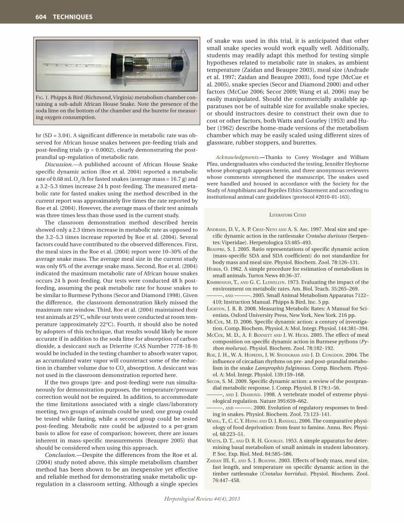
Herpetological Review 44(4), 2013
604 TECHNIQUES
hr (SD = 3.04). A significant difference in metabolic rate was ob-served for African house snakes between pre-feeding trials and post-feeding trials (p = 0.0002), clearly demonstrating the post-prandial up-regulation of metabolic rate.
Discussion.—A published account of African House Snake specific dynamic action (Roe et al. 2004) reported a metabolic rate of 0.68 mL O
2/h for fasted snakes (average mass = 16.7 g) and
a 3.2–5.3 times increase 24 h post-feeding. The measured meta-bolic rate for fasted snakes using the method described in the current report was approximately five times the rate reported by Roe et al. (2004). However, the average mass of their test animals was three times less than those used in the current study.
The classroom demonstration method described herein showed only a 2.3 times increase in metabolic rate as opposed to the 3.2–5.3 times increase reported by Roe et al. (2004). Several factors could have contributed to the observed differences. First, the meal sizes in the Roe et al. (2004) report were 10–30% of the average snake mass. The average meal size in the current study was only 6% of the average snake mass. Second, Roe et al. (2004) indicated the maximum metabolic rate of African house snakes occurs 24 h post-feeding. Our tests were conducted 48 h post-feeding, assuming the peak metabolic rate for house snakes to be similar to Burmese Pythons (Secor and Diamond 1998). Given the difference, the classroom demonstration likely missed the maximum rate window. Third, Roe et al. (2004) maintained their test animals at 25°C, while our tests were conducted at room tem-perature (approximately 22°C). Fourth, it should also be noted by adopters of this technique, that results would likely be more accurate if in addition to the soda lime for absorption of carbon dioxide, a desiccant such as Drierrite (CAS Number 7778-18-9) would be included in the testing chamber to absorb water vapor, as accumulated water vapor will counteract some of the reduc-tion in chamber volume due to CO
2 absorption. A desiccant was
not used in the classroom demonstration reported here.If the two groups (pre- and post-feeding) were run simulta-
neously for demonstration purposes, the temperature/pressure correction would not be required. In addition, to accommodate the time limitations associated with a single class/laboratory meeting, two groups of animals could be used; one group could be tested while fasting, while a second group could be tested post-feeding. Metabolic rate could be adjusted to a per-gram basis to allow for ease of comparison; however, there are issues inherent in mass-specific measurements (Beaupre 2005) that should be considered when using this approach.
Conclusion.—Despite the differences from the Roe et al. (2004) study noted above, this simple metabolism chamber method has been shown to be an inexpensive yet effective and reliable method for demonstrating snake metabolic up-regulation in a classroom setting. Although a single species
of snake was used in this trial, it is anticipated that other small snake species would work equally well. Additionally, students may readily adapt this method for testing simple hypotheses related to metabolic rate in snakes, as ambient temperature (Zaidan and Beaupre 2003), meal size (Andrade et al. 1997; Zaidan and Beaupre 2003), food type (McCue et al. 2005), snake species (Secor and Diamond 2000) and other factors (McCue 2006; Secor 2009; Wang et al. 2006) may be easily manipulated. Should the commercially available ap-paratuses not be of suitable size for available snake species, or should instructors desire to construct their own due to cost or other factors, both Watts and Gourley (1953) and Hu-ber (1962) describe home-made versions of the metabolism chamber which may be easily scaled using different sizes of glassware, rubber stoppers, and burettes.
Acknowledgments.—Thanks to Corey Woslager and William Pfau, undergraduates who conducted the testing, Jennifer Heyborne whose photograph appears herein, and three anonymous reviewers whose comments strengthened the manuscript. The snakes used were handled and housed in accordance with the Society for the Study of Amphibians and Reptiles Ethics Statement and according to institutional animal care guidelines (protocol #2010-01-163).
literature cited
andrade, d. V., a. P. cruZ-neto and a. s. aBe. 1997. Meal size and spe-cific dynamic action in the rattlesnake Crotalus durissus (Serpen-tes: Viperidae). Herpetologica 53:485–493.
BeauPre, s. J. 2005. Ratio representations of specific dynamic action (mass-specific SDA and SDA coefficient) do not standardize for body mass and meal size. Physiol. Biochem. Zool. 78:126–131.
huBer, o. 1962. A simple procedure for estimation of metabolism in small animals. Turtox News 40:36–37.
kimBrough, t., and g. c. lleWellyn. 1973. Evaluating the impact of the environment on metabolic rates. Am. Biol. Teach. 35:265–269.
———, and ———. 2005. Small Animal Metabolism Apparatus 7122–410; Instruction Manual. Phipps & Bird, Inc. 5 pp.
lighton, J. r. B. 2008. Measuring Metabolic Rates: A Manual for Sci-entists. Oxford University Press, New York, New York. 216 pp.
mccue, m. d. 2006. Specific dynamic action: a century of investiga-tion. Comp. Biochem. Physiol. A: Mol. Integr. Physiol. 144:381–394.
mccue, m. d., a. F. Bennett and J. W. hicks. 2005. The effect of meal composition on specific dynamic action in Burmese pythons (Py-thon molurus). Physiol. Biochem. Zool. 78:182–192.
roe, J. h., W. a. hoPkins, J. W. snodgrass and J. d. congdon. 2004. The influence of circadian rhythms on pre- and post-prandial metabo-lism in the snake Lamprophis fulginosus. Comp. Biochem. Physi-ol. A: Mol. Integr. Physiol. 139:159–168.
secor, s. m. 2009. Specific dynamic action: a review of the postpran-dial metabolic response. J. Comp. Physiol. B 179:1–56.
———, and J. diamond. 1998. A vertebrate model of extreme physi-ological regulation. Nature 395:659–662.
———, and ———. 2000. Evolution of regulatory responses to feed-ing in snakes. Physiol. Biochem. Zool. 73:123–141.
Wang, t., c. c. y. hung and d. J. randall. 2006. The comparative physi-ology of food deprivation: from feast to famine. Annu. Rev. Physi-ol. 68:223–51.
Watts, d. t., and d. r. h. gourley. 1953. A simple apparatus for deter-mining basal metabolism of small animals in student laboratory. P. Soc. Exp. Biol. Med. 84:585–586.
Zaidan iii, F., and s. J. BeauPre. 2003. Effects of body mass, meal size, fast length, and temperature on specific dynamic action in the timber rattlesnake (Crotalus horridus). Physiol. Biochem. Zool. 76:447–458.
Fig. 1. Phipps & Bird (Richmond, Virginia) metabolism chamber con-taining a sub-adult African House Snake. Note the presence of the soda lime on the bottom of the chamber and the burette for measur-ing oxygen consumption.

Herpetological Review 44(4), 2013
HERPETOCULTURE 605
Herpetological Review, 2013, 44(4), 605–610.© 2013 by Society for the Study of Amphibians and Reptiles
Captive Reproduction and Husbandry of Adult Ozark Hellbenders, Cryptobranchus alleganiensis bishopi
Hellbenders (Cryptobranchus alleganiensis) are large, aquat-ic salamanders inhabiting cool, highly oxygenated, fast-flowing streams in sections of eastern and central North America (Conant and Collins 1998; Nickerson and Mays 1973; Petranka 1998). Cur-rently, there are two described subspecies of Hellbenders (Croth-er 2012). The Eastern Hellbender (C. a. alleganiensis) occurs from New York south to Georgia and west to central Missouri, whereas the Ozark Hellbender (C. a. bishopi) is found in the Ozark High-lands of southern Missouri and northern Arkansas. Herein, we focus mainly on the Ozark Hellbender subspecies, but biology and reproductive behavior are similar for both subspecies. Cur-rently, Ozark Hellbender populations are known to occur in the North Fork of the White River, Bryant Creek, Eleven Point River, and Current River, with some individuals possibly still present in the Jacks Fork and Spring River (Briggler et al. 2007b). Historical-ly, Ozark Hellbenders were abundant in these rivers (Nickerson and Mays 1973; Peterson 1988; Trauth et al. 1992; USFWS 2011). Yet subsequent surveys indicate that populations have declined throughout the species’ range by at least 70% since the 1970s (Trauth et al. 1992; Wheeler et al. 2003) and revealed a shift in age class structure predominated by increasingly larger individuals and diminished or absent young age classes. Wild populations of Ozark Hellbenders were estimated to be approximately 600 indi-viduals, an extremely low number compared to an estimated car-rying capacity of over 11,000 individuals (Briggler et al. 2007b). In addition, Ozark Hellbenders currently exist as small isolated populations, making them vulnerable to individual catastrophic events and reducing the likelihood of re-colonization following localized extirpations.
As a result of drastic population declines, the Ozark Hell-bender was designated as critically imperiled and state endan-gered in Missouri in 2003 (MDC 2003). It has been protected from take or possession in Arkansas since 1990 (K. Irwin, pers. comm.), and was recently listed as federally endangered in 2011 (USFWS 2011). In 2001, the Ozark Hellbender Working Group was formed and is composed of individuals from Federal and State agencies, academia, zoos, non-governmental organizations, and individu-als interested in the conservation of the Hellbender. The group has played a significant role in identifying information needs and guiding conservation and recovery efforts for Hellbenders in Mis-souri and Arkansas, including development of comprehensive conservation strategies (Briggler et al. 2010; Briggler et al. 2012) that addresses captive propagation, augmentation, and reintro-duction goals, especially the need to captive-breed the species.
While the primary reason for population declines is un-known, several potential factors have been identified: degraded water quality, habitat alteration and degradation, disease, illegal harvest, human disturbances, and predation by non-native fish (USFWS 2011). The Ozark Hellbender population declines have necessitated the use of captive propagation efforts to ensure the long-term survival and recovery of the subspecies by maintain-ing/bolstering populations through augmentation and if neces-sary reintroduction while investigating the causes of the decline.
Herein we report our efforts on the first successful captive re-production and husbandry of the Ozark Hellbender at the Saint Louis Zoo (STLZ). Although the reproductive biology of wild Hellbenders is well-known (Nickerson and Mays 1973; Nick-erson and Tohulka 1986; Peterson 1988), there is limited infor-mation in the literature of captive-breeding of Cryptobranchus prior to this account. Smith (1907) was successful in artificial breeding of the Eastern Hellbender via mixing sperm and eggs obtained by sacrificing ripe females. Breeding habits of the giant cryptobranchids in Japan (Andrias japonicus) and China (A. da-vidianus) is relatively similar to the North American Hellbender (Okada et al. 2008; Zheng 2006). Biologists have been extremely successful in the captive-breeding of Andrias for several decades (Kuwabara et al. 1989; Qiang et al. 2012). However, attempts over the past decade or so by many zoos, including Saint Louis Zoo, and other institutions to naturally breed Hellbenders in captiv-ity have been unsuccessful (Wheeler and Trauth 2002; J. Ettling,
HERPETOCULTUREHERPETOCULTURE
JEFFERY A. ETTLING1*e-mail: [email protected] D. WANNER1
e-mail: [email protected] D. SCHUETTE1
e-mail: [email protected] L. ARMSTRONG1
e-mail: [email protected] S. PEDIGO1
e-mail: [email protected] T. BRIGGLER2
e-mail: [email protected]
1 WildCare Institute, Saint Louis Zoo, One Government Drive, St. Louis, Missouri 63110, USA
2 Missouri Department of Conservation, 2901 W. Truman Blvd, Jefferson City, Missouri 65109, USA
* Corresponding author

Herpetological Review 44(4), 2013
606 HERPETOCULTURE
pers. comm.). We believe the information provided herein will be useful in future efforts to captive-breed this species range-wide.
Brood stock.—Our goal was to obtain 8–10 wild, adult Ozark Hellbenders from each genetically distinct river drainage to es-tablish breeding groups (Sabatino and Routman 2009; Crow-hurst et al. 2011; Tonione et al. 2011). Therefore, breeding stock was collected from the North Fork of White River (NFWR), Eleven Point River (EPR), and Current River (CR). These animals were
collected from 2003–2007 for the NFWR, 2009–2011 for EPR, and 2010–2011 for CR. Each animal was im-planted with unique identification numbers using a passive integrated transponder (PIT) tag inserted into the tail region. To date STLZ has 8 animals (5 males, 3 females) from NFWR, 9 animals (5 males, 4 females) from EPR, and 9 animals (5 males, 4 females) from CR (Table 1). The sex ratio of breeding stock was slightly skewed towards males to provide increased mate se-lection for females and increased genetic diversity (Table 1). Different sizes of males were targeted for collection to ensure healthy sperm. If possible, only animals that appeared healthy were collected. How-ever, several of the animals obtained from the various rivers were compromised (e.g., missing appendages, open sores, etc.). The removal of animals was limited to only a few individuals per site to reduce effects on the resident population and to avoid inbreeding de-pression by collecting from several sites scattered along the river.
Quarantine.—Hellbender brood stock were quar-antined and housed separately by river of origin in 151.4-liter aquariums (91.4 x 40.6 x 30.5 cm) that were part of an eight-aquarium rack system with integrated filtration system. Animals remained in quarantine until appropriate disease (e.g., amphibian chytrid fungus [Batrachochytrium dendrobatidis-Bd], bacte-rial, viral, etc.) and health screening (e.g., CBC blood panel, parasites, physical conditions, etc.) tests were conducted by veterinarians and animal management staff. To minimize stress, animals were placed into the stream systems as quickly as possible and in most cases less than 30 days. All animals brought into the
STLZ were treated as if they were positive for Bd as this fungus is already known to occur in these river systems on Hellbend-ers (Briggler et al. 2007a, 2008). As part of the health screening, a minimum of two time-sequenced PCR assays were conducted for each individual to determine if the animal should be treated for Bd or managed as a Bd-positive animal in breeding stock al-ready infected with the fungus. Based on our experiences over past decade, it typically takes approximately six months for an adult Hellbender collected from the wild to adjust to captive conditions. To decrease stress and risk of mortality while the wild animals acclimate to captive conditions, abundant cover was provided for animals (e.g., multiple rocks, artificial shelters, covered tanks, etc.) and lighting was reduced during quarantine.
Artificial streams.—Adult brood stock were maintained and managed in one indoor artificial stream (NFWR brood stock) and two outdoor artificial streams (one for EPR brood stock and one for CR brood stock) once cleared from quarantine. The indoor stream, which is located in the basement of the Herpetarium, is 9.7 m long x 1.7 m wide x 0.6 m high with an average water depth of 0.3 m (Fig. 1). The indoor stream water flow (227 liters/min.) is more circular in nature due to the arrangement of a middle divider running the length of the stream and the filtration system input and output location within the stream. This indoor stream was operational since September 2002, but was not inhabited with brood stock until 2004. The two outdoor streams (11.3 m long x 1.5 m wide x 1.4 m high and 11.7 m long x 1.8 m wide x 1.4 m high) have been in operation since September 2011 (Fig. 2). The water input is located on one end of the stream and outputs are located in the middle and on the opposite end to allow water
taBle 1. Brood stock and associated data (mean, SD) for Ozark Hellbenders (Cryptobranchus alleganiensis bishopi) collected from the North Fork of the White River (NFWR), Current River (CR), and Eleven Point River (EPR), Missouri.
River Artificial stream No. No. SVL TL Mass males females (cm) (cm) (g)
NFWR Indoor 5 3 32.1 ± 3.0 47.0 ± 4.9 631.1 ± 194.0
CR Outdoor 5 4 28.7 ± 5.0 43.6 ± 7.0 449.9 ± 211.8
EPR Outdoor 5 4 28.2 ± 1.9 42.8 ± 3.4 491.3 ± 125.5
Fig. 2. Two outdoor artificial streams and associated life-support building at the Ron Goellner Center for Hellbender Conservation, Saint Louis Zoo, containing brood stock from the Current River and Eleven Point River.
Fig. 1. Indoor artificial stream at the Ron Goellner Center for Hellbender Con-servation, Saint Louis Zoo, containing brood stock from the North Fork of the White River.

Herpetological Review 44(4), 2013
HERPETOCULTURE 607
flow (454 liters/min) across the length and width of the stream. Water depth in the outdoor streams varies considerably to mimic natural contour of river bottom. Water depths are shallow (0.6 m) on the input and output ends with a gentle sloping towards the middle of the stream, thus reaching a maximum water depth of 1.2 m. The bottom of each stream was covered with 10.2–15.2 cm of natural substrate (gravel). A variety of large flat rocks (moss rock and/or sandstone) and artificial nest boxes (Briggler and Ackerson 2012) were provided for shelter and breeding places (Fig. 3). With the success of nest boxes in the wild (Briggler and Ackerson 2012), we installed 10 nest boxes on 23 August 2011 in each of the outdoor streams prior to breeding season, and later installed six nest boxes in the indoor stream on 14 November 2011. Nest boxes can be easily accessed for observation and egg collection, as well as provide secure locations for males to defend from other Hellbenders. A minimum of two cover rocks and/or nest boxes were provided per individual to allow animals to select.
For the breeding streams and their location (i.e., indoor verses outdoor), we attempted to mimic natural conditions (e.g., pho-toperiod, precipitation, water temperature, water quality, etc.) of the river of origin of the brood stock to reproductively cycle the animals. Water temperature for both the indoor and outdoor streams was controlled using large multi-temperature chillers. Water temperatures for each stream were adjusted daily for the actual temperature acquired via data loggers from the respec-tive river of origin of the brood stock. The water temperature of NFWR, EPR, and CR generally range between 4.4° and 22.2°C seasonally. Therefore, the outdoor streams also have boiler sys-tems to prevent the streams from dropping below 4.4°C during the winter months. For the indoor stream the precipitation and photoperiod were manipulated to simulate natural cycles experi-enced in southern Missouri. Photoperiod was adjusted daily us-ing a digital timer and precipitation was mimicked using a manu-ally controlled misting system and adjusting water levels (Fig. 3). For the outdoor raceways photoperiod and precipitation condi-tions were provided naturally. In addition, animals in the indoor stream are closely monitored with infrared video cameras.
All artificial streams have mechanical, chemical and biologi-cal filtration as well as ultraviolet sterilization. Dedicated keeper staff and assignment of separate equipment for each stream reduces potential of disease transmission via cross-contamina-tion. Water quality was intensively monitored to ensure animal health and encourage reproductive cycling. Water quality stan-dards in Table 2 are based on water quality management guide-lines established at STLZ staff for all hellbender streams and were recorded every two weeks or more often if warranted. A re-verse osmosis filtration system was added in summer 2011 to the indoor stream and in spring 2012 for the outdoor streams to bet-ter mimic ionic compositions occurring in the rivers. Total dis-solved solids (TDS) were adjusted similar to river conditions as it pertains to provide appropriate electrolyte balance, improving sperm production and defining the role of ionic composition on sperm activation. Chlorine and chloramines were removed from the water using carbon prefilters and/or dechlorinators, such as sodium thio-sulfate and/or reverse osmosis filtration.
Husbandry of brood stock.—In addition to mimicking water, substrate, and weather conditions to encourage natural breed-ing of the animals in the artificial streams, husbandry and health were important to ensure fitness of the animals to reproduce. A variety of foods were provided to meet the nutritional require-ments of the Hellbenders (Dierenfeld et al. 2009). Adults were
primarily fed live foods including crayfish (Orconectes spp., Pro-cambarus spp.), sculpins (Cottus bairdi, C. carolinae), darters (Ethoeostoma spp.), shiners (Notropis spp.), and night crawlers (Lumbricus terrestris). The fish species listed above are collected occasionally from the wild and are augmented in the artificial streams to better represent the natural ecosystem from which the brood stock originated. Commercially obtained night crawl-ers were fed at a rate of two worms/Hellbender/week. Frozen-thawed fish such as Lake Smelt (Osmerus mordax), Rainbow Trout (Oncorhynchus mykiss), and Atlantic Herring (Clupea harengus), with Thiamin E paste supplementation, were offered once per week via tong feeding. Live food items were cleaned, quarantined, and treated prior to feeding to reduce transmis-sion of ectoparasites, bacteria, fungus, and other pathogens.
Fig. 3. Habitat (shelter rock and artificial nest boxes) utilized by Ozark Hellbenders (Cryptobranchus alleganiensis bishopi) in the ar-tificial streams.
taBle 2. Water quality standards maintained in the artificial streams for Ozark Hellbenders (Cryptobranchus alleganiensis bishopi).
Parameter Standard
pH 7.6–8.2
Nitrate (NO3) < 10 mg/L
Nitrite (NO2) < 0.2 mg/L
Nitrogen Ammonia (NH3) < 0.05 mg/L
Phosphates (PO4) < 0.40 mg/L
% Dissolved Oxygen (DO) > 90%
Total Dissolved Solids (TDS) 175–300 mg/L

Herpetological Review 44(4), 2013
608 HERPETOCULTURE
Treatment and cleaning included: 1) soaking live crayfish for five minutes in a 50 ppt salt solution (dosed at 50 g/L), then rinsing in a fresh water solution for five minutes (Johnson et al. 2003); 2) rinsing soil from night crawlers; and 3) maintaining/isolating live fish for a three week period in a 5–7 ppt salt solution. Fish were also monitored during the quarantine period for signs of disease and/or parasites. Additional food items were provided April to June and November to January to increase the fitness of the animals prior to and immediately following the breed-ing season. Records were maintained for each individual Hell-bender and included: 1) quarterly physical examinations, mass and length; 2) food intake, with conversion rates and graphs of weight changes over time; 3) medical; and 4) behavior.
Reproduction.—Since 2002 many actions were employed to mimic the seasonal environmental re-gimes (e.g., photoperiod, water temperature, and precipitation patterns) of the natural river conditions in an effort to trigger breeding activity in the artificial streams, as well as to increase fitness of the animals to produce gametes. Males developed the typical cloa-cal “donut” swelling and females exhibited abdominal swelling associated with egg production similar to wild animals. Between 2007 and 2011 eggs were laid every year in the indoor stream for a total of 10 clutch-es (Table 3), which all proved to be infertile. However, on 18 October 2011 two clutches of fertilized eggs with different stages of development were discovered in one of the nest boxes in the outdoor stream housing the EPR Ozark Hellbender population (Fig. 4), eventu-ally yielding 151 larval Hellbenders. As noted earlier, this population is comprised of five males and four females. These animals were removed from the wild from 2009 to 2011 and were augmented into the out-door stream on 20 September 2011. The two females that deposited eggs were recently collected from the wild (August and September 2011). However, the male that fertilized the eggs and guarded the nest was housed and environmentally cycled indoors since 25 August 2009.
In 2012, natural reproduction occurred in both outdoor streams (EPR and CR brood stock) and finally the indoor stream (NFWR brood stock) resulting in eight fertilized egg clutches and the sequential hatching of over 2500 embryos. All three females deposited eggs in the indoor stream with the NFWR brood stock, two females in the EPR brood stock in one outdoor stream, and three females in the CR brood stock in the other outdoor stream (Table 3). Dates of egg laying in 2012 were earlier than previous years with eggs being laid from 22 September to 01 October (Table 3). All eggs were laid in nest boxes, and in two instances (NFWR and CR) two different females laid eggs within one nest box. In all cases, males guarded the nest site by protecting the nest box entrance tunnel from conspecifics. The male that successfully guarded the first fertilized clutch in the EPR outdoor stream in 2011 was not the guardian of either fertilized clutch in 2012.
While checking nest boxes on 01 October 2012, we witnessed one male and one female in the act of spawning in EPR outdoor stream. The two animals were in a circular position with heads and tails overlapping. There were three visible eggs protruding from her cloaca, and the inside of the nest box chamber was foaming with seminal fluid in a cloudy mass or stringy threads. Immediately the lid of the nest box was put into place to decrease disturbance, and on the following morning the nest chamber was full of fertilized (cleavage furrows visible) eggs.
In the artificial streams it is typical for animals to share cover rocks and nest boxes outside of the breeding season. This chang-es in late summer however, as males become increasingly aggres-sive and establish themselves in certain nest boxes or cover rocks as the spawning season approaches in late-September to mid-October. Prior to the 2012 breeding season, aggression among the animals was relatively minor; however, during the major breeding event that occurred in 2012 several animals (males and females) had lost limbs, severe lacerations on the limbs, and bite marks along the lateral folds.
Discussion.— Intensive captive propagation efforts for the Ozark Hellbender have been pursued at the Saint Louis Zoo
taBle 3. Egg-laying history and associated estimated dates for female Ozark Hellbenders (Cryptobranchus alleganiensis bishopi) collected for brood stock from North Fork of White River, Current River, and Eleven Point River, Missouri. * = did not lay eggs; 1 = two females deposited eggs within one nest box for their respective stream.
PIT Tag No. 2007 2008 2009 2010 2011 2012
North Fork of White River (indoor stream)
102-806-018 13 Oct * 14 Oct 04 Oct 07 Oct 22 Sept1
066-882-540 * 9 Oct 14 Oct 08 Oct 10 Oct 25 Sept
093-624-774 18 Oct * * * 18 Oct 22 Sept1
Current River (outdoor stream)
051-599-288 – – – – * 28 Sept1
051-584-041 – – – – * *
066-292-573 – – – – * 01 Oct
066-284-079 – – – – * 01 Oct1
Eleven Point River (outdoor stream)
066-290-098 – – – – 02 Oct1 22 Sept
094-070-830 – – – – * *
057-810-624 – – – – 10 Oct1 *
075-341-564 – – – – * 01 Oct
Fig. 4. A fertilized clutch of Ozark Hellbender (Cryptobranchus al-leganiensis bishopi) eggs with attending male in an artificial nest box in an outdoor stream. Nest box lid was removed to view and photo-graph nest chamber.
Herpetological Review 44(4), 2013
HERPETOCULTURE 609
since 2002. Seasonal changes in photoperiod, water tempera-ture, and precipitation patterns are thought to trigger breeding activity (Nickerson and Mays 1973). Through seasonal manipu-lation of the water temperature, photoperiod, precipitation, and feeding regime, Hellbenders housed in the indoor stream consis-tently cycled at exactly the same time as their wild counterparts (JTB, unpubl. data). For example, when eggs were being laid in the wild on the NFWR, the NFWR brood stock at the STLZ would be laying either on the same day or within a day or two. Even with consistent egg-laying in the indoor stream since 2007, fer-tilization did not occur until 2012 in the indoor stream. Further investigation revealed that the males comprising our breeding group were producing sperm with deformities and/or low mo-tility. Based on a review of the literature the ionic composition of the water appears to affect sperm development and activa-tion during spawning for fishes (Yeung et al. 2006). With that in mind, the use of reverse osmosis filtration was put into place in August 2011 for the indoor stream and May 2012 for the outdoor streams to adjust the ionic composition (e.g., TDS) of water used in our Hellbender streams in an effort to closely match that of river water. An evaluation of sperm quality on 08 October 2011 for males housed in the indoor raceway indicated motility of 85%, the highest recorded for these animals to date (T. Crabill, pers. comm.). Follow-up sperm assessments in 2012 showed in-creased sperm quality for animals in all three streams, thus re-sulting in successful fertilization of all eight clutches deposited by females.
In addition to adjusting the ionic composition of the streams, the nest boxes also played a significant role in the increased level of success occurring in 2011 and 2012. The first fertilized clutch of eggs occurred in a nest box installed in summer 2011 in the outdoor streams. With the success of this fertilized clutch at the STLZ and in the wild (Briggler and Ackerson 2012), nest boxes were later installed in the indoor stream for the upcoming 2012 breeding season, thus resulting in eight fertilized clutches in six nest boxes in both the indoor and outdoor streams. In previous years, it was not uncommon for both male and female Hellbend-ers in the indoor stream to consume conspecifics eggs under cover rocks provided. Although cannibalism of eggs is known to occur in wild populations (Bishop 1941; Smith 1907), it seemed difficult for males in the streams at the STLZ to protect nests under cover rocks from conspecifics due to many animals that would dig their way into the nest chamber from various sides of the cover rock to consume or remove eggs. Therefore, we believe the nest boxes not only provided ideal conditions for success-ful spawning, but also provided a long, narrow, single-entrance tunnel from which males could protect nests from conspecifics. Success towards achieving our goal to naturally breed Hellbend-ers in captivity was steady in the earlier years; however, we be-lieve the addition of nest boxes and mimicking ionic composi-tions of the river water paved the way to the remarkable breeding successes that occurred in the past two years.
Considerable Hellbender life history data were observed during the process to achieve captive-breeding of the species. It is unclear if female Hellbenders in the wild lay eggs annually. However, Topping and Ingersol (1981) and Peterson et al. (1989) data suggest that not all gravid females lay their eggs during the same season. For the NFWR brood stock, two females consis-tently laid eggs annually while the third female rarely laid eggs; however, the health of this animal was compromised in 2009 and 2010 (Table 3). We have limited egg-laying history for CR and EPR brood stock, but one female deposited eggs in both 2011
and 2012 (Table 3). Similar to wild Hellbenders, there appears to be variation among captive-held females for their egg laying his-tories, but it is not uncommon for females to lay eggs annually.
In all cases of successful fertilization of eggs in 2011 and 2012, an individual male protected and remained with his respective clutches by defending the nest box entrance tunnel similar to natural nests found in the wild (Nickerson and Mays 1973; JTB, pers. obs.). However, this does not preclude males foraging on their own eggs as documented via ultrasound by EPR male in 2011. Based on our observation of all streams and video evidenc-es of the indoor stream, it appeared that guardian males were the sole gamete contributors to their respective clutches. How-ever, multiple paternity has been reported within a nest cavity for the related Andrias species (Kawamichi and Ueda 1998), and several male Hellbenders have been observed within a nest cav-ity in the wild in Missouri (JTB, pers. obs.), but only one male attends the nest. To further examine whether multiple paternity has occurred within these artificial streams, we plan to conduct genetic parenting of fertilized clutches. Also, on three occasions multiple maternity was witnessed within a given nest box in each of the streams (Table 3); this is further supported in the literature that males will accept several females into the nest cavity (Bish-op 1941; Crowhurst et al. 2009; Smith 1907). Hellbender behavior and reproduction (e.g., multi-parenting, egg laying history, nest box selection, etc.) observed in captivity will likely provide ad-ditional insight into the breeding activities of wild populations.
Conclusion.—The first successful captive reproduction of Ozark Hellbenders in 2011, repeated in 2012, at the STLZ was believed to be the result of a combination of six factors: 1) Space—providing ample space allows individual animals the opportunity to establish their own home territory. With this in mind, the streams were designed and constructed to be as large as possible. 2) Cover rocks and nesting sites—a combination of large rocks and nest boxes were provided in both the indoor and outdoor streams. Appropriate nesting sites are ones that can be easily defended by the attending male. 3) Environmental cy-cling—simulation of seasonal photoperiod, water temperature, and precipitation changes resulted in the Hellbenders becom-ing reproductive at the same time as their wild counterparts. 4) Water quality—maintenance of high water quality standards, in-cluding adjustments to ionic composition, dissolved solids, etc., was vital to overall health including sperm development and ac-tivation. 5) Sex ratio—a good ratio of both sexes simulates natu-ral breeding dynamics and provides females with mate selection opportunities. 6) Animal health—increase food availability dur-ing late winter and early spring to improve fitness of animals to produce gametes. The factors described above could be applied to other ex situ management plans in zoos and conservation organizations in need of propagating the species range-wide. Through the significant achievement of the successful captive-breeding of the Ozark Hellbender in the past two years at the STLZ, many of these offspring will be released into the wild to bolster populations.
Acknowledgments.—This work was funded and/or supported by the Saint Louis Zoo, Missouri Department of Conservation, U.S. Fish and Wildlife Service, U.S. Forest Service, National Park Service, and Arkansas Game and Fish Commission. This manuscript is dedicated in the memory of R. Goellner of the Saint Louis Zoo for his passion and vision to initiate the captive-breeding program of this species, as well as to the dedicated efforts of K. Goellner for her involve-ment, passion, and being an avid champion for the species. Special

Herpetological Review 44(4), 2013
610 HERPETOCULTURE
thanks to T. Crabill and S. Boutelle for sperm assessments over the years, K. Irwin for involvement and encouragement, and Y. Huang for hormone assessments. We also thank J. Ackerson, C. Bodinof, T. Crabill, K. Irwin, K. Goellner, R. Goellner, B. Heatherly, C. Hoessle, M. Laturno, J. Kiger, B. Phillips, A. J. Pratt, J. Quinn, R. Rimer, R. Roy, P. Taylor, R. Turnquist, J. Utrup, B. Wanner, and D. Woods for assis-tance in the field collection of brood stock, and other individuals (too many to mention) assisting with various aspects of captive propaga-tion efforts. We acknowledge K. Kuwabara of Japan and L. Gang of China for sharing their knowledge and expertise of captive breeding of Andrias. Research was conducted in compliance with applicable animal care guidelines and appropriate permits. Brood stock was collected by J. Briggler of the Missouri Department of Conservation under the authority of the Wildlife Code of Missouri.
literature cited
BishoP, s. c. 1941. The Salamanders of New York. New York State Mus. Bull. 324:1–365.
Briggler, J. t., and J. r. ackerson. 2012. Construction and use of arti-ficial shelters to supplement habitat for hellbenders (Cryptobran-chus alleganiensis). Herpetol. Rev. 43(3):412–416.
———, t. l. craBill, k. J. irWin, c. daVidson, J. a. ciViello, m. d. Wan-ner, c. d. schuette, s. l. armstrong, V. grant, t. daVidson, and J. a. ettling. 2012. Propagation, Augmentation, and Reintroduction Plan for the Ozark Hellbender (Cryptobranchus alleganiensis bish-opi), Ozark Hellbender Propagation Committee, Jefferson City, Missouri. 30 pp.
———, ———, ———, ———, J. utruP, and a. salVeter (eds.). 2010. Hellbender Conservation Strategy: An Action Plan for the Recov-ery of the Ozark and Eastern Hellbender in the Ozark Highlands of Missouri and Arkansas. Ozark Hellbender Working Group, Jef-ferson City, Missouri. 59 pp.
———, J. ettling, m. Wanner, c. schuette, m. duncan, and k. goellner. 2007a. Cryptobranchus alleganiensis (hellbender). Chytrid fungus. Herpetol. Rev. 38:174.
———, k. a. larson, and k. J. irWin. 2008. Presence of the amphibian chytrid fungus (Batrachochytrium dendrobatidis) on hellbenders (Cryptobranchus alleganiensis) in the Ozark Highlands. Herpetol. Rev. 39:443–444.
———, J. utruP, c. daVidson, J. humPhries, J. groVes, t. Johnson, J. et-tling, m. Wanner, k. traylor-holZer, d. reed, V. lingren, and o. Byers (eds.). 2007b. Hellbender Population and Habitat Viability Assess-ment: Final Report. IUCN/SSC Conservation Breeding Specialist Group, Apple Valley, Minnesota. 118 pp.
conant, r., and J. t. collins. 1998. A Field Guide to Reptiles and Am-phibians of Eastern and Central North American. Houghton-Miff-lin Co., Boston, Massachusetts. 616 pp.
crother, B. i. (ed.). 2012. Scientific and Standard English Names of Amphibians and Reptiles of North America North of Mexico, with Comments Regarding Confidence in our Understand (7h ed.). So-ciety for the Study of Amphibians and Reptiles. Herpetological Circular No. 39.
croWhurst, r. s., J. t. Briggler, J. B. koPPelman, k. m. lohraFF, and J. a. ciViello. 2009. Cryptobranchus alleganiensis alleganiensis (eastern hellbender): multiple clutches. Herpetol. Rev. 40:195.
———, k. m. Faries, J. collantes, J. t. Briggler, J. B. koPPelman, and l. s. eggert. 2011. Genetic relationships of hellbenders in the Ozark highlands of Missouri and conservation implications for the Ozark subspecies (Cryptobranchus alleganiensis bishopi). Conserv. Gen-et. 12:637–646.
dierenFeld, e. s., k. J. mcgraW, k. Fritsche, J. t. Briggler, and J. ettling. 2009. Nutrient composition of whole crayfish (Orconectes and
Procambarus species) consumed by hellbender (Cryptobranchus alleganiensis). Herpetol. Rev. 40:324–330.
Johnson, m. l., l. Berger, l. PhiliPs, and r. sPeare. 2003. Fungicidal effects of chemical disinfectants, UV light, desiccation and heat on the amphibian chytrid Batrachochytrium dendrobatidis. Dis. Aquat. Organ. 57:255–260.
kaWamichi, t., and h. ueda. 1998. Spawning at nest extra-large males in the giant salamander Andrias japonicus. J. Herpetol. 32:133–136.
kuWaBara, k., n. suZuki, F. WakaBayashi, h. ashikaga, t. inoue, and J. ko-Bara. 1989. Breeding the Japanese giant salamander (Andrias ja-ponicus) at Asa Zoological Park. Int. Zoo. Ybk. 28:22–31.
[mdc] missouri dePartment oF conserVation. 2003. Missouri Species and Communities of Conservation Concern Checklist. Jefferson City, Missouri. 29 pp.
nickerson, m. a., and c. e. mays. 1973. The Hellbenders: North Ameri-can “Giant Salamanders.” Milwaukee Public Museum, Milwaukee, Wisconsin. 106 pp.
———, and m. d. tohulka. 1986. The nest and nest site selection by Ozark hellbenders, Cryptobranchus alleganiensis bishopi Grob-man. Trans. Kansas Acad. Sci. 89:66–69.
okada, s, t. utsunomiya, t. okada, Z. i. Felix, and i. Fumihiko. 2008. Characteristics of Japanese giant salamander (Andrias japonicus) populations in two small tributary streams in Hiroshima Prefec-ture, western Honshu, Japan. Herpetol. Conserv. Biol. 3:192–202.
Peterson, c. l. 1988. Breeding activities of the hellbender in Missouri. Herpetol. Rev. 19:28–29.
———, c. a. ingersol, and r. F. Wilkinson. 1989. Winter breeding of Cryptobranchus alleganiensis bishopi in Arkansas. Copeia 1989(4):1031–1035.
Petranka, J. W. 1998. Salamanders of the United States and Canada. Smithsonian Institution Press, Washington, DC. 587 pp.
Qiang, d, W. yueZhao, and l. gang. 2012. Conservation status of Chi-nese giant salamander (Andrias davidianus). Chengdu Institute of Biology, Chinese Acad. Sci. 44 pp.
saBatino, s. J., and e. J. routman. 2009. Phylogeography and conserva-tion genetics of the hellbender salamander (Cryptobranchus alle-ganiensis). Conserv. Genet. 10(5):1235–1246.
smith, B. g. 1907. The life history and habits of Cryptobranchus al-leganiensis. Biol. Bull. 13:5–39.
trauth, s. e., J. d. Wilhide, and P. daniel. 1992. Status of the Ozark hellbender, Cryptobranchus bishopi (Urodela, Cryptobranchidae) in the Spring River, Fulton County, Arkansas. Proc. Arkansas Acad. Sci. 46:83–86.
tonione, m., J. r. Johnson, and e. J. routman. 2011. Microsatellite anal-ysis supports mitochondrial phylogeography of the hellbender (Cryptobranchus alleganiensis). Genetica 139:209–219.
toPPing, m. s., and c. a. ingersol. 1981. Fecundity in the hellbender, Cryptobranchus alleganiensis. Copeia 1981(4):873–876.
[usFWs] u.s. Fish and WildliFe serVice. 2011. Endangered and threat-ened wildlife and plants: endangered status for the Ozark hell-bender salamander. Fed. Reg. 76(194):61956–61978.
Wheeler, B. a., e. Prosen, a. mathis, and r. F. Wilkinson. 2003. Popula-tion declines of a long-lived salamander: a 20+ year study of hell-benders, Cryptobranchus alleganiensis. Biol. Conserv. 109:151–156.
———, and s. e. trauth. 2002. Captive propagation of the Ozark hellbender. Report, Arkansas Game and Fish Commission, Little Rock, Arkansas. 19 pp.
yeung, c. h., J. P. BarField, and t. g. cooPer. 2006. Physiological volume regulation by spermatozoa. Mol. Cell. Endocrinol. 205:98–105.
Zheng, h. x. 2006. The population ecology and ecological adaptation characteristic of the Chinese giant salamander (Andrias davidi-anus) in Lushi County, Henan Province, China. Ph.D. dissertation. East China Normal University. [In Chinese, with English abstract]

Herpetological Review 44(4), 2013
AMPHIBIAN DISEASES 611
Herpetological Review, 2013, 44(4), 611–614.© 2013 by Society for the Study of Amphibians and Reptiles
Detection of Batrachochytrium dendrobatidis in River Frogs (Genus Amietia) on Mount Kilimanjaro, Tanzania
Within Africa, the amphibian chytrid fungus Batrachochy-trium dendrobatidis (Bd) has been reported from southern, east-ern, and central parts of the continent, whereas it is currently undetected in West Africa (Penner et al. 2013 and citations there-in). Although Bd has been found in some African species with-out aquatic larval stages (Bell et al. 2011; Conradie et al. 2011a; Gower et al. 2012), the majority of species affected by the fungus are from high altitude and inhabit mostly flowing streams (Mur-ray and Skerratt 2012). River frogs of the genus Amietia seem to be particularly susceptible, showing the highest zoospore loads known among African frogs in Kenya (Kielgast et al. 2010), Ma-lawi (Conradie et al. 2011a), South Africa, and Lesotho (Weldon 2005). Bd infection also has been detected in Uganda (Viertel et al. 2012), yet records from the Udzungwa Mountains in Tanzania have not detected Bd (Moyer and Weldon 2006). Here, we report the occurrence of Bd in Amietia angolensis and A. wittei from Mt. Kilimanjaro, northern Tanzania.
Mount Kilimanjaro is the remnant of a large volcano located on the border with Kenya (3.07583°S, 37.35333°E; Fig. 1). It rises from a hot and dry savanna plain at 700 m to an ice-clad summit of 5895 m, thereby encompassing several different bioclimatic zones. At 1700 m, the borders of the Kilimanjaro National Park delimit the montane forest, whereas the lower part of the slope has been almost entirely converted into a traditional agroforest-ry system (mainly banana and coffee trees) and monocultures (mainly maize, sunflowers, and beans). Amietia angolensis in-habits the lowland (1000–1700 m), whereas A. wittei occurs from 1700 m to 3500 m on the Shira Plateau (GZ, unpubl. data).
Samples for Bd analysis were collected in 2011 (from March to June–long rains; October and November–short rains) during surveys of Amietia tadpoles for other research purposes. Be-cause we observed tadpoles with jaw sheaths and tooth rows completely colorless (Fig. 2d), we gently brushed the mouthparts with common cotton swabs and stored them dry in safe-lock tubes. Diagnostic analysis was performed in October 2012 at the Amphibian Disease Diagnostic Center at Washington State Uni-versity (Pullman, Washington, USA). Fungal DNA was extracted using DNeasy extraction kits (Qiagen). Detection and quantifi-cation of Bd was performed using standards (1000, 100, 10, 1, and 0.1) from serial dilutions by means of RT-qPCR according to Boyle et al. (2004).
Although sample size was low (N = 17), we detected Bd in at least 40% of the specimens for both species (Table 1). Overall, the Bd infection levels appeared low in individual samples (< 5 zoospore equivalents). We believe these results do not reflect
realistic levels of pathogen load, but they are very likely due to a combination of technical factors. First, this non-lethal tech-nique for detecting Bd on tadpoles is less effective than histo-logical diagnosis (Retallick et al. 2006). In their research, the au-thors scraped the tadpoles’ mouthparts with wooden toothpicks whereas we used swabs which are less invasive (i.e., they are less likely to damage mouthparts and influence the ability of tadpoles to feed). However, swabs may not collect Bd DNA as effectively as toothpicks, which regularly break pieces of labial teeth (Retallick et al. 2006). Second, during field work, samples were stored in a freezer to avoid high temperatures, but the power supply at the field station was irregular, resulting in frequent blackouts and consequent thawing of the freezer’s contents. High tempera-tures and long storage periods can result in reduced recovery of Bd DNA from swabs (van Sluys et al. 2008). These factors likely induced degradation of DNA in our samples; still, we were able to detect Bd, suggesting that the pathogen load may have been high originally.
This is the first record of Bd-positive Amietia from Tanzania. All specimens presenting depigmentation of keratinized mouth-parts were Bd-positive; however, the sample size was too low for a statistically significant correlation between oral deformity and Bd presence. During the whole field season, we observed larval deformities with similar gross morphology at other sites, but we never encountered dead or moribund adults of Amietia or of other species occurring in the area. Similar observations of a general lack of mortality among Bd-infected animals have been reported in other studies from Africa, including Kenya (Kielgast et al. 2010), Uganda (Goldberg et al. 2007), and South Africa (Conradie et al. 2011b). The high prevalence of Bd and apparent low virulence may suggest that the pathogen is enzootic within those regions, thereby providing support for the “out of Africa” hypothesis (Weldon et al. 2004). However, a lack of connection
GIULIA ZANCOLLI*Department of Animal Ecology and Tropical Biology, Biocentre, University of Würzburg, Am Hubland, 97074 Würzburg, GermanyANDREW STORFER School of Biological Sciences, Washington State University, Pullman, Washington 99164, USAMARK-OLIVER RÖDELMuseum für Naturkunde, Leibniz Institute for Research on Evolution and Biodiversity, Invalidenstr. 43 10115 Berlin, Germany *Corresponding author; e-mail: [email protected]
AMPHIBIAN DISEASESAMPHIBIAN DISEASES

Herpetological Review 44(4), 2013
612 AMPHIBIAN DISEASES
between Bd presence and population decline has been reported elsewhere, for instance in eastern North America (Longcore et al. 2007). The question about the origin of Bd remains open.
Kielgast et al. (2010) suggested a taxonomic basis for varia-tion in apparent Bd susceptibility among host species, and the genus Amietia, in particular, showed the highest pathogen load and prevalence in their study. Other studies have also provided support for aquatic life histories contributing to Bd occurrence patterns (e.g., Bancroft et al. 2011; Olson et al. 2013). During our surveys, we regularly observed tadpoles of both species in the water. Amietia angolensis adults are semi-aquatic and active throughout the year (Channing and Howell 2006); the tadpoles have a prolonged development and may take up to two years to complete metamorphosis (Channing 2004). Thus, river frogs can act as reservoirs for Bd and facilitate disease persistence in the system. Moreover, Amietia species cover most of the elevational range of Mt. Kilimanjaro, and this might allow Bd to survive dur-ing unfavorable conditions within spatially disparate microhabi-tats (e.g., by persisting at higher elevations during summer and at lower elevations during winter).
Local dispersal of Bd may be facilitated by humans. Kiliman-jaro National Park is a tourist attraction with over 35,000 climbers a year (Peaty 2012), and each climber has at least three porters to conform with park regulations. The first campsites encoun-tered along the climbing routes are usually allocated at the upper ridge of the forest (ca. 3000 m) and close to ponds or streams which are used by A. wittei for breeding (GZ, unpubl. data). We observed porters collecting water (e.g., for cooking and human
consumption) at the same water bodies where tadpoles tested Bd-positive. Porters are able to walk through the mountain for long distances within a day, and this may translocate zoospores from one site to another. Below the national park borders, the slope is characterized by an extensive network of water canals built by the local human population for irrigation, and by which Bd may disperse, or river frogs or other transmission vectors may spread the pathogen (Morgan et al. 2007; Johnson and Speare 2005).
Our results showed the presence of Bd infection in river frog populations on Mt. Kilimanjaro. Considering the high suscepti-bility of Amietia species and their potential to serve both as Bd reservoirs and vectors to nearby areas (especially the lowland A. angolensis), it would be beneficial to intensify surveys for Bd detection in the East African highlands and in particular in the Eastern Arc Mountains. The few data available to date are from the southern Udzungwa Mountains, where Bd has been detected in 12 species (Moyer and Weldon 2006; www.Bd-maps.net, ac-cessed 16 April 2013), and from the northern East Usambara Mountains with negative records dated 2006 (www.Bd-maps.net). This hotspot of biodiversity harbors an outstanding num-ber of endemic species (Burgess et al. 2007) that may still be na-ïve to the fungus and thus will suffer population declines if the infection reaches those forests.
Fig. 1. Mount Kilimanjaro, Tanzania, where Batrachochytrium den-drobatidis (Bd) was detected. Records of Bd infections from the East-ern Arc Mountains are still poor (www.bd-maps.net, accessed 16 April 2013).
Fig. 2. Typical creek within the subalpine zone at ca. 3000 m, Kili-manjaro National Park, Tanzania; a) breeding site of Amietia wittei close to the Machame Huts campsite where porters collect water; b) larvae of Amietia wittei; c) oral disc with unchanged morphology; d) mouthparts with depigmentation of jaw sheaths and labial teeth.

Herpetological Review 44(4), 2013
AMPHIBIAN DISEASES 613
Acknowledgments.−This study was financed by a grant of the German Excellence Initiative to the Graduate School of Life Sciences, University of Würzburg. We acknowledge the Tanzanian Commission for Science and Technology (COSTECH; research permit 2010-363-NA-96-44), Tanzania National Parks (TANAPA), Tanzania Wildlife Research Insti-tute (TAWIRI) and Kilimanjaro National Park for issuing the research permits. Field work was possible thanks to Ingolf Steffan-Dewenter, the DFG-Research Unit FOR1246, and the personnel at the research station in Nkweseko. We thank Oli-ver Konopik for taking care of shipping the samples to Wash-ington State University, and to Jesse Brunner for suggesting writing this paper.
literature cited
BancroFt, B. a., B.a. han, c. l. searle, l. m. Biga, d. h. olson, l. B. kats, J. J. laWler, and a. r. Blaustein. 2011 Species-level correlates of susceptibility to the pathogenic amphib-ian fungus Batrachochytrium dendrobatidis in the United States. Biodivers. Conserv. 20:1911–1920.
Bell, r. c., a. V. g. garcia, B. l. stuart, and k. r. Zamudio. 2011. High prevalence of the amphibian chytrid pathogen in Ga-bon. EcoHealth 8:116–120.
Boyle, d. g., d. B. Boyle, V. olsen, J. a. t. morgan, and a. d. hy-att. 2004. Rapid quantitative detection of chytridiomycosis (Batrachochytrium dendrobatidis) in amphibian samples using real-time Taqman PCR assay. Dis. Aquat. Org. 60:141–148.
Burgess, n.d, t. Butynski, n. cordeiro, n. doggart, J. FJeldså, k. m. hoWell, F. kilahama, s. P. loader, J. c. loVett, and B. mBilinyi. 2007. The biological importance of the Eastern Arc Mountains of Tanzania and Kenya. Biol. Conserv. 134:209–231.
channing, a. 2004. Afrana angolensis (Bocage, 1866). In L. R. Minter, M. Burger, J. A. Harrison, H. H. Braack, P. J. Bish-op, and D. Kloepfer (eds.), Atlas and Red Data Book of the Frogs of South Africa, pp. 270–271. Smithsonian Institution, Washington D.C.
———, and k. m. hoWell. 2006. Amphibians of East Africa. Cornell University Press, New York.
conradie, W., J. harVey, a. kotZé, d. l. dalton, and m. J. cun-ningham. 2011a. Confirmed amphibian chytrid in Mount Mulanje area, Malawi. Herpetol. Rev. 42:369–371.
———, c. Weldon, k. g. smith, and l. h. du PreeZ. 2011b. Sea-sonal pattern of chytridiomycosis in common river frog (Amietia angolensis) tadpoles in the South African Grass-land Biome. Afr. Zool. 46:95–102.
goldBerg, t. l., a. m. readel, and m. h. lee. 2007. Chytrid fun-gus in frogs from an equatorial African montane forest in western Uganda. J. Wildl. Dis. 43:521–524.
goWer, d. J., t. m. doherty-Bone, r. k. aBerra, a. mengistu, s. scwaller, M. Menegon, r. De sá, s. a. saBer, a. a. cunnInghaM, and s. P. loader. 2012. High prevalence of the amphibian chytrid fungus (Batrachochytrium dendrobatidis) across multiple taxa and localities in the highlands of Ethiopia. Herpetol. J. 22:225–233.
Johnson, m. l., and r. sPeare. 2005. Possible modes of dissemi-nation of the amphibian chytrid Batrachochytrium dendro-batidis in the environment. Dis. Aquat. Org. 65:181–186.
kielgast, J., d. rödder, m. Veith, and s. lötters. 2010. Wide-spread occurrence of the amphibian chytrid fungus in Ke-nya. Anim. Conserv. 13, Suppl.1 (2010):36–43.
longcore, J. r., J. e. longcore, a. P. Pessier, and W. a. halteman. 2007. Chytridiomycosis widespread in anurans of north-eastern United States. J. Wildlife Manage. 71:435–444.
morgan, J. a. t., V. t. VredenBurg, l. J. rachoWicZ, r. a. knaPP, m. J. stice, t. tunstall, r. e. Bingham, J. m. Parker, J. e. longcore, t
aB
le 1
. Bat
rach
och
ytri
um
den
dro
bati
dis
infe
ctio
n d
ata
for
Am
ieti
a an
gole
nsi
s an
d A
. wit
tei f
rom
Mo
un
t Kil
iman
jaro
, Tan
zan
ia. A
ll sa
mp
les
are
fro
m la
rvae
. Bec
ause
of t
he
extr
emel
y lo
w a
mo
un
t o
f Bd
DN
A, i
t was
no
t po
ssib
le to
det
erm
ine
Bd
pre
sen
ce w
ith
ab
solu
te c
erta
inty
for
som
e sa
mp
les.
Un
cert
ain
res
ult
s ar
e re
po
rted
as
susp
ect p
osi
tive
(su
sp.p
os.
) o
r n
egat
ive
(su
sp.n
eg.)
.
Spec
ies
Dat
es
Loca
liti
es
Co
ord
inat
es
Ele
vati
on
W
ater
bo
die
s N
Bd
-po
siti
ve
N
Zo
osp
ore
M
ou
thp
arts
(m)
and
hab
itat
sa
mp
les
sam
ple
s eq
uiv
alen
ts
(m
in–m
ax)
A. a
ngo
len
sis
18 O
ct 2
011
War
amu
riv
er
3.19
59°S
15
32
stre
am
0 1
n
orm
al
37.2
555°
E
cu
ltiv
ated
are
a
A. a
ngo
len
sis
18 O
ct 2
011
Nku
u
3.18
08°S
16
92
cree
k 4
5 0.
001–
2.62
1 d
epig
men
ted
37
.251
1°E
cult
ivat
ed a
rea
(2 s
usp
.neg
.)
A. a
ngo
len
sis
29 O
ct 2
011
Um
bwe
rou
te
3.18
45°S
17
05
stre
am
0 1
n
orm
alA
. wit
tei
37.2
789°
E
d
istu
rbed
fore
st
A. w
itte
i 05
May
201
1 M
wek
a ro
ute
3.
2038
°S
1992
cr
eek
0 1
n
orm
al
37.3
485°
E
m
on
tan
e fo
rest
A. w
itte
i 26
Oct
201
1 M
aran
gu r
ou
te
3.20
18°S
23
59
stre
am
1 1
0.08
7 d
epig
men
ted
37
.517
5°E
mo
nta
ne
fore
st
(su
sp.p
os.
)
A. w
itte
i 03
Nov
201
1 M
ach
ame
hu
ts
3.09
40°S
29
78
stre
am
3 3
0.01
4–1.
656
dep
igm
ente
d
37.2
657°
E
m
oo
rlan
d
(1 s
usp
.po
s.)
A. w
itte
i 01
Nov
201
1 M
ach
ame
hu
ts
3.09
67°S
30
23
cree
k 2
5 0.
004–
1.52
6 d
epig
men
ted
37
.266
7°E
mo
orl
and
(1
su
sp.n
eg.)

Herpetological Review 44(4), 2013
614 AMPHIBIAN DISEASES
Herpetological Review, 2013, 44(4), 614–615.© 2013 by Society for the Study of Amphibians and Reptiles
Batrachochytrium dendrobatidis in Amphibians of Northern Calhoun County, Illinois, USA
We assessed Batrachochytrium dendrobatidis (Bd) infection rates among amphibian species occupying aquatic ecosystems (lake, wetland, and creek) within the Great Rivers region, Illinois, USA. Our data provide a baseline for future research to determine how Bd is affecting amphibians in this area.
We surveyed for amphibians by nocturnal and diurnal visual encounter surveys throughout all habitats on the McCully Heritage Project from 14 to 19 July 2008. The study area is located in northeast-ern Calhoun County, Illinois along the Illinois River, approximately 1.6 km south of Kampsville, Illinois (www.mccullyheritage.org). The site consists of ap-proximately 3.8 km2 of upland and lowland forest, forest edge, and hill prairie habitat, as well as a lake, wetland, and creeks. We swabbed the ventral surface and inside surface of the legs of each amphibian en-countered using sterile technique (Livo 2004). We stored swabs individually in 70% EtOH. All swabs were ana-lyzed by either Pisces Molecular LLC (Boulder, CO, USA) within 3 mo of collection or the Amphibian Disease Laboratory, San Diego Zoo Institute for Conservation Research within 3 yr of collection. There were no differences between the methods used in the two different laboratories, hence we pooled all data for statistical analysis.
We detected Bd in 42 of 105 (40%) amphibians, in 7 of 9 (78%) species sampled (Table 1). No animals were observed show-ing symptoms of the disease chytridiomycosis. Although there was variation in infection level among species (c2 test of inde-pendence; c 2= 18.2, p = 0.019), our low sample sizes likely pre-cluded detection if Bd prevalence was low (Skerratt et al. 2008). Nevertheless, infection levels were similar to reports of 31% in-fection level from Sparta Training Center, Randolph County, Il-linois (Lannoo et al. 2011). Among species in our sample with relatively greater sample sizes (N > 10), Eurycea longicauda and Hyla versicolor may have had lower infection levels due their ter-restrial habitat use (Phillips et al. 1999). Bd has been reported to affect amphibians that are dependent on aquatic ecosystems more severely than those species that are more terrestrial (Lips et al. 2003). Lithobates sphenocephalus showed the highest level of infection, which is interesting considering anti-microbial skin
taBle 1. Amphibians tested for Batrachochytrium dendrobatidis (Bd) at Mc-Cully Heritage Project, Calhoun Co., Illinois, USA.
Species Common name No. Bd-positive/ Total sampled (%)
Acris crepitans Northern Cricket Frog 25/52 (48.1%)
Hyla crucifer Spring Peeper 1/1 (100%)
Hyla versicolor Grey Treefrog 3/13 (23.1%)
Lithobates clamitans Green Frog 0/1
Lithobates palustris Pickerel Frog 0/1
Lithobates catesbeianus American Bullfrog 2/2 (100%)
Lithobates sphenocephalus Southern Leopard Frog 8/13 (61.5%)
Anaxyrus americanus American Toad 1/3 (33.3%)
Eurycea longicauda Long-tailed Salamander 2/19 (10.5%)
c. moritZ, c. J. Briggs, and J. W. taylor. 2007. Population genetics of the frog-killing fungus Batrachochytrium dendrobatidis. Proc. Nat. Acad. Sci. 104:13845–13850.
moyer, d., and c. Weldon. 2006. Chytrid distribution and pathoge-nicity among frogs of the Udzungwa Mountains Tanzania. Final report. CEPF Small Grant Final Project Completion. March 2006. Available at: www.cepf.net/Documents/Final.Chytrid.report.pdf.
murray, k. a., and l. F. skerratt. 2012. Predicting wild hosts for am-phibian chytridiomycosis: integrating host life-history traits with pathogen environmental requirements. Hum. Ecol. Risk Assess. 18:200-224.
olson, d. h., d. m. aanensen, k. l. ronnenBerg, c. i. PoWell, s. F. Walk-er, J. BielBy, t. W. J. garner, g. WeaVer, the bd maPPing grouP, and m. c. Fisher. 2013. Mapping the global emergence of Batracho-chytrium dendrobatidis, the amphibian chytrid fungus. PLoS ONE 8(2):e56802.
Peaty, d. 2012. Kilimanjaro tourism and what it means for local por-ters and for the local environment. Journal of Ritsumeikan Social Sciences and Humanities 4:1–11.
Penner, J., g. B. adum, m. t. mcelroy, t. doherty-Bone, m. hirschFeld, l. sandBerger, c. Weldon, a. a. cunningham, t. ohst, e. WomBWell, d. m. Portik, d. reid, a. hillers, c. oFori-Boateng, W. oduro, J. Plötner,
a. ohler, a. d. leaché, and m-o. rödel. 2013. West Africa - a safe ha-ven for frogs? A sub-continental assessment of the chytrid fungus. PLoS ONE 8:e56236.
retallick, r. W. r., V. miera, k. l. richards, k. J. Field, and J. P. col-lins. 2006. A non-lethal technique for detecting the chytrid fun-gus Batrachochytrium dendrobatidis on tadpoles. Dis. Aquat. Org. 72:77–85.
Van sluys, m., k. m. kriger, a. d. Phillott, r. camPBell, l. F. skerratt, and J-m. hero. 2008. Storage of samples at high temperatures re-duces the amount of amphibian chytrid fungus Batrachochytrium dendrobatidis DNA detectable by PCR assay. Dis. Aquat. Org. 81:93–97.
Viertel, B., m. Veith, s. schick, a. channing, s. kigoolo, o. BaeZa-urrea, u. sinsch, and s. lötters. 2012. The stream-dwelling larva of the ruwenzori river frog, Amietia ruwenzorica, its buccal cavity and pathology of chytridiomycosis. Zootaxa 3400:43–57.
Weldon, c. 2005. Chytridiomycosis in South Africa. Ph.D. thesis, North-West University, Potchefstroom, South Africa.
———, l. h. du PreeZ, a. d. hyatt, r. muller, and r. sPeare. 2004. Ori-gin of the amphibian chytrid fungus. Emerg. Infect. Dis. 10:2100–2105.
PETER J. MUELLEMANBiology Department, Truman State University, Kirksville, Missouri 63501, USACHAD E. MONTGOMERY*Biology Department, Truman State University, Kirksville, Missouri 63501, USA
*Corresponding author; e-mail: [email protected]

Herpetological Review 44(4), 2013
AMPHIBIAN DISEASES 615
Herpetological Review, 2013, 44(4), 615–618.© 2013 by Society for the Study of Amphibians and Reptiles
Detection of Ranavirus in Ohio, USA Of the many proximal factors that potentially contribute
alone or in combination to the global concern about amphib-ian population declines (e.g., Stuart et al. 2004), habitat con-version (e.g., Homan et al. 2004) and infectious diseases (e.g., Daszak et al. 2003; Miller et al. 2011) are dominant concerns. To examine the differential patterns of habitat use for adult am-phibians, the amphibian community of Taylor-Ochs Pond, Ohio, USA (40.0538°N, 82.3105°W) has been extensively studied since Spring 2005 (Homan et al. 2008, 2010). This 0.1-ha temporary pond supports breeding populations of Ambystoma maculatum (Spotted Salamanders), Lithobates sylvaticus (Wood Frogs), and Anaxyrus americanus (American Toads). As part of our long-term study, we have surveyed the pond by dip-netting weekly each summer since 2005. Prior to the summer of 2012, the only mass mortality of larval amphibians witnessed was clearly asso-ciated with early drying of the pond. However, in late May 2012,
a time in which the pond had a suitably high water level, routine dip-netting of Taylor-Ochs Pond revealed evidence of mass mor-tality among larval amphibians. Larval individuals of four spe-cies, A. maculatum, L. sylvaticus, A. americanus, and, uncharac-teristically, Lithobates clamitans (Green Frogs), were present in the pond at that time. Although we did not attempt to formally
peptides that inhibit Bd growth have been shown to occur in ra-nid frogs (Rollins-Smith et al. 2002). However, L. sphenocephalus is highly aquatic and therefore may be more exposed to Bd.
Although examination of seasonal patterns of infection is limited by our low sample sizes and haphazard sampling design, of the two species (A. crepitans and L. sphenocephalus) that were sampled during both spring (May) and summer (July), L. sphe-nocephalus sampled in May (N = 8) were all Bd-positive whereas those sampled in July (N = 5) were all Bd-negative. Seasonal vari-ation in infection level has been reported previously, and may be due to the more unfavorable conditions (decreased humidity, increased temperature) for Bd or the potential for individual am-phibians to increase their body temperature to clear themselves of the infection during the summer (Berger et al. 2004; Ribas et al. 2009; Richards-Zawecki 2010).
When all individuals were pooled for analysis by habitat type, there tended to be a difference in infection rate among animals captured in different habitats (lake, wetland, creek: c 2 test of independence; c 2= 0.49, p = 0.058). Amphibians captured in wetlands showed the highest infection rate (49.1%). Wetland habitat was used for breeding by all anurans within the com-munity, likely increasing the chance for both Bd occurrence and transmission (Kriger and Hero 2007). Although our sample siz-es were relatively low, restricting comparisons among habitats within species, the species with the greatest sample in multiple habitats, A. crepitans, showed no difference in infection level be-tween habitats (c 2 test of independence; c 2= 4.91, p = 0.18).
Our data represent a baseline starting point for understand-ing Bd infection levels in this area. Increased sampling and both seasonal and annual monitoring is needed to better understand patterns of infection.
Acknowledgments.—This study was supported by the National Great Rivers Research and Education Center and Truman State University. All research was approved by the Illinois Department of Natural Resources permit to CEM and was conducted in accordance with Truman State University IACUC guidelines. We thank M. Berg-Vogel and the staff at McCully Heritage Project for assistance in the field and logistical support.
literature cited
Berger, l., r. sPeare, h. B. hines, g. marantelli, a. d. hyatt, k. r. mc-donald, l. F. skerratt, V. olsen, J. m. clarke, g. gillesPie, m. mahony, n. shePPard, c. Williams, and m. J. tyler. 2004. Effect of season and temperature on mortality in amphibians due to chytridiomycosis. Aust. Vet. J. 82(7):31–36.
kriger, k. m., and J. hero. 2007. The chytrid fungus Batrachochytri-um dendrobatidis is non-randomly distributed across amphibian breeding habitats. Div. Dist. 13:781–788.
lannoo, m. J., c. Petersen, r. e. loVich, P. nanJaPPa, c. PhilliPs, J. c. mitchell, and i. macallister. 2011. Do frogs get their kicks on route 66? Continental U.S. transect reveals spatial and temporal patterns of Batrachochytrium dendrobatidis infection. PLOS One 6(7):1–9.
liPs, k. r., J. d. reeVe, and l. r. Witters. 2003. Ecological traits predict-ing amphibian population declines in Central America. Conserv. Biol. 17(4):1078–1088.
liVo, l. J. 2004. Methods for obtaining Batrachochytrium dendro-batidis (Bd) samples for PCR testing. In K. B. Rogers [ed.], Boreal Toad Research Report 2003, pp. 64–68. Colorado Division of Wild-life, Fort Collins, Colorado. http://wildlife.state.co.us/Research/Aquatic/BorealToad.
PhilliPs, c. a., r. a. Brandon, and e. o. moll. 1999. Field Guide to Am-phibians and Reptiles of Illinois. Illinois Nat. Hist. Surv. Manual 8. 282 pp.
riBas, l., m. li, B. J. doddington, J. roBert, J. a. seidel, J. s. kroll, l. B. Zimmerman, n. c. grassly, t. W. J. garner, and m. c. Fisher. 2009. Expression profiling the temperature-dependent amphibian re-sponse to infection by Batrachochytrium dendrobatidis. PLoS ONE 4(12):1–10.
richards-ZaWacki, c. l. 2010 Thermoregulatory behavior affects prev-alence of chytrid fungal infections in a wild population of Pana-manian golden frogs. Proc. Roy. Soc. B 277:519–528.
rollins-smith, l. a., c. carey, J. longcore, J. k. doerson, a. Boutte, J. e. BruZgal, and J. m. conlon. 2002. Activity of antimicrobial skin peptides from ranid frogs against Batrachochytrium dendrobati-dis, the chytrid fungus associated with global amphibian declines. Devel. Comp. Immunol. 26:471–479.
skerratt, l. F., l. Berger, h. B. hines, k. r. mcdonald, d. mendeZ, and r. sPeare. 2008. Survey protocol for detecting chytridiomycosis in all Australian frog populations. Dis. Aquat. Organ. 80:85–94.
REBECCA N. HOMAN*JUSTINA R. BARTLINGROBERT J. STENGER Denison University, Department of Biology, Granville, Ohio 43023, USA JESSE L. BRUNNERWashington State University, School of Biological Sciences, Pullman, Washington 99164, USA
*Corresponding author; e-mail: [email protected]

Herpetological Review 44(4), 2013
616 AMPHIBIAN DISEASES
quantify the magnitude of mortality in the pond, we estimate that hundreds of A. maculatum, L. clamitans, and L. sylvaticus showed signs of illness in the field; only A. americanus seemed unaffected.
We suspected ranavirus might have been associated with and contributed to the high mortality based on the signs of indi-viduals, which included hemorrhaging, swelling, erythema, and lethargy (Miller et al. 2011). To determine whether ranavirus was present, on 24 May 2012 we haphazardly collected five each of A. maculatum and A. americanus larvae, and seven each of L. clam-itans and L. sylvaticus larvae from Taylor-Ochs Pond using dip-nets. Each animal was classified as either moribund (presence of any gross signs consistent with ranavirus infection, as described above) or apparently well (no signs present) at the time of cap-ture. Each animal was bagged individually at the pond, eutha-nized using MS-222 (2.5g/L buffered to a pH of 7), frozen indi-vidually at –80°C, and screened for ranavirus as described below.
Additionally, on 29 and 30 May 2012, we haphazardly collect-ed 49 apparently healthy A. americanus larvae from Taylor-Ochs
Pond with dip-nets and brought them back to the laboratory. Given their apparent health in the field, we were interested in carefully monitoring individuals over time to document whether they experienced mortality and whether or not that mortality was associated with ranavirus infection. Larvae were housed in-dividually in 500-mL plastic cups with approximately 300 mL of water. Water, either aged tap water or a blend of aged tap and Taylor-Ochs Pond water, was changed every three days. We var-ied the water source because we originally intended to look for effects of continued exposure to pond water on the mortality and infection status of the larvae; however, the infection rates were the same among the treatments, so we pooled the data to look only at infection and survival. Larvae were fed Tetra fish food every three days ad libitum, and were kept in the laboratory at room temperature (21–24°C) in a natural light:dark cycle. Larvae were kept for a period of 21 days. If an animal metamorphosed before the end of that period, it was euthanized and frozen as described above. If a pre-metamorphic animal died before the end of the study, it was bagged individually and frozen at –80°C. All surviving animals at day 21 were euthanized and frozen as above. At the completion of the study, all animals were tested for ranavirus infection, as described below.
In late June, after the confirmation of ranavirus in Taylor-Ochs Pond (see below), we sampled one pond directly upstream of Taylor-Ochs (Griesse Pond) and four ponds downstream of Taylor-Ochs Pond (Gurvis, Olde Minnow, Spring Peeper, and Wood Duck Ponds, Fig. 1). Griesse Pond is a permanent pond that directly feeds Taylor-Ochs Pond via a single stream chan-nel. It has a surface area of ~1.5 ha, and is 200 m upstream of Taylor-Ochs Pond. Gurvis Pond is a permanent pond with an area of ~1.3 ha and is 487 m downstream of Taylor-Ochs Pond; Olde Minnow Pond is a permanent pond with an area of ~0.1 ha and is 684 m downstream of Taylor-Ochs Pond; Spring Peeper Pond is a temporary pond with an area of ~0.1 ha and is 783 m
Fig. 1. Ponds sampled for larval amphibians in Granville, Ohio, USA. No larval amphibians were found in either Gurvis Pond or Wood Duck Pond; a recent metamorph was sampled from Wood Duck Pond. Griesse Pond is upstream of Taylor-Ochs Pond, while Gurvis, Olde Minnow, Spring Peeper, and Wood Duck Ponds are downstream.
Fig. 2. Log10
virus titer (mean ± SE) of apparently well and moribund larval samples collected from Taylor-Ochs Pond, Ohio, USA. For ap-parently well Ambystoma maculatum (N = 2), Anaxyrus americanus (N = 5), Lithobates clamitans (N = 2), L. sylvaticus (N = 2). For mori-bund A. maculatum (N = 3), A. americanus (N = 0), L. clamitans (N = 5), L. sylvaticus (N = 5).
Fig. 3. Kaplan-Meier survival curves for infected (N = 8) and unin-fected (N = 41) Anaxyrus americanus larvae from Taylor-Ochs Pond, Ohio, USA. Dots represent individuals that were removed during or at the end of the experiment, but were alive at the time of removal.

Herpetological Review 44(4), 2013
AMPHIBIAN DISEASES 617
downstream; Wood Duck Pond is a semi-permanent pond with an area of ~0.2 ha and is 806 m downstream of Taylor-Ochs pond (Fig. 1). Surface areas of ponds and distances between ponds were determined using ImageJ software and satellite photos (Schneider et al. 2012). To avoid potential cross-contamination among ponds, equipment was disinfected with a 10% bleach so-lution between ponds. In all cases, larvae in these ponds were sampled haphazardly (see Table 1 for sample sizes). We could not find any amphibian larvae in either Gurvis Pond or Wood Duck Pond, but one recent A. americanus metamorph was col-lected from the shore of Wood Duck Pond. All animals were euthanized and frozen as described above and tested for rana-virus. Individuals were kept isolated from one another through-out all procedures.
To screen for ranavirus, we extracted DNA from a combina-tion of tissues from each animal (liver, interrenal glands, and the upper intestine, dissected with sterilized forceps and scis-sors) using the Qiagen DNeasy Blood and Tissue kit following the manufacturer’s instructions (QIAGEN Inc, Valencia, Califor-nia, USA). We ground up the entirety of very small individuals in 2-mL screw cap tubes filled with a small volume of 1.0-mm diameter silicon-carbide sharp particles (BioSpec Products, Bartlesville, Oklahoma, USA), and then topped up with tissue lysis buffer. These individuals were then homogenized for 45 s using a Mini-BeadBeater-16 (BioSpec Products, Bartlesville, Oklahoma) and DNA was extracted from a 200-μL aliquot of the homogenate. The concentration of extracted DNA was measured using a NanoDrop-2000 (Thermo-Scientific) and, if necessary, diluted to approximately 20ng DNA/μL with elution buffer.
Extracted DNA from each sample was screened for ranavi-rus in triplicate 20-μL reactions on 96-well plates on a StepOne-Plus (Applied Biosystems, Foster City, California, USA) with 5 μL of DNA template (~100ng) using a Taqman realtime polymerase chain reaction (qPCR) with primers and probe that amplify a 70-bp region within the major capsid protein of all known ranavi-ruses (Brunner and Collins 2009). The unknown samples were quantified by comparing against DNA extracted from a frog vi-rus 3-like ranavirus grown in Epithilium papilloma cyprinia cells and serially diluted from 102 to 107 plaque-forming units (pfu). Each plate also had two no-template controls. Samples with am-plification in two or three wells within 35 cycles were scored as positive. Those without amplification in any of the wells were scored as negative. Ambiguous samples were re-run and if at least one well showed amplification the sample was scored as
positive. Viral quantities for positive samples are reported as the mean of the log
10 (pfu) across all wells of the sample.
In addition, virus isolation was attempted from two mori-bund, ranavirus-positive animals, one L. sylvaticus and one L. clamitans. For each animal, the sample was homogenized in 2% fetal bovine serum in minimum essential medium with Hanks salts (HMEM; HyClone, Ottawa, Ontario, Canada) with the Mini-Beadbeater-16 as described above. The homogenates were then filtered through a 0.45-μm filter and 1 mL inoculated onto 90% confluence monolayers of fathead minnow (FHM) cells in 6-well plates for 1 h before being overlayed with 3 mL of 5% FBS-HMEM growth media. Samples were passed two to three times and ob-served for signs of cytopathic effects using an inverted light mi-croscope. Isolation of ranavirus was verified using conventional PCR with primers 4 & 5 for the major capsid protein (MCP) gene (Mao et al. 1996). The ~500 pb product was sequenced with an ABI 3730 (Applied Biosystems, Foster City, California, USA) and compared with published ranavirus sequences on GenBank us-ing a BLAST search.
We found that all 24 specimens initially collected from Taylor-Ochs Pond were infected with ranavirus (Fig. 2). We successfully isolated a Ranavirus from two moribund frogs. The sequence of the MCP gene of these viruses was >99% identical to the type ranavirus frog virus 3 (FV3). To examine the patterns of log
10 vi-
ral quantities among the four amphibian species in Taylor-Ochs Pond, we ran a two-factor ANOVA to look for effects of species, condition, and any interaction between the two (JMP 10.0, 2012). Excluding A. americanus, none of which were moribund, moribund tadpoles had a much higher titer than those that were apparently well (F
1,17 = 17.5, P < 0.001). Although sample sizes
were small, L. clamitans had a significantly higher mean virus titer than A. maculatum, A. americanus, and L. sylvaticus among apparently well individuals (F
3,7 = 13.5, P = 0.003; Tukey HSD, P <
0.05), whereas there was no significant difference in virus titers among moribund individuals of different species (F
2,10 = 1.8, P =
0.22, Fig. 2). Among the 49 A. americanus larvae that were maintained in
the laboratory, ranavirus DNA was detected in eight. We gener-ated Kaplan-Meier Survival Curves and used log-rank tests (JMP 10.0, 2012) to look for differences in survivorship between infect-ed and uninfected animals, as well as ANOVA to look for differ-ences in the log
10 viral titers among those that died or survived.
Infected individuals had significantly lower survivorship than uninfected individuals (c2
1= 15.2, P < 0.0001; Fig. 3). Three of the
taBle 1. Summary of ranavirus infection in Ambystoma maculatum, Lithobates catesbeianus, Hyla versicolor, L. clamitans, and Anaxyrus amer-icanus in ponds upstream (Griesse) and downstream (Olde Minnow, Spring Peeper and Wood Duck) of Taylor-Ochs Pond, in central Ohio, USA. The 95% confidence interval in the proportion infected is calculated using the method of Wilson (1927) with a correction for continuity (Newcombe 1988).
Site Date Coordinates Species Infected/Tested 95% CI
Griesse Pond 21 June 2012 40.0546°N, 82.3102°W A. maculatum 0/4 0–0.604
Olde Minnow Pond 21 & 25 June 2012 40.0506°N, 82.3057°W L. catesbeianus 3/3 0.310–1
Spring Peeper Pond 21 & 25 June 2012 40.0501°N, 82.3055°W A. maculatum 1/1 0.055–1
H. versicolor 0/4 0–0.604
L. clamitans 1/2 0.027–0.973
Wood Duck Pond 21 June 2012 40.0500°N, 82.3057°W A. americanus 0/1 0.055–1

Herpetological Review 44(4), 2013
618 AMPHIBIAN DISEASES
eight infected larvae survived to the end of the 21-day experi-ment; two of these survivors metamorphosed during the study. Among the larvae that were infected, there was no difference in virus titer between those that died during the study (mean ± SD = 3.67 ± 3.45 log
10 pfu) and those that survived (mean ± SD = 0.90
± 0.53 log10
pfu; t6 = –1.76, P = 0.22), nor was there a difference in
virus titer between the surviving metamorphs (mean ± SD = 1.0 ± 0.66 log
10 pfu) and the others (mean ± SD = 3.16 ± 3.34 log
10 pfu; t
6
= –0.85, P = 0.43). However, our statistical power was low for both tests (Power = 0.21 and 0.11, respectively).
In our upstream and downstream sampling, we did not detect ranavirus in the four A. maculatum larvae found in Griesse Pond, upstream of Taylor-Ochs, but we did find ranavirus downstream, in L. catesbeianus from Olde Minnow Pond and in A. maculatum and L. clamitans, but not Hyla versicolor, from Spring Peeper Pond (Table 1). Given our small sample sizes it is not possible to exclude ranavirus occurrence from any site or make meaningful comparisons in prevalence among ponds or species.
In the summer of 2012, Taylor-Ochs Pond in Granville, Ohio experienced a mass die-off event concurrent with the confirmed presence of an FV3-like ranavirus. To our knowledge, this is the first published report of ranavirus in Ohio although ranaviruses have been confirmed in other nearby states, including Illinois and Wisconsin (J. Sekowska, pers. comm.), New York (Brunner et al. 2011), Pennsylvania (Glenney et al. 2010), and Tennessee (Gray et al. 2009; Green et al. 2002). While we cannot be sure that ranavirus caused the observed mortality event, the prevalence of infection among A. maculatum, L. sylvaticus, and L. clami-tans, each of which experienced high levels of mortality, as well as the positive relationship between infection and mortality in the laboratory-housed A. americanus, suggest that ranavirus likely played a role. In addition to finding ranavirus in our focal pond, we also confirmed its presence in two downstream ponds. Across these three ponds, we identified ranavirus infection among A. maculatum, L. catesbeianus, L. clamitans, L. sylvaticus, and A. americanus, all of which have had documented ranavirus infections previously (as reviewed by Miller et al. 2011). Whether H. versicolor was infected and we simply did not have the sample size to detect it remains unknown, although ranavirus infection in this species has been documented elsewhere (Duffus et al. 2008).
Among the larvae initially sampled from Taylor-Ochs Pond, ranavirus titers were higher in moribund animals than in ap-parently healthy animals, which is consistent with the finding of Brunner et al. (2005) that higher viral doses are associated with more rapid expression of symptoms. Interestingly, appar-ently well L. clamitans, a species not found in Taylor-Ochs in the preceding seven years, had viral titers that were higher than those of the other species. We hypothesize that a large spring rain event that occurred in 2012 brought the L. clamitans larvae to Taylor-Ochs from the upstream Griesse Pond. Although we cannot confirm that L. clamitans larvae were the source of the Taylor-Ochs infection, their unusual presence in the community as well as their relatively high viral loads are consistent with this hypothesis. We plan to continue monitoring Taylor-Ochs Pond, as well as the upstream and downstream ponds to document further occurrence and potential impacts of continuing ranavi-rus presence. Understanding the role of potential co-infection of Batrachochytrium dendrobatidis, or another pathogen, in larval health is a secondary objective for future monitoring.
Acknowledgments.—We thank Mr. and Mrs. Griesse and Mrs. Gurvis for allowing us to interview them and sample the ponds on their respective properties. Funding of this project was possible through the Anderson and Bowen Endowments. Animal handling procedures were approved by Denison University’s Institutional Ani-mal Care and Use Committee (IACUC) and by a Wild Animal Permit (14-113) provided to RNH by the Ohio Division of Wildlife.
literature cited
Brunner, J. l., k. e. Barnett, c. J. gosier, s. a. mcnulty, m. J. ruBBo, and m. B. koloZVary. 2011. Ranavirus infection die-offs of vernal pool amphibians in New York, USA. Herpetol. Rev. 42:76–79.
———, and J. P. collins. 2009. Testing assumptions of the trade-off theory of the evolution of parasite virulence. Evol. Ecol. Res. 11:1169–1188.
———, k. richards, and J. P. collins. 2005. Dose and host charac-teristics influence virulence of ranavirus infections. Oecologia 144:399–406.
dasZak, P., a. a. cunningham, and a. d. hyatt. 2003. Infectious disease and amphibian population declines. Diversity and Distributions 9:141–150.
duFFus, a. l. J., B. d. Pauli, k. WoZney, c. r. Brunetti, and m. Berrill. 2008. Frog virus 3-like infections in aquatic amphibian communi-ties. J. Wildl. Dis. 44:109–120.
glenney, g. W., J. t. Julian, and W. m. QuartZ. 2010. Preliminary am-phibian health survey in the Delaware Water Gap National Recre-ation Area. J. Aquat. Anim. Health 22:102–114.
gray, m. J., d. l. miller, and J. t. hoVerman. 2009. First report of rana-virus infecting lungless salamanders. Herpetol. Rev. 40:316–319.
green, d. e., k. a. conVerse, and a. k. schrader. 2002. Epizootiology of sixty-four amphibian morbidity and mortality events in the USA, 1996–2001. Ann. New York Acad. Sci. 969:323–339.
homan, r. n., m. a. atWood, a. J. dunkle, and s. B. karr. 2010. Move-ment orientation by adult and juvenile wood frogs (Rana sylvat-ica) and American toads (Bufo americanus) over multiple years. Herpetol. Conserv. Biol. 5:64–72.
———, B. s. Windmiller, and J. m. reed. 2004. Critical thresholds asso-ciated with habitat loss for two vernal pool-breeding amphibians. Ecol. Appl. 14:1547–1553.
———, c. d. Wright, g. l. White, l. F. michael, B. s. slaBy, and s. a. edWards. 2008. Multiyear study of the migration orientation of Am-bystoma maculatum (spotted salamanders) among varying terres-trial habitats. J. Herpetol. 42:600–607.
mao, J., t. n. tham, g. a. gentry, a. auBertin, and V. g. chinchar. 1996. Cloning, sequence analysis, and expression of the major capsid protein of the iridovirus Frog Virus 3. Virology 216:431–436.
miller, d., m. gray, and a. storFer. 2011. Ecopathology of ranaviruses infecting amphibians. Viruses 3:2351–2373.
neWcomBe, r. g. 1998. Two-sided confidence intervals for the single proportion: Comparison of seven methods. Statistics in Medicine 17:857–872.
schneider, c. a., W. s. rasBand, and k. W. eliceiri. 2012. NIH Image to ImageJ: 25 years of image analysis. Nature Methods 9:671–675.
stuart, s. n., J. s. chanson, n. a. cox, B. e. young, a. s. l. rodrigues, d. l. Fischman, and r. W. Waller. 2004. Status and trends of amphibian declines and extinctions worldwide. Science 306:1783–1786.
Wilson, e. B. 1927. Probable inference, the Law of Succession, and statistical inference. J. Amer. Stat. Assoc. 22:209–212.
Herpetological Review 44(4), 2013
GEOGRAPHIC DISTRIBUTION 619
CAUDATA — SALAMANDERS
AMBYSTOMA MACRODACTYLUM (Long-toed Salamander). USA: ALASKA: ketchikan gateWay Borough: Revillagigedo Island: Ward Lake Area (55.41°N, 131.70°W; WGS84). 19 July 2010. Josh-ua T. Ream, Verified by J. Andres Lopez. UAM Herpetology 367. Southernmost record of species in Alaska and first record of the species from Revillagigedo Island (Norman and Hassler 1995. Field investigations of the herpetological taxa in southeast Alas-ka. California Cooperative Fishery Unit, Humboldt State Uni-versity. Report 29-95). Previous southernmost record in Alaska at Sokolof Island, approximately 130 km N (Norman and Has-sler, op. cit.). Adult specimen (60 mm SVL; 4.65 g) found under rock on side of trail from Frog Pond to Ward Lake. Collection was made under Alaska Department of Fish and Game Fish Resource Permit # SF2010-0004.
JOSHUA TAYLOR REAM, 7620 Pleasure View Circle, Anchorage, Alas-ka 99515, USA; e-mail: [email protected].
HEMIDACTYLIUM SCUTATUM (Four-toed Salamander). USA: MICHIGAN: huron co.: Albert E. Sleeper State Park (43.973611°N, 83.210611°W; WGS84). 6 May 2013. David A. Mifsud and Sean Zera. Verified by Gregory Schneider. UMMZ image database 973–975. First record for county (Holman 2012. The Amphibians and Reptiles of Michigan. Wayne State University Press, Detroit. xix+291 pp.). Extends the range into “thumb” region of state, ap-proximately 90 km from nearest known record. Two individu-als found under logs at water’s edge along a forested interdunal swale 0.9 km inland from Lake Huron. One individual also ob-served 4 May 2011 at same locality.
DAVID A. MIFSUD, P.O. Box 110, Chelsea, Michigan 48118, USA (e-mail: [email protected]); SEAN ZERA, 5635 Thomas Road, Ann Arbor, Michigan, USA (e-mail: [email protected]).
NECTURUS MACULOSUS (Mudpuppy). USA: GEORGIA: Fannin co.: Fightingtown Creek, tributary of the Toccoa River. Accessed from Highway 2, approximately 6 miles NW of Blue Ridge, Geor-gia (34.893919°N, 84.4157°W; WGS 84). 26 May 2011. Verified J. T. Briggler. Georgia Museum of Natural History (GMNH 50332). New county record (Jensen et al. 2008. Amphibians and Reptiles of Georgia. Univ. of Georgia Press, Athens. 575 pp.). This record fills gap within the predicted range. Egg mass found under large rock in river below a small man-made dam.
SHEM D. UNGER, Savannah River Ecology Laboratory, University of Georgia, Drawer E, Aiken, South Carolina 29802, USA (e-mail: [email protected]); BART T. KRAUS, Forestry & Natural Resources, Purdue University, 715 West State Street, West Lafayette, Indiana 47907, USA (e-mail: [email protected]); THOMAS FLOYD, Georgia Department of Natural Resources, 116 Rum Creek Drive, Forsyth, Georgia 31029, USA (e-mail: [email protected]).
NOTOPHTHALMUS VIRIDESCENS PIAROPICOLA (Peninsula Newt). USA: FLORIDA: lee co.: Florida Gulf Coast University (FGCU) campus (26.461406°N, 81.777247°W; WGS 84). 18 May 2011. Verified by K. L. Krysko. Florida Museum of Natural History (FLMNH 170244 photo voucher). New county record (Krysko et al. 2011. Atlas of Amphibians and Reptiles in Florida. Final Re-port, Project Agreement 08013, Florida Fish and Wildlife Con-servation Commission, Tallahassee. 524 pp.). Fills a gap among Charlotte, Collier, Glades, and Hendry counties. Several adult individuals have been captured with minnow and Breder traps in multiple wetlands on the FGCU campus, both before and after university construction began in 1995 (DAC and DWC, unpubl. data).
DEAN A. CROSHAW, Department of Biological Sciences, Florida Gulf Coast University, Fort Myers, Florida 33965, USA (e-mail: [email protected]); DAVID W. CEILLEY, Department of Marine and Ecological Sciences, Florida Gulf Coast University, Fort Myers, Florida 33965, USA (e-mail: [email protected]).
PLETHODON GLUTINOSUS (Northern Slimy Salamander). USA: TENNESSEE: marshall co.: Hickory Ridge Trail near Riv-er Road at Henry Horton State Park, Chapel Hill, Tennessee, (35.5935397°N, 86.7067119°W; WGS 84). 15 March 2013. Nicole Foster and Colleen Conway. Verified by A. Floyd Scott. Aus-tin Peay State University (APSUMZ 19414). New county record (Scott and Redmond 2008 [latest update: 1 August 2013]. Atlas of Amphibians in Tennessee. The Center for Field Biology, Austin Peay State University, Clarksville, Tennessee. Available at http://apsu.edu/amatlas/ [accessed 1 August 2013]). Found under a moss-covered rock near a tree.
NICOLE FOSTER, Department of Science Math and Technology (e-mail: [email protected]); COLLEEN CONWAY, Columbia State Community College, Columbia, Tennessee 38401, USA (e-mail: [email protected]).
PSEUDOTRITON RUBER (Red Salamander). USA: TENNESSEE: daVidson co.: Beaman Park Nature Center, Nashville, Tennessee (36.2522°N, 86.8944°W; WGS 84). 23 March 2013. Cynthia Rout-ledge. Verified by A. Floyd Scott. Austin Peay State University (APSU 19407, color photo). First vouchered record for Davidson Co. (Scott and Redmond 2008 [latest update: 30 May 2013. At-las of Reptiles in Tennessee. The Center for Field Biology, Austin Peay State University, Clarksville, Tennessee. Available at http://apsu.edu/reptatlas/). Specimen found under rotting log in de-ciduous woods, approximately 1 m off the main paved hiking trail. A second specimen was observed under another rotting log in the immediate area. This species was mentioned by Ashton (1966. J. Tennessee Acad. Sci. 41:106–111) as occurring within Davidson Co. but remained unvouchered.
CYNTHIA ROUTLEDGE, 1515 N. Willow Bend Court, Clarksville, Ten-nessee 37043, USA; e-mail: [email protected].
GEOGRAPHIC DISTRIBUTION

Herpetological Review 44(4), 2013
620 GEOGRAPHIC DISTRIBUTION
ANURA — FROGS
ALLOBATES CAERULEODACTYLUS. BRAZIL: AMAZONAS: manicoré municiPality: 05.47756°S, 61.57783°W (datum WGS 84, 56 m elev.). 18 January 2005. T. M. Sanaiotti. Amphibian and Reptile Collection, Instituto Nacional de Pesquisas da Amazô-nia - INPA, Manaus, Brazil, (INPA-H 31437, 31438, two males; Permit Number IBAMA 06/2005/NUFAS/RAN/AM). Verified by A. P. Lima. The specimens were found calling during a diurnal survey in the reserve RDS Rio Amapá, only in the leaf litter in mainland forest, with presence of Brazil nuts trees, about 100 m from a periodically flooded forest at left margin of Amapá River. This endemic species was previously known from three localities in Amazonas State, Brazil: the type locality (40 km S of Manaus, at Km12 on the road to Autazes), municipality of Castanho (03.46742°S, 59.81919°W, datum WGS 84, coordinates corrected from the original paper according to information by the author A. P. Lima); municipality of Borba about 102 km to the SE of type locality (04.43417°S, 59.62361°W, datum WGS 84), and Canutama municipality about 665 km SW of type locality (06.9468333°S, 64.2003611°W, datum WGS 84) (Lima and Cald-well 2001. Herpetologica 57[2]:180–189; Simões 2007. Herpetol. Rev. 38[4]:473; Cordeiro et al. 2009. Herpetol. Rev. 40[2]:233). The new record extends the distribution 230 km SW of Castanho and 340 km NE from Canutama and is the first record within a protected area.
TÂNIA M. SANAIOTTI, Instituto Nacional de Pesquisas da Amazônia - INPA, Coordenação de Biodiversidade, Av. André Araújo, no. 2936, Manaus, Amazonas, Brazil, CEP 69060-001; e-mail: [email protected].
ELEUTHERODACTYLUS PLANIROSTRIS (Greenhouse Frog). USA: FLORIDA: gulF co.: 1913 Cypress Ave, Port St. Joe (29.799053ºN, 85.293014ºW; WGS 84). 13 December 2011. Matt Greene. Verified by Craig Guyer. Auburn University Museum (AHAP-D 687-688). New county record (Krysko et al. 2011. At-las of Amphibians and Reptiles in Florida. Final Report, Project Agreement 08013, Florida Fish and Wildlife Conservation Com-mission, Tallahassee. 524 pp.), and has been documented in neighboring counties (Bay, Franklin) and five other Panhandle counties. Several adult individuals observed on nearby conser-vation lands 6 miles to the south (St. Joseph Bay State Buffer Pre-serve). Individuals also observed on 22 July 2012.
MATT GREENE, National Forests in Alabama, The Nature Conser-vancy, 2946 Chestnut Street, Montgomery, Alabama 36107, USA; e-mail [email protected].
EXERODONTA JUANITAE (Juanita’s Earless Treefrog). MÉXICO: GUERRERO: iliatenco municiPality: Cruztomahuac (17.043179°N, 98.62038°W; WGS84), 1450 m elev. 12 January 2005. E. Beltrán-Sánchez. MZFC 17903, 17906, 17909. metlatonoc municiPality: Zitlaltepec, Sierra de Malinaltepec (17.1192°N, 98.4385°W; WGS 84), 1550 m elev. 21 December 2009. E. Pérez-Ramos. MZFC 27023, 27024. All verified by Luis Canseco Márquez. New munici-pality records fill a ca. 364 km distributional gap between San Vicente de Benitez and San Andrés de la Cruz in the Sierra Madre del Sur, Guerrero, and San Gabriel Mixtepec and Pluma Hidalgo in the Sierra Madre del Sur, Oaxaca (Duellman 2001. Hylid Frogs of Middle America, Vol. 2 [Supplement]. SSAR Contrib. Herpe-tol. 18: 916, 926–928, 1126). The Zitlaltepec locality is higher than the previously known localities for this species in Guerrero and Oaxaca (1080 and 1530 m, respectively) (Duellman 2001, op. cit.).
Frogs from both localities were found calling in remnant patches of cloud forest.
EDMUNDO PÉREZ-RAMOS (e-mail: [email protected]) and ITZUE WENDOLIN CAVIEDES-SOLIS (e-mail: [email protected]), Facultad de Ciencias, Universidad Nacional Autónoma de México, A.P. 70-399, México, DF 04510; ELIZABETH BELTRÁN-SÁNCHEZ, Instituto de In-vestigación Científica, Área de Ciencias Naturales, Universidad Autónoma de Guerrero. Av Lázaro Cardenas S/N, Ciudad Universitaria, Interior del Jardín Botánico CP 39000, Chilpancingo, Guerrero, México (e-mail: [email protected]).
FEJERVARYA PIERREI (Pierre’s Cricket Frog). INDIA: HARY-ANA: Kalesar National Park and Wildlife Sanctuary (01.482030°N, 09.16377°E; 597 m elev.; WGS 84). 22 May 2008. A. Bahuguna. Northern Regional Centre, Zoological Survey of India (ZSI NRC 779). Verified by Karan Bahadur Shah, Natural History Museum, Kathmandu, Nepal. Previously known from central and eastern midlands of Nepal; and in India, from Arunachal Pradesh, Assam, and Nagaland States (Ao et al. 2003. Zoos’ Print J. 18[6]:1117–1125; Mathew and Sen 2010. Pictorial Guide to the Amphibians of North East India. Zoological Survey of India, Kolata. 144 pp.). First record for Haryana State.
ARCHANA BAHUGUNA, Northern Regional Centre, Zoological Sur-vey of India, 218 Kaulagarh Road, Dehradun 248 195, Uttarakhand, India; e-mail: [email protected].
HYLA CINEREA (Green Treefrog). USA: INDIANA: sPencer co.: Unnamed bottomland slough on Ohio River floodplain, Ohio Township (37.814056°N, 87.110167°W; WGS 84) 11 June 2013. Mi-chael J. Lodato and Noah M. Gordon. Verified by Chris Phillips. Illinois Natural History Survey (INHS 2013b,photo voucher). New county record (Minton 2001. Amphibians and Reptiles of Indiana, 2nd ed., revised. Indiana Academy of Science. vii + 404 pp.; Lodato et al. 2004. Herpetol.Rev. 35:281). Chorus of singing males audio-recorded and archived with photo voucher. This new locality lies about 23 km SE of the nearest known Indiana population in War-rick Co. and is immediately north of confirmed populations in adjacent Daviess Co., Kentucky (John MacGregor, pers. comm.). Two additional choruses of this treefrog heard on 11 June 2013 in wetlands to the south and southwest of the recorded population.
MICHAEL J. LODATO, 925 Park Plaza Drive, Evansville, Indiana 47715, USA (e-mail: [email protected]); NOAH M. GORDON, Depart-ment of Biology, University of Evansville, 1800 Lincoln Avenue, Evansville, Indiana 47722, USA (e-mail: [email protected]).
HYLA SQUIRELLA (Squirrel Treefrog). USA: ARKANSAS: union co.: 4.5 km NE of El Dorado (33.230°N, 92.620°W; WGS 84). 21 May 2013. Tobin Fulmer, Kim Fulmer, and Matthew Connior. Verified by Renn Tumlison. Henderson State University Museum of Zoology (HSU 1645, adult male). Extends the known range 79.5 km NW from nearest known location in Ouachita Parish, Louisi-ana (Dundee and Rossman 1989. The Amphibians and Reptiles of Louisiana. Louisiana State University Press, Baton Rouge. 316 pp.). New state record. This population was discovered after a small chorus of males was heard in a flooded grassy area follow-ing heavy thunderstorms. Several males and two females were observed. This is most likely a naturally occurring population, due to the number of individuals discovered and the contiguous potential habitat between this location and the nearest known population in Louisiana.
TOBIN FULMER, 1033 Magnolia Drive, El Dorado, Arkansas 71730, USA (e-mail: [email protected]); MATTHEW B. CONNIOR, Health

Herpetological Review 44(4), 2013
GEOGRAPHIC DISTRIBUTION 621
and Natural Sciences, South Arkansas Community College, 300 S. West Ave., El Dorado, Arkansas 71730, USA (e-mail: [email protected]).
HYLA VERSICOLOR (Gray Treefrog). USA: PENNSYLVANIA: cen-tre co.: Forest edge 650 m S of PA State Route 45 (40.70995°N, 77.94130°W; WGS 84). 11 May 2012. B. Carlson. Verified by Da-vid Laurencio, AUM AHAP-D 678 (digital photograph file). New county record (Hulse et al., 2001. Amphibians and Reptiles of Pennsylvania and the Northeast. Cornell University Press, Ithaca, New York.; Meshaka and Collins 2010. A Pocket Guide to Pennsylvania Frogs & Toads. The Commonwealth of Pennsyl-vania). Audio recordings taken at the same site on 20 May 2013 confirm the identification as H. versicolor. Recording verified by S. Graham, AUM AHAP-C 0020 (digital sound file). This record extends the distribution of H. versicolor in central Pennsylvania north from the nearest records approximately 39 km SW in Blair County and 55 km S in Huntingdon County.
CHRISTOPHER J. THAWLEY (e-mail: [email protected]) and BRADLEY E. CARLSON (e-mail: [email protected]), Department of Biol-ogy, Pennsylvania State University, 208 Mueller Laboratory, University Park, Pennsylvania 16802, USA.
HYLARANA TYTLERI (Yellow-striped Frog). BANGLADESH: DINAJPUR DISTRICT: Singra National Park (25.882139°N, 88.564028°E, WGS 84; 52 m elev.). 28 June 2013. Md. Abdur Raz-zaque Sarker and Md Momin Mehedi Selim. Verified by Saibal Sengupta. Museum of Herpetology Laboratory, Ichamati College, Dinajpur, Bangladesh (MHLB-HT02). Adult beside a Saccharum plantation field at 1700 h. First record for Dinajpur District, northern Bangladesh; described from Dhaka District (Theobald 1868. J. Asiatic Soc. Bengal 37:i–vi, 7–88, i–iii), and rediscovered at type locality (Mahony et al. 2009. Hamadryad 34:80–94); also recorded from Nilphamari District (Sarker and Howlader 2011. Herpetol. Rev. 43:299), Chittagong District (Asmat et al. 2003. Univ. Rajshahi J. Zool. 22:141–143), and Barisal District (How-lader 2010. Russian J. Herpetol. 17:255–256).
MD. MOMIN MEHEDI SELIM (e-mail: [email protected]), SUL-TANA NACIRA DITI (e-mail: sultana07211@yahoo. com), and MD ABDUR RAZZAQUE SARKER, Department of Zoology, University of Dhaka, and Herpetology Laboratory Bangladesh, Society for Research and Develop-ment, House No. E-23, Floor-5A, Road-02, Block-D, Bashundhara R/A, Dha-ka 1229, Bangladesh (e-mail: razzaqsciencebd@gmail. com).
INCILIUS NEBULIFER (Gulf Coast Toad). USA: MISSISSIPPI: la-mar co.: residential neighborhood in West Hattiesburg, ca. 450 ft SSE of intersection of West 4th Street and Pecan Grove Drive (31.33535°N, 89.36198°W; WGS 84). 30 July 2013. Brandon C. Morris and Jennifer Y. Lamb. Verified by Carl P. Qualls. MMNS 19380. New county record (Mendelson III 2005. In M. Lannoo [ed.], Amphibian Declines: The Conservation Status of United States Species, pp. 424-427. Univ. California Press, Berkeley; Rob-ert L. Jones, pers. comm.). A second specimen (MMNS 19381) was collected on 12 August 2013. These are the second and third of this species found at this location. An earlier specimen was captured, pho-tographed, and released on 8 July 2013. Closest known local-ity is 19 miles W of Wiggins, Mississippi, off Hwy 26 in Pearl River County (MMNS 8560). The differences in age suggest that breeding may be occurring in the area, but calling has not been heard. These specimens were collected under Mississippi Depart-ment of Wildlife Fisheries and Parks Permit #0422131.
JENNIFER Y. LAMB, Department of Biological Sciences, The University of Southern Mississippi, Hattiesburg, Mississippi 39406, USA; e-mail: [email protected].
KALOULA PULCHRA (Asiatic Painted Frog). PHILIPPINES: Min-danao Island, Davao del Norte Province, Panabo City, Barangay San Francisco (07.306389°N, 125.676389°E; WGS 84). Emerson Y. Sy. 16 August 2013. Photographic voucher, Raffles Museum of Biodiversity Research, National University of Singapore (ZRC [IMG] 1.44a–b). Verified by Indraneil Das. Observed in residen-tial area. SVL 39.5 mm. First record from Mindanao Island. An introduced species in the Philippines, previously recorded on Luzon (Aurora, Bulacan, Cagayan, Ilocos Norte, Isabela, Laguna, and National Capital Region) and Palawan (Puerto Princesa) (Brown et al. 2013. ZooKeys 266:1–120; Brown et al. 2012. Check List 8:469–490; Diesmos et al. 2006. J. Env. Sci. Manage. 9:41–53; Fidenci 2009. Herpetol. Rev. 40:446; Siler et al. 2011. Check List 7:182–195). Pathway may be through accidental introduction with agricultural and horticultural products.
EMERSON Y. SY, Philippine Center for Terrestrial and Aquatic Re-search, 1198 Benavidez St., Unit 1202, Tondo, Manila, Philippines; e-mail: [email protected].
LITHOBATES CAPITO (Gopher Frog). USA: FLORIDA: liBerty co.: Apalachicola National Forest, Little Camel Pond (30.2920°N, 84.9942°W; WGS84). 30 April 2013. Kevin M. Enge. Verified by Kenneth L. Krysko. Florida Museum of Natural History, UF 169926. New county record (Krysko et al. 2011. Atlas of Amphibi-ans and Reptiles in Florida. Final report, Florida Fish and Wildlife Conservation Commission, Tallahassee. 524 pp.). The only other record from between the Apalachicola and Ochlockonee rivers is ca. 57 km SE near Carrabelle, Franklin Co., in 1948 (UIMH 25907). The nearest records are ca. 26 km SW in Wewahitchka, Gulf Co., in 1946 (FMNH 48210–48229). suWannee co.: 0.25 km SE jct 196th Terrace and 35th Drive, Wellborn (30.0924°N, 82.8153°W; WGS84). 30 May 2013. Kevin M. Enge. Verified by Kenneth L. Krysko. UF 170295 (color photo). First voucher record for the county (Krysko et al. 2011, op. cit.). Collected tadpole (108 mm TL) with hind legs while dipnetting a depression marsh in xeric hammock habitat.
KEVIN M. ENGE (e-mail: [email protected]) and CAROLYN M. ENLOE, Florida Fish and Wildlife Conservation Commission, 1105 S.W. Wil-liston Road, Gainesville, Florida 32601, USA.
LITHOBATES MAGNAOCULARIS (Northwest Mexico Leop-ard Frog). MÉXICO: JALISCO: municiPality oF cañadas de oB-regón: Temacapulín, 88 km NE from Guadalajara (21.187605°N, 102.70635°W; WGS84), 1628 m elev. 30 November 2010. Jesús M. Rodríguez-Canseco, Krystal L. González-Estupiñán, and Luand E. López-Rodríguez. Verified by Jacobo Reyes Velasco. UTADC 7614, 7615. First municipality record and a 175 km E range exten-sion from its nearest known locality at Huajimic, Nayarit (Pfeiler and Markow 2008. Mol. Phylogenet. Evol. 49:343–348). The pho-tographed frog and several others were found in a thermal water pool inside the town, and more were observed nearby in Arroyo Colorado and Río Verde, which is lined by gallery forest.
JESÚS MAURICIO RODRÍGUEZ-CANSECO (e-mail: [email protected]), KRYSTAL LUCÍA GONZÁLEZ-ESTUPIÑÁN (e-mail: [email protected]), and LUAND ELIUD LÓPEZ-RODRÍGUEZ (e-mail: [email protected]), Centro Universitario de Ciencias Biológicas y Ag-ropecuarias, Universidad de Guadalajara, Zapopan, Jalisco, 45110, México.

Herpetological Review 44(4), 2013
622 GEOGRAPHIC DISTRIBUTION
LITHOBATES SPECTABILIS (Showy Leopard Frog). MÉXICO: JALISCO: MunIcIPalIty oF tecolotlán: Sierra de Quila, near Teco-lotlán, 100 km SW of Guadalajara (20.24833°N, 104.0566°W; WGS 84), 1326 m elev. 19 February 2011. Jesus Rodriguez-Canseco and Alberto Ayon Escobedo. Verified by Jacobo Reyes-Velasco. UTADC 7616. First municipality record and only second for Jalis-co, representing a 100 km E range extension from the only other Jalisco record from Tuxcueca; the next closest locality is from 312 km SE at Morelia, Michoacán (Santos-Barrera and Flores-Villela 2004. In IUCN 2013. IUCN Red List of Threatened Species. Ver-sion 2013.1; Hillis and Frost 1985. Occas. Pap. Mus. Nat. Hist. Univ. Kansas 117:1–14). The frog was active at night in a perma-nent stream surrounded by tropical deciduous forest, along with individuals of L. neovolcanica, L. psilonota, and L. forreri.
JESÚS MAURICIO RODRÍGUEZ-CANSECO (e-mail: [email protected]), ALBERTO AYÓN-ESCOBEDO (e-mail: [email protected]), and KRYSTAL LUCÍA GONZÁLEZ-ESTUPIÑÁN (e-mail: [email protected]), Centro Universitario de Ciencias Biológi-cas y Agropecuarias, Universidad de Guadalajara, Zapopan, Jalisco, 45110, México.
LITHOBATES SPECTABILIS (Showy Leopard Frog). MÉXICO: JALISCO: municiPality oF tuxcueca: near Tuxcueca, 61 km SE of Guadalajara (20.1397777°N, 103.1514444°W; WGS84), 1635 m elev. 12 August 2011. Jesús Rodriguez-Canseco and Rodrigo Quiroz. Verified by Jacobo Reyes-Velasco. UTADC 7617. First re-cord for Jalisco, extending its range 213 km NW from its nearest locality in Morelia, Michoacán (Santos-Barrera and Flores-Vil-lela 2004. In IUCN 2013. IUCN Red List of Threatened Species. Version 2013.1; Hillis and Frost 1985. Occas. Pap. Mus. Nat. Hist. Univ. Kansas 117:1–14). The frog was found active during day-time in a streambed with a few small pools surrounded by tropi-cal deciduous forest.
JESÚS MAURICIO RODRÍGUEZ-CANSECO (e-mail: [email protected] ) and RODRIGO QUIROZ (e-mail: [email protected]), Centro Universitario de Ciencias Biológicas y Agropecuarias, Univer-sidad de Guadalajara, Zapopan, Jalisco, 45110, México.
LITHOBATES SPHENOCEPHALUS (Southern Leopard Frog). USA: TEXAS: kleBerg co.: North Padre Island, within the Padre Island National Seashore, 0.48 km W of the intersection of Bird Island Basin Road and Park Road 22 (27.465447°N, 97.295205°W; NAD 83, elev. 1.2 m). 25 May 2005. David Hall. Verified by Tra-vis LaDuc. Texas Natural History Collection (TNHC 65562). New county record (Dixon 2013. Amphibians and Reptiles of Texas, 3rd ed. Texas A&M University Press, College Station. 447 pp.). L. sphenocephala occurs sympatrically with L. berlandieri at this locality, and some specimens are intermediate in appearance between the two species (TCWC 93885–93887). The two species were audio-recorded calling simultaneously from a pond near this locality.
C. MIKE DURAN, The Nature Conservancy, 200 E. Grayson, Suite 202, San Antonio, Texas 78215, USA (e-mail: [email protected]); DAVID W. HALL, Department of Genetics, the University of Georgia, Fred C. Davison Life Sci-ences Complex, Athens, Georgia 30602, USA (e-mail: [email protected]).
PSEUDACRIS CRUCIFER (Spring Peeper). USA: MISSOURI: ste. geneVieVe co.: Weingarten. 0.5 km SE of Miller Switch and 1 km N of Jonca Creek at Horton Farm Conservation Area (37.8551°N, 90.2650°W; WGS84). 15 June 2013. Andrew P. Braun. Verified by Dustin S. Siegel. University of Missouri-Columbia (UMC 2381P). Juvenile specimen. Replaces missing county record (Edmond
and Daniel 2013 [latest update: 5 April 2013], Missouri Herpe-tological Atlas Project. Available at http://atlas.moherp.org/; ac-cessed 27 June 2013). Found on bank of fishless artificial pond.
ANDREW P. BRAUN, Department of Biology, Southeast Missouri State University, Cape Girardeau, Missouri 63701, USA; e-mail: [email protected].
RHINOPHRYNUS DORSALIS (Sapo Borracho). HONDU-RAS: OLANCHO: Juticalpa, Barrio el Portillo (14.673056°N, 86.221389°W, WGS84), 504 m elev. 10 September 2010. Victor Ed-uardo Henríquez Aguilar. Verified by Larry David Wilson. USNM 579869. This locality fills a wide gap in the known distribution of the species, extending the range ca. 240 km SE from the nearest Honduras locality, 2 km W of Tegucigalpa, Cortés (McCranie 2006. Smithson. Herpetol. Infor. Serv. 137:224–232), and ca. 245 km W from the nearest Nicaraguan locality at Waspam (Köhler 2001. Anfibios y Reptiles de Nicaragua. Herpeton, Verlag Elke Köhler, Offenbach, Germany. 208 pp.). The specimen was caught at the edge of town while active behind a house after a heavy rain, at the base of small mountain supporting secondary xeric forest.
VICTOR E. HENRRIQUEZ AGUILAR, Escuela de Biología, Universidad Nacional Autónoma de Honduras, Departamento de Francisco Morazán, Honduras (e-mail: [email protected]); JOSIAH H. TOWNSEND, De-partment of Biology, Indiana University of Pennsylvania, Indiana, Pennsyl-vania 15705-1081, USA (e-mail: [email protected]).
SCAPHIOPUS HOLBROOKII (Eastern Spadefoot). USA: TEN-NESSEE: hickman co.: 6132 Parker Road, Lyles (35.838446°N, 87.341899°W; WSG 84). 4 May 2013. Nicole Foster and Mary Christensen. Verified by A. Floyd Scott. Austin Peay State Uni-versity (APSUMZ 19416). Found in the road. New county record (Scott and Redmond 2008 [latest update: 1 August 2013]. Atlas of Amphibians in Tennessee. The Center for Field Biology, Austin Peay State University, Clarksville, Tennessee. Available at http://apsu.edu/amatlas/ [accessed 1 August 2013]).
NICOLE FOSTER, Department of Science, Math and Technology, Columbia State Community College, Columbia, Tennessee 38401, USA (e-mail: [email protected]); MARY CHRISTENSEN, Columbia State Community College, Columbia, Tennessee 38401, USA.
TESTUDINES — TURTLES
CHRYSEMYS PICTA (Painted Turtle). USA: GEORGIA: chero-kee co.: Waleska, Reinhardt University campus, 0.25 km SE of intersection of State Route 108 and Grady Street (34.31884°N, 84.54663°W; WGS 84). 21 April 2013. Z. Felix. UF 170290. Digi-tal photograph. Verified by Kenneth Krysko. New county record (Jensen et al. [eds.], 2008. Amphibians and Reptiles of Georgia, University of Georgia Press, Athens. 575 pp.). One adult female captured in hoop trap in shrubby marsh.
SUSAN SCRIVNER, TERRAN LEWIS, and ZACH I. FELIX, Biology Department, Reinhardt University, Waleska, Georgia, USA (e-mail: [email protected]).
CHRYSEMYS PICTA (Painted Turtle). USA: TENNESSEE: Bledsoe Co.: on SR 30, 350 m NW of junction with SR 101 (35.67114°N, 85.24368°W; WGS 84). 18 July 2013. C. Thawley. Verified by David Laurencio. AUM AHAP-D 708 (digital photograph). New county record (Scott and Redmond 2008. Atlas of Reptiles in Tennessee. Center for Field Biology, Austin Peay State University, Clarksville, Tennessee. http://www.apsu.edu/reptatlas [updated 1 August 2013; accessed 2 August 2013]). Adult male found alive on road.

Herpetological Review 44(4), 2013
GEOGRAPHIC DISTRIBUTION 623
C. picta is presumably distributed statewide in Tennessee, but records in the central portion of the state are lacking. This record fills a distributional gap between the nearest records approxi-mately 35 km ESE in Rhea Co. and 9 km W in Van Buren Co. This specimen showed characteristics of both C. picta picta (a cara-pace with aligned, as opposed to alternate scutes) and C. picta marginata (a plastron with characteristic splotching); as such, it was entered into AUM as a C. picta picta x marginata intergrade. Funding for this research was provided by IOS-1051367 to T. Langkilde.
CHRISTOPHER J. THAWLEY, Department of Biology, Pennsylvania State University, 208 Mueller Laboratory, University Park, Pennsylvania 16802, USA; e-mail: [email protected].
DERMOCHELYS CORIACEA (Leatherback Sea Turtle). BRAZIL: AMAPÁ: municiPality oF calçoene: Praia do Goiabal (2.633333°N, 50.816667°W; datum WGS84). 17 April 2012. Verified by C. Bellini. Photographic voucher deposited in the Coleção Científica Fauna do Amapá of Instituto de Pesquisas Científicas e Tecnológicas do Estado do Amapá, Brazil (IEPA 000254). The species is known from Atlantic, Pacific, and Indian oceans, and from the Mediter-ranean Sea. In the American coasts of the Atlantic ocean, it is known from Canada to Argentina, and the main spawning areas are located in Suriname and French Guiana (Girondo et al. 2007. Chelon. Conserv. Biol. 6[1]:37–46; Fossette et al. 2008. J. Exp. Mar. Biol. Ecol. 356:69–82). In Brazil, the range of the species includes the states of Rio de Janeiro, Santa Catarina, and Rio Grande do Sul, and the only recurrent breeding sites are located along the northern coast of the state of Espírito Santo although spawning has been occasionally recorded in other states (Loebmann et al. 2008. Herpetol. Rev. 39[1]:81–81). New state record, filling a gap of 365 km from French Guiana and 1225 km from Piauí state. The occurrence of Dermochelys coriacea in Amapá may be an indica-tion of the resumption of an old migration route or use of new territory due to ideal conditions of food supply or possible new breeding areas.
CARLOS EDUARDO COSTA-CAMPOS, Universidade Federal do Ama-pá, Departamento de Ciências Biológicas e da Saúde, Laboratório de Her-petologia, CEP 68903-499, Macapá, AP, Brazil (e-mail: [email protected]); JANAINA REIS FERREIRA LIMA, Divisão de Zoologia - Centro de Pesquisas Zoobotânicas e Geológicas – CPZG do Instituto de Pesquisas Cientificas e Tecnológicas do Estado do Amapá – IEPA, Campus da Fazen-dinha), Brazil (e-mail: [email protected]); JUCIVALDO DIAS LIMA, Divi-são de Zoologia, Centro de Pesquisas Zoobotânicas e Geológicas – CPZG do Instituto de Pesquisas Cientificas e Tecnológicas do Estado do Amapá – IEPA, Campus da Fazendinha), Brazil (e-mail: [email protected]); RO-BERTA SÁ LEITÃO BARBOZA, Universidade Federal do Amapá, Departa-mento de Meio Ambiente e Desenvolvimento, CEP 68903-499, Macapá, AP, Brazil (e-mail: [email protected]).
EMYDOIDEA BLANDINGII (Blanding’s Turtle). USA: MICHI-GAN: BenZie co.: west edge of shrub swamp, Upper Herring Lake Nature Preserve, east of M-22, 5.6 km SE of Elberta, Blaine Twp (T25N R16W) Section 13 SW1/4 (44.565647°N, 86.193547°W; WGS84; elev. 182 m). 21 April 2012. Thomas F. Beauvais. Verified by Chris Phillips. Illinois Natural History (2012g Survey digital color photo voucher). New county record (Casper and Anton 2008. An Amphibian and Reptile Inventory of Sleeping Bear Dunes National Lakeshore. Natural Resource Technical Report NPS/GLKN/NRTR--2008/147. 47 pp.; Holman 2012 . The Am-phibians and Reptiles of Michigan. Wayne State Univ. Press, De-troit, Michigan. 291 pp.).
THOMAS F. BEAUVAIS, P.O. Box 130137, Ann Arbor, Michigan 48113, USA; e-mail: [email protected].
GOPHERUS MORAFKAI (Sonoran Desert Tortoise). USA: ARI-ZONA: santa cruZ co.: Coronado National Forest, Atascosa and Pajarito Mountains. Forest Road 39 (31.401933°N, 111.144819ºW; WGS 84; elev. 1401 m), 22 September 2006. R. D. Babb. UAZ 57470-PSV, photo voucher. Verified by George Bradley. New county record (Brennan and Holycross 2009. Amphibians and Reptiles in Arizona. Arizona Game and Fish Department, Phoe-nix, Arizona, 150 pp.), an adult female ca. 228 mm straightline carapace. Three other specimens were found (UAZ 57471-PSV) on 11 August 2007, (UAZ 57166-PSV) on 26 July 2008, and (UAZ 57566-PSV) on 24 July 2013. Current eastern distributional limits of Gopherus morafkai in Arizona are delineated as extending southward through cen-tral Gila Co., eastern Graham Co., to northeastern Cochise Co. and falling westward through southern Pima County into the Altar Valley to the international boundary on the Buenos Aires National Wildlife Refuge (Brennan and Holycross 2009, op. cit.). Closest known record is UAZ 56244 southwest of the town of Arivaca, Pima Co. These records represent an extension of the known range of G. morafkai ca. 23 km to the east and a ca. 40 km extension to the south, and places them in another major drain-age, Sycamore Creek, which drains into the Río de la Concepcion in Sonora, Mexico. All tortoises were associated with the more xeric, rocky south and southwest facing slopes of the Atascosa and Pajarito Mountains. Although the habitat could be considered an eco-tone between Sonoran desert scrub, Chihuahuan desert scrub, and Madrean evergreen woodland in places (Brown 1994. Biotic Communities: Southwestern United States and Northwestern Mexico. University of Utah Press, Salt Lake City. 342 pp.) it is pre-dominately Madrean evergreen woodland, atypical habitat for this species in Arizona.
RANDALL D. BABB, Arizona Game and Fish Department, Region VI Of-fice, 7200 E. University Dr., Mesa, Arizona 85207, USA (e-mail: [email protected]); THOMAS C. BRENNAN, 8445 E. Natal Circle, Mesa, Arizona 85209, USA (e-mail: [email protected]); DAVID D. KANDIYELI, 10747 E. Enid Ave., Mesa, Arizona 85208, USA (e-mail: [email protected]); CHRISTINA M. AKINS (e-mail: [email protected]) and THOMAS R. JONES, Arizona Game and Fish Department, Nongame Branch, 500 W. Carefree Highway, Phoenix, Arizona 85086, USA (e-mail: [email protected]).
GRAPTEMYS PSEUDOGEOGRAPHICA (False Map Turtle). USA: MISSOURI: Perry co.: Brazeau Township, near bank of Missis-sippi River on northwest corner of Cottonwood Island, 0.8 km W of Grand Tower Island and 2.5 km S of Tower Rock (37.6094°N, 89.5128°W; WGS84). 2 July 2013. Andrew P. Braun and John West. Verified by Richard E. Daniel. University of Missouri-Columbia (UMC 2391P). New county record (Edmond and Daniel 2013 [lat-est update: 5 April 2013]). Missouri Herpetological Atlas Project. Available at http://atlas.moherp.org/; accessed 9 July 2013). Fe-male specimen, captured in fyke net.
ANDREW P. BRAUN, Department of Biology, Southeast Missouri State University, Cape Girardeau, Missouri 63701, USA; e-mail: [email protected].
GRAPTEMYS PSEUDOGEOGRAPHICA PSEUDOGEOGRAPHI-CA (False Map Turtle). USA: TENNESSEE: lauderdale co.: Chick-asaw National Wildlife Refuge, 30 m S boat ramp on Barr Road

Herpetological Review 44(4), 2013
624 GEOGRAPHIC DISTRIBUTION
(35.878611°N, 89.70583°W; WGS 84). Michael C. Fulbright and James P. Flaherty. Verified by A. Floyd Scott. Austin Peay State University Museum of Zoology (APSU 19408). New county re-cord (Scott and Redmond 2008 [latest update: 01 August 2013]. Atlas of Reptiles in Tennessee. The Center for Field Biology, Austin Peay State University, Clarksville, Tennessee. Available at http://apsu.edu/reptatlas/ [accessed 15 July 2013]). Captured by hand while basking on log, both eyes were injured.
MICHAEL C. FULBRIGHT (e-mail: [email protected]), JAMES P. FLAHERTY and C. M. GIENGER, Center of Excellence for Field Biology/Department of Biology, Austin Peay State University, Clarksville, Tennessee 37040, USA.
KINOSTERNON SUBRUBRUM (Eastern Mud Turtle). USA: GEORGIA: cherokee co.: Ball Ground, Flatbottom Trail 0.11 km S of intersection with State Route 372 (34.327324°N, 84.366347°W; WGS 84). 7 July 2013. Z. Felix. UGA 50356; digital photograph. Verified by Nikole Castleberry. New county record (Jensen et al. [eds.] 2008. Amphibians and Reptiles of Georgia, University of Georgia Press, Athens. 575 pp.). One adult found dead on road.
ZACH I. FELIX, Biology Department, Reinhardt University, Waleska, Georgia, USA; e-mail: [email protected].
MACROCHELYS TEMMINCKII (Alligator Snapping Turtle). USA: ALABAMA: hale co.: Individual found in a shallow stream 40 m N of Hale County Road 28 and 8.2 km W of Greensboro (32.707624°N, 87.683859°W; WGS84). 26 March 2013. Will Selman and Al Cherry. Verified by Jim Godwin. Florida Museum of Natu-ral History Herpetology Department (UF 170047 photo voucher). New county record (Mount 1975. The Reptiles and Amphibians of Alabama. University of Alabama Press, Tuscaloosa. 270 pp.; Al-abama Natural Heritage Program Database, http://www.alnhp.org/). This record fills a large distributional gap between south-ern Clark and southern Tuscaloosa counties (approximately 215 river miles along the Black Warrior River).
WILL SELMAN, Rockefeller Wildlife Refuge, Louisiana Department of Wildlife and Fisheries, 5476 Grand Chenier Hwy, Grand Chenier, Louisiana 70643, USA (e-mail: [email protected]); AL CHERRY, Greensboro, Ala-bama 36744, USA.
STERNOTHERUS ODORATUS (Eastern Musk Turtle). USA: TENNESSEE: dyer co.: Tigrett Wildlife Management Area (35.994874°N, 89.263393°W; WGS 84). 3 November 2012. James P. Flaherty. Verified by A. Floyd Scott. Austin Peay State Univer-sity Museum of Zoology (APSU 19418). New county record (Scott and Redmond. 2008 [latest update: 30 May 2013]. Atlas of Rep-tiles in Tennessee. The Center for Field Biology, Austin Peay State University, Clarksville, Tennessee. Available at http://apsu.edu/reptatlas/ [accessed 9 July 2013]). Nearest record ca. 21 km SE in Crockett Co. Desiccated shell found in mostly dry cypress slough.
JAMES P. FLAHERTY (e-mail: [email protected]), MICHAEL C. FULBRIGHT, and C. M. GIENGER, Center of Excellence for Field Biology/Department of Biology, Austin Peay State University, Clarksville, Tennessee 37040, USA.
TERRAPENE CAROLINA (Eastern Box Turtle). USA: ALABAMA: randolPh co.: Found dead on road, along Hwy 22 near the town of Dickert (33.13285°N, 85.47071°W; WGS84/NAD83). 25 July 2013. R. Birkhead, M. Birkhead, and S. Birkhead. AHAP-D-716 (Digital photo). hale co.: Found dead on road, on Hwy 69 south of Havana (32.82587°N, 87.6028°W; WGS84/NAD83). 8 August 2013. R. Birkhead. AHAP-D-719 (Digital photos). All specimens
verified by David Laurencio. New county records (Mount 1996. The Reptiles and Amphibians of Alabama. University of Alabama Press, Tuscaloosa. xi+347 pp.). T. carolina is assumed to occur statewide; however, verified records are lacking for many Ala-bama counties.
ROGER D. BIRKHEAD, Alabama Science In Motion, Auburn University, Alabama 36849-5414, USA; e-mail: [email protected]
SQUAMATA — LIZARDS
ABRONIA LYTHROCHILA (Red-lipped Arboreal Alligator Liz-ard): GUATEMALA: HUEHUETENANGO: humid Caribbean slope of Sierra de Cuchumatanes, 2862 m elev. 2 October 2010. Monica Torres. Verified by Ted Papenfuss. USAC 3335. First re-cord for Guatemala, extending the range 31 km from the nearest known locality in the Municipality of Comitán de Dominguez, Chiapas, México (MVZ 57170; Global Biodiversity Information Facility, Univ. California, Berkeley. Electronic data accessed 21 August 2011 at http://data.gbif.org/occurrences/201617419/). USAC 3335 and three other adult individuals placed into the breeding collection of Guatemalan NGO Zootropic were found in a remnant pine-oak cloud forest inside a felled pine tree cov-ered with epiphytes. The area also contains the recently redis-covered A. frosti (Ariano et al. 2011. Herpetol. Rev. 42:196–198), which represents the first case of sympatry between subgenera Lissabronia (A. frosti) and Auriculabronia (A. lithrochila). As in other cases of sympatry between related species, there seems to be niche partitioning within the shared habitat; A. lythrochila appears to be restricted to conifer patches within the pine-oak forest, while A. frosti seems to be limited to oak patches.
MONICA TORRES (e-mail: [email protected]) and ANTONIO URBINA (e-mail: [email protected]), Zootropic, 12 calle 1-25 zona 10, edificio Geminis 10, torre sur, nivel 18, oficina 1801A, Guatemala City, Guatemala; CARLOS VÁSQUEZ-ALMAZÁN, Museo de Historia Natural, Universidad de San Carlos de Guatemala (e-mail: [email protected]); TODD PIERSON, Odum School of Ecology, University of Georgia, Athens, Georgia, USA; DANIEL ARIANO-SÁNCHEZ, Escuela de Biología, Univer-sidad del Valle de Guatemala, Ciudad Universitaria zona 12, Guatemala, Guatemala (e-mail: [email protected]).
ANOLIS ALLISONI (Allison’s Anole). HONDURAS: ISLAS DE LA BAHÍA: Utila, Mango Inn (16.083602°N, 86.883486°W; WGS84), 12 m elev. 25 February 2012. Andrea M. Martinez and Steven M. Clayson. Verified by James R. McCranie. MPM P766. First record for Isla de Utila (McCranie et al. 2005. Amphibians & Reptiles of the Bay Islands and Cayos Cochinos, Honduras. Bibliomania, Salt Lake City, Utah. xii + 210 pp.) and likely represents a con-temporary introduction from Islas de Roatán, which lies ca. 33.3 km NE of Isla de Utila. The anole was active above a roof gutter at Mango Inn.
ANDREA M. MARTINEZ, Kanahau Utila Research and Conservation Facility, Pumpkin Hill, Utila, Bay Islands, Honduras (e-mail: [email protected]); STEVEN M. CLAYSON, Kanahau Utila Research and Conser-vation Facility, Pumpkin Hill, Utila, Bay Islands, Honduras (e-mail: [email protected])
ANOLIS SAGREI (Brown Anole). USA: FLORIDA: gulF co.: St. Jo-seph Bay State Buffer Preserve office (29.717942°N, 85.304058°W; WGS 84). 1 April 2012. Matt Greene, Verified by Craig Guyer. Au-burn University Museum (AHAP-D 689–690). New county record (Krysko et al. 2011. Atlas of Amphibians and Reptiles in Florida. Final report, Project Agreement 08013, Florida Fish and Wildlife

Herpetological Review 44(4), 2013
GEOGRAPHIC DISTRIBUTION 625
Conservation Commission, Tallahassee, Florida. 524 pp.). Has been documented in neighboring Bay and Franklin counties and four other Panhandle counties. Several adult and juvenile indi-viduals observed on Preserve’s office deck. Dozens of individuals were also observed at the author’s residence and in downtown, both in Port St. Joe (6 and 8 miles N of Buffer Preserve, respec-tively), indicating an established population with widespread distribution in southern Gulf Co. Brown Anoles appeared more numerous in residential areas than in the Buffer Preserve and surrounding natural areas.
MATT GREENE, National Forests in Alabama, The Nature Conser-vancy, 2946 Chestnut Street, Montgomery, Alabama 36107, USA; e-mail [email protected].
CALOTES VERSICOLOR (Common Garden Lizard). PHILIP-PINES: NATIONAL CAPITAL REGION: Luzon Island, Parañaque City, Barangay Don Bosco (14.485833°N, 121.016389°E; WGS 84). Emerson Y. Sy. 3 August 2013. Photographic voucher, Raffles Museum of Biodiversity Research, National University of Singa-pore (ZRC [IMG] 2.181a–d). Verified by I. Das. Five juveniles and two adults observed perched on ornamental shrubs and bam-boo fences within 500 m2 in residential area between 1230–1400 h. SVL 49–101 mm. First country record; possible introduction pathway as cargo stowaways due to close proximity to inter-national airport. Native of Iran, Afghanistan, Pakistan, Nepal, Bhutan, Bangladesh, India, Sri Lanka, southern China including Hainan, Cambodia, Peninsular Malaysia, Myanmar, Thailand, and Vietnam (Auffenberg and Rehman 1993. Asiatic Herpetol. Res. 5:14–30; Hallermann 2000. Bonner zool. Beitr. 49:155–163; Radder 2006. Curr. Sci. 91:1354–1363). Introduced on Borneo (Brunei Darussalam), Sumatra (Indonesia), Maldives, Mauritius, Réunion, Seychelles, Singapore, and Florida (USA) (Chou 1994. Herpetol. Rev. 25:75–76; Das et al. 2008. Curr. Herpetol. 27:109–112; Enge and Krysko 2004. Biol. Sci. 67:226–230; Hasen Didi 1993. Hamadryad 18:42; Henkel and Schmidt 2000. Amphibians and Reptiles of Madagascar and the Mascarene, Seychelles, and Comoro Islands. Krieger Publ. Co., Malabar, Florida. 316 pp.).
EMERSON Y. SY, Philippine Center for Terrestrial and Aquatic Re-search, 1198 Benavidez Street, Unit 1202, Tondo, Manila, Philippines; e-mail: [email protected].
COLOPTYCHON RHOMBIFER (Isthmian Alligator Lizard). COSTA RICA: Puntarenas: Cantón Aguirre, San Andres de Mata-palo (09.350025°N, 83.950002°N; WGS84), 515 m elev. 7 October 2012. Juan Andres Lopez. Verified by Jonathan A. Campbell and William W. Lamar. UTADC 7635–7636. Northwestern most record for this species, extending the range by ca. 70 km NW from the closest known locality plotted on the map in Savage (2002. The Amphibians and Reptiles of Costa Rica: A Herpetofauna between Two Continents, between Two Seas. Univ. Chicago Press, Chica-go, Illinois. xx + 934 pp.). The lizard was found on the ground at the edge of a clearing in tropical humid forest.
QUETZAL DWYER (e-mail: [email protected]) and ROEL DE PLECKER (e-mail: [email protected]), Parque Reptilandia, Platanillo de Perez Zeledon, San José, Costa Rica.
DAREVSKIA ALPINA (Alpine Lizard). RUSSIA: KABARDINO-BALKARIA: Zolsky District, upper reaches of the Malka River, terrain Dzhily-Su (= Castle Valley) (43.4329771°N, 42.5333118°E), 2400 m elev. 22 July 2013. It is a new extreme northwestern point of species distribution range (Darewskij 1984. Lacerta caucasica Méhely, 1909 – Kaukasische Felseidechse // Böhme W. [Hrsg.].
Handbuch der Reptilien und Amphibien Europas. Band 2/I. Echsen II [Lacerta]. Wiesbaden: AULA-Verlag. S. 225–238). The new record extends the range of this species by 20 km to the northwest. This is the first record of D. alpina on the northern slopes of Mount Elbrus, the highest mountain in the Caucasus, where it was previously recorded on the southern and western slopes. This suggests that this species is more widespread on Mount Elbrus than previously thought. At the Sultan Waterfall, we observed 10 adult specimens and one juvenile per 300 m of the route (a width of about 2 m). We also observed an adult male and a juvenile specimen at a site more distant from the waterfall at a distance of 60 m, where the humidity was lower; the fewer individuals farther from the waterfall may be due to fewer insect prey on the sparse vegetation distant from the water. These new records are within 15 km of populations of D. saxicola and we assume that these species are sympatric in the upper part of the valley of the River Malka.
IGOR V. DORONIN, Department of Herpetology, Zoological Institute of Russian Academy of Sciences, St. Petersburg 199034, Russia (e-mail: [email protected]); KONSTANTIN J. LOTIEV, Sochi National Park, Sochi 354000, Russia (e-mail: [email protected]).
ECHINANTHERA AMOENA (Corredeira-do-mato). BRAZIL: MINAS GERAIS: alto caParaó municiPality: Parque Nacional do Caparaó: Vale Verde (20.419164°S, 41.846123°W, datum WGS 84; elev. ca. 1330 elev.). 8 November 2008. M. T. Rodrigues. Museu de Zoologia da Universidade de São Paulo, São Paulo, Brazil (MZUSP 17986, adult male; M. T. Rodrigues field number MTR 15853). Verified by F. F. Curcio. Found at 1400 h in an area of dense and preserved forest. E. amoena is extremely rare in col-lections, and known only from a few records. It is known from the Atlantic Rainforest areas in Paraná, São Paulo, Rio de Janeiro, and Minas Gerais states (Di-Bernardo 1992. Comun. Mus. Cienc. PUCRS, sér. Zool. Porto Alegre 5:225–256; Silveira et al. 2010. Herpetol. Rev. 41:108). This new record extends the known dis-tribution ca. 235 km NE of Reserva Ecológica de Guapiaçu, in Rio de Janeiro state (Silveira et al., op. cit.), the nearest documented locality, and ca. 250 km NE of Parque Estadual do Ibitipoca, the nearest locality in Minas Gerais state.
JOSÉ CASSIMIRO (e-mail: [email protected]), MAURO TEIXEIRA JR. (e-mail: [email protected]), RENATO SOUSA RECODER (e-mail: [email protected]), and MIGUEL TREFAUT RODRIGUES, Departamen-to de Zoologia, Instituto de Biociências, Universidade de São Paulo, Caixa Postal 11.461, CEP 05422-970, São Paulo, SP, Brazil (e-mail: [email protected]).
HEMIDACTYLUS FRENATUS (Common House Gecko). MÉXI-CO: OAXACA: MunIcIPalIty oF oaxaca De juárez: Oaxaca City (17.063326°N, 96.706898°W; WGS84), 1545 m elev. 18 June 2013. Vicente Mata-Silva. Verified by Aurelio Ramírez-Bautista. Photo-graphic voucher, UTEP G 2013.1. First municipality record. The distribution of this species in southern Mexico includes lowland regions on the Pacific and Gulf versants (mapped in Farr 2011. Southwest. Nat. 56:265–273) and in Oaxaca within the floristic-faunistic regions 8 (Planicie Costera del Pacifico) and 9 (Istmo de Tehuantepec), from 0 to 1000 m elev. (Casas-Andreu et al. 1996. Acta Zool. Mex. 69:1–35; Casas-Andreu et al. 2004. In Gar-cia-Mendoza et al. [eds.], Biodiversidad de Oaxaca, pp. 375–390. Inst. Biol. UNAM, Mexico D.F.). The record reported herein is from a higher elevation within floristic-faunistic region 6 (Valles Centrales) (Casas-Andreu et al. 1996, op. cit.). The gecko was ob-served on the outside brick wall of a house; additional individu-als were observed by VMS and ARB on walls at other locations

Herpetological Review 44(4), 2013
626 GEOGRAPHIC DISTRIBUTION
within the city. We assume that successful colonization of Oax-aca City by H. frenatus was likely due to passive dispersal by ve-hicles traveling from coastal regions. We thank J. C. Bolán-Pérez, J. C. Bolán-Mata, C. C. Mata-Saa-vedra, and M. R. Saavedra-Sánchez for their company and assis-tance.
VICENTE MATA-SILVA (e-mail: [email protected]) and JERRY D. JOHNSON (e-mail: [email protected]), Department of Biological Sci-ences, The University of Texas at El Paso, El Paso, Texas 79968, USA; LARRY DAVID WILSON, 16010 S. W. 207th Avenue, Miami, Florida 33187, USA, and Centro Zamorano de Biodiversidad, Escuela Agrícola Panamericana Zamo-rano, Depto. Francisco Morazán, Honduras (e-mail: [email protected]); ARTURO ROCHA, Department of Biology, El Paso Community College, El Paso, Texas 79998, USA; JESÚS GARCÍA-GRAJALES, Instituto de Recursos, Universidad del Mar, Campus Puerto Escondido, Oaxaca, México (e-mail: [email protected]).
HEMIDACTYLUS TURCICUS (Mediterranean Gecko). MÉXICO: CHIHUAHUA: municiPality oF chihuahua: Ciudad Chihua-hua (28.637965°N, 106.075392 °W; WGS84), 1425 m elev. 28 Sep-tember 2012. Rubén Alonso Carbajal Márquez and Martín Lara. Verified by Luis Canseco Márquez (UAA-CV 0354). First mu-nicipality record, third record for the Chihuahua, extending the known distributional range of the species in the state ca. 153.7 km (airline) NW from the closest known locality, La Perla, Muni-cipality of Camargo, Chihuahua (Lemos-Espinal et al. 2009. Anfi-bios y Reptiles del Estado de Chihuahua. UNAM, Tlalnepantla, México, México; Univ. Colorado, Boulder; CONABIO, México, D.F. 613 pp.), and ca. 346.6 km (airline) S from Ciudad Juárez (Ga-tica-Colima et al. 2009. Herpetol. Rev. 40:452). This exotic gecko was found on a downtown street.
RUBÉN ALONSO CARBAJAL-MÁRQUEZ (e-mail: [email protected]), Centro de Investigaciones Biológicas del Noroeste, Instituto Politécnico Nacional No.195 Col. Playa Palo de Santa Rita Sur, C.P. 23096, La Paz, Baja California Sur, México; GUSTAVO E. QUINTERO-DÍAZ, Uni-versidad Autónoma de Aguascalientes, Centro de Ciencias Básicas, Depar-tamento de Biología, Avenida Universidad No. 940, Aguascalientes, Ags. 20131, México; ZAIRA YANETH GONZÁLEZ-SAUCEDO, Universidad Autónoma de Querétaro, Facultad de Ciencias, Av. de la Ciencias S/N, Col. Juriquilla, C.P. 76230, Querétaro, Qro., México; MARTÍN LARA, Juan Mata Ortíz, #4042, C.P. 32230, Ciudad Juárez, Chihuahua, México.
PHELSUMA GRANDIS (Madagascar Day Gecko). USA: FLORI-DA: monroe co.: Key Largo, near Bass Avenue and Kingfish Street (25.13713°N, 80.39987°W; WGS84; elev. 2 m). 28 May 2013. Eliza-beth Frampton and James G. Duquesnel. Verified by Laurence L. Connor. Florida Museum of Natural History (UF-Herpetology 170288). New island record and tenth known island in the Flor-ida Keys to which this species has been introduced (Krysko and Sheehy 2005. Carib. J. Sci. 41:169–172, Krysko and Hooper 2007. Gekko 5:33–38). This specimen was reported by Key Largo resi-dent Elizabeth Frampton via the State’s Invasive Species Hotline and collected within an hour by JGD. When found, the gecko was alive and mobile but exhibited a penetrating injury with entry and exit wounds on opposite sides of the body. Several maggots were observed emerging from the wounds, and the gecko died soon after capture. Based on the diameter of the wounds, we sus-pect that the gecko had been shot with a pellet rifle or similar weapon. Additional P. grandis have been observed on Key Largo, one (UF-Herpetology 170996) of which was collected on 22 July 2013 near Oceanview Drive, ca. 0.51 km SW of our first record above. This is from the southern end of the same neighborhood
suggesting a more widespread population than currently known. Several residents reported that P. grandis had been more abun-dant locally in the Keys prior to record-setting cold weather of January and February 2010. Our new vouchers are also ca. 22.5 km NE of the closest known verified voucher (UF-Herpetology 135121) from Plantation Key.
KENNETH L. KRYSKO, Division of Herpetology, Florida Museum of Natural History, Dickinson Hall, University of Florida, Gainesville, Florida 32611, USA (e-mail: [email protected]); JAMES G. DUQUESNEL, 385 Pom-pano Drive, Key Largo, Florida 33037, USA (e- mail: [email protected]); ROBERT N. REED, U.S. Geological Survey, 2150 Centre Ave, Bldg C, Ft. Col-lins, Colorado 80526, USA (e-mail: [email protected]).
PLESTIODON FASCIATUS (Common Five-lined Skink). USA: TENNESSEE: Van Buren Co.: Fall Creek Falls State Park (35.65663°N, 85.37927°W; WGS 84). 24 June 2013. C. Thawley. Verified by David Laurencio, AUM AHAP-D 685 (digital photo-graph file). New county record (Scott and Redmond 2008. Atlas of Reptiles in Tennessee. Center for Field Biology, Austin Peay State University, Clarksville, Tennessee. http://www.apsu.edu/reptatlas (updated 30 May 2013; accessed 25 June 2013). P. fas-ciatus is presumably distributed statewide in Tennessee, and this record fills a distributional gap between the nearest records ap-proximately 33 km WSW in Warren Co., 20 km N in White Co., and 12 km E in Bledsoe Co. Funding for this research was pro-vided by IOS-1051367 to T. Langkilde.
CHRISTOPHER J. THAWLEY,, Department of Biology, Pennsylvania State University, 208 Mueller Laboratory, University Park, Pennsylvania 16802, USA; e-mail: [email protected].
PLESTIODON LATICEPS (Broad-headed Skink). USA: GEOR-GIA: cherokee co.: Ball Ground, Flatbottom Road, 0.56 km S of Northridge Road (34.30526°N, 84.38816°W; WGS 84). 10 May 2013. Z. Felix and K. Bonno. (UF 170289 digital image). Veri-fied by Kenneth Krysko. New county record (Jensen et al. [eds.] 2008. Amphibians and Reptiles of Georgia. University of Georgia Press, Athens. 575 pp.). One adult male found dead on road near thinned pine stand.
ZACH I. FELIX, Biology Department, Reinhardt University, Waleska, Georgia USA; e-mail: [email protected].
SCELOPORUS LUNDELLI (Lundell’s Spiny Lizard). MÉXICO: QUINTANA ROO: MunIcIPIo BenIto juárez: Puerto Morelos, Cro-cocun Zoo (20.88°N, 86.89°W; WGS84), 15 m elev. 16 July 2012. P. Charruau and R. F. Rojo García. Verified by F. R. Méndez de la Cruz and N. Martínez Méndez. CNAR-IBH-RF 40 (a–b). municiPio solidaridad: Playa del Carmen, Río Secreto, Carretera Federal Li-bre Chetumal - Puerto Juárez, km 283.5 Col. Ejido Sur (20.62°N, 87.14°W; WGS84), 19 m elev. 2 November 2012, P. Charruau and R. F. Rojo García. Verified by F. R. Méndez de la Cruz and N. Mar-tínez Méndez. CNAR IBH-RF 41 (a–b). New municipality records for the species in Quintana Roo, extending the range ca. 74 km E (Puerto Morelos record) of the nearest locality at Nuevo X-Can, Quintana Roo (Lee 1996. The Amphibians and Reptiles of the Yu-catán Peninsula. Cornell Univ. Press, Ithaca, New York. 500 pp.). These records are also located ca. 160 and 120 km NE, respec-tively, from a supposedly isolated record at Santa Teresa Station, Sian Ka’an Biosphere Reserve, Quintana Roo (Calderón-Mandu-jano and Mora-Tembre 2004. Herpetol. Rev. 35:295–296). Both localities were composed of medium semi-evergreen forest. Other populations of the species have been recorded from cen-tral Belize, northeastern Guatemala, and northcentral portions

Herpetological Review 44(4), 2013
GEOGRAPHIC DISTRIBUTION 627
of the Yucatan Peninsula in Mexico (Lee 1996, op. cit.), Calakmul Biosphere Reserve, Campeche (Calderon et al. 2003. Herpetol. Rev. 34:269–272), near Tenosique, Tabasco (Barragán et al. 2004. Herpetol. Rev. 35:188), and Bahía de Chetumal, Quintana Roo (Cedeño-Vázquez et al. 2003 Herpetol. Rev. 34:393–395). Present records confirm that S. lundelli is endemic to the Yucatan Pen-insula. However, more field work is needed from the central and southern portions of the Peninsula to determine if its range is continuous or fragmented.
PIERRE CHARRUAU, Laboratorio de Herpetología, Departamento de Zoología, Instituto de Biología, Universidad Nacional Autónoma de México, A.P. 70163 México 04510, Distrito Federal, México (e-mail: [email protected]); ROBERTO F. ROJO GARCÍA, Río Secreto, Carretera federal libre Chetumal - Puerto Juárez, km. 283.5 Col. Ejido Sur, Playa del Carmen, Quintana Roo, México (e-mail: [email protected]).
TROPIDOPHORUS LAOTUS (Laotian Water Skink). THAILAND: UTTARADIT PROVINCE: nam Paad district: Nam Paad Wildlife Sanctuary, Huay Sum Ma Kow (17.781340°N, 100.886650°E; ca. 350 m elev.; WGS 84). At night, on small stream bank (water depth 2–3 cm) in mixed deciduous forest and released after photogra-phy. Thiti Sonsa. 7 September 2013. Khon Kaen University Verte-brate Collection (KKUD 2013.4a–e). Verified by Sunchai Makchai. First record for Uttaradit Province, ca. 300 km NW of nearest lo-cality (Phu Kradueng National Park, Loei Province; Taylor 1963. Univ. Kansas Sci. Bull. 44:687–1077). Previously known only from northeast Thailand, in Loei and Nong Khai provinces (Ch-uaynkern and Chuaynkern. 2012. J. Wildl. Thailand 19:75–162).
YODCHAIY CHUAYNKERN (e-mail: [email protected]) and CHAN-TIP CHUAYNKERN, Department of Biology, Faculty of Science, Khon Kaen University, Muang, Khon Kaen 40002, Thailand; KRAIRAT EIAMAMPAI, THITI SORNSA, KUNTIDA ITTIPORN, CHALERMCHAI OUCHAN, Bung-boraphet Wildlife Research Station, Pranon, Nakhon Sawan, 60000, Thai-land; and PRATEEP DUENGKAE, Department of Forest Biology, Faculty of Forestry, Kasetsart University, Jatujak, Bangkok 10900, Thailand, and Cen-ter for Advanced Studies in Tropical Natural Resource, Kasetsart University, Jatujak, Bangkok, Thailand.
TROPIDURUS JAGUARIBANUS. BRAZIL: CEARÁ: Castanhão Ecological Station, municipality of Nova Jaguaribara (5.593701°S, 38.485622°W, datum SAD69; 108 m elev.). 19 March 2004. I. J. Ro-berto and T. Pinto, Coleção Herpetológica da Universidade de Brasília (UNB), Brasília, Distrito Federal, Brazil (CHUNB 56546); Aiauba Ecological Station, municipality of Aiuaba (6.650833°S, 40.135000°W, datum SAD69; 466 m elev.). 1 September 2007. S. Cardozo Ribeiro. Coleção Herpetológica da Universidade Re-gional do Cariri (URCA) Crato, Ceará, Brazil (URCA-H 1915). PIAUÍ: APA Chapada do Araripe, Municipality of Caldeirão Grande do Piauí: (7.3449°S, 40.6033°W, datum SAD69; 737 m elev.). 25 June 2013. I. J. Roberto, S. Cardozo Ribeiro and J. A. Araujo Filho. (URCA-H 5935, adult male 7.3 cm SVL). Verified by R. W. Ávila. This species was previously known only for the state of Ceará, at Jaguaribe River Valley, municipalities of São João do Jaguaribe (type locality) and Banabuiú (Passos et al. 2011. Zoo-taxa 2930:60–68). These new records show a wider occurrence for T. jaguaribanus, outside the Jaguaribe River Valley, occurring at elevations of 100–740 m. Also this is the first record for the state of Piauí, extending the known distribution of the species ca. 288 km SW from the municipality of Banabuiú, state of Ceará, Brazil (Passos et al., op. cit.).
IGOR JOVENTINO ROBERTO, JOÃO ANTÔNIO ARAUJO FILHO, De-partamento de Ciências Físicas e Biológicas, Laboratório de Zoologia, Uni-
versidade Regional do Cariri (URCA), Rua Cel. Antônio Luiz Pimenta, 1161, CEP 63105-000, Crato, Ceará, Brazil (e-mail: [email protected]); SAMUEL CARDOZO RIBEIRO, Programa de Pós-Graduação em Ciências Biológicas, Departamento de Sistemática e Ecologia, Laboratório/Coleção de Herpetologia, Universidade Federal da Paraíba – UFPB, Cidade Universi-tária, Campus I, CEP 58059-900, João Pessoa, Paraíba, Brazil (e-mail: [email protected]).
UROSAURUS ORNATUS (Ornate Tree Lizard). USA: CALIFOR-NIA: imPerial co.: Property at 2110 W Holt Avenue, El Centro (32.7881°N, 115.5797°W; WGS 84; elev. -10 m) 19 Mar 2008. Tyler J. Grant. Verified by Bradford D. Hollingsworth. San Diego Natural History Museum (SDNHM 72855–72857). First record from the Imperial Valley (Peralta 2009. Historical demography and lineage diversification of the ornate tree lizard Urosaurus ornatus. M.S. Thesis, San Diego State University). The specimens were collect-ed in the yard of a house. One specimen was collected on a wood fence; two were collected on the house. Several other specimens were observed. This species was also observed in two other loca-tions in El Centro where they seem to be rather common in the artificial landscapes. The nearest vouchered population is 65 km NNE at Beal Well in the Chocolate Mountains (SDNHM 13545). It seems possible that U. ornatus colonized the Imperial Val-ley through anthropogenic features such as vegetation along the All-American canal. It seems equally likely it was introduced as a hitchhiker, in a manner similar to a population of U. ornatus in San Bernardino, California (Bass and Hakim 2010. Herpetol. Rev. 41:515). To determine the origin of the Imperial Co. population, the collected specimens were included in a phylogeographic analysis (Peralta 2009, op. cit.). The results were inconclusive. The El Centro specimens occurred in clades with southwestern Arizona specimens as might be expected if they were colonizers. However, they were not very differentiated from Maricopa Co., Arizona specimens. If they were naturally occurring in the Im-perial Valley, they might be expected to be more differentiated. Therefore it seems likely that they were introduced.
TYLER J. GRANT, Department of Natural Resource Ecology and Man-agement, Iowa State University, Ames, Iowa 50011, USA; e-mail: [email protected].
VARANUS NUCHALIS (White-headed Water Monitor Lizard). PHILIPPINES: ROMBLON PROVINCE: Tablas Island, Munici-pality of Ferrol, Barangay Tubigon (12.360833°N, 121.949444°E; WGS 84). Ernest Kurt Tan. 22 August 2013. Photographic vouch-er, Raffles Museum of Biodiversity Research, National University of Singapore (ZRC[IMG] 2.182 a–c). Verified by Rafe M. Brown. Dead on road. SVL 24.8 cm; TL 63.2 cm. First record from Tablas Island. Species known from Panay, Negros, Cebu, Masbate, Ti-cao, and Sibuyan (Koch et al. 2007. In Horn et al. [eds.], Advances in Monitor Research III, pp. 109–180. Mertensiella 16, Rhein-bach; Siler et al. 2012. Check List 8[3]:443–462).
EMERSON Y. SY, Philippine Center for Terrestrial and Aquatic Re-search, 1198 Benavidez St., Unit 1202, Tondo, Manila, Philippines (e-mail: [email protected]); ERNEST KURT TAN, Philippine Native Plants Conservation Society Incorporated, 11 Ipo St., Salvacion, Quezon City, Phil-ippines (e-mail: [email protected]).
SQUAMATA — SNAKES
COLUBER (=MASTICOPHIS) FLAGELLUM FLAGELLUM (East-ern Coachwhip) USA: ALABAMA: crenshaW co.: US Route 331, 200 m S of Johnson Rd. intersection (31.78786°N 86.30514°W;
Herpetological Review 44(4), 2013
628 GEOGRAPHIC DISTRIBUTION
WGS 84). 2 July 2013. M. Herr and S. Graham. Verified by David Laurencio. AUM 40267. New county record (Mount 1975. The Reptiles and Amphibians of Alabama. Auburn Printing Co., Au-burn, 347 pp.). Collected DOR. Funding was provided by a Penn State Department of Un-dergraduate Education Summer Discovery Grant to Mark Herr and a National Science Foundation grant (IOS-1051367, DEB- 0949483) to Tracy Langkilde.
MARK W. HERR (e-mail: [email protected]) and SEAN P. GRAHAM, Department of Biology, Pennsylvania State University, 208 Mueller Labora-tory, University Park, Pennsylvania 16802, USA (e-mail: [email protected]).
CROTALIS HORRIDUS (Timber Rattlesnake). USA: ALABAMA: JeFFerson co.: Leeds, 2.1 km SE of I20/I459 interchange, 600 m due S of Cahaba River on a ridgetop (33.534548°N, 86.623225°W; WGS 84). 1 May 2013. Kristin A. Bakkegard and Zachery L. Na-pier. Verified by David Laurencio. AUM AHAP-D 706 (photo voucher). New county record (Mount 1975, Amphibians and Reptiles of Alabama. Auburn University Agricultural Experi-ment Station, Auburn, Alabama. 347 pp.). Search of 61 museum databases (HerpNET2, http://www.herpNet2.org/ accessed 23 July 2013) and the AUM database show no voucher specimens for Jefferson Co. even though this species is found nearly state-wide in Alabama.
KRISTIN A. BAKKEGARD, Department of Biological and Environ-mental Sciences, Samford University, Birmingham, Alabama 35229, USA (e-mail: [email protected]); ZACHERY L. NAPIER, Freshwater Land Trust, Birmingham, Alabama 35203, USA (e-mail: [email protected]).
CROTALUS HORRIDUS (Timber Rattlesnake). USA: ARKANSAS: lee co.: 100 m E of County Rd. 222 (34.71366°N 90.72717°W; WGS 84). 13 May 2013. S. Graham, C. Thawley, and G. McCormick. Ver-ified by David Laurencio. AUM AHAP-D 677 (digital photograph file). New county record (Trauth et al. 2004. The Amphibians and Reptiles of Arkansas. University of Arkansas Press, Fayetteville, Arkansas. 421 pp.). Adult specimen found in ambush posture in slash pile at edge of private agricultural field. This record fills a distributional gap between the nearest records approximately 48 km N in Cross Co. and 11 km SE in Phillips Co.. Funding for this research was provided by IOS-1051367 to T. Langkilde.
CHRISTOPHER J. THAWLEY (e-mail: [email protected]), GAIL L. McCORMICK (e-mail: [email protected]), and SEAN P. GRAHAM, Depart-ment of Biology, Pennsylvania State University, 208 Mueller Laboratory, University Park, Pennsylvania 16802, USA (e-mail: [email protected]).
CROTALUS HORRIDUS (Timber Rattlesnake). USA: OKLAHO-MA: choctaW co.: off St. Hwy. 93 just N of county road E2040 in Messer (34.056280°N, 95.469327°W, WGS 84). 17 August 2013. C. Courtwright. Verified by S. E. Trauth. Arkansas State University Museum of Zoology, Herpetological Collection (photographic voucher, ASUMZ 32768). New county record (Oklahoma Bio-logical Survey, Distribution of Oklahoma Amphibians and Rep-tiles by Recorded Sightings [DOKARRS Search], http://www/snomnh.ou.edu; Sam Noble Oklahoma Museum of Natural His-tory Reptile Database [SNOMNH Search], http://www.snomnh.ou.edu). Specimen is a large adult (total length = 1880 mm); sev-eral C. horridus from the state are housed in the SNOMNH from adjacent Bryan and McCurtain counties, and McAllister (2012. Herpetol. Rev. 43:620) recently reported a specimen from nearby Pushmataha Co. Interestingly, Mahaffey (1954. Oklahoma Game Fish News 10:10–11) reported a rattlesnake from Choctaw Co.
(near the Red River bottoms) that Webb (1970. Reptiles of Okla-homa. University of Oklahoma Press, Norman. 370 pp.) listed as “probably horridus” but there is apparently no voucher speci-men. In addition, there are records of C. horridus from just south of Choctaw Co. across the Red River in Fannin, Lamar, and Red River counties, Texas (Dixon 2000. Amphibians and Reptiles of Texas. Texas A&M University Press, College Station. 421 pp.).
CHRIS T. McALLISTER, Science and Mathematics Division, Eastern Oklahoma State College, Idabel, Oklahoma 74745, USA; e-mail: [email protected].
DRYMARCHON COUPERI (Eastern Indigo Snake). USA: GEOR-GIA: Pierce co.: Blackshear (31.300588°N, 82.246831°W; WGS 84). 17 May 2013. Alexander D. McKelvy. Verified by Nicole Castle-berry (GMNH 5033 photo voucher and tissue). New county re-cord (Jensen et al. 2008. Amphibians and Reptiles of Georgia. University of Georgia Press, Athens. 575 pp.) Field series record # ADM559. Male measuring ~1.9 m found DOR in urban area on State Hwy 84/38. Potentially suitable indigo habitat is located ~2.8 km E of this location along the NE bank of the Satilla River. Funding for ADM was provided in part by the Theodore Roos-evelt Memorial Fund of the American Museum of Natural History.
ALEXANDER D. McKELVY, Department of Biology, The Graduate School, City University of New York, New York, New York 10016, USA; and Department of Biology, 6S-143, College of Staten Island, 2800 Victory Blvd., Staten Island, New York 10314, USA (e-mail: [email protected]); DIRK J. STEVENSON, The Orianne Society, Indigo Snake Initiative, 414 Club Drive, Hinesville , Georgia 31313, USA (e-mail: [email protected]); MATT ELLIOTT, Georgia Department of Natural Resources, Wildlife Resources Division, 2065 U.S. Hwy 278, SE, Social Circle, Georgia 30025, USA (e-mail: [email protected]).
FARANCIA ABACURA (Red-bellied Mudsnake). USA: TEN-NESSEE: carroll co.: U.S. Route 79 ca. 6.5 km SW of McKenzie (36.073982°N, 88.564575°W; NAD 83). 8 August 2013. James Fla-herty, Michael Fulbright, Aaron Ross, and Shawn Settle. Verified by A. Floyd Scott. Austin Peay State University Museum of Zool-ogy (APSU 19424). New county record (Scott and Redmond. 2008 [latest update: 1 August 2013]. Atlas of Reptiles in Tennessee. The Center for Field Biology, Austin Peay State University, Clarksville, Tennessee. Available at http://apsu.edu/reptatlas/ [accessed 14 August 2013]). A second specimen (APSU 19425) was found within 1 km of the first, just two days later. Both specimens were found dead on a heavily trafficked road that bisects a large, veg-etated wetland.
JAMES P. FLAHERTY (e-mail: [email protected]), MICHAEL C. FULBRIGHT, AARON ROSS, SHAWN SETTLE, and C. M. GIENGER, Center of Excellence for Field Biology/Department of Biology, Austin Peay State University, Clarksville, Tennessee 37040, USA
HETERODON SIMUS (Southern Hog-nosed Snake). USA: FLOR-IDA: Bay co: Pine Log State Forest, 0.45 km N jct. State Road 79 and Gumlock Cemetery Road (30.3955°N, 85.8732°W; WGS84). 16 June 2008. Andrea N. Drayer. Verified by Kevin M. Enge. Flori-da Museum of Natural History, UF 170589 (photo voucher). New county record (Krysko et al. 2011. Atlas of Amphibians and Rep-tiles in Florida. Final report, Florida Fish and Wildlife Conserva-tion Commission, Tallahassee. 524 pp.). Gravid female captured in sandhill habitat.
ANDREA N. DRAYER, Department of Forestry, 105 T. P. Cooper Build-ing, University of Kentucky, Lexington, Kentucky 40546, USA; e-mail: [email protected].
Herpetological Review 44(4), 2013
GEOGRAPHIC DISTRIBUTION 629
IMANTODES GEMMISTRATUS (Central American Tree Snake). MÉXICO: QUINTANA ROO: municiPality oF Bacalar: Sitio Experi-mental San Felipe Bacalar, Zona 1 (18.748253°N, 88.327936°W; WGS84), 30 m elev. 19 November 1981. Juan Nava-Solorio. Veri-fied by Luis Canseco-Márquez. Herpetological Collection, Museo de Fauna Silvestre Juan Nava Solorio, Instituto Nacional de Inves-tigaciones Forestales, Agrícolas y Pecuarias, Sitio Experimental San Felipe Bacalar, Quintana Roo (SESFB-H-058). New munici-pality record, extending known range ca. 70 km S from the closest known locality, 16.9 mi (ca. 27 km) S Felipe Carrillo Puerto (Lee 1996. The Amphibians and Reptiles of the Yucatán Peninsula. Cornell Univ. Press, Ithaca, New York. xii + 500 pp.). Additionally, this record extends the range ca. 103 km E from the closest local-ity in Campeche (Calderón-Mandujano et al. 2003. Herpetol. Rev. 34:269–272) and closes the gap between the northeastern and southwestern populations on the Yucatán Peninsula. The speci-men is a juvenile found in tropical deciduous forest. We thank J. Rogelio Cedeño-Vázquez for assistance, and the staff of Sitio Experimental San Felipe Bacalar for logistic support during the project Mantenimiento de la Infraestructura de In-vestigación en la Colección de Fauna Silvestre de San Felipe Ba-calar (652154A-SINCOPT).
SERGIO A. TERÁN-JUÁREZ, Instituto Tecnológico de Ciudad Victo-ria, Blvd. Emilio Portes Gil No. 1301, C.P. 87010, Cd. Victoria, Tamaulipas, México (e-mail: [email protected]); TERESA ALFARO-REYNA, Insti-tuto Nacional de Investigaciones Forestales Agrícolas y Pecuarias, Campo Experimental Chetumal, C.P. 77000, Chetumal, Quintana Roo, México (e-mail: [email protected]); R. GERARDO CÁMARA-CASTILLO, Benemérita Universidad Autónoma de Puebla, C.P. 75482, Tecamachalco, Puebla, México (e-mail: [email protected]); MIGUEL TUN-XIS, Instituto Tecnológico de Chetumal, Av. Insurgentes No. 330, A.P. 267, C.P. 77013, Chetumal, Quintana Roo, México.
LAMPROPELTIS NIGRA (Black Kingsnake). USA: TENNESSEE: laWrence co.: Front yard of 106 Hunter Road, Summertown, Ten-nessee (35.388345°N, 87.396551°W; WSG 84). 6 April 2013. Kayla Clark and Nicole Foster. Verified by A. Floyd Scott. Austin Peay State University (APSUMZ 19415). New county record (Scott and Redmond 2008 [latest update: 1 August 2013]. Atlas of Reptiles in Tennessee. The Center for Field Biology, Austin Peay State Uni-versity, Clarksville, Tennessee. Available at http://apsu.edu/rep-tatlas/ [accessed 1 August 2013]).
KAYLA CLARK, Columbia State Community College, Columbia, Ten-nessee 38401, USA (e-mail: [email protected]); NICOLE FOSTER, Department of Science, Math and Technology, Columbia State Community College, Columbia, Tennessee 38401, USA (e-mail: [email protected]).
OPHEODRYS AESTIVUS AESTIVUS (Northern Rough Green-snake) USA: ALABAMA: escamBia co.: DOR on Escambia County Rd. 4, 25 m E of Escambia County Rd. 11, Conecuh National For-est (31.06919°N, 86.79368°W; WGS 84). 27 June 2013. S. Graham, M. Herr, J. Williams, and M. Goldy-Brown. Verified by David Lau-rencio (AUM 40269). New county record (Mount 1975. The Rep-tiles and Amphibians of Alabama. Auburn Printing Co., Auburn. 347 pp.). Funding was provided by a Penn State Department of Un-dergraduate Education Summer Discovery Grant to Mark Herr and a National Science Foundation grant (IOS-1051367, DEB- 0949483) to Tracy Langkilde.
MARK W. HERR (e-mail: [email protected]), SEAN P. GRAHAM (e-mail: [email protected]), and MARK GOLDY-BROWN (e-mail: mjg5402@
psu.edu), Department of Biology, Pennsylvania State University, 208 Muel-ler Laboratory, University Park, Pennsylvania 16802, USA; JENNIE WIL-LIAMS, Department of Biology, Northeastern University, 413 Mugar Life Science, 360 Huntington Avenue, Boston, Massachusetts 02115, USA (e-mail: [email protected]).
OPHEODRYS AESTIVUS AESTIVUS (Northern Rough Greensnake). USA: ARKANSAS: PoPe Co.: Dover city limits. (35.401471°N, 93.114341°W, WGS 84). 17 May 2013. J. Kremers. Verified by S. E. Trauth. Arkansas State University Museum of Zoology, Herpetological Collection (ASUMZ 32751). New county record; fills gap among previous records in Johnson (Trauth et al. 2004. Amphibians and Reptiles of Arkansas. Univ. Arkansas Press, Fayetteville. 421 pp.), Van Buren (Engelbert et al. 2007. Herpetol. Rev. 38:221), and Yell counties (Robison and McAllister 2008. Herpetol. Rev. 39:244–245).
CHRIS T. McALLISTER, Science and Mathematics Division, Eastern Oklahoma State College, Idabel, Oklahoma 74745, USA (e-mail: [email protected]); HENRY W. ROBISON, Department of Biology, Southern Arkansas University, Magnolia, Arkansas 71754, USA (e-mail: [email protected]).
PANTHEROPHIS SPILOIDES (Gray Ratsnake). USA: TENNES-SEE: carroll co.: Milan Army Ammunition Plant (35.85388°N, 88.67111°W; WGS 84). 14 May 2013. Michael C. Fulbright and James P. Flaherty. Verified by A. Floyd Scott. Austin Peay State University Museum of Zoology (APSU 19428). New county re-cord (Scott and Redmond 2008 [latest update: 01 August 2013]). Atlas of Reptiles in Tennessee. The Center for Field Biology, Aus-tin Peay State University, Clarksville, Tennessee. Available at http://apsu.edu/reptatlas/ [accessed 10 August 2013]).
MICHAEL C. FULBRIGHT (e-mail: [email protected]), JAMES P. FLAHERTY, and C. M. GIENGER, Center of Excellence for Field Biology/Department of Biology, Austin Peay State University, Clarksville, Tennessee 37040, USA.
PANTHEROPHIS VULPINUS (Eastern Foxsnake). USA: WIS-CONSIN: rock co.: Town of Avon, Avon Bottoms Wildlife Area (42.53693°N, 89.30708°W; WGS 84). 1 July 2013. Karl M. Rutzen and Joshua M. Kapfer. Verified by Erik Wild. University of Wis-consin-Whitewater Collections (UWWHERP 0043 photo vouch-er). New county record (Casper 1996. Geographic Distributions of the Amphibians and Reptiles of Wisconsin. Milwaukee Public Museum, 87 pp.; Casper 1999. Herpetol. Rev. 30:181–182). Fills the gap in the currently known range of this species in Wiscon-sin. A juvenile individual was captured under an artificial cover object. Surrounding habitat was primarily open prairie with sandy soils (associated plant species observed include Trades-cantia sp., Lupinus sp.) with adjacent sedge meadow wetland. A second road-killed juvenile was obtained on 8 July 2013. Most land parcels in the immediate vicinity appeared to be active ag-riculture.
KARL M. RUTZEN, S. 67 W. 26030 Benson Ave., Waukesha, Wisconsin 53189, USA; PHILIP K. PIEPER, W3831 Elmwood Rd., Hustisford, Wiscon-sin 53034, USA; JOSHUA M. KAPFER, Department of Biological Sciences, University of Wisconsin-Whitewater, Whitewater, Wisconsin 53190, USA (e-mail: [email protected]).
PITUOPHIS CATENIFER SAYI (Bullsnake). USA: ILLINOIS: lee co.: Nachusa Grasslands, 5.9 km NW Franklin Grove (41.88511°N, 89.34151°W; WGS 84). 15 August 2013. T. G. Anton and D. Mauger. Verified by Chris Phillips at the Illinois Natural History Survey

Herpetological Review 44(4), 2013
630 GEOGRAPHIC DISTRIBUTION
(Unvouch 42959, digital image). New county record (Phillips et al. 1999. Field Guide to Amphibians and Reptiles of Illinois. Illi-nois Natural History Survey, Urbana, Illinois. 282pp). Adult male, 154 cm S-VL, found alive on the shoulder of Lowden Rd. at 1459 h. Grassland-dominated habitat was present on both sides of the road. The species has been documented from adjacent Ogle County and reported from Nachusa Grasslands without prior documentation with photos or specimens. The 2013 Herpetofaunal Survey was funded by a grant from The Friends of Nachusa Grasslands with access and support pro-vided by The Nature Conservancy.
THOMAS G. ANTON, Division of Amphibians and Reptiles, The Field Museum, Roosevelt Road at Lakeshore Drive, Chicago, Illinois 60614, USA (e-mail [email protected]); DAVID MAUGER, Forest Preserve Dis-trict of Will County (retired), 17029 Henry Street, Lansing, Illinois 60438, USA; BILL KLEIMAN, The Nature Conservancy, 8772 South Lowden Rd., Franklin Grove, Illinois 61031, USA.
RENA DULCIS (Texas Threadsnake). USA: COLORADO: Baca co.: Two Buttes SWA, 0.5 mi S of Prowers Co. line (37.6364778°N, 102.53639167°W; WGS84). 14 May 2012. Verified by J. Hobert. University of Northern Colorado Museum of Natural History (UNC-MNH 4685). The specimen represents a roughly 80 km northward range extension in Colorado (Hammerson 1999. Amphibians and Reptiles in Colorado: A Colorado Field Guide, 2nd ed. University Press of Colorado and Colorado Division of Wildlife, Niwot, Colorado. 484 pp.). Single adult female (2.63 g; 21.9 cm SVL; 1.1 cm TL) encountered alive under rock in sand sage prairie. A species of special concern known from only a few specimens in Colorado. Specimen was collected under Colorado Division of Wildlife Scientific Collection license (#12HP950) issued to SPM.
CONNOR P. MONTGOMERY and CHAD E. MONTGOMERY, Depart-ment of Biology, Truman State University, Kirksville, Missouri 63501, USA (e-mail: [email protected]); STEPHEN P. MACKESSY, School of Biological Sciences, University of Northern Colorado, Greeley, Colorado 80639-0017, USA (e-mail: [email protected]).
STORERIA DEKAYI (Dekay’s Brownsnake). USA: GEOR-GIA: henry co.: on private residence off of Hearthstone Drive (33.560223°N, 84.16194°W; WSG 84) May 2013. J. Agan. Verified by Nikki Castleberry. UGA 50352. New county record (Jensen et al. [eds.] 2008. Amphibians and Reptiles of Georgia. University of Georgia Press, Athens. 575 pp.). Adult under leaf debris in Oc-mulgee River floodplain.
JUSTIN AGAN, Clayton State University, 2000 Clayton State Blvd., Mor-row, Georgia 30260, USA; e-mail: [email protected].
STORERIA OCCIPITOMACULATA (Red-bellied Snake). USA: ALABAMA: lauderdale co.: Shoal Creek Preserve Forever Wild Tract (34.916437°N, 87.618602°W; WGS84). 27 July 2010. Ashley S. Peters and Brian D. Holt. Photographic voucher (AUM AHAP-D 645). Verified by Craig Guyer. New county record (Mount 1975. Reptiles and Amphibians of Alabama. Agricultural Experiment Station, Auburn University, Alabama. 347 pp.). Hand captured juvenile on predominantly hardwood slope with sandy soils. An adult (AUM AHAP-D 646 photo voucher) was observed under a cover board on 23 September 2010. We thank Craig Guyer for verifying these records and Eric Soehren for reviewing this note.
ASHLEY S. PETERS (e-mail:[email protected]) and BRIAN D. HOLT, Alabama Department of Conservation and Natural Re-
sources, State Lands Division, Natural Heritage Section, 64 N. Union Street, Suite 464, Montgomery, Alabama 36130, USA.
STORERIA STORERIOIDES (Mexican Brownsnake). MÉXICO: GUANAJUATO: municiPality oF cortaZar: Cerro El Culiacán, 6 km ENE of Victoria de Cortazar (20.345000°N, 100.976028°W; WGS84), 2646 m elev. 26 July 2009. José Carlos Arenas and Chris-tian Martin García Balderas. Verified by Edmundo Pérez Ra-mos. MZFC 27073. New municipality record and third locality from Guanajuato, bridging a gap between Sierra de Santa Rosa, Guanajuato populations (Ernst 2008. Cat. Amer. Amphib. Rept. 860:1–4) and Tzitzio, Michoacán populations (Duellman 1961. Univ. Kansas Publ. Mus. Nat. His. 15:1–148). The snake was found under a rock in oak forest.
JOSÉ CARLOS ARENAS-MONROY, Universidad Autónoma de Aguascalientes, Ciudad Universitaria, C.P. 20100, Aguascalientes, Ags., México (e-mail: [email protected]); RUBÉN ALONSO CARBAJAL-MÁRQUEZ, Centro de Investigaciones Biológicas del Noroeste, Instituto Politécnico Nacional No. 195 Col. Playa Palo de Santa Rita Sur, C.P. 23096, La Paz, Baja California Sur, México.
TANTILLA BOCOURTI (Bocourt’s Black-headed Snake). MEXI-CO: OAXACA: MunIcIPalIty oF oaxaca De juárez: Agencia San Luis Beltrán (17.095514°N, 96.688878°W; WGS84), 1550 m elev. 30 June 2013. Elí García-Padilla. Verified by Larry D. Wilson. Pho-tographic voucher, UTEP G 2013.2. This individual represents a new municipality record, the second report for Oaxaca, and a range extension of ca. 160 km SSE from the nearest known lo-cality in the vicinity of Santiago Chazumba (Canséco-Márquez and Gutiérrez-Mayén 2010. Anfibios y Reptiles del Valle de Tehu-acán-Cuicatlán. Comisión Nacional para el Conocimiento y Uso de la Biodiversidad, Fundación para la Reserva de la Biósfera Cuicatlán A.C., Benemérita Universidad Autónoma de Puebla, México, D.F. xvi + 302 pp.). The snake was found in an empty lot surrounded by an urban housing development.
ELÍ GARCÍA-PADILLA, Oaxaca de Juárez, Oaxaca 68023, México (e-mail: [email protected]); VICENTE MATA-SILVA, Department of Biological Sciences, The University of Texas at El Paso, El Paso, Texas 79968, USA (e-mail: [email protected]).
THAMNOPHIS RADIX (Plains Gartersnake). USA: ILLINOIS: ogle co.: Nachusa Grasslands, 5.9 km NW Franklin Grove (41.9045°N, 89.3281°W; WGS 84). 2 September 2010. M. King and R. B. King. Verified by Chris Phillips. Illinois Natural History Sur-vey (Unvouch 42967 digital image). New county record (Phillips et al. 1999. Field Guide to Amphibians and Reptiles of Illinois. Illinois Natural History Survey, Urbana, Illinois. 282 pp.). Fills gap among Stephenson, Winnebago, and Lee counties. Juvenile found under cover board in Clear Creek Unit between Lowden and Carthage roads in grassland-dominated habitat. Nine ad-ditional individuals have been observed at the Clear Creek and Holland Prairie units in Ogle County managed by Nachusa Grasslands. The 2013 Herpetofaunal Survey was funded by a grant from The Friends of Nachusa Grasslands with access and support pro-vided by The Nature Conservancy.
THOMAS G. ANTON, Division of Amphibians and Reptiles, The Field Museum, Roosevelt Road at Lakeshore Drive, Chicago, Illinois 60614, USA (e-mail [email protected]); RICHARD B. KING, Department of Bio-logical Sciences, Northern Illinois University, DeKalb, Illinois 60115, USA; BILL KLEIMAN, The Nature Conservancy, 8772 South Lowden Rd., Franklin Grove, Illinois 61031,USA.

Herpetological Review 44(4), 2013
GEOGRAPHIC DISTRIBUTION 631
THAMNOPHIS SIRTALIS PARIETALIS (Red-sided Garter-snake). USA: ARKANSAS: marion Co.: Yellville (36.226182°N, 92.684888°W; WGS 84). August 2005. Unknown collector. Verified by S. E. Trauth. Arkansas State University Museum of Zoology, Herpetological Collection (ASUMZ 32764, photo voucher of 26 July 2013). First definitive record from the state; previous record for this subspecies from Lawrence Co. (McDaniel and Byrd 1975. Herpetol. Rev. 6:116) is not valid given the variation in “red” specimens from eastern Arkansas (S. E. Trauth, pers. comm.). Specimen is an adult female that gave birth to a litter of 20 live young in 2005 and was subsequently donated to the Arkansas Game and Fish Commission Fred Berry Conservation Education Center (CEC) in Yellville, where it remains for viewing. Trauth et al. (2004. Amphibians and Reptiles of Arkansas. Univ. Arkansas Press, Fayetteville. 421 pp.) noted that Conant and Collins (1998. Amphibians and Reptiles of Eastern and Central North America. 3rd ed. expanded, Houghton Mifflin Co., Boston, Massachusetts. 450 pp.) indicated the presence of T. s. parietalis in western Ben-ton Co., Arkansas. However, Trauth et al. (op. cit.) further men-tioned they were unaware of any voucher specimens for this subspecies in the state; thus, we report the first one. We thank M. Doran, Facility Manager for the Fred Berry CEC, for collection data on this snake, and S. E. Trauth (ASUMZ) for information on previous specimens.
CHRIS T. McALLISTER, Science and Mathematics Division, Eastern Oklahoma State College, Idabel, Oklahoma 74745, USA (e-mail: [email protected]); HENRY W. ROBISON, Department of Biology, Southern Arkansas University, Magnolia, Arkansas 71754, USA (e-mail: [email protected]).
THAMNOPHIS SIRTALIS SIRTALIS (Eastern Gartersnake). USA: ALABAMA: escamBia co.: Escambia County Rd. 4, 10 m E of Es-cambia County Rd. 11, Conecuh National Forest (31.06922°N
86.79408°W; WGS 84). 25 June 2013. S. Graham and M. Herr. Veri-fied by David Laurencio. AUM AHAP-D 683 (digital photo vouch-er). New county record (Mount 1975. The Reptiles and Amphibi-ans of Alabama. Auburn Printing Co., Auburn, Alabama. 347 pp.). Funding was provided by a Penn State Department of Un-dergraduate Education Summer Discovery Grant to Mark Herr and a National Science Foundation grant (IOS-1051367, DEB- 0949483) to Tracy Langkilde.
MARK W. HERR (e-mail: [email protected]), SEAN P. GRAHAM (e-mail: [email protected]), and MARK GOLDY-BROWN (e-mail: [email protected]), Department of Biology, The Pennsylvania State University, 208 Mueller Laboratory, University Park, Pennsylvania 16802, USA; JENNIE WILLIAMS, Department of Biology, Northeastern University, 413 Mugar Life Science, 360 Huntington Avenue, Boston, Massachusetts 02115, USA (e-mail: [email protected]).
VIPERA AMMODYTES (Long-nosed or Nose-horned Vi-per). GREECE: CYCLADES PreF.: Rhinia Island (37.384338°N, 25.246775°E; WGS 84; 39 m elev.). 23 June 2011. Stephanos A. Roussos and Llewellyn D. Densmore III. Verified by M. Dimaki and J. Foufopoulos. University of Michigan Museum of Zoology, Division of Reptiles and Amphibians (Digital Image Collection number 1230, photographic voucher, one individual). New is-land record (Chondropoulos 1989. Herpetozoa 2:3–36). This re-cord fills a gap in the viper’s distribution of the northern islands in the Cyclades among the most proximal islands: Delos, Myko-nos, and Tinos. Three individuals were found on the southern part of the island; two females under rocks and one male in a rock wall.
STEPHANOS A. ROUSSOS, Department of Biological Sciences, MS 43131, Texas Tech University, Lubbock, Texas 79409-3131, USA; e-mail: [email protected].
Herpetological Review, 2013, 44(4), 631–645.© 2013 by Society for the Study of Amphibians and Reptiles
New Distributional Records for Amphibians and Reptiles from the State of Tamaulipas, México III This is the third and final contribution to a series of ar-ticles reporting new distributional information on the herpe-tofauna of Tamaulipas, Mexico (Farr et al. 2007, 2009). Twen-ty-nine species are reported herein, including two new state records. In addition to noting range extensions discovered in the course of conducting fieldwork and examining museum specimens, we include notes on several species previously reported from Tamaulipas in the literature, sometimes from a single specimen or locality, or from a very limited number of localities in outdated sources (e.g., Amaral 1930 (1929); Duellman 1958; Martin 1955, 1958; Oliver 1948; Peters 1951; Smith 1947; Stuart 1941). Although the occurrence of these species in Tamaulipas is widely accepted and reflected in contemporary literature, their exact distributions within the state have never been examined or clearly defined, despite the accumulation of additional museum vouchers over the last half century. We update and clarify the distributions of a number of such species by reporting a single record form each municipality the species is known to occur in (although
some are known from several localities in each given munici-pality), including records that delineate the distributional limits and maximum elevations within the state known to us. Also included are notes on three species that have been in-ferred to occur in Tamaulipas in previous literature and web-sites based on records from adjacent areas of neighboring
WILLIAM L. FARR11019 Wainfleet Lane, Houston, Texas 77096, USA; e-mail: [email protected] LAZCANOUniversidad Autónoma de Nuevo León, Facultad de Ciencias Biológicas, Laboratorio de Herpetología, Apartado Postal 513, San Nicolás de los Garza, Nuevo León, C.P. 66450, México; e-mail: [email protected] A. LAVÍN MURCIOLaboratorio de Biodiversidad, ICB, Universidad Autónoma de Ciudad Juárez, Ciudad Juárez, Chihuahua, C.P 32310, México; e-mail: [email protected]

Herpetological Review 44(4), 2013
632 GEOGRAPHIC DISTRIBUTION
states; even though those species might occur in the state, we are unaware of any confirmed records. Comments on some problematic taxonomic issues involving Tamaulipas species that are in need of status updates are also noted. The following accounts are based on a review of the literature: 16,133 museum voucher specimens from the subsequent insti-tutions (abbreviations follow Sabaj Pérez 2013), including 11,071 examined by WLF from: AMNH, ANSP, BYU, CAS, CM, FMNH,
INHS, KU, LACM, LSUMZ, LSUS, MPM, MVZ, OMNH, OSUS, ROM, SDSNH, SRSU, TCWC, TNHC, TU, UANL, UCM, UF, UI-MNH, UMMZ, USNM, UTA, and UTEP, and additional unexam-ined museum records from: FWM, JFBM, MCZ, MSUM, NMSU, SMBU, UAZ. An additional 2735 field records and photo vouch-ers were collected during fieldwork by WLF during 2003–2009 (316 field days). DL conducted statewide surveys of the Tamauli-pas herpetofauna during 1996–1997 and made additional irregu-lar fieldtrips in the state during 1977–2009 (118 field days). PALM lived in Tamaulipas for 24 years (1978–2002), and conducted statewide surveys while working from 1996 to 2002 at ITCV (In-stituto Tecnólogico de Ciudad Victoria), during which time he assembled a collection of ca. 950 specimens from the state that was subsequently transferred to UANL in 2009; the UANL col-lection currently holds 2377 voucher specimens from Tamauli-pas. All museum specimens referenced below were verified by Javier Banda-Leal and UTADC photo vouchers by Jonathan A. Campbell, unless stated otherwise. Distances are expressed in air (not road) km, unless otherwise noted. Locality coordinates and elevations of original field data were determined using a GPS device using map datum WGS84. For older museum and literature locality records that did not include coordinates and elevations we have supplemented these data using topographic maps and Google Earth (map datum WGS84) and present those here in brackets [ ]. The supplemental data in brackets should be regarded as approximate collecting localities. For a few particu-larly vague localities requiring some speculation, coordinates are preceded by “ca.” Martin (1958), which to this day is prob-ably the single most significant publication on the Tamaulipas herpetofauna, referenced Aserradero del Paraíso, Aserradero del Refugio I, and Aserradero del Refugio II, long abandoned lumber camps and sawmills that were active in the region from 1951 to the early 1970s, but have since been reclaimed by the forest; we were unable to identify them with exact coordinates. Also, many of the elevations for localities given by Martin (1958) were im-precise or generalized and do not always agree with elevations provided by current topographic maps, GPS units, and Google Earth. Elevations that differ significantly are also presented in brackets [ ] following the original elevations. Common names follow Liner and Casas-Andreu (2008), with one exception: we follow Keiser (1982), who noted in his review of Oxybelis aeneus that it is highly variable in color and pattern and preferred the name Neotropical Vinesnake over Brown Vinesnake. In the fol-lowing text, when referring to municipalities, they are specified as such (e.g., Municipality of Tula) and the capital community is referred to only by its name (e.g., Tula) (Fig. 1).
CAUDATA — SALAMANDERS
AMBYSTOMA (Mole Salamanders). The following are comments on what is known about the distributions of Ambystoma mavor-tium (Barred Tiger Salamander) and Ambystoma velasci (Plateau Tiger Salamander) in Tamaulipas. Despite several records of A. mavortium in adjacent counties of Texas (Dixon 2013), and re-cords of A. velasci in the vicinity of Doctor Arroyo in neighboring Nuevo León (Irschick and Shaffer 1997; Liner et al. 1976; Reese 1971), and San Luis Potosí (Lemos Espinal and Dixon 2013; Tay-lor 1952, 1953), as well as implications that both species occur in the state (AmphibiaWeb 2013; Gehlbach 1967; Hammerson et al. 2004; Shaffer et al. 2010), we are unaware of any verifying literature records, museum records, or field records for any Am-bystoma from Tamaulipas. Although speculative, the southern
Fig. 1. Map of Tamaulipas, México showing the generalized distri-bution of its 43 municipalities (equivalent to U.S. counties) and the capital city or town (= •) of each bearing the same name: 1. Abasolo; 2. Aldama; 3. Altamira; 4. Antiguo Morelos; 5. Burgos; 6. Bustaman-te; 7. Camargo; 8. Casas; 9. Ciudad Madero; 10. Cruillas; 11. Gómez Farías; 12. González; 13. Güémez; 14. Guerrero; 15. Gustavo Díaz Or-daz; 16. Hidalgo; 17. Jaumave; 18. Jiménez; 19. Llera; 20. Mainero; 21. Mante, El; 22. Matamoros; 23. Méndez; 24. Mier; 25. Miguel Alemán; 26. Miquihuana; 27. Nuevo Laredo; 28. Nuevo Morelos; 29. Ocampo; 30. Padilla; 31. Palmillas; 32. Reynosa; 33. Río Bravo; 34. San Carlos; 35. San Fernando; 36. San Nicolás; 37. Soto La Marina; 38. Tampico; 39. Tula; 40. Valle Hermoso; 41. Victoria; 42. Villagrán; 43. Xicoténcatl.
Herpetological Review 44(4), 2013
GEOGRAPHIC DISTRIBUTION 633
distributional limit of A. mavortium in Texas— occurring exactly at the USA-Mexican boundary (Irschick and Shaffer 1997; Moraf-ka 1977; Petranka 1998)—could be explained if Ambystoma was introduced into the region by Texas fishermen as commercially available bait. We are unaware if Ambystoma was ever commer-cially distributed or customarily used as bait in Tamaulipas.
ANURA — FROGS
ANAXYRUS SPECIOSUS (Texas Toad). municiPality oF casas: On Hwy 75 (Soto La Marina-Casas), 11 km. NW of Soto La Marina (23.8318°N, 98.3077°W), 42.6 m elev. 10 October 2005. William L. Farr. UANL 6445. municiPality oF el mante: Mante, southern city limits, irrigation canal [ca. 22.7184°N, 98.9805°W, 88 m elev.]. Edmund D. Keiser, Jr., Don E. Ashurst, and Robert A. Thomas. 21 August 1967. LSUMZ 63523. MunIcIPalIty oF gonzález: Magis-catzin [22.800°N, 98.700°W, 51 m elev.]. 6 July 1951. E. R. Burdine. TCWC 6992. municiPality oF san carlos: Hwy 89, 10 km. SE of San Carlos (24.4930°N, 98.8900°W), 362 m elev. 7 August 2005. Wil-liam L. Farr. UTADC 7576. municiPality oF soto la marina: Rancho La Rosita, 10 km E of Soto La Marina [23.7978°N, 98.0621°W, 37 m elev.]. 11 March 2000. Angel D. Hernández García. UANL 7408. We have not found any detailed reviews that included specific localities and museum records for this species in Tamaulipas and available maps provide only generalized ranges in the state (Conant and Collins 1998; Morafka 1977; Smith 1947; Stebbins 2003). The above localities define its southern distributional limit on the coastal plain of Tamaulipas and UTADC 7576 is from the highest elevation reported from the state. We have avail-able 351 museum records (326 examined) for this species from 19 municipalities (Burgos, Camargo, Casas, Cruillas, El Mante, González, Güémez, Jiménez, Llera, Matamoros, Méndez, Mier, Miguel Alemán, Padilla, Reynosa, San Carlos, San Fernando, Soto la Marina, Victoria), and our field records include observations from three additional municipalities (Guerrero, Gustavo Díaz Ordaz, Hidalgo). A museum specimen (TCWC 9261) with an im-precise locality, “25 mi N Ciudad Mante” [40.22 road km N on Hwy 85, ca. 23.076°N, 99.1066°W, 113 m elev.], likely originated from the municipality of Xicoténcatl, or possibly Gomez Farías or Llera. Anaxyrus speciosus appears to be absent from the Sierra de Tamaulipas, and has not been recorded significantly south of the Río Soto La Marina to the northeast of that mountain range. However, southwest of the Sierra de Tamaulipas it occurs as far as Ciudad Mante and Magiscatzin, but no records are available east of Magiscatzin on the southern coastal plain. Even though the maximum elevation recorded from Tamaulipas was in the foothills of the Sierra San Carlos at 362 m, it reaches higher el-evations in Chihuahuan Desert environments in SE New Mexico (900–1300 m, Degenhardt et al. 1996) and a little higher (1314 m) on the Indio Mountains in Trans-Pecos, Texas (Couvillon 2011).
INCILIUS NEBULIFER (Gulf Coast Toad). municiPality oF tula: Tula [23.0000°N, 99.7104°W, 1183 m elev.]. 20 July 1996. David Lazcano, Alan Kardon, Mike Bishop, David Jacobo Galván. uanl 4117–4118. Carretera Tula-Ocampo en Sierra de Tula [state Hwy 66, 22.9508°N, 99.6062°W, 1461 m elev.]. 7 September 1996. David Lazcano and David Jacobo Galván. UANL 4218. 14 km NE of Tula in the Sierra Mocha (23.0311°N, 99.5841°W), 1740 m elev. 16 Sep-tember 2009. William L. Farr. UTADC 7593. Near Ejido San Pablo, 15 km SW of Tula (22.8972°N, 99.8236°W), 1064 m elev. 12 Octo-ber 2007. William L. Farr. UTADC 7592. Respective range exten-sions of 37 km W, 26.5 km WSW, 24.5 km WNW, and 49.5 km SW
from the nearest record “near Aserradero del Refugio No 1, 1050 m elev.,” Municipality of Ocampo [ca. 22.9833°N, 99.3499°W, 971 m elev.] (Martin 1958). These are first municipality records, westernmost localities for the state, and first records from the arid interior of the plateau region of Tamaulipas (Mendelson 1998; Mulcahy and Mendelson 2000; Porter 1964). Additional field observations include a few from the Municipality of Miqui-huana on the night of 11 August 2003 on the road from La Peña [23.5584°N, 99.6962°W, 1807 m elev.] SE toward Hwy 101 [ca. 23.4936°N, 99.6423°W, 1724 m elev.] in Chihuahua Desert scrub. We have 1140 museum records (921 examined) including veri-fied records from 32 municipalities (Abasolo, Aldama, Altamira, Antiguo Morelos, Camargo, Casas, Cruillas, El Mante, Gómez Farías, González, Güémez, Guerrero, Hidalgo, Jaumave, Jiménez, Llera, Ciudad Madero, Mainero, Matamoros, Méndez, Miguel Alemán, Nuevo Morelos, Ocampo, Padilla, San Carlos, San Fer-nando, Soto la Marina, Tampico, Tula, Victoria, Villagrán, Xico-téncatl) and our field records include observations from three additional municipalities (Miquihuana, Reynosa, San Nicolás). Among the remaining municipalities of Tamaulipas, it should be anticipated with near certainty in Burgos, Gustavo Díaz Or-daz, Mier, Nuevo Laredo, Río Bravo, and Villa Hermoso; its oc-currence in Palmillas is likely, but its presence in Bustamante is questionable. Martin (1958) reported the maximum elevations in Tamaulipas at 1520–1550 m from La Joya de Salas [23.1722°N, 99.3000°W]. In field surveys, I. nebulifer was common in the Sier-ra Madre Oriental between 400–1250 m elevations, significantly less common from 1250–1500 m, and only observed twice be-tween 1500–1800 m. Incilius nebulifer is one of the most abun-dant species of the herpetofauna and nearly ubiquitous in Tam-aulipas, being absent only from areas above ca. 1800 m elevation in the Sierra Madre. We have observed it near sea level no more than 45 m from coastal mangrove lagoons, and in thorn scrub, tropical deciduous forest, cloud forest, Chihuahuan Desert flats, and other vegetation zones and ecotones, as well as urban envi-ronments and agricultural areas. It is possible that recent chang-es in land use, such as construction of manmade water tanks and irrigation practices associated with commercial agriculture have facilitated the dispersal and bolstered populations of I. nebulifer in the desert flats of Tula Valley. However, those elements would seem less likely to be factors regarding their occurrences in the relatively undisturbed Miquihuana Valley, and the species is known to occur in other eastern regions of the Chihuahua Des-ert (Flores-Villela 1993; Lemos-Espinal and Smith 2007; Morafka 1977; Schmidt and Owen 1944)
ELEUTHERODACTYLUS DENNISI (Dennis’s Chirping Frog). municiPality oF el mante: Grutas de Quintero, 1.5 mi [2.4 km] SW Quintero [22.6486°N, 99.0416°W, 201 m. elev.]. 13 July 1964. Sherman A. and Madge R. Minton. AMNH 182639. municiPal-ity oF gómeZ Farías: Río Frío Dulgés, cueva del nacimiento del río [ca. 22.9876°N, 99.1525°W, 205 m. elev.]. 16 February 1970. V. Wade, TNHC 83571. In a cave a near Gómez Farías (23.0411°N, 99.1333°W), 270 m. elev. 9 August 2004. William L. Farr and Tim Burkhardt. UANL 6739. First municipality records for each and range extensions of five, 43.5, and 47 km N, respectively, from the only other known locality for the species at its type locality, Mu-nicipality of Antiguo Morelos at “a cave near El Pachón 8 km. N. Antiguo Morelos, Tamaulipas, México, 250 m” (Frost 2013; Lynch 1970). El Pachón is now officially recognized by both state and federal governments as Praxedis Guerrero and depicted by that name on current maps. Reddell and Mitchell (1971) identified

Herpetological Review 44(4), 2013
634 GEOGRAPHIC DISTRIBUTION
and mapped five caves in the area, and in our field surveys we explored four caves within 2.5 km of Praxedis Guerrero, and lo-cals informed us of more. Presumably the cave referred to by Reddell and Mitchell (1971) as Cueva de el Pachón [22.6070°N, 99.0487°W], which is closest to the town, is the type locality al-though this was not made explicitly clear by Lynch (1970). We did not observe E. dennisi in this cave in our field surveys, but did find them in other caves nearby. Five E. dennisi were found be-neath rocks on the floor and on walls within nooks and crannies less than 1 m above floor in a cave near Gómez Farías, of which only one was retained as a voucher specimen. The surrounding vegetation there was tropical deciduous forest. Eleutherodacty-lus dennisi occur in caves in the Tamaulipas section of the Sierra Cucharas (= Sierra de El Abra in Reddell and Mitchell 1971) and adjacent areas of the Sierra de Guatemala between 201–270 m elevation. Its occurrence is anticipated in the Sierra Cucharas of San Luis Potosí if its distribution mirrors that of Lepidophyma micropholis, a species that shares the type locality and seemingly the same microhabitat (Bezy and Camarillo 2002). There is a profound need for a review, at the species level, of Eleutherodactylus previously assigned to the genus Syrrhophus (Lynch 1970) and subgenus Syrrhophus (Hedges 1989). We tenta-tively follow Lynch’s (1970) characters of color pattern and small tympanum size in recognizing E. dennisi herein, although there are some indications that E. dennisi might represent a clinal vari-ant in lower elevational populations of E. longipes.
LEPTODACTYLUS MELANONOTUS (Sabinal Frog). municiPal-ity oF aldama: on road to El Sabino 2 mi. E of Aldama [22.915°N, 98.047°W, 80 m elev.]. 8 April 1949. Paul Martin, Richard Robins, and William Heed. UMMZ 101187 A-I. municiPality oF altamira: 8 mi N Tampico [ca. 22.406°N, 97.931°W, 20 m elev.]. 27 June 1953. J. W. Graber. OMNH 26476, 26477, 26478. municiPality oF el mante: Approx. 5 rd. mi. SW. Cd. Mante, El Nacimiento Rd. [22.69344°N, 99.0291°W, 94 m elev.]. 8 July 1965. Douglas A. Ross-man, Edmond D. Keiser Jr. and R. Earl Olson. LSUMZ 11087. municiPality oF gómeZ Farías: Km 610, 7 km. E. Gómez Farías [23.031°N, 99.093°W, 99 m elev.]. 2 July 1961. Ernest A. Liner. UI-MNH 64617. MunIcIPalIty oF gonzález: Río Guayalejo near Magis-catzin [22.7972°N, 98.7121°W, 38 m elev.]. 22 December 1939. Archie F. Carr, Jr. UMMZ 88243. municiPality oF ocamPo: In the town of Ocampo (22.8522°N, 99.3363°W), 347 m elev. 22 Septem-ber 2006. William L. Farr, James R. Dixon, and Toby J. Hibbitts. UTADC 7577. municiPality oF soto la marina: Los Ébanos Rancho; 6.0 km SW, 4.8 km S Tepehuajes [23.477°N, 97.800°W, 19 m elev.]. 8 January 1992. James R. Dixon. TCWC 70394. municiPality oF xi-coténcatl: El Encino, 6 mi S of El Encino at Los Kikos [= Los Kikos, 9 km S of El Encino, 23.0830°N, 99.1103°W, 118 m elev.]. 4 July 1966. Ernest A. Liner and Larry D. Wilson. CM 63953 and AMNH 177381–177383. An additional museum record (JFBM 4617) from the Municipality of Tampico, at Tampico [22.240°N, 97.857°W, 5 m elev.] collected 26 July 1957 by J. H. Campbell has not been examined by us. Martin (1958) reported L. melanonotus from the Gómez Farías region and correctly speculated that it occurred throughout the coastal plain of southern Tamaulipas south of the Tropic of Cancer, but presented no specific data. Heyer (1970) only included Tamaulipas specimens in his review that were collected by Paul Martin and associates from the Gómez Farías area and considered locality data for a specimen (UIMNH 29817), allegedly from Star County, Texas, to be in error. Some internet accounts underestimate the distribution in Tamaulipas (e.g., AmphibiaWeb 2013; Solís et al. 2010). We have 84 museum
vouchers (72 examined) including eight verified municipality re-cords (Aldama, Altamira, El Mante, Gómez Farías, González, Oc-ampo, Soto La Marina, Xicoténcatl) and one unexamined record from the Municipality of Tampico. Leptodactylus melanonotus typically occurs in tropical deciduous forest and in riparian areas on the coastal plain of southern Tamaulipas at elevations below 347 m, including the Chamal Valley west of the Sierra Cucharas and the Ocampo Valley west of the Sierra Tamalave. It appears to be absent from the Sierra de Tamaulipas. In field surveys we found it to be locally abundant in Tamaulipas, but unevenly distributed and apparently absent from many areas supporting appropriate habitat. On the night of 10 September 2007 one of us (WLF), accompanied by Tim Burkhardt, heard a chorus call-ing from a flooded field near the intersection of Hwy 180 and Hwy 70 (23.7318°N, 98.2205°W), 17 m elev., which represented the northernmost locality for the species, although no voucher specimens or photographs were taken. We are of the opinion that locality data for a specimen (LSUMZ 11060) allegedly collected at Rancho del Cielo [23.1°N, 99.1925°W, 1190 m elev., in cloud forest] is in error and that it was probably found at a lower eleva-tion in tropical deciduous forest a few km to the east.
PSEUDACRIS CLARKII (Spotted Chorus Frog). municiPality oF matamoros: km 10, Camino El Refugio-La Burrita [25.8989°N, 97.3378°W, 6 m elev.]. June 2000. Pablo A. Lavín Murcio. UANL 7333–7334. This is only the second locality recorded for the species in Mexico and a range extension of ca. 22 km E of the other Mexican records (CAS-SUR 315449–450) from 5 km W of Matamoros, Tamaulipas [ca. 25.8693°N, 97.5530°W, 10 m elev.] (Duellman 2001; Lynch 1965), published as 5 mi W of Matam-oros by Lynch (1965). The ITCV catalogue had four specimens listed from this locality (ITCV 417, 605–607), however when the collection was transferred and re-catalogued at the UANL only two specimens were found and examined. The locality is in the vicinity of a Resaca in the flood plain of the Río Grande, which supports grassland and thorn scrub vegetation. Additional ma-terial (not examined) includes a lot of tadpoles (OMNH 26519) collected by O. Sanders with no date or locality other than Tam-aulipas, Mexico.
SQUAMATA — LIZARDS
COPHOSAURUS TEXANUS (Greater Earless Lizard). munici-Pality oF casas: Cd. Victoria, 33 mi [53 km] E Soto La Marina Rd. [Mexican Hwy 70, 23.6448°N, 98.6460°W, 233 m elev.]. 6 July 1963. Sabath McKinney. TNHC 30326. municiPality oF güémeZ: 25 km N Ciudad Victoria [ca. 23.9604°N, 99.1145°W, 187 m elev.]. 13 August 1965. A. C. Echternacht. KU 95534. municiPality oF hi-dalgo: [Santa] La Cruz, 66 km N Ciudad Victoria, 1 km W Hwy 1 [24.1860°N, 99.3650°W, 275 m elev.]. 5 August 1950. Collector unknown. UIMNH 19267. municiPality oF JiméneZ: 44 mi [70.7 km] S jct. De las Conchas River [= Río San Juan, = Rio San Fer-nando] on Rt. 101 [24.2815°N, 98.4580°W, 191 m elev.]. 22 Feb-ruary 1971, J. Barry Kirby. LSUMZ 70677. municiPality oF Padilla: [Antigua] Padilla, 1 mi [1.6 km] S of [23.993°N, 98.7772°W, 127 m elev.]. 26 August 1963. Sherman A. and Madge B. Minton. UMMZ 125710. municiPality oF Victoria: 5 mi [8 km] S Ciudad Victoria [ca. 23.6642°N, 99.1009°W, 326 m elev.]. 23 July 1950. A. W. Jackson. TCWC 7011. New municipality records and range extensions of 114 km SSE, 74 km SSW, 61 km SW, 67 km SE, 66 km SSW, and 107 km S, respectively, from the nearest published locality in the Municipality of San Carlos at Marmolejo [24.6258°N, 99.0031°W,
Herpetological Review 44(4), 2013
GEOGRAPHIC DISTRIBUTION 635
550 m elev.] (UMMZ 69216–17; Gaige, 1937; Peters, 1951). Each of these six localities represents significant extensions of the spe-cies on the Gulf coastal plain of Tamaulipas (Conant and Collins 1998; Morafka 1977; Peters 1951; Stebbins 2003). All localities for the species in Tamaulipas are west of the Bordas Escarpment as illustrated by Johnston (1963). We did not encounter C. texanus in any of the municipalities reported above in our field surveys or in any of the localities reported by Martin (1958) in the mu-nicipalities of Gómez Farías, Jaumave, and Ocampo in the south of the state, all of which are over 40 years old, which suggests the species is either rare, declining in numbers, or receding in distri-bution in those areas of the state. In field surveys we found C. tex-anus to be abundant on the plateau in the Municipality of Tula and occasionally observed in the Municipality of Miquihuana at 1038–1900 m. elev.; along the coastal plain and in the Sierra San Carlos we found the species to be abundant in the Municipality of San Carlos and occasionally observed it in the Municipalities of Hidalgo, Mainero, and Villagrán between 344–601 m elev. We observed C. texanus in a variety of arid and semiarid vegetation zones including rocky stream beds within oak forest of the Sierra San Carlos and well into the canyons on the eastern versant of the Sierra Madre Oriental draining onto the Gulf coastal plain.
HOLBROOKIA LACERATA (Spot-tailed Earless Lizard). Al-though the literature implies this species occurs in the far north-western “pan handle” region of Tamaulipas (Axtell 1956, 1968, 1998; Conant and Collins 1998; Morafka 1977), we are unaware of any museum vouchers or specific literature records from the state. Due to records in adjacent areas of Texas and Coahuila (Axtell 1968; Dixon 2013; Lemos-Espinal and Smith 2007), par-ticularly Falcon Heights, near Falcon Dam, in Star County, Texas [26.565°N, 99.125°W, 98 m elev.] (Axtell 1998), H. lacerata most likely occurs in Tamaulipas. On 6 October 2007, in the Municipal-ity of Miguel Aleman, 16 km SSW of Miguel Aleman (26.2556°N, 99.0604°W, 100 m elev.), one of us (WLF) observed a lizard that appeared to be H. lacerata, but it eluded capture, so its identifi-cation is not unequivocal or verifiable. This species has recently been reported to be in serious decline and extirpated from some areas of its historic range in Texas (Duran and Axtell 2010).
HEMIDACTYLUS TURCICUS (Mediterranean House Gecko). municiPality oF tula: Hotel Jorge in Tula (22.9900°N, 99.7206°W), 1156 m elev. 19 September 2009. William L. Farr. UANL 7094. This is the first record of this exotic species from the plateau region of Tamaulipas west of the Sierra Madre Oriental (Conant and Col-lins 1998; McCoy 1970), first municipality record, and a range extension of 75 km WNW at El Limón, in the Municipality of El Mante [22.83°N, 99.012°W, 73 m elev.], UMMZ 104077 (Martin 1958).
SCINCELLA SILVICOLA (Taylor’s Ground Skink). municiPal-ity oF antiguo morelos: 10.9 mi [17.5 km] S. Mante on Hwy 85 [22.6086°N, 99.0236°W, 272 m elev.]. 31 March 1972. P. J. Conzel-mann. LSUMZ 72299. municiPality oF Bustamante: 7.5 mi [12 km] W Palmillas, Hwy 101 [23.2921°N, 99.6638°W, 1726 m elev.]. 26 June 1976. James R. Dixon and Carl S. Lieb. TCWC 52506. munici-Pality oF JaumaVe: On the road to Avila y Urbina, 31 km NW of Jau-mave (23.6019°N, 99.6025°W, 1703.1 m elev.). 15 October 2006. William L. Farr and Andrew Godambe. UTADC 7578. municiPality oF Palmillas (very near Tula boundary): 21 km S of Palmillas on the road through San Vicente (23.1105°N, 99.5497°W), 1425 m elev. 9 July 2005. William L. Farr and José Cortés Larriva. UTADC
7578. municiPality oF tula: On the road to Lagunas Las Hondas, 24 km SE of Tula (22.8125°N, 99.5583°W), 1229 m elev. 24 August 2006. William L. Farr. UANL 6494. municiPality oF Victoria: Altas Cumbres Carr. Victoria km 157 [23.6067°N, 99.2055°W, 1270 m elev.]. 24 November 1996. David Lazcano. UANL 4305. We were unable to find a comprehensive or detailed review on the distri-bution of S. silvicola in Tamaulipas. We have 39 museum records (35 examined) available from 20 localities in eight municipalities in the state and an additional 33 field records from 11 localities in five municipalities. Previous records include Martin (1958) who reported a number of records from the Gómez Farías region where several municipality boundaries converge, including: Mu-nicipality of Gómez Farías, Rancho del Cielo [23.1°N, 99.1925°W], 1050 [1177] m elev. and 6 km N Gomez Farías [ca. 23.1079°N, 99.1538°W], 120 [135] m elev.; Municipality of Jaumave, La Joya de Salas [23.1666°N, 99.2999°W], 1500 m elev.; Municipality of Ocampo, slopes N of El Tigre [ca. 22.9785°N, 99.3603°W], 1000–1050 m elev.; Municipality of Tula, Sierra de Tula at 1260 elev. [ca. 22.9602°N, 99.6501°W, 1363 m elev.]; Municipality of Xicoténcatl, Mesa Josefeña, 120–300 m elev. [23.1083°N, 99.0851°W, 217 m elev.]. Two additional literature records include SMBU 8594-6 (not examined) from Municipality of Ocampo, 5 mi W Chamal [22.8170°N, 99.2666°W, 462 m elev.] (Auth et al. 2000) and MZFC 8514 (not examined) from Municipality of Gómez Farías, Gómez Farías [23.0472°N, 99.1551°W, 390 m elev.] (García-Vázquez et al. 2010). We have thus verified records of S. silvicola from nine municipalities in Tamaulipas (Antiguo Morelos, Bustamante, Gómez Farías, Jaumave, Ocampo, Palmillas, Tula, Victoria, Xi-coténcatl), from low to moderate elevations (ca. 135–1726 m) in the Sierra Madre Oriental. Museum records and field surveys suggest it is absent from higher elevations in these mountains, which reach over 3400 m in Tamaulipas, and the arid interior ranges west of the Tula Valley. Canseco-Márquez et al. (2004) previously reported a range of 300–1800 m elev. throughout the Sierra Madre Oriental.
SQUAMATA — SNAKES
RAMPHOTYPHLOPS BRAMINUS (Brahminy Blindsnake). municiPality oF el mante: Hwy 85, ca. 4 km S of Ciudad Mante (22.7108°N, 98.9900°W), 82 m elev. 10 June 2004. 2004 UTA Mexico field Trip. UTA 53054. First record for Tamaulipas. Ram-photyphlops braminus is an introduced species and a compre-hensive review detailing its distribution in Mexico has not been published. It has been reported from adjacent Texas (Cameron and Hidalgo counties, Merino et al. 2009) and from other states in northeastern Mexico, including: Nuevo León (Nevárez 1999), Veracruz (Mendoza-Quijano et al. 1993), Hidalgo (Hernández-Salinas and Ramírez-Bautista 2010), and Querétaro (Dixon and Hendricks 1979). This snake was found under a cinder block at a gas station and it is unknown if it represents an established population.
ADELPHICOS NEWMANORUM (Newman’s Earth Snake). mu-niciPality oF gómeZ Farías: Gómez Farías, ca. 1 mi [1.6 km] NW of, on Road to Rancho del Cielo [ca. 23.0769°N, 99.1768°W, 791 m elev.]. 26 August 1972. Jeremy F. Jacobs and George J. Jacobs. USNM 244768. First municipality record and a range exten-sion of 23 km NE from the nearest locality, 13 km N of Ocam-po [22.9659°N, 99.3654°W, 1000 m elev.] in the Municipality of Ocampo (Martin, 1955, 1958). Martin (1955, 1958) reported the only published records for A. quadrivirgatus newmanorum in

Herpetological Review 44(4), 2013
636 GEOGRAPHIC DISTRIBUTION
Tamaulipas based on a series of 11 specimens collected in a day and a half in April 1953 with help from a 30 man road-building crew. Adelphicos newmanorum was not encountered in our state surveys and the lack of additional museum records from the heavily surveyed Gómez Farías region suggests this species is highly secretive or rare in Tamaulipas. The genus Adelphicos is in need of an updated systematic review. A number of authors appear to have missed Martin’s re-cords, and the northern distributional limit of the genus on the eastern versant of Mexico has often been reported in the litera-ture as central Veracruz (e.g., Campbell and Ford 1982; Köhler 2003; Lee 1996, 2000; Peters and Orejas-Miranda 1970). Con-treras-Arquieta (1989) and Lazcano et al. (2004, 2005) reported records of A. q. newmanorum in the neighboring state of Nuevo Leon. Additionally, five variants or combinations of names have been applied to populations from Tamaulipas and adjacent areas of NE Mexico in past and recent years: A. quadrivirgatus newmanorum (Martin 1955, 1958; Smith et al. 2001; Lazcano et al. 2004, 2005); A. quadrivirgatus (Canseco-Márquez et al. 2004; Dixon and Lemos-Espinal 2010; Lavin-Murcio, et al. 2005a; Mu-rillo et al. 2003); A. quadrivirgatum (LaDuc 1995; Lemos-Espinal and Dixon 2013; Ramírez-Bautista et al. 2010); A. quadrivirgatum newmanorum (Liner and Casas-Andreu 2008); and A. newmano-rum (Taylor 1950; LaDuc 1996; Wilson and Johnson 2010; Wilson et al. 2013). Wilson and Johnson (2010) and Wilson et al. (2013) recognized A. newmanorum (Taylor 1950) as a full species with-out comment, although this was based largely on LaDuc (1996; unpubl. thesis) who indicated this population was diagnosable and allopatric (a separate evolutionary lineage) from A. quadri-virgatum that ranges from central Veracruz southeastward into Central America. Murillo et al. (2003) and Ramírez-Bautista et al. (2010) reported A. quadrivirgatum from NE Hidalgo in areas adjacent to LaDuc’s (1996) records for A. newmanorum in SE San Luis Potosí and NE Querétaro, and although they did not de-scribe morphological characters, based on distribution we ten-tatively regard them as A. newmanorum here. Future collecting could produce more specimens within the hiatus between the Hidalgo and Veracruz records, but we tentatively follow Taylor (1950), LaDuc (1996), Wilson and Johnson (2010), and Wilson et al. (2013) in recognizing A. newmanorum as a distinct species pending new information resolving the issue.
FICIMIA HARDYI (Hardy’s Hook-nosed Snake). municiPality oF Bustamante: Near La Joya de Herrera, 7 km W of Bustamante (23.4225°N, 99.8211°W), 1947 m elev. 19 September 2007. Tim Burkhardt and William L. Farr. UANL 6480. municiPality oF Jau-maVe: On the road to Avila y Urbina, 33 km NW of Jaumave (23.6017°N, 99.6023°W), 1738 m elev. 5 September 2005. Wil-liam L. Farr. UANL 6427. First state and municipality records, northernmost localities from throughout the species range, and extensions of 95.5 km and 125 km, respectively, NE from the nearest record at San José de las Flores, Municipality of Gua-dalcázar, San Luis Potosí (22.7380°N, 100.3900°W), 1850 m elev. (Hernandez-Ibarra et al. 1999; Ramírez-Bautista et al. 1999). The Bustamante specimen was found in high elevation Chihuahuan Desert scrub where adjacent slopes supported stands of juniper trees (Juniperus angostturana, J. flaccida). The Jaumave snake was found in moderately open dry pine-oak forest; both were found under rocks. This species was described by Mendoza-Qui-jano and Smith 1993) and is known form only a few specimens, all found on the semiarid interior western slopes and ranges of the Sierra Madre Oriental in Hidalgo, San Luis Potosí, and
Tamaulipas (Hernandez-Ibarra et al. 1999; Mendoza-Quijano and Smith 1993).
HETERODON KENNERLYI (Mexican Hog-nosed Snake). mu-niciPality oF matamoros: Matamoros [ca. 25.8860°N, 97.5010°W, 9 m elev. January–February, 1853 fide Conant (1968)]. Lieut. Darius Nash Couch. USNM 1282 [two specimens]. 5 mi [8.04 km] S Mat-amoros [25.8025°N, 97.5235°W, 9 m elev.]. 11 June 1958. J. Shaff-ner, D.A. Langebartel, and J. Winkelmann. UIMNH 42848. munic-iPality oF Padilla: 6 mi [9.65 km] W Padilla [24.0739°N, 98.9826°W, 166 m elev.]. 12 June 1950. A. F. Dittman and W. P. Kerr. TCWC 7001–7002. municiPality oF reynosa: On Hwy 97, 7 mi [11.26 km] S jct. Hwys 97 and 2 (40) S of Reynosa [25.9357°N, 98.2697°W, 60 m elev.]. 19 July 1970. R. W. McDiarmid. LACM 114105. municiPal-ity oF san Fernando: 41.2 mi [66.29 km] S Matamoros [25.3669°N, 97.8721°W, 10 m elev.]. 30 June 1963. Ernest A. Liner and Richard Whitten. AMNH 158668. An additional specimen is from Rancho Santa Ana, 8 mi [12.87 km] SE of [Antiguo] Padilla [23.9768°N, 98.7246°W, 126 m elev.]. 27 December 1941. Archie F. Carr Jr., MCZ R-46335 (not examined). These eight specimens from four Tamaulipan municipalities include all museum and literature records known to us and we did not encounter H. kennerlyi in field surveys. The distribution of H. kennerlyi in Tamaulipas has been overestimated in previous literature (Conant and Collins 1998; Platt 1969; Stebbins 2003; Walley and Eckerman 1999; Wer-ler and Dixon 2000). In the state, we have no records of the spe-cies occurring within the Sierra Madre Oriental, on the interior plateau, or south of 23.97°N on the Gulf coastal plain. In Tam-aulipas, H. kennerlyi is known from four records within 12 km of the Río Grande, three isolated records along the Río Purificación in the Municipality of Padilla, and one record from 66 km S of Matamoros. The most recent museum record known to us was collected 42 years ago, although we have viewed photographs of H. kennerlyi informally circulated on the internet, purportedly taken during the last five years in the vicinity of Antiguo Padilla [24.0096°N, 98.7762°W, 136 m elev.]. All localities except Reynosa (LACM 114105) were from the general vicinity of riparian habi-tats on the coastal plain from near sea level to 166 m elev. All those habitats have been severely degraded or entirely altered by urbanization, agriculture, or in one instance submerged after the construction of Presa Vicente Guerrero.
IMANTODES CENCHOA (Blunt-headed Tree Snake). municiPal-ity oF aldama: 9.5 mi [15.28 km] N Aldama [23.0505°N, 98.0322°W, 237 m elev.]. June 1975. L. Landry. LACM 119185. municiPal-ity oF antiguo morelos: Carretera Nuevo Morelos-Antiguo Mo-relos [Hwy 80], 20.32 km al E de Nuevo Morelos (22.5518°N, 99.1376°W) [303 m elev.]. 28 March 1997. Ramiro David Jacobo-Galván. UANL 4474. municiPality oF casas: On the “old” highway from Victoria to Soto La Marina (23.5725°N, 98.5227°W), 441.9 m elev. 24 October 2004. William L. Farr. UANL 6383. municiPality oF llera: Hwy 85, 6 km N of El Encino (23.1902°N, 99.0980°W), 238.8 m elev. 9 August 2004. William L. Farr and Tim Burkhardt. UANL 6362. municiPality oF nueVo morelos: Hwy 80, 1 mi [1.6 km] E Nuevo Morelos [22.5437°N, 99.2026°W, 272 m elev.] 7 Au-gust 1975. Jonathan A. Campbell et al. UTA 4672. municiPality oF ocamPo: Hwy between Santa María de Guadalupe and Ocampo (22.8707°N, 99.3927°W), 382 m elev. 19 September 2004. Wil-liam L. Farr. UANL 6366. municiPality oF soto la marina: Carret-era 180 Aldama-Soto La Marina, km 61 [23.4719°N, 98.0102°W, 74 m elev.]. 20 July 1996. David Lazcano Villarreal and “Brown Tracks” (Alan Kardon, Mike Bishop, David Jacobo Galván and

Herpetological Review 44(4), 2013
GEOGRAPHIC DISTRIBUTION 637
students). UANL 4116. All first municipality records. A complete range-wide review of this species is needed (fide McCranie 2011), and although its occurrence in southern Tamaulipas is widely re-ported in the literature, its distribution within the entire state has never been addressed. Martin (1955, 1958) reported two locali-ties in the municipality of Gómez Farías: source of the Río Frío ca. 5 km [6.2 km] SSE of Gómez Farías [22.9927°N, 99.1444°W, ca. 150 m (95 m) elev.]; Pano Ayuctle (= El Azteca) [23.1130°N, 99.1434°W], 100 m elev. [123 m]. The latter is located on the Río Sabinas, which forms the border of the municipalities of Gómez Farías and Xicoténcatl. We have records of 22 specimens (19 ex-amined) from 21 localities, including verified records from eight municipalities (Aldama, Antiguo Morelos, Casas, Gómez Farías, Llera, Nuevo Morelos, Ocampo, Soto La Marina). Imantodes cen-choa most often inhabits tropical deciduous forest associated with the foothills and lower elevations of the Sierra Madre Orien-tal, Sierra de Tamaulipas, and adjacent areas of the Gulf coastal plain at elevations up to 441 m in southern Tamaulipas. It is less commonly found in tropical thorn forest and occasionally in coastal plain grasslands.
LAMPROPELTIS SPLENDIDA (Desert Kingsnake). municiPal-ity oF guerrero: 10 road mi [16.09 km] S Laredo on Route 85 [27.3211°N, 99.5874°W, 150 m elev.]. 30 August 1968. E. D. Keiser, Jr. and P. A. Keiser. LSUMZ 76222. 21 km S Nuevo Laredo on Mex-ican Hwy 85 [27.2940°N, 99.6026°W, 143 m elev.]. 30 June 1986. D. G. Barker and J. R. Forester UTA 17793. municiPality oF nueVo laredo: 8 mi [12.87 km] S Nuevo Laredo [27.3480°N, 99.5734°W, 120 m elev.]. 7 May 1967. James R. McCranie and L. Porras. UCM 50387. This species was not encountered in our field surveys and the three localities listed above include every museum record from Tamaulipas known to us, all of which are from the same stretch (ca. 40 km) on Hwy 85 S of Nuevo Laredo, in the extreme NW panhandle region. Blaney (1977) accurately mapped the distribution of L. getula splendida in Tamaulipas, including an additional record unknown to us from the vicinity of Ciudad Camargo or Gustavo Díaz Ordaz. However, subsequent litera-ture has significantly overestimated its range on the Gulf coastal plain of Tamaulipas (Conant and Collins 1998; Morafka 1977; Pyron and Burbrink 2009a, 2009b; Stebbins 2003), including all areas between “dots” in S Texas and extreme NW Tamaulipas as well as records to the south from San Luis Potosí on the Mexi-can plateau, apparently with little consideration for the lack of any records and the geographic features (Sierra Madre Oriental) separating these localities. We have no evidence suggesting that L. splendida occurs in Tamaulipas south of the above records and consider that maps in Blaney (1977), Werler and Dixon (2000) and Dixon and Werler (2005) illustrate its distribution in south Texas and Tamaulipas most accurately. In their review of L. getula, Pyron and Burbrink (2009a,b), who elevated L. g. splendida and L. g. holbrooki to full species, did not include samples from S Texas or NE Mexico, but predicted Lam-propeltis holbrooki would occur there based on ecological niche models, even though the precise western extent of the range was unclear and a question mark was included on their map in the vicinity of the Tamaulipas populations referred to herein. Histor-ically the populations occurring in S Texas and Tamaulipas have been considered L. g. splendida based on morphological char-acters, including dorsal scale counts and color patterns (Blaney 1977; Conant and Collins 1998; Dixon 2000; Stebbins 2003; Wer-ler and Dixon 2000). In their species diagnosis, Pyron and Bur-brink (2009a) cite Blaney (1977) as having defined L. holbrooki
with 19–25 midbody scale rows and L. splendida typically with 23–25, indicting an overlap in that character between the two taxa. In our reading of Blaney (1977), the definitions indicated L. g. splendida with 23 or 25 dorsal scale rows and 21 in L. g. hol-brooki; individuals in a putative intergrade zone had 23 dorsal scale rows. Based on our examination of morphological charac-ters of the Tamaulipan material (mid-body dorsal scale rows >23, dorsal patterns with head and neck predominantly black fol-lowed by a row of distinct and well defined black body blotches from neck to tail, and ventral patterns predominantly black from the posterior neck region to the tail with lighter colors encroach-ing on the lateral edges, although lighter colors occasionally tra-verse the ventral surface [on UCM 50387]), these specimens are unambiguously consistent with L. splendida.
LEPTODEIRA MACULATA (Southwestern Cat-eyed Snake). mu-niciPality oF llera: 37 rd. mi [59.5 km] S Victoria, 0.6 mi [0.96 km] S jct. with Llera rd. on Rt. 85 [23.3156°N, 98.9888°W, 257 m elev.]. 18 August 1971. Edmund D. Keiser, Pat A. Keiser, and Mark Keiser. LSUMZ 76307. municiPality oF ocamPo: On Hwy between Nico-lás Bravo and Ocampo (22.8705°N, 99.3919°W), 385.8 m elev. 20 September 2004. William L. Farr. UANL 6367. municiPality oF soto la marina: Río Soto la Marina, Rancho la Rosita 127 km NE of Ciudad Victoria (23.7738°N, 98.2166°W), 12 m elev. 11 October 2000. Angel D. Hernández García. UANL 7976. The Soto La Ma-rina specimen is the northernmost record from throughout the species distribution, including the Gulf coastal plain, extending the range 174 km N from Tampico [22.240°N, 97.857°W, 5 m elev.] (Duellman 1958). The Llera specimen is the northernmost interi-or record in the state, extending the range 15.75 km NW from Ha-cienda La Clementina, near Forlón [23.2642°N, 98.8453°W, 200 m elev.] (Duellman 1958). The Ocampo specimen is the western-most record in Tamaulipas, extending the range 17 km W over the Sierra de Tamalave into the Ocampo Valley from 4 km NW of Chamal [22.8614°N, 99.2261°W, 174 m elev.] in the Chamal Valley (Duellman 1958). Duellman (1958) is the most recent publica-tion to report any localities from Tamaulipas. There has been some confusion regarding the status of this taxon in recent literature. Duellman (1958) described L. annu-lata cussiliris and 50 years of subsequent literature referred to the Tamaulipas population by that name. Duellman (1958) also recognized L. maculata as a distinct species from the west coast of Mexico. Recent molecular-based studies of phylogenetic rela-tionships within Leptodeira have demonstrated that L. annulata and L. septentrionalis are polyphyletic (Daza et al. 2009; Mulcahy 2007) and elevated the former subspecies L. a. cussiliris to full spe-cies, L. cussiliris. Daza et al. (2009) established that L. maculata was nested within L. a. cussiliris and concluded that L. maculata “should therefore be synonymized” with L. cussiliris; then stated that “Species delimitation and description is, however, outside the scope of this study, and taxonomic changes will be treated elsewhere,” in opposition to their earlier assessment, leaving the status somewhat unstable. Wilson et al. (2013) and Uetz (2013) pointed out that L. maculata (Hallowell, 1861; as Megalops macu-latus) has priority over L. cussiliris, although we note the type lo-cality of L. maculata has a convoluted history (Duellman 1958); until future taxonomic analyses counter the patterns supported by Mulcahy (2007) and Daza (2009), the correct name should be L. maculata. There is an increasingly common practice of system-atic reviews identifying polyphyletic species and other incongru-ous taxonomy, but concluding without a clear arrangement and updated nomenclature (see Rhadinaea below).

Herpetological Review 44(4), 2013
638 GEOGRAPHIC DISTRIBUTION
Sixty-six museum specimens (60 examined) are known to us from the state, including verified records from ten municipalities (Aldama, Antiguo Morelos, El Mante, Gómez Farías, González, Llera, Ocampo, Soto la Marina, Tampico, Xicoténcatl). Leptodei-ra maculata occurs on the southern Gulf coastal plain of Tam-aulipas from near sea level to 510 m elev. including the Chamal Valley and the Ocampo Valley in tropical deciduous forest, thorn scrub, and thorn forest. Martin (1958) stated Leptodeira macu-lata invariably occurred near water; however we have found them on several occasions well over a kilometer from any known source of permanent water.
LEPTODEIRA SEPTENTRIONALIS (Northern Cat-eyed Snake). municiPality oF miQuihuana: 14.5 km SE of Miquihuana (23.4975°N, 99.6422°W), 1724 m elev. 14 October 2006. William L. Farr and Andrew Godambe. UANL 6504. municiPality oF Palmillas: 15 km south of Palmillas, on the dirt road through San Vicente and on to Tula. (23.1672°N, 99.5538°W), 1506.6 m elev. 11 July 2004. Wil-liam L. Farr, James R. Dixon, Ricardo Enrique-Nuñez, Mike Forst-ner, Gilberto Herrera-Patiño, Oscar M. Hinojosa-Falcón, Pablo A. Lavín-Murcio, Armando Martínez. UTADC 7580. municiPal-ity oF tula: 3 km N of El Refugio, 18.5 km NE of Tula (23.0472°N, 99.545°W), 1137.3 [1175] m elev. 16 October 2006. William L. Farr and Andrew Godambe. UTADC 7581. In the forest on the north side of Hwy 66, 15 air km E of Tula (22.9308°N, 99.5861°W), 1376 m elev. 18 September 2009. William L. Farr. UTADC 7582. First municipality records for each, westernmost localities for the species in Tamaulipas, and westward range extensions of 50 km NW, 26 km W, 28 km SW, and 39.5 km SW, respectively, from the nearest locality at La Joya de Salas [23.1722°N, 99.3000°W, 1540 m elev.] (Duellman 1958; Martin 1958). These records effectively extend the distribution westward from the Gulf coastal plain and humid eastern slopes of the Sierra Madre Oriental, into the arid interior western slopes and onto the threshold of the Mexican plateau where the Sierra Madre transitions into the Chihuahuan Desert. We have 125 museum vouchers (106 examined), includ-ing verified records from 23 municipalities (Aldama, Altamira, Burgos, Casas, Cruillas, Gómez Farías, González, Güemez, Jau-mave, Jiménez, Llera, Matamoros, Miquihuana, Nuevo Morelos, Ocampo, Padilla, Palmillas, San Carlos, San Fernando, Soto La Marina, Tula, Victoria, and Xicotencatl). An additional unexam-ined specimen (UAZ 27016) from the municipality of Antiguo Morelos, collected 6 August 1967 by Robert Bezy and Charles J. Cole at “El Pachon (cave at), 8 km. (road) NNE of Antiguo More-los” [22.6070°N, 99.0487°W] is likely valid; however, due to the re-markable resemblance between L. septentrionalis and the sym-patric L. maculata we regard this unexamined record with slight caution. Leptodeira septentrionalis occurs in a wide spectrum of vegetation zones in Tamaulipas (Martin 1958) from near sea level up to 1724 m elev. The specimen from Miquihuana was found DOR in Chihuahuan Desert scrub characterized by Agave lechu-guilla, scattered juniper trees (Juniperus angosturana, J. flac-cida), and barrel cactus (Ferocactus sp.) in the adjacent foothills.
LEPTOPHIS MEXICANUS (Mexican Parrot Snake). municiPality oF aldama: Aldama, NE of Rancho Nuevo, 9 mi [14.48 km.] N of Barra del Tordo [23.1843°N, 97.7790°W, 11 m elev.]. 17 May 1978. Diderot Gicca. USNM 209853. municiPality oF altamira: Altami-ra [22.4023°N, 97.9330°W, 24 m elev.]. 14 April 1898. Edward A. Goldman USNM 046524. municiPality oF antiguo morelos: On the dirt road from Adolfo López Mateos (aka Chamal) to Hwy 85 just N of Antiguo Morelos (22.6072°N, 99.0847°W), 170.7 m elev. 24
September 2004. William L. Farr. UANL 6370. municiPality oF el mante: Mexico Route 85, 0.8 mi N El Abra [22.6330°N, 99.0225°W, 119 m elev.]. 4 July 1977. John E. Cadle. MVZ 146962. municiPal-ity oF llera: El Guayabo, Carr. Ciudad Victoria-Mante [23.1980°N, 99.0988°W, 271 m elev.]. 15 October 1989. Pablo A. Lavín-Murcio. UANL 7982. municiPality oF ocamPo: Chamal Tamps, 2 mi [3.22 km] N [22.8766°N, 99.1942°W, 151 m elev.]. 18 July 1965. Har-rison Gilbert. TNHC 32772. municiPality oF soto la marina: km 14.5, Carr. La Pesca-Soto la Marina at naval base, rumbo a Soto la Marina (23.8113°N, 97.9380°W), [12 m elev.]. 19 July 1997. Da-vid Lazcano and students. UANL 4886. On “new” highway 70, 3 km W of Soto La Marina (23.7644°N, 98.2405°W), 37.2 m elev. 16 May 2007. William L. Farr and Oscar Gallardo. UANL 6525. Km 60, Carr. Casas-Soto la Marina, rumbo a Soto la Marina [ca. 23.6600°N, 98.3416°W, 245 m elev.]. 18 July 1997. David Lazcano and students. UANL 4871. municiPality oF tula: On Hwy 66, 15 air km E of Tula (22.9308°N, 99.5861°W), 1376.4 m elev. 18 Sep-tember 2009. William L. Farr. UANL 7093. municiPality oF Victoria: Cañón de Las Burras [within Cañón del Novillo, ca. 23.7142°N, 99.2769°W, 600–900 m elev.]. 23 March 2001. Teresa Alfaro. UANL 7984. We have found no comprehensive review or map detail-ing the distribution of Leptophis mexicanus in Tamaulipas more recent than Oliver (1948), who stated L. mexicanus occurred in extreme southern Tamaulipas and included two dots on his map from the vicinity of Altamira and Tampico. Oliver (1948) also listed two examined specimens, one from Altamira (oddly none from Tampico), and another from central Tamaulipas in the Mu-nicipality of Padilla but that was not mapped or discussed, at “La Laguna, Doña [Santa] Ana, Mexico” [23.9768°N, 98.7246°W, 126 m elev.] [submerged by Presa Vicente Guerrero in the late 1970s], collected 22 December 1941 by Archie Carr Jr. MCZ R-46372 (not examined). Martin (1958) reported 21 records, mostly from the Municipality of Gómez Farías, although six records were from Pano Ayuctle [= El Azteca, 23.1130°N, 99.1434°W, 123 m elev.], located on the Xicoténcatl-Gómez Farías border, and four oth-ers from the Municipality of Ocampo. Mertens (1973) only in-cluded records from Tampico. We have records for 55 museum specimens (48 examined) from Tamaulipas, including verified records from 10 municipalities (Aldama, Altamira, Antiguo Mo-relos, El Mante, Gómez Farías, Llera, Ocampo, Soto la Marina, Tula, Victoria), and an unexamined literature record from the Municipality of Padilla (northernmost record). Its occurrence in the Municipality of Xicoténcatl should be anticipated with near certainty. Martin (1958) stated the species was limited to tropical deciduous forest and lower sections of the cloud forest. However, it is obvious from museum records and our field observations that the species inhabits more than those two vegetation zones in Tamaulipas. Leptophis mexicanus occurs throughout the Gulf coastal plain south of 23.9768°N where it has been found in grasslands, tropical thorn forest, tropical deciduous forest, tropi-cal evergreen forest, lower sections of cloud forest, and pine-oak forests of the Sierra de Tamaulipas and eastern slopes of Sierra Madre Oriental at elevations up to1376 m.
MASTIGODRYAS MELANOLOMUS (COMMON LIZARD EAT-ER). municiPality oF antiguo morelos: 10 km N Antiguo Morelos [22.6083°N, 99.0241°W, very near El Mante border, 274 m elev.]. 28 May 1966. E. L. Roth. TCWC 76954. 7.4 km W Antigua Morelos on Mex. Hwy 80 [22.5517°N, 99.1463°W, very near Nuevo Morelos municipality border, 382 m elev.]. 22 August 1985. Steve Ham-mack and others. UTA 16124. municiPality oF gómeZ Farías: Ej. Alta Cima [23.0588°N, 99.1967°W, 903 m elev.]. 25 May 1995. Oscar

Herpetological Review 44(4), 2013
GEOGRAPHIC DISTRIBUTION 639
Hinojosa-Falcón. UANL 8000. 1.9 mi [3 km] E Gómez Farías town square by Gómez Farías Rd. [23.0257°N, 99.1396°W, 292 m elev.]. 3 July 1977. John E. Cadle. MVZ 146965. municiPality oF llera: Ga-leana, 34 mi [54.7 km] N Ciudad Mante on Hwy 85 [23.2066°N, 99.1020°W, 296 m elev.]. 20 July 1970. R. W. McDiarmid. LACM 114115. municiPality oF ocamPo: 2 km W Chamal [22.8328°N, 99.2147°W, 158 m elev.]. 30 July 1976. Sherman A. Minton and Madge R. Minton. AMNH 162221. First municipality records for each except Gómez Farías and range extensions of 45.4 km SE for TCWC 76954; 47 km SSE for UTA 16124; 15.2 km S for AMNH 162221, from the nearest record at Aserradero del Paraíso (Mar-tin 1958); and 11.2 km NW for LACM 114115 from the nearest record at Pano Ayuctle (Martin 1958). The Llera record (LACM 114115) is the northernmost record from throughout the species range and the Gómez Farías locality (UANL 8000) is the highest confirmed elevation in Tamaulipas. Although the distribution of M. melanolomus is widely reported to include southern Tamau-lipas, we are unaware of any detailed review or map of its distri-bution there. Stuart (1941) included Tamaulipas in the distribu-tion of M. melanolomus, but he did not list or map any localities from the state. Previous records include Auth et al. (2000), from 5 mi [8.04 km] E of Gómez Farías [23.0184°N, 99.1187°W, 122 m elev.] (SMBU BCBF 67-1204, not examined) and Martin (1958) from Pano Ayuctle [23.1127°N, 99.1433°W], 100 m elev. (UMMZ 101231–101232); Gómez Farías [23.0471°N, 99.1552°W], 350 m elev. (UMMZ 110832); 3 km WNW of Pano Ayuctle [23.1054°N, 99.1567°W], 300 [148] m elev. (UMMZ 111253); Aserradero del Paraiso [ca. 22.9688°N, 99.2352°W], 450 (765) m elev. (UMMZ 111101); and Rancho del Cielo [23.1°N, 99.1925°W], 1050 [1176] m elev. (UMMZ 112911). All of those records could have originat-ed from the Municipality of Gómez Farías, although Pano Ayuctle [= El Azteca] is located on the Xicoténcatl-Gómez Farías bound-ary. Also, Aserradero del Paraíso, the site of a sawmill abandoned in the mid-1960s, could have been in either the Municipality of Ocampo or Gómez Farías. Martin (1958) doubted the validity of the locality for UMMZ 112911 from Rancho del Cielo in cloud forest. The Alta Cima record (UANL 8000) reported here, at 903 m elevation, is just below the lower limit of the cloud forest and likely represents the elevation limit in the state. In Costa Rica, Savage (2002) reported it ranging from 4–1760 m elevation, but we are unaware of any records below 122 m elev. in Tamaulip-as. All totaled, we have only 13 museum records available from Tamaulipas (12 examined), with confirmed occurrences in four municipalities (Antiguo Morelos, Gómez Farías, Llera, Ocampo), although it is to be anticipated in the Municipalities of El Mante, Nuevo Morelos, and Xicoténcatl. In Tamaulipas, M. melanolo-mus has only been recorded in a remarkably narrow longitudinal zone (99.0241°W–99.2352°W) in the south central region of the state, S of Llera, at elevations between 122–903 m on the eastern slopes of the Sierra Madre Oriental and in the Chamal Valley in tropical deciduous forest and evergreen forest.
OXYBELIS AENEUS (Neotropical Vinesnake). municiPality oF casas: On “old” highway 70, 37 km SE of Casas (23.6044°N, 98.3986°W), 455 m elev. 25 October 2006. William L. Farr. UANL 6517. municiPality oF casas or soto la marina: very near their bor-ders on Hwy 70 (the old southern Casas-Soto Marina road), 26 km SW of Soto La Marina (23.6104°N, 98.3911°W), 447 m elev. 5 August 2005. William L. Farr. UANL 6420. municiPality oF ocamPo: 21 km NW of Ocampo (22.9919°N, 99.4711°W), 663 m elev. 16 Oc-tober 2006. William L. Farr and Andrew Godambe. UANL 6505. First municipality records for Casas and Ocampo (and possibly
for Soto La Marina). Northernmost records for Tamaulipas and range extensions of 48.9 and 49.1 km N, respectively, for the Ca-sas and Casas/Soto La Marina records from the nearest record (MCZ 55821, not examined) at Las Yucas, Aldama [23.2291°N, 98.1448°W, 332 m elev.] (Keiser 1974). Westernmost record from Tamaulipas and a range extension of 33 km W for the Ocampo record from the nearest locality at Gómez Farías [ca. 23.0471°N, 99.1552°W, 390 m elev.] (Martin 1958). Keiser (1974) reviewed the range-wide distribution of O. aeneus and although listing collections utilized, he did not identify specimens examined or specific localities, and included no records north of the Tropic of Cancer on the eastern versant of Mexico. Keiser (1982) included one record from the vicinity of Monterrey, Nuevo León. A speci-men listed in the ITCV (601) catalogue from the Municipality of Victoria, Cañón del La Peregrina, Ej. La Libertad [ca. 23.7769°N, 99.2090°W, 390 m elev.] collected on 16 March 2000 by Arnulfo Moreno was not found when that collection was transferred to UANL and is presumed lost. However, that locality seems plau-sible considering the record from Monterrey, Nuevo León. We have 18 records (16 examined) from Tamaulipas, including 14 verified records from five municipalities (Aldama, Casas, Gómez Farías, Llera, Ocampo), four records from localities very near municipality boundary lines (1 Casas-Soto La Marina, 1 Gómez Farías-Xicoténcatl, 2 El Mante-Antiguo Morelos), and one addi-tional unverifiable record from the Municipality of Victoria. In Tamaulipas, O. aeneus occurs mostly in the lower foothills of the Sierra Madre Oriental and the Sierra de Tamaulipas and adjacent areas supporting tropical deciduous forest, and less frequently in thorn forest and riparian areas on the Gulf coastal plain at eleva-tions from near sea level to 663 m.
PLIOCERCUS BICOLOR (Northern False Coral Snake). munici-Pality oF ocamPo: in the forest just E of the road to Emperadores Aztecas (Lagunas Las Hondas), 24 km SE of Tula (22.8036°N, 99.5530°W), 1213 m elev. 6 October 2008. William L. Farr. UTADC 7583. municiPality oF tula: In forest S of Hwy 66, 15 km E of Tula (22.9419°N, 99.5933°W), 1456 m elev. 24 September 2006. James R. Dixon, William L. Farr, and Toby J. Hibbitts. UANL 6621. First municipality records for each and southwestward range exten-sions of 43.5 and 39.5 km, respectively, from the nearest local-ity at Rancho Viejo, Municipality of Gomez Farías [23.0571°N, 99.2288°W, 1330 m elev.] [UMMZ 111036] (Martin 1958). These records also fill a gap between previously reported Tamaulipas localities in the cloud forest of the Sierra de Guatemala and records to the S at El Salto Falls, San Luis Potosí [22.5864°N, 99.3823°W, 425 m elev.], which is the type locality of Pliocercus bicolor hobartsmithi (Liner 1960). Although both Savage and Crother (1989) and Smith and Chiszar (2001b) provided maps that illustrated the species distribution in Tamaulipas, neither provided a list of specimens examined or data, and both maps include dots that appear to represent the type locality of Plio-cercus elapoides celatus (Smith 1943), a junior synonym of P. b. bicolor, at “Ciudad Victoria.” Ciudad Victoria would represent the northernmost record for the genus, although locality data for this record were noted to be in error (Martin 1958; Smith and Chiszar 2001b), a conclusion with which we agree. However, Martin’s (1958) speculation that the specimen probably origi-nated from the mountains immediately west of Ciudad Victoria is currently not supported by over half a century of additional museum records and field collections, no Pliocercus records have been reported from that far north or from anywhere on the lowland Gulf coastal plain of Tamaulipas. We have 64 museum
Herpetological Review 44(4), 2013
640 GEOGRAPHIC DISTRIBUTION
records (59 examined) and one additional field voucher. Among those 65 records, 62 are from the Municipality of Gómez Farías, one each from Ocampo and Tula, and one doubtful record from Victoria. Both of the snakes reported here were found inside rot-ten logs on the floor of humid oak forest. Curiously, Martin (1958) noted that the distributions of Mi-crurus and its mimic Pliocercus did not overlap in the Gómez Farías region and in our statewide analysis of 54 years of addi-tional records, we have no evidence to counter that observation. In Tamaulipas, P. bicolor has only been recorded in the Sierra Madre Oriental between 1000–1456 m elev. in cloud forest and humid pine-oak forest (Martin 1958). Micrurus tener has recently been recorded at elevations of 1066 and 1120 m in the state (Farr et al. 2009), but only in arid and semiarid environments. Micru-rus tamaulipensis occurs in humid pine-oak forest in the Sierra de Tamaulipas between 750–1000 m (Lavín-Murcio and Dixon 2004), but no Pliocercus have been recorded from that isolated mountain range. The systematics of the Pliocercus elapoides complex is highly contentious and many follow Savage and Crother (1989) in rec-ognizing Pliocercus elapoides as monotypic, but which is highly variable in color and pattern. The arrangements of Smith and Chiszar (1996, 2001a,b), recognizing several species and subspe-cies in the complex, is based solely on color pattern variations correlated with geographic distributions and has been criticized (Wilson and Johnson 2010; Wilson and McCranie, 1997). Our recognition of P. bicolor should not be interpreted as support for Smith and Chiszar’s arrangement of southern populations of the complex or for recognition of subspecies. However, Smith and Chiszar (2001a,b) demonstrated that P. bicolor is uniquely char-acterized among the complex, in addition to color pattern, by a distinguishing feature of scutellation (posterior infralabial fused with the second labiogenial) and a relatively isolated distribu-tion, if not entirely allopatric in the Sierra Madre Oriental.
RHADINAEA GAIGEAE (Gaige’s Pine Forest Snake). municiPality oF tula: 13.5 km ENE of Tula in the Sierra Mocha, on the road to El Refugio (La Union) (23.0216°N, 99.5847°W), 1700 m elev. 16 September 2009. William L. Farr. UANL 7092. Carr. Tula-Ocam-po entronque a terraceria km 20 [22.9407°N, 99.5921°W, 1486 m elev.]. 7 September 1996. David Lazcano. UANL 4221. West-ward range extensions of ca. 28 and 30 km, respectively, from “Aserradero del Refugio No 2.” [Municipality of Ocampo, ca. 23.0137°N, 99.3092°W, 1662 m elev.]. First municipality records (Dixon et al., 2011 referenced UANL 4221, but did not include data) and westernmost locality within the state (Martin 1958). Two additional records from the Municipality of Victoria, which would represent the northernmost records in the state, are prob-lematic. One specimen (UANL 6802) lacks any data other than the locality “Cañón del Novillo” [ca. 23.6991°N, 99.2615°W, 1001 m elev.]. A second unverified specimen (ITCV 687) listed as R. montana from La Reja, found on 1 August 2001 by Oscar Manuel Hinojosa Falcón, Gilberto Herrera Patiño, and Ricardo Enrique Nuñez, was not located when the ITCV collection was transferred and re-catalogued at UANL, and is presumed lost. Previously published records include Martin (1958) who reported what he considered R. crassa (see below) from a number of localities in the Gómez Farías region near the borders of five municipalities, including indisputable localities in the Municipalities of Gómez Farías (Rancho del Cielo) and Ocampo (see above), and other imprecise localities along trails through the Sierra de Guatemala that likely encompassed the Municipality of Jaumave, “La Joya
de Salas, 4 km SE” [ca. 23.1433°N, 99.2746°W, 1683 m elev.], and the Municipality of Llera “ca. 4 km N of Rancho del Cielo, 1380 m” [23.1355°N, 99.2002°W, 1380 m elev.]. Myers (1974) included an additional locality from the Municipality of Antiguo Morelos “cave at El Pachón, 8 rd. km. NNE Antiguo Morelos” (the type locality of Eleutherodactylus dennisi (see note in that account above) and Farr et al. (2009) reported a record from the Sierra de Tamaulipas in the Municipality of Casas. We have 109 museum records (102 examined) of Rhadinaea from Tamaulipas, includ-ing five verified municipality records (Antiguo Morelos, Casas, Gomez Farías, Ocampo, Tula) and additional imprecise locali-ties and unverifiable specimens that likely include three more municipalities (Jaumave, Llera, Victoria). Members of the genus occur in cloud forest and both humid and subhumid pine-oak forests in the Sierra Madre Oriental E and S of the Jaumave Valley, and in the Sierra de Tamaulipas between 1000–1829 m elev. The Antiguo Morelos (UAZ 26582) record is notably unusual, origi-nating from tropical deciduous forest in the Sierra Cucharas at 250 m elev. and the data for this specimen is not explicitly clear if it was found in the general vicinity of the cave, or inside the cave, which would also be notable. Lavín-Murcio et al. (2005a,b) first included Rhadinaea mon-tana among the herpetofauna of Tamaulipas based on an unex-amined museum specimen (KU 129234), for which no data were provided. García-Vázquez (2012) incorrectly reported R. mon-tana for the first time from Tamaulipas (Municipality of Gómez Farías, rd. from La Gloria-Las Palmas; 23.0710ºN, 99.1420ºW, 1900 m elev.). One of us (WLF) subsequently examined KU 129234 from Agua Linda, Sierra de Guatemala [ca. 23.0888ºN, 99.2611ºW, 2034 m elev., Municipality of Gómez Farías] and the vertebral stripe precisely fits the pattern illustrated by Myers (1974). However, we are reluctant to include R. montana among the herpetofauna of Tamaulipas in light of the discussion below.The taxonomic status of populations of Rhadinaea in Tamauli-pas is problematic. Martin (1958) reported 78 R. crassa from the Gómez Farías region and suggested that both R. montana and R. crassa were conspecific with R. gaigeae. Myers’ (1974) system-atic review of the genus synonymized R. crassa with R. gaigeae, but he continued to recognize as valid R. montana from Nuevo Leon, known from only four individuals. Myers (1974) also rec-ognized as valid R. quinquelineata, known from one specimen, and also reported that scutellation of R. montana fell within the range of R. gaigeae, so the two species were distinguishable only by subtle variations in vertebral and paravertebral stripes. Re-cently Dixon et al. (2011), in a non-peer reviewed journal, and McCoid and Dixon (1981) reviewed the relationship of R. quin-quelineata, R. gaigeae, and R. montana based on morphology using a substantially larger series of 27 specimens from Nuevo Leon (R. montana by inference), two R. quinquelineata, and ad-ditional new R. gaigeae from a number of states including Tam-aulipas. They demonstrated that the variation in color patterns used to separate R. montana from R. gaigeae were more variable in both species than was evident to Myers (1974), and noted a generalized cline from south to north of increased numbers of ventrals and subcaudals in all three species, although the counts still largely overlapped. Dixon et al. (2011) were also indecisive in their final arrangement, presenting two possible scenarios for the taxonomic status of the three species: they could be regarded as distinct species or all three could be regarded subspecies of R. quinquelineata; they also concluded that “a genetic comparison could prove we were wrong.” Despite these shortcomings, Dixon et al. (2011) did find color pattern types used to distinguish the

Herpetological Review 44(4), 2013
GEOGRAPHIC DISTRIBUTION 641
three species were more variable among the various populations than previously known, and the apparent allopatric distributions among them have left their taxonomic status problematic and somewhat unstable. We concur with the allopatric distribution hypothesis based on our extensive time on the ground in the re-gion, although this might ultimately prove to be wrong with fu-ture collecting, especially in light of the one record noted above from 250 m elev. in tropical deciduous forest. We tentatively follow the full species scenario of Myers (1974) and Dixon et al. (2011) in referring to Tamaulipas populations of R. gaigeae based on the likelihood of allopatry with Nuevo Leon populations of R. montana. However, this only highlights the critical need for further review of Rhadinaea from the Sierra Madre Oriental of northeastern Mexico.
SPILOTES PULLATUS (Tropical Tree Snake). municiPality oF aldama: Rancho Nuevo [23.1935°N, 97.7961°W, 34 m elev.]. May 1978. Diderot F. Gicca. UF 49504 and 49506. municiPality oF ca-sas: 54.3 mi [87.3 km] E of Jct. of Hwy 70 and 85 in Victoria on hwy70 [23.5934°N, 98.4173°W, 483 m elev.]. 28 July 1982. H. K. McCrystal. TCWC 61135. 0.45 km N of El Piruli, on “old” Hwy 70, 26 km SE of Casas (23.5766°N, 98.5347°W), 375 m elev. 18 Oc-tober 2005. William L. Farr and Tiffany Kosch. UANL 6454. mu-niciPality oF llera: El Encino, 2 mi [3.2 km] W of at Río Sabinas [23.1360°N, 99.1488°W, 139 m elev.]. 13 November 1960. James Edward Heath. LACM 20255. First municipality records for each, a range extension of 88.7 km N for the Aldama locality from Al-tamira [Municipality of Altamira, 22.4030°N, 97.9369°W, 13 m elev.] (Smith and Taylor 1945), and range extensions of 91 km, 80 km, and 2.6 km NE, respectively, from Pano Ayuctle (= El Azteca), Municipality of Gómez Farías [23.1130°N, 99.1434°W, 123 m elev.] (Martin 1958). The records from the Municipality of Casas represent the northernmost verifiable accounts within the entire species range (but see below). Spilotes pullatus has not been the subject of a range-wide review since Amaral 1930 (1929), which has since been described as inadequate (McCranie 2011; Stu-art 1948), and did not include any Tamaulipas material. Velasco (1892) included S. pullatus in his list of Tamaulipas herpetofauna but did not provide any locality information. Martin (1958) re-ported 10 specimens from three localities in the Municipalities of Gómez Farías, Ocampo, and Xicoténcatl. We have 22 museum records (19 examined) composed of seven verified municipal-ity records (Aldama, Altamira, Casas, Gómez Farías, Llera, Oc-ampo, Xicoténcatl). Our field accounts include an observation at the maximum elevation of 551 m for the state from W of Gómez Farías (23.0452°N, 99.1693°W), 25 September 2004, although Johnson et al. (2010) listed a maximum of 1500 m elev. in SE Mexico. One of us (PALM) observed an individual at the top of a small tree in the Municipality of Soto La Marina, 19 km W of La Pesca [23.8222°N, 97.9500°W, 57 m elev.], in July 1985, represent-ing the northernmost observation for the species throughout its range. In Tamaulipas, S. pullatus occurs most often in tropical deciduous forest and tropical semi-evergreen forest, but also oc-casionally in thorn scrub and thorn forest.
STORERIA HIDALGOENSIS (Mexican Yellow-bellied Brown-snake). municiPality oF miQuihuana: 11 km La Peña-Joya Pelona [23.6027°N, 99.7072°W, 2648 m elev.]. 11 April 1996. Xochitl M. Sampablo Angel. UANL 8077. First verified municipality record and a range extension of ca. 72 km from the nearest known local-ity in the state, “on the trail from R. El Cielo to Lagua Zarca” [ca. 23.1484°N, 99.2013°W, 1450 m elev.], 23 August 1950, William B.
Heed. UMMZ 102986 (Martin 1958). This trail passes through the Municipalities of Gómez Farías and Llera; records from Nuevo Leon might be closer. Martin (1958) reported additional records in the state, all from the Municipality of Gómez Farías, although its occurrence should be anticipated with near certainty in adja-cent areas in the Municipalities of Jaumave, Llera, and Ocampo in the Sierra de Guatemala. Martin (1958) had doubts about their occurrence in cloud forest, but a series of 10 specimens from La Gloria (23.04657°N, 99.25188°W, 1631 m elev.), 13 Septem-ber 1975, R. McCranie and L. Porras, LACM 122912–122921, as well as our field records, confirm its occurrence in that vegeta-tion zone. Ernst (2008) reviewed S. hidalgoensis, which included western Tamaulipas (citing Martin 1958) in the description of distribution section, and included photographs of a specimen and habitat from Municipality of Miquihuana, although there are no dots on his map for the state. We confirm the occurrence in Tamaulipas with 38 museum vouchers (37 examined). It has been found in Madrean pine-oak woodlands, humid pine-oak forest, and cloud forest from ca. 900–3203 m elev. in the munici-palities of Gómez Farías, Miquihuana, and probably Llera.
TANTILLA NIGRICEPS (Plains Black-headed Snake). munici-Pality oF camargo: 12 km S of Camargo, on the road to Comales (26.2072°N, 98.8897°W), 53.4 m elev. 8 July 2007. William L. Farr. UANL 6536. municiPality oF guerrero: 16.7 mi [26.87 km] S Nuevo Laredo on Mex. Hwy 85 [27.2517°N, 99.6245°W, 130 m elev.]. 2 June 1979. Darrel R. Frost. UTA 8829. municiPality oF lle-ra: km 196 on Carr. 85 between Ciudad Victoria-Ciudad Mante (23.5036°N, 98.9783°W), [301 m elev.]. 27 March 1997. Terry A. Hammes. UANL 4443. municiPality oF Victoria: Cañón del Novillo [ca. 23.6958°N, 99.2049°W, 435 m elev.]. August 1997. Oscar M. Hinojosa-Falcón. UANL 8086. First municipality records for all. The Guerrero record is also a range extension of 102.5 km NW from the nearest Tamaulipas records at Mier, Municipality of Mier [26.4303°N, 99.1482°W, 65 m elev.], USNM 46584–46585 (Smith 1938); literature records from Texas are closer (Werler and Dixon 2000). The Camargo record fills a gap between the preced-ing Mier locality and 16.3 km SW of Reynosa on Mex. Hwy 40 [26.0173°N, 98.4661°W, 118 m elev.], AMNH 160359 (Liner and Dundee 1977) in the Municipality of Reynosa. The Llera and Vic-toria records are the southernmost records from throughout the species’ entire range, and southward range extensions of 122 km and 103 km, respectively, from 2.6 mi (4.18 km) WNW San Carlos [24.6053°N, 98.9782°W, 1600 ft (488 m) elev.], TCWC 48204 (Cole and Hardy 1981). These eight records all represent verifiable T. nigriceps museum vouchers known to us from Tamaulipas, and include seven municipality records (Camargo, Guerrero, Llera, Mier, Reynosa, San Carlos, Victoria). Two additional specimens (TCWC 58114, 58120) from San Carlos could not be located and another (UANL 8087) lacked any data. All records are from areas W of Bordas Escarpment on the Gulf coastal plain and adjacent canyons and foot hills of the Sierra Madre Oriental and the Si-erra San Carlos from 53 to 488 m elev. Tantilla nigriceps occurs in three seemingly disjunct regions of Tamaulipas, in the far northwest panhandle region, in the Sierra San Carlos, and in the general region around Ciudad Victoria. Nevertheless, the inter-vening habitats between those areas present no obvious barriers and future fieldwork might fill these gaps.
AGKISTRODON TAYLORI (Taylor’s Cantil). municiPality oF san Fernando: Near Carboneras, Carr. Estatal No. 340 [24.6294°N, 97.7222°W, 5 m elev.]. 18 January 2000. Fernando Jiménez

Herpetological Review 44(4), 2013
642 GEOGRAPHIC DISTRIBUTION
Guzmán. UANL 5622. First municipality record, northeastern most record from throughout the species range, and a range ex-tension of 94 km N of the nearest literature record, 8 km west of La Pesca [23.788°N, 97.851°W, 5 m elev.] (Burchfield 1982) and 117 km N from the nearest museum record (UTA 8117) from 12.4 mi [19.95 km] S of Soto la Marina [23.6283°N, 98.12361°W, 40 m elev.] (Burchfield 1982; Campbell and Lamar 2004; Gloyd and Conant 1990). The vegetation in the area is Tamaulipan thorn scrub. Burchfield (1982) reported “problematic localities” from the San Carlos Mountains based solely on information from local inhabitants and Campbell and Lamar (2004) explicitly included the Sierra San Carlos in the distribution of the species. Although its occurrence there seems plausible, the Sierra San Carlos has been well surveyed by us and others (Gaige 1937; Hendricks and Landry 1976), and we are unaware at this time of any confirmed literature, museum, or field records from that mountain range. We have 27 museum specimens (25 examined, including 5 from zoo collections without detailed locality data), with confirmed records from seven municipalities (Aldama, Casas, Llera, Maine-ro, Ocampo, San Fernando, and Soto La Marina). A specimen from the Municipality of Victoria (ITCV 561) Cañón del Novillo, collected by Aldo A. Guevara Camzales and Gevson Herrera Rive-ra in October 1999 was not found when the ITCV collection was transferred and re-catalogued in the UANL collection and is pre-sumed lost. There are numerous and significant gaps within the known distribution of the species. Confirmed elevations in Tam-aulipas range from near sea level to ca. 500 m. although a maxi-mum elevation of 914 m has been reported from neighboring areas of San Luis Potosí at 18 km W of El Naranjo, [ca. 22.4956°N, 99.4171°W], TAIC 2684 (now AMNH) collected by Allan Chaney 11 August 1971 (Gloyd and Conant 1990).
CROTALUS MOLOSSUS (Black-tailed Rattlesnake). municiPal-ity oF Bustamante: On the road to La Tapona, 29 km SW of Busta-mante (23.12319°N, 99.99118°W), 1869 m elev. 9 October 2008. William L. Farr. UANL 6612. This specimen is the head from a badly damaged snake found DOR. Additional records include a skin and tail without a head (UANL 4832) and three field records. municiPality oF miQuihuana: 5 km. NE of Miquihuana (23.59980°N, 99.71370°W), 2573 m elev. 14 October 2006. William L. Farr and Andrew Godambe. UANL 6499. First record from the Municipal-ity of Bustamante and range extension of 48 km SW from the closest known locality, a literature record at 7 km N of Pamillias at 1280 m [municipality of Pamillias ca. 23.34537°N, 99.58489°W, 1385 m elev.] collected 7 August 1979 (Burchfield et al. 1982). Also, first museum record with published data for Miquihuana and a range extension of 31 km NW, from the above literature record (Burchfield et al. 1982). Early accounts of C. molossus distribution did not include Tamaulipas (Klauber 1972; Price 1980) and more recent ones did not include specific locality data (Anderson and Greenbaum 2012; Campbell and Lamar 2004), but were likely based on a re-cord from Sierra El Macho, rd from La Pena to Valle Hermoso, 3.9 mi N of La Pena-Miquihuana rd jct. (Municipality of Miqui-huana: ca. 23.5803°N, 99.7132°W, 2245 m elev.), 31 August 1986,J. F. Copp, D. E. Breedlove, R. R. Riviere, and B. R. Anderson, CAS 165227. Burchfield et al. (1982) reported collecting additional specimens (presumably for zoo display; no museum numbers given) on 8 August 1979, at 2286 m elev. and 9 August 1979 at 2377 m elev., from unspecified localities in the vicinity of Miquihuana. Burchfield et al. (1982), based on personal communications of observations by others, speculated that C. molossus occurred in
the Sierra de Guatemala in areas where Martin (1958) had previ-ously reported C. totonacus. Confusion between the two species is understandable, as Campbell and Lamar (2004) indicated the C. molossus (as understood at that time) and C. totonacus ap-peared very similar morphologically and recent molecular re-search (Anderson and Greenbaum 2012; Wüster et. al. 2005) has shown C. totonacus to be the sister taxon of populations located in the northeastern part of the range of the C. molossus complex; these were previously considered by Campbell and Lamar (2004) to be subspecies of C. molossus (C. m. molossus and C. m. nigres-cens). Wüster et al (2005) were the first to show that northern C. m. molossus was paraphyletic, with the eastern (northern Chi-huahuan Desert) populations being sister to C. totonacus, while the western populations (northern Sonoran Desert) were sister to C. basiliscus. Anderson and Greenbaum (2012) resurrected the name C. ornatus for the northern Chihuahuan Desert popula-tions. Data presented by both Wüster et al (2005) and Anderson and Greenbaum (2012) indicate that C. m. nigrescens is basal to the C. ornatus-C. totonacus clade, and to the C. m. molossus-C. basiliscus clade. To date, only C. m. nigrescens has be reported from Tamaulipas and a few specimens were used in the two molecular studies noted above. According to Campbell and La-mar (2004), C. m. nigrescens is the only C. molossus occurring in southern Chihuahuan Desert areas. All available museum records and our field accounts from Sierra de Guatemala are C. totonacus. Based on eight mu-seum records (1 CAS, 7 UANL) and additional literature and field records, C. molossus can be confirmed from the munici-palities of Bustamante, Miquihuana, and Palmillas between 1385–2573 m elev. in Madrean pine-oak woodlands, Chi-huahuan Desert mountains, and juniper-chaparral ecotone zones. During our field surveys we anticipated it occurring in the adjacent municipalities of Jaumave and Tula based on available habitat, but we are unable to confirm its presence there, suggesting the possibility that the two taxa are allopat-ric in Tamaulipas, although no obvious barriers are apparent in the Tula and the Jaumave Valleys.
Acknowledgments.—Fieldwork was conducted under SEMAR-NAT permits 7150/97, FAUT-0045, 1624/05, 0800/06, 1085/07, 1255/08, 2263/09. We thank Toby Hibbitts, James Dixon, and Heather Prestridge for allowing use of the TCWC collection and library, ac-commodating loans from various institutions on our behalf, and for their ongoing advice and encouragement. Houston Zoo, Inc. provid-ed partial funding and time for fieldwork and research travel. We also express gratitude to Javier Banda-Leal and Jonathan A. Campbell for verifying specimens. We thank the following individuals for the opportunity to visit their institutions, examine Tamaulipas material, and their assistance and advice: David Kizirian, David Dickey, Margaret Arnold, Amanda Schweitzer, Robert Pascocello, Darrel Frost (AMNH), Ned Gilmore (ANSP) Alan Resetar, Kathleen Kelly, John Murphy (FMNH), Rafe Brown, David Blackburn, Andrew Campbell, Linda Trueb, William Duellman (KU); Gregory Pauly, Neftali Camacho (LACM), Christo-pher Austin, Jeffery Boundy, Eric Rittmeyer (LSUMZ), Laurence Har-dy, Amanda Crnkovic (LSUS), Carol Spencer, Jimmy McGuire, (MVZ), Travis LaDuc, Jessica Rosales (TNHC), Harold Dundee, Nelson Rios (TU), Kenneth Krysko, Max Nickerson (UF), Christopher Phillips, Dan Wylie, Michael Dreslik, Eric Johnston (UIMNH and INHS), Greg Schneider, Ronald Nussbaum (UMMZ), Traci Hartsell, Steve Gotte, Kenneth Tighe, Robert Wilson, Ronald Heyer (USNM), Carl Frank-lin, and Jonathan Campbell (UTA). The following individuals kindly provided loans of Tamaulipas material for examination on our behalf to the TCWC: Jack Sites (BYU), Jens Vindum (CAS), Stephen Rogers

Herpetological Review 44(4), 2013
GEOGRAPHIC DISTRIBUTION 643
(CM), Robert Henderson (MPM), Jessa Watters, Laurie Vitt (OMNH), Cybil Smith, Karen McBee (OSUS), Bradford Hollingsworth, Laura Williams (SDSNH), Steven Platt (SRSU), Mariko Kageyama (UCM), and Carl Lieb and Ross Couvillon (UTEP). Robert Murphy, Amy Lath-rop, and Ross MacCulloch (ROM) provided data and digital images for two Tamaulipas specimens. The following individuals provided data for specimens held in institutions that have yet to be examined: James Dixon (FWM), Tony Gamble (JFBM), José Rosado (MCZ), Laura Abraczinskas (MSUM), Craig Benkman (NMSU), and George Bradley (UAZ). For assistance in the field we express thanks to George Brandy, Tim Burkhardt, Jerry Caraviotis, José Cortes-Lariva, James R. Dixon, Ricardo Enrique-Nuñez, Oscar M. Hinojosa-Falcón, Adam Ferguson, Michael R. J. Forstner, Oscar Gallardo, Eli García-Padilla, Andrew Godambe, Gilberto Herrera-Patiño, Toby Hibbitts, Tiffany Kosch, Ar-mando Martínez, Omar Martínez-Alvirde, and David Rodríguez.
literature cited
amPhiBiaWeB 2013. Information on amphibian biology and conser-vation (web application). Berkeley, California, available at http://amphibiaweb.org/ (accessed 28 June 2013).
amaral, a. do. 1930 (1929). Estudos sobre ophidios neotropicos. XIX-Revisao do genero Spilotes Wagler, 1830. Mem. Inst. Butantan 4:275–298.
anderson, c. d., and e. greenBaum. 2012. Phylogeography of northern populations of the black-tailed rattlesnake (Crotalus molossus Baird and Girard, 1853), with the reevaluation of C. ornatus Hal-lowell, 1854. Herpetol. Monogr. 26:19–57.
auth, d. l., h. m. smith, B. c. BroWn, and d. lintZ. 2000. A description of the Mexican amphibian and reptile collection of the Strecker Museum. Bull. Chicago Herpetol. Soc. 35:65–85.
axtell, r. W. 1956. A solution to the long neglected Holbrookia lacerata problem, and the description of two new subspecies of Holbrookia. Bull. Chicago Acad. Sci. 10:163–179.
———. 1968. Holbrookia lacerata. Cat. Amer. Amphib. Rept. 56.1–56.2.
———. 1998. Interpretive Atlas of Texas Lizards. Holbrookia lacerata. Privately printed, Edwardsville, Illinois 20:1–11.
BeZy, r. l., and J. l. camarillo r. 2002. Systematics of the xantusiid lizards of the genus Lepidophyma. Contr. Sci. Nat. Hist. Mus. Los Angeles Co. 493:1–41.
Blaney, r. m. 1977. Systematics of the common kingsnake, Lampro-peltis getulus (Linnaeus). Tulane Stud. Zool. Bot. 19:47–103.
BurchField, P. m. 1982. Additions to the natural history of the crota-line snake Agkistrodon bilineatus taylori. J. Herpetol. 16:376–382.
———, t. F. Beimler, and c. s. doucette hairston. 1982. A range exten-sion for Crotalus molossus nigrescens in Tamaulipas, Mexico. Her-petol. Rev. 13:131–132.
camPBell, J. a., and l. s. Ford. 1982. Phylogenetic relationships of the colubrid snakes of the genus Adelphicos in the highlands of Middle America. Occ. Pap. Mus. Nat. Hist. Univ. Kansas 100:1–22.
———, and W. W. lamar. 2004. The Venomous Reptiles of the Western Hemisphere. 2 vols. Comstock Publ. Assoc. Cornell Univ. Press, Ithaca, New York. xviii + 870 pp.
canseco-Márquez, l., F. MenDoza-quIjano, anD g. gutíerrez-Mayén. 2004. Análisis de la distribución de la herpetofauna. In I. Luna, J. J. Morrone and D. Espinosa (eds.), Biodiversidad de la Sierra Madre Oriental, pp. 417–437. Las Prensas de Ciencias, México, D.F.
cole, c. J., and l. m. hardy. 1981. Systematics of North American col-ubrid snakes related to Tantilla planiceps (Blainville). Bull. Amer. Mus. Nat. Hist. 171:199–284.
conant, r. 1968. Zoological exploration in Mexico—the route of Lieut. D. N. Couch in 1853. Amer. Mus. Nov. 2350:1–14.
———, and J. t. collins.1998. A Field Guide to Reptiles and Amphib-ians, Eastern and Central North America, 3rd ed. Houghton Mifflin Co., Boston, Massachusetts. xviii + 616 pp.
contreras-arQuieta, a. 1989. Adelphicos quadrivirgatus newmano-rum (Serpentes: Colubridae) Nuevo registro generic para Nuevo León, México. Pub. Biol. F.C.B., U.A.N.L. 3:35–36.
couVillon, r. o. 2011. Ecogeographic Distribution of the Herpeto-fauna of Indio Mountains Research Station, Hudspeth County, Texas. MS Thesis, University of Texas at El Paso, El Paso, Texas. 100 pp.
daZa, J. m., e. n. smith, V. P. PàeZ, and c. l. Parkinson. 2009. Complex evolution in the Neotropics: the origin and diversification of the widespread genus Leptodeira (Serpentes: Colubridae). Mol. Phylo-gen. Evol. 53:653–667.
degenhardt, W. g., c. W. Painter, and a. h. Price. 1996. Amphibians and Reptiles of New Mexico. Univ. New Mexico Press, Albuquer-que, New Mexico. xix + 431 pp.
dixon, J. r. 2000. Amphibians and Reptiles of Texas: With Keys, Taxo-nomic Synopses, Bibliography, and Distribution Maps, 2nd ed. Texas A&M Univ. Press, College Station, Texas. 421 pp.
———. 2013. Amphibians and Reptiles of Texas: With Keys, Taxo-nomic Synopses, Bibliography, and Distribution Maps, 3rd ed. Tex-as A&M Univ. Press, College Station, Texas. viii + 447 pp.
———, and F. s. hendricks. 1979. The wormsnakes (family Typhlopi-dae) of the neotropics, exclusive of the Antilles. Zoolog. Verhand. 173:1–39.
———, d. laZcano, J. Banda-leal, and J. a. chaVeZ-cisneros. 2011. Notes on Mexican herpetofauna 17: the status of Rhadinaea montana, R. gaigeae, and R. quinquelineata of the Sierra Madre Oriental of eastern Mexico. Bull. Chicago Herpetol. Soc. 46:69–73.
———, and J. a. lemos-esPinal. 2010. Amphibians and Reptiles of the State of Querétaro, Mexico. Texas A&M Univ. Press, College Sta-tion, Texas, UNAM, Tlalnepantla, México, and CONABIO, México, D.F. xiv + 428 pp.
———, and J. e. Werler. 2005. Texas Snakes: A Field Guide. Univ. of Texas Press, Austin, Texas. xviii + 364 pp.
duellman, W. e. 1958. A monographic study of the colubrid snake ge-nus Leptodeira. Bull. Amer. Mus. Nat. His. 114:1–152.
———. 2001. The Hylid Frogs of Middle America, Vol. I and II. SSAR Contr. Herpetol. 18: xvi + 1159 pp.
duran, m., and r. W. axtell. 2010. A Rangewide Inventory and Habi-tat Model for the Spot-tailed Earless Lizard, Holbrookia lacerata. Final report to the Texas Parks and Wildlife Department, Austin, Texas. 36 pp.
ernst, c. h. 2008. Storeria hidalgoensis. Cat. Amer. Amphib. Rept. 859:1–3.
Farr, W. l., P. a. laVín murcio, and d. laZcano. 2007. New distributional records for amphibians and reptiles from the state of Tamaulipas, Mexico. Herpetol. Rev. 38:226–233.
———, d. laZcano, and P. a. laVín murcio. 2009. New distributional records for amphibians and reptiles from the state of Tamaulipas, Mexico II. Herpetol. Rev. 40:459–467.
Flores-Villela, o. 1993. Herpetofauna Mexicana: Annotated list of the species of amphibians and reptiles of Mexico, recent taxonomic changes, and new species. Carnegie Mus. Nat. Hist. Spec. Publ. (17): i–v, 1–73.
Frost, d. r. 2013. Amphibian Species of the World: an Online Refer-ence. Version 5.6 (9 January 2013). Electronic database accessible at http://research.amnh.org/herpetology/amphibia/index.html. American Museum of Natural History, New York.
gaige, h. t. 1937. Some amphibians and reptiles from Tamaulipas. In L. B. Kellum (ed.), The Geology and Biology of the San Carlos Mountains, Tamaulipas, Mexico, pp. 301–304. Univ. Michigan Press, Ann Arbor, Michigan.
garcía-vázquez, u. o. 2012. Geographic distribution: Mexico: Tamau-lipas: Rhadinaea montana. Herpetol. Rev. 43:309.
———, l. canseco-màrQueZ, and a. nieto-montes de oca. 2010. A new species of Scincella (Squamata: Scincidae) from the Cuatro Ciéne-gas Basin, Coahuila, Mexico. Copeia 2010:373–381.
gehlBach, F. r. 1967. Ambystoma tigrinum. Cat. Amer. Amphib. Rept. 52:1–4.

Herpetological Review 44(4), 2013
644 GEOGRAPHIC DISTRIBUTION
gloyd, h. k., and r. conant. 1990. Snakes of the Agkistrodon Complex, A Monographic Review. SSAR Contr. Herpetol. 6: vi + 614 pp.
halloWell, e. 1861. Report upon the Reptilia of the North Pacific Ex-ploring Expedition, under command of Capt. John Rogers, U.S.N. Proc. Acad. Nat. Sci. Philadelphia, for 1861:480–509.
hammerson, g., B. shaFFer, d. church, g. Parra-olea, and d. B. Wake. 2004. Ambystoma tigrinum. In IUCN 2012. IUCN Red List of Threatened Species. Version 2012.1. www.iucnredlist.org (down-loaded on 14 August 2012).
hedges, s. B. 1989. Evolution and biogeography of West Indian frogs of the genus Eleutherodactylus: slow-evolving loci and the major groups. In C. A. Woods (ed.), Biogeography of the West Indies: Past, Present, and Future, pp. 305–370. Sandhill Crane Press, Inc., Gainsville, Florida.
hendricks, F. s., and a. m. landry. 1976. Vertebrate fauna of the San Carlos Mountains, Tamaulipas. Mexico: identification keys, an-notated checklist of the species, and field study techniques. Texas A&M University, Wildlife and Fisheries Sciences Report (Class 300) [Unpublished]. 43–176 pp.
hernandeZ-iBarra, x., a. ramireZ-Bautista, and r. torres-cerVantes. 1999. Geographic distribution: Mexico: San Luis Potosi: Ficimia hardyi. Herpetol. Rev. 30:253.
hernandeZ-salinas, u., and a. ramireZ-Bautista. 2010. Geographic dis-tribution: Mexico: Hidalgo: Ramphotyphlops braminus. Herpetol. Rev. 41:519.
heyer, W. r. 1970. Studies on the Frogs of the Genus Leptodactylus (Amphibia: Leptodactylidae). VI. Biosystematics of the melanono-tus Group. Contr. Sci. 191:1–48.
irschick, d. J., and h. B. shaFFer. 1997. The polytypic species revisited: morphological differentiantion among tiger salamanders (Ambys-toma tigrinum) (Amphibia: Caudata). Herpetologica 53:30–49.
Johnson, J. d., V. mata-silVa, and a. ramíreZ-Bautista. 2010. Geographic distribution and conservation of the herpetofauna of southeastern Mexico. In L. D. Wilson, J. H. Townsend, and J. D. Johnson (eds.), Conservation of Middle American Amphibians and Reptiles, pp. 323–369. Eagle Mountain Publs, LC, Eagle Mountain Utah.
Johnston, m. c. 1963. Past and present grasslands of southern Texas and northeastern Mexico. Ecology 44:456–466.
keiser, Jr., e. d. 1974. A systematic study of the Neotropical vine snake Oxybelis aeneus (Wagler). Texas Mem. Mus. Bull. 22:1–51.
———. 1982. Oxybelis aeneus. Cat. Amer. Amph. Rept. 305:1–4.klauBer, l. m. 1972. Rattlesnakes: Their Life Histories, and Influences
on Mankind, 2nd ed., in Two Vols. Univ. California Press, Berkeley, California. xlvi, xix + 1533 pp.
köhler, g. 2003. Reptiles of Central America. Herpeton, Verlag Elke Köhler, Offenbach, Germany. 367 pp.
laduc, t. J. 1995. The nomenclatural status and gender of Adelphicos. J. Herpetol. 29:141–142.
———. 1996. A Taxonomic Revision of the Adelphicos quadrivirga-tum Species Group (Serpentes: Colubridae). MS Thesis, Dept. Biol. Sci., The University of Texas at El Paso, El Paso, Texas. 94 pp.
laVin-murcio, P. a., and J. r. dixon. 2004. A new species of coral snake (Serpentes, Elapidae) from the Sierra de Tamaulipas, Mexico. Phyllomedusa 3:3–7.
———, o. m. hinoJosa-Falcón, g. herrera-Patiño, r. e. nuñeZ-lara y l. h. VéleZ-horta. 2005a. Anfibios y reptiles de Tamaulipas. In L. Barrientos, A. Correa, J. Horta y J. Jiménez (eds.), Biodiversidad Tamaulipeca, Vol. I, pp.185–192. DGIT, México.
———, x. m. samPaBlo angel, o. m. hinoJosa Falcon, J. r. dixon, and d. laZcano. 2005b. La herpetofauna. In G. Sánchez-Ramos, P. Reyes-Castillo, and R. Dirzo (eds.), Historia Natural de la Reserva de la Biosfera “El Cielo” Tamaulipas, Mexico, pp. 489–509. Univ. Autón, Tamaulipas.
laZcano, d., g. castañeda, c. garcia-de la Peña, c. solis-roJas, and s. contreras-arQuieta. 2004. Depredación de la Araña pescadora Dolomedes tenebrosus (Araneae: Pisauridae) sobre la culebra zacatera de Newman Adelphicos quadrivirgatus newmanorum. Bol. Soc. Herpetol. Mex. 12:49–51.
———, s. contreras, c. garcia-de la Peña, g. castañeda g., and c. solis. 2005. Adelphicos quadrivirgatus newmanorum: predation. Herpe-tol. Rev. 36:186.
lee, J. c. 1996. The Amphibians and Reptiles of the Yucatán Penin-sula. Comstock Publ. Assoc., Cornell Univ. Press, Ithaca, New York. xii + 500 pp.
———. 2000. A Field Guide to the Amphibians and Reptiles of the Maya World. Comstock Publ. Assoc., Cornell Univ. Press, Ithaca, New York. xi + 402 pp.
lemos esPinal, J. a., and J. r. dixon. 2013. Amphibians and Reptiles of San Luis Potosí. Eagle Mountain Publ., LC, Eagle Mountain Utah. xii + 300 pp.
———, and h. m. smith. 2007. Amphibians and Reptiles of the State of Coahuila, Mexico. UNAM (CONABIO), Mexico D.F. xii + 550 pp.
liner, e. a. 1960. A new subspecies of false coral snake (Pliocercus elapoides) from San Luis Potosi, Mexico. Southwest. Nat. 5:217–220.
———, and g. casas-andreu. 2008. Standard Spanish, English and scientific names of the amphibians and reptiles of Mexico, 2nd ed. SSAR Herpetol. Circ. 38:1–162.
———, and h. a. dundee. 1977. Geographic distribution: Mexico: Tamaulipas: Tantilla nigriceps fumiceps. Herpetol. Rev. 8:85.
———, r. m. Johnson, and a. h. chaney. 1976. Geographic distribu-tion: Amphibian and reptile records and range extensions for Mexico. Herpetol. Rev. 7:177.
lynch, J. d. 1965. A record of the hylid genus Pseudacris in Mexico. J. Ohio Herpetol. Soc. 5:31.
———. 1970. A taxonomic revision of the leptodactylid frog genus Syrrhophus Cope. Univ. Kansas Publ. Mus. Nat. Hist. 20:1–45.
martin, P. s. 1955. Herpetological records from the Gómez Farías re-gion of southwest Tamaulipas, Mexico. Copeia 1955:173–180.
———. 1958. A biogeography of reptiles and amphibians in the Gó-mez Farías region, Tamaulipas, Mexico. Misc. Publ. Mus. Zool., Univ. Michigan 101:1–102.
mccoid, m. J., and J. r. dixon. 1981. Notes on Mexican Rhadinaea (Serpentes: Colubridae). Southwest. Nat. 26:436–437.
mccoy, c. J. 1970. Hemidactylus turcicus. Cat. Amer. Amphib. Rept. 87:1–2.
mccranie, J. r. 2011. The Snakes of Honduras. SSAR. Contr. Herpetol. 26: x + 714 pp.
mendelson iii, J. r. 1998. Geographic variation in Bufo valliceps (An-ura: Bufonidae), a widespread toad in the United States and Mid-dle America. Sci. Pap. Nat. Hist. Mus. Univ. Kansas 8:1–12.
mendoZa QuiJano, F., and h. m. smith. 1993. A new species of hooknose snake, Ficimia (Reptilia, Serpentes). J. Herpetol. 27:406–410.
———, m. mancilla moreno, and a. rendon roJas. 1993. Geographic distribution: Mexico: Veracruz: Ramphotyphlops braminus. Her-petol. Rev. 24:110.
merino, J., l. Baines, and F. Zaidan iii. 2009. Geographic distribution: USA: Texas: Ramphotyphlops braminus. Herpetol. Rev. 40:366.
mertens, r. 1973. Bemerkenswerte schlanknattern der neotropisch-en gattung Leptophis. Stud. Neotrop. Fauna 8:141–154.
moraFka, d. J. 1977. A Biogeographical Analysis of the Chihuahua Desert Through its Herpetofauna. Dr. W. Junk B. V., Publs., The Hague. viii + 313 pp.
mulcahy, d. g. 2007. Molecular systematics of neotropical cat-eyed snakes: a test of the monophyly of Leptodeirini (Colubridae: Dip-sadinae) with implications for character evolution and biogeogra-phy. Biol. J. Linn. Soc. 92:483–500.
———, and J. r. mendelson, iii. 2000. Phylogeography and specia-tion of the morphologically variable, widespread species Bufo val-liceps, based on molecular evidence from mtDNA. Mol. Phylog. Evol. 17:173–189.
murillo, s., t. alVareZ solorZano, and n. matias Ferrer 2003. Geograph-ic distribution: Mexico: Hidalgo: Adelphicos quadrivirgatus. Her-petol Rev. 34:167.
myers, c. W. 1974. The systematics of Rhadinaea (Colubridae), a ge-nus of new world snakes. Bull. Amer. Mus. Nat. His. 153:1–262.

Herpetological Review 44(4), 2013
GEOGRAPHIC DISTRIBUTION 645
neVareZ, m. 1999. Geographic distribution: Mexico: Nuevo Leon: Ramphotyphlops braminus. Herpetol. Rev. 30:114.
oliVer, J. a. 1948. The relationship and zoogeography of the genus Thalerophis Oliver. Bull. American Mus. Nat. Hist. 92:157–280.
Peters, J.a. 1951 Studies on the lizard Holbrookia texana (Troschel) with descriptions of two subspecies. Occ. Pap. Mus. Zool. Univ. Michigan 537:1–20.
———, and B. oreJas-miranda. 1970. Catalogue of the Neotropical squamata: part I. snakes. U.S. Nat. Mus. Bull. 297: vii + 347.
Petranka, J. W. 1998. Salamanders of the United States and Canada. Smithsonian Books, Washington, D.C. xvi + 587 pp.
Platt, d. r. 1969. Natural history of the hognose snakes Heterodon platyrhinos and Heterodon nasicus. Univ. Kansas Publ. Mus. Nat. Hist. 18:253–420.
Porter, k. r. 1964 (1963). Distribution and taxonomic status of seven species of Mexican Bufo. Herpetologica 19:229–24.
Price, a. h. 1980. Crotalus molossus. Cat. Amer. Amphib. Rept. 242:1–2.
Pyron, r. a., and F. t. BurBrink. 2009a. Systematics of the common kingsnake (Lampropeltis getula; Serpentes: Colubridae) and the burden of heritage in taxonomy. Zootaxa 2241:22–32.
———, and ———. 2009b. Lineage diversification in a widespread species: roles for niche divergence and conservation in the com-mon kingsnake, Lampropeltis getula. Mol. Ecol. 18:3443–3457.
ramíreZ-Bautista, a., x. hernandeZ-iBarra, r. torres-cerVantes, and h. m. smith. 1999. External morphological variation in Hardy’s hook-nose snake Ficimia hardyi (Squamata: Colubridae). Bull. Mary-land Herpetol. Soc. 35:81–84.
———, u. hernánDez-salInas, F. MenDoza-quIjano, r. cruz-elIzalDe, B. P. stePhenson, V. d. Vite-silVa, and a. leyte-manriQue. 2010. Lista Anotada de los Anfibios y Reptiles de Hidalgo, México. UAEH, Pa-chuca, Hidalgo, and CONABIO. Tlalpan, México, D.F. x + 104 pp.
reddell, J., r., and r. W. mitchell. 1971. A checklist of the cave fauna of Mexico. I. Sierra de el Abra, Tamaulipas and San Luis Potosi. In J. R. Reddell and R. W. Mitchell (eds.), Studies on the Cavernicole Fauna of Mexico, pp. 137–180. Assoc. Mexican Cave Stud. Bull. 4.
reese, r. W. 1971. Notes on a small herpetological collection from northeastern Mexico. J. Herpetol. 5:67–69.
saBaJ PéreZ, m. h. (ed.). 2013. Standard symbolic codes for institu-tional resource collections in herpetology and ichthyology: an on-line reference. Version 4.0 (28 June 2013). Electronically accessible at http://www.asih.org/, American Society of Ichthyologists and Herpetologists, Washington, D.C.
saVage, J. m. 2002. The Amphibians and Reptiles of Costa Rica, A Her-petofauna Between Two Continents, Between Two Seas. Univ. Chi-cago Press, Chicago, Illinois. xx + 934 pp.
———, and B. i. crother. 1989. The status of Pliocercus and Urotheca (Serpentes: Colubridae), with a review of included species of coral snake mimics. Zool. J. Linn. Soc. 95:335–362.
schmidt, k. P., and d. W. oWens. 1944. Amphibians and reptiles of northern Coahuila, Mexico. Zool. Ser. Field Mus. Nat. Hist. 29:97–115.
shaFFer, B., g. Parra-olea, d. Wake, and P. Ponce-camPos. 2010. Am-bystoma velasci. In IUCN 2012. IUCN Red List of Threatened Species. Version 2012.1. www.iucnredlist.org. Downloaded on 28 June 2013.
smith, h. m. 1938. Additions to the herpetofauna of Mexico. Copeia 1938:149–150.
———. 1943. Another Mexican snake of the genus Pliocercus. J. Washington Acad. Sci. 33:344–345.
———. 1947. Subspecies of the Sonoran Toad (Bufo compactilis Wiegmann). Herpetologica 4:7–13.
———, and d. chisZar. 1996. Species-group taxa of the false coral snake genus Pliocercus. Ramus Publ., Inc. Pottsville, Pennsylvania. 112 pp.
———, and ———. 2001a. Pliocercus. Cat. Amer. Amphib. Rept. 735:1–9.
———, and ———. 2001b. Pliocercus bicolor. Cat. Amer. Amphib. Rept. 737:1–4.
———, ———, and m. mancilla moreno. 2001. Nomenclature of the earth snakes (Adelphicos) of the A. quadrivirgatus complex. Bull. Maryland Herpetol. Soc. 37:39–41.
———, and e. h. taylor. 1945. An annotated checklist and key to the snakes of Mexico. U.S. Nat. Mus. Bull., 187:i–iv, 1–239.
solís, F., r. IBáñez, g. santos-Barrera, j. lee, j. savage, t. grant, a. al-MenDárIz, F. Bolaños, g. chaves, P. Ponce-caMPos. 2010. Leptodactylus melanonotus. In IUCN 2012. IUCN Red List of Threatened Species. Version 2012.2. www.iucnredlist.org. Downloaded 28 June 2013.
steBBins, r. c. 2003. A Field Guide to Western Reptiles and Amphib-ians, 3rd ed. Houghton Mifflin Co., Boston, Massachusetts. 533 pp.
stuart, l. c. 1941. Studies of Neotropical Colubrinae, VIII. A revision of the genus Dryadophis Stuart, 1939. Misc. Publ. Mus. Zool., Univ. Michigan 49:1–106.
———. 1948. The amphibians and reptiles of Alta Verapaz, Guate-mala. Misc. Publ. Mus. Zool., Univ. Michigan 69:1–109.
taylor, e. h. 1950. Second contribution to the herpetology of San Luis Potosi. Univ. Kansas Sci. Bull. 33:441–457
———. 1952. Third contribution to the herpetology of San Luis Po-tosi. Univ. Kansas Sci. Bull. 34:793–815.
———. 1953. Fourth contribution to the herpetology of San Luis Po-tosi. Univ. Kansas Sci. Bull. 35:1587–1614.
uetZ, P. (ed.). 2013. The Reptile Database, http://www.reptile-data-base.org, accessed 16 October 2013.
Velasco, a. l. 1892. Geografía y Estadística del Estado de Tamaulipas. Geografia y Estadística de la República Mexicana, Vol. 12. Oficina Tip. De la Secretaria de Fomento, Mexico, D.F. 204 pp.
Walley, h. d., and c. m. eckerman. 1999. Heterodon nasicus. Cat. Amer. Amphib. Rept. 698.1–698.10.
Werler, J. e., and J. r. dixon. 2000. Texas Snakes, Identification, Dis-tribution, and Natural History. Univ. of Texas Press, Austin, Texas. xv + 437 pp.
Wilson, l. d., and J. d. Johnson. 2010. Distributional patterns of the herpetofauna of Mesoamerica, a biodiversity hotspot. In L. D. Wil-son, J. A. Townsend, and J. D. Johnson (eds.), Conservation of Me-soamerican Amphibians and Reptiles, pp. 32–235. Eagle Mountain Publ. LC, Eagle Mountain, Utah.
———, V. mata-silVa, and J. d. Johnson. 2013. A conservation reassess-ment of the reptiles of Mexico based on the EVS measure. Amphib. Rept. Cons. 7:1–47.
———, and J. r. mccranie. 1997. Publication in non-peer-reviewed outlets: The case of Smith and Chiszar’s “Species-group taxa of the false coral snake genus Pliocercus.” Herpetol. Rev. 28:18–21.
Wüster, W., J. e. Ferguson, J. a. QuiJada-mascareñas, c. e. Pook, m. da graça salomão, and r. s. thorPe. 2005. Tracing an invasion: landbridges, refugia, and phylogeography of the Neotropical rattlesnake (Serpentes: Viperidae: Crotalus durissus). Mol. Ecol. 14:1095–1108.

Herpetological Review 44(4), 2013
646 GEOGRAPHIC DISTRIBUTION
Herpetological Review, 2013, 44(4), 646–649.© 2013 by Society for the Study of Amphibians and Reptiles
New Distributional Records for Amphibians from Durango, Mexico Durango, located in the northwestern portion of Mexico, is the fourth largest state in the country (Álvarez 1987), and contains a wide variety of natural habitats and associated bio-diversity (González-Elizondo et al. 2007). Knowledge about Durango’s herpetofauna has been progressing at a slow rate. The number of amphibian species currently known to occur in Durango is 33 (Valdez-Lares et al. 2013), some of which are based on only one or two specimens, or from records made over 50 years ago. However, a substantial amount of informa-tion exists in scientific collections, both within Mexico and in other countries. One example in Mexico is the Herpetological Collection of the Centro Interdisciplinario de Investigación para el Desarrollo Integral Regional, Instituto Politécnico Na-cional, Durango, Durango, which is the institution where all voucher specimens reported herein were deposited (CIIDIR-DGO; Sabaj-Pérez 2013) . We report on 18 species of amphib-ians (two salamanders and 16 anurans), all assembled be-tween 1984 and 2009 by a group of collectors whose names are indicated in the text. Accounts include a new addition to the herpetofauna of Durango (Hyla wrightorum), confir-mation of a historical record for Smilisca baudinii (Günther 1885–1902), and a list of 48 new records from 15 municipali-ties within the state (Fig. 1). Determination of the new geo-graphic distribution records (e.g., new state and municipality accounts) is based on the lack of substantiating evidence in the literature. We also record new elevational records for two anuran species and discuss systematic relationships between Hyla eximia and H. wrightorum. Locality coordinates were taken with a GPS device using map datum WGS84 and stan-dard English names follow Liner and Casas-Andreu (2008). In many cases, we collected more individuals from different localities within identified municipalities, but only listed the oldest record in the accounts; data for the others are available through CIIDIR-DGO. Vegetation types in areas surrounding collecting sites were identified, if known. We take all respon-sibility for accurately verifying the identification of the spe-cies. All specimens were collected under SEMARNAT permits issued to Raúl Muñiz-Martínez (FAUT 0003).
CAUDATA — SALAMANDERS
AMBYSTOMA ROSACEUM (Tarahumara Salamander). mu-niciPality oF durango: 48.71 km SE El Salto, Salto de Agua llovida
(23.53913°N, 104.95685°W), 2308 m elev. 31 August 2005. Rolan-do González-Trápaga. CIIDIR-DGO 1230. municiPality oF nueVo ideal: 4.4 km WSW Pinos Altos (24.93761°N, 105.20472°W), 2690 m elev. 26 August 1999. Rolando González-Trápaga. CIIDIR-DGO 766. municiPality oF tePehuanes: 11.8 km S, 37.2 km W Los Her-reras (25.05233°N, 105.86916°W), 2530 m elev. 31 October 1995. Raúl Muñiz-Martínez. CIIDIR-DGO 505. The three localities re-flect new municipality records. All salamanders were found in pine forest.
AMBYSTOMA SUBSALSUM (Alchichica Salamander). munici-Pality oF nueVo ideal: 4.75 km N, 5.6 km E Guatimape (24.85081°N, 104.86673°W), 2040 m elev. 13 November 1999. Rolando González-Trápaga. CIIDIR-DGO 712. municiPality oF tamaZula: 10.8 km N, 11.7 km E El Durazno (25.57116°N, 106.80646°W), 1923 m elev. October 2004. Raúl Muñiz-Martínez. CIIDIR-DGO 1049-1050. Both localities are new municipality records. Even though the taxonomic status of some populations of the Ambys-toma tigrinum complex in Durango is questionable, we provi-sionally follow Webb (2004) for assigning these specimens to A. subsalsum.
ANURA — FROGS
ANAXYRUS COGNATUS (Great Plains Toad). municiPal-ity oF meZQuital: 4.75 km N, 5.6 km E Guatimape (24.85081°N, 104.86673°W), 2040 m elev. 27 August 1999. Rolando González-Trápaga CIIDIR-DGO 761. municiPality oF nueVo ideal: 0.6 km ENE Miguel Negrete (25.01558°N, 105.15566°W), 1994 m elev. 26 August 2000. Rolando González-Trápaga. CIIDIR-DGO 664. municiPality oF Poanas: La Joya (23.8375°N, 103.99916°W), 1890 m elev. 30 October 1984. Raúl Muñiz-Martínez. CIIDIR-DGO 07. All three localities represent new municipality records. The Mezquital toad was found in disturbed oak forest, and the one from Poanas was captured within desert shrubland.
ANAXYRUS COMPACTILIS (Plateau Toad). municiPality oF canat-lán: Rancho El Durangueño, 17.29 km SW Canatlán (24.45861°N, 104.93194°W), 2396 m elev. 2 October 2005. Rolando González-Trápaga. CIIDIR-DGO 1238. municiPality oF Pánuco De coro-nado: 22.31 km N, 9.75 km W Francisco I. Madero, H. Galeana (24.59944°N, 104.42138°W), 1953 m elev. 12 September 2008. Raúl Muñiz-Martínez. CIIDIR-DGO 1365. Both localities repre-sent new municipality records. No information on vegetation type is available.
ANAXYRUS MEXICANUS (Mexican Madre Toad). municiPal-ity oF santiago PaPasQuiaro: 14.2 km S, 22.8 km W Los Herreras (25.03025°N, 105.72309°W), 2430 m elev. October 1995. Raúl Mu-ñiz-Martínez. CIIDIR-DGO 517–518. New municipality record. An additional toad from San Juan de Camarones, Santiago Pa-pasquiaro (24.92716°N, 106.41147°W; 580 m elev.) (CIIDIR-DGO 538) was caught by Raúl Muñiz-Martínez on 28 June 1997 in sub-tropical deciduous forest at an elevation 1000 m lower than pre-viously reported for the species (Oliver-López et al. 2009, Wilson and Johnson 2010).
ROSAURA VALDEZ-LARES*GERARDO A. MARTÍN-MUÑOZ DE COTERAÚL MUÑIZ-MARTÍNEZLaboratorio de Fauna Silvestre, Centro Interdisciplinario de Investigación para el Desarrollo Integral Regional-Unidad Durango, Sigma 119, Fracc, 20 de Noviembre II, C.P. 34220, Durango, Dgo, MéxicoGEORGINA SANTOS-BARRERADepartamento de Biología Evolutiva, Facultad de Ciencias, UNAM, Av. Universidad 3000, Ciudad Universitaria, Delegación Coyoacán, C.P. 04510, D.F., México
*Corresponding author; e-mail: [email protected]

Herpetological Review 44(4), 2013
GEOGRAPHIC DISTRIBUTION 647
ANAXYRUS PUNCTATUS (Red Spotted Toad). municiPal-ity oF canatlán: Rancho El Durangueño 17.29 km SW Canatlán (24.45861°N, 104.93194°W), 2396 m elev. 14 October 2005. Rolan-do González-Trápaga. CIIDIR-DGO 1188. municiPality oF el oro: 1 km S, 18 km E Emiliano Martínez (25.44727°N, 105.0082°W), 1980 m elev. 18 August 2005. Rosaura Valdez-Lares. CIIDIR-DGO 1079–1083. municiPality oF nomBre de dios: 5 km S, 4.1 km W Nom-bre de Dios (23.80194°N, 104.28611°W), 1720 m elev. 28 Septem-ber 1987. Raúl Muñiz-Martínez. municiPality oF súchil: 0.7 km S, 4.6 km W San Miguel de la Michilía (23.58638°N, 104.10805°W), 2020 m elev. 12 July 1985. Raúl Muñiz-Martínez. CIIDIR-DGO 112. municiPality oF tePehuanes: 11.8 km S, 37.2 km W Los Her-reras (25.05233°N, 105.86916°W), 2530 m elev. 1 November 1995. Raúl Muñiz-Martínez. CIIDIR-DGO 508–509. municiPality oF Vi-cente guerrero: 1.8 km N, 9.9 km E Vicente Guerrero (23.74861°N, 103.89027°W), 2070 m elev. 13 September 1983. Raúl Muñiz-Mártinez. CIIDIR-DGO 28–29. All six localities represent new municipality records. The toads were found in pine forest and desert scrubland.
INCILIUS MAZATLANENSIS (Sinaloa Toad). municiPal-ity oF meZQuital: 12.1 km S, 2.0 km E Huazamota (22.41222°N, 104.47361°W), 700 m elev. 4 June 1992. Raül Muñiz-Martínez. CI-IDIR-DGO 280. municiPality oF santiago PaPasQuiaro: San Juan de Camarones (24.92716°N, 106.41147°W), 580 m elev. 26 June 1997. Raúl Muñiz- Martínez. CIIDIR-DGO 540–542, 544–545. Both lo-calities represent new municipality records and only the second and third known from Durango. The previously listed locality is from Municipality of Pueblo Nuevo, 6 miles (9.6 km) S of Pueblo Nuevo (Webb and Baker 1962). All toads were captured in tropi-cal deciduous forest.
INCILIUS OCCIDENTALIS (Pine Toad). municiPality oF Vicente guerrero: 2.0 km S, 1.5 km W Vicente Guerrero (23.71361°N, 104.00055°W), 1920 m elev. 28 July 1989. Raúl Muñiz-Martínez. CIIDIR-DGO 246. New municipality record. The toad was found in an agricultural field.
RHINELLA MARINA (Cane Toad). municiPality oF santiago PaPas-Quiaro: San Juan de Camarones (24.92717°N, 106.41147°W), 580
Fig. 1. A map of Durango, Mexico, showing municipalities where new amphibian records were document-ed. Numbers refer to the following municipalities: 1 = Durango; 2 = Nuevo Ideal; 3 = Tepehuanes; 4 = Tama-zula; 5 = El Mesquital; 6 = Poanas; 7. Canatlán; 8 = Pánuco de Coronado; 9 = Santiago Papasquiaro; 10 = El Oro; 11 = Nombre de Dios; 12 = Súchil; 13 = Vicente Guerrero; 14 = Guanaceví; 15 = Pueblo Nuevo.

Herpetological Review 44(4), 2013
648 GEOGRAPHIC DISTRIBUTION
m elev. 27 June 1997. Raúl Muñiz- Martínez. CIIDIR-DGO 539, 546. municiPality oF tePehuanes: 11.8 km S, 37.2 km W Los Her-reras (25.05233°N, 105.86917°W), 2530 m elev. Raúl Muñiz-Mar-tínez. CIIDIR-DGO 507. Both localities represent new municipal-ity records. This species was previously known in the state only from the Municipalities of Mezquital (Kellogg 1932) and San Di-mas (Günther 1885–1902). The Tepehuanes toad sets a record for highest elevation reported for this wide-ranging species (Oliver-López et al. 2009; Wilson and Johnson 2010).
CRAUGASTOR VOCALIS (Pacific Stream Frog). municiPality oF santiago PaPasQuiaro: 0.4 km S, 0.35 km W San Juan de Camarones (24.92277°N, 106.41711°W), 590 m elev. 12 September 2000. Raúl Muñiz-Martínez. CIIDIR-DGO 626. New municipality record, and only the second from the state. The other known locality in Durango is from Municipality of Pueblo Nuevo, 6 mi (9.6 km) S of Pueblo Nuevo (Webb and Baker 1962). The frog was found in tropical deciduous forest.
HYLA ARENICOLOR (Canyon Treefrog). municiPality oF canat-lán: Rancho El Durangueño, 17.29 km SW Canatlán, (24.45861°N, 104.93194°W), 2396 m elev. 14 October 2005. Rolando González-Trápaga. CIIDIR-DGO 1242–1243. municiPality oF nomBre de dios: 10.5 km N, 7.0 km W Nombre de Dios (23.94083°N, 104.300°W), 1800 m elev. 15 June 1984. Raúl Muñiz-Martínez. CIIDIR-DGO 03. municiPality oF nueVo ideal: 4.4 km SWW Pinos Altos (24.93761°N, 105.20472°W), 2690 m elev. 26 August 1999. Rolando González-Trápaga. CIIDIR-DGO 762, 764–765. municiPality oF Poanas: 4 km N, 7.8 km E La Joya (23.87638°N, 103.92111°W), 2017 m elev. March 1995. Raúl Muñiz-Martínez. CIIDIR-DGO 521–523. mu-niciPality oF santiago PaPasQuiaro: 0.7 km S, 2.95 km E Los Herreras (25.15388°N, 105.47083°W), 1964 m elev. 17 April 1995. Raúl Mu-ñiz-Martínez. CIIDIR-DGO 504. municiPality oF súchil: 0.7 km S, 4.6 km W San Miguel de la Michilía (23.58638°N, 104.10805°W), 2020 m elev. 17-19 July 1985. Raúl Muñiz-Martínez. CIIDIR-DGO 100–106. municiPality oF tamaZula: 11.1 km S, 2.9 km W Durazno (25.36672°N, 106.94055°W), 1506 m elev. 27 May 2002. Raúl Mu-ñiz-Martínez. CIIDIR-DGO 638. municiPality oF Vicente guerrero: 2.0 km S, 1.5 km W Vicente Guerrero (23.71361°N, 104.00055°W), 1920 m elev. 28 July 1989. Raúl Muñiz-Martínez. CIIDIR-DGO 247. All eight localities are new municipality records. Frogs were found in a variety of habitats, including desert scrub, oak forest, subtropical deciduous forest, and coniferous forest.
HYLA EXIMIA (Mountain Treefrog). municiPality oF gua-naceVí: Ciénega de la Vaca (25.92205°N, 106.16444°W), 3000 m elev. 29 September 2001. Karina Grajales-Tam. CIIDIR-DGO 648–649. municiPality oF meZQuital: 1.5 km N, 3.8 km E Charcos (23.02222°N, 104.26611°W), 2900 m elev. 25 September 1985. Raúl Muñiz-Martínez. CIIDIR-DGO 107. municiPality oF nomBre de dios: 2.8 km S, 4.0 km W San Isidro de Morillos (23.69611°N, 104.08277°W), 2080 m elev. 16 July 1985. Raúl Muñiz-Martínez. CIIDIR-DGO 99. municiPality oF nueVo ideal: 4.75 km N, 5.6 km E Guatimapé (24.85081°N, 104.86673°W), 2040 m elev. 20 October 2000. Rolando González-Trápaga. CIIDIR-DGO 710. municiPal-ity oF Poanas; San Atenógenes (23.99139°N, 104.01806°W), 1910 m elev. 12 October 1984. Raúl Muñiz-Martínez. CIIDIR-DGO 01–02. municiPality oF santiago PaPasQuiaro: 14.5 km N, 81.7 km W Santiago Papasquiaro (25.17522°N, 106.23432°W), 2350 m elev. 25 June 1997. Raúl Muñiz-Martínez. CIIDIR-DGO 565. municiPality oF tePehuanes: 11.8 km S, 37.2 km W Los Herreras (25.05233°N, 105.86916°W), 2530 m elev. 01 November 1995.
Raúl Muñiz-Martínez. CIIDIR-DGO 514. municiPality oF Vicente guerrero: 3.7 km S, 3.45 km W Vicente Guerrero (23.69877°N, 104.02142°W), 1950 m elev. July 2004. Raúl Muñíz-Martínez. CIIDIR-DGO 1062. All eight municipality records are new for Durango, and the Guanaceví locality is the northernmost record for Hyla eximia in Mexico, narrowing the range to 90 km NE of our first reported record of H. wrightorum for Durango (see be-low) and 63 km SE for that species from El Vergel, Chihuahua (Lemos-Espinal and Smith 2007). Frogs were found in a variety of habitats including pine forest, oak forest, grasslands, and des-ert scrub edges. It should be noted that our identification of H. eximia and H. wrightorum were based on morphology, primarily size differ-ence between those two previously synonymized species. Hyla wrightorum is considered to be larger than H. eximia and is re-ported to have different mating calls and distinctive molecular qualities (Duellman 2001). Duellman (2001) also considered that the southern distributional limit of H. wrightorum was located in the Sierra Madre Occidental of SW Chihuahua, but Frost’s (2013) amphibian website, apparently based on personal communica-tion with E. Gergus, indicated that its range continues southeast-ward along the Sierra Madre Occidental almost to the state of México. That pattern specifies that H. eximia and H. wrightorum have a substantial zone of overlapping ranges from northern Du-rango (records reported herein) to the southeast as determined by Duellman’s (2001) depiction of the distribution of H. eximia. Therefore, additional data on mating calls, molecular features, and other systematic characters from the perceived overlapping zones might well alter the taxonomy and established geographic ranges of the two species.
HYLA WRIGHTORUM (Arizona Treefrog). municiPality oF tama-Zula: 1 km N San Juan de Tecuan (25.56972°N, 106.98472°W), 2457 m elev. 22 August 2008. Raúl Muñiz- Mártinez. CIIDIR-DGO 1106–1107. First published record for Durango, extending the known range 99.77 km SW from the nearest known locality, 12.3 km SW of El Vergel, Municipality of Balleza, Chihuahua (Lemos-Espinal and Smith 2007). However, see the Hyla eximia account above.
SMILSICA BAUDINII (Mexican Treefrog). municiPality oF santiago PaPasQuiaro: San Juan de Camarones (24.92716°N, 106.41147°W), 580 m elev. 26 June 1997, 12 September 2000. Raúl Muñiz-Martínez. CIIDIR-DGO 547–548, 628. First municipality records and first for Durango since Günther (1885–1902) report-ed that Forrer collected Hyla baudinii in the Municipality of San Dimas near Ventanas; Duellman (2001) did not depict this record on his distribution map. The Santiago Papasquiaro records result in a 58.5 km NNE range extension from the closest known local-ity, 51 km SSE of Culiacán, Municipality of Cosalá, Sinaloa (Du-ellman 2001), and 133 km NW from the locality mentioned by Günther (1885–1902). CIIDIR-DGO 547–548 (numbers 1969 and 1970 in Muñiz-Martínez 2000) were previously misidentified as Pachymedusa dacnicolor. All specimens were found in tropical deciduous forest.
LITHOBATES BERLANDIERI (Rio Grande Leopard Frog). mu-niciPality oF tamaZula: Los Frailes (25.64347°N, 106.90050°W), 2527 m elev. October 2003. Raúl Muñiz-Mártinez. CIIDIR-DGO 1045–1048. First confirmed records from the municipality. Webb and Baker (1984) reported ranid frogs from this and adjacent localities in Chihuahua as belonging to the Rana pipiens group.

Herpetological Review 44(4), 2013
GEOGRAPHIC DISTRIBUTION 649
Lemos-Espinal and Smith (2007) tentatively assigned some frogs from the eastern slopes of central and southern Sierra Madre Oc-cidental, including those from the localities mentioned by Webb and Baker (1984), to L. berlandieri, although they also mentioned the possibility that the frogs belonged to a distinct species. Litho-bates berlandieri had previously been recorded definitely from the eastern portion of the state (Castañeda-Gaytan et al. 2005; Gadsden et al. 2006).
LITHOBATES CHIRICAHUENSIS (Chiricahua Leopard Frog). MunIcIPalIty oF canatlán: 17.29 km SW Canatlán, Rancho El Du-rangueño (24.45861°N, 104.93194°W), 2396 m elev. 9 July 2005. Rolando González-Trápaga. CIIDIR-DGO 1234–1235. municiPal-ity oF nueVo ideal: 7.8 km N, 3.1 km E Guatimapé (24.87623°N, 104.8912°W), 1940 m elev. 25 November 1995. Rolando González-Trápaga. CIIDIR-DGO 727-729. The two localities represent new municipality records for Durango. No vegetation information is available for these records.
LITHOBATES MAGNAOCULARIS (Northwest Mexico Leopard Frog). municiPality oF santiago PaPasQuiaro: San Juan de Cama-rones (24.92717°N, 106.41147°W), 580 m elev. 26–27 June 1997, 3 February 2000, 12–13 September 2000. Raúl Muñiz-Martínez. CIIDIR-DGO 555–564, 597–599, 612, 629. New municipality re-cords and only the third locality known from Durango (Webb 1991). The other two records are both from the Municipality of Mezquital: Paso de Sihuacori, ca. 46 km S, 25 km W of Teneraca, 6 km S, 4 km W of Mezquital (Webb 1991). All frogs were found in tropical deciduous forest.
SPEA MULTIPLICATA (Mexican Spadefoot). municiPal-ity oF meZQuital: 18.25 km S, 0.9 km W Canoas (22.44972°N, 104.30556°W), 2420 m elev. 6 June 1992. Raúl Muñiz-Martínez. CIIDIR-DGO 284–286. municiPality oF nueVo ideal: 4.75 km N, 5.6 km E Guatimapé (24.85082°N, 104.86673°W), 2040 m elev. 2 June 2002, 24 August 2000, 20 October 2000 Rolando González-Trápa-ga. CIIDIR-DGO 703–705, 708–709. municiPality oF PueBlo nueVo: 6.5 km S, 0.2 km W Pueblo Nuevo (23.32389°N, 105.37250°W), 1560 m elev. 18 November 1993. Raúl Muñiz-Martínez CIIDIR-DGO 331–333. municiPality oF tePehuanes: 11.8 km S, 37.2 km W Los Herreras (25.05233°N, 105.86917°W), 2530 m elev. 1 Novem-ber 1995. Raúl Muñiz-Martínez CIIDIR-DGO 506. municiPal-ity oF Vicente guerrero: 1.0 km N, 1.5 km E Gabriel Hernández (23.82056°N, 104.00833°W), 1900 m elev. 11 August 1984. Raúl Muñiz-Martínez. CIIDIR-DGO 08. Mexican Spadefoots were found in a variety of habitats, including desert shrubland, pine-oak forest, pine forest, and subtropical deciduous forest.
literature cited
álvarez, j. r. 1987. Enciclopedia de México. Vol. IV. Compañía Editora de Enciclopedias de México, S. A. de SEP. México, D.F.
castañeda-gaytan, g., c. garcía-de la Peña, d. laZcano, and a. salas-WestPhal. 2005. Notes on herpetofauna 7: herpetological diversity of the low basin of the Nazas River in Durango, Mexico. Bull. Chi-cago Herpetol. Soc. 40:34–37.
duellman, W. e. 2001. The Hylid Frogs of Middle America, 2 Vols. SSAR Contrib. Herpetol. 18. Cornell Univ., Ithaca, New York. 1159 pp.
Frost, d. r. 2013. Amphibian Species of the World: an Online Refer-ence. Version 5.6 (9 January 2013. Electronic Database accessible at http://research.amnh.org/herpetology/amphibia/index.html. American Museum of Natural History, New York.
gadsden, h., J. l. estrada-rodrígueZ, and s. V. leyVa-Pacheco. 2006. Checklist of amphibians and reptiles of the Comarca Lagunera in Durango-Coahuila, Mexico. Bull. Chicago Herpetol. Soc. 41:2–9.
gonzález-elIzonDo, s., M. gonzález-elIzonDo, anD M. a. Márquez-linares. 2007. Vegetación y Ecorregiones de Durango. Plaza y Valdés, S.A. de C.V. México, D.F. 219 pp.
günther, a. c. 1885–1902. Biología Centrali-Americana. Reptilia and Batrachia. SSAR reprint, 1978. Porter, London Porter. 306 pp.
kellogg, r. 1932. Mexican tailless amphibians in the United States National Museum. U.S. Nat. Mus. Bull. 160:i–iv, 1–224.
lemos-esPinal, J. a., and h. m. smith. 2007. Anfibios y Reptiles del Es-tado de Chihuahua, Mexico. UNAM, CONABIO. México, D.F. 613 pp.
liner, e. a., and g. casas-andreu. 2008. Standard Spanish, English and Scientific Names of the Amphibians and Reptiles of México. SSAR Herpetol. Circ. 38. vi + 162 pp.
muñiZ-martíneZ, r. 2000. Pachymedusa dachnycolor (Cope, 1864): Un nuevo hílido para el estado de Durango, México (Amphibia: Hyli-dae). Vertebr. Mexicana 9:7–9.
oliVer-lóPeZ, l. g. a. Woolrich-Piña, and J. a. lemos-esPinal. 2009: La Familia Bufonidae en México. UNAM, Facultad de Estudios Supe-riores-Iztacala, CONABIO, México, D.F. 130 pp.
saBaJ-PéreZ, m. h. (ed.) 2013. Standard Symbolic Codes for Institu-tional Resource Collections in Herpetology and Ichthyology: an Online Reference, Version 4.0 (28 June 2013). Electronically acces-sible at http://www.asih.org/, ASIH, Washington, D.C. 43 pp.
ValdeZ-lares, r., r, muñiZ-martíneZ, h. gadsden, g. aguirre-león, g. castañeDa-gaytán, anD r. gonzález-tráPaga. 2013. Checklist of am-phibians and reptiles of the state of Durango, México. Checklist 9:714–724.
WeBB, r. g. 1991. The identity of Rana trilobata Mocquard (Anura: Ranidae). Herpetologica 47:13–21.
———, and r. h. Baker. 1962. Terrestrial vertebrates of the Pueblo Nuevo area of southwestern Durango, Mexico. Amer. Mid. Nat. 68:325–333.
———, and ———. 1984. Terrestrial vertebrates of the Cerro Mohi-nora region, Chihuahua, Mexico. Southwest. Nat. 29:243–246.
Wilson, l. d., and J. d. Johnson. 2010. Distributional patterns of the herpetofauna of Mesoamerica, a biodiversity hotspot. In L. D. Wil-son, et al. (eds.), Conservation of Mesoamerican Amphibians and Reptiles, pp. 31–235. Eagle Mountain Publ. LC, Eagle Mountain, Utah.

Herpetological Review 44(4), 2013
650650 NATURAL HISTORY NOTES
CAUDATA — SALAMANDERS
AMBYSTOMA JEFFERSONIANUM (Jefferson Salamander). PREDATION BY THAMNOPHIS SIRTALIS. Ambystoma jefferso-nianum is primarily a fossorial species, seldom seen outside of a brief spring breeding season (Petranka 1998. Salamanders of the United States and Canada. Smithsonian Inst. Press, Washing-ton, DC. 587 pp.). Most knowledge of their predators concerns the developing larvae; there is limited documentation of preda-tion upon juveniles and adults (Petranka 1998, op. cit.). They are particularly vulnerable when migrating to and from the ponds in which they breed (Petranka 1998, op. cit.). The Eastern Garter-snake (Thamnophis sirtalis sirtalis) is a generalist predator that would be expected to commonly co-occur with these salaman-ders, based upon geographic range and habitat preference (Ernst and Ernst 2003. Snakes of the United States and Canada. Smith-sonian Books, Washington, DC. 668 pp.; Petranka 1998, op. cit.). They are known to feed on other ambystomatids, including A. opacum, A. laterale, and A. maculatum (Ernst and Ernst 2003, op. cit.). At 2100 h on 10 April 2013 in the State Game Lands No. 176, Centre Co., Pennsylvania, USA (40.77170°N, 77.95856°W; WGS 84) a T. s. sirtalis was observed in the process of swallowing an adult A. jeffersonianum tail first, which it ultimately completely con-sumed (Fig. 1). This occurred within 1 m of a vernal pool breeding pond used by A. jeffersonianum. To our knowledge, this is the first record of predation by T. s. sirtalis on A. jeffersonianum.
Funding for this research was provided by NSF 428-15 (#60YW) Global Change REU to Tracy Langkilde.
JENNIE WILLIAMS (e-mail: [email protected]) and JA-SON LANGSHAW, Department of Biology, Northeastern University, 413
Mugar Life Science, 360 Huntington Avenue, Boston, Massachusetts 02115, USA (e-mail: [email protected]); SEAN P. GRAHAM, Department of Biology, 508 Mueller Lab, The Pennsylvania State University, University Park, Pennsylvania 16868, USA (e-mail: [email protected]).
AMBYSTOMA OPACUM (Marbled Salamander). LARVAL AG-GREGATION. Over several years and at several small-pond breeding sites in Wayne and Jennings counties, Indiana, USA, we observed larval Marbled Salamanders associated with recently laid egg masses of Wood Frogs (Lithobates sylvaticus) during March (Fig. 1). Although larval Marbled Salamanders are known to feed on embryos and tadpoles of Wood Frogs in the laboratory and the field (Cortwright and Nelson 1990. Oecologia 83:123–131; Walters 1975. J. Herpetol. 9:267–279), the only published statement intimating aggregation behavior in Marbled Salaman-der larvae is that by Walters (1975, op. cit.) who observed that a “clump of about 30 freshly laid masses [of sylvaticus] was sur-rounded by over 50 larval A. opacum.”
On the night of 19 March 2013, nine days after the local peak of Wood Frog breeding at an ephemeral pond in old-growth for-est (ca. 38.96555°N, 85.62527°W) on the Crosley Wildlife Man-agement Area, Jennings Co., Indiana, USA, we undertook visual surveys around the entire pond margin to test the hypothesis that Marbled Salamander larvae in this pond were concentrated at the site of Wood Frog egg deposition. With only a single LED headlight, we identified two adjacent clusters of Wood Frog egg masses centered about 0.75 m from the shore. We turned on the light for about 15 seconds at the first cluster and quickly counted the number of larvae in a quadrat area of ca. 0.75 m x 0.75 m. In the dark we moved clockwise around the pond, stopping every
NATURAL HISTORY NOTESNATURAL HISTORY NOTES
Fig. 1. Thamnophis sirtalis sirtalis ingesting an adult Ambystoma jef-fersonianum tail first, Centre Co., Pennsylvania.
Fig. 1. Larval Ambystoma opacum (white circles) in association with egg masses of Lithobates sylvaticus, Jennings Co., Indiana.

Herpetological Review 44(4), 2013
NATURAL HISTORY NOTES 651
ca. 2 m (about three paces) to repeat the count, centered on the same distance from the shoreline. All sample sites were similar in microhabitat, but varied in depth from ca. 5 to 15 cm. The sec-ond count was located over the second Wood Frog egg cluster, but eggs were found at no other site around the pond. Our larval counts were 25, 25, 2, 0, 0, 0, 4, 3, 3, 5, 9, and 7. The mean larval count away from the egg masses (3.3; N = 10) was nearly an or-der of magnitude fewer than at the egg masses, confirming that Marbled Salamander larvae were strongly associated with Wood Frog eggs. Although we did not sample quantitatively the distri-bution of Marbled Salamander larvae before Wood Frogs began laying eggs, during our general amphibian surveys at this pond (and others) during November, December, and early March over more than a decade we never found Marbled Salamander lar-vae concentrated at particular sites along the shoreline prior to Wood Frog breeding. We presume that this larval concentration is facilitated by olfactory cues, and that the association is diet related (references above).
JOHN B. IVERSON (e-mail: [email protected]) and MATTHEW P. LACHUISA, Department of Biology, Earlham College, Richmond, Indiana 47374, USA.
CRYPTOBRANCHUS ALLEGANIENSIS ALLEGANIENSIS (East-ern Hellbender). TERRESTRIAL MOVEMENT. In the Toccoa River (Ocoee River) drainage, a tributary to Blue Ridge Lake, Fannin Co., Georgia, USA, a larval hellbender (evident external gills present; 7 cm TL) was observed moving terrestrially over fine sand and grav-el substrate along the margin of a stream ca. one hour before sun-set on 16 July 2012. This individual appeared to be emerging from a cover rock (55 cm x 25 cm) with a small, shallow (barely deep enough for submergence) puddle underneath. The cover rock was located adjacent to a riffle of a 5-m-wide stream and was 50 cm from flowing water and 30 cm from open standing water. Terrestri-al movement of adult hellbenders has been observed in captivity and in the wild, but to our knowledge this is the first observation of the terrestrial movement of a larval hellbender. Diurnal terrestrial movements have been observed by captive hellbenders at River-banks Zoo and Gardens (Columbia, South Carolina) on several occasions, where hellbenders are maintained in captivity within a 2.5 m x 1.5 m x 3 m exhibit that contains a coldwater-flowing stream as well as planted, dry land areas with a soil and leaf lit-ter substrate. Multiple individual hellbenders have been observed exiting the stream and walking along the rear wall of the exhibit and then reentering the water. All of these observations have been made during daylight hours and the hellbenders could be moving on land at night without being observed. In North Carolina, local fishermen have reported observing hellbenders moving on land some distance from the water. Beck (1965. Field and Stream 69:64–66,109–113.) reported catching hellbenders < 1 m from the water’s edge with meat-baited mammal traps on more than one occasion along the Allegany River in Pennsylvania. Terrestrial movement may support biogeographic theory in explaining the distribution of hellbender populations within portions of drainages upstream of apparent barriers to movement (e.g., a hellbender population found upstream of a ca. 35 m waterfall within the Nottely River watershed in Georgia).
Species identification of the larval specimen from this obser-vation was verified from a voucher photograph (K. Krysko and M. Nickerson, University of Florida, Florida Museum of Natural History).
THOMAS M. FLOYD, Georgia Department of Natural Resources, Non-game Conservation Section, 116 Rum Creek Drive, Forsyth, Georgia 31029,
USA (e-mail: [email protected]); THERESA S. M. STRAT-MANN, 125 Trent House Road, Irmo, South Carolina 29063, USA; GROVER J. BROWN, III, 14 McKelvey Court, NW, Cartersville, Georgia 30121, USA; C. SCOTT PFAFF, Riverbanks Zoo and Garden, 500 Wildlife Parkway, Colum-bia, South Carolina 29210, USA.
CRYPTOBRANCHUS ALLEGANIENSIS ALLEGANIENSIS (East-ern Hellbender). CANNIBALISM. Although cannibalism in Cryptobranchus alleganiensis has been previously reported (Nickerson and Mays 1973. The Hellbenders: North American Giant Salamanders. Milwaukee Public Mus. Press; 106 pp.; Pe-tranka 1998. Salamanders of the United States and Canada. Smithsonian Institution Press, Washington, D.C. 587 pp.; Phil-lips and Humphries 2005. In Lannoo [ed.], Amphibian Declines: The Conservation Status of United States Species, pp 648–651. Univ. California Press, Berkeley, California), additional cases are worthy of note, since only a few specific reports of this behavior have been recorded from wild caught hellbenders, and there has been no discussion on the possible causes of this behavior. Can-nibalism was first reported in C. a. alleganiensis by Reese (1903. Sci. Monthly 62:526–531). In captivity, he observed a larger hell-bender consuming a smaller conspecific. He was able to remove the ingested smaller specimen with forceps, and it swam away unharmed when released in its enclosure. Smith (1907. Biol. Bull. 13:5–39) reported a two year-old hellbender in northwest-ern Pennsylvania, that when placed in quiet water after capture, regurgitated a partly digested 6-cm larval conspecific. The size of the larger specimen was between 12.0 cm and 12.3 cm. The only other reported observation of cannibalism in this species from a wild specimen is that of Humphries et al. (2005. Herpetol. Rev. 36:428) who reported that a larger, wild caught, adult male C. a. alleganiensis (37.2 cm TL) regurgitated a smaller individual (18.5 cm TL) in the field. The North Carolina population where this occurred is very dense and comprised of all size classes (J. Humphries, pers. comm. and L. Williams, pers. obs.). Here we report another field case of cannibalism from a North Carolina population.
On 29 June 2010, an adult female (39 cm TL) Cryptobranchus a. alleganiensis was collected from a fast riffle, in a section of the French Broad River, Transylvania Co., North Carolina, USA (specific locality is recorded with the North Carolina Wildlife Re-sources Commission and is withheld to protect the population). After data collection, the specimen was held in a mesh bag and lowered into the water in strong, swift current in preparation for its release. During this process and after being subjected to the strong current, it regurgitated a smaller hellbender (21 cm TL) while still in the mesh bag. The consumed hellbender was de-caying, and there was a strong odor of rotten flesh (Fig. 1). From our observations and photographs of the carcass, it appears that the adult hellbender grasped the smaller hellbender laterally on its right side (tooth marks identified on dorso-lateral surface of body confirmed by North Carolina Zoo pathologist, Brigid Tro-van). Unfortunately, this regurgitated hellbender was not saved due to its advanced state of decay. The locality where this obser-vation was made contains a robust, reproductively active popu-lation of hellbenders of all age classes.
In a similar example, Max A. Nickerson (pers. comm. 2012) informed us of wild-caught Ozark Hellbenders (C. alleganiensis bishopi) eating smaller conspecifics from the North Fork of the White River in Missouri when they were placed in coolers under crowded conditions. On 12 March 1972, two Ozark Hellbender gilled larvae (9.5 and 13.0 cm TL) were cannibalized when placed

Herpetological Review 44(4), 2013
652 NATURAL HISTORY NOTES
in a cooler with 12 adults, between field collection and arrival at Nickerson’s laboratory in Milwaukee, Wisconsin. In March 1977, three gilled larvae and eight adults were placed in a cooler that was primarily ice filled and transported to the St. Louis, Mis-souri, area overnight en route to Milwaukee. Nickerson’s purpose for cooling these animals was that it might prevent cannibal-ism. However, all three larvae had been consumed before water and ice were drained and replaced upon arrival in St. Louis. All of these gilled larvae were reported by Nickerson et al. (2003. Southeast. Nat. 2:619–629), but his observation of their cannibal-ism was not mentioned in that publication.
Cryptobranchus a. alleganiensis feeds primarily on crayfish but also eats other aquatic species including snails, freshwater crabs, fish (Nickerson and May 1973, op. cit.), anurans (Smith 1907, op. cit.), and other salamander species (Alexander 1927. Buffalo Soc. Nat. Sci. 7:13–18; Hill 2011. Herpetol. Rev. 42:580;
and pers. obs.). Hellbenders are opportunistic foragers and scav-engers and are attracted to food by visual, chemical, and tactile stimuli (Nickerson and Mays 1973, op. cit.). It is possible that cannibalism in this species is a density-dependent behavior, primarily related to population size. Denser populations may provide adults with more opportunity to find younger, smaller hellbenders during foraging activities. All reported cases of hell-bender cannibalism have come from dense populations (Smith 1907, op cit.; Humphries et al. 2005, op. cit.) or in the confines of captivity. Another possible contributing factor to hellbend-er cannibalism is that in denser populations less food may be available and cannibalism may increase due to fewer or more dispersed food resources. A similar explanation of this behavior has been suggested for other salamanders (Duellman and Trueb 1986. Biology of Amphibians. McGraw Hill, New York. 670 pp.). Our observations and reports from other field biologists working with hellbenders suggest that crayfish are less abundant in dens-er hellbender populations than in smaller or possibly declining hellbender populations.
We thank Max Nickerson for sharing his observations of can-nibalism in hellbenders and for allowing us to publish them. Thanks to Brigid Trovan for examining our photographs. We also thank the many volunteers who worked with us throughout our hellbender surveys for their time and efforts.
JOHN D. GROVES, North Carolina Zoological Park, 4401 Zoo Parkway, Asheboro, North Carolina 27205, USA (e-mail: [email protected]); LORI A. WILLIAMS, North Carolina Wildlife Resources Commission, 177 Mountain Laurel Lane, Fletcher, North Carolina 28732, USA (e-mail: [email protected]).
PLETHODON CINEREUS (Eastern Red-backed Salamander). MORPHOLOGY. Plethodon cinereus is the most common wood-land salamander in the northeastern United States (Conant and Collins 1998. A Field Guide to Reptiles and Amphibians of East-ern and Central North America. Houghton Mifflin Co., Boston, Massachusetts. 616 pp.) This species is polymorphic, and three color variations are commonly recognized and described. The striped or “red-backed” morph is black with a deep red dorsal stripe, the unstriped or “lead-backed” morph is black and lacks a dorsal stripe, and the “erythristic” morph is red with varying degrees of dark mottling. There is also a “leucistic” morph, which is far less common across the range of P. cinereus. This morph has general reduction in pigmentation, but retains regular black
eyes and is not considered albino. Across the range of P. cinereus, three leucistic individuals have been reported from Maryland (Mitchell and Mazur 1998. Northeast. Nat. 5[4]:367–369), two in-dividuals from Nova Scotia (Moore and Gilhen 2011. Can. Field Nat. 125:58–60) and Massachusetts (Lotter and Scott 1977. Co-peia 1977:681–690; Mendyk et al. 2010 Herpetol. Rev. 41[2]:189–190), and one individual each from West Virginia (Pauley 1974. The Restart 42:104), Ontario (Rye 1991. Can. Field Nat. 10:573–574), New York (Mendyk et al. 2010, op. cit.), Quebec (Moore and Gilhen 2011, op. cit.), and New Brunswick (Jongsma 2012. Herpetol. Rev. 43[3]:460). Two leucistic individuals from Ohio were discovered on 14 Sept 2011 and 14 October 2012 at the West Woods in Russell Township, Geauga Co. The first individual, an adult female, was found under a partially decomposed hard-wood log fragment and the second individual, a subadult, was found under an artificial cover object (slate floor tile). Striped and erythristic phenotypes have also been observed at this site.
Fig. 1. Cannibalized juvenile Cryptobranchus alleganiensis, Transyl-vania Co., North Carolina.
Fig. 1. Leucistic individuals of the Eastern Red-backed Salamander, Plethodon cinereus, from Geauga Co., Ohio. A) Adult female (33.9 mm SVL) observed 14 Sept 2011. B) Subadult (sex unrecorded; 25.9 mm SVL) observed 14 October 2012.
COLO
R RE
PRO
DU
CTI
ON
SU
PPO
RTED
BY
THE
THO
MA
S BE
AU
VAIS
FU
ND

Herpetological Review 44(4), 2013
NATURAL HISTORY NOTES 653
These are the first two confirmed records of leucistic P. cinereus for Ohio and it is notable that both specimens were observed at the same locality. These individuals were not collected; however photo vouchers (Fig. 1) were deposited in the herpetology collec-tion of the Cleveland Museum of Natural History (CMNH 14071, 14072).
DANIEL J. PALUH (e-mail: [email protected]), CARL D. ANTHONY (e-mail: [email protected]), and CARI-ANN M. HICKERSON, Department of Biology, John Carroll University, University Heights, Ohio 44118, USA (e-mail: [email protected]).
ANURA — FROGS
CRAUGASTOR LATICEPS. POSSIBLE OVOVIVIPARITY. All spe-cies of Craugatsor (Craugastoridae) are thought to be direct de-velopers (Hedges et al. 2008. Zootaxa 1737:1–182). However, we report an instance in which indirect evidence suggests that C. laticeps is ovovivparous. We collected an adult female C. laticeps (MVZ 269388) on the ground about 1–2 h after nightfall on 25 April 2011 in the vicinity of San Isidro (15.11666°N, 88.93333°W, datum WGS 84; elev. 1350 m), Copán, Honduras. The frog was placed alone in a plastic collecting bag. The next morning, upon inspection, the female, together with 44 eggs, each of which included a froglet in an advanced stage of development (tails nearly completely resorbed, limbs well developed, etc.), but still within the egg membranes (with the exception of one, which had emerged from its still present membranes), were present in the bag (Fig. 1A). The female measures 66 mm SVL. The oviducts of the female are highly contracted and only slightly convoluted, rather than dilated and flaccid as we had anticipated (based on the MHW’s experience with recently post parturient viviparous caecilians). The ovaries are contracted and compressed dorsally. A large compacted dark gray mass of presumably undigested dirt in the terminal part of the intestine suggests that the female had been feeding; her stomach was compressed dorsally and the large coelomic space is open and evacuated. The froglet that had emerged from its egg membrane (MVZ 269391; not illustrated owing to positional distortion) is 14.0 mm SVL. A tiny egg tooth is present; it is bifid, each side terminating as a black tip. Its tail is fully resorbed. The SVL of a froglet freed from its membrane (MVZ 269390) is 13.5 mm; that of two slightly smaller ones (included in MVZ 269389; the 42 other eggs/frog-lets in the clutch), still in their membranes (but with curvature accounted for) are 12.8 mm and 13.8 mm (the latter featured in Fig. 1B). These dimensions appear to approximate the SVL range for the 44 froglets. One of the two smaller froglets in membranes just mentioned has an irregularly shaped, somewhat flattened, approximately 1.8 x 1.2 mm mass of unresorbed yolk in its gut. All of the froglets appear to retain some yolk. External egg diam-eters range from 9.5 to 12.0 mm. All of the unemerged froglets (MVZ 269389) are in the same position in their membranes; the hind limbs are folded such that the tibio-femoral joint and feet are near the face; the forelimbs are tucked below the jaw so that the froglets are nearly round objects inside the egg membranes. As seen through the membranes at 60x, most of the froglets have resorbed their tails, but a few appeared to have a small frag-ment remaining and it is attached to the egg membrane. The bifid black-tipped egg tooth appears to be present in all of the froglets. The pigmentation of the froglets is quite uniform in pat-tern, and strongly resembles that of the adult female. They have a brown dorsum and a cream venter; the hind legs have bars of
brown on the cream ground color laterally, and the forearms have laterodorsal brown bars. The head is darker brown and more densely pigmented. All along the upper jaw, the brown pig-ment is interrupted by short, vertical, cream bars; there are two prominent white bars medially over each premaxilla. The bars are prominent and close together, then followed laterally by a broad brown bar. These color data demonstrate the advanced stage of development in the froglets. The presence of the advanced, egg-membrane-bound frog-lets in the collecting bag a few hours after the introduction of a single adult female suggests that the species might be ovovivipa-rous. Furthermore, JRM has never found a clutch of C. laticeps eggs during his years of extensive collecting in Honduras. Given the single adult specimen and the circumstances of the discov-ery, however, a number of presumptions and questions require additional data to verify and resolve. For example, we assume that fertilization in the species is internal, likely by cloacal ap-position as in Eleutherodactylus coqui (Eleutherodactylidae), although females of that species very shortly ovulate their fertil-ized eggs whereupon direct development ensues (Townsend et al. 1980. Science 212:469–471), in order for developing embryos to be retained in the oviducts of the female. We wonder whether
Fig. 1. Craugastor laticeps female and egg clutch. A) 66 mm SVL fe-male Craugastor laticeps with 42 of her 44 eggs, each of which is a membrane enclosing a fully metamorphosed froglet. B) Close-up of froglets, one (13.8 mm SVL) in center with egg membranes parted. Note its color pattern.
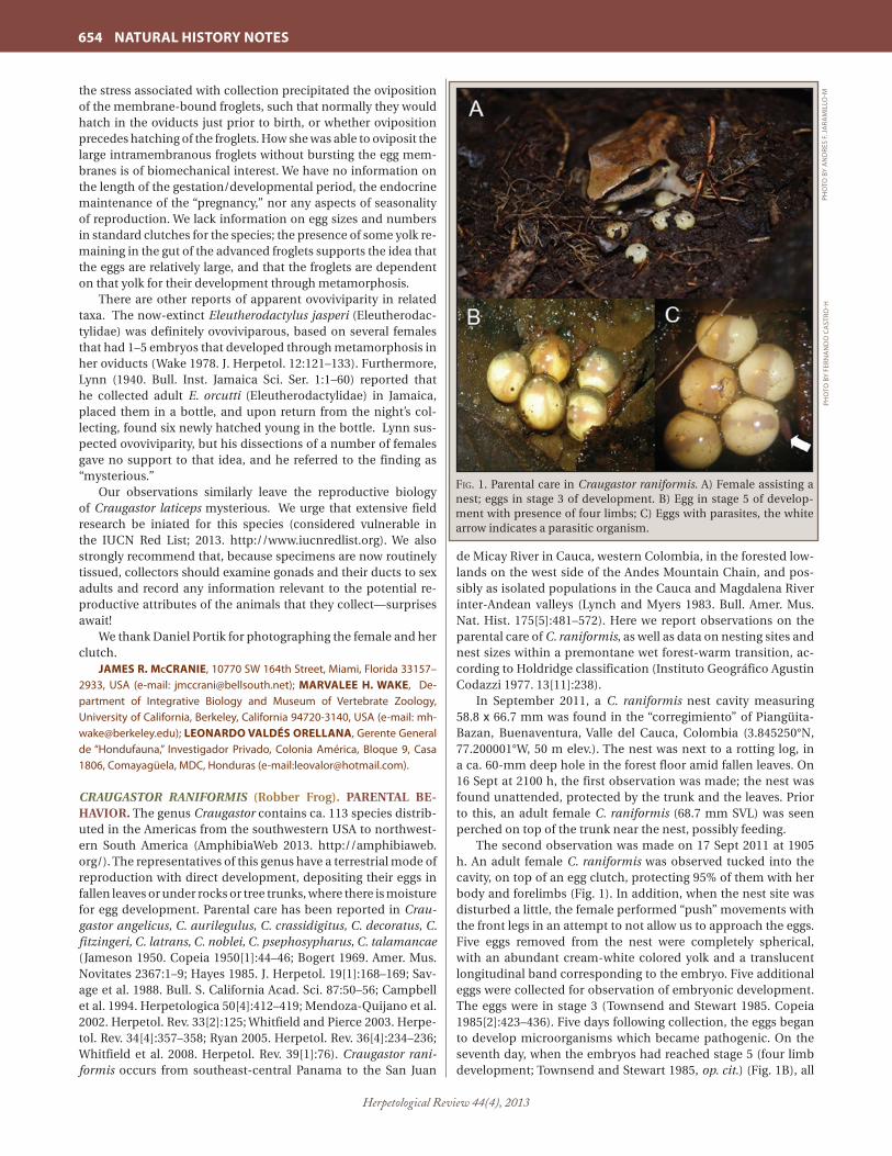
Herpetological Review 44(4), 2013
654 NATURAL HISTORY NOTES
the stress associated with collection precipitated the oviposition of the membrane-bound froglets, such that normally they would hatch in the oviducts just prior to birth, or whether oviposition precedes hatching of the froglets. How she was able to oviposit the large intramembranous froglets without bursting the egg mem-branes is of biomechanical interest. We have no information on the length of the gestation/developmental period, the endocrine maintenance of the “pregnancy,” nor any aspects of seasonality of reproduction. We lack information on egg sizes and numbers in standard clutches for the species; the presence of some yolk re-maining in the gut of the advanced froglets supports the idea that the eggs are relatively large, and that the froglets are dependent on that yolk for their development through metamorphosis. There are other reports of apparent ovoviviparity in related taxa. The now-extinct Eleutherodactylus jasperi (Eleutherodac-tylidae) was definitely ovoviviparous, based on several females that had 1–5 embryos that developed through metamorphosis in her oviducts (Wake 1978. J. Herpetol. 12:121–133). Furthermore, Lynn (1940. Bull. Inst. Jamaica Sci. Ser. 1:1–60) reported that he collected adult E. orcutti (Eleutherodactylidae) in Jamaica, placed them in a bottle, and upon return from the night’s col-lecting, found six newly hatched young in the bottle. Lynn sus-pected ovoviviparity, but his dissections of a number of females gave no support to that idea, and he referred to the finding as “mysterious.” Our observations similarly leave the reproductive biology of Craugastor laticeps mysterious. We urge that extensive field research be iniated for this species (considered vulnerable in the IUCN Red List; 2013. http://www.iucnredlist.org). We also strongly recommend that, because specimens are now routinely tissued, collectors should examine gonads and their ducts to sex adults and record any information relevant to the potential re-productive attributes of the animals that they collect—surprises await! We thank Daniel Portik for photographing the female and her clutch.
JAMES R. McCRANIE, 10770 SW 164th Street, Miami, Florida 33157–2933, USA (e-mail: [email protected]); MARVALEE H. WAKE, De-partment of Integrative Biology and Museum of Vertebrate Zoology, University of California, Berkeley, California 94720-3140, USA (e-mail: [email protected]); LEONARDO VALDÉS ORELLANA, Gerente General de “Hondufauna,” Investigador Privado, Colonia América, Bloque 9, Casa 1806, Comayagüela, MDC, Honduras (e-mail:[email protected]).
CRAUGASTOR RANIFORMIS (Robber Frog). PARENTAL BE-HAVIOR. The genus Craugastor contains ca. 113 species distrib-uted in the Americas from the southwestern USA to northwest-ern South America (AmphibiaWeb 2013. http://amphibiaweb.org/). The representatives of this genus have a terrestrial mode of reproduction with direct development, depositing their eggs in fallen leaves or under rocks or tree trunks, where there is moisture for egg development. Parental care has been reported in Crau-gastor angelicus, C. aurilegulus, C. crassidigitus, C. decoratus, C. fitzingeri, C. latrans, C. noblei, C. psephosypharus, C. talamancae (Jameson 1950. Copeia 1950[1]:44–46; Bogert 1969. Amer. Mus. Novitates 2367:1–9; Hayes 1985. J. Herpetol. 19[1]:168–169; Sav-age et al. 1988. Bull. S. California Acad. Sci. 87:50–56; Campbell et al. 1994. Herpetologica 50[4]:412–419; Mendoza-Quijano et al. 2002. Herpetol. Rev. 33[2]:125; Whitfield and Pierce 2003. Herpe-tol. Rev. 34[4]:357–358; Ryan 2005. Herpetol. Rev. 36[4]:234–236; Whitfield et al. 2008. Herpetol. Rev. 39[1]:76). Craugastor rani-formis occurs from southeast-central Panama to the San Juan
de Micay River in Cauca, western Colombia, in the forested low-lands on the west side of the Andes Mountain Chain, and pos-sibly as isolated populations in the Cauca and Magdalena River inter-Andean valleys (Lynch and Myers 1983. Bull. Amer. Mus. Nat. Hist. 175[5]:481–572). Here we report observations on the parental care of C. raniformis, as well as data on nesting sites and nest sizes within a premontane wet forest-warm transition, ac-cording to Holdridge classification (Instituto Geográfico Agustin Codazzi 1977. 13[11]:238).
In September 2011, a C. raniformis nest cavity measuring 58.8 x 66.7 mm was found in the “corregimiento” of Piangüita-Bazan, Buenaventura, Valle del Cauca, Colombia (3.845250°N, 77.200001°W, 50 m elev.). The nest was next to a rotting log, in a ca. 60-mm deep hole in the forest floor amid fallen leaves. On 16 Sept at 2100 h, the first observation was made; the nest was found unattended, protected by the trunk and the leaves. Prior to this, an adult female C. raniformis (68.7 mm SVL) was seen perched on top of the trunk near the nest, possibly feeding.
The second observation was made on 17 Sept 2011 at 1905 h. An adult female C. raniformis was observed tucked into the cavity, on top of an egg clutch, protecting 95% of them with her body and forelimbs (Fig. 1). In addition, when the nest site was disturbed a little, the female performed “push” movements with the front legs in an attempt to not allow us to approach the eggs. Five eggs removed from the nest were completely spherical, with an abundant cream-white colored yolk and a translucent longitudinal band corresponding to the embryo. Five additional eggs were collected for observation of embryonic development. The eggs were in stage 3 (Townsend and Stewart 1985. Copeia 1985[2]:423–436). Five days following collection, the eggs began to develop microorganisms which became pathogenic. On the seventh day, when the embryos had reached stage 5 (four limb development; Townsend and Stewart 1985, op. cit.) (Fig. 1B), all
Fig. 1. Parental care in Craugastor raniformis. A) Female assisting a nest; eggs in stage 3 of development. B) Egg in stage 5 of develop-ment with presence of four limbs; C) Eggs with parasites, the white arrow indicates a parasitic organism.
PHO
TO B
Y A
ND
RES
F. J
ARA
MIL
LO-M
PHO
TO B
Y FE
RNA
ND
O C
AST
RO-H
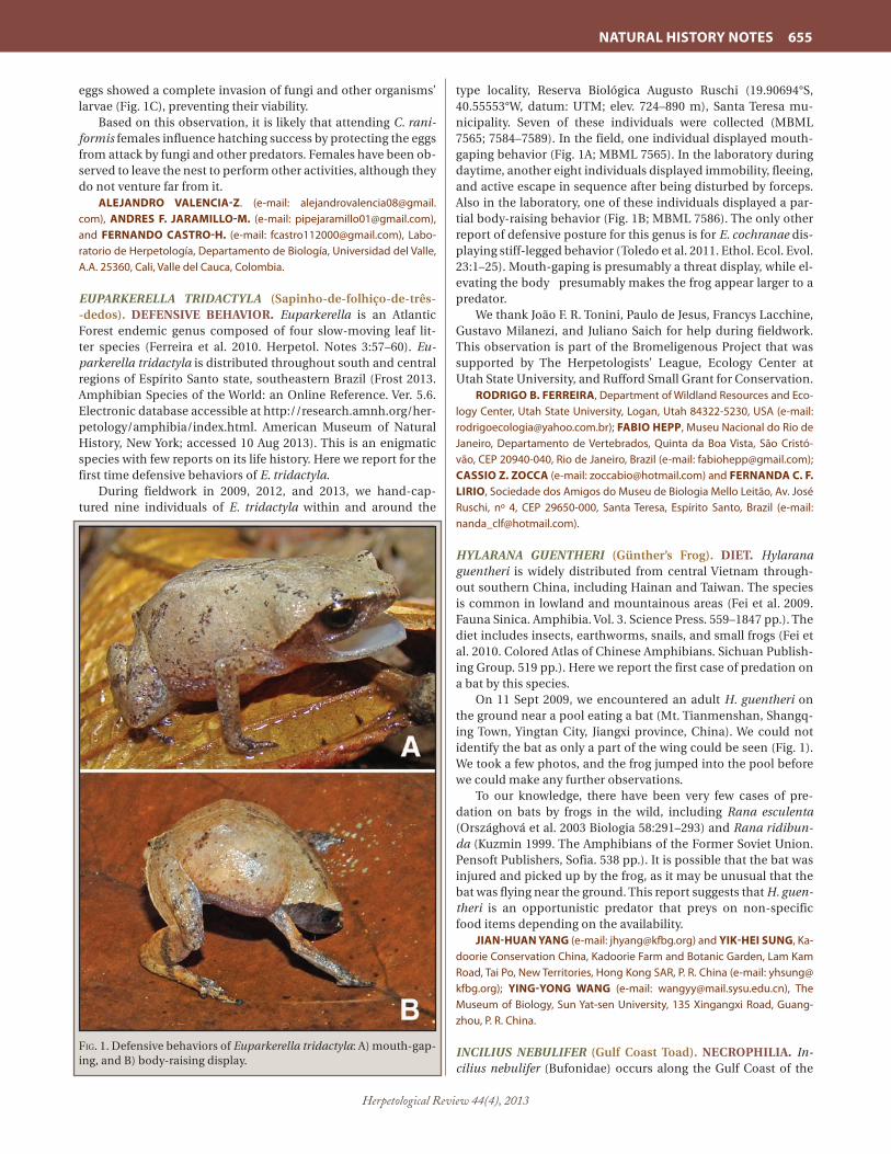
Herpetological Review 44(4), 2013
NATURAL HISTORY NOTES 655
eggs showed a complete invasion of fungi and other organisms’ larvae (Fig. 1C), preventing their viability.
Based on this observation, it is likely that attending C. rani-formis females influence hatching success by protecting the eggs from attack by fungi and other predators. Females have been ob-served to leave the nest to perform other activities, although they do not venture far from it.
ALEJANDRO VALENCIA-Z. (e-mail: [email protected]), ANDRES F. JARAMILLO-M. (e-mail: [email protected]), and FERNANDO CASTRO-H. (e-mail: [email protected]), Labo-ratorio de Herpetología, Departamento de Biología, Universidad del Valle, A.A. 25360, Cali, Valle del Cauca, Colombia.
EUPARKERELLA TRIDACTYLA (Sapinho-de-folhiço-de-três--dedos). DEFENSIVE BEHAVIOR. Euparkerella is an Atlantic Forest endemic genus composed of four slow-moving leaf lit-ter species (Ferreira et al. 2010. Herpetol. Notes 3:57–60). Eu-parkerella tridactyla is distributed throughout south and central regions of Espírito Santo state, southeastern Brazil (Frost 2013. Amphibian Species of the World: an Online Reference. Ver. 5.6. Electronic database accessible at http://research.amnh.org/her-petology/amphibia/index.html. American Museum of Natural History, New York; accessed 10 Aug 2013). This is an enigmatic species with few reports on its life history. Here we report for the first time defensive behaviors of E. tridactyla. During fieldwork in 2009, 2012, and 2013, we hand-cap-tured nine individuals of E. tridactyla within and around the
type locality, Reserva Biológica Augusto Ruschi (19.90694°S, 40.55553°W, datum: UTM; elev. 724–890 m), Santa Teresa mu-nicipality. Seven of these individuals were collected (MBML 7565; 7584–7589). In the field, one individual displayed mouth-gaping behavior (Fig. 1A; MBML 7565). In the laboratory during daytime, another eight individuals displayed immobility, fleeing, and active escape in sequence after being disturbed by forceps. Also in the laboratory, one of these individuals displayed a par-tial body-raising behavior (Fig. 1B; MBML 7586). The only other report of defensive posture for this genus is for E. cochranae dis-playing stiff-legged behavior (Toledo et al. 2011. Ethol. Ecol. Evol. 23:1–25). Mouth-gaping is presumably a threat display, while el-evating the body presumably makes the frog appear larger to a predator. We thank João F. R. Tonini, Paulo de Jesus, Francys Lacchine, Gustavo Milanezi, and Juliano Saich for help during fieldwork. This observation is part of the Bromeligenous Project that was supported by The Herpetologists’ League, Ecology Center at Utah State University, and Rufford Small Grant for Conservation.
RODRIGO B. FERREIRA, Department of Wildland Resources and Eco-logy Center, Utah State University, Logan, Utah 84322-5230, USA (e-mail: [email protected]); FABIO HEPP, Museu Nacional do Rio de Janeiro, Departamento de Vertebrados, Quinta da Boa Vista, São Cristó-vão, CEP 20940-040, Rio de Janeiro, Brazil (e-mail: [email protected]); CASSIO Z. ZOCCA (e-mail: [email protected]) and FERNANDA C. F. LIRIO, Sociedade dos Amigos do Museu de Biologia Mello Leitão, Av. José Ruschi, nº 4, CEP 29650-000, Santa Teresa, Espírito Santo, Brazil (e-mail: [email protected]).
HYLARANA GUENTHERI (Günther’s Frog). DIET. Hylarana guentheri is widely distributed from central Vietnam through-out southern China, including Hainan and Taiwan. The species is common in lowland and mountainous areas (Fei et al. 2009. Fauna Sinica. Amphibia. Vol. 3. Science Press. 559–1847 pp.). The diet includes insects, earthworms, snails, and small frogs (Fei et al. 2010. Colored Atlas of Chinese Amphibians. Sichuan Publish-ing Group. 519 pp.). Here we report the first case of predation on a bat by this species.
On 11 Sept 2009, we encountered an adult H. guentheri on the ground near a pool eating a bat (Mt. Tianmenshan, Shangq-ing Town, Yingtan City, Jiangxi province, China). We could not identify the bat as only a part of the wing could be seen (Fig. 1). We took a few photos, and the frog jumped into the pool before we could make any further observations.
To our knowledge, there have been very few cases of pre-dation on bats by frogs in the wild, including Rana esculenta (Országhová et al. 2003 Biologia 58:291–293) and Rana ridibun-da (Kuzmin 1999. The Amphibians of the Former Soviet Union. Pensoft Publishers, Sofia. 538 pp.). It is possible that the bat was injured and picked up by the frog, as it may be unusual that the bat was flying near the ground. This report suggests that H. guen-theri is an opportunistic predator that preys on non-specific food items depending on the availability.
JIAN-HUAN YANG (e-mail: [email protected]) and YIK-HEI SUNG, Ka-doorie Conservation China, Kadoorie Farm and Botanic Garden, Lam Kam Road, Tai Po, New Territories, Hong Kong SAR, P. R. China (e-mail: [email protected]); YING-YONG WANG (e-mail: [email protected]), The Museum of Biology, Sun Yat-sen University, 135 Xingangxi Road, Guang-zhou, P. R. China.
INCILIUS NEBULIFER (Gulf Coast Toad). NECROPHILIA. In-cilius nebulifer (Bufonidae) occurs along the Gulf Coast of the
Fig. 1. Defensive behaviors of Euparkerella tridactyla: A) mouth-gap-ing, and B) body-raising display.

Herpetological Review 44(4), 2013
656 NATURAL HISTORY NOTES
United States and Mexico (Mulcahy and Mendelson 2000. Mol. Biol. Evol. 17:173–189). At 2255 h on 18 May 2010, in Hidalgo Co., Texas, USA (26.19620°N, 98.12411°W; datum WGS 84), an adult male Incilius nebulifer was found amplexed with a deceased fe-male of the same species. The temperature was 25.5°C and ap-proximately 7.2 cm of rainfall occurred that day. The pair was found on a dirt road. The amplexus was photographed and a copy of the image was deposited in the University of Texas– Pan American vertebrate museum (voucher #05109). Extremely dry habitats impose unique and intense pressures on amphibians. To our knowledge this is the first time this be-havior has been observed for this species. This is a noteworthy observation because it may provide insight into the reproductive strategy of these toads in semiarid habitats. There may be an ad-vantage to amplexing dead females if the eggs are still viable and can be extracted and fertilized by the male. This behavior has been described in other anurans and termed “functional necro-phile strategy” by Izzo et al. (2012. J. Nat. Hist. 46:47–48).
MAYRA OYERVIDES (e-mail: [email protected]) and FREDERIC ZAIDAN III, Department of Biology, Center for Subtropical Studies, University of Texas-Pan American, 1201 W. University Dr., Edin-burg, Texas 78539, USA (e-mail: [email protected]).
LEPTODACTYLUS FRAGILIS (Mexican White-lipped Frog). ENDOPARASITES. Leptodactylus fragilis ranges from southern Texas to central Colombia and northern Venezuela (Dodd 2013. Frogs of the United States and Canada, Vol. 1, Johns Hopkins University Press, Baltimore, Maryland. 460 pp.). The purpose of this note is to add to the list of known helminth endoparasites of L. fragilis. A sample of eight L. fragilis (mean SVL = 32.4 mm ± 3.5 SD, range = 27–39 mm) was examined for helminths. Frogs were collected in Guanacaste Province, Costa Rica during 1963–64 and 1973–74 (LACM 157201, 157211, 157212, 157226, 157230, 157234, 57240, 157250). Two nematode species were found in the large intestines. They were identified as Schrankiana formosula (N = 68; preva-lence, number infected frogs/number examined frogs at 100x = 25%, mean intensity, mean number infected individuals = 34.0 ± 14.1 SD, range = 24– 44) and Cosmocerca podicipinus (N = 3; prevalence = 38%, mean infection intensity = 1.0). Voucher specimens were deposited in the United States National Para-site Collection (USNPC) as: Cosmocerca podicipinus (USNPC 107220) and Schrankiana formosula (USNPC 107221). Schran-kiana formosula is known from Leptodactylus fuscus from Bra-zil and L. elenae from Paraguay (Goldberg et al. 2007. Comp. Parasitol. 74:327–342). Leptodactylus fragilis is the third anuran species known to harbor S. formulosa and represents a new host record for Schrankiana formosula; furthermore, Costa Rica is a new locality record for this nematode. Cosmocerca podicipinus is widespread in Meso and South American anurans (Goldberg et al., op. cit.) and was previously found in L. fragilis in Guanacaste Province, Costa Rica (Bursey and Brooks 2010. Comp. Parasitol. 77:221–231). We thank G. Pauly (LACM) for permission to examine L. fra-gilis.
STEPHEN R. GOLDBERG, Department of Biology, Whittier Col-lege, Whittier, California 90608, USA (e-mail: [email protected]); CHARLES R. BURSEY, Biology Department, Pennsylvania State Univer-sity, Shenango Campus, Sharon, Pennsylvania 16146, USA (e-mail: [email protected]); JEANETTE ARREOLA, Department of Biology, Whittier College, Whittier, California 90608, USA (e-mail: jarreola @poets.whittier.edu).
LIMNONECTES GRUNNIENS. SALINE WATER TOLERANCE. Individuals of Limnonectes grunniens were observed and col-lected (Museum of Vertebrate Zoology; ALS 297–305, 339) along the banks of saltwater channels in the Aru Islands, Maluku, Indo-nesia. The Aru Islands are a network of 95 islands in eastern In-donesia, most of which are separated by narrow brackish chan-nels. Located on the Sahul Shelf, the Aru Islands share much of their fauna with New Guinea and formed a land bridge with New Guinea and Australia during glacial maxima. Ten Limnonectes grunniens were collected at night on 30 September 2011 along a saltwater channel adjacent to Desa Kongan, Kecamatan Aru Tengah, Kabupaten Kepulauan Aru (6.50277°S, 134.39137°E). All individuals were found along the banks of these saltwater chan-nels, though two specimens collected were found on a small rock (2 m x 5 m) surrounded by water inside the channel. These rocks were located at least 3 m from other land and any access to them would require these frogs voluntarily entering saline water even at low tide. In addition, L. grunniens frequently jumped into the saline water as an escape response, often attempting to hide motionless under dead leaves or vegetation to avoid detection. Using a refractometer, we measured the salinity of the water in the channel to be 27 parts per thousand, just below the salinity of seawater which is ca. 31–37 parts per thousand. We conclude that this population of L. grunniens is capable of tolerating saline water at least for brief periods and represents another example of salt-water tolerance in dicroglossid frogs as also noted for Fe-jervarya cancrivora.
BENJAMIN R. KARIN (e-mail: [email protected]) and ALEXAN-DER L. STUBBS (e-mail: [email protected]), Museum of Vertebrate Zoology, 3101 Valley Life Sciences Building, Berkeley, California 94720-3161, USA; UMILAELA ARIFIN (e-mail: [email protected]), School of Life Sciences and Technology - Institut Teknologi Bandung, Jl. Ganeca 10 Tamansari, Bandung, Indonesia 40132.
OSTEOPILUS SEPTENTRIONALIS (Cuban Treefrog). POST-INVASION SPREAD. Osteopilus septentrionalis, a native hylid of Cuba, was first recorded in Florida, USA, in 1931 in Key West, Monroe Co., where it may or may not have been native (Meshaka 2001. The Cuban Treefrog in Florida: Life History of a Successful Colonizing Species. Univ. Florida Press, Gainesville. 191 pp.). The Cuban Treefrog is highly vagile, able to thrive in urban settings and disturbed habitats, and has spread throughout peninsular Florida with a few isolated occurrences in the panhandle (Me-shaka 2001, op. cit.; Meshaka 2010. A Runaway Train in the Mak-ing: The Exotic Amphibians, Reptiles, Turtles, and Crocodilians of Florida. Monograph 1. Herpetol. Cons. Biol. 6:1–101). Cuban Treefrogs have successfully invaded hardwood hammocks and other natural habitats, but further spread is limited by low rela-tive humidity during the dry season in subtropical Florida and cold climate farther north (Meshaka 2001, op. cit.).
Cuban Treefrogs were first observed on the Archbold Biologi-cal Station (ABS; Highlands Co., Florida; 27.181°N, 81.350°W; elev. 42 m) more than 20 years ago, based on specimens deposited in the ABS Vertebrate Collection. The earliest ABS specimens were collected in 1987 (from a site 15 km N of ABS) and 1993 (found on the main research building at ABS). However, the first pub-lished record for Highlands Co. was in the late 1970s (Stevenson 1976. Vertebrates of Florida. Univ. Florida Press, Gainesville. 607 pp.). In July 2009, we saw and heard adult Cuban Treefrogs near a granite-lined drainage ditch that bisects the main grounds of ABS, and individuals were often seen on the exterior of the re-search building at night.

Herpetological Review 44(4), 2013
NATURAL HISTORY NOTES 657
To determine the extent to which Cuban Treefrogs have invaded adjacent scrub habitats, we installed an array of 50 3.9-cm-diameter PVC pipes extending east and west from the main ditch, with the pipe grid covering an area of ca. 0.1 km2. Pipes were checked on average every seven days from October 2009 through November 2010. Captured Cuban Treefrogs were euthanized and preserved, and native hylids were relocated well outside the grid to prevent recapture. We also measured snout–urostyle length (SUL) of most individuals to the nearest mm. In total, we captured nine Cuban Treefrogs (two at the ditch, four at 50 m, one at 100 m, and two at 150 m east of the ditch) and 76 native hylids, including 60 Hyla squirella, 14 H. femoralis, and two H. cinerea as far as 200 m away from the ditch. Mean SUL ± SD was 37.3 ± 6.7 mm for O. septentrionalis (range 28–45; N = 7), 28.7 ± 5.3 mm for H. femoralis (19–37; N = 13), 23.8 ± 4.3 mm for H. squirella (18–36; N = 47), and 41.0 mm for H. cinerea (N = 1).
Although 32% of the captures of native hylids occurred af-ter 28 January 2010, no Cuban Treefrogs were captured after this date. January 2010 was a particularly cold month at ABS with record-setting minimum temperatures. Daily minimum temperatures at ABS were at or below 0° C for six consecutive days, followed by a low temperature of 2.8°C, and then five more days below 0°C; there were three days in January 2010 when the temperature dropped to -7°C. It is plausible that this extended period of extreme cold reduced the population of Cuban Tree-frogs at ABS. Furthermore, Cuban Treefrogs have not colonized undisturbed oak scrub and scrubby flatwoods surrounding the developed part of ABS, despite the presence of many seasonally inundated wetlands. In addition to low humidity during the dry season, the frequent application of prescribed fire at ABS may impose a constraint on this species, either directly or indirectly via effects on vegetation structure and availability of suitable ref-uges. Refuge availability has previously been shown to be a factor limiting abundance of this species (Meshaka 2001, op. cit.).
We thank Steve Johnson (Univ. Florida) and his 2009 Conser-vation of Amphibians and Reptiles class for assistance installing the pipe sampling grid; Chris Jennings, John Vanek, Nick Swain, and Josh Daskin for assistance with data collection; and Walter Meshaka for providing valuable suggestions on the manuscript.
BETSIE B. ROTHERMEL (e-mail: [email protected]), ZACHERY R. FORSBURG, and NAIMA C. PHILLIPS, Archbold Biological Station, 123 Main Drive, Venus, Florida 33960, USA.
RHINELLA GRANULOSA (Granulated Toad). PREDATION. An-urans are preyed upon by several kinds of invertebrates, such as water bugs (Giaretta and Menin 2004. J. Nat. Hist. 38:1711–1722), scorpions (Villanueva-Rivera et al. 2000. Herpetol. Rev. 31:100), spiders (Menin et al. 2005. Phyllomedusa 4:39–47), and all ver-tebrate groups (Pombal Jr. 2007. Rev. Bras. Zool. 24: 841–843; Toledo et al. 2005 Herpetol. Rev. 36:395–400). The present com-munication reports predation of Rhinella granulosa (Bufonidae) by a banana-spider of the Amazonian genus Phoneutria (spe-cies unidentified; Ctenidae). At 2118 h on 10 July 2013, the spi-der was observed predating an adult male of R. granulosa (62.3 mm SVL). Our observation occurred at a residence in an urban area in the municipality of Santana, State of Amapá, North Bra-zil (0.022778°S, 51.164722°W; datum WGS84). The observation lasted at least eight minutes and during this time the spider had already captured the anuran with the aid of its pedipalps, and its chelicerae were inserted in the prey’s lateral body.
DANIEL WELTON SILVA-SILVA (e-mail: [email protected]), KEL-LY JULIANA GAYA CORRÊA (e-mail: [email protected]), and
CARLOS E. COSTA-CAMPOS, Laboratório de Herpetologia, Departamen-to Ciências Biológicas e da Saúde, Universidade Federal do Amapá, Cam-pus Marco Zero, 68903- 280, Macapá, AP, Brazil (e-mail: [email protected]).
RHINELLA SCHNEIDERI (Cururu Toad). DIET. Rhinella schnei-deri is a large bufonid found in open and urban areas, and is widely distributed in South America, inhabiting some regions of Brazil, Paraguay, Bolivia, Argentina, and Uruguay (Pramuk 2006. Zool. J. Linn. Soc. 146:407–452). This species feeds mainly on arthropods (Batista et al. 2011. Herpetol. Notes 4:17–21; Duré et al. 2009. Phyllomedusa 8:27–39; Lajmanovich 1994. Rev. Hydro-biol. Trop. 27:107–112; Moreira and Barreto 1996. Rev. Bras. Zool. 123:313–320), however, like many anurans, it is considered to be a generalist and opportunistic predator. On 08 February 2013, we collected a female R. schneideri (SVL = 905.5 mm; CFBH 35704) from a ranch on the Tietê River, lo-cated within the municipality of Barbosa, São Paulo state, south-east Brazil (21.25048°S, 49.92132°W; datum WGS84; elev. 371 m). Stomach contents included ten individuals of newly meta-morphosed Dermatonotus muelleri (Mueller’s Narrow-mouthed Frogs; mean SVL = 16.1 mm). It is known that adults of R. schnei-deri, as well as some congeneric species (Camilotti and Barreto-Lima 2011. Bioscience J. 27:993–994) feed on small vertebrates, such as birds (Vaz-Silva 2003, op. cit.) and legless lizards (Oda and Landgraf 2012. Bol. Asoc. Herpetol. Esp. 23:57–59). However, to our knowledge, this is the first record of an adult R. schneideri preying upon an anuran. At the time our observation was made, there was a massive emergence of D. muelleri in the area, which corroborates the opportunistic feeding behavior of R. schneideri. All specimens are housed at Coleção de Anfíbios “Célio F. B. Had-dad” at UNESP, Rio Claro, Brazil. We thank Celso Gavira for field and handling assistance. Fundação de Amparo à Pesquisa do Estado de São Paulo (FAPESP) provided financial support. Collecting permit was is-sued by IBAMA (process number 22028-1).
RODRIGO SAMUEL BUENO GAVIRA (e-mail: [email protected]), RAFAEL PARELLI BOVO (e-mail: [email protected]), and DENIS VIEIRA ANDRADE, Departamento de Zoologia, Instituto de Bio-ciências, Universidade Estadual Paulista, Rio Claro, São Paulo, Brazil, Caixa Postal 199, CEP 13506–900 (e-mail: [email protected]).
GYMNOPHIONA — CAECILIANS
DERMOPHIS GLANDULOSUS. PREDATION BY MICRURUS ALLENI. Little is known regarding predation on caecilians, due mainly to their fossorial nature and the difficulty to find them. It has been suggested that various semifossorial and fossorial snakes appear to be major predators of these amphibians (Sav-age 2002. The Amphibians and Reptiles of Costa Rica: A Herpe-tofauna between Two Continents, between Two Seas. Univ. Chi-cago Press, Chicago, Illinois. 934 pp.). Roze (1996. Coral Snakes of the Americas: Biology, Identification, and Venoms. Krieger Publ. Co., Malabar, Florida. 340 pp.) found that the coral snake Micru-rus nigrocinctus eats caecilians, however he did not identify prey species. Solórzano (2004. Serpientes de Costa Rica: Distribución, taxonomía e historia natural. INBio, Heredia, Costa Rica. 792 pp.) suggested that other species of Micrurus also include caecilians in their diet.
On 18 April 2012, a juvenile male Micrurus alleni (UCR 21534) was found near Rio Sonador in Volcán of Buenos Aires, Puntar-enas, Costa Rica (9.29797°N, 83.47062°W, WGS84; 1120 m elev.).

Herpetological Review 44(4), 2013
658 NATURAL HISTORY NOTES
This region is classified as premontane rainforest (Bolaños et al. 2005. Mapa Ecológico de Costa Rica. Centro Científico Tropical, San José, Costa Rica). A few hours after collection the snake (420 mm TL) regurgitated a caecilian Dermophis glandulosus (UCR 21533; 240 mm TL). This specimen showed mechanical damage in the head and nuchal region, suggesting that it was ingested head first.
This is the first record of a predator for D. glandulosus, and also the first report of the species in the diet of M. alleni, sup-porting the hypothesis that coral snakes feed on caecilians. Solórzano (2005. Rev. Biol. Trop. 53:227–228) indicated that the diet of M. alleni is composed primarily of Marbled Swamps Eels (Symbranchus marmoratus); however, this eel is found in Costa Rica from sea level to 650 m elev. (Bussing 1998. Rev. Biol. Trop. 46:1–468), while M. alleni is relatively common from sea level to 1500 m elev. (Solórzano 2004, op. cit.), suggesting a dietary shift at higher elevations.
ERICK ARIAS (e-mail: [email protected]) and GERARDO CHAVES, Museo de Zoología, Escuela de Biología, Universidad de Costa Rica, San Pedro, 11501-2060, San José, Costa Rica (e-mail: [email protected]).
TESTUDINES — TURTLES
CHRYSEMYS PICTA PICTA (Eastern Painted Turtle). PREDA-TION. On 22 August 2012 at 1030 h we observed a second-year Bald Eagle (Haliaeetus leucocephalus) kill and consume an adult Chrysemys picta picta along the bank of a farm impoundment at Chesapeake Farms, Kent Co., Maryland, USA (39.1962°N, 76.21707°W). The C. p. picta was basking ca. 1.5 m up the grad-ual, muddy bank of the 6.5-acre impoundment when the eagle landed beside the turtle, used both feet to turn the turtle on edge, and grasped the turtle in its left talon. The bird then flew to a height of ca.15 m and dropped the turtle on the bank, at which point the turtle attempted to right itself. This pattern was re-peated several times until the turtle remained motionless upon landing. The H. leucocephalus then picked up the C. p. picta and flew to a perch ca. 250 m from the capture sight and ca. 10 m above the ground, where it was seen consuming the apparently dead C. p. picta.
Ernst and Lovich (2009. Turtles of the United States and Canada. Johns Hopkins University Press, Baltimore, Maryland. 840 pp.) lists H. leucocephalus among the many predators of C. p. picta, but careful scrutiny of references therein (e.g., Vogt 1981. Natural History of Amphibians and Reptiles of Wiscon-sin. Milwaukee Public Museum, Milwaukee, Wisconsin. 205 pp.; Ross 1989. Wisconsin Endangered Species Report 59:1–33) and other studies of the diet of H. leucocephalus (e.g., Smith 1936. Auk 53:301–305; Imler and Kalmbach 1955. U.S. Fish Wildl. Serv. Circ. 30; Clark 1982. J. Field. Ornithol. 53:49–51) reveals that such assertions are based on observations of C. p. picta shells below or in active H. leucocephalus nests. It is thus unclear if the turtles noted in these studies were depredated or collected as carrion, as H. leucocephalus is a frequent and voracious scavenger (e.g., Buehler 2000. In: The Birds of North America, No. 506: 40; Wilm-ers et al. 2003. Ecol. Lett. 6:996–1003). Further, if these C. p. picta were indeed depredated, the studies fail to document the means by which the turtles were killed by H. leucocephalus or if the turtles were consumed by non-nestling H. leucocephalus. Thus this observation apparently represents the first reported direct observation of predation of C. p. picta by H. leucocephalus and provides evidence for the inclusion of C. p. picta in the diet of non-nestling H. leucocephalus.
We thank Travis LaDuc and Erin Counihan for helpful com-ments on this note.
AARON R. KROCHMAL, Department of Biology, Washington College, 300 Washington Avenue, Chestertown, Maryland 21620, USA (e-mail: [email protected]); STEVE HAMILTON, 27996 Redmill Street, Ken-nedyville, Maryland 21645, USA.
LEPIDOCHELYS OLIVACEA (Olive Ridley Sea Turtle). LEU-CISTIC HATCHLING. Along the Odisha coast of eastern India, “arribadas” (simultaneous mass nestings) of Olive Ridley Sea Turtles occur at three beaches viz., Gahirmatha, Devi, and Ru-shikulya. Gahirmatha Beach, within Bhitarkanika National Park, was once regarded as the largest reported arribada nesting ground for Olive Ridleys in the world (Bustard 1976. Tigerpaper 3:25). The other two sites, Devi and Rushikulya, are outside the protected area, but are regularly monitored by the Odisha Forest Department during the nesting period.
Fig. 1. Map showing the three arribada sites in Odisha coast. Solid fill triangle = site of observation at Rushikulya Beach. The coastline between these sites is used by Lepidochelys olivacea for sporadic nesting.
Fig. 2. Lepidochelys olivacea hatchlings observed at Rushikulya. A) Leucistic hatchling; arrow marks showing normal eye color and black sutures between plates of the carapace. B) Typical black hatch-lings. C) Cluster of normal hatchlings. D) Arrow indicates the narrow white line surrounding the carapace of normal hatchlings.

Herpetological Review 44(4), 2013
NATURAL HISTORY NOTES 659
Rushikulya Beach (19.3945°N, 85.0896°E; WGS 84) is the southernmost nesting site along the Odisha coast (Pandav et al. 1994. A Status Survey of Olive Ridley Sea Turtle [Lepidochelys olivacea] and its Nesting Habitats along the Orissa Coast, India. Wildlife Institute of India, Dehradun. 48 pp.). The site falls in the Ganjam district and is north of the Rushikulya River mouth from Purunabandha to Kantiagada village (Fig. 1). In 2012, an arriba-da occurred during the first week of March at Rushikulya Beach and the associated hatching event started after approximately 50 days (mid-April).
On 22 April 2012, while observing the hatching event we came across a rare leucistic hatchling of L. olivacea, along with the normal gray/black form (Fig. 2A). The white hatchling looked similar to the black form in size, shape, and structures (Fig. 2B). It was not a true albino because its eyes were of normal black color (typically red in albinos). It had a grayish tinge on the head and edge of the flippers. The sutures between the plates of the carapace and upper part of the beak were blackish. Normal hatchlings are dark gray, but appear black when wet (Fig. 2C), and a narrow white line surrounds the carapace, as well as the trailing edge of the fore and hind flippers (Fig. 2D). The leucistic hatchling was not collected as the species is protected. A litera-ture survey suggests that the leucistic form of L. olivacea has not been reported earlier from the Indian coast. Media photographic records are available for leucistic sea turtles from Florida, and some studies have been performed on malformations in hatch-lings of L. olivacea outside India (Ibarra and Gasca 2009. Vet. Méx. 40:371–380). A thorough monitoring of nesting sites in In-dia during peak season may reveal additional aberrancies or de-formities which might provide new insights on the biology and physiology of this species.
ANGSHUMAN RAHA (e-mail: [email protected]) and PROSENJIT DAWN (e-mail: [email protected]), Zoological Sur-vey of India, Prani Vigyan Bhavan, M-Block, New Alipur, Kolkata-700053, West Bengal, India
TERRAPENE CAROLINA (Eastern Box Turtle). DIET. Terra-pene carolina is known to consume carrion and small snakes,
including snakes in the genus Agkistrodon (Jensen 1999. Her-petol. Rev. 30[2]:95; Ernst and Lovich 2009. Turtles of the United States and Canada. Johns Hopkins Univ. Press, Baltimore, Mary-land. 840 pp.). On 5 July 2013, at approximately 1745 h, BD ob-served a road-killed A. piscivorus in Jackson Co., Mississippi, USA (30.528056°N, 88.548889°W, datum WGS84/NAD83). Upon re-turning 5 minutes later he discovered an adult T. carolina feeding on the snake carcass (Fig. 1). Photos were taken and the turtle was left undisturbed. Later that same evening, the turtle could not be located but the snake had been pulled to the margin of the road, possibly by the turtle. Similar behavior was noted by Jensen (op. cit.). This is the first diet record of a T. carolina consuming A. piscivorus. This observation also highlights an additional vulner-ability of T. carolina to road mortality while scavenging carcasses.
BRYAN DAVIS, 3700 Howard Davis Rd, Moss Point, Mississippi 39562, USA (e-mail: [email protected]); ROGER D. BIRKHEAD, Ala-bama Science In Motion, Auburn University, Auburn, Alabama 36849-5414, USA (e-mail: [email protected]).
CROCODYLIA — CROCODILIANS
ALLIGATOR MISSISSIPPIENSIS (American Alligator). MAXI-MUM CLUTCH SIZE. Alligator mississippiensis is an oviparous crocodilian species that is characteristic of the Gulf Coast and lower Atlantic Coastal Plains of the USA (Conant and Collins 1998. A Field Guide to Reptiles and Amphibians of Eastern and Central North America. 3rd ed. Houghton Mifflin Co., Boston, Massachusetts. 616 pp.). The nesting ecology and clutch charac-teristics of this species have been well documented (Hall 1991. J. Herpetol. 25:133–141; Joanen and McNease 1989. Am. Zool. 29:987–998). The maximum clutch size previously reported for A. mississippiensis is 75 eggs (Platt et al. 2004. Florida Field Nat. 32:102–106), collected in 1925 from a nest at Lake Miccosuke, Jef-ferson Co., Florida, USA.
On 27 June 2012 two of us (LWJ and JJ) collected a clutch from a single nest containing 91 eggs. Further examination of the eggs for banding patterns (i.e., to determine viability) revealed that of the 91 eggs, 82 were fertile and 9 were infertile. The clutch was subsequently taken and hatched in captivity under optimum conditions. The clutch produced 60 hatchling alligators (66% success) on 30 August 2012. The clutch was collected on Eagle Lake in southeastern Colorado Co., Texas, USA (29.55870°N, 96.35287°W, WGS84; 46.6 m elev.)
Detailed examination of the eggs revealed essentially identi-cal size and shape. In oviparous reptiles, data suggest that vari-ability in egg width is restricted by the diameter of the pelvic ap-erture (Congdon and Gibbons 1985. Herpetologica 41:194–205; Thorbjarnarson 1994. Copeia 1994:907–919; Werner 1989. Israeli J. Zool. 35:199–213). Consequently, because of the egg homoge-neity coupled with the uniform hatching date, we hypothesize this clutch was most likely produced by a single gravid female.
CORD B. EVERSOLE (e-mail: [email protected]) and SCOTT E. HENKE, Department of Animal, Rangeland, and Wildlife Sci-ences, Caesar Kleberg Wildlife Research Institute (MSC 218), Texas A&M University-Kingsville, Kingsville, Texas 78363, USA; RANDY L. POWELL, Department of Biological and Health Sciences (MSC 158), Texas A&M Uni-versity-Kingsville, Kingsville, Texas 78363, USA; LARRY W. JANIK and JA-SON JANIK , Janik Alligators LLC, El Campo, Texas 77437, USA.
ALLIGATOR MISSISSIPPIENSIS (American Alligator). NESTS USED BY OTHER REPTILES IN COASTAL LOUISIANA. Alliga-tor nests have been documented to serve as commensal nesting
Fig. 1. Eastern Box Turtle (Terrapene carolina) scavenging road-killed Cottonmouth (Agkistrodon piscivorus), Jackson Co., Mississippi, USA.

Herpetological Review 44(4), 2013
660 NATURAL HISTORY NOTES
sites for other reptiles in the southeastern USA (see review and references in Enge et al. 2000. J. Herpetol. 34:497–503). Several species of turtle have been identified to nest in alligator nests in these studies, including Pseudemys nelsoni (Florida Redbelly), Apalone ferox (Florida Softshell), Kinosternon subrubrum (Flori-da Mud Turtle), K. baurii (Striped Mud Turtle), and Sternotherus odoratus (Common Musk Turtle) (Deitz and Jackson 1979. J. Her-petol. 13:510–512; Enge et al., op. cit.; Kushlan and Kushlan 1980. Copeia 1980:930–932). Eggs of Anolis carolinensis (Green Anole) have also been found in alligator nests (Deitz and Jackson, op. cit.; Kushlan and Kushlan, op. cit.), although far less common-ly than turtle eggs. Snake eggs are also rarely found in alligator nests, but Deitz and Jackson (op. cit.) described Farancia aba-cura (Mud Snake) eggs in one alligator nest in Florida, as well as several specimens of Seminatrix pygaea (Swamp Snake) and Nerodia cyclopion (Green Water Snake).
In Louisiana, four clutches of F. abacura reinwardtii (Western Mud Snake) eggs and one female were found in two alligator nest mounds; one nest had been depredated and the other appeared to be a false nest (Hall and Meier 1993. Copeia 1993:219–222). Additionally, S. odoratus eggs were located within two alligator nests in southwestern Louisiana, with two additional nests con-taining F. abacura specimens and eggs (Carbonneau 1987. Un-publ. M.S. thesis. Louisiana State Univ. 54 pp.). We herein report on findings of new reptiles using alligator nests for egg deposi-tion sites in coastal Louisiana, which to our knowledge have not been previously documented.
Alligator eggs were collected from numerous nests on Rock-efeller Wildlife Refuge in Cameron and Vermilion parishes for various research projects between 25 June and 2 July 2013. Two clutches of snake eggs were incidentally found within alligator nests. The snake eggs (clutch 1, N = 12; clutch 2, N = 4) were in the nest mound, but not within the egg cavities in which the alliga-tor eggs were deposited. The snake eggs were collected and incu-bated in a field incubator in the same manner as alligator eggs, as previously described (Joanen and McNease 1977. Proc. World Mariculture Soc. 8:483–489). On 23 July we noted the presence of shed snake skins adhering to the wire mesh container in which the eggs had been placed; regrettably the ventilation holes were large enough to allow escape of the hatched snakes. Examination of the eggshells suggested 8 of 12 eggs from the larger clutch had hatched. The shed skins were collected, and viewed under a dis-secting microscope. The presence of one preocular scale and two postocular scales from one shed skin was indicative the hatched snakes were likely Lampropeltis getula holbrooki (Speckled King-snakes).
One egg of the second clutch of four eggs was then dissected, as we were concerned they were non-viable. A near-term Speck-led Kingsnake was noted and the remainder of the clutch was then moved to a more secure container for continued incuba-tion. On the morning of 26 July we found a live Speckled King-snake had hatched; it measured 21.0 mm TL, 18.5 SVL, and had a mass of 4.07 g. That afternoon a second snake was beginning to emerge. We attempted to assist it in liberating itself from the egg by gently cutting the eggshell further. The snake did not readily emerge so we replaced the egg mass in the incubator. The follow-ing morning that egg had hatched, the snake measured 22.5 mm TL, 20.0 mm SVL, and 4.42 g. Both snakes were released to the wild near the field incubator on the day of hatching. The remain-ing egg appeared desiccated and non-viable.
On 29 July 2013, we visited six additional alligator nests to collect vegetation and soil samples for an associated research
project. Two nests contained presumed skink eggs, but we had not previously attempted to incubate these, although we com-monly encounter them in alligator nests. Each clutch contained approximately five eggs; some may have been overlooked due to their small size. One egg in one clutch appeared collapsed and non-viable when collected and incubated in our field incuba-tor. On 5 August two hatchling Ground Skinks (Scincella latera-lis) were found in the incubator; they each had a mass of 0.14 g. These were released near the field incubator that afternoon. On 8 August six more S. lateralis were found hatched. Four had masses of 0.17 g, and one had a mass of 0.11 g and another of 0.15 g. We suspect the two smaller skinks may have hatched with those measured on 5 August but were undetected, having been burrowed in the nesting vegetation at the time.
We incidentally collected twenty clutches of alligator eggs from nests on White Lake Conservation Area in Vermilion Parish, Louisiana on 23 June 2009. One nest contained ten Pseudemys concinna (River Cooter) eggs. These were incubated at our field laboratory and seven turtles hatched on 2 August. We occasion-ally find turtle eggs in coastal marsh nests on Rockefeller Refuge, but far less frequently than in Florida lakes as per Enge et al.’s (2000) multi-year, multi-site study (26.6% of 1586 active alligator nests had P. nelsoni eggs).
Kushlan and Kushlan (op. cit.) noted that commensal nesting in vertebrates is rare, thus our finding of three previously unre-ported reptile species using alligator nests is of interest. We regu-larly see L. getula holbrooki locally, and S. lateralis was the most commonly encountered reptile in a herpetofaunal study con-ducted in a nearby chenier habitat (W. Selman, unpubl. data). The River Cooter, however, occurs less commonly in southwest-ern Louisiana marsh habitats (Dundee and Rossman 1989. The Amphibians and Reptiles of Louisiana. Louisiana State Univ. Press, Baton Rouge, Louisiana. 300 pp.). The advantages and dis-advantages of turtles nesting in alligator nests were discussed in detail by Enge et al. (op. cit., and references therein). Hall and Meier (op. cit.) also discussed strategic advantages of commen-sal nesting in alligator nests for nesting turtles and snakes (and their eggs). Similar benefits and adverse effects may play a role in other reptiles using alligator nests, such as the taxa described herein: L. getula holbrooki, S. lateralis, and P. concinna.
We thank Jeff Boundy and Thomas Rainwater for helpful dis-cussion about these findings.
RUTH M. ELSEY (e-mail: [email protected]), WILL SELMAN, RYAN KING, MICKEY MILLER, Louisiana Department of Wildlife and Fisheries, Rockefeller Wildlife Refuge, 5476 Grand Chenier Highway, Grand Chenier, Louisiana 70643, USA; STEVEN G. PLATT, Wildlife Conservation Society, 2300 Southern Blvd. Bronx, New York 10460, USA (e-mail: [email protected]).
SQUAMATA — LIZARDS
ANOLIS EQUESTRIS (Cuban Knight Anole). NOCTURNAL AC-TIVITY. Anolis equestris is native to Cuba and has been intro-duced to Florida, USA (Kraus 2009. Alien Reptiles and Amphibi-ans: A Scientific Compendium and Analysis. Springer, Dordrecht, Netherlands. 563 pp.; Krysko et al. 2003. Florida Sci. 66:74–79). This species consumes a wide variety of animals and plants in-cluding vertebrates, invertebrates, and fruit (Camposano et al. 2008. Iguana 15:212–219, Giery et al., unpubl.). Documented ac-tivity times for populations in southern Florida indicate that A. equestris activity falls between mid-morning and late afternoon, ceasing at sunset (Meshaka et. al. 2004. The Exotic Amphibians
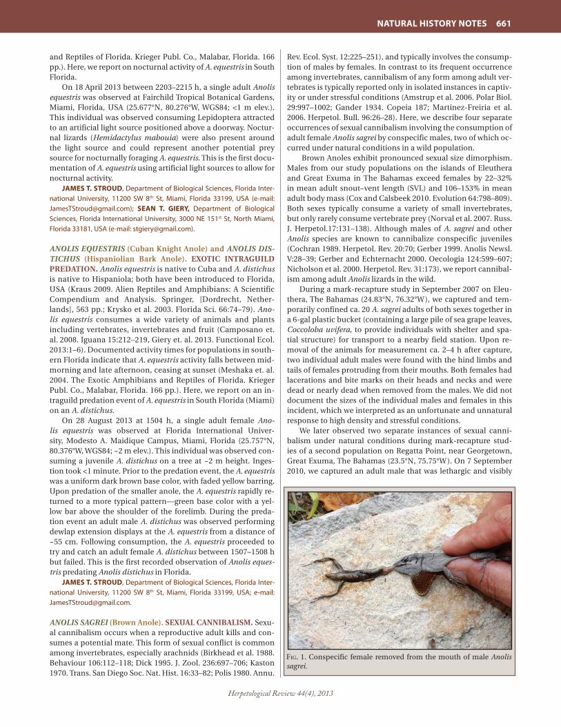
Herpetological Review 44(4), 2013
NATURAL HISTORY NOTES 661
and Reptiles of Florida. Krieger Publ. Co., Malabar, Florida. 166 pp.). Here, we report on nocturnal activity of A. equestris in South Florida.
On 18 April 2013 between 2203–2215 h, a single adult Anolis equestris was observed at Fairchild Tropical Botanical Gardens, Miami, Florida, USA (25.677°N, 80.276°W, WGS84; <1 m elev.). This individual was observed consuming Lepidoptera attracted to an artificial light source positioned above a doorway. Noctur-nal lizards (Hemidactylus mabouia) were also present around the light source and could represent another potential prey source for nocturnally foraging A. equestris. This is the first docu-mentation of A. equestris using artificial light sources to allow for nocturnal activity.
JAMES T. STROUD, Department of Biological Sciences, Florida Inter-national University, 11200 SW 8th St, Miami, Florida 33199, USA (e-mail: [email protected]); SEAN T. GIERY, Department of Biological Sciences, Florida International University, 3000 NE 151st St, North Miami, Florida 33181, USA (e-mail: [email protected]).
ANOLIS EQUESTRIS (Cuban Knight Anole) and ANOLIS DIS-TICHUS (Hispaniolian Bark Anole). EXOTIC INTRAGUILD PREDATION. Anolis equestris is native to Cuba and A. distichus is native to Hispaniola; both have been introduced to Florida, USA (Kraus 2009. Alien Reptiles and Amphibians: A Scientific Compendium and Analysis. Springer, [Dordrecht, Nether-lands], 563 pp.; Krysko et al. 2003. Florida Sci. 66:74–79). Ano-lis equestris consumes a wide variety of animals and plants including vertebrates, invertebrates and fruit (Camposano et. al. 2008. Iguana 15:212–219, Giery et. al. 2013. Functional Ecol. 2013:1–6). Documented activity times for populations in south-ern Florida indicate that A. equestris activity falls between mid-morning and late afternoon, ceasing at sunset (Meshaka et. al. 2004. The Exotic Amphibians and Reptiles of Florida. Krieger Publ. Co., Malabar, Florida. 166 pp.). Here, we report on an in-traguild predation event of A. equestris in South Florida (Miami) on an A. distichus.
On 28 August 2013 at 1504 h, a single adult female Ano-lis equestris was observed at Florida International Univer-sity, Modesto A. Maidique Campus, Miami, Florida (25.757°N, 80.376°W, WGS84; ~2 m elev.). This individual was observed con-suming a juvenile A. distichus on a tree at ~2 m height. Inges-tion took <1 minute. Prior to the predation event, the A. equestris was a uniform dark brown base color, with faded yellow barring. Upon predation of the smaller anole, the A. equestris rapidly re-turned to a more typical pattern—green base color with a yel-low bar above the shoulder of the forelimb. During the preda-tion event an adult male A. distichus was observed performing dewlap extension displays at the A. equestris from a distance of ~55 cm. Following consumption, the A. equestris proceeded to try and catch an adult female A. distichus between 1507–1508 h but failed. This is the first recorded observation of Anolis eques-tris predating Anolis distichus in Florida.
JAMES T. STROUD, Department of Biological Sciences, Florida Inter-national University, 11200 SW 8th St, Miami, Florida 33199, USA; e-mail: [email protected].
ANOLIS SAGREI (Brown Anole). SEXUAL CANNIBALISM. Sexu-al cannibalism occurs when a reproductive adult kills and con-sumes a potential mate. This form of sexual conflict is common among invertebrates, especially arachnids (Birkhead et al. 1988. Behaviour 106:112–118; Dick 1995. J. Zool. 236:697–706; Kaston 1970. Trans. San Diego Soc. Nat. Hist. 16:33–82; Polis 1980. Annu.
Rev. Ecol. Syst. 12:225–251), and typically involves the consump-tion of males by females. In contrast to its frequent occurrence among invertebrates, cannibalism of any form among adult ver-tebrates is typically reported only in isolated instances in captiv-ity or under stressful conditions (Amstrup et al. 2006. Polar Biol. 29:997–1002; Gander 1934. Copeia 187; Martinez-Freiria et al. 2006. Herpetol. Bull. 96:26–28). Here, we describe four separate occurrences of sexual cannibalism involving the consumption of adult female Anolis sagrei by conspecific males, two of which oc-curred under natural conditions in a wild population.
Brown Anoles exhibit pronounced sexual size dimorphism. Males from our study populations on the islands of Eleuthera and Great Exuma in The Bahamas exceed females by 22–32% in mean adult snout–vent length (SVL) and 106–153% in mean adult body mass (Cox and Calsbeek 2010. Evolution 64:798–809). Both sexes typically consume a variety of small invertebrates, but only rarely consume vertebrate prey (Norval et al. 2007. Russ. J. Herpetol.17:131–138). Although males of A. sagrei and other Anolis species are known to cannibalize conspecific juveniles (Cochran 1989. Herpetol. Rev. 20:70; Gerber 1999. Anolis Newsl. V:28–39; Gerber and Echternacht 2000. Oecologia 124:599–607; Nicholson et al. 2000. Herpetol. Rev. 31:173), we report cannibal-ism among adult Anolis lizards in the wild.
During a mark-recapture study in September 2007 on Eleu-thera, The Bahamas (24.83°N, 76.32°W), we captured and tem-porarily confined ca. 20 A. sagrei adults of both sexes together in a 6-gal plastic bucket (containing a large pile of sea grape leaves, Coccoloba uvifera, to provide individuals with shelter and spa-tial structure) for transport to a nearby field station. Upon re-moval of the animals for measurement ca. 2–4 h after capture, two individual adult males were found with the hind limbs and tails of females protruding from their mouths. Both females had lacerations and bite marks on their heads and necks and were dead or nearly dead when removed from the males. We did not document the sizes of the individual males and females in this incident, which we interpreted as an unfortunate and unnatural response to high density and stressful conditions.
We later observed two separate instances of sexual canni-balism under natural conditions during mark-recapture stud-ies of a second population on Regatta Point, near Georgetown, Great Exuma, The Bahamas (23.5°N, 75.75°W). On 7 September 2010, we captured an adult male that was lethargic and visibly
Fig. 1. Conspecific female removed from the mouth of male Anolis sagrei.
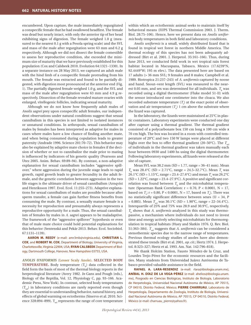
Herpetological Review 44(4), 2013
662 NATURAL HISTORY NOTES
encumbered. Upon capture, the male immediately regurgitated a conspecific female that he had swallowed headfirst. The female was dead but nearly intact, with only the anterior tip of her head exhibiting signs of digestion. The female weighed 1.0 g (mea-sured to the nearest 0.1 g with a Pesola spring scale) and the SVL and mass of the male after regurgitation were 63 mm and 6.2 g respectively. Although we did not dissect the female comestible to assess her reproductive condition, she exceeded the mini-mum size of maturity that we have previously established for this population (Cox and Calsbeek 2010. Evolution 64:1321–1330). In a separate instance on 28 May 2013, we captured an adult male with the hind limb of a conspecific female protruding from his mouth. The female was extracted and found to be partially di-gested, with digestion most pronounced at the anterior end (Fig. 1). The partially digested female weighed 1.0 g, and the SVL and mass of the male after regurgitation were 63 mm and 4.9 g re-spectively. Dissection of the female revealed mature ovaries with enlarged, vitellogenic follicles, indicating sexual maturity.
Although we do not know how frequently adult males of Anolis sagrei prey upon conspecific adult females, our indepen-dent observations under natural conditions suggest that sexual cannibalism in this species is not limited to isolated instances or unnatural conditions. In arthropods, sexual cannibalism of males by females has been interpreted as adaptive for males in cases where males have a low chance of finding another mate, and when being consumed during copulation leads to greater paternity (Andrade 1996. Science 201:70–72). This behavior may also be explained by adaptive mate choice in females if the deci-sion of whether or not to cannibalize the male prior to mating is influenced by indicators of his genetic quality (Pearsons and Utez 2005. Anim. Behav. 69:89–94). By contrast, a non-adaptive explanation for sexual cannibalism includes “aggressive spill-over,” where aggression during the juvenile stage leads to rapid growth, rapid growth leads to greater fecundity in the adult fe-male, and the genetic correlation between aggression in the two life stages is the ultimate driver of sexual cannibalism (Arnqvist and Henriksson 1997. Evol. Ecol. 11:255–273). Adaptive explana-tions for sexual cannibalism of males are possible because after sperm transfer, a female can gain further fitness benefits from consuming the male. By contrast, a sexually mature female is a necessity for reproduction and presumably always represents a reproductive opportunity for a male. Thus, the sexual cannibal-ism of females by males in A. sagrei appears to be maladaptive. The framework of the “aggressive spillover” hypothesis or even that of male mate choice could hold potential explanations for this behavior (Sentenská and Pekár 2013. Behav. Ecol. Sociobiol. 67:1131–1139).
AARON M. REEDY (e-mail: [email protected]), CHRISTIAN L. COX, and ROBERT M. COX, Department of Biology, University of Virginia, Charlottesville, Virginia 22904, USA; RYAN CALSBEEK Department of Biol-ogy, Dartmouth College, Hanover, New Hampshire 03755, USA.
ANOLIS UNIFORMIS (Lesser Scaly Anole). SELECTED BODY TEMPERATURE. Body temperature (T
b) data collected in the
field form the basis of most of the thermal biology reports in the herpetological literature (Avery 1982. In Gans and Pough [eds.], Biology of the Reptilia, Vol. 12, Physiology C, pp. 93–166. Aca-demic Press, New York). In contrast, selected body temperatures (T
sel) in laboratory conditions are rarely reported even though
they are essential to understanding behavior, natural history, and effects of global warming on ectotherms (Sinervo et al. 2010. Sci-ence 328:894–899). T
sel represents the range of core temperature
within which an ectothermic animal seeks to maintain itself by behavioral means (IUPS Thermal Commission 2003. J. Therm. Biol. 28:75–106). Hence, here we present data on Anolis unifor-mis body temperatures in both field and laboratory conditions.
Anolis uniformis is a small, widely distributed lizard that is found in tropical wet forest in northern Middle America. The thermal ecology of this species has not been adequately ad-dressed (Birt et al. 2001. J. Herpetol. 35:161–166). Thus, during June 2013, we conducted field work in wet tropical rain forest habitat located in Macuspana, Tabasco, Mexico (17.6239°N, 92.4449°W; 195 m elev.). The data presented here are based on 17 adults (> 36 mm SVL; 9 females and 8 males; Campbell et al. 1989. Biotropica 21:237–243) of A. uniformis captured by noose and hand. Snout–vent length (SVL) was measured to the near-est 0.05 mm, and sex was determined for all individuals. T
b was
recorded using a digital thermometer (Fluke model 51-II) with the sensor introduced one centimeter into the cloaca. We also recorded substrate temperature (T
s) at the exact point of obser-
vation and air temperature (Ta) 1 cm above the substrate where
the lizard was captured.In the laboratory, the lizards were maintained at 25°C in plas-
tic containers. Laboratory experiments were conducted one day after capture using a thermal gradient. The thermal gradient consisted of a polycarbonate box 150 cm long x 100 cm wide x 70 cm high. The box was located in a room with controlled tem-perature of 20°C and two 150 W lamps were placed at different highs over the box to offer thermal gradient (20–50°C). The T
sel
of individuals in the thermal gradient was taken manually each hour between 0930 and 1400 hs using the digital thermometer. Following laboratory experiments, all lizards were released at the site of capture.
Mean SVL was 38.2 mm (SD = 1.77, range = 36–41 mm). Mean T
b was 28.4°C (SD = 2.71°C, range = 24.5–32.7°C). Mean T
s was
26.1°C (SD = 1.15°C, range = 23.4–27.6°C) and mean Ta was 26.2°C
(SD = 1.22°C, range = 23.4–27.9°C). A positive and significant cor-relation was found between T
b and the microhabitat tempera-
ture (Spearman Rank Correlation: r = 0.70, P < 0.0001, N = 17, based on T
s; r = 0.80, P < 0.0001, N = 17, based on T
a). There was
no statistically significant difference between sexes (U = 34, P = 0.885). Mean T
sel was 30.1°C (SD = 1.90°C, range = 22–34.4°C).
Interquartile of 25% and 75% was 28.9 and 30.9°C, respectively. T
b shows that A. uniformis analyzed in this study was thermo-
passive, a mechanism where individuals do not need to invest time and energy actively selecting microhabitats for thermoreg-ulation in tropical habitats (Huey and Slatkin 1976. Q. Rev. Biol. 51:363–384). T
sel suggests that A. uniformis can be considered a
stenothermic species due to the narrow range of temperatures. Previous thermal ecology studies of anoles have also demon-strated these trends (Birt et al. 2001, op. cit.; Hertz 1974. J. Herpe-tol. 8:323–327; Hertz et al. 1993. Am. Nat. 142:796–818).
We thank Holcim Station, Fausto Méndez-de la Cruz, and Lourdes Trejo-Pérez for the economic resources and the facili-ties. Many students from Universidad Juárez Autónoma de Ta-basco provided valuable assistance in the field.
RAFAEL A. LARA-RESENDIZ (e-mail: [email protected]), ANÍBAL H. DÍAZ DE LA VEGA-PÉREZ (e-mail: [email protected]), Posgrado en Ciencias Biológicas, Instituto de Biología, Laboratorio de Herpetología, Universidad Nacional Autónoma de México, AP 70515, CP 04510, Distrito Federal, México; PIERRE CHARRUAU, Laboratorio de Herpetología, Departamento de Zoología, Instituto de Biología, Universi-dad Nacional Autónoma de México, AP 70515, CP 04510, Distrito Federal, México (e-mail: [email protected]).

Herpetological Review 44(4), 2013
NATURAL HISTORY NOTES 663
ASPIDOSCELIS EXSANGUIS (Chihuahuan Spotted Whiptail). FEEDING BEHAVIOR. Aspidoscelis exanguis is a parthenoge-netic species of whiptail lizard that primarily ranges within the Chihuahuan Desert ecoregion of the southwestern United States and northern Mexico (Babb 2009. In Jones and Lovich [eds.]. Lizards of the American Southwest: A Photographic Field Guide, pp. 338–341. Rio Nuevo Publ., Tucson, Arizona). The species is an active diurnal forager that feeds mostly on arthropods, with termites, grasshoppers, beetles, and arachnids being important prey items (Degenhardt et al. 1996. Amphibians and Reptiles of New Mexico. Univ. New Mexico Press, Albuquerque. 431 pp.; Scudday and Dixon 1973. Southwest. Nat. 18:279–289; Smith 1989. Southwest. Nat. 34:418–429). Most published information on food preference is based on removed stomach contents iden-tified to higher taxonomic categories, so little is known about specific food items taken and on the feeding behavior of the liz-ard in the wild. Herein, we report on the ambushing, subjuga-tion, and consumption of a tarantula by an adult A. exanguis in a Black Gramma (Bouteloua eriopoda) dominated Chihuahuan Desert grassland landscape.
On 17 June 2009, at 0920 h, on Fort Bliss, McGregor Range in Otero Co., New Mexico, USA (32.418611°N, 105.852778°W, WGS 84; 1606 m elev.), an A. exsanguis was observed ambushing and consuming an adult tarantula (Aphonopelma sp.) (Fig. 1). The lizard was observed subjugating the tarantula by grasping the tubercle dorsal portion of the thorax, utilizing vigorous head and body twisting, reminiscent of larger, predatory lizard behavior when overcoming large prey. The time frame it took for subduing the prey lasted ca. two minutes until the spider showed no signs of movement and was then consumed. Scudday and Dixon (op. cit.), mention that A. exanguis exhibit a higher foraging activity and more aggression towards prey (i.e., act of slamming prey on the ground) than other sympatrically occuring Aspidoscelis spe-cies, from which this observation appears to reflect those behav-iors. To our knowledge, this is the first documented sighting of A. exsanguis subjugating and comsuming large prey of comparable size (Aphonopelma sp.).
We thank Steven Dilks for his assistance in the field. ARTURO ROCHA (e-mail: [email protected]), VICENTE MATA-
SILVA, and JERRY D. JOHNSON (e-mail: [email protected]), Depart-ment of Biological Sciences, University of Texas at El Paso, El Paso, Texas 79968, USA.
ASPIDOSCELIS EXSANGUIS (Chihuahuan Spotted Whiptail), ASPIDOSCELIS NEOMEXICANA (New Mexico Whiptail), ASPI-DOSCELIS UNIPARENS (Desert Grassland Whiptail). BIFUR-CATED TAILS. We report occurrences of bifurcated tails in three parthenogenetic whiptail species (Jones and Lovich 2009. Liz-ards of the American Southwest: A Photographic Field Guide. Rio Nuevo Publ. Tucson, Arizona. 567 pp.) captured during a long-term study (Bateman et al. 2008. Restor. Ecol. 16:180–190). Bifur-cated tails have also been reported from museum specimens in A. velox, another parthenogenetic whiptail (Cordes and Walker 2013. Herpetol. Rev. 44:319–320).
We captured and released lizards from late May to mid-Sep-tember from 2000 to 2006 from riparian forest habitat along the middle Rio Grande in central New Mexico from Albuquerque (35.008380°N, 106.681805°W) to Bosque del Apache National Wildlife Refuge (33.805122°N, 106.859980°W). We measured lizard snout–vent length (SVL), vent–tail length (VTL), and mass. When we encountered lizards with regenerated tails, we recorded the length from the vent to the start of tail regenera-tion (original tail length, OTL). For some lizards with bifurcated tails, we measured the length of the regenerated portion of the “second” tail (bifid tail length, BTL). The bifurcation typically began at the regenerated or damaged tail segment, as observed by Cordes and Walker (2013, op. cit.). In other cases, the bifurca-tion stemmed from within a portion of a regenerated tail as ob-served by Gogliath et al. (2012. Herpetol. Rev. 43:129) in Ameiva ameiva.
We captured four individual A. exsanguis with bifurcated tails (20 July 2001, SVL = 73 mm, VTL = 92 mm, OTL = 27 mm, mass = 9.5 g; 27 July 2001, SVL = 82 mm, VTL = 141 mm, OTL = 56 mm, mass = 12.0 g; 11 July 2005, SVL = 60 mm, VTL = 116 mm, OTL = 61 mm; and 1 Aug 2005, SVL = 70 mm, VTL = 135 mm, OTL = 99 mm, BTL = 7 mm, mass = 5.5 g). We captured four individual A. neomexicana with bifurcated tails (10 June 2002, SVL = 71 mm, VTL = 87 mm, OTL = 15 mm, BTL = 60, mass = 10.5 g; 18 July 2002, SVL = 81 mm, VTL = 170 mm, OTL = 150 mm, BTL = 20, mass = 17.0 g; 27 June 2006, SVL = 77 mm, VTL = 99 mm, OTL = 19 mm, mass = 8.5 g; and 17 Aug 2006, SVL = 82 mm, VTL = 152 mm, OTL = 125 mm, BTL = 27, mass = 12.5 g). We captured three individual A. uniparens with bifurcated tails (7 July 2001, SVL = 69 mm, VTL = 117 mm, OTL = 86 mm, mass = 6.5 g; 13 July 2001, SVL = 65 mm, VTL = 121 mm, OTL = 87 mm, mass = 7.5 g; 11 June 2002, SVL = 67 mm, VTL = 123 mm, OTL = 85 mm, mass = 9.0 g). These records are among the first reports of bifurcated tails in A. exsanguis, A. neomexicana, and A. uniparens.
The Middle Rio Grande Conservancy District, Bosque del Apache National Wildlife Refuge, and Albuquerque Open Space permitted access to study sites. The University of New Mexico Animal Care and Use Committee approved field techniques (pro-tocol #20415). The long-term herpetofaunal study was funded by the USDA Forest Service – RMRS Middle Rio Grande Ecosystem Management Unit, Joint Fire Sciences Program, National Fire Plan, US FWS Bosque Improvement Initiative, and USFS S&PF NM Collaborative Forest Restoration Program.
HEATHER L. BATEMAN, Arizona State University at the Polytechnic campus, 7231 E. Sonoran Arroya Mall, Mesa, Arizona 85212, USA (e-mail: [email protected]); ALICE CHUNG-MacCOUBREY, Inventory & Monitoring Program Manager, Sierra Nevada Network, Sequoia & Kings Canyon National Parks, 47050 Generals Highway, Three Rivers, California 93271, USA (e-mail: [email protected]).Fig.1. Adult Aspidoscelis exanguis consuming a tarantula (Aphono-
pelma sp.) in Otero County, New Mexico, USA.
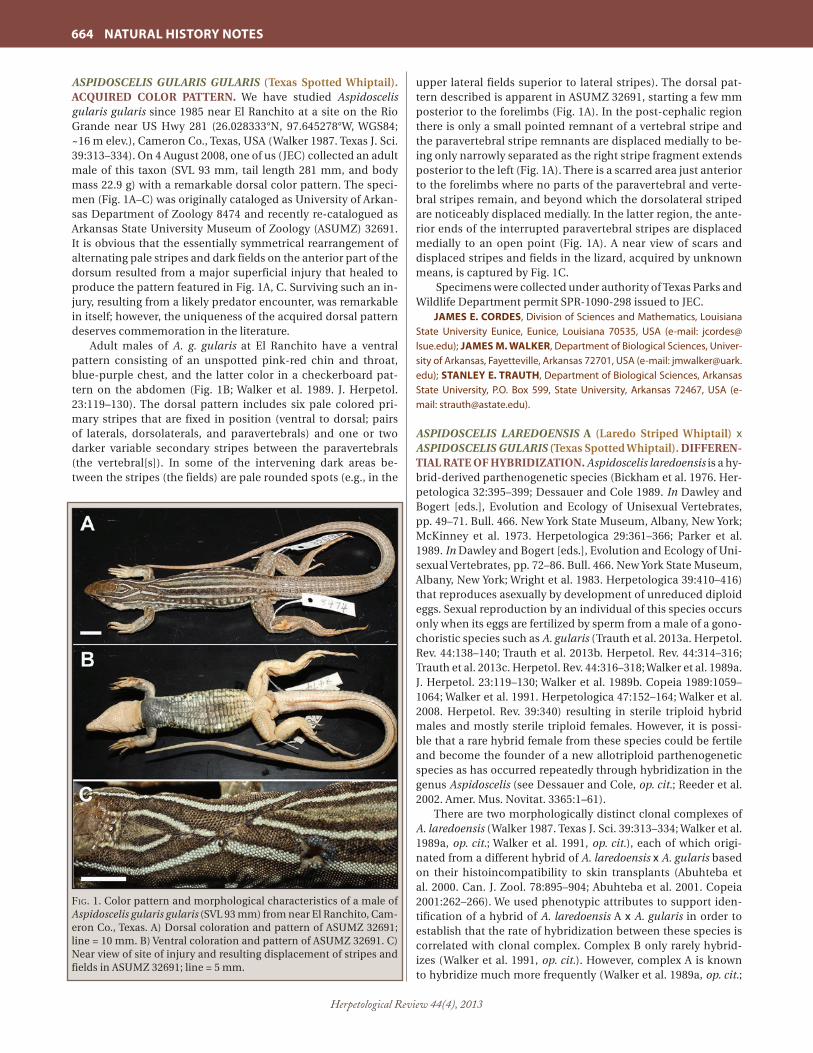
Herpetological Review 44(4), 2013
664 NATURAL HISTORY NOTES
ASPIDOSCELIS GULARIS GULARIS (Texas Spotted Whiptail). ACQUIRED COLOR PATTERN. We have studied Aspidoscelis gularis gularis since 1985 near El Ranchito at a site on the Rio Grande near US Hwy 281 (26.028333°N, 97.645278°W, WGS84; ~16 m elev.), Cameron Co., Texas, USA (Walker 1987. Texas J. Sci. 39:313–334). On 4 August 2008, one of us (JEC) collected an adult male of this taxon (SVL 93 mm, tail length 281 mm, and body mass 22.9 g) with a remarkable dorsal color pattern. The speci-men (Fig. 1A–C) was originally cataloged as University of Arkan-sas Department of Zoology 8474 and recently re-catalogued as Arkansas State University Museum of Zoology (ASUMZ) 32691. It is obvious that the essentially symmetrical rearrangement of alternating pale stripes and dark fields on the anterior part of the dorsum resulted from a major superficial injury that healed to produce the pattern featured in Fig. 1A, C. Surviving such an in-jury, resulting from a likely predator encounter, was remarkable in itself; however, the uniqueness of the acquired dorsal pattern deserves commemoration in the literature.
Adult males of A. g. gularis at El Ranchito have a ventral pattern consisting of an unspotted pink-red chin and throat, blue-purple chest, and the latter color in a checkerboard pat-tern on the abdomen (Fig. 1B; Walker et al. 1989. J. Herpetol. 23:119–130). The dorsal pattern includes six pale colored pri-mary stripes that are fixed in position (ventral to dorsal; pairs of laterals, dorsolaterals, and paravertebrals) and one or two darker variable secondary stripes between the paravertebrals (the vertebral[s]). In some of the intervening dark areas be-tween the stripes (the fields) are pale rounded spots (e.g., in the
upper lateral fields superior to lateral stripes). The dorsal pat-tern described is apparent in ASUMZ 32691, starting a few mm posterior to the forelimbs (Fig. 1A). In the post-cephalic region there is only a small pointed remnant of a vertebral stripe and the paravertebral stripe remnants are displaced medially to be-ing only narrowly separated as the right stripe fragment extends posterior to the left (Fig. 1A). There is a scarred area just anterior to the forelimbs where no parts of the paravertebral and verte-bral stripes remain, and beyond which the dorsolateral striped are noticeably displaced medially. In the latter region, the ante-rior ends of the interrupted paravertebral stripes are displaced medially to an open point (Fig. 1A). A near view of scars and displaced stripes and fields in the lizard, acquired by unknown means, is captured by Fig. 1C.
Specimens were collected under authority of Texas Parks and Wildlife Department permit SPR-1090-298 issued to JEC.
JAMES E. CORDES, Division of Sciences and Mathematics, Louisiana State University Eunice, Eunice, Louisiana 70535, USA (e-mail: [email protected]); JAMES M. WALKER, Department of Biological Sciences, Univer-sity of Arkansas, Fayetteville, Arkansas 72701, USA (e-mail: [email protected]); STANLEY E. TRAUTH, Department of Biological Sciences, Arkansas State University, P.O. Box 599, State University, Arkansas 72467, USA (e-mail: [email protected]).
ASPIDOSCELIS LAREDOENSIS A (Laredo Striped Whiptail) x ASPIDOSCELIS GULARIS (Texas Spotted Whiptail). DIFFEREN-TIAL RATE OF HYBRIDIZATION. Aspidoscelis laredoensis is a hy-brid-derived parthenogenetic species (Bickham et al. 1976. Her-petologica 32:395–399; Dessauer and Cole 1989. In Dawley and Bogert [eds.], Evolution and Ecology of Unisexual Vertebrates, pp. 49–71. Bull. 466. New York State Museum, Albany, New York; McKinney et al. 1973. Herpetologica 29:361–366; Parker et al. 1989. In Dawley and Bogert [eds.], Evolution and Ecology of Uni-sexual Vertebrates, pp. 72–86. Bull. 466. New York State Museum, Albany, New York; Wright et al. 1983. Herpetologica 39:410–416) that reproduces asexually by development of unreduced diploid eggs. Sexual reproduction by an individual of this species occurs only when its eggs are fertilized by sperm from a male of a gono-choristic species such as A. gularis (Trauth et al. 2013a. Herpetol. Rev. 44:138–140; Trauth et al. 2013b. Herpetol. Rev. 44:314–316; Trauth et al. 2013c. Herpetol. Rev. 44:316–318; Walker et al. 1989a. J. Herpetol. 23:119–130; Walker et al. 1989b. Copeia 1989:1059–1064; Walker et al. 1991. Herpetologica 47:152–164; Walker et al. 2008. Herpetol. Rev. 39:340) resulting in sterile triploid hybrid males and mostly sterile triploid females. However, it is possi-ble that a rare hybrid female from these species could be fertile and become the founder of a new allotriploid parthenogenetic species as has occurred repeatedly through hybridization in the genus Aspidoscelis (see Dessauer and Cole, op. cit.; Reeder et al. 2002. Amer. Mus. Novitat. 3365:1–61).
There are two morphologically distinct clonal complexes of A. laredoensis (Walker 1987. Texas J. Sci. 39:313–334; Walker et al. 1989a, op. cit.; Walker et al. 1991, op. cit.), each of which origi-nated from a different hybrid of A. laredoensis x A. gularis based on their histoincompatibility to skin transplants (Abuhteba et al. 2000. Can. J. Zool. 78:895–904; Abuhteba et al. 2001. Copeia 2001:262–266). We used phenotypic attributes to support iden-tification of a hybrid of A. laredoensis A x A. gularis in order to establish that the rate of hybridization between these species is correlated with clonal complex. Complex B only rarely hybrid-izes (Walker et al. 1991, op. cit.). However, complex A is known to hybridize much more frequently (Walker et al. 1989a, op. cit.;
Fig. 1. Color pattern and morphological characteristics of a male of Aspidoscelis gularis gularis (SVL 93 mm) from near El Ranchito, Cam-eron Co., Texas. A) Dorsal coloration and pattern of ASUMZ 32691; line = 10 mm. B) Ventral coloration and pattern of ASUMZ 32691. C)Near view of site of injury and resulting displacement of stripes and fields in ASUMZ 32691; line = 5 mm.

Herpetological Review 44(4), 2013
NATURAL HISTORY NOTES 665
this study) based on records for 20+ years (1986–2008) in an area near the Rio Grande (26.233889°N, 98.560833°W, WGS84; ~ 40 m elev.) in the vicinity of Los Ebanos, Hidalgo Co., Texas, USA. The male of A. gularis, male hybrid, and female of A. laredoen-sis A featured in Fig. 1A–F, originally cataloged as University of Arkansas Department of Zoology 8386, 8390, and 8389, respec-tively, and re-catalogued as Arkansas State University Museum of Zoology (ASUMZ) 32688, 32689, and 32690, respectively, were collected by one of us (JEC) on 22 June 2008 in the aforemen-tioned area. We have also collected hybrids of A. laredoensis A x A. gularis from other sites in Dimmit, La Salle, Webb, Star, and Hidalgo counties, Texas, and Tamaulipas State, Mexico (Walker et al. 1989a, op. cit.), The distinctive Los Ebanos area, within a prominent oxbow of the Rio Grande, supports a thriving guild of whiptail lizards comprising large numbers of A. gularis (gono-choristic) and both clonal complexes A and B of A. laredoen-sis (diploid parthenogens). Here, these congeners are locally
dispersed such that opportunities for hybridization of A. gularis with each of A. laredoensis A and B are essentially present when-ever lizards are active.
Prior to preservation, hybrid ASUMZ 32689 had a SVL of 76 mm, tail length of 263 mm, and body mass of 10.6 gm. This male could not be identified to A. gularis because of divergent fea-tures of both dorsal and ventral color patterns, as well as scutel-lation (see Conant and Collins 1998. A Field Guide to Reptiles and Amphibians of Eastern and Central North America, 3rd ed. Houghton Mifflin Co., New York, New York. 640 pp.). It lacked the low contrast darkly hued dorsal stripes/spots and fields, and lacked the intense purple-blue ventral suffusion of the mesop-tychium, fore limbs, and thoracic/abdominal regions typical of A. gularis (Fig. 1A, D). Although the specimen resembles A. laredoensis A (Fig. 1C, F) more closely than A. gularis (Fig. 1A, D) or A. laredoensis B, characters indicating its hybrid origin are these aspects of color pattern: two pairs of pale straight-margined primary stripes (dorsolaterals, and paravertebrals), one pair of pale unevenly margined primary stripes resulting from contact with pale rounded spots and vertical bars, large distinct spots in brown to brown-black fields above (upper lat-erals) and below (lower laterals) lateral stripes nearest to ven-tral scales, a single indistinct and discontinuous vertebral stripe past midbody, thoracic and abdominal scales narrowly edged with purple-blue, and pink-red ventral surface of the head (Fig. 1B, E). In comparison, the typically patterned female of A. lar-edoensis depicted in Fig. 1C, F) lacks large spots in the upper and lower lateral dark fields, does not have unevenly margined lateral stripes, and has a cream-white venter absent a pink-red throat and purple-blue edged thoracic and abdominal scales.
Fig. 1. Color pattern and morphological characteristics of a male of Aspidoscelis gularis (SVL 76 mm), hybrid male of A. laredoensis A x A. gularis (SVL 76 mm), and females of A. laredoensis A (SVL 76 mm) from the vicinity of Los Ebanos, Hidalgo County, Texas. A–C. Dorsal coloration and pattern of A. gularis (ASUMZ 32688), hybrid (ASUMZ 32689), and A. laredoensis A (ASUMZ 32690); line = 1 mm. D–F. Ven-tral coloration and pattern of A. gularis (ASUMZ 32688), hybrid (ASUMZ 32689), and A. laredoensis A (ASUMZ 32690); line = 1 mm.
Fig. 2. Enlarged postantebrachial scales of the hybrid (ASUMZ 32689); line = 1 mm.

Herpetological Review 44(4), 2013
666 NATURAL HISTORY NOTES
Also indicative of a hybrid origin for ASUMZ 32689 (comparison with A. laredoensis in paren) are the greatly enlarged mesopty-chial scales bordering the gular fold (rather than enlarged); en-larged postantebrachial scales on posterior aspect of forelimbs (Fig. 2, rather than moderately enlarged), and 80 granular scales around midbody (compared with 91.8 ± 0.76 based on Walker et al. 1989b, op. cit.).
The specimen collected in 2008 described herein establishes that production of viable, if infertile, hybrids of A. laredoensis A x A. gularis has been ongoing in the Los Ebanos vicinity for 20+ years. The first of these was collected by combinations of the au-thors at Los Ebanos a few hundred meters from the Rio Grande in 1986 (Walker et al. 1989a, op. cit.) followed by collection of other hybrids over the years up to 2008. Remarkably, there are apparently strong pre-mating/post-mating isolating mecha-nisms to hybridization between A. laredoensis B x A. gularis, as only three hybrids representing this combination (Walker et al. 1991, op. cit.) have been collected in Texas and Mexico during ~75 expeditions since 1984. Internal examination of reproduc-tive organs in the present hybrid by one of us (SET) revealed the following: the testes were enlarged and appeared to be function-al; the epididymis was expanded and appeared to have sperm present.
Specimens were collected under authority of Texas Parks and Wildlife Department permit SPR1090-298 and USDI/LRGVNWR special use permit 21552-09-13-MS. Special thanks are given to Mitch Sternberg of the USFWS at Santa Ana NWR for providing the special use permit.
STANLEY E. TRAUTH, Department of Biological Sciences, Arkansas State University, P.O. Box 599, State University, Arkansas 72467, USA (e-mail: [email protected]); JAMES E. CORDES, Division of Sciences and Mathematics, Louisiana State University Eunice, Eunice, Louisiana 70535, USA (e-mail: [email protected]); MARK A. PAULISSEN, Department of Natural Sciences, Northeastern State University, Tahlequah, Oklahoma 74464, USA (e-mail: [email protected]); JAMES M. WALKER, Depart-ment of Biological Sciences, University of Arkansas, Fayetteville, Arkansas 72701, USA (e-mail: [email protected]).
ASPIDOSCELIS LAREDOENSIS A (Laredo Striped Whiptail) x ASPIDOSCELIS GULARIS (Texas Spotted Whiptail). STOMACH CONTENTS. Diploid parthenogenetic Aspidoscelis laredoen-sis clonal complexes A and B, and gonochoristic parental spe-cies A. gularis, are syntopic at many sites in Texas and Mexico, mostly in the vicinity of the Rio Grande (Walker 1987. Texas J. Sci. 39:313–334). During the past 20+ years (1986–2008), it has not been unusual to find one or more hybrids of A. laredoensis A x A. gularis among collections from sites such as the one near the Rio Grande (26.233889°N, 98.560833°W, WGS84, ~40 m elev.) in the vicinity of Los Ebanos, Hidalgo Co., Texas (Walker et al. 1989. J. Herpetol. 23:119–130). Recently, SET examined stomach contents of hybrid male Arkansas State University Museum of Zoology (ASUMZ) 32689 (Trauth et al. 2013. Herpetol. Rev. 44: in press) collected by JEC on 22 June 2008 at the aforementioned site. We use this individual of 76 mm SVL as the basis for the first reported observations on the diet of a hybrid whiptail. Upon opening the body cavity of ASUMZ 32689, the stomach was seen to be distended to maximum capacity. The contents consisted almost entirely of termites (order Isoptera). By counting intact termites and detached heads it was found that the hybrid had consumed 181 of these insects. Although it would require sev-eral minutes for a lizard to consume such a large number of prey items while relatively stationary at an exposed termite colony
(and thus be susceptible to predation), such a bonanza discov-ery would yield far more calories per unit of time than the more difficult tactic of harvesting grasshoppers which are typically in evidence at Los Ebanos. Paulissen 2001. (J. Herpetol. 35:282–292) and Paulissen et al. 1992 (J. Herpetol. 26:153–158) reported that termites comprised the numerical majority of prey consumed by both of the parental species of the hybrid, A. laredoensis A and A. gularis, at several sites of syntopy in Texas.
The specimen was collected under authority of Texas Parks and Wildlife Department permit SPR-1090-298 and USDI/LRGVNWR special use permit 21552-09-13-MS. Special thanks are given to Mitch Sternberg of the USFWS at Santa Ana NWR for providing the special use permit.
STANLEY E. TRAUTH, Department of Biological Sciences, Arkansas State University, P. O. Box 599, State University, Arkansas USA, 72467 (e-mail: [email protected]); JAMES E. CORDES, Division of Sciences and Mathematics, Louisiana State University Eunice, Eunice, Louisiana USA, 70535 (e-mail: [email protected]); MARK A. PAULISSEN, Department of Natural Sciences, Northeastern State University, Tahlequah, Oklahoma USA, 74464 (e-mail: [email protected]): JAMES M. WALKER, Depart-ment of Biological Sciences, University of Arkansas, Fayetteville, Arkansas USA, 72701 (e-mail: [email protected].
ASPIDOSCELIS NEOMEXICANA (New Mexico Whiptail) x AS-PIDOSCELIS INORNATA LLANURAS (Plains Striped Whip-tail). PHENOTYPE. Aspidoscelis neomexicana, a hybrid-derived parthenogenetic species (Cole et al. 1988. Amer. Mus. Novitat. 2905:1–38; Parker and Selander 1984. Herpetologica 40:245–252), reproduces asexually by development of unreduced diploid eggs. Sexual reproduction in the species occasionally occurs when its eggs are fertilized by sperm from a male of A. inornata (Axtell 1966. Herpetologica 22:241–253; Cuellar and McKinney 1976. J. Exp. Zool. 196:341–350; Taylor and Medica 1966. Univ. Colorado, Ser. Biol. 22:1–9; Taylor and Walker 1996. Copeia 1996:945–954; Wright and Lowe 1967. Amer. Mus. Novitat. 2286:1–36) resulting in sterile triploid hybrid males or females.
Herein, we describe phenotypic attributes of the first hybrid of A. neomexicana x A.inornata llanuras reported as involving this subspecies and the first one reported from the western-most part of the distribution of these species in New Mexico (see map in Cole et al., op. cit.). This female (originally cata-logued as University of Arkansas Department of Zoology 6081 and re-catalogued as Arkansas State University Museum of Zo-ology 32423) was collected by JEC on 13 July 1998 in Hidalgo Co.: 21.3‒21.6 km NW jct of US Hwy 90 and New Mexico Hwy 70, on 70 in the vicinity of the site at 32.515000°N, 108.876667°W (datum WGS84). Also obtained here during six visits between 1998 and 2001 were 17 (19.3%) A. inornata llanuras (gonocho-ristic), 22 (25.0%) A. neomexicana (diploid parthenogen), and 48 (54.5%) A. uniparens (triploid parthenogen). Prior to preser-vation, the hybrid had a SVL of 62 mm, tail length of 217 mm, and body mass of 6.3 gm. It could not be identified to A. inorna-ta llanuras because of the presence of spots (i.e., rounded pale areas) in the dark fields between the stripes; this taxon is char-acterized by the lack of spots throughout ontogeny (Sullivan et al. 2013. Copeia 2013[3]:366–377; Wright and Lowe 1993. J. Arizona-Nevada Acad. Sci. 27:129–157). Although the specimen resembles A. neomexicana more closely than A. inornata llan-uras, obvious phenotypic consequences of the hybrid genome are these intermediate aspects of color pattern (Fig. 1A–B): paired lateral and dorsolateral stripes relatively straight-mar-gined, paired paravertebral stripes slightly unevenly margined,

Herpetological Review 44(4), 2013
NATURAL HISTORY NOTES 667
single vertebral stripe with several interruptions and unevenly margined, strongly contrasting pale stripes and dark fields, in-cipient rather than distinct spots; and off-white to very pale blue rather than gray-blue venter. Data for four morphological characters are also consistent with the hypothesis for a hybrid origin for ASUMZ 32423 (comparison with A. neomexicana in paren): low number of granules around midbody (compared to 75–83, N = 22); moderately enlarged mesoptychial scales (Fig. 1C, rather than small); slightly enlarged postantebrachial scales (Fig. 1D, rather than granular), and incomplete circumorbital scales (Fig. 1E, rather than complete).
That production of viable, if not fertile, hybrids of A. neomex-icana x A. inornata llanuras is a rare event in the guild sampled at the Hidalgo Co. site is indicated by ASUMZ 32423 (0.01%) being among 88 specimens collected (i.e., < 33% of observed whiptail lizards) in six visits from 1998 to 2001. The hybrid is an “immature” female with both oviducts highly regressed, exhib-iting minimal enlargement posteriorly. Both gonads appear as minimally-expanded, linear strips of translucent tissue, exhibit-ing no evidence of developing follicles. Abdominal fat bodies are modestly enlarged. Based on these observations, we infer that the hybrid was sterile.
Specimens in Hidalgo Co. were collected under authority of per-mit number 1850 issued to JEC by the New Mexico Department of Game and Fish.
JAMES M. WALKER, Department of Biological Sciences, University of Arkansas, Fayetteville, Arkansas 72701, USA (e-mail: [email protected]); JAMES E. CORDES, Division of Sciences and Mathematics, Louisiana State University Eunice, Eunice, Louisiana 70535, USA (e-mail: [email protected]); STANLEY E. TRAUTH, Department of Biological Sciences, Arkansas State University, P.O. Box 599, State University, Arkansas 72467, USA (e-mail: [email protected]).
Fig. 1. Color pattern and morphological characteristics of a putative hybrid female specimen of Aspidoscelis neomexicana x A. inornata llan-uras. A) Dorsal coloration and pattern of ASUMZ 32423; line = 10 mm for A and B. B) Ventral coloration of ASUMZ 32423. C) Moderately enlarged mesoptychial scales of ASUMZ 32433; line = 1 mm. D) Slightly enlarged postantebrachial scales of ASUMZ 32423; line = 1 mm. E) Incomplete circumorbital scale series (see arrow) in ASUMZ 32423; line = 1 mm.

Herpetological Review 44(4), 2013
668 NATURAL HISTORY NOTES
ASPIDOSCELIS SEXLINEATA (Six-lined Racerunner). ARTIFI-CIAL HABITAT. Aspidoscelis sexlineata is a “species in greatest conservation need” in Minnesota. It typically lives in sand prai-ries or bluff prairies, although Oldfield and Moriarty (1994. Am-phibians and Reptiles Native to Minnesota. University of Min-nesota Press, Minneapolis, Minnesota. 237 pp.) noted that it can inhabit the rock and cinder fill of railroad tracks and dike roads. We examined racerunner use of artificial habitat associated with the Prairie Island dike near Winona, Winona Co., Minnesota, USA. Completed in 1967, the dike is topped by a two-lane black-top road approximately 7–8 m above normal river level (less dur-ing periods of flooding). Sampling was conducted in a 4.7-km stretch extending northwest from Mississippi River Lock-and-Dam 5A toward Minnesota City. The river (northeastern) side of the dike was rip-rapped with boulders and chunks of concrete, whereas the southwestern side, which bordered bottomland marsh and forest, consisted of prairie-like habitat with patches of open sandy substrate. A remnant sand prairie that formerly existed near the southeastern end of the study stretch, where racerunners were observed as recently as the early 2000s, pre-sumably served as the source of racerunners that inhabited the dike, but the site is now occupied by artificial wetlands.
Paired transect surveys, one on each side of the road, were conducted in 2010 on 15 July (5 pairs), 16 July (10), 21 August (10), 22 August (10), and 1 September (10). The 50-m transects were parallel to the road and randomly positioned with respect to distance from the southeastern end of the study stretch (0–4.7 km) and distance from the roadside (0–13 m). Data were collect-ed by two observers using an event recorder to tally the number of racerunners observed within 1 m of each side of the transect. Substrate and shaded air temperature was measured at the start of each transect.
Racerunners were observed over a distance of 3 km on the southwestern side of the road (N = 17) and 1 km on the northeast-ern side (N = 2). Racerunners occurred from 2 m to 13 m from the edge of the road; mean distance on the southwestern side (7.23 m) and the northeastern side (5.50 m) were not significantly dif-ferent (t = 0.674, df = 13, p = 0.512). The number of racerunners per transect was significantly greater on the southwestern side of the dike (paired t-test, t = 3.028, df = 44, p = 0.004). This may have been related to the greater substrate temperature on the southwestern side (paired t-test, t = 3.305, df = 44, p = 0.002), but racerunners may have been less detectable among the boulders on the northeastern side.
The substrate temperature for transects when racerunners were observed ranged from 26°C to 46°C, with a mean (32.8°C) slightly less than substrate temperatures selected in oak savan-nah (34.7°C) and open grassland (39.2°C) in Wisconsin (Kapfer and Pauers 2006. Herpetol. Rev. 37:420–423). Shaded air temper-ature ranged from 22°C to 34°C. Oldfield and Moriarty (op. cit.) reported that racerunners have been observed active in Minne-sota at air temperatures as low as 22°C, and Fitch (1958. Univ. Kansas Publ. Mus. Nat. Hist. 11:11–62) reported an optimal air temperature in Kansas of 34°C.
Expanding the estimated densities of racerunners observed on the 50 m × 2 m transects on the southwestern (0. 00378/m2) and northeastern (0.00044/m2) sides of the dike to the total area of potential habitat on each side yielded total estimates of 229 and 26 racerunners, respectively. These are likely underestimates because of imperfect detectability (Smolensky and Fitzgerald 2010. J. Herpetol. 44:372–381). Nevertheless, they fall within the range of densities reported by Bellis (1964. Herpetologica
20:9–16) in South Carolina (0. 0245/m2) and Warner (2000. Trans. Illinois St. Acad. Sci. 93:239–248) in northwestern Illinois.
The southwestern-facing slope of the Prairie Island dike pro-vides extensive artificial habitat similar to the bluff prairies typi-cally inhabited by racerunners in southeastern Minnesota. This sort of artificial habitat may become increasingly important as bluff prairies decline in area due to encroachment by invasive woody plants. Kraszewski (2004. A Survey of Native Flowering Plant Species on Goat Prairies in Winona, Minnesota. Unpubl. B.A. thesis, Saint Mary’s University of Minnesota, Winona. 29 pp.) used aerial photographs to estimate the areas of 10 bluff prairie remnants in the Winona area, some of which are inhabited by racerunners. Prairie area varied from 0.25 ha to 3.14 ha (mean = 1.49 ha). When Gerty (2013. Species Richness in Relation to Both Remnant Size and Leafy Spurge [Euphorbia esula] Invasion. Un-publ. B.A. thesis, Saint Mary’s University of Minnesota, Winona. 19 pp.) remeasured the same areas on the ground, she found them broken into 20 remnants of 0.03–0.56 ha. In comparison, each side of the Prairie Island dike provides 6.07 ha of potential habitat, although racerunners do not necessarily occur along the entire road and available habitat is reduced during years with periods of high water.
This research was funded in part by a grant from Beta Beta Beta. We thank Madison Sienko for field assistance.
MICHELLE L. GOSSEN and PHILIP A. COCHRAN, Biology Depart-ment, Saint Mary’s University of Minnesota, 700 Terrace Heights, Winona, Minnesota 55987, USA (e-mail: [email protected]).
ASPIDOSCELIS SEXLINEATA VIRIDIS (Prairie Racerunner). BURROW SITE FIDELITY. The burrows and burrowing activity of Prairie Racerunner, Aspidoscelis sexlineata viridis, have been well documented (Fitch 1958. Univ. Kansas Publ., Mus. Nat. Hist. 11:11–62; Hardy 1962. Univ. Kansas Sci. Bull. 43:3–73; Leuck 1982. Copeia 1982:416–424; Trauth et al. 2004. The Amphibians and Reptiles of Arkansas. Univ. of Arkansas Press, Fayetteville, 421 pp.). Burrow types, excavation sites, space use, and lizard ac-tivity were described by Hardy (op. cit.) and Leuck (op. cit.) from their observations of the lizards in outdoor enclosures, whereas Fitch (op. cit.) and Trauth et al. (op. cit.) described or illustrated burrows as reported from field studies on this species. In the present study, we report on an unusual incident of burrow site fidelity in A. s. viridis.
On 18 June 2013, one of us (SET) observed burrowing activ-ity of a large (78 mm SVL) male Prairie Racerunner on a gradu-al-sloping, west-facing, red clay, highway embankment from ca. 1800–1840 h. The site is located on the Salem Plateau in the Ozarks Mountains (Randolph County, Arkansas 36.28215°N, 91.253°W). About 2 m2 of ground surface had been disrupted earlier in the day with a potato rake by SET while searching for lizard egg clutches. The excavated site had previously contained numerous surface burrows prior to unearthing and had been photographed on 7 May 2010 (Fig. 1). What made this particular observation signifi-cant was that the lizard moved toward this disrupted excavation site from the surrounding grassy habitat and stopped abruptly at a point directly in front of the observer (within 1 m) before it be-gan digging in the loosened soil. The lizard moved back and forth across the torn ground presumably searching for former burrow shelters. The lizard soon departed the area, but it then returned and repeated the searching and digging behavior in and around the dirt pile. A repetition of this searching/digging behavior in the dirt pile continued several times during a time span of ca. 35 min. Eventually, SET attempted to catch the lizard as it had partially

Herpetological Review 44(4), 2013
NATURAL HISTORY NOTES 669
dug a burrow shelter, but the lizard evaded the attempted capture and darted into the adjacent grassy habitat. Soon, the lizard re-turned and walked directly beside the observer’s feet and began digging again into the dirt pile. Finally, the lizard dug into the same loose soil where it had previously evaded capture and soon disap-peared far enough within its burrow so as to allow for its capture.
The digging behavior of this racerunner was similar to ob-servations reported by Hardy (op. cit.), although not in the con-text of burrow fidelity. She termed the shelter created as being an “inactivity-tunnel,” a nighttime refuge or one for periods of pro-longed inactivity. The particular lizard reported here would dig with alternating front legs and by pushing into the soil inward with its snout; then, it would turn around and push the tailings out of the hole with its front legs and chest. This behavior was repeated several times until the lizard’s body was well hidden inside the burrow. The loose soil tailings created by this type of burrowing behavior are shown in Fig. 1.
We have collectively studied A. s. viridis in many parts of the range in Arkansas and other states, and have often observed fleeing lizards work their way to what appeared to be a targeted burrow. We suggest that burrow fidelity, in addition to conserva-tion of energy by continued use of a burrow, can also serve as a targeted escape route for a lizard.
STANLEY E. TRAUTH, Department of Biological Sciences, Arkansas State University, P.O. Box 599, State University, Arkansas 72467, USA (e-mail: [email protected]); JAMES M. WALKER, Department of Biological Sciences, University of Arkansas, Fayetteville, Arkansas 72701, USA (e-mail: [email protected]); JAMES E. CORDES, Division of Sciences and Math-ematics, Louisiana State University Eunice, Eunice, Louisiana 70535, USA (e-mail: [email protected]).
ASPIDOSCELIS SEXLINEATA VIRIDIS (Prairie Racerunner). SKELETAL REMAINS. Between 1973 and 1980, Trauth (1983. Amer. Midl. Nat. 109:289–299) studied nesting habitat of Aspi-doscelis sexlineata (as Cnemidophorus sexlineatus) across 10 states, during which 195 clutches of eggs and large numbers of lizards were excavated. The excavations took place mostly from within well-drained red clay exposures facing S to W along sec-ondary highways and rural dirt roads. Most of this work was con-ducted in the Interior Highlands Region of Arkansas while study-ing A. s. viridis (Prairie Racerunner) where it was noted that, with
the hundreds of hours of excavation work, remains of expired lizards were never found. Thus, on 20 September 2003 SET was surprised by the discovery of clearly identifiable skeletal remains which we agree are those of a large Prairie Racerunner (Fig. 1) in a burrow shelter uncovered from beneath a flat rock (ca. 25 cm in diameter) on a gradual-sloping, south-facing, red clay embankment at 1435 h. The site is located within the Arkansas Army National Guard controlled Fort Chaffee Maneuver Training Center in the Arkansas River Valley, Sebastian Co., Arkansas, USA (35.20034°N, 94.15700°W; elev. 241 m). The lizard was occupying a nesting chamber that contained eggshells from a previous egg clutch (Fig. 1). Bacterial action on the lizard’s undetached skin and/or the remaining flesh along the spine and hind legs cast a greenish tint to the exposed dorsum. Moreover, and based upon the presence of this greenish coloration, there appeared to be the corpse of another vertebrate beside the lizard, although its iden-tity was not determined at the time of the discovery.
We have collectively studied A. sexlineata in many parts of the range in Arkansas and other states, and only SET has ob-served another instance of a dead racerunner in a burrow shel-ter. That instance occurred on 29 July 1982 in Pasco Co., Florida, in which the partially decomposed body of an Eastern Six-lined Racerunner, A. s. sexlineata, was infested with what appeared to be fire ants. These examples of deceased racerunners located in burrow retreats provide preliminary support for our hypothesis that many of them die by means other than predation. Never-theless, it seems likely that skeletal remains of racerunners are ephemeral given their rare discovery during the large number of excavations performed by SET.
STANLEY E. TRAUTH, Department of Biological Sciences, Arkansas State University, P.O. Box 599, State University, Arkansas 72467, USA (e-mail: [email protected]); JAMES M. WALKER, Department of Biological Sciences, University of Arkansas, Fayetteville, Arkansas 72701, USA (e-mail: [email protected]); JAMES E. CORDES, Division of Sciences and Math-ematics, Louisiana State University Eunice, Eunice, Louisiana 70535, USA (e-mail: [email protected]).
Fig. 1. Finger points to the open mouth of a Prairie Racerunner bur-row photographed on 7 May 2010 (see text for site description). Ar-row points to fresh soil tailing (an indication of the presence of a liz-ard) from a recently excavated burrow shelter. Fig. 1. Skeletal remains of a Prairie Racerunner photographed on 20
September 2003 (see text for site description). Characteristics of this species include a combination of the following structures (designat-ed by labeled arrows): (a) a wedge-shaped head, (b) rectangular belly scutes, and (c) an elongated fourth toe. Eggshells (d) from a previous egg clutch of A. s. viridis are also evident. Line = 10 mm.

Herpetological Review 44(4), 2013
670 NATURAL HISTORY NOTES
BOGERTIA LUTZAE. PREDATION BY LEPTOPHIS AHAETUL-LA. Bogertia lutzae is a small bromelicolous lizard (Avila et al. 2010. J. Helminthol. 84:199–201) endemic to the northeast region of Brazil and is found in different ecosystems, mainly inhabit-ing sandbanks, Atlantic forest, and Caatinga (Rodrigues 1987. Arq. Zool. 1:105–230). Leptophis ahaetulla is a snake with a wide geographical distribution, occurring from southern Mexico to northern Uruguay (Albuquerque et al. 2007. J. Nat. Hist. 41:1237–1243; Carvalho et al. 2007. Biol. Geral Exper. 7:41–59). It is a pri-marily diurnal and semi-arboreal snake and is reported to occur over a wide range of habitats. The diet of L. ahaetulla consists predominantly of hylid frogs; however, there are also records of Anolis sp., Thecadactylus rapicauda, Mastigodryas boddaerti, and young birds in their diet (Albuquerque et al. 2007, op. cit.).
On 8 April 2013 at 1230 h we observed an adult L. ahaetulla preying upon an adult B. lutzae. This event occurred at Tapacurá Ecological Station, Pernambuco, Brazil (8.036°S, 35.199°W; da-tum WGS 84). The event was recorded at a forest edge. This is the first record of predation reported for B. lutzae by L. ahaetulla. The image of predation is available in the collection of images of Herpetology and Paleontology Laboratory of the Federal Rural University of Pernambuco - UFRPE, Recife, Pernambuco, Brazil.
DANIELA FILIPA FERREIRA TEIXEIRA (e-mail: [email protected]) and GERALDO JORGE BARBOSA DE MOURA, Universidade Fed-eral Rural de Pernambuco, UFRPE, Paleoherpetological and Herpetological Laboratory, Rua Dom Manoel de Medeiros, s/n, Dois Irmãos, CEP, 52171-900, Recife, Pernambuco, Brazil.
CALLOPISTES MACULATUS (Chilean Iguana). SAUROPHAGY ON LIOLAEMUS. The highly endemic reptile diversity of Chile is characterized by lower numbers of lizard species, of predomi-nantly small body sizes (SVL <180 mm), compared to adjacent countries. Among these species, Callopistes maculatus, the larg-est known lizard (and only teiid) occurring in the country (SVL average: 150.5 ± 10.9 mm, range [adult size estimated from the largest 2/3 of a 38-specimen sample]: 135–173 mm), and the Li-olaemus evolutionary radiation, one of the richest amniote gen-era on Earth and the richest vertebrate genus in Chile (Pincheira-Donoso et al. 2008. Zootaxa 1800:1–85), stand out as some of the most prominent elements of the indigenous herpetofauna. All
geographic areas where C. maculatus has been recorded are also inhabited by one or more species of Liolaemus. In these com-mon areas, both groups of lizards are found in the same micro-habitats, and overlap temporally (they are all diurnal). Surpris-ingly, however, ecological interactions among Callopistes and Liolaemus have only rarely been documented in the literature (e.g., see Jaksic 1998. Ecología de los Vertebrados Terrestres de Chile. Pont. Univ. Católica Press, Santiago. 262 pp.). Here, we re-port a predation event between C. maculatus and L. nitidus, one of the largest known Liolaemus species (SVL average: 92.5 mm, range: 85.4–107.5 mm; Pincheira-Donoso et al. 2008. BMC Evol. Biol. 8:68).
The feeding ecology of Callopistes maculatus is primarily insectivorous and secondarily carnivorous (including both het-erospecific and conspecific small lizards, as well as birds and mammals), while other reports also describe consumption of plant material (Fuentes 1976. Ecology 57:3–17; Mellado 1982. Donana Acta Verteb. 9:372–373; Vidal and Ortiz 2003. Herpetol. Rev. 34:364–365). Within the lizard component of C. maculatus diet, only small Liolaemus species (L. lemniscatus, SVL average = 48.8 mm; L. tenuis, SVL average = 56.9 mm) have been observed as prey.
Near the city of Ovalle (30.39861°S, 71.22889°W), Coquimbo Region of Chile, we observed an adult male Callopistes macula-tus actively preying on an adult Liolaemus nitidus (Fig. 1). The observation took place on 10 January 2009, at 1310 h, and the recorded air temperature was 27°C. The feeding behavior of the C. maculatus consisted of multiple attacks to the body of the L. nitidus, which at the time of our initial observation was lying dead ~50 cm from a shrub (Fabiana imbricata). The C. macula-tus was using this shrub as an intermittent shelter (possibly as a result of our presence) while periodically returning to the prey item to continue its attacks. Every attack to the lifeless L. nitidus lasted only a few seconds, and given the adult size of the prey, the predatory lizard showed signs of difficulty in handling it. Within 72 sec. of observation, the C. maculatus eventually moved the body of L. nitidus out of sight into the vegetation. Although it re-mains unknown whether the predator managed to eat the cap-tured prey item, our report shows that C. maculatus will target other lizard species with only slightly smaller body sizes. In ad-dition, from the ecological perspective of the prey, our observa-tion reveals that the largest species of the genus Liolaemus are not only preyed upon by considerably larger vertebrates, such as
Fig. 1. Adult Leptophis ahaetulla preying upon an adult Bogertia lut-zae in Pernambuco, Brazil.
Fig. 1. Saurophagy of Callopistes maculatus on Liolaemus nitidus in central Chile.

Herpetological Review 44(4), 2013
NATURAL HISTORY NOTES 671
mammalian carnivores, birds of prey, or snakes, but also by oth-er lizards. A recent study (Pincheira-Donoso 2012. Anim. Biol. 62:277–287) showed that saurophagy within Liolaemus mostly involves adults feeding on heterospecific and conspecific (i.e., cannibalism) newborns and juveniles. Collectively, our report challenges the generality for predators of Liolaemus lizards. This note provides a record of the largest known prey consumed by C. maculatus, while expanding the diversity of reptiles this large species feeds on.
MANUEL JARA, Facultad de Recursos Naturales, Universidad Catolica de Temuco, Temuco, Chile (e-mail: [email protected]); current ad-dress: Laboratory of Evolutionary Ecology of Adaptations, School of Life Sciences, University of Lincoln, Lincoln, LN2 2LG, United Kingdom; DAN-IEL PINCHEIRA-DONOSO, Laboratory of Evolutionary Ecology of Adap-tations, School of Life Sciences, University of Lincoln, Lincoln, LN2 2LG, United Kingdom (e-mail: [email protected]).
CNEMIDOPHORUS OCELLIFER (Spix’s Whiptail). PREDATION. Predation events are difficult to record in the natural environ-ment (Shepard 2007. Herpetologica 63:193–202). Such reports are relevant because they present ecological interactions within ecosystem dynamics, thus providing key information concern-ing diet and natural life history of the species (Morin 1983. Ecol. Monogr. 53:119–138).
In natural environments, lizards serve as potential prey for arthropods (Bocchiglieri and Mendonça 2009. Herpetol. Rev. 40:438–438) and vertebrates such as other lizards (Faria and Araujo 2004. Braz. J. Biol. 64:775–786), snakes (Silva and Araújo 2008. Ecologia dos Lagartos Brasileiros. Technical Books, Rio de Janeiro. 271 pp.), and birds (Gogliath et al. 2010. Herpetol. Bull. 114:36–38). In this note, we describe a predation event of a ter-restrial lizard, Cnemidophorus ocellifer, by a Great Egret (Ardea alba) on a sand dune beach complex in northeastern Brazil.
On 19 September 2012 at 1530 h during fieldwork at Mosqueiro’s Beach, Aracaju, Brazil (11.0885°S, 37.1166°W) we observed a predation attack by a Great Egret on a C. ocellifer. The egret was observed walking slowly along the dunes, and suddenly changed its stance, beginning to walk faster with the neck stretched toward and facing upright. It then stopped close
to some herbaceous vegetation and remained still with the neck held in an S-shape, the typical posture of this species during stalking behavior. A few seconds later the egret attacked and captured the lizard with its bill (Fig. 1).
We thank Bolista FAPITEC for funding and support of our re-search.
BRUNO JACKSON ALMEIDA, Núcleo de Pós-Graduação em Desen-volvimento e Meio Ambiente, Universidade Federal de Sergipe, 49.000-000, São Cristóvão, Sergipe, Brazil (e-mail: [email protected]); RAFAEL ALVES DOS SANTOS, Núcleo de Pós-Graduação em Ecologia e Conservação, Universidade Federal de Sergipe, 49.000-000, São Cristóvão, Sergipe, Brazil; BRUNO DUARTE DA SILVA, Núcleo de Pós-Graduação em Ecologia e Conservação, Universidade Federal de Sergipe, 49.000-000, São Cristóvão, Sergipe, Brazil.
COPESGLOSSUM AFF. NIGROPUNCTATUM (Amazonian Skink). PREDATION. The Amazonian Pygmy-Owl (Glaucidium hardyi) typically feeds on arthropods and is considered insec-tivorous, although it may less commonly predate on small ver-tebrates such as birds, mammals, amphibians and reptiles (Sick 1997. Ornitologia Brasileira. Editora Nova Fronteira, Rio de Ja-neiro. 912 pp.). At 1000 h on 1 November 2012, while I was bird-watching on “ZF-2 Tower,” at “Reserva Cuieiras,” property of In-stituto Nacional de Pesquisas da Amazônia, 80 km from Manaus, Amazonas, Brazil, I saw a G. hardyi perched near the tower. I ap-proached quietly and took some photos. After a few minutes, the owl flew to a 30 m-high platform on the tower where it remained for a few seconds, as if in pursuit of prey. Upon returning to its perch, the owl was holding a Copesglossum aff. nigropunctata in its talons. The skink tried to escape, but the owl bit the dorsal region of the neck, killing it, and flew away with the prey. This is the first record of predation on a C. aff. nigropunctata by an Amazonian Pygmy-owl, and a rare record of vertebrate predation by this species.
I am grateful to Teresa C. S. de Avila-Pires of Emilio Goeldi Paraense Museum for her help with the identification of the liz-ard, Fernanda Dragan for English revision, CNPq for the PhD scholarship (#145304/2009-4), and Atend Ltda for research fund-ing.
FELIPE BITTIOLI R. GOMES, Programa de Pós-graduação em Eco-logia, Instituto Nacional de Pesquisas da Amazônia, C.P. 478, 69011-970, Manaus, Amazonas, Brazil and COAVAP – Vale do Paraíba Birdwatchers Club; e-mail: [email protected].
Fig. 1. Great Egret predation upon Cnemidophorus ocellifer at Mosqueiro’s Beach, Aracaju, Brazil.
Fig. 1. An Amazonian Pygmy-Owl (Glaucidium hardyi) holding an Amazonian Skink (Copesglossum aff. nigropunctata).

Herpetological Review 44(4), 2013
672672 NATURAL HISTORY NOTES
GEKKO JAPONICUS (Japanese Gecko). COLD WEATHER AG-GREGATIONS. There is little in the literature discussing brumat-ing aggregations of Gekko japonicus (Hisai 1997. Misc. Rep. Inst. Nat. Stud. 28:1–5 [In Japanese]; Hisai 1998. Misc. Rep. Inst. Nat. Stud. 29:1–3 [In Japanese]) and, to the best of my knowledge, nothing in English. Hisai (1997, 1998, op. cit.) continuously ob-served two brumating aggregations of G. japonicus behind sign boards on a building and also noted that this species has been re-ported to utilize woodpiles, potted plants, retaining walls, loose bark, and crevices of utility poles and buildings. Here I report on observations of four large aggregations of G. japonicus in weep holes (Fig. 1) in retaining walls at two sites in the city of Fukuoka, Japan. The aggregations were initially found during a survey of utilization of retaining walls by G. japonicus. As time permitted, observations were repeated from November 2011 through April 2012 (Table 1). These aggregations occurred in holes that were 1–1.5 m from the ground. Holes were viewed with a 3-LED flash-light and observation times were limited to 1 minute or less in an attempt to minimize disturbance to the animals. The geckos remained responsive on all but the coldest days (air temp < 3°C).
The aggregations were primarily comprised of adults, though ju-veniles were often present. Consistent with Hisai’s observations, the geckos moved to and from holes throughout their bruma-tion period. The aggregations fluctuated in size, peaking in De-cember or January and grew smaller from January to April. The largest aggregation, consisting of at least 11 individuals, was ob-served on 24 December 2011 among group 4 at Site 2. At each site, the aggregations were in neighboring holes, though many other holes were available. All holes housing the aggregations had also been sites of oviposition as evidenced by the presence of unhatched gecko eggs and eggshells.
Among all groups, the geckos typically clustered near the back of the holes. However, in the latter half of April, after the brumation period had clearly ended, geckos were always ob-served near the mouths of the holes, retreating to the back upon being approached. During that time, the aggregations largely dispersed, though four geckos were observed occupying one hole at Site 1 during the final observation on 27 April.
Aggregating in lizards mitigates the effects of abiotic environ-mental factors, such as temperature fluctuations and evapora-tive water loss; it has been postulated that aggregative behavior evolved among multiple gecko species to take advantage of this (Lancaster et al. 2006. Anim. Behav. 72:199–207; Shah et al. 2003. Behaviour 140:1039–1052). The overall patterns observed in these G. japonicus aggregations, which peaked in late December and January, suggests that aggregative behavior during bruma-tion in this species could be an adaptation to exploit the thermal inertia of a group as a means of protection against drops in tem-perature.
I thank Xi Hong for providing English translations of the Japa-nese literature cited.
KENT R. CALDWELL, 1-22-8 205 Yoshizuka, Hakata-ku, Fukuoka-shi, Fukuoka-ken, 812-0041 Japan; e-mail: [email protected].
GEKKO MONARCHUS (Spotted House Gecko). REPRODUC-TION. Gekko monarchus ranges from the Philippines and south-ern Thailand through Peninsular Malaysia and Singapore, east-ward through the Indo-Australian Archipelago (Grismer 2011. Lizards of Peninsular Malaysia, Singapore and their Adjacent Ar-chipelagos. Chimaira, Frankfurt am Main. 728 pp.). There is an-ecdotal information on its reproduction in Grismer (op. cit.) and
Fig. 1. Aggregation of six Gekko japonicus occupying a weep hole at a park in Fukuoka, Japan (33.597266°N, 130.3748°E) observed at 1400 h, 6 March 2012.
taBle 1. Observations of aggregations of Japanese Geckos (Gekko japonicus) in Fukuoka, Japan conducted from November 2011 to April 2012. Geocoordinates based on datum WGS84. A = # of G. japonicus observed in aggregation.
Site 1 Coordinates: 33.597266°N, 130.3748°E Natural Ground Elevation: 21 m
Date 10 Dec 25 Dec 29 Dec 18 Jan 30 Jan 18 Feb 6 Mar 27 Mar 9 Apr 16 Apr 27 Apr
Time 1430 h 1030 h 1030 h 1130 h 1230 h 1330 h 1400 h 1230 h 1230 h 1300 h 1545 h
°C 7.5° 4.5° 7° 9° 8.5° 1° 12° 15° 21.5° 22° 19°
A group 1 4 5 4 7 6 6 6 3 7 2 4
A group 2 5 6 7 7 5 6 6 5 6 3 0
Site 2 Coordinates: 33.589433°N, 130.458316°E Natural Ground Elevation: 14 m
Date 28 Nov 15 Dec 24 Dec 4 Jan 12 Deb 4 Mar 8 Apr 15 Apr 22 Apr
Time 1830 h 1900 h 1800 h 1500 h 1830 h 1730 h 1500 h 1700 h 1600 h
°C 17° 9° 6° 3° 6.5° 9° 22° 20° 22°
A group 3 4 7 7 8 6 7 4 1 1
A group 4* 5 10 11 8 6 5 4 2 1
*View partially obscured by a small can placed in hole. # of animals present may be higher.
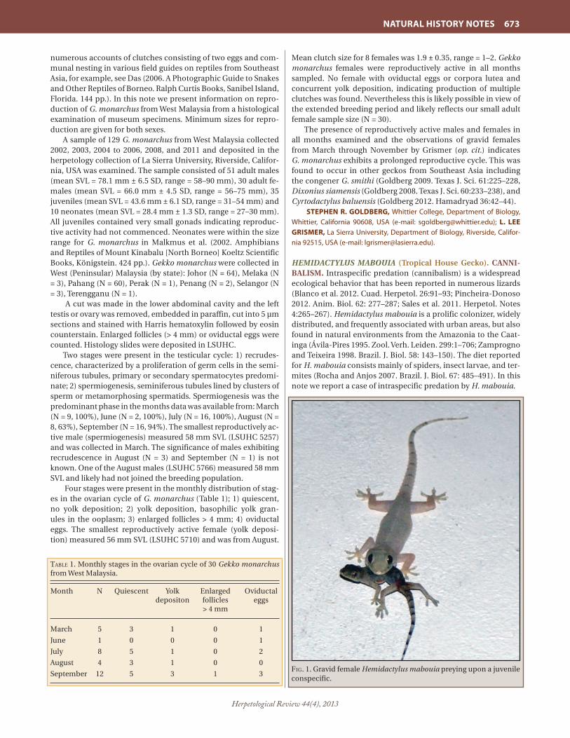
Herpetological Review 44(4), 2013
NATURAL HISTORY NOTES 673
numerous accounts of clutches consisting of two eggs and com-munal nesting in various field guides on reptiles from Southeast Asia, for example, see Das (2006. A Photographic Guide to Snakes and Other Reptiles of Borneo. Ralph Curtis Books, Sanibel Island, Florida. 144 pp.). In this note we present information on repro-duction of G. monarchus from West Malaysia from a histological examination of museum specimens. Minimum sizes for repro-duction are given for both sexes.
A sample of 129 G. monarchus from West Malaysia collected 2002, 2003, 2004 to 2006, 2008, and 2011 and deposited in the herpetology collection of La Sierra University, Riverside, Califor-nia, USA was examined. The sample consisted of 51 adult males (mean SVL = 78.1 mm ± 6.5 SD, range = 58–90 mm), 30 adult fe-males (mean SVL = 66.0 mm ± 4.5 SD, range = 56–75 mm), 35 juveniles (mean SVL = 43.6 mm ± 6.1 SD, range = 31–54 mm) and 10 neonates (mean SVL = 28.4 mm ± 1.3 SD, range = 27–30 mm). All juveniles contained very small gonads indicating reproduc-tive activity had not commenced. Neonates were within the size range for G. monarchus in Malkmus et al. (2002. Amphibians and Reptiles of Mount Kinabalu [North Borneo] Koeltz Scientific Books, Königstein. 424 pp.). Gekko monarchus were collected in West (Peninsular) Malaysia (by state): Johor (N = 64), Melaka (N = 3), Pahang (N = 60), Perak (N = 1), Penang (N = 2), Selangor (N = 3), Terengganu (N = 1).
A cut was made in the lower abdominal cavity and the left testis or ovary was removed, embedded in paraffin, cut into 5 µm sections and stained with Harris hematoxylin followed by eosin counterstain. Enlarged follicles (> 4 mm) or oviductal eggs were counted. Histology slides were deposited in LSUHC.
Two stages were present in the testicular cycle: 1) recrudes-cence, characterized by a proliferation of germ cells in the semi-niferous tubules, primary or secondary spermatocytes predomi-nate; 2) spermiogenesis, seminiferous tubules lined by clusters of sperm or metamorphosing spermatids. Spermiogenesis was the predominant phase in the months data was available from: March (N = 9, 100%), June (N = 2, 100%), July (N = 16, 100%), August (N = 8, 63%), September (N = 16, 94%). The smallest reproductively ac-tive male (spermiogenesis) measured 58 mm SVL (LSUHC 5257) and was collected in March. The significance of males exhibiting recrudescence in August (N = 3) and September (N = 1) is not known. One of the August males (LSUHC 5766) measured 58 mm SVL and likely had not joined the breeding population.
Four stages were present in the monthly distribution of stag-es in the ovarian cycle of G. monarchus (Table 1); 1) quiescent, no yolk deposition; 2) yolk deposition, basophilic yolk gran-ules in the ooplasm; 3) enlarged follicles > 4 mm; 4) oviductal eggs. The smallest reproductively active female (yolk deposi-tion) measured 56 mm SVL (LSUHC 5710) and was from August.
Mean clutch size for 8 females was 1.9 ± 0.35, range = 1–2. Gekko monarchus females were reproductively active in all months sampled. No female with oviductal eggs or corpora lutea and concurrent yolk deposition, indicating production of multiple clutches was found. Nevertheless this is likely possible in view of the extended breeding period and likely reflects our small adult female sample size (N = 30).
The presence of reproductively active males and females in all months examined and the observations of gravid females from March through November by Grismer (op. cit.) indicates G. monarchus exhibits a prolonged reproductive cycle. This was found to occur in other geckos from Southeast Asia including the congener G. smithi (Goldberg 2009. Texas J. Sci. 61:225–228, Dixonius siamensis (Goldberg 2008. Texas J. Sci. 60:233–238), and Cyrtodactylus baluensis (Goldberg 2012. Hamadryad 36:42–44).
STEPHEN R. GOLDBERG, Whittier College, Department of Biology, Whittier, California 90608, USA (e-mail: [email protected]); L. LEE GRISMER, La Sierra University, Department of Biology, Riverside, Califor-nia 92515, USA (e-mail: [email protected]).
HEMIDACTYLUS MABOUIA (Tropical House Gecko). CANNI-BALISM. Intraspecific predation (cannibalism) is a widespread ecological behavior that has been reported in numerous lizards (Blanco et al. 2012. Cuad. Herpetol. 26:91–93; Pincheira-Donoso 2012. Anim. Biol. 62: 277–287; Sales et al. 2011. Herpetol. Notes 4:265–267). Hemidactylus mabouia is a prolific colonizer, widely distributed, and frequently associated with urban areas, but also found in natural environments from the Amazonia to the Caat-inga (Ávila-Pires 1995. Zool. Verh. Leiden. 299:1–706; Zamprogno and Teixeira 1998. Brazil. J. Biol. 58: 143–150). The diet reported for H. mabouia consists mainly of spiders, insect larvae, and ter-mites (Rocha and Anjos 2007. Brazil. J. Biol. 67: 485–491). In this note we report a case of intraspecific predation by H. mabouia.
taBle 1. Monthly stages in the ovarian cycle of 30 Gekko monarchus from West Malaysia.
Month N Quiescent Yolk Enlarged Oviductal depositon follicles eggs > 4 mm
March 5 3 1 0 1
June 1 0 0 0 1
July 8 5 1 0 2
August 4 3 1 0 0
September 12 5 3 1 3Fig. 1. Gravid female Hemidactylus mabouia preying upon a juvenile conspecific.

Herpetological Review 44(4), 2013
674 NATURAL HISTORY NOTES
During the rainy season on 21 May 2013 at ca. 2110 h, we ob-served an adult female H. mabouia (57.5 mm in SVL) with two eggs in its abdomen (viewable through the skin) and a conspecif-ic juvenile gecko (36.5 mm in SVL) inside its mouth (Fig.1). The adult had a firm bite hold on the lateral region of the juvenile’s torso. Our observation occurred in a residence in the urban area (0.002694°S, 51.091306°W, datum WGS84; 31 m elev.), municipal-ity of Macapá, State of Amapá, northern Brazil. The observations here lasted 20–35 minutes. Seven minutes subsequent to inges-tion, the adult female gecko regurgitated the juvenile. The regur-gitation may have been caused by the body size of the juvenile, which represented more than half of SVL female adult. A previ-ous case of cannibalism in this species was reported from south-eastern Brazil (Pombal and Pombal, Jr. 2010. Herpetol. Rev. 41: 223–224). Cannibalism in H. mabouia might represent opportu-nistic feeding based on an increase in the juvenile population or as a strategy to minimize the ecological costs during periods of low arthropod availability.
CARLOS E. COSTA-CAMPOS (e-mail: [email protected]) and MAYARA F. M. FURTADO, Laboratório de Herpetologia, Departa-mento Ciências Biológicas e da Saúde, Universidade Federal do Amapá, Campus Marco Zero, 68903-280, Macapá, AP, Brazil (e-mail: [email protected]).
HEMIDACTYLUS MABOUIA (African House Gecko). PREDA-TION BY CALLITHRIX PENICILLATA. Hemidactylus mabouia is an exotic lizard that broadly distributed in Brazil from forests to urban areas (Sousa and Freire 2010. Biotemas 23:231–234) but native to Africa. This is a nocturnal species, sheltering in crev-ices and under tree bark during the day. Marmosets (Callithrix spp.) are endemic Brazilian primates that consume different kinds of food, from plant reproductive parts to animals, such as insects, toads, and lizards (e.g., Anolis, Mabuya) (Martins and Setz 2000. Int. J. Primatol. 21:476–476; Passamani and Rylands 2000. Primates 41:27–38; Rylands et al. 2009. In Ford et al. [eds.], The Smallest Anthropoids: the Marmoset/Callimico Radiation, pp. 25–61. Springer, New York). In this short note, we describe an autotomy event in H. mabouia followed by its predation by C. penicillata (É. Geoffroy, 1812).
On 18 June 2013, while monitoring a group of Callithrix peni-cillata (eight individuals), we observed the capture and preda-tion of an individual Hemidactylus mabouia by two marmosets (a juvenile and an adult). This event occurred in a Cerrado rem-nant, located in the urban area of Campo Grande, state of Mato Grosso do Sul, Brazil (20.301418°S, 54.365219°W). At 0929 h (am-bient temperature = 28°C), a juvenile marmoset (< 6 months) was foraging on a trunk (ca. 60 cm circumference) at ca. 1.5 m above ground. It tracked and captured a H. mabouia. Instantaneously, the lizard released the tail while in the hands of the primate, launched itself to the ground, and remained motionless. The ju-venile marmoset started to eat the tail, whereas the adult mar-moset (> 2 years) searched the ground intensively. After 1 min (0930 h), the adult marmoset came down to the ground, leaped onto the lizard, captured it and brought it to its mouth. Later, the same individual marmoset climbed a tree at 2 m above ground and started to eat the lizard (Fig. 1). The juvenile marmoset made successive requests for food to the adult, which shared a small part of the lizard. From the tracking to the end of the ingestion approximately 6 min elapsed.
Tail autotomy in lizards is a mechanism that may reduce mor-tality by predation, since tail movements (postautotomy) may distract or confound the predator (Congdon et al. 1974. Science
184:1379–1380). However, it is possible that this antipreda-tor mechanism is more efficient against solitary predators than against individuals that forage in groups, such as marmosets.
We thank Juliana Terra and Sabine Borges for the identifica-tion of the lizard and Coordination for the Improvement of High-er Education Personnel (CAPES) for granting the scholarship for the first two authors.
GILSON DA ROCHA-SANTOS (e-mail: [email protected]) and EDER BARBIER, Programa de Pós-graduação em Biologia Animal, Centro de Ciências Biológicas e da Saúde, Universidade Federal de Mato Grosso do Sul, CEP 79.070-900, Av. Costa e Silva, s/n, Campo Grande, Mato Grosso do Sul, Brazil (e-mail: [email protected]); DAYANE FER-NANDES DA SILVA, Rua Zeferino Vicente Almeida, 300, CEP 79.050-560, Campo Grande, Mato Grosso do Sul, Brazil (e-mail: [email protected]).
HOLASPIS GUENTHERI (Günther’s Gliding Lizard). PREDA-TION. On 13 January 2010, at 1117 h, one of us (SI) observed and photographed an adult Senegal Kingfisher (Coraciiformes: Alcedinidae: Halcyon senegalensis) holding an adult Holaspis guentheri in its beak. The bird was perched on the landing strip barrier of the Joachim Mahothes Magouindi Airport (2.79°S, 10.06°E), Gamba, Ogooué-Maritime Province, southwestern Gabon. Neither the bird nor the lizard was collected, but their respective color patterns readily distinguish them from related taxa recorded in the region (Angehr et al. 2006. In Alonso et al. Gamba, Gabon: Biodiversité d’une forêt équatoriale africaine, pp. 327–351. Bull. Biol. Soc. Washington 12; Pauwels and Vande
Fig. 1. An adult Callithrix penicillata preying on Hemidactylus mabouia in Campo Grande, Mato Grosso do Sul, Brazil.

Herpetological Review 44(4), 2013
NATURAL HISTORY NOTES 675
weghe 2008. Reptiles du Gabon. Smithsonian Institution, Wash-ington. 272 pp.). It is the first time that this specific predator-prey relationship is reported.
We thank ornithologists Patrice Christy and Jean Pierre Vande weghe (Libreville) for the confirmation of the identifica-tion of the kingfisher and discussions on its ecology.
OLIVIER S. G. PAUWELS, Département des Vertébrés Récents, Institut Royal des Sciences naturelles de Belgique, Rue Vautier 29, B-1000 Brussels, Belgium (e-mail: [email protected]); SAFFIAN ITAM (e-mail: [email protected]).
LIOLAEMUS MONTICOLA (Mountain Lava Lizard). PREDA-TION ON LIOLAEMUS LEMNISCATUS. Liolaemus is a diverse genus of small to medium-sized, omnivorous lizards, distribut-ed in the southern hemisphere of the Neotropical realm. Liolae-mus monticola is endemic to Chile and is distributed in Andean scrub from Coquimbo to Maule regions, from 500–3000 m elev. (Pincheira-Donoso and Nuñez 2005. Publ. Ocas. Mus. Nac. Hist. Nat. Chile 59:1–486). This species is an active forager on insects with more than 50% of its reported diet as ants (Fuentes and Ip-inza 1979. J. Herpetol. 13:123–124; Vidal and Labra 2008. Herpe-tología de Chile. Science Verlag, Santiago. 593 pp.). Here I report an observation of saurophagy.
On 21 January 2011, at 1100 h, on a sunny day with an ambi-ent temperature of 22°C, an adult L. monticola (mean SVL 157
mm and tail length at 94 mm) was observed attacking and then ingesting a juvenile Liolaemus lemniscatus (SVL = 45 mm) at Fundo Las Lomas, Colina County, Metropolitan region, Central Chile (33.065546°S, 70.670172°W, WGS84; elev. 810 m). The adult was resting in the soil and then chased the L. lemniscatus when it approached, taking ca. five minutes to capture and ingest the small lizard, and then sought refuge under some rocks. The hab-itat was an open and rocky scrubland dominated by Baccharis linearis and Proustia cuneifolia, with an additional assemblage of annual forbs. The area is not currently pristine with the origi-nal vegetation showing anthropogenic disturbance as a result of wood and charcoal extraction, and livestock use. To my knowl-edge, this is the first record of saurophagy for L. monticola as well as for the entire genus of Liolaemus (cf. Vidal and Labra, op. cit.).
JUAN C. TORRES-MURA, Unión de Ornitólogos de Chile, Mosqueto 459 Of 103, Santiago, Chile; e-mail: [email protected].
LIOLAEMUS WIEGMANNII. PREDATION. Lizards are typical prey items for birds, however direct observations of predation are relatively rare in the field. The sand lizard Liolaemus wieg-mannii (SVL = 42–64 mm), is largely restricted to sandy soils of a vast region of Argentina and Uruguay (Cei 1993. Reptiles del Noroeste, Nordeste y Este de la Argentina. Museo Regionale di Scienze Naturali. Torino, Italy. 949 pp.). This species occurs along coastal sand dunes of the Buenos Aires Province in semi-fixed dunes, usually far away from open spaces and beach (Block et al. 2012. J. Herpetol. 46:608–613). This note reports a single observa-tion of predation on Liolaemus wiegmannii by a Burrowing Owl (Athene cunicularia). The Burrowing Owl is commonly found in grassy plains of Argentina, being the most abundant owl in agro-ecosystems (Bellocq and Kravetz 1994. Ecol. Austral. 4:29–34). Reptiles, like the sand lizard, are generally less common prey item in the owl’s diet (0.3%, Sanchez et al. 2008. Ornitol. Neotrop. 19:71–80).
During a lizard survey through coastal sand dunes in Arenera Galati (37.38658°S, 57.05781°W; datum Campo Inchauspe), Bue-nos Aires Province, on 31 October 2008 at 1030 h, we observed the carcass of an adult female of L. wiergmannii (SVL = 52 mm; total length = 64 mm) lying a few centimeters away from an A. cunicularia burrow. The burrow was located on a dune and the owls were present at the moment of the observation. The lizard’s body was completely intact suggesting that the lizard had been recently caught. The specimen of L. wiegmannii (UNMdP 1778) was deposited in the Herpetological Collection of the Universi-dad Nacional de Mar del Plata, Buenos Aires, Argentina.
CAROLINA BLOCK (e-mail: [email protected]), OSCAR A. STEL-LATELLI (e-mail: [email protected]), and LAURA E. VEGA, Laboratorio de Vertebrados, IIMyC, CONICET - Facultad de Ciencias Exactas y Natura-les, Universidad Nacional de Mar del Plata, Funes 3250 (B7602AYJ) Mar del Plata, Buenos Aires, Argentina (e-mail: [email protected]).
MEROLES CUNEIROSTRIS (Wedge-snouted Sand Lizard). CANNIBALISM. Cannibalism has been observed in a diverse number of reptile species; numerous studies have shown that it is not only common, but that it may play an integral role in shaping the ecology of a species and community dynamics (Fox 1975. Annu. Rev. Ecol. Syst. 6:87–106; Jenssen et al. 1989. Anim. Behav. 38:1054–1061; Keren-Rotem et al. 2006. Behav. Ecol. So-ciobiol. 59:723–731). Studies suggest that cannibalism is com-mon in generalist predators that might prey on conspecifics opportunistically. This pattern has been observed in diverse
Fig. 1. A Senegal Kingfisher (Halcyon senegalensis) with an adult Günther’s Gliding Lizard (Holaspis guentheri).
Fig. 1. Liolaemus monticola eating a juvenile L. lemniscatus.

Herpetological Review 44(4), 2013
676 NATURAL HISTORY NOTES
taxa, and most reported cases involve larger individuals feed-ing on younger, smaller conspecifics (e.g., Polis and Myers 1985. J. Herpetol. 19:99–107; Pincheira-Donoso 2012. Anim. Biol. 62:277–287; Siqueira and Rocha 2008. S. Am. J. Herpetol. 3:82–87). Among lacertid lizards, cannibalism has been mainly docu-mented among island species characterized by high population densities and limited food resources (Amat et al. 2008. Amphib-ia-Reptilia 29:329–336; Pafilis et al. 2009. Naturwissenschaften 96:1107–1113; Perez-Mellado and Corti 1993. Bonn. Zool. Beitr. 44:193–220; Žagar et al. 2012. Acta Herpetol. 7:29–39). This be-havior, however, has been poorly documented among continen-tal species.
On 27 December 2011 near the Gobabeb Training and Re-search Centre, Erongo, Namibia (23.561986°S, 15.041616°E, da-tum WGS84; 408 m elev.) we observed a juvenile Meroles cunei-rostris being eaten by a conspecific adult female (SVL = 44 mm; tail length = 72 mm; 2.6 g). At ca. 1100 h, while attempting to capture the juvenile, an adult female dashed from underneath a small bush and began to chase the juvenile. The chase lasted less than 30 sec. before the female captured the juvenile, bit-ing down on its head and then violently shook her own head. The juvenile then briefly escaped but was recaptured in the same manner. The female relocated to a spot underneath the bush where initially observed and continued to grip the head and neck of the juvenile as it struggled to escape, tightening her grip every few moments. After several minutes the juvenile became lifeless, and the female began to swallow the juvenile whole, headfirst. The entire consumption of the juvenile was completed in ca. 3 min., during which time the female appeared to have no trouble swallowing. Afterward the female remained under the bush for nearly 5 min. and then sprinted ca. 30 m away across the open sand.
There have been no prior examples of cannibalism in any Meroles species to date. The diet of M. cuneiorostris is primarily insectivorous, and relative food availability is thought to remain constantly low throughout the year in this temperate desert cli-mate (Goldberg and Robinson 1979. Herpetologica 35:169–175). At the time of this observation, median juvenile SVL for this pop-ulation was 33 mm (range = 27–42 mm; N = 53).
JACKIE L. CHILDERS, Museum of Vertebrate Zoology, 3101 Valley Life Sciences Building, Berkeley, California 94720-3161, USA (e-mail: [email protected]); DOUGLAS A. EIFLER, Environmental Science Program, Haskell Indian Nations University, Lawrence, Kansas 66046, USA (e-mail: [email protected]).
MESALINA OLIVIERI (Olivier’s Sand Lizard). CESTODE EN-DOPARASITES. Mesalina olivieri is known from Algeria, West-ern Sahara, Tunisia, Libya, Egypt, Israel, Jordan, Iraq, and Saudi Arabia (Bar and Haimovitch 2011. A Field Guide to Reptiles and Amphibians of Israel. Herzliya, Israel. 245 pp.). We know of no endoparasite records for this species. The purpose of this note is to establish the initial helminth list for M. olivieri.
One female M. olivieri (SVL = 48 mm) collected April 1956 in the Central Negev Region, Israel and deposited in the her-petology collection of the Tel Aviv Museum of Natural History (TAUM), Tel Aviv, Israel as TAUM 2201, was examined for endo-parasites. A lateral slit was made on the left side and the coe-lomic cavity was examined for endoparasites using a dissecting microscope. Found were 30 oblong whitish, bodies measuring ca. 1 mm in length. They were regressively stained in hematoxy-lin, cleared in xylol, mounted in balsam, studied under a com-pound microscope and identified as tetrathyridia larvae of the
cestode, Mesocestoides sp. Voucher helminths were deposited in the United States National Parasite Collection, USNPC, Belts-ville, Maryland as USNPC 107017. The life cycle of Mesocestoides sp. is unknown but is thought to utilize three hosts, a vertebrate definitive host, a vertebrate second intermediate host and an arthropod first intermediate host (Rausch 1994. In Khalil et al. [eds.], Keys to the Cestode Parasites of Vertebrates, pp. 309–314. CAM International, Oxon, U.K.). Tetrathyridia are commonly found in the body cavities of amphibians, reptiles, and rodents (Padgett and Boyce 2004. J. Parasitol. 90:108–113). Mesocestoides sp. was previously reported in the congener M. guttulata from Israel (Goldberg and Bursey 2012. Herpetol. Rev. 43:136). Meso-cestoides sp. in Mesalina olivieri is a new host record.
We thank Shai Meiri (TAUM) for permission to examine M. olivieri, Ezra Maza for facilitating the loan, and the National Col-lections of Natural History at Tel Aviv University for providing M. olivieri for this study.
STEPHEN R. GOLDBERG, Department of Biology, Whittier College, PO Box 634, Whittier, California 90608, USA (e-mail: [email protected]); CHARLES R. BURSEY, Department of Biology, Pennsylvania State University, Shenango Campus, Sharon, Pennsylvania 16146, USA (e-mail: [email protected]).
MESALINA OLIVIERI (Olivier’s Sand Lizard). REPRODUC-TION. Mesalina olivieri is known from Algeria, Western Sahara, Tunisia, Libya, Egypt, Israel, Jordan, Iraq, and Saudi Arabia (Bar and Haimovitch 2011. A Field Guide to Reptiles and Amphib-ians of Israel. Pazbar LTD, Herzliya, Israel. 245 pp.). Schleich et al. (1995. Amphibians and Reptiles of North Africa. Koeltz Scien-tific Publishers, Koenigstein. 627 pp.) reported M. olivieri from Oran, Algeria commenced mating in October after emergence from aestivation, females with fully developed eggs occurred from mid-October to the end of May, and at least two clutches of 2–4 eggs were produced. In Jordan, clutches of 4–8 eggs were produced (Disi et al. 2001. Amphibians and Reptiles of the Hash-emite Kingdom of Jordan. Edition Chimaira, Frankfurt am Main. 408 pp.). In this note I present information on the reproductive cycle of M. olivieri from Israel.
A sample of 39 M. olivieri collected 1941 to 1965 in Israel con-sisting of 16 adult males (mean SVL = 44.6 mm ± 2.9 SD, range = 40–50 mm), 17 adult females (mean SVL = 44.6 mm ± 3.3 SD, range = 40–51 mm), 2 juvenile males (mean SVL = 37.0 mm ± 2.8 SD, range = 35–39 mm) and 4 juvenile females mean SVL = 38 mm ±1.7 SD, range = 35–39 mm) was examined from the Nation-al Collections of Natural History at Tel Aviv University (TAUM), Tel Aviv, Israel by Region Name: A’rava Valley TAUM 1832, 2602, 4113; Central Negev TAUM 1121, 1480, 1481, 1483, 1486, 1490, 1731, 1741, 2201, 2475, 2500, 2501, 3438, 4367, 5964, 5982; North-ern Negev TAUM 1116, 1131, 1445, 1479, 1484, 1485, 1492, 1496,
taBle 1. Monthly stages in the ovarian cycle of 17 Mesalina olivieri females from Israel; one April female (*) with oviductal eggs was also undergoing yolk deposition.
Month N Quiescent Early yolk Follicles Oviductal depositon > 4 mm eggs
January 1 0 0 1 0
March 6 1 2 1 2
April 9 2 3 1 3*
November 1 1 0 0 0

Herpetological Review 44(4), 2013
NATURAL HISTORY NOTES 677
1967, 3682, 3951, 3991, 3996; Southern Coastal Plain TAUM 1493, 1495, 2064, 2456, 2953, 2954, 5965.
The left testis was removed from males and the left ovary was removed from females for histological examination. Gonads were embedded in paraffin and histological sections were cut at 5 µm and stained with hematoxylin followed by eosin coun-terstain. Enlarged ovarian follicles (> 4 mm) and oviductal eggs were counted. No histology was performed on them. Histology slides were deposited at TAUM.
Two stages were observed in the testicular cycle: Spermio-genesis, (seminiferous tubules are lined by sperm or clusters of metamorphosing spermatids); Recrudescence (proliferation of germ cells prior to spermiogenesis), was observed in two juve-nile males from September, measuring 35 mm and 39 mm SVL, respectively). Males exhibiting spermiogenesis by month were January (N = 1), March (N = 4), April (N = 8), May (N = 1), Novem-ber (N = 1). The smallest reproductively active male measured 40 mm SVL (TAUM 1121) and was collected in April. The presence of the November male undergoing spermiogenesis suggests M. olivieri exhibits a testicular cycle similar to the congener Mesa-lina guttulata from Israel (Goldberg 2012. Zool. Mid. East 56:27–30) which also initiates sperm formation in autumn.
Four stages were noted in the ovarian cycle of M. olivieri (Table 1): Quiescent (no yolk deposition; Early yolk deposition (yolk granules in the ooplasm); Enlarged ovarian follicles > 4 mm; Oviductal eggs. The smallest reproductively active M. oliv-ieri females measured 40 mm SVL (TAUM 1731) 3 follicles > 4 mm, early yolk deposition (TAUM 2501) both collected in March. Four smaller females with quiescent ovaries, 35 (September), 38 (December), 38 (January), 39 mm (January) SVL, respectively, were considered juveniles. Mean clutch size (N = 8) was 3.4 ± 0.74, range = 2–4. One female from April with oviductal eggs was undergoing concurrent yolk deposition for a subsequent clutch indicating M. olivieri can produce multiple clutches in the same reproductive season in Israel.
I thank Shai Meiri (TAUM) for permission to examine M. ol-ivieri, Ezra Maza for facilitating the loan and the National Col-lections of Natural History at Tel Aviv University for providing M. olivieri for this study.
STEPHEN R. GOLDBERG, Department of Biology, Whittier College, PO Box 634, Whittier, California 90608, USA; e-mail: [email protected].
NOROPS PENTAPRION (Lichen Anole). DIRECTED AERIAL DESCENT. Lizards with gliding or directed aerial descent be-haviors are well known from the Old World Tropics (e.g., Draco, Ptychozoon), and snakes and frogs exhibiting these behaviors are found both in the Old and New World Tropics (Dudley et al. 2007. Annu. Rev. Ecol. Evol. Syst. 38:179–201). However, lizards show-ing directed aerial descent have not been reported from the New World Tropics. Here we report on directed aerial descent capa-bility and behavior in Norops pentaprion, a canopy lizard from eastern Costa Rica, southern Nicaragua, and western Panama (Köhler 2010. Zootaxa 2354:1–18).
The initial discovery of this behavior in N. pentaprion was serendipitous in July 2001 when an individual was captured on a walk-up meteorological tower at canopy level (~ 25 m) at La Selva Biological Station in the Atlantic lowlands of Costa Rica. After identification, the animal was returned to the original location on the tower, but upon release to a horizontal tower brace, it ex-ecuted a controlled aerial descent to a tree in the distance below. Individuals of N. pentaprion have been occasionally observed on
our meteorological towers subsequent to our first observation of directed aerial descent. The lizards are typically at canopy level but are sometimes found well above the canopy on the highest levels of the towers (up to 42 m). While the typical response of N. pentaprion to the presence of personnel on the tower is to race
Fig. 1. Frames from video of a Norops pentaprion individual execut-ing an aerial rotation during directed aerial descent. Arrows track the lizard’s descent.

Herpetological Review 44(4), 2013
678 NATURAL HISTORY NOTES
down the tower or hide on the opposite side of vertical supports, the animals will occasionally jump from the tower to escape when approached. Since 2001 we have observed directed aerial descent by N. pentaprion from canopy towers on several occasions. In at least two instances the lizard landed at lower levels on the tower, but in other occurrences they covered substantial distance to ad-jacent trees including aerial rotations of near 180º. During more than one observation, animals appeared to glide with near-hori-zontal trajectories towards termination of the descent.
Similar to some snakes, frogs, lizards, and ants showing di-rected aerial descent, N. pentaprion does not have strongly spe-cialized features associated with gliding behavior such as skin flaps, skin extensions, or webbed feet. Norops pentaprion has a relatively flattened head and wide body. During aerial descent this lizard proceeds headfirst, with limbs partially extended and the body strongly flattened, a position that may take advantage of regions of relatively loose skin along the sides of the body (Guyer and Donnelly 2005. Amphibians and Reptiles of La Sel-va, Costa Rica, and the Caribbean Slope, Univ. California Press, Berkeley, California. 299 pp.).
At La Selva Biological Station this species is uncommon in the understory and is usually found on trees limbs. Norops pen-taprion is a member of a closely-related group of anoline lizards subjected to a recent analysis of morphology and morphomet-rics (Köhler, op. cit.); these related taxa share similar body size characteristics with N. pentaprion and are frequently arboreal. Future observations of some of these species may result in the discovery of similar directed-aerial descent behavior.
Canopy pioneer Donald Perry reported lizards with a rose dewlap parachuting between trees in the canopy in Costa Ri-can forests (Perry 1986. Life Above the Jungle Floor, Simon and Schuster, Inc. New York, New York. 170 pp.), but the species was not identified. In their description of Norops pentaprion, Guyer and Donnelly (op. cit.) indicated that parachuting behavior likely occurs in this species on the basis of our initial observations and those of Perry. Our repeated observations verify directed aerial descent in this species and confirm that the lizard observed by Perry was N. pentaprion, the only lizard in the region with ma-genta dewlap coloration.
These observations were made possible by frequent canopy access provided with support from NSF grants BE 0421178 and DEB-0842235.
STEVEN F. OBERBAUER, Department of Biological Sciences, Florida International University, Miami, Florida 33199, USA (e-mail: [email protected]); CRAIG GUYER, Department of Biological Sciences, 331 Funchess Hall, Auburn University, Auburn, Alabama 36849, USA (e-mail: [email protected]); VINICIO PANIAGUA, La Selva Biological Station, Organization for Tropical Studies, Puerto Viejo de Sarapiqui, Costa Rica; DIEGO DIERICK, Department of Biological Sciences, Florida International University, Miami, Florida 33199, USA; MAUREEN A. DONNELLY, Department of Biological Sciences, Florida International University, Miami, Florida 33199, USA (e-mail: [email protected]).
OPLURUS CUVIERI CUVIERI (Madagascan Spiny-tailed Igua-na). PREY. Several studies have shown that Oplurus cuvieri cuvi-eri (Opluridae) primarily feeds on invertebrates with a high pro-portion of ants, but also consumes plant matter such as leaves, twigs, flower buds and seeds (Bloxam et al. 1996. Prim. Rep. 46:189–201; Brillet 1982. Rev. Ecol. Terre Vie 36:79–148; Mori and Randriamahazo 2002. Afr. J. Ecol. 40:61–64; Randriamahazo and Mori 2012. Current Herpetol. 31:8–13). Brillet (op. cit.) also re-ported that O. c. cuvieri feeds on small mammals, but it is unclear
whether this was an observation under natural conditions or in captivity. In addition, O. c. cuvieri has been suggested as a poten-tial predator on nestlings of birds based on the observation that adult Rufous Vangas (Schetba rufa) intensely attacked an indi-vidual iguana that came close to a nest (Eguchi 2005. In Social Organization of the Rufous Vanga, pp. 83–104. Kyoto Univ. Press, Kyoto). Here, I report on a case of predation on a bird by O. c. cuvieri.
During a field study in Ampijoroa forest in Ankarafantsika National Park, Madagascar, I observed a predation event of an adult O. c. cuvieri on a fledgling of the Madagascar Bulbul (Hypsi-petes madagascariensis). At ca. 1130 h on 28 November 2011, a fledgling H. madagascariensis fell to the ground from a nest in a tree approximately 4 m high. Several avian species such as, the Sickle-billed Vanga (Falculea palliate) and the Broad-billed Roller (Eurystomus glaucurus) repeatedly flew down and pecked at the fledgling, attempting to capture it, but the parent birds of the fledgling mobbed the other birds and protected the fledgling from their attacks. At 1215 h, a male O. c. cuvieri rushed to the fledgling from ca. 10 m away while several birds were still at-tempting to attack the fledgling, but the lizard failed to capture the fledgling due of the parents’ mobbing. This iguana and sever-al birds continued their predation attempts on the fledgling, and the parents of the fledgling had gradually let up mobbing over the course of almost 3 hours of protecting their offspring. Then at 1415 h, the iguana successfully bit and captured the fledgling in its forth attack (Fig. 1), 2 h following its first attempted attack. The parents did not mob the iguana after it captured the fledg-ling. The iguana tore the body and began consumption, bang-ing the bird on the ground and then against the trunk of the tree while climbing many times. This is the first reported evidence for the predation of vertebrates by O. c. cuvieri in the wild.
This study was financially supported by JSPS Grant-in-Aid for Scientific Research (B: nos. 21405007 and 24405008) and JSPS Grant-in-Aid for Young Scientists (B: no. 25870369). I thank Akira Mori for his valuable comments and staff of Madagascar Nation-al Park for their assistance in the field study.
RYO ITO, Primate Research Institute, Kyoto University, Inuyama, Aichi, 484-8506 Japan; e-mail: [email protected].
PETRACOLA VENTRIMACULATUS (Spotted Lightbulb Lizard). CANNIBALISM. Petracola ventrimaculatus is a gymnophthalmid
Fig. 1 Oplurus cuvieri cuvieri captured by a fledgling Madagascar Bulbul.

Herpetological Review 44(4), 2013
NATURAL HISTORY NOTES 679
lizard endemic to the northern Peruvian Andes (Kizirian et al. 2008. Zootaxa. 1700:53–62). The natural history of this species is poorly known and there is no published information regarding its diet. However, species of the related genus Proctoporus are known to feed on arthropods—mostly beetles, ants, and spiders (Doan 2008. J. Herpetol. 42:16–21).
While clearing and staining three specimens of P. ventrimac-ulatus for a taxonomic review, a hatchling (SVL = 22.93 mm) of P. ventrimaculatus was found in the stomach of an adult male conspecific (SVL = 65.2 mm), collected on 18 September 2006 at Puente Hierba Buena (6.981131°S, 78.379764°W), Province of Celendín, Cajamarca Region, Perú. The stomach contents of the other two specimens included one mite, coleopterans (three adults and two larvae), and one amblypygid. This is the first re-cord of cannibalism in the Gymnophthalmidae, as well as the first prey items reported for the diet of P. ventrimaculatus.
The male specimen of Petracola ventrimaculatus is depos-ited in the herpetological collection of the Museo de la Universi-dad San Marcos, Lima, Perú (MUSM 26233). We thank C. Torres for allowing access to the collection of the MUSM.
LOURDES Y. ECHEVARRIA (e-mail: [email protected]) and PABLO J. VENEGAS, División de Herpetología-Centro de Ornitología y Biodiversidad (CORBIDI), Santa Rita No. 105 Of. 202, Urb. Huertos de San Antonio, Surco, Lim, Perú (e-mail: [email protected]).
PHYMATURUS EXTRILIDUS. PREDATION. On 28 November 2011 at 1710 h, we observed a female Red-backed Hawk (Bu-teo polyosoma) presenting an adult Phymaturus extrilidus to a nest of two approximately 1.5-week-old chicks above a canyon known as Aguada de Pinchagua at the Multiple Use Nature Re-serve Don Carmelo, Ullum Department, San Juan Province, Ar-gentina (30.977504°S, 69.083017°W, 3122 m elev.). The hawk had removed the head of the lizard prior to arriving at the nest to feed it to the chicks (Fig. 1). Although Red-backed Hawks are known to eat lizards (Jiménez 1995. Hornero 14:1–9), this is the first re-cord of one eating this species and the first record of a confirmed predator for P. extrilidus (Lobo et al. 2012. Copeia 2012:12–22). R. E. Espinoza confirmed the identity of the lizard.
MAURICIO ARMANDO PÉREZ, Facultad de Ciencias Exactas, Físicas, y Naturales, Departamento de Biología, Universidad Nacional de San Juan, San Juan, Argentina (e-mail:[email protected]); CARLOS EDU-ARDO BORGHI (e-mail:[email protected]) and VERÓNICA ALE-
JANDRA BENINATO, Facultad de Ciencias Exactas, Físicas, y Naturales, Instituto y Museo de Ciencias Naturales, Universidad Nacional de San Juan, San Juan, Argentina (e- mail: [email protected]).
PHYMATURUS SPECTABILIS. PREDATION. Phymaturus spec-tabilis is a medium-sized liolaemid lizard endemic to northwest-ern Patagonia, found in rocky outcrops near Ingeniero Jacobac-ci, Departamento 25 de Mayo, southwestern Río Negro Province, Argentina. Predation data are absent for this species, but it has been reported to share its habitat with potential reptile preda-tors such as the colubrid snakes Philodryas patagoniensis and P. trilineata, and the viperid Rhinocerophis ammodytoides (Scolaro et al. 2008. Zootaxa 1786:48–70). Here I report an observation of predation of P. spectabilis by a Lesser Grison (Galictis cuja).
On 7 December 2011 during a field trip to Paraje Yuquiche, Río Negro Province, northwestern Patagonia, Argentina (41.434050°S, 69.751983°W, datum WGS84; elev. 942 m), I ob-served a Lesser Grison moving below a shrub (Stillingia pata-gonica) at the bottom of a rocky outcrop, where I had earlier ob-served three individuals of Liolaemus elongatus basking. When I approached to get a closer view I realized that the grison was holding a dead Phymaturus spectabilis in its mouth. As soon as it saw me, the grison released the dead lizard from its mouth and retreated. The dead lizard was identified as a juvenile female (69.1 mm SVL). The head of the lizard had been completely crushed, indicating that the grison had captured the lizard by this part of its body. It has been reported previously that Lesser Grisons in Patagonia prey secondarily on reptiles (Diuk-Wasser and Cassini 1998. Stud. Neotrop. Fauna Environ. 33:3–6), however, this is the first case of predation on Phymaturus lizards.
Erika Kubisch verified the identifications and the P. spectabi-lis (PH229) was deposited in the Herpetological Collection of the Centro Regional Universitario Bariloche (CRUB), San Carlos de Bariloche, Río Negro.
FACUNDO CABEZAS-CARTES, Departamento de Zoología, Labora-torio de Ecofisiología e Historia de vida de Reptiles, INIBIOMA–CONICET, Centro Regional Universitario Bariloche, Universidad Nacional del Coma-hue, San Carlos de Bariloche (8400), Río Negro, Argentina; e-mail: [email protected].
PHYSIGNATHUS LESUEURII (Australian Water Dragon). MU-TILATION BY RAVENS. During spring and summer 2005 I ob-served two instances of Australian Ravens (Corvus coronoides) removing parts of the tail from adult Australian Water Dragons (Physignathus lesueurii) at the Australian National Botanic Gar-dens, Canberra, Australia (35.27894°S, 149.11048°E). In the first instance, a raven approached and landed on a pavement ce-ment path approximately 2 m from an adult male dragon (size of the lizard was nearly 1 m in total length). The dragon showed little response other than obvious visual contact. The raven then clasped the dragon’s tail (about 10–15 cm from the tip) with its bill and dragged the dragon by the tail in different directions. This occurred for nearly one minute, the dragon running in place as it was being pulled. The raven then ran in circles around the dragon, still holding the tail, with the lizard spinning in one spot and still running in place. After another 30 seconds of this behav-ior part of the tail snapped off, and the raven flew away with it. A second similar event involved an adult female dragon and possi-bly the same raven about two weeks later. The raven was first ob-served already clasping the tail of the dragon, again about 10–15 cm from the tip, on the same path as the first event. This time the raven shook the tail vigorously, breaking off the tip within 20 sec
Fig. 1. Female Buteo polyosoma feeding an adult Phymaturus extrili-dus to two nestlings.

Herpetological Review 44(4), 2013
680 NATURAL HISTORY NOTES
of first being observed. The raven flew off with the tail piece. I could not confirm whether the raven(s) consumed the tails.
Tail breakage induced or caused by ravens appeared to be de-liberate, rather than a failed attempt at predation. It seems likely that the ravens consumed the tails (or the muscle component). Corvids are known for their problem solving abilities (e.g., Weir et al. 2002. Science 297:981), and ravens may be responsible for previous observations of incomplete tails in Australian Water Dragon populations. Thompson (1993. Wildl. Res. 20:613–619) speculated that the 41–47% rate of tail breakage/regeneration in adult water dragons in another population was due to predators, rather than intraspecific fighting. My observations partially sup-port this idea, but also suggest deliberate “mutilation” by ravens. Further observations of the proportion of tail loss in a popula-tion, along with confirmation of ravens consuming the tails (or parts of them), are needed to determine both the cognitive un-derpinning of tail removal by ravens and its importance to Aus-tralian Water Dragon populations.
J. SEAN DOODY, Department of Ecology and Evolutionary Biology, University of Tennessee, 569 Dabney Hall, Knoxville, Tennessee 37996-1610, USA; e-mail: [email protected].
PLESTIODON FASCIATUS (Five-lined Skink). ARTIFICAL HAB-ITAT USE. In central Ontario, Canada, from 1 May to 31 August 2012, systematic surveys were conducted to examine the im-pacts of roads on reptiles. Surveys were conducted along 13 km of Highway 69/400 in Parry Sound District and 6 km of Highway 529 in Magnetawan First Nation. Of the 28 Plestiodon fasciatus found on or within 3 m of a road, 3 (11%) were dead, while the remaining 25 (89%) were alive and interacting with the road. In-teractions were categorized as either crossing the road (3 indi-viduals; 12%) or hiding under cover alongside the roadside (22 individuals; 88%; Fig. 1). Only 2 individuals (9%) were found un-der natural cover objects (rock, grass thicket), while 20 individu-als (91%) were found under anthropogenic debris (garbage, tires, sections of retread and cardboard). During haphazard surveys of non-road sites, 7 individuals were observed, 2 (29%) of which were under natural cover and 3 (43%) were under artificial cover (habitat was not recorded for 2 skinks).
Interestingly, only 6% of all of the skinks found on Magne-tawan First Nation (both road and non-road sites; Fig. 1) were found under natural cover, suggesting that artificial cover may be preferred habitat within this population. If this is the case, the high level of refuse found along the roadside may attract skinks to areas that subject them to an increased likelihood of road mortality. Road mortality is a known threat to P. fasciatus popu-lations in Ontario (COSEWIC 2007. COSEWIC assessment and update status report on the Five-lined Skink, Eumeces fasciatus, in Canada. Committee on the Status of Endangered Wildlife in Canada. Ottawa. vii + 50 pp.), and elsewhere across the species’ range (Illinois; COSEWIC 2007, op. cit., and Florida; Aresco 2003. J. Wildl. Manage. 69:549–560).
It has been previously documented that P. fasciatus only oc-casionally uses artificial cover objects in some populations (e.g., Hecnar 1994. Can. J. Zool. 72:1510–1516). In central Ontario, P. fasciatus is thought to select habitat based on specific microhab-itat characteristics; specifically, rocks averaging 55 cm in length that provide thermal conditions that vary by no more than 1.99°C (Quirt et al. 2006. J. Herpetol. 40:335–342) and that pro-vide an optimum cover temperature range of 28°C to 36°C (Fitch 1954. Univ. Kansas Publ. Mus. Nat. Hist. 8:1–156). The specificity of this species’ habitat preference is so strong that degradation
or alteration of microhabitat has been linked to population de-clines (COSEWIC 2007, op. cit.). In our observations, tempera-ture underneath artificial cover averaged 23.5°C (N = 16), which is 4.5°C cooler than the previously reported selected minimum optimum cover temperature (28°C) for this species. So, why would P. fasciatus be selecting these artificial cover objects? One possible explanation may be that factors other than tempera-ture, such as an increase in potential prey, are contributing to the increased skink abundance around roadways.
During the course of our road surveys, insects were collected to examine insect road mortality, with an estimated total of 12,900 dead insects collected after colliding with vehicles (Baxter-Gilbert et al., unpubl. data). Insects are known to be a major food source for P. fasciatus (COSEWIC 2007, op. cit.), and a fecal sample from one of the live Five-lined Skinks on Magnetawan First Nation was comprised mostly of beetle elytra. It might be that the abundance of P. fasiciatus around roadways is correlated with the increased availability of immobilized prey items. Our observations demon-strate a need to study the relationship between lizard ecology and roadways to determine if anthropogenically-created resources, such as roadside refuse and road-killed insects, are attracting these reptiles to a known population threat.
We thank Jenn Baxter-Gilbert, Sean Boyle, Damien Buttin-eau, Ron Maleau, and Chris Neufeld for their field assistance. Financial support for this research was provided by Magnetawan First Nation, Laurentian University, the Ontario Ministry of Nat-ural Resources, and the Ontario Ministry of Transportation. All research was conducted under an approved Laurentian Univer-sity Animal Care Committee protocol, and authorized by Mag-netawan First Nation’s Chief and Band Council and the Ontario Ministry of Natural Resources.
JAMES BAXTER-GILBERT (e-mail: [email protected]), JULIA L. RILEY (e-mail: [email protected]), and JACQUELINE D.
Fig. 1. Distribution of Plestiodon fasciatus on Magnetawan First Na-tion in Central Ontario differentially labeled by living (black circles) or deceased (red crosses) individuals. Most skinks were found in close proximity to human development, including roads and rail lines (grey lines) and buildings (brown squares).

Herpetological Review 44(4), 2013
NATURAL HISTORY NOTES 681
LITZGUS, Department of Biology, Laurentian University, Sudbury, Ontario, P3E 2C6, Canada (e-mail: [email protected]).
PLESTIODON GILBERTI (Gilbert’s Skink). PREDATION. Ples-tiodon gilberti typically occur in open grassy areas where bur-rows or rocks provide quick cover. They also occupy woodlands, streams, and other wetland areas (Behler and King 1979. The National Audubon Society Field Guide to North American Rep-tiles and Amphibians. Alfred A. Knopf, New York, New York. 744 pp.; Stebbins and McGinnis 2012. Field Guide to Amphibians and Reptiles of California. Univ. California Press, Berkeley. 538 pp.). Their association with wetland areas increases their risk of predation by wading birds, including Great Egrets (Ardea alba), which are opportunistic predators that take a variety of prey, such as crustaceans, insects, frogs, tadpoles, lizards, snakes, and small mammals. Great Egrets have been observed in the Florida Keys searching for Anolis lizards (McCrimmon et al. 2011. In A. Poole [ed.], The Birds of North America Online. Cornell Lab of Ornithology, Ithaca, New York; doi: 10.2173/bna.570).
On 19 April 2013, while hiking along a trail approximately 675 m E of the San Joaquin River in Fresno Co., California, USA (36.8765°N, 119.7796°W; 90 m elev.), we observed a Great Egret in an upland area struggling with something in its beak. Upon initial observation the item appeared to be a snake but after further examination of photographs taken of the egret, the prey was identified as an adult P. gilberti. The skink was whipping its tail back and forth, forcing the bird to attempt a better grip. The bird tossed the lizard into the air and recaptured it with its beak, swallowing the skink whole in less than 30 sec. Although likely a common occurrence based on the similar habitat associations of these two species, this observation, to our knowledge, is the first to be reported.
HOWARD O. CLARK, JR. (e-mail: [email protected]) and SUSAN I. HAGEN, H. T. Harvey & Associates, 7815 N Palm Ave., Suite 310, Fresno, California 93711-5511, USA.
PSEUDOTRAPELUS SINAITUS (Sinai Agama). ENDOPARA-SITES. Pseudotrapelus sinaitus is widely distributed in the Mid-dle East where it inhabits rocky desert habitats (Bar and Haimov-itch 2011. A Field Guide to Reptiles and Amphibians of Israel. Pazbar Ltd, Herzliya, Israel. 245 pp.). To our knowledge there are no reports of endoparasites from this species. The purpose of this note is to establish the initial parasite list for P. sinaitus.
The body cavity of one female (SVL = 83 mm) P. sinaitus col-lected 23 April 1952 in Israel, Northern Negev Region and depos-ited in the Tel Aviv University Museum (TAUM), Israel as TAUM 688 was examined for endoparasites. A mid-ventral incision was made and the coelomic contents were examined under a dissecting microscope. Two nematodes were found. The stom-ach wall was punctured in preservation and some contents had spilled into the body cavity. Thus it is likely the infection site for the nematodes was the stomach. The nematodes were cleared on a glass slide in a drop of lactophenol, cover slipped, examined under a compound microscope, and identified as two immature ascarid nematodes. Voucher nematodes were deposited in the United States National Parasite Collection (USNPC), Beltsville, Maryland as USNPC 106981.
Ascaridoids commonly utilize vertebrates as intermediate hosts; development to the adult nematode occurs when the in-termediate host (in this case a lizard) containing infective larvae is eaten by the definitive host (Anderson 2000. Nematode Para-sites of Vertebrates, Their Development and Transmission, CABI Publishing, Oxon, UK. 650 pp.). Pseudotrapelus sinaitus repre-sents a new host record for larval ascarid nematodes.
We thank Shai Meiri (TAUM) for permission to examine P. sinaitus, Erez Maza for facilitating the loan, and the National Collections of Natural History at Tel Aviv University for providing the P. sinaitus for this study.
STEPHEN R. GOLDBERG, Whittier College, Department of Biol-ogy, Whittier, California 90608, USA (e-mail: [email protected]); CHARLES R. BURSEY, Pennsylvania University, Department of Biology, Shenango Campus, Sharon, Pennsylvania 16146, USA (e-mail: [email protected]).
PSEUDOTRAPELUS SINAITUS (Sinai Agama). REPRODUC-TION. Pseudotrapelus sinaitus is widely distributed in the Mid-dle East and is known from Libya, Egypt, Israel, Jordan, Syria, Saudi Arabia, Arabian Peninsula, U.A.E., Oman, Sudan, Ethio-pia, and Eritrea where it inhabits rocky desert habitats (Bar and Haimovitch 2011. A Field Guide to Reptiles and Amphibians of Israel. Pazbar Ltd, Herzliya, Israel. 245 pp.). There is anecdotal information on its reproduction found in Bar and Haimovitch (op. cit.; Disi et al. 2001. Amphibians and Reptiles of the Hash-emite Kingdom of Jordan, An Atlas and Field Guide. Edition Chimaira, Frankfurt am Main, Germany. 408 pp.; Schleich et al. 1996. Amphibians and Reptiles of North Africa. Koeltz Scien-tific Publications, Koenigstein, Germany. 630 pp.), indicating a spring-summer breeding season with clutches of 5–9 eggs pro-duced. In this note I provide additional information on P. sinai-tus reproduction from a histological examination of gonads from museum specimens.
A sample of 24 P. sinaitus deposited in the Zoological Muse-um of the Department of Zoology, Tel Aviv University (TAUM), Tel Aviv, Israel, was examined. The sample consisted of 13 males (mean SVL = 78.2 mm ± 4.3 SD, range = 71–83 mm), 10 females (mean SVL = 75.3 mm ± 5.1 SD, range = 68–73 mm), and one juvenile female (SVL = 54 mm) collected 1941–1958 in Israel (32.01587°N, 34.78740°E). Vouchers utilized in this study include:
Fig. 1. Great Egret (Ardea alba) predating a Gilbert’s Skink (Plestiodon gilberti) in Fresno Co., California

Herpetological Review 44(4), 2013
682 NATURAL HISTORY NOTES
TAUM 688, 696–698, 701, 705, 709–711, 713–716, 855, 1123, 1279, 1317, 1460, 1839, 2146, 2595, 2985, 3048, 3119.
An incision was made in the lower abdominal cavity and the left testis or ovary was removed, embedded in paraffin, cut at 5 µm sections, and stained with Harris hematoxylin followed by eosin counterstain. Enlarged follicles > 4 mm or oviductal eggs were counted. Histology slides are deposited in TAUM.
Two stages were observed in the testicular cycle: 1) recrudes-cence. A proliferation of germ cells occurs for the next period of sperm formation; primary and/or secondary spermatocytes predominate; 2) spermiogenesis. Lumina of the seminiferous tu-bules are lined by sperm or rows of metamorphosing spermatids. Monthly distribution of stages were: March (N = 3, spermiogen-esis); April (N = 7, 1 recrudescence, 6 spermiogenesis); May (N =1, spermiogenesis); October (N = 1, recrudescence); November (N = 1, recrudescence). The smallest reproductively active male (spermiogenesis) measured 72 mm SVL (TAUM 2146) and was collected in March.
Four stages were observed in the monthly ovarian cycle (Ta-ble 1): 1) quiescent, no yolk deposition; 2) early yolk deposition, basophilic yolk granules in the ooplasm; 3) enlarged ovarian fol-licles > 4 mm; 4) oviductal eggs. Mean clutch size for five females was 4.4 ± 1.3 SD, range = 3–6. The three smallest reproductively active females each measured 73 mm SVL; (1) TAUM 2595, early yolk deposition, collected in April; (2 and 3) TAUM 696, 2985, 3 follicles > 4 mm, collected in April. Schleich (op. cit.) also report-ed 73 mm SVL as the minimum size for reproductive maturity of P. sinaitus females. I arbitrarily considered one slightly smaller reproductively inactive female from April (TAUM 701, SVL = 70 mm) as being an adult. One reproductively inactive smaller fe-male (TAUM 1123, SVL = 54 mm) was clearly a juvenile.
My histological findings confirm P. sinaitus exhibits a re-productive cycle typical of numerous temperate zone lizards. Breeding occurs in spring; renewal of germinal epithelium (re-crudescence) in males for the forthcoming spring spermiogen-esis occurs in autumn. No reproductive activity was exhibited in autumn females. Three is a new minimum clutch size for P. sinaitus.
I thank Shai Meiri (TAUM) for permission to examine P. sinaitus, Erez Maza for facilitating the loan, and the National Collections of Natural History at Tel Aviv University for providing samples of P. sinaitus for this study.
STEPHEN R. GOLDBERG, Whittier College, Department of Biology, P.O. Box 634, Whittier, California 90608, USA; e-mail: [email protected].
SCELOPORUS ANAHUACUS (Anahuacan Graphic Lizard). SE-LECTED BODY TEMPERATURE. Field body temperatures of reptiles have been widely reported (Avery 1982, In Gans and Pough [eds.], Biology of the Reptilia, Vol. 12, Physiology C, pp. 93–166. Academic Press, New York), but reports on preferred or
selected body temperature is actually quite rare in the literature (Sinervo et al. 2010. Science 328:894–899) because measurement in a laboratory thermal gradient under standard conditions is re-quired (Huey 1982. In Gans and Pough [eds.], Biology of the Rep-tilia, Vol. 12, Physiology C, pp. 25–91. Academic Press, New York). Sceloporus anahuacus is a viviparous lizard of the grammicus group, which inhabits high elevation pine forest. This species is endemic to central Mexico in Sierra Nevada, Distrito Federal, and Estado de México (Ramírez-Bautista et al. 2009. Herpetofau-na del Valle de México, Diversidad y Conservación. CONABIO-UAH. 210 pp.). There have been several reports detailing thermal ecology in grammicus group members (Güizado-Rodríguez et al. 2011. Southwest. Nat. 56:120–124; Lemos-Espinal et al. 1995. Can. J. Zool. 73:2184–2191; Woolrich-Piña et al. 2006. Acta Zool. Mex. 22:137–150), and specific to S. anahuacus within field con-ditions (Ávila-Bocanegra et al. 2012. Herpetol. Bull. 121:27–29), but the selected body temperature has not been documented. Here, we provide preliminary selected temperatures of wild-caught individuals of this species under laboratory conditions.
During May 2007, one of us (UOGV) conducted field work in the Ejido Magdalena Petlatalco, Delegación Magdalena Con-treras, Sierra del Ajusco, Distrito Federal, México (19.2210°N, 99.2856°W; 3500–3930 m elev.), located in a forested area domi-nated by pine trees (Pinus sp.). The data presented here are based on 10 adults (> 46 mm SVL; 6 females and 4 males) of S. anahuacus captured by hand. In the laboratory, the lizards were separated by gender and maintained at 25°C in plastic contain-ers with soil, trunks, and grass, and hydrated with a sprinkler every day. Laboratory experiments were conducted two days af-ter capture using a thermal gradient, which consisted of a wood box (150 cm long x 150 cm wide x 70 cm high) divided into ten tracks of 15 cm each to prevent interaction between lizards. The box was located in a room with a controlled temperature of 20°C and three 150 W lamps were placed at different heights over the box to offer the thermal gradient (20–50°C). Body temperature data were collected manually using a digital thermometer (Fluke model 51-II) with the sensor introduced one centimeter into the cloaca during each hour between 1330 and 1930 hs.
Mean selected body temperature was 32.64°C (SD = 2.73°C, range = 26.0–36.5°C). Interquartile of 25% and 75% was 32.1 and 34.2°C, respectively. These results suggest S. anahuacus main-tains its thermal requirements similar to others species of the grammicus group (Güizado-Rodríguez et al 2011, op. cit.; Lara-Resendiz and Díaz de la Vega-Pérez, in press, Herpetol. Rev.; Lemos-Espinal et al. 1995, op. cit.; Woolrich-Piña et al. 2006, op. cit.). Specifically, the mean selected body temperature is higher than that reported by Ávila-Bocanegra et al. (2012, op. cit.) for this species under field conditions. Finally, we propose for fu-ture research to assess the thermal biology of the S. grammicus group based in tolerance ranges, selected body temperatures in laboratory, body temperatures taken in the field coupled with environmental temperatures, in order to address the patterns of thermoregulation and to determine if the thermal biology in this group of lizards is limited by the environment or if it is evolution-arily conservative.
Financial support for this trip was provided by grants Univer-sidad Nacional Autónoma de México, SDEI-PTID-02 (Manejo de Ecosistemas y Desarrollo Humano).
RAFAEL A. LARA-RESENDIZ, Posgrado en Ciencias Biológicas, In-stituto de Biología, Laboratorio de Herpetología, Universidad Nacional Autónoma de México, AP 70515, CP 04510, Distrito Federal, México (e-mail: [email protected]); URI OMAR GARCÍA-VÁZQUEZ, Labora-
taBle 1. Monthly stages in the ovarian cycle of 10 Pseudotrapelus sinaitus females from Israel.
Month N Quiescent Yolk Enlarged Oviductal deposition follicles > 4 mm eggs
April 6 2 1 2 1
May 2 0 0 1 0
Sept 1 1 0 0 0
Nov 1 1 0 0 0

Herpetological Review 44(4), 2013
NATURAL HISTORY NOTES 683
torio de Herpetología, Museo de Zoología, Facultad de Ciencias, Universi-dad Nacional Autónoma de México, AP 70399, CP 04510, Distrito Federal, México (e-mail: [email protected]).
SCELOPORUS GRANDAEVUS (Cerralvo Island Sator). REPRO-DUCTION. Sceloporus grandaveus is known only from Isla Cer-ralvo in the Gulf of California where it is most commonly found in rocky arroyos (Grismer 2002. Amphibians and Reptiles of Baja California, Including its Pacific Islands and the Islands in the Sea of Cortés, University California Press, Berkeley. 399 pp.). Grismer (op. cit.) observed gravid females from June through October. The purpose of this note is to present additional information on S. grandaevus reproduction from a histological examination of museum specimens.
A sample of 11 S. grandaevus was examined consisting of 9 males (mean SVL = 72.1 mm ± 3.8 SD, range = 67–78 mm) and 2 females (mean SVL = 58.5 mm ± 0.71 SD, range = 58–59 mm), col-lected August 1969 at Isla Cerralvo, (24.250000°N, 109.91666°W) Baja California Sur, Mexico and deposited in the herpetology collection, University of Arizona (UAZ), Tucson, Arizona as UAZ 31487, 31493, 31494, 31539, 31500, 31622, 31742, 31743, 31745–31747.
A cut was made in the lower abdominal cavity and the left testis or ovary was removed, embedded in paraffin, cut at 5µm and stained with Harris hematoxylin followed by eosin counter-stain. Enlarged ovarian follicles > 5 mm or oviductal eggs were counted. No histology was performed on them. Histology slides were deposited at UAZ.
Eight of the nine males exhibited spermiogenesis (lumina of the seminiferous tubules were lined by sperm or rows of meta-morphosing spermatids), one male had regressed seminiferous tubules which contained spermatogonia and Sertoli cells, how-ever the epididymis contained moderate amounts of sperm. The smallest reproductively active male (spermiogenesis in progress) measured 67 mm SVL (UAZ 31494).
Both females contained clutches of 6 and 7 eggs, respectively (N = 2, mean = 6.5 ± 0.71 SD, range = 6–7 eggs). The smallest re-productively active female (7 oviductal eggs) measured 58 mm SVL (UAZ 31539).
I thank George L. Bradley (UAZ) for permission to examine S. grandaevus.
STEPHEN R. GOLDBERG, Whittier College, Department of Biology, Whittier, California 90608, USA; e-mail:[email protected].
SCELOPORUS SCALARIS (Light-bellied Bunchgrass Lizard). CLUTCH SIZE. Sceloporus scalaris is an oviparous species with the breeding season occurring during the summer (Vázquez-Díaz and Quintero-Díaz. 2005. Anfibios y Reptiles de Aguascali-entes. CONABIO y CIEMA. 2nd ed. México, D.F. 318 pp.). Some information has been published on the reproduction of S. sca-laris, including a clutch of 4–8 eggs reported from México Valley (Ramírez-Bautista et al. 2009. Herpetofauna del Valle de México: Diversidad y Conservación. UAEH, CONABIO, Pachuca. 213 pp.). Additionally, one specimen from a population in extreme western Nuevo León deposited 5 eggs on 11 August (Lemos-Espinal and Dixon 2013. Amphibians and Reptiles of San Luis Potosí. Eagle Mountain Publishing, LC, Eagle Mountain, Utah. 300 pp.). Populations from Aguascalientes have been reported to lay clutches of 9–13 eggs, where it was observed that the females retain the eggs until they are almost fully developed embryos (Vázquez-Díaz and Quintero-Díaz 2005, op. cit.). Herein we pres-ent the largest clutch size yet reported for S. scalaris.
On 14 August 2013 at 1700 h, we found a female S. sca-laris with a clutch of 15 eggs (Fig. 1) beneath a rock in oak sa-vanna at Mesa Montoro, Aguascalientes, México (22.019912°N, 102.562113°W, datum WGS84; elev. 2366 m). Although the female fled after being disturbed, S. scalaris is the only species of ovipa-rous lizard in the area.
RUBÉN A. CARBAJAL-MÁRQUEZ, Centro de Investigaciones Biológi-cas del Noroeste, Instituto Politécnico Nacional No. 195, Col. Playa Palo de Santa Rita Sur, C.P. 23096, La Paz, Baja California Sur, México (e-mail: [email protected]); GUSTAVO E. QUINTERO-DÍAZ, Universidad Autónoma de Aguascalientes, Centro de Ciencias Básicas, Departamento de Biología, Avenida Universidad No. 940, Aguascalientes, Aguascalientes 20131, México (e-mail: [email protected]).
SCELOPORUS SCALARIS (Light-bellied Bunchgrass Lizard). MATING BEHAVIOR. Little is known about the reproductive behavior of Sceloporus scalaris. In the literature there is only in-formation about the mating or courtship of this widely distrib-uted species from La Michilia, Durango, México (Gutiérrez and Ortega1986. Bull. Maryland Herpetol. Soc. 22:23–25). We provide here an observation of mating behavior of Sceloporus scalaris in a population from Aguascalientes, México.
During a field survey on 29 May 2013 at 1040 h, at Mesa Las Preñadas, El Llano, Aguascalientes, México (21.93409°N,
Fig. 1. Clutch of 15 eggs of Sceloporus scalaris found at Mesa Mon-toro, Aguascalientes, Mexico.
Fig. 1. Male and female Sceloporus scalaris displaying mating behav-ior at Mesa Las Preñadas, Aguascalientes, México.
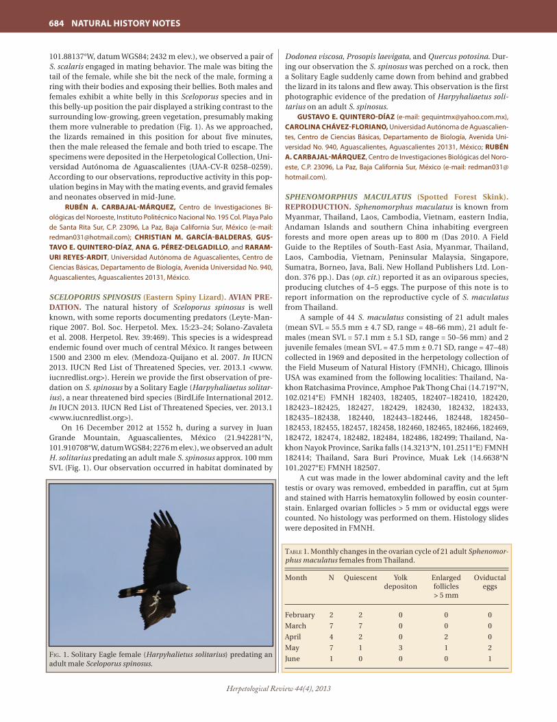
Herpetological Review 44(4), 2013
684 NATURAL HISTORY NOTES
101.88137°W, datum WGS84; 2432 m elev.), we observed a pair of S. scalaris engaged in mating behavior. The male was biting the tail of the female, while she bit the neck of the male, forming a ring with their bodies and exposing their bellies. Both males and females exhibit a white belly in this Sceloporus species and in this belly-up position the pair displayed a striking contrast to the surrounding low-growing, green vegetation, presumably making them more vulnerable to predation (Fig. 1). As we approached, the lizards remained in this position for about five minutes, then the male released the female and both tried to escape. The specimens were deposited in the Herpetological Collection, Uni-versidad Autónoma de Aguascalientes (UAA-CV-R 0258–0259). According to our observations, reproductive activity in this pop-ulation begins in May with the mating events, and gravid females and neonates observed in mid-June.
RUBÉN A. CARBAJAL-MÁRQUEZ, Centro de Investigaciones Bi-ológicas del Noroeste, Instituto Politécnico Nacional No. 195 Col. Playa Palo de Santa Rita Sur, C.P. 23096, La Paz, Baja California Sur, México (e-mail: [email protected]); CHRISTIAN M. GARCÍA-BALDERAS, GUS-TAVO E. QUINTERO-DÍAZ, ANA G. PÉREZ-DELGADILLO, and RARAM-URI REYES-ARDIT, Universidad Autónoma de Aguascalientes, Centro de Ciencias Básicas, Departamento de Biología, Avenida Universidad No. 940, Aguascalientes, Aguascalientes 20131, México.
SCELOPORUS SPINOSUS (Eastern Spiny Lizard). AVIAN PRE-DATION. The natural history of Sceloporus spinosus is well known, with some reports documenting predators (Leyte-Man-rique 2007. Bol. Soc. Herpetol. Mex. 15:23–24; Solano-Zavaleta et al. 2008. Herpetol. Rev. 39:469). This species is a widespread endemic found over much of central México. It ranges between 1500 and 2300 m elev. (Mendoza-Quijano et al. 2007. In IUCN 2013. IUCN Red List of Threatened Species, ver. 2013.1 <www.iucnredlist.org>). Herein we provide the first observation of pre-dation on S. spinosus by a Solitary Eagle (Harpyhaliaetus solitar-ius), a near threatened bird species (BirdLife International 2012. In IUCN 2013. IUCN Red List of Threatened Species, ver. 2013.1 <www.iucnredlist.org>).
On 16 December 2012 at 1552 h, during a survey in Juan Grande Mountain, Aguascalientes, México (21.942281°N, 101.910708°W, datum WGS84; 2276 m elev.), we observed an adult H. solitarius predating an adult male S. spinosus approx. 100 mm SVL (Fig. 1). Our observation occurred in habitat dominated by
Dodonea viscosa, Prosopis laevigata, and Quercus potosina. Dur-ing our observation the S. spinosus was perched on a rock, then a Solitary Eagle sud denly came down from behind and grabbed the lizard in its talons and flew away. This observation is the first photographic evidence of the predation of Harpyhaliaetus soli-tarius on an adult S. spinosus.
GUSTAVO E. QUINTERO-DÍAZ (e-mail: [email protected]), CAROLINA CHÁVEZ-FLORIANO, Universidad Autónoma de Aguascalien-tes, Centro de Ciencias Básicas, Departamento de Biología, Avenida Uni-versidad No. 940, Aguascalientes, Aguascalientes 20131, México; RUBÉN A. CARBAJAL-MÁRQUEZ, Centro de Investigaciones Biológicas del Noro-este, C.P. 23096, La Paz, Baja California Sur, México (e-mail: [email protected]).
SPHENOMORPHUS MACULATUS (Spotted Forest Skink). REPRODUCTION. Sphenomorphus maculatus is known from Myanmar, Thailand, Laos, Cambodia, Vietnam, eastern India, Andaman Islands and southern China inhabiting evergreen forests and more open areas up to 800 m (Das 2010. A Field Guide to the Reptiles of South-East Asia, Myanmar, Thailand, Laos, Cambodia, Vietnam, Peninsular Malaysia, Singapore, Sumatra, Borneo, Java, Bali. New Holland Publishers Ltd. Lon-don. 376 pp.). Das (op. cit.) reported it as an oviparous species, producing clutches of 4–5 eggs. The purpose of this note is to report information on the reproductive cycle of S. maculatus from Thailand.
A sample of 44 S. maculatus consisting of 21 adult males (mean SVL = 55.5 mm ± 4.7 SD, range = 48–66 mm), 21 adult fe-males (mean SVL = 57.1 mm ± 5.1 SD, range = 50–56 mm) and 2 juvenile females (mean SVL = 47.5 mm ± 0.71 SD, range = 47–48) collected in 1969 and deposited in the herpetology collection of the Field Museum of Natural History (FMNH), Chicago, Illinois USA was examined from the following localities: Thailand, Na-khon Ratchasima Province, Amphoe Pak Thong Chai (14.7197°N, 102.0214°E) FMNH 182403, 182405, 182407–182410, 182420, 182423–182425, 182427, 182429, 182430, 182432, 182433, 182435–182438, 182440, 182443–182446, 182448, 182450–182453, 182455, 182457, 182458, 182460, 182465, 182466, 182469, 182472, 182474, 182482, 182484, 182486, 182499; Thailand, Na-khon Nayok Province, Sarika falls (14.3213°N, 101.2511°E) FMNH 182414; Thailand, Sara Buri Province, Muak Lek (14.6638°N 101.2027°E) FMNH 182507.
A cut was made in the lower abdominal cavity and the left testis or ovary was removed, embedded in paraffin, cut at 5µm and stained with Harris hematoxylin followed by eosin counter-stain. Enlarged ovarian follicles > 5 mm or oviductal eggs were counted. No histology was performed on them. Histology slides were deposited in FMNH.
Fig. 1. Solitary Eagle female (Harpyhalietus solitarius) predating an adult male Sceloporus spinosus.
taBle 1. Monthly changes in the ovarian cycle of 21 adult Sphenomor-phus maculatus females from Thailand.
Month N Quiescent Yolk Enlarged Oviductal depositon follicles eggs > 5 mm
February 2 2 0 0 0
March 7 7 0 0 0
April 4 2 0 2 0
May 7 1 3 1 2
June 1 0 0 0 1

Herpetological Review 44(4), 2013
NATURAL HISTORY NOTES 685
All males examined from February (N = 2), March (N = 7), April (N = 9), June (N = 1), July (N = 1) were undergoing spermio-genesis (seminiferous tubules are lined by sperm or groups of metamorphosing spermatids). It thus appears the S. maculatus males examined exhibited an extended period of sperm produc-tion. The only male examined from August contained a regressed testis in which the seminiferous tubules contained spermatogo-nia and Sertoli cells. Before the significance of this single male with a regressed testis can be ascertained, examination of addi-tional specimens are needed. The smallest reproductively active males both measured 48 mm SVL (FMNH 182450, 182407) and were collected in February and April, respectively.
Monthly stages in the ovarian cycle are in Table 1. Four stages were observed: 1) quiescent, no yolk deposition; 2) early yolk de-position, vitellogenic granules in ooplasm; 3) enlarged follicles > 5 mm; 4) oviductal eggs. Mean clutch size (N = 6) = 3.7 ± 1.6 SD, range = 2–6. Clutches of two and six eggs represent new mini-mum and maximum clutch sizes for S. maculatus. The smallest reproductively active female 51 mm SVL (FMNH 182474) con-tained two enlarged ovarian follicles (> 5 mm) and was collected in May. Whether the nine S.maculatus females from February–March with quiescent ovaries indicates a period of female repro-ductive inactivity will require further study.
I thank Alan Resetar (FMNH) for permission to examine S. maculatus.
STEPHEN R. GOLDBERG, Whittier College, Department of Biology, P.O. Box 634, Whittier, California 90608, USA; e-mail: [email protected].
STROPHURUS CILIARIS (Northern Spiny-tailed Gecko). COM-MUNAL NESTING. Communal nesting is widespread in reptiles, and is more common than currently appreciated because the eggs and nests of many species are either unknown or rarely re-corded in nature (Doody et al. 2009. Quart. Rev. Biol. 84:229–252). One group with secretive nests are the diplodactylid geckos. Un-like the hard-shelled eggs of gekkonid geckos, which are often deposited in relatively conspicuous sites (e.g., under bark, logs, or rocks, on cave walls, in houses), the pliable-shelled eggs of di-plodactylid geckos are generally deposited underground where higher humidity or soil moisture is required to promote success-ful development (Bustard 1968. Copeia 1968:162–164). Details of the eggs and nests of most of the ~125 diplodactylid species are thus, unknown. Accordingly, communal nesting is rarely re-ported in these species (Doody et al., op. cit.). Herein we report on eight communal nests of Strophurus ciliaris that were found while excavating the nesting warrens (groups of burrows) of the Yellow-Spotted Monitor (Varanus panoptes) in tropical northern Australia.
During May and June 2013 we studied the nesting biology of V. panoptes (data reported elsewhere) at El Questro Wilderness Park in the east Kimberley Region of Western Australia (15.895033°S, 128.132456°E). The area consists of savannah woodland situated in the wet-dry tropics, with distinct wet (Nov–March) and dry (April–Oct) seasons. The nesting warrens, each comprising 6–21 burrows within a ca. 10 m2 area, were in sandy soils of sandhill habitat along creeks and rivers. Warrens were excavated by hand (with the aid of shovels and picks) to a depth of up to 3.6 m (Doody et al. 2014. J. Herpetol. 48: in press). During the excavation process we found 15 S. ciliaris eggs and 48 eggshells comprising 11 nests in small, back-filled chambers constructed in the floor of burrows of four V. panoptes nesting warrens at two locations. One warren contained no V. panoptes eggs in that year
(but contained eggshells from previous years). Nest depths were < 1 m; depths of three of the nests averaged 45 cm (range = 30–60 cm). We incubated four of these eggs to hatching under ambient temperatures in a makeshift field laboratory; all were confirmed to be S. ciliaris. Hatching dates were 10, 16, 22, and 28 June. We assumed that the remaining eggs and eggshells were also S. ciliaris based on their similar size and shape, and the absence of other pliable-shelled gecko species in the study area.
There are three interesting implications of our findings. First, like other geckos, S. ciliaris lays two eggs. Thus, eight (73%) of the clutches represented communal nests. Second and relatedly, it is interesting (and possibly a novel finding) that S. ciliaris mothers added their eggs to other clutches that were completely buried. In at least six of the communal nests the eggs were together within the same back-filled chamber excavated by the first mother. Communal back-filled reptile nests generally involve clusters of nests, rather than egg complements of multiple mothers in one chamber (Doody et al., op. cit.). If the alternative is true, that multiple mothers excavated or entered a single nest to lay at the same time, this also represents an interesting and novel finding (although it is considered less likely). Third, our data suggest that S. ciliaris is a common burrow associate of V. panoptes, at least during nesting. The distributions of the two species overlap in the Kimberley region of Western Australia, the Top End of the Northern Territory, and the gulf country and central desert regions of Queensland (Wilson and Swan. 2008. A Complete Guide to Reptiles of Australia, New Holland, Sydney. 512 pp.). The severe population-level declines incurred by V. panoptes due to poisoning by invasive Cane Toads, Bufo marinus (Doody et al. 2009. Anim. Conserv. 12:46–53), could thus eventually reduce nesting sites for S. ciliaris and other burrow associates (we also found the eggs of V. gouldii, and adults of 14 species of reptiles and amphibians in the warrens). The functional role of V. panoptes burrows in the savannah woodland ecosystem thus needs more scientific attention, particularly in areas ahead of the Cane Toad front in the west Kimberley region.
J. SEAN DOODY, Department of Ecology and Evolutionary Biology, University of Tennessee, 569 Dabney Hall, Knoxville, Tennessee 37996-1610, USA (e-mail: [email protected]); HUGH JAMES, School of Environmental and Life Sciences, University of Newcastle, Callaghan, New South Wales 2308, Australia (e-mail: [email protected]); DEREK DUNLOP, Department of Ecology and Evolutionary Biology, University of Tennessee, 569 Dabney Hall, Knoxville, Tennessee 37996-1610, USA (e-mail: [email protected]); DOMENIC D’AMORE, Department of Natural Sciences, Daemen College, 4380 Main Street, Amherst, New York 14226, USA (e-mail: [email protected]); MATT EDGAR, School of Environmental and Life Sciences, University of Newcastle, Callaghan, New South Wales 2308, Australia (e-mail: [email protected]); MAIK FIDEL, 2/151 Main Road, Lower Plenty, Victoria 3093, Australia (e-mail: [email protected]); DAVID MEADOWS, Department of Natural Sciences, Daemen College, 4380 Main Street, Amherst, New York 14226, USA (e-mail: [email protected]); CHRISTOPHER W. WALMSLEY, Department of Anatomy and Developmental Biology, Monash University, Clayton, Victoria 3800, Australia (e-mail: [email protected]); SIMON CLULOW, School of Environmental and Life Sciences, University of Newcastle, Callaghan, New South Wales 2308, Australia (e-mail: [email protected]); COLIN McHENRY, Department of Anatomy and Developmental Biology, Monash University, Clayton, Victoria 3800, Australia (e-mail: [email protected]).
TRAPELUS SAVIGNII (Egyptian Sand Agama). REPRODUC-TION. Trapelus savignii is known from Egypt and Israel where

Herpetological Review 44(4), 2013
686 NATURAL HISTORY NOTES
it is diurnal and found in sandy habitats (Baha El Din 2006. A Guide to the Reptiles and Amphibians of Egypt. The American University in Cairo Press, Cairo. 359 pp.; Bar and Haimovitch 2011. A Field Guide to Reptiles and Amphibians of Israel. Pazbar LTD 1989, Herzliya, Israel. 245 pp.). There is anecdotal informa-tion on its reproduction in Bar and Haimovitch (op. cit.). The purpose of this note is to add information on the reproductive biology of T. savignii utilizing information from a histological analysis of gonadal material.
A sample of 35 T. savignii collected in Israel from 1946 to 2012 and deposited in the herpetology collection of the Natural His-tory Collections of Tel Aviv University (TAUM) was examined, consisting of 9 adult males (mean snout–vent length, SVL = 76.6 mm ± 7.1 SD, range = 68–85 mm) 10 adult females (mean SVL = 87.9 mm ± 5.6 SD, range = 81–98 mm), 7 subadult males (mean SVL = 52.9 mm ± 4.3 SD, range = 48–59 mm), 7 subadult females (mean SVL = 55.0 mm ± 6.2 SD, range = 45–63 mm) and 2 sub-adults of indeterminate sex (mean SVL = 44.5 mm ± 2.1 SD, range = 43–46 mm).
A cut was made in the lower abdomen and the left testis or ovary was removed, embedded in paraffin, cut into 5 µm sections and stained with Harris’ hematoxylin followed by eosin counter-stain. Enlarged follicles > 5 mm or oviductal eggs were quanti-fied. Histology slides were deposited in TAUM. Trapelus savignii from Israel examined (by region) from TAUM were: Northern Ne-gev Region: TAUM 635–637, 639, 640, 643–645, 1213, 1227, 1917, 2545, 2632, 2633, 5162, 5534, 5535, 5995, 5999, 6005, 6416, 6420, 6421–6426, 6428, 6398, 6399, 6439, 8475, 16128, Southern Coastal Region: TAUM 1233.
Three stages were noted in the testicular cycle (Table 1): 1) regressed, seminiferous tubules reduced in size, containing spermatogonia and interspersed Sertoli cells; 2) recrudescent, proliferation of germ cells for the next period of spermiogenesis (sperm formation); 3) spermiogenesis, seminiferous tubules lined by sperm or clusters of metamorphosing spermatids. The presence of spermiogenic males in March and April indicate T. savignii is a spring breeder. The smallest mature males (spermio-genesis in progress) all measured 68 mm SVL and were collected
in March TAUM 5999, 6399, 6439. Testes of smaller males (48–59 mm) were either regressed or in recrudescence and were classi-fied as juveniles.
Four stages were noted in the ovarian cycle (Table 2): 1) qui-escent (no yolk deposition); 2) yolk deposition (basophilic gran-ules in ooplasm); 3) enlarged pre-ovulatory follicles > 5 mm; 4) oviductal eggs. The period of female reproductive activity en-compassed April through August (Table 2). Mean clutch size (N = 3) was 6.3 ± 3.8 SD, range = 2–9. The smallest reproductively active female (TAUM 5535), measured 81 mm SVL, exhibited early yolk deposition and was collected in April. There was no indication (oviductal eggs and concurrent yolk deposition in the same female) to suggest T. savignii produces multiple clutches in the same reproductive season. However, the apparent absence of females in the process of preparing a second clutch may be a re-flection of small sample size (N = 10). Six smaller females (range 45–63 mm) exhibited quiescent ovaries and were considered as juveniles. One T. savignii (TAUM 6421) of indeterminate sex (SVL = 46 mm) was also considered a juvenile.
My finding of one female with oviductal eggs and one with enlarged pre-ovulatory follicles from August (Table 2) confirms the statement in Bar and Haimovitch (op. cit.) that T. savignii ovi-posits in August. The presence of reproductive activity in March and April (Tables 1, 2) indicates T. savignii commences repro-duction early in the spring in Israel. The clutch values reported herein, to my knowledge, represent the first reported for T. savi-gnii. In addition, it appears males mature at smaller body sizes than females.
I thank Shai Meiri (TAUM) for permission to examine T. savi-gnii, Erez Maza for facilitating the loan and the National Collec-tions of Natural History at Tel Aviv University for providing speci-mens of T. savignii for this study.
STEPHEN R. GOLDBERG, Whittier College, Department of Biology, PO Box 634, Whittier, California 90608, USA; e-mail: [email protected].
UROSAURUS BICARINATUS (Tropical Tree Lizard). BIFURCA-TION. Urosaurus bicarinatus is a small phrynosomatid endemic to Mexico, found from southern Sonora to Chiapas, primar-ily along the Pacific versant and interior valleys (Feldman et al. 2011. Mol. Phyl. Evol. 61:714–725; Köhler 2008. Reptiles of Cen-tral America, 2nd ed., Herpeton Verlag Elke Köhler, Offenbach. 400 pp.; Wiens 1993. Herpetologica 49:399–420; Wilson and Johnson 2010. In Wilson et al [eds.], Conservation of Mesoameri-can Amphibians and Reptiles, pp. 31–235. Eagle Mountain Publ., LC, Eagle Mountain, Utah), from near sea level to 2500 m eleva-tion (Wilson and Johnson 2010, op. cit.). Herein, we report an incidence of tail bifurcation in a U. bicarinatus from the coastal lowlands of Oaxaca, Mexico.
On 23 June 2013, at 0940 h, an adult female U. bicarinatus was observed resting on a tree trunk on a slope with tropical deciduous forest, ca. 3 km N of La Soledad, Municipality of Tu-tutepec, Oaxaca (18.157883°N, 96.091658°W, WGS 84; elev. 25 m). When the lizard was caught, we noticed that it had a bifurcated tail (Fig. 1). Bifurcation of the tail was represented by the end sec-tion of the original tail and the new regenerated tail. This bifur-cation was located 41 mm posterior to the cloaca. Length of the original segment after the bifurcation was 15 mm (left side) and the regenerated section was 30 mm (right side). The new regen-erated tail section was likely the result of an incomplete autoto-mization of the original tail (Goin and Goin 1971. Introduction to Herpetology, 2nd ed. W. H. Freeman and Co., San Francisco,
taBle 2. Monthly stages in the ovarian cycle of 10 adult female Trapelus savignii from Israel.
Month N Quiescent Yolk Enlarged Oviductal depositon follicles eggs > 5 mm
April 1 0 1 0 0
May 1 0 1 0 0
June 2 1 0 0 1
August 2 0 0 1 1
September 4 4 0 0 0
taBle 1. Monthly stages in the testicular cycle of 9 adult male Trapelus savignii from Israel.
Month N Regressed Recrudescent Spermiogenesis
March 5 0 0 5
April 2 0 0 2
August 1 1 0 0
September 1 1 0 0

Herpetological Review 44(4), 2013
NATURAL HISTORY NOTES 687
California, 353 pp.; Clark 1973. HISS News-J. 1:158). Bifurcat-ed or multiple tail regeneration cases have been published for many species of lizards, including members in the family Phry-nosomatidae (see Cordes and Walker 2013. Herpetol. Rev. 44:319; Gogliath et al. 2012. Herpetol. Rev. 43:129; Kumbar and Ghadage 2011. Herpetol. Rev. 42:94; Mata-Silva et al. 2010. Herpetol. Rev. 41:352–353; Mitchell et al. 2012. Herpetol. Rev. 43:650; Tamar et al. 2013. Herptol. Rev. 44:135–136, and citations therein). To the best of our knowledge this is the first report of tail bifurcation in U. bicarinatus.
We thank E. V. Rosas-López, Y. U. Rosas-Cortéz, J. Rosas-Cruz, E. Mata-Silva, and A. Mata-Silva for their help and great com-pany in the field.
VICENTE MATA-SILVA, Department of Biological Sciences, The Uni-versity of Texas at El Paso, El Paso, Texas 79968, USA (e-mail: [email protected]); ARTURO ROCHA, Department of Biological Sciences, El Paso Com-munity College, El Paso, Texas, USA (e-mail: [email protected]); JERRY D. JOHNSON, Department of Biological Sciences, The University of Texas at El Paso, El Paso, Texas 79968, USA (e-mail: [email protected]); LARRY DAVID WILSON, 16010 S. W. 207th Avenue, Miami, Florida 33187, USA, and Centro Zamorano de Biodiversidad, Escuela Agrícola Panamericana Zamo-rano, Depto. Francisco Morazán, Honduras (e-mail: [email protected]).
VARANUS GLEBOPALMA (Black-palmed Monitor). NOCTUR-NAL ACTIVITY AND FORAGING. Monitor lizards are thought to be diurnal, based on a body of ecological research and on eye structure. Studies of the eye structure of Varanus exanthematicus and V. griseus have suggested that prey capture in these species is reliant on visual cues, based on the presence of cone cells in the eyes (Bhattacharjee 1992. Exp. Eye Res. 55:243; Röll and Horn 2000. Mertensiella 2:291–306). The morphological similarity of monitor lizards led Röll and Horn (op. cit.) to conclude that all Varanus species possess the same or similar eye structure and therefore all are diurnal.
However, a number of observations suggest that some moni-tor lizards occasionally undertake crepuscular and/or nocturnal activity. Of the 53 known species of monitor lizards, field obser-vations of crepuscular or nocturnal activity have been recorded in nine species (V. spenceri, V. panoptes, V. tristis V. glebopalma, V. rosenbergi, V. melinus, V. glauerti, V. salvator, and V. dumerilli), and six species in captivity (V. acanthurus, V. giganteus, V. indicus, V. keithhorni, V. mertensi, and V. varius) (Bohme and Ziegler 1997. Herpetofauna 19:26–34; Cota et al. 2008. Biawak 2:152–158; Fyfe
1980. Herpetofauna 12:15; Irwin et al. 1996. Herpetofauna 26:50; Irwin et al. 1996. Herpetol. Rev. 27:192–194; Jones 1998. Herpeto-fauna, 28:50; Rokylle 1989. Herpetofauna 7:4–8; Trembath 2000. Herpetofauna 30:52; Uyeda et al 2013. Biawak 7:25–30; Valentic 1995. Herpetofauna 25:49–50; Yong et al. 2008. Nature in Singa-pore 1:21–22).
In particular, there are several reports of crepuscular activity in V. glebopalma. Christian (1977. Victoria Herpetol. Soc. Newsl. 6:11–13) suggested that V. glebopalma does most of its foraging during the first couple of hours after sunset. Wilson and Knowles (1988. Australia’s Reptiles: A Photographic Reference to the Ter-restrial Reptiles of Australia. Cornstalk, Pymbie. 447 pp.) stated that “although active by day, it is commonly crepuscular and can often be disturbed foraging just after sundown.” Two individu-als were spotlighted active at night between 1900 and 2100 h by Shea et al (1988. West. Austral. Herpetofauna 18:16), and Valentic (1994. South Austral. Herpetol. Group Newsl. 87:11) observed an individual active in twilight period (1830–1900 h) catching and swallowing prey. It was thus surprising that the only quantitative study on V. glebopalma resulted in a lack of nocturnal behavior. In a radio-telemetry study of the spatial ecology of V. glebopalma, Sweet (1999. Mertensiella 2:317–366) failed to record any crepus-cular or nocturnal activity other than specimens that had been disturbed after dark, despite considerable effort to support pre-vious accounts. Sweet concluded that previous observations of nocturnal/crepuscular activity were a result of the animal being disturbed by the observer. It is only through the continual docu-mentation of observations that we can fully understand the ap-parent disparity between these findings. This note presents two further observations of nocturnal activity from V. glebopalma in tropical northwestern Australia.
At 2300 h on 6 May 2012, an adult male V. glebopalma (SVL 253 mm, TL 751 mm) was observed moving across a large flat rock approximately 0.5 m in height and through a clump of spinifex grass. The animal continued to move for another 8 m across another flat section of rock where it was captured. At the time of the observation, there was a full moon and little to no cloud cover. Although the surrounding area was being burned by a small, slow moving fire, the nearest fire front was 300 m from the animal’s location. However, it is possible that the ani-mal detected the fire through olfaction from its roost. In a second observation, while spotlighting at approximately 1950 h 28 June 2012, a frog’s alarm call drew the attention of observers to a rocky area on the side of a sandstone gorge. Upon closer inspection a V. glebopalma was observed for 15 minutes feeding on a frog (unidentified Litoria sp.). When observers moved closer to con-firm the species of frog, the animal became wary and swallowed the frog prior to retreating into a hole among fallen rocks and boulders. A short search failed to relocate the animal. The tem-perature at the time of observation was 19.5°C. Sunset that night occurred at 1722 h, and there was a half moon.
Our observations provide further evidence that V. glebopalma may forage nocturnally. The lack of crepuscular and nocturnal behavior in Sweet’s (op. cit.) study may reflect population and/or seasonal variation. Alternatively, observations of nocturnal activity in the species may reflect lizards that were flushed from their roost sites by potential predators (S. Sweet, pers. comm.). However, the growing number of observations suggests that noc-turnal behavior occurs in some populations. Interestingly, Sweet (op. cit.) noted that V. glebopalma adopts a sit and wait, or am-bush, foraging strategy that may be unique to monitor lizards. This strategy may allow V. glebopalma to forage during low light
Fig. 1. Female Tropical Tree Lizard (Urosaurus bicarinatus) with tail bifurcation found near La Soledad, Oaxaca, México.

Herpetological Review 44(4), 2013
688 NATURAL HISTORY NOTES
conditions. Investigations into the eye structure of V. glebopalma should be undertaken to test for any differences between this and other species of monitor lizards.
DAVID RHIND, Department of Anatomy and Developmental Biology, Monash University, Clayton, Victoria, Australia, 3800 (e-mail: [email protected]); J. SEAN DOODY, Department of Ecology and Evolutionary Biology, University of Tennessee, Knoxville, Tennessee, USA (e-mail: [email protected]); RYAN ELLIS, Department of Terrestrial Zoology, Western Australian Museum, 49 Kew Street, Welshpool, Western Australia, Australia 6106 (e-mail: [email protected]); AMY RICKETTS, El Questro Wilderness Park, PO Box 909, Kununurra, Western Australia, Australia 6743 (e-mail: [email protected]); GAVIN SCOTT, El Questro Wilderness Park, PO Box 909, Kununurra, Western Australia, Australia, 6743, Australia (e-mail: [email protected]); SIMON CLULOW, School of Environmen-tal and Life Sciences, University of Newcastle, Callaghan, New South Wales, Australia 2308 (e-mail: [email protected]); COLIN McHENRY, Department of Anatomy and Developmental Biology, Monash University, Clayton, Victoria, Australia, 3800 (e-mail: [email protected]).
SQUAMATA — SNAKES
CHILOMENISCUS STRAMINEUS (Variable Sandsnake). ENDO-PARASITES. Chilomeniscus stramineus occurs in two disjunct populations. The eastern population ranges from southwest-ern Arizona, south through western Sonora, Mexico to northern Sinaloa, Mexico; the western population is restricted to Baja Cali-fornia (Grismer 2002. Amphibians and Reptiles of Baja California Including its Pacific Islands and the Islands in the Sea of Cortés. Univ. Calif. Press, Berkeley. 399 pp.). To our knowledge there are no helminths reported from C. stramineus. The purpose of this note is to establish the initial helminth list for C. stramineus as part of an ongoing survey of the helminths of reptiles from Baja California.
The coelomic cavity of one male C. stramineus (SVL = 203 mm) collected at 24 km S Loreto (26.0167°N, 111.3500°W, datum: WGS84), Baja California Sur, Mexico in January 1974 and depos-ited in the herpetology collection of the Natural History Museum of Los Angeles County (LACM), Los Angeles, California, USA as LACM 138142 was examined. Two oval shaped whitish endopar-asites ca. 2 mm in length, found in the body cavity, were studied under a dissecting microscope. Based on body shape, pseudoan-nulation, and hook root morphology, the parasites were identi-fied as an oligacanthorhynchid acanthocephalan cystacanth. It was deposited in the United States National Parasite Collection, Beltsville, Maryland, USA as USNPC 106939. Acanthocephalans require an arthropod intermediate host in which the cystacanth develops (Kennedy 2006. Ecology of the Acanthocephala. Cam-bridge Univ. Press, Cambridge, UK. 249 pp.). Chlomeniscus stra-mineus likely become infected with cystacanths when feeding on infected insects (Grismer, op. cit.). Because development beyond the cystacanth does not occur, snakes likely serve as paratenic (= transport) hosts. Chlomeniscus stramineus represents a new host record for oligacanthorhynchid acanthocephalan cystacanths.
We thank Greg Pauly (LACM) for permission to examine C. stramineus.
STEPHEN R. GOLDBERG, Whittier College, Department of Biol-ogy, Whittier, California 90608, USA (e-mail: [email protected]); CHARLES R. BURSEY, Pennsylvania State University, Shenango Campus, Department of Biology, Sharon, Pennsylvania 16146, USA (e-mail: [email protected]); JEANETTE ARREOLA, Whittier College, Department of Biology, Whittier, California 90608, USA (e-mail: [email protected]).
CROTALUS ADAMANTEUS (Eastern Diamondback Rattle-snake). ANTI-PREDATOR BEHAVIOR. Snakes exhibit a variety of defensive behaviors when confronted with potential preda-tors. Among rattlesnakes, the most common defensive behaviors include fleeing, coiling, head hiding, body bridging, body flat-tening, hissing, tongue-flicking, discharging scent gland secre-tions, vibrating the tail (rattle), and striking (Klauber 1972. Rattle-snakes: Their Habits, Life Histories, and Influence on Mankind. Univ. California Press, Berkeley. 1533 pp.; Weldon and Burghardt 1979. J. Chem. Ecol. 5:141–151). Although primarily terrestrial, rattlesnakes are adept swimmers and will enter water readily in order to pursue food, mates and refuge, and to escape harass-ment or danger (Klauber, op. cit.). While defensive/anti-predator behaviors of most rattlesnakes have been well-described (Klau-ber, op. cit.), few if any reports exist regarding anti-predator behavior of rattlesnakes while in water. Here we describe anti-predator behavior exhibited by C. adamanteus while swimming.
On 28 December 2003 at approximately 1200 h, we ob-served an adult (Total Length [TL] ca. 150 cm) C. adamanteus swimming across a small lake (25.40005°N, 80.65630°W, datum: WGS84) on Long Pine Key in Everglades National Park, Florida, USA. The snake was swimming along the shoreline approximate-ly 30 m from land with its head elevated and majority of its body, though not the tip of its tail, visible above the water’s surface. As the snake continued swimming, it passed an adult (TL ca. 210 cm) Alligator mississippiensis (American Alligator) in shal-low water along the shoreline. The alligator turned and rapidly began to pursue the snake from directly behind. When the al-ligator was within approximately 2.5 m of the snake, the snake suddenly stopped swimming and raised the tip of its tail out of the water. The alligator, at this point approximately 1 m from the snake, abruptly stopped swimming and ceased moving towards the snake. Due to a strong breeze and moderate wave action, we were unable to discern if the snake vibrated its tail (and rattle). After approximately 15 sec, the rattlesnake lowered its tail and resumed swimming towards the shoreline. The alligator allowed the snake to open up a distance of approximately 5 m between them before slowly resuming pursuit. However, within approxi-mately 1 min the snake reached the shore and crawled into the adjacent pine forest.
It is unclear what component of the rattlesnake’s behavior deterred the alligator from attacking. Rattlesnakes, like many snakes when threatened, discharge scent gland secretions from the cloaca and multiple reports contend that alligators can dis-tinguish between venomous and non-venomous snakes, pre-sumably by chemical cues (McIlhenny 1935. The Alligator’s Life History. Ten Speed Press, Berkeley, California. 117 pp.; Neill 1971. Last of the Ruling Reptiles. Columbia Univ. Press, New York. 486 pp.). However, controlled laboratory studies with captive alliga-tors and snakes failed to corroborate this suggestion (Weldon and McNease 1991. Herpetologica 47:403–406). Whether the raised tail of the rattlesnake resulted in a chemical (scent gland secretion), auditory (vibrating rattle), or visual (tail color) cue re-mains unknown, but the behavior clearly appeared to be a delib-erate and effective warning display.
In addition to the observed defensive behavior by the rattle-snake, to our knowledge this is also the first report of attempted predation by A. mississippiensis on C. adamanteus in the wild. Given that these two species are often sympatric, encounters be-tween these animals are likely not uncommon.
We thank Allan Woodward and Paul Moler for sharing their knowledge of rattlesnake and alligator interactions in Florida.

Herpetological Review 44(4), 2013
NATURAL HISTORY NOTES 689
The findings and conclusions in this note are those of the au-thors and do not necessarily represent the views of the U.S. Fish and Wildlife Service.
RUSTIN M. GOODEN, U.S. Fish and Wildlife Service, Sewee Visitor and Environmental Education Center, Awendaw, South Carolina 29429, USA; THOMAS R. RAINWATER, U.S. Fish and Wildlife Service, Ecological Ser-vices Field Office, Charleston, South Carolina 29407, USA (e-mail: [email protected]); STEVEN G. PLATT, Wildlife Conservation Society, Myan-mar Program, Office Block C-1, Aye Yeik Mon 1st Street, Hlaing Township, Yangon, Myanmar.
CROTALUS ATROX X CROTALUS HORRIDUS (Western Dia-mond-backed Rattlesnake x Timber Rattlesnake). NATURAL HYBRID. Hybridization in rattlesnakes has been unequivocally demonstrated in captivity as a result of intentional or uninten-tional breeding (Klauber 1972. Rattlesnakes: Their Habits, Life Histories, and Influence on Mankind. Univ. California Press, Berkeley. x + 1533 pp.; Rubio 1998. Rattlesnake: Portrait of a Predator. Smithsonian Press, Washington. 240 pp.). In nature, however, documentation of putative hybrid rattlesnakes is scant and ascertained primarily by way of morphological characters (e.g., Campbell et al. 1989. Herpetologica 45:344–349); molecular methods (allozymes, mtDNA, msats) are rarely used (e.g., Mur-phy and Crabtree 1988. Herpetologica 44:119–123; Gerard et al. 2011. BMC Evol. Biol. 11:291; see Campbell and Lamar 2004. Ven-omous Reptiles of the Western Hemisphere. Cornell Univ. Press, Ithaca, New York. 870 pp.). Here, using the mtDNA genes ATPase 8 and ATPase 6 as markers and assessing scalation and color pat-tern, we report on a subject that we suspect to be a natural hy-brid of Crotalus atrox x Crotalus horridus.
On 2 May 2007 a large (~ 1 m total length) rattlesnake was found coiled on a trail 4.8 km N of the town of Blue, 64 km NE of Austin, Lee Co., Texas, USA. The area is categorized as Post Oak Savannah but the immediate landscape that surrounds this property is disturbed and largely agricultural. Using the methods described in Douglas et al. (2006. Mol. Ecol. 15:3353–3374), we obtained DNA from a sample of shed skin. A comparative analy-sis of the sequence information (ATPase 8 and 6) obtained from the hybrid to a pre-existing phylogeny of rattlesnakes and other North American pitvipers (M. E. Douglas et al., unpubl. data) re-vealed that the haplotype was that of C. horridus. Thus, owing to matrilineal inheritance of mtDNA, we assigned the hybrid’s mother to be C. horridus. The overall appearance of the subject appears intermediate to that of the putative parents (Fig. 1A). The paired parietal marking, post-ocular stripe, and mottled venter pattern (not visible) are C. horridus-like, whereas the broad dorsal blotches and black and white rings on the tail are C. atrox-like.
To our knowledge, this is the second record of natural hy-bridization between C. atrox and C. horridus. Meik et al. (2008. Southwest. Nat. 53:196–200), using morphological characters (e.g., scales, color pattern), provided robust evidence for hybrid-ization between C. atrox and C. horridus in a subadult (SVL = 667) female subject collected 50 km W of Denton, in Wise Co., Texas (University of Texas at Arlington [UTA] R-52942; Fig. 1B). This specimen bears a striking resemblance to the hybrid reported here (Fig. 1A). Our analysis of scalation and color pattern of the present hybrid essentially mirrors the results of Meik et al. (op. cit.). However, in our analysis of interoculabials we followed the definition provided by Klauber (op. cit.), which includes count-ing a subocular and a supralabial. Our count for interoculabials was 5-5 (both sides of the head). Using this definition, the hybrid
described in Meik et al. (2008, op. cit.) is 5-5 rather than 3 (i.e., 3-3) as reported (J. Meik, pers. comm.).
We thank Hugh Brown for alerting us to the snake we report here. Margie Crisp helped with care and assisted in obtain-ing photographs of the hybrid. Louis Porras helped to measure scales. Jesse Meik and Carl Franklin discussed hybridization in rattlesnakes, and they allowed us to use their image of the hy-brid rattlesnake (C. atrox x C. horridus) discussed in their 2008 publication. Ryan Sawby kindly prepared the digital image for publication.
WILLIAM B. MONTGOMERY, P.O. Box 656, Elgin, Texas 78621, USA (e-mail: [email protected]); GORDON W. SCHUETT, Department of Biology and Center for Behavioral Neuroscience, Georgia State University, 33 Gilmer Street SE, Atlanta, Georgia 30303-3088, USA (e-mail: [email protected]); MARLIS R. DOUGLAS and MICHAEL E. DOUGLAS, Depart-ment of Biological Sciences, University of Arkansas, Fayetteville, Arkansas 72701, USA.
DROMICODRYAS BERNIERI (Bernier’s Striped Snake). DIET. Few studies have investigated the ecology of Madagascar’s more common diurnal colubrid snake species (Glaw and Vences 2007. A Field Guide to the Amphibians and Reptiles of Madagascar. 3rd ed. Vences & Glaw, Köln. 496 pp.; Kaloloha et al. 2011. Herpetol. Notes 4:397–402). Dromicodryas bernieri has only been docu-mented preying upon saurian prey and is assumed to be strictly saurophagous (Cadle 2003. In Goodman and Benstead [eds.],
Fig. 1. A) The adult male hybrid (Crotalus atrox x C. horridus) from Lee Co., Texas. B) The subadult female hybrid (C. atrox x C. horridus) described by Meik et al. (op. cit.) from Wise Co., Texas.
COLO
R RE
PRO
DU
CTI
ON
SU
PPO
RTED
BY
THE
THO
MA
S BE
AU
VAIS
FU
ND

Herpetological Review 44(4), 2013
690 NATURAL HISTORY NOTES
The Natural History of Madagascar, pp. 997–1004. Univ. Chicago Press, Chicago and London); Gehring 2006. Zeitschrift des Köl-ner Zoo - Heft 49:127–141). Here we report on the predation of an amphibian by a D. bernieri.
On 2 August 2011, at 1130 h, at the edge of the main thorough-fare in Mariarano Village, North West Madagascar (15.44878°S, 46.69146°E, datum: WGS84) we encountered a D. bernieri with an adult frog, Boophis doulioti, that it had seized and partially in-gested, head first (Fig. 1). For approximately 3 min we observed the snake attempting to consume the frog; however, our pres-ence seemed to distract the snake and it moved further into the undergrowth with its amphibian prey in its mouth. Considering that B. doulioti is locally abundant in this area, even during the long dry season, it is reasonable to assume that this predation event is not unique and that this amphibian forms a regular part of the diet of D. bernieri at this location.
We thank the Transitional Government of Madagascar, the Ministère de l’Environnement et des Forêts for allowing us to undertake this work (permit 54710/MEF/SG/DGE/DREF.BOE), Operation Wallacea for funding, Development and Biodiversity Conservation Action for Madagascar (DBCAM) for logistics and our local guides and team at Mariarano Village.
ROBERT GANDOLA (e-mail: [email protected]), STU-ART GRAHAM, MARC RABENANDRASNA, and CATRIONA HENDRY, 27 Ayrfield Park, Dublin 13, Ireland.
DRYMARCHON MELANURUS (Central American Indigo Snake). DIET. Drymarchon melanurus is known to feed on a wide variety of prey, including small mammals, frogs, fish, hatchling turtles, birds, and snakes (Irwin et al. 2003. J. Kansas Herpetol. 7:13–18; Stevenson et al. 2010. Southwest. Nat. 9:1–18). On 5 October 2011, at about 1430 h, AVN found an adult D. mel-anurus near the CIE (Centro Interpretativo Ecológico) inside the Biosphere Reserve of “El Cielo” (23.88555°N, 99.16861°W, datum WGS84; elev. 360 m), municipality of Gómez Farías, state of Tam-aulipas, México. The snake was in the process of feeding on an adult Spilotes pullatus (Fig. 1). After an hour, the D. melanurus had ingested half of the prey and subsequently it crawled under a rock with the prey in its mouth. When checked 2 h later, the prey was found, regurgitated and abandoned by the predator. To our knowledge this represents the first record of D. melanurus preying on S. pullatus.
ALFONSO HERNÁNDEZ-RÍOS, Laboratorio de Herpetología, Institu-to de Biología, Universidad Nacional Autónoma de México, Distrito Federal 04510, México (e-mail: [email protected]); ELÍ GARCÍA-PADILLA,
Jaguares #18 Casa del Sol, Oaxaca de Juárez 68128, México (e-mail: [email protected]); ADRIÁN VILLEGAS-NIETO, Centro Interpretativo Ecológico, Reserva de la Biósfera El Cielo, Gómez Farías, Tamaulipas, México (e-mail: [email protected]).
CHILABOTHRUS (=EPICRATES) CHRYSOGASTER CHRYSO-GASTER (Turks Island Boa). CLIMBING BEHAVIOR. Chilabo-thrus (=Epicrates) chrysogaster chrysogaster is considered to be a rare example of an active terrestrial foraging boid (Reynolds and Gerber 2012. J. Herpetol. 46:578–586). Out of over 350 ob-servations of active boas on Big Ambergris Cay, Turks and Caicos
Fig. 1. Dromicodryas bernieri consuming an adult Boophis doulioti in Mariarano, northwest Madagascar.
Fig. 1. Chilabothrus chrysogaster chrysogaster demonstrating rarely documented climbing behavior.
Fig. 1. Drymarchon melanurus attempting to prey on a Spilotes pul-latus in Tamaulipas, México.
PHO
TO B
Y A
DRI
ÁN
VIL
LEG
AS
NIE
TO

Herpetological Review 44(4), 2013
691NATURAL HISTORY NOTES 691
Islands, British West Indies, only two individuals were found ac-tive off of the ground (Reynolds and Gerber, op. cit.). It is thought that juveniles of this species, like others of the genus, might be largely arboreal, though this has not been conclusively dem-onstrated (Reynolds and Gerber, op. cit.). While Big Ambergris Cay is mostly scrub and coppice, some arboreal habitat does exist, though boas have never been found higher than 2 m off the ground (Reynolds and Gerber, op. cit.). However, on larger islands in the Turks and Caicos such as North and Middle Caicos, remnant tropical dry forest and pine barrens exist, which pro-vide a great deal of arboreal habitat.
On 12 October 2012, BNM observed a previously unobserved climbing behavior in this species on Pine Cay, Turks and Caicos Islands (Fig. 1). The individual (SVL ca. 1000 mm; likely female) was climbing a telephone pole located along a road in secondary tropical dry forest habitat with a canopy height of ~7 m. The in-dividual was using a stereotyped version of concertina locomo-tion often associated with other snake species, such as rat snakes (genus Pantherophis) and boa constrictors (Boa constrictor). The motion consists of lateral undulation against a scaffold (Jayne and Herrmann 2011. J. Exp. Biol. 214:2189–2201). This represents a rare observation of climbing in this species, and suggests that individuals employ a similar strategy to other frequent climbers such as rat snakes and boa constrictors.
R. GRAHAM REYNOLDS, Department of Biology, University of Mas-sachusetts, Boston, Massachusetts 02125, USA (e-mail: [email protected]); B. NAQQI MANCO, Department of Environment and Maritime Affairs, Turks and Caicos Island Government, Turks and Caicos Islands, Brit-ish West Indies.
HEMACHATUS HAEMACHATUS (Rinkhals). DIET / CANNIBAL-ISM. Although African elapids are well known to be ophiopha-gous (FitzSimons 1962. Snakes of Southern Africa. Purnell and Sons Ltd., Johannesburg. 423 pp.) and may consume conge-ners (Shine et al. 2007. J. Zool. 272:183–193), only one species is known to be cannibalistic (Naja nivea; FitzSimons, op. cit.). On 10 December 2012 on the farm Ryswick (27.9009°S, 029.2107°E, datum WGS84; elev. 1790 m) in the district of Harrismith of the Free State province, South Africa, I collected an adult male He-machatus haemachatus that had been shot by the landowner. Upon dissection, the snake (SVL = 877 mm; tail length = 200 mm; mass = 661 g) was found to have consumed a smaller female conspecific (SVL = 509 mm, tail length = 118 mm, mass = 105.95 g). The adult male had also re-cently consumed three toads (Amietophrynus sp.; mass = 130.08 g), while a single partially digested toad was found in the digestive tract of the female (Amietophrynus sp.; mass = 11.04 g). I thank Graham Alexander for assisting with snake dissection and manuscript preparation.
SHIVAN PARUSNATH, School of Animal, Plant, and Environ-mental Sciences, University of the Witwatersrand, Johannesburg, South Africa, P.O. Wits, 2050; e-mail: shivan.parusnath@gmail.
HETERODON PLATIRHINOS (Eastern Hog-nosed Snake). DIET. Heterodon platirhinos primarily feeds upon amphibians, but it has been reported to eat rep-tiles, mammals, birds, fishes, and invertebrates (Ernst and Ernst 2003. Snakes of the United States and Canada. Smithsonian Books, Washington, D.C. 668 pp.). The only reports of avian consumption were an unidentified bird (Surface 1906. Bull. Pennsylvania State Dept. Agric. Div. Zool. 4:113–208.) and a nestling sparrow (Conant 1938.
Am. Midl. Nat. 20:1–200.). Here, we report predation upon an adult Northern Mockingbird (Mimus polyglottos) by a female H. platirhinos (SVL = 658 mm) from 5.3 km S of Howey-in-the-Hills, Lake Co., Florida, USA. When captured in June 2004, the snake’s body was distended by a large prey item. In captivity, the snake died two days after regurgitating the partially digested bird. Both the snake and bird are deposited in the Florida Museum of Natu-ral History (UF 169545).
MARK B. ROBERTSON, 5610 NW 31st Terrace, Gainesville, Florida 32653, USA; KEVIN M. ENGE, Florida Fish and Wildlife Conservation Com-mission, 1105 SW Williston Road, Gainesville, Florida 32601, USA (e-mail: [email protected]).
HETERODON PLATIRHINOS (Eastern Hog-nosed Snake). RE-PRODUCTION. There is a lack of consensus regarding the ovipo-sition frequency of Heterodon platirhinos. Heterodon spp. have been described as laying eggs every other year or less frequently (Greene 1997. Snakes: The Evolution of Mystery in Nature. Univ. California Press, Berkeley. 365 pp.). In South Carolina, the small proportion of mature female H. platirhinos bearing ovidu-cal eggs suggests that annual oviposition is uncommon (Smith 1976. Ecology 57:252–264), whereas body mass data in Kansas has been interpreted to suggest that mature females oviposit an-nually (Platt 1969. Univ. Kansas Publ., Mus. Nat. Hist. 18[4]:253–420). Moreover, observations of captive H. platirhinos producing two clutches in the same breeding season (Wenzel 1990. Bull. Chicago Herpetol. Soc. 25:86) suggest that this trait is plastic and may be influenced by resource availability.
We radiotracked 17 adult H. platirhinos (10 females, 7 males) between April 2009 and November 2011 at Cape Cod National Seashore, Barnstable Co., Massachusetts, USA. The study area was located at the northern terminus of the Cape Cod penin-sula in an approximately 1800 ha early-successional sand dune landscape. Snakes were relocated approximately once every three days from May–August and less frequently during the early spring and fall. When not observed directly, oviposition was in-ferred by measuring body mass. Females oviposited from mid-June to mid-July (Table 1). Of the seven females successfully ra-diotracked during the nesting season, six deposited eggs. Four of these were successfully radiotracked in consecutive years and all
taBle 1. Summary of Heterodon platirhinos oviposition events from Cape Cod National Seashore, Massachusetts, USA, 2009–2011. Asterisk (*) indi-cates that snake was observed actively depositing eggs.
Snake ID SVL Year Pre-oviposition RCM (%) Oviposition Hatch body mass (g) date date
D 685 2009 370 47.8 08–12 Jul -
H 610 2009 308 34.4 11–15 Jul -
F 580 2009 - - 11 Jul* -
D 685 2010 415 42.9 22 Jun* 14–16 Aug
H 610 2010 382 37.7 22–24 Jun -
K 780 2010 541 44.7 22–24 Jun -
L 585 2010 245 41.6 07 Jul* 01 Sep
N 520 2010 198 52.0 22–27 Jun -
K 780 2011 - - 22–23 Jun* -
N 520 2011 - - 29–30 Jun* -
Mean - - 43.0 - - (SE) - - (2.2) - -

Herpetological Review 44(4), 2013
692 NATURAL HISTORY NOTES
four deposited eggs in both years. Moreover, we observed well-developed eggs in the oviducts of an eighth radiotracked female that was predated on 01 July 2009. Thus, during this three-year period there were 12 cases where it was possible to determine if the females we were tracking produced a clutch of eggs and/or oviposited. In 11 of 12 instances (~92%) they did so, indicat-ing that at our study site, a high proportion of mature female H. platirhinos oviposit annually. Although it may seem surprising to document annual reproduction near the northern extent of this species’ range, moderate coastal temperatures, which appear to extend the activity season relative to other H. platirhinos sites at similar latitudes, combined with a locally abundant prey base (Anaxyrus fowleri and Scaphiopus holbrookii) may explain the high oviposition frequency at our study site. The mean relative clutch mass (RCM) for H. platirhinos in our study (mean = 0.430, SE = 0.022, N = 7 events) is similar to that reported by Seigel and Fitch (1984 Oecologia 61:293–301; mean RCM = 0.439).
Work was carried out under scientific collecting permit #017.10SCRA issued by the Massachusetts Division of Fisher-ies and Wildlife and scientific research and collecting permits #CACO-2011-SCI-0005 and #CACO-2011-SCI-0018 issued by the National Park Service. Work was approved by the Institutional Animal Care and Use Committee of Montclair State University (Ref #2009-01).
SCOTT W. BUCHANAN, Department of Biology and Molecular Biol-ogy, Montclair State University, Montclair, New Jersey 07043, USA (e-mail: [email protected]); BRAD C. TIMM, Department of Environ-mental Conservation, UMASS, Amherst, Massachusetts 01003, USA (e-mail: [email protected]); ROBERT P. COOK, Cape Cod National Seashore, Wellfleet, Massachusetts 02667, USA (e-mail: [email protected]); RICHARD COUSE, Department of Environmental Studies, Antioch Uni-versity New England, Keene, New Hampshire 03431, USA (e-mail: [email protected]).
LACHESIS MUTA (Surucucu, Atlantic Forest Bushmaster). PAR-ASITISM. Snakes are parasitized by a wide variety of helminths, mainly nematodes (Silva et al. 2001. Rev. Brasil. Parasitol. Vet-erinaria 10:91–93). Lachesis muta is a tropical rainforest species and the largest viperid of the Americas. Herein we report the nematodes found in a L. muta from an Atlantic Forest popula-tion in northeastern Brazil.
At 1840 h on 5 August 2012 at the Natural Heritage Re-serve (RPPN) Pedra D’Antas, State of Pernambuco (8.69339°S, 35.85824°W, datum SAD 69; elev. 584 m) we found a dead female L. muta (total length = 104 cm; Fig. 1A). The snake was under-weight with visible vertebral columns. During a necropsy we found one male (30.35 mm) and three female (too damaged to measure) Hastospiculum onchocercum in the coelomatic cavity
(Fig. 1B). The snake is housed in the Herpetological Collection of the Universidade Regional do Cariri (URCA-H 4184). Nema-todes are housed in the Parasitological Collection of the Univers-idade Regional do Cariri (URCA-P 362). Although Hastospiculum has been found in the coelomatic cavity of snakes and lizards throughout Central and South America, this is the first record of H. onchocercum parasitizing L. muta.
JOÃO ANTONIO DE ARAUJO FILHO (e-mail: [email protected]), CÍCERO RICARDO DE OLIVEIRA, AND ROBSON WALDEMAR ÁVILA, Departamento de Ciências Biológicas, Universidade Regional do Cariri – URCA, R. Cel. Antônio Luiz, 1161, Campus do Pimenta, 63105-000, Crato, Ceará, Brazil; IGOR JOVENTINO ROBERTO, Programa de Pós-Graduação em Bioprospecção Molecular, Universidade Regional do Cariri – URCA. R. Cel. Antônio Luiz, 1161, Campus do Pimenta, 63105-000, Crato, Ceará, Brazil; WALTÉCIO DE OLIVEIRA ALMEIDA, Departamento de Química Biológica, Centro de Ciências Biológicas e da Saúde – CCBS, Universidade Regional do Cariri – URCA, Campus do Pimenta, CEP 63105-100, Crato, Ceará, Brazil.
LAMPROPELTIS SPLENDIDA (Desert Kingsnake). DIET. Lam-propeltis splendida consumes other snakes, including those within the genus Thamnophis (Werler and Dixon. 2000. Texas Snakes: Identification, Distribution, and Natural History. Univ. Texas Press, Austin. 437 pp.; Ernst and Ernst. 2003. Snakes of the United States and Canada. Smithsonian Institution Press, Wash-ington D. C., 668 pp.; Lemos-Espinal and Smith 2007. Anfibios y Reptiles del Estado de Chihuahua, México. UNAM, Tlalnepantla, México. 613 pp.). Thamnophis marcianus is also consumed by snakes within the genus Lampropeltis (Ernst and Ernst 2003, op. cit.); however, species-specific observations are lacking. We herein present the first confirmed record of L. splendida feeding on T. marcianus.
On 15 September 2012 at 1419 h a female L. splendida (SVL = 660 mm; tail length = 102 mm; 111 g) was found crossing a dirt road in the Municipality of Chihuahua, Chihuahua, Mexico (28.675959°N, 105.964771°W, datum WGS84; elev. 1368 m). Fol-lowing capture, the snake regurgitated a female T. marcianus (SVL = 485 mm, tail length =124 mm, 86 g) that had been swal-lowed head-first. The L. splendida was released but the T. mar-cianus was deposited in the Herpetological Collection, Universi-dad Autónoma de Aguascalientes (UAA-CV-0353).
RUBÉN A. CARBAJAL-MÁRQUEZ, Centro de Investigaciones Bi-ológicas del Noroeste, Instituto Politécnico Nacional No. 195 Col. Playa Palo de Santa Rita Sur, C.P. 23096, La Paz, Baja California Sur, México (e-mail: [email protected]); ZAIRA Y. GONZÁLEZ-SAUCEDO, Uni-versidad Autónoma de Querétaro, Facultad de Ciencias Naturales, Cerro de las Campanas s/n, Querétaro, Querétaro 76017, México; GUSTAVO E. QUINTERO-DÍAZ, Universidad Autónoma de Aguascalientes, Centro de Ciencias Básicas, Departamento de Biología, Avenida Universidad No. 940, Aguascalientes, Aguascalientes 20131, México; MARTÍN LARA, Juan Mata Ortíz, #4042, C.P. 32230, Ciudad Juárez, Chihuahua, México.
LEPTODEIRA ANNULATA (Banded Cat-eyed Snake). DIET. Lep-todeira annulata is a nocturnal semi-arboreal colubrid snake that inhabits forested areas in the cerrado, pantanal, caatinga, and rain forest regions of Brazil (Freitas 2003. Serpentes Brasilei-ras. Malha de Sapo Publicações, Lauro de Freitas-BA. 160 pp). It is usually found near water bodies and feeds primarily on amphibians, with occasional records of small lizards and oth-er snakes in its diet (Cantor and Pizzatto 2008. Herpetol. Rev. 39:462–463). Here we report the first record of a L. annulata preying on Crossodactylus bokermanni, a frog species endemic
Fig. 1. A) Dead female Lachesis muta from Atlantic Forest in the Per-nambuco state, northeastern Brazil; B) the coelomatic cavity of the snake.
PHO
TO B
Y IG
OR.
J. R
OBE
RTO
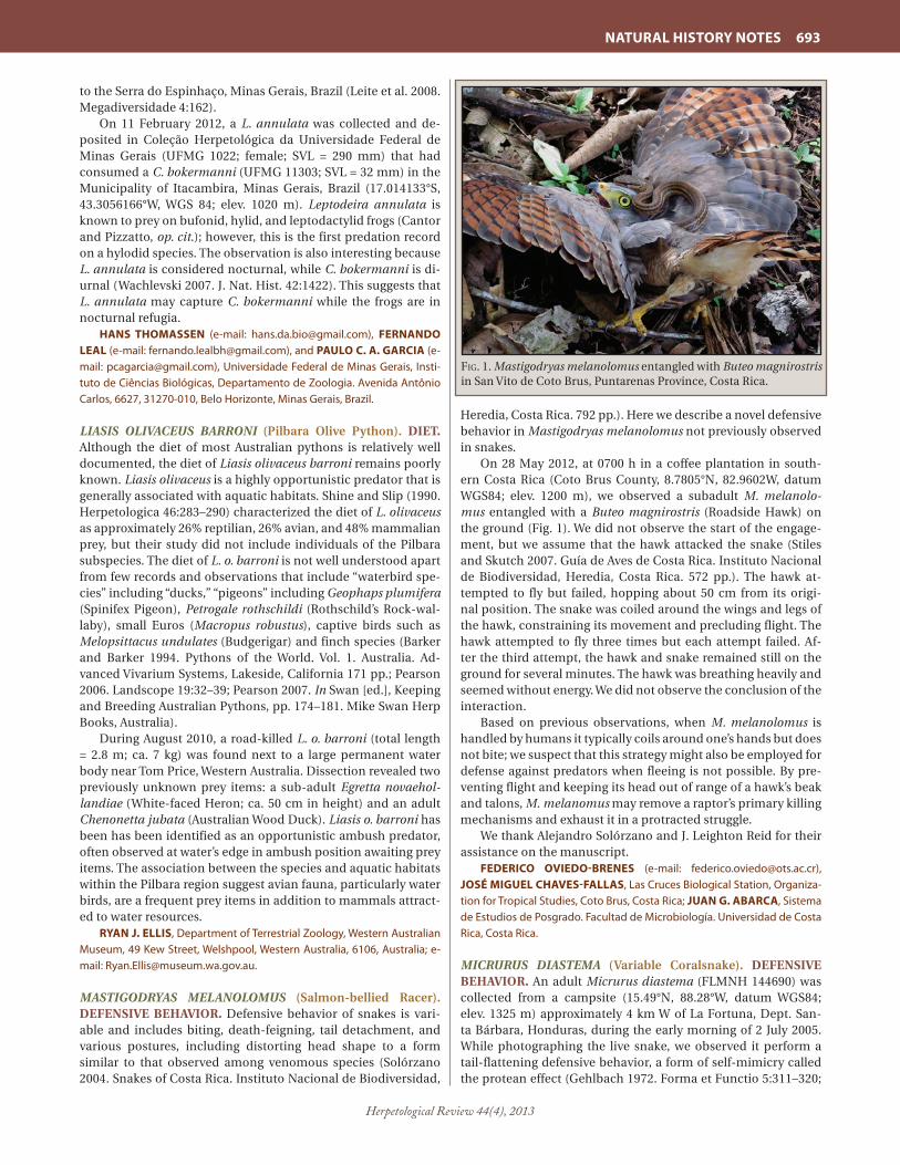
Herpetological Review 44(4), 2013
NATURAL HISTORY NOTES 693
to the Serra do Espinhaço, Minas Gerais, Brazil (Leite et al. 2008. Megadiversidade 4:162).
On 11 February 2012, a L. annulata was collected and de-posited in Coleção Herpetológica da Universidade Federal de Minas Gerais (UFMG 1022; female; SVL = 290 mm) that had consumed a C. bokermanni (UFMG 11303; SVL = 32 mm) in the Municipality of Itacambira, Minas Gerais, Brazil (17.014133°S, 43.3056166°W, WGS 84; elev. 1020 m). Leptodeira annulata is known to prey on bufonid, hylid, and leptodactylid frogs (Cantor and Pizzatto, op. cit.); however, this is the first predation record on a hylodid species. The observation is also interesting because L. annulata is considered nocturnal, while C. bokermanni is di-urnal (Wachlevski 2007. J. Nat. Hist. 42:1422). This suggests that L. annulata may capture C. bokermanni while the frogs are in nocturnal refugia.
HANS THOMASSEN (e-mail: [email protected]), FERNANDO LEAL (e-mail: [email protected]), and PAULO C. A. GARCIA (e-mail: [email protected]), Universidade Federal de Minas Gerais, Insti-tuto de Ciências Biológicas, Departamento de Zoologia. Avenida Antônio Carlos, 6627, 31270-010, Belo Horizonte, Minas Gerais, Brazil.
LIASIS OLIVACEUS BARRONI (Pilbara Olive Python). DIET. Although the diet of most Australian pythons is relatively well documented, the diet of Liasis olivaceus barroni remains poorly known. Liasis olivaceus is a highly opportunistic predator that is generally associated with aquatic habitats. Shine and Slip (1990. Herpetologica 46:283–290) characterized the diet of L. olivaceus as approximately 26% reptilian, 26% avian, and 48% mammalian prey, but their study did not include individuals of the Pilbara subspecies. The diet of L. o. barroni is not well understood apart from few records and observations that include “waterbird spe-cies” including “ducks,” “pigeons” including Geophaps plumifera (Spinifex Pigeon), Petrogale rothschildi (Rothschild’s Rock-wal-laby), small Euros (Macropus robustus), captive birds such as Melopsittacus undulates (Budgerigar) and finch species (Barker and Barker 1994. Pythons of the World. Vol. 1. Australia. Ad-vanced Vivarium Systems, Lakeside, California 171 pp.; Pearson 2006. Landscope 19:32–39; Pearson 2007. In Swan [ed.], Keeping and Breeding Australian Pythons, pp. 174–181. Mike Swan Herp Books, Australia).
During August 2010, a road-killed L. o. barroni (total length = 2.8 m; ca. 7 kg) was found next to a large permanent water body near Tom Price, Western Australia. Dissection revealed two previously unknown prey items: a sub-adult Egretta novaehol-landiae (White-faced Heron; ca. 50 cm in height) and an adult Chenonetta jubata (Australian Wood Duck). Liasis o. barroni has been has been identified as an opportunistic ambush predator, often observed at water’s edge in ambush position awaiting prey items. The association between the species and aquatic habitats within the Pilbara region suggest avian fauna, particularly water birds, are a frequent prey items in addition to mammals attract-ed to water resources.
RYAN J. ELLIS, Department of Terrestrial Zoology, Western Australian Museum, 49 Kew Street, Welshpool, Western Australia, 6106, Australia; e-mail: [email protected].
MASTIGODRYAS MELANOLOMUS (Salmon-bellied Racer). DEFENSIVE BEHAVIOR. Defensive behavior of snakes is vari-able and includes biting, death-feigning, tail detachment, and various postures, including distorting head shape to a form similar to that observed among venomous species (Solórzano 2004. Snakes of Costa Rica. Instituto Nacional de Biodiversidad,
Heredia, Costa Rica. 792 pp.). Here we describe a novel defensive behavior in Mastigodryas melanolomus not previously observed in snakes.
On 28 May 2012, at 0700 h in a coffee plantation in south-ern Costa Rica (Coto Brus County, 8.7805°N, 82.9602W, datum WGS84; elev. 1200 m), we observed a subadult M. melanolo-mus entangled with a Buteo magnirostris (Roadside Hawk) on the ground (Fig. 1). We did not observe the start of the engage-ment, but we assume that the hawk attacked the snake (Stiles and Skutch 2007. Guía de Aves de Costa Rica. Instituto Nacional de Biodiversidad, Heredia, Costa Rica. 572 pp.). The hawk at-tempted to fly but failed, hopping about 50 cm from its origi-nal position. The snake was coiled around the wings and legs of the hawk, constraining its movement and precluding flight. The hawk attempted to fly three times but each attempt failed. Af-ter the third attempt, the hawk and snake remained still on the ground for several minutes. The hawk was breathing heavily and seemed without energy. We did not observe the conclusion of the interaction.
Based on previous observations, when M. melanolomus is handled by humans it typically coils around one’s hands but does not bite; we suspect that this strategy might also be employed for defense against predators when fleeing is not possible. By pre-venting flight and keeping its head out of range of a hawk’s beak and talons, M. melanomus may remove a raptor’s primary killing mechanisms and exhaust it in a protracted struggle.
We thank Alejandro Solórzano and J. Leighton Reid for their assistance on the manuscript.
FEDERICO OVIEDO-BRENES (e-mail: [email protected]), JOSÉ MIGUEL CHAVES-FALLAS, Las Cruces Biological Station, Organiza-tion for Tropical Studies, Coto Brus, Costa Rica; JUAN G. ABARCA, Sistema de Estudios de Posgrado. Facultad de Microbiología. Universidad de Costa Rica, Costa Rica.
MICRURUS DIASTEMA (Variable Coralsnake). DEFENSIVE BEHAVIOR. An adult Micrurus diastema (FLMNH 144690) was collected from a campsite (15.49°N, 88.28°W, datum WGS84; elev. 1325 m) approximately 4 km W of La Fortuna, Dept. San-ta Bárbara, Honduras, during the early morning of 2 July 2005. While photographing the live snake, we observed it perform a tail-flattening defensive behavior, a form of self-mimicry called the protean effect (Gehlbach 1972. Forma et Functio 5:311–320;
Fig. 1. Mastigodryas melanolomus entangled with Buteo magnirostris in San Vito de Coto Brus, Puntarenas Province, Costa Rica.

Herpetological Review 44(4), 2013
694 NATURAL HISTORY NOTES
Roze 1996. Coral Snakes of the Americas: Biology, Identification, and Venoms. Krieger Publishing, Malabar, Florida. xii + 328 pp.). The snake flattened its yellow-banded tail and extended the pos-terior portion of its body, moving the tail in a manner similar to typical anterior/head movement (erecting the tail while flatten-ing it to appear like a head), while simultaneously tucking its actual yellow-banded head under its body (Fig. 1). This type of defensive behavior has been seen from numerous genera within the Elapidae, including Micrurus surinamensis (Roze, op. cit.) but this is the first documented case from M. diastema. The behav-ior serves several purposes, including predator distraction (Roze, op. cit.).
We thank Roberto Downing M. for arranging the logistics of our work, and helping to obtain collection and export per-mits. Conrado Gonzáles, Martha Moreno, Ibrahim Padilla, and Carla Cárcamo of the Departamento de Areas Protegidas y Vida Silvestre (DAPVS) of the Administración Forestal del Estado Corporación Hondureña de Desarrollo Forestal (AFE-COHDE-FOR), Honduras, generously provided permission to collect and export scientific specimens. Fieldwork was supported by Op-eration Wallacea and the Reptile and Amphibian Conservation Corps (RACC).
BROOKE L. TALLEY, Department of Zoology, Southern Illinois Uni-versity – Carbondale, Carbondale, Illinois 62901, USA (e-mail: [email protected]); LARRY D. WILSON Centro Zamorano de Biodiversidad, Escuela Agrícola Panamericana Zamorano, Departamento de Francisco Morazán, Honduras; JOSIAH H. TOWNSEND, Department of Biology, Indiana Uni-versity of Pennsylvania, Indiana, Pennsylvania 15705-1081, USA, and Cen-tro Zamorano de Biodiversidad, Escuela Agrícola Panamericana Zamorano, Departamento de Francisco Morazán, Honduras (e-mail: [email protected]).
MICRURUS NIGROCINCTUS (Central American Coralsnake). DIET. Micrurus nigrocinctus is a relatively common semi-fosso-rial snake that ranges from southern Mexico to northern Colom-bia; its diet is known to include caecilians, lizards, and snakes (Ray et al. 2012. Herpetol. Rev. 43:148). On 30 November 2012 at 0245 h an adult male M. nigrocinctus (SVL = 44.7 cm, total length = 52.0 cm; La MICA Biological Station/Team Snake Panama #2012-210) was found on the entrance road to El Copé, Coclé Province, Republic of Panama, between the communities of Las Tibias and El Copé (08.62343°N, 080.57100°W; datum WGS84). The M. nigrocinctus was attempting to feed on a dead female Erythrolamprus bizona (SVL = 33.7, total length = 39.0 cm; La MICA Biological Station/Team Snake Panama #2012-211), which was flattened and stuck to the road. When approached and
poked with a stick the M. nigrocinctus continued to try to ingest the E. bizona for approximately 1 min, after which the E. bizona was released. This is the first record of M. nigrocinctus attempt-ing to feed on a snake within the genus Erythrolamprus and of M. nigrocinctus attempting to feed on carrion. I thank La MICA Biological Station for logistical support, ANAM for permit (SE/A-22-12) and P. Santana for field assistance.
JULIE M. RAY, La MICA Biological Station, El Copé de La Pintada, Coclé Province, Republic of Panama; e-mail: [email protected].
MICRURUS TENER (Texas Coralsnake). ABERRANT PATTERN. Here we describe unusual dorsal blotches, or a “target” pattern, appearing on Micrurus tener, a species that normally bears a pattern of tri-colored rings. Aberrant pattern variations are occa-sionally reported in snakes; Bechtel (1995. Reptile and Amphib-ian Variants: Colors, Patterns, and Scales. Krieger Publ. Co., Mal-abar, Florida. 224 pp.) reviewed many examples. Strecker (1935. Baylor Univ. Bull. 38:1–69) reported exceptional color patterns in M. tener and Gloyd (1938. Herpetologica 1:121–124) reported a bite involving a melanistic specimen. On 6 October 2012 one of us (TW) found an aberrantly colored M. tener crossing Chandler Road (30.757137°N, 95.660561°W, datum WGS84; elev. 94 m) in Crabbs Prairie, Walker Co., Texas, USA. The snake (Fig. 1; adult female; 47.6 g), which exhibited scutellation patterns consistent with M. tener, was donated to Houston Zoo and accessioned into the live collection (HZI 27381).
The typical black, red, and yellow colors characteristic of the species are present, however, the pattern is far from typical. The head and neck are predominantly yellow, with the black re-stricted to a mask like pattern around the eyes and connecting through the rostral and adjacent scales. The black nuchal band is reduced to a dorsal blotch, barely in contact with the parietals, the center of which has faint flecks of red and yellow. The ground color of the body is red, with many scales being posteriorly tipped with black, but less so on the lateral scale rows. There are 17 black body “bands” appearing as blotches. Each blotch has the appearance of a target, the first (outer) ring being yellow and
Fig. 1. Micrurus diastema performing the first stages of tail-flattening defensive behavior by elongating its posterior to draw attention.
Fig. 1. A) Dorsal view of a Micrurus tener exhibiting an aberrant pat-tern; B) Ventral view showing the tail and portions of the body.
COLO
R RE
PRO
DU
CTI
ON
SU
PPO
RTED
BY
THE
THO
MA
S BE
AU
VAIS
FU
ND

Herpetological Review 44(4), 2013
NATURAL HISTORY NOTES 695
the second black, with sharply defined edges. The inner rings are red with a yellow center but, with indistinct edges bleed into one another. The venter is nearly uniform red with minute black specks, occurring on a few scales, and some yellow from the dor-sal blotches entering onto the extreme lateral edges of some ven-trals. The subcaudals are predominantly orange, with a “wash” of red appearing at the medium seams of the divided subcaudals. We thank Houston Zoo, Inc. for consent to report on this unusual specimen.
WILLIAM L. FARR, 11019 Wainfleet Lane, Houston, Texas 77096, USA (e-mail: [email protected]); THAD WARREN, 67 Chandler Road, Huntsville, Texas 77320, USA (e-mail: [email protected]).
NERODIA FLORIDANA (Florida Green Watersnake). DIET. Nerodia floridana, North America’s largest watersnake, occupies most of Florida, small parts of southern Alabama and Georgia, and a portion of southern South Carolina. Fish and adult frogs have been reported to comprise the bulk of its diet; however, it has also been known to consume salamanders, tadpoles, small turtles, and invertebrates (Ashton and Ashton 1981. Handbook of Reptiles and Amphibians of Florida. Part 1. The Snakes. Wind-ward Publ., Miami, Florida. 176 pp.). Here we report several new diet records for N. floridana.
In the course of long-term monitoring of aquatic snake pop-ulations on the U.S Department of Energy’s Savannah River Site (SRS), Aiken and Barnwell counties, South Carolina, USA, we documented the following diet items from individual N. flori-dana captured in plastic minnow traps: 6 June 2006 (33.286533°N, 81.488019°W, datum WGS84), Erimyzon sucetta (Lake Chub-sucker); 1 July 2006 (33.255185°N, 81.585210°W), Lepomis gu-losus (Warmouth) and Lithobates sphenocephalus (Southern Leopard Frog); 29 March 2007 (33.161047°N, 81.692606°W), two larval Ambystoma opacum (Marbled Salamander); 25 June 2008 (33.161047°N, 81.692606°W), two Acantharchus pomotis (Mud Sunfish; total length = 48 and 75 mm). On 2 July 2010, a N. flori-dana (SVL = 534 mm) was collected dead in hoop trap set for turtles at Pond 2 on the SRS (33.255185°N, 81.585210°W). Dis-section revealed two crayfish (species not determined). No addi-tional prey were found, indicating this was not a case of second-ary ingestion. Crayfish are considered a rare diet item for Nerodia spp. and have not been documented in N. floridana (Gibbons and Dorcas 2004. North American Watersnakes: a Natural His-tory. Univ. Oklahoma Press, Norman. 438 pp.). Finally, at 2025 h on 18 December 2012, an adult N. floridana (SVL = ~ 70 cm) was
seen consuming a large Ameiurus sp. (bullhead catfish; Fig. 1) at the edge of a wetland in Grassy Waters Preserve (26.816926°N, 80.174894°W), Palm Beach Co., Florida, USA.
PHILIP VOGRINC, JOHN D. WILLSON, University of Arkansas, De-partment of Biological Sciences, Fayetteville, Arkansas 72701, USA (e-mail: [email protected]); ANDREW M. DURSO, Utah State University, Logan, Utah 84322, USA; LARRY A. BRYAN, Savannah River Ecology Laboratory, PO Drawer E, Aiken, South Carolina 29803, USA; ZACH ROSS, New York University, New York, New York 10003, USA; JOSHUA HOLBROOK, Florida Atlantic University, Boca Raton, Florida 33431, USA; DON FILIPIAK, Fort Lauderdale, Florida 33301, USA.
NERODIA SIPEDON (Northern Watersnake). DIET. Although Gibbons and Dorcas (2004. North American Watersnakes: a Nat-ural History. Univ. Oklahoma Press, Norman. 438 pp.) listed eight species of ictalurid catfish that have been reported in the diet of Nerodia sipedon, their list did not include Pylodictis olivaris (Flathead Catfish). On 4 September 2012, we encountered an adult N. sipedon (total length = 95 cm) in the process of swallow-ing a juvenile P. olivaris (total length = 26 cm). They were found in shallow water along the shoreline of Pollander Lake, an Up-per Mississippi River backwater, immediately upstream from the spillway that extends from Lock and Dam 5A to the Minnesota shoreline, Winona Co., Minnesota, USA (44.080°N, 91.684°W; datum WGS 84). The snake had swallowed most of the head of the catfish, which was still alive when they were observed. Previ-ously, P. olivaris has been found in the diet of three other Nerodia species (Gibbons and Dorcas, op. cit.; Tyson et al. 2008. Herpetol. Rev. 39:472).
MARK A. ROSS (e-mail: [email protected]) and PHILIP A. CO-CHRAN (e-mail: [email protected]), Biology Department, Saint Mary’s University of Minnesota, 700 Terrace Heights, Winona, Minnesota 55987, USA.
PELAMIS PLATURA (Yellow-bellied Seasnake). REPRODUC-TION / MATING BEHAVIOR. The elapid seasnake Pelamis pla-tura has the widest distribution range of any snake, including most of tropical and subtropical Pacific and Indian oceans from Central America to Madagascar. Its reproductive cycle is considered to be continuous throughout the year (Ineich 1988. L’Année Biologique, 4ème sér. 27:93–117) but two birthing peaks have been reported in Costa Rica, one in December and January and another in July and August (Solorzano 2004. Serpientes de Costa Rica. Editorial INBio, Santo Domingo Heredia, Costa Rica. 791 pp.). Solorzano and Sasa (2011. Herpetol. Rev. 42:443–444)
Fig. 1. Nerodia floridana consuming an Ameiurus sp. (bullhead cat-fish) at Grassy Waters Preserve, Palm Beach Co., Florida, USA.
Fig. 1. Copulating Pelamis platura observed on 22 January 2012 off Puerto Escondido, Oaxaca, Mexico.
PHO
TO B
Y M
. DU
NE/
DO
RIS.
FFES
SM.F
R

Herpetological Review 44(4), 2013
696 NATURAL HISTORY NOTES
reported the first observation of mating behavior for the spe-cies. They observed copulating specimens floating at the sea surface in Costa Rica in August 2009. We here report a second copulation observation (Fig. 1) made in natural conditions off Puerto Escondido (Oaxaca, Mexico) ca. 4 km from the coast (15.60°N, 97.13°W, datum WGS84; 22 January 2012). Copulation occurred just below the water’s surface, as in the first observa-tion reported above, and lasted over 15 min; the diver (MD) left before its end. Both snakes moved repeatedly from vertical to horizontal position with their tails intertwined and most often maintained a vertical position. Water temperature was ca. 23°C. This is the second observation of mating behavior in the species and is consistent with a continuous or bimodal mating season in Central America.
We wish to thank the DORIS website (http://doris.ffessm.fr/) that enables information exchange among scientists, biologists, divers, and underwater photographers.
IVAN INEICH, Muséum National d’Histoire Naturelle, Département de Systématique et Évolution, UMR CNRS 7205 (Origine, Structure et Évolution de la Biodiversité), 25 rue Cuvier, CP 30, F-75005 Paris, France (e-mail : [email protected]); MICHEL DUNE, 304 rue Thimonnier, F-69400 Villefranche sur Saône, France (e-mail : [email protected]).
PHILODRYAS TRILINEATA (Argentina Mousehole Snake). DIET. Philodryas trilineata is a large (maximum length = 200 cm) species endemic to the Monte biogeographic region in western Argentina (Giraudo and Scrocchi 2002. Smithson. Herpetol. In-form. Serv. 132:1–53). The diet of P. trilineata is known to include birds, mammals, lizards, and other snakes; some individuals are known to hunt domestic fowl or rabbits (Cei 1993. Reptiles del Noroeste, Nordeste y Este de la Argentina. Herpetofauna de las Selvas Subtropicales, Puna y Pampas. Mus. Reg. Sci. Nat. Torino. 945 pp.; Laspiur et al. 2012. Herpetol. Rev. 43:151–152). Rodents are frequently reported as prey, but consistent data about diet of this species are lacking. Here we report the first vouchered case of predation by P. trilineata on Microcavia australis.
On 12 February 2011, at 1015 h, we collected an adult P. trilin-eata (total length = 1635 mm) near Puerto Madryn city, Chubut, Argentina (42.7894°S, 65.0049°W, datum WGS84; elev. 11 m). During transport to the laboratory, the snake regurgitated a par-tially digested juvenile M. australis (Fig. 1). Based on comparison with Microcavia specimens from Chubut (N = 33), we estimate a
total length of 130 mm for the prey. The snake and its prey were deposited in the Herpetological Collection LJAMM of Centro Nacional Patagónico (LJAMM-CNP 8236). We thank D. Udrizar Sauthier for Microcavia identification and revision of compara-tive material.
IGNACIO MINOLI (e-mail: [email protected]), CRISTIAN HER-NAN FULVIO PEREZ, CENPAT-CONICET, Boul. Alte. Brown 2915, Chubut, Argentina; DIEGO JANISH ALVARES, Laboratório de Herpetologia, De-partamento de Zoologia, Inst. Biociencias, UFRGS, Av. Bento Gonçalves, 9500, Porto Alegre, RS, Brazil; LUCIANO JAVIER AVILA, CENPAT-CONICET, Boul. Alte. Brown 2915, Chubut, Argentina (e-mail: á[email protected]).
PITUOPHIS CATENIFER AFFINIS (Sonoran Gophersnake). PREDATION. On 9 October 2012, 1400 h, one of us (ECB) found a juvenile Pituophis catenifer affinis (SVL ca. 500 mm) entangled with a Scolopendra heros (Giant Centipede; total length ca. 178 mm; photo voucher UAZ 57469-PSV) on a path near the west-ern base of Cathedral Rock near Sedona, Yavapai Co., Arizona, USA (34.886467ºN, 111.801529ºW, datum WGS84; elev. 1524 m). When found, the centipede was wrapped around the posterior two thirds of the snake. The anterior one third of the snake was free, outstretched in an attempt to escape. The animals were dis-entangled and it became apparent that the centipede was indeed preying upon the snake. The latter exhibited a sizable mid-dor-sal wound where the centipede had gnawed through the snake’s back. When freed the gophersnake moved slowly away while the centipede rapidly departed.
RANDALL D. BABB, Arizona Game and Fish Department, 7200 E. University Drive, Mesa, Arizona 85207, USA (e-mail: [email protected]); E. CHUCK BUSBOOM, 2625 S. Terrapin Circle, Mesa, Arizona 85209, USA (e-mail: [email protected]).
PITUOPHIS CATENIFER (Gophersnake). DIET. Pituophis cat-enifer is primarily a predator of rodents, but also takes birds, bird eggs, and lizards (Rodriguez-Robles 2002. Biol. J. Linn. Soc. 77:165–183). Here we describe the first confirmed predation by P. catenifer on Dipodomys ingens (Giant Kangaroo Rat), a federally and state listed (endangered) species. Pituophis catenifer have been observed within colonies of D. ingens, and use their bur-row systems as shelter (Williams and Kilburn 1991. Mammalian Species 377:1–7). On 12 December 2012 a D. ingens burrow sys-tem in the Carrizo Plain, San Luis Obispo Co., California, USA (35.3081°N, 119.8787°W; elev. 655 m) was being excavated for
Fig. 1. Philodryas trilineata and partially digested adult Microcavia australis from Chubut Province, Argentina.
Fig. 1. Scolopendra heros predating a young-of-year Pituophis catenifer.

Herpetological Review 44(4), 2013
NATURAL HISTORY NOTES 697
preservation purposes. A female P. catenifer (SVL = 989 mm; tail length = 144) sheltering within the burrow was inadvertently fa-tally injured during the excavation process. Upon examining the snake a prey bolus was found and extracted. The prey was deter-mined to be an adult male D. ingens based on hind foot length (48.8 mm). Predation on D. ingens by P. catenifer has long been suspected and our observation provides conclusive evidence. This predation event is also notable both due to the relative size of the prey item (88 adult male Giant Kangaroo Rats at this site averaged 123 g; s.d. ± 12.9) and the time of the year. Although winter days in the Carrizo Plain can afford clear skies and moder-ate daytime temperatures, offering opportunities for heliother-mic temperature regulation, nighttime temperatures frequently fall below 0°C, and prolonged periods of precipitation and cloud cover can occur. The anterior portion of this D. ingens, including the head, had been digested, suggesting that the snake had been able to maintain body temperatures sufficient to permit diges-tion despite the season. We thank M. Huising, M. Hemenez, and B. Windecker for their assistance. SunPower Corporation and NRG Energy provided additional support.
HOWARD O. CLARK, JR. (e-mail: [email protected]), KIRK W. SETSER, and ROBERT K. BURTON, H. T. Harvey & Associates, 7815 N Palm Ave., Suite 310, Fresno, California 93711-5511, USA.
PLIOCERCUS ELAPOIDES (Variegated False Coralsnake). RE-PRODUCTION / CLUTCH SIZE. Little information has been published on the reproduction of Pliocercus elapoides. However, a clutch of 4–8 eggs (August deposition) is known from northern Guatemala (Köhler 2003. Reptiles of Central America. Herpeton, Verlag Elke Köhler, Offenbach. Germany. 367 pp.; Lee 2000. A Field Guide to the Amphibians and Reptiles of the Maya World, The Lowlands of Mexico, Northern Guatemala, and Belize. Cor-nell Univ. Press. Ithaca, New York. 402 pp.). The natural history of this uncommon species in Mexico is poorly known.
On 22 April 2010 at 1827 h, we found a female P. elapoides (UAA-CV 0355, SVL = 350 mm; tail length = 220 mm) in a mon-tane cloud forest in the Sierra Madre Oriental at Alaquines, San Luis Potosí, México (22.122500°N, 99.504890°W, datum WGS84; elev. 1405 m). On 1 May 2010 at 0100 h (after nine days in cap-tivity) she deposited seven eggs in a depression in the sub-strate below leaf litter. The eggs averaged 15 mm in length (SE = 0.8164).
RUBÉN A. CARBAJAL-MÁRQUEZ, Centro de Investigaciones Biológi-cas del Noroeste, Instituto Politécnico Nacional No. 195, Col. Playa Palo de Santa Rita Sur, C.P. 23096, La Paz, Baja California Sur, México (e-mail: [email protected]); GUSTAVO E. QUINTERO-DÍAZ, Universidad Autónoma de Aguascalientes, Centro de Ciencias Básicas, Departamento de Biología, Avenida Universidad No. 940, Aguascalientes, Aguascalientes 20131, México; ZAIRA Y. GONZÁLEZ-SAUCEDO, Universidad Autónoma de Querétaro, Facultad de Ciencias Naturales, Cerro de las Campanas s/n, Querétaro, Querétaro 76017, México; GUILLERMO MARTÍNEZ-DE LA VEGA, Instituto de Investigación de Zonas Desérticas de la Universidad Autónoma de San Luis Potosí, Altair No. 200, Fracc. Del Llano, C.P. 78377, San Luis Potosí, SLP, México.
SPILOTES PULLATUS (Tiger Ratsnake). DIET. Spilotes pullatus is a diurnal generalist that typically feeds on small rodents, birds, and bird eggs (Hartmann et al. 2009. Pap. Avul. Zool. 49:343–360; Mendonça et al. 2011. Herpetol. Notes 4:425–427). On 11 Decem-ber 2012, at 2145 h, on the property of Chico Mendes Institute for Biodiversity Conservation, ICMBIO (7.38383°S, 39.3544°W; datum WGS 84) in the Araripe National Forest, State of Ceará,
Brazil, we observed an attempted predation by S. pullatus on the eggs of Turdus rufiventris (Rufous-bellied Thrush). Predation was not successful due to “mobbing” behavior of the adult T. ru-fiventris (Yamamoto and Ades 2002. Rev. Etol. 4:75–94), followed by intense vocalization away from their nest. This is the first re-cord of S. pullatus attempting predation on T. rufiventris.
JOSIVAN SOARES DA SILVA (e-mail: [email protected]), CARINA CARNEIRO DE MELO MOURA (e-mail: [email protected]), GERALDO JORGE BARBOSA DE MOURA (e-mail: [email protected]), Universidade Federal Rural de Pernambuco, Paleoherpe-tological and Herpetological Laboratory, UFRPE, Rua Dom Manoel de Me-deiros, Dois Irmãos - CEP: 52171-900, Recife, PE, Brazil.
STORERIA DEKAYI (Dekay’s Brownsnake). SEASONAL ACTIV-ITY. The annual activity period of Storeria dekayi varies with latitude (Ernst and Ernst. 2003. Snakes of the United States and Canada. Smithsonian Univ. Press, Washington, D.C. 668 pp.). Few data are available for northern populations, but Vogt (1981. Natural History of Amphibians and Reptiles of Wisconsin. Mil-waukee Public Museum, Milwaukee. 205 pp.) stated that they emerge in late April and are most often seen in May and October as they cross roads while moving to and from hibernacula. Here I present data on activity of S. dekayi from a population on the western side of Pheasant Branch Marsh, Dane Co., Wisconsin, USA (43.110°N, 89.493°W; datum WGS 84; Cochran 1988. Bull. Chicago Herpetol. Soc. 23:69–71).
On most days from mid-1981 to mid-1984, I drove Pheasant Branch Road along the slope of a ground moraine adjacent to marsh and observed S. dekayi on the road only during spring and fall (Fig. 1). The only three living individuals were recorded on 2 May, 11 May, and 5 October, consistent with the dates for the 25 dead snakes. Mean first sighting date for 1982–1984 was 26 April
Fig. 1. Dot plot showing dates on which Storeria dekayi were ob-served on Pheasant Branch Road, Dane Co., Wisconsin (N = 25 dead and 3 living snakes). The dashed vertical lines indicate 1 June and 1 September.
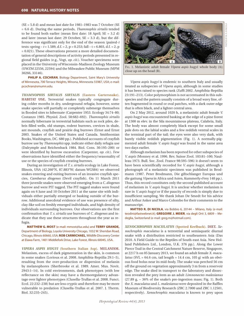
Herpetological Review 44(4), 2013
698 NATURAL HISTORY NOTES
(SE = 5.8 d) and mean last date for 1981–1983 was 7 October (SE = 6.6 d). During the same periods, Thamnophis sirtalis tended to be found both earlier (mean first date: 18 April; SE = 3.2 d) and later (mean last date: 29 October, SE = 3.3 d), but the dif-ference was significant only for the end of the season (paired-t tests: spring – t = 1.589, d.f. = 2, p = 0.253; fall – t = 6.803, d.f. = 2, p = 0.021). These observations present a more detailed documen-tation of general descriptions of activity periods presented in re-gional field guides (e.g., Vogt, op. cit.). Voucher specimens were placed in the University of Wisconsin-Madison Zoology Museum (UWZM 22556, 22594) and the Milwaukee Public Museum (MPM 30266, 33146).
PHILIP A. COCHRAN, Biology Department, Saint Mary’s University of Minnesota, 700 Terrace Heights, Winona, Minnesota 55987, USA; e-mail: [email protected].
THAMNOPHIS SIRTALIS SIRTALIS (Eastern Gartersnake). HABITAT USE. Terrestrial snakes typically congregate dur-ing colder months in dry, underground refugia; however, some snake species will partially or completely submerge themselves in flooded sites to hibernate (Carpenter 1953. Ecology 34:74–80; Costanzo 1985. Physiol. Zool. 58:682–692). Thamnophis sirtalis normally hibernate in terrestrial habitats such as rock piles, de-bris filled wells, old stumps, rodent burrows, crevices in shale, ant mounds, crayfish and prairie dog burrows (Ernst and Ernst 2003. Snakes of the United States and Canada. Smithsonian Books, Washington, DC. 680 pp.). Published accounts of crayfish burrow use by Thamnophis spp. indicate either daily refugia use (Dalrymple and Reichenback 1984. Biol. Cons. 30:195–200) or were identified by burrow excavation (Carpenter, op. cit.). Few observations have identified either the frequency/seasonality of use or the species of crayfish creating burrows.
During an investigation of T. s. sirtalis ecology in Lake Forest, Illinois, USA (42.260°N, 87.883°W; datum WGS84) we observed snakes entering and exiting burrows of an invasive crayfish spe-cies, Cambarus diogenes (Devil crayfish). On 11 March 2011 three juvenile snakes were observed emerging from a C. diogenes burrow and were PIT tagged. The PIT tagged snakes were found again on 6 June and 10 October 2011 at the same site with indi-viduals either partially emerged or basking outside of the bur-row. Additional anecdotal evidence of use was presence of silty, clay-like soil on freshly emerged individuals, and high density of individuals surrounding burrows. Our observations are the first confirmation that T. s. sirtalis use burrows of C. diogenes and in-dicate that they use these structures throughout the year as re-fugia.
MATTHEW G. MOST (e-mail: [email protected]) and TERRY GRANDE, Department of Biology, Loyola University Chicago, 1032 W. Sheridan Road, Chicago, Illinois 60660, USA; ROB CARMICHAEL, Wildlife Discovery Center at Elawa Farm, 1401 Middlefork Drive, Lake Forest, Illinois 60045, USA.
VIPERA ASPIS HUGYI (Southern Italian Asp). MELANISM. Melanism, excess of dark pigmentation in the skin, is common in some snakes (Lorioux et al. 2008. Amphibia-Reptilia 29:1–5), resulting from the over-production or dispersion of melanin by melanophores (Sherbrooke et al. 1989. Amer. Mus. Novit. 2943:1–14). In cold environments, dark phenotypes (with low reflectance on the skin) may have a thermoregulatory advan-tage over lighter phenotypes (Clusella-Trullas et al. 2008. Funct. Ecol. 22:232–238) but are less cryptic and therefore may be more vulnerable to predation (Clusella-Trullas et al. 2007. J. Therm. Biol. 32:235–245).
Vipera aspis hugyi is endemic to southern Italy and usually treated as subspecies of Vipera aspis, although in some studies it has been raised to species rank (Zuffi 2002. Amphibia-Reptilia 23:191–213). Color polymorphism is not accentuated in this sub-species and the pattern usually consists of a broad wavy line, of-ten fragmented in round or oval patches, with a dark outer edge that is often black, and a lighter central area.
On 2 May 2012, around 1020 h, a melanistic adult female V. aspis hugyi was encountered basking at the edge of a pine forest at 1300 m elev. in the Sila mountainous plateau, Calabria, Italy. The body was almost completely black except for some small pale dots on the labial scales and a few reddish ventral scales in the terminal part of the tail; the eyes were also very dark, with barely visible reddish pigmentation (Fig. 1). A normally-pig-mented adult female V. aspis hugyi was found in the same area two days earlier.
Although melanism has been reported for other subspecies of V. aspis (Monney et al. 1996. Rev. Suisse Zool. 103:81–100; Naul-leau 1973. Bull. Soc. Zool. France 98:595–596) it doesn’t seem to have been scientifically recorded for V. aspis hugyi, although a photograph of a melanistic specimen was published in Brod-mann (1987. Peter Brodmann, Die giftschlanger Europas und die gattung Vipera in Africa und Asien, Kummerly+Frey 148 pp.). Thus, this record constitutes only the second published account of melanism in V. aspis hugyi. It is unclear whether melanism is rare in V. aspis hugyi or if the paucity of records is simply due to insufficient sampling. We thank Johan De Smedt for his advice and Arthur Anker and Marco Colombo for their comments to the manuscript.
MATTEO R. DI NICOLA, via Bobbio 6, 20144 – Milano, Italy (e-mail: [email protected]); GREGOIRE J. MEIER, via degli Orti 3, 6809 – Me-deglia, Switzerland (e-mail: [email protected]).
XENOCHROPHIS MACULATUS (Spotted Keelback). DIET. Xe-nochrophis maculatus is a terrestrial and semiaquatic diurnal snake with a distribution restricted to southeastern Asia (Das 2010. A Field Guide to the Reptiles of South-east Asia. New Hol-land Publishers Ltd., London, U.K. 376 pp.). Along the Lower Pierce Trail in the Central Catchment Nature Reserve, Singapore, at 2217 h on 05 January 2013, we found an adult female X. macu-latus (SVL = 64.6 cm, tail length = 14.4 cm, 105 g) with an obvi-ous food bolus near its mid-body. The snake was perched 56 cm off the ground on vegetation approximately 3 m from a reservoir edge. The snake died in transport to the laboratory and dissec-tion revealed the prey item as an adult Limnonectes malesianus (27.89 g, ~ 36% of the snake’s pre-ingestion mass; Fig. 1). Both the X. maculatus and L. malesianus were deposited in the Raffles Museum of Biodiversity Research (ZRC 2.7000 and ZRC 1.12501, respectively). Xenochrophis maculatus is known to prey upon
Fig. 1. Melanistic adult female Vipera aspis hugyi: whole body (A); close up on the head (B).
PHO
TOS
BY M
. R. D
I NIC
OLA

Herpetological Review 44(4), 2013
699NATURAL HISTORY NOTES 699
frogs and fish (David and Vogel 1996. The Snakes of Sumatra: An Annotated Checklist and Key with Natural History Notes. Edi-tion Chimaira, Frankfurt, Germany. 260 pp.); however, we were unable to find any references documenting specific prey or prey size. We thank the National Parks Board of Singapore for issuing us work permit NP/RP11-03-1 and Kelvin K. P. Lim at the Raffles Museum of Biodiversity Research for accessioning specimens.
ALEX FIGUEROA, Department of Biological Sciences, University of New Orleans, New Orleans, Louisiana 70122, USA (e-mail: [email protected]); RYAN JAMES ROBERT McCLEARY, Department of Biological Sciences, National University of Singapore, Singapore 117543.
XENOPHOLIS SCALARIS (Wucherer’s Ground Snake). DE-FENSIVE BEHAVIOR. Xenopholis scalaris is a dipsadid snake found in leaf litter in humid tropical forests of Bolivia, Brazil, Peru, Ecuador, Colombia, Suriname, and French Guiana (Lehr and Doan 2013. In IUCN Red List of Threatened Species, ver-sion 2013.1. <www.iucnredlist.org>. Downloaded 5 March 2013). Defensive behavior of this species is relatively unknown. We observed defensive behavior of X. scalaris on three occasions in southern Bahia, Brazil. The observations occurred on 1) 17 Sep-tember 2012 at 2230 h in an area of Atlantic forest at Michelin Ecological Reserve (13.816667°S, 39.133333°W; datum SAD69),
Igrapíuna municipality; 2) 3 May 2009 at 1600 h on a trail be-tween a secondary forest and cocoa plantation at RPPN Serra Bonita (15.42387°S; 39.54736°W), Camacan municipality; and 3) 24 February 1992 at the Centro de Pesquisas do Cacau, CEPEC/CEPLAC (14.767492°S; 39.228062°W), Ilhéus municipality. In all observations, the snakes flattened their bodies, hid their heads, and remained motionless for several minutes (Fig. 1). The third observation was of two neonates. Although body flattening be-havior has been described for this species (Martins 1996. In K. Del Claro [ed.], Anais do XIV Encontro Annual de Etologia, pp. 185–199. Sociedade Brasileira de Etologia, Universidade Federal de Uberlândia, Brazil) this is the first time the head-hiding be-havior (Greene 1988. In Gans and Huey [eds.], Biology of the Rep-tilia, pp.1–152. Alan R. Liss, New York) has been noted.
CAIO VINICIUS DE MIRA MENDES, RENAN MANOEL DE OLIVEI-RA, DANILO SILVA RUAS, IURI RIBEIRO DIAS, and ANTÔNIO JORGE SUZART ARGÔLO (e-mail: [email protected]), Universidade Estadual de Santa Cruz – UESC, Campus Soane Nazaré de Andrade, Rodovia Jorge Amado, Km 16, Bairro Salobrinho, CEP 45662-900, Ilhéus, Bahia, Brazil.
Fig. 1. Xenochrophis maculatus with ingested Limnonectes malesia-nus in Central Catchment Nature Reserve, Singapore.
Fig. 1. Xenopholis scalaris from southern Bahia, Brazil, exhibiting flattening and head hiding defensive behavior.

Herpetological Review 44(4), 2013
700 BOOK REVIEWS
Herpetological Review, 2013, 44(4), 700–701.© 2013 by Society for the Study of Amphibians and Reptiles
Reptiles and Amphibians of the Pacific IslandsGeorge R. Zug. 2013. University of California Press, Berkeley (www.ucpress.edu). x + 306 pp., 35 pls, 5 maps. Hardcover. US $70.00. ISBN 9780520274952 (also available in Softcover and e-Book formats for US $34.95).
ALLEN ALLISONBishop Museum, 1525 Bernice Street,Honolulu, Hawaii 96817, USAe-mail: [email protected]
The Pacific Ocean is the world’s largest geographic feature, comprising about a third of the globe, but its tropical oceanic islands take up less than 0.05% of the Earth’s land surface, a collective area around the size of the U.S. state of West Virginia or the country of Costa Rica. However, these Pacific oceanic islands, most of which are tiny, include well
over half the world’s island total and are home to a diverse and fascinating herpetofauna.
Organizing the Pacific islands into regions has long vexed ge-ographers. In a highly influential work, de Brosses (1756) coined the term Polynesia, applying it to all the islands of the Pacific. Later, one of the great Pacific explorers, Dumont d’Urville (1832) subdivided the central and south Pacific into three contiguous ar-eas, Polynesia, Micronesia, and Melanesia, based on the physical and cultural attributes of the people. These terms remain in cur-rent use but are generally now regarded as somewhat simplistic.
Biogeographers have been less successful in dividing up the Pacific (Kay 1979), and have generally tried to force adjacent islands and island groups into regional assemblages based on faunal or floral relationships. The various proposed schemes remained stubbornly incongruent. In an insightful review, Stod-dart (1992) distinguished between different island types, viz., atolls, elevated limestone (makatea), and high islands. He pro-duced a scheme, based on the high islands, that remains broad-ly consistent with the biogeography of various Pacific taxa. He demonstrated for a number of taxa that New Guinea was a ma-jor source area for much of the Pacific, with species richness on high islands diminishing with distance from New Guinea (Stod-dart 1992). This pattern is very clear in the herpetofauna of the western Pacific (Allison 1993).
George Zug, in his outstanding and highly useful book, takes a very practical but somewhat unusual approach, dividing the Pacific into forty regional areas based on political and geograph-ic criteria. He also includes within his treatment five islands or island groups in the Eastern Pacific, Islas Revillagigedo, Isla del Coco, Isla Malpelo, Clipperton Island, and the Galapagos. While
this scheme tends to bring together taxa from very different evo-lutionary lineages, it helps to put the greater Pacific herpetofau-na into a clear regional perspective. Although there are no native species shared between the Eastern and Western Pacific, many of the wide-ranging human commensals, such as the geckos Hemi-dactylus frenatus and Lepidodactylus lugubris, now occur in both areas.
Overall, Zug treats a total of 187 taxa. Of these, 151 are native or probably native to some part of the Pacific (i.e., they didn’t dis-perse via human transport), and 35 are introduced. One species of skink, Carla tutela, endemic to Halmahera Island west of New Guinea, was once thought to have been introduced to Guam but those populations are genetically similar to Carla ailanpa-lai which is endemic to the Admiralty Archipelago north of New Guinea and may have been introduced to Guam during WWII (Austin et al. 2011).
In his preface Zug makes it clear that his intended readership is “casual to avid naturalists who live on or visit Pacific islands.” He also mentions that the book contains much new data and hopes that it will also be useful to professional herpetologists. In both regards he has succeeded admirably.
His introduction, which is barely six pages long, is one of the finest summaries I’ve ever encountered that puts the Pacific into a historical and regional perspective, starting with the geologi-cal history and ending with a discussion of patterns of human settlement.
Although this is a minor point in the context of this book, the introduction mentions Pangaea but not Gondwana. Inasmuch as the geological history of much of the Pacific, particularly the richest areas in the southwest, involved the breakup of Gond-wana following its separation from Pangaea, it might have been useful to mention a few general details about that.
I would have also liked to see a bit more about the navigation skills of the Pacific peoples as many of the reptiles that are wide-spread in the Pacific were transported by humans. It is increas-ingly clear that the early Polynesians had acquired much more than “elementary navigation skills” and were navigating across vast distances long before European explorers reached the area.
In the next section Zug explains his regionalization scheme for the Pacific, characterizing it as “arbitrary” and includes a checklist of species known to inhabit each of the major islands or island groups. This checklist will assist naturalists in gaining a familiarity with the species known from the various groups. It will also serve as something of a snapshot of current patterns of distribution and diversity. As Zug points out in his preface, mo-lecular studies have shown that a number Pacific species former-ly thought to be widespread, are actually complexes of closely re-lated taxa. As more groups are studied this subdivision of species will undoubtedly continue. In addition new species continue to be discovered.
The next section introduces terms used to identify species. I quite liked the fact that Zug explicitly defines what he means
BOOK REVIEWSBOOK REVIEWS

Herpetological Review 44(4), 2013
BOOK REVIEWS 701
by small, medium, midsize, etc. He gets by with a minimum of jargon. This would certainly not surprise those who know him.
The next 70 pages include 35 color plates, each presented on facing pages, and including virtually all species treated. The quality of the plates varies but generally they are excellent.
Most of the rest of the book, 221 pages, includes detailed ac-counts of the various species. Each major group, e.g., frogs, is treated in a brief introduction, followed by treatment of each family followed by a detailed, standardized account of each spe-cies. Details on the calls of the frogs (all but three of which are introduced) are included under reproduction. The accounts are excellent and include many useful historical and ecological de-tails. Although the book lacks an identification key, I don’t think that this is needed given the generally small number of species found in each area and the high quality of the images. In a few cases there are line drawings to facilitate the identification of similar species.
Each species treatment also includes a common name. Zug has generally chosen names that are in widespread use for well-known taxa but has coined many new names for more obscure taxa. These names clearly reflect Zug’s overall emphasis on prac-ticality. I was pleased to see that for species of skinks in the ge-nus Emoia he dispensed with the use of “emo skink,” a name that has somehow crept into the popular literature. Moreover, a quick Google search will quickly confirm that “emo” has many other meanings. Instead Zug focuses on physical features and habitat preferences that are diagnostic and easy for an avid naturalist to remember.
The book concludes with an overall checklist of species, each with a little square “tick” box in front of the scientific name fol-lowed by the common name, together with an appendix on the sources of illustrations and indexes to common and scientific names. I think that it would have been useful to also include an index to place names mentioned in the text but this is a minor point.
There do not appear to be any major typographical errors in the book. It is produced to a high standard of quality and is well organized and highly readable. Anyone with an interest in the Pacific, including naturalists, conservation biologists, biogeog-raphers or herpetologists, will find this book indispensable. In my view George Zug has encapsulated a big chunk of his life’s work into a masterpiece that should be on everyone’s bookshelf.
literature cited
allison, a. 1996. Zoogeography of amphibians and reptiles of New Guinea and the Pacific region. In A. Keast and S. E. Miller (eds.), The Origin and Evolution of Pacific Island Biotas, New Guinea to Eastern Polynesia, pp. 407–436. SPB Academic Publishing, Am-sterdam.
austin, c. c., e. n. rittmeyer, l. a. oliVer, J. o. andermann, g. r. Zug, g. h. rodda, and n. d. Jackson. 2011. The bioinvasion of Guam: infer-ring geographic origin, pace, pattern and process of an invasive lizard (Carlia) in the Pacific using multi-locus genomic data. Biol. Invasions 13:1951–1967.
de Brosses, c. 1756. Histoire des Navigations aux Terres Australes, Vol 2. Chez Durand, Paris. 513 pp.
kay, e. a. 1980. Little worlds of the Pacific. An essay on Pacific ba-sin biogeography. University of Hawaii Harold L. Lyon Arboretum Lecture 9:1–40.
d’urVille, J.s.c.d. 1832 Sur les îles du Grand Océan. Bull. Soc. Géo-graph.17:1–21.
stoddart, d. r. 1992. Biogeography of the tropical Pacific. Pacific Sci. 46:276–293.
Herpetological Review, 2013, 44(4), 701–702.© 2013 by Society for the Study of Amphibians and Reptiles
Lissemys punctata, the Indian Flap-shelled TurtleDieter Gramentz. 2011. Chimaira Buchhandelsgesellschaft mbH, Frankfurt am Main, Germany (www.chimaira.de). 278 pp. Hardcover. US $79.95 (Zoo Book Sales). ISBN 978-3-89973-496-6.
Rafetus, the Curve of ExtinctionPeter C.H. Pritchard. 2012. Living Art Publishing, Ada, Oklahoma (www.livingartpublishing.com). 174 pp. Hardcover. US $69.50. ISBN 978-0-9787556-9-0.
PETER A. MEYLANNatural Sciences, Eckerd College, 4200 54th Ave. S., St. Petersburg, Florida 33711, USAe-mail: [email protected]
It is great to see this focused interest in softshelled turtles. The Trionychidae is a family of bizarre reptiles whose members are easily recognized because they are so mor-phologically distinctive; but otherwise the group may be underappreciated. Each book treats a single species of softshell. These two species are at opposite ends of the softshell turtle spectrum—a small, common, and very terrestrial cyclanorbine and a rare, gi-gantic, and completely aquatic trionychine. And these volumes are at opposite ends of the spectrum of species treatments. The Gramentz volume strikes me as “Catalogue of American Amphibians and Reptiles ac-count” meets “Biology of the Reptilia.” It is a minutely detailed recounting of the exten-sive literature on a species that was listed on Appendix 1 of CITES in 1975 when it probably should not have been. The Pritchard volume is classic Pritchardian pontifica-tion. It has the subtitle, “The Story of the Giant Softshell Turtle of the Yangtze and Red Rivers.” A complex story it is; it is a very personal voyage through the author’s interactions with Rafe-tus swinhoei, one of those species whose living representatives might just be counted on the fingers of one hand. It was listed by CITES in 2005 (TRAFFIC 2013) when it should have been recog-nized as endangered at least 50 years ago. Despite the differences between the volumes a great deal of trionychid biology is packed into each one.
The Gramentz volume is a summary and compilation of the surprisingly extensive published literature on what I would ar-gue is the softshell that exhibits the most primitive morphology among living forms. It is astounding that the author identified 346 references that are relevant to understanding the biology of Lissemys punctata. It is clear from this work that Lissemys is an important model organism for biological research on the Indian subcontinent. In contrast, much of the Pritchard book is a sum-mary of his observations on the very poorly known species, Rafe-tus swinhoei, from the literature, interviews, and observations

Herpetological Review 44(4), 2013
702 BOOK REVIEWS
made during numerous travels. Pritchard establishes for the first time that it is a truly giant species and he verifies that it is on the brink of extinction. But he goes far afield with chapters on the or-igin of turtles, a chapter on museums, and a chapter in which he considers candidates for the rarest turtle. Whereas the Lissemys volume is tightly organized with an extensive bibliography, the Rafetus volume is disorganized, and amazingly, its bibliophile author has not provided a literature cited!
Although I have played a supporting role in understanding the genus Rafetus and I have tried to keep up with developments in its conservation, I found the Pritchard volume to be tough to follow at times and then repetitive at others. It would have ben-efited from a more critical peer review, serious editing, and re-organization. My sense is that it was probably published pretty much as it was first drafted. Furthermore, it is not clear who the target audience is. If it were the scientific and conservation com-munities, it seems that it would have seen more attention in re-view and a bibliography would have been added. However, the way it is written it assumes that the reader is familiar with softs-hell turtle literature, some of which is quite obscure (i.e., Heude 1880).
The Lissemys volume will prove valuable to anyone doing comparative biology that involves turtles. As I have said else-where (Meylan 1991), it seems unfortunate that the biology of “the turtle” is too frequently based on Trachemys scripta. This Lissemys volume should prove useful in making more readily ac-cessible the voluminous literature on a very different lineage of cryptodire. The volume also makes it quite clear that Lissemys has very interesting biology for a trionychid. It is kinosternid-like in that it is the only living softshell that regularly aestivates on land for a significant portion of the year and it seems to have em-bryonic diapause so that clutches laid at different times all hatch with the same monsoon rains. The reproductive biology of this species and its control appear to be remarkably well understood.
Although the Lissemys volume is better organized and documented, it is the Rafetus volume that covers new ground. Pritchard makes the very important work of Heude (1880) avail-able in English for the first time. He lists all of the known speci-mens of this elusive species including many that he discovered via careful examination of museum collections in China and Vietnam. He establishes its giant size and makes a good argu-ment that the largest turtles, living or extinct, may have been softshells. He identifies the most likely areas where additional living R. swinhoei might survive. Also, in covering all aspects of the genus Rafetus, he does a reasonable job of summarizing what is known about the smaller species in the genus, R. euphra-ticus, which could also use some serious conservation attention.
In the section in which he argues for R. swinhoei being the rarest of all turtles, it becomes clear that collectors of rare turtles are a real problem. It is also clear why this one is so rare, and it fits a pattern that Pritchard buries in his rambling introduction, that big animals are high on the curve of extinction. But you have to be really interested in softshelled turtles to make it through this book. It’s not that the author gets off track; it seems that there often is no track. So, I am not sure to whom I could recommend this book beyond turtle aficionados. For biologists and libraries, the Lissemys volume is likely to be more useful, if only as a guide back to the original literature which one hopes would be cited in place of this volume in most cases.
It is interesting that Lissemys was CITES listed in 1975 but has still served as a model species. It is still harvested in large numbers and undoubtedly has suffered declines throughout its
range (what turtle species has not?), but appears to have biologi-cal properties that make it resilient. The giant R. swinhoei, on the other hand, is dedicated to river channels and oxbows. It may turn up a few more times; but only the most extraordinary mea-sures will prolong the existence of this species. What other large softshells do we need to look at as quickly as possible to deter-mine their true conservation status? The rest of the diversity of the Cyclanorbinae is in Africa and all four taxa are poorly known. The largest of them, Cyclanorbis elegans, has apparently become very hard to find. Is it the next Rafetus?
literature cited
heude, P. m. 1880. Mémoire sur les Trionyx. Mémoires Concernant l’Histoire Naturelle de l’Empire Chinois par des Pères de la Com-pagne de Jésus, vol. 1. Mission Catholique, Chang-Hai. 38 pp.
meylan, P. a. 1991. Review of: Turtles of the World by C. H. Ernst and R. W. Barbour, 1989. Q. Rev. Biol. 66:90.
tRAFFIC. 2013. Proposal (A): Inclusion of the following species of the Family Trionychidae in Appendix II: Aspideretes leithii, Dogania subplana, Nilssonia formosa, Palea steindachneri, Pelodiscus axenaria, P. maackii, P. parviformis, and Rafetus swinhoei (B): Transfer of the following species from Appendix II to Appendix I: Chitra chitra and Chitra vandijki www.traffic.org/cites-cop-papers/CoP16_Prop38_Analysis.pdf (accessed 17 August 2013).
Herpetological Review, 2013, 44(4), 702–704.© 2013 by Society for the Study of Amphibians and Reptiles
The Eponym Dictionary of AmphibiansBo Beolens, Michael Watkins, and Michael Grayson. 2013. Pelagic Publishing, Exeter, United Kingdom (www.pelagicpublishing.com). xiii + 244 pp. Hardcover. US $49.99. ISBN 978-1-907807-41-1.
AARON M. BAUERDepartment of Biology, Villanova University, 800 Lancaster Avenue,Villanova, Pennsylvania 19085, USAe-mail: [email protected]
Scientific names are sources of endless fascination for professionals and amateurs alike: Who was that frog named after? Is that smithii named for Malcolm or Hobart? Here are the answers. Beolens et al. here com-plete the cataloguing of tetrapod eponyms, having previously considered birds (Beolens and Watkins 2003), mammals (Beolens et al. 2009), and reptiles (Beolens et al. 2011).
This is a compact volume of double-columned text. Although it appears sturdy at first glance, the book is not as well produced as the authors’ reptile eponym work, although it is also consid-erably less expensive. The text is not illustrated, but the cover of the book is graced by a striking image entitled Frog Cabinet by Madeline von Foerster. It depicts 13 frog species (only five of which have eponymous names) in the cubbies of a cabinet, with two closed drawers meant to represent “all the frog species not yet discovered, and those who might die out before becoming known to us” (p. ii).
As in the reptile volume, a brief introduction explains the “rules” applied in the book. Accounts are provided, in alphabeti-cal order, for those persons who are commemorated by either scientific names or common names (the latter, unfortunately,

Herpetological Review 44(4), 2013
BOOK REVIEWS 703
based on Frank and Ramus (1995), which coined many arbitrary, incorrectly derived, and inappropriate names). Also included are other nouns that have served as the source of names that might be mistaken as being eponyms. For example, Hyla andersonii is named for Anderson, South Carolina and Psychrophtynella il-limani for Nevado Illimani, a mountain in Bolivia. Not surpris-ingly, many of the same people have been commemorated in the names of both amphibians and reptiles, thus a good number of biographic entries in this book were also in The Eponym Diction-ary of Reptiles, some repeated nearly verbatim and others signifi-cantly expanded or shortened.
I enjoyed this book and learned a lot from it. However, I found it unsatisfying on two levels. First, the authors’ choice of biographic details often seems arbitrary. Authorship of a single publication is often mentioned, but this is as likely to be a minor paper as it is to be the subject’s most important and well-known treatise. Examples include Carlo Gené, whose most important herpetological work (Gené 1839), the first treatise of the amphib-ians and reptiles of Sardinia, is not cited, G. K. Noble, whose in-fluential book Biology of the Amphibia (Noble 1931) is not men-tioned, J. G. Schneider, none of whose numerous herpetological books are noted, and Ronald Crombie, who is cited as the author of a list of Smithsonian herpetological publications rather than for his original work on the West Indies, the Philippines, or Palau.
One might argue that more comprehensive biographies are warranted for lesser known people, however, it seems a bit un-balanced to allocate only eight lines to Carl Gans, for decades one of the world’s leading herpetologists, whereas Philippe Brunneau de Miré and Sir Basil Thomson, both fascinating in their own right, but not herpetological giants, are accorded 36 and 44 lines respectively—among the most extensive entries in the book.
An inherent problem of the style “so-and-so is at such-and-such an institution” is that a retirement or death immediately makes the account seem outdated. Many of those honored by amphibian eponyms have indeed done one or the other since their biographies were written. For example, Hobart Smith, Paolo Vanzolini, Eugenio Izecksohn, and Jorge Jim, all treated as living in the book, have all since passed away, although, to be fair, all but Jim (who died in 2011) were still living when the book went to press. A more difficult to explain and embarrassing mis-take is listing Robert Stebbins as having passed away in 2000, 13 years before his actual demise.
A large number (although a small percentage) of the approxi-mately 1600 entries include factual errors of one sort or another. For example, Thomas Bell was not an assistant to J. E. Gray (Adler 1989), Ernst Ahl was never the director of the Berlin Museum (Paepke 2013), Mario Peracca did not visit the Cape Verde Islands (Andreone and Gavetti 2007), and Giacomo Doria was the found-er of a museum in Genoa, not Turin (Adler 2012). According to Boelens et al., the Russian scientist Leopold Schrenk “believed that mammoths which he found preserved in the permafrost must have died recently, and thought that they were subterra-nean animals that ate earth!” (p. 193). In fact, Schrenk (1871), as part of his studies of Siberian mammoths, recorded these as the beliefs of Arctic peoples; they were not his own views.
At least one entry appears to refer to the wrong person. That for Wagner cites Johann Andreas Wagner as the person com-memorated in the name Leptodactylus wagneri (Peters, 1862); however, Wilhelm Peters, in fact, named this frog for Friedrich Johann Carl Mortiz Wagner (better known as Moritz Wagner), who collected the holotype in Ecuador (Wagner et al. 2012).
Boelens et al. (2011) also linked the name of the snake Diapho-rolepis wagneri Jan, 1863 to Johann Andreas Wagner, but it too was collected by Moritz Wagner. Its description does not indicate the source of the name, but Moritz is again the likely honoree. I caught these errors because I have worked on both the speci-mens described by Peters and those collected by Moritz Wagner, but I wonder how many other misattributed eponyms I may have missed!
Among contemporary herpetologists, institutions and time-lines are sometimes incorrect. For example, Fritz Jürgen Obst was trained in the German Democratic Republic and spent his entire professional career in Dresden (formerly in East Germa-ny), not partly in the west, and Robert C. Drewes has worked at the California Academy of Sciences since 1970, not 1998 as stated by Boelens et al. American university names seem to have been especially confused, as Richard Thomas is stated to have received his doctorate from the University of Louisiana [sic, Lou-isiana State University] and Victor Twitty’s undergraduate degree is stated to be from Butler College, Princeton University [it was from Butler University in Indianapolis].
A number of eponyms are noted as having uncertain origin, but at least some are quite easily determined. For example, I was easily able to find the meaning of the specific epithet of Pseu-doeurycea tlilicxitl, the black-footed salamander, in an online Nauhatl dictionary — not surprisingly it means black foot. Other names of unknown derivation could have perhaps been checked by asking the authors of these names themselves, as was clearly done in the case for some names (e.g., those proposed by Anna-marie Ohler that honor members of her family).
Typographical errors are relatively few, e.g., Port Elisabeth [sic, Elizabeth] (p. 84) and Alfred Russel Wallace’s Brazilian ex-pedition began in 1848 not 1948 (p. 227), but seem to plague accounts of German namesakes: Ein Frauenfahrt urn [sic, um] die Welt (p. 101), Ostafrikanischen Insen [sic, Inseln], Detley [sic, Detlef] Langer (p. 119).
Boelens et al. provide the authors and dates of the Latin names they present. At one level this is helpful, but it would have been more useful if they had followed the convention of placing author and date in parentheses if the specific epithet is not used in conjunction with the generic name with which it was origi-nally paired.
On the positive side, in this book we learn a little about a lot of people, some quite obscure, who have been immortalized in nomenclature. Among some of the more interesting entries are those that reveal that Allobates algorei honors former American vice president Al Gore, that Hyla carnifex is named after John Lynch (carnifex = hangman, to hang unlawfully is to lynch, get it?), and that two African frog names have epithets derived from the name of Charles Lemaire, who may have been the inspira-tion for the central character of Kurtz in Joseph Conrad’s Heart of Darkness (although other people have also been proposed as Conrad’s model for Kurtz).
On the whole, The Eponym Dictionary of Amphibians leaves a lot to be desired. However, it is the only book that does what it does. Relatively detailed biographies of herpetologists may be found in Kraig Adler’s Contributions to the History of Herpetol-ogy series, but no other book provides thumbnail sketches of all those who have had amphibians named after them (including a very large number of non-herpetologists). For those with an interest in the history of herpetology, a curiosity about etymol-ogy, or an abiding love of trivia, this book will make an enjoyable read, if not a reliable reference.

Herpetological Review 44(4), 2013
704 BOOK REVIEWS
literature cited
adler, k. 1989. Herpetologists of the past. In K. Adler (ed.), Contribu-tions to the History of Herpetology. SSAR Contributions to Her-petology 5, pp. 5–142. Society for the Study of Amphibians and Reptiles, Ithaca, New York.
———. 2012. Herpetologists of the past, Part 3. In K. Adler (ed.), Con-tributions to the History of Herpetology, Vol 3. Contributions to Herpetology 29, pp. 9–386. Society for the Study of Amphibians and Reptiles, Vancouver, Canada.
andreone, F., and e. gaVetti. 2007. The Herpetological Contributions of Mario Giacinto Peracca (1861–1923). Society for the Study of Amphibians and Reptiles, Villanova, Pennsylvania. 570 pp.
Beolens, B., and m. Watkins. 2003. Whose Bird? Christopher Helm/A. and C. Black, London. 400 pp.
———, ———, and m. grayson. 2009. The Eponym Dictionary of Mammals. Johns Hopkins University Press, Baltimore, Maryland. xiii + 574 pp.
———, ———, and ———. 2011. The Eponym Dictionary of Reptiles. Johns Hopkins University Press, Baltimore, Maryland. xiii + 296 pp.
Frank, n., and e. ramus. 1995. A Complete Guide to Scientific and Common Names of Reptiles and Amphibians of the World. N G Publishing Inc., Pottsville, Pennsylvania. 377 pp.
gené, J. 1839. Synopsis Reptilium Sardiniae Indigenorum. Mem. Reale Accad. Sci. Torino, ser. II, 1:257–288, [1], 5 pls.
noBle, g. k. 1931. Biology of the Amphibia. McGraw-Hill Book Com-pany, New York. xiii + 577 pp.
PaePke, h.-J. 2013. Ernst Ahl (1898–1945), sein Leben und sein Bedeu-tung für die Herpetologie und Terrarienkunde. Sekretär 13(2):15–46.
schrenk, l. 1871. Bericht über neuerdings im Norden Sibirien’s ange-blich zum Vorschein gekommene Mammuthe, nebst Bemerkun-gen über den Modus der Erhaltung und die vermeintliche Häu-figkeit ganzen Mammuthleichen. St. Pétersbourg Acad. Sci. Bull. 16:col. 147–173.
Wagner, P., a. m. Bauer, and W. Böhme. 2012. Amphibians and reptiles collected by Moritz Wagner, with a focus on the ZFMK collection. Bonn Zool. Bull. 61:216–240.
Herpetological Review, 2013, 44(4), 704–705.© 2013 by Society for the Study of Amphibians and Reptiles
Herpetofauna of Armenia and Nagorno-KarabakhMarine S. Arakelyan, Felix D. Danielyan, Claudia Corti, Roberto Sindaco, and Alan E. Leviton. 2011. Society for the Study of Amphibians and Reptiles. Ithaca, New York. iii + 149 pp. Hardcover. US $40.00. ISBN: 978-0-916984-84-7.
STEVEN C. ANDERSONUniversity of the Pacific, Stockton, California 95211, USAand California Academy of Sciences, San Francisco, California 94118, USAe-mail: [email protected]
Although the Caucasus Ranges have been designated a biodiversity hotspot, there are no recent reviews of the her-petofauna and literature of Armenia or Nagorno-Karabakh, two of the southern-
most republics of that region. Indeed, most people in the West would be hard put to place a finger on them on a map. This slim volume was designed to fill that gap. Nagorno-Karabakh
is surrounded by Armenia, apart from its southern border, which abuts Iran. Armenia is also bordered by Iran; it lies between Azerbaijan to the east, Georgia in the north, and Turkey to the west. The history of Nagono-Karabakh is com-plicated; the current government considers it to be an inde-pendent republic, while Armenia and Azerbaijan both claim the territory. It is currently an unsettled dispute. The region has previously been claimed by Persia, the Russian Empire, and the Soviet Union. Internationally, it is considered a prov-ince of Azerbaijan, although its ethnic composition is pri-marily Armenian.
The first four authors accomplished the field work over some two decades; the editing and bibliography reveal the sure hand of Leviton. The book is traditional in its layout, the taxonomic treatment preceded by a discussion of the geog-raphy of the region, climate, biodiversity, conservation, and habitats. The authors recognize five zones, based on vegeta-tion types, and five azonal habitats. A table correlates each species with these categories; thus much important informa-tion is presented in an economical space. These zones and habitats are described in the text. There are two pages of paleontological notes by David Vasilyan indicating that the modern Armenian herpetofauna dates back at least to the Upper Miocene. For those of us interested in the history of herpetology, there is a section on the history of herpetology of Armenia, similar in structure to those appearing in other SSAR publications.
The heart of the book consists of the taxonomic section, beginning with keys to the herpetofauna, based on easily seen external characters. The major characters are shown in photographs of actual specimens, rather than the more familiar generalized line drawings. Such a presentation has both costs and benefits, but the photographs are clear and show the characters effectively without too much distract-ing extraneous information. The species accounts each be-gin with the Latin name and author and an English common name. This is followed by a primary synonymy applying to specimens from Armenia and Nagorno-Karabakh, the type locality, notes on taxonomy, general distribution, distribu-tion in the study area, general information, and a status of conservation statement. Importantly, and rare in taxonomic publications, each species account contains a section listing the references applying to the individual species. Lacking from a taxonomist’s point of view, are the museum registry number of the holotype, a citation to the publication of the original description, a diagnosis, and lists of collecting and observation localities. However, these omissions are second-ary to the intended purpose of the book, which is an intro-duction of the fauna and useful to the general reader and to non-herpetology specialists. In any case, the extensive bib-liography will lead the interested researcher and collection curator to this and other information.
Following the text, there are 152 figures in 32 excellent color plates showing each species, a distribution map with unnamed and unnumbered spot localities, these followed by ecozone and habitat photographs. The bibliography alone covers 28 pages. It attempts to cover all publications covering the study area, from the earliest literature to that of the year of publication. The format is similar to that used in Leviton and Anderson (2010).
A trivial complaint perhaps, but Table 1 lists six reptile species endemic to Armenia. Endemism has no biological

Herpetological Review 44(4), 2013
BOOK REVIEWS 705
significance when applied to national boundaries. On the other hand, it may be useful politically in grant proposals for government funds, protecting national fauna, etc.
Those of us who are taxonomists know that taxonomy is a moving target, as generic and species names change all-too-frequently, and common names have never been stabilized as they are for birds. The recognition of family and higher cat-egories also changes, especially frequently today with the ad-vent of phylogenetic and cladistic classifications. This makes the publication of synonymies and bibliographies especially important. This is exemplified by the following quote from the current book:
Bufo variabilis, because it is considered a member of the “viridis” complex, was transferred to the new genus Pseude-pidalea when they established it for the Bufo viridis complex (Frost et al. 2006). However, Dubois and Bour (2010.23) ar-gue that this is invalid because, in their view, it creates no-menclatural confusion with respect to the application of the nomen Bufo. Thus, they conclude, Pseudepidalea must be considered a junior objective synonym of Bufotes Rafin-esque 1815, a subgenus they include within Bufo, and which would include viridis and would include all species included in the Bufo viridis, complex, or, if treated as a genus, then , e.g. Bufotes viridis, Bufotes variabilis, et al. (Dubois and Bour 2010:24–25).
Anyone confused yet? In any case, Dubois and Bour (2010) not withstanding, many or most taxonomists have accepted the nomenclature of Frost et al. (2006), if prematurely. This leaves West Asian Bufo viridis with an entirely new name, Pseudipedalea variabilis, which does not occur in combina-tion in the literature prior to 2006. So much for nomencla-tural stability.
This book creates a new template for subsequent similar volumes in local and national taxonomic accounts. It needs a place in all collection libraries and in the hands of field biolo-gists, reserve managers, and others with responsibilities for the fauna of Armenia and/or Nagorno-Karabakh.
literature cited
duBois, a., and r. Bour. 2010. The nomenclatural status of the nomina of amphibians and reptiles created by Garsault (1764), with a par-simonious solution to an old nomenclatural problem regarding the genus Bufo (Amphibia, Anura), comments on the taxonomy of this genus, and comments on some nomina created by Laurenti (1768). Zootaxa (2447):1–52.
Frost, d. r. et al. 2006. The amphibian tree of life. Bull. Amer. Mus. Nat. Hist. 297:1–370.
leViton, a. e., and s. c. anderson. 2010. The herpetological literature for Southwestern Asia. Occas. Pap. California Acad. Sci. 157. 638 pp.
Herpetological Review, 2013, 44(4), 705–708.© 2013 by Society for the Study of Amphibians and Reptiles
Texas Amphibians. A Field GuideBob L. Tipton, Terry L. Hibbitts, Troy D. Hibbitts, Toby J. Hibbitts, and Travis J. LaDuc. 2012. The University of Texas Press, Austin, Texas (http://utpress.utexas.edu/). xiv + 309 pp. Softcover. US $24.95. ISBN 978-0-292-73735-8.
CHRISTOPHER J. BELLDepartment of Geological Sciences,The University of Texas at Austin,Austin, Texas 78712, USAe-mail: [email protected]
For decades, the herpetofauna of Texas captured the attention of both profes-sional and avocational herpetologists. The remarkably diverse herpetological biota of Texas is, at least in part, a reflection of the diversity of biotic provinces that are united geopolitically in that vast state. To judge by the books and monographs that were avail-able historically, especially those readily available to the public, the point of emphasis for Texas herpetology traditionally was centered on the snakes. Among the earliest of the modern vol-umes on the herpetology of Texas was a pocket field guide to the snakes of Bexar County (Davenport, 1943). It was soon followed by the first of many editions and impressions of the popular booklet on venomous snakes by Werler (e.g., 1950, 1953, 1978), and later by works that addressed medical aspects of venomous snakes in Texas (Huang et al. 1975), focused on the famous rattle-snake ‘round-ups’ in the state (Kilmon and Shelton 1981; Adams and Thomas 2008), and provided updates to Werler’s work (Price 1998, 2009). More expansive volumes that provided coverage of non-venomous as well as venomous species appeared by 1965 (Raun 1965), and were substantial and widely available by the mid 1980s (Tennant 1984, 1985, 2006; Vermersch and Kuntz 1986; Dunlap 1994; Werler and Dixon 2000). The popular appeal of snakes in the state remains undiminished, and is reflected to-day by the wide availability in garden shops, nurseries, and even coffee shops of a waterproof, quick-reference guide (Pustejovsky 2010). The snakes of Texas have been well served, and served in style, in the literature.
To be sure, amphibians and reptiles were treated collectively in some detail in other volumes, but early summaries were more technical, were intended for specialists, and generally were pre-sented as relatively dry annotated lists without (Brown 1950) or with (Raun and Gehlbach 1972) maps, but devoid of pho-tographs and descriptions (another such list was published by Kutac and Caran in 1994). Dixon’s volume on amphibians and reptiles (1987, 2000; see also Dixon et al. 2007) provided a wel-come change and included expanded discussion of natural his-tory data, up-to-date taxonomic keys, distribution maps (with ranges documented at the county level), extensive review of the literature, and photos of select species in black-and-white for-mat. The latter were helpful, but still lacked the appeal that so often drew the non-specialist to the natural history section of the (now almost defunct) local book store.
Departure from that traditional state of affairs began with the volume produced by Garrett and Barker (1987) who ushered in a new tradition of publishing non-technical field guides with

Herpetological Review 44(4), 2013
706 BOOK REVIEWS
descriptions, color photographs, and distribution maps. The vol-ume by Bartlett and Bartlett (1999) was an important update in a similar format. The recently released third edition of Amphib-ians and Reptiles of Texas by Dixon (2013) brings together for all readers a balanced coverage of non-technical text, maps, color photographs, and an extensive review of the literature on the herpetofaunal species of Texas. It certainly will serve to increase awareness of, and interest in, the herpetofauna of the state. But it is not easily carried in the field.
I have heard colleagues decry the field guide as a dying genre of published literature. Some view that demise as collateral dam-age in the broader and general decline of print media in favor of readily portable digital devices. Others apparently interpret concerns about the decline in outdoor activities among mod-ern children (Louv 2008) as a harbinger of doom for the pocket guide. I admit that my proclivities tend towards an accumulation of books, and I embrace the accusatory labels of ‘fuddy-duddy’ and ‘walking anachronism’ that are proffered by students and colleagues who stand with jaws agape in disbelief when they learn that I do not carry a cell phone (“How do you text?”) or tab-let or other digital devices of our modern age. I also welcome the proximity of those same companions when they gaze over my shoulder as I pull out a field guide to aid in the identification of a darting damselfly, and am happy when they ask to thumb the volume on their own. Field guides clearly continue to serve the many purposes they have served for decades, as handy quick-references, teaching aides, inexpensive navigation guides for the budding naturalist who is grappling for the first time with the complexities of some small component of the world’s biodiver-sity, and succinct guides for the professional or specialist explor-ing a new geographic area or a new taxonomic group.
Texas Amphibians is the latest release in the Texas Natural History Guides series published by The University of Texas Press, and is the third in that series to embrace a herpetological subject matter. It was preceded, perhaps unsurprisingly, by two volumes addressing snakes; one by Dixon and Werler (2005) and another by Price (2009). The volumes in the series are produced with an emphasis on a portable format that still permits (and, indeed, promotes) quality imagery. The book is sized for easy transport; at 4.5 in x 7.5 in I can slide this field guide into the back pocket of my jeans, drop it in a vest pocket, or carry it effortlessly in a back-pack or daypack. The volume sells for $24.95, but a 33% discount is available for purchases made through the website for the press (http://utpress.utexas.edu/index.php/books/tiptep), thus drop-ping the cost to only $16.72.
The size of the book conforms to the others in the series, and that placed some limits on format, content, and presentation of data. The end product suffers little from those restrictions, and is a handy and helpful compendium that is both informative and easy to use. A four-page table of contents is followed by a brief foreword from James Dixon, who sets the book in the context of both geography and conservation. The 328 numbered pages include introductory sections on the basic biology of amphib-ians, conservation concerns (with Texas exemplars placed in the broader context of global amphibian declines), notes and tips on how to observe and photograph amphibians, an informative dis-cussion of the laws and regulations that govern the collection of amphibians in Texas, notes on captive maintenance, comments on taxonomic practices adopted by the authors, keys to the sala-mander and frog species of Texas, a generic key to amphibian larvae, and systematic accounts of 72 species (30 salamanders and 42 anurans) with known records from Texas, to which are
appended accounts of an additional six species that possibly oc-cur in the state, but require verification. The bulk of the text (234 pp.) is dedicated to the systematic accounts. A list of state and U.S.-based international herpetological societies is provided in an appendix, along with notes on three museums, and some relevant web sites. The final sections of the book are dedicated to a glossary of terms, a brief bibliography, and indices of com-mon and scientific names. The outer edges of the inside of the front and back covers provide rulers in inches and centimeters, respectively.
A few black-and-white illustrations are provided in the keys to highlight anatomical features, but the book is stunningly adorned with 177 color photographs, all but one of which depict living animals (the sole exception is a photograph depicting the photography set-up used by the authors in the field). The photos are good or excellent throughout, and the majority are sized to fit 1/3 of the height of the page. Most species are depicted in two or more photographs, permitting ready visualization of pattern or color variations, or anatomical details in close-up views. There is at least one photograph for all but two species; the exceptions are an as-yet unnamed species of Eurycea from the Pedernales River, and Eurycea robusta (known only from the holotype, an image of which was subsequently published by Dixon 2013).
A map depicting geographic distribution is provided for each of the 72 species known to occur within Texas. The maps are of a standard size, county boundaries of the state are depicted, and geographic range is indicated by gray-scale shading. Care ap-pears to have been taken to depict the known (or inferred) range of the species as accurately as possible, given the scale of the maps (i.e., shading, does not conform solely to county boundar-ies). Geographic ranges of nominal subspecies are not differenti-ated. Several species of Eurycea are documented only from local-ized springs that would be minuscule if depicted on maps of this scale; for those, standard 1 cm dots are centered on the known locality or localities. The only obvious error in the maps is that the range of the ‘Rio Grande’ species of Siren was copied onto the map for Siren intermedia (see discussion by Dixon [2013] for a re-view of the taxonomic complications surrounding the two taxa, and a map for comparison).
Species accounts are organized taxonomically and according to a standardized format, with text grouped under the sequen-tial headings Size, Description, Similar Species, Distribution, Natural History, Reproduction, Subspecies (when relevant), and Comments and Conservation. Both scientific and common names are provided at the beginning of each account, and with few exceptions follow Crother (2008). Only the common name is provided in the photo captions, but the photos are conveniently placed in close proximity to the text for each species. This format is common to all volumes in The University of Texas Natural His-tory Guides series, and represents a significant stylistic difference from the traditional Peterson Field Guide series, in which taxon illustrations were, until recently, placed in a collection of plates bound near the center of the volume (e.g., Conant and Collins 1991). A style somewhat similar to the Peterson one is followed by the Audubon guides, in which images are clustered at the front of the book, with text and maps in the back (e.g., Behler and King 1988). I confess to a degree of ambivalence about these varying organizational styles; each serves a purpose and a particular category of user, and I have found myself at different times, and with different guides, variably favoring each style over the other, depending often upon the relative degree of my ignorance with the subject matter. Perhaps the harmonious accommodation
Herpetological Review 44(4), 2013
BOOK REVIEWS 707
of both styles in the newer editions of the Peterson Series (e.g., Conant and Collins 1998) is the way to achieve maximum utility in the service of the broadest possible community.
The third edition of Dixon’s Amphibians & Reptiles of Texas (2013) was published after the release of Texas Amphibians (2012). The two volumes ultimately will serve different (but over-lapping) audiences, and some comparative comments are in or-der. At the time of publication of Texas Amphibians, the photos it contained were unique, but 11 of them were reprinted by Dixon (2013). The duplicated images are of Ambystoma tigrinum, Eu-rycea latitans, Eurycea quadridigitata, Eurycea tridentifera, No-tophthalmus viridescens, Pseudacris clarkii, Pseudacris crucifer, Gastrophryne carolinensis, Scaphiopus hurteri, Lithobates sphe-nocephala, and Eleutherodactylus planirostris. Interestingly, and somewhat disturbingly, in the case of three shared images, the county-level provenience information provided in the two vol-umes is different (Pseudacris clarkii, either Sterling County or Iri-on County; Pseudacris crucifer, either Jasper County or Houston County; Gastrophryne carolinensis, either Houston County or Anderson County). In twelve other instances, the images in the two volumes are clearly of the same individual animals, taken on the same habitat setting, presumably in a single photo-shoot, but the animals have slightly different orientations or positions (Ambystoma maculatum, Eurycea naufragia, Eurycea sosorum, Desmognathus auriculatus; Necturus beyeri, Notophthalmus viridescens, Anaxyrus debilis; Hyla chrysoscelis, Hyla cinerea, Gastrophryne olivacea; Hypopachus variolosus, and Lithobates grylio). The similarity in photos undoubtedly stems from the fact that photos for Dixon’s volume were provided by Toby Hibbitts, one of the authors of Texas Amphibians. In almost all cases, the images published by Tipton et al. are of a sharper quality and have greater clarity than the images reproduced in Dixon’s book. Taxonomic coverage in the two volumes is nearly identical, with equal treatment of the anurans, but the Tipton et al. volume cov-ers two additional and unnamed species of Eurycea.
Any book published on amphibians in recent years has ad-dressed a taxonomic group that now is experiencing declining populations across the globe, and challenging threats to persis-tence. One of the unique features of the Texas amphibian fauna is the taxonomic diversity of Eurycea in the state. Many of these salamanders are tied to the karst geology and hydrology of the eastern edge of the Edwards Plateau. The morphologic, genetic, and taxonomic diversity among these salamanders continues to be a topic of intense exploration, and the elucidation of even basic natural history data for many species remains an elusive goal. The extremely localized geographic distribution of many of the species makes them a particularly challenging group, both for basic biological study, and for conservation efforts. Many of these issues are necessarily touched upon in this field guide, and special attention was called to the conservation status of the various species. Since the publication of Texas Amphibians, the conservation status of at least four species has changed. Eurycea tonkawae is now listed as threatened by the U.S. Fish & Wildlife Service. Eurycea waterlooensis now is listed as endangered by the same agency, and the listing status of both Eurycea chishol-mensis and Eurycea naufragia now is ‘proposed endangered’ (see U.S. Fish & Wildlife Service species profiles at http://ecos.fws.gov/speciesProfile/profile/speciesProfile.action?spcode=D02T, ...=D033, ...=D02X, and ...=DO2S, respectively). As further testi-mony to the ever-changing picture on Texas amphibians, I note that the final book manuscript was sent to press a few weeks be-fore the massive Bastrop County fire swept through important
habitats for the Houston toad, Anaxyrus houstonensis (Travis La-Duc, pers. com. Sept., 2013); the impact on toad populations has not yet been fully assessed. These issues only serve to enhance the importance of this book, especially for the young people of the state of Texas who will utilize this guide to acquaint them-selves with what may well be a rapidly changing component of their native biota.
In some respects, this book may considered to be long over-due. The only other book dedicated solely to the amphibians of Texas is a field guide to the anurans of Big Bend National Park (Dayton et al. 2007). The volume by Tipton et al. clearly fills an obvious need for a portable, low-cost guide to the amphibians of Texas. Indeed, with the appearance of this volume, lizards now stand as the most prominent group still in need of greater at-tention and documentation (lizard lovers in Texas may decorate their shelves only with Frank Blair’s 1960 monograph on Scelo-porus olivaceus, Vermersch’s 1992 volume documenting species from the south-central portion of the state, a more expansive regional guide that covers Texas and the southwest [Jones and Lovich 2009], and Axtell’s detailed distribution maps issued in 30 parts over approximately 20 years [e.g., Axtell 1987, 2005]).
The quality photographs in Texas Amphibians enhance the value of the book for non-specialists, and as the most up-to-date summary of the known amphibian fauna of Texas, it is of direct interest to all professional herpetologists. Beyond its obvious utility as a reference guide in the field, the book also will serve as an excellent introduction to the amphibians of Texas and will be useful as a classroom resource for K-12 teachers who seek useful tools to shape lesson plans and discussions about biodiversity, conservation, and natural resources of Texas. The low cost of this volume should, I think, guarantee a broad circulation, and serves as an additional enticement towards the maintenance of print media. Although internet photos are available for many (not all) of the species discussed in this book, they are sometimes of low quality, may be difficult to compare efficiently, and, if your eyes are as bad as mine, are almost always difficult to appreciate on sunny days when viewed on hand-held screens (and at night those screens destroy your night-vision).
literature cited
adams, c. e., and J. k. thomas. 2008. Texas Rattlesnake Roundups. Tex-as A&M University Press, College Station, Texas. 113 pp.
axtell, r. W. 1987. Sceloporus cyanogenys. Interpretive Atlas of Texas Lizards 2:1–7, 1 map.
———. 2005. Uta stansburiana Baird and Girard. Interpretive Atlas of Texas Lizards 30:1–17, Map 16.
Bartlett, r. d., and P. P. Bartlett. 1999. A Field Guide to Texas Reptiles & Amphibians. Gulf Publishing Company, Houston, Texas. 331 pp.
Behler, J. l., and F. W. king. 1988. The Audubon Society Field Guide to North American Reptiles and Amphibians. Alfred A. Knopf, Inc., New York, New York. 743 pp.
Blair, W. F. 1960. The Rusty Lizard. A Population Study. University of Texas Press, Austin, Texas. 185 pp.
BroWn, B. c. 1950. An Annotated Check List of the Reptiles and Am-phibians of Texas. Baylor University Press, Waco, Texas. 259 pp.
conant, r., and J. t. collins. 1991. A Field Guide to Reptiles and Amphibians. Eastern and Central North America. Third Edition. Houghton Mifflin Company, Boston, Massachusetts. 450 pp.
———, and ———. 1998. A Field Guide to Reptiles & Amphibians. Eastern and Central North America. Third Edition, Expanded. Houghton Mifflin Company, Boston, Massachusetts. 616 pp.
crother, B. i. (ed.). 2008. Scientific and Standard English Names of Amphibians and Reptiles of North America North of Mexico, with

Herpetological Review 44(4), 2013
708 BOOK REVIEWS
Comments Regarding Confidence in Our Understanding. Sixth Edition. Society for the Study of Amphibians and Reptiles, Herpe-tological Circular 37. 84 pp.
daVenPort, J. W. 1943. Field Book of the Snakes of Bexar County, Texas and Vicinity. A Simplified Key and Notes on Their Behavior. Witte Memorial Museum, San Antonio, Texas. 129 pp.
dayton, g. h., r. skiles, and l. dayton. 2007. Frogs & Toads of Big Bend National Park. Texas A&M University Press, College Station, Texas. 51 pp.
dixon, J. r. 1987. Amphibians and Reptiles of Texas with Keys, Taxo-nomic Synopses, Bibliography, and Distribution Maps. Texas A&M University Press, College Station, Texas. 434 pp.
———. 2000. Amphibians and Reptiles of Texas. Texas A&M Univer-sity Press, College Station. 421 pp.
———. 2013. Amphibians & Reptiles of Texas. With Keys, Taxonomic Synopses, Bibliography, and Distribution Maps. Third Edition, Revised & Updated. Texas A&M University Press, College Station, Texas. 447 pp.
———, t. J. hiBBitts, and m. r. J. Forstner. 2007. Texas Herpetological Literature and County Records 2000 to 2006–07. Texas Herpetol. Soc. Publ. 1:1–58.
———, and J. e. Werler. 2005. Texas Snakes. A Field Guide. The Uni-versity of Texas Press, Austin, Texas. 364 pp.
dunlaP, J. 1994. Slitherin’ Around Texas. A Field Guide for People Who Don’t Like Snakes. Republic of Texas Press, Wordware Publishing, Inc., Plano, Texas. 177 pp.
garrett, J. m., and d. g. Barker. 1987. A Field Guide to Reptiles and Amphibians of Texas. Texas Monthly Press, Austin, Texas. 225 pp.
huang, t. t., s. r. leWis, and s. B. lucas, iii. 1975. Venomous snakes. In M. D. Ellis (ed.), Dangerous Plants, Snakes, Arthropods & Marine Life of Texas, pp. 123–166. U.S. Department of Health, Education, and Welfare, Public Health Service. U.S. Government Printing Of-fice, Washington, D.C.
Jones, l. l. c., and r. e. loVich (eds.). 2009. Lizards of the American Southwest. A Photographic Field Guide. Rio Nuevo Publishers, Tucson, Arizona. 567 pp.
kilmon, J., and h. shelton. 1981. Rattlesnakes in America and a Histo-ry of the Sweetwater Jaycees Rattlesnake Roundup. Shelton Press, Sweetwater, Texas. 234 pp.
kutac, e. a., and s. c. caran. 1994. Birds & Other Wildlife of South Central Texas. The University of Texas Press, Austin, Texas. 203 pp.
louV, r. 2008. Last Child in the Woods. Saving Our Children from Na-ture-Deficit Disorder. Updated and Expanded. Algonquin Books of Chapel Hill, Chapel Hill, North Carolina. 391 pp.
Price, a. h. 1998. Poisonous Snakes of Texas. Texas Parks and Wildlife Press. 112 pp.
———. 2009. Venomous Snakes of Texas. A Field Guide. The Univer-sity of Texas Press, Austin, Texas. 116 pp.
PusteJoVsky, c. 2010. Snakes of Central Texas. A Guide to Common and Notable Species. Quick Reference Publishing, Inc., Austin, Texas. 1 foldout, laminated sheet.
raun, g. 1965. A Guide to Texas Snakes. Texas Mem. Mus. Notes 9. 85 pp.
raun, g. g., and F. r. gehlBach. 1972. Amphibians and Reptiles in Tex-as. Taxonomic Synopsis, Bibliography, County Distribution Maps. Dallas Mus. Nat. Hist. Bull. 2:1–61, 140 maps.
tennant, a. 1984. The Snakes of Texas. Texas Monthly Press, Inc., Aus-tin, Texas. 561 pp.
———. 1985. A Field Guide to Texas Snakes. Gulf Publishing Com-pany, Houston, Texas. 260 pp.
———. 2006. Texas Snakes. Third Edition. Lone Star Field Guide, Tay-lor Trade Publishing, Lanham, Maryland. 263 pp.
Vermersch, t. g. 1992. Lizards and Turtles of South-Central Texas. Ea-kin Press, Austin, Texas. 170 pp.
———, and r. e. kuntZ. 1986. Snakes of South-Central Texas. Eakin Press, Austin, Texas. 137 pp.
Werler, J. e. 1950. The poisonous snakes of Texas and the first aid treatment of their bites. Texas Game and Fish 8:25–41.
———. 1953. The Poisonous Snakes of Texas and the First Aid Treat-ment of Their Bites. Texas Game and Fish Comm.Bull. 31:1–40. [Reprinted from February, 1950 issue of Texas Game and Fish].
———. 1978. Poisonous Snakes of Texas. Revised. Texas Parks Wildlf. Dept. Bull. 31:1–53.
———, and J. r. dixon. 2000. Texas Snakes: Identification, Distribution, and Natural History. University of Texas Press, Austin, Texas. 437 pp.
Herpetological Review, 2013, 44(4), 708–709.© 2013 by Society for the Study of Amphibians and Reptiles
Amphibians of Eastern Europe. Part 1. Order CaudataYevgen Pysanets. 2012. Yevgen Pysanets, Kiev, Ukraine (available through amazon.com). 207 pp. Softcover. US $29.00. ISBN 978-966-02-6514-1.
AARON M. BAUERDepartment of Biology, Villanova University, 800 Lancaster Avenue,Villanova, Pennsylvania 19085, USAe-mail: [email protected]
This book deals with the salamander fauna of Eastern Europe, defined as the European portions of the former Soviet Union. Ten species in six genera in the families Hynobiidae and Salamandridae are treated in the dual Ukrainian/English text. Ten species may not seem like very many, but the area cov-ered by the book has a continental climate with great tempera-ture extremes and some of the taxa covered barely extend into the region. For example, Salamandra salamandra, Ichthyosaura alpestris, and Lissotriton montandoni all enter Eastern Europe only in the Ukrainian Carpathians.
As this is intended as the first volume in a larger work on the amphibians of the region, it actually includes some more com-prehensive features, such as illustrated keys to both the sala-manders and frogs of Eastern Europe. The scientific names and authorities of these 30 amphibian species, along with their ver-nacular names in Ukrainian and English, are provided in a table. Threats to and conservation measures for amphibians are also provided in tabular form, as is a summary of frog and salaman-der conservation status (including listings by IUCN, the Bern Convention, and national red lists).
Other general information is included in a 17-page section providing biographies of zoologists who have contributed to the study of the Eurasian herpetofauna. These span five centuries from Conrad Gessner to Heinz Wermuth and draw heavily for both text and images from Kraig Adler’s biographies in the SSAR Contributions to the History of Herpetology series.
The heart of the book is its species accounts. These follow very abbreviated family and genus comments and include sub-sections dealing with taxonomy, distribution, karyology, sexual dimorphism, morphology and coloration, life history, repro-duction, development, behavior, and diet. In the account for Salamandrella keyserlingii the English text is about one page in length, whereas the Ukrainian is about six times longer. The dis-crepancy in other accounts is not as marked but if one wishes to get the fullest picture of a particular species, it may be necessary for non-Ukrainian readers to use Google Translate.

Herpetological Review 44(4), 2013
BOOK REVIEWS 709
Accounts are each illustrated by a shaded distribution map (within Eastern Europe only), habitat photos, and photos of breeding males and females, as well as larvae, paedomorphs, non-breeding adults, and color or pattern variants, as appro-priate. These are all in color and of high quality, although their printed size is quite small (mostly less than 80 mm in maximum dimension).
For the most part, the English portions of the book are not ac-tual translations, but rather extended synopses of the Ukrainian content. This can be seen, for example, on page 26, where pre-, pro- and climax metamorphosis are discussed in Ukrainian, but not in English. In other places the English captions for certain figures do not provide all of the information given in the Ukrai-nian (p. 27), and some figures are not referenced in the English text at all (e.g., figures 7–9 in the section dealing with reproduc-tion on pages 18–19). Principal components figures on pages 162–163 have English captions, but the relevant information printed on the plots themselves (the localities of sampled Lis-sotriton) are only in Ukrainian (except for two Romanian place names). Page headers are given only in Ukrainian. In most cases Ukrainian section headers are followed by English, and the as-sociated text starts with the English version, then the Ukrainian, but in the section on the basics of amphibian systematics start-ing on page 51 the text is entirely in Ukrainian except for English figure legends. In the taxonomic accounts, Ukrainian comes first for families and genera, but species accounts start with English.
Translation is an issue in this book and typos are rife in the English text. The map on page 10, for example, incorrectly trans-lates “western” as “eastern.” Genera is rendered as “genuses” (p. 79), taxa as “taxons” (p. 86), marked as “market” (p. 86), and en-dangered as “indangered” (p. 153). Wording is awkward in some places, but understandable. Only on page 69 was I truly unable to decipher the English text. On page 145 the English figure cap-tions are incorrect, having been duplicated from those on page 147. An errata sheet has been glued inside the back cover of the book, but none of the examples noted above are included.
One of the book’s most useful features is its extensive (16 pp.) literature cited, with about 325 entries, arranged with references written in languages using the Cyrillic alphabet first, followed
by those using the Roman alphabet. Many references, particu-larly those published in smaller regional journals, are likely to be unfamiliar to readers outside the region. Coverage appears to be quite good, especially for Ukraine, which is both home to the author and the richest region for salamanders within the scope of the book. Unfortunately, there are many typos in this section as well (e.g., Univercity), and Latin names are often not italicized. There is no index to the book in either language.
Salamanders of Eastern Europe have been treated in a num-ber of other books that are available in English, most notably Kuzmin’s (1999) Amphibians of the Former Soviet Union. Al-though this is now getting rather out of date, Kuzmin (2013) has just released a new volume with the same title. Kuzmin’s work is both broader in scope and much more massive than that of Pysanets, but also considerably more expensive. Detailed species accounts, though again dated, also appear (in German) in the salamander volumes of Handbuch der Reptilien und Amphibien Europas (Grossenbacher and Thiesmeier 1999, 2003; Thiesmeier and Grossenbacher 2004). Despite numerous language and edi-torial issues, Pysanets’ book provides a compact, affordable, and up-to-date overview of the caudate fauna from the Carpathians to the Urals and from the Arctic Sea to the Caucasus.
literature cited
grossenBacher, k., and B. thiesmeier (eds.). 1999. Handbuch der Rep-tilien und Amphibian Europas. Band 4/I. Schwanzlurche I. AULA-Verlag, Wiesbaden. pp. 1–406 + [1].
———, and ——— (eds.). 2003. Handbuch der Reptilien und Am-phibian Europas. Band 4/IIA. Schwanzlurche IIA. AULA-Verlag, Wibelsheim. [6] + pp. 407–758.
kuZmin, s. 1999. The Amphibians of the Former Soviet Union. Pensoft Publishers, Sofia. vi + 538 pp.
———. 2013. The Amphibians of the Former Soviet Union. Pensoft Publishers, Sofia. 384 pp. + CD.
thiesmeier, B., and k. grossenBacher (eds.). 2004. Handbuch der Reptilien und Amphibian Europas. Band 4/IIB. Schwanzlurche IIB. AULA-Verlag, Wiebelsheim. [6] + pp. 759–1151.
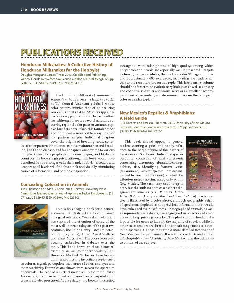
Herpetological Review 44(4), 2013
710 BOOK REVIEWS
Honduran Milksnakes: A Collective History of Honduran Milksnakes for the HobbyistDouglas Mong and James Tintle. 2013. ColdBlooded Publishing, Valrico, Florida (www.facebook.com/ColdBloodedPublishing). 170 pp. Softcover. US $49.95. ISBN 978-0-9897804-0-7.
The Honduran Milksnake (Lampropeltis triangulum hondurensis), a large (up to 2.4 m TL) Central American colubrid whose color pattern mimics that of co-occuring venomous coral snakes (Micrurus spp.), has become very popular among herpetocultur-ists. Although there are several naturally oc-curring regional color pattern variants, cap-tive breeders have taken this founder stock and produced a remarkable array of color and pattern morphs. Individual chapters cover the origins of breeding stock, genet-
ics of color pattern inheritance, captive maintenance and breed-ing, health and disease, and four chapters are devoted to various morphs. Color photographs occupy most pages, and likely ac-count for the book’s high price. Although this book would have benefited from a stronger editorial hand, hobbyist breeders and keepers at all levels will find this a rich and visually stimulating source of information and perhaps inspiration.
Concealing Coloration in AnimalsJudy Diamond and Alan B. Bond. 2013. Harvard University Press, Cambridge, Massachusetts (www.hup.harvard.edu). Hardcover. x, [2], 271 pp. US $29.95. ISBN 978-0-674-05235-2.
This is an engaging book for a general audience that deals with a topic of broad biological relevance. Concealing coloration has captured the attention of some of the most well known zoologists of the past two centuries, including Henry Bates (of Bates-ian mimicry fame), Alfred Russel Wallace, and Ernst Mayr. Even Theodore Roosevelt became embroiled in debates over the topic. This book draws on these historical examples, as well as modern work by Hopi Hoekstra, Michael Nachman, Bree Rosen-blum, and others, to investigate topics such
as color as signal, perception, the nature of color, and eyes and their sensitivity. Examples are drawn from across the spectrum of animals. The case of industrial melanism in the moth Biston betularia is, of course, explored but many cases of herpetological crypsis are also presented. Appropriately, the book is illustrated
throughout with color photos of high quality, among which phrynosomatid lizards are especially well represented. Despite its brevity and accessibility, the book includes 30 pages of notes and approximately 600 references, facilitating the reader’s ac-cess to the rich literature on this topic. This inexpensive volume should be of interest to evolutionary biologists as well as sensory and cognitive scientists and would serve as an excellent accom-paniment to an undergraduate seminar class on the biology of color or similar topics.
New Mexico’s Reptiles & Amphibians: A Field GuideR. D. Bartlett and Patricia P. Bartlett. 2013. University of New Mexico Press, Albuquerque (www.unmpress.com). 228 pp. Softcover. US $24.95. ISBN 978-0-8263-5207-1.
This book should appeal to general readers wanting a quick and handy refer-ence to the herpetofauna of this corner of the American Southwest. Individual species accounts—consisting of brief statements concerning taxonomy, abundance/range, habitat, size, identifying features, voice (for anurans), similar species—are accom-panied by small (25 x 25 mm), shaded dis-tribution maps showing range only within New Mexico. The taxonomy used is up to date, but the authors note cases where dis-agreement remains (e.g., Rana vs. Litho-bates, Bufo vs. Anaxyrus, Masticophis vs. Coluber). Each spe-cies is illustrated by a color photo, although geographic origin of specimens depicted is not provided, information that would have enhanced their usefulness. Photographs of animals, as well as representative habitats, are aggregated in a section of color plates to keep printing costs low. The photographs should make it possible for users to identify the majority of species, while in other cases readers are directed to consult range maps to deter-mine species ID. Those requiring a more detailed treatment of New Mexico’s herpetofauna will want to consult Degenhardt et al.’s Amphibians and Reptiles of New Mexico, long the definitive treatment of the subject.
PUBLICATIONS RECEIVEDPUBLICATIONS RECEIVED

Herpetological Review 44(4), 2013
BOOK REVIEWS 711
A growing number of titles have appeared in recent years that present stories of hunting for amphibians and reptiles (“herping”), following in the tradition made famous by Carl Kauffeld (Snakes and Snake Hunting and Snakes: The Keeper and the Kept). However, books in the post-Kauffeld era mostly do not come close to matching those classics. Here, we take brief looks at some recent additions to this genre of herpetological publishing.
More than Snake HuntingCraig Trumbower. 2012. ECO Herpetological Publishing, Rodeo, New Mexico. 278 pp. Hardcover. US $29.95 ISBN 978-0-9852936-2-8.
Trumbower offers ten chapters of snake
hunting adventures set in South Carolina, Florida, Texas, Arizona, and California. These are presented in “storytelling” fashion, and often are as much about the hunters as about the snakes they seek. Specific chapters re-call field searches for, among other targets, Blacktail, Eastern Diamondback, and Cane-brake (= Timber) rattlesnakes, Eastern Indigo Snakes, and Rosy Boas. A long chapter on Lampropeltis alterna (the Gray-banded King-snake) includes field-hunting vignettes from
a handful of veteran “alterna hunters.” One chapter captures the essence of reptile “expos”—the sometimes-huge trade shows where live animals are bought and sold. This work has the feel of an “insider” book, written for and about a circle of the author’s closest friends and field companions, but those outside the circle will likely find these tales entertaining.
Rattlesnake Adventures: North, South, East and WestJohn W. Kemnitzer, Jr. 2011. Serpent’s Tale Natural History Book Distributors, Lanesboro, Minnesota (www.zoobooksales.com). 183 pp. Softcover. US $17.95. ISBN 978-1-885209-66-5.
This collection—something of a sequel to Kemnitzer’s Rattlesnake Adventures: Hunting with the Old Timers, published in 2006—con-sists of 21 individually authored stories and articles. It includes original contributions as well as previously published material, but all focus on the subject of rattlesnakes. The mix of authors includes familiar contempo-raries (e.g., Richard Bartlett, David Barker, John Behler, William Brown) and others likely to be encountered for the first time in print. Some names are from long ago—Howard
Gloyd, Moody Lentz (an associate of Marlin Perkins), and Isa-belle Kauffeld (then-wife of Carl Kauffeld, but later married to Roger Conant and best known for her photographs in Conant’s field guides). Subject matter comprises tales of those who hunt-ed rattlesnakes for hides or illegal collection for the pet trade, the search for zoo specimens in remote areas, and includes Gloyd’s famous description of the magnificent Prairie Rattlesnake dens of South Dakota. A number of grainy, black-and-white photos are scattered throughout.
A Cobra Hijacked My Camera Bag! Snakes and Stories from TaiwanHans Breuer. 2012. Coachwhip Publications, Landisville, Pennsylvania (www.coachwhipbooks.com). 243 pp. Hardcover. US $29.95. ISBN 1-61646-129-2.
Naturalist Hans Breuer, a German nation-al transplanted to Taiwan and later to Bor-neo, shares stories of discovery of Taiwanese herpetofauna. Breuer enthusiastically re-counts his progression from rank amateur, fully ignorant of the local fauna, to one with growing expertise and who has since made several important discoveries. His writing is engaging, entertaining, and scientifically in-formed. Mixed in with the field stories, read-ers will encounter interesting facts about the ophidiofauna, herp keeping, wildlife smug-gling, Taiwanese culture, and much more. Much of the narrative focuses on field experiences with particular species or groups of snakes (elapids, pitvipers, ratsnakes, etc.), with color photos in-cluded for all.

Herpetological Review 44(4), 2013
712712

SSAR COMMITTEE CHAIRSAND COORDINATORS
CHAIRPERSONSConservationBETSIE ROTHERMELArchbold Biological Station
Roger Conant Grants-in-HerpetologyJOSHUA M. KAPFERUniversity of Wisconsin–Whitewater
Kennedy Student AwardLYNNETTE SIEVERTEmporia State University
Long Range Planning CommitteeKRAIG ADLERCornell University
Metter Memorial AwardJOSEPH J. BEATTYOregon State University
NominatingGREGORY WATKINS-COLWELLYale Peabody Museum of Natural History
ResolutionsROBERT DENTONThe Ohio State University
Seibert AwardsRAFE BROWNUniversity of Kansas
Standard English and Scientific NamesBRIAN I. CROTHERSoutheastern Louisiana University
Student Travel AwardsVINCENT FARALLOOhio University
MICHAEL JORGENSENOhio University
Student ParticipationDAWN WILSONSouthwestern Research Station
Poster AwardsTIFFANY DOANState College of Florida, Manatee-Sarasota
Meeting Management & PlanningROBERT ALDRIDGESaint Louis University
Live Auction CoordinatorGREG WATKINS-COLWELLYale Peabody Museum of Natural History
WebmasterRAUL E. DIAZLa Sierra [email protected]
MembershipMARINA GERSONCalifornia State University Stanislaus
MentorshipKRISTINE KAISERUniversity of California, Riverside
COORDINATORSElectorDANIEL NOBLEMacquarie University, Sydney
Symposium CoordinatorRICHARD D. DURTSCHENorthern Kentucky University
COLOR LAYOUT AND DESIGNMEGAN DAVIESARWEN AIKELELOUIS PORRASeaglemountainpublishing.com
INFORMATION FOR CONTRIBUTORS Herpetological Review is a peer-reviewed quarterly that publishes, in English, articles and notes concern-ing the study of amphibians and reptiles, as well as book reviews, commentaries, regional and international herpetological society news, and letters from readers directed to the field of herpetology. Articles reporting the results of experimental research, descriptions of new taxa, or taxonomic revisions are not published in HR, but should be submitted to the Journal of Herpetology (for information, please refer to: http://www.ssarherps.org/pages/JHinstr.php). To submit a manuscript to HR, please consult the SSAR webpage at:
<http://www.ssarherps.org/pages/HRinfo.php>
REPRINTS AND PAGE PROOFS Page proofs are sent electronically to authors of all articles, book reviews, and obituaries, but are not prepared for natural history, geographic distribution, or herpetoculture notes. Proofs are sent as pdf files approximately 2–4 weeks prior to publication. High resolution electronic (pdf) reprints of notes, articles, book reviews, and obituaries are provided at no charge to the primary/corresponding author as a benefit of SSAR membership. Reprint files will normally be sent prior to the issue mailing date from the printer. Authors who are not current SSAR members may purchase pdf reprints from the SSAR Publications Office.
ADVERTISING Herpetological Review accepts commercial advertising. Rates and copy information are available from the SSAR web page (http://www.ssarherps.org/pages/HRinfo.php).
Herpetological Review (ISSN: 0018-084X) is published quarterly (March, June, September, and December) by the Society for the Study of Amphibians and Reptiles at the Department of Natural Sciences, Williams Baptist College, P.O. Box 3692, Walnut Ridge, AR 72476, USA. Periodicals postage paid at Walnut Ridge, AR 72476 and at additional mailing offices. All rights reserved. No part of this periodical may be reproduced without written permission of the Editor, except that authors and their agents are permitted to reproduce and distribute their own articles and notes. POSTMASTER: Send address changes to Breck Bartholomew, SSAR Publications Secretary, P.O. Box 58517, Salt Lake City, UT 84158. MISSING OR DAMAGED ISSUE? Please notify Breck Bartholomew, SSAR Publications Secretary (e-mail: [email protected]) for a replacement.
SSAR PUBLICATIONS SSAR publishes seven different series: Journal of Herpetology, Herpetological Review, Facsimile Reprints in Herpetology, Contributions to Herpetology, Herpetological Circulars, Herpetological Conservation, and the Catalogue of American Amphibians and Reptiles (see below for CAAR details). SSAR members receive pre-publication discounts on all Facsimiles and Circulars and on volumes in the Contributions and Herpetological Conservation series. A complete pricelist of Society publications is available at: http://www.ssarbooks.com/.
CATALOGUE OF AMERICAN AMPHIBIANS AND REPTILES The Catalogue consists of loose-leaf accounts of taxa prepared by specialists, including synonymy, defini-tion, description, distribution map, and comprehensive list of literature for each taxon. Covers amphibians and reptiles of the entire Western Hemisphere. Available for purchase from the SSAR Bookstore (http://www.ssarbooks.com/). Use the prices below to order back issues.
COMPLETE SET: NUMBERS 1–880 ..........................check website below for current pricingINDEX TO ACCOUNTS 1–400: Cross-referenced, 64 pages ...................................................$6INDEX TO ACCOUNTS 401–600: Cross-referenced, 32 pages ...............................................$6SYSTEMATIC TABS (Ten tabs to fit binder: “Class Amphibia,” “Order Caudata,” etc.) .......$6IMPRINTED POST BINDER (Note: one binder holds about 200 accounts).......................$35INCOMPLETE SET: NUMBERS 1–190 .............................................................................$75 191–410 .............................................................................$85 411–840 ...........................................................................$320
To order: make checks payable to “SSAR” and mail to Breck Bartholomew, SSAR Publications Secretary, P.O. Box 58517, Salt Lake City, Utah 84158, USA (fax 801/453-0489). e-mail: [email protected]. Online orders at: http://www.ssarbooks.com/.

The Official Bulletin of the SOCIETY FOR THE STUDY OF AMPHIBIANS AND REPTILES
ISSN 0018-084X Herpetological Review
Herpetological ReviewVOLUME 44, NUMBER 4
DECEMBER 2013
ABOUT OUR COVER ........................................................................................... 533
SSAR BUSINESS ................................................................................................. 533
NEWSNOTES ...................................................................................................... 547
MEETINGS .......................................................................................................... 548
CURRENT RESEARCH ......................................................................................... 550
OBITUARIES ....................................................................................................... 554
GEOGRAPHIC DISTRIBUTION............................................................................ 619
NATURAL HISTORY NOTES ................................................................................ 650
BOOK REVIEWS ........................................................................................... 700
ARTICLES Caretta caretta in Tunisia: Natural History and Report of a New Regular Nesting AreaJ. BEN HASSINE, D. ESCORIZA ...................................................................... 557
Inferring Sea Turtle Recapture Rates Using Photographic IdentificationA. G. HALL, J. B. McNEILL .............................................................................. 561
Limestone Cave as a Cradle of the Ryukyu Ground Gecko, Goniurosaurus kuroiwaeT. KURITA, R. KAWAMURA, M. TODA ............................................................. 569
Parasitism of Reptiles by the Blacklegged Tick (Ixodes scapularis) and Western Blacklegged Tick (Ixodes pacificus) with New Records of I. scapularis from Arkansas and Oklahoma Lizards: Implications for Lyme Disease EpidemiologyC. T. McALLISTER, L. A. DURDEN, M. B. CONNIOR, H. W. ROBISON ................. 572
Ontogenetic Shift in Habitat Use in the Western Fence Lizard (Sceloporus occidentalis) in Northern CaliforniaM. R. LAMBERT, C. M. YASUDA, B. D. TODD ................................................... 579
CONSERVATIONStatus and Conservation of a Gondwana Legacy: Bullock’s False Toad, Telmatobufo bullocki (Amphibia: Anura: Calyptocephalellidae)D. B. FENOLIO, V. MORENO-PUIG, M. G. LEVY, J. J. NÚÑEZ, W. W. LAMAR, M. O. FABRY, M. S. TIRADO, M. L. CRUMP, A. CHARRIER .......................................... 583
Deconstructing a Southern Portuguese Monster: The Effects of a Children’s Story on Children’s Perceptions of GeckosL. M. P. CERÍACO, M. P. MARQUES ................................................................. 590
TECHNIQUESNew Experimental Method for Semen Extraction in Freshwater TurtlesJ. LEFEBVRE, S. CARTER, S. W. MOCKFORD ................................................... 595
The Use of Photo Identification on Dickerson’s Collared Lizard (Crotaphytus dickersonae) L. FABER, M. PLASMAN, M. J. DUCHATEAU ................................................... 600
A Simple and Effective Technique for Demonstrating Post-prandial Metabolic Response of Snakes in the ClassroomW. H. HEYBORNE .......................................................................................... 603
HERPETOCULTURECaptive Reproduction and Husbandry of Adult Ozark Hellbenders, Cryptobranchus alleganiensis bishopiJ. A. ETTLING, M. D. WANNER, C. D. SCHUETTE, S. L. ARMSTRONG, A. S. PEDIGO, J. T. BRIGGLER ........................................................................ 605
AMPHIBIAN DISEASESDetection of Batrachochytrium dendrobatidis in River Frogs (Genus Amietia) on Mount Kilimanjaro, TanzaniaG. ZANCOLLI, A. STORFER , M. RÖDEL .......................................................... 611
Batrachochytrium dendrobatidis in Amphibians of Northern Calhoun County, Illinois, USAP. J. MUELLEMAN, C. E. MONTGOMERY ........................................................ 614
Detection of Ranavirus in Ohio, USAR. N. HOMAN, J. R. BARTLING, R. J. STENGER, J. L. BRUNNER......................... 615
GEOGRAPHIC DISTRIBUTIONNew Distributional Records for Amphibians and Reptiles from the State of Tamaulipas, México IIIW. L. FARR, D. LAZCANO, P. A. L. MURCIO ..................................................... 631
New Distributional Records for Amphibians from Durango, MexicoR. VALDEZ-LARES, G. A. MARTÍN-MUÑOZ DE COTE, R. MUÑIZ-MARTÍNEZ, G. SANTOS-BARRERA .................................................................................. 646