Genetic Transformation in Stevia rebaudiana
25
Genetic Transformation in Stevia rebaudiana. Mubarak, M.H* A.H. Belal Taymour M. N. EL –Din ** and E. I. El Sarag* *Plant Production Department. Fac. Environ. Agric. Sci. (El-Arish), Suez Canal Univ., Egypt ** Agricultural Genetic Engineering Research Institute. AGERI. ARC, Egypt Corresponding: [email protected] [email protected] ABSTRACT This study was carried out in plant Tissue Culture Laboratory in Faculty of Environmental Agricultural Sciences El-Arish, North Sinai, Suez Canal University during the period from 2009 to 2012 .The molecular studies were carried out in Micropropagation Technology Laboratory (MPT) at the Agricultural Genetic Engineering Research Institute (AGERI), Agriculture Research Center (ARC), Giza, Egypt. Regeneration and transformation systems on stevia var.Spanti, as an attempt to produce transgenic stevia plants carrying bar gene as a selectable marker for herbicide bialaphos resistance and gus gene as a reporter marker, using biolistic gene gun protocol. At acceleration pressure of 900 psi 8 out of the 20 assayed callus stevia expressed the GUS blue spots and the total number of blue spots was 20. While, when the acceleration pressure was increased to 1100 psi, the number of embryos revealing the GUS blue spots increased to reach 20 callus Stevia expressing a total of 101 blue spots. Transformation efficiency has been calculated as the number of regenerated plantlets/ number of bombarded callus. The 1
Transcript of Genetic Transformation in Stevia rebaudiana

Genetic Transformation in Stevia rebaudiana.
Mubarak, M.H* A.H. Belal Taymour M. N. EL –Din ** and E. I. El Sarag*
*Plant Production Department. Fac. Environ. Agric. Sci. (El-Arish), Suez Canal Univ., Egypt
** Agricultural Genetic Engineering Research Institute. AGERI. ARC, Egypt
Corresponding: [email protected][email protected]
ABSTRACT
This study was carried out in plant Tissue Culture Laboratory in
Faculty of Environmental Agricultural Sciences El-Arish, North
Sinai, Suez Canal University during the period from 2009 to
2012 .The molecular studies were carried out in Micropropagation
Technology Laboratory (MPT) at the Agricultural Genetic
Engineering Research Institute (AGERI), Agriculture Research
Center (ARC), Giza, Egypt. Regeneration and transformation
systems on stevia var.Spanti, as an attempt to produce transgenic
stevia plants carrying bar gene as a selectable marker for
herbicide bialaphos resistance and gus gene as a reporter marker,
using biolistic gene gun protocol. At acceleration pressure of
900 psi 8 out of the 20 assayed callus stevia expressed the GUS
blue spots and the total number of blue spots was 20. While, when
the acceleration pressure was increased to 1100 psi, the number
of embryos revealing the GUS blue spots increased to reach 20
callus Stevia expressing a total of 101 blue spots.
Transformation efficiency has been calculated as the number of
regenerated plantlets/ number of bombarded callus. The
1

transformation frequency of 1100 psi was considerably higher (8.3
%) than that of 900 psi (3.3 %).
Key words: Stevia rebaudiana, Tissue culture, Genetic transformation, Callus
Introduction
Stevia rebaudiana Bertoni is a small shrub of the Asteraceae
(Composite) family. Stevia is an herb with an extensive root system
and brittle stems producing small, elliptic leaves. The tiny white
florets are perfect, borne in small corymbs of 2–6 florets. Corymbs
are arranged in loose panicles. Oddone (1997) considered that stevia
is self-incompatible and insect pollinated. Additionally, he mentioned
that clear seeds are infertile. Seeds are contained in slender
achenes, about 3 mm in length. Each achene has about 20 persistent
pappus bristles. The native occurrence of Stevia rebaudiana is between
22-24 °S and 53-56°W in Paraguay and Brazil. The plant growth requires
mild temperature between 15°and 38°C and relative humidity of about 80
% (Soejarto et al., 1983).The cultivation of such a plant could be an
alternative for the new land reclamation projects to meet the sugar
demands of the Egyptian markets and generate income for the growers
(El-Zifzafi,2003 and Ibrahim et al.,2008).
Stevia is helpful for hypoglycemia and diabetes because it
nourishes pancreas and thereby helps to restore its normal
function (Soejarto et al., 1983). Oviedo (1971) reported that
35.2% fall in normal blood sugar levels 6-8 hours following the
ingestion of stevia leaf extract (Miyazaki et al., 1978). Also,
stevia leaves extract can be used as anti fungal and anti
bacteria agent. Therefore, it lowers incidence of cold and flu.
2

Poor seed germination is one of the factors limiting large
scale cultivation. In this concern, referred to Shock (1982);
Duke, (1993) and Carneiro et al. (1997) who reported that poor
viable seeds were produced by stevia with very low germination
percentage (Felippe and Lucas, 1971 and Toffler and Orio, 1981;
Latha and Usha, 2003) .Seed germination rate is often poor, less
than 10% are common (Miyazaki et al., 1978; Goettemoeller and
Ching, 1999).
A lot of the advances in plant biotechnology have been based
on transformation; the ability to introduce DNA genes into plant
cells to recover whole transgenic plants with high yield quality
and quantity as well as resistant to biotic and abiotic stresses.
Gene transfer via biolistic particle bombardment (Biolistic gene
gun) is one of the useful techniques to overcome such problems as
previously reported in other plants (Yadav et al., 2010, Bassry,
2011). So, there is an urgent need for developing an efficient
gene transfer protocol for Stevia rebaudiana.
MATERIALS AND METHODS
`Seedlings of Stevia rebaudiana var. Spanti were kindly obtained
from Sugar Crops Research Institute, Agric., Research Center,
Ministry of Agric, Egypt, which were grown in the greenhouse of
the Faculty of Environmental Agricultural Sciences El-Arish.
Actively growing shoots were used as the explants source
during period from February to March. The terminal shoots were
collected from growing plants which were 2-3 months age and were
cut into 1-1.5 cm pieces
1.Callus induction.
3

Leaves were washed under running tap water with mild
detergent for 5 minutes, followed by distilled water. Thereafter,
in aseptic conditions, the explants were soaked in 5% (v/v)
Clorox for 10 minutes after has been washed three times with
sterile distilled water. Later, it was immersed in 0. 15%
mercuric chloride (HgCl2) solution for 1 minute and finally four
washes with sterilized distilled water to remove all traces of
HgCl2.
All steps of the sterilization had been done under aseptic
conditions inside the culture cabinet (Laminar air flow hood) and
by using sterilized instruments. The purpose of this procedure
was to disinfect the plant tissue from fungi, bacteria, and other
sources of contamination, without harming the regenerative
capacity of the explant. These leaves cultured in MS (Murashige
and Skoog, 1962) medium with five different 2,4-D concentration
(MS1, MS2, MS3, MS4 and MS5) were used in this study (Table, 1)
according Mohammed et al., (2006).
Table (1): Media composition for callus induction.
2,4-D (mgl-1 )Medium code
MSControl MS+1mgl-1
2,4-DMS1
MS+2m gl-1 2,4-DMS2
MS+3m gl-1 2,4-DMS3
MS+4m gl-1 2,4-DMS4
MS+5m gl-1 2,4-D MS5
4

The media were solidified with 2.5 gl-1phytagel. The media were
made up in distilled water, the pH adjusted to 5.8 and
sterilized by autoclaving for 20 minutes at 121 oC and 15 psi.
2.Callus regeneration.
Regeneration protocol as described by Tavazza et al. ( 1988 )
was counducted as the following steps:
1- callus was transfered as a cluster on the surface of
regeneration medium containing MS medium with 100 mgl-1myo-
inositol, 1 ml MS vitamins, 3 % sucrose, 1 mgl-1indol-3-
acetic acid (IAA), 10 mgl-1gibberellic acid (GA3) and 1 mgl-1
6-benzylaminopurine (BA).
2- The clusters from callus were incubated at 25 ± 1°C under a
photoperiod of 16 hr/day and 3000 Lux for 60 days.
3- The regenerated shoots which reach 2-3 cm in length (after 70
days from culture) in each callus tissue were calculated.
Shoots were transferred to medium consisted of MS basal
salts supplemented with 3 % sucrose, 100 mgl-1myo-inositol, 1
mgl-1GA3, 1 ml thiamin-HCl, 2 mgl-1calcium pantothenate and 2.5
gl-1 phytagel and the pH was adjusted to 5.6-5.8
3. Plant expression vector.
The plant expression vector pCGP1258 was used in
this study which is kindly provided from Dr A. Markis at
Mediterranean Agronomic Institute of China (MAICH) Crete,
Greece.
5

Bar gene for herbicide resistance
The bar gene which is controlled by cauliflower mosaic
caulimovirus (CaMV) 35S promoter and nopaline syntheases (nos)
terminator, and encodes the enzyme phosphinothricin acetyl
transferase (PAT) which inactivates phosphinothricin (De Block et
al. 1987). The active ingredient of the herbicide bialaphos was
used as selectable marker.
Preparation of micro carriers for bombardment.
Isolation of DNA plasmid (pCGP1258)
Preparation of DNA plasmid was performed according to
the method by Sambrook et al. (1989).
Polymerase chain reaction (PCR).
The polymerase chain reaction (PCR) is a method that
allows rapid detection of the presence or absence of a target
DNA. The DNA extracted from putatively transformed and non-
transformed control tissues were subjected to the polymerase
chain reaction (PCR) using a DNA thermal cycler.
In case of bar and gus genes, two specific primers for each
gene were used to detect the integrated bar and gus genes into
the genome of transformed stevia tissues by PCR analysis.
Primers were synthesized (DNA Synthesizer 392, Applied
Biosystems) at AGERI, ARC and used as shown in Table (2).
6
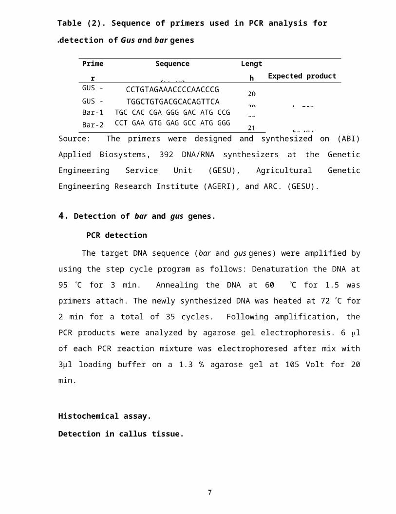
Table (2). Sequence of primers used in PCR analysis for
detection of Gus and bar genes.Prime
r
Sequence
(5-'3)'Lengt
h Expected productsizeGUS -
1CCTGTAGAAACCCCAACCCG 20
750 bpGUS -
2TGGCTGTGACGCACAGTTCA 20Bar-1 TGC CAC CGA GGG GAC ATG CCG 23
484 bpBar-2 CCT GAA GTG GAG GCC ATG GGG 21
Source: The primers were designed and synthesized on (ABI)
Applied Biosystems, 392 DNA/RNA synthesizers at the Genetic
Engineering Service Unit (GESU), Agricultural Genetic
Engineering Research Institute (AGERI), and ARC. (GESU).
4. Detection of bar and gus genes.
PCR detection
The target DNA sequence (bar and gus genes) were amplified by
using the step cycle program as follows: Denaturation the DNA at
95 °C for 3 min. Annealing the DNA at 60 °C for 1.5 was
primers attach. The newly synthesized DNA was heated at 72 °C for
2 min for a total of 35 cycles. Following amplification, the
PCR products were analyzed by agarose gel electrophoresis. 6 l
of each PCR reaction mixture was electrophoresed after mix with
3µl loading buffer on a 1.3 % agarose gel at 105 Volt for 20
min.
Histochemical assay.
Detection in callus tissue.
7

β-glucuronidase (GUS) assay (Beckman and Engler, 1994).
X-Glue produces the final insoluble blue precipitate dichloro-
dibromoindigo (CIBr-indigo).
Number of transformed explants and control were soaked in
500μl GUS buffer (Daniell et al. 1990) containing 50 μl X-Glu ( 5-
bromo-4-chloro-3-indoly) glucuronide and rapped with aluminum
foil, to prevent light effect ( Jefferson et al. 1987) . A blue
staining was often visible in 24 h or less, even though tissues
were incubated in the substrate solution overnight at 37˚C.
After this incubation period, the tissues were cleared with 5%
colorx for easily visualization the blue staining. Blue color
was detected under the binocular microscope.
5.Statistical analysis:Data were statistically analyzed by using a randomized
complete design (RCD) according to (Snedecore and Cochran, 1990).
Mean separations were done by using SPSS computer program V.10
(1991) and to compare between means least significant differences
(Duncan, 1955) was used. Data were recorded every six weeks.
Every treatment had four replicates
Results and Discussion
Confirmation of transformation.
Two methods were used in this study to detect the presence of
the gus and bar genes in the putatively transformed tissues.
8

These methods were the gus histochemical staining technique and
PCR analysis
1 .Transient expression of gus gene in callus.The use of 5-bromo-4-chloro-3-indolyl glucuronide (x-
gluc) as a substrate for the gus enzyme provide an effective
methods for monitoring the expression of GUS DNA introduced
into plant cells. X-gluc is the best substrate currently
available for histochemical localization of β-glucuronidase
activity in tissue and cells. This substrate works very
well, giving a blue precipitate at the site of enzyme
activity Our results indicated that GUS activity in
transformed stevia explants (callus slices) which co-
cultivated with gen gun could be detected histchemicaly for
GUS expression after two week as the first evidence of
transformation .Most of the tissues turned callus to deep
blue color compared with control (Fig. 1). These results are
in agreement with other findings reported by different
researchers such as (Martin et al. 1990), they reported that,
the GUS marker system is useful for early selection, as
bacterial expression of the marker severely compromises its
effectiveness The β-glucuronidase gene fusion system became
the most popular reporter gene in plant histochemical
research. In many cases, however, as in whole-moment
preparation and thick handmade section, the pigments present
in the most plant tissue can partially or even totally mask
the sits of GUS – activity and consequently hinder the
detection of it. A passage through an ethanol series or a
9

10% commercial bleach solution is known to remove pigments
such as chlorophyll from the tissue.
Fig .(1): Histochemical assay of β-glucuronidase (GUS) gene in callus explants
of stevia transformed via gen gun after two weeks of transformation.(A)Transformed explant of stevia showing blue color.
(B )None transformed explants.
10
A B

2. The best pressure for stevia transformation using biolistic
gene gun.In the present investigation, the transient expression of
the uidA gene was studied as a preliminary test of the
transformation efficiency and for selecting the most appropriate
bombardment acceleration pressure. The transformation experiment
was carried out with two different bombardment acceleration
pressures i.e. 900 and 1100 psi. The total number of callus
tested was 40. At acceleration pressure of 900 psi 8 out of the
20 assayed callus of stevia expressed the GUS blue spots and the
total number of blue spots was 20. While, the acceleration
pressure was increased to 1100 psi, the number of callus
revealing the GUS blue spots increased to reach 20 callus stevia
expressing a total of 101 blue spots. Therefore, from this
experiment it might be concluded that, the bombardment at an
acceleration pressure of 1100 psi revealed a significantly higher
number of callus expressing the uid A gene than the bombardment
acceleration at 900 psi, thus reflecting a higher transient
transformation efficiency.
Table (3): The uid A gene expression (gus) as revealed by the
average number of blue spots.plan Pressure No. of
callus in No. of callus with blue No. of spots
11

t petri dish spots Stev
ia 1100 20 18a 101a
900 20 8b 20b
Numbers with the same letters are not significantly different at 5%.
These results are in agreement with Cho et al. (1998) and
Harwood et al. (2000). They used gas pressure of 1100 psi to
obtain higher expression of gus gene and fertile barley plants.
Similarly, Manoharan and Dahleen (2002) found that the number of
GUS spots present in the callus 48 hrs after bombardment at 1100
psi was greater than that following bombardment at either 900 psi
or 1300 psi. In addition, El-Itriby et al. (2003) demonstrated
that the use of osmotic medium pre- and post treatment with
acceleration pressure of 1100 psi and double shots per plate was
most efficient in transformation of maize immature embryos,
judging from the transient gus expression. Moreover, Assem et al.
(2006) demonstrated that the pressure 1100 psi revealed highest
Gus expression when compared to the 900 and 1300psi acceleration
pressure in barley. ( Bassry, 2011) concluded that the
bombardment at an acceleration pressure of 1100 psi revealed a
significantly higher number of embryos expressing the uid A gene
than the bombardment acceleration at 900 psi, thus reflecting a
higher transient transformation efficiency.
3. Callus recovery and selection.
12

The transformed callus were allowed to recover for one
week on callus induction medium without selection pressure.
Bahieldin et al. (2000) found that the recovery period
increased the transformation efficiency. In contrast,
Manoharan and Dahleen (2002) transferred the transformed
callus to callus induction medium supplemented with
bialaphos for selection directly after bombardment. After one week, selection of transformed cells were
carried out by transferring the transformed callus to callus
induction medium containing 1.5 mg-1 bialaphos for 2 weeks. The
callus were transferred onto fresh selective medium containing 3
mg-1 bialaphos, with continuous subculture every 2 weeks (Fig 2).
All cultures were kept at 28°C in a dark growth chamber.
Fig ( 2): Transient bar expression in transformed callus stevia
and regeneration : expression of bar at selectable media.
13

Selection of putatively transgenic callus .Infected callus showed normal growth on co-cultivation and
resting media without selection with loss of some callus that
failed to grow after transformation. Selection was started by
transferring stevia callus to selective medium containing 1.5
mg-1bialaphos, for 2 weeks. Then, selection pressure was raised
by transferring callus to selective medium containing 3 mg-1
bialaphos. Resistant callus were subcultured every two weeks.As shown in Table (4), Stevia revealed the highest number of
putatively transgenic callus after 2 weeks on selective medium.
However, as the selection time increased, the number of stevia
resistant callus decreased significantly.Resistant callus which grew uniformly on the selective media
by Tavazza et al. ( 1988 )
Table 4. Number of bombarded explants, number of surviving callus, number of plantlets, and transformation frequency
for stevia.
pressure
No. ofbombarde
dexplants
.
No. ofsurvive
dcallus
No. ofplantle
ts
Transformati
on frequency
%
1100 120 40a 10 a 8.3 a900 120 15b 4 b 3.3 b
Numbers with the same letters are not significantly different at
14

5%
Results in Table (4) indicated that, in total, 240 explants
were bombarded using the biolistic gene gun transformation
method. Putative transgenic lines have been obtained from the
transformation experiments of the two pressures. Transformation
efficiency has been calculated as the number of regenerated
plantlets/ number of bombarded callus. The transformation
frequency of 1100 psi was considerably higher (8.3 %) than that
of 900 psi (3.3 %).In the present study, the concentration of bialaphos, used
as a selective agent started at 1.5 mg-1 and then increased to 3
mg-1. Similar concentrations of bialaphos were applied by Gordon-
Kamm et al. (1990), Wan et al. (1995), Frame et al. (2002) and Assem
et al. (2010). The other investigators employed PPT at different
concentrations. Wan and Lemeaux (1994), Cho et al. (1998),
Horvath et al. (2000), Sivamani et al. (2000), Manoharan and Dahleen
(2002) and Pellegrineschi et al. (2004) used 5 mg-1 bialaphos in
selection induction medium. In contrast, Kishchenko et al. (2005)
increased PPT concentration from 5.0 to 10 mg-1 for selection of
transformed suspension cells of sugar beet. Moreover, Pauk et al.
(2002) and Chen et al. (2007) used 10 mg-1Bialaphos as a selective
agent.
4. Detection of integrated genes (gus and bar genes). A number of callus tissues transformed by biolistic gene
gun were subjected to molecular analysis to confirm the
integration of the foreign genes (GUS and bar gene) into callus
15

genome incorporated into the genome of stevia tissues transformed
by particle bombardment . PCR analysis.
The polymerase chain reaction (PCR) is a very useful technique
for the detection of the presence or absence of a specific DNA
fragment in the plant genome. Lassner et al. (1989) reported the
potential of using PCR for analysis of foreign genes in plant
tissues in order to check the integration of a full-length gene
(s) of interest. Total genomic DNA from transformed callus and
control (non-transformed) Stevia tissues, were analyzed using
primers specific to the coding region of the GUS gene and the
coding region of the bar gene Table 2 illustrates the sequence of
the primers used in PCR assays and their sizes. The expected
amplification product was (750 bp) for GUS gene and (484 bp) for
the bar gene
As shown in Fig. 3 The amplified fragments for GUS have the
expected size of coding region of GUS gene (750bp), lane (1)
positive control resulted from amplified plasmid pCGP1258 and
lane (2) negative control resulted from non-transformed. Fig. 3
shows the presence of bar-DNA fragment at expected molecular
weight (484bp), lane (1) positive control and lane (2) negative
control resulted from non-transgenic stevia tissues.Therefore, it is evident that both GUS and bar genes were
physically present in the genomic background of the transformed
stevia tissues. Using PCR as indicator for the presence of
foreign genes into transformed stevia tissues has been reported
16

by many investigators (Smigoki and Hammerschlag, 1991 and
Xiaojian et al. 1994) reported that PCR analysis confirmed that in
most of the putative transformats. It addition, tansgenic tissues
at different developmental stages were selected, and PCR analysis
of transgenic material will be presented. Furthermore, this
method should allow the development of assays for the transient
and homogeneous expression of promoters of various genes in Stevia
rebaudiana
(Bumpus and Dhir, 2011.)
Fig (3): PCR product of bar and Gus genes amplifying partial length (484 and 750 bp) in putative Stevia rebaudiana tissuestransformed via Biolistic gun. The arrow indicates the amplified for fragment.M= 100 bp DNA ladder (Marker)+ve = positive control. -ve = negative control.
17
484 pb
M ev+ev+ ev-
057 pb
REFERENCES
Assem, S. K.; E. H. Hussein; H. A. Hussein and B. Sara (2010).
Transformation of the salte-tolerance gene BI-GST into
Egyptian maize inbred lines.Arab J. Biotech.13:99-114.
Assem S. K.; Borg N. and El-Itraby H. A. (2006). Agrobacterium-
mediated transformation of maize inbred lines using immature
embryos and a standard binary vector system. Egypian j. of
Genetics and Cytology, 35(2):173-186.
Bahieldin, A., R. Qu, W. Dyer, A. S. Haider and M. A. Madkour
(2000). A modified procedure for rapid recovery of transgenic
wheat plants. Egypt. J. Genet. Cytol. 29: 11-23.
Bassry, Mahmoud Abd El-Raheem.(2011). Genetic transformation of
some cereal plants targeting abiotic stress. Department of
genetics, M.Sc. Thesis, Faculty of AgricultureCairo
University,EGYPT.
Beckman, T. and G. Engler (1994). An easy technique for the
cleaning of histochemically stained plant tissue. Plant Mol.
Bio. Reporter, 12: 37-42.
Bumpus . P. N. and S. K. Dhir(2011). Plant Regeneration andGenetic transformation of Stevia rebaudiana Using ParticleBombardment. Association of Research Directors,Inc.16thBiennial Research SymposiumApril 9 – 13, AtlantaMarriott Marquis Atlanta, Georgia Program& Abstracts.
Carneiro, J.W.P.; A.S. Muniz and T.A. Guedes.( 1997). Green house
bedding plant production of Stevia rebaudiana (Bert) Bertoni.
Can. J. Plant Sci. 77:473–474.
18

Chen, M.; Chen, Q.-J.; Niu, X.-G.; Zhang, R.; Lin, H.-Q.; Xu, C.-Y.; Wang, X. C.; Wang, G.-Y. and J. Chen (2007). Expression of OsNHX1 gene in maize confers salt tolerance and promotes plant growth in the field. Plant Soil Environ.,53(11): 490–498.
Cho, M. J.; W. Jiang and P. O. Lemaux (1998). Transformation of
recalcitrant barley cultivars through improvement of
regenerability and decreased albinism. Plant Sci., 138:229-
224.
Daniell, H.; J. Vivekanada ; B. Nielson. ; K. K. Tewari and J. C.
Sanford (1990). Transient foreign gene expression in
chloroplasts of cultured tobacoo cells following biolistic
delivery of chloroplast vectors. Proc. Natl. Acad. Sci., USA.
De Block, M., J. Botterman, M. Vandewiele, J. Dockx, C. Thoem, V.
Gossele, N. Roamovva, G. Thompson, M. Vanmontagu and J.
Leemans (1987). Engineering herbicide resistance in plants by
expression of aoxifin enzyme. EMBO J. 6: 2513-2518.
Duke, J. (1993). Stevia rebaudiana In: handbook of ''Alternative
Cash Crops''. CRC Press, Boca Raton, 422–424.
Duncan,B.D.(1955).Multiple range and multiple F.test. Biometrics.
11:1- 42.El Zifzafi, M. A. (2003). Physiological studies on Stevia rebaudiana
Bertoni through tissue cultures techniques and its
suitability for desert regions. Ph.D. Thesis, Faculty of
Agriculture, Cairo University, Egypt .66-70.
El-Itriby, H. A.; Assem, S. K.; Hussein, Ebtissam. H. A.;
Abdel-Galil, F. M. and Madkour, M. A. (2003).
Regeneration and transformation of Egyptian maize
19

inbred lines via immature embryo culture and biolistic
particle delivery system. In vitro Cell Dev. Biol. Plant,
39 (5): 524-531.
Felippe, G.M. and N.M.C. Lucas (1971). Estudo daviabilidade dos
frutos de Stevia rebaudiana Bert. Hoehnea, 1: 95-105.In: InVitro
Cellular and Developmental Biology Plant. 39(5): 523.
Frame, B.R.; H. Shou; R.K.; Z. Chikwamba ; C. Xiang ;
Fonger,; T.M. Pegg ; B. Li ; D. Nettletonb; D. Pei and
K. Wang (2002). Agrobacterium tumefaciens mediated
transformation of maize embryos using a standard binary
vector system. Plant Physiol., 129:13– 22.
Goettemoeller,J. and A. Ching (1999). Seed germination in Stevia
rebaudiana. Perspectives on new crops and new uses.
J.tanick(ed.),ASHS press,Alexandria,VA.,510-511.
Gordon-Kamm, W. G.; T. M Spencer; M. L. Mangano ; T. R. Adams;
R. G. Daines; W. G. O. Start ; G. V. Brien; S. A.
Chamber; Adams, W. R.; G. N. G. Willetts ; T. H.
B. Rice; C. G. Backey; R. W. Krueger ; A. P. Kausch
and P.G. Lemaux (199 0).Transformation of maize cells
and regeneration of fertile transgenic plants. Plant
Cell, 2:603-618.
Harwood, W.A.; Ross, S. M.; Cilento, P. and Snape, J. W.
(2000). The effect of DNA/gold particle preparation
technique and particle bombardment device, on the
transformation of barley ( Hordeum vulgare). Euphytica,
111:67-76.
Horvath, H.; Huang, J.; Wong, O.; Kohl, E.; Okita, T.; Kannangara, C. O. and Wettstein D. V. (2000). The production
20

of recombinant proteins in transgenic barley grains. Proc. Natl. Acad. Sci. USA, 97:1914-1919.
Ibrahim. I.A.; M.I. Nasr; B.R .Mohammed and M.M. EL-Zifzafi
(2008). Plant growth regulators affecting in vitro cultivation
of Stevia rebaudiana. Sugar Tech. 3 (10) :248-253.
Jefferson, R. A.; T. A. Kavanagh and M. Bevan (1987) GUS
fusion: beta glucuronidase as a sensitive and versatile gene
fusion marker in higher plants. EMBO J., 6:3901–3907.
Kishchenko, E.M.; Komarnitskii, I. K. and Kuchuk, N. V. (2005).
Production of transgenetic sugarbeet (Beta vulgaris L.) plants
resistant to phosphinothricin. Cell Biol. Int., 29: 15-19.
Lassner, M. W., P. Peterson and J. I. Yoder (1989). Simultaneous
amplification of multiple DNA fragments by polymerase chain
reaction in the analysis of transgenic plants and their progeny.
Plant Mol. Biol. Reporter 7: 116 – 127
Latha, S. and M. Usha (2003). In vitro culture studies on Stevia
rebaudiana. InVitro Cellular and Developmental Biology Plant.
39(5): 520-523.
Manoharan, M. and L. S. Dahleen (2002). Genetic
transformation of commercial barley (Hordeum vulgare L.)
cultivar Conlon by particle bombardment of callus.
Plant Cell Rep.,21:76-80.
Martin, G. C., A. N. Miller, L. A. Castle, J. W. Morris, R. O.
Morris and A. M. Dandeker (1990). Feasibility Studies Using
-Glucuronidase as a gene fusion marker in apple, peach, and
radish. J. Amer. Soc. Hort. Sci. 115: 686-691. (Abst.).
Miyazaki, Y.; H. Watanabe and T. Watanabe(1978). Studies on the
cultivation of Stevia rebaudiana Bertoni. Yield and stevioside
21

content of 2-year-old plants. Eisi Shikenjo hokoku., 96: 86-
89.
Mohammed, S. U.; S. H. Mohammad; M. M. Muoztaba; B.U. Mohammad;
A. Romel and M. B. Azizul (2006). In vitro propagation of Stevia
rebaudiana Bert. in Bangladesh. African Journal of
Biotechnology . 5 : (13), 1238-1240.
Murashige, T. and F.Skoog (1962). A revised medium for rapid
growth and bioassays with tobacco tissue cultures.
Physiol., Plant ., 15: 473-497.
Oddone, B. (1997). How to grow stevia. Technical manual. Guarani
Botanicals, Pawtucket, CT.
http://www.scribd.com/doc/6390515/Advances-in-Agronomy-Volume-
89.
Oviedo, C.A.( 1971). Accion hipoglicemiante de la Stevia rebaudiana
Bertoni (Kaa-he-e). Excerpta Medica. 208:92-93.
Pauk, J.; Ertugrul, F.; Bartók, T.; Mihály, R.; Kiss, O.; Cseuz,
L. and Dudits, D. (2002). Improvement of wheat abiotic
stress resistance via genetic transformation. Plant
Physiology, 46:5-7.
Sambrook, J., E. F. Fritsch and T. Manitatis (1989). Molecular
cloning: A laboratory Manual. Second Edition, Cold Spring
Harbor Lab. Press, New York, USA 2nd Ed. No.pp.1626. (Cited
from Adawy. 1998).
Sndecor, G. W. and W. G. Cochran (1990). Statistical Methods. 7th
Ed. Lowa State Univ. Press, Ames-Iowa, USA., 507.
22

Sivamani, E.; Bahieldin, A.; Wraith, J.M.; Al-Niemi, T.; Dyer,
W.E.; Ho, T.D. and Qu, R. (2000). Improved biomass
productivity and water use efficiency under water deficit
conditions in transgenic wheat constitutively expressing the
barley HVA1 gene. Pl. Sci., 155(1): 1-9.
Soejarto, D.D.; C.M Compadre; P.J Medon; S.K. Kamath and
A.D.Kinghorn (1983). Potential sweetening agents of plant
origin. Economic Botany, 37(1): 71-79.
http://www.springerlink.com/content/xw7052776224j631
Shock, C.C. ( 1982). Experimental cultivation of rebaudi’s stevia
in California. Univ. California, Davis Agron. Progr. Rep.
122.
Smigocki, A. C. and F. A. Hammerschlag (1991). Regeneration of
plants from peach embryo cells infected with a shooty mutant
strain of Agrobacterium. J. Amer. Soc. Hort. Sci. 116:1092—1097.
Tavazza R., Ordas R.J., Ancora G.(1988). Procedure for the
regeneration of plants from cell suspension protoplasts of
tetraploid potato (Solanum tuberosum L.) cv. Desirée. Plant
Sci., 58: 223-230.
Toffler, F. and O. A. Orio ( 1981). Acceni sulla pin˜
atatropicale ‘Kaa-he-e’ ou ‘erba dolce’. Rev. Soc. It.Sci.
Aliment., 4: 225-230.
http://www.springerlink.com/content/3g266784t3053253
Wan Y. and Lemaux P. G. (1994). Generation of large numbers pf indepently transformed fertile barley plants. Plant physiol., 104:37-48.
23

Wan, Y.; Widholm, J. M. and P. G. Lemaux (1995). Type I callus
as a bombardment target for generating fertile transgenic
maize (Zea mays L.). Planta, 196:7–14.
Xiaojian, Y. E., R. Scorza and J. Sanford (1994). Genetic
transformation of peach tissues by particle bombardment. J.
Amer. Soc. Hort. Sci. 119: 367-373.
Yadav, M.; D. Chaudhary; M. Sainger and P. K. Jaiwal (2010).
Agrobacterium tumefaciens-mediated genetic transformation of
sesame (Sesamum indicum L.). Plant Cell Tiss. Organ. Cult.
ارك�� م�حمدح�سن� �د م�ب �اح -ع�ب ب" لال ح�لمي' ال�ف# �مورب� ي' صر - ت�" ن� ن�# ي7' مان�2ال�د ل -اي;' ال�سراح� اس�ماع�ب'اة" ال�سوي�'س ت" ام�عة" ق�" �-ح� Jال�عري�'س �ة" ب� ي' Oئ ب' Qة" ال�ئ راع�ي' ة" ال�علوم ال�ز# ك�لي'
2- ة" اع�ي' ة" ال�زر# ي' J;دسة" ال�ورات حوثJ ال�هب# �ة"–م�عهد ب� ر# ي' �ال�ج �ة" ب� راع�ي' حوثJ ال�ز# �مرك�ز# ال�بي' ة ال�دراسة ف# ي7'ت" ه�د# ر �د اج� ة" ام�عمل ق�" راع�ي' ة" ال�علوم ال�ز# ي' ك�لي' ة" ف# ي' ت;" ا �ب ة ال�ئ# �سج راعة" الاي�# ة" ر# ي' Oئ ب' Qاة"ل�ئ ت# ام�عة" ق�" �اء ، ح� ب# مال س�ئ' Jس� ، Jال�عري�'س
ي"رة" م�ن� ال�سوي�'س ، م�صر ي' ال�ف# دسة"2012-2009ف# حوثJ ال�هب# �معهدx ب� �ة" ي� وي�' ة" ال�حي' ي' ن# ق{ معمل ال�ت" �ة" ي� ي' J;ي7'ت" ال�دراس�اث" ال�ورات ر �ما اج� ي# ئ� Qب�
ة" راع�ي' ة" ال�ز# ي' J;ال�ورات– - ة" ر# ي' �ال�ج �ة" ب� راع�ي' حوثJ ال�ز# �هدف#مرك�ز# ال�ب ة" ال�دراسة" ت�" ا الى ه�د# ب' ف# ت' اث" الاس�ئ" �ب Jى' ل�ئ# حول ال�وراث; ماي�'ز# وال�ب{ اع�ادة" ال�ي"
24

ا اب�# ودب;' �ي ت�� ا ر ب" ئ� �ف# اس�ب دام ال�صت# خ# م اس�ت" ثJ ت�" ة" ح�ي' ي' J;دسة" ال�ورات ة" ال�هب# ي' ن# ق{ دام ت�" خ# اس�ت" �ي;'زه�ا ب� طو هاون�" ت" مئ' ي# ا ل�ئ{ ب' J;اث" م�عدلة" ورات اب;" �ب اح� ت�# ب" ت;� لان� ي' �دام ح� خ# اس�ت" �ن� barب� ي' �ل�ك� ح� سJ وك�د# Oاي� Jد ال�حش ب' Qاومة" م�ئ ل�ك� gus ل�مق" اث" د# ب# ن' �ف# ال�خ اد# ة" ق�" ي' ن# ق{ دام ت�" خ# اس�ت" �#ى' ب� ل ل�وث� عطي' دل�ب' ي' ت�' ال�د#
دام خ# اس�ت" �ر ب� Jاش �ع ال�مب هار# ال�دف�# �ة ج� ف" ت7' ي Biolistic gene gun ط�ر ن# ن' �ل ال�خ ق" ة" ال�ت# طاع�طث" . لاح�داثJ ع�ملي' ع# وة" ال�ض# ق"ط 8 ح�والى' 900 ع# وة" ال�ض# دام ق" خ# اس�ت" �اء ب� رق�" ع ال�ز# ق" �ادث" ال�ت ي#ما ر# ئ� Qاء ب� رق�" ع ر# ق" �مالى' 20واع�طث" 1100 ت� �اء م�ن� اج� رق�" عة" ر# ق" �101 ت�
ط ك�الس ع# وة" ال�ض# Jى' ل�ق" حول ال�وراث; اءة" ال�ب{ ة" ك�ق# �سي ط 8.3% و 3.3 هي' 900وك�اي�#ت" ي�# ع# وة" ال�ض# .1100% ل�ق"
25
![A review on the improvement of stevia [ Stevia rebaudiana (Bertoni)]](https://static.fdokumen.com/doc/165x107/632499d2c9c7f5721c01a9ad/a-review-on-the-improvement-of-stevia-stevia-rebaudiana-bertoni.jpg)