Genetic and Functional Analyses of SHANK2 Mutations Suggest a Multiple Hit Model of Autism Spectrum...
17
Genetic and Functional Analyses of SHANK2 Mutations Suggest a Multiple Hit Model of Autism Spectrum Disorders Claire S. Leblond 1,2,3 , Jutta Heinrich 4 , Richard Delorme 1,2,5 , Christian Proepper 4 , Catalina Betancur 6,7,8 , Guillaume Huguet 1,2,3 , Marina Konyukh 1,2,3 , Pauline Chaste 1,2,3 , Elodie Ey 1,2,3 , Maria Rastam 9 , Henrik Anckarsa ¨ ter 10 , Gudrun Nygren 11 , I. Carina Gillberg 11 , Jonas Melke 12 , Roberto Toro 1,2,3 , Beatrice Regnault 13 , Fabien Fauchereau 1,2,3 , Oriane Mercati 1,2,3 , Nathalie Lemie `re 1,2,3 , David Skuse 14 , Martin Poot 15 , Richard Holt 16 , Anthony P. Monaco 16 , Irma Ja ¨ rvela ¨ 17 , Katri Kantoja ¨ rvi 17 , Raija Vanhala 17 , Sarah Curran 18 , David A. Collier 19 , Patrick Bolton 18,19 , Andreas Chiocchetti 20 , Sabine M. Klauck 20 , Fritz Poustka 21 , Christine M. Freitag 21 , Regina Waltes 21 , Marnie Kopp 21 , Eftichia Duketis 21 , Elena Bacchelli 22 , Fiorella Minopoli 22 , Liliana Ruta 23 , Agatino Battaglia 24 , Luigi Mazzone 25 , Elena Maestrini 22 , Ana F. Sequeira 26,27,28 , Barbara Oliveira 26,27,28 , Astrid Vicente 26,27,28 , Guiomar Oliveira 29 , Dalila Pinto 30 , Stephen W. Scherer 30 , Diana Zelenika 31 , Marc Delepine 31 , Mark Lathrop 31 , Dominique Bonneau 32,33 , Vincent Guinchat 34 , Franc ¸oise Devillard 35 , Brigitte Assouline 34 , Marie-Christine Mouren 5 , Marion Leboyer 36,37,38 , Christopher Gillberg 11,39 , Tobias M. Boeckers 4 , Thomas Bourgeron 1,2,3 * 1 Human Genetics and Cognitive Functions, Institut Pasteur, Paris, France, 2 CNRS URA 2182 ‘‘Genes, synapses and cognition,’’ Institut Pasteur, Paris, France, 3 University Denis Diderot Paris 7, Paris, France, 4 Institute of Anatomy and Cell Biology, Ulm University, Ulm, Germany, 5 Assistance Publique-Ho ˆ pitaux de Paris, Robert Debre ´ Hospital, Department of Child and Adolescent Psychiatry, Paris, France, 6 INSERM, U952, Paris, France, 7 CNRS, UMR 7224, Paris, France, 8 UPMC Univ Paris 06, Paris, France, 9 Department of Clinical Sciences in Lund, Lund University, Lund, Sweden, 10 Institute of Clinical Sciences, Lund University, Malmo ¨ , Sweden, 11 Gillberg Neuropsychiatry Centre, University of Gothenburg, Go ¨ teborg, Sweden, 12 Institute of Neuroscience and Physiology, Department of Pharmacology, Gothenburg University, Go ¨ teborg, Sweden, 13 Eukaryote Genotyping Platform, Genopole, Institut Pasteur, Paris, France, 14 Behavioural and Brain Sciences Unit, Institute of Child Health, University College London, London, United Kingdom, 15 Department of Medical Genetics, University Medical Center Utrecht, Utrecht, The Netherlands, 16 Wellcome Trust Centre for Human Genetics, University of Oxford, Oxford, United Kingdom, 17 Department of Medical Genetics, University of Helsinki, Helsinki, Finland, 18 Academic Department of Child and Adolescent Psychiatry, Institute of Psychiatry, King’s College London, London, United Kingdom, 19 Social Genetic Developmental Psychiatry Centre, Institute of Psychiatry, King’s College London, London, United Kingdom, 20 Division of Molecular Genome Analysis, German Cancer Research Center (DKFZ), Heidelberg, Germany, 21 Department of Child and Adolescent Psychiatry, Psychosomatics and Psychotherapy, Goethe University, Frankfurt am Main, Germany, 22 Department of Biology, University of Bologna, Bologna, Italy, 23 Division of Child Neurology and Psychiatry, Department of Paediatrics, University of Catania, Catania, Italy, 24 Stella Maris Clinical Research Institute for Child and Adolescent Neuropsychiatry, Pisa, Italy, 25 Division of Child Neurology and Psychiatry, Department of Pediatrics, University of Catania, Catania, Italy, 26 Instituto Nacional de Saude Dr Ricardo Jorge, Lisbon, Portugal, 27 Instituto Gulbenkian de Ciencia, Oeiras, Portugal, 28 Center for Biodiversity, Functional and Integrative Genomics, Faculdade de Cie ˆncias da Universidade de Lisboa, Lisboa, Portugal, 29 Unidade Neurodesenvolvimento e Autismo, Centro Investigac ¸a ˜o e Formac ¸a ˜o Clinica, Hospital Pedia ´trico Coimbra e Faculdade Medicina, Universidade Coimbra, Coimbra, Portugal, 30 The Centre for Applied Genomics and Program in Genetics and Genomic Biology, The Hospital for Sick Children, Toronto, Canada, 31 Centre National de Ge ´ notypage, Evry, France, 32 INSERM U771 and CNRS 6214, Angers, France, 33 De ´partement de Biochimie et Ge ´ne ´tique, Centre Hospitalier Universitaire, Angers, France, 34 CADIPA–Centre de Ressources Autisme Rho ˆ ne-Alpes, Saint Egre `ve, France, 35 Genetics Department, Ho ˆ pital Couple-Enfant, Grenoble, France, 36 INSERM, U955, Psychiatrie Ge ´ne ´ tique, Cre ´ teil, France, 37 Universite ´ Paris Est, Faculte ´ de Me ´decine, Cre ´teil, France, 38 AP-HP, Ho ˆ pital H. Mondor–A. Chenevier, De ´ partement de Psychiatrie, Cre ´teil, France, 39 Institute of Child Health, University College London, London, United Kingdom Abstract Autism spectrum disorders (ASD) are a heterogeneous group of neurodevelopmental disorders with a complex inheritance pattern. While many rare variants in synaptic proteins have been identified in patients with ASD, little is known about their effects at the synapse and their interactions with other genetic variations. Here, following the discovery of two de novo SHANK2 deletions by the Autism Genome Project, we identified a novel 421 kb de novo SHANK2 deletion in a patient with autism. We then sequenced SHANK2 in 455 patients with ASD and 431 controls and integrated these results with those reported by Berkel et al. 2010 (n = 396 patients and n = 659 controls). We observed a significant enrichment of variants affecting conserved amino acids in 29 of 851 (3.4%) patients and in 16 of 1,090 (1.5%) controls (P = 0.004, OR = 2.37, 95% CI = 1.23–4.70). In neuronal cell cultures, the variants identified in patients were associated with a reduced synaptic density at dendrites compared to the variants only detected in controls (P = 0.0013). Interestingly, the three patients with de novo SHANK2 deletions also carried inherited CNVs at 15q11–q13 previously associated with neuropsychiatric disorders. In two cases, the nicotinic receptor CHRNA7 was duplicated and in one case the synaptic translation repressor CYFIP1 was deleted. These results strengthen the role of synaptic gene dysfunction in ASD but also highlight the presence of putative modifier genes, which is in keeping with the ‘‘multiple hit model’’ for ASD. A better knowledge of these genetic interactions will be necessary to understand the complex inheritance pattern of ASD. PLoS Genetics | www.plosgenetics.org 1 February 2012 | Volume 8 | Issue 2 | e1002521
Transcript of Genetic and Functional Analyses of SHANK2 Mutations Suggest a Multiple Hit Model of Autism Spectrum...
Genetic and Functional Analyses of SHANK2 MutationsSuggest a Multiple Hit Model of Autism SpectrumDisordersClaire S. Leblond1,2,3, Jutta Heinrich4, Richard Delorme1,2,5, Christian Proepper4, Catalina Betancur6,7,8,
Guillaume Huguet1,2,3, Marina Konyukh1,2,3, Pauline Chaste1,2,3, Elodie Ey1,2,3, Maria Rastam9, Henrik
Anckarsater10, Gudrun Nygren11, I. Carina Gillberg11, Jonas Melke12, Roberto Toro1,2,3, Beatrice
Regnault13, Fabien Fauchereau1,2,3, Oriane Mercati1,2,3, Nathalie Lemiere1,2,3, David Skuse14, Martin
Poot15, Richard Holt16, Anthony P. Monaco16, Irma Jarvela17, Katri Kantojarvi17, Raija Vanhala17, Sarah
Curran18, David A. Collier19, Patrick Bolton18,19, Andreas Chiocchetti20, Sabine M. Klauck20, Fritz
Poustka21, Christine M. Freitag21, Regina Waltes21, Marnie Kopp21, Eftichia Duketis21, Elena Bacchelli22,
Fiorella Minopoli22, Liliana Ruta23, Agatino Battaglia24, Luigi Mazzone25, Elena Maestrini22, Ana F.
Sequeira26,27,28, Barbara Oliveira26,27,28, Astrid Vicente26,27,28, Guiomar Oliveira29, Dalila Pinto30,
Stephen W. Scherer30, Diana Zelenika31, Marc Delepine31, Mark Lathrop31, Dominique Bonneau32,33,
Vincent Guinchat34, Francoise Devillard35, Brigitte Assouline34, Marie-Christine Mouren5, Marion
Leboyer36,37,38, Christopher Gillberg11,39, Tobias M. Boeckers4, Thomas Bourgeron1,2,3*
1 Human Genetics and Cognitive Functions, Institut Pasteur, Paris, France, 2 CNRS URA 2182 ‘‘Genes, synapses and cognition,’’ Institut Pasteur, Paris, France, 3 University
Denis Diderot Paris 7, Paris, France, 4 Institute of Anatomy and Cell Biology, Ulm University, Ulm, Germany, 5 Assistance Publique-Hopitaux de Paris, Robert Debre
Hospital, Department of Child and Adolescent Psychiatry, Paris, France, 6 INSERM, U952, Paris, France, 7 CNRS, UMR 7224, Paris, France, 8 UPMC Univ Paris 06, Paris, France,
9 Department of Clinical Sciences in Lund, Lund University, Lund, Sweden, 10 Institute of Clinical Sciences, Lund University, Malmo, Sweden, 11 Gillberg Neuropsychiatry
Centre, University of Gothenburg, Goteborg, Sweden, 12 Institute of Neuroscience and Physiology, Department of Pharmacology, Gothenburg University, Goteborg,
Sweden, 13 Eukaryote Genotyping Platform, Genopole, Institut Pasteur, Paris, France, 14 Behavioural and Brain Sciences Unit, Institute of Child Health, University College
London, London, United Kingdom, 15 Department of Medical Genetics, University Medical Center Utrecht, Utrecht, The Netherlands, 16 Wellcome Trust Centre for Human
Genetics, University of Oxford, Oxford, United Kingdom, 17 Department of Medical Genetics, University of Helsinki, Helsinki, Finland, 18 Academic Department of Child
and Adolescent Psychiatry, Institute of Psychiatry, King’s College London, London, United Kingdom, 19 Social Genetic Developmental Psychiatry Centre, Institute of
Psychiatry, King’s College London, London, United Kingdom, 20 Division of Molecular Genome Analysis, German Cancer Research Center (DKFZ), Heidelberg, Germany,
21 Department of Child and Adolescent Psychiatry, Psychosomatics and Psychotherapy, Goethe University, Frankfurt am Main, Germany, 22 Department of Biology,
University of Bologna, Bologna, Italy, 23 Division of Child Neurology and Psychiatry, Department of Paediatrics, University of Catania, Catania, Italy, 24 Stella Maris Clinical
Research Institute for Child and Adolescent Neuropsychiatry, Pisa, Italy, 25 Division of Child Neurology and Psychiatry, Department of Pediatrics, University of Catania,
Catania, Italy, 26 Instituto Nacional de Saude Dr Ricardo Jorge, Lisbon, Portugal, 27 Instituto Gulbenkian de Ciencia, Oeiras, Portugal, 28 Center for Biodiversity, Functional
and Integrative Genomics, Faculdade de Ciencias da Universidade de Lisboa, Lisboa, Portugal, 29 Unidade Neurodesenvolvimento e Autismo, Centro Investigacao e
Formacao Clinica, Hospital Pediatrico Coimbra e Faculdade Medicina, Universidade Coimbra, Coimbra, Portugal, 30 The Centre for Applied Genomics and Program in
Genetics and Genomic Biology, The Hospital for Sick Children, Toronto, Canada, 31 Centre National de Genotypage, Evry, France, 32 INSERM U771 and CNRS 6214, Angers,
France, 33 Departement de Biochimie et Genetique, Centre Hospitalier Universitaire, Angers, France, 34 CADIPA–Centre de Ressources Autisme Rhone-Alpes, Saint
Egreve, France, 35 Genetics Department, Hopital Couple-Enfant, Grenoble, France, 36 INSERM, U955, Psychiatrie Genetique, Creteil, France, 37 Universite Paris Est, Faculte
de Medecine, Creteil, France, 38 AP-HP, Hopital H. Mondor–A. Chenevier, Departement de Psychiatrie, Creteil, France, 39 Institute of Child Health, University College
London, London, United Kingdom
Abstract
Autism spectrum disorders (ASD) are a heterogeneous group of neurodevelopmental disorders with a complex inheritancepattern. While many rare variants in synaptic proteins have been identified in patients with ASD, little is known about theireffects at the synapse and their interactions with other genetic variations. Here, following the discovery of two de novoSHANK2 deletions by the Autism Genome Project, we identified a novel 421 kb de novo SHANK2 deletion in a patient withautism. We then sequenced SHANK2 in 455 patients with ASD and 431 controls and integrated these results with thosereported by Berkel et al. 2010 (n = 396 patients and n = 659 controls). We observed a significant enrichment of variantsaffecting conserved amino acids in 29 of 851 (3.4%) patients and in 16 of 1,090 (1.5%) controls (P = 0.004, OR = 2.37, 95%CI = 1.23–4.70). In neuronal cell cultures, the variants identified in patients were associated with a reduced synaptic densityat dendrites compared to the variants only detected in controls (P = 0.0013). Interestingly, the three patients with de novoSHANK2 deletions also carried inherited CNVs at 15q11–q13 previously associated with neuropsychiatric disorders. In twocases, the nicotinic receptor CHRNA7 was duplicated and in one case the synaptic translation repressor CYFIP1 was deleted.These results strengthen the role of synaptic gene dysfunction in ASD but also highlight the presence of putative modifiergenes, which is in keeping with the ‘‘multiple hit model’’ for ASD. A better knowledge of these genetic interactions will benecessary to understand the complex inheritance pattern of ASD.
PLoS Genetics | www.plosgenetics.org 1 February 2012 | Volume 8 | Issue 2 | e1002521
Citation: Leblond CS, Heinrich J, Delorme R, Proepper C, Betancur C, et al. (2012) Genetic and Functional Analyses of SHANK2 Mutations Suggest a Multiple HitModel of Autism Spectrum Disorders. PLoS Genet 8(2): e1002521. doi:10.1371/journal.pgen.1002521
Editor: Matthew State, Yale University School of Medicine, United States of America
Received March 23, 2011; Accepted December 11, 2011; Published February 9, 2012
Copyright: � 2012 Leblond et al. This is an open-access article distributed under the terms of the Creative Commons Attribution License, which permitsunrestricted use, distribution, and reproduction in any medium, provided the original author and source are credited.
Funding: This work was supported by the Institut Pasteur, INSERM, APHP, ANR (ANR-08-MNPS-037-01—SynGen), Neuron-ERANET (EUHF-AUTISM), FondationOrange, Fondation pour la Recherche Medicale, RTRS Sante Mentale (Foundation FondaMental), the Deutsche Forschungsgemeinschaft DFG (BO1718:3-1 andSFB497/B8), the Dutch Foundation for Brain Research (Hersenstichting, grant # 2008(1).34), and Wellcome Trust core grant (075491/Z/04). D Pinto is supported bya postdoctoral fellowship from the Canadian Institutes of Health Research (#213997). SW Scherer holds the GlaxoSmithKline-CIHR Pathfinder Chair in Geneticsand Genomics at the University of Toronto and the Hospital for Sick Children. The funders had no role in study design, data collection and analysis, decision topublish, or preparation of the manuscript.
Competing Interests: The authors have declared that no competing interests exist.
* E-mail: [email protected]
Introduction
Autism spectrum disorders (ASD) are characterized by
impairments in reciprocal social communication and stereotyped
behaviors [1]. The prevalence of ASD is about 1/100, but closer
to 1/300 for typical autism [2]. ASD are more common in males
than females, with a 4:1 ratio. Previously, twin and family studies
have conclusively described ASD as the most ‘‘genetic’’ of
neuropsychiatric disorders, with concordance rates of 82–92% in
monozygotic twins versus 1–10% in dizygotic twins [3], but a
recent study finds evidence for a more substantial environmental
component [4]. In the absence of Mendelian inheritance
patterns, ASD were first considered to be polygenic, i.e., a
disorder caused by multiple genetic risk factors, each of weak
effect. More recently, an alternative model was proposed that
considered ASD as a group of disorders caused by heterogeneous
genetic risk factors influencing common neuronal pathways
[5,6]. It was supported by the identification of apparently
monogenic forms of ASD, each affecting a limited number of
patients (1–2% for the most replicated genes) [7–14]. In this
model, eventually a single highly penetrant mutation would be
sufficient to produce ASD. However, the occurrence of two or
more deleterious copy number variants (CNV) or mutations in a
subset of patients also suggested that independent loci could act
in concert to induce the development of ASD [9,13–16]. In line
with these findings, the recent observation that patients with a
deletion at 16p12.1 were more likely to carry an additional large
CNV agrees with a ‘‘two-hit model’’ for developmental disorders
[17].
The genetic causes of ASD are diverse [18], but the main
category of genes associated with the disorder is related to the
development and function of neuronal circuits [6,19]. Mutations
of genes coding for synaptic cell adhesion molecules and
scaffolding proteins, such as neuroligins (NLGN), neurexins
(NRXN) and SHANK, have been recurrently reported in patients
with ASD [7–10,13,14,20]. These proteins play a crucial role in
the formation and stabilization of synapses [21], as well as in
synaptic homeostasis [22]. SHANK2 and SHANK3 code for
scaffolding proteins located in the postsynaptic density (PSD) of
glutamatergic synapses. Deletions of ProSAP2/SHANK3 at chro-
mosome 22q13 are one of the major genetic abnormalities in
neurodevelopmental disorders [20], and mutations of ProSAP2/
SHANK3 have been identified in patients with ASD, intellectual
disability (ID) and schizophrenia [7,23–25]. Mutations of
ProSAP1/SHANK2 have also recently been reported in both,
ASD and ID [9,26]. The difference in clinical outcome of
mutation carriers has been attributed to the presence of still
uncharacterized additional genetic, epigenetic and/or environ-
mental factors [27].
In order to better understand the role of the NRXN-NLGN-
SHANK pathway in ASD, we first aimed to describe SHANK2
isoform expression in different tissues of healthy individuals. To
investigate the role of this pathway in ASD, we screened for
SHANK2 CNVs and coding mutations in a large sample of patients
with ASD and controls. We provide genetic and functional
evidence that SHANK2 is associated with ASD, and that its
mutations affect the number of synapses. Additionally, we report
the co-occurrence of SHANK2 de novo deletions and inherited
CNVs altering neuronal genes, suggesting that epistasis between
specific loci in the genome could modulate the risk for ASD.
Results
SHANK2 isoforms are differentially expressed in humantissues
In order to characterize all isoforms of SHANK2, we scanned
genomic databases for specific Expressed Sequence Tags (ESTs)
and spliced isoforms. The human SHANK2 gene (NM_012309.3)
spans 621.8 kb and contains 25 exons (Figure 1). The longest
SHANK2 isoform (SHANK2E, AB208025) contains ankyrin (ANK)
repeats at the N-terminus, followed by a Src homology 3 (SH3)
domain, a PSD95/DLG/ZO1 (PDZ) domain, a proline-rich
region and a sterile alpha motif (SAM) domain at its C-terminus
region. All these domains are involved in protein-protein
interactions that bridge glutamate receptors, scaffolding proteins
and intracellular effectors to the actin cytoskeleton [28,29]. Two
additional isoforms, ProSAP1A (AB208026) and ProSAP1
(AB208027), originating from distinct promoters, were previously
detected in the rat [30,31]. Finally, the shortest isoform
(AF141901), also originally described in the rat, results in
premature termination of the transcription before the SAM
domain due to an alternative 39 end in exon 22 [32] (Figure 1A).
To validate these SHANK2 isoforms in humans, we used specific
RT-PCRs and sequencing (Figure 1B). Almost all tissues tested
(brain, liver, placenta, kidney, lung, pancreas and lymphoblastoid
cell lines) expressed SHANK2 mRNA, except heart and skeletal
muscle, for which no expression was detected. We observed inter-
individual differences in the relative amount of SHANK2 mRNA
that were confirmed by using independent RT-PCRs and primers
(not shown). Such differences have been previously reported for
other synaptic genes such as NLGN1-4Y, PCHD11X/Y, and
SHANK3 [7,8,33] and might be the consequence of polymor-
phisms located in specific regulatory sequences and/or activity
dependent expression of this family of post-synaptic proteins [34].
Notably, exons 19, 20 and 23 were found to be expressed only in
brain in all individuals tested (Figure 1C). Such brain specific
splicing has been already observed for exon 18 in SHANK3 [7],
which is similar to exon 19 and 20 in SHANK2. These ‘brain-
SHANK2 Mutations in Autism Spectrum Disorders
PLoS Genetics | www.plosgenetics.org 2 February 2012 | Volume 8 | Issue 2 | e1002521
specific exons’ code for a region in SHANK2/3 located between
the PDZ and the proline rich domains. Finally, in contrast to
previous results [26], we detected the longest SHANK2E isoform in
all independent samples of human brain, with high expression in
the cerebellum (Figure 1, Figure S1). This Shank2E isoform was
also expressed in the cerebellum and in the liver of rat embryo at
E19 (Figure S1).
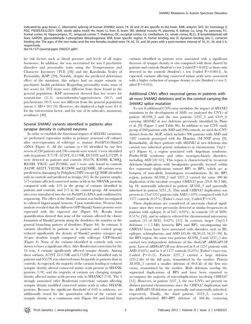
A de novo deletion of SHANK2 in a patient with ASDBerkel et al. 2010 recently identified two independent de novo
SHANK2 deletions in two patients, one with ID and another one
with ASD [26]. In addition, whole genome analysis performed by
the Autism Genome Project (AGP) using Illumina 1M single
nucleotide polymorphism (SNP) arrays detected one additional de
novo SHANK2 deletion in a patient (6319_3) with ASD [9] (the
second patient described by the AGP, 5237_3, is patient SK0217-
003 reported in Berkel et al. 2010 [26]). Recently, a 3.4 Mb de
novo deletion including SHANK2 was observed in a female patient
with speech and developmental delay [35]. To follow up on these
results, we genotyped an independent sample of 260 patients with
ASD using Illumina 1M Duo SNP arrays (Table S1). In this
sample, we detected a 421.2 kb deletion within SHANK2 in patient
AU038_3 with autism and moderate ID (see patient section in
Materials and Methods, and Table S2). The deletion covered
twelve exons (E5–E16) and altered all SHANK2 isoforms
(Figure 2A). No other deleterious variants in the remaining copy
of SHANK2 were detected by sequencing. The parents did not
carry the deletion, indicating a de novo event. The deletion was
validated by quantitative PCR analysis using DNA from an
independent blood sample from all members of the family and
SNP analysis indicated that the deletion originated on the
maternal chromosome (Figure S2). SHANK2 deletions were absent
in more than 5000 controls [9,26] and not listed in the Database of
Genomic Variants (DGV; http://projects.tcag.ca/variation/).
SHANK2 coding variants affecting conserved amino acidsare enriched in patients with ASD
To probe for additional mutations, we first sequenced all exons
of the longest SHANK2E isoform in 230 patients with ASD and
230 controls. We then sequenced an additional sample of 225
patients and 201 controls (Table S1) for the ProSAP1A isoform that
corresponds to the major SHANK2 isoform in the brain. Since we
screened all SHANK2 isoforms, we used a nomenclature including
the SHANK2E isoform that differed from Berkel et al. 2010 [26].
Within the 9 coding exons specific to SHANK2E, we identified
R174C (rs7926203) listed in dbSNP in 2 independent patients with
ASD and R185Q in one patient with ASD. For this isoform, no
variant was identified in the control sample. Within the ProsSAP1A
isoform, we identified 24 non-synonymous variations. When these
results are integrated with those obtained by Berkel et al. 2010, a
total of 40 variants of ProsSAP1A including 3 already reported in
dbSNP were identified (Figure 2B, Table 1, Figure S3). Only two
variants (Y967C and R569H) with MAF.1% are detected and
there is no enrichment of rare variants of SHANK2 (MAF,1%) in
patients with ASD compared with controls. Because variants
affecting conserved amino acids in the SHANK proteins are most
likely to have a functional effect, we tested whether there was an
enrichment of these variants in patients compared to controls. The
alignment of the SHANK protein sequences and the conservation
of the variants are indicated in the Table S5. In both mutation
screening studies, the first performed by Berkel et al. 2010 and the
second presented here, we observed an enrichment of variants
affecting conserved amino acids in patients compared with
controls (Figure 2C, Table S5 and Table S7). Overall, 12 of 15
(80%) of the variants identified only in the patient sample affected
conserved amino acids compared with only 6 of 17 (35.3%) in
controls (Fisher’s exact test 1-sided, P = 0.013, OR = 6.83, 95%
IC = 1.19–53.40). Because several independent patients carried
these variants (Table 1), the enrichment is even more significant
when the number of carriers was considered. The variants
affecting conserved amino acids were observed in 29 of 851
(3.4%) patients and in 16 of 1090 (1.5%) controls (Fisher’s exact
test 1-sided, P = 0.004, OR = 2.37, 95% CI = 1.23–4.70). A total of
8 variants were identified in patients and controls. Among these 8
variants, 2 affected conserved amino acids (R818H and S557N).
The variant S557N was observed in 9 of 851 (1.06%) independent
families with ASD and in 3 of 1090 (0.28%) controls (Fisher’s
Exact Test one sided, P = 0.029, OR = 3.87; 95% CI = 0.96–
22.29). It affects a conserved serine with a high probability of being
phosphorylated and located in the SH3 domain of all SHANK
proteins. This domain binds to GRIP and b-PIX, two proteins
linking SHANK to glutamate AMPA receptors and actin skeleton,
respectively [36]. In our initial mutation screen, R818H was
observed in 5 of 230 patients with ASD and 0 of 230 controls. In
order to determine if R818H was more frequent in the patients
with ASD, we screened an additional sample of 3020 individuals
with ASD, 1783 controls from European descent, and the Human
Genome Diversity Panel (HGDP) control dataset (Table S3 and
S4). R818H was virtually absent outside Europe and had the
highest allelic frequency (2.37%) in Finland, but overall its
frequency was not higher in patients with ASD compared with
controls (ASD 32/3250 (1.0%); controls 27/2030 (1.33%); Fisher’s
exact test 2-sided, P = 0.28) (Table S3).
Finally, and unexpectedly, during this additional mutation
screening, we detected a variation (IVS22+1G.T) altering the
consensus donor splice site of exon 22 in a Swedish control,
SWE_Q56_508 (Figure 3A). This variant was predicted to disrupt
all SHANK2 isoforms by deleting the proline rich and the SAM
domain, except for the shortest isoform AF141901, where the
mutation is located in the open reading frame (ORF) and should
lead to a G263V change. This variant was not observed in 1786
patients or 1407 controls, and is not listed in dbSNP. This control
female was part of a previous epidemiological study [37] and had
been extensively examined for anthropometrics and cardiovascu-
Author Summary
Autism spectrum disorders (ASD) are a heterogeneousgroup of neurodevelopmental disorders with a complexinheritance pattern. While mutations in several genes havebeen identified in patients with ASD, little is known abouttheir effects on neuronal function and their interactionwith other genetic variations. Using a combination ofgenetic and functional approaches, we identified novelSHANK2 mutations including a de novo loss of one copy ofthe SHANK2 gene in a patient with autism and severalmutations observed in patients that reduced neuronal cellcontacts in vitro. Further genomic analysis of three patientscarrying de novo SHANK2 deletions identified additionalrare genomic imbalances previously associated withneuropsychiatric disorders. Taken together, these resultsstrengthen the role of synaptic gene dysfunction in ASDbut also highlight the presence of putative modifier genes,which is in keeping with the ‘‘multiple hit model’’ for ASD.A better knowledge of these genetic interactions will benecessary to understand the complex inheritance patternof ASD.
SHANK2 Mutations in Autism Spectrum Disorders
PLoS Genetics | www.plosgenetics.org 3 February 2012 | Volume 8 | Issue 2 | e1002521
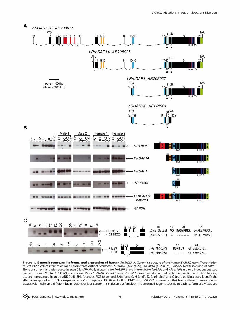
Figure 1. Genomic structure, isoforms, and expression of human SHANK2. A. Genomic structure of the human SHANK2 gene. Transcriptionof SHANK2 produces four main mRNA from three distinct promoters: SHANK2E (AB208025), ProSAP1A (AB208026), ProSAP1 (AB208027) and AF141901.There are three translation starts: in exon 2 for SHANK2E, in exon1b for ProSAP1A, and in exon1c for ProSAP1 and AF141901; and two independent stopcodons: in exon 22b for AF141901 and in exon 25 for SHANK2E, ProSAP1A and ProSAP1. Conserved domains of protein interaction or protein bindingsite are represented in color: ANK (red), SH3 (orange), PDZ (blue) and SAM (green), H (pink), D, (dark blue) and C (purple). Black stars identify thealternative spliced exons (‘brain-specific exons’ in turquoise: 19, 20 and 23). B. RT-PCRs of SHANK2 isoforms on RNA from different human controltissues (Clontech), and different brain regions of four controls (2 males and 2 females). The amplified regions specific to each isoform of SHANK2 are
SHANK2 Mutations in Autism Spectrum Disorders
PLoS Genetics | www.plosgenetics.org 4 February 2012 | Volume 8 | Issue 2 | e1002521
lar risk factors such as blood pressure and levels of all major
hormones. In addition, she was ascertained for axis I psychiatric
disorders and personality traits using the Temperament and
Character Inventory (TCI) [38] and the Karolinska Scales of
Personality (KSP) [39]. Notably, despite the predicted deleterious
effect of the mutation, this subject had no major somatic or
psychiatric health problems. Regarding personality traits, none of
her scores for TCI items were different from those found in the
general population. KSP assessment showed that her scores for
neuroticism (51.3), nonconformity/aggressiveness (56.7), and
psychoticism (50.5) were not different from the general population
(mean 6 SD = 50610). However, she displayed a high score (61.4)
for the extraversion factor, and for one of its subscales, monotony
avoidance [40].
Several SHANK2 variants identified in patients altersynapse density in cultured neurons
In order to establish the functional impact of SHANK2 variations,
we performed expression studies in primary neuronal cell cultures
after over-expression of wild-type vs. mutant ProSAP1A/Shank2A
cDNA (Figure 4). All the variants (n = 16) identified by our first
screen of 230 patients and 230 controls were tested: 5 were identified
only in patients (V717F, A729T, G1170R, D1535N and L1722P), 6
were detected in patients and controls (S557N, R569H, K780Q,
R818H, Y967C and P1586L) and 5 were only found in controls
(L629P, A822T, V823M, R1290W and Q1308R). All variants were
predicted as damaging by Polyphen2 DIV except Q1308R identified
only in controls and predicted as benign [41]. In the patient sample,
5/5 variants affected conserved amino acids in the SHANK proteins
compared with only 2/6 in the group of variants identified in
patients and controls, and 2/5 in the control group. All mutation
sites were introduced into the rat ProSAP1A cDNA and confirmed by
sequencing. The effect of the Shank2 variants was further investigated
in cultured hippocampal neurons. Upon transfection, Western blot
analysis revealed that the different GFP-Shank2 fusion proteins were
expressed with the expected size (Figure S4). Results from
quantification showed that none of the variants affected the cluster
formation of Shank2 protein along the dendrites, the number or the
general branching pattern of dendrites (Figure S4). In contrast, 8
variants identified in patients or in patients and control group
reduced significantly the density of Shank2 positive synapses per
10 mm dendrite length compared with wild-type GFP-Shank2
(Figure 4). None of the variants identified in controls only were
shown to have a significant effect. After Bonferonni correction for the
16 tests, 4 variants significantly affected synapse density. Among
these variants, A729T, G1170R and L1722P were identified only in
patients and S557N was observed more frequently in patients than in
controls. As expected, the majority of the variants leading to reduced
synaptic density altered conserved amino acids present in SHANK
proteins (7/8), and the majority of variants not changing synaptic
density affected amino acids present only in SHANK2 (7/8). The 4
strongly associated (after Bonferroni correction) variants affecting
synaptic density modified conserved amino acids in other SHANK
proteins. Because the significant threshold of 0.05 is arbitrary, we
additionally tested for the quantitative effect of the variant on
synaptic density as a continuous trait (Figure S4) and found that
variants identified in patients were associated with a significant
decrease of synapse density in vitro compared with those shared by
patients and controls (Student’s t test 2-sided P = 0.022) or those only
detected in the controls (Student’s t test 2-sided P = 0.0013). As
expected, variants affecting conserved amino acids were associated
with a higher reduction of synapse density in vitro (Student’s t test 2-
sided P = 0.014).
Additional CNVs affect neuronal genes in patients withde novo SHANK2 deletions and in the control carrying theSHANK2 splice mutation
To test if additional CNVs may modulate the impact of SHANK2
mutations in the development of ASD, we analyzed the CNVs of
patient AU038_3 and the two patients (5237_3 and 6319_3)
carrying SHANK2 de novo deletions previously identified by Pinto
et al. [9] (Figure 5 and Table S6). In addition to our CNV study
group of 260 patients with ASD and 290 controls, we used the CNV
dataset from the AGP, which includes 996 patients with ASD and
1287 controls genotyped with the Illumina 1M SNP array [9].
Remarkably, all three patients with SHANK2 de novo deletions also
carried rare inherited genetic imbalances at chromosome 15q11–
q13 (Figure 6), a region associated with Angelman syndrome,
Prader-Willi syndrome and other neuropsychiatric disorders,
including ASD [42–61]. This region is characterized by recurrent
deletions/duplications with breakpoints generally located within
five segmental duplications named BP1 to BP5, which act as
hotspots of non-allelic homologous recombination. In the BP5
region, patients AU038_3 and 5237_3 carried the same 496 kb
duplication of the nicotinic receptor CHRNA7 gene (29.8–30.3 Mb,
hg 18; maternally inherited in patient AU038_3 and paternally
inherited in patient 5237_3). This small CHRNA7 duplication was
present in 13 of 1257 patients with ASD (1.03%) compared with 9 of
1577 controls (0.57%) (Fisher’s exact test, 2-sided P = 0.19).
These duplications are considered of uncertain clinical signif-
icance since they were previously detected at similar frequencies in
patients with epilepsy (6 of 647, 0.93%), in controls (19 of 3699,
0.51%) [50], and in subjects referred for chromosomal microarray
analysis (55 of 8832, 0.62%) [51]. In contrast, larger 15q13.3
deletions (,1.5 Mb) between BP4 and BP5, encompassing the
CHRNA7 locus have been associated with disorders such as ID,
epilepsy, schizophrenia, and ASD [43,46–48,50,52–54,57–59]. In
the BP4 region, the same two patients AU038_3 and 5237_3 also
carried two independent deletions of the rhoGAP ARHGAP11B
gene. Loss of ARHGAP11B was detected in 8 of 1257 patients with
ASD (0.64%) and in 4 of 1577 controls (0.25%) (Fisher’s exact test,
2-sided P = 0.15). Patient 5237_3 carried a large deletion
(235.2 kb) of the full gene, transmitted by the mother. Patient
AU038_3 carried a smaller deletion of 49.8 kb of the first two
exons, transmitted by the mother. Both deletions overlap the
segmental duplications of BP4 and have been reported to
accompany the majority of microduplications involving CHRNA7
[51]. However, in patient 5237_3, the two CNVs are present on
distinct parental chromosomes since the CHRNA7 duplication and
the ARHGAP11B deletion are paternally and maternally inherited,
respectively. Finally, the third patient, 6319_3, carried a
paternally-inherited BP1-BP2 deletion of 468 kb, removing
indicated by gray boxes. C. Alternative splicing of human SHANK2; exons 19, 20 and 23 are specific to the brain. ANK, ankyrin; SH3, Src homology 3;PDZ, PSD95/DLG/ZO1; SAM, sterile alpha motif; He, heart; Li, liver; B, brain; SM, skeletal muscle; Pl, placenta; K, kidney; Lu, lung; Pa, pancreas; FC,frontal cortex; Hi, hippocampus; TC, temporal cortex; T, thalamus; OC, occipital cortex; Ce, cerebellum; Cx, whole cortex; BLCL, B lymphoblastoid celllines; GAPDH, glyceraldehyde 3-phosphate dehydrogenase; BSR, brain specific region; H, homer binding site; D, dynamin binding site; C, cortactinbinding site. The ages of the two males and the two females studied were 74, 42, 55, and 36 years with a post-mortem interval of 10, 21, 24, and 2 h,respectively.doi:10.1371/journal.pgen.1002521.g001
SHANK2 Mutations in Autism Spectrum Disorders
PLoS Genetics | www.plosgenetics.org 5 February 2012 | Volume 8 | Issue 2 | e1002521
NIPA1, NIPA2, CYFIP1, and TUBGCP5. This deletion was
observed in 4 of 1257 patients with ASD (0.32%) and in 4 of
1577 controls (0.25%) (Fisher’s exact test, 2-sided P = 0.74). The
BP1-BP2 deletion is associated with phenotypic variability and has
been reported in individuals with neurodevelopmental disorders
[20], schizophrenia [53,60], ASD [44–46,49], and epilepsy [61].
In a recent screen for large CNVs (.400 kb) performed on 15,767
children with ID and various congenital defects, and 8,329
unaffected adult controls [20], deletions affecting CYFIP1, NIPA1,
NIPA2 and TUBGCP5 were associated with neurodevelopmental
disorder (P = 4.7361026), epilepsy (P = 1.4861023) and autism
(P = 1.9961022).
Figure 2. SHANK2 mutations in patients with ASD. A. A heterozygous deletion of SHANK2 was identified with the Illumina Human 1M-Duo SNParray in a patient with autism (AU038_3). The deletion spans 421 kb on chromosome 11q13.3, covers twelve exons of the human SHANK2 and is notpresent in the parents. Each dot shows Log R Ratio (LRR; in red) and B allele frequency (BAF; in green). QuantiSNP score is represented with a blue lineand indicates the deletion size. B. Location of the CNV and sequence variants (from this study and Berkel et al. 2010) along the SHANK2 protein: in redthe variations specific to ASD, in orange the variations shared by ASD and controls and in green the variations specific to controls [26]. Thebreakpoints of the SHANK2 deletion in AU038_3 are represented with a dotted line on the protein. Stars indicate the variants affecting conservedamino acids. C. A total of 40 variants were identified and variants affecting conserved amino acids in other SHANK proteins are enriched in patientswith ASD (nconserved = 12 and nnon-conserved = 3) compared with controls (nconserved = 6 and nnon-conserved = 11) (Fisher’s exact test 1-sided, P = 0.013,OR = 6.83, 95% IC = 1.19–53.40). D. The percentage of carriers of SHANK2 variants in patients with ASD and Controls. Variants affecting a conservedamino acid among the SHANK proteins are enriched in patients with ASD (nconserved = 29 and nnon-conserved = 822) compared with controls(nconserved = 16 and nnon-conserved = 1074) (Fisher’s exact test 1-sided, P = 0.004, OR = 2.37, 95% CI = 1.23–4.70). Open squares and filled squaresrepresent the non-conserved and conserved amino acids, respectively. ANK, Ankyrin repeat domain; SH3, Src homology 3 domain; PDZ, postsynapticdensity 95/Discs large/zona occludens-1 homology domain; SAM, sterile alpha motif domain; BSR, brain specific region; H, homer binding site; D,dynamin binding site; C, cortactin binding site. The proline-rich region is represented as a horizontal gray line.doi:10.1371/journal.pgen.1002521.g002
SHANK2 Mutations in Autism Spectrum Disorders
PLoS Genetics | www.plosgenetics.org 6 February 2012 | Volume 8 | Issue 2 | e1002521
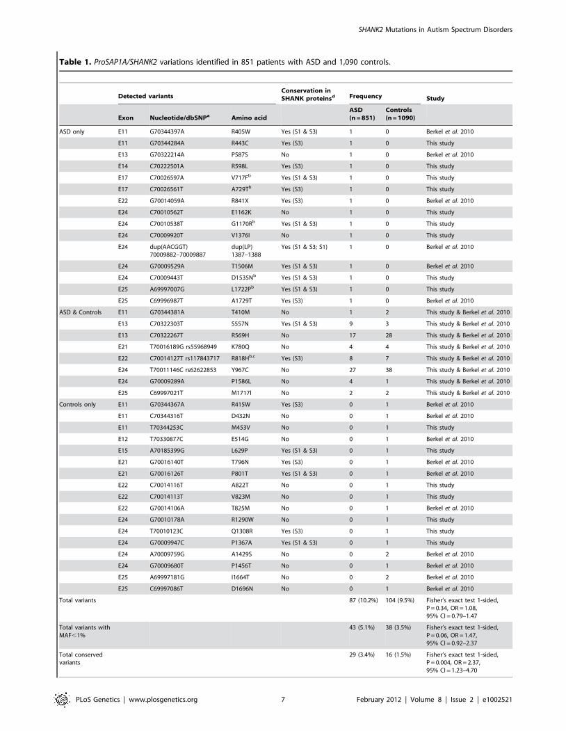
Table 1. ProSAP1A/SHANK2 variations identified in 851 patients with ASD and 1,090 controls.
Detected variantsConservation inSHANK proteinsd Frequency Study
Exon Nucleotide/dbSNPa Amino acidASD(n = 851)
Controls(n = 1090)
ASD only E11 G70344397A R405W Yes (S1 & S3) 1 0 Berkel et al. 2010
E11 G70344284A R443C Yes (S3) 1 0 This study
E13 G70322214A P587S No 1 0 Berkel et al. 2010
E14 C70222501A R598L Yes (S3) 1 0 This study
E17 C70026597A V717Fb Yes (S1 & S3) 1 0 This study
E17 C70026561T A729Tb Yes (S3) 1 0 This study
E22 G70014059A R841X Yes (S3) 1 0 Berkel et al. 2010
E24 C70010562T E1162K No 1 0 This study
E24 C70010538T G1170Rb Yes (S1 & S3) 1 0 This study
E24 C70009920T V1376I No 1 0 This study
E24 dup(AACGGT)70009882–70009887
dup(LP)1387–1388
Yes (S1 & S3; S1) 1 0 Berkel et al. 2010
E24 G70009529A T1506M Yes (S1 & S3) 1 0 Berkel et al. 2010
E24 C70009443T D1535Nb Yes (S1 & S3) 1 0 This study
E25 A69997007G L1722Pb Yes (S1 & S3) 1 0 This study
E25 C69996987T A1729T Yes (S3) 1 0 Berkel et al. 2010
ASD & Controls E11 G70344381A T410M No 1 2 This study & Berkel et al. 2010
E13 C70322303T S557N Yes (S1 & S3) 9 3 This study & Berkel et al. 2010
E13 C70322267T R569H No 17 28 This study & Berkel et al. 2010
E21 T70016189G rs55968949 K780Q No 4 4 This study & Berkel et al. 2010
E22 C70014127T rs117843717 R818Hb,c Yes (S3) 8 7 This study & Berkel et al. 2010
E24 T70011146C rs62622853 Y967C No 27 38 This study & Berkel et al. 2010
E24 G70009289A P1586L No 4 1 This study & Berkel et al. 2010
E25 C69997021T M1717I No 2 2 This study & Berkel et al. 2010
Controls only E11 G70344367A R415W Yes (S3) 0 1 Berkel et al. 2010
E11 C70344316T D432N No 0 1 Berkel et al. 2010
E11 T70344253C M453V No 0 1 This study
E12 T70330877C E514G No 0 1 Berkel et al. 2010
E15 A70185399G L629P Yes (S1 & S3) 0 1 This study
E21 G70016140T T796N Yes (S3) 0 1 Berkel et al. 2010
E21 G70016126T P801T Yes (S1 & S3) 0 1 Berkel et al. 2010
E22 C70014116T A822T No 0 1 This study
E22 C70014113T V823M No 0 1 This study
E22 G70014106A T825M No 0 1 Berkel et al. 2010
E24 G70010178A R1290W No 0 1 This study
E24 T70010123C Q1308R Yes (S3) 0 1 This study
E24 G70009947C P1367A Yes (S1 & S3) 0 1 This study
E24 A70009759G A1429S No 0 2 Berkel et al. 2010
E24 G70009680T P1456T No 0 1 Berkel et al. 2010
E25 A69997181G I1664T No 0 2 Berkel et al. 2010
E25 C69997086T D1696N No 0 1 Berkel et al. 2010
Total variants 87 (10.2%) 104 (9.5%) Fisher’s exact test 1-sided,P = 0.34, OR = 1.08,95% CI = 0.79–1.47
Total variants withMAF,1%
43 (5.1%) 38 (3.5%) Fisher’s exact test 1-sided,P = 0.06, OR = 1.47,95% CI = 0.92–2.37
Total conservedvariants
29 (3.4%) 16 (1.5%) Fisher’s exact test 1-sided,P = 0.004, OR = 2.37,95% CI = 1.23–4.70
SHANK2 Mutations in Autism Spectrum Disorders
PLoS Genetics | www.plosgenetics.org 7 February 2012 | Volume 8 | Issue 2 | e1002521
Several additional CNVs also altered compelling candidate genes
for susceptibility to ASD. In patient AU038_3 we detected a
previously unreported paternally inherited intronic duplication of
CAMSAP1L on chromosome 1q32.1, coding for a calmodulin
regulated spectrin-associated protein highly expressed in the brain.
Patient 5237_3 carried a de novo deletion altering the coding
sequence of the tyrosine phosphatase DUSP22 on chromosome
6p25.3 and a maternally inherited intronic duplication of NLGN1 on
chromosome 3q26.3 [9]. These CNVs were observed at similar
frequencies in patients with ASD compared with controls. DUSP22
deletions were observed in 8 of 1257 patients with ASD (0.64%) and
in 14 of 1577 controls (0.89%), while NLGN1 intronic duplications
were observed in 60 of 1257 patients with ASD (4.77%) and in 62
of 1577 controls (3.93%). Finally, patient 6319_3 carried an
unreported maternally inherited intronic deletion of contactin
CNTN4, a gene on chromosome 3p26.3 associated with ASD [62],
as well as a paternally inherited deletion within the protocadherin
PCDHA1-10 gene cluster on chromosome 5q31.3. Interestingly, this
deletion removes the first exon of both PCDH8 and PCDH9 and was
significantly less frequent in patients with ASD compared with
controls (ASD: 62 of 1257; controls: 132 of 1577; Fisher’s exact test,
2-sided P = 0.0003; OR = 0.57; 95% CI = 0.41–0.78).
We also analyzed the genome of the Swedish control
SWE_Q56_508 carrying the SHANK2 splice mutation using the
Human Omni2.5 BeadChip array from Illumina (Figure 3B). Two
close duplications on 2p25.3 were detected, altering four genes,
LOC391343, SNTG2, PXDN and MYT1L. The inheritance of these
two duplications could not be investigated, because DNA samples
from the parents were not available. However, 2 of 1577 controls
also carried of the same close duplications, suggesting that these
CNVs are located on the same chromosome. Among the affected
genes, syntrophin-c2 (SNTG2) and myelin transcription factor 1-
like (MYT1L) are expressed in the brain. Alterations of SNTG2 and
MYT1L have been previously reported in patients with ASD
[20,63,64] and schizophrenia [65], respectively. SNTG2 is a
scaffolding protein interacting with the NLGN3/4X proteins [66]
aNucleotide positions are according to NM 012309.3 from NCBI36/hg18 on the positive DNA strand; The patients with ASD and the controls used for this analysis camefrom this study (455 ASD & 431 controls) and from the study of Berkel et al. 2010 (396 ASD & 659 controls);
bA screening of V717F, A729T, R818H, G1170R, D1535N and L1722P was performed in 948 subjects from the Human Genome Diversity Panel (V717F = 0/948; A729T = 0/948; R818H = 5/948; G1170R = 0/948; D1535N = 0/948; L1722P = 0/948);
cA screen of R818H was performed in additional patients and controls (ASD 32/3250 (1.0%); controls 27/2030 (1.33%); Fisher’s exact test 2-sided, P = 0.28). Fisher’s exacttest was used for statistical analysis;
d‘‘Yes’’ indicates if amino acid is conserved in SHANK1 (S1), SHANK3 (S3) or both (S1 & S3); MAF, Minor Allele Frequency.doi:10.1371/journal.pgen.1002521.t001
Table 1. Cont.
Figure 3. Genetic alterations identified in the control subject SWE_Q56_508. A. SHANK2 splice mutation (IVS22+1G.T) detected in aSwedish female control, SWE_Q56_508. The mutation altered the donor splicing site of exon 22 and led to a premature stop in all SHANK2 isoformsexcept for the AF1411901 isoform, where it altered the protein sequence (G263V). B. CNVs in the same individual altering LOC339822, SNTG2, PXDNand MYT1L. The two close duplications span 264 kb and 245 kb on chromosome 2 and altered LOC339822 and SNTG2, and PXDN and MYT1L,respectively. Dots show the B allele frequency (BAF; in green), Log R ratio (LRR; in red), and QuantiSNP score (in blue). Lower panel: all CNVs listed inthe Database of Genomic Variants (DGV) are represented: loss (in red), gain (in blue), gain or loss (in brown). H, homer binding site; D, dynaminbinding site; C, cortactin binding site.doi:10.1371/journal.pgen.1002521.g003
SHANK2 Mutations in Autism Spectrum Disorders
PLoS Genetics | www.plosgenetics.org 8 February 2012 | Volume 8 | Issue 2 | e1002521
and a component of the dystrophin glycoprotein complex [67].
MYT1L is a myelin transcription factor required to convert mouse
embryonic and postnatal fibroblasts into functional neurons [68].
Discussion
Deleterious SHANK2 variations are enriched in patientswith ASD, but also observed in controls
The identification of mutations in synaptic proteins such as
NRXN1, NLGN3/4X and SHANK2/3 has demonstrated that a
synaptic defect might be at the origin of ASD [5,6]. Here we
confirm the presence of SHANK2 de novo deletions in individuals
with ASD, with a prevalence of 0.38% (1/260) in our cohort of
ASD patients analyzed with the Illumina 1M SNP array. This
frequency is similar to the one reported previously by the AGP in a
larger sample of 996 patients with ASD (0.2%) [9]. SHANK2
deletions altering exons were not detected in controls, in
agreement with previous findings [9,26]. As reported for SHANK3
[7], no other coding variations were detected in the remaining
SHANK2 allele of the deletion carriers, suggesting that, in some
Figure 4. Characterization of the functional impact of SHANK2 mutations in cultured neuronal cells. A. The colocalization of ProSAP1A/SHANK2-EGFP (postsynaptic marker) and Bassoon (presynaptic marker) indicated that the mutations did not disturb the formation of SHANK2 clustersat excitatory synapses along the dendrites. B. The quantification of synapse density was performed on 20 transfected hippocampal neurons perconstruct from at least three independent experiments. The majority of the ProSAP1A variants affecting a conserved amino acid among SHANKproteins reduced significantly the synaptic density compared with the variants that affect amino acid non conserved among SHANK proteins (Mann-Whitney U-test: nWT = 20, nmut = 20; US557N = 82.5, pS557N = 0.001; UR569H = 124, pR569H = 0.04; UL629P = 149, pL629P = 0.17; UV717F = 114, pV717F = 0.02;UA729T = 73, pA729T = 0.000; UK780Q = 154, pK780Q = 0.221; UR818H = 108, pR818H = 0.012; UA822T = 154.5, pA822T = 0.224; UV823M = 129, pV823M = 0.056;UY967C = 134, pY967C = 0.076; UG1170R = 78, pG1170R = 0.001; UR1290W = 142, pR1290W = 0.121; UQ1308R = 162, pQ1308R = 0.314; UD1535N = 97, pD1535N = 0.005;UP1586L = 137, pP1586L = 0.910; UL1722P = 79, pL1722P = 0.001, *p,0.05, **p,0.01, ***p,0.001). C. Effect of the variants on synaptic density. The y-axisrepresents 2log P compared to WT (P obtained with Mann-Whitney test). After Bonferroni correction for 16 tests, only P values,0.003 wereconsidered as significant. Variants represented in red were specific to ASD, in orange were shared by ASD and controls, and in green were specific tothe controls. Open circles and filled circles represent non conserved and conserved amino acids, respectively. Prim, primary; second, secondary.doi:10.1371/journal.pgen.1002521.g004
SHANK2 Mutations in Autism Spectrum Disorders
PLoS Genetics | www.plosgenetics.org 9 February 2012 | Volume 8 | Issue 2 | e1002521
individuals, a de novo deletion of a single allele of SHANK2 might be
sufficient to increase the risk for ASD. In one case, a patient
carried two rare SHANK2 variants predicted as deleterious and
inherited from different parents, indicating that they were separate
alleles.
For the remaining SHANK2 variants, patients were heterozygous
for non-synonymous rare variations inherited from one of their
parents (Figure S3). Since parents were apparently asymptomatic,
the causative role of these variants in ASD remains difficult to
ascertain. However, we observed a significant enrichment of
SHANK2 variants affecting conserved amino acids in patients with
ASD compared with controls. This was also the case in the
previous mutation screening by Berkel et al. 2010 [26]. The
majority of the variants affecting conserved residues and identified
in the patients were shown to alter the ability of SHANK2 to
increase the number of synapses in vitro. Importantly, the assays
performed in this study show that the variants can potentially
impact on the function of the protein, but they do not confirm that
they have deleterious effects on neuronal function in vivo in people
that carry them. However, these results are consistent with
Figure 5. Characterization of CNVs in three patients carrying a de novo deletion of SHANK2. Paternally or maternally inherited CNVs areindicated by squares and circles, respectively. De novo CNVs are indicated by stars. Deletions and duplications are indicated in red and blue,respectively. CNVs hitting exons or only introns are filled with grey and white, respectively. Squares and circles within star represent de novo CNV ofpaternal or maternal origin; circles within squares represent CNV inherited by father or mother. ABCC6, ATP-binding cassette, sub-family C, member 6pseudogene 2; ADAM, ADAM metallopeptidase; AMY1, amylase (salivary); AMY2A, amylase (pancreatic); ARHGAP11B, Rho GTPase activating protein11B; CAMSAP1L1, calmodulin regulated spectrin-associated protein 1-like 1; CHRNA7, cholinergic receptor, nicotinic, alpha 7; CNTN4, contactin 4;CTNNA3, catenin (cadherin-associated protein), alpha 3; CYFIP1, cytoplasmic FMR1 interacting protein 1; DUSP22, dual specificity phosphatase 22;GALM, galactose mutarotase; GCNT2, glucosaminyl (N-acetyl) transferase 2; GOLGA, golgi autoantigen, golgin subfamily a; GSTT1, glutathione S-transferase theta 1; HLA-DRB, major histocompatibility complex, class II, DR beta; LAMA4, laminin, alpha 4; NIPA, non imprinted in Prader-Willi/Angelman syndrome; NLGN1, neuroligin 1; NME7, non-metastatic cells 7; OR, olfactory receptor; PCDHA, protocadherin alpha; RFPL4B, ret fingerprotein-like 4B; RHD, Rh blood group, D antigen; SFMBT1, Scm-like with four mbt domains 1; SHANK2, SH3 and multiple ankyrin repeat domains 2;SMC2, structural maintenance of chromosomes 2; TNS3, tensin 3; TUBGCP5, tubulin, gamma complex associated protein 5; UGT2B17, UDPglucuronosyltransferase 2 family, polypeptide B17.doi:10.1371/journal.pgen.1002521.g005
SHANK2 Mutations in Autism Spectrum Disorders
PLoS Genetics | www.plosgenetics.org 10 February 2012 | Volume 8 | Issue 2 | e1002521
previous findings showing that inherited variants of SHANK2 and
SHANK3 cause synaptic defects in vitro [7,69,70]. Recently, Berkel
et al. 2011 showed that two inherited (L1008_P1009dup, T1127M)
and one de novo (R462X) SHANK2 mutations identified in patients
with ASD affect spine volume and reduced Shank2 cluster sizes
[70]. This deleterious effect was also observed in vivo since mice
expressing rAAV-transduced Shank2-R462X present a specific
long-lasting reduction in miniature postsynaptic AMPA receptor
currents [70].
In patients, the only feature associated with carriers of SHANK2
mutations compared with other patients was a trend for low IQ
(P = 0.025, OR = 3.75, 95% CI = 1.1–20.0) (Table S8). But, as
observed for SHANK3 mutations, this correlation could differ from
one individual to another (i.e. the patient with a SHANK2 de novo
stop mutation reported by Berkel et al. 2010 presented with high-
functioning autism [26]).
Our result also showed that potentially deleterious SHANK2
variants were detected in a heterozygous state in parents and in the
general population without causing severe phenotypic conse-
quences. Indeed, we showed that almost 5% of the Finnish
population is heterozygous for the SHANK2 R818H variation,
which modifies a conserved amino acid and is associated with
Figure 6. Inherited 15q11–q13 CNVs identified in three ASD patients carrier of a de novo SHANK2 deletion. Deletions (del) andduplications (dup) are indicated in red and blue, respectively. Paternally and maternally imprinted genes are indicated in yellow and pink,respectively. Genes altered by the CNVs are indicated in blue or red. The bottom part of the figure indicates the location of the deletions/duplicationspreviously associated with neuropsychiatric disorders [43–61]. BP, breakpoint; Inh_M, inherited by mother; Inh_F, inherited by father; AS, Angelmansyndrome; ASD, Autism spectrum disorders; ADHD, attention deficit-hyperactivity disorder; BP, bipolar disorder; DD: developmental delay; DBD,disruptive behavior disorder; EPI, epilepsy; GAD, generalized anxiety disorder; OCD, obsessive-compulsive disorder; ID, intellectual disability; PWS,Prader-Willi syndrome; SCZ, schizophrenia.doi:10.1371/journal.pgen.1002521.g006
SHANK2 Mutations in Autism Spectrum Disorders
PLoS Genetics | www.plosgenetics.org 11 February 2012 | Volume 8 | Issue 2 | e1002521
lower synaptic density in vitro. Furthermore, we identified a
SHANK2 splice site mutation in a control female without any
apparent psychiatric disorders. Similarly, two frame-shift muta-
tions and one splice site mutation of SHANK2 are listed in dbSNP
and in the 1000 genomes project [71]. These nonsense variations
should be interpreted with caution since none of them has been
validated by Sanger sequence technology. Taken together,
variants affecting conserved amino acids of SHANK2 might act
as susceptibility variants for ASD, but, in some cases, additional
genetic, epigenetic or environmental factors seem to be necessary
for the emergence of the disorder.
Additional CNVs in subjects with SHANK2 mutations maymodulate the risk for ASD
In order to detect risk and protective genetic factors, we
analyzed the CNV burden of the individuals carrying deleterious
variations of SHANK2. Notably, the three ASD patients with de
novo SHANK2 deletions also carried CNVs on chromosome 15q11–
q13, a region associated with ASD [43,47,48,50–52,72]. In
contrast, the patient reported by Berkel et al. 2010, who did not
meet all the diagnostic criteria for ASD, seemed to have no CNV
at chromosome 15q [26]. Although the probability to observe the
co-occurrence of a de novo SHANK2 deletion and a duplication of
CHRNA7 at 15q is very low, two of the three patients carrying a de
novo SHANK2 deletion also carried the CHRNA7 duplication. While
the numbers are small, this finding could suggest epistasis between
these two loci. The role of CHRNA7 in ASD was recently
supported by the observation of low levels of CHRNA7 mRNA in
the post-mortem brain from patients with ASD [73]. Interestingly,
it was also found that, in contrast to the gene copy number, the
transcript levels CHRNA7 were reduced in neuronal cells [74] or
brain samples with maternal 15q duplication [75]. Finally,
functional studies have shown that NLGN and NRXN, which
belong to the same synaptic pathway, are key organizers of the
clustering of nicotinic receptors at the synapse [76–78]. Therefore
the co-occurrence of a deletion of SHANK2 and a duplication of
the nicotinic receptor CHRNA7 could act together within the same
pathway to increase the risk of ASD in patients AU038_3 and
5237_3. In patient 6319_3 carrying the BP1–BP2 deletion, several
genes might also play a role in the susceptibility to ASD. Among
them, NIPA1 and TUBGCP5 encoding a magnesium transporter
and a tubulin gamma associated protein, respectively, are highly
expressed in the brain. However, the most compelling candidate in
the deleted region is CYFIP1 [45,53], which codes for a binding
partner of FMRP, the protein responsible for fragile X syndrome.
Both CYFIP1 and FMRP are involved in the repression of
synaptic translation [79], one of the major biological mechanisms
associated with ASD [80]. Therefore, the co-occurrence of a loss
of one copy of SHANK2 and CYFIP1 might increase the risk of
abnormal synaptic function in patient 6319_3.
If some individuals have a higher risk to develop ASD when a
deleterious SHANK2 variant is present, others individuals may
experience a protective effect by additional genetic factors. For
example, control SWE_Q56_508 carried a SHANK2 splice
mutation, but clinical examination revealed no major disorders.
In addition, this control individual also carried a partial duplication
of SNTG2 and MYT1L. Based on a single control subject, it is not
possible to formally prove that these additional hits at SNTG2 and/
or MYT1L acted as suppressor mutations, counteracting the
phenotypic effects of the SHANK2 splice mutation. However, the
encoded proteins may interact with the NRXN-NLGN-SHANK
pathway. Both SNTG2 and SHANK2 are scaffolding proteins
localized in actin rich structures [81–83] and bind directly to
neuroligins [66]. Furthermore, mutations of NLGN3/4X identified
in patients with ASD decrease their protein binding to SNTG2 [66].
In addition, MYT1L is a myelin transcription factor that is
sufficient, with only two other transcription factors, ASCL1 and
BRN2, to convert mouse embryonic and postnatal fibroblasts into
functional neurons in vitro [67]. Therefore, alterations of SNTG2
and/or MYTL1 might modulate synapse physiology and counter-
act the effect of the SHANK2 splice site mutation. We recently
highlighted the key role of synaptic gene dosage in ASD and the
possibility that a protein imbalance at the synapse could alter
synaptic homeostasis [6]. In the future, animal models should be
developed to test whether the effect of a primary mutation in a
synaptic protein complex (e.g. Shank2) can be reduced or suppressed
by a second mutation (e.g. Sntg2 or Myt1l). A similar suppressor
effect has been demonstrated by the decrease of abnormal behavior
of the Fmr1 mutant mice carrying a heterozygous mutation of the
metabotropic glutamate receptor mGluR5 [84].
Conclusions and perspectivesIn summary, we confirmed that de novo SHANK2 deletions are
present in patients with ASD and showed that several SHANK2
variants reduce the number of synapses in vitro. The genomic
profile of the patients carrying deleterious de novo SHANK2
deletions also points to a possible genetic epistasis between the
NRXN-NLGN-SHANK pathway and 15q11–q13 CNVs.
CHRNA7 and CYFIP1 were already proposed as susceptibility
genes for neuropsychiatric disorders [43,45,49,51], and our study
provides additional support for this association. Therefore, as
previously observed for ID [85], our results suggest that the co-
occurrence of de novo mutations, together with inherited variations
might play a role in the genetic susceptibility to ASD. Finally, our
analyses suggest the interesting possibility that deleterious
mutations of neuronal genes (e.g. SNTG2 and MYT1L) could
potentially counteract the effect of synaptic deleterious mutations
(e.g. SHANK2). The identification of risk and protective alleles
within the same subject is one of the main challenges for
understanding the inheritance of ASD. Initial results from the
1000 genomes project has estimated that, on average, each person
carries approximately 250 to 300 loss-of-function variants in
annotated genes and 50 to 100 variants previously implicated in
inherited disorders [71]. To date, it is not clear how many loci can
regulate synaptic homeostasis and how these variants interact with
each other to modulate the risk for ASD [6]. A better knowledge of
these genetic interactions will be necessary to understand the
complex inheritance pattern of ASD.
Materials and Methods
Ethics statementThis study was approved by the local Institutional Review
Board (IRB) and written inform consents were obtained from all
participants of the study. The local IRB are the ‘‘Comite de
Protection des Personnes’’ (Ile-de-France Hopital Pitie-Salpetriere
Paris) for France; the Sahlgrenska Academy Ethics committee,
University of Gothenburg for Sweden; the local IRB of the
medical faculty of JW Goethe University Frankfurt/Main for
Germany; the Committee #3 of the Helsinki University Hospital,
Finland; the ‘‘Comitato Etico IRCCS Fondazione Stella Maris’’ at
Stella Maris Institute, Calambrone (Pisa), Italy; the ‘‘Comitato
Etico Azienda Ospedaliera-Universitaria Policlinico-Vittorio Ema-
nuele’’, Catania, Italy.
PatientsPatients with ASD and analyzed for CNV analysis and/or
mutation screening are presented in Table S1. Patients were
SHANK2 Mutations in Autism Spectrum Disorders
PLoS Genetics | www.plosgenetics.org 12 February 2012 | Volume 8 | Issue 2 | e1002521
recruited by the PARIS (Paris Autism Research International
Sibpair) study at specialized clinical centers disposed in France,
Sweden, Germany, Finland, UK. The Autism Diagnostic
Interview-Revised (ADI-R) and Autism Diagnostic Observation
Schedule (ADOS) were used for clinical evaluation and diagnosis.
In Sweden, in some cases, the Diagnostic Interview for Social and
Communication Disorders (DISCO-10) was applied instead of the
ADI-R. Patients were included after a clinical and medical check-
up with psychiatric and neuropsychological examination, standard
karyotyping, fragile-X testing and brain imaging and EEG
whenever possible. All patients were from Caucasian ancestry.
The patient AU038_3 with a de novo SHANK2 deletion is an
11.05 year-old boy diagnosed with autism and moderate ID (Table
S2). He was the only child of non-consanguineous parents from
European descent. His parents had no relevant personal and
familial history of psychiatric or medical illness. He was born at 40
weeks of gestation, after normal pregnancy and delivery. Birth
weight, length and occipitofrontal head circumference were
2500 g (5th percentile), 48 cm (22nd percentile) and 31 cm (2nd
percentile), respectively. Apgar scores were 7 and 10 at 1 and
5 minutes, respectively. In the first year of life, the pediatrician
reports did not mention signs of hypotonia. At 2 months, he was
operated for an inguinal hernia. Motor acquisition was apparently
normal (sitting at 6 months), but with a late acquisition of walking,
at 18 months. Speech was severely delayed, without any apparent
regressive phase. Only a few words and sentences appeared when
he was 4 y and 6.5 y, respectively. His expressive language
remained limited to restrictive sentences, mainly dyssyntaxic. A
formal diagnostic assessment for autism was performed when he
was 11 years old. The scores of the Autism Diagnostic Interview-
Revised (ADI-R) domains were: social 24, communication 23, and
behavior 6 (cut-offs for autism diagnosis are 10, 8 (verbal autism)
and 3, respectively); the age at first symptoms was before 36
months. Cognitive evaluation with the Kaufman Assessment
Battery for Children (K-ABC) showed moderate intellectual deficit
(composite score 40). He required assistance with basic activities
such as eating and dressing. At examination, he had a normal
facial appearance, with a prominent chin. General and neurolog-
ical examinations were normal, except for hypermetropia and
astigmatism. High-resolution karyotype, fragile X testing, MLPA
analysis of telomeres and microdeletion/microduplication syn-
dromes, and metabolic screening for inherited disorders of
metabolism (urine amino acids, mucopolysaccharides and organic
acids, uric acid in blood and urine) were all normal. No significant
epileptic event was reported on the electroencephalogram.
The two male patients with de novo SHANK2 deletions reported
by Pinto et al. 2010 [9] (5237_3 and 6319_3) shared several
clinical features with patient AU038_3. Patient 5237_3 is a
Canadian subject diagnosed with autism (based on ADI-R and
ADOS) associated with below average non verbal IQ (,1st
percentile) and language (,1st percentile). He had minor
dysmorphic features including 5th finger clinodactyly and several
curled toes, and no history of epilepsy. Patient 6319_3 was
recruited in the same geographic area as patient AU038_3
(Grenoble, France) and was clinically diagnosed with PDD-NOS.
The ADI-R scores were: social 14, communication 8, behaviors 2
(cut-off for autism: 3); with an age at first symptoms ,36 months).
He had mild ID as evaluated with the WISC-III (full scale IQ 60,
performance IQ 60, verbal IQ 67). His language was delayed
(first words 24 m, first sentences 48 m), but functional. He had no
history of regression or epilepsy. The physical exam was normal,
except for large and prominent ears and flat feet; the neurological
exam was also normal. Similarly to patient AU038_3, he had
hypermetropia.
The control female carrying the splice site mutation
(IVS22+1G.T) was part of a cohort of 172 females recruited
for a study on obesity, anthropometrics, and cardiovascular risk
factors [37]. In addition, these women were assessed for axis I
psychiatric disorders and for personality traits using the Temper-
ament and Character Inventory (TCI) [38] and the Karolinska
Scales of Personality (KSP) [39]. This subject had no psychiatric
disorders and her TCI and KSP scores were similar to those found
in the general population.
Genomic structure and transcripts analysis of SHANK2To define the genomic structure of the human SHANK2 gene,
we used the two reference sequence genes from UCSC
(NM_012309 and NM_133266), one human mRNA from
GenBank (DQ152234) and three Rattus reference sequence genes
from UCSC (NM_201350, NM_133441 and NM_133440).
SHANK2 is transcribed in four isoforms described in GenBank
(AB208025, AB208026, AB208027 and AF141901) and is com-
posed of 25 exons. Transcript analysis of SHANK2 was performed
in human brain regions from four independent controls (two
females and two males) and in human tissues (heart, brain,
placenta, lung, liver, skeletal muscle, kidney, pancreas and B
lymphoblastoid cell lines) using the Clontech Multiple Tissue
cDNA panel (Clontech). Total RNA was isolated from control
human brain tissues by the acid guanidinium thiocyanate phenol
chloroform method and reverse transcribed by oligodT priming
using SuperScript II Reverse Transcriptase (Invitrogen). The PCR
was performed with HotStar Taq polymerase (Qiagen) and the
protocol used was 95uC for 15 min, followed by 40 cycles at 95uCfor 30 sec, 55 to 58uC for 30 sec, 72uC for 30 sec to 1 min, with a
final cycle at 72uC for 10 min. PCR primers were designed to
detect the ANK domain, the SH3 domain, the PDZ domain, and
the SAM domain in order to distinguish the four SHANK2 isoforms
and are indicated in Table S11. All RT-PCR products were
directly sequenced. The expression of SHANK2E isoform was also
studied by SYBR-Green real-time PCR approach. The fluores-
cence was read with the Applied Biosystems 7500 Real-Time PCR
System. Each assay was conducted in three replicates. GAPDH
was used for the DCt calculation and total brain was used as the
reference for relative quantification calculation (RQ). The relative
RQ of transcripts was calculated as 22DDCT with the magnitude of
upper error as 22(DDCT2SEM)-22DDCT and the magnitude of lower
error as 22DDCT-22(DDCT+SEM). The primers specific to SHANK2E
isoform are indicated in Table S11. In situ hybridization was
performed essentially as described previously [28]. Transcripts
encoding the different ProSAP1/Shank2 cDNAs (ProSAP1/Shank2
starting with the PDZ domain, ProSAP1A, starting with the SH3
domain and ProSAP1E/Shank2E, starting with the ankyrin repeats)
were detected with isoform specific S35 labeled cDNA antisense
oligonucleotides purchased from MWG-Biotech (Ebersberg,
Germany) directed against the ATG regions of the different
mRNAs. All variants were evaluated for potential pathogenicity
using the HumDIV method for rare alleles of PolyPhen2 [41].
CNV detection and validationDNA was extracted from blood leukocytes or B lymphoblastoid
cell lines. The SHANK2 CNV was detected with the Illumina
Human 1M-Duo BeadChip, which interrogates 1 million SNPs
distributed over the human genome. For the Swedish control
SWE_Q56_508 carrying the SHANK2 splice mutation we used the
Illumina Human Omni2.5 BeadChip array. The genotyping was
performed at the Centre National de Genotypage (CNG) and the
Institut Pasteur. Only samples that met stringent quality control
(QC) criteria were included: call rate $99%; high confidence score
SHANK2 Mutations in Autism Spectrum Disorders
PLoS Genetics | www.plosgenetics.org 13 February 2012 | Volume 8 | Issue 2 | e1002521
log Bayes factor $15; standard deviation of the log R ratio (LRR)
#0.35 and of the B allele frequency (BAF)#0.13; number of
consecutive probes for CNV detection $5; CNV size $1 kb.
When the QC criteria were met, we used two CNV calling
algorithms, QuantiSNP [86] and PennCNV [87], and the CNV
viewer, SnipPeep (http://snippeep.sourceforge.net/). To obtain
high-confidence calls, the CNVs identified by QuantiSNP were
validated by visual inspection of the LRR and BAF values.
PennCNV was used to confirm inheritance status of the resulting
CNV calls. CNVs were validated by quantitative PCR analysis
using the Universal Probe Library (UPL) system from Roche. UPL
probes were labeled with FAM and the fluorescence was read with
the Applied Biosystems 7500 Real-Time PCR System. Each assay
was conducted in four replicates for target region probe-set and
control region probe-set. Relative levels of region dosage were
determined using the comparative CT method assuming that there
were two copies of DNA in the control region. The relative copy
number for each target region was calculated as 22DDCT with the
magnitude of upper error as 22(DDCT2SEM)-22DDCT and the
magnitude of lower error as 22DDCT-22(DDCT+SEM). UPL probes
and primers are indicated in Table S12. For comparisons between
patients and controls, statistical significance for each CNV was
assessed using a 2-sided Fisher’s exact test.
Mutation screeningThe 24 coding exons of SHANK2 were amplified and sequenced
for mutation screening. The PCR was performed on 20–40 ng of
genomic DNA template with HotStar Taq polymerase from
Qiagen for all exons the protocol used was 95uC for 15 min,
followed by 35–40 cycles at 95–97uC for 30 sec, 55–62uC for
30 sec, 72uC for 30 sec to 90 sec, with a final cycle at 72uC for
10 min. Sequence analysis was performed by direct sequencing of
the PCR products using a 373A automated DNA sequencer
(Applied Biosystems). Genotyping of R185Q, V717F, A729T,
R818H, G1170R, D1535N and L1722P was performed by direct
sequencing or Taqman SNP Genotyping Assays system from
Applied Biosystems designed with Custom TaqMan Assay Design
Tool. All primers are indicated in Table S9. Enrichment of
SHANK2 variations in the ASD sample compared with controls
was assessed using a 1-sided Fisher’s exact test (hypothesizing that
cases will show an excess of SHANK2 variants compared to
controls).
In vitro mutagenesis and transfection studies inhippocampal neurons
Rat GFP-ProSAP1A (Shank2A) cDNA was mutated according to
the human mutations using the site directed mutagenesis kit
(Stratagene). The mutagenesis primers were listed in Table S10.
We have tested all the variants (n = 16) identified in our first
screen of 230 patients with ASD and 230 controls: 5 were
detected only in patients (V717F, A729T, G1170R, D1535N and
L1722P), 6 were detected in patients and controls (S557N,
R569H, K780Q, R818H, Y967C and P1586L) and 5 were only
found in controls (L629P, A822T, V823M, R1290W and
Q1308R). All mutated amino acids were conserved among
human, rat and mouse ProSAP1/Shank2. All cDNAs were
sequenced and subsequently tested for expression by Western blot
analysis. After expression of the constructs in Cos7 cells, the cell
homogenate was separated on a gel, transferred to a nitrocellu-
lose membrane and subsequently protein bands were detected
using a rabbit anti-GFP antibody. Thereafter, the cDNAs were
transfected into primary hippocampal neurons. Cell culture
experiments of rat hippocampal primary neurons (embryonic day
18–21: E18-21) were performed as described previously [88]. In
brief, after preparation, hippocampal neurons were seeded on
poly-l-lysine (0.1 mg/ml; Sigma-Aldrich, Steinheim, Germany)
coated coverslips at a density of 46104 cells/well (transfection
experiments) or 26104 cells/well (immunological staining). Cells
were grown in Neurobasal medium (Invitrogen, Karlsruhe,
Germany), complemented with B27 supplement (Invitrogen),
0.5 mM L-glutamine (Invitrogen), and 100 U/ml penicillin/
streptomycin (Invitrogen) and maintained at 37uC in 5% CO2.
Hippocampal cells were transfected using Lipofectamine 2000,
according to the manufacturer’s recommendation (Invitrogen).
Fluorescence images were obtained using a camera attached to a
fluorescence microscope. For immunofluorescence, the primary
cultures were fixed with ice cold 4% paraformaldehyde/1.5%
sucrose/PBS for 20 min at 4uC and processed for immunohis-
tochemistry. After washing three times with 16PBS for 5 min at
room temperature the cells were permeabilized for 3 min on ice
in a buffer containing 0.1% Triton X-100/0.1% Na-Citrate/PBS
and washed again three times with 16 PBS. Blocking was
performed with 10% fetal calf serum/PBS for 1 h at room
temperature followed by incubation with the primary antibody
(mouse anti-Bassoon) overnight at room temperature. After a
further washing-step the cells were incubated with the secondary
antibody coupled to Alexa555 (red) (Molecular Probes, Invitro-
gen) for 90 min at room temperature, washed first with 16PBS
and then with aqua bidest for 5 min and mounted in Mowiol
(with or without DAPI for staining of the nucleus). All animal
experiments were performed in compliance with the guidelines
for the welfare of experimental animals issued by the Federal
Government of Germany, the National Institutes of Health and
the Max Planck Society.
Image acquisition and quantificationIn morphological studies, dendrites were considered primary
when processes extended directly from the cell body, and
secondary when processes branched off primary dendrites. Twenty
transfected neurons were chosen randomly for quantification from
at least three independent experiments for each construct.
Morphometric measurements were performed using Axiovision
Zeiss microscope and Axiovision software with a 406 magnifica-
tion. For the quantification of excitatory synapse number, cells
were counterstained with anti-Bassoon antibodies. From randomly
chosen transfected neurons, Bassoon-positive spots from primary
dendrites were counted and the length of dendrites was measured.
The total number of spines was expressed as density per 10 mm
length of dendrite. Measured data were exported to Excel software
(Microsoft), and the data of each variant were compared by using
the Mann-Whitney U test. The comparisons of synaptic density for
each phenotypic or conservation categories were performed using
the Student’s t test.
Supporting Information
Figure S1 SHANK2E expression in human multiple tissue panel
and in rat embryos. A. Quantitative RT-PCR in human tissues.
Primers and probe were designed to detect SHANK2E isoform.
GAPDH was used for the DCt calculation and total brain was used
as the reference for relative quantification calculation (RQ 6
SEM). B. In situ hybridization of rat fetus sagittal sections with
ProSAP1/Shank2 isoform specific oligonucleotides. The ProSAP1/
Shank2 isoform starting with the PDZ domain is solely expressed in
brain, brain stem and medulla. The same holds true for the
ProSAP1A/Shank1A isoform that starts with the SH3 domain. In
some sections bone tissue also gave some moderately positive
signals. The expression of ProSAP1E/Shank1E (with the ankyrin
SHANK2 Mutations in Autism Spectrum Disorders
PLoS Genetics | www.plosgenetics.org 14 February 2012 | Volume 8 | Issue 2 | e1002521
repeats) is especially seen in the liver and some glandular tissue. In
the brain, the ProSAP1E/Shank2E mRNA is only detectable within
the cerebellum (arrow).
(TIF)
Figure S2 Characterization and validation of the SHANK2 CNV
in family AU038. A. Pedigree of the AU038 family showing that
the deletion is de novo on the maternal chromosome. SNPs were
genotyped using the Illumina 1M duo array. B. SHANK2 CNV
validation by quantitative PCR of exon E4–E6, E15–E17 of
SHANK2 using genomic DNA from the father, mother and the
proband of family AU038. Results from QPCR analysis confirmed
that the deletion is de novo and removes exon E5 to Exon E16. Bars
represent mean of RQ 6 SEM.
(TIF)
Figure S3 Pedigree of families with ASD carrier of non-
synonymous SHANK2 mutations. Variations specific to ASD or
shared by ASD and controls are indicated in red and orange,
respectively. Clinical phenotypes are specified in the figure.
(TIF)
Figure S4 Functional analysis of shank2 mutations in cultured
hippocampal neurons. A. Rat hippocampal neurons transfected
with wild-type or mutated ProSAP1A/SHANK2-EGFP cDNA
constructs were detected by EGFP expression. B. The quantifica-
tion of dendrite number was performed on 20 transfected
hippocampal neurons per construct from at least 3 independent
experiments. No mutation affected the number of dendrites per
neuron (Bars represent mean 6 SD, Mann-Whitney U test and
Kolmogorov-Smirnov Z test; no significant differences; nWT = 20,
nmut = 20). Western blot analysis of ProSAP1A/SHANK2-EGFP
cDNA constructs revealed similar sizes of WT fusion ProSAP1A/
SHANK2-EGPF protein and the constructs carrying the mutations.
C. The variants identified in patients with ASD were associated
with reduced synaptic density compared with those identified in
controls. D. The variants affecting conserved amino acids in other
SHANK proteins were associated with reduced synaptic density
compared with those affecting non conserved amino acids.
Variations represented in red were specific to ASD, those in
orange were shared by ASD and controls, and those in green were
specific to controls.
(TIF)
Table S1 Description of the population of patients with ASD
analyzed in this study.
(DOC)
Table S2 Clinical description of the patients carrying predicted
deleterious SHANK2 variations. Abbreviations: ADI-R, Autism
Diagnosis Interview-Revised; ASD, autism spectrum disorder; CT,
computed tomography; ERG, electroretinogram; F, female; FSIQ,
full scale IQ; HC, head circumference; K-ABC, Kaufman
Assessment Battery for Children; M, male; MRI, magnetic
resonance imaging; MZ, monozygotic; NA, not available; ND,
not done; PDD-NOS, pervasive developmental disorder not
otherwise specified; PET, positron emission tomography; PIQ,
performance IQ; PEP-R, Psychoeducational Profile-Revised;
VIQ, verbal IQ; WISC-III, Wechsler Intelligence Scale for
Children-Third Edition; WPPSI, Wechsler Preschool and Primary
Scale of Intelligence; IQ, intellectual quotient.
(DOC)
Table S3 Frequency of SHANK2 R818H variation in 3250
patients with ASD and 2013 controls. OR, odds ratio; P, p-value.
(DOC)
Table S4 Frequency of SHANK2 R818H variation in 948
individuals from the Human Genome Diversity Panel.
(DOC)
Table S5 Evolutionary conservation of SHANK2 protein
sequence. Variations identified only in patients with ASD, only
in controls or shared by patients and controls are indicated in red,
green and orange, respectively. Hu, human; Ch, chimpanzee; Ma,
macaque; Ra, rat; Mo, mouse; Ck, chicken; Zn, zebrafinsh; Zf,
zebrafish; Xe, xenopus.
(DOC)
Table S6 List of all CNVs observed in ASD patients carrying a
de novo deletion of SHANK2. The 6319_3 and 5237_3 patients were
described by the AGP [9]. QSNP, QuantiSNP; PCNV,
PennCNV; IP, iPattern.
(DOC)
Table S7 Distribution of SHANK2 variants affecting conserved
or non conserved amino acids. All variants came from this study
(26) and from Berkel et al. 2010 (24). *Several variants shared by
patients with ASD and controls were identified in both studies.
(DOC)
Table S8 Clinical comparison of patients with ASD carrying
SHANK2 variants with the rest of the cohort of patients. We used
the Wilcoxon test for the continuous traits and the Fisher’s exact
test (2-sided) for the discontinuous traits. OR is given with 95%
confidence interval. OR, odds ratio; P, p-value; ADI-R, Autism
Diagnosis Interview revised.
(DOC)
Table S9 Primers used for mutation screening.
(DOC)
Table S10 Primers used for in vitro mutagenesis.
(DOC)
Table S11 Primers used for mRNA analysis of SHANK2
isoforms. * Primers were used for relative quantification study of
SHANK2E isoform. The other primers were used for RT-PCR
analysis of each SHANK2 isoform.
(DOC)
Table S12 Primers used for CNV validation.
(DOC)
Acknowledgments
We thank the cell bank of Cochin and Salpetriere hospitals, the Plateforme
de Ressource Biologique (B. Ghaleh), the Clinical Investigation Centers of
Mondor-Chenevier, and Robert Debre hospitals for their assistance and
the Psychiatry Genetics team (S. Jamain) of Inserm U955 for collection and
management of DNAs. We thank the International Molecular Genetic
Study of Autism Consortium (IMGSAC) for use of their samples. We thank
C. Bouchier for the use of sequencing facilities and L. Lemee for the
Illumina genotyping arrays at the Pasteur Genopole. We thank the Human
Evolutionary Genetics laboratory of L. Quintana-Murci who provided us
control DNA samples.
Author Contributions
Conceived and designed the experiments: T Bourgeron, CS Leblond, C
Gillberg, TM Boeckers, J Heinrich, SW Scherer, D Pinto. Performed the
experiments: CS Leblond, J Heinrich, D Pinto, G Huguet, B Oliveira, N
Lemiere, M Delepine, K Kantojarvi, A Chiocchetti. Analyzed the data: T
Bourgeron, CS Leblond, TM Boeckers, J Heinrich, SW Scherer, D Pinto,
G Huguet, E Ey. Contributed reagents/materials/analysis tools: C
Proepper, G Huguet, M Konyukh, M Kopp, P Chaste, E Ey, M Rastam,
H Anckarsater, G Nygren, IC Gillberg, J Melke, R Toro, B Regnault, F
Fauchereau, O Mercati, D Skuse, M Poot, R Holt, AP Monaco, I Jarvela,
S Curran, DA Collier, P Bolton, SM Klauck, F Poustka, CM Freitag, E
SHANK2 Mutations in Autism Spectrum Disorders
PLoS Genetics | www.plosgenetics.org 15 February 2012 | Volume 8 | Issue 2 | e1002521

Bacchelli, F Minopoli, A Battaglia, L Mazzone, E Maestrini, AF Sequeira,
A Vicente, G Oliveira, D Zelenika, M Lathrop, V Guinchat, F Devillard, B
Assouline, M-C Mouren, M Leboyer, C Gillberg, E Duketis, R Waltes, R
Vanhala, D Bonneau, L Ruta. Wrote the paper: T Bourgeron, C Gillberg,
M Leboyer, CS Leblond, R Delorme, C Betancur, R Toro.
References
1. American Psychiatric Association (1994) Diagnostic and Statistical Manual of
Mental Disorders, 4th Ed American Psychiatric Press, Washington D.C.
2. Fernell E, Gillberg C (2010) Autism spectrum disorder diagnoses in Stockholm
preschoolers. Res Dev Disabil 31: 680–685.
3. Freitag CM (2007) The genetics of autistic disorders and its clinical relevance: areview of the literature. Mol Psychiatry 12: 2–22.
4. Hallmayer J, Cleveland S, Torres A, Phillips J, Cohen B, et al. (2011) GeneticHeritability and Shared Environmental Factors Among Twin Pairs With
Autism. Arch Gen Psychiatry.
5. Bourgeron T (2009) A synaptic trek to autism. Curr Opin Neurobiol 19:231–234.
6. Toro R, Konyukh M, Delorme R, Leblond C, Chaste P, et al. (2010) Key rolefor gene dosage and synaptic homeostasis in autism spectrum disorders. Trends
Genet: in press.
7. Durand CM, Betancur C, Boeckers TM, Bockmann J, Chaste P, et al. (2007)Mutations in the gene encoding the synaptic scaffolding protein SHANK3 are
associated with autism spectrum disorders. Nat Genet 39: 25–27.
8. Jamain S, Quach H, Betancur C, Rastam M, Colineaux C, et al. (2003)
Mutations of the X-linked genes encoding neuroligins NLGN3 and NLGN4 are
associated with autism. Nat Genet 34: 27–29.
9. Pinto D, Pagnamenta AT, Klei L, Anney R, Merico D, et al. (2010) Functional
impact of global rare copy number variation in autism spectrum disorders.Nature 466: 368–372.
10. Szatmari P, Paterson AD, Zwaigenbaum L, Roberts W, Brian J, et al. (2007)
Mapping autism risk loci using genetic linkage and chromosomal rearrange-ments. Nat Genet 39: 319–328.
11. Reddy KS (2005) Cytogenetic abnormalities and fragile-X syndrome in AutismSpectrum Disorder. BMC Med Genet 6: 3.
12. O’Roak BJ, Deriziotis P, Lee C, Vives L, Schwartz JJ, et al. (2011) Exome
sequencing in sporadic autism spectrum disorders identifies severe de novomutations. Nat Genet 43: 585–589.
13. Sanders SJ, Ercan-Sencicek AG, Hus V, Luo R, Murtha MT, et al. (2011)Multiple Recurrent De Novo CNVs, Including Duplications of the 7q11.23
Williams Syndrome Region, Are Strongly Associated with Autism. Neuron 70:863–885.
14. Levy D, Ronemus M, Yamrom B, Lee YH, Leotta A, et al. (2011) Rare de novo
and transmitted copy-number variation in autistic spectrum disorders. Neuron70: 886–897.
15. Poot M, Beyer V, Schwaab I, Damatova N, Van’t Slot R, et al. (2010)Disruption of CNTNAP2 and additional structural genome changes in a boy
with speech delay and autism spectrum disorder. Neurogenetics 11: 81–89.
16. Schaaf CP, Sabo A, Sakai Y, Crosby J, Muzny D, et al. (2011) Oligogenicheterozygosity in individuals with high-functioning autism spectrum disorders.
Hum Mol Genet 20: 3366–3375.
17. Girirajan S, Rosenfeld JA, Cooper GM, Antonacci F, Siswara P, et al. (2010) A
recurrent 16p12.1 microdeletion supports a two-hit model for severe
developmental delay. Nat Genet 42: 203–209.
18. Betancur C (2011) Etiological heterogeneity in autism spectrum disorders: More
than 100 genetic and genomic disorders and still counting. Brain Res 1380:42–77.
19. Gilman SR, Iossifov I, Levy D, Ronemus M, Wigler M, et al. (2011) Rare de
novo variants associated with autism implicate a large functional network ofgenes involved in formation and function of synapses. Neuron 70: 898–907.
20. Cooper GM, Coe BP, Girirajan S, Rosenfeld JA, Vu TH, et al. (2011) A copynumber variation morbidity map of developmental delay. Nat Genet 43:
838–846.
21. Sudhof TC (2008) Neuroligins and neurexins link synaptic function to cognitivedisease. Nature 455: 903–911.
22. Yu LM, Goda Y (2009) Dendritic signalling and homeostatic adaptation. CurrOpin Neurobiol 19: 327–335.
23. Moessner R, Marshall CR, Sutcliffe JS, Skaug J, Pinto D, et al. (2007)
Contribution of SHANK3 mutations to autism spectrum disorder. Am J HumGenet 81: 1289–1297.
24. Gauthier J, Champagne N, Lafreniere RG, Xiong L, Spiegelman D, et al. (2010)De novo mutations in the gene encoding the synaptic scaffolding protein
SHANK3 in patients ascertained for schizophrenia. Proc Natl Acad Sci U S A
107: 7863–7868.
25. Hamdan FF, Gauthier J, Araki Y, Lin DT, Yoshizawa Y, et al. (2011) Excess of
de novo deleterious mutations in genes associated with glutamatergic systems innonsyndromic intellectual disability. Am J Hum Genet 88: 306–316.
26. Berkel S, Marshall CR, Weiss B, Howe J, Roeth R, et al. (2010) Mutations in the
SHANK2 synaptic scaffolding gene in autism spectrum disorder and mentalretardation. Nat Genet 42: 489–491.
27. Persico AM, Bourgeron T (2006) Searching for ways out of the autism maze:genetic, epigenetic and environmental clues. Trends Neurosci 29: 349–358.
28. Boeckers TM, Kreutz MR, Winter C, Zuschratter W, Smalla KH, et al. (1999)
Proline-rich synapse-associated protein-1/cortactin binding protein 1 (Pro-
SAP1/CortBP1) is a PDZ-domain protein highly enriched in the postsynaptic
density. J Neurosci 19: 6506–6518.
29. Sheng M, Kim E (2000) The Shank family of scaffold proteins. J Cell Sci 113(Pt
11): 1851–1856.
30. McWilliams RR, Gidey E, Fouassier L, Weed SA, Doctor RB (2004)
Characterization of an ankyrin repeat-containing Shank2 isoform (Shank2E)
in liver epithelial cells. Biochem J 380: 181–191.
31. Boeckers TM, Winter C, Smalla KH, Kreutz MR, Bockmann J, et al. (1999)
Proline-rich synapse-associated proteins ProSAP1 and ProSAP2 interact with
synaptic proteins of the SAPAP/GKAP family. Biochem Biophys Res Commun
264: 247–252.
32. Lim S, Naisbitt S, Yoon J, Hwang JI, Suh PG, et al. (1999) Characterization of
the Shank family of synaptic proteins. Multiple genes, alternative splicing, and
differential expression in brain and development. J Biol Chem 274:
29510–29518.
33. Durand CM, Kappeler C, Betancur C, Delorme R, Quach H, et al. (2006)
Expression and genetic variability of PCDH11Y, a gene specific to Homo
sapiens and candidate for susceptibility to psychiatric disorders. Am J Med
Genet B Neuropsychiatr Genet 141: 67–70.
34. Gutierrez RC, Flynn R, Hung J, Kertesz AC, Sullivan A, et al. (2009) Activity-
driven mobilization of post-synaptic proteins. Eur J Neurosci 30: 2042–2052.
35. Wischmeijer A, Magini P, Giorda R, Gnoli M, Ciccone R, et al. (2010)
Olfactory Receptor-Related Duplicons Mediate a Microdeletion at
11q13.2q13.4 Associated with a Syndromic Phenotype. Mol Syndromol 1:
176–184.
36. Brandstatter JH, Dick O, Boeckers TM (2004) The postsynaptic scaffold proteins
ProSAP1/Shank2 and Homer1 are associated with glutamate receptor
complexes at rat retinal synapses. J Comp Neurol 475: 551–563.
37. Rosmond R, Bjorntorp P (1998) Psychiatric ill-health of women and its
relationship to obesity and body fat distribution. Obes Res 6: 338–345.
38. Cloninger CR, Svrakic DM, Przybeck TR (1993) A psychobiological model of
temperament and character. Arch Gen Psychiatry 50: 975–990.
39. Schalling D, Asberg M, Edman G, Oreland L (1987) Markers for vulnerability to
psychopathology: temperament traits associated with platelet MAO activity.
Acta Psychiatr Scand 76: 172–182.
40. Melke J, Westberg L, Nilsson S, Landen M, Soderstrom H, et al. (2003) A
polymorphism in the serotonin receptor 3A (HTR3A) gene and its association
with harm avoidance in women. Arch Gen Psychiatry 60: 1017–1023.
41. Adzhubei IA, Schmidt S, Peshkin L, Ramensky VE, Gerasimova A, et al. (2010)
A method and server for predicting damaging missense mutations. Nat Methods
7: 248–249.
42. Marshall CR, Noor A, Vincent JB, Lionel AC, Feuk L, et al. (2008) Structural
variation of chromosomes in autism spectrum disorder. Am J Hum Genet 82:
477–488.
43. Miller DT, Shen Y, Weiss LA, Korn J, Anselm I, et al. (2009) Microdeletion/
duplication at 15q13.2q13.3 among individuals with features of autism and other
neuropsychiatric disorders. J Med Genet 46: 242–248.
44. Depienne C, Moreno-De-Luca D, Heron D, Bouteiller D, Gennetier A, et al.
(2009) Screening for genomic rearrangements and methylation abnormalities of
the 15q11–q13 region in autism spectrum disorders. Biol Psychiatry 66:
349–359.
45. van der Zwaag B, Staal WG, Hochstenbach R, Poot M, Spierenburg HA, et al.
(2010) A co-segregating microduplication of chromosome 15q11.2 pinpoints two
risk genes for autism spectrum disorder. Am J Med Genet B Neuropsychiatr
Genet 153B: 960–966.
46. Shen Y, Dies KA, Holm IA, Bridgemohan C, Sobeih MM, et al. (2010) Clinical
genetic testing for patients with autism spectrum disorders. Pediatrics 125:
e727–735.
47. Sharp AJ, Mefford HC, Li K, Baker C, Skinner C, et al. (2008) A recurrent
15q13.3 microdeletion syndrome associated with mental retardation and
seizures. Nat Genet 40: 322–328.
48. Masurel-Paulet A, Andrieux J, Callier P, Cuisset JM, Le Caignec C, et al. (2010)
Delineation of 15q13.3 microdeletions. Clin Genet 78: 149–161.
49. Doornbos M, Sikkema-Raddatz B, Ruijvenkamp CA, Dijkhuizen T, Bijlsma EK,
et al. (2009) Nine patients with a microdeletion 15q11.2 between breakpoints 1
and 2 of the Prader-Willi critical region, possibly associated with behavioural
disturbances. Eur J Med Genet 52: 108–115.
50. Helbig I, Mefford HC, Sharp AJ, Guipponi M, Fichera M, et al. (2009) 15q13.3
microdeletions increase risk of idiopathic generalized epilepsy. Nat Genet 41:
160–162.
51. Szafranski P, Schaaf CP, Person RE, Gibson IB, Xia Z, et al. (2010) Structures
and molecular mechanisms for common 15q13.3 microduplications involving
CHRNA7: benign or pathological? Hum Mutat 31: 840–850.
52. Ben-Shachar S, Lanpher B, German JR, Qasaymeh M, Potocki L, et al. (2009)
Microdeletion 15q13.3: a locus with incomplete penetrance for autism, mental
retardation, and psychiatric disorders. J Med Genet 46: 382–388.
SHANK2 Mutations in Autism Spectrum Disorders
PLoS Genetics | www.plosgenetics.org 16 February 2012 | Volume 8 | Issue 2 | e1002521
53. Stefansson H, Rujescu D, Cichon S, Pietilainen OP, Ingason A, et al. (2008)
Large recurrent microdeletions associated with schizophrenia. Nature 455:232–236.
54. International Schizophrenia Consortium (2008) Rare chromosomal deletions
and duplications increase risk of schizophrenia. Nature 455: 237–241.55. Tam GW, van de Lagemaat LN, Redon R, Strathdee KE, Croning MD, et al.
(2010) Confirmed rare copy number variants implicate novel genes inschizophrenia. Biochem Soc Trans 38: 445–451.
56. Murthy SK, Nygren AO, El Shakankiry HM, Schouten JP, Al Khayat AI, et al.
(2007) Detection of a novel familial deletion of four genes between BP1 and BP2of the Prader-Willi/Angelman syndrome critical region by oligo-array CGH in a
child with neurological disorder and speech impairment. Cytogenet GenomeRes 116: 135–140.
57. Shinawi M, Schaaf CP, Bhatt SS, Xia Z, Patel A, et al. (2009) A small recurrentdeletion within 15q13.3 is associated with a range of neurodevelopmental
phenotypes. Nat Genet 41: 1269–1271.
58. van Bon BW, Mefford HC, Menten B, Koolen DA, Sharp AJ, et al. (2009)Further delineation of the 15q13 microdeletion and duplication syndromes: a
clinical spectrum varying from non-pathogenic to a severe outcome. J MedGenet 46: 511–523.
59. Pagnamenta AT, Wing K, Akha ES, Knight SJ, Bolte S, et al. (2009) A 15q13.3
microdeletion segregating with autism. Eur J Hum Genet 17: 687–692.60. Kirov G, Grozeva D, Norton N, Ivanov D, Mantripragada KK, et al. (2009)
Support for the involvement of large copy number variants in the pathogenesisof schizophrenia. Hum Mol Genet 18: 1497–1503.
61. de Kovel CG, Trucks H, Helbig I, Mefford HC, Baker C, et al. (2010) Recurrentmicrodeletions at 15q11.2 and 16p13.11 predispose to idiopathic generalized
epilepsies. Brain 133: 23–32.
62. Cottrell CE, Bir N, Varga E, Alvarez CE, Bouyain S, et al. (2010) Contactin 4 asan autism susceptibility locus. Autism Res.
63. Rosenfeld JA, Ballif BC, Torchia BS, Sahoo T, Ravnan JB, et al. (2010) Copynumber variations associated with autism spectrum disorders contribute to a
spectrum of neurodevelopmental disorders. Genet Med 12: 694–702.
64. Christian SL, Brune CW, Sudi J, Kumar RA, Liu S, et al. (2008) Novelsubmicroscopic chromosomal abnormalities detected in autism spectrum
disorder. Biol Psychiatry 63: 1111–1117.65. Vrijenhoek T, Buizer-Voskamp JE, van der Stelt I, Strengman E, Sabatti C,
et al. (2008) Recurrent CNVs disrupt three candidate genes in schizophreniapatients. Am J Hum Genet 83: 504–510.
66. Yamakawa H, Oyama S, Mitsuhashi H, Sasagawa N, Uchino S, et al. (2007)
Neuroligins 3 and 4X interact with syntrophin-gamma2, and the interactions areaffected by autism-related mutations. Biochem Biophys Res Commun 355:
41–46.67. Alessi A, Bragg AD, Percival JM, Yoo J, Albrecht DE, et al. (2006) gamma-
Syntrophin scaffolding is spatially and functionally distinct from that of the
alpha/beta syntrophins. Exp Cell Res 312: 3084–3095.68. Vierbuchen T, Ostermeier A, Pang ZP, Kokubu Y, Sudhof TC, et al. (2010)
Direct conversion of fibroblasts to functional neurons by defined factors. Nature463: 1035–1041.
69. Durand CM, Perroy J, Loll F, Perrais D, Fagni L, et al. (2011) SHANK3mutations identified in autism lead to modification of dendritic spine
morphology via an actin-dependent mechanism. Mol Psychiatry.
70. Berkel S, Tang W, Trevino M, Vogt M, Obenhaus HA, et al. (2011) Inheritedand de novo SHANK2 variants associated with autism spectrum disorder impair
neuronal morphogenesis and physiology. Hum Mol Genet.
71. Durbin RM, Abecasis GR, Altshuler DL, Auton A, Brooks LD, et al. (2010) A
map of human genome variation from population-scale sequencing. Nature 467:
1061–1073.
72. The international schizophrenia consortium (2008) Rare chromosomal deletions
and duplications increase risk of schizophrenia. Nature 455: 237–241.
73. Yasui DH, Scoles HA, Horike SI, Meguro-Horike M, Dunaway KW, et al.
(2011) 15q11.2–13.3 chromatin analysis reveals epigenetic regulation of
CHRNA7 with deficiencies in Rett and autism brain. Hum Mol Genet.
74. Meguro-Horike M, Yasui DH, Powell W, Schroeder DI, Oshimura M, et al.
(2011) Neuron-specific impairment of inter-chromosomal pairing and transcrip-
tion in a novel model of human 15q-duplication syndrome. Hum Mol Genet 20:
3798–3810.
75. Hogart A, Leung KN, Wang NJ, Wu DJ, Driscoll J, et al. (2009) Chromosome
15q11–13 duplication syndrome brain reveals epigenetic alterations in gene
expression not predicted from copy number. J Med Genet 46: 86–93.
76. Triana-Baltzer GB, Liu Z, Gounko NV, Berg DK (2008) Multiple cell adhesion
molecules shaping a complex nicotinic synapse on neurons. Mol Cell Neurosci
39: 74–82.
77. Cheng SB, Amici SA, Ren XQ, McKay SB, Treuil MW, et al. (2009)
Presynaptic targeting of alpha4beta 2 nicotinic acetylcholine receptors is
regulated by neurexin-1beta. J Biol Chem 284: 23251–23259.
78. Neff RA, 3rd, Gomez-Varela D, Fernandes CC, Berg DK (2009) Postsynaptic
scaffolds for nicotinic receptors on neurons. Acta Pharmacol Sin 30: 694–701.
79. Schenck A, Bardoni B, Langmann C, Harden N, Mandel JL, et al. (2003)
CYFIP/Sra-1 controls neuronal connectivity in Drosophila and links the Rac1
GTPase pathway to the fragile X protein. Neuron 38: 887–898.
80. Kelleher RJ, 3rd, Bear MF (2008) The autistic neuron: troubled translation? Cell
135: 401–406.
81. Boeckers TM, Bockmann J, Kreutz MR, Gundelfinger ED (2002) ProSAP/
Shank proteins - a family of higher order organizing molecules of the
postsynaptic density with an emerging role in human neurological disease.
J Neurochem 81: 903–910.
82. Sugiyama Y, Kawabata I, Sobue K, Okabe S (2005) Determination of absolute
protein numbers in single synapses by a GFP-based calibration technique. Nat
Methods 2: 677–684.
83. Nagai R, Hashimoto R, Tanaka Y, Taguchi O, Sato M, et al. (2010)
Syntrophin-2 is required for eye development in Drosophila. Exp Cell Res 316:
272–285.
84. Dolen G, Osterweil E, Rao BS, Smith GB, Auerbach BD, et al. (2007)
Correction of fragile X syndrome in mice. Neuron 56: 955–962.
85. Vissers LE, de Ligt J, Gilissen C, Janssen I, Steehouwer M, et al. (2010) A de
novo paradigm for mental retardation. Nat Genet 42: 1109–1112.
86. Colella S, Yau C, Taylor JM, Mirza G, Butler H, et al. (2007) QuantiSNP: an
Objective Bayes Hidden-Markov Model to detect and accurately map copy
number variation using SNP genotyping data. Nucleic Acids Res 35: 2013–2025.
87. Wang K, Li M, Hadley D, Liu R, Glessner J, et al. (2007) PennCNV: an
integrated hidden Markov model designed for high-resolution copy number
variation detection in whole-genome SNP genotyping data. Genome Res 17:
1665–1674.
88. Boeckers TM, Liedtke T, Spilker C, Dresbach T, Bockmann J, et al. (2005) C-
terminal synaptic targeting elements for postsynaptic density proteins ProSAP1/
Shank2 and ProSAP2/Shank3. J Neurochem 92: 519–524.
SHANK2 Mutations in Autism Spectrum Disorders
PLoS Genetics | www.plosgenetics.org 17 February 2012 | Volume 8 | Issue 2 | e1002521