Fli-1 Is Required for Murine Vascular and Megakaryocytic Development and Is Hemizygously Deleted in...
11
Immunity, Vol. 13, 167–177, August, 2000, Copyright 2000 by Cell Press Fli-1 Is Required for Murine Vascular and Megakaryocytic Development and Is Hemizygously Deleted in Patients with Thrombocytopenia Introduction The Fli-1 proto-oncogene is a member of the ETS family of winged helix-turn-helix transcription factors that bind a purine-rich consensus sequence GGA(A/T) (Nye et al., Adam Hart, 1,8 Fabrice Melet, 1 Paul Grossfeld, 4 Kenneth Chien, 4 Christopher Jones, 5 Alan Tunnacliffe, 6 Remi Favier, 7 and Alan Bernstein 1,2,3 1 Program in Molecular Biology and Cancer 1992). Transcriptional activation of Fli-1 by either chro- Samuel Lunenfeld Research Institute mosomal translocation or proviral insertion leads to Ew- Mount Sinai Hospital ings sarcoma in humans (Delattre et al., 1992) and eryth- 600 University Avenue roleukemia in mice, respectively (Ben David et al., 1991). Toronto, Ontario M5G 1X5 The human Fli-1 gene is rearranged in .90% of Ewings 2 Department of Medical Genetics and Microbiology sarcomas, a common pediatric tumor of neuroectoder- University of Toronto mal origin (Ida et al., 1995). In this disease, the DNA Toronto, Ontario M5S 1A8 binding (ETS) domain of Fli-1 is translocated to the EWS 3 Canadian Institutes of Health Research locus on chromosome 22 [t(11;22)(q24;q12)], forming a 410 Laurier Avenue West fusion protein with altered expression and transactiva- Postal Locator 4209A tion properties (Bailly et al., 1994). In mice infected with Ottawa, Ontario K1A 0W9 the Friend murine leukemia virus (F-MuLV), 75% of the Canada resulting erythroleukemias have insertional activation of the Fli-1 gene (Ben David et al., 1991). Thus, dysregula- 4 UCSD–Salk Program in Molecular Medicine tion of Fli-1 is a common factor in the progression of at University of California, San Diego least two distinct tumor types. School of Medicine The human Fli-1 gene maps to chromosome 11q24 in 9500 Gilman Drive a region that is deleted in Jacobsen and Paris-Trousseau La Jolla, California 92093 syndrome in humans (Breton-Gorius et al., 1995). Jacob- 5 Department of Experimental Hematology sen syndrome is a relatively infrequent (z1/100,000) St. Bartholomew’s and Royal London Medical congenital disorder that often includes growth and men- and Dental School tal retardation, cardiac defects (Grossfeld et al., 1999), Turner Street dysmorphogenesis of the digits and face, pancytopenia, London E1 2AD and thrombocytopenia (Penny et al., 1995). Most Jacob- 6 Institute of Biotechnology sen syndrome deletions occur distal to the FRA11B lo- University of Cambridge cus at 11q23.3, resulting in deletion of the Fli-1 gene Tennis Court Road (Tunnacliffe et al., 1999). Cambridge CB2 1QT Murine Fli-1 is expressed at E8.5 in the blood islands United Kingdom of the extra embryonic visceral yolk sac in a pattern 7 INSERM U.91 consistent with its expression in putative erythroid/ endothelial precursors or hemangioblasts present at this Hopital D’enfants Armand-Trousseau time (Melet et al., 1996). Later in gestation, Fli-1 is ex- 26, avenue du Arnold-Netter pressed in the developing vasculature and within the 75571 Paris liver, coincident with fetal hematopoiesis. Homologs of France the mammalian Fli-1 gene have been identified in several other vertebrates, including quail (Mager et al., 1998), Xenopus (Meyer et al., 1995), and zebrafish (Brown et Summary al., 2000). The sequence of Fli-1 and its expression in developing vascular endothelial cells is highly con- The ETS gene Fli-1 is involved in the induction of eryth- served throughout vertebrate evolution. roleukemia in mice by Friend murine leukemia virus The biological function of Fli-1 has been addressed and Ewings sarcoma in children. Mice with a targeted by examining its DNA binding specificity, pattern of ex- null mutation in the Fli-1 locus die at day 11.5 of em- pression during development, and phenotypic effects bryogenesis with loss of vascular integrity leading to in mutant mice. In cell culture experiments, Fli-1, and/ bleeding within the vascular plexus of the cerebral or closely related members of the ETS family, bind to enhancer sequences and transactivate a number of meninges and specific downregulation of Tek/Tie-2, genes involved in vasculogenesis, hematopoiesis, and the receptor for angiopoietin-1. We also show that cell adhesion, including the endothelial-specific vascu- dysmegakaryopoiesis in Fli-1 null embryos resembles lar endothelial–cadherin (VE-CAD) (Gory et al., 1998), that frequently seen in patients with terminal deletions Tek/Tie-2 (Dube et al., 1999), intercellular cellular adhe- of 11q (Jacobsen or Paris-Trousseau Syndrome). We sion (I-CAM) genes (de Launoit et al., 1998), and the map the megakaryocytic defects in 14 Jacobsen pa- megakaryocyte-specific genes glycoprotein IIB (GpIIb) tients to a minimal region on 11q that includes the Fli-1 (Zhang et al., 1993), GpIX (Bastian et al., 1996), Von gene and suggest that dysmegakaryopoiesis in these Willebrand factor (VWF) (Schwachtgen et al., 1997), and patients may be caused by hemizygous loss of Fli-1. platelet factor 4 (PF4) (Lemarchandel et al., 1993). In addition, overexpression of Fli-1 in K562 cells correlates with, and can induce, megakaryocytic differentiation 8 To whom correspondence should be addressed (e-mail: hart@ mshri.on.ca). (Athanasiou et al., 1996).
Transcript of Fli-1 Is Required for Murine Vascular and Megakaryocytic Development and Is Hemizygously Deleted in...

Immunity, Vol. 13, 167–177, August, 2000, Copyright 2000 by Cell Press
Fli-1 Is Required for Murine Vascular andMegakaryocytic Development and Is HemizygouslyDeleted in Patients with Thrombocytopenia
Introduction
The Fli-1 proto-oncogene is a member of the ETS familyof winged helix-turn-helix transcription factors that binda purine-rich consensus sequence GGA(A/T) (Nye et al.,
Adam Hart,1,8 Fabrice Melet,1 Paul Grossfeld,4
Kenneth Chien,4 Christopher Jones,5
Alan Tunnacliffe,6 Remi Favier,7
and Alan Bernstein1,2,3
1 Program in Molecular Biology and Cancer1992). Transcriptional activation of Fli-1 by either chro-Samuel Lunenfeld Research Institutemosomal translocation or proviral insertion leads to Ew-Mount Sinai Hospitalings sarcoma in humans (Delattre et al., 1992) and eryth-600 University Avenueroleukemia in mice, respectively (Ben David et al., 1991).Toronto, Ontario M5G 1X5The human Fli-1 gene is rearranged in .90% of Ewings2 Department of Medical Genetics and Microbiology sarcomas, a common pediatric tumor of neuroectoder-
University of Toronto mal origin (Ida et al., 1995). In this disease, the DNAToronto, Ontario M5S 1A8 binding (ETS) domain of Fli-1 is translocated to the EWS3 Canadian Institutes of Health Research locus on chromosome 22 [t(11;22)(q24;q12)], forming a410 Laurier Avenue West fusion protein with altered expression and transactiva-Postal Locator 4209A tion properties (Bailly et al., 1994). In mice infected withOttawa, Ontario K1A 0W9 the Friend murine leukemia virus (F-MuLV), 75% of theCanada resulting erythroleukemias have insertional activation of
the Fli-1 gene (Ben David et al., 1991). Thus, dysregula-4 UCSD–Salk Program in Molecular Medicinetion of Fli-1 is a common factor in the progression of atUniversity of California, San Diegoleast two distinct tumor types.School of Medicine
The human Fli-1 gene maps to chromosome 11q24 in9500 Gilman Drivea region that is deleted in Jacobsen and Paris-TrousseauLa Jolla, California 92093syndrome in humans (Breton-Gorius et al., 1995). Jacob-5 Department of Experimental Hematologysen syndrome is a relatively infrequent (z1/100,000)St. Bartholomew’s and Royal London Medicalcongenital disorder that often includes growth and men-and Dental Schooltal retardation, cardiac defects (Grossfeld et al., 1999),
Turner Street dysmorphogenesis of the digits and face, pancytopenia,London E1 2AD and thrombocytopenia (Penny et al., 1995). Most Jacob-6 Institute of Biotechnology sen syndrome deletions occur distal to the FRA11B lo-University of Cambridge cus at 11q23.3, resulting in deletion of the Fli-1 geneTennis Court Road (Tunnacliffe et al., 1999).Cambridge CB2 1QT Murine Fli-1 is expressed at E8.5 in the blood islandsUnited Kingdom of the extra embryonic visceral yolk sac in a pattern7 INSERM U.91 consistent with its expression in putative erythroid/
endothelial precursors or hemangioblasts present at thisHopital D’enfants Armand-Trousseautime (Melet et al., 1996). Later in gestation, Fli-1 is ex-26, avenue du Arnold-Netterpressed in the developing vasculature and within the75571 Parisliver, coincident with fetal hematopoiesis. Homologs ofFrancethe mammalian Fli-1 gene have been identified in severalother vertebrates, including quail (Mager et al., 1998),Xenopus (Meyer et al., 1995), and zebrafish (Brown etSummaryal., 2000). The sequence of Fli-1 and its expression indeveloping vascular endothelial cells is highly con-
The ETS gene Fli-1 is involved in the induction of eryth- served throughout vertebrate evolution.roleukemia in mice by Friend murine leukemia virus The biological function of Fli-1 has been addressedand Ewings sarcoma in children. Mice with a targeted by examining its DNA binding specificity, pattern of ex-null mutation in the Fli-1 locus die at day 11.5 of em- pression during development, and phenotypic effectsbryogenesis with loss of vascular integrity leading to in mutant mice. In cell culture experiments, Fli-1, and/bleeding within the vascular plexus of the cerebral or closely related members of the ETS family, bind to
enhancer sequences and transactivate a number ofmeninges and specific downregulation of Tek/Tie-2,genes involved in vasculogenesis, hematopoiesis, andthe receptor for angiopoietin-1. We also show thatcell adhesion, including the endothelial-specific vascu-dysmegakaryopoiesis in Fli-1 null embryos resembleslar endothelial–cadherin (VE-CAD) (Gory et al., 1998),that frequently seen in patients with terminal deletionsTek/Tie-2 (Dube et al., 1999), intercellular cellular adhe-of 11q (Jacobsen or Paris-Trousseau Syndrome). Wesion (I-CAM) genes (de Launoit et al., 1998), and themap the megakaryocytic defects in 14 Jacobsen pa-megakaryocyte-specific genes glycoprotein IIB (GpIIb)tients to a minimal region on 11q that includes the Fli-1(Zhang et al., 1993), GpIX (Bastian et al., 1996), Von
gene and suggest that dysmegakaryopoiesis in these Willebrand factor (VWF) (Schwachtgen et al., 1997), andpatients may be caused by hemizygous loss of Fli-1. platelet factor 4 (PF4) (Lemarchandel et al., 1993). In
addition, overexpression of Fli-1 in K562 cells correlateswith, and can induce, megakaryocytic differentiation8 To whom correspondence should be addressed (e-mail: hart@
mshri.on.ca). (Athanasiou et al., 1996).

Immunity168
Figure 1. Targeting the Fli-1 Gene
(A) The targeting strategy. The FliresZ and PNTFliZ targeting constructs replace the DNA binding (ETS) domain of Fli-1 with a b-galactosidase(LacZ) reporter cassette and puromycin- or neomycin-resistance cassettes. RI, EcoRI; HIII, HindIII; B, BamHI.(B) Targeted ES cell clones were isolated following electroporation of the R1 cell line and positive/negative selection in G418 (PNTFliZ) orpuromycin (FliresZ) and gancyclovir followed by screening of EcoRI-digested DNA by Southern analysis with a 59 external probe (probe 1).One of four clones obtained by screening of 800 independent PNTFliZ-transformed clones is shown in lane 3.(C) Germline transmission was confirmed in the progeny of chimeric mice by Southern analysis of BamHI-digested tail DNA with an external39 probe (probe 2).(D) Homozygous Fli-12/2 ES cell lines were established following transformation of a PNTFliZ-targeted cell line (lanes 1, 2, and 3) with FliresZ,selection in puromycin and gancyclovir and Southern analysis of BamHI-digested DNA with a 39 external probe (probe 2). Two independentFli-12/2 cell lines are shown in lanes 3 and 4.(E) Western analysis of E9.5 whole-embryo proteins probed with Fli-1 antiserum (Zhang et al., 1995). The Fli-1-specific 51 kDa band is presentin wild-type (lane 1) and at a reduced level in heterozygote (lane 2) but is absent in the Fli-12/2 homozygous mutant (lane 3).
We have previously described the generation of mu- Friend virus integration, consistent with the conclusionthat the targeted Fli-1 locus in these previous studiestant mice with either gain- or loss-of-function mutations
in Fli-1. Transgenic mice carrying the murine Fli-1 cDNA was not a null allele (Melet et al., 1996).Therefore, to evaluate further the role of Fli-1 in mam-under the control of the H-2Kk promoter express Fli-1
at high levels in the spleen and thymus, exhibit increased malian development, we have generated, by gene tar-geting, ES cells and embryos with a null Fli-1 mutation.lymphopoiesis, and develop a severe autoimmune dis-
ease culminating in glomerular nephritis (Zhang et al., Phenotypic analysis of mutant and chimeric embryosdemonstrated essential functions for Fli-1 in embryo-1995). In contrast, mice with a targeted hypomorphic
mutation in Fli-1 are viable but display a mild defect genesis, with Fli-12/2 embryos dying at midgestationwith marked defects in vascular development and mega-in lymphopoiesis (Melet et al., 1996). The hypomorphic
Fli-1 allele expresses low levels of a truncated Fli-1 pro- karyopoiesis.tein that arises from an internal translational initiationsite and alternative splicing around the neo-selectable
Resultsmarker. This mutant protein retains the known functionaldomains of Fli-1, including the N-terminal transactiva-tion domain and the DNA binding ETS domain. In addi- Targeting of the Fli-1 Locus
To address the biological function of Fli-1 in vivo, wetion, homozygous Fli-1TP/TP mice retained susceptibilityto erythroleukemia induction by Friend virus, although generated mice carrying a germline mutation in Fli-1
by homologous recombination in embryonic stem (ES)the latency period was significantly increased. Impor-tantly, the mutated Fli-1 allele remained a target for cells. We constructed two targeting vectors, PNTFliZ

Fli-1 in Vascular Development and Megakaryopoiesis169
targeted lacZ reporter cassette during embryogenesis.Table 1. Embryonic Lethality of Fli-1 MutantsWhole-mount staining for b-galactosidase activity pro-
Fli-1 Genotype duced a pattern of gene expression consistent with thatpreviously observed by RNA in situ hybridization forAge 1/1 1/2 2/2murine Fli-1 (Melet et al., 1996) (Figure 2B). At embryonic
E7.5 5 13 4 day 8.0, expression was restricted to endothelial cellsE8.5 8 16 6
comprising the vascular plexus of the extraembryonicE9.5 12 28 10visceral yolk sac and individual cells within the bloodE11.5 20 54 4islands (data not shown). Vasculogenesis within the yolkE12.5 16 48 2a
sac was unaffected in Fli-12/2 mutants at this stage. ByE15 9 26 04 weeks 19 45 0 embryonic day 9.5, high levels of Fli-1 expression were
evident throughout the developing vasculature and en-Progeny of Fli-11/2 heterozygote intercrosses genotyped at five timedocardium. Expression of the LacZ reporter proteinpoints during embryonic (E) development and at weaning (4 weeks).reached a maximum around E10.5 (Figure 3A). At thisa Fli-1 null embryos that survived until E12.5 were severely hemor-stage, expression was highest in vascular endothelialrhaged.cells and the endocardium of the heart. The intersomiticblood vessels, aorta, and cerebral blood vessels alsoexhibited high levels of Fli-1 expression, while lowerand FliresZ (Figure 1A), designed to replace the DNAexpression was observed in the peripheral blood vesselsbinding (ETS) domain in exon 9 with a lacZ reporterand otic cup.gene and a cassette for positive selection of transformed
Vasculogenesis and angiogenesis in the embryo ap-colonies. Two independently targeted ES cell clonespeared normal until E11.5–E12.5 when multifocal intra-were isolated for each construct (Figure 1B) and aggre-cranial hemorrhages began to appear (Figure 2C). Highgated with CD-1 embryos to produce chimeric founders,levels of LacZ expression were detected in endothelialand germline transmission was achieved following mat-cells of the midbrain and forebrain meninges at the sitesings between chimeric males and CD-1 or C57BL/6 fe-where hemorrhages had occurred. Bleeding at thesemales (Figure 1C). Independent lines of mutant micesites appeared to be the result of disruption of the vascu-were generated from both PNTFliZ- (Fli-1Z/Z) and FliresZ-lar plexus, possibly due to attenuation of the cell–cell(Fli-1LZ/LZ ) targeted ES clones, then backcrossed at leastadhesion between the endothelial cells that comprisefour times onto the C57BL/6 strain prior to phenotypicthe meninges (Figure 2D).analysis. Homozygous Fli-12/2 mutants presented here
carry the Fli-1LZ/LZ allele unless otherwise indicated. Wealso derived homozygous mutant Fli-1Z/LZ ES cell clones Fli-12/2 Cells Are Lost from the Vasculaturein order to assess their differentiation capacity and abil- and Fetal Liver of Chimeric Embryosity to contribute to all tissues in competition with wild- In order to determine if Fli-1-deficient embryonic stemtype ES cells in chimeric mice. The Fli-1 null ES cells cells were intrinsically impaired in their capacity to com-were generated following retargeting of PNTFliZ-tar- plete differentiation in any of the cell lineages in thegeted heterozygous cell lines with the FliresZ vector, embryo, we generated Fli-12/2 ES cells and aggregatedselection in puromycin, and screening by Southern anal- these cells with CD-1 morula stage embryos to produceysis (Figure 1D). Western analysis of proteins from E9.5 chimeric mice. Chimeric analysis makes it possible toembryos with Fli-1-specific antiserum (Zhang et al., assay the development and proliferative potential of1993) demonstrated a reduction of Fli-1 protein in het- cells lacking a specific gene(s) within the context of aerozygous mutants and a complete loss of detectable normal developing organism and to identify any late-Fli-1 protein in mutant embryos (Figure 1E). Similarly, acting effects of a gene by rescuing tissue-specific de-Fli-1 transcripts were absent in homozygous mutant em- fects in the early embryo (for example, see Shalaby etbryos at E9.5 (Figure 5, top panel). al., 1995; Puri et al., 1999). We then examined the ability
of the Fli-12/2 cells to contribute to the developing vascu-Embryonic Lethality at E11.5 lature of the meninges and major cerebral blood vessels,Heterozygous Fli-11/2 mice were viable, fertile, and phe- the endocardium, and megakaryocytes. Chimeric micenotypically indistinguishable from their wild-type lit- generated from aggregations of CD-1 embryos with twotermates. The F1 progeny of Fli-11/2 matings were geno- independent Fli-11/2 heterozygous cell lines and twotyped by Southern analysis at 4 weeks of age (Figure Fli-12/2 cell lines (Figure 1D, lanes 4 and 5) were analyzed1C). No viable Fli-12/2 homozygotes were identified, and, by whole-mount b-galactosidase staining and histologi-as we did not observe any postnatal mortality in this cal examination at E10.5, E12.5, E14.5, and 6 weeks ofcross, we proceeded with analysis of embryonic viabil- age. We found that heterozygous and homozygous Fli-1ity. Therefore, we screened embryos from heterozygous mutant ES cells were equally competent to contributeFli-11/2 matings at embryonic days 7.5, 8.5, 9.5, 11.5, to all tissues of chimeric embryos at E10.5, as judged12.5, and 15 by PCR of DNA extracted from embryonic by the contribution of the AA isoform of glucose-6-phos-yolk sac or tail. Fli-12/2 homozygous embryos were pres- phate isomerase (GPI) from the targeted R1 ES cellsent up to 12.5 dpc (Table 1), but most displayed obvious (Nagy and Rossant, 1993) (data not shown). However,intracranial hemorrhaging in the midbrain/forebrain by E12.5, significantly fewer LacZ-expressing cells wereboundary and in the hindbrain during early to midgesta- observed in the cerebral meninges of chimeric embryostion, between E9.5 and 11.5 (Figure 2A). derived from Fli-12/2 aggregations, compared with con-
trol Fli-11/2 aggregations, and LacZ-expressing cellswere not observed at all by E14.5 (compare Figures 3AFli-1 Expression and Hemorrhage
To understand the midgestation lethality in Fli-12/2 em- and 3D). Fli-12/2 cells were also compromised in theirability to contribute to the vascular endothelium of thebryos, we first examined the temporal and tissue-spe-
cific expression of Fli-1 by following expression of the major cerebral blood vessels (compare Figures 3B and

Immunity170
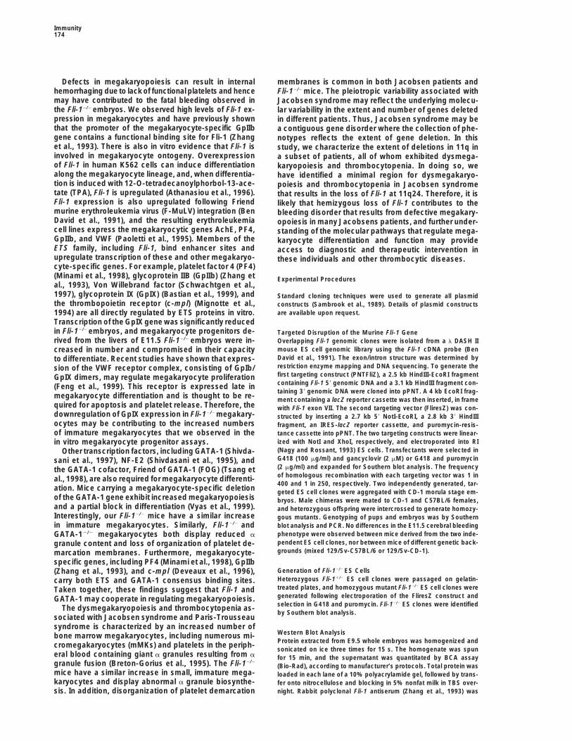
Figure 2. Embryonic Lethality due to Hemor-rhage around E11.5
(A) Fli-1 Heterozygote and homozygote at day11.5 of development. Homozygous Fli-12/2
embryos that were alive at E11.5 had signifi-cant intracranial hemorrhages.(B) Fli-1 expression assessed by whole-mount b-galactosidase staining of a Fli-1 het-erozygote at E11.5. Intersomitic blood ves-sels (small arrow) and major cerebral bloodvessels (large arrow). Magnification, 53.(C) Fli-1 expression assessed by b-galactosi-dase activity is present at the sites of in-tracranial hemorrhage. Saggital section ofE11.5 Fli-12/2 homozygote. Intracranial hem-orrhages (arrows) are present in the forebrain.Magnification, 103. Asterisk shows arrow in-dicating area shown at higher magnification.(D) Higher magnification of E11.5 Fli-12/2 sag-gital section. Magnification, 503. Endothelialcells of the forebrain meninges expressingthe lacZ reporter appear attenuated and dis-continuous (arrow)
3E). Taken together, these results suggest that the loss progenitors migrate to the fetal liver, prior to definitivehematopoiesis. Between E10.5 and E12.5, many LacZ-of Fli-1 leads to a cell-autonomous defect, which mani-expressing cells were present in the fetal liver, the majorfests in a lower proliferative capacity or increased apo-site of hematopoiesis during midgestation. Later in ges-ptosis of endothelial cells. Similarly, we found that Fli-tation and in the adult, LacZ reporter expression was1-deficient cells had a severely reduced capacity torestricted to large, multinucleated AChE-positive cellscontribute to the fetal liver. Heterozygous Fli-11/2 cellsin the spleen and liver.expressing the LacZ reporter were abundant within the
Total cell counts of peripheral blood from adult Fli-11/2livers of Fli-11/2;CD-1 chimeric mice at E14.5, but veryheterozygotes indicated that both red and white bloodfew Fli-12/2 cells expressing LacZ could be detected incells, hemoglobin, hematocrit, mean cell volume, plate-Fli-12/2;CD-1 chimeras at this stage (compare Figureslets, and mean platelet volume were not significantly3C and 3F). Similarly, Fli-11/2 ES cell lines contributeddifferent from that of wild-type littermates (data notto the megakaryocyte lineage within the bone marrowshown). In addition, heterozygous Fli-11/2 adult miceof adult chimeric mice, while Fli-12/2 megakaryocytesdisplayed normal platelet function measured by durationcould not be detected (data not shown). The relativeof bleeding from tail cuts. To characterize hematopoieticcontribution of Fli-12/2 ES cell lines and CD-1 embryonicprogenitors in embryos lacking Fli-1, fetal liver cells werecells within the chimeric embryos was determined byisolated from E11.5 embryos and plated in methylcellu-chromatography of GPI isoforms (Figure 3G), and em-lose media supplemented with cytokines. The relativebryos with approximately equal proportions of each iso-numbers of erythroid, macrophage, granulocyte, andform were analyzed.mixed progenitors were not significantly different inFli-12/2 homozygotes compared to their heterozygous
Fli-12/2 Megakaryocytes Are Blocked Early or wild-type littermates (data not shown).in Differentiation Megakaryopoiesis was evaluated in hematopoieticFli-1 activation following F-MuLV proviral insertion progenitors derived from E11.5 fetal liver by colony-causes erythroleukaemia in mice (Ben David et al., 1991), forming assays in collagen-based media supplementedand altered levels of Fli-1 expression have been associ- with thrombopoietin (Tpo), IL-3, IL-6, and IL-11. Mega-ated with both erythroid and megakaryocytic differentia- karyocyte colonies that stained positive for acetylcholin-tion (Athanasiou et al., 1996; Tamir et al., 1999). There- esterase (AChE) were counted after 6 days. The relativefore, we examined the expression of the LacZ reporter number of megakaryocyte progenitors was elevated inin these lineages and assessed the proliferative and Fli-12/2-deficient mice compared with wild-type and het-differentiation capacity of erythroid and megakaryocyte erozygous littermates (p . 0.0001 in unpaired t test)progenitors in colony-forming assays. Expression of the (Figure 4A). Cytological analysis of mutant and wild-typeLacZ reporter was detected within blood islands of the colonies revealed an increase in colonies containingextraembryonic visceral yolk sac, coincident with com- small megakaryocytes with round nuclei and low nu-mon progenitors of endothelial and hematopoietic lin- clear/cytoplasmic ratios characteristic of undifferenti-eages. Circulating blood cells expressing LacZ were ated phenotypes (compare Figures 4B and 4C). Trans-
mission electron microscopy of Fli-12/2and Fli-11/2 E11.5observed between E8.5 and E10.5 at a time when early

Fli-1 in Vascular Development and Megakaryopoiesis171
Figure 3. Analysis of Fli-11/2 and Fli-12/2 ES Cell Contribution to Endothelial and Megakaryocyte Lineages in E14.5 Chimeric Embryos
(A) Fli-11/2;CD-1 chimera, saggital section. Fli-11/2 cells were identified in the meninges by LacZ expression (arrow).(B) Fli-11/2;CD-1 chimera, coronal section. Arrow indicates interior carotid artery.(C) Fli-11/2;CD-1 chimera, fetal liver. Arrow indicates megakaryocytes in the liver of E14.5 chimeric mice.(D) Fli-12/2;CD-1 chimera, saggital section.(E) Fli-12/2;CD-1 chimera, coronal section.(F) Fli-12/2;CD-1 chimera, fetal liver section. Reduced numbers of Fli-12/2 (LacZ-expressing) cells were detected in the livers of E14.5 chimericmice (arrow).(G) Analysis of glucose-6-phosphate isomerase (GPI) activity in chimeric embryos. Chimeric embryos with approximately equal contributionfrom mutant R1 ES cells (GPIAA isoform) and CD-1 morula stage embryos (GPIBB isoform) were selected for analysis. Lane 1: ES cells; lanes2, 3, 4, 5, 6, and 7: chimeric embryos shown in (A), (B), (C), (D), (E), and (F), respectively; lane 8: CD-1 embryo.
fetal liver revealed abnormal ultrastructural features in RNA transcripts for the Tek/Tie-2 and GpIX genes weremarkedly reduced in Fli-12/2 embryos at this stage (Fig-the mutant megakaryocytes. Many of the megakaryo-
cytes analyzed in Fli-12/2 embryos shared a common ure 5). Transcripts corresponding to the cell adhesiongenes VE-CAD (Gory et al., 1998), and I-CAM-1 (de Lau-appearance not observed in the Fli-11/2 sections, includ-
ing reduced numbers of a granules, disorganization of noit et al., 1998) and hematopoietic genes c-kit (Ratajc-zak et al., 1998) and c-fms (Ross et al., 1998) were alsothe platelet demarcation membranes, and reduced over-
all size (compare Figures 4D and 4E). examined and found to be equivalent in heterozygousFli-11/2 and mutant Fli-12/2 embryos at this stage (datanot shown).Regulation of Endothelial- and Megakaryocyte-
Specific Genes in Fli-1 Mutant EmbryosThe transcriptional regulation regions of a number of Dysmegakaryopoiesis in Jacobsen Syndrome
Maps to 11q24endothelial- and megakaryocyte-specific genes containFli-1 consensus binding sites in their promoters. There- Jacobsen and Paris-Trousseau syndrome are associ-
ated with a constellation of clinical defects, includingfore, we examined the expression of several genes thatcontain potential Fli-1 binding sites and that are known severe heart abnormalities (Grossfeld et al., 1999), trigo-
nocephaly, mental retardation, dysmorphogenesis ofto bind ETS proteins in vitro. Expression of the endothe-lial-restricted genes Tie (Iljin et al., 1999), Tek/Tie-2 the hands and face, thrombocytopenia (Penny et al.,
1995), and deletions of the long arm of chromosome 11,(Dube et al., 1999), and VEGFR-1/Flt-1 (Wakiya et al.,1996) and the megakaryocyte-specific genes GpIX (Bas- where the human Fli-1 gene is located (Tunnacliffe et al.,
1999). We therefore examined more closely 14 patientstian et al., 1996), GpIIb (Zhang et al., 1993), c-mpl (De-veaux et al., 1996), and vwf (Schwachtgen et al., 1997) diagnosed with Jacobsen syndrome, all of whom exhib-
ited defects in megakaryopoiesis determined by lightwas assessed by semiquantitative reverse transcriptasepolymerase chain reaction (RT-PCR) of E11.5 whole- and electron microscopy (data not shown). To define
precisely the extent of deletions in these patients, weembryo mRNA from Fli-11/2 and Fli-12/2 littermates. Ex-pression of flk-1, flt-1, Tie, GpIIb, c-mpl, and vwf was performed fluorescence in situ hybridization (FISH) us-
ing a set of PAC clones derived from an 11q YAC contignot altered in Fli-12/2 mutants. In contrast, the levels of

Immunity172
Figure 4. Analysis of Megakaryopoiesis inFli-12/2 Fetal Liver
(A) Relative numbers of megakaryocyte col-ony-forming units (CFU-Mk) in wild-type andFli-1 mutant mice. Hematopoietic progenitorsfrom E11.5 fetal livers were cultured for 6 daysin the presence of Tpo, IL-3, IL-6, and IL-11.Megakaryocyte colonies were identified bystaining for AChE activity. n, number of em-bryos analyzed; *p . 0.0001, unpaired t test.(B) Fli-12/2 AchE-positive CFU-Mk. Fli-12/2
colonies often showed immature megakaryo-cyte phenotypes including reduced staining,small nuclei, and high nucleus/cytoplasmicratio.(C) Fli-11/2 AchE-positive colony after 6 daysin culture.(D) Transmission electron micrograph (TEM)of Fli-11/2 fetal liver megakaryocyte (scalebar 5 5000 nm). The inset is a higher magnifi-cation of the cytoplasm (large arrow) a gran-ule; (Nu) nucleus; (small arrow) platelet de-marcation membrane (scale bar 5 2000 nm).(E) Fli-12/2 megakaryocyte from E11.5 fetalliver (scale bar 5 5000 nm). The insert showsa higher magnification of the cytoplasm withfew a granules present and disorganizedplatelet demarcation membranes (scale bar 5
1000 nm).
(Tunnacliffe et al., 1999). All 14 of the patients had termi- situ differentiation of primary angioblasts to form vascu-lar channels (vasculogenesis), followed by the prolifera-nal deletions of varying sizes of the long arm of chro-
mosome 11 with breakpoints centromeric to Fli-1 and tion and sprouting of existing vascular endothelial cellsto complete the circulatory system (angiogenesis) (Ri-therefore resulting in the hemizygous loss of this gene
(Figure 6). sau and Flamme, 1995). Embryos lacking Fli-1 are ableto form a functional network of blood vessels, indicatingthat the initial stages of vasculogenesis and angiogen-Discussionesis can proceed in the absence of this transcriptionfactor. Late embryonic angiogenesis and vascular re-Transcriptional activation of Fli-1 by either chromosomalmodeling are dependent on recruitment of pericytes andtranslocation or proviral insertion leads to Ewings sar-smooth muscle cells to form the blood vessel wall (Risaucoma in children and erythroleukemia in mice, re-and Flamme, 1995). In the brain, where vascularizationspectively. We have shown previously that Fli-1 over-occurs following angiogenic sprouting, pericytes andexpression in the lymphoid compartment can inducesmooth muscle cells are recruited from the cephaliclymphopoiesis and autoimmune disease in the kidneysmesenchyme and neural crest. Our previous studiesof transgenic mice (Zhang et al., 1995). In Xenopus, Fli-1have shown that Fli-1 is expressed in both the endothe-overexpression disrupts embryonic polarity, inhibitslial and putative neural crest lineages (Melet et al., 1996).erythropoiesis, and results in phenotypes suggestiveTherefore, it is possible that Fli-1 plays a role in theof altered cell adhesion properties (Remy et al., 1996).expression of guidance cues and/or the modification ofTherefore, Fli-1 can affect developmental programs incellular adhesion properties in migrating vascular sup-a variety of cell lineages, and dysregulation of this tran-port cells, or in the endothelial cells themselves. In orderscription factor in humans and mice results in tumori-to decide whether the requirement for Fli-1 is intrinsicgenesis. In this study, we have assessed the biologicalto endothelial cells or is indirect through supportingfunction of Fli-1, a member of the ETS family of transcrip-cells, we analyzed the ability of Fli-12/2 cells to contributetion factors, by generating mice with a targeted nullto the developing vasculature in chimeric animals con-mutation in the Fli-1 gene. The results described heretaining a mixture of Fli-12/2 and Fli-11/1 cells. This analy-demonstrate that Fli-1 is essential for embryogenesissis clearly demonstrated that the absence of Fli-1 leadsand that the absence of Fli-1 affects both vascular devel-to an intrinsic or cell-autonomous defect resulting inopment and megakaryopoiesis, resulting in loss of bloodthe presence of Fli-12/2 cells in the vasculature of E10.5vessel integrity after embryonic day 11 and a partialembryos but their absence by E14.5. We conclude thatblock in megakaryocyte differentiation.Fli-1 plays an essential role in endothelial cells at thisThe formation of blood vessels within the developing
embryo is a two-stage process characterized by the in stage of development.

Fli-1 in Vascular Development and Megakaryopoiesis173
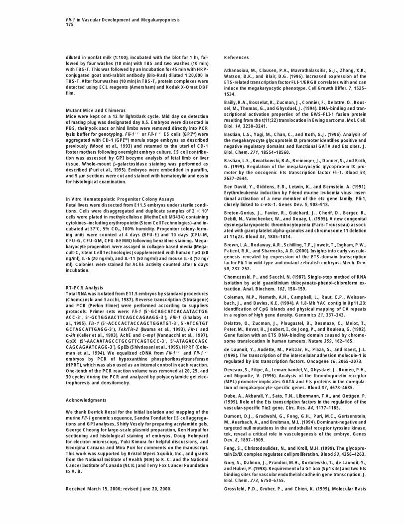
Figure 6. Mapping of Chromosome 11q Deletions in 14 JacobsenPatients with Dysmegakaryopoiesis
(Solid bar) The extent of 11q retained in each individual is shownwith the most distal marker retained (1). (Shaded bar) Terminaldeletion breakpoint regions are shaded and the most proximal de-leted marker is indicated (2). Patients 1–9 attended the Division ofPediatric Hematology, Hopital D’enfants Armand Trousseau [R. F.],and patients 10–14 were identified at the second annual 11q Re-search and Resource conference in 1998 at the University of Califor-nia, San Diego [P. G. and K. C.]. Patients 1 and 3 are from Joneset al. (2000).
within the embryo has not been examined. It will beof interest to determine whether the vascular defectsobserved in both Fli-1 and Tel mutant embryos reflectbiochemical interactions between these two ETS pro-teins in vivo.
In addition to the ETS family of transcription factors,embryonic vasculogenesis and angiogenesis requireFigure 5. Downregulation of Potential Fli-1 Target Genes in Mutantsignaling through a number of receptor tyrosine kinasesEmbryos(Risau, 1998). Null mutations generated by ourselvesSemiquantitative RT-PCR analysis of endothelial- and megakaryo-and others in the endothelial-specific receptor tyrosinecyte-specific genes in Fli-11/2 and Fli-12/2 embryos. Equalized cDNAkinases Flk-1 (Shalaby et al., 1995), Tie (Puri et al., 1995),samples from E11.5 embryos were analyzed by competitive PCRand Tek/Tie-2 (Dumont et al., 1994) have revealed anfor hprt and potential Fli-1 target genes. (Fli-1) Fli-1 transcript is
absent in Fli-12/2 embryo; (flk-1) endothelial lineage marker; (Tie, essential requirement for these receptors in the develop-Tek/Tie-2, and flt-1) endothelial-specific, potential Fli-1 target ment and maintenance of the vasculature. In particular,genes; (AchE) megakaryocyte lineage marker; (GpIIb, c-mpl, and it is interesting to note that embryos lacking the Tek/GpIX) megakaryocyte-specific, potential Fli-1 target genes. Tie-2 receptor die at approximately the same time as
the Fli-12/2 embryos described here. Importantly, weobserved that Tek/Tie-2 was specifically downregulated
Other members of the ETS gene family have also been in Fli-12/2 mutant embryos compared to their heterozy-implicated in the regulation of angiogenesis in develop- gous Fli-11/2 littermates. The reduction of Tek/Tie-2 RNAment and neoplasia. For example, germline mutation in transcripts in Fli-12/2 embryos was not simply due tothe widely expressed ETS gene Tel causes defective the loss of endothelial cells in these dying embryos, asangiogenesis in the embryonic yolk sac and apoptosis the expression of both Flk-1 and Tie, two other receptorof mesenchymal and neural cells resulting in embryonic tyrosine kinases whose expression is entirely restricteddeath at E10.5–E11.5 (Wang et al., 1997). TEL is a tran- to endothelial cells, was unaffected in these embryos.scription factor whose activity is modulated by dimeriza- Recent in vitro experiments have demonstrated that ETStion with the Fli-1 protein (Kwiatkowski et al., 1998) and binding sites in the Tek/Tie-2 promoter regulate tran-possibly other members of the ETS family of transcrip- scription of this gene (Dube et al., 1999). Therefore, thetion factors that contain a “pointed” domain. Heterodi- Tek/Tie-2 receptor may be directly regulated by Fli-1 inmerization of Fli-1 with Tel inhibits the transactivational endothelial cells, and loss of Tek/Tie-2 expression mayactivity of the Fli-1 protein. We observed normal yolk sac be contributing to the loss of vascular integrity that we
observed in homozygous Fli-12/2 embryos.vascularization in Fli-12/2 embryos, although apoptosis
Immunity174
Defects in megakaryopoiesis can result in internal membranes is common in both Jacobsen patients andhemorrhaging due to lack of functional platelets and hence Fli-12/2 mice. The pleiotropic variability associated withmay have contributed to the fatal bleeding observed in Jacobsen syndrome may reflect the underlying molecu-the Fli-12/2 embryos. We observed high levels of Fli-1 ex- lar variability in the extent and number of genes deletedpression in megakaryocytes and have previously shown in different patients. Thus, Jacobsen syndrome may bethat the promoter of the megakaryocyte-specific GpIIb a contiguous gene disorder where the collection of phe-gene contains a functional binding site for Fli-1 (Zhang notypes reflects the extent of gene deletion. In thiset al., 1993). There is also in vitro evidence that Fli-1 is study, we characterize the extent of deletions in 11q ininvolved in megakaryocyte ontogeny. Overexpression a subset of patients, all of whom exhibited dysmega-of Fli-1 in human K562 cells can induce differentiation karyopoiesis and thrombocytopenia. In doing so, wealong the megakaryocyte lineage, and, when differentia- have identified a minimal region for dysmegakaryo-tion is induced with 12-O-tetradecanoylphorbol-13-ace- poiesis and thrombocytopenia in Jacobsen syndrometate (TPA), Fli-1 is upregulated (Athanasiou et al., 1996). that results in the loss of Fli-1 at 11q24. Therefore, it isFli-1 expression is also upregulated following Friend likely that hemizygous loss of Fli-1 contributes to themurine erythroleukemia virus (F-MuLV) integration (Ben bleeding disorder that results from defective megakary-David et al., 1991), and the resulting erythroleukemia opoiesis in many Jacobsens patients, and further under-cell lines express the megakaryocytic genes AchE, PF4, standing of the molecular pathways that regulate mega-GpIIb, and VWF (Paoletti et al., 1995). Members of the karyocyte differentiation and function may provideETS family, including Fli-1, bind enhancer sites and access to diagnostic and therapeutic intervention inupregulate transcription of these and other megakaryo- these individuals and other thrombocytic diseases.cyte-specific genes. For example, platelet factor 4 (PF4)(Minami et al., 1998), glycoprotein IIB (GpIIb) (Zhang et Experimental Proceduresal., 1993), Von Willebrand factor (Schwachtgen et al.,1997), glycoprotein IX (GpIX) (Bastian et al., 1999), and Standard cloning techniques were used to generate all plasmidthe thrombopoietin receptor (c-mpl) (Mignotte et al., constructs (Sambrook et al., 1989). Details of plasmid constructs
are available upon request.1994) are all directly regulated by ETS proteins in vitro.Transcription of the GpIX gene was significantly reducedin Fli-12/2 embryos, and megakaryocyte progenitors de- Targeted Disruption of the Murine Fli-1 Generived from the livers of E11.5 Fli-12/2 embryos were in- Overlapping Fli-1 genomic clones were isolated from a l DASH II
mouse ES cell genomic library using the Fli-1 cDNA probe (Bencreased in number and compromised in their capacityDavid et al., 1991). The exon/intron structure was determined byto differentiate. Recent studies have shown that expres-restriction enzyme mapping and DNA sequencing. To generate thesion of the VWF receptor complex, consisting of GpIb/first targeting construct (PNTFliZ), a 2.5 kb HindIII-EcoRI fragmentGpIX dimers, may regulate megakaryocyte proliferationcontaining Fli-1 59 genomic DNA and a 3.1 kb HindIII fragment con-(Feng et al., 1999). This receptor is expressed late intaining 39 genomic DNA were cloned into pPNT. A 4 kb EcoRI frag-
megakaryocyte differentiation and is thought to be re- ment containing a lacZ reporter cassette was then inserted, in framequired for apoptosis and platelet release. Therefore, the with Fli-1 exon VII. The second targeting vector (FliresZ) was con-downregulation of GpIX expression in Fli-12/2 megakary- structed by inserting a 2.7 kb 59 NotI-EcoRI, a 2.8 kb 39 HindIIIocytes may be contributing to the increased numbers fragment, an IRES-lacZ reporter cassette, and puromycin-resis-of immature megakaryocytes that we observed in the tance cassette into pPNT. The two targeting constructs were linear-
ized with NotI and XhoI, respectively, and electroporated into RIin vitro megakaryocyte progenitor assays.(Nagy and Rossant, 1993) ES cells. Transfectants were selected inOther transcription factors, including GATA-1 (Shivda-G418 (100 mg/ml) and gancyclovir (2 mM) or G418 and puromycinsani et al., 1997), NF-E2 (Shivdasani et al., 1995), and(2 mg/ml) and expanded for Southern blot analysis. The frequencythe GATA-1 cofactor, Friend of GATA-1 (FOG) (Tsang etof homologous recombination with each targeting vector was 1 inal., 1998), are also required for megakaryocyte differenti-400 and 1 in 250, respectively. Two independently generated, tar-
ation. Mice carrying a megakaryocyte-specific deletion geted ES cell clones were aggregated with CD-1 morula stage em-of the GATA-1 gene exhibit increased megakaryopoiesis bryos. Male chimeras were mated to CD-1 and C57BL/6 females,and a partial block in differentiation (Vyas et al., 1999). and heterozygous offspring were intercrossed to generate homozy-Interestingly, our Fli-12/2 mice have a similar increase gous mutants. Genotyping of pups and embryos was by Southernin immature megakaryocytes. Similarly, Fli-12/2 and blot analysis and PCR. No differences in the E11.5 cerebral bleeding
phenotype were observed between mice derived from the two inde-GATA-12/2 megakaryocytes both display reduced apendent ES cell clones, nor between mice of different genetic back-granule content and loss of organization of platelet de-grounds (mixed 129/Sv-C57BL/6 or 129/Sv-CD-1).marcation membranes. Furthermore, megakaryocyte-
specific genes, including PF4 (Minami et al., 1998), GpIIbGeneration of Fli-12/2 ES Cells(Zhang et al., 1993), and c-mpl (Deveaux et al., 1996),Heterozygous Fli-11/2 ES cell clones were passaged on gelatin-carry both ETS and GATA-1 consensus binding sites.treated plates, and homozygous mutant Fli-12/2 ES cell clones wereTaken together, these findings suggest that Fli-1 andgenerated following electroporation of the FliresZ construct andGATA-1 may cooperate in regulating megakaryopoiesis.selection in G418 and puromycin. Fli-12/2 ES clones were identifiedThe dysmegakaryopoiesis and thrombocytopenia as-by Southern blot analysis.
sociated with Jacobsen syndrome and Paris-Trousseausyndrome is characterized by an increased number of
Western Blot Analysisbone marrow megakaryocytes, including numerous mi-Protein extracted from E9.5 whole embryos was homogenized andcromegakaryocytes (mMKs) and platelets in the periph- sonicated on ice three times for 15 s. The homogenate was spun
eral blood containing giant a granules resulting from a for 15 min, and the supernatant was quantitated by BCA assaygranule fusion (Breton-Gorius et al., 1995). The Fli-12/2
(Bio-Rad), according to manufacturer’s protocols. Total protein wasmice have a similar increase in small, immature mega- loaded in each lane of a 10% polyacrylamide gel, followed by trans-karyocytes and display abnormal a granule biosynthe- fer onto nitrocellulose and blocking in 5% nonfat milk in TBS over-
night. Rabbit polyclonal Fli-1 antiserum (Zhang et al., 1993) wassis. In addition, disorganization of platelet demarcation
Fli-1 in Vascular Development and Megakaryopoiesis175
diluted in nonfat milk (1:100), incubated with the blot for 1 hr, fol- Referenceslowed by four washes (10 min) with TBS and two washes (10 min)with TBS-T. This was followed by an incubation for 45 min with HRP- Athanasiou, M., Clausen, P.A., Mavrothalassitis, G.J., Zhang, X.K.,conjugated goat anti-rabbit antibody (Bio-Rad) diluted 1:20,000 in Watson, D.K., and Blair, D.G. (1996). Increased expression of theTBS-T. After four washes (10 min) in TBS-T, protein complexes were ETS-related transcription factor FLI-1/ERGB correlates with and candetected using ECL reagents (Amersham) and Kodak X-Omat DBF induce the megakaryocytic phenotype. Cell Growth Differ. 7, 1525–film. 1534.
Bailly, R.A., Bosselut, R., Zucman, J., Cormier, F., Delattre, O., Rous-sel, M., Thomas, G., and Ghysdael, J. (1994). DNA-binding and tran-Mutant Mice and Chimerasscriptional activation properties of the EWS-FLI-1 fusion proteinMice were kept on a 12 hr light/dark cycle. Mid day on detectionresulting from the t(11;22) translocation in Ewing sarcoma. Mol. Cell.of mating plug was designated day 0.5. Embryos were dissected inBiol. 14, 3230–3241.PBS, their yolk sacs or hind limbs were removed directly into PCR
lysis buffer for genotyping. Fli-11/2 or Fli-12/2 ES cells (GPIAA) were Bastian, L.S., Yagi, M., Chan, C., and Roth, G.J. (1996). Analysis ofaggregated with CD-1 (GPIBB) morula stage embryos as described the megakaryocyte glycoprotein IX promoter identifies positive andpreviously (Wood et al., 1993) and returned to the uteri of CD-1 negative regulatory domains and functional GATA and Ets sites. J.foster mothers following overnight embryo culture. ES cell contribu- Biol. Chem. 271, 18554–18560.tion was assessed by GPI isozyme analysis of fetal limb or liver Bastian, L.S., Kwiatkowski, B.A., Breininger, J., Danner, S., and Roth,tissue. Whole-mount b-galactosidase staining was performed as G. (1999). Regulation of the megakaryocytic glycoprotein IX pro-described (Puri et al., 1995). Embryos were embedded in paraffin, moter by the oncogenic Ets transcription factor Fli-1. Blood 93,and 5 mm sections were cut and stained with hematoxylin and eosin 2637–2644.for histological examination.
Ben David, Y., Giddens, E.B., Letwin, K., and Bernstein, A. (1991).Erythroleukemia induction by Friend murine leukemia virus: inser-tional activation of a new member of the ets gene family, Fli-1,In Vitro Hematopoietic Progenitor Colony Assaysclosely linked to c-ets-1. Genes Dev. 5, 908–918.Fetal livers were dissected from E11.5 embryos under sterile condi-
tions. Cells were disaggregated and duplicate samples of 2 3 105Breton-Gorius, J., Favier, R., Guichard, J., Cherif, D., Berger, R.,
cells were plated in methylcellulose (MethoCult M3434) containing Debili, N., Vainchenker, W., and Douay, L. (1995). A new congenitalcytokines–including erythropoietin (Stem Cell Technologies)–and in- dysmegakaryopoietic thrombocytopenia (Paris-Trousseau) associ-cubated at 378C, 5% CO2, 100% humidity. Progenitor colony-form- ated with giant platelet alpha-granules and chromosome 11 deletioning units were counted at 4 days (BFU-E) and 10 days (CFU-M, at 11q23. Blood 85, 1805–1814.CFU-G, CFU-GM, CFU-GEMM) following benzidine staining. Mega-
Brown, L.A., Rodaway, A.R., Schilling, T.F., Jowett, T., Ingham, P.W.,karyocyte progenitors were assayed in collagen-based media (Mega-Patient, R.K., and Sharrocks, A.D. (2000). Insights into early vasculo-cult-C, Stem Cell Technologies) supplemented with human TpO (50genesis revealed by expression of the ETS-domain transcriptionng/ml), IL-6 (20 ng/ml), and IL-11 (50 ng/ml) and mouse IL-3 (10 ng/factor Fli-1 in wild-type and mutant zebrafish embryos. Mech. Dev.ml). Colonies were stained for AChE activity counted after 6 days90, 237–252.incubation.Chomcznski, P., and Sacchi, N. (1987). Single-step method of RNAisolation by acid guanidinium thiocyanate-phenol-chloroform ex-
RT-PCR Analysis traction. Anal. Biochem. 162, 156–159.Total RNA was isolated from E11.5 embryos by standard procedures
Coleman, M.P., Nemeth, A.H., Campbell, L., Raut, C.P., Weissen-(Chomcznski and Sacchi, 1987). Reverse transcription (Stratagene)bach, J., and Davies, K.E. (1994). A 1.8-Mb YAC contig in Xp11.23:and PCR (Perkin Elmer) were performed according to suppliersidentification of CpG islands and physical mapping of CA repeatsprotocols. Primer sets were: Fli-1 (59-GCAGCATCACAATACTGGin a region of high gene density. Genomics 21, 337–343.ACC-39, 59-GCTGGAACTTCAGCCAGAAGG-39), Flk-1 (Shalaby etDelattre, O., Zucman, J., Plougastel, B., Desmaze, C., Melot, T.,al., 1995), Tie-1 (59-ACCCACTACCAGCTGGATGT-39, 59-ATCGTGTPeter, M., Kovar, H., Joubert, I., de Jong, P., and Rouleau, G. (1992).GCTAGCATTGAGG-39), Tek/Tie-2 (Iwama et al., 1993), Flt-1 andGene fusion with an ETS DNA-binding domain caused by chromo-c-kit (Keller et al., 1993), AchE and c-mpl (Vannucchi et al., 1997),some translocation in human tumours. Nature 359, 162–165.GpIX (59-AACAATAGCCTGCGTTCAGTGCC-39, 59-ATAGACCAGC
CAGCAGAATCAGG-39), GpIIb (Shivdasani et al., 1995), HPRT (Cole- de Launoit, Y., Audette, M., Pelczar, H., Plaza, S., and Baert, J.L.man et al., 1994). We equalized cDNA from Fli-11/2 and Fli-12/2
(1998). The transcription of the intercellular adhesion molecule-1 isembryos by PCR of hypoxanthine phosphoribosyltransferase regulated by Ets transcription factors. Oncogene 16, 2065–2073.(HPRT), which was also used as an internal control in each reaction.
Deveaux, S., Filipe, A., Lemarchandel, V., Ghysdael, J., Romeo, P.H.,One-tenth of the PCR reaction volume was removed at 20, 25, andand Mignotte, V. (1996). Analysis of the thrombopoietin receptor30 cycles during the PCR and analyzed by polyacrylamide gel elec-(MPL) promoter implicates GATA and Ets proteins in the coregula-trophoresis and densitometry.tion of megakaryocyte-specific genes. Blood 87, 4678–4685.
Dube, A., Akbarali, Y., Sato, T.N., Libermann, T.A., and Oettgen, P.Acknowledgments (1999). Role of the Ets transcription factors in the regulation of the
vascular-specific Tie2 gene. Circ. Res. 84, 1177–1185.We thank Derrick Rossi for the initial isolation and mapping of the
Dumont, D.J., Gradwohl, G., Fong, G.H., Puri, M.C., Gertsenstein,murine Fli-1 genomic sequence, Sandra Tondat for ES cell aggrega-M., Auerbach, A., and Breitman, M.L. (1994). Dominant-negative andtions and GPI analyses, Shirly Vesely for preparing acrylamide gels,targeted null mutations in the endothelial receptor tyrosine kinase,George Cheong for large-scale plasmid preparation, Ken Harpal fortek, reveal a critical role in vasculogenesis of the embryo. Genessectioning and histological staining of embryos, Doug HolmyardDev. 8, 1897–1909.for electron microscopy, Yuki Kimura for helpful discussions, and
Georgina Caruana and Mira Puri for comments on the manuscript. Feng, S., Christodoulides, N., and Kroll, M.H. (1999). The glycopro-This work was supported by Bristol Myers Squibb, Inc., and grants tein Ib/IX complex regulates cell proliferation. Blood 93, 4256–4263.from the National Institute of Health (NIH) to K. C. and the National
Gory, S., Dalmon, J., Prandini, M.H., Kortulewski, T., de Launoit, Y.,Cancer Institute of Canada (NCIC) and Terry Fox Cancer Foundation
and Huber, P. (1998). Requirement of a GT box (Sp1 site) and two Etsto A. B.
binding sites for vascular endothelial cadherin gene transcription. J.Biol. Chem. 273, 6750–6755.
Grossfeld, P.D., Gruber, P., and Chien, K. (1999). Molecular BasisReceived March 15, 2000; revised June 20, 2000.

Immunity176
of Cardiovascular Disease, K. Chien, ed. (Cambridge, MA: W.B. Puri, M.C., Partanen, J., Rossant, J., and Bernstein, A. (1999). Inter-action of the TEK and TIE receptor tyrosine kinases during cardio-Saunders), pp. 135–166.vascular development. Development 126, 4569–4580.Ida, K., Kobayashi, S., Taki, T., Hanada, R., Bessho, F., Yamamori,Ratajczak, M.Z., Perrotti, D., Melotti, P., Powzaniuk, M., Calabretta,S., Sugimoto, T., Ohki, M., and Hayashi, Y. (1995). EWS-FLI-1 andB., Onodera, K., Kregenow, D.A., Machalinski, B., and Gewirtz, A.M.EWS-ERG chimeric mRNAs in Ewing’s sarcoma and primitive neu-(1998). Myb and ets proteins are candidate regulators of c-kit ex-roectodermal tumor. Int. J. Cancer 63, 500–504.pression in human hematopoietic cells. Blood 91, 1934–1946.
Iljin, K., Dube, A., Kontusaari, S., Korhonen, J., Lahtinen, I., Oettgen,Remy, P., Senan, F., Meyer, D., Mager, A.M., and Hindelang, C.P., and Alitalo, K. (1999). Role of ets factors in the activity and(1996). Overexpression of the Xenopus Xl-fli gene during early em-endothelial cell specificity of the mouse Tie gene promoter. FASEBbryogenesis leads to anomalies in head and heart development andJ. 13, 377–386.erythroid differentiation. Int. J. Dev. Biol. 40, 577–589.
Iwama, A., Hamaguchi, I., Hashiyama, M., Murayama, Y., Yasunaga,Risau, W. (1998). Development and differentiation of endothelium.K., and Suda, T. (1993). Molecular cloning and characterization ofKidney Int. Suppl. 67, S3–S6.mouse TIE and TEK receptor tyrosine kinase genes and their expres-
sion in hematopoietic stem cells. Biochem. Biophys. Res. Commun. Risau, W., and Flamme, I. (1995). Vasculogenesis. Annu. Rev. Cell195, 301–309. Dev. Biol. 11, 73–91.
Jones, C., Mullenbach, R., Grossfeld, P., Auer, R., Favier, R., Chien, Ross, I.L., Yue, X., Ostrowski, M.C., and Hume, D.A. (1998). Interac-K., James, M., Tunnacliffe, A., and Finbarr, C. (2000). Co-localisation tion between PU.1 and another Ets family transcription factor pro-of CCG-repeats and chromosome deletion breakpoints in Jacobsen motes macrophage-specific Basal transcription initiation. J. Biol.Syndrome: evidence for a common mechanism of chromosome Chem. 273, 6662–6669.breakage. Hum. Mol. Gen. 9, 1201–1208.
Sambrook, J., Fritsh, E.F., and Maniatis, T. (1989). Molecular Cloning,Keller, G., Kennedy, M., Papayannopoulou, T., and Wiles, M. (1993). Second Edition (Cold Spring Harbor, NY: Cold Spring Harbor Labo-Hematopoietic commitment during embryonic stem cell differentia- ratory Press).tion in culture. Mol. Cell. Biol. 13, 473–486.
Schwachtgen, J.L., Janel, N., Barek, L., Duterque-Coquillaud, M.,Kwiatkowski, B.A., Bastian, L.S., Bauer, T.R., Jr., Tsai, S., Zielinska- Ghysdael, J., Meyer, D., and Kerbiriou-Nabias, D. (1997). Ets tran-Kwiatkowska, A.G., and Hickstein, D.D. (1998). The ets family mem- scription factors bind and transactivate the core promoter of theber Tel binds to the Fli-1 oncoprotein and inhibits its transcriptional von Willebrand factor gene. Oncogene 15, 3091–3102.activity. J. Biol. Chem. 273, 17525–17530. Shalaby, F., Rossant, J., Yamaguchi, T.P., Gertsenstein, M., Wu,
X.F., Breitman, M.L., and Schuh, A.C. (1995). Failure of blood-islandLemarchandel, V., Ghysdael, J., Mignotte, V., Rahuel, C., and Ro-meo, P.H. (1993). GATA and Ets cis-acting sequences mediate formation and vasculogenesis in Flk-1-deficient mice. Nature 376,
62–66.megakaryocyte-specific expression. Mol. Cell. Biol. 13, 668–676.
Shivdasani, R.A., Rosenblatt, M.F., Zucker-Franklin, D., Jackson,Mager, A.M., Grapin-Botton, A., Ladjali, K., Meyer, D., Wolff, C.M.,C.W., Hunt, P., Saris, C.J., and Orkin, S.H. (1995). TranscriptionStiegler, P., Bonnin, M.A., and Remy, P. (1998). The avian fli genefactor NF-E2 is required for platelet formation independent of theis specifically expressed during embryogenesis in a subset of neuralactions of thrombopoietin/MGDF in megakaryocyte development.crest cells giving rise to mesenchyme. Int. J. Dev. Biol. 42, 561–572.Cell 81, 695–704.
Melet, F., Motro, B., Rossi, D.J., Zhang, L., and Bernstein, A. (1996).Shivdasani, R.A., Fujiwara,Y., McDevitt, M.A., and Orkin, S.H. (1997).Generation of a novel Fli-1 protein by gene targeting leads to aA lineage-selective knockout establishes the critical role of tran-defect in thymus development and a delay in Friend virus-inducedscription factor GATA-1 in megakaryocyte growth and platelet de-erythroleukemia. Mol. Cell. Biol. 16, 2708–2718.velopment. EMBO J. 16, 3965–3973.
Meyer, D., Stiegler, P., Hindelang, C., Mager, A.M., and Remy, P.Tamir, A., Howard, J., Higgins, R.R., Li, Y.J., Berger, L., Zacksen-(1995). Whole-mount in situ hybridization reveals the expression ofhaus, E., Reis, M., and Ben David, Y. (1999). Fli-1, an Ets-relatedthe Xl-Fli gene in several lineages of migrating cells in Xenopustranscription factor, regulates erythropoietin- induced erythroidembryos. Int. J. Dev. Biol. 39, 909–919.proliferation and differentiation: evidence for direct transcriptional
Mignotte, V., Vigon, I., Boucher, D.C., Romeo, P.H., Lemarchandel, repression of the Rb gene during differentiation. Mol. Cell. Biol.V., and Chretien, S. (1994). Structure and transcription of the human 19, 4452–4464.c-mpl gene (MPL). Genomics 20, 5–12.
Tsang, A.P., Fujiwara, Y., Hom, D.B., and Orkin, S.H. (1998). FailureMinami, T., Tachibana, K., Imanishi, T., and Doi, T. (1998). Both Ets-1 of megakaryopoiesis and arrested erythropoiesis in mice lacking theand GATA-1 are essential for positive regulation of platelet factor 4 GATA-1 transcriptional cofactor FOG. Genes Dev. 12, 1176–1188.gene expression. Eur. J. Biochem. 258, 879–889.
Tunnacliffe, A., Jones, C., Le Paslier, D., Todd, R., Cherif, D., Birdsall,Nagy, A., and Rossant, J. (1993). Production of completely ES cell M., Devenish, L., Yousry, C., Cotter, F.E., and James, M.R. (1999).derived fetuses. In Gene Targeting: A Practical Approach, A. Joyner, Localization of Jacobsen syndrome breakpoints on a 40-Mb physi-ed. (New York: IRL Press). cal map of distal chromosome 11q. Genome Res. 9, 44–52.
Nye, J.A., Petersen, J.M., Gunther, C.V., Jonsen, M.D., and Graves, Vannucchi, A.M., Linari, S., Cellai, C., Koury, M.J., and Paoletti, F.B.J. (1992). Interaction of murine ets-1 with GGA-binding sites estab- (1997). Constitutive and inducible expression of megakaryocyte-lishes the ETS domain as a new DNA-binding motif. Genes Dev. 6, specific genes in Friend erythroleukaemia cells. Br. J. Haematol. 99,975–990. 500–508.
Paoletti, F., Vannucchi, A.M., Mocali, A., Caporale, R., and Burstein, Vyas, P., Ault, K., Jackson, C.W., Orkin, S.H., and Shivdasani, R.A.S.A. (1995). Identification and conditions for selective expression of (1999). Consequences of GATA-1 deficiency in megakaryocytes andmegakaryocytic markers in Friend erythroleukemia cells. Blood 86, platelets. Blood 93, 2867–2875.2624–2631.
Wakiya, K., Begue, A., Stehelin, D., and Shibuya, M. (1996). A cAMPPenny, L.A., Dell’Aquila, M., Jones, M.C., Bergoffen, J., Cunniff, C., response element and an Ets motif are involved in the transcriptionalFryns, J.P., Grace, E., Graham, J.M., Jr., Kousseff, B., and Mattina, regulation of flt-1 tyrosine kinase (vascular endothelial growth factorT. (1995). Clinical and molecular characterization of patients with receptor 1) gene. J. Biol. Chem. 271, 30823–30828.distal 11q deletions. Am. J. Hum. Genet. 56, 676–683.
Wang, L.C., Kuo, F., Fujiwara, Y., Gilliland, D.G., Golub, T.R., andPuri, M.C., Rossant, J., Alitalo, K., Bernstein, A., and Partanen, J. Orkin, S.H. (1997). Yolk sac angiogenic defect and intra-embryonic(1995). The receptor tyrosine kinase TIE is required for integrity and apoptosis in mice lacking the Ets-related factor TEL. EMBO J. 16,
4374–4383.survival of vascular endothelial cells. EMBO J. 14, 5884–5891.

Fli-1 in Vascular Development and Megakaryopoiesis177
Wood, S.A., Allen, N.D., Rossant, J., Auerbach, A., and Nagy, A.(1993). Non-injection methods for the production of embryonic stemcell-embryo chimaeras. Nature 365, 87–89.
Zhang, L., Lemarchandel, V., Romeo, P.H., Ben David, Y., Greer,P., and Bernstein, A. (1993). The Fli-1 proto-oncogene, involved inerythroleukemia and Ewing’s sarcoma, encodes a transcriptionalactivator with DNA-binding specificities distinct from other Ets fam-ily members. Oncogene 8, 1621–1630.
Zhang, L., Eddy, A., Teng, Y.T., Fritzler, M., Kluppel, M., Melet, F.,and Bernstein, A. (1995). An immunological renal disease in trans-genic mice that overexpress Fli-1, a member of the ets family oftranscription factor genes. Mol. Cell. Biol. 15, 6961–6970.