FIGO recommendations on the management of postpartum ...
48
Int J Gynecol Obstet. 2022;157(Suppl. 1):3–50. | 3 wileyonlinelibrary.com/journal/ijgo DOI: 10.1002/ijgo.14116 SUPPLEMENT ARTICLE FIGO recommendations on the management of postpartum hemorrhage 2022 Maria Fernanda Escobar 1,2 | Anwar H. Nassar 3 | Gerhard Theron 4,5 | Eythan R. Barnea 6 | Wanda Nicholson 7 | Diana Ramasauskaite 8 | Isabel Lloyd 9,10 | Edwin Chandraharan 11 | Suellen Miller 12 | Thomas Burke 13,14 | Gabriel Ossanan 15 | Javier Andres Carvajal 1,2 | Isabella Ramos 1,2 | Maria Antonia Hincapie 1,2 | Sara Loaiza 1,2 | Daniela Nasner 1,2 | FIGO Safe Motherhood and Newborn Health Committee* 1 Obstetric High Complexity Unit, Fundación Valle del Lili, Cali, Colombia 2 Department of Obstetrics and Gynecology, School of Medicine, Universidad Icesi, Cali, Colombia 3 Department of Obstetrics and Gynecology, American University of Beirut Medical Center, Beirut, Lebanon 4 Department of Obstetrics and Gynecology, Faculty of Medicine and Health Sciences, Stellenbosch University, Stellenbosch, South Africa 5 Tygerberg Hospital, Cape Town, South Africa 6 Society for Investigation or Early Pregnancy (SIEP), New York, New York, USA 7 Department of Obstetrics and Gynecology, University of North Carolina, Chapel Hill, North Carolina, USA 8 Center of Obstetrics and Gynecology, Vilnius University Medical Faculty, Vilnius, Lithuania 9 Department of Obstetrics and Gynecology, Universidad de Panamá, Panama City, Panamá 10 Hospital Santo Tomas, Panama City, Panamá 11 Department of Obstetrics and Gynecology, St George’s University Hospitals NHS Foundation Trust, London, UK 12 Department of Obstetrics, Gynecology and Reproductive Sciences, University of California, San Francisco, California, USA 13 Division of Global Health and Human Rights, Massachusetts General Hospital, Department of Emergency Medicine, Harvard Medical School, Boston, Massachusetts, USA 14 Harvard T.H. Chan School of Public Health, Boston, USA 15 Department of Obstetrics and Gynecology, Federal University of Minas Gerais, Belo Horizonte, Brazil Correspondence Anwar H. Nassar, Department of Obstetrics and Gynecology, American University of Beirut Medical Center, Beirut, Lebanon. Email: [email protected] © 2022 The Authors. International Journal of Gynecology & Obstetrics published by John Wiley & Sons Ltd on behalf of International Federation of Gynecology and Obstetrics. *Members of the FIGO Safe Motherhood and Newborn Health Committee, 2018–2021, are listed at the end of the document. This is an open access article under the terms of the Creative Commons Attribution-NonCommercial License, which permits use, distribution and reproduction in any medium, provided the original work is properly cited and is not used for commercial purposes. Author Contributions Conceptualization: MFE, AN, GT, EB, WN, DR, IL. Manuscript writing: MFE, AN, GT, TB, EB, WN, DR, IL, EC, SM, RB, GO, JC, IR, MAI, SL, DN. Review and approval of manuscript: MFE, AN, GT, EB, WN, DR. Conflicts of Interest GT reports a research grant from the South African Medical Research Council to fund Sinapi Biomedical to develop the Ellavi UBT and conduct associated research. EB reports part ownership of BioIncept. EC was a member of the Guideline Development Group for the RCOG’s PPH Greentop Guideline (2016), and the FIGO Guideline on Placenta Acreta Spectrum (2018). SM reports that Regents, University of California receives a royalty fee from LifeWrap-NASG for the use of the trademark name (“LifeWrap”) for a Non-pneumatic Anti-Shock Garment (NASG). TB reports PPH research funded by the Gates Foundation; PPH Implementation efforts funded by RzHC; PPH Implementation efforts funded by UK AID; PPH Implementation efforts funded by Grand Challenges Canada; PPH efforts and research funded by USAID; PPH efforts by Norway Government. Other authors report no conflicts of interest.
-
Upload
khangminh22 -
Category
Documents
-
view
1 -
download
0
Transcript of FIGO recommendations on the management of postpartum ...

Int J Gynecol Obstet. 2022;157(Suppl. 1):3–50. | 3wileyonlinelibrary.com/journal/ijgo
DOI: 10.1002/ijgo.14116
S U P P L E M E N T A R T I C L E
FIGO recommendations on the management of postpartum hemorrhage 2022
Maria Fernanda Escobar1,2 | Anwar H. Nassar3 | Gerhard Theron4,5 | Eythan R. Barnea6 | Wanda Nicholson7 | Diana Ramasauskaite8 | Isabel Lloyd9,10 | Edwin Chandraharan11 | Suellen Miller12 | Thomas Burke13,14 | Gabriel Ossanan15 | Javier Andres Carvajal1,2 | Isabella Ramos1,2 | Maria Antonia Hincapie1,2 | Sara Loaiza1,2 | Daniela Nasner1,2 | FIGO Safe Motherhood and Newborn Health Committee*1Obstetric High Complexity Unit, Fundación Valle del Lili, Cali, Colombia2Department of Obstetrics and Gynecology, School of Medicine, Universidad Icesi, Cali, Colombia3Department of Obstetrics and Gynecology, American University of Beirut Medical Center, Beirut, Lebanon4Department of Obstetrics and Gynecology, Faculty of Medicine and Health Sciences, Stellenbosch University, Stellenbosch, South Africa5Tygerberg Hospital, Cape Town, South Africa6Society for Investigation or Early Pregnancy (SIEP), New York, New York, USA7Department of Obstetrics and Gynecology, University of North Carolina, Chapel Hill, North Carolina, USA8Center of Obstetrics and Gynecology, Vilnius University Medical Faculty, Vilnius, Lithuania9Department of Obstetrics and Gynecology, Universidad de Panamá, Panama City, Panamá10Hospital Santo Tomas, Panama City, Panamá11Department of Obstetrics and Gynecology, St George’s University Hospitals NHS Foundation Trust, London, UK12Department of Obstetrics, Gynecology and Reproductive Sciences, University of California, San Francisco, California, USA13Division of Global Health and Human Rights, Massachusetts General Hospital, Department of Emergency Medicine, Harvard Medical School, Boston, Massachusetts, USA14Harvard T.H. Chan School of Public Health, Boston, USA15Department of Obstetrics and Gynecology, Federal University of Minas Gerais, Belo Horizonte, Brazil
CorrespondenceAnwar H. Nassar, Department of Obstetrics and Gynecology, American University of Beirut Medical Center, Beirut, Lebanon.Email: [email protected]
© 2022 The Authors. International Journal of Gynecology & Obstetrics published by John Wiley & Sons Ltd on behalf of International Federation of Gynecology and Obstetrics.
*Members of the FIGO Safe Motherhood and Newborn Health Committee, 2018– 2021, are listed at the end of the document.
This is an open access article under the terms of the Creative Commons Attribution-NonCommercial License, which permits use, distribution and reproduction in any medium, provided the original work is properly cited and is not used for commercial purposes.
Author ContributionsConceptualization: MFE, AN, GT, EB, WN, DR, IL. Manuscript writing: MFE, AN, GT, TB, EB, WN, DR, IL, EC, SM, RB, GO, JC, IR, MAI, SL, DN. Review and approval of manuscript: MFE, AN, GT, EB, WN, DR.
Conflicts of InterestGT reports a research grant from the South African Medical Research Council to fund Sinapi Biomedical to develop the Ellavi UBT and conduct associated research. EB reports part ownership of BioIncept. EC was a member of the Guideline Development Group for the RCOG’s PPH Greentop Guideline (2016), and the FIGO Guideline on Placenta Acreta Spectrum (2018). SM reports that Regents, University of California receives a royalty fee from LifeWrap- NASG for the use of the trademark name (“LifeWrap”) for a Non- pneumatic Anti- Shock Garment (NASG). TB reports PPH research funded by the Gates Foundation; PPH Implementation efforts funded by RzHC; PPH Implementation efforts funded by UK AID; PPH Implementation efforts funded by Grand Challenges Canada; PPH efforts and research funded by USAID; PPH efforts by Norway Government. Other authors report no conflicts of interest.

4 | ESCOBAR Et Al.
METHODS
The recommendations were developed as a synthesis and update of evidence from the literature. They are based on the FIGO Safe Motherhood and Newborn Health Committee (SMNH) guidelines that were published in 20121 and include research and consensus guide-lines. For the present document, a bibliographic review was performed, and studies from LMICs and across regions were identified using the search engines PubMed, Medline, Embase, Science Direct, and Google Scholar. According to the GRADE (Grading of Recommendations Assessment, Development and Evaluation) approach, this update does not generate a universal level of evidence. However, each section and the generated conclusions and recommendations use the degrees of evidence that were identified in the bibliographic review.
R E FE R E N C E 1. Lalonde A. International Federation of Gynecology and Obstetrics. Prevention and treatment of postpartum hemorrhage in low- resource settings.
Int J Gynecol Obstet. 2012;117:108- 118.
PURPOSE AND SCOPE
The purpose of this document is to update key concepts in the management of postpartum hemorrhage (PPH) and give clear and precise tools to health personnel in low- and middle- income countries (LMICs) to perform evidence- based treatments, with the aim of reducing related maternal morbidity and mortality.
TARGET AUDIENCE
Gynecologists, obstetricians, midwives, nurses, general practitioners, and other health personnel in charge of the care of pregnant women with PPH.
Keywords: FIGO recommendations, management, postpartum hemorrhage, PPH, PPH prevention, PPH treatment
DisclaimerThese FIGO recommendations are not intended to be a sole source of guidance or prescriptive protocol in managing PPH. They are designed to assist stakeholders by providing an evidence- based framework for decision- making in a PPH setting. The clinical judgment of the doctor or other practitioner, in the context of the clinical presentation of the patient and the available resources for diagnosis and treatment, should always inform the choice of clinical procedure and treatment plan.
| 5ESCOBAR Et Al.
Content s
1. Executive summary 7
2. FIGO recommendations for the prevention and treatment of postpartum hemorrhage 8
2.1. FIGO recommendations for prevention of postpartum hemorrhage 8
2.2. FIGO recommendations for treatment of postpartum hemorrhage 8
3. Background 10
3.1. Introduction 10
3.2. Past FIGO recommendations for PPH 10
3.3. Definition of postpartum hemorrhage 10
3.4. Etiologies/risk factors 10
4. Postpartum hemorrhage bundle care 13
5. Shock index evidence in postpartum hemorrhage evaluation and management 15
5.1. Assessment of circulating blood volume in postpartum hemorrhage 15
6. Review of guidelines around the world 17
6.1. Guidelines that address the prevention of postpartum hemorrhage 17
6.2 Guidelines that address the treatment of postpartum hemorrhage 20
7. Medical prevention and treatment 27
7.1. Carbetocin versus oxytocin use in PPH: recent evidence 27
7.1.1. Clinical evidence for PPH prevention: oxytocin versus carbetocin (vaginal delivery) 27
7.1.2. Clinical evidence for PPH prevention: oxytocin versus carbetocin (cesarean delivery) 27
8. Tranexamic acid 30
8.1. Administration of TXA 30
8.2. TXA as a prophylactic measure 30
8.3. Adverse reactions to TXA 31
8.4. Contraindications 31
8.5. Implementation of treatment with TXA 31
9. Nonsurgical conservative management 32
9.1. Nonpneumatic antishock garment (NASG) 32
9.1.1. Safety 32
9.1.2. Effectiveness and advantages 32
9.1.3. Indications 33
9.1.4. Instructions for use 33
9.1.5. Monitoring and removal 33
9.1.6. Adverse effects 34
9.1.7. Contraindications 34
9.2. Uterine balloon tamponade 34
9.2.1. Available UBT devices 35
9.2.2. Tamponade effect 35
9.2.3. Free flow tamponade device 35
9.2.4. Drainage port 35
9.2.5. Correct placement 35
9.2.6. Assessment of effect 35
9.2.7. Transfer 36
9.2.8. UBT after cesarean delivery 36
9.2.9. Combining UBT with compression sutures 36
9.2.10. Other uses of UBT 36
9.3. Uterine artery embolization 37
9.3.1. Complications 38
6 | ESCOBAR Et Al.
9.3.2. Implementation of treatment 38
10. Surgical treatment 39
10.1. Uterine compression sutures for PPH 39
10.1.1. Commonly used compression sutures for managing PPH 39
10.2. Uterine artery ligation 40
10.3. Bilateral internal iliac artery ligation 41
10.4. Hysterectomy 41
11. Assessment and resuscitation 43
11.1. Damage control resuscitation in PPH 43
11.1.1. Decision for damage control surgery 43
11.1.2. Bleeding control 43
11.1.2.1. Initial laparotomy 43
11.1.2.2. Resuscitation – ICU 44
11.1.2.3. Definitive surgery 44
11.1.2.4. Definitive closure of abdominal wall and cavity 44
11.1.3. Complications 44
11.1.4. Final objectives in resuscitation 44
11.2. Resuscitation 45
11.2.1. Hypotensive resuscitation 45
11.2.2. Intravenous fluids 45
11.2.3. Targeted blood pressure 45
11.2.4. Aggressive approach and adverse outcomes 45
11.2.5. Evidence 45
11.2.6. Hemostatic resuscitation 46
11.2.7. Transfusion ratios 46
11.2.8. Fibrinogen and cryoprecipitate 46
11.2.9. Massive transfusion protocols 46
11.2.10. Adverse outcomes 47
12. Key statements 48
13. Implementation of the FIGO recommendations by health systems and national societies for the management of postpartum hemorrhage
50
Members of the FIGO Safe Motherhood and Newborn Health Committee, 2018– 2021 50
| 7ESCOBAR Et Al.
1 | E XECUTIVE SUMMARY
FIGO (International Federation of Gynecology and Obstetrics) is actively contributing to the global effort to reduce maternal death and disability around the world. Its mission statement re-flects a commitment to promoting health, human rights, and well- being of all women, especially those at the most significant risk of death and disability associated with childbearing. FIGO provides evidence- based interventions that can reduce the incidence of maternal morbidity and mortality when applied with informed consent.
Postpartum hemorrhage (PPH) continues to be the leading cause of maternal morbidity and mortality in most countries around the world. Despite multiple collaborative efforts at all levels, there is still a lack of implementation or adherence to the recommendations for management of PPH when faced with
this obstetric emergency. In part, this delay in implementation lies in the lack of information from current evidence and a lack of unification of the multiple guidelines for diagnosis and strat-egies to control bleeding. To provide clear and practical tools to approach this obstetric emergency, especially for low- and middle- income countries (LMICs), the FIGO Safe Motherhood and Newborn Health Committee (SMNH), supported by a group of experts worldwide, developed this updated review. It aims to provide multiple alternatives for the diagnosis and management of PPH tailored to the resources available at the institutional, local, or regional level. This document reflects the best available evidence, drawn from scientific literature and expert opinion, on the prevention and treatment of PPH in low- resource settings. FIGO believes that the greatest impediment in the adoption of a given strategy is the absence of an effective implementation tool.

8 | ESCOBAR Et Al.
2 | FIGO RECOMMENDATIONS FOR THE PRE VENTION AND TRE ATMENT OF POSTPARTUM HEMORRHAGE
Health workers at all levels of care (particularly in LMICs) need to have access to appropriate medications1 and training in PPH prevention and management procedures. All attempts should be made to reduce PPH using cost- effective, resource- appropriate interventions. At first, all should be done to avoid PPH and re-duce the need for expensive, lifesaving surgical interventions. The routine use of active management of the third stage of labor by all attendants, regardless of where they practice, should be rec-ommended.2 All birth attendants must know how to provide safe care (physiologic management) to prevent PPH in the absence of uterotonic drugs.3
2.1 | FIGO recommendations for prevention of postpartum hemorrhage
1. The use of uterotonics for prevention of PPH during the third stage of labor is recommended for all births.4,5 Oxytocin (10 IU intravenously/intramuscularly [IV/IM]) is recommended for the prevention of PPH for vaginal delivery and cesarean section.4,5 In settings where oxytocin is used, attention should be paid to the oxytocin cold chain.6
2. In settings where oxytocin is unavailable or its quality can-not be guaranteed, the use of other injectable uterotonics (if appropriate ergometrine/methylergometrine 200 μg IM/IV; hypertensive disorders can be safely excluded prior to its use) or oral misoprostol (400–600 µg orally) or carbetocin 100 µg IM/IV is recommended for the prevention of PPH.4,5
3. The combinations of ergometrine plus oxytocin or misoprostol plus oxytocin may be more effective uterotonic drug strategies for the prevention of PPH ≥500 ml compared with the current standard, oxytocin. This comes at the expense of a higher risk of adverse ef-fects (vomiting and hypertension with ergometrine and fever with misoprostol).7
4. In settings where skilled birth attendants are not present to ad-minister injectable uterotonics and oxytocin is unavailable, the administration of misoprostol (400– 600 μg orally) by community healthcare workers and lay health workers is recommended for the prevention of PPH.4,5
5. In settings where skilled birth attendants are unavailable, con-trolled cord traction (CCT) is not recommended.4
6. Sustained uterine massage is not recommended as an interven-tion to prevent PPH in women who have received prophylactic oxytocin.8
7. Postpartum abdominal uterine tonus assessment for early identi-fication of uterine atony is recommended for all women.4
8. Oxytocin (IV or IM) and CCT is the recommended method for removal of the placenta for the prevention of PPH in cesarean delivery.4
2.2 | FIGO recommendations for treatment of postpartum hemorrhage
1. Intravenous oxytocin alone is the recommended first- line uterotonic drug for the treatment of PPH.3,4
2. If intravenous oxytocin is unavailable, or if the bleeding does not respond to oxytocin, the use of intramuscular er-gometrine, oxytocin– ergometrine fixed dose, or a prosta-glandin drug (including sublingual misoprostol, 800 μg) is recommended.3,4,9,10
3. There is no evidence about the safety and efficacy of an additional 800- μg dose of misoprostol for treatment of PPH when given to women who have already received 600 μg of prophylactic misopros-tol orally.
4. The use of isotonic crystalloids is recommended in preference to the use of colloids for the initial intravenous fluid resuscitation of women with PPH.4,11
5. Early use of intravenous tranexamic acid as soon as PPH is diag-nosed but within 3 h of birth in addition to standard care is rec-ommended for women with clinically diagnosed PPH following vaginal birth or cesarean delivery.12– 14
6. Administration of 1 g (100 mg/ml) tranexamic acid intravenously at 1 ml/min (i.e. administered over 10 min), with a second dose of 1 g in-travenously if bleeding continues after 30 min, or if bleeding restarts within 24 h of completing the first dose. Reducing maternal deaths due to bleeding through scaling up of tranexamic acid for PPH treat-ment could have a positive impact on health equity and improve out-comes among disadvantaged women, especially in LMICs.15
7. Uterine massage is recommended for the treatment of PPH.3,4
8. The use of bimanual uterine compression or external aortic compression for the treatment of PPH due to uterine atony after vaginal birth is recommended as a temporizing measure until ap-propriate care is available.3,4
9. If women do not respond to treatment using uterotonics, or if uterotonics are unavailable, the use of uterine balloon tam-ponade is recommended as an effective nonsurgical technique that can potentially improve survival in women with PPH due to uterine atony after ruling out retained products of conception or uterine rupture as a contributing factor.3,4,16
10. Use of the nonpneumatic antishock garment is recommended as a temporizing measure until appropriate care is available.3,4
11. The use of uterine packing is not recommended for the treat-ment of PPH due to uterine atony after vaginal birth.3,4
12. Uterine artery embolization can be another conservative man-agement measure for PPH if technical conditions and skilled human resources are available for its use.17
13. If bleeding does not stop despite treatment using uterotonics and other available conservative interventions (e.g. uterine mas-sage, balloon tamponade), the use of surgical interventions is recommended.3,4 Surgical interventions include the use of com-pression suture techniques,18 uterine and hypogastric artery li-gation, and hysterectomy.
| 9ESCOBAR Et Al.
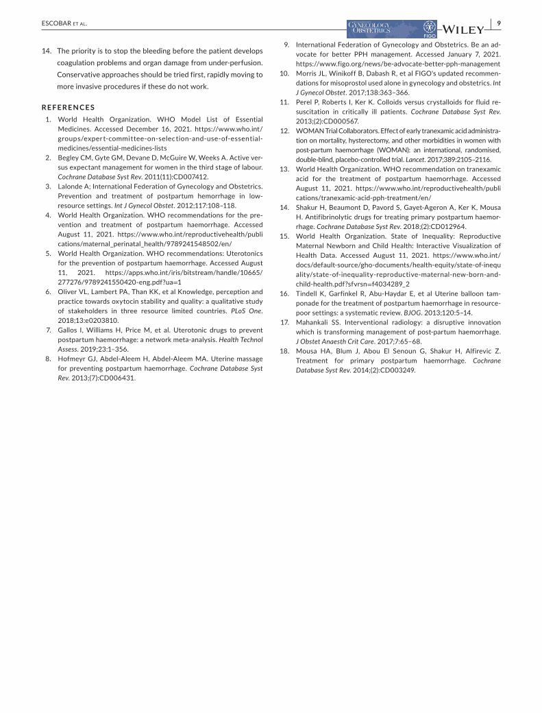
14. The priority is to stop the bleeding before the patient develops coagulation problems and organ damage from under- perfusion. Conservative approaches should be tried first, rapidly moving to more invasive procedures if these do not work.
R E FE R E N C E S 1. World Health Organization. WHO Model List of Essential
Medicines. Accessed December 16, 2021. https://www.who.int/group s/exper t- commi ttee- on- selec tion- and- use- of- essen tial- medic ines/essen tial- medic ines- lists
2. Begley CM, Gyte GM, Devane D, McGuire W, Weeks A. Active ver-sus expectant management for women in the third stage of labour. Cochrane Database Syst Rev. 2011(11):CD007412.
3. Lalonde A; International Federation of Gynecology and Obstetrics. Prevention and treatment of postpartum hemorrhage in low- resource settings. Int J Gynecol Obstet. 2012;117:108– 118.
4. World Health Organization. WHO recommendations for the pre-vention and treatment of postpartum haemorrhage. Accessed August 11, 2021. https://www.who.int/repro ducti vehea lth/publi catio ns/mater nal_perin atal_healt h/97892 41548 502/en/
5. World Health Organization. WHO recommendations: Uterotonics for the prevention of postpartum haemorrhage. Accessed August 11, 2021. https://apps.who.int/iris/bitst ream/handl e/10665/ 27727 6/97892 41550 420- eng.pdf?ua=1
6. Oliver VL, Lambert PA, Than KK, et al Knowledge, perception and practice towards oxytocin stability and quality: a qualitative study of stakeholders in three resource limited countries. PLoS One. 2018;13:e0203810.
7. Gallos I, Williams H, Price M, et al. Uterotonic drugs to prevent postpartum haemorrhage: a network meta- analysis. Health Technol Assess. 2019;23:1– 356.
8. Hofmeyr GJ, Abdel- Aleem H, Abdel- Aleem MA. Uterine massage for preventing postpartum haemorrhage. Cochrane Database Syst Rev. 2013;(7):CD006431.
9. International Federation of Gynecology and Obstetrics. Be an ad-vocate for better PPH management. Accessed January 7, 2021. https://www.figo.org/news/be- advoc ate- bette r- pph- manag ement
10. Morris JL, Winikoff B, Dabash R, et al FIGO's updated recommen-dations for misoprostol used alone in gynecology and obstetrics. Int J Gynecol Obstet. 2017;138:363– 366.
11. Perel P, Roberts I, Ker K. Colloids versus crystalloids for fluid re-suscitation in critically ill patients. Cochrane Database Syst Rev. 2013;(2):CD000567.
12. WOMAN Trial Collaborators. Effect of early tranexamic acid administra-tion on mortality, hysterectomy, and other morbidities in women with post- partum haemorrhage (WOMAN): an international, randomised, double- blind, placebo- controlled trial. Lancet. 2017;389:2105– 2116.
13. World Health Organization. WHO recommendation on tranexamic acid for the treatment of postpartum haemorrhage. Accessed August 11, 2021. https://www.who.int/repro ducti vehea lth/publi catio ns/trane xamic - acid- pph- treat ment/en/
14. Shakur H, Beaumont D, Pavord S, Gayet- Ageron A, Ker K, Mousa H. Antifibrinolytic drugs for treating primary postpartum haemor-rhage. Cochrane Database Syst Rev. 2018;(2):CD012964.
15. World Health Organization. State of Inequality: Reproductive Maternal Newborn and Child Health: Interactive Visualization of Health Data. Accessed August 11, 2021. https://www.who.int/docs/defau lt- sourc e/gho- docum ents/healt h- equit y/state - of- inequ ality/ state - of- inequ ality - repro ducti ve- mater nal- new- born- and- child - health.pdf?sfvrs n=f4034 289_2
16. Tindell K, Garfinkel R, Abu- Haydar E, et al Uterine balloon tam-ponade for the treatment of postpartum haemorrhage in resource- poor settings: a systematic review. BJOG. 2013;120:5– 14.
17. Mahankali SS. Interventional radiology: a disruptive innovation which is transforming management of post- partum haemorrhage. J Obstet Anaesth Crit Care. 2017;7:65– 68.
18. Mousa HA, Blum J, Abou El Senoun G, Shakur H, Alfirevic Z. Treatment for primary postpartum haemorrhage. Cochrane Database Syst Rev. 2014;(2):CD003249.

10 | ESCOBAR Et Al.
3 | BACKGROUND
3.1 | Introduction
Postpartum hemorrhage (PPH) is an obstetric emergency complicat-ing 1%– 10% of all deliveries.1 It continues to be the leading obstetric cause of maternal death.1 In 2015, it was reported to be responsible for more than 80 000 maternal deaths worldwide.1 Its distribution varies across regions, with the highest prevalence of 5.1%– 25.7% reported in Africa, followed by North America at 4.3%– 13% and Asia at 1.9%– 8%.2 The incidence of PPH has also been on the rise,2– 5 in-creasing from 5.1%– 6.2% in Canada between 2003 and 2010,3 and from 2.9%– 3.2% in the USA between 2010 and 2014.4
3.2 | Past FIGO recommendations for PPH
FIGO has made several recommendations in the past 20 years for the management and treatment of PPH (Table 1). This document will update the recommendations and discuss new approaches.
3.3 | Definition of postpartum hemorrhage
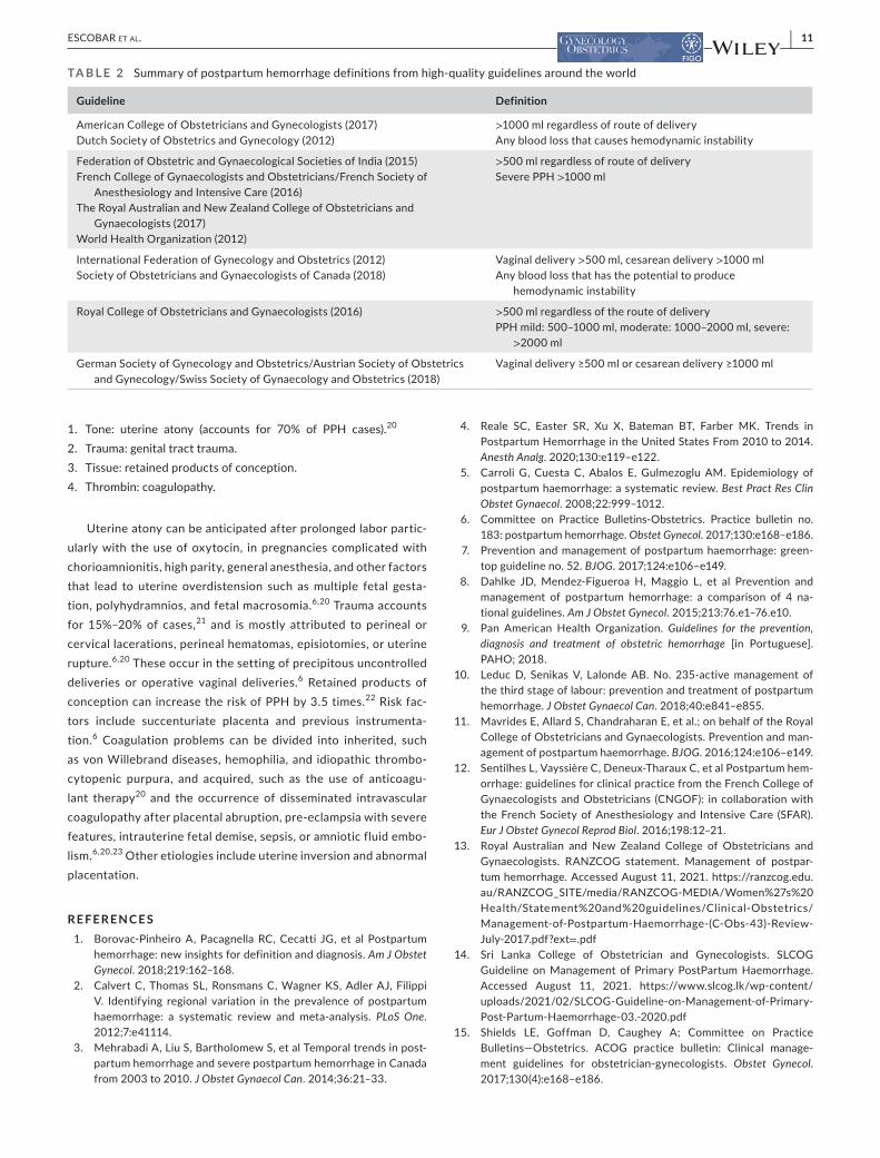
The lack of consistency in the definition of PPH has been a major limitation to the ability to compare prevalence in different stud-ies (Table 2). Classically, it was defined as quantified bleeding of more than 500 ml for vaginal deliveries and more than 1000 ml for cesarean deliveries, occurring within the first 24 h of delivery.1
However, this definition did not focus on clinical signs and symp-toms of hemorrhage, and thus prevented early detection in many cases. Therefore, in 2017, the American College of Obstetricians and Gynecologists (ACOG) changed the definition to blood loss of more than or equal to 1000 ml, or blood loss that was accompanied by signs or symptoms of hypovolemia occurring within 24 h after birth, regardless of the mode of delivery.6 In contrast, the Royal College of Obstetricians and Gynaecologists (RCOG) defines PPH according to the volume of blood lost: minor (between 500 and 1000 ml) and major (>1000 ml).7 However, the volume of estimated blood loss remains unreliable in many cases, and therefore much attention should be directed to the general clinical status of the patient instead.8 Several tools for assessment of blood loss have been used as accurate estimation will directly influence the diag-nosis and management of PPH. Many groups cite visual estimation as part of blood loss assessment, but as it has high potential to un-derestimate hemorrhage, use of additional tools for more objective estimation, such as gravimetric measurement, direct blood collec-tion techniques, and evaluation of clinical parameters, have been proposed.9– 17 Recently, some guidelines have incorporated the shock index9,11,14,17 and obstetric early warning systems into their recommendations to evaluate bleeding.11,14,17
3.4 | Etiologies/risk factors
While there exist several identifiable risk factors for PPH, most cases occur unexpectedly.6,18 An easy way to remember the most common etiologies is to remember the four T’s19:
TA B L E 1 FIGO recommendations on the management of postpartum hemorrhage
FIGO recommendation Year References
Management of the third stage of labor to prevent post- partum hemorrhage
2003 International Confederation of Midwives; International Federation of Gynaecologists and Obstetricians. Joint statement: management of the third stage of labour to prevent post- partum haemorrhage. J Midwifery Womens Health. 2004 Jan- Feb;49(1):76– 7.
Postpartum hemorrhage today: ICM/FIGO initiative 2004– 2006
2006 Lalonde A, Daviss BA, Acosta A, Herschderfer K. Int J Gynecol Obstet. 2006;94:243– 253.
Prevention and treatment of post- partum haemorrhage: new advances for low resource settings
2006 Joint Statement: ICM and FIGO https://www.who.int/pmnch/ event s/2006/figo2 006st ateme nteng.pdf
Prevention and treatment of postpartum hemorrhage in low- resource settings
2012 Lalonde A; International Federation of Gynecology and Obstetrics. Int J Gynecol Obstet. 2012;117:108– 118.
Prevention of postpartum hemorrhage with misoprostol 2012 International Federation of Gynecology and Obstetrics. Int J Gynecol Obstet. 2012;119:213– 214.
Treatment of postpartum hemorrhage with misoprostol 2012 International Federation of Gynecology and Obstetrics. Int J Gynecol Obstet. 2012;119:215– 216.
Non- pneumatic anti- shock garment to stabilize women with hypovolemic shock secondary to obstetric hemorrhage
2015 FIGO Safe Motherhood and Newborn Health Committee; International Federation of Gynecology and Obstetrics. Int J Gynecol Obstet. 2015;128:194– 195.
FIGO's updated recommendations for misoprostol used alone in gynecology and obstetrics
2017 Morris JL, Winikoff B, Dabash R, et al. Int J Gynecol Obstet. 2017;138:363– 366.
Affordable and low- maintenance obstetric devices 2019 Ayres- de- Campos D, Stones W, Theron G; FIGO Safe Motherhood and Newborn Health Committee. Int J Gynecol Obstet. 2019;146:25– 28.
| 11ESCOBAR Et Al.
1. Tone: uterine atony (accounts for 70% of PPH cases).20
2. Trauma: genital tract trauma.3. Tissue: retained products of conception.4. Thrombin: coagulopathy.
Uterine atony can be anticipated after prolonged labor partic-ularly with the use of oxytocin, in pregnancies complicated with chorioamnionitis, high parity, general anesthesia, and other factors that lead to uterine overdistension such as multiple fetal gesta-tion, polyhydramnios, and fetal macrosomia.6,20 Trauma accounts for 15%– 20% of cases,21 and is mostly attributed to perineal or cervical lacerations, perineal hematomas, episiotomies, or uterine rupture.6,20 These occur in the setting of precipitous uncontrolled deliveries or operative vaginal deliveries.6 Retained products of conception can increase the risk of PPH by 3.5 times.22 Risk fac-tors include succenturiate placenta and previous instrumenta-tion.6 Coagulation problems can be divided into inherited, such as von Willebrand diseases, hemophilia, and idiopathic thrombo-cytopenic purpura, and acquired, such as the use of anticoagu-lant therapy20 and the occurrence of disseminated intravascular coagulopathy after placental abruption, pre- eclampsia with severe features, intrauterine fetal demise, sepsis, or amniotic fluid embo-lism.6,20,23 Other etiologies include uterine inversion and abnormal placentation.
R E FE R E N C E S 1. Borovac- Pinheiro A, Pacagnella RC, Cecatti JG, et al Postpartum
hemorrhage: new insights for definition and diagnosis. Am J Obstet Gynecol. 2018;219:162– 168.
2. Calvert C, Thomas SL, Ronsmans C, Wagner KS, Adler AJ, Filippi V. Identifying regional variation in the prevalence of postpartum haemorrhage: a systematic review and meta- analysis. PLoS One. 2012;7:e41114.
3. Mehrabadi A, Liu S, Bartholomew S, et al Temporal trends in post-partum hemorrhage and severe postpartum hemorrhage in Canada from 2003 to 2010. J Obstet Gynaecol Can. 2014;36:21– 33.
4. Reale SC, Easter SR, Xu X, Bateman BT, Farber MK. Trends in Postpartum Hemorrhage in the United States From 2010 to 2014. Anesth Analg. 2020;130:e119– e122.
5. Carroli G, Cuesta C, Abalos E, Gulmezoglu AM. Epidemiology of postpartum haemorrhage: a systematic review. Best Pract Res Clin Obstet Gynaecol. 2008;22:999– 1012.
6. Committee on Practice Bulletins- Obstetrics. Practice bulletin no. 183: postpartum hemorrhage. Obstet Gynecol. 2017;130:e168– e186.
7. Prevention and management of postpartum haemorrhage: green- top guideline no. 52. BJOG. 2017;124:e106– e149.
8. Dahlke JD, Mendez- Figueroa H, Maggio L, et al Prevention and management of postpartum hemorrhage: a comparison of 4 na-tional guidelines. Am J Obstet Gynecol. 2015;213:76.e1– 76.e10.
9. Pan American Health Organization. Guidelines for the prevention, diagnosis and treatment of obstetric hemorrhage [in Portuguese]. PAHO; 2018.
10. Leduc D, Senikas V, Lalonde AB. No. 235- active management of the third stage of labour: prevention and treatment of postpartum hemorrhage. J Obstet Gynaecol Can. 2018;40:e841– e855.
11. Mavrides E, Allard S, Chandraharan E, et al.; on behalf of the Royal College of Obstetricians and Gynaecologists. Prevention and man-agement of postpartum haemorrhage. BJOG. 2016;124:e106– e149.
12. Sentilhes L, Vayssière C, Deneux- Tharaux C, et al Postpartum hem-orrhage: guidelines for clinical practice from the French College of Gynaecologists and Obstetricians (CNGOF): in collaboration with the French Society of Anesthesiology and Intensive Care (SFAR). Eur J Obstet Gynecol Reprod Biol. 2016;198:12– 21.
13. Royal Australian and New Zealand College of Obstetricians and Gynaecologists. RANZCOG statement. Management of postpar-tum hemorrhage. Accessed August 11, 2021. https://ranzc og.edu.au/RANZC OG_SITE/media/ RANZC OG- MEDIA/ Women %27s%20Hea lth/State ment%20and %20gui delin es/Clini cal- Obste trics/ Manag ement - of- Postp artum - Haemo rrhag e- (C- Obs- 43)- Revie w- July- 2017.pdf?ext=.pdf
14. Sri Lanka College of Obstetrician and Gynecologists. SLCOG Guideline on Management of Primary PostPartum Haemorrhage. Accessed August 11, 2021. https://www.slcog.lk/wp- conte nt/uploa ds/2021/02/SLCOG - Guide line- on- Manag ement - of- Prima ry- Post- Partu m- Haemo rrhag e- 03.- 2020.pdf
15. Shields LE, Goffman D, Caughey A; Committee on Practice Bulletins— Obstetrics. ACOG practice bulletin: Clinical manage-ment guidelines for obstetrician- gynecologists. Obstet Gynecol. 2017;130(4):e168– e186.
TA B L E 2 Summary of postpartum hemorrhage definitions from high- quality guidelines around the world
Guideline Definition
American College of Obstetricians and Gynecologists (2017)Dutch Society of Obstetrics and Gynecology (2012)
>1000 ml regardless of route of deliveryAny blood loss that causes hemodynamic instability
Federation of Obstetric and Gynaecological Societies of India (2015)French College of Gynaecologists and Obstetricians/French Society of
Anesthesiology and Intensive Care (2016)The Royal Australian and New Zealand College of Obstetricians and
Gynaecologists (2017)World Health Organization (2012)
>500 ml regardless of route of deliverySevere PPH >1000 ml
International Federation of Gynecology and Obstetrics (2012)Society of Obstetricians and Gynaecologists of Canada (2018)
Vaginal delivery >500 ml, cesarean delivery >1000 mlAny blood loss that has the potential to produce
hemodynamic instability
Royal College of Obstetricians and Gynaecologists (2016) >500 ml regardless of the route of deliveryPPH mild: 500– 1000 ml, moderate: 1000– 2000 ml, severe:
>2000 ml
German Society of Gynecology and Obstetrics/Austrian Society of Obstetrics and Gynecology/Swiss Society of Gynaecology and Obstetrics (2018)
Vaginal delivery ≥500 ml or cesarean delivery ≥1000 ml

12 | ESCOBAR Et Al.
16. Schlembach D, Helmer H, Henrich W, et al. Peripartum haemor-rhage, diagnosis and therapy. Guideline of the DGGG, OEGGG and SGGG (S2k Level, AWMF Registry No. 015/063, March 2016). Geburtshilfe Frauenheilkd. 2018;78:382– 399.
17. Fawcus S. Alerts for managing postpartum haemorrhage. S Afr Med J. 2018;108:1013– 1017.
18. Newsome J, Martin JG, Bercu Z, Shah J, Shekhani H, Peters G. Postpartum hemorrhage. Tech Vasc Interv Radiol. 2017;20:266– 273.
19. Anderson J, Etches D. Prevention and management of postpartum hemorrhage. Am Fam Physician. 2007;75:875– 882.
20. Oyelese Y, Anant CV. Postpartum hemorrhage: epidemiology, risk factors, and causes. Clin Obstet Gynecol. 2010;53:147– 156.
21. Sentilhes L, Merlot B, Madar H, Sztark F, Brun S, Deneux- Tharaux C. Postpartum haemorrhage: prevention and treatment. Expert Rev Hematol. 2016;9:1043– 1061.
22. Sheiner E, Sarid L, Levy A, Seidman DS, Hallak M. Obstetric risk fac-tors and outcome of pregnancies complicated with early postpar-tum hemorrhage: a population- based study. J Matern Fetal Neonatal Med. 2005;18:149– 154.
23. Evensen A, Anderson JM, Fontaine P. Postpartum hemorrhage: prevention and treatment. Am Fam Physician. 2017;95:442– 449.
| 13ESCOBAR Et Al.
4 | POSTPARTUM HEMORRHAGE BUNDLE C ARE
Multimodal strategies have been implemented in high- income coun-tries to control pathologies with high mortality rates such as PPH. These initiatives that involve multiple intervention points and actors have been called “bundles” or intervention packages, which consist of the implementation of a group of interventions as well as multi-disciplinary programs that standardize and comprehensively address the management of pathologies.1– 6 Bundles represent a selection of existing guidelines and recommendations in a form that aids system-atic implementation and a consistency of practice.
The California Maternal Quality Care Collaborative (CMQCC) Working Group on obstetrical hemorrhage developed the Improving the Health Care Response to Obstetric Bleeding Toolkit in 2010 to help obstetric providers, clinical staff, hospitals, and healthcare
organizations develop methods within their facility for timely rec-ognition and an organized and rapid response to bleeding. In March 2015, version 2.0 was updated with the latest evidence- based changes.6
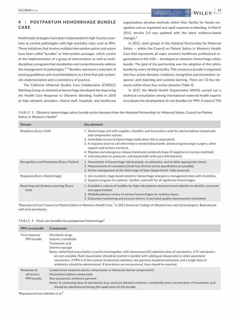
In 2015, work groups of the National Partnership for Maternal Safety — within the Council on Patient Safety in Women's Health Care that represents all major women's healthcare professional or-ganizations in the USA — developed an obstetric hemorrhage safety bundle. The goal of the partnership was the adoption of the safety bundle by every birthing facility. This consensus bundle is organized into four action domains: readiness; recognition and prevention; re-sponse; and reporting and systems learning. There are 13 key ele-ments within these four action domains (Table 3).
In 2017, the World Health Organization (WHO) carried out a technical consultation among international maternal health experts to evaluate the development of care bundles for PPH. A total of 730
TA B L E 3 Obstetric hemorrhage safety bundle action domains from the National Partnership for Maternal Safety, Council on Patient Safety in Women's Healtha
Domain Key elements
Readiness (Every Unit) 1. Hemorrhage cart with supplies, checklist, and instruction cards for uterine balloon tamponade and compression sutures.
2. Immediate access to hemorrhage medications (kit or equivalent).3. A response team to call when help is needed (blood bank, advanced gynecologic surgery, other
support and tertiary services).4. Massive and emergency- release transfusion protocols (type- O negative or uncross- matched).5. Unit education on protocols, unit- based drills (with post drill debriefs).
Recognition and Prevention (Every Patient) 1. Assessment of hemorrhage risk (prenatal, on admission, and at other appropriate times).2. Measurement of cumulative blood loss (formal and as quantitative as possible).3. Active management of the third stage of labor (department- wide protocol).
Response (Every Hemorrhage) 1. Uni- standard, stage- based obstetric hemorrhage emergency management plan with checklists.2. Support program for patients, families, and staff for all significant hemorrhages.
Reporting and Systems Learning (Every Unit)
1. Establish a culture of huddles for high- risk patients and post- event debriefs to identify successes and opportunities.
2. Multidisciplinary review of serious hemorrhages for systems issues.3. Outcomes monitoring and process metrics in perinatal quality improvement committee.
aReproduced from Council on Patient Safety in Women's Health Care.7 © 2015 American College of Obstetricians and Gynecologists. Reproduced with kind permission.
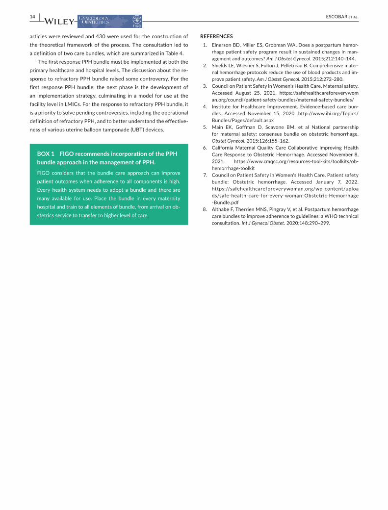
TA B L E 4 Final care bundles for postpartum hemorrhagea
PPH care bundle Components
First response PPH bundle
Uterotonic drugsIsotonic crystalloidsTranexamic acidUterine massageNotes: Initial fluid resuscitation is performed together with intravenous (IV) administration of uterotonics. If IV uterotonics
are not available, fluid resuscitation should be started in parallel with sublingual misoprostol or other parenteral uterotonics. If PPH is in the context of placental retention, the placenta should be extracted, and a single dose of antibiotics should be administered. If lacerations are encountered, they should be repaired.
Response to refractory PPH bundle
Compressive measures (aortic compression or bimanual uterine compression)Intrauterine balloon tamponadeNon- pneumatic antishock garmentNotes: A continuing dose of uterotonics (e.g. oxytocin diluted in isotonic crystalloids) and a second dose of tranexamic acid
should be administered during the application of this bundle.
aReproduced from Althabe et al.8
14 | ESCOBAR Et Al.
articles were reviewed and 430 were used for the construction of the theoretical framework of the process. The consultation led to a definition of two care bundles, which are summarized in Table 4.
The first response PPH bundle must be implemented at both the primary healthcare and hospital levels. The discussion about the re-sponse to refractory PPH bundle raised some controversy. For the first response PPH bundle, the next phase is the development of an implementation strategy, culminating in a model for use at the facility level in LMICs. For the response to refractory PPH bundle, it is a priority to solve pending controversies, including the operational definition of refractory PPH, and to better understand the effective-ness of various uterine balloon tamponade (UBT) devices.
REFERENCES 1. Einerson BD, Miller ES, Grobman WA. Does a postpartum hemor-
rhage patient safety program result in sustained changes in man-agement and outcomes? Am J Obstet Gynecol. 2015;212:140– 144.
2. Shields LE, Wiesner S, Fulton J, Pelletreau B. Comprehensive mater-nal hemorrhage protocols reduce the use of blood products and im-prove patient safety. Am J Obstet Gynecol. 2015;212:272– 280.
3. Council on Patient Safety in Women's Health Care. Maternal safety. Accessed August 25, 2021. https://safeh ealth caref oreve rywom an.org/counc il/patie nt- safet y- bundl es/mater nal- safet y- bundl es/
4. Institute for Healthcare Improvement. Evidence- based care bun-dles. Accessed November 15, 2020. http://www.ihi.org/Topic s/Bundl es/Pages/ defau lt.aspx
5. Main EK, Goffman D, Scavone BM, et al National partnership for maternal safety: consensus bundle on obstetric hemorrhage. Obstet Gynecol. 2015;126:155– 162.
6. California Maternal Quality Care Collaborative Improving Health Care Response to Obstetric Hemorrhage. Accessed November 8, 2021. https://www.cmqcc.org/resou rces- tool- kits/toolk its/ob- hemor rhage - toolkit
7. Council on Patient Safety in Women's Health Care. Patient safety bundle: Obstetric hemorrhage. Accessed January 7, 2022. https://safeh ealth caref oreve rywom an.org/wp- conte nt/uploa ds/safe- healt h- care- for- every - woman - Obste tric- Hemor rhage - Bundle.pdf
8. Althabe F, Therrien MNS, Pingray V, et al. Postpartum hemorrhage care bundles to improve adherence to guidelines: a WHO technical consultation. Int J Gynecol Obstet. 2020;148:290– 299.
BOX 1 FIGO recommends incorporation of the PPH bundle approach in the management of PPH.
FIGO considers that the bundle care approach can improve patient outcomes when adherence to all components is high. Every health system needs to adopt a bundle and there are many available for use. Place the bundle in every maternity hospital and train to all elements of bundle, from arrival on ob-stetrics service to transfer to higher level of care.
| 15ESCOBAR Et Al.
5 | SHOCK INDE X E VIDENCE IN POSTPARTUM HEMORRHAGE E VALUATION AND MANAGEMENT
Shock refers to a reduction in tissue perfusion, which is insuf-ficient to meet the metabolic requirements of tissues and organs. Insufficient blood flow may be clinically identified as the develop-ment of one or more of the following: lactic acidosis, altered men-tal status, oliguria, and tachycardia. Vital signs monitoring is key to hemodynamic assessment and prompt intervention.1 In healthy pregnant and postpartum women, cardiologic physiologic com-pensatory mechanisms prevent changes in vital signs until a large volume of blood has been lost (usually >1000 ml). Hence, changes in clinical and vital signs that result from hemorrhage appear late in the process and may not lead to early identification of PPH. This in turn makes it difficult to establish cutoff points to trigger clinical interventions. Moreover, because traditional vital signs change late and are less reliable as triggers for clinical actions, other indicators could help to characterize maternal hypovolemia caused by bleeding.2 Although the use of conventional individual vital signs (pulse and systolic blood pressure) may lack accuracy in the assessment of hypotension, a simple combination of both may transform routine clinical parameters into a more accurate indica-tor of hypovolemia, such as the shock index (SI). SI is defined as the ratio of heart rate to systolic blood pressure.3,4 The SI may im-prove the predictive capability of individual clinical signs, which aids early identification of women at risk of hypovolemia as the result of obstetric causes.5 Moreover, the SI has been proposed as a reliable indicator of adverse maternal outcomes,6 and its val-ues have been set to indicate clinical management.7 However, the association between shock parameters and advanced treatment modalities in severe PPH has yet to be reported.
5.1 | Assessment of circulating blood volume in postpartum hemorrhage
The essential cornerstone of management of PPH involves prompt diagnosis and rapid replacement of lost blood volume, as well as the oxygen- carrying capacity of blood, accompanied by immediate medical and surgical measures to address the underlying cause(s), and hence prevent more loss. To assess the patient's condition, SI has been introduced as a simple and clinically effective vital sign.
The SI has been shown to have an inverse linear relation-ship with left ventricular stroke work in acute circulatory failure. Therefore, a concurrent reduction of left ventricular stroke work (induced by hemorrhage, trauma, or sepsis) was associated with an elevation of the SI and a deterioration in left ventricular mechanical performance. Poor left ventricular function or persistent abnormal elevation of the SI after aggressive therapy and hemodynamic sta-bilization was associated with increased mortality in critically ill, traumatized patients.8 In obstetric and nonobstetric circumstances, the absence of a significant drop in blood pressure in patients with
PPH may mask the actual hypovolemic status due to physiological compensatory mechanisms.9 For that reason, the SI was the only promising marker that indicated the severity of blood loss.2,5
The SI, together with the rule of 30, are important tools that may aid clinicians in an emergency to determine the amount of blood loss and the degree of hemodynamic instability. Before the fall in systolic blood pressure, heart rate rises to compensate for the blood loss, and thus the SI increases. The rule of 30 is an approximated blood loss of 30% of normal (70 ml/kg in adults, 100 ml/kg throughout pregnancy), defined by a fall of 30% in hematocrit, a fall of 30% in hemoglobin (approximately 3 g/dl), a fall of 30 mm Hg in systolic blood pressure, and a rise in pulse rate by 30 beats per minute.10 It has been shown that an SI ≥0.9 is associated with increased mortal-ity and an SI>1 increases the likelihood of blood transfusion.11,12 To date, standard obstetric SI has been defined as 0.7– 0.9 compared with 0.5– 0.7 for the nonpregnant population, taking into account that the hemodynamic changes of pregnancy may delay the recogni-tion of hypovolemia.5 If intravascular volume depletion is suspected, a rapid clinical assessment is required because the patient's clinical condition can deteriorate, leading to the development of hemor-rhagic shock rapidly. Proper medical record- taking skills may high-light symptoms associated with shock such as pain and overt blood loss, as well as general malaise, anxiety, and dyspnea. Notably, in set-tings where few PPH treatment options exist, and in cases of home deliveries, diagnosis and treatment or referral must occur even ear-lier than in hospital settings to improve outcomes. For that reason, SI may be a valuable threshold in LMICs, where mortality is highest and is often related to delays in complication recognition, transpor-tation, and level of care at the facility.2 A threshold of SI ≥0.9 should be tested to alert community healthcare providers of the need for urgent transfer.13
R E FE R E N C E S 1. Schorn MN. Measurement of blood loss: review of the literature. J
Midwifery Womens Health. 2010;55:20– 27. 2. Borovac- Pinheiro A, Pacagnella RC, Cecatti JG, et al Postpartum
hemorrhage: new insights for definition and diagnosis. Am J Obstet Gynecol. 2018;219:162– 168.
3. Arulkumaran S, Karoshi M, Keith LG, Lalonde AB, B- Lynch C. A com-prehensive textbook of postpartum hemorrhage: an essential clinical reference for effective management. Sapiens Publishing; 2012.
4. Rady MY, Nightingale P, Little RA, Edwards JD. Shock index: a re- evaluation in acute circulatory failure. Resuscitation. 1992;23:227– 234.
5. Pacagnella RC, Souza JP, Durocher J, et al A systematic review of the relationship between blood loss and clinical signs. PLoS One. 2013;8:e57594.
BOX 2 FIGO recommends use of the shock index in the diagnosis and management of PPH.
FIGO considers that the shock index can be a marker of the severity of PPH and can alert teams to hemodynamic instabil-ity when its value is greater than 0.9.

16 | ESCOBAR Et Al.
6. El Ayadi AM, Nathan HL, Seed PT, et al Vital sign prediction of ad-verse maternal outcomes in women with hypovolemic shock: the role of shock index. PLoS One. 2016;11:e0148729.
7. Le Bas A, Chandraharan E, Addei A, Arulkumaran S. Use of the “ob-stetric shock index” as an adjunct in identifying significant blood loss in patients with massive postpartum hemorrhage. Int J Gynecol Obstet. 2014;124:253– 255.
8. Rady MY, Smithline HA, Blake H, Nowak R, Rivers E. A compar-ison of the shock index and conventional vital signs to identify acute, critical illness in the emergency department. Ann Emerg Med. 1994;24:685– 690.
9. Troiano NH, Witcher PM, Baird SM. High- Risk & Critical Care Obstetrics. Lippincott Williams & Wilkins; 2018.
10. Chandraharan E, Arulkumaran S. Obstetric and Intrapartum Emergencies: A Practical Guide to Management. Cambridge University Press; 2012.
11. Vandromme MJ, Griffin RL, Kerby JD, McGwin G Jr, Rue LW 3rd, Weinberg JA. Identifying risk for massive transfusion in the rela-tively normotensive patient: utility of the prehospital shock index. J Trauma. 2011;70(2):384– 390; discussion 388– 90.
12. Cannon CM, Braxton CC, Kling- Smith M, Mahnken JD, Carlton E, Moncure M. Utility of the shock index in predicting mortality in traumatically injured patients. J Trauma. 2009;67:1426– 1430.
13. Nathan HL, El Ayadi A, Hezelgrave NL, et al Shock index: an ef-fective predictor of outcome in postpartum haemorrhage? BJOG. 2015;122:268– 275.
| 17ESCOBAR Et Al.
6 | RE VIE W OF GUIDELINES AROUND THE WORLD
Guidelines are defined as systematically developed statements that assist practitioners to take decisions about appropriate health care in specific clinical circumstances.1 Over the past dec-ades, many national and international PPH guidelines have been developed and become part of obstetric clinical practice around the world. PPH guidelines usually address similar topics (e.g. di-agnosis, prevention, and treatment of PPH) but may differ in their recommendations.2– 4 These differences are because most of the recommendations are based on observational studies, clinical judgment, and expert opinion. There are few randomized con-trolled trials available to produce strong recommendations for the management of PPH due to the emergency of the condition that hinders this type of study. In the absence of randomized trials, guidelines gather the best available evidence. In addition, popula-tion characteristics, cultural aspects, resources availability, as well as frequency and timing of updates may influence the guidelines’ contents and justify some disparities.1– 4
R E FE R E N C E S 1. Woolf SH, Grol R, Hutchinson A, Eccles M, Grimshaw J. Clinical
guidelines: potential benefits, limitations, and harms of clinical guidelines. BMJ. 1999;318:527– 530.
2. Sentilhes L, Goffinet F, Vayssière C, Deneux- Tharaux C. Comparison of postpartum haemorrhage guidelines: discrepancies underline our lack of knowledge. BJOG. 2017;124:718– 722.
3. Bohlmann MK, Rath W. Medical prevention and treatment of post-partum hemorrhage: a comparison of different guidelines. Arch Gynecol Obstet. 2014;289:555– 567.
4. Dahlke JD, Mendez- Figueroa H, Maggio L, et al Prevention and management of postpartum hemorrhage: a comparison of 4 na-tional guidelines. Am J Obstet Gynecol. 2015;213(1):76.e1– 76.e10.
6.1 | Guidelines that address the prevention of postpartum hemorrhage
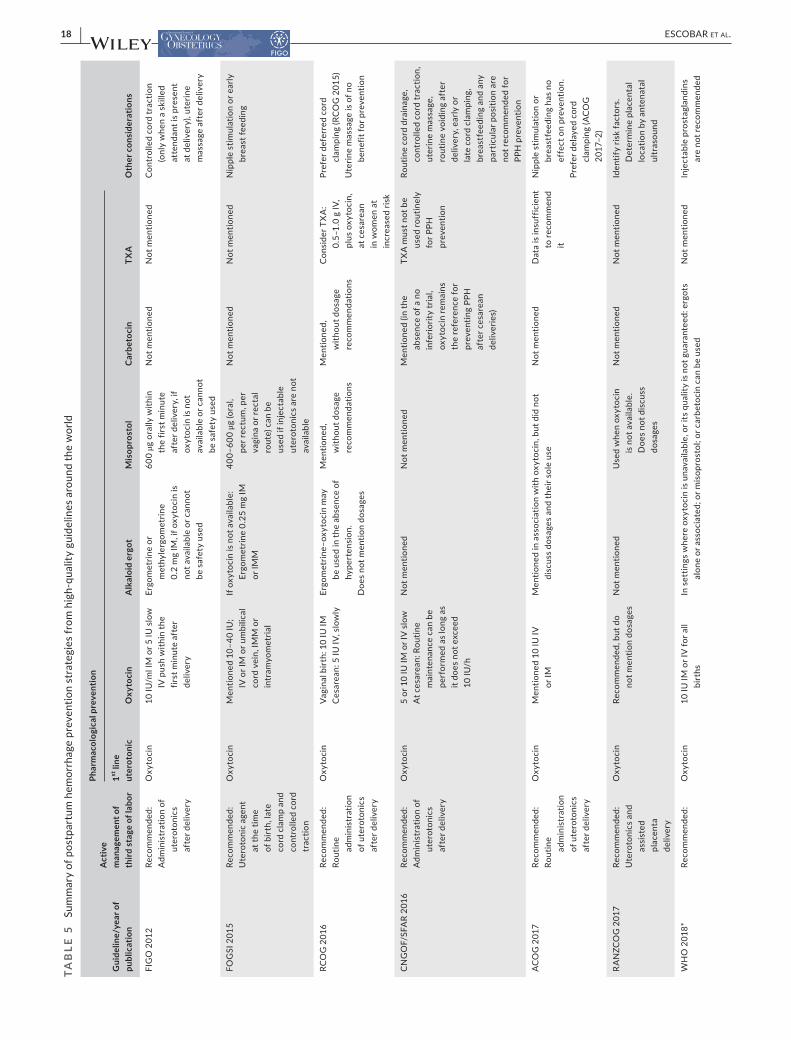
Active management of third stage of labor is frequently discussed in guidelines.1– 7 There is consensus that all women should receive uterotonics after delivery as it has proven to reduce PPH rates. Oxytocin has been cited as the drug of choice by most guidelines, but its dosages and route of administration vary largely, especially when considering mode of delivery.1– 5,7– 10 In 2012, FIGO established recommendations for the prevention of PPH11 and in 2018 WHO updated its recommendation for pharmacological PPH prevention and reinforced the use of oxytocin (10 IU intramuscularly or intra-venously) as the drug of choice.12 WHO also recommends the use of carbetocin (if cost- effective), ergot alkaloids (alone or combined if there are no contraindications), or oral misoprostol in settings where oxytocin is not available or its quality cannot be guaranteed. Misoprostol is also recommended when the use of other injectable uterotonics is not possible due to unavailability or contraindication to use such as hypertension in the context of ergometrine.12,13 The
Society of Obstetricians and Gynaecologists of Canada (SOGC) has updated its publication and reinforced the use of carbetocin as a first- line uterotonic for prevention at cesarean delivery or vaginal delivery with one risk factor.6 The German/Austrian/Swiss guideline mentions that prophylaxis during cesarean delivery can consist of administering either oxytocin or carbetocin.5 Other prophylactic strategies have been proposed in guidelines, but many had no great consensus or no clear benefits.1– 7 Table 5 summarizes various PPH prevention strategies described by different societies worldwide.
R E FE R E N C E S 1. Prevention and management of postpartum haemorrhage: green-
top guideline no. 52. BJOG. 2017;124:e106– e149. 2. Sentilhes L, Vayssière C, Deneux- Tharaux C, et al Postpartum hem-
orrhage: guidelines for clinical practice from the French College of Gynaecologists and Obstetricians (CNGOF): in collaboration with the French Society of Anesthesiology and Intensive Care (SFAR). Eur J Obstet Gynecol Reprod Biol. 2016;198:12– 21.
3. World Health Organization. WHO Recommendations for the Prevention and Treatment of Postpartum Haemorrhage. Accessed August 11, 2021. https://www.who.int/repro ducti vehea lth/publi catio ns/mater nal_perin atal_healt h/97892 41548 502/en/
4. Committee on Practice Bulletins- Obstetrics. Practice bulletin no. 183: postpartum hemorrhage. Obstet Gynecol. 2017;130:e168– e186.
5. Schlembach D, Helmer H, Henrich W, et al. Peripartum haemor-rhage, diagnosis and therapy. Guideline of the DGGG, OEGGG and SGGG (S2k Level, AWMF Registry No. 015/063, March 2016). Geburtshilfe Frauenheilkd. 2018;78:382– 399.
6. Leduc D, Senikas V, Lalonde AB. No 235- active management of the third stage of labour: prevention and treatment of postpartum hemorrhage. J Obstet Gynaecol Can. 2018;40:e841– e855.
7. Organization PAH. Guidelines for the prevention, diagnosis and treat-ment of obstetric hemorrhage [in Portuguese]. PAHO; 2018.
8. Fawcus S. Alerts for managing postpartum haemorrhage. S Afr Med J. 2018;108:1013– 1017.
9. Sri Lanka College of Obstetrician and Gynecologists. SLCOG Guideline on Management of Primary PostPartum Haemorrhage. Accessed August 11, 2021. https://www.slcog.lk/wp- conte nt/uploa ds/2021/02/SLCOG - Guide line- on- Manag ement - of- Prima ry- Post- Partu m- Haemo rrhag e- 03.- 2020.pdf
10. Fuchther C, Ortiz EI, Escobar MF, Lizaola H. Hemorragia Postparto: en donde estamos y hacia donde vamos. Federación Latinoamericna de Sociedades de Ginecologia y Obstetricia. Accessed August 11,
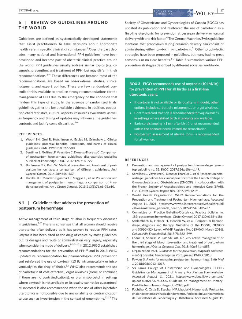
BOX 3 FIGO recommends use of oxytocin (10 IM/IV) for prevention of PPH for all births as a first- line uterotonic agent.
• If oxytocin is not available or its quality is in doubt, other options include carbetocin, misoprostol, or ergot alkaloids.
• Controlled cord traction is recommended for vaginal births in settings where skilled birth attendants are available.
• Early cord clamping (<1 min after birth) is not recommended unless the neonate needs immediate resuscitation.
• Postpartum assessment of uterine tonus is recommended for all women.
18 | ESCOBAR Et Al.
TAB
LE 5
Su
mm
ary
of p
ostp
artu
m h
emor
rhag
e pr
even
tion
stra
tegi
es fr
om h
igh-
qual
ity g
uide
lines
aro
und
the
wor
ld
Gui
delin
e/ye
ar o
f pu
blic
atio
n
Act
ive
man
agem
ent o
f th
ird s
tage
of l
abor
Phar
mac
olog
ical
pre
vent
ion
Oth
er c
onsi
dera
tions
1st li
ne
uter
oton
icO
xyto
cin
Alk
aloi
d er
got
Mis
opro
stol
Car
beto
cin
TXA
FIG
O 2
012
Reco
mm
ende
d:A
dmin
istr
atio
n of
ut
erot
onic
s af
ter d
eliv
ery
Oxy
toci
n10
IU/m
l IM
or 5
IU s
low
IV
pus
h w
ithin
the
first
min
ute
afte
r de
liver
y
Ergo
met
rine
or
met
hyle
rgom
etrin
e 0.
2 m
g IM
, if o
xyto
cin
is
not a
vaila
ble
or c
anno
t be
saf
ety
used
600
μg o
rally
with
in
the
first
min
ute
afte
r del
iver
y, if
ox
ytoc
in is
not
av
aila
ble
or c
anno
t be
saf
ety
used
Not
men
tione
dN
ot m
entio
ned
Con
trol
led
cord
trac
tion
(onl
y w
hen
a sk
illed
at
tend
ant i
s pr
esen
t at
del
iver
y), u
terin
e m
assa
ge a
fter
del
iver
y
FOG
SI 2
015
Reco
mm
ende
d:U
tero
toni
c ag
ent
at th
e tim
e of
birt
h, la
te
cord
cla
mp
and
cont
rolle
d co
rd
trac
tion
Oxy
toci
nM
entio
ned
10– 4
0 IU
; IV
or I
M o
r um
bilic
al
cord
vei
n, IM
M o
r in
tram
yom
etria
l
If ox
ytoc
in is
not
ava
ilabl
e:
Ergo
met
rine
0.25
mg
IM
or IM
M
400–
600
μg (o
ral,
per r
ectu
m, p
er
vagi
na o
r rec
tal
rout
e) c
an b
e us
ed if
inje
ctab
le
uter
oton
ics
are
not
avai
labl
e
Not
men
tione
dN
ot m
entio
ned
Nip
ple
stim
ulat
ion
or e
arly
br
east
feed
ing
RCO
G 2
016
Reco
mm
ende
d:Ro
utin
e ad
min
istr
atio
n of
ute
roto
nics
af
ter d
eliv
ery
Oxy
toci
nVa
gina
l birt
h: 1
0 IU
IMC
esar
ean:
5 IU
IV, s
low
lyEr
gom
etrin
e– ox
ytoc
in m
ay
be u
sed
in th
e ab
senc
e of
hy
pert
ensi
on.
Doe
s no
t men
tion
dosa
ges
Men
tione
d,
with
out d
osag
e re
com
men
datio
ns
Men
tione
d,
with
out d
osag
e re
com
men
datio
ns
Con
side
r TX
A:
0.5–
1.0
g IV
, pl
us o
xyto
cin,
at
ces
area
n in
wom
en a
t in
crea
sed
risk
Pref
er d
efer
red
cord
cl
ampi
ng (R
CO
G 2
015)
Ute
rine
mas
sage
is o
f no
bene
fit fo
r pre
vent
ion
CN
GO
F/SF
AR
2016
Reco
mm
ende
d:A
dmin
istr
atio
n of
ut
erot
onic
s af
ter d
eliv
ery
Oxy
toci
n5
or 1
0 IU
IM o
r IV
slo
wA
t ces
area
n: R
outin
e m
aint
enan
ce c
an b
e pe
rfor
med
as
long
as
it do
es n
ot e
xcee
d 10
IU/h
Not
men
tione
dN
ot m
entio
ned
Men
tione
d (in
the
abse
nce
of a
no
infe
riorit
y tr
ial,
oxyt
ocin
rem
ains
th
e re
fere
nce
for
prev
entin
g PP
H
afte
r ces
area
n de
liver
ies)
TXA
mus
t not
be
used
rout
inel
y fo
r PPH
pr
even
tion
Rout
ine
cord
dra
inag
e,
cont
rolle
d co
rd tr
actio
n,
uter
ine
mas
sage
, ro
utin
e vo
idin
g af
ter
deliv
ery,
ear
ly o
r la
te c
ord
clam
ping
, br
east
feed
ing
and
any
part
icul
ar p
ositi
on a
re
not r
ecom
men
ded
for
PPH
pre
vent
ion
AC
OG
201
7Re
com
men
ded:
Rout
ine
adm
inis
trat
ion
of u
tero
toni
cs
afte
r del
iver
y
Oxy
toci
nM
entio
ned
10 IU
IV
or IM
Men
tione
d in
ass
ocia
tion
with
oxy
toci
n, b
ut d
id n
ot
disc
uss
dosa
ges
and
thei
r sol
e us
eN
ot m
entio
ned
Dat
a is
insu
ffic
ient
to
reco
mm
end
it
Nip
ple
stim
ulat
ion
or
brea
stfe
edin
g ha
s no
ef
fect
on
prev
entio
n.Pr
efer
del
ayed
cor
d cl
ampi
ng (A
CO
G
2017
– 2)
RA
NZC
OG
201
7Re
com
men
ded:
Ute
roto
nics
and
as
sist
ed
plac
enta
de
liver
y
Oxy
toci
nRe
com
men
ded,
but
do
not m
entio
n do
sage
sN
ot m
entio
ned
Use
d w
hen
oxyt
ocin
is
not
ava
ilabl
e.
Doe
s no
t dis
cuss
do
sage
s
Not
men
tione
dN
ot m
entio
ned
Iden
tify
risk
fact
ors.
D
eter
min
e pl
acen
tal
loca
tion
by a
nten
atal
ul
tras
ound
WH
O 2
018*
Reco
mm
ende
d:O
xyto
cin
10 IU
IM o
r IV
for a
ll bi
rths
In s
ettin
gs w
here
oxy
toci
n is
una
vaila
ble,
or i
ts q
ualit
y is
not
gua
rant
eed:
erg
ots
alon
e or
ass
ocia
ted;
or m
isop
rost
ol; o
r car
beto
cin
can
be u
sed
Not
men
tione
dIn
ject
able
pro
stag
land
ins
are
not r
ecom
men
ded

| 19ESCOBAR Et Al.
Gui
delin
e/ye
ar o
f pu
blic
atio
n
Act
ive
man
agem
ent o
f th
ird s
tage
of l
abor
Phar
mac
olog
ical
pre
vent
ion
Oth
er c
onsi
dera
tions
1st li
ne
uter
oton
icO
xyto
cin
Alk
aloi
d er
got
Mis
opro
stol
Car
beto
cin
TXA
*(Su
pers
edes
pre
viou
s 20
12 W
HO
gu
idel
ine
reco
mm
enda
tions
of
ute
roto
nics
for
the
prev
entio
n of
PH
H)
WH
O g
uide
line
(201
2):
Ute
roto
nics
, la
te c
ord
clam
p an
d co
ntro
lled
cord
trac
tion
if sk
illed
at
tend
ant
Rega
rdle
ss o
f rou
te o
f de
liver
yEr
gom
etrin
e or
M- E
rgom
etrin
e:
0.2
mg
IM o
r IV
or
Com
bina
tion:
oxy
toci
n 5
IU+
Ergo
met
rine
0.5
mg
IM (a
fter
hyp
erte
nsio
n is
ex
clud
ed p
rior t
o its
use
)
Mis
opro
stol
400
μg
or 6
00 μ
g,
oral
ly.
If in
ject
able
ut
erot
onic
s ar
e no
t fea
sibl
e
Car
beto
cin:
100
μg
IM
or IV
.W
here
its
cost
is
com
para
ble
to
othe
r eff
ectiv
e ut
erot
onic
s
Reco
mm
ende
d fo
r the
pr
even
tion
of P
PH.
(Car
bopr
ost o
r su
lpro
ston
e)
SOG
C 20
18Re
com
men
ded:
U
tero
toni
cs,
late
cor
d cl
amp
and
cont
rolle
d co
rd tr
actio
n
Vagi
nal l
ow ri
sk:
Oxy
toci
nC
esar
ean:
Car
beto
cin
Vagi
nal:
10 IU
IM o
r ox
ytoc
in, 2
0– 40
IU
in 1
000
ml,
150
ml
per h
our.
Ergo
novi
ne, 0
.2 m
g IM
Whe
n ox
ytoc
in is
not
ava
ilabl
e60
0– 80
0 μg
(ora
l, su
blin
gual
, or
rect
al ro
ute)
Whe
n ox
ytoc
in is
no
t ava
ilabl
e
Car
beto
cin,
100
μg
give
n as
an
IV b
olus
ov
er 1
min
Not
men
tione
dSu
gges
t con
side
ring
carb
etoc
in fo
r hig
h- ris
k w
omen
del
iver
ing
vagi
nally
DG
GG
/OEG
GG
/SSG
O
2018
Reco
mm
ende
d:
Oxy
toci
n af
ter
birt
h
Vagi
nal:
Oxy
toci
nC
esar
ean:
O
xyto
cin
or
carb
etoc
in
3– 5
IU s
low
IVN
ot m
entio
ned
Not
men
tione
d10
0 µg
by
shor
t inf
usio
n or
slo
w IV
infu
sion
Not
men
tione
dIm
med
iate
cor
d cl
ampi
ng
and
cont
rolle
d co
rd
trac
tion
have
no
impa
ct
on re
duci
ng P
PH a
nd
shou
ld n
ot b
e ca
rrie
d ou
t
FLA
SOG
201
8Re
com
men
ded
Oxy
toci
n10
IU IM
or I
VN
ot m
entio
ned
600
ug o
ral w
hen
oxyt
ocin
is n
ot
avai
labl
e
Not
Men
tione
dN
ot m
entio
ned
Con
trol
led
cord
trac
tion,
(o
nly
whe
n a
skill
ed
atte
ndan
t is
pres
ent
at d
eliv
ery)
, ute
rine
mas
sage
aft
er d
eliv
ery
Abb
revi
atio
ns: T
XA
, tra
nexa
mic
aci
d; F
IGO
, Int
erna
tiona
l Fed
erat
ion
of G
ynec
olog
y an
d O
bste
tric
s; IU
, int
erna
tiona
l uni
t; IM
, int
ram
uscu
lar;
IV, i
ntra
veno
us; F
OG
SI, F
eder
atio
n of
Obs
tetr
ic a
nd
Gyn
aeco
logi
cal S
ocie
ties
of In
dia;
IMM
, int
ram
amm
ary;
RCO
G, R
oyal
Col
lege
of O
bste
tric
ians
and
Gyn
aeco
logi
sts;
CN
GO
F/SF
AR,
Col
lege
of G
ynae
colo
gist
s an
d O
bste
tric
ians
/Fre
nch
Soci
ety
of
Ane
sthe
siol
ogy
and
Inte
nsiv
e C
are;
ACO
G, A
mer
ican
Col
lege
of O
bste
tric
ians
and
Gyn
ecol
ogis
ts; R
AN
ZCO
G, R
oyal
Aus
tral
ian
and
New
Zea
land
Col
lege
of O
bste
tric
ians
and
Gyn
aeco
logi
sts;
WH
O, W
orld
H
ealth
Org
aniz
atio
n; S
OG
C, S
ocie
ty o
f Obs
tetr
icia
ns a
nd G
ynae
colo
gist
s of
Can
ada;
DG
GG
/OEG
GG
/SSG
O, G
erm
an S
ocie
ty o
f Gyn
ecol
ogy
and
Obs
tetr
ics/
Aus
tria
n So
ciet
y of
Obs
tetr
ics
and
Gyn
ecol
ogy/
Swis
s So
ciet
y of
Gyn
aeco
logy
and
Obs
tetr
ics,
FLA
SOG
, Fed
erac
ión
Latin
oam
eric
ana
de S
ocie
dade
s de
Obs
tetr
icia
y G
inec
olog
ía.
TAB
LE 5
(C
ontin
ued)

20 | ESCOBAR Et Al.
2021. https://www.flasog.org/stati c/libro s/Hemor ragia - Postp ar to- 17OCT UBRE.pdf
11. Lalonde A; International Federation of Gynecology and Obstetrics. Prevention and treatment of postpartum hemorrhage in low- resource settings. Int J Gynaecol Obstet. 2012;117(2):108– 118.
12. World Health Organization. WHO recommendations: Uterotonics for the prevention of postpartum haemorrhage. Accessed August 25, 2021. https://apps.who.int/iris/bitst ream/handl e/10665/ 27727 6/97892 41550 420- eng.pdf?ua=1
13. Althabe F, Therrien MNS, Pingray V, et al. Postpartum hemorrhage care bundles to improve adherence to guidelines: A WHO technical consultation. Int J Gynecol Obstet. 2020;148:290– 299.
6.2 | Guidelines that address the treatment of postpartum hemorrhage
PPH guidelines frequently recommend a multidisciplinary approach for reaching effective early control of bleeding. Treatment should be directed to the specific cause of PPH (uterine atony, genital trauma, retained placenta, and/or coagulopathy) and therapeutic steps should move from the less invasive method to the more complex and radical approach. A set of initial measures also seems to be consen-sual in most guidelines and consist of maintenance of two large IV lines, supplementation of oxygen, strict monitoring of women, crys-talloids infusion, and measures to avoid hypothermia and evaluate the PPH cause.1– 9
If atony is the etiology, most guidelines suggest perform-ing temporary mechanical measures, such as uterine massage or uterine bimanual compression, with concurrent pharmaco-logical treatment.1– 10 Uterotonics are considered the first- line treatment for uterine atony. Intravenous oxytocin is usually the preferred drug and route of administration, but its dosage varies widely. When oxytocin fails to control PPH, guidelines recom-mend the use of an additional drug, such as ergot alkaloids, in-jectable prostaglandins, or misoprostol.1– 4,6– 8,10 SOGC mentions carbetocin as a uterotonic available for treatment,5 and the German/Austrian/Swiss guidelines highlight that the use of car-betocin to treat PPH is currently not sufficiently investigated4 (Table 6).
Since publication of the World Maternal Antifibrolytic (WOMAN) trial,11 tranexamic acid (TXA), an antifibrinolytic drug, has been in-corporated into PPH guidelines around the world.4,6– 10,12,13 WHO has updated this topic and recommends the use of TXA, as soon as possible, within the first 3 h from birth, at a dose of 1 g intravenously, regardless of the route of birth.12,13 However, some guidelines do not cite it5 or do not add it in a definitive manner because their last update occurred before the WOMAN trial results.1,2 Another drug that has been discussed in many guidelines is recombinant activated factor VII for massive PPH; however, there is no consensus about its use.1– 6
When pharmacological treatment fails in controlling hemor-rhage, guidelines usually recommend some mechanical, radiologi-cal, and more conservative surgical approaches before performing hysterectomy. The available guidelines are summarized in Table 6.
The most cited ones are uterine balloon tamponade (UBT), uterine compressive sutures (UCS), pelvic vascular ligation (PVL), and embo-lization.1– 7,10 Uterine packing with gauze is also mentioned in some guidelines, but its use is controversial.3,5 ACOG mentions the use of a gauze soaked with thrombin.6
German/Austrian/Swiss guidelines cite the use of intrauter-ine packing with a gauze coated with a hemostatic agent.4 Due to the lack of evidence, guidelines do not necessarily recommend these conservative approaches in a well- defined, step- wise pro-gression, and their utilization depends mainly on the availability of resources, professional familiarity with technique, and clinical circumstances.10
UBT is typically indicated as the treatment of choice when uter-ine atony is refractory to uterotonics after vaginal delivery as it is less invasive than the other procedures.1– 3,6– 9 This should be con-sidered after ruling out retained products of conception, ruptured uterus, or vaginal or cervical laceration as a contributing factor. If UBT fails to control bleeding in these cases, invasive treatments by arterial embolization or, most commonly, by surgical approaches are recommended (Table 7). UCS and/or PVL are recommended to avoid hysterectomy when laparotomy is performed. The most mentioned UCS techniques in guidelines are B- Lynch, Hayman, and/or Cho su-tures, while the most cited PVL techniques are bilateral uterine and/or utero- ovarian vessel ligations and, less frequently, hypogastric ligature.1– 10 PVL and UCS can also be used together.
Embolization is also cited as a conservative therapeutic strat-egy; however, it is most often indicated in places where spe-cialized equipment and professionals are available in a timely manner.1– 5,7,10 Therefore, these conditions may limit its use in acute bleedings.
If bleeding is severe, especially in low- resource settings, some guidelines recommend the use of temporizing measures such as aortic compression and, more recently, the nonpneumatic antishock garment (NASG), to reduce blood loss until appropriate care is avail-able or while awaiting transfer to a higher- level facility.3,7,14,15 NASG has been recommended by WHO and FIGO for postpartum women with severe hemorrhage showing signs of shock or hemodynamic instability at all levels of care, especially at the primary healthcare level or if transport to a higher facility is necessary.3,14,15
Regarding hysterectomy, it is usually recommended when other procedures have failed to control massive bleeding or when they were not indicated. Guidelines highlight that hysterectomy should be performed “sooner rather than later,” before the patient devel-ops coagulopathy.1– 7,9,10 Recently, damage control surgery (intra- abdominal or pelvic packing) has been mentioned in some PPH guidelines as an approach for critically ill patients with persistent bleeding after hysterectomy.4,7
Finally, many guidelines also address statements for the manage-ment of genital tract trauma (especially uterine inversion and uterine rupture), coagulopathy disorders, and retained placenta in obstetric patients. Because of the high maternal morbidity and mortality re-lated to the placenta accreta spectrum and its relation to the high rates of cesarean deliveries in many countries around the world,

| 21ESCOBAR Et Al.
TAB
LE 6
Su
mm
ary
of p
harm
acol
ogic
al tr
eatm
ent f
rom
hig
h- qu
ality
gui
delin
es a
roun
d th
e w
orld
Gui
delin
e/ye
ar o
f pu
blic
atio
n
Phar
mac
olog
ical
trea
tmen
t
1st li
ne
uter
oton
icO
xyto
cin
Ergo
t alk
aloi
dsM
isop
rost
olIn
ject
able
pro
stag
land
inTX
AC
arbe
toci
n
FIG
O 2
012
Oxy
toci
n10
IU IM
or 20– 4
0 IU
in 1
L o
f nor
mal
sal
ine
at 6
0 dr
ops
per m
inut
eC
ontin
ue o
xyto
cin
infu
sion
(2
0 IU
in 1
L o
f IV
flui
d at
40
dro
ps p
er m
inut
e) u
ntil
hem
orrh
age
stop
s
Ergo
met
rine
or
met
hyle
rgom
etrin
e (u
sed
if ox
ytoc
in is
no
t ava
ilabl
e or
if
blee
ding
con
tinue
s de
spite
hav
ing
used
ox
ytoc
in):
0.2
mg
IM
or c
an b
e gi
ven
slow
ly
IV, r
epea
ted
ever
y 2–
4 h
(max
imum
of 5
do
ses)
If ox
ytoc
in is
not
av
aila
ble
or
adm
inis
trat
ion
is
not f
easi
ble)
:Si
ngle
dos
e of
800
μg
subl
ingu
ally
(4
×20
0 μg
tabl
ets)
Car
bopr
ost (
shou
ld b
e of
fere
d as
the
third
lin
e of
trea
tmen
t):
0.25
mg
IM Q
15 m
in
(max
imum
2 m
g)
Not
men
tione
dC
arbe
toci
n re
com
men
ded
as a
sec
ond-
lin
e tr
eatm
ent:
100
μg IM
or
IV o
ver 1
min
WH
O 2
012
Oxy
toci
nD
oes
not s
peci
fyEr
gom
etrin
e or
oxy
toci
n-
ergo
met
rine
if IV
ox
ytoc
in fa
ils o
r if i
t is
not a
vaila
ble.
Dos
ages
no
t spe
cifie
d
800
μg s
ublin
gual
if
IV o
xyto
cin
fails
or i
f it i
s no
t av
aila
ble
Doe
s no
t spe
cify
Use
in a
ll ca
ses
of P
PH,
rega
rdle
ss o
f the
ca
use.
Use
d as
soo
n as
pos
sibl
e. D
ose:
1
g IV
, ove
r 10
min
, w
ithin
3 h
of b
irth,
w
ith a
sec
ond
dose
of
1 g
IV if
ble
edin
g co
ntin
ues
afte
r 30
min
or
rest
art w
ithin
24
h of
com
plet
ing
the
first
do
se
FOG
SI 2
015
Oxy
toci
n10
– 40
IU in
1 L
of n
orm
al s
alin
eM
ethy
lerg
onov
ine
0.2
mg
IM80
0– 10
00 μ
g re
ctal
ly;
200
μg o
rally
pl
us 4
00 μ
g su
blin
gual
ly;
or 200
μg o
rally
plu
s 40
0 μg
rect
ally
250
μg IM
eve
ry 1
5– 90
m
in, a
s ne
eded
, up
to
a to
tal d
ose
of 2
mg
Not
men
tione
dN
ot m
entio
ned
RCO
G 2
016
Oxy
toci
n (p
refe
rred
in
itial
ly)
Oxy
toci
n 5
IU, b
y sl
ow IV
in
ject
ion
(may
hav
e re
peat
do
se);
oxyt
ocin
infu
sion
(4
0 IU
in 5
00 m
l iso
toni
c cr
ysta
lloid
s at
125
ml/
h)
unle
ss fl
uid
rest
rictio
n is
ne
cess
ary
Ergo
met
rine
0.5
mg,
sl
ow IV
or I
M
(con
trai
ndic
ated
in
wom
en w
ith
hype
rten
sion
)
800
μg s
ublin
gual
Car
bopr
ost 0
.25
mg
IM,
repe
ated
at i
nter
vals
of
not
less
than
15
min
to a
max
imum
of
8 d
oses
(use
with
ca
utio
n in
wom
en
with
ast
hma)
Con
side
r TX
A 1
g IV
(Con
tinue
s)
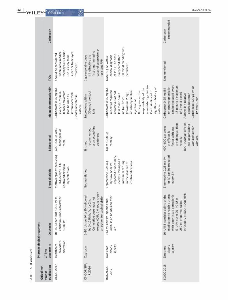
22 | ESCOBAR Et Al.
Gui
delin
e/ye
ar o
f pu
blic
atio
n
Phar
mac
olog
ical
trea
tmen
t
1st li
ne
uter
oton
icO
xyto
cin
Ergo
t alk
aloi
dsM
isop
rost
olIn
ject
able
pro
stag
land
inTX
AC
arbe
toci
n
ACO
G 2
017
Hea
lthca
re
prov
ider
's di
scre
tion
10– 4
0 IU
per
500
– 100
0 m
l as
cont
inuo
us in
fusi
on (I
V) o
r 10
IU IM
Met
hyle
rgon
ovin
e 0.
2 m
g IM
, eve
ry 2
– 4 h
.C
ontr
aind
icat
ed in
hy
pert
ensi
on
600–
100
μg, o
ral,
subl
ingu
al, o
r re
ctal
Car
bopr
ost 0
.25
mg
IM,
ever
y 15
– 90
min
, 8
dose
s m
axim
um
(can
be
used
as
intr
amyo
met
rial).
Con
trai
ndic
ated
in
asth
ma
Shou
ld b
e co
nsid
ered
w
hen
initi
al m
edic
al
ther
apy
fails
. Ear
lier
use
is li
kely
to b
e su
perio
r to
dela
yed
trea
tmen
t
CN
GO
F/SF
A
R 20
16O
xyto
cin
5– 10
IU s
low
IV o
r IM
follo
wed
by
5– 1
0 IU
/h, I
V, fo
r 2 h
. C
umul
ativ
e do
se m
ust n
ot
exce
ed 4
0 IU
(IM
rout
e is
onl
y an
opt
ion
for v
agin
al b
irth)
Not
men
tione
dIt
is n
ot
reco
mm
ende
d as
a s
econ
d- lin
e tr
eatm
ent
Sulp
rost
one
with
in
30 m
in, i
f oxy
toci
n fa
ils
1 g,
rene
wab
le o
nce
if in
effe
ctiv
e th
e fir
st ti
me,
lim
ited
to
case
s of
sul
pros
tone
- re
sist
ant P
PH
RA
NZC
OG
20
17D
oes
not
clea
rly
spec
ify
5 IU
by
slow
IV in
ject
ion
and
40 IU
in a
n IV
infu
sion
ove
r 4
h
Ergo
met
rine
0.25
mg
by s
low
IV o
r IM
, re
peat
ed if
nec
essa
ry,
ever
y 5
min
up
to a
m
axim
um o
f 1 m
g;
in th
e ab
senc
e of
co
ntra
indi
catio
ns
Up
to 1
000
μg
rect
ally
Car
bopr
ost 0
.25
mg
IM,
repe
at a
s re
quire
d at
inte
rval
s of
not
le
ss th
an 1
5 m
in
up to
8 d
oses
(m
axim
um 2
mg)
; or
intr
amyo
met
rial
inje
ctio
n of
0.
5 m
g, u
nder
the
resp
onsi
bilit
y of
the
adm
inis
terin
g cl
inic
ian
Con
trai
ndic
ated
if
sign
ifica
nt h
isto
ry o
f as
thm
a
Dos
e: 1
g IV
, with
a
clin
ical
dia
gnos
is
of P
PH. T
he d
ose
is re
peat
ed a
fter
30
min
if b
leed
ing
was
pe
rsis
tent
SOG
C 20
18D
oes
not
clea
rly
spec
ify
10 IU
IM (c
onsi
der a
bilit
y of
the
med
icat
ion
to re
ach
a ut
erus
w
ith p
oor t
issu
e pe
rfus
ion)
; 5
IU IV
pus
h; 2
0– 40
IU in
25
0 m
l of n
orm
al s
alin
e,
infu
sed
IV a
t 500
– 100
0 m
l/h
Ergo
met
rine
0.25
mg
IM
or IV
, can
be
repe
ated
ev
ery
2 h
400–
800
μg; o
nset
of
eff
ects
is
fast
er w
ith o
ral
or s
ublin
gual
than
re
ctal
ly80
0– 10
00 μ
g; e
ffec
ts
are
long
er la
stin
g w
ith re
ctal
than
w
ith o
ral
Car
bopr
ost 0
.25
mg
IM
or in
tram
yom
etria
lly;
can
be re
peat
ed e
very
15
min
, to
a m
axim
um
of 2
mg
(8 d
oses
)A
sthm
a is
a re
lativ
e co
ntra
indi
catio
nC
arbe
toci
n: 1
00 µ
g IM
or
IV o
ver 1
min
Not
men
tione
dC
arbe
toci
n re
com
men
ded
TAB
LE 6
(C
ontin
ued)

| 23ESCOBAR Et Al.
Gui
delin
e/ye
ar o
f pu
blic
atio
n
Phar
mac
olog
ical
trea
tmen
t
1st li
ne
uter
oton
icO
xyto
cin
Ergo
t alk
aloi
dsM
isop
rost
olIn
ject
able
pro
stag
land
inTX
AC
arbe
toci
n
Ger
man
/A
ustr
ian/
Sw
iss
2018
Oxy
toci
n3 –
– 5 IU
in 1
0 m
l of N
aCl 0
.9%
sl
ow IV
bol
us. I
f nec
essa
ry,
it is
follo
wed
by
10 –
40 IU
ox
ytoc
in in
500
- 100
0mL
salin
e as
a c
ontin
uous
in
fusi
onM
axim
um o
f 6 IU
und
ilute
d ox
ytoc
in c
an b
e ad
min
iste
red
IV, s
low
ly. O
xyto
cin
IM o
nly
if ne
cess
ary
Not
reco
mm
ende
d ro
utin
ely.
If u
sed,
ca
utio
n w
ith a
dver
se
effe
cts.
Met
hyle
rgom
etrin
e sh
ould
not
be
used
as
IV b
olus
. Doe
s no
t m
entio
n do
sage
s
If fir
st- li
ne
uter
oton
ics
are
not e
ffec
tive:
80
0– 10
00 μ
g re
ctal
ly o
r 600
μg
oral
lyA
fter
adm
inis
trat
ion
of o
xyto
cin
may
be
con
side
red
If fir
st- li
ne u
tero
toni
cs
are
inef
fect
ive.
O
ptio
n: s
ulpr
osto
ne
500
μg in
500
ml I
V
(pum
p). I
nitia
l dos
e:
100–
500
ml/
h.M
aint
enan
ce:
100
ml/
h. M
axim
um
1000
μg/
10 h
or
1500
μg
daily
. In
tram
yom
etria
l ap
plic
atio
n is
co
ntra
indi
cate
d
Early
use
if re
quire
d.1–
2 g
(15 –
30
mg/
kg),
to b
e re
peat
ed a
s ne
eded
Car
beto
cin
not
reco
mm
ende
d
FLA
SOG
20
18O
xyto
cin
40 IU
in 5
00 m
l cry
stal
loid
s at
60
ml p
er h
our.
Dos
e fr
om
80– 1
60 th
ousa
nd u
nits
per
m
inut
e
Met
hyle
rgon
ovin
e 0.
2 m
g IM
, rep
eat
if ne
cess
ary,
aft
er
20 m
in a
nd th
en
ever
y 4
h up
to a
m
axim
um o
f 5 d
oses
800
ug s
ublin
gual
or
rect
al s
ingl
e do
seN
ot m
entio
ned
1 g
IV, t
he s
ame
dose
ca
n be
repe
ated
aft
er
30 m
in if
ble
edin
g pe
rsis
ts
Not
men
tione
d
Abb
revi
atio
ns: T
XA
, tra
nexa
mic
aci
d; F
IGO
, Int
erna
tiona
l Fed
erat
ion
of G
ynec
olog
y an
d O
bste
tric
s; IU
, int
erna
tiona
l uni
t; IM
, int
ram
uscu
lar;
IV, i
ntra
veno
us; W
HO
, Wor
ld H
ealth
Org
aniz
atio
n; F
OG
SI,
Fede
ratio
n of
Obs
tetr
ic a
nd G
ynae
colo
gica
l Soc
ietie
s of
Indi
a; R
COG
, Roy
al C
olle
ge o
f Obs
tetr
icia
ns a
nd G
ynae
colo
gist
s; A
COG
, Am
eric
an C
olle
ge o
f Obs
tetr
icia
ns a
nd G
ynec
olog
ists
; CN
GO
F/SF
AR,
Col
lege
of G
ynae
colo
gist
s an
d O
bste
tric
ians
/Fre
nch
Soci
ety
of A
nest
hesi
olog
y an
d In
tens
ive
Car
e; R
AN
ZCO
G, R
oyal
Aus
tral
ian
and
New
Zea
land
Col
lege
of O
bste
tric
ians
and
Gyn
aeco
logi
sts;
SO
GC
, Soc
iety
of O
bste
tric
ians
and
Gyn
aeco
logi
sts
of C
anad
a; D
GG
G/O
EGG
G/S
SGO
, Ger
man
Soc
iety
of G
ynec
olog
y an
d O
bste
tric
s/A
ustr
ian
Soci
ety
of O
bste
tric
s an
d G
ynec
olog
y/Sw
iss
Soci
ety
of
Gyn
aeco
logy
and
Obs
tetr
ics,
FLA
SOG
, Fed
erac
ión
Latin
oam
eric
ana
de S
ocie
dade
s de
Obs
tetr
icia
y G
inec
olog
ía.
TAB
LE 6
(C
ontin
ued)
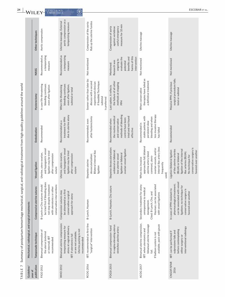
24 | ESCOBAR Et Al.
TAB
LE 7
Su
mm
ary
of p
ostp
artu
m h
emor
rhag
e m
echa
nica
l, su
rgic
al, a
nd ra
diol
ogic
al tr
eatm
ent f
rom
hig
h- qu
ality
gui
delin
es a
roun
d th
e w
orld
Gui
delin
e/ye
ar o
f pu
blic
atio
n
Mec
hani
cal,
radi
olog
ical
, and
sur
gica
l tre
atm
ents
Tam
pona
de te
chni
que
Com
pres
sive
ute
rine
sutu
res
Vess
el li
gatio
nEm
boliz
atio
nH
yste
rect
omy
NA
SGO
ther
tech
niqu
es
FIG
O 2
012
Bim
anua
l com
pres
sion
of
the
uter
us (e
xter
nal
or in
tern
al).
IBT
reco
mm
ende
d
B- Ly
nch
or C
ho s
utur
es s
houl
d be
trie
d fir
st if
ble
edin
g do
es
not s
top
desp
ite tr
eatm
ent
with
ute
roto
nics
or o
ther
co
nser
vativ
e in
terv
entio
ns
Ute
rine,
ute
ro- o
varia
n,
and
hypo
gast
ric v
esse
l lig
atio
n m
ay b
e tr
ied
afte
r com
pres
sion
su
ture
Reco
mm
ende
dIn
cas
e lif
e- th
reat
enin
g bl
eedi
ng c
ontin
ues
even
aft
er li
gatio
n
Reco
mm
ende
d as
a
tem
poriz
ing
mea
sure
Aor
tic c
ompr
essi
on
WH
O 2
012
Bim
anua
l ute
rine
com
pres
sion
: te
mpo
rizin
g m
easu
re fo
r at
ony
at v
agin
al d
eliv
ery.
IBT:
if u
tero
toni
cs fa
il or
are
not
ava
ilabl
e;
Ute
rine
pack
ing
is n
ot
reco
mm
ende
d
Com
pres
sion
sut
ures
may
be
att
empt
ed a
s a
first
co
nser
vativ
e su
rgic
al
appr
oach
for a
tony
Ute
rine,
ute
ro- o
varia
n,
and
hypo
gast
ric v
esse
l lig
atio
n m
ay b
e tr
ied
afte
r com
pres
sion
su
ture
Reco
mm
ende
d as
a
trea
tmen
t for
PPH
du
e to
ute
rine
aton
y
Whe
n lif
e- th
reat
enin
g bl
eedi
ng c
ontin
ues
even
aft
er li
gatio
n,
subt
otal
or t
otal
Reco
mm
ende
d as
a
tem
poriz
ing
mea
sure
Ute
rine
mas
sage
. Ext
erna
l ao
rtic
com
pres
sion
as
a te
mpo
rizin
g m
easu
re
RCO
G 2
016
IBT:
reco
mm
ende
d as
firs
t- lin
e “s
urgi
cal”
inte
rven
tion
B- Ly
nch,
Hay
man
Step
wis
e ut
erin
e de
vasc
ular
izat
ion;
Bi
late
ral i
nter
nal i
liac
ligat
ion
Reco
mm
ende
d, e
ven
afte
r hys
tere
ctom
ySo
oner
rath
er th
an la
ter (
to
disc
uss
with
a s
econ
d ex
perie
nced
clin
icia
n if
feas
ible
) Tec
hniq
ue:
subt
otal
is p
refe
rred
Not
men
tione
dC
ompr
essi
on o
f the
aor
taRu
b up
the
uter
ine
fund
us
FOG
SI 2
015
Bim
anua
l com
pres
sion
– han
d in
vag
ina
elev
atin
g ut
erus
st
retc
hes
uter
ine
arte
ry
B- Ly
nch,
Hay
man
, Cho
sut
ure
Step
wis
e de
vasc
ular
izat
ion:
un
ilate
ral o
r bila
tera
l; lo
w u
terin
e ve
ssel
lig
atio
n or
bila
tera
l ov
aria
n ve
ssel
liga
tion
Reco
mm
ende
d w
hen
the
rout
ine
med
ical
an
d co
nser
vativ
e m
etho
ds o
f ble
edin
g co
ntro
l hav
e be
en
trie
d an
d no
t fou
nd
effe
ctiv
e
Last
opt
ion,
it re
flect
s th
e fa
ilure
of a
ll ot
her
met
hods
at s
topp
ing
PPH
Men
tione
d:Re
sear
ch w
as
ongo
ing
to
eval
uate
the
pote
ntia
l be
nefit
s an
d ha
rms
of th
is
inte
rven
tion
Com
pres
sion
of a
orta
ag
ains
t ver
tebr
ae
usef
ul e
mer
genc
y m
easu
re fo
r 10
min
AC
OG
201
7Bi
man
ual u
terin
e co
mpr
essi
on.
IBT:
whe
n ut
erot
onic
s an
d bi
man
ual u
terin
e m
assa
ge
fail.
If ba
lloon
sys
tem
is n
ot
avai
labl
e, p
ack
with
gau
ze
Seco
ndar
y tr
eatm
ent f
or a
tony
un
resp
onsi
ve to
med
ical
m
anag
emen
t.C
ited:
B- L
ynch
, Cho
, and
H
aym
an; c
an b
e as
soci
ated
w
ith v
esse
l lig
atio
ns
Whe
n le
ss in
vasi
ve
appr
oach
es fa
il; b
ilate
ral
uter
ine
arte
ry (m
ost
com
mon
), ut
ero-
ov
aria
n ve
ssel
s, a
nd/o
r in
tern
al il
iac
arte
ry (l
ess
freq
uent
lyus
ed)
Reco
mm
ende
d in
st
able
pat
ient
, with
pe
rsis
tent
slo
w
blee
ding
and
whe
n le
ss in
vasi
ve th
erap
y ha
s fa
iled
Whe
n co
nser
vativ
e th
erap
ies
have
faile
d as
a
defin
itive
trea
tmen
t
Not
men
tione
dU
terin
e m
assa
ge
CN
GO
F/SF
AR
2016
IBT:
can
be
perf
orm
ed if
su
lpro
ston
e fa
ils a
nd
befo
re c
onte
mpl
atin
g ei
ther
sur
gery
or
inte
rven
tiona
l rad
iolo
gy
In c
ases
of P
PH re
sist
ant t
o ph
arm
acol
ogic
al tr
eatm
ent;
can
be a
ssoc
iate
d w
ith v
esse
l lig
atio
ns; n
o te
chni
que
for
cons
erva
tive
surg
ery
is
favo
red
over
ano
ther
Liga
tion
(bila
tera
l lig
atio
n of
the
uter
ine
arte
ries
(BLU
A) o
r bila
tera
l lig
atio
n of
the
inte
rnal
ili
ac a
rter
ies
(BLI
IA).
No
tech
niqu
e fo
r co
nser
vativ
e su
rger
y is
fa
vore
d ov
er a
noth
er
Reco
mm
ende
dM
assi
ve P
PH; i
f pre
viou
s su
rgic
al p
roce
dure
fails
; to
tal o
r sub
tota
l
Not
men
tione
dU
terin
e m
assa
ge
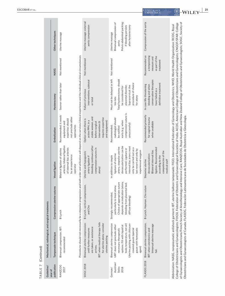
| 25ESCOBAR Et Al.
Gui
delin
e/ye
ar o
f pu
blic
atio
n
Mec
hani
cal,
radi
olog
ical
, and
sur
gica
l tre
atm
ents
Tam
pona
de te
chni
que
Com
pres
sive
ute
rine
sutu
res
Vess
el li
gatio
nEm
boliz
atio
nH
yste
rect
omy
NA
SGO
ther
tech
niqu
es
RA
NZC
OG
20
17Bi
man
ual c
ompr
essi
on; I
BT:
reco
mm
ende
dB-
Lync
hBi
late
ral l
igat
ion
of u
terin
e ar
terie
s; b
ilate
ral l
igat
ion
of in
tern
al il
iac
arte
ries
by a
n ex
perie
nce
oper
ator
Reco
mm
ende
d; it
nee
ds
equi
pmen
t and
ex
pert
ise;
sho
uld
not p
recl
ude
othe
r ap
proa
ches
Soon
er ra
ther
than
late
rN
ot m
entio
ned
Ute
rine
mas
sage
Proc
edur
es s
houl
d no
t nec
essa
rily
be a
ste
pwis
e pr
ogre
ssio
n an
d bo
th o
rder
and
util
izat
ion
will
dep
end
on th
e se
rvic
es/c
linic
al e
xper
ienc
e an
d th
e in
divi
dual
clin
ical
circ
umst
ance
s
SOG
C 20
18Bi
man
ual u
terin
e co
mpr
essi
on:
until
furt
her m
easu
res
are
take
n, o
r ass
ista
nce
arriv
es.
IBT:
whe
n m
edic
al th
erap
y fa
ils
for u
terin
e at
ony;
con
side
r ut
erin
e pa
ckin
g
B- Ly
nch,
ver
tical
com
pres
sion
, an
d C
hoU
terin
e an
d hy
poga
stric
ar
tery
liga
tion
if bl
eedi
ng c
ontin
ues
(aft
er
com
pres
sion
sut
ures
in
aton
y)
Opt
ion
whe
n th
ere
is
activ
e PP
H, i
n a
stab
le w
oman
, and
be
fore
sur
gica
l in
terv
entio
n (it
re
quire
s ex
pert
ise
and
equi
pmen
t)
Failu
re o
f pre
viou
s in
terv
entio
ns; s
ubto
tal
or to
tal
Not
men
tione
dU
terin
e m
assa
ge; e
xter
nal
aort
ic c
ompr
essi
on
Ger
man
/A
ustr
ian/
Swis
s 20
18
Bim
anua
l ute
rine
com
pres
sion
UBT
: doe
s no
t pre
clud
e ot
her
nece
ssar
y th
erap
eutic
op
tions
. Fill
with
liqu
id a
t bo
dy te
mpe
ratu
re, n
ot a
ir)U
terin
e pa
ckin
g w
ith c
hito
san-
co
vere
d ga
uze
(spe
cial
ga
uze
with
hem
osta
tic
agen
t)
Stro
ngly
reco
mm
ende
d,
part
icul
arly
to tr
eat a
tony
. C
hoic
e of
app
ropr
iate
sut
ure
depe
nds
on in
dica
tion
(ato
ny,
blee
ding
from
pla
cent
al b
ed,
diff
use
blee
ding
)
In a
dditi
on to
sim
ple
ligat
ure
of u
terin
e ar
tery
, ste
pwis
e ut
erin
e de
vasc
ular
izat
ion
can
be
perf
orm
ed. L
igat
ure
or
inte
rnal
ilia
c ar
tery
mus
t on
ly b
e ca
rrie
d ou
t as
a la
st re
sort
and
onl
y by
ex
perie
nced
sur
geon
Reco
mm
ende
d;
radi
olog
ist s
houl
d be
not
ified
ea
rly (e
.g. w
hen
com
pres
sion
sut
ure
is
unsu
cces
sful
)
Mus
t not
be
dela
yed
or le
ft
too
late
.To
tal h
yste
rect
omy:
sho
uld
be c
onsi
dere
d fo
r pl
acen
tal d
isor
der.
Supr
acer
vica
l: th
e pr
oced
ure
of c
hoic
e fo
r ato
ny
Not
men
tione
dU
terin
e m
assa
geBi
man
ual c
ompr
essi
on o
f ao
rta
Pelv
is- a
bdom
inal
pac
king
: if
blee
ding
per
sist
s af
ter h
yste
rect
omy
FLA
SOG
201
8Bi
man
ual u
terin
e co
mpr
essi
on.
IBT:
whe
n ut
erot
onic
s an
d bi
man
ual u
terin
e m
assa
ge
fail.
B- Ly
nch,
Hay
man
, Cho
sut
ure
Step
wis
e ut
erin
e de
vasc
ular
izat
ion;
Bi
late
ral i
nter
nal i
liac
ligat
ion.
Rec
omm
ende
d ac
cord
ing
to th
e co
mpe
tenc
ies
of th
e m
edic
al s
taff
Reco
mm
ende
d pr
imar
ily
for v
agin
al tr
aum
a w
hen
avai
labl
e
In c
ase
life-
thre
aten
ing
blee
ding
and
whe
n co
nser
vativ
e th
erap
ies
have
faile
d as
a
defin
itive
trea
tmen
t
Reco
mm
ende
d as
a
tem
poriz
ing
mea
sure
and
as
par
t of t
he
trea
tmen
t
Com
pres
sion
of t
he a
orta
Abb
revi
atio
n: N
ASG
, non
pneu
mat
ic a
ntis
hock
gar
men
t; IB
T, u
terin
e ba
lloon
tam
pona
de; F
IGO
, Int
erna
tiona
l Fed
erat
ion
of G
ynec
olog
y an
d O
bste
tric
s; W
HO
, Wor
ld H
ealth
Org
aniz
atio
n; R
COG
, Roy
al
Col
lege
of O
bste
tric
ians
and
Gyn
aeco
logi
sts;
FO
GSI
, Fed
erat
ion
of O
bste
tric
and
Gyn
aeco
logi
cal S
ocie
ties
of In
dia;
ACO
G, A
mer
ican
Col
lege
of O
bste
tric
ians
and
Gyn
ecol
ogis
ts; C
NG
OF/
SFA
R: C
olle
ge
of G
ynae
colo
gist
s an
d O
bste
tric
ians
/Fre
nch
Soci
ety
of A
nest
hesi
olog
y an
d In
tens
ive
Car
e; R
AN
ZCO
G, R
oyal
Aus
tral
ian
and
New
Zea
land
Col
lege
of O
bste
tric
ians
and
Gyn
aeco
logi
sts;
SO
GC
, Soc
iety
of
Obs
tetr
icia
ns a
nd G
ynae
colo
gist
s of
Can
ada;
FLA
SOG
, Fed
erac
ión
Latin
oam
eric
ana
de S
ocie
dade
s de
Obs
tetr
icia
y G
inec
olog
ía.
TAB
LE 7
(C
ontin
ued)

26 | ESCOBAR Et Al.
FIGO and other medical societies have specific guidelines for the placenta accreta spectrum.16– 19
R E FE R E N C E S 1. Prevention and management of postpartum haemorrhage: green-
top guideline no. 52. BJOG. 2017;124:e106– e149. 2. Sentilhes L, Vayssière C, Deneux- Tharaux C, et al Postpartum hem-
orrhage: guidelines for clinical practice from the French College of Gynaecologists and Obstetricians (CNGOF): in collaboration with the French Society of Anesthesiology and Intensive Care (SFAR). Eur J Obstet Gynecol Reprod Biol. 2016;198:12– 21.
3. World Health Organization. WHO Recommendations for the Prevention and Treatment of Postpartum Haemorrhage. Accessed August 18, 2021. https://www.who.int/repro ducti vehea lth/publi catio ns/mater nal_perin atal_healt h/97892 41548 502/en/
4. Schlembach D, Helmer H, Henrich W, et al. Peripartum haemor-rhage, diagnosis and therapy. Guideline of the DGGG, OEGGG and SGGG (S2k Level, AWMF Registry No. 015/063, March 2016). Geburtshilfe Frauenheilkd. 2018;78:382– 399.
5. Leduc D, Senikas V, Lalonde AB. No. 235- active management of the third stage of labour: prevention and treatment of postpartum hemorrhage. J Obstet Gynaecol Can. 2018;40:e841– e855.
6. Committee on Practice Bulletins- Obstetrics. Practice bulletin no. 183: postpartum hemorrhage. Obstet Gynecol. 2017;130:e168– e186.
7. Organization PAH. Guidelines for the prevention, diagnosis and treat-ment of obstetric hemorrhage [in Portuguese]. PAHO; 2018.
8. Fawcus S. Alerts for managing postpartum haemorrhage. S Afr Med J. 2018;108:1013– 1017.
9. Sri Lanka College of Obstetrician and Gynecologists. SLCOG Guideline on Management of Primary PostPartum Haemorrhage. Accessed August 25, 2021. https://www.slcog.lk/wp- conte nt/uploa ds/2021/02/SLCOG - Guide line- on- Manag ement - of- Prima ry- Post- Partu m- Haemo rrhag e- 03.- 2020.pdf
10. Royal Australian and New Zealand College of Obstetricians and Gynaecologists. RANZCOG statement. Management of postpar-tum hemorrhage. Accessed August 25, 2021. https://ranzc og.edu.au/RANZC OG_SITE/media/ RANZC OG- MEDIA/ Women %27s%20Hea lth/State ment%20and %20gui delin es/Clini cal- Obste trics/ Manag ement - of- Postp artum - Haemo rrhag e- (C- Obs- 43)- Revie w- July- 2017.pdf?ext=.pdf
11. WOMAN Trial Collaborators. Effect of early tranexamic acid adminis-tration on mortality, hysterectomy, and other morbidities in women with post- partum haemorrhage (WOMAN): an international, randomised, double- blind, placebo- controlled trial. Lancet. 2017;389:2105– 2116.
12. World Health Organization. WHO Recommendation on Tranexamic Acid for the Treatment of Postpartum Haemorrhage. Accessed August 21: 2021. https://www.who.int/repro ducti vehea lth/publi catio ns/trane xamic - acid- pph- treat ment/en/
13. Vogel JP, Oladapo OT, Dowswell T, Gülmezoglu AM. Updated WHO recommendation on intravenous tranexamic acid for the treatment of post- partum haemorrhage. Lancet Glob Health. 2018;6:e18– e19.
14. Althabe F, Therrien MNS, Pingray V, et al. Postpartum hemorrhage care bundles to improve adherence to guidelines: a WHO technical consultation. Int J Gynecol Obstet. 2020;148:290– 299.
15. FIGO Safe Motherhood and Newborn Health Committee, International Federation of Gynecology and Obstetrics. Practice Guideline. Non- pneumatic anti- shock garment to stabilize women with hypovolemic shock secondary to obstetric hemorrhage. Int J Gynecol Obstet. 2015;128:194– 195.
16. Jauniaux E, Ayres- de- Campos D; FIGO Placenta Accreta Diagnosis and Management Expert Consensus Panel. FIGO consensus guide-lines on placenta accreta spectrum disorders: Introduction. Int J Gynecol Obstet. 2018;140:261– 264.
17. Jauniaux E, Alfirevic Z, Bhide AG, et al. Placenta prae-via and placenta accreta: diagnosis and management. BJOG. 2019;126(1):e1– e48.
18. American College of Obstetricians and Gynecologists; Society for Maternal- Fetal Medicine. Obstetric care consensus no. 7: placenta accreta spectrum. Obstet Gynecol. 2018;132:e259– e275.
19. Fuchtner C, Ortiz EI, Escobar MF, Lizaola H. Hemorragia Postparto: en donde estamos y hacia donde vamos. Federación Latinoamericana de Sociedades de Ginecologia y Obstetricia. Accessed August 11, 2021. https://www.flasog.org/stati c/libro s/Hemor ragia - Postp arto- 17OCT UBRE.pdf
BOX 4 FIGO recommends the preparedness of obstetric care teams for the management of PPH according to the updated guidelines established locally, regionally, or globally.
• These guidelines should include medical and nonmedical treatment options according to the degree of development of each country or region.
• Local, regional, and national medical and nonmedical op-tions for prevention and treatment of PPH need to con-sider the resources available locally.
• National societies are encouraged to lobby to establish a PPH bundle approach and ensure availability of medical supplies and surgical equipment.
| 27ESCOBAR Et Al.
7 | MEDIC AL PRE VENTION AND TRE ATMENT
7.1 | Carbetocin versus oxytocin use in PPH: recent evidence
Oxytocin has been traditionally used as the first- line uterotonic for the prevention of PPH. Since 1992, carbetocin has been extensively tested in the management of PPH.1,2 Beyond its uterotonic effects, carbetocin may also promote blood coagulation.3
7.1.1 | Clinical evidence for PPH prevention: oxytocin versus carbetocin (vaginal delivery)
The Heat- Stable Carbetocin versus Oxytocin to Prevent Hemorrhage after Vaginal Birth (CHAMPION) study provided new evidence comparing oxytocin and carbetocin.4 It studied 29 645 women with cervical dilation <6 cm having a singleton pregnancy and planned for vaginal delivery. Patients were rand-omized to receive intramuscular injection of 100 µg heat- stable carbetocin (Paban) or 10 IU oxytocin in 10 different countries. The data showed that for PPH prevention, heat- stable carbetocin was not inferior to oxytocin administration for blood loss of at least 500 ml (RR 1.01; 95% CI, 0.95– 1.06) or the use of additional uterotonic agents. Noninferiority was not shown for the outcome of blood loss of at least 1000 ml (RR 1.04; 95% CI, 0.87– 1.25). In this study, when the two drugs were compared, the following re-sults were obtained: in women with >500 ml blood loss, additional uterotonic agents were required in almost 15% of cases in both arms. When blood loss exceeded 1000 ml, the requirement for additional uterotonics was only 1.5% in both arms.3 Therefore, ac-cess to drugs is key since oxytocin has problems with drug purity in low- resource settings. The safety profile appeared to be similar for both drugs.
More recently, a network meta- analysis including 140 random-ized trials with data from 88 947 women was developed to identify the most effective uterotonic drug(s) to prevent PPH and generate a ranking according to their effectiveness and adverse effect pro-file.5 The data showed that compared with oxytocin, the combina-tion of ergometrine plus oxytocin had a risk ratio of 0.69 (95% CI, 0.57– 0.83), and was more effective; however, with only moderate quality evidence and with higher risk of adverse effects such as vomiting and hypertension. For carbetocin, the risk ratio was 0.72 (95% CI, 0.52– 1.00), and although it was significant, there was very low- quality evidence. Finally, for misoprostol plus oxytocin the risk ratio was 0.73 (95% CI, 0.60– 0.90), with moderate quality evidence and higher risk of fever.5 In a low- risk setting there may be advan-tages to using well- tested and readily available drugs. When the carbetocin effect was compared with the use of rectal misoprostol for preventing PPH in low- risk patient populations in a random-ized controlled trial among 150 pregnant women, the data showed
the superiority of carbetocin for efficacy.6 This was evidenced in all indices tested; beyond blood loss, patients had a shorter third stage of labor and reduced need for additional uterotonic drugs. Additionally, adverse effects such as diarrhea, shivering, and fever were more pronounced with misoprostol administration.6 In a ran-domized controlled trial of patients with severe pre- eclampsia, car-betocin was shown to be a good alternative to oxytocin, requiring a lower volume per dose.7
7.1.2 | Clinical evidence for PPH prevention: oxytocin versus carbetocin (cesarean delivery)
Cesarean delivery is a surgical procedure requiring high skill, oper-ating time, anesthesia, intravenous fluids, and different drugs. The addition of carbetocin, therefore, is expected to be of benefit and the need for a single drug administration reduces the need for an intensive management team, which is generally required following a cesarean delivery. A Cochrane review including 11 studies showed that despite a statistically significant reduction in the need for fur-ther uterotonics in the carbetocin group compared with oxytocin in women undergoing a cesarean delivery, no statistically significant differences in terms of the risk of PPH were noted.8 In 2018 a meta- analysis to analyze the effectiveness of carbetocin compared with oxytocin for the prevention of PPH in cesarean deliveries (seven studies involving 2012 patients) demonstrated a significant reduc-tion in the rates of PPH (RR 0.79; 95% CI, 0.66– 0.94, P = 0.009), use of additional uterotonics (RR 0.57; 95% CI, 0.49– 0.65, P < 0.001), and transfusion (RR 0.31; 95% CI, 0.15– 0.64, P = 0.002) when carbe-tocin was used instead of oxytocin. However, despite the potential benefits, the disparity between the cost of carbetocin and oxytocin suggests that a locoregional cost- effectiveness analysis should be performed before making the decision to adopt carbetocin for rou-tine prophylaxis.9
In 2020, sequential trial analysis of five randomized controlled trials of studies with data on women (a total of 1214) undergoing nonelective cesarean deliveries, where carbetocin was compared with oxytocin, was performed.10 The need for additional uteroton-ics was reduced with carbetocin compared with oxytocin (OR 0.30; 95% CI, 0.11– 0.86; I2, 90.60%). Trial sequential analysis (TSA) con-firmed that the information size needed to show a significant re-duction in the need for additional uterotonics had been exceeded. No significant differences were shown with respect to any of the secondary outcomes, but there was significant heterogeneity be-tween the studies. The authors concluded that further trials utiliz-ing consistent core outcomes are needed to determine an effect on PPH.10
On the other hand, a single center study was carried out to evaluate the cost- effectiveness of carbetocin compared with oxy-tocin when used for prophylaxis against PPH. With approximately 3000 cesarean deliveries per year, the study showed that the use of carbetocin could prevent 108 episodes of PPH, 104 episodes
28 | ESCOBAR Et Al.
of transfusion, and 455 patients would require fewer uteroton-ics. Their cost- saving per PPH case was USD$ 278.70.11 Another study where 1500 cesarean deliveries (both elective and emer-gency) were analyzed over 12 months, carbetocin use, as opposed to oxytocin use, reduced 30 (88 vs 58) PPH events (>500 ml blood loss) and saved £27 518. Carbetocin had a 91.5% better outcome and was 69.4% cheaper.12 When compared with the need for ad-ditional uterotonics in the carbetocin group, none required addi-tional medication versus 71.5% in the oxytocin group (P < 0.01). In another prospective cohort study, carbetocin used for 400 patients undergoing cesarean delivery led to reduced PPH and the use of other oxytocic agents, saving £68.93 per patient.13 In 2019, a systematic review of the cost- effectiveness of uterotonic agents for the prevention of PPH was published with 15 studies across all income categories that compared misoprostol versus no uterotonic (five studies) or versus oxytocin (one study), carbeto-cin versus oxytocin (eight studies), and one study comparing nu-merous uterotonics.14 The evidence on the cost- effectiveness of various uterotonic agents was not generalizable to different con-texts. In the absence of reliable evidence, the choice of uterotonic will likely be highly influenced by uterotonic price and contextual factors. In the context of cesarean delivery, carbetocin was more cost favorable than oxytocin. The authors concluded the quality of the evidence provided by these studies has a lack of sensitivity analyzes and incomplete description of the methods of outcome and costs measurements in most studies. In addition, some studies from middle- income countries favoring carbetocin were funded by the manufacturer. They interpreted findings from these studies and those of the conference abstracts with limited data cautiously, owing to the potential risk of bias and imprecision. Therefore, it can be easily inferred that the relative cost- effectiveness of car-betocin is still inconclusive.
Beyond its primary efficacy, which is reduced blood loss, a drug's safety profile must also be considered before wide- scale utilization. The adverse effects of carbetocin and oxytocin were examined in a small, randomized study.15 The data showed that car-betocin had a lower rate of nausea. In contrast with other clinical indices, including the need to stabilize systolic blood pressure and heart rate, no other differences were noted.15 Since anesthesiolo-gists administer carbetocin for cesarean delivery, the requirement for increased dose beyond the standard 100 ug in some instances was raised.16 The longer half- life of carbetocin prevents additional dosing in case of a persistent bleed and does not permit additional oxytocin administration. The increase in carbetocin dose from 100 µg to 140 µg injection increases cardiovascular complications.17 Thus, in cases of pre- eclampsia, close attention is warranted for preserving patient safety before high dose carbetocin is adminis-tered. Also, in those cases, oxytocin doses must be increased to control bleeding beyond the standard treatment, mainly if the drug was used during labor, which may also increase complications rate. A case report showed that carbetocin administration for a patient with asthma led to nearly fatal bronchospasm and cardiac arrest
requiring resuscitation, which was successful.18 Therefore, this drug should be used with caution in patients with asthma or those with advanced pulmonary pathology. This and other studies show inconsistency in adverse events data and indicate that the utility of carbetocin should be further assessed in additional pregnancy pathologies to establish the population in which the risk– benefit profile is favorable.
R E FE R E N C E S 1. Hunter DJ, Schulz P, Wassenaar W. Effect of carbetocin, a long-
acting oxytocin analog on the postpartum uterus. Clin Pharmacol Ther. 1992;52:60– 67.
2. Arrowsmith S, Wray S. Oxytocin: its mechanism of action and receptor signalling in the myometrium. J Neuroendocrinol. 2014;26:356– 369.
3. Gallos ID, Coomarasamy A. Carbetocin: worth the extra expense? Best Pract Res Clin Obstet Gynaecol. 2019;61:55– 65.
4. Widmer M, Piaggio G, Nguyen TMH, et al Heat- stable carbetocin versus oxytocin to prevent hemorrhage after vaginal birth. N Engl J Med. 2018;379:743– 752.
5. Gallos ID, Williams HM, Price MJ, et al. Uterotonic agents for preventing postpartum haemorrhage: a network meta- analysis. Cochrane Database Syst Rev. 2018;(4):CD011689.
6. Maged AM, Waly M, Fahmy RM, et al Carbetocin versus rectal misoprostol for management of third stage of labor among women with low risk of postpartum hemorrhage. Int J Gynecol Obstet. 2020;148:238– 242.
7. Reyes OA, Gonzalez GM. Carbetocin versus oxytocin for preven-tion of postpartum hemorrhage in patients with severe preeclamp-sia: a double- blind randomized controlled trial. J Obstet Gynaecol Can. 2011;33:1099– 1104.
8. Su LL, Chong YS, Samuel M. Carbetocin for preventing postpartum haemorrhage. Cochrane Database Syst Rev. 2012;(2):CD005457.
9. Voon HY, Suharjono HN, Shafie AA, Bujang MA. Carbetocin versus oxytocin for the prevention of postpartum hemorrhage: A meta- analysis of randomized controlled trials in cesarean deliveries. Taiwan J Obstet Gynecol. 2018;57:332– 339.
10. Onwochei DN, Owolabi A, Singh PM, Monks DT. Carbetocin com-pared with oxytocin in non- elective Cesarean delivery: a systematic review, meta- analysis, and trial sequential analysis of randomized- controlled trials. Can J Anaesth. 2020;67(11):1524– 1534.
11. Voon HY, Shafie AA, Bujang MA, Suharjono HN. Cost effectiveness analysis of carbetocin during cesarean section in a high volume ma-ternity unit. J Obstet Gynaecol Res. 2018;44:109– 116.
12. Van der Nelson HA, Draycott T, Siassakos D, Yau CWH, Hatswell AJ. Carbetocin versus oxytocin for prevention of post- partum haemorrhage at caesarean section in the United Kingdom: an economic impact analysis. Eur J Obstet Gynecol Reprod Biol. 2017;210:286– 291.
BOX 5 FIGO reaffirms its recommendation regarding oxytocin as the first choice for prevention of PPH in vaginal and cesarean deliveries.
In settings where oxytocin is unavailable (or its quality can-not be guaranteed), the use of other uterotonics (carbe-tocin, ergometrine/methylergometrine, or misoprostol) is recommended.

| 29ESCOBAR Et Al.
13. Luni Y, Borakati A, Matah A, Skeats K, Eedarapalli P. A prospec-tive cohort study evaluating the cost- effectiveness of carbetocin for prevention of postpartum haemorrhage in caesarean sections. J Obstet Gynaecol. 2017;37:601– 604.
14. Lawrie TA, Rogozińska E, Sobiesuo P, Vogel JP, Ternent L, Oladapo OT. A systematic review of the cost- effectiveness of uterotonic agents for the prevention of postpartum hemorrhage. Int J Gynaecol Obstet. 2019;146(1):56– 64.
15. Mannaerts D, Van der Veeken L, Coppejans H, Jacquemyn Y. Adverse effects of carbetocin versus oxytocin in the prevention of postpartum haemorrhage after caesarean section: a randomized controlled trial. J Pregnancy. 2018;2018:1374150.
16. Muggleton E. Oxytocin study raises concerns about carbetocin use. Anesth Analg. 2018;126:1423.
17. Nguyen- Lu N, Carvalho JC, Farine D, Seaward G, Ye XY, Balki M. Carbetocin at cesarean delivery for labour arrest: a sequential allocation trial to determine the effective dose. Can J Anaesth. 2015;62(8):866– 874.
18. Pérez- Nieto OR, Castañón- González JA, Lima- Lucero IM, Delsol LAG. Near fatal bronchospasm and bradycardia after carbetocin administration. Med Intensiva. 2018;42:319– 321.
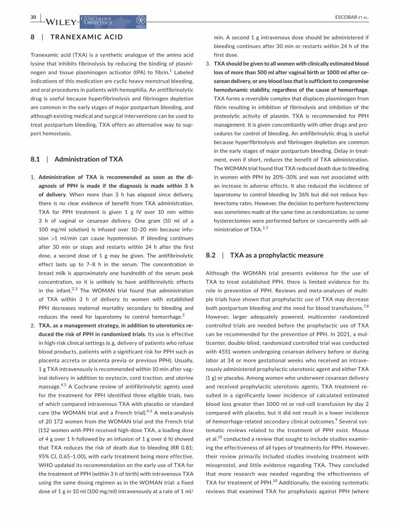
30 | ESCOBAR Et Al.
8 | TR ANE X AMIC ACID
Tranexamic acid (TXA) is a synthetic analogue of the amino acid lysine that inhibits fibrinolysis by reducing the binding of plasmi-nogen and tissue plasminogen activator (tPA) to fibrin.1 Labeled indications of this medication are cyclic heavy menstrual bleeding, and oral procedures in patients with hemophilia. An antifibrinolytic drug is useful because hyperfibrinolysis and fibrinogen depletion are common in the early stages of major postpartum bleeding, and although existing medical and surgical interventions can be used to treat postpartum bleeding, TXA offers an alternative way to sup-port hemostasis.
8.1 | Administration of TXA
1. Administration of TXA is recommended as soon as the di-agnosis of PPH is made if the diagnosis is made within 3 h of delivery. When more than 3 h has elapsed since delivery, there is no clear evidence of benefit from TXA administration. TXA for PPH treatment is given 1 g IV over 10 min within 3 h of vaginal or cesarean delivery. One gram (10 ml of a 100 mg/ml solution) is infused over 10– 20 min because infu-sion >1 ml/min can cause hypotension. If bleeding continues after 30 min or stops and restarts within 24 h after the first dose, a second dose of 1 g may be given. The antifibrinolytic effect lasts up to 7– 8 h in the serum. The concentration in breast milk is approximately one hundredth of the serum peak concentration, so it is unlikely to have antifibrinolytic effects in the infant.2,3 The WOMAN trial found that administration of TXA within 3 h of delivery to women with established PPH decreases maternal mortality secondary to bleeding and reduces the need for laparotomy to control hemorrhage.2
2. TXA, as a management strategy, in addition to uterotonics re-duced the risk of PPH in randomized trials. Its use is effective in high- risk clinical settings (e.g. delivery of patients who refuse blood products, patients with a significant risk for PPH such as placenta accreta or placenta previa or previous PPH). Usually, 1 g TXA intravenously is recommended within 10 min after vag-inal delivery in addition to oxytocin, cord traction, and uterine massage.4,5 A Cochrane review of antifibrinolytic agents used for the treatment for PPH identified three eligible trials, two of which compared intravenous TXA with placebo or standard care (the WOMAN trial and a French trial).4,6 A meta- analysis of 20 172 women from the WOMAN trial and the French trial (152 women with PPH received high- dose TXA, a loading dose of 4 g over 1 h followed by an infusion of 1 g over 6 h) showed that TXA reduces the risk of death due to bleeding (RR 0.81; 95% CI, 0.65– 1.00), with early treatment being more effective. WHO updated its recommendation on the early use of TXA for the treatment of PPH (within 3 h of birth) with intravenous TXA using the same dosing regimen as in the WOMAN trial: a fixed dose of 1 g in 10 ml (100 mg/ml) intravenously at a rate of 1 ml/
min. A second 1 g intravenous dose should be administered if bleeding continues after 30 min or restarts within 24 h of the first dose.
3. TXA should be given to all women with clinically estimated blood loss of more than 500 ml after vaginal birth or 1000 ml after ce-sarean delivery, or any blood loss that is sufficient to compromise hemodynamic stability, regardless of the cause of hemorrhage. TXA forms a reversible complex that displaces plasminogen from fibrin resulting in inhibition of fibrinolysis and inhibition of the proteolytic activity of plasmin. TXA is recommended for PPH management. It is given concomitantly with other drugs and pro-cedures for control of bleeding. An antifibrinolytic drug is useful because hyperfibrinolysis and fibrinogen depletion are common in the early stages of major postpartum bleeding. Delay in treat-ment, even if short, reduces the benefit of TXA administration. The WOMAN trial found that TXA reduced death due to bleeding in women with PPH by 20%– 30% and was not associated with an increase in adverse effects. It also reduced the incidence of laparotomy to control bleeding by 36% but did not reduce hys-terectomy rates. However, the decision to perform hysterectomy was sometimes made at the same time as randomization, so some hysterectomies were performed before or concurrently with ad-ministration of TXA.2,3
8.2 | TXA as a prophylactic measure
Although the WOMAN trial presents evidence for the use of TXA to treat established PPH, there is limited evidence for its role in prevention of PPH. Reviews and meta- analyses of multi-ple trials have shown that prophylactic use of TXA may decrease both postpartum bleeding and the need for blood transfusions.7,8 However, larger adequately powered, multicenter randomized controlled trials are needed before the prophylactic use of TXA can be recommended for the prevention of PPH. In 2021, a mul-ticenter, double- blind, randomized controlled trial was conducted with 4551 women undergoing cesarean delivery before or during labor at 34 or more gestational weeks who received an intrave-nously administered prophylactic uterotonic agent and either TXA (1 g) or placebo. Among women who underwent cesarean delivery and received prophylactic uterotonic agents, TXA treatment re-sulted in a significantly lower incidence of calculated estimated blood loss greater than 1000 ml or red- cell transfusion by day 2 compared with placebo, but it did not result in a lower incidence of hemorrhage- related secondary clinical outcomes.9 Several sys-tematic reviews related to the treatment of PPH exist. Mousa et al.10 conducted a review that sought to include studies examin-ing the effectiveness of all types of treatments for PPH. However, their review primarily included studies involving treatment with misoprostol, and little evidence regarding TXA. They concluded that more research was needed regarding the effectiveness of TXA for treatment of PPH.10 Additionally, the existing systematic reviews that examined TXA for prophylaxis against PPH (where
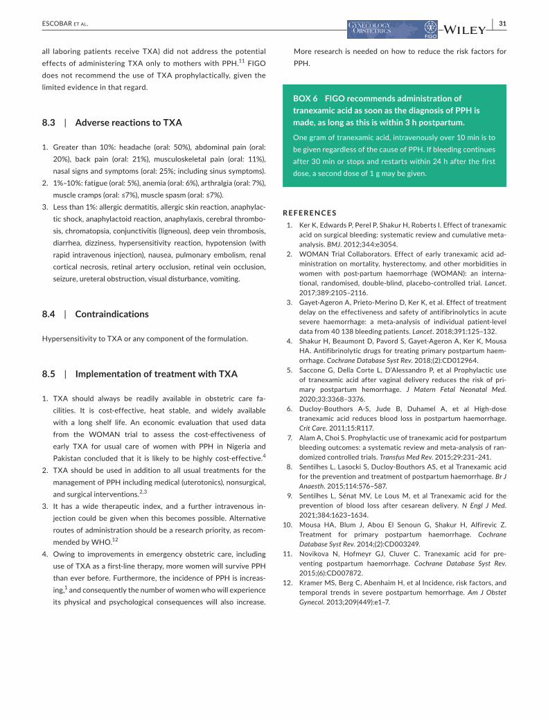
| 31ESCOBAR Et Al.
all laboring patients receive TXA) did not address the potential effects of administering TXA only to mothers with PPH.11 FIGO does not recommend the use of TXA prophylactically, given the limited evidence in that regard.
8.3 | Adverse reactions to TXA
1. Greater than 10%: headache (oral: 50%), abdominal pain (oral: 20%), back pain (oral: 21%), musculoskeletal pain (oral: 11%), nasal signs and symptoms (oral: 25%; including sinus symptoms).
2. 1%– 10%: fatigue (oral: 5%), anemia (oral: 6%), arthralgia (oral: 7%), muscle cramps (oral: ≤7%), muscle spasm (oral: ≤7%).
3. Less than 1%: allergic dermatitis, allergic skin reaction, anaphylac-tic shock, anaphylactoid reaction, anaphylaxis, cerebral thrombo-sis, chromatopsia, conjunctivitis (ligneous), deep vein thrombosis, diarrhea, dizziness, hypersensitivity reaction, hypotension (with rapid intravenous injection), nausea, pulmonary embolism, renal cortical necrosis, retinal artery occlusion, retinal vein occlusion, seizure, ureteral obstruction, visual disturbance, vomiting.
8.4 | Contraindications
Hypersensitivity to TXA or any component of the formulation.
8.5 | Implementation of treatment with TXA
1. TXA should always be readily available in obstetric care fa-cilities. It is cost- effective, heat stable, and widely available with a long shelf life. An economic evaluation that used data from the WOMAN trial to assess the cost- effectiveness of early TXA for usual care of women with PPH in Nigeria and Pakistan concluded that it is likely to be highly cost- effective.4
2. TXA should be used in addition to all usual treatments for the management of PPH including medical (uterotonics), nonsurgical, and surgical interventions.2,3
3. It has a wide therapeutic index, and a further intravenous in-jection could be given when this becomes possible. Alternative routes of administration should be a research priority, as recom-mended by WHO.12
4. Owing to improvements in emergency obstetric care, including use of TXA as a first- line therapy, more women will survive PPH than ever before. Furthermore, the incidence of PPH is increas-ing,1 and consequently the number of women who will experience its physical and psychological consequences will also increase.
More research is needed on how to reduce the risk factors for PPH.
R E FE R E N C E S 1. Ker K, Edwards P, Perel P, Shakur H, Roberts I. Effect of tranexamic
acid on surgical bleeding: systematic review and cumulative meta- analysis. BMJ. 2012;344:e3054.
2. WOMAN Trial Collaborators. Effect of early tranexamic acid ad-ministration on mortality, hysterectomy, and other morbidities in women with post- partum haemorrhage (WOMAN): an interna-tional, randomised, double- blind, placebo- controlled trial. Lancet. 2017;389:2105– 2116.
3. Gayet- Ageron A, Prieto- Merino D, Ker K, et al. Effect of treatment delay on the effectiveness and safety of antifibrinolytics in acute severe haemorrhage: a meta- analysis of individual patient- level data from 40 138 bleeding patients. Lancet. 2018;391:125– 132.
4. Shakur H, Beaumont D, Pavord S, Gayet- Ageron A, Ker K, Mousa HA. Antifibrinolytic drugs for treating primary postpartum haem-orrhage. Cochrane Database Syst Rev. 2018;(2):CD012964.
5. Saccone G, Della Corte L, D’Alessandro P, et al Prophylactic use of tranexamic acid after vaginal delivery reduces the risk of pri-mary postpartum hemorrhage. J Matern Fetal Neonatal Med. 2020;33:3368– 3376.
6. Ducloy- Bouthors A- S, Jude B, Duhamel A, et al High- dose tranexamic acid reduces blood loss in postpartum haemorrhage. Crit Care. 2011;15:R117.
7. Alam A, Choi S. Prophylactic use of tranexamic acid for postpartum bleeding outcomes: a systematic review and meta- analysis of ran-domized controlled trials. Transfus Med Rev. 2015;29:231– 241.
8. Sentilhes L, Lasocki S, Ducloy- Bouthors AS, et al Tranexamic acid for the prevention and treatment of postpartum haemorrhage. Br J Anaesth. 2015;114:576– 587.
9. Sentilhes L, Sénat MV, Le Lous M, et al Tranexamic acid for the prevention of blood loss after cesarean delivery. N Engl J Med. 2021;384:1623– 1634.
10. Mousa HA, Blum J, Abou El Senoun G, Shakur H, Alfirevic Z. Treatment for primary postpartum haemorrhage. Cochrane Database Syst Rev. 2014;(2):CD003249.
11. Novikova N, Hofmeyr GJ, Cluver C. Tranexamic acid for pre-venting postpartum haemorrhage. Cochrane Database Syst Rev. 2015;(6):CD007872.
12. Kramer MS, Berg C, Abenhaim H, et al Incidence, risk factors, and temporal trends in severe postpartum hemorrhage. Am J Obstet Gynecol. 2013;209(449):e1– 7.
BOX 6 FIGO recommends administration of tranexamic acid as soon as the diagnosis of PPH is made, as long as this is within 3 h postpartum.
One gram of tranexamic acid, intravenously over 10 min is to be given regardless of the cause of PPH. If bleeding continues after 30 min or stops and restarts within 24 h after the first dose, a second dose of 1 g may be given.
32 | ESCOBAR Et Al.
9 | NONSURGIC AL CONSERVATIVE MANAGEMENT
9.1 | Nonpneumatic antishock garment (NASG)
The NASG is a low- technology, affordable, first- aid compres-sion device for the management and stabilization of women with hypovolemic shock due to PPH and management of refractory PPH.1 This device serves as a temporizing measure to recover hemodynamic stability to allow definitive surgical interventions, blood transfusions, or transfer to more specialized healthcare facilities.2– 4
The NASG is a lightweight, lower body compression device com-prising six articulated neoprene and hook- and- loop fastener seg-ments that provide lower body circumferential counterpressure to improve cardiac output and blood pressure.5
The estimated circumferential pressure applied by the NASG is around 20– 40 mm Hg. Direct abdominal and pelvic compression reduces the total vascular space in the lower body and decreases pelvic perfusion to pelvic compartment organs and smaller pelvic blood vessels, promoting hemorrhage arrest.6 Additionally, the pres-sure applied increases cardiac output and the central circulation, al-lowing an increased distribution of blood flow to vital upper body organs (heart, lung, brains) and contributing to a rapid recovery from shock.1,7
Likewise, the direct compression of the descending aorta re-duces bleeding from the uterine arteries and the mesenteric bed's vasculature, perhaps decreasing up to 90% of blood flow at the level of the superior mesenteric artery.1,8
A further property of the NASG that makes it useful for maternal hemorrhage is the easy access to the vagina and perineum, thereby facilitating genital medical and surgical interventions. Additionally, it is extremely cost- effective even for very low- resource settings, and is reusable as it can be disinfected and washed over 100 times without losing its compressive effects.3 It is also adjustable for many women of different girths and heights.
9.1.1 | Safety
According to human studies, the NASG is a safe device, with no ad-verse effects such as abdominal wall lesions or other types of po-tential injuries secondary to the circumferential pressure applied, independent of the patient's body mass index or the strength used to close each of the segments of the garment.2 In multiple studies, there were no adverse events associated with NASG use: no in-creased rates of dyspnea, cardiac, or renal dysfunctions. In fact, in all studies of NASG involving extremely ill women with severe PPH, there were decreased rates of all severe maternal morbidities.4
Doppler studies of the distal aorta when the NASG is applied have shown a drop in the flow rate (volume/time) up to 33%. Additionally, when the garment's abdominal and pelvic segments are
placed, the resistive index (inversely proportional to flow rate) of the internal iliac arteries increases.7
9.1.2 | Effectiveness and advantages
Multiple studies have been carried out to assess the efficacy and ef-fectiveness of the NASG. Observational quasi- experimental studies at referral centers in Egypt, Nigeria, India, Zambia, and Zimbabwe showed a 48% drop in maternal mortality related to hypovolemic shock (secondary to PPH).2 Overall, the groups that received NASG treatment required fewer transfers to higher complexity facilities as well as abdominal hysterectomies (for definitive therapeutic ap-proach) than women in the control groups, with a relative risk of 0.44 (95% CI, 0.23– 0.86).9
A randomized controlled trial implemented in Zambia and Zimbabwe demonstrated that early placement of the NASG at pri-mary healthcare centers was associated with a statistically signifi-cant decline in shock recovery time (defined as shock index <0.9), with a median time to recovery of 170 min for the NASG group ver-sus 209 min for the control group, with a hazard ratio of 1.2 (95% CI, 1.02– 1.52, P = 0.03). Although the reduction in mortality and severe maternal morbidities (54%) in this study was not statistically signif-icant (perhaps due to much smaller sample size than was necessary to show statistical significance), it is still significant considering the seriousness of the outcomes.10
In 2015, a systematic review of quasi- experimental studies and the trial above compared PPH standard care versus standard care plus NASG. It found that the use of the NASG combined with stan-dard PPH care reduces both maternal mortality (48%), with a relative risk of 0.52 (95% CI, 0.36– 0.77) and a 68% reduction in combined adverse outcomes, mortality, and severe morbidities (RR 0.31; 95% CI, 0.17– 0.59). Furthermore, safety was demonstrated by the ab-sence of differences in adverse events among groups.2
Effectiveness outside of clinical trials has been reported in one large implementation project in Tanzania that was conducted in 280 healthcare facilities in four rural regions, with over 1700 women with PPH, 24.5% (n = 419) of whom had severe PPH and hypovolemic shock.11 Of these women, 70.8% received the NASG, which indicated high utilization and high acceptability. There was also a temporal as-sociation of a 67% reduced risk of mortality among women with se-vere PPH during the project (RR 0.33; 95% CI, 0.16– 0.60).
Use of the NASG is recommended for the clinical stabilization and safe transfer to high complexity centers in the clinical scenario of PPH. This device is currently used in over 50 countries world-wide, and it has been recommended in many national and interna-tional guidelines such as GLOWM, WHO, FIGO, and others.12– 15 This device should be present in every healthcare facility, as it has proven to be effective and safe, granting significant reductions in PPH- related mortality.3,15,16 Besides the acceptability demonstrated in the Tanzania study, qualitative research in a few settings has re-ported high acceptability from women and providers.11,17– 19
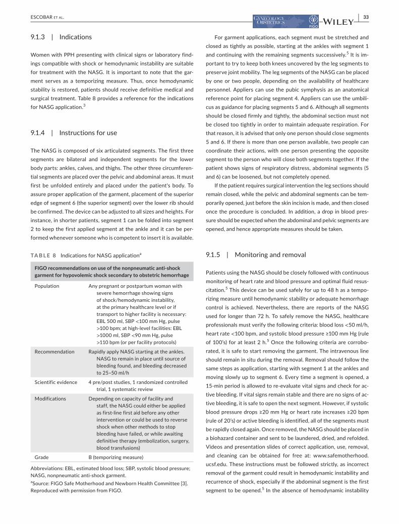
| 33ESCOBAR Et Al.
9.1.3 | Indications
Women with PPH presenting with clinical signs or laboratory find-ings compatible with shock or hemodynamic instability are suitable for treatment with the NASG. It is important to note that the gar-ment serves as a temporizing measure. Thus, once hemodynamic stability is restored, patients should receive definitive medical and surgical treatment. Table 8 provides a reference for the indications for NASG application.3
9.1.4 | Instructions for use
The NASG is composed of six articulated segments. The first three segments are bilateral and independent segments for the lower body parts: ankles, calves, and thighs. The other three circumferen-tial segments are placed over the pelvic and abdominal areas. It must first be unfolded entirely and placed under the patient's body. To assure proper application of the garment, placement of the superior edge of segment 6 (the superior segment) over the lower rib should be confirmed. The device can be adjusted to all sizes and heights. For instance, in shorter patients, segment 1 can be folded into segment 2 to keep the first applied segment at the ankle and it can be per-formed whenever someone who is competent to insert it is available.
For garment applications, each segment must be stretched and closed as tightly as possible, starting at the ankles with segment 1 and continuing with the remaining segments successively.5 It is im-portant to try to keep both knees uncovered by the leg segments to preserve joint mobility. The leg segments of the NASG can be placed by one or two people, depending on the availability of healthcare personnel. Appliers can use the pubic symphysis as an anatomical reference point for placing segment 4. Appliers can use the umbili-cus as guidance for placing segments 5 and 6. Although all segments should be closed firmly and tightly, the abdominal section must not be closed too tightly in order to maintain adequate respiration. For that reason, it is advised that only one person should close segments 5 and 6. If there is more than one person available, two people can coordinate their actions, with one person presenting the opposite segment to the person who will close both segments together. If the patient shows signs of respiratory distress, abdominal segments (5 and 6) can be loosened, but not completely opened.
If the patient requires surgical intervention the leg sections should remain closed, while the pelvic and abdominal segments can be tem-porarily opened, just before the skin incision is made, and then closed once the procedure is concluded. In addition, a drop in blood pres-sure should be expected when the abdominal and pelvic segments are opened, and hence appropriate measures should be taken.
9.1.5 | Monitoring and removal
Patients using the NASG should be closely followed with continuous monitoring of heart rate and blood pressure and optimal fluid resus-citation.5 This device can be used safely for up to 48 h as a tempo-rizing measure until hemodynamic stability or adequate hemorrhage control is achieved. Nevertheless, there are reports of the NASG used for longer than 72 h. To safely remove the NASG, healthcare professionals must verify the following criteria: blood loss <50 ml/h, heart rate <100 bpm, and systolic blood pressure ≥100 mm Hg (rule of 100’s) for at least 2 h.5 Once the following criteria are corrobo-rated, it is safe to start removing the garment. The intravenous line should remain in situ during the removal. Removal should follow the same steps as application, starting with segment 1 at the ankles and moving slowly up to segment 6. Every time a segment is opened, a 15- min period is allowed to re- evaluate vital signs and check for ac-tive bleeding. If vital signs remain stable and there are no signs of ac-tive bleeding, it is safe to open the next segment. However, if systolic blood pressure drops ≥20 mm Hg or heart rate increases ≥20 bpm (rule of 20’s) or active bleeding is identified, all of the segments must be rapidly closed again. Once removed, the NASG should be placed in a biohazard container and sent to be laundered, dried, and refolded. Videos and presentation slides of correct application, use, removal, and cleaning can be obtained for free at: www.safem other hood.ucsf.edu. These instructions must be followed strictly, as incorrect removal of the garment could result in hemodynamic instability and recurrence of shock, especially if the abdominal segment is the first segment to be opened.5 In the absence of hemodynamic instability
TA B L E 8 Indications for NASG applicationa
FIGO recommendations on use of the nonpneumatic anti- shock garment for hypovolemic shock secondary to obstetric hemorrhage
Population Any pregnant or postpartum woman with severe hemorrhage showing signs of shock/hemodynamic instability, at the primary healthcare level or if transport to higher facility is necessary: EBL 500 ml, SBP <100 mm Hg, pulse >100 bpm; at high- level facilities: EBL >1000 ml, SBP <90 mm Hg, pulse >110 bpm (or per facility protocols)
Recommendation Rapidly apply NASG starting at the ankles. NASG to remain in place until source of bleeding found, and bleeding decreased to 25– 50 ml/h
Scientific evidence 4 pre/post studies, 1 randomized controlled trial, 1 systematic review
Modifications Depending on capacity of facility and staff, the NASG could either be applied as first- line first aid before any other intervention or could be used to reverse shock when other methods to stop bleeding have failed, or while awaiting definitive therapy (embolization, surgery, blood transfusions)
Grade B (temporizing measure)
Abbreviations: EBL, estimated blood loss; SBP, systolic blood pressure; NASG, nonpneumatic anti- shock garment.aSource: FIGO Safe Motherhood and Newborn Health Committee [3]. Reproduced with permission from FIGO.
34 | ESCOBAR Et Al.
signs, the whole garment can be removed in 1 h and 15 min. Routine follow- up laboratories, including hemoglobin and hematocrit are encouraged. Values over 7.5 mg/dl of hemoglobin and a hematocrit >23% are recommended; however, there is neither consensus nor evidence about these values, and further research is needed.
9.1.6 | Adverse effects
Currently, no adverse effects have been documented with correct implementation of the NASG4; no increase in shock- related organ dysfunction adverse effects have been noted.9
9.1.7 | Contraindications
Currently, there are no absolute medical contraindications for NASG use.3 Cardiac and pulmonary comorbidities such as heart failure, severe mitral stenosis, and pulmonary hypertension are considered relative contraindications in nonobstetric patients. Although in se-vere hemorrhage, benefits are thought to outweigh any potential harm.5
R E FE R E N C E S 1. Escobar MF, Füchtner CE, Carvajal JA, et al Experience in the use of
non- pneumatic anti- shock garment (NASG) in the management of postpartum haemorrhage with hypovolemic shock in the Fundación Valle Del Lili, Cali, Colombia. Reprod Health. 2017;14:58.
2. Pileggi- Castro C, Nogueira- Pileggi V, Tunçalp Ö, Oladapo OT, Vogel JP, Souza JP. Non- pneumatic anti- shock garment for improving maternal survival following severe postpartum haemorrhage: a sys-tematic review. Reprod Health. 2015;12:28.
3. FIGO Safe Motherhood and Newborn Health Committee, International Federation of Gynecology and Obstetrics. Non- pneumatic anti- shock garment to stabilize women with hypovole-mic shock secondary to obstetric hemorrhage. Int J Gynecol Obstet. 2015;128:194– 195.
4. Althabe F, Therrien MNS, Pingray V, et al. Postpartum hemorrhage care bundles to improve adherence to guidelines: a WHO technical consultation. Int J Gynecol Obstet. 2020;148:290– 299.
5. Miller S, Martin HB, Morris JL. Anti- shock garment in post-partum haemorrhage. Best Pract Res Clin Obstet Gynaecol. 2008;22:1057– 1074.
6. Kerr NL, Hauswald M, Tamrakar SR, Wachter DA, Baty GM. An in-expensive device to treat postpartum hemorrhage: a preliminary proof of concept study of health provider opinion and training in Nepal. BMC Pregnancy Childbirth. 2014;14:81.
7. Lester F, Stenson A, Meyer C, Morris J, Vargas J, Miller S. Impact of the Non- pneumatic Antishock Garment on pelvic
blood flow in healthy postpartum women. Am J Obstet Gynecol. 2011;204(409):e1– 5.
8. Hauswald M, Greene ER. Regional blood flow after pneu-matic anti- shock garment inflation. Prehosp Emerg Care. 2003;7:225– 228.
9. Mourad- Youssif M, Ojengbede OA, Meyer CD, et al Can the Non- pneumatic Anti- Shock Garment (NASG) reduce adverse maternal outcomes from postpartum hemorrhage? Evidence from Egypt and Nigeria. Reprod Health. 2010;7:24.
10. Miller S, Bergel EF, El Ayadi AM, et al Non- pneumatic anti- shock garment (NASG), a first- aid device to decrease maternal mortality from obstetric hemorrhage: a cluster randomized trial. PLoS One. 2013;8:e76477.
11. Mbaruku G, Therrien MS, Tillya R, et al Implementation project of the non- pneumatic anti- shock garment and m- communication to enhance maternal health care in rural Tanzania. Reprod Health. 2018;15:177.
12. World Health Organization. Managing complications in pregnancy and childbirth: a guide for midwives and doctors. Accessed August 11, 2021. https://apps.who.int/iris/bitst ream/handl e/10665/ 25576 0/97892 41565 493- eng.pdf;jsess ionid =8D2D1 DE3DF 5C2CC ED6EF CE711 D80B8 04?seque nce=1
13. Weeks A. The prevention and treatment of postpartum haem-orrhage: what do we know, and where do we go to next? BJOG. 2015;122:202– 210.
14. Hofmeyr GJ, Qureshi Z. Preventing deaths due to haemorrhage. Best Pract Res Clin Obstet Gynaecol. 2016;36:68– 82.
15. Tunçalp O, Souza JP, Gülmezoglu M. World Health Organization. New WHO recommendations on prevention and treat-ment of postpartum hemorrhage. Int J Gynecol Obstet. 2013;123:254– 256.
16. Lalonde A; International Federation of Gynecology and Obstetrics. Prevention and treatment of postpartum hemorrhage in low- resource settings. Int J Gynecol Obstet. 2012;117:108– 118.
17. Jordan K, Butrick E, Yamey G, Miller S. Barriers and facilita-tors to scaling up the non- pneumatic anti- shock garment for treating obstetric hemorrhage: a qualitative study. PLoS One. 2016;11:e0150739.
18. Onyewuenyi A, Tilla R, Kinyonge I, et al. A qualitative approach to understanding the impact of misuse and misdiagnosis: Monitoring use of the non- pneumatic antishock garment in Tanzania. Annals of Global Health. 2016;82:489– 490.
19. Berdichevsky K, Tucker C, Martínez A, Miller S. Acceptance of a new technology for management of obstetric hemorrhage: a qualitative study from rural Mexico. Health Care Women Int. 2010;31:444– 457.
9.2 | Uterine balloon tamponade
Despite active management of the third stage of labor, 2%– 7% of women experience postpartum blood loss of more than 500 ml.1 Uterine balloon tamponade (UBT) is both a diagnostic and thera-peutic tool. If bleeding does not stop after its insertion, then it is better to review the etiology of PPH. Placement of a UBT device should be considered early when emergency measures for man-agement of PPH are initiated. It should be performed by compe-tent healthcare providers who are trained in its insertion to avoid complications that can arise from misplacement. Perineal, vaginal, and cervical tears and ruptured uterus must be excluded, and the placenta carefully assessed for completeness to rule out retained fragments of the placenta. First- line emergency interventions for atonic PPH include uterine massage, initiation of large intravenous
BOX 7 FIGO recommends that all healthcare facilities have the NASG as an effective nonsurgical device that can be used as a temporary measure to recover hemodynamic stability for the management and transfer of a patient to a high level of care.
| 35ESCOBAR Et Al.
access, emptying the uterus and bladder, and administration of oxytocin and TXA (first response bundle). If the uterus fails to contract, ergometrine or misoprostol should be administered, bi-manual uterine or aortic compression should be initiated, a UBT placed, and the NASG applied (response to refractory PPH bun-dle).2 These two sets of approaches have recently been defined as the “PPH bundle”.3
Observational studies have reported UBT success rates between 83% and 95% overall with nearly 100% survival among women with uncontrolled hemorrhage when a UBT device was placed before the onset of advanced shock.4– 6 The integration of UBT into PPH clinical pathways in high- income countries has been shown to avert hyster-ectomy and significantly reduce the need for PPH- related invasive procedures such as artery ligation, uterine compression sutures, and arterial embolization.7– 10
A 2020 systematic review and meta- analysis showed an overall pooled success rate of 85.9% (95% CI, 83.9– 87.9%) for the treatment of PPH, with higher pooled success rates corresponding to PPH due to uterine atony and placenta previa, and lower pooled success rates for PPH due to placenta accreta spectrum (PAS) and retained prod-ucts of conception.11 A safety trial in Kenya and Sierra Leone on a uniquely designed condom uterine tamponade device (ESM- UBT) demonstrated no increased risk of infections or perineal injuries, and success rates of condom UBT devices appear to be comparable to those of Bakri balloons.12 Available UBT devices and specific consid-erations are described below.
9.2.1 | Available UBT devices
Uterine balloon devices can be divided into two groups:
1. Fixed volume devices: condom uterine balloons (ESM- UBT kit, CG balloon), Bakri balloon, Rusch balloon, and Ebb system.12– 16
2. Free flow devices: the glove balloon, Ellavi UBT, and Zukovski balloon.17– 19 The glove and Ellavi balloons allow intrauterine pres-sure control according to systolic blood pressure. The Zukovski balloon uses 50 cm (3.8 cmHg) pressure for inflating the balloon.
9.2.2 | Tamponade effect
The mechanism of action of UBT devices is likely multifactorial. Two proposed mechanisms include device stimulation of receptors, which in turn stimulates uterine contraction, as well as the direct application of hydrostatic pressure against the bleeding sinuses. It has been proposed that if uterine wall pressure exerted by a UBT equals the systolic arte-rial pressure of the bleeding sinuses, a tamponade effect is created.14 However, Georgiou20 measured intraluminal pressures in Bakri bal-loons in two cases where UBTs had been placed and discovered that tamponade occurred at UBT device pressures lower than systolic blood pressures. Further research is needed to better understand the precise biomechanical relationship between a UBT device and a bleeding uterus.
9.2.3 | Free flow tamponade device
Blood is supplied to the uterus via maternal arterial blood that passes through the myometrium through low resistance sinuses.21 Following delivery of the placenta, the muscle fibers of the myome-trium contract and retract, closing off the sinuses and limiting blood loss. While the immediate aim in PPH due to atonic uterus is to arrest blood loss, the ultimate goal is to employ best practice interventions to facilitate contraction of the myometrium. The surgical glove bal-loon, the Ellavi UBT, and the Zukovsky balloon are free flow systems that allow water to be expelled from the balloon to theoretically better allow for the natural physiological process of contraction and retraction of the myometrium once the uterus has recovered.17– 19 However, almost all commercially available UBT devices are low- pressure, high- volume systems that allow for easy UBT expulsion when the uterus regains its tone and begins to contract.
9.2.4 | Drainage port
The need for a drainage port with a UBT device is questionable.13,14,16 Drainage ports are only a few millimeters in diameter and there-fore are easily obstructed by clotted blood. Furthermore, since the source of bleeding in atonic uterus is from the site of placental im-plantation, which encompasses only about 20% of the uterine cavity, a drainage port is less likely to be in proximity to the bleeding site.
9.2.5 | Correct placement
Multiple techniques have been described on the best approach to place a UBT device. The approach should in part be determined by the device being used (e.g. stiffness of the introducing end and num-ber of balloons). To ensure that the balloon could engage within the area that is actively bleeding, all UBTs should be inserted through the cervix and positioned to sit in the upper uterine segment. One approach includes placing the hand into the uterus and ensuring that the fingertips reach the thick anterior and posterior layers of the upper segment. The UBT is then fed into the upper segment of the uterus with the free hand. When withdrawing the uterine hand, the tubing is held with the free hand. While the balloon is filled, two fin-gers are held in the vagina below the cervix to prevent inadvertent balloon expulsion.
9.2.6 | Assessment of effect
Multiple studies have shown that women with uncontrolled PPH from an atonic uterus will almost universally survive if a UBT device is placed in a timely fashion. If UBTs are placed before patients pro-gress to advanced shock, success rates approach 100%.6 However, studies such as by Dumont et al.22 and Natarajan et al.23 have shown that improvised UBTs, delays in recognition of PPH, and lack of

36 | ESCOBAR Et Al.
action on actual UBT placement may be barriers to improved out-comes. While different UBT systems may fill to their desired states at different rates, it is even more important to recognize that impro-vised and untested UBTs are less often used. Moreover, whenever they are, their use is often delayed, they are more difficult to place, fall out more easily, and are associated with overall lower success rates.12– 14,17,19 Therefore, immediate recognition of PPH and emer-gency action with a quality UBT device is imperative to save lives.
9.2.7 | Transfer
Many studies report high rates of hemorrhage control after UBT insertion thus rendering transfers unnecessary. Most often, incor-rect diagnosis and misplacement are the cause of UBT failure. If bleeding recurs or the patient continues to be unstable after UBT insertion, then consideration of transfer to higher levels of care should be made. Transfer can occur with both fixed- volume bal-loons and closed- off free- flow balloons, but all UBTs must be care-fully secured to prevent accidental displacement. A transfer plan should be in place in all obstetric settings, regardless of whether it will be needed or not. This is an absolute necessity in PPH bundles.
9.2.8 | UBT after cesarean delivery
Use of UBTs for hemorrhage control after cesarean delivery is a commonly performed technique worldwide, but there is scant sup-porting literature. There are a few reports on successful outcomes with use of UBT in placenta previa. It appears that UBT has higher success rates in women with PPH after vaginal delivery than after cesarean delivery.11,18,24– 26
9.2.9 | Combining UBT with compression sutures
Placing a UBT after application of uterine compression sutures requires control of the amount of pressure exerted in the uterus to prevent uterine necrosis. Although uterine necrosis is possible following the insertion of compression sutures alone, the risk un-derstandably would increase with pressure exerted from inside the uterus when UBT is used additionally. Cases with uterine necro-sis following insertion of compression sutures have been reported with and without UBT.27,28 The surgical glove balloon and the Ellavi UBT are UBT devices that allow the intrauterine pressure to be controlled by adjusting the height of the intravenous fluid bag or supply bag above the patient.17,18 With systolic blood pressure of 100 mm Hg and the supply bag or intravenous infusion fluid 1.3 m above the patient, the pressure exerted by the balloon will be 100 mm Hg. The specific gravity of mercury is 13 times more than water. The device used for the balloon must be large enough to be inflated with fluid without requiring any expansion pressure. This is achieved by using a surgical glove and the Ellavi UBT.17,18
9.2.10 | Other uses of UBT
While the use of UBT for uncontrolled PPH is fairly well estab-lished, published case reports and senior leaders in obstetrics and gynecology have described successful use of UBTs in other cir-cumstances, such as in molar pregnancy- associated hemorrhage, placenta accreta, retained placenta, and in cases of intentional vaginal and cervical placement to temporize uncontrolled bleed-ing from lacerations.29
WHO recently published recommendations regarding use of UBT in the context of PPH due to uterine atony after vaginal birth in women who do not respond to standard first- line treatment.30 According to WHO, among the prerequisites for use of UBT, evi-dence supports the need for access to surgical intervention and blood products, as well as the availability of health personnel skilled in its use. They recognize that conditions may not be operationalized across all clinical settings and that observational studies suggest a substantial reduction in the risk of maternal morbidity and mortality following uterine balloon tamponade, and that further research is needed in low- resource settings.
R E FE R E N C E S 1. Begley CM, Gyte GM, Devane D, McGuire W, Weeks A. Active ver-
sus expectant management for women in the third stage of labour. Cochrane Database Syst Rev. 2011;(11):CD007412.
2. Escobar MF, Füchtner CE, Carvajal JA, et al Experience in the use of non- pneumatic anti- shock garment (NASG) in the man-agement of postpartum haemorrhage with hypovolemic shock in the Fundación Valle Del Lili, Cali, Colombia. Reprod Health. 2017;14:58.
3. Althabe F, Therrien MNS, Pingray V, et al. Postpartum hemorrhage care bundles to improve adherence to guidelines: a WHO technical consultation. Int J Gynecol Obstet. 2020;148:290– 299.
4. Laas E, Bui C, Popowski T, Mbaku OM, Rozenberg P. Trends in the rate of invasive procedures after the addition of the intrauterine tamponade test to a protocol for management of severe postpar-tum hemorrhage. Am J Obstet Gynecol. 2012;207(281):e1– 7.
5. Revert M, Cottenet J, Raynal P, Cibot E, Quantin C, Rozenberg P. Intrauterine balloon tamponade for management of severe post-partum haemorrhage in a perinatal network: a prospective cohort study. BJOG. 2017;124:1255– 1262.
6. Burke TF, Danso- Bamfo S, Guha M, Oguttu M, Tarimo V, Nelson BD. Shock progression and survival after use of a condom uterine balloon tamponade package in women with uncontrolled postpar-tum hemorrhage. Int J Gynecol Obstet. 2017;139:34– 38.
7. Gauchotte E, De La Torre M, Perdriolle- Galet E, Lamy C, Gauchotte G, Morel O. Impact of uterine balloon tamponade on the use of invasive procedures in severe postpartum hemorrhage. Acta Obstet Gynecol Scand. 2017;96(7):877– 882.
BOX 8 FIGO recommends uterine balloon tamponade in the context of refractory PPH.
Uterine balloon tamponade has proven to be an effective nonsurgical technique so that when employed rapidly by a properly trained person, as the only prerequisite for its use, it can potentially improve survival in women with PPH.
| 37ESCOBAR Et Al.
8. Revert M, Rozenberg P, Cottenet J, Quantin C. Intrauterine balloon tamponade for severe postpartum hemorrhage. Obstet Gynecol. 2018;131:143– 149.
9. Pendleton AA, Natarajan A, Ahn R, Nelson BD, Eckardt MJ, Burke TF. Emergency hysterectomy for uncontrolled postpartum hemor-rhage may be averted through uterine balloon tamponade in Kenya and Senegal. Int J Gynecol Obstet. 2016;133:124.
10. Pendleton AA, Natarajan A, Ahn R, Nelson BD, Eckardt MJ, Burke TF. A qualitative assessment of the impact of a uterine balloon tam-ponade package on decisions regarding the role of emergency hys-terectomy in women with uncontrolled postpartum haemorrhage in Kenya and Senegal. BMJ Open. 2016;6:e010083.
11. Suarez S, Conde- Agudelo A, Borovac- Pinheiro A, et al Uterine balloon tamponade for the treatment of postpartum hemorrhage: a systematic review and meta- analysis. Am J Obstet Gynecol. 2020;222:293.e1– 293.e52.
12. Ramanathan A, Eckardt MJ, Nelson BD, et al Safety of a condom uterine balloon tamponade (ESM- UBT) device for uncontrolled pri-mary postpartum hemorrhage among facilities in Kenya and Sierra Leone. BMC Pregnancy Childbirth. 2018;18:168.
13. Mishra N, Agrawal S, Gulabani K, Shrivastava C. Use of an inno-vative condom balloon tamponade in postpartum haemorrhage: a report. J Obstet Gynaecol India. 2016;66:63– 67.
14. Tolosa JE, Bakri YN, Arulkumaran S. Intrauterine balloon tampon-ade for control of postpartum hemorrhage. This topic last updated: March 12, 2018. Accessed August 25, 2021. https://sniv3 r2.github.io/d/topic.htm?path=intra uteri ne- ballo on- tampo nade- for- contr ol- of- postp artum - hemor rhage
15. Georgiou C. Balloon tamponade in the management of postpartum haemorrhage: a review. BJOG. 2009;116:748– 757.
16. Dildy GA, Belfort MA, Adair CD, et al Initial experience with a dual- balloon catheter for the management of postpartum hemorrhage. Am J Obstet Gynecol. 2014;210(136):e1– 6.
17. Fawcus S, Moodley J. A Monograph of the Management of Postpartum Haemorrhage. National Committee for the Confidential Enquiries into Maternal Deaths in South Africa. National Department of Health South Africa. 2010.
18. Theron GB. Management of postpartum hemorrhage with free- flow pressure controlled uterine balloon. Int J Gynecol Obstet. 2018;142:371– 373.
19. Usage of the Zhukovsky Balloon Tamponade for PPH Free Flow balloon tamponade. 2014. Accessed August 25, 2021. http://www.youtu be.com/watch ?v=qrT8x suB980
20. Georgiou C. Intraluminal pressure readings during the establish-ment of a positive ‘tamponade test’ in the management of postpar-tum haemorrhage. BJOG. 2010;117:295– 303.
21. Cronjé HS, Cilliers JBF, du Toit MA. Clinical Obstetrics – a Southern African perspective, 4th edn. Van Schaik Publishers; 2016.
22. Dumont A, Bodin C, Hounkpatin B, et al Uterine balloon tampon-ade as an adjunct to misoprostol for the treatment of uncontrolled postpartum haemorrhage: a randomised controlled trial in Benin and Mali. BMJ Open. 2017;7:e016590.
23. Natarajan A, Alaska Pendleton A, Nelson BD, et al Provider expe-riences with improvised uterine balloon tamponade for the man-agement of uncontrolled postpartum hemorrhage in Kenya. Int J Gynecol Obstet. 2016;135:210– 213.
24. Cho HY, Park YW, Kim YH, Jung I, Kwon JY. Efficacy of intrauterine Bakri balloon tamponade in cesarean section for placenta previa patients. PLoS One. 2015;10:e0134282.
25. Soyama H, Miyamoto M, Sasa H, et al. Effect of routine rapid inser-tion of Bakri balloon tamponade on reducing hemorrhage from pla-centa previa during and after cesarean section. Arch Gynecol Obstet. 2017;296:469– 474.
26. Maher MA, Abdelaziz A. Comparison between two management protocols for postpartum hemorrhage during cesarean section in
placenta previa: Balloon protocol versus non- balloon protocol. J Obstet Gynaecol Res. 2017;43:447– 455.
27. Treloar EJ, Anderson RS, Andrews HS, Bailey JL. Uterine necrosis following B- Lynch suture for primary postpartum haemorrhage. BJOG. 2006;113:486– 488.
28. Lodhi W, Golara M, Karangaokar V, Yoong W. Uterine necrosis fol-lowing application of combined uterine compression suture with intrauterine balloon tamponade. J Obstet Gynaecol. 2012;32:30– 31.
29. Makin J, Suarez- Rebling DI, Varma Shivkumar P, Tarimo V, Burke TF. Innovative uses of condom uterine balloon tamponade for post-partum hemorrhage in India and Tanzania. Case Rep Obstet Gynecol. 2018;2018:4952048.
30. World Health Organization. WHO recommendation on uterine bal-loon tamponade for the treatment of postpartum haemorrhage. WHO; 2021. Accessed August 25, 2021. https://apps.who.int/iris/handl e/10665/ 340796
9.3 | Uterine artery embolization
Since the first report of the successful use of uterine artery emboliza-tion (UAE) for the treatment of PPH in 1979, success rates have been frequently reported at 90%. However, unlike the medical and surgi-cal treatments for PPH, no randomized controlled trials regarding its effectiveness have been conducted. The decision to perform uterine artery embolization in patients with persistent uterine bleeding should be made in consultation with an interventional radiologist. Although embolization may be considered as the technique of choice for manag-ing PPH by some authors, certain practical issues must be considered.1
Uterine artery embolization has been shown to be effective in the treatment of various causes of PPH including placenta accreta: Both UAE and uterine artery ligation have reported success rates of greater than 90% with low complication rates. If both techniques are available, embolization is the preferred first choice as it obviates lap-arotomy. Ligation can be attempted subsequently if embolization is unsuccessful. In contrast, after an unsuccessful uterine artery ligation, embolization may be extremely difficult or even impossible, leaving hysterectomy as the only remaining option. Some cases may require hysterectomy because of failure of embolization. UAE has shown to be effective in controlling secondary bleeding and the placenta was found to gradually decrease in size in all patients. UAE plays an important role in managing these patients because it is effective at reducing uteropla-cental blood flow to further induce thrombosis of the intervillous space and to achieve necrosis of the retained placental tissue.2– 7 Selective UAE is effective in the control of PPH, thus avoiding hysterectomy.
Availability of resources to perform the procedure: A radiologist trained in embolization techniques is a prerequisite, as is the appro-priate equipment for vascular intervention. Digital road- mapping may be required, and the possible complexity means that this proce-dure can only be performed in a fully equipped X- ray department.8 The interventional radiologist inserts sheaths into both femoral ar-teries to deploy a deflated occlusion balloon into each of the internal iliac arteries.9 The sheaths are removed, and the deflated balloons remain in place until after delivery. After the patient has undergone a cesarean delivery, the interventional radiologist will inflate the balloons if needed based on the amount of bleeding. If inflating the
38 | ESCOBAR Et Al.
balloons is postponed, the balloons may remain in place for up to a few days until the patient's providers are certain she will not begin to bleed again; the balloons can then be removed.9
UAE has become recognized as a relatively safe technique when preserving the patient's fertility is a priority10– 12: Catheterization of the uterine arteries with temporary occlusion by endovascular bal-loon is considered a valid option in the management of patients at high risk of bleeding, in particular those with a pre- existing diagnosis of placenta accreta in patients requesting to preserve their uterus. Ojala et al.13 and Badawy14 described a series of cases in patients who underwent embolization of the uterine arteries. Catheterization averted hysterectomy in close to 80% of cases.
9.3.1 | Complications
Some studies have documented resumption of menses and even pregnancies after the procedure.15 Complications of pelvic embo-lization for PPH occur at a rate of 8.7%. The most common com-plication is low- grade fever. Less common complications are pelvic infection, groin hematoma, iliac artery perforation, transient buttock ischemia, transient foot ischemia, and bladder gangrene. Lee et al.16 listed dissection of the uterine arteries, hematoma at the puncture site, paresthesia, and lower limb edema as complications.
Currently, most PPH cases requiring hysterectomy are related to placenta previa.17 . These patients are commonly diagnosed before delivery and are usually delivered by elective cesarean delivery. This planning may allow increased use of invasive radiological services in the management of such cases by inserting the intra- arterial bal-loons before going to surgery.
9.3.2 | Implementation of treatment
1. A radiologist trained in embolization techniques is a prerequisite, as is the appropriate equipment for vascular intervention. Digital road- mapping may be required, and the possible complexity means that this procedure can only be performed in a fully equipped X- ray department.8
2. The obstetrics staff and interventional radiologist select the ap-propriate operative procedure (UAE or cesarean hysterectomy) based on the patient's clinical condition and a refractory response to conservative medical management.8
3. UAE is preferred for patients who have a stable systolic and dias-tolic blood pressure or heart rate.
R E FE R E N C E S 1. Kim TH, Lee HH, Kim JM, Ryu AL, Chung SH, Seok LW. Uterine
artery embolization for primary postpartum hemorrhage. Iran J Reprod Med. 2013;11:511– 518.
2. Ganguli S, Stecker MS, Pyne D, Baum RA, Fan CM. Uterine artery embolization in the treatment of postpartum uterine hemorrhage. J Vasc Interv Radiol. 2011;22:169– 176.
3. Bros S, Chabrot P, Kastler A, et al Recurrent bleeding within 24 hours after uterine artery embolization for severe postpartum hemorrhage: are there predictive factors? Cardiovasc Intervent Radiol. 2012;35:508– 514.
4. Hong TM, Tseng HS, Lee RC, Wang JH, Chang CY. Uterine artery embolization: an effective treatment for intractable obstetric hae-morrhage. Clin Radiol. 2004;59:96– 101.
5. Jung HN, Shin SW, Choi S- J, et al Uterine artery embolization for emergent management of postpartum hemorrhage associated with placenta accreta. Acta Radiol. 2011;52:638– 642.
6. Soyer P, Morel O, Fargeaudou Y, et al Value of pelvic embolization in the management of severe postpartum hemorrhage due to pla-centa accreta, increta or percreta. Eur J Radiol. 2011;80:729– 735.
7. Diop AN, Chabrot P, Bertrand A, et al Placenta accreta: manage-ment with uterine artery embolization in 17 cases. J Vasc Interv Radiol. 2010;21:644– 648.
8. Wee L, Barron J, Toye R. Management of severe postpar-tum haemorrhage by uterine artery embolization. Br J Anaesth. 2004;93:591– 594.
9. Mahankali SS. Interventional radiology: a disruptive innovation which is transforming management of post- partum haemorrhage. Journal of Obstetric Anaesthesia and Critical Care. 2017;7:65– 68.
10. Angstmann T, Gard G, Harrington T, Ward E, Thomson A, Giles W. Surgical management of placenta accreta: a cohort series and sug-gested approach. Am J Obstet Gynecol. 2010;202(38):e1– 9.
11. Mushtaq S, Kurdi W, Al- Shammari M. Prophylactic catheters place-ment and intraoperative internal iliac artery embolisation in a pa-tient with placenta accreta. J Obstet Gynaecol. 2007;27:853– 855.
12. Thon S, McLintic A, Wagner Y. Prophylactic endovascular place-ment of internal iliac occlusion balloon catheters in parturients with placenta accreta: a retrospective case series. Int J Obstet Anesth. 2011;20:64– 70.
13. Ojala K, Perälä J, Kariniemi J, Ranta P, Raudaskoski T, Tekay A. Arterial embolization and prophylactic catheterization for the treatment for severe obstetric hemorrhage. Acta Obstet Gynecol Scand. 2005;84:1075– 1080.
14. Badawy SZ, Etman A, Singh M, Murphy K, Mayelli T, Philadelphia M. Uterine artery embolization: the role in obstetrics and gynecology. Clin Imaging. 2001;25:288– 295.
15. Gizzo S, Saccardi C, Patrelli TS, et al. Fertility rate and subse-quent pregnancy outcomes after conservative surgical techniques in postpartum hemorrhage: 15 years of literature. Fertil Steril. 2013;99(7):2097– 2107.
16. Lee HY, Shin JH, Kim J, et al Primary postpartum hemorrhage: out-come of pelvic arterial embolization in 251 patients at a single insti-tution. Radiology. 2012;264:903– 909.
17. Gibbins KJ, Einerson BD, Varner MW, Silver RM. Placenta previa and maternal hemorrhagic morbidity. J Matern Fetal Neonatal Med. 2018;31(4):494– 499.
BOX 9 FIGO recommends uterine artery embolization for refractory bleeding uncontrolled by medical and nonsurgical treatment modalities with the availability of trained personnel and necessary equipment for using this technology.
| 39ESCOBAR Et Al.
10 | SURGIC AL TRE ATMENT
10.1 | Uterine compression sutures for PPH
When a decision is made for surgical intervention, adequate opera-tive theatre should be set up with adequate anesthesia, surgical and nursing staff, and blood should be available for transfusion. Upon laparotomy, bleeding at the surgical site should be minimized by ad-equate packing to minimize time lost for suturing while active bleed-ing is ongoing. In all cases the uterus should be fully exteriorized, if possible. Blood loss should be minimized while fully extending the uterus, causing vasoconstriction and a tourniquet placed around the low segment until surgical compression sutures are successfully ap-plied.1 This enables the surgical team to use the following described methods by applying the uterine compression sutures with a high degree of safety. In addition, injections of vasopressin at a dose of 4 U in 20 ml of saline in the placental bed after removal of the placenta may lead to reduced blood loss as well.2 Placement of en-douterine square hemostatic sutures may also be used to control bleeding from the placental bed.3
Hemostatic suturing technique as a second- line strategy for con-trol of uterine bleeding due to uterine atony is an effortless, fast, and conservative surgical procedure. It can be performed satisfactorily after a cesarean delivery or after a vaginal delivery.4 Complications related to its application have been identified but there are no re-ports of deaths related to compression sutures. Complications can include uterine synechia or ischemia, uterine necrosis,5 intrauterine infection,6 and strangulation of the intestinal loop and abdominal omentum when nonabsorbable stitches are used,7 but nonabsorb-able sutures can be used if other types of sutures are not available.
While most births are uncomplicated, obstetricians are often faced with PPH at the time of vaginal or cesarean delivery.8,9 If ini-tial management with fundal massage, manual uterine compression, or uterotonics (oxytocin, misoprostol, methergine, or carboprost tromethamine) does not adequately reduce bleeding, placement of compression sutures can be done to slow bleeding and stabilize the patient, avoid hysterectomy, and ultimately reduce maternal mor-bidity or death. The three most used compression suture techniques for PPH are B- Lynch,10 Hayman,11 and Pereira.12 Other compression sutures are cited in the literature, such as Cho, Ouahba, Hackethal, and Massuba.8 If a Bakri balloon is available, the obstetrician can combine any of these three compression sutures with the placement of the balloon to tamponade bleeding. In some instances, expedient placement of an O’Leary stitch for temporary devascularization of the uterine arteries can decrease bleeding and allow additional time for placement of the compression sutures. As with the conduct of cesarean delivery, specific practical issues have to be considered. First, the obstetrician should be well versed in sterile pelvic vascular operative techniques, as placement of sutures will occur at the time of cesarean delivery or with laparotomy following vaginal delivery and PPH. Second, since time is critical to reducing maternal blood loss, an operative team with the technical skills for additional an-esthesia, administration of blood products, and surgical assistance
is crucial to the success of surgical management. Three commonly used compression sutures are described below and existing data are summarized on the effectiveness of each compression technique.
10.1.1 | Commonly used compression sutures for managing PPH
The B- Lynch suture was first introduced in 1997 by Christopher Lynch to address hemorrhage with open hysterotomy at the time of cesarean delivery.10 A suture is placed on the right side of the hysterotomy, entering at the base of the incision and exiting at the top.13 The suture is then carried across the top of the right fundus and a posterior stitch is placed, with the entrance of the suture at the same level of the anterior suture and exiting on the left posterior side of the uterus at the level of the uterine incision. Suture is then looped over the left fundus, and another stitch is placed at the top of the left side of the uterine incision and exiting at the base of the incision. The suture is then tugged tightly, compressing the body of the uterus with placement of a surgeon's knot. Following placement of the B- Lynch suture, the transverse hysterotomy is closed in the usual fashion. In the original description of B- Lynch, a chromic su-ture was used. Subsequent case studies have reported successful use of a monocryl suture.
Additional options for compression sutures include the Hayman and Pereira. In 2002, the Hayman suture was designed to be a technically easier alternative to the B- Lynch suture that could be placed expediently and not require hysterotomy. The steps for placing a Hayman suture include two to four longitudinal (on the left and right side of the uterus) sutures that pass directly from the anterior uterine wall and through the posterior uterine wall and are tied using a surgeon's knot at the fundus. A transverse cervicoisthmic suture traversing the anterior and posterior uterine walls can be placed to address bleeding from the lower uterine segment.
The Pereira compression technique includes five sutures, com-bining two longitudinal sutures with three transverse sutures. The advantage of the Pereira sutures is that placement occurs in the submucosal region and avoids the endometrial cavity. However, at-tention is needed for correct placement of the transverse sutures to prevent damage to the uterine vessels and ureters.
Based on the available literature, the success rate of compres-sion sutures in achieving hemostasis is 76%– 100%.14 To date, no randomized controlled trials or controlled studies have compared the efficacy of the different compression sutures. In the three orig-inal studies for B- Lynch, Hayman, and Pereira, success rates were reported as 100% (5/5 for B- Lynch, 3/3 for Hayman, and 7/7 for Pereira). In a systematic review,14 the overall success rate of com-pression sutures was reported as 91.7%. Given the broad use of compression sutures and the impact on maternal morbidity and mortality, it is unlikely that a randomized controlled trial to compare the efficacy of different compression techniques will be conducted. However, an observational study comparing the effectiveness of
40 | ESCOBAR Et Al.
different compression sutures on estimated blood loss and measures of maternal morbidity is an obvious next step to move the literature forward.
R E FE R E N C E S 1. Huijgen QCA, Gijsen AF, Hink E, Van Kesteren PJM. Cervical tourni-
quet in case of uncontrollable haemorrhage during caesarean sec-tion owing to a placenta accreta. Case Reports. 2013;2013(apr22 1):bcr2013009237.
2. Kato S, Tanabe A, Kanki K, et al Local injection of vasopressin re-duces the blood loss during cesarean section in placenta previa. J Obstet Gynaecol Res. 2014;40:1249– 1256.
3. Arduini M, Epicoco G, Clerici G, Bottaccioli E, Arena S, Affronti G. B- Lynch suture, intrauterine balloon, and endouterine hemostatic suture for the management of postpartum hemorrhage due to pla-centa previa accreta. Int J Gynecol Obstet. 2010;108(3):191– 193.
4. Kayem G, Kurinczuk JJ, Alfirevic Z, Spark P, Brocklehurst P, Knight M. Specific second- line therapies for postpartum haemorrhage: a national cohort study. BJOG. 2011;118:856– 864.
5. Joshi VM, Shrivastava M. Partial ischemic necrosis of the uterus following a uterine brace compression suture. BJOG. 2004;111:279– 280.
6. Ochoa M, Allaire AD, Stitely ML. Pyometria after hemostatic square suture technique. Obstet Gynecol. 2002;99:506– 509.
7. Wu HH, Yeh GP. Uterine cavity synechiae after hemostatic square suturing technique. Obstet Gynecol. 2005;105(5 Pt 2):1176– 1178.
8. Mousa HA, Blum J, Abou El Senoun G, Shakur H, Alfirevic Z. Treatment for primary postpartum haemorrhage. Cochrane Database Syst Rev. 2014;(2):CD003249.
9. Cunningham FG, Leveno KJ, Bloom SL, et al. Williams Obstetrics, 23rd edn. McGrawHill; 2010.
10. B- Lynch C, Coker A, Lawal AH, Abu J, Cowen MJ. The B- Lynch surgical technique for the control of massive postpartum haem-orrhage: an alternative to hysterectomy? Five cases reported. Br J Obstet Gynaecol. 1997;104:372– 375.
11. Hayman RG, Arulkumaran S, Steer PJ. Uterine compression sutures: surgical management of postpartum hemorrhage. Obstet Gynecol. 2002;99:502– 506.
12. Pereira A, Nunes F, Pedroso S, Saraiva J, Retto H, Meirinho M. Compressive uterine sutures to treat postpartum bleeding second-ary to uterine atony. Obstet Gynecol. 2005;106:569– 572.
13. Price N, B- Lynch C. Technical description of the B- Lynch brace su-ture for treatment of massive postpartum hemorrhage and review of published cases. Int J Fertil Womens Med. 2005;50:148– 163.
14. Doumouchtsis SK, Papageorghiou AT, Arulkumaran S. Systematic review of conservative management of postpartum hemorrhage: what to do when medical treatment fails. Obstet Gynecol Surv. 2007;62:540– 547.
10.2 | Uterine artery ligation
Uterine artery ligation has been described for the control of PPH since 1960; it is considered a simple, fast- performing technique1 and one of the most popular fertility- preserving surgical techniques.2 It
is an effective management strategy for bleeding control with suc-cess rates of 42% and 88% described.3– 6
The recent evidence on the effectiveness of uterine artery ligation to control bleeding in PPH is centered on case reports, mainly asso-ciated with other techniques and with the use of tranexamic acid.7– 11 The success rates for the procedure are higher depending on the time of the intervention from the beginning of the bleeding, the presence of coagulopathy, and the expertise of the surgeons.12,13 The latest Cochrane systematic review of mechanical and surgical interventions for treating primary PPH found no controlled clinical trials evaluating its effectiveness for uterine artery ligation.14
R E FE R E N C E S 1. O'Leary JL, O'Leary JA. Uterine artery ligation in the control
of intractable postpartum hemorrhage. Am J Obstet Gynecol. 1966;94(7):920– 924.
2. Verit FF, Çetin O, Keskin S, Akyol H, Zebitay AG. Does bilateral uterine artery ligation have negative effects on ovarian reserve markers and ovarian artery blood flow in women with postpartum hemorrhage? Clin Exp Reprod Med. 2019;46(1):30– 35.
3. Joshi VM, Otiv SR, Majumder R, Nikam YA, Shrivastava M. Internal iliac artery ligation for arresting postpartum haemorrhage. BJOG. 2007;114:356– 361.
4. Papp Z, Tóth- Pál E, Papp C, et al. Hypogastric artery ligation for intractable pelvic hemorrhage. Int J Gynecol Obstet. 2006;92(1): 27– 31.
5. Ledee N, Ville Y, Musset D, Mercier F, Frydman R, Fernandez H. Management in intractable obstetric haemorrhage: an audit study on 61 cases. Eur J Obstet Gynecol Reprod Biol. 2001;94:189– 196.
6. Unal O, Kars B, Buyukbayrak EE, Karsidag AY, Turan C. The effec-tiveness of bilateral hypogastric artery ligation for obstetric hem-orrhage in three different underlying conditions and its impact on future fertility. J Matern Fetal Neonatal Med. 2011;24:1273– 1276.
7. Abbas AM, Shady NW, Sallam HF. Bilateral uterine artery ligation plus intravenous tranexamic acid during cesarean delivery for pla-centa previa: a randomized double- blind controlled trial. J Gynecol Obstet Hum Reprod. 2019;48(2):115– 119.
8. Kaya B, Tuten A, Daglar K, et al B- Lynch uterine compression su-tures in the conservative surgical management of uterine atony. Arch Gynecol Obstet. 2015;291(5):1005– 1014.
9. Kaya B, Tuten A, Daglar K, et al Balloon tamponade for the man-agement of postpartum uterine hemorrhage. J Perinat Med. 2014;42(6):745– 753.
10. Danisman N, Kahyaoglu S, Celen S, et al. The outcomes of surgical treatment modalities to decrease "near miss" maternal morbidity caused by peripartum hemorrhage. Eur Rev Med Pharmacol Sci. 2014;18(7):1092– 1097.
11. Kaya B, Damarer Z, Daglar K, Unal O, Soliman A, Guralp O. Is there yet a role for internal iliac artery ligation in obstetric hemorrhage with the current gain in popularity of other uterus sparing tech-niques? J Matern Fetal Neonatal Med. 2017;30(11):1325– 1332.
12. American College of Obstetricians and Gynecologists. ACOG Practice Bulletin: Clinical Management Guidelines for
BOX 10 FIGO recommends compression sutures as one of the options to control PPH when medical and nonsurgical treatment modalities fail.
BOX 11 FIGO recommends uterine artery ligation as one of the options to rapidly control PPH when medical, nonsurgical approaches, and compression sutures fail.

| 41ESCOBAR Et Al.
Obstetrician- Gynecologists Number 76, October 2006: postpar-tum hemorrhage. Obstet Gynecol. 2006;108(4):1039– 1047.
13. Vanwinkel S, Claes L, Van den Bosch T. Obstetrical outcome after B- Lynch sutures and ligation of uterine arteries: a case report. Case Rep Womens Health. 2021;30:e00303.
14. Kellie FJ, Wandabwa JN, Mousa HA, Weeks AD. Mechanical and surgical interventions for treating primary postpartum haemor-rhage. Cochrane Database Syst Rev. 2020;(7):CD013663.
10.3 | Bilateral internal iliac artery ligation
Internal iliac artery ligation was first performed by Kelly in 1894,1 with reported success rates between 40% and 100%.2,3 It reduces pelvic blood flow by half and pulse pressure by 85%, thereby simu-lating a venous rather than an arterial circulation, and promoting hemostasis.1,2 Although this technique provides a rapid and effec-tive way to control hemorrhage, it remains underused because of understandable fear of injury to surrounding structures: internal and external iliac arteries and ureters2,4 and should only be considered if experienced pelvic surgeons are available. Internal iliac artery liga-tion may avoid the need for hysterectomy in the context of uterine atony.2 In addition, in cases of traumatic PPH such as uterine rupture and extensive genital injuries, internal artery ligation clears the op-erative field and facilitates hysterectomy.2
During the procedure, the common iliac artery is identified. After dissection, the ureter is visualized crossing the common iliac artery at its bifurcation. The common iliac artery branches into an internal iliac (hypogastric) artery, coursing medially and inferiorly, and the external iliac artery coursing laterally and superiorly. The arteries are not infrequently adherent to the underlying veins and require appropriate dissection.5 With right- angled forceps a su-ture is introduced between the artery and the vein, and a knot is made.4,5
Noteworthy is that even after bilateral internal iliac artery li-gation, the vascular supply to the pelvis is not completely compro-mised due to the extensive collateral circulation that exists.4,5 The revascularization is provided by the deep femoral artery, the anas-tomosis between the medial femoral circumflex and obturator ar-tery, and that between the lateral femoral circumflex and superior gluteal artery, and the ovarian artery.4,5 Obstetricians ought to be more familiar with this procedure as it is an effective and rapid way to control PPH.
R E FE R E N C E S 1. Burchell RC. Physiology of internal iliac artery ligation. J Obstet
Gynaecol Br Commonw. 1968;75:642– 651. 2. Joshi VM, Otiv SR, Majumder R, Nikam YA, Shrivastava M. Internal
iliac artery ligation for arresting postpartum haemorrhage. BJOG. 2007;114:356– 361.
3. Wang CY, Pan HH, Chang CC, Lin CK. Outcomes of hypogastric artery ligation and transcatheter uterine artery embolization in women with postpartum hemorrhage. Taiwan J Obstet Gynecol. 2019;58:72– 76.
4. Selçuk İ, Uzuner B, Boduç E, Baykuş Y, Akar B, Güngör T. Step- by- step ligation of the internal iliac artery. J Turk Ger Gynecol Assoc. 2019;20:123– 128.
5. B- Lynch C, Keith LG, Campbell WB. Internal iliac (hypogastric) ar-tery ligation. In: Arulkumaran S, Karoshi M, Keith LG, Lalonde AB, B- Lynch C. eds. A Comprehensive Textbook of Postpartum Hemorrhage, 2nd edn. Sapiens Publishing; 2012:441– 447.
10.4 | Hysterectomy
Peripartum hysterectomy is performed in the treatment of PPH when all other conventional methods fail to control the bleeding.1 The prevalence is higher in low- income regions than in high- income regions (2.8 and 0.7 per 1000 deliveries, respectively).2 The in-cidence of this procedure has been on the rise, increasing by 15% in the USA between 1994 and 2007.3 Similarly, in Turkey, the rate has increased from 0.03% in 2000– 2006 to 0.07% in 2007– 2013. In most of the studies, this was attributed to the increased rates of cesarean delivery.1– 5
The most common indications for peripartum hysterectomy are placental pathology such as abnormal placentation, placenta previa, and placental abruption (38%), uterine atony (27%), and uterine rup-ture (26%).2 Peripartum hysterectomy is associated with high rates of maternal morbidity and complications due to the need for blood transfusions, coagulation disorders, injury to the urinary tract, the need for re- exploration in case of persistent bleeding, and postop-erative fever.1
The maternal mortality rate varies according to countries, rang-ing from 0% in Turkey,6 the UK,7 and New Zealand,8 to 3.2% in Australia,9 and as high as 11.8% in Nigeria.10
R E FE R E N C E S 1. Demirci O, Tuğrul AS, Yilmaz E, Tosun Ö, Demirci E, Eren YS.
Emergency peripartum hysterectomy in a tertiary obstetric center: nine years evaluation. J Obstet Gynaecol Res. 2011;37:1054– 1060.
BOX 12 FIGO recommends internal iliac artery ligation as one of the options to rapidly control PPH for management of PPH when medical, nonsurgical approaches, and compression sutures fail.
FIGO also recommends that all obstetricians familiarize them-selves with the internal iliac artery location and with the tech-nique of its ligation.
BOX 13 FIGO recommends abdominal hysterectomy as treatment for PPH when all other medical, nonsurgical, and surgical treatment modalities have failed to control PPH.
Care must be taken to have adequate blood supplies. Subtotal hysterectomy can shorten the procedure and is important in a hemodynamically unstable patient.
42 | ESCOBAR Et Al.
2. Van den Akker T, Brobbel C, Dekkers OM, Bloemenkamp KW. Prevalence, indications, risk indicators, and outcomes of emer-gency peripartum hysterectomy worldwide: a systematic review and meta- analysis. Obstet Gynecol. 2016;128:1281– 1294.
3. Bateman BT, Mhyre JM, Callaghan WM, Kuklina EV. Peripartum hysterectomy in the United States: nationwide 14 year experience. Am J Obstet Gynecol. 2012;206(63):e1– 8.
4. Temizkan O, Angın D, Karakuş R, Şanverdi İ, Polat M, Karateke A. Changing trends in emergency peripartum hysterectomy in a tertiary obstetric center in Turkey during 2000– 2013. J Turk Ger Gynecol Assoc. 2016;17:26– 34.
5. Chawla J, Arora D, Paul M, Ajmani SN. Emergency obstetric hyster-ectomy: a retrospective study from a Teaching Hospital in North India over eight years. Oman Med J. 2015;30:181– 186.
6. Danisman N, Baser E, Togrul C, Kaymak O, Tandogan M, Gungor T. Emergency peripartum hysterectomy: experience of a major
referral hospital in Ankara. Turkey. J Obstet Gynaecol. 2015;35: 19– 21.
7. Jones B, Zhang E, Alzouebi A, et al Maternal and perinatal out-comes following peripartum hysterectomy from a single tertiary centre. Aust N Z J Obstet Gynaecol. 2013;53:561– 565.
8. Wong TY. Emergency peripartum hysterectomy: a 10- year review in a tertiary obstetric hospital. N Z Med J. 2011;124:34– 39.
9. Awan N, Bennett MJ, Walters WA. Emergency peripartum hyster-ectomy: a 10- year review at the Royal Hospital for Women, Sydney. Aust N Z J Obstet Gynaecol. 2011;51:210– 215.
10. Akintayo AA, Olagbuji BN, Aderoba AK, Akadiri O, Olofinbiyi BA, Bakare B. Emergency peripartum hysterectomy: a multicenter study of incidence, indications and outcomes in Southwestern Nigeria. Matern Child Health J. 2016;20:1230– 1236.
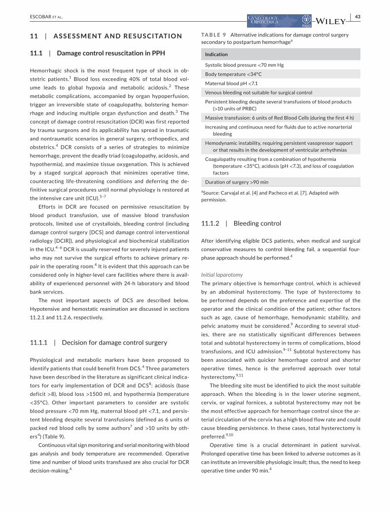
| 43ESCOBAR Et Al.
11 | A SSESSMENT AND RESUSCITATION
11.1 | Damage control resuscitation in PPH
Hemorrhagic shock is the most frequent type of shock in ob-stetric patients.1 Blood loss exceeding 40% of total blood vol-ume leads to global hypoxia and metabolic acidosis.2 These metabolic complications, accompanied by organ hypoperfusion, trigger an irreversible state of coagulopathy, bolstering hemor-rhage and inducing multiple organ dysfunction and death.3 The concept of damage control resuscitation (DCR) was first reported by trauma surgeons and its applicability has spread in traumatic and nontraumatic scenarios in general surgery, orthopedics, and obstetrics.4 DCR consists of a series of strategies to minimize hemorrhage, prevent the deadly triad (coagulopathy, acidosis, and hypothermia), and maximize tissue oxygenation. This is achieved by a staged surgical approach that minimizes operative time, counteracting life- threatening conditions and deferring the de-finitive surgical procedures until normal physiology is restored at the intensive care unit (ICU).5– 7
Efforts in DCR are focused on permissive resuscitation by blood product transfusion, use of massive blood transfusion protocols, limited use of crystalloids, bleeding control (including damage control surgery [DCS] and damage control interventional radiology [DCIR]), and physiological and biochemical stabilization in the ICU.4– 6 DCR is usually reserved for severely injured patients who may not survive the surgical efforts to achieve primary re-pair in the operating room.4 It is evident that this approach can be considered only in higher- level care facilities where there is avail-ability of experienced personnel with 24- h laboratory and blood bank services.
The most important aspects of DCS are described below. Hypotensive and hemostatic reanimation are discussed in sections 11.2.1 and 11.2.6, respectively.
11.1.1 | Decision for damage control surgery
Physiological and metabolic markers have been proposed to identify patients that could benefit from DCS.4 Three parameters have been described in the literature as significant clinical indica-tors for early implementation of DCR and DCS8: acidosis (base deficit >8), blood loss >1500 ml, and hypothermia (temperature <35°C). Other important parameters to consider are systolic blood pressure <70 mm Hg, maternal blood pH <7.1, and persis-tent bleeding despite several transfusions (defined as 6 units of packed red blood cells by some authors7 and >10 units by oth-ers4) (Table 9).
Continuous vital sign monitoring and serial monitoring with blood gas analysis and body temperature are recommended. Operative time and number of blood units transfused are also crucial for DCR decision- making.4
11.1.2 | Bleeding control
After identifying eligible DCS patients, when medical and surgical conservative measures to control bleeding fail, a sequential four- phase approach should be performed.4
Initial laparotomyThe primary objective is hemorrhage control, which is achieved by an abdominal hysterectomy. The type of hysterectomy to be performed depends on the preference and expertise of the operator and the clinical condition of the patient; other factors such as age, cause of hemorrhage, hemodynamic stability, and pelvic anatomy must be considered.9 According to several stud-ies, there are no statistically significant differences between total and subtotal hysterectomy in terms of complications, blood transfusions, and ICU admission.9– 11 Subtotal hysterectomy has been associated with quicker hemorrhage control and shorter operative times, hence is the preferred approach over total hysterectomy.9,11
The bleeding site must be identified to pick the most suitable approach. When the bleeding is in the lower uterine segment, cervix, or vaginal fornices, a subtotal hysterectomy may not be the most effective approach for hemorrhage control since the ar-terial circulation of the cervix has a high blood flow rate and could cause bleeding persistence. In these cases, total hysterectomy is preferred.9,10
Operative time is a crucial determinant in patient survival. Prolonged operative time has been linked to adverse outcomes as it can institute an irreversible physiologic insult; thus, the need to keep operative time under 90 min.4
TA B L E 9 Alternative indications for damage control surgery secondary to postpartum hemorrhagea
Indication
Systolic blood pressure <70 mm Hg
Body temperature <34ºC
Maternal blood pH <7.1
Venous bleeding not suitable for surgical control
Persistent bleeding despite several transfusions of blood products (>10 units of PRBC)
Massive transfusion: 6 units of Red Blood Cells (during the first 4 h)
Increasing and continuous need for fluids due to active nonarterial bleeding
Hemodynamic instability, requiring persistent vasopressor support or that results in the development of ventricular arrhythmias
Coagulopathy resulting from a combination of hypothermia (temperature <35°C), acidosis (pH <7.3), and loss of coagulation factors
Duration of surgery >90 min
aSource: Carvajal et al. [4] and Pacheco et al. [7]. Adapted with permission.
44 | ESCOBAR Et Al.
As in other surgical specialties, packing is the cornerstone of obstetric DCS.4,7 Pelvic packing should be performed with at least 7– 10 compresses, according to reported experience.2,12 Aside from pelvic packing, temporary abdominal closure is performed along-side, as a complementary feature of the procedure. The latter can be achieved using a negative pressure system like vacuum pack or partial closure with a Viaflex bag (Bogota Bag) without the need for negative pressure.2,12 Currently, there is neither consensus nor sufficient evidence regarding the use of prophylactic antibiotics in patients undergoing DCS with abdominal packing.4 Several surgi-cal guidelines recommend the administration of a single preopera-tive dose of broad- spectrum antibiotics, which theoretically should provide sufficient coverage for aerobic and anaerobic microorgan-isms.4,13,14 Other experts suggest administration of prophylactic broad- spectrum antibiotics every 6– 8 h until the abdominal packing is removed. Nevertheless, further studies are necessary to provide evidence- based recommendations in the obstetric population.7
Resuscitation – ICUAt this stage, the patient must be transferred to the ICU to address the physiologic derangements of the hemorrhagic patient: coagula-tion disorders and metabolic abnormalities.6 Interdisciplinary care involving the obstetrician and critical care specialist is key for these patients as complications could arise at any time.4 It is crucial to ac-curately quantify the accumulation of blood in the abdominal cavity. The optimal device to do so, after partial abdominal closure, is the vacuum pack, which allows a more precise quantification of bleeding during the postoperative period. In patients without coagulopathy, the drainage of >400 ml/h of blood through the vacuum pack repre-sents an early indication for laparotomy.4
Definitive surgeryAfter restoring normal physiology, it is considered safe to review the abdominal cavity. Ideally, this should be performed 48– 72 h after the initial surgical procedure.4 Patients may require one or more surgical interventions, depending on the operative findings. In cases where further interventions are warranted, it is recommended to continue with techniques of temporary closure of the abdominal cavity.15
Definitive closure of abdominal wall and cavityThe final stage is the definitive closure of the abdominal wall, which is performed after all surgeries have been successfully completed and all additional damage has been repaired.4
11.1.3 | Complications
The majority of DCS complications depend on the time of closure of the abdominal fascia. It is important to consider that the number of reinterventions is directly related to a higher percentage of infec-tious complications, wound dehiscence, and abdominal wall closure problems.16 The main complications include infection of the surgical wound in 28% of cases, presence of intra- abdominal collections in
20%, and evisceration in 10% of patients.2 Implementation of pro-phylactic antibiotic therapy could counteract these complications, as it targets the polybacterial flora present in the female genital tract. An interdisciplinary approach could be required in case of complica-tions derived from injuries secondary to the surgical procedure such as urinomas, perforations, or fistulas, among others.17
11.1.4 | Final objectives in resuscitation
DCR should be maintained as long as there are signs of bleeding and coagulopathy. A continuous assessment of the hemodynamic and physiological status of the patient is required. Monitoring several resuscitation parameters is essential until tissue hypoxia reverts. These parameters include pH, base deficit, lactate, hematocrit, and coagulation— ideally evaluated with conventional laboratory tests and point- of- care testing (POCT) of viscoelastic coagulation such as thromboelastography (TEG) and rotational thromboelastometry (ROTEM). In patients with a clinical trend toward improvement, the decision to perform definitive surgery is appropriate.4
R E FE R E N C E S 1. Pacheco LD, Saade GR, Gei AF, Hankins GD. Cutting- edge advances
in the medical management of obstetrical hemorrhage. Am J Obstet Gynecol. 2011;205:526– 532.
2. Ordóñez CA, Nieto AJ, Carvajal JA, et al Damage control surgery for the management of major obstetric hemorrhage: experience from the Fundación Valle del Lili, Cali, Colombia. Panamerican Journal of Trauma, Critical Care and Emergency Surgery. 2017;6:1– 7.
3. Clark SL, Hankins GD. Preventing maternal death: 10 clinical dia-monds. Obstet Gynecol. 2012;119(2 Pt 1):360– 364.
4. Carvajal JA, Ramos I, Kusanovic JP, Escobar MF. Damage- control resuscitation in obstetrics. J Matern Fetal Neonatal Med. 2022;35:785– 798.
5. Bogert JN, Harvin JA, Cotton BA. Damage control resuscitation. J Intensive Care Med. 2016;31:177– 186.
6. West N, Dawes R. Trauma resuscitation and the damage control approach. Surgery. 2018;36:409– 416.
7. Pacheco LD, Lozada MJ, Saade GR, Hankins GDV. Damage- control surgery for obstetric hemorrhage. Obstet Gynecol. 2018;132:423– 427.
8. Ordoñez CA, Badiel M, Pino LF, et al Damage control resuscita-tion: Early decision strategies in abdominal gunshot wounds using an easy “ABCD” mnemonic. J Trauma Acute Care Surg. 2012;73:1074– 1078.
9. Gungor T, Simsek A, Ozdemir AO, Pektas M, Danisman N, Mollamahmutoglu L. Surgical treatment of intractable postpartum
BOX 14 FIGO recommends that damage control resuscitation (DCR) should be implemented in the management algorithms for major obstetric hemorrhage.
All countries should establish one or more referral hospital(s) and develop expert teams that are familiar with this strategy, the technique, and indications to be able to offer DCR.
| 45ESCOBAR Et Al.
hemorrhage and changing trends in modern obstetric perspective. Arch Gynecol Obstet. 2009;280:351– 355.
10. Mukherjee S, Arulkumaran S. Post- partum haemorrhage. Obstet Gynaecol Reprod Med. 2009;19:121– 126.
11. Zhang Y, Yan J, Han Q, et al Emergency obstetric hysterectomy for life- threatening postpartum hemorrhage: a 12- year review. Medicine. 2017;96:e8443.
12. Escobar MF, García A, Fonseca J, Herrera E, Guerrero JE. Cirugía de control de daños: un concepto aplicable en ginecología y obstetri-cia. Colombia Médica. 2005;36:110– 114.
13. Goldberg SR, Anand RJ, Como JJ, et al Prophylactic antibiotic use in penetrating abdominal trauma: an Eastern Association for the Surgery of Trauma practice management guideline. J Trauma Acute Care Surg. 2012;73(5 Suppl 4):S321– S325.
14. Godat L, Kobayashi L, Costantini T, Coimbra R. Abdominal damage control surgery and reconstruction: world society of emergency surgery position paper. World J Emerg Surg. 2013;8:53.
15. Griggs C, Butler K. Damage control and the open abdomen: challenges for the nonsurgical intensivist. J Intensive Care Med. 2016;31:567– 576.
16. Kirkpatrick AW, Roberts DJ, De Waele J, et al Intra- abdominal hy-pertension and the abdominal compartment syndrome: updated consensus definitions and clinical practice guidelines from the World Society of the Abdominal Compartment Syndrome. Intensive Care Med. 2013;39:1190– 1206.
17. Burlew CC. The open abdomen: practical implications for the prac-ticing surgeon. Am J Surg. 2012;204:826– 835.
11.2 | Resuscitation
Once the shock has occurred in PPH, it is estimated that the mortality of patients will increase dramatically.1– 3 To mitigate metabolic complications, strategies such as hypotensive fluid re-suscitation and transfusion protocols have been studied for hemo-static reanimation.
There are two strategies for fluid resuscitation in patients with hemorrhage: the aggressive approach and the hypotensive resusci-tation approach. Aggressive resuscitation refers to the traditionally used strategy in which the key principle is restoring the effective circulating blood volume, and rapid normalizing of blood pressure with administration of large amounts of crystalloids.2,4 Hypotensive resuscitation, also called permissive hypotension, consists of restric-tive crystalloid resuscitation during the early stages of a hemorrhagic shock to maintain lower than normal systolic or mean blood pressure, sustaining organ perfusion until control of the bleeding occurs.2,5
In contrast, hemostatic reanimation is based on early and ag-gressive blood product replacement, transfusing red blood cells (PRBC), fresh frozen plasma (FFP), and platelets (PLT) in the same proportion as found in circulating blood to correct coagulopathy.6– 8 Hypotensive resuscitation and hemostatic reanimation are the fun-damentals for DCR.9,10
11.2.1 | Hypotensive resuscitation
The concept of hypotensive resuscitation is because administering small crystalloid volumes reduces the risk of dilutional coagulopathy
and maintaining a lower blood pressure is less likely to disintegrate the pre- formed blood clots. Aggressive resuscitation may worsen coagu-lopathy and hemorrhage by increasing intravascular hydrostatic pres-sures, diluting coagulation factors, and inducing more hypothermia, which results in deterioration of the triad of death.4,10,11 Furthermore, an excessive rise in blood pressure could also result in higher red blood cell loss leading to more hypoxia and acidosis in tissues.4,11
11.2.2 | Intravenous fluids
Among the initial strategies for reanimation, the administration of crystalloids in small boluses of 500 ml is recommended.10 Scientific evidence recommends the use of balanced crystalloid solutions such as Ringer's lactate owing to the risk of hyperchloremic acidosis and the worsening of kidney function with chlorine- rich fluids (saline so-lution).7 This is particularly important for LMICs, where saline- based solutions are in abundance. After the administration of each bolus, physicians must assess the clinical status of patients, looking for an im-provement in signs and symptoms of shock resulting from blood loss.10
11.2.3 | Targeted blood pressure
The difference between aggressive and hypotensive resuscitation lies within targeted blood pressure management.4 Mean arterial pressure (MAP) represents the perfusion of the majority of organs, therefore providing the target for clinicians to guide fluid adminis-tration.11 Hemorrhagic shock animal models have demonstrated a positive benefit in survival with MAP between 55– 60 mm Hg dur-ing active bleeding.10 The European guideline on management of major bleeding and coagulopathy following trauma recommends permissive hypotension with a systolic blood pressure target of 80– 90 mm Hg (MAP 50– 60 mm Hg) until major bleeding has been con-trolled (Recommendation Grade 1C).12
11.2.4 | Aggressive approach and adverse outcomes
During hemorrhagic shock the endothelial glycocalyx becomes thin-ner and administration of large amounts of crystalloids exacerbates this state, leading to fluid extravasation that may cause cerebral, cardiac, and pulmonary edema.7,10,11 Third spacing may also lead to cardiac dysfunction, worsen hemodynamics, and decrease kidney perfusion. Decreased kidney perfusion occurs because of an in-crease in intra- abdominal pressure, which can additionally result in abdominal compartment syndrome.7,11
11.2.5 | Evidence
Several studies in trauma patients have demonstrated that hy-potensive resuscitation is correlated with benefits to survival,
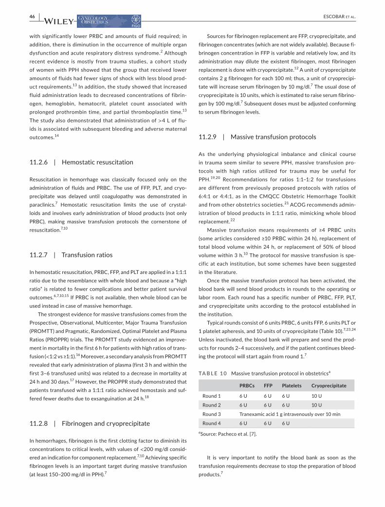
46 | ESCOBAR Et Al.
with significantly lower PRBC and amounts of fluid required; in addition, there is diminution in the occurrence of multiple organ dysfunction and acute respiratory distress syndrome.2 Although recent evidence is mostly from trauma studies, a cohort study of women with PPH showed that the group that received lower amounts of fluids had fewer signs of shock with less blood prod-uct requirements.13 In addition, the study showed that increased fluid administration leads to decreased concentrations of fibrin-ogen, hemoglobin, hematocrit, platelet count associated with prolonged prothrombin time, and partial thromboplastin time.13 The study also demonstrated that administration of >4 L of flu-ids is associated with subsequent bleeding and adverse maternal outcomes.14
11.2.6 | Hemostatic resuscitation
Resuscitation in hemorrhage was classically focused only on the administration of fluids and PRBC. The use of FFP, PLT, and cryo-precipitate was delayed until coagulopathy was demonstrated in paraclinics.7 Hemostatic resuscitation limits the use of crystal-loids and involves early administration of blood products (not only PRBC), making massive transfusion protocols the cornerstone of resuscitation.7,10
11.2.7 | Transfusion ratios
In hemostatic resuscitation, PRBC, FFP, and PLT are applied in a 1:1:1 ratio due to the resemblance with whole blood and because a “high ratio” is related to fewer complications and better patient survival outcomes.6,7,10,15 If PRBC is not available, then whole blood can be used instead in case of massive hemorrhage.
The strongest evidence for massive transfusions comes from the Prospective, Observational, Multicenter, Major Trauma Transfusion (PROMTT) and Pragmatic, Randomized, Optimal Platelet and Plasma Ratios (PROPPR) trials. The PROMTT study evidenced an improve-ment in mortality in the first 6 h for patients with high ratios of trans-fusion (<1:2 vs ≥1:1).16 Moreover, a secondary analysis from PROMTT revealed that early administration of plasma (first 3 h and within the first 3– 6 transfused units) was related to a decrease in mortality at 24 h and 30 days.17 However, the PROPPR study demonstrated that patients transfused with a 1:1:1 ratio achieved hemostasis and suf-fered fewer deaths due to exsanguination at 24 h.18
11.2.8 | Fibrinogen and cryoprecipitate
In hemorrhages, fibrinogen is the first clotting factor to diminish its concentrations to critical levels, with values of <200 mg/dl consid-ered an indication for component replacement.7,10 Achieving specific fibrinogen levels is an important target during massive transfusion (at least 150– 200 mg/dl in PPH).7
Sources for fibrinogen replacement are FFP, cryoprecipitate, and fibrinogen concentrates (which are not widely available). Because fi-brinogen concentration in FFP is variable and relatively low, and its administration may dilute the existent fibrinogen, most fibrinogen replacement is done with cryoprecipitate.12 A unit of cryoprecipitate contains 2 g fibrinogen for each 100 ml; thus, a unit of cryoprecipi-tate will increase serum fibrinogen by 10 mg/dl.7 The usual dose of cryoprecipitate is 10 units, which is estimated to raise serum fibrino-gen by 100 mg/dl.7 Subsequent doses must be adjusted conforming to serum fibrinogen levels.
11.2.9 | Massive transfusion protocols
As the underlying physiological imbalance and clinical course in trauma seem similar to severe PPH, massive transfusion pro-tocols with high ratios utilized for trauma may be useful for PPH.19,20 Recommendations for ratios 1:1– 1:2 for transfusions are different from previously proposed protocols with ratios of 6:4:1 or 4:4:1, as in the CMQCC Obstetric Hemorrhage Toolkit and from other obstetrics societies.21 ACOG recommends admin-istration of blood products in 1:1:1 ratio, mimicking whole blood replacement.22
Massive transfusion means requirements of ≥4 PRBC units (some articles considered ≥10 PRBC within 24 h), replacement of total blood volume within 24 h, or replacement of 50% of blood volume within 3 h.10 The protocol for massive transfusion is spe-cific at each institution, but some schemes have been suggested in the literature.
Once the massive transfusion protocol has been activated, the blood bank will send blood products in rounds to the operating or labor room. Each round has a specific number of PRBC, FFP, PLT, and cryoprecipitate units according to the protocol established in the institution.
Typical rounds consist of 6 units PRBC, 6 units FFP, 6 units PLT or 1 platelet apheresis, and 10 units of cryoprecipitate (Table 10).7,23,24 Unless inactivated, the blood bank will prepare and send the prod-ucts for rounds 2– 4 successively, and if the patient continues bleed-ing the protocol will start again from round 1.7
It is very important to notify the blood bank as soon as the transfusion requirements decrease to stop the preparation of blood products.7
TA B L E 1 0 Massive transfusion protocol in obstetricsa
PRBCs FFP Platelets Cryoprecipitate
Round 1 6 U 6 U 6 U 10 U
Round 2 6 U 6 U 6 U 10 U
Round 3 Tranexamic acid 1 g intravenously over 10 min
Round 4 6 U 6 U 6 U
aSource: Pacheco et al. [7].
| 47ESCOBAR Et Al.
11.2.10 | Adverse outcomes
Although early transfusions are lifesaving and in theory help to achieve hemostasis faster, thereby decreasing the number of blood products administered, the application of multiple units of blood products could be associated with a higher incidence of transfusion- related complications. These complications include hy-perkalemia, hypocalcemia, citrate toxicity, transfusion- related im-munomodulation, transfusion- related circulatory overload (TACO), transfusion- related kidney injury, transfusion- related acute lung injury (TRALI) (0.1 per 1000 units transfused), transfusion- related febrile nonhemolytic reactions (0.8 per 1000 units transfused), and acute hemolytic transfusion reaction (0.19 per 1000 units trans-fused).22,24,25 Transfusion- related infectious diseases are uncom-mon (less than 1/100 000– 1 000 000).22,25
R E FE R E N C E S 1. American College of Obstetricians and Gynecologists. ACOG
Practice Bulletin: Clinical Management Guidelines for Obstetrician- Gynecologists Number 76, October 2006: postpartum hemor-rhage. Obstet Gynecol. 2006;108:1039– 1047.
2. Owattanapanich N, Chittawatanarat K, Benyakorn T, Sirikun J. Risks and benefits of hypotensive resuscitation in patients with traumatic hemorrhagic shock: a meta- analysis. Scand J Trauma Resusc Emerg Med. 2018;26:107.
3. Cannon JW. Hemorrhagic shock. N Engl J Med. 2018;378:370– 379. 4. Wang H, Chen MB, Zheng XW, Zheng QH. Effectiveness and safety
of hypotensive resuscitation in traumatic hemorrhagic shock: a pro-tocol for meta- analysis. Medicine. 2019;98:e18145.
5. Carrick MM, Leonard J, Slone DS, Mains CW, Bar- Or D. Hypotensive resuscitation among trauma patients. Biomed Res Int. 2016;2016:8901938.
6. Johansson PI, Oliveri RS, Ostrowski SR. Hemostatic resuscita-tion with plasma and platelets in trauma. J Emerg Trauma Shock. 2012;5:120– 125.
7. Pacheco LD, Saade GR, Costantine MM, Clark SL, Hankins GD. An update on the use of massive transfusion protocols in obstetrics. Am J Obstet Gynecol. 2016;214:340– 344.
8. Zwinkels RLJ, Endeman H, Hoeks SE, de Maat MPM, den Hartog D, Stolker RJ. The clinical effect of hemostatic resusci-tation in traumatic hemorrhage; a before- after study. J Crit Care. 2020;56:288– 293.
9. Spinella PC, Pidcoke HF, Strandenes G, et al. Whole blood for hemo-static resuscitation of major bleeding. Transfusion. 2016;56(Suppl 2):S190– 202.
10. Carvajal JA, Ramos I, Kusanovic JP, Escobar MF. Damage- control resuscitation in obstetrics. J Matern Fetal Neonatal Med. 2022;35:785– 798.
11. Bouglé A, Harrois A, Duranteau J. Resuscitative strategies in trau-matic hemorrhagic shock. Ann Intensive Care. 2013;3:1.
12. Spahn DR, Bouillon B, Cerny V, et al. The European guideline on management of major bleeding and coagulopathy following trauma. Crit Care. 2019;23:98.
13. Gillissen A, van den Akker T, Caram- Deelder C, et al. Association between fluid management and dilutional coagulopathy in severe postpartum haemorrhage: a nationwide retrospective cohort study. BMC Pregnancy Childbirth. 2018;18:398.
14. Henriquez DDCA, Bloemenkamp KWM, Loeff RM, et al. Fluid resuscitation during persistent postpartum haemorrhage and ma-ternal outcome: a nationwide cohort study. Eur J Obstet Gynecol Reprod Biol. 2019;235:49– 56.
15. Pham HP, Shaz BH. Update on massive transfusion. Br J Anaesth. 2013;111(Suppl 1):i71– 82.
16. Holcomb JB, del Junco DJ, Fox EE, et al. The prospective, obser-vational, multicenter, major trauma transfusion (PROMMTT) study: comparative effectiveness of a time- varying treatment with com-peting risks. JAMA Surg. 2013;148:127– 136.
17. del Junco DJ, Holcomb JB, Fox EE, et al. Resuscitate early with plasma and platelets or balance blood products gradually: findings from the PROMMTT study. J Trauma Acute Care Surg. 2013;75(1 Suppl 1):S24– 30.
18. Holcomb JB, Tilley BC, Baraniuk S, et al. Transfusion of plasma, platelets, and red blood cells in a 1:1:1 vs a 1:1:2 ratio and mortal-ity in patients with severe trauma: the PROPPR randomized clinical trial. JAMA. 2015;313:471– 482.
19. Tanaka H, Matsunaga S, Yamashita T, et al. A systematic review of massive transfusion protocol in obstetrics. Taiwan J Obstet Gynecol. 2017;56:715– 718.
20. Clark SL, Hankins GD. Preventing maternal death: 10 clinical dia-monds. Obstet Gynecol. 2012;119(2 Pt 1):360– 364.
21. Bingham D, Melsop K, Main E. CMQCC Obstetric Hemorrhage Toolkit Hospital Level Implementation Guide. The California Maternal Quality Care Collaborative (CMQCC). Stanford University School of Medicine; 2010.
22. Committee on Practice Bulletins- Obstetrics. Practice bulletin no. 183: postpartum hemorrhage. Obstet Gynecol. 2017;130: e168– e186.
23. Pacheco LD, Saade GR, Costantine MM, Clark SL, Hankins GD. The role of massive transfusion protocols in obstetrics. Am J Perinatol. 2013;30:1– 4.
24. Kogutt BK, Vaught AJ. Postpartum hemorrhage: Blood prod-uct management and massive transfusion. Semin Perinatol. 2019;43:44– 50.
25. Vasudev R, Sawhney V, Dogra M, Raina TR. Transfusion- related ad-verse reactions: from institutional hemovigilance effort to National Hemovigilance program. Asian J Transfus Sci. 2016;10:31– 36.
BOX 15 FIGO recommends that all obstetricians are familiar with resuscitative measures in the context of PPH including massive transfusion protocols.
48 | ESCOBAR Et Al.
12 | KE Y STATEMENTS
1. Care bundles have been associated with improved patient outcomes when adherence is high. The goal of the PPH bun-dle is to reduce the frequency of severe hemorrhage and improve maternal outcomes. There is still a need for good- quality research to determine whether the bundle approach will ultimately make a difference in saving women's lives from PPH.
2. In recent years, new scientific evidence and the availability of new technologies have driven important reflections about PPH. To guarantee quality of guidelines it is important to update the evidence and base recommendations on the best quality stud-ies available. It is also essential to stimulate production of new research on the key themes of PPH to reduce disparities among guidelines. PPH guidelines have helped improve the care of women around the world and their use should be stimulated in all scenarios.
3. The shock index indicator, defined as the ratio of heart rate to systolic blood pressure, is simple to use. It has better predictive ability than other vital signs because it detects acute changes in the maternal cardiovascular system and appears as an early marker to predict adverse outcomes.
4. Evidence continues to support oxytocin as the first- line drug in the prevention of PPH in all births.
5. Women with PPH should receive a fixed dose of 1 g tranexamic acid in 10 ml (100 mg/ml) intravenously (1 ml/min) as soon as possible after delivery and no more than 3 h after birth. A sec-ond dose of 1 g should be given intravenously if bleeding con-tinues after 30 min or restarts within 24 h of the first dose. Tranexamic acid should be given in addition to usual treatments for the management of PPH including medical (uterotonics), nonsurgical, and surgical interventions, regardless of the cause of hemorrhage or the mode of delivery.
6. The nonpneumatic antishock garment (NASG) is an effective nonsurgical device that should be in every healthcare facility. The NASG, when used as a temporizing measure to recover hemodynamic stability in the context of PPH, decreases mor-bidity, mortality, and hypovolemic shock, and is of special impor-tance in LMICs. While considerable research is still necessary to improve knowledge on implementation issues, professional as-sociations and national and international guidelines should con-tinue to encourage NASG use for refractory PPH management.
7. Uterine balloon tamponade (UBT) is an effective nonsurgical technique that when employed rapidly by a properly trained person with a proven device in the context of quality PPH care (the PPH bundle) improves survival of women with refractory PPH. National and international guidelines should include UBT use in the management of PPH.
8. Interventional radiology may be indicated for the prevention and treatment of PPH either before delivery, in cases of known placental abnormalities and implantation, or after delivery when the patient is hemorrhaging. Prophylactic catheterization of the
uterine arteries, with embolization, can be considered an impor-tant therapeutic strategy that is safe and effective for reducing maternal and fetal morbidity and mortality by controlling blood loss.
9. Regardless of the compression suture chosen, obstetricians must first quickly recognize the onset of PPH, safely admin-ister uterotonics, and place emergent compression sutures if needed. In the immediate postoperative period, women should be monitored for complications such as hematometra, pyometra, and uterine synechiae. Most important is the ability of the obstetrician to control bleeding using medical and sur-gical interventions, stabilize the mother, and reduce maternal morbidity.
10. PPH is a serious life- threatening obstetric emergency and early recognition of this condition results in better outcomes. In cases where medical treatment fails to control the bleeding, several surgical options exist. To date, no randomized controlled trials have assessed these techniques, nor compared the superiority of one to another, and all the data are based on case reports and case series. Moreover, the use of these methods largely depends on the facilities provided in each institution and on the care pro-viders’ skills and familiarity with the procedures. Hence, an ef-fort should be exercised for better training of all care providers in these life- saving techniques. Future fertility rate following these procedures remains underassessed due to the lack of long- term follow- up for these cases. In the future, more research should target these surgical techniques to shy away from the more morbid procedures, thereby improving maternal and perinatal outcomes.
11. Damage control resuscitation (DCS) is a combination of resuscita-tion and surgical interventions with the purpose of restoring he-mostasis and normal physiology. These techniques have proven to be applicable in obstetrics, with satisfactory results controlling re-fractory PPH and an overall decreased mortality of critically ill pa-tients, especially in patients in whom conventional treatment can be linked to a high risk of failure. DCS is an available therapeutic approach for the management of severe PPH, thus proper training must be widespread to implement this technique.
12. Basic recommendations for resuscitation in PPH include admin-istration of crystalloids in small boluses of 500 ml, checking clini-cal signs and looking for its improvement, and use of balanced crystalloid solutions such as Ringer's lactate, which is preferred over chlorine- rich solutions. The target blood pressure in hypo-tensive resuscitation is 80– 90 mm Hg for systolic blood pressure or 50– 60 mm Hg for mean blood pressure.
13. In hemostatic reanimation, fewer crystalloids are administered and, instead, transfusional replacement is started earlier with high ratios of transfusion (1:1:1). During massive transfusions, the target for serum fibrinogen is 150– 200 mg/dl and the usual dose of cryoprecipitate is 10 units; it is estimated that these will raise the serum fibrinogen by 100 mg/dl. The protocol for mas-sive transfusion is specific at each institution. Physicians should be familiar with their hospitals’ protocol and recommendations.

| 49ESCOBAR Et Al.
14. Anyone who attends a birth can be taught simple home- based life- saving skills. The evidence- based prevention and management of PPH can be achieved with the use of relatively inexpensive drugs. Women should be monitored closely during the first hour after de-livery of the baby and placenta, and accurate blood loss measure-ment should be implemented. Barriers and gaps can be addressed through providing an enabling environment through supportive
policies, designing a formal plan for supplies, task shifting strategies, and use of guidelines and protocols for successful implementation.
15. Simulation training on how to manage PPH is encouraged, and each hospital should have a protocol on the management of PPH. OBGYN societies should lobby to have the essential medi-cations that are needed to prevent and treat PPH readily avail-able in all maternity centers.
50 | ESCOBAR Et Al.
13 | IMPLEMENTATION OF THE FIGO RECOMMENDATIONS BY HE ALTH SYSTEMS AND NATIONAL SOCIETIES FOR THE MANAGEMENT OF POSTPARTUM HEMORRHAGE
1. FIGO must lobby and work with other international organi-zations to reduce maternal mortality and morbidity due to PPH.
2. FIGO recommends that all national societies work in collaboration with nurses and midwives to lobby with their respective regional and national organizations to promote and implement these recommendations.
3. All OBGYN societies in conjunction with other healthcare socie-ties must endorse a strategy for effective prevention and treat-ment of PPH.
4. All national societies must lobby with their local national governments to establish a PPH bundle approach and make the medical supplies and surgical equipment needed for the management of PPH readily available in all regions of their countries.
5. All health systems are obligated to provide respectful care of the woman, the infant, and the associated family. Health systems must provide the appropriate and effective medications, water, oxygen, equipment, training, and transfer mechanisms to save the lives of women and newborns.
MEMBERS OF THE FIGO SAFE MOTHERHOOD AND NE WBORN HE ALTH COMMIT TEE , 2018– 2021
Anwar H. Nassar (Chair), Gerard H. Visser (Past Chair), Eytan R. Barnea, Maria Fernanda Escobar, Yoon Ha Kim, Wanda Kay Nicholson, Rodolfo Pacagnella, Diana Ramasauskaite, Gerhard Theron, Alison Wright.
How to cite this article: Escobar MF, Nassar AH, Theron G, et al. FIGO recommendations on the management of postpartum hemorrhage 2022. Int J Gynecol Obstet. 2022;157(Suppl. 1):3– 50. doi:10.1002/ijgo.14116