FACE SYMMETRY ASSESSMENT: EDUCATIONAL AND ... - CORE
53
FACE SYMMETRY ASSESSMENT: EDUCATIONAL AND CLINICAL IMPLICATIONS OF EXPERTISE IN ORTHODONTISTS Tate H. Jackson, DDS A thesis submitted to the faculty of the University of North Carolina at Chapel Hill in partial fulfillment of the requirements for the degree of Master of Science in the School of Dentistry (Orthodontics). Chapel Hill 2013 Approved by: Tung T. Nguyen, DMD, MS William R. Proffit, DDS, PhD Jessica Y. Lee, DDS, MPH, PhD Stephen R. Mitroff, PhD brought to you by CORE View metadata, citation and similar papers at core.ac.uk provided by Carolina Digital Repository
-
Upload
khangminh22 -
Category
Documents
-
view
0 -
download
0
Transcript of FACE SYMMETRY ASSESSMENT: EDUCATIONAL AND ... - CORE

FACE SYMMETRY ASSESSMENT: EDUCATIONAL AND CLINICAL
IMPLICATIONS OF EXPERTISE IN ORTHODONTISTS
Tate H. Jackson, DDS
A thesis submitted to the faculty of the University of North Carolina at Chapel Hill in partial fulfillment of the requirements for the degree of Master of Science in the School of Dentistry
(Orthodontics).
Chapel Hill 2013
Approved by:
Tung T. Nguyen, DMD, MS William R. Proffit, DDS, PhD Jessica Y. Lee, DDS, MPH, PhD Stephen R. Mitroff, PhD
brought to you by COREView metadata, citation and similar papers at core.ac.uk
provided by Carolina Digital Repository

ii
© 2013
Tate H. Jackson ALL RIGHTS RESERVED

iii
ABSTRACT
TATE H. JACKSON: Face Symmetry Assessment: Educational and Clinical Implications of Expertise in Orthodontists
(Under the direction of Dr. Tung Nguyen)
The accurate assessment of face symmetry is necessary for the development of a
dentofacial diagnosis in orthodontics. The enhancement of this ability is an important component
of dental education, and an understanding of individual differences in perception of face
symmetry between patients and providers is needed to facilitate successful treatment.
Orthodontic residents and faculty, dental students, general dentists, and control participants
completed a series of tasks to assess face symmetry. Judgments were made on pairs of upright
faces (similar to the longitudinal assessment of photographic patient records), inverted faces, and
dot patterns. Participants completed questionnaires regarding clinical practice, education level,
and self-confidence ratings for symmetry assessment abilities. Orthodontists showed expertise
compared to controls (p<0.001), while dentists showed no advantage compared to controls.
Orthodontists performed better than dentists, however, in only the most difficult face symmetry
judgments (p=0.006). For both orthodontists and dentists, accuracy increased significantly when
assessing symmetry in upright vs. inverted faces (t=3.7, p=0.001; t=2.7, p=0.02). Residents
showed a significant advantage in assessing face symmetry compared to control participants
(p=0.002), while faculty members were better only in the most difficult face symmetry
judgments compared to controls (p<0.001), and dental students showed no advantage over
controls. Both residents and faculty members were better able to assess their own performance
than other groups. The diagnostic skill of face symmetry assessment appears to be determined by

iv
more than just experience over time and may be subject to the testing effect, and accurate self-
assessment may be one important benchmark of clinical skill acquisition. Orthodontists show
expertise in assessing face symmetry compared to both laypersons and general dentists and are
more accurate when judging upright than inverted faces. When using longitudinal photographic
records to assess changing face symmetry, orthodontists are likely to be incorrect in less than
15% of cases, suggesting assistance from some additional technology is infrequently needed for
diagnosis.

v
ACKNOWLEDGEMENTS
I thank the members of my thesis committee, Dr.’s Nguyen, Proffit, Lee, and Mitroff for
their guidance support, and mentorship.
Thank you to Elise Darling, Shanley Lestini, Caroline Albea, Sasha Malinchoc, and
Adam Biggs for their invaluable help in collecting data.
Very special thanks go to Kait Clark and Steve Mitroff for taking a chance on
collaborating with me and embarking on the journey that has resulted in the work contained here
in this thesis. Without you this would not have been possible.
This work was supported in part by grant T90DE-021986-01 from the National Institute
of Dental and Craniofacial Research. The TSA data were collected through support from a
subcontract with the Institute for Homeland Security Solutions, a research consortium sponsored
by the Human Factors Division in the Department of Homeland Security (DHS). This material is
based upon work supported by the DHS under Contract No. HSHQDC-08-C-00100. Any
opinions, findings, and conclusions or recommendations expressed in this material are those of
the authors and do not necessarily reflect the official policy or position of DHS or of the U.S.
Government. The study is approved for public release.

vi
TABLE OF CONTENTS
LIST OF TABLES.................................................................................................................. vi
LIST OF FIGURES ............................................................................................................... vii
1. INTRODUCTION............................................................................................................... 1
2. CLINICAL SKILL ACQUISITION: TEST-EHANCED LEARNING OF SYMMETRY ASSESSMENT IN DENTALEDUCATION……………………........ 2
2.1 Introduction........................................................................................................... 3
2.2 Materials and Methods.......................................................................................... 5
2.3 Results................................................................................................................... 9
2.4 Discussion............................................................................................................ 11
2.5 Conclusion .......................................................................................................... 14
2.6 Tables and Figures…………………………...…………………………….…….15
3. FACE SYMMETRY ASSESSMENT ABILITIES: CLINICAL IMPLICATIONS FOR DIAGNOSINGASYMETRY…...................................... ............22
3.1 Introduction.......................................................................................................... 23
3.2 Materials and Methods.........................................................................................24
3.3 Results................................................................................................................. 28
3.4 Discussion............................................................................................................ 30
3.5 Conclusion .......................................................................................................... 34
3.5 Tables and Figures……………...............…………………………….…....……35
4. CONCLUSION..................................................................................................................41
REFERENCES ..................................................................................................................... 42

vii
LIST OF TABLES
Clinical Skill Acquisition: Test-Enhanced Learning of Symmetry Assessment in Dental Education
2.1: Accuracy, Response Times, and Confidence Scores for Symmetry Tasks: Orthodontic Residents, Orthodontic Faculty, Dental Students, and Controls…………..……………………………..……….…..16
Face Symmetry Assessment Abilities: Clinical Implications for Diagnosing Asymmetry
3.1: Accuracy, Response Times, and Confidence Scores for Symmetry Tasks: Orthodontists, General Dentists, and Controls………………….35

viii
LIST OF FIGURES
Clinical Skill Acquisition: Test-Enhanced Learning of Symmetry Assessment in Dental Education
2.1A: Example of Stimuli Presentation and Instructions for Face Symmetry Assessment…………..………………………………..……………....……..16 2.1B: Example of Stimuli Presentation and Instructions for Dot Pattern Symmetry Detection………………………………………….…….…….……..17 2.2: Example of Face Stimuli………………………………………………..…...……..18 2.3A: Graph of Mean Accuracy by Task and Group……………….……….…....……..19 2.3B: Graph of Mean Response time by Task and Group…………………….….……..20 2.3C: Graph of Confidence Scores by Task and Group……………………….….…….21
Face Symmetry Assessment Abilities: Clinical Implications for Diagnosing Asymmetry
3.1A: Example of Stimuli Presentation and Instructions for Face Symmetry Assessment……………………………………………...…….….…...……..35 3.1B: Example of Stimuli Presentation and Instructions for Dot Pattern Symmetry Detection………………………… ………………….…............…..36 3.2: Example of Face Stimuli…………………….……………….….……...…..……..37 3.3A: Graph of Mean Accuracy by Task and Group……………..…….….…....….…..38 3.3B: Graph of Mean Response time by Task and Group…………..….…….…….…..39 3.3C: Graph of Confidence Scores by Task and Group………………….…..….….….40

1. Introduction
A comprehensive diagnosis necessitates more than just inspection of the teeth and oral
cavity, and includes facial form analysis for symmetry and proportions. The close inspection of a
patient’s face for significant or subtle asymmetry is recommended throughout all fields of
dentistry to identify problems ranging from minor esthetic concerns to severe pathologic
problems.1-4 Faces are ubiquitous visual stimuli, and are thought to be perceived using
specialized processes in the brain.5 Both neuroimaging and behavioral research indicates that the
perception of face symmetry is a process that is distinct from the perception of symmetry in non-
face objects, such as mouths or teeth.6, 7 Accordingly, the diagnostic assessment of face
symmetry is a perceptual process that is unique compared to other visual-spatial tasks in
dentistry and should be investigated both for this fact and because of its clinical significance.
The specific aims of the first paper, Clinical Skill Acquisition: Test-Enhanced Learning
of Symmetry Assessment in Dental Education, were to 1) explore whether dental professionals
do, in fact, demonstrate expertise in assessing face symmetry and to 2) characterize the nature
and development of this ability in the hopes of informing both pre- and post-doctoral education
in dentistry. The aims of the second manuscript, Clinical Implications of Face Symmetry
Assessment Abilities: Diagnostic Skill Using Longitudinal Patient Photographs, were to
determine if orthodontists posses expertise in assessing face symmetry compared to general
dentists and laypersons and to explore the nature of this ability with the hope of informing
clinical practice and patient communication.

2
2. Clinical Skill Acquisition: Test-Enhanced Learning of Symmetry Assessment in Dental
Education
Authors:
Tate H. Jackson, DDS
Stephen R. Mitroff, PhD
Kait Clark, BS
William R. Proffit, DDS, PhD
Jessica Y. Lee, DDS, MPH, PhD
Tung. T. Nguyen, DMD, MS

3
2.1 INTRODUCTION
Successful dentistry requires the accurate diagnosis of oral health problems and the
formulation of a treatment plan before finally rendering care to the patient. It is this final step, the
delivery of clinical care, which has been the primary focus of research exploring the spatial
reasoning and perceptual abilities of dentists and dental students. A large number of studies have
attempted to correlate performance on varying perceptual and motor tests with the ability to
perform manual and operative procedures in a clinical setting.8-10 Other investigations have
focused on visual skills in the context of new technology or diagnostic schemes.11-15 Fewer
studies have focused on perceptual ability alone as a key component of diagnosis or on how
visual perception change over the course of dental education and experience.16, 17
A comprehensive diagnosis necessitates more than just inspection of the teeth and oral
cavity, and includes facial form analysis for symmetry and proportions. The close inspection of a
patient’s face for significant or subtle asymmetry is recommended throughout all fields of
dentistry to identify problems ranging from minor esthetic concerns to severe pathologic
problems.1-4 Faces are ubiquitous visual stimuli, and are thought to be perceived using
specialized processes in the brain.5 Both neuroimaging and behavioral research indicates that the
perception of face symmetry is a process that is distinct from the perception of symmetry in non-
face objects, such as mouths or teeth.6, 7 Accordingly, the diagnostic assessment of face
symmetry is a perceptual process that is unique compared to other visual-spatial tasks in
dentistry and has been investigated both for this fact and because of its clinical significance.
A recent study in which participants were asked to objectively compare asymmetry
between different individuals’ faces suggests that orthodontists and oral and maxillofacial

4
surgeons might judge facial symmetry more accurately than other groups.18 This study is
suggestive, but leaves room for alternative explanations due to the nature of the stimuli and
tasks. Specifically, the faces that were used as stimuli had pathologic deviations from normal
symmetry and from normal proportions. Since the participants were asked to rate how the faces
differed in terms of deformity from normal rather than in symmetry explicitly, the role of
symmetry in their judgment was not clear. Another investigation related to the perception of face
symmetry similarly reports dental expertise but suffers from the fact that participants were asked
to rate attractiveness rather than symmetry itself.19 Finally, a study using simulated “three-
dimensional” face stimuli suggests that orthodontists and oral surgeons show no meaningful
advantage in judging face symmetry when compared to laypersons.20 Importantly, none of these
investigations were designed to explore expertise explicitly, but rather preferences and thresholds
for the detection of problems. In short, current evidence is equivocal at best as to whether dental
professionals actually possess expertise in assessing face symmetry compared to laypersons.
The aims of the current study were to 1) explore whether dental professionals do, in fact,
demonstrate expertise in assessing face symmetry and to 2) characterize the nature and
development of this ability. In an effort to overcome the limitations of previous research and to
address the first aim, we used visual cognition tests designed specifically for the task of
evaluating face symmetry assessment. To address the second aim, we compared performance on
these tasks across dental students, orthodontic residents, orthodontic faculty members, and
untrained controls. With an understanding of if and how expertise in face symmetry assessment
can be developed, curricula and methodology in dental education may be improved.

5
2.2 MATERIALS AND METHODS
Participants
This study was deemed exempt from IRB review by the Office of Human Research
Ethics at the University of North Carolina at Chapel Hill and approved as an addendum to an
ongoing study by the Institutional Review Board of Duke University. Participants with face
symmetry training were recruited from the Department of Orthodontics at the University of
North Carolina School of Dentistry (UNC). Residents were all from the Department of
Orthodontics (n=16, 5 female, mean age=30.65 years, SD=2.94) in various stages of the three-
year program (6 in first year, 5 in second year, 5 in third year) that includes formal didactic
training (a total of two lecture hours) and practical experience in assessing facial symmetry
(during treatment planning for each of ~100 patients treated over the course of the program). Full
and part-time faculty members were recruited from the Department of Orthodontics at UNC
(n=15, 3 female, mean age=57.31, SD=11.46). This group reported an average of 27.4 years of
clinical practice (SD=12.45 years). Dental students, four each in their second, third, and fourth
years of training, (n=12, 6 female, mean age=26.49, SD=2.58) were also recruited from the UNC
School of Dentistry. All dental students had received minimal training in face symmetry
assessment via textbook readings and online teaching modules as a part of the pre-doctoral
orthodontics curriculum. Orthodontic residents, orthodontic faculty, and dental students were all
compensated $10/hour for their time.
Control participants without symmetry training were recruited from two sources: non-
professional laypersons from the Duke University community (non-professionals) and TSA
officers employed at Raleigh-Durham International Airport. Non-professionals represent a

6
population from which dental professionals might be developed since pre-professional study is
required to matriculate to dental school. Non-professionals (n=23, 13 female age=20.87 years,
SD=4.5) were compensated with course credit or paid $10/hour for their participation. TSA
officers represent a population that is know to have enhanced visual cognition abilities unrelated
to face symmetry assessment. 21 The TSA officers (n=10, 2 female, age=42.33 years, SD=10.20)
were not directly compensated since their data were collected during normal working hours as
part of their employment. Their participation was completely voluntary and confidential (see
Biggs et al for details).21 Two additional participants in the TSA group and one in the Duke
student group had overall face accuracy scores that fell two standard deviations below the mean
overall face accuracy score for all participants, and their data were excluded from all analyses.
All participants confirmed 20/20 vision or the use of corrective lenses at the time of data
collection.
Apparatus
Data were acquired in three separate locations using identical protocols and
environments: orthodontic resident, orthodontic faculty, and dental student data were collected at
the UNC School of Dentistry, Duke student data were collected at Duke University in the Visual
Cognition Laboratory, and TSA officer data were collected at Raleigh-Durham International
Airport in a private testing room. The experiment was run in a dimly lit room; participants at
Duke and UNC viewed the experiments on a Dell Inspiron computer with a 20-inch CRT
monitor, and participants at RDU viewed the experiments on Dell Vostro 260 computers and
23.6-inch computer displays that were adjusted so all participants were presented with stimuli of
the same physical size. Participants were seated at a viewing distance of approximately 57 cm
with no head restraint. Stimuli were presented and responses were recorded using MATLAB

7
(The MathWorks, Natick, MA) using the Psychophysics Toolbox (Version 3.0.8, Brainard, 1997;
Pelli, 1997; Kleiner, Brainard, & Pelli, 2007). Questionnaire data were collected using the
Qualtrics Research Suite (Qualtrics Labs, Inc., 2012).
Procedures and Stimuli
All participants completed three visual cognition tasks related to symmetry and presented
in a blocked design; order was counterbalanced across all participants and tasks. Each task began
with a series of practice trials that were immediately followed by the experimental segment
during which trial-by-trial accuracy and response time were recorded. At the start of each trial, a
fixation cross was presented for 500ms, followed by the stimulus. Participants responded to each
trial with one of two possible keys, and no feedback was provided.
Task 1: Symmetry assessment of upright faces
Participants assessed symmetry in 96 trials of upright faces by making a two-alternative
forced-choice judgment between two versions of the same face presented side by side (see Figure
2.1A). Stimuli were presented on a black background, and the participant was instructed to press
the ‘z’ key if the face on the left appeared more symmetric and the ‘/’ key if the face on the right
appeared more symmetric. Stimuli were displayed on the screen until the participant responded.
Trials were counter-balanced for each participant as to whether the right or left face was more
symmetric. Stimuli consisted of black-and-white photographs of faces of sixteen (8 female)
Caucasian individuals morphed to varying levels of asymmetry while preserving averaged
proportions (see Rhodes, Proffit, Grady, & Sumich, 1998 for details on stimuli generation).22
Veridical hairstyles (i.e. the hairstyles from the unaltered faces) were maintained for all versions
of each face by editing the original stimuli set from Rhodes using Adobe® Photoshop

8
Elements10®. This modification was made so that the hair could not be used as a cue to
symmetry. Four versions of each face, varying in symmetry were used: the veridical face, the
face with perfect symmetry, the face with symmetry increased 50% from the veridical, and the
face with symmetry decreased 50% from the veridical (See Figure 2.2). By pairing each face
version with all the iterations of that face, six possible pairings were created (veridical with
perfect symmetry, veridical with high symmetry, veridical with low symmetry, high symmetry
with perfect symmetry, high symmetry with low symmetry, and low symmetry with perfect
symmetry). These stimuli were presented at random in terms of both the levels of symmetry
being compared and the individual’s face that was used. Participants viewed all possible pairings
of each face over the course of Task 1.
Task 2: Symmetry assessment of inverted faces
This task was identical to Task 1, but all stimuli were presented upside-down. The
sequence of presentation was randomized separately from Task 1. Faces were presented upside-
down both because clinicians often view patients this way when administering care and because
inverted faces are processed by different cognitive mechanisms compared to upright faces.23
Task 3: Symmetry detection in dot patterns
Participants judged whether a dot pattern presented as a centered image on a black
background was perfectly symmetric about its vertical axis (see Figure 2.1B). Each dot image
was displayed for 2000ms, after which participants were asked to make a response using the ‘z’
key to indicate that the dot pattern was symmetric and the ‘/’ key to indicate that the dot pattern
was not symmetric. The 2000ms display time was used to maintain consistency with a previously
used experimental protocol.24 Stimuli were 18 dot patterns based on the body patterns of animal

9
with bilaterally symmetric bodies (see Evans, Wenderoth, & Cheng, 2000 for details).25 Each
pattern was presented in random order in both upright and inverted conditions for a total of 36
trials.
Immediately following the completion of all visual tasks, each participant completed a
web-based questionnaire that asked about demographic information, strategies employed during
symmetry assessment, and subjective confidence self-ratings for the tasks completed using the
Royal College of Physicians Confidence Rating Scale.26 Residents, faculty, and dental students
were asked whether the patient is most frequently upright or inverted when they assess face
symmetry clinically and about duration of training or clinical practice. Non-professionals and
TSA officers were asked whether they had any training or experience in symmetry assessment.
2.3 RESULTS
Descriptive statistics for accuracy, response time, and confidence ratings for each task
may be found in Table 2.1. Normality of data were confirmed using Q-Q plots and homogeneity
of variance was confirmed using Levene’s test. Accuracy and response times were compared
among groups using one-way analysis of variance and Tukey’s HSD. Paired t-tests or Wilcoxon
Signed Rank tests (for confidence ratings) were used to make within subject comparisons;
statistical significance was set at p=0.05.
Orthodontic residents showed a statistically significant advantage in judging face
symmetry overall (F=4.9, p=0.002) and in both upright (F=4.0, p=0.006) and inverted (F=3.7,
p=0.009) face conditions compared to both non-professionals and TSA officers (Tukey’s HSD
p<0.05 for all comparisons). Accuracy did not vary significantly among residents in different
years within their program. Neither orthodontic faculty nor dental students showed a significant

10
difference in accuracy for assessing symmetry in faces, whether upright or inverted, when
compared to non-professionals or TSA officers. In the most difficult trials (those in which the
differences in symmetry between faces were smallest; e.g. perfect symmetry compared to high
symmetry; see Figure 2.2), orthodontic faculty showed a significant advantage in accuracy
(F=6.6, p<0.001) over both non-professionals (p=0.03) and TSA officers (p=0.05).
There was a significant difference in response time overall (F=9.5 p<0.001), and for both
upright (F=8.6, p<0.001) and inverted (F=5.5, p=0.001) faces between orthodontic residents and
faculty compared to non-professionals (p<0.02 for all comparisons), but not between orthodontic
residents and faculty compared to TSA officers. Non-professionals took less time to judge
symmetry than residents, faculty, or TSA officers. There were no other differences in response
time among any groups.
There were no statistically significant differences in accuracy or response time for dot
stimuli among any of the groups as expected for this control task.
Orthodontic residents and faculty also both reported confidence ratings for each task that
mirrored their performance. For example, these groups respectively showed significant within-
subject differences in accuracy between upright faces and dot patterns (t=10.0, p<0.001; t=6.0,
p<0.001). Likewise, there were statistically significant differences in confidence ratings for
residents between upright faces and dots (z=3.3, p=0.001) and for faculty between upright faces
and dots (z=2.9, p=0.004). The other participant groups did not show the same consistent
association of confidence with accuracy (see Figure 2.3 A, C).

11
All but six total participants - two each in the resident, faculty, and dental student groups
- responded that when they assess face symmetry clinically, the patient is in an upright position.
No non-professionals or TSA officers reported training or experience in judging face symmetry.
2.5 DISCUSSION
Our results seem to defy the common sense logic that the ability to assess face symmetry
would simply continue to improve with increased training and practice over time, and research
peripherally related to our study is equivocal as to whether previous experience with visual
diagnosis provides an advantage in accuracy on related tasks.11-17 Why do faculty members with
decades of experience not outperform dental students who have minimal training? Across
orthodontic faculty, residents, and dental students, there were no significant differences in face
symmetry assessment abilities (see figure 2.3A). In fact, orthodontic residents demonstrated a
clear advantage in assessing face symmetry over untrained controls while orthodontic faculty
showed an advantage only in the most difficult cases, and dental students showed no advantage
compared to controls. It appears that experience alone may not be enough to develop robust
expertise in face symmetry assessment. In the absence of longitudinal data following individuals
from pre-doctoral studies, to a graduate residency program, and then to a faculty position,
examining each of our experimental groups in sequence does give some insight to the apparent
nature of face symmetry assessment as an ability and how it might best be enhanced as a clinical
skill.
Dental Students
Participants in the dental student group did not statistically differentiate themselves in
accuracy from any other group. They were neither better than untrained controls nor worse than

12
orthodontic faculty with decades of clinical experience. One might suggest that this lack of
difference is a product of inadequate statistical power, but we show significant differences in this
study between groups with fewer participants. A better explanation might be that dental student
performance simply lies near the center of a continuum. They have training in face symmetry
assessment but almost no practical experience. So while dental students are more accurate than
non-professionals and TSA officers (see figure 2.3A), but less accurate than residents, these
differences are not large enough to be statistically significant.
Residents
Supporting the idea that face symmetry assessment ability follows pattern of subtle
change, residents did not statistically outperform dental students in accuracy. Yet they show
robust expertise when compared to untrained controls. Why did the residents demonstrate clear
expertise compared to controls while other groups did not? One possibility is that motivation bias
affected resident performance: they were simply trying harder. Since the experimental
assessment of face symmetry took place with no time constraints, one might expect to see
participants who are trying harder take more time on each trial. In fact, residents did take more
time to assess face symmetry than non-professionals, and the residents were more accurate.
Response time between residents and TSA officers, however, was nearly identical, and residents
still were significantly more accurate (see Figures 2.3A, 2.3B). So, the increased response time
that residents showed compared to non-professionals does not appear to be significant
confounder. Instead, their performance appears to be the result of some real advantage.
Orthodontic Faculty

13
The orthodontic faculty group’s abilities in assessing face symmetry were again
statistically indistinguishable from either dental students or residents. Faculty did show an
advantage compared to untrained controls, but only in the most difficult cases. It is unexpected
that faculty members, who had on average nearly thirty years of clinical experience, would not
perform better in most cases (as the residents did) than control participants with no formal
training or experience at all. How is it possible that residents appear to be more adept at face
symmetry assessment than their faculty? Certainly one possibility is that their training was
different. Faculty members who were residents decades ago may not have benefitted from the
same basic skill training as residents today. Another explanation centers on one important aspect
of residents’ current clinical education: they are actively tested on their ability to assess face
symmetry.
Test-Enhanced Learning
Residents in the Department of Orthodontics at UNC follow a curriculum that includes,
from the beginning of the program, individual case presentations of routine orthodontic patients
to faculty members. Residents also present patients to groups of orthodontists and oral surgeons
when surgical-orthodontic treatment might be required. In both settings, the resident must
prepare and offer his or her assessment of the patient’s face symmetry as it relates to diagnosis
and treatment planning. Attending faculty members confirm or reject the resident’s assessment
based on their own inspection of the patient, either clinically or using photographs. In this way,
the resident is tested repeatedly on his or her ability to assess symmetry in a face. The resident is
given immediate feedback from the faculty member(s), who serve as the authority as to whether
the resident was accurate. If the resident disagrees with the faculty assessment, then he or she has
the opportunity to present countering evidence. This pattern of preparation, presentation, and

14
feedback constitutes a testing situation, and the positive effects of testing as a learning tool are
well documented.27-29 Students show enhanced learning when testing is used as compared to
other effective methods of education, and more is gained from a test with timely feedback than
simply studying or practicing.30, 31 While much of the research evaluating testing as an
educational tool has focused on classroom settings, including those in dental education,32 the
same principle may apply in clinical learning environments. In fact, a recent study of medical
education methodology suggests that test-enhanced learning is effective not only in didactic, but
also in patient care settings.33 Another study further proposes that the effect of test-enhanced
learning on skill acquisition may be long lasting.34 Together, this framework of knowledge offers
the rational to suggest that the residents’ expertise is the product of test-enhanced learning rather
than simply training and repeated experience over time.
Confidence as a Component of Expertise
Despite the fact that faculty members did not show the robust advantage in symmetry
assessment that residents had over untrained controls, both residents and faculty members
demonstrated their expertise by accurately assessing their own abilities. As seen in Figure 2.3 (A
and C), both resident and faculty groups showed decreasing confidence in their performance as
their actual performance dropped. Non-professionals, TSA officers, and dental students all failed
to show the same ability to consistently assess their own performance. For example, TSA agents
rated their confidence in assessing symmetry in upright face equivalent to their ability to detect
symmetry in a dot pattern despite the fact that their accuracy scores for these two tasks were
drastically different. The ability to accurately self-evaluate performance is correlated with
expertise,35 and our data suggest that this is indeed the case when it comes to assessing face
symmetry.

15
2.6 CONCLUSIONS
Implications for Dental Education
Longitudinal data following individuals from pre-dentistry, through dental school,
residency, and clinical practice is required to make definitive statements regarding the relative
contributions of training, experience, and potential inherent ability to the acquisition of clinical
skills. Until that data is available, clinical dental education, whether pre- or post-doctoral, should
be targeted to provide more than simple repeated experiences.
When considering the acquisition of a diagnostic skill, such as face symmetry
assessment, the effect of testing by providing critical feedback during training may enhance
learning in a meaningful way. Accurate self-assessment is one facet expertise and may be an
important end-point for the evaluation of clinical skill acquisition. Incorporating these principles
into clinical curricula and methodology will enhance dental education.

16
2.6 TABLES AND FIGURES
Table 2.1
Figure 2.1A
Table 1. Accuracy (%correct), Response Time (sec.), and Confidence Scores
DDS StudentsOrthodontic Residents
Orthodontic Faculty
Non-Professionals
TSA Officers
All Faces 84.11 (7.95) 86.88 (3.69) 83.96 (3.43) 79.73 (6.74) 79.1 (6.04)Upright Faces 84.55 (10.0) 88.41 (3.90) 91.67 (3.97) 80.84 (7.93) 79.79 (6.66)Inverted Faces 83.68 (8.61) 85.35 (4.38) 82.43 (4.8) 78.62 (7.08) 78.39 (6.21)Dots 65.05 (10.2) 64.76 (8.94) 62.78 (11.87) 65.82 (10.77) 63.89 (9.07)
Response Time mean (std. dev.)All Faces 5.51 (2.0) 7.56 (3.17) 7.38 (2.96) 3.26 (1.46) 7.68 (3.92)Upright Faces 5.15 (2.1) 7.54 (2.92) 7.58 (3.77) 3.19 (1.55) 7.74 (4.44)Inverted Faces 5.88 (2.73) 7.58 (5.07) 7.17 (3.3) 3.34 (1.51) 7.63 (3.95)Dots 0.78 (0.45) 0.89 (0.49) 0.86 (0.3) 0.64 (0.26) 0.93 (0.46)
Confidence % repsonses indicating confidence a (n)Upright Faces 75 (9) 100 (16) 100 (15) 91.3 (21) 70 (7)Inverted Faces 41.7 (5) 50.1 (8) 73.4 (11) 52.1 (12) 50 (5)Dots 58.4 (7) 25 (4) 60 (9) 52.1 (12) 70 (7)
a Royal College of Physicians Confidence Rating Scale - 4: Fully Confident in most cases 3: Confident in some cases 2: Satisfactory but lacking confidence 1: Not confident (responses of 3 or 4 indicate confidence)
Accuracy mean (std. dev.)

17
Figure 2.1B
Example stimuli and instructions presented to participants for (A) Tasks 1 and 2 and (B) Task 3.
The face stimuli of Tasks 1 and 2 were presented with no time constraints, whereas the dot
patterns used in Task 3 were presented for 2000ms (followed by the instructions screen that
remained until response).

18
Figure 2.2
Example of face stimuli showing four morphed versions of one individual’s face: the actual face
(veridical), a version 50% less symmetric (low symmetry), a version 50% more symmetric (high
symmetry), and a version with perfect symmetry. Note that the hairstyle for each version of the
face is the identical, veridical hairstyle.
19
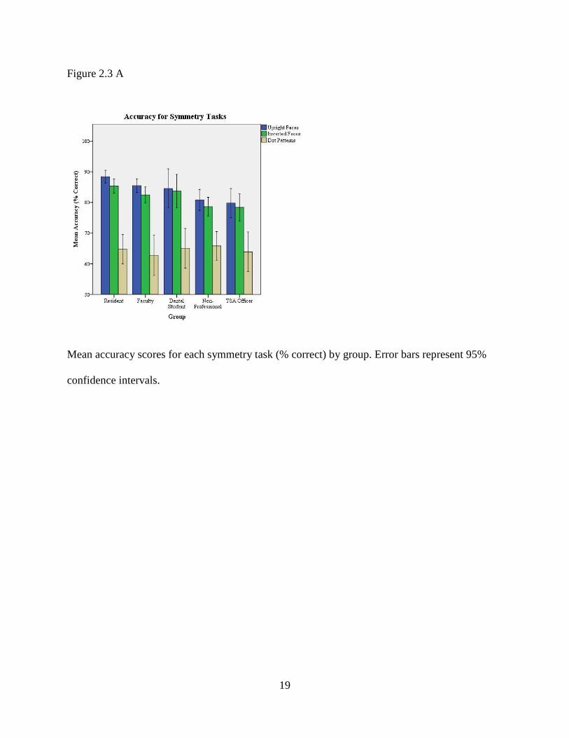
Figure 2.3 A
Mean accuracy scores for each symmetry task (% correct) by group. Error bars represent 95%
confidence intervals.

20
Figure 2.3B
Mean response time for each symmetry task (seconds) by group. Error bars represent 95%
confidence intervals. Note that only dot pattern tasks had a restricted presentation time of
2000ms.

21
Figure 2.3C
Proportion of responses indicating confidence for each symmetry task by group. Royal College
of Physicians Confidence Rating Scale – Responses indicating confidence: 4) Fully confident in
most cases 3) Confident in some cases; Responses not indicating confidence: 2) Satisfactory but
lacking confidence 1) Not confident

22
3. Face Symmetry Assessment Abilities: Clinical Implications for Diagnosing Asymmetry
Authors:
Tate H. Jackson, DDS
Stephen R. Mitroff, PhD
Kait Clark, BS
William R. Proffit, DDS, PhD
Jessica Y. Lee, DDS, MPH, PhD
Tung. T. Nguyen, DMD, MS

23
3.1 INTRODUCTION
Symmetry is an important biologically-based determinant of facial attractiveness,36 and
assessment of symmetry in a patient’s face is paramount to the development of a complete
dentofacial diagnosis. In an era of modern orthodontics when the soft-tissue paradigm and
patient perception often dictate the success of treatment outcomes, it is not acceptable for the
orthodontist to simply identify problems and proceed with treatment.1 An understanding of
individual differences in perception of face symmetry across orthodontists, their patients, and
other providers of dental care is needed to facilitate communication among these groups and to
ensure optimal treatment results. To these ends, recent research has attempted to establish
thresholds for the perception of a problem in face symmetry in different professional and non-
professional groups.
Huisinga-Fischer and co-workers asked participants to objectively compare asymmetry
between different individuals’ faces.18 Their results suggest that orthodontists and surgeons
might judge facial symmetry more accurately than other groups but leave room for alternative
explanations due to the nature of the stimuli and tasks. Specifically, the face stimuli they used
had pathologic deviations from normal symmetry and from normal proportions, and the
participants were asked to rate how the faces differed in terms of deformity from normal rather
than in symmetry explicitly. Accordingly, the role of symmetry in the participant’s judgment was
unclear. Another study related the perception of face symmetry suffers from the fact that
participants rated attractiveness rather than symmetry itself.19 Finally, a study using virtual
“three-dimensional” face stimuli suggested that while thresholds for the perception of an
asymmetric nose or chin exist, orthodontists and oral surgeons show no meaningful advantage in

24
judging face symmetry when compared to laypersons.20 It is important to note that none of these
studies were designed to investigate expertise explicitly.
Of equal importance to the threshold for detection of a problem is an understanding of the
orthodontist’s perceptual ability in general and in relation to the patient. Are orthodontists
experts at assessing face symmetry? To date, the data are equivocal at best because studies have
not been adequately designed to answer that question. 18, 19, 20 If an orthodontist is better at
judging face symmetry than the patient, then he or she may confidently help the patient decide if
treatment is warranted. If the orthodontist is not better at assessing face symmetry, then treatment
outcomes as viewed by the patient may not meet their goals. With the availability of three-
dimensional imaging of facial surfaces, which allows for the exact quantification of facial
symmetry,37, 38 understanding perceptual differences actually may become increasingly relevant.
If an orthodontist understands his or her abilities in relation to the patient, he or she may better
be able to determine when the use of such technology is needed to assist in diagnosis.
The aims of this study were to determine if orthodontists posses expertise in assessing
face symmetry and to explore the nature of this ability with the hope of informing clinical
practice and patient communication. To accomplish these goals, we compared performance on
symmetry judgment tasks across orthodontists, general dentists, and control participants with no
training in face symmetry assessment.
3.2 MATERIALS AND METHODS
Participants
This study was considered exempt from IRB review by the Office of Human Research
Ethics at the University of North Carolina at Chapel Hill and approved as an addition to a

25
separate ongoing study by the Institutional Review Board of Duke University. Orthodontists
(n=31, 8 female, mean age=43.5 years, SD=15.8) were recruited from the University of North
Carolina at Chapel Hill School of Dentistry (UNC) and included residents in various stages of
the three-year program as well as full and part-time faculty members. The faculty participants
reported an average of 27.4 years of clinical practice (SD=12.45). General dentists were also
recruited from the UNC School of Dentistry and included residents and faculty members (n=12,
3 female, mean age=53.1 years, SD=13.2). Orthodontic residents, orthodontic faculty, and
general dentists were all compensated $10/hour for their time.
Control participants without symmetry training were recruited from two sources: non-
professional laypersons from the Duke University community (non-professionals) and TSA
officers employed at Raleigh-Durham International Airport. Non-professionals represent a
population of laypersons without any known special visual skills. Non-professionals (n=23, 13
female age=20.87 years, SD=4.5) were compensated with course credit or paid $10/hour for their
participation. TSA officers represent a population that is known to have enhanced visual
cognition abilities unrelated to face symmetry assessment. 21 The TSA officers (n=10, 2 female,
age=42.33 years, SD=10.20) were not directly compensated as their data were collected during
normal working hours as part of their employment. Participation in this study by the TSA
officers was entirely confidential and voluntary.21 Two additional participants in the TSA group
and one in the non-professional group had overall face accuracy scores that fell two standard
deviations below the mean overall face accuracy score for all participants, and their data were
excluded from all analyses. All participants confirmed 20/20 vision or the use of corrective
lenses at the time of data collection.
Apparatus

26
Data were acquired in three separate locations using identical protocols and
environments: orthodontic resident, orthodontic faculty, and general dentist data were collected
at the UNC School of Dentistry, Duke student data were collected at Duke University in the
Visual Cognition Laboratory, and TSA officer data were collected at Raleigh-Durham
International Airport in a private testing room. The experiment was run in a dimly lit room;
participants at Duke and UNC viewed the experiments on a Dell Inspiron computer with a 20-
inch CRT monitor, and participants at RDU viewed the experiments on Dell Vostro 260
computers and 23.6-inch computer displays that were adjusted so all participants were presented
with stimuli of the same physical size. Participants were seated at a viewing distance of
approximately 57 cm with no head restraint. Stimuli were presented and responses were recorded
using MATLAB (The MathWorks, Natick, MA) using the Psychophysics Toolbox (Version
3.0.8, Brainard, 1997; Pelli, 1997; Kleiner, Brainard, & Pelli, 2007). Questionnaire data were
collected using the Qualtrics Research Suite (Qualtrics Labs, Inc., 2012).
Procedures and Stimuli
All participants completed three visual cognition tasks related to symmetry and presented
in a blocked design; order was counterbalanced across all participants and tasks. Each task began
with a series of practice trials, which were immediately followed by the experimental segment
during which trial-by-trial accuracy and response time were recorded. At the start of each trial, a
fixation cross was presented for 500ms, followed by the stimulus. Participants responded to each
trial with one of two possible keys, and no feedback was provided.
Task 1: Symmetry assessment of upright faces

27
Participants assessed symmetry in 96 trials of upright faces by making a two-alternative
forced-choice judgment between two versions of the same face presented side by side (see Figure
3.1A). Stimuli were presented on a black background, and the participant was instructed to press
the ‘z’ key if the face on the left appeared more symmetric and the ‘/’ key if the face on the right
appeared more symmetric. Stimuli were presented until the participant responded. Trials were
counter-balanced for each participant as to whether the right or left face was more symmetric.
Stimuli consisted of black-and-white photographs of faces of sixteen (8 female) Caucasian
individuals morphed to varying levels of asymmetry while preserving averaged proportions (see
Rhodes, Proffit, Grady, & Sumich, 1998 for details on stimuli generation).22 Veridical hairstyles
(i.e. the unaltered hairstyle) were maintained for all versions of each face by editing the original
stimuli set from Rhodes using Adobe® Photoshop Elements10® so that the hair could not be
used as a cue to symmetry. Four versions of each face, varying in symmetry were used: the
veridical (original) face, the face with perfect symmetry, the face with symmetry increased 50%
from the veridical, and the face with symmetry decreased 50% from the veridical (See Figure
3.2). By pairing each face version with all the iterations of that face, six possible pairings were
created (veridical with perfect symmetry, veridical with high symmetry, veridical with low
symmetry, high symmetry with perfect symmetry, high symmetry with low symmetry, and low
symmetry with perfect symmetry). These stimuli were presented at random in terms of both the
levels of symmetry being compared and the individual’s face that was used. Participants viewed
all possible pairings of each face during Task 1.
Task 2: Symmetry assessment of inverted faces
This task was identical to Task 1, but all stimuli were presented upside-down. The
sequence of presentation was randomized separately from Task 1.

28
Task 3: Symmetry detection in dot patterns
Participants judged whether a dot pattern presented as a centered image on a black
background was perfectly symmetric about its vertical axis (see Figure 3.1B). Each dot image
was displayed for 2000ms, after which participants were asked to make a response using the ‘z’
key to indicate that the dot pattern was symmetric and the ‘/’ key to indicate that the dot pattern
was not symmetric. The 2000ms display time was used to maintain consistency with a previously
used experimental protocol.24 Stimuli were 18 dot patterns based on the body patterns of animal
with bilaterally symmetric bodies (see Evans, Wenderoth, & Cheng, 2000 for details).25 Each
pattern was presented in random order in both upright and inverted conditions for a total of 36
trials.
Immediately following the completion of all visual tasks, each participant completed a
web-based questionnaire that asked about demographic information, strategies employed during
symmetry assessment, and subjective confidence self-ratings for the tasks completed using the
Royal College of Physicians Confidence Rating Scale.26 Orthodontists and general dentists were
asked whether the patient is most often upright or inverted when they assess face symmetry
clinically and about duration of training or clinical practice. Non-professionals and TSA officers
were asked whether they had any training or experience in symmetry assessment.
3.3 RESULTS
Descriptive statistics for accuracy, response time, and confidence ratings for each task
may be found in Table 3.1. Normality of data was confirmed using Q-Q plots and homogeneity
of variance between groups was confirmed using Levenes’s test. Accuracy and response times
were compared among groups using one-way analysis of variance and Tukey’s HSD. Within

29
subject comparisons were made using paired t-tests or Wilcoxon signed rank tests (for
confidence ratings); statistical significance was set at p=0.05.
Orthodontists showed a statistically significant advantage in judging face symmetry
overall (F=6.6, p=0.001) and in both upright (F=5.9, p=0.001) and inverted (F=4.8, p=0.004)
face conditions compared to both non-professionals and TSA officers (Tukey’s HSD p<0.05 for
all comparisons), but not compared to general dentists. General dentists did not show a
significant difference in accuracy overall for assessing symmetry in faces, whether upright or
inverted, when compared to non-professionals or TSA officers. In the most difficult trials (those
in which the differences in symmetry between faces were smallest; e.g. perfect symmetry
compared to high symmetry), orthodontists showed a significant advantage (F=9.2, p<0.001)
over general dentists (p=0.01) as well as both non-professionals (p<0.001) and TSA officers
(p=0.002).
There was a significant difference in response time overall (F=7.2, p<0.001), and for
both upright (F=7.9, p<0.001) and inverted (F=4.7, p=0.005) faces between orthodontists and
general dentists compared to non-professionals (Tukey’s HSD p<0.02 for all comparisons), but
not between orthodontists and general dentists compared to TSA officers. Non-professionals
took less time to judge symmetry than orthodontists, general dentists, or TSA officers. There
were no other differences in response time among any groups.
There were no statistically significant differences in accuracy or response time for dot
stimuli among any of the groups.
Orthodontists showed within-subject differences in accuracy between each pair of tasks:
upright vs. inverted faces (t=3.7, p=0.001), upright faces vs. dot patterns (t=10.7, p<0.001),

30
inverted faces vs. dot patterns (t=10.6, p<0.001). General dentists also showed an advantage in
accuracy for upright vs. inverted faces (t=2.7, p=0.02).
Orthodontists also demonstrated significant differences in confidence ratings for each
pair of tasks: upright vs. inverted faces (z=4.5, p<0.001), upright faces vs. dot patterns (z=4.3,
p<0.001), inverted faces vs. dot patterns (z=2.5, p=0.01). No other groups showed significant
differences in confidence ratings for all pairs of tasks.
All but four participants in the orthodontist group (n=31) and two in the general dentist
group (n=12) reported clinically assessing symmetry with the patient upright. No non-
professionals or TSA officers reported training or experience in judging face symmetry.
3.4 DISCUSSION
Orthodontic Expertise
Our results indicate that orthodontists show a clear advantage in assessing face symmetry
compared to laypersons, and an advantage over general dentists in the most difficult cases. One
might suggest that the orthodontists’ enhanced performance is the result of motivation bias; they
were simply trying harder because this was an evaluation of a skill that they knew they should
possess. An appraisal of response time rules out that possibility (see Figure 3.3B). Orthodontists
took longer, on average, to respond when judging face symmetry than non-professionals. TSA
officers took just as long as orthodontists, however, and they were significantly worse at
assessing symmetry. Despite the fact that they took significantly longer to respond, the TSA
officer group’s accuracy matched that of the non-professionals. In short, increased response time,
which is a logical indicator of motivation, does not equate to greater accuracy. Orthodontists
truly appear to have an enhanced skill.

31
Faces as Special Visual Stimuli
It is important to note, however, that all participants, whether orthodontists, general
dentists, or untrained controls, showed accuracy scores that indicate some aptitude in judging
face symmetry. Even the lowest mean accuracy score of 78.4% (see Table 3.1) represents a real
increase above the 50% score one might expect from random chance alone given that the face
symmetry tasks all included only two possible responses. That all of our participants showed
some skill may be due to the nature of faces and how humans tend to perceive them. Faces are
ubiquitous visual stimuli, and behavioral and neuroimaging research has indicated that they are
processed by special cognitive mechanisms in the brain which provide an advantage in
perceptual abilities when it comes to looking at and evaluating faces (for a review see Kanwisher
et al 2006).5 Face symmetry also is likely to be governed by special processes which provide a
perceptual advantage compared to the inspection of symmetry in non-face objects, such as dot
patterns or teeth.6, 7, 39
Inverted Faces and Symmetry Assessment
One aspect of face symmetry processing that is of interest to orthodontists and general
dentists is that the orientation of the face when symmetry is judged appears to have a significant
effect. Both orthodontists and general dentists were significantly better at assessing symmetry in
upright faces compared to inverted faces. This finding supports research that when a face is
inverted, it is not fully processed by the usual neural pathways of the brain that provide a
perceptual advantage.7, 39, 40It may be clinically meaningful that orthodontists and general
dentists are better at judging symmetry when a face is upright. When administering dental care,
the patient is often reclined, and their face is inverted. Our data support the recommendation that

32
the patient should be upright when face symmetry is assessed in order to achieve maximum
accuracy. Interestingly, all but four orthodontists and two general dentists reported that their
routine clinical practice included assessment of face symmetry with the patient upright rather
than inverted.
Implications for Clinical Practice
It is important to remark that the clinical setting for which our data are most applicable
involves the longitudinal comparison of patient records when they are being monitored for a
progressive asymmetric deformity. Our experimental design, comparing two versions of the
same person’s face, is most similar to the clinical activity of comparing accurate standardized
photographic records taken over time for diagnostic purposes.
In an era when surface-scanning 3D technology allows the computer-assisted assessment
of face symmetry and changes in it over time41, 42 our data may also be an aid to understanding
when technology such as this is needed. In a setting in which a patient’s records are being
compared over time, our data suggests that orthodontists will correctly identify symmetry in 87%
of the cases, if the faces are viewed in an upright position using accurate photographs. So,
perhaps less than 15% of these situations require the aid of additional tools, such as surface 3D
superimpositions, to identify progressive aberrations in face symmetry.
Recent studies using 3D laser surface scanning estimate the overall asymmetry in
normally developing child and adolescent faces to range on average from 8% - 68% with
standard deviations as high as nearly 14%.37, 38, 43, 44 Interestingly, our study used stimuli that
varied the symmetry of the whole face from 50% less symmetric than the true face to perfect
symmetry. Accordingly, the stimuli we used provide a reasonable representation of the range of

33
asymmetry encountered in clinical practice, and our data give further insight to the nature of face
symmetry judgment both by clinicians and laypersons.
The relative skill of all participants in our study has implications for patient
communication. Providers should be aware that faces are unique visual stimuli45 and that judging
symmetry in faces is a perceptual process distinct from other tasks to evaluate dentofacial
esthetics. While orthodontists are experts at face symmetry assessment, patients are likely to
posses some inherent skill as well. Similarly, orthodontists are only better at judging face
symmetry than general dentists in difficult cases. In our study, this difference became evident
when the dissimilarity between stimuli was only a 25% change in overall face symmetry (see
Figure 3.2). Orthodontists may use this evidence as a framework for understanding the
perceptual abilities of both patients and colleagues with whom they communicate.
Orthodontists may also use our evidence as a basis for understanding their own abilities.
Confidence ratings for symmetry tasks followed performance for this group (see Figure 3.3 A,
C). That is to say that orthodontists rated themselves most confident in the tasks for which they
were most accurate and least confident in the tasks where they performed the worst. An ability to
accurately evaluate one’s own performance is an indicator of expertise 35 and consistent with this
tenet, general dentists, non-professionals, and TSA officers failed to show the same pattern. Our
evidence suggests that an orthodontist’s self-assessment of performance is more likely to be
consistent with their actual accuracy than untrained laypersons or general dentists making the
same judgment. This finding may facilitate the reconciliation of differences in face symmetry
perception between patient and provider.

34
3.5 CONCLUSIONS
Orthodontists demonstrate robust expertise in assessing face symmetry when compared to
laypersons, and expertise in only the most difficult judgments compared to general dentists.
Both orthodontists and general dentists show a significant advantage judging face
symmetry with upright compared to inverted faces.
When photographic patient records are being compared over time, our data suggests that
orthodontists will incorrectly identify symmetry in less than 15% of these situations.

3.6 TABLES AND FIGURES
Table 3.1
Figure 3.1A
Orthodontists (n=31)
All Faces 85.5 (3.8)Upright Faces 87.0 (4.1)Inverted Faces 84.0 (4.7)Dots 63.8 (10.3)
Response Time mean (std. dev.)
All Faces 7.5 (3.0)Upright Faces 7.6 (3.3)Inverted Faces 7.4 (4.2)Dots 0.9 (0.40)
Confidence % repsonses indicating confidence
Upright Faces 100Inverted Faces 61.3Dots 41.9
Accuracy (%correct), Response Time (sec.), and Confidence Scores for Symmetry Tasks
Accuracy mean (std. dev.)
a Royal College of Physicians Confidence Rating Scale - 4: Fully confident in most cases 3:
Confident in some cases 2: Satisfactory but lacking confidence 1: Not confident (responses of 3 or 4 indicate confidence)
35
Orthodontists (n=31)
General Dentists (n=12)
Non-Professionals (n=23)
TSA Officers (n=10)
85.5 (3.8) 82.4 (5.0) 79.7 (6.7) 79.1 (6.0)87.0 (4.1) 84.8 (6.8) 80.8 (7.9) 79.8 (6.7)84.0 (4.7) 80.0 (4.8) 78.6 (7.1) 78.4 (6.2)63.8 (10.3) 61.6 (6.1) 65.8 (10.8) 63.9 (9.1)
7.5 (3.0) 8.5 (7.7) 3.3 (1.5) 7.7 (3.9)7.6 (3.3) 8.3 (6.6) 3.2 (1.6) 7.7 (4.4)7.4 (4.2) 8.8 (9.3) 3.3 (1.5) 7.6 (4.0)0.9 (0.40) 0.8 (0.2) 0.6 (0.3) 0.9 (0.5)
% repsonses indicating confidence a
100 91.7 91.3 7061.3 75 52.1 5041.9 66.7 52.1 70
Accuracy (%correct), Response Time (sec.), and Confidence Scores for Symmetry Tasks
Royal College of Physicians Confidence Rating Scale - 4: Fully confident in most cases 3: Confident in some cases 2: Satisfactory but lacking confidence 1: Not confident (responses of 3
TSA Officers (n=10)
79.1 (6.0)79.8 (6.7)78.4 (6.2)63.9 (9.1)
7.7 (3.9)7.7 (4.4)7.6 (4.0)0.9 (0.5)
Accuracy (%correct), Response Time (sec.), and Confidence Scores for Symmetry Tasks
Royal College of Physicians Confidence Rating Scale - 4: Fully confident in most cases 3: Confident in some cases 2: Satisfactory but lacking confidence 1: Not confident (responses of 3

36
Figure 3.1B
Example stimuli and instructions presented to participants for (A) Tasks 1 and 2 and (B) Task 3.
The face stimuli of Tasks 1 and 2 were presented with no time constraints, whereas the dot
patterns used in Task 3 were presented for 2000ms (followed by the instructions screen that
remained until response).

Figure 3.2
Example of face stimuli showing four morphed versions of one individual’s
(veridical), a version 50% less symmetric (low symmetry), a version 50% more symmetric (high
symmetry), and a version with perfect symmetry. Note that the hairstyle for each version of the
face is the identical, veridical hairstyle.
37
Example of face stimuli showing four morphed versions of one individual’s face: the actual face
(veridical), a version 50% less symmetric (low symmetry), a version 50% more symmetric (high
symmetry), and a version with perfect symmetry. Note that the hairstyle for each version of the
face is the identical, veridical hairstyle.
face: the actual face
(veridical), a version 50% less symmetric (low symmetry), a version 50% more symmetric (high
symmetry), and a version with perfect symmetry. Note that the hairstyle for each version of the

38
Figure 3.3A
Mean accuracy scores for each symmetry task (% correct) by group. Error bars represent 95%
confidence intervals.

39
Figure 3.3B
Mean response time for each symmetry task (seconds) by group. Error bars represent 95%
confidence intervals. Note that only dot pattern tasks had a restricted presentation time of
2000ms.

40
Figure 3.3C
Proportion of responses indicating confidence for each symmetry task by group. Royal College
of Physicians Confidence Rating Scale – Responses indicating confidence: 4) Fully confident in
most cases 3) Confident in some cases; Responses not indicating confidence: 2) Satisfactory but
lacking confidence 1) Not confident

41
4. CONCLUSION
Longitudinal data following individuals from pre-dentistry, through dental school,
residency, and clinical practice is required to make definitive statements regarding the relative
contributions of training, experience, and potential inherent ability to the acquisition of clinical
skills. Until that data is available, clinical dental education, whether pre- or post-doctoral, should
be targeted to provide more than simple repeated experiences. When considering the acquisition
of a diagnostic skill, such as face symmetry assessment, the effect of testing by providing critical
feedback during training may enhance learning in a meaningful way. Accurate self-assessment is
one facet expertise and may be an important end-point for the evaluation of clinical skill
acquisition. Incorporating these principles into clinical curricula and methodology will enhance
dental education.
Orthodontists demonstrate robust expertise in assessing face symmetry when compared to
laypersons, and expertise in only the most difficult judgments compared to general dentists. Both
orthodontists and general dentists show a significant advantage judging face symmetry with
upright compared to inverted faces. When photographic patient records are being compared over
time, our data suggests that orthodontists will incorrectly identify symmetry in less than 15% of
these situations.

42
REFERENCES
1. Proffit WR, Fields HW, Sarver DM, Ackerman JL. Contemporary orthodontics. 5th ed. St. Louis, MO: Elsevier; 2012.
2. Rosenstiel S, Land M, Fujimoto J. Contemporary fixed prosthodontics. 4th ed. St. Louis, MO: Elservier; 2006.
3. Contemporary oral and maxillofacial surgery. 5th ed. Hupp J, Ellis III E, Tucker M, editors. St. Louis, MO: Elsevier; 2008.
4. Little J, Falace D, Miller C, Rhodus N. Dental management of the medically compromised patient. 7th ed. St. Louis, MO: Elsevier; 2008.
5. Kanwisher N, Yovel G. The fusiform face area: A cortical region specialized for the perception of faces. Philos Trans R Soc Lond B Biol Sci. 2006 Dec 29;361(1476):2109-28.
6. Chen CC, Kao KL, Tyler CW. Face configuration processing in the human brain: The role of symmetry. Cereb Cortex. 2007 Jun;17(6):1423-32.
7. Rhodes G, Peters M, Lee K, Morrone MC, Burr D. Higher-level mechanisms detect facial symmetry. Proc Biol Sci. 2005 Jul 7;272(1570):1379-84.
8. Ranney RR, Wilson MB, Bennett RB. Evaluation of applicants to predoctoral dental education programs: Review of the literature. J Dent Educ. 2005 Oct;69(10):1095-106.
9. Urbankova A, Engebretson SP. The use of haptics to predict preclinic operative dentistry performance and perceptual ability. J Dent Educ. 2011 Dec;75(12):1548-57.
10. Nick DR, Clark M, Miler J, Ordelheide C, Goodacre C, Kim J. The ability of dental students and faculty to estimate the total occlusal convergence of prepared teeth. J Prosthet Dent. 2009 Jan;101(1):7-12.
11. El-Housseiny AA, Jamjoum H. Evaluation of visual, explorer, and a laser device for detection of early occlusal caries. J Clin Pediatr Dent. 2001 Fall;26(1):41-8.
12. Fung L, Smales R, Ngo H, Moun G. Diagnostic comparison of three groups of examiners using visual and laser fluorescence methods to detect occlusal caries in vitro. Aust Dent J. 2004 Jun;49(2):67,71; quiz 101.
13. Fyffe HE, Deery C, Nugent ZJ, Nuttall NM, Pitts NB. In vitro validity of the dundee selectable threshold method for caries diagnosis (DSTM). Community Dent Oral Epidemiol. 2000 Feb;28(1):52-8.

43
14. Bengtson AL, Gomes AC, Mendes FM, Cichello LR, Bengtson NG, Pinheiro SL. Influence of examiner's clinical experience in detecting occlusal caries lesions in primary teeth. Pediatr Dent. 2005 May-Jun;27(3):238-43.
15. Zandona AG, Al-Shiha S, Eggertsson H, Eckert G. Student versus faculty performance using a new visual criteria for the detection of caries on occlusal surfaces: An in vitro examination with histological validation. Oper Dent. 2009 Sep-Oct;34(5):598-604.
16. Hellen-Halme K, Petersson GH. Influence of education level and experience on detection of approximal caries in digital dental radiographs. an in vitro study. Swed Dent J. 2010;34(2):63-9.
17. Dimitrijevic T, Kahler B, Evans G, Collins M, Moule A. Depth and distance perception of dentists and dental students. Oper Dent. 2011 Sep-Oct;36(5):467-77.
18. Huisinga-Fischer CE, Souren JP, v d Werken F, Prahl-Andersen B, van Ginkel F. Perception of symmetry in the face. J Craniofac Surg. 2004 Jan;15(1):128-34.
19. Naini FB, Donaldson AN, McDonald F, Cobourne MT. Assessing the influence of asymmeftry affecting the mandible and chin point on perceived attractiveness in the orthognathic patient, clinician, and layperson. J Oral Maxillofac Surg. 2012 Jan;70(1):192-206.
20. Meyer-Marcotty P, Stellzig-Eisenhauer A, Bareis U, Hartmann J, Kochel J. Three-dimensional perception of facial asymmetry. Eur J Orthod. 2011 Dec;33(6):647-53.
21. Biggs A, Cain M, Clark K, Darling E, Mitroff S. Assessing visual search performance differences between transportation security adiminstration officers and non-professional visual searchers (under review). In press .
22. Rhodes G, Proffit F, Grady JM, Sumich A. Facial symmetry and the perception of beauty. Psychnomic Bulletin and Review. 1998;5(4):659-69.
23. Valentine T. Upside-down faces: A review of the effect of inversion upon face recognition. Br J Psychol. 1988 Nov;79 ( Pt 4)(Pt 4):471-91.
24. Oinonen KA, Mazmanian D. Facial symmetry detection ability changes across the menstrual cycle. Biol Psychol. 2007 May;75(2):136-45.
25. Evans CS, Wenderoth P, Cheng K. Detection of bilateral symmetry in complex biological images. Perception. 2000;29(1):31-42.
26. George JT, Warriner D, McGrane DJ, Rozario KS, Price HC, Wilmot EG, et al. Lack of confidence among trainee doctors in the management of diabetes: The trainees own perception of delivery of care (TOPDOC) diabetes study. QJM. 2011 Sep;104(9):761-6.
27. Roediger HL, Karpicke JD. Test-enhanced learning: Taking memory tests improves long-term retention. Psychol Sci. 2006 Mar;17(3):249-55.

44
28. McDaniel MA, Roediger HL,3rd, McDermott KB. Generalizing test-enhanced learning from the laboratory to the classroom. Psychon Bull Rev. 2007 Apr;14(2):200-6.
29. Butler AC. Repeated testing produces superior transfer of learning relative to repeated studying. J Exp Psychol Learn Mem Cogn. 2010 Sep;36(5):1118-33.
30. Larsen DP, Butler AC, Roediger HL,3rd. Repeated testing improves long-term retention relative to repeated study: A randomised controlled trial. Med Educ. 2009 Dec;43(12):1174-81.
31. Karpicke JD, Blunt JR. Retrieval practice produces more learning than elaborative studying with concept mapping. Science. 2011 Feb 11;331(6018):772-5.
32. Jackson TH, Hannum WH, Koroluk L, Proffit WR. Effectiveness of web-based teaching modules: Test-enhanced learning in dental education. J Dent Educ. 2011 Jun;75(6):775-81.
33. Larsen DP, Butler AC, Lawson AL, Roediger HL,3rd. The importance of seeing the patient: Test-enhanced learning with standardized patients and written tests improves clinical application of knowledge. Adv Health Sci Educ Theory Pract. 2012 May 23.
34. Kromann CB, Bohnstedt C, Jensen ML, Ringsted C. The testing effect on skills learning might last 6 months. Adv Health Sci Educ Theory Pract. 2010 Aug;15(3):395-401.
35. Kruger J, Dunning D. Unskilled and unaware of it: How difficulties in recognizing one's own incompetence lead to inflated self-assessments. J Pers Soc Psychol. 1999 Dec;77(6):1121-34.
36. Rhodes G. The evolutionary psychology of facial beauty. Annu Rev Psychol. 2006;57:199-226.
37. Djordjevic J, Toma AM, Zhurov AI, Richmond S. Three-dimensional quantification of facial symmetry in adolescents using laser surface scanning. Eur J Orthod. 2011 Jul 27.
38. Primozic J, Perinetti G, Zhurov A, Richmond S, Ovsenik M. Assessment of facial asymmetry in growing subjects with a three-dimensional laser scanning system. Orthod Craniofac Res. 2012 Nov;15(4):237-44.
39. Rhodes G, Peters M, Ewing LA. Specialised higher-level mechanisms for facial-symmetry perception: Evidence from orientation-tuning functions. Perception. 2007;36(12):1804-12.
40. Jones RM, Victor JD, Conte MM. Detecting symmetry and faces: Separating the tasks and identifying their interactions. Atten Percept Psychophys. 2012 Jul;74(5):988-1000.
41. Kau CH, Zhurov A, Scheer R, Bouwman S, Richmond S. The feasibility of measuring three-dimensional facial morphology in children. Orthod Craniofac Res. 2004 Nov;7(4):198-204.
42. Souccar NM KC. Methods of measuring the three-dimensional face. Sem Orthod. 2012;18(3 (September)):187-92.

45
43. Djordjevic J, Pirttiniemi P, Harila V, Heikkinen T, Toma AM, Zhurov AI, et al. Three-dimensional longitudinal assessment of facial symmetry in adolescents. Eur J Orthod. 2011 Feb 7.
44. Primozic J, Perinetti G, Richmond S, Ovsenik M. Three-dimensional evaluation of facial asymmetry in association with unilateral functional crossbite in the primary, early, and late mixed dentition phases. Angle Orthod. 2012 Aug 13.
45. Masella RS, Meister M. The neuroanatomic basis of facial perception and variable facial discrimination ability: Implications for orthodontics. Am J Orthod Dentofacial Orthop. 2007 Sep;132(3):293-301.