Interactions ofAvocado(Persea americana) Cytochrome P-450withMonoterpenoids
Volume 52(2):257-265, 2008Acta Biologica Szegediensis
http://www.sci.u-szeged.hu/ABS
Article
1Institute of Biophysics, Biological Research Center, Hungarian Academy of Sciences, Szeged, Hungary, 2Department of Biology, University of Antwerp, Antwerp, Belgium
expression and purification of the recombinant mouse tumor supressor cytochrome b561 protein
Alajos Bérczi1*, Han Asard2
ABStrAct It has recently been recognized that ascorbate-reducible cytochrome b561 (Cyt-b561) proteins constitute a well-distinguished protein family amongst the two-heme containing b-type cytochromes, ubiquitously present in animals and plants. Of the six isoforms that have been identified in mammals, three isoformes (called CGCytb, DCytb, and LCytb) have been cloned and expressed in yeast and/or bacterial cells. The recombinant proteins have been characterized in some detail. A particular gene product of the 3p21.3 (human) and 9F1 (mouse) chromosomal region, a so-called tumor supressor protein (101F6, TSP10), was identified as a Cyt-b561 protein by sequence homology. We have cloned and expressed the mouse tumor supressor Cyt-b561 protein (TSCytb) in yeast (Saccharomyces serevisiae), without and with a His6-tag on either the N- or the C-terminus. The C-terminal His6-tagged recombinant protein was purified on Ni-NTA His•Bind resin to almost homogeneity. Using optical spectroscopy we show that TSCytb is in-deed an ASC-reducible cytochrome b561 protein and that ASC-reducibility is not affected by the presence of a His6-tag on the C-terminus. Minor differences in the properties of TSCytb and the other three mammalian Cyts-b561 are discussed. Acta Biol Szeged 52(2):257-265 (2008)
Key WordS
101F6ASCrecombinant cytochrome b561mouseTSP10tumor suppressor proteinyeast expression
Accepted Sept 19, 2008*Corresponding author. E-mail: [email protected]
257
b-Type cytochromes are metallo proteins containing one or more iron:protoporphyrine-IX complexes as a chromophore. A recently discovered group of b-type cytochromes, called cy-tochromes b561 (Cyts-b561), consists of (i) ascorbate(ASC)-reducible, (ii) trans-membrane proteins with six trans-mem-brane helices, which (iii) have two heme b centers (iv) coor-dinated in a particular way by four well-conserved histidine residues to four consecutive trans-membrane α-helices (Bashtovyy et al. 2003; Tsubaki et al. 2005). The two heme b prosthetic groups, with different redox potential values, reside on opposite sides of the trans-membrane proteins making them ideal for serving as trans-membrane electron transporters. The Cytb-561 that was discovered first is the chromaffin granule Cyt-b561 that had already been known as chromomembrin B (Flatmark and Terland 1971; Silsand and Flatmark 1974; Apps et al. 1980). This protein functions as an electron transporter providing electrons from cytosolic ASC through the chromaffin granule membrane to intravesicular dopamine β-hydroxylase (Njus and Kelley 1993; Njus et al. 2001). After resolving the full genomic sequence of a large variety of organisms, and improving in silico techniques for comparing gene and protein sequences, numerous proteins were identified similar to CGCytb in various organisms, including invertebrates, vertebrates, and plants (Verelst and Asard 2003; Tsubaki et al. 2005). Three mammalian and one
plant Cyts-b561, namely the chromaffin granule Cyt-b561 (CGCytb), the duodenal Cyt-b561 (DCytb; [McKie et al. 2001]), the lysosomal Cyt-b561 (LCytb; [Zhang et al. 2006]), and the tonoplast Cyt-b561 (TCytb; [Griesen et al. 2004]), showed ASC-dependent trans-membrane ferrireductase activity when expressed in yeast cells (Su and Asard 2006; Bérczi et al. 2007). However, CGCytb is participating in ASC regeneration in chromaffin granules (Kent and Fleming 1987; Njus and Kelley 1993) while another isoform, DCytb, appears to be involved in extracellular recycling of ASC of human erythrocytes (Su et al. 2006). Only DCytb has already been shown to be involved in iron metabolism (McKie et al. 2001; Vargas et al. 2003). The biological function of LCytb and TCytb has not yet been elucidated.
At the level of genomic and predicted protein sequences, a human tumor supressor protein (the 101F6 gene product) has been identified as putative member of the Cyts-b561 (Lerman and Minna 2000; Ponting 2001). The mouse homologue was also discovered, sequenced, and shown to be 85% and 95% identical with the human sequences on the cDNA and protein sequence level, respectively (Lerman and Minna 2000). Both proteins have (i) 6 trans-membrane α-helices with (ii) 222 residues, (iii) the N- and C-termini in the cytoplasm, and (iv) 4 well-conserved histidine residues for binding two heme b prosthetic groups. 101F6 mRNA is widely expressed in animal tissues, and the mouse mRNA is especially abundant in liver, kidney, and lung (Mizutani et al. 2007), while the human protein is most abundant in liver, placenta, and lung
258
Bérczi, Asard
(Lerman and Minna 2000). Expression of wild-type101F6 in tumor cells significantly inhibited cell growth and intra-tumoral injection of recombinant adenovirus-101F6 gene vectors, as well as systemic administration of protamine-complexed adenovirus-101F6 gene vectors, significantly suppressed tumor xenografts growth (Ji et al. 2002). Ohtani et al. (2007) have recently found that nanoparticle-mediated 101F6 gene transfer and a subpharmacologic concentration of ASC synergistically and selectively inhibited tumor cell growth by caspase-independent apoptosis and autophagy in vitro and in vivo. C terminal myc-tagged mouse 101F6 protein has been expressed in Chinese hamster ovary (CHO) cells and immunofluorescence microscopy was used to lo-calize the recombinant proteins; they were found in small vesicles, including endosomes and endoplasmic reticulum of the perinuclear region (Mizutani et al. 2007). It was also shown that CHO cells expressing the C terminal myc-tagged mouse 101F6 protein showed higher ferric ion and azo-dye reduction level than the control CHO cells. Mizutani et al. (2007) concluded that mouse 101F6 proteins played roles in the ferrireduction via a yet unresolved mechanism.
In this paper we report on the cloning and expression of the mouse 101F6 gene in a yeast expression system, both with and without a His
6-tag on either the C-terminus or the
N-terminus. It is shown that the recombinant mouse tumor suppressor protein is indeed an ASC-reducible b-type cyto-chrome and the His
6-tag on the C-terminus does not affect
ASC-reducibility.
Materials and Methods
chemicals
The protease inhibitor cocktail tablet „cOmplete”, NBT (Nitro blue tetrazolium chloride), BCIP (5-Bromo-4-chloro-3-indolyl phosphate, toluidine salt) in 67% DMSO (v/v), were from Roche (Mannheim, Germany); Sucrose monolaurate (SML) was from Dojindo (Tokyo, Japan); Ni-NTA His•Bind Resin was from Novagen (Darmstadt, Germany); DifcoTM yeast nitrogen base w/o amino acids was from BD (Sparks, MD, USA); other chemicals were analytical grade and from Acros (Geel, Belgium) or Fluka (Buchs, Switzerland) or Serva (Heidelberg, Germany).
Molecular biology works
For expression of untagged proteins, the cDNA of mouse TSCytb (the sequence corresponds to GenBank protein entry, NP_062694; see Fig. 1) was amplified from mouse whole brain RNA and was cloned into a pESC-His expres-sion vector (Stratagene, La Jolla. CA), downstream of the GAL10 galactose-inducible promoter, using EcoRI and SpeI restriction sites. Yeast cells (Saccharomyces cerevisiae, strain YPH499: ura3-52 lys2-801amberade2-101ochre trp1-Δ63 his3-Δ200 leu2-Δ1) were transformed and grown according to the manufacturer’s instructions (Stratagene).
For expression of His6-tagged proteins, a standard PCR
method was used to amplify the gene for TSCytb from mouse
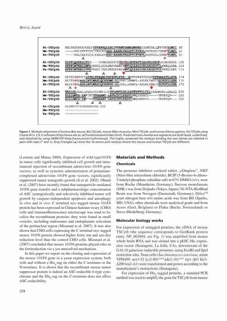
Figure 1. Multiple alignment of bovine (Bos taurus, Bt) CGCytb, mouse (Mus musculus, Mm) TSCytb, and human (Homo sapiens, Hs) TSCytb using Clustal W (v. 2.0.1) software (http://www.ebi.ac.uk/Tools/clustalw2/index.html). Predicted trans-membrane segments are bold faced, underlined, and obtained by using HMMTOP (http://www.enzim.hu/hmmtop/). The highly conserved His residues binding the two hemes are labelled in pairs with stars (* and +). Gray triangles (▲) show the 10 amino acid residues where the mouse and human TSCytb are different.

259
Tumor supressor cytochrome b561
whole brain RNA. Primers were designed to generate EcoRI and SpeI sites for cloning into the pESC-His expression vec-tor (Stratagene, La Jolla, CA), downstream of the GAL10 galactose-inducible promoter, and with a His
6 tag either on
the C- or the N-terminus. Four oligonucleotide primers were synthesized and used for the amplification:
C-terminus, sense: 5’-GGGGGAATTCGCCACCATG-GCCCTTTCTGTGGAGACG-3’
C-terminus, antisense: 5’-CCCCACTAGTCTAGTGATGGTGATGATGGTGT-
GGCTGGATCCTCTTGCGGT-3’N-terminus, sense:5’-GGGGGAATTCACCATGCACCATCATCACCAT-
CACGCCCTTTCTGTGGAGACGGAG-3’N-terminus, antisense: 5’-CCCCACTAGTTCATGGCT-
GGATCCTCTTGCGGT-3’First, the PCR-amplified products were inserted into
pGEM-T Easy vectors (Promega Corp., Madison, WI, USA), and the resulting vectors were transformed into E. coli DH5α cells using calcium chloride competent cells and the Nucleo-Spin Plasmid QuickPure kit (Macherey-Nagel GmbH & Co., Düren, Germany). The pGEM-T Easy vectors with the inserts were isolated with the NucleoSpin Plasmid QuickPure kit and inserted into empty pESC-His vectors by using the restriction enzymes EcoRI and SpeI. The integrity of the His
6-tagged
TSCytb/pESC-His vectors was confirmed by restriction digestion and DNA sequencing. Finally, pESC-His verctors were transformed into competent yeast cells (Saccharomyces cerevisiae, strain YPH499, ura3-52 lys2-801amberade2-101ochre trp1-Δ63 his3-Δ200 leu2-Δ1) and the transformed cells were tested on SD-dropout medium (http://www.stratagene.com/manuals/217451.pdf) where the lack of histidine was the selective factor. For induction of protein expression and pro-duction of the recombinant proteins, overnight cultures were grown in SD-dropout medium, transferred to SG-dropout medium (http://www.stratagene.com/manuals/217451.pdf) and grown for 24-28 hours.
yeast membrane preparation
Cells grown as 2 x 350 ml cultures in SG-His medium were collected by low-speed centrifugation (at 4,000 g
max and
10°C for 10 min) when the OD600
(d=1cm) reached a value of 0.8 ± 0.05. Collected cells were washed once with ice cold phosphate buffer (20 mM KH
2PO
4, 100 mM NaCl, pH 7) and
pelleted as above. Washed cells were resuspended in 15 ml of ice cold homogenization buffer (50 mM MES-KOH, pH 7.0, 5 mM EDTA, 150 mM KCl, 150 mM sucrose) supple-mented with freshly added 0.1% (w/v) Na-ASC and protease inhibitors (1 mM PMSF and aprotinin, pepstatin, leupeptin, antipain, chymostatin in the form of 1 dissolved cOmplete tablet). Cells were broken in a Bead Beater (Biospec Prod-ucts, Bartlesville, OK, USA) with four 30 s cycles, with 2 min cooling intervals, using 0.5 mm glass beads. Beads were
rinsed with 35 ml of ice cold homogenization buffer. The homogenate (50 ml) was centrifuged at 4,000 g
max and 5°C
for 10 min to remove unlysed cells and cell debris. A yeast microsomal membrane (YM) fraction was obtained after centrifugation of the 4,000 g
max supernatant at 75,000 g
max
and 4°C for 60 min (Sorvall Discovery 90 ultracentrifuge and T-647.5 rotor; Sorvall Products, L.P., Newtown, CT, USA). After centrifugation, the pellet was resuspended in 20 mM phosphate buffer, pH 7, and the membrane vesicles were stripped as before (Bérczi et al. 2005). Stripped microsomal membrane (SYM) vesicles were resuspended in phosphate buffer (50 mM NaH
2PO
4, 10% (w/v) glycerol, pH 7), and
stored at −80°C until use.
Sucrose density gradient fractionation
For membrane fractionation, after stripping and final centrifu-gation, SYM vesicles were resuspended in 4 ml of MES-KOH buffer (50 mM MES, 10% (w/v) sucrose, pH 7) and layered on the top of a discontinuous sucrose density gradient made of 2 ml of 70% (w/v), 10 ml of 45% (w/v), 9 ml of 32% (w/v), and 8 ml of 20% (w/v) sucrose solutions in 50 mM MES-KOH buffer, pH 7. Fractionation was by centrifuga-tion in an AH-629 swing-out rotor (Sorvall Products, L.P., Newtown, CT, USA) in a Discovery 90 ultracentrifuge at 28,900 rpm (g
max=150,000g), at 5ºC for 3 hours. The cloudy
bands at the interfaces were collected with a Pasteur pipette, diluted ten-fold by 20 mM MES-KOH, pH 7, and pelleted by centrifugation (Sorvall T-647.5 rotor and Discovery 90 ultracentrifuge) at 75,000 g
max and 4°C for 90 min. After
centrifugation, the pellet was resuspended in phosphate buffer (50 mM NaH
2PO
4, pH 7, containing 10% (w/v) glycerol), and
stored at −80°C until use.
Protein solubilization and purification by His6-tag affinity chromatography
Frozen membrane vesicles (about 60 mg protein) in 50 mM NaH
2PO
4, 10% (w/v) glycerol, pH 7, were thawn and proteins
were solubilized by sucrose monolaurate (SML) at 1 mg ml−1 protein concentration and 3:1 (w/w) detergent:protein ratio. The mixture was incubated on a rocker at 5°C for 90 min. Insoluble material was pelleted by high-speed centrifugation (T-647.5 rotor and Sorvall Discovery 90 ultracentrifuge) at 75,000 g
max and 5°C for 60 min. The supernatant containing
the detergent-solubilized proteins was supplemented with 500 mM NaCl and 10 mM imidazole, the pH was adjusted to 7.8 and then mixed with 1 ml (bed volume) of Ni-NTA His•Bind resin (Novagen, Madison, WI, USA) that had been pre-equilibrated with 50 mM NaH
2PO
4, pH 7.8, containing
10% (w/v) glycerol, 1%(w/v) SML, 500 mM NaCl and 10 mM imidazole. The affinity resin was incubated with the solubilized proteins at room temperature for 30 min. After incubation, the Ni-NTA His•Bind resin with bound proteins was collected in a 10-ml disposable column and washed 3
260
Bérczi, Asard
times with 5 bed volumes of 50 mM NaH2PO
4, pH 7.8, con-
taining 10% (w/v) glycerol, 0.3% (w/v) SML, 500 mM NaCl and 10 mM imidazole. His
6-tagged proteins were eluted with
4 bed volumes of elution buffer (50 mM NaH2PO
4, pH 7.0,
containing 10% (w/v) glycerol, 0.3 %(w/v) SML, 150 mM NaCl and 250 mM imidazole). The affinity resin was further washed with 10 bed volumes of elution buffer while the eluate was concentrated by centrifugation using Centricon-YM100 centrifugal filter unit (Millipore, Bedford, MA, USA) and desalted by using a PD-10 desalting column (Amersham Biosciences, Uppsala, Sweden). The desalted fraction was concentrated again by centrifugation using Centricon-YM100 centrifugal filter units and the concentrated fraction was stored in 50 mM NaH
2PO
4, 10% (w/v) glycerol, 0.3 % (w/v)
SML, pH 7, at −80°C until use.
Absorption spectroscopy
Absorption spectra with SYM vesicles were recorded in dual wavelength mode between 500 nm and 600 nm (with 601 nm as reference wavelength) while absorption spectra with the solubilized protein fractions were recorded in split beam mode between 350 nm and 650 nm (with buffer as ref-erence) by using an OLIS-updated SLM-Aminco DW2000 spectrophotometer (OLIS Co., Bogart, GA, USA) with 2 nm slit-width at room temperature and under continuous stirring. First, cytochromes were oxidized by addition of ferricyanide (0.1-0.2 mM, K
3[Fe(CN)
6]) and the fully oxidized spectrum
was recorded. Then ASC was added and the ASC-reduced spectrum was recorded. Finally Na-dithionite (DTH) was added (2-5 mM final concentration) and the DTH-reduced
spectrum was obtained at room temperature. When improve-ment of the signal to noise ratio was needed, multiple scans were averaged. Amount of cytochromes was calculated from the reduced-minus-oxidized difference spectra by using a millimolar extinction coefficient of ε
561nm=30 mM−1∙cm−1
(Tsubaki et al. 1997; Liu et al. 2005).
Gel electrophoresis and Western blotting
Protein content of samples (both of membranes and of deter-gent micelles) was estimated by the modified Lowry method (Markwell et al. 1978) with deoxycholate as detergent and BSA as standard.
Proteins were resolved by SDS-PAGE (Laemmli 1970) by using 4.5% (stacking) and 12% (running) polyacrylamide gels. Samples were not heated or boiled prior to loading on gels because harsh temperature treatment is known to cause the highly hydrophobic integral membrane proteins to ag-gregate, preventing them from penetrating into the 12% run-ning gel (Duong and Fleming 1982; Thomas and McNamee 1990). Low Range and Kaleidoscope Prestained molecular weight standards (BioRad, Hercules, CA, USA) were used for the Coomassie staining and for the Western blotting, respectively.
For protein visualization, gels were stained with 0.2% (w/v) Coomassie Brillant Blue R-250 in 50% (v/v) methanol, 10% (v/v) acetic acid for 2 hours and destained in 40% (v/v) methanol, 10% (v/v) acetic acid in three 30 min steps. Finally the destained gels were rinsed in 40% (v/v) methanol, 10% (v/v) acetic acid, 5% (v/v) glycerol and dried between two layers of wet cellophane sheets at room temperature.
For visualization and identification of His6-tagged TSCytb,
gels were transferred onto polyvinylidene difluoride mem-branes (PVDF) with a Mini Trans-Blot Electrophoretic Trans-fer Cell (both from BioRad, Hercules, CA, USA) in 25 mM Tris, 192 mM glycine, 20% (v/v) methanol. A mouse mono-clonal antibody to the 6X-His tag (Abcam, Cambridge, UK) was used to detect the recombinant proteins. Protein-antibody complexes were visualized by alkaline phosphatase-coupled rabbit-anti-mouse secondary antibodies (Pierce, Product #: 31332, Rockford, IL, USA) after reacting with NBT/BCIP (http://www.cshprotocols.org/cgi/content/full/2007/15/pdb.rec11089).
All results shown are representative from 2 to 4 indepen-dent repetitions.
results and discussion
expression and purification
The YPH499 yeast cell line and pESC-His vector has already been successfully used for cloning His
6-tagged Cytbs-561
(Griesen et al. 2004; Bérczi et al. 2005; Zhang et al. 2006) and the expression system provided recombinant CGCytb with physico-chemical properties similar to the native pro-
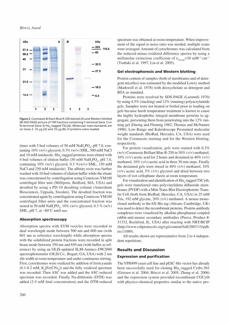
Figure 2. Coomassie Brillant Blue R-250 stained (A) and Western blotted (B) SDS-PAGE picture of YM fractions containing C-terminal (lane 1) or N-terminal (lane 3) His6-tagged TSCytb. Molecular mass standards are on lanes 2. 25 µg (A) and 10 µg (B) of proteins were loaded.

261
Tumor supressor cytochrome b561
tein (Bérczi et al. 2005). Other yeast cell lines and vectors were also successfully employed in high yield expression of mammalian Cyts-b561 (Liu et al. 2005; Su and Asard 2006). It was therefore plausible to use a yeast expression system for producing appropriate amounts of a novel recombinant Cytb-561 and establishing the basic physico-chemical properties of the expressed protein. N-terminal or C-terminal His
6-tagged
TSCytb was expressed and the YM fraction was used to check the expression levels. Figure 2 shows that the con-centration of the N-terminal His
6-tagged TSCytb in the YM
fraction was considerably lower than that of the C-terminal His
6-tagged TSCytb (hereafter (H
6C)TSCytb). This result
is in agreement with former observations obtained with the His
6-tagged CGCytb and TCytb proteins (data not published),
although the reason of the difference in expression levels is not yet known.
When mammalian or plant Cyts-b561 were expressed in ferrireductase-deficient yeast cells (Δfre1Δfre2 mutant lines), the capacity of these cells to reduce extracellular ferric-compounds was restored (Su and Asard 2006; Bérczi et al. 2007). These results were indirect proof that the recombi-nant Cyts-b561 was at least in part, targeted to the yeast cell plasma membrane.
Continuous sucrose density gradient fractionation of YM vesicles revealed that plasma membrane vesicles assembled at position of rather high sucrose concentrations (>45% (w/v); Roberg et al. 1997). When the distribution of (H
6C)
TSCytb in the SYM fraction was studied by discontinuous sucrose density gradient centrifugation and Western blotting, (H
6C)TSCytb was almost evenly distributed in membrane
fractions at all sucrose density steps (Fig. 3). In fact, the low-est concentration of TSCytb appears at the highest sucrose
Figure 3. Coomassie Brillant Blue R-250 stained (top) and Western blotted (bottom) SDS-PAGE picture of SYM fractions containing (H6C)TSCytb. Lanes are: 1 for molecular mass standards, 2 for SYM, 3 for SYM at the 10%/20% sucrose step interface, 4 for SYM at the 20%/32% sucrose step interface, 5 for SYM at the 32%/45% sucrose step interface, 6 for SYM at the 45%/70% sucrose step interface, and 7 for BSA and cytochrome c. 18 µg (top) and 2 µg (bottom) of protein-were loaded in lanes 2 through 6. a - 66.2 kDa for BSA; b - 12.4 kDa for horse cytochrome c.
Figure 4. Coomassie Brillant Blue R-250 stained (A) and Western blot-ted (B) SDS-PAGE picture of highly purified (H6C)TSCytb after PD-10 chromatography and concentration. 13 µg (A) and 3 µg (B) of proteins were loaded.

262
Bérczi, Asard
concentration step employed, i.e. the location at which the yeast plasma membrane is expected. Mizutani et al. (2007) showed that myc-tagged TSCytb was localized in small vesicles, including endosomes and endoplasmic reticulum of the perinuclear region, using an immunofluorescent staining technique. Therefore, the localization of (H
6C)TSCytb in the
lighter SYM fractions was not a surprise. However, the rather homogeneous distribution of (H
6C)TSCytb in different and
lighter populations of yeast membrane vesicles appears to be different from the mostly yeast plasma membrane localization of the other three recombinant mouse Cyts-b561 expressed in yeast. This result pointed out that enrichment of a membrane fraction containing (H
6C)TSCytb by differential and/or den-
sity gradient centrifugation before membrane solubilization could not be achieved.
As the protein balance sheet shows (Table 1), stripping of the YM vesicles resulted in the loss of about 30% of the proteins. Treatment of the SYM vesicles with the nonionic detergent SML, at a detergent:protein ratio of 3:1 (w/w) and 1 mg/ml protein concentration, resulted in solubilization of about 45% of the membrane proteins. When the solubilized fraction was concentrated by using a membrane filter with 100 kDa cut-off value, >90% of solubilized proteins were retained. About 75% of the solubilized and concentrated proteins did not bind the Ni-NTA His•Bind resin (Table 1, unbound fraction), however, only about 8% of the bound proteins were strongly associated with the resin, and eluted using the elution buffer (Table 1, (H
6C)TSCytb fraction). In
order to exchange the high salt and imidazole-containing
elution buffer to a widely-used phosphate buffer for storage of (H
6C)TSCytb, the coral red fraction was passed through
a PD-10 column and then concentrated. Figure 4 shows that the final (H
6C)TSCytb fraction contained mostly His
6-tagged
TSCytb, but small amounts of contaminating proteins can be seen on the gel. Even the fraction of proteins, which did not enter the separation gel, contained (H
6C)TSCytb, according
to the Western blot staining. After solubilization, protein con-centration, affinity chromatography, PD-10 chromatography, and a second protein concentration step, the ASC-reducible TSCytb recovery was between 40 and 50% which is in good agreement with former results (Bérczi et al. 2007).
Spectroscopy
TSCytb was classified as member of the Cyts-b561 on the basis of its sequence homology and predicted structural similarity to bovine CGCytb (Ponting 2001; Tsubaki et al. 2005; Su and Asard 2006). Figure 5 shows that TSCytb is
Fraction Total protein, mg ASC-reduced TSCytbpmol∙mg−1
YM 198 ± 47 n.m.SYM 143 ± 31 77± 18100P 72 ± 15 n.m.100S 60 ± 13 n.m.Filtrate 5.7 ± 1.4v n.m.Unbound 46 ± 7 n.m.(H6C)TSCytb 1.2 ± 0.3 3850 ± 341 (§)
Table 1. Protein balance sheet. For each His6-tag affinity chro-matography step, 100S fractions from two independent experi-ments were used; for each solubilization step, SYM fractions from two independent membrane preparations were used; and at each membrane preparation, 700 ml of yeast cell cultures with OD600(d=1 cm) of 0.8±0.05 was used. Results are the means ± s.d. from 8 independent His6-tag affinity chromatography steps except for the last value (§) which is mean ± s.d. from 4 independent purifications. YM – yeast microsomal membranes, SYM – stripped yeast microsomal membranes, 100P – unsolubilized membranes (the 100,000 gmax pellet after the solubilization step), 100S – the concentrated protein fraction after solubilization, Filtrate – solu-bilized protein fraction which passed through the 100,000 kDa membrane filter, Unbound – solubilized proteins unbound to the Ni-NTA His•Bind Resin, (H6C)TSCytb – concentrated His6-tagged TSCytb proteins eluted from the Ni-NTA His•Bind Resin. n.m. is for “not measured”.
Figure 5. ASC-reduced (a) and dithionite-reduced (d) minus oxidized difference spectra of SYM fraction containing untagged (A) and His6-tagged (B) TSCytb. 7.5 mg∙ml−1 was the protein concentration in both cases.
263
Tumor supressor cytochrome b561
indeed a membrane-associated b-type cytochrome, with an α-band absorbance maximum around 560 nm. TSCytb is also ASC reducible which is the basic property of Cyts-b561. It is evident that the presence of a His
6-tag on the C terminus
does not affect the ASC reducibility of the protein. However, the specific ASC-reducible cytochrome b content of SYM vesicles (between 50 and100 pmol∙mg−1 protein), tested with 25 mM ASC, was much lower than that obtained with other Cyts-b561 expressed in yeast (CGCytb: about 1500 pmol.mg−1 [Bérczi et al. 2005]; TCytb: about 400 pmol.mg−1 [Bérczi et al. 2007]; LCytb: about 150 pmol.mg−1 [Zhang et al. 2006]). It is also evident that the untagged and His
6-tagged
protein in the SYM fraction had considerably lower ASC re-ducibility, as compared to dithionate reducibility, than either CGCytb or TCytb (see later).
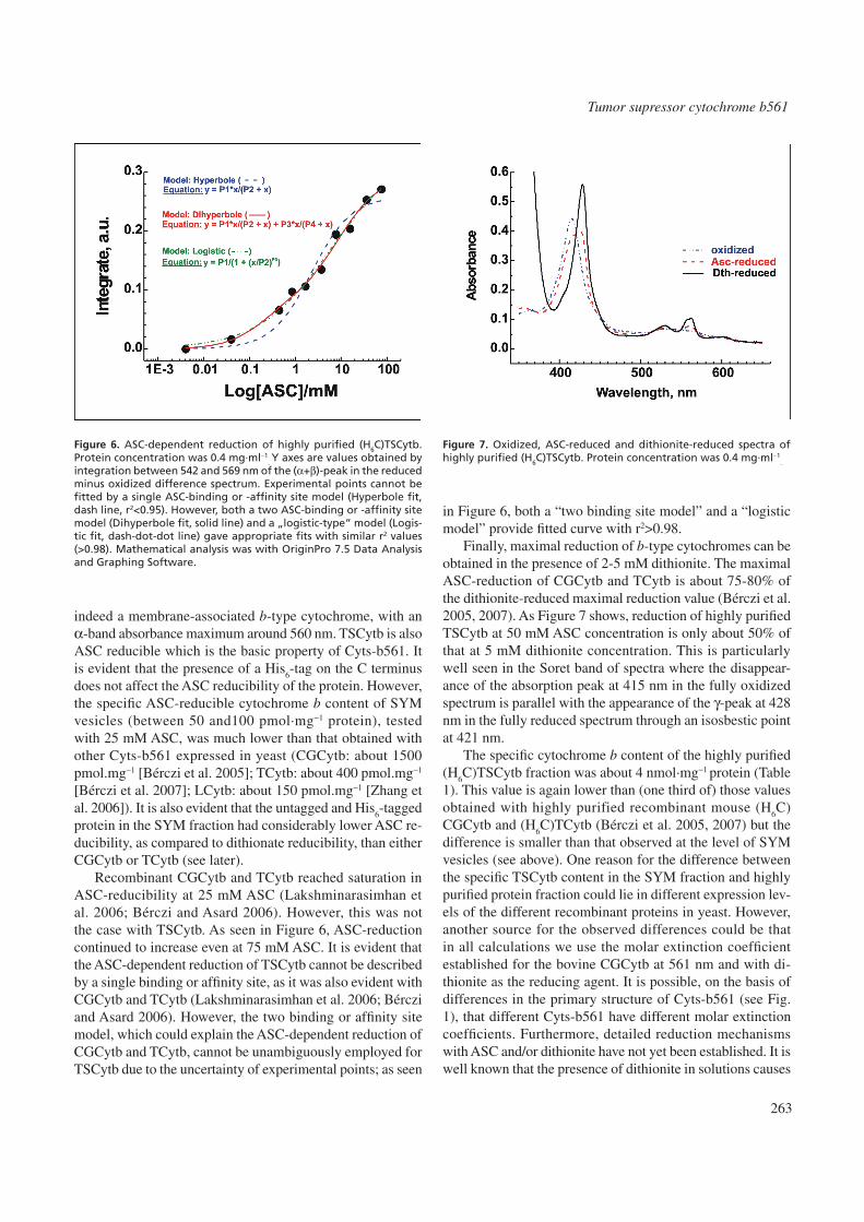
Recombinant CGCytb and TCytb reached saturation in ASC-reducibility at 25 mM ASC (Lakshminarasimhan et al. 2006; Bérczi and Asard 2006). However, this was not the case with TSCytb. As seen in Figure 6, ASC-reduction continued to increase even at 75 mM ASC. It is evident that the ASC-dependent reduction of TSCytb cannot be described by a single binding or affinity site, as it was also evident with CGCytb and TCytb (Lakshminarasimhan et al. 2006; Bérczi and Asard 2006). However, the two binding or affinity site model, which could explain the ASC-dependent reduction of CGCytb and TCytb, cannot be unambiguously employed for TSCytb due to the uncertainty of experimental points; as seen
in Figure 6, both a “two binding site model” and a “logistic model” provide fitted curve with r2>0.98.
Finally, maximal reduction of b-type cytochromes can be obtained in the presence of 2-5 mM dithionite. The maximal ASC-reduction of CGCytb and TCytb is about 75-80% of the dithionite-reduced maximal reduction value (Bérczi et al. 2005, 2007). As Figure 7 shows, reduction of highly purified TSCytb at 50 mM ASC concentration is only about 50% of that at 5 mM dithionite concentration. This is particularly well seen in the Soret band of spectra where the disappear-ance of the absorption peak at 415 nm in the fully oxidized spectrum is parallel with the appearance of the γ-peak at 428 nm in the fully reduced spectrum through an isosbestic point at 421 nm.
The specific cytochrome b content of the highly purified (H
6C)TSCytb fraction was about 4 nmol∙mg−1 protein (Table
1). This value is again lower than (one third of) those values obtained with highly purified recombinant mouse (H
6C)
CGCytb and (H6C)TCytb (Bérczi et al. 2005, 2007) but the
difference is smaller than that observed at the level of SYM vesicles (see above). One reason for the difference between the specific TSCytb content in the SYM fraction and highly purified protein fraction could lie in different expression lev-els of the different recombinant proteins in yeast. However, another source for the observed differences could be that in all calculations we use the molar extinction coefficient established for the bovine CGCytb at 561 nm and with di-thionite as the reducing agent. It is possible, on the basis of differences in the primary structure of Cyts-b561 (see Fig. 1), that different Cyts-b561 have different molar extinction coefficients. Furthermore, detailed reduction mechanisms with ASC and/or dithionite have not yet been established. It is well known that the presence of dithionite in solutions causes
Figure 6. ASC-dependent reduction of highly purified (H6C)TSCytb. Protein concentration was 0.4 mg∙ml−1
. Y axes are values obtained by integration between 542 and 569 nm of the (α+β)-peak in the reduced minus oxidized difference spectrum. Experimental points cannot be fitted by a single ASC-binding or -affinity site model (Hyperbole fit, dash line, r2<0.95). However, both a two ASC-binding or -affinity site model (Dihyperbole fit, solid line) and a „logistic-type” model (Logis-tic fit, dash-dot-dot line) gave appropriate fits with similar r2 values (>0.98). Mathematical analysis was with OriginPro 7.5 Data Analysis and Graphing Software.
Figure 7. Oxidized, ASC-reduced and dithionite-reduced spectra of highly purified (H6C)TSCytb. Protein concentration was 0.4 mg∙ml−1
.

264
Bérczi, Asard
oxygen depletion. Oxygen depletion is transient, depending on the starting concentration of dithionite and contact of the solution with air (that is mostly the case in spectrophotometer cuvettes). Since the role of oxygen as an oxidant to Cyts-b561 is not yet clear, it may affect the final reduction levels of the protein when any reducing agent is added. Taken together, we conclude that a reliable quantitative comparison between the properties of different Cyts-b561 will be possible only after establishing the proper molar extinction coefficient for each protein and the role of disolved oxygen for the reduction mechanism(s) of Cyts-b561.
comparison of tScytb to cGcytb and tcytb
In this paper, we have reported the expression of the His6-
tagged recombinant mouse TSCytb in the very same yeast expression system previously used for mouse CGCytb, DCytb, LCytb, and TCytb from Arabidopsis thaliana. It is shown that (i) TSCytb is an ASC-reducible b-type cytochrome but its sensitivity to ASC is considerably lower than that ob-served with CGCytb and TCytb, (ii) the specific content of TSCytb is lower than those of CGCytb and TCytb in the very same SYM fractions, (iii) the ASC-reducibility of TSCytb is lower than that of CGCytb and TCytb when compared to the maximal reduction of these Cyts-b561 by dithionite. These minor differences in the basic ASC-reducible properties might be explained by detailed sequence comparison of these proteins. Indeed, it was found that CGCytb, DCytb, LCytb, and even TCytb are evolutionary much closer to each other than to TSCytb (Tsubaki et al. 2005; Su and Asard 2006), supporting that these proteins might have little but important differences in structures which is manifested in different physico-chemical properties.
Acknowledgements
The authors thank Dr. Dan Su (Yale University, New Haven, CT, USA), Dr. Sándor Bottka (Hungarian Academy of Sci-ences, Hungary), Mrs Kim van Beek, Mrs Ciska Malgorzata and Mr Michiel Janssens (University of Antwerp, Antwerp, Belgium) for helpful discussions, and particularly for synthe-sizing the primers (Dr. Bottka), for production of pESC-His vectors containing the non-tagged (Dr. Su) and His
6-tagged
(Mrs Van Beek, Mrs Malgorzata, Mr Janssens) cDNA. This work was supported by grants from the University of Antwerp (to H.A.).
referencesApps DK, Pryde JG, Phillips JH (1980) Cytochrome b561 is identical with
chromomembrin B, a major polypeptide of chromaffin granule mem-branes. Neuroscience 5:2279-2287.
Asard H, Kapila J, Verelst W, Bérczi A (2001) Higher-plant plasma mem-brane cytochrome b561: a protein in search of a function. Protoplasma 217:77–93.
Bashtovyy D, Bérczi A, Asard H, Páli T (2003) Structure prediction for de-heme cytochrome b561 protein family. Protoplasma 221:31-40.
Bérczi A, Su D, Lakshminarasimhan M, Vargas A, Asard H (2005) Heter-ologous expression and site-directed mutagenesis of an ASC-reducible cytochrome b561. Arch Biochem Biophys 443:82-92.
Bérczi A, Asard H (2006) Characterization of an ASC-reducible cytochrome b561 by site-directed mutagenesis. Acta Biol Szeged 50:55-59.
Bérczi A, Su D, Asard H (2007) An Arabidopsis cytochrome b561 with trans-membrane ferrireductase capability. FEBS Lett 581:1505-1508.
Duong LT, Fleming PJ (1982) Isolation and properties of cytochrome b561 from bovine adrenal chromaffin granules. J Biol Chem 257:8561-8564.
Flatmark T, Terland O (1971) Cytochrome b561
of the bovine adrenal chro-maffin granules. A high potential b-type cytochrome. Biochim Biophys Acta 253:487-491.
Griesen D, Su D, Bérczi A, Asard H (2004) Localization of an ASC-reducible cytochrome b561 in the plant tonoplast. Plant Physiol 134:726-734.
Ji L, Nizhizaki M, Gao B, Burbee D, Kondo M, Kamibayashi C, Xu K, Yen N (2002) Expression of several genes in the human chromosome 3p21.3 homozygous deletion region by an adenovirus vector results in tumor suppressor activities in vitro and in vivo. Cancer Res 62:2715-2720.
Kent UM, Fleming PJ (1987) Purified cytochrome b561 catalyzes trans-membrane electron transfer for dopamine beta-hydroxylase and peptidyl glycine alpha-amidating monooxygenase activities in reconstituted systems. J Biol Chem 262:8174-8178.
Lakshminarasimhan M, Bérczi A, Asard H (2006) Substrate-dependent reduction of a recombinant chromaffin granule Cyt-b561 and its R72A mutant. Acta Biol Szeged 50:61-65.
Laemmli U (1970) Cleavage of structural proteins during the assembly of the head of bacteriophage T4. Nature 227:680-685.
Lerman M, Minna JD (2000) The 630-kb lung cancer homozygous deletion region on human chromosome 3p21.3: Identification and evaluation of the resident candidate tumor suppressor genes. Cancer Res 60:6116-6133.
Liu W, Kamensky Y, Kakkar R, Foley E, Kulmacz RJ, Palmer G (2005) Purification and characterization of bovine adrenal cytochrome b561 expressed in insect and yeast cell systems. Protein Expr Purif 40:429-439.
Markwell MAK, Haas SB, Bieber LL, Tolbert NE (1978) A modification of the Lowry procedure to simplify protein determination in membrane and lipoprotein samples. Anal Biochem 87:206-210.
McKie AT, Barrow D, Latunde-Dada GO, Rolfs A, Sager G, Mudaly E, Mudaly M, Richardson C, Barlow D, Bomford A, Peters RJ, Raja KB, Shirali S, Hediger MA, Farzaneh F, Simpson RJ (2001) An iron-regulated ferric reductase associated with the absorption of dietary iron. Science 291:1755-1759.
Mizutani A, Sanuki R, Kakimoto K, Kojo S, Taketani S (2007) Involvement of 101F6, a homologue of cytochrome b
561, in the reduction of ferric
ions. J Biochem 142:699-705.Njus D, Kelley PM (1993) The secretory vesicle ASC-regenerating system:
a chain of concerted H+/e−-transfer reactions. Biochim Biophys Acta 1144:235-248.
Njus D, Wigle M, Kelley PM, Kipp BH, Schlegel HB (2001) Mechanism of ascorbic acid oxidation by cytochrome b561. Biochemistry 40:11905-11911.
Ponting CP (2001) Domain homologues of dopamine β-hydroxylase and fer-ric reductase: Roles for iron metabolism in neurodegenerative disorders. Human Mol Gen 10:1853-1858.
Ohtani O, Iwamaru A, Deng W, Ueda K, Wu G, Jayachandran G, Kondo S, Atkinson EN, Minna JD, Roth JA, Ji L (2007) Tumor suppressor 101F6 and ASC synergistically and selectively inhibit non–small cell lung cancer growth by caspase-independent apoptosis and autophagy. Cancer Res 67:6293-6303.
Roberg KJ, Rowley N, Kaiser CA (1997) Physiological regulation of mem-brane protein sorting late in the secretory pathway of Saccharomyces cerevisiae. J Cell Biol 137:1469-1482.
Silsand T, Flatmark T (1974) Purification of cytochrome b-561. An integral heme protein of the adrenal chromaffin granule membrane. Biochim Biophys Acta 359:257-266.
265
Tumor supressor cytochrome b561
Su D, Asard H (2006) Three mammalian cytochrome b561
are ASC dependent ferrireductases. FEBS J 273:3722-3734.
Su D, May JM, Koury MJ, Asard H (2006) Human erythrocyte membranes contain a cytochrome b561 that may be involved in extracellular ASC recycling. J Biol Chem 281:39852-39859.
Thomas TC, McNamee MG (1990) Purification of membrane proteins. Methods Enzymol 182:499-520.
Tsubaki M, Takeuchi F, Nakanashi N (2005) Cytochrome b561 protein family: Expanding roles and versatile transmembrane electron transfer abilities as predicted by a new classification system and protein sequence
motif analyses. Biochim Biophys Acta 1753:174-190.Vargas JD, Herpers B, McKie AT, Gledhill S, McDonnell J, van den Heuvel
M, Davies KE, Ponting CP (2003) Stromal cell-derived receptor 2 and cytochrome b561 are functional ferric reductases. Biochim Biophys Acta 1651:116-123.
Verelst W, Asard H (2003) A phylogenetic study of cytochrome b561 pro-teins. Genome Biol 4:R38.
Zhang D, Su D, Bérczi A, Vargas A, Asard H (2006) An ASC-reducible cy-tochrome b561 is localized in macrophage lysosomes. Biochim Biophys Acta 1760:1903-1913.
Copyright © 2022 FDOKUMEN