Exploring the cortical evidence of a sensory-discrimination process
14
doi: 10.1098/rstb.2002.1100 , 1039-1051 357 2002 Phil. Trans. R. Soc. Lond. B Ranulfo Romo, Adrián Hernández, Antonio Zainos, Carlos Brody and Emilio Salinas process discrimination - Exploring the cortical evidence of a sensory References http://rstb.royalsocietypublishing.org/content/357/1424/1039#related-urls Article cited in: Email alerting service here right-hand corner of the article or click Receive free email alerts when new articles cite this article - sign up in the box at the top http://rstb.royalsocietypublishing.org/subscriptions go to: Phil. Trans. R. Soc. Lond. B To subscribe to This journal is © 2002 The Royal Society on July 15, 2011 rstb.royalsocietypublishing.org Downloaded from
-
Upload
independent -
Category
Documents
-
view
1 -
download
0
Transcript of Exploring the cortical evidence of a sensory-discrimination process
doi: 10.1098/rstb.2002.1100, 1039-1051357 2002 Phil. Trans. R. Soc. Lond. B
Ranulfo Romo, Adrián Hernández, Antonio Zainos, Carlos Brody and Emilio Salinas process
discrimination−Exploring the cortical evidence of a sensory
Referenceshttp://rstb.royalsocietypublishing.org/content/357/1424/1039#related-urls
Article cited in:
Email alerting service hereright-hand corner of the article or click Receive free email alerts when new articles cite this article - sign up in the box at the top
http://rstb.royalsocietypublishing.org/subscriptions go to: Phil. Trans. R. Soc. Lond. BTo subscribe to
This journal is © 2002 The Royal Society
on July 15, 2011rstb.royalsocietypublishing.orgDownloaded from

Published online 2 August 2002
Exploring the cortical evidence of asensory-discrimination process
Ranulfo Romo*, Adrian Hernandez, Antonio Zainos, Carlos Brodyand Emilio Salinas
Instituto de Fisiologıa Celular, Universidad Nacional Autonoma de Mexico, 04510 Mexico, DF, Mexico
Humans and monkeys have similar abilities to discriminate the difference in frequency between two con-secutive mechanical vibrations applied to their fingertips. This task can be conceived as a chain of neuraloperations: encoding the two consecutive stimuli, maintaining the first stimulus in working memory, com-paring the second stimulus with the memory trace left by the first stimulus and communicating the resultof the comparison to the motor apparatus. We studied this chain of neural operations by recording andmanipulating neurons from different areas of the cerebral cortex while monkeys performed the task. Theresults indicate that neurons of the primary somatosensory cortex (S1) generate a neural representationof vibrotactile stimuli which correlates closely with psychophysical performance. Discrimination based onmicrostimulation patterns injected into clusters of S1 neurons is indistinguishable from that produced bynatural stimuli. Neurons from the secondary somatosensory cortex (S2), prefrontal cortex and medialpremotor cortex (MPC) display at different times the trace of the first stimulus during the working-memory component of the task. Neurons from S2 and MPC appear to show the comparison betweenthe two stimuli and correlate with the behavioural decisions. These neural operations may contribute tothe sensory-discrimination process studied here.
Keywords: behaving monkeys; sensory discrimination; cerebral cortex
1. INTRODUCTION
An important problem in brain physiology is the isolationof the sensory representations that guide behaviouraldecisions. This problem has been investigated in behav-ioural tasks where the sensory stimuli are under precisequantitative control and the subject’s psychophysical per-formances are quantitatively measured (Talbot et al. 1968;Newsome et al. 1989; Mountcastle et al. 1990). This strat-egy has allowed the investigation of which attributes of theneural responses elicited by a sensory stimulus are sensor-ily meaningful (Romo & Salinas 1999, 2001). Indeed, ithas been shown that the sensory areas of the cerebral cor-tex generate representations of the sensory stimuli thatcorrelate closely with psychophysical performances(Newsome et al. 1989; Vogels & Orban 1990; Hernandezet al. 2000). Alternatively, behavioural decisions arereported through motor actions, but it is not clear whereand how a sensory representation is converted into amotor output. To answer this question, neurophysiologistshave studied the neuronal responses of motor areas duringperceptual tasks, and have found that a fraction of theneurons show a link between the sensory inputs and thebehavioural decisions (Romo et al. 1997, 1993; Merchantet al. 1997; Salinas & Romo 1998; Horwitz & Newsome1999). However, decision making is more than a simple
* Author for correspondence ([email protected]).
One contribution of 14 to a Discussion Meeting Issue ‘The physiologyof cognitive processes’.
Phil. Trans. R. Soc. Lond. B (2002) 357, 1039–1051 1039 2002 The Royal SocietyDOI 10.1098/rstb.2002.1100
input–output operation (Shadlen & Newsome 2001). Forexample, regardless of the perceptual task, subjects reacha behavioural decision after the comparison of the currentsensory input against a sensory referent, which can bestored in working memory or in long-term memory.Therefore, to understand how the brain carries a percep-tual process, we need to isolate where and in what formthe current sensory input interacts with a sensory referentthat is stored in the memory (Hernandez et al. 2002). Thisneural operation, we believe, is the key to understandinghow the neuronal circuits elaborate a perceptual process.
We have addressed some of the issues mentioned herein a behavioural task where monkeys discriminate the dif-ference in frequency between two consecutive mechanicalvibrations delivered to their fingertips (figure 1; for detailssee Hernandez et al. (1997) and Mountcastle et al.(1990)). In this task the stimulus can be finely controlled;the same primary afferents are activated by the two stim-uli; there is sensory and motor lateralization; it involves aworking-memory mechanism; and decision making isbased on the comparison between the current sensoryinput and the memory trace left by the first stimulus.Thus, the task can be viewed as a chain of neuronal oper-ations. In this paper, we review recent results obtained inthis sensory-discrimination task. § 2 contains a briefdescription of the general organization of the somatosen-sory system and the experiments that paved the way tostudying the neuronal processes involved. § 3 thendescribes the neuronal correlates that seem to be associa-ted with the different components of the vibrotactile dis-crimination.
on July 15, 2011rstb.royalsocietypublishing.orgDownloaded from

1040 R. Romo and others Cortical discrimination processes
10 18 26 346
18
30
42
85
94
84
91
87
84
base (Hz)
85
86
%
89
93
92
87
89
89
12 16 20 24 28
12
16
20
24
28
9281755752
91
86
75
63
59
74
82
93
627983
%
93
com
pari
son
(Hz)
base comparisonPD KD KU PB
500 ms
keystimulator
(a)
(b)
(c) (d)
push-buttons
Figure 1. The discrimination task. (a) Drawing of a monkeyworking in the discrimination task. (b) The sequence ofevents during the discrimination trials. The mechanicalprobe is lowered, indenting the glabrous skin of one digit ofthe hand (PD); the monkey places his free hand on animmovable key (KD); the probe oscillates vertically, at thebase stimulus frequency; after a delay, a second mechanicalvibration is delivered at the comparison frequency; themonkey releases the key (KU) and presses either a laterallyplaced or medially placed push-button (PB) to indicatewhether the comparison frequency was higher or lower thanthe base. (c,d) Stimulus sets used during recordings. Eachbox indicates a base–comparison frequency stimulus pairused; the numbers inside the box indicate the overallpercentage of correct trials for the base–comparison pair.The stimulus set illustrated in (c) is used to determine thediscrimination thresholds; the stimulus set illustrated in (d )is used to explore the working-memory component of thetask. The combinations of both sets are often used duringthe recording sessions.
2. THE SOMATOSENSORY SYSTEM
The somatic and visual systems are useful models forinvestigating stimulus information processing, and somegeneral principles behind the functional organization ofthe brain. There are some elements of the organization ofthe somatosensory system that are relevant in investigatingneural coding of sensory stimuli both at the periphery andin the brain. For the sake of simplicity, we restrict thisreview to the cutaneous information-processing channel.
Phil. Trans. R. Soc. Lond. B (2002)
(a) Cutaneous primary afferentsThe human hand contains four types of cutaneous affer-
ent fibres that transmit information of the mechanicalstimulus features to the central nervous system (Talbot etal. 1968; Darian-Smith 1984; Vallbo & Johansson 1984;Vallbo 1995). Two of these afferent fibres are rapidlyadapting: one is anatomically linked to QA and the otherto PC. The other two afferent fibres are slowly adaptingand are linked to SA-I and SA-II, respectively. The mon-key hand possesses these afferent fibres, except SA-II.Although all these afferent fibres respond to a cutaneousstimulus, they become specialized to encoding spatiotem-poral features of the stimuli (Talbot et al. 1968; Phillipsand Johnson 1981). This has been demonstrated in well-designed experiments aimed at exploring their capacities.The degree of sensitivity of these afferent fibres is evi-denced by the fact that a psychophysical observer candetect even a single spike evoked in one single primaryafferent (Vallbo & Johansson 1984; Vallbo 1995).
(b) Neocortical somatosensory areasAfter a relay in the dorsal column nuclei and in the basal
complex nuclei of the thalamus, somatosensory infor-mation reaches S1. Primate S1 is subdivided into fourareas (area 3a, 3b, 1 and 2), each containing a somato-topic representation of the body (Kaas et al. 1979; Nelsonet al. 1980). Tactile information is processed mainly byareas 3b, 1 and 2, which are interconnected (Shanks et al.1985). To a certain extent, neurons in S1 replicate thefunctional properties of QA, SA-I and PC afferent fibres(Powell & Mountcastle 1959; Mountcastle et al. 1969; Suret al. 1984) and are referred to as QA, SA and PC neu-rons. These subtypes are clustered in columns(Mountcastle 1957; Powell & Mountcastle 1959; Sur etal. 1984).
Information flows from S1 to the posterior parietal cor-tex and to the lateral somatosensory areas. As for the vis-ual system, it appears that there is also a dorsal streamand a ventral stream in the cortical organization of thesomatosensory system (Mishkin 1979; Murray & Mishkin1984). According to this organization, the dorsal streamflows through areas 5 and 7b (Pearson & Powell 1985;Shanks et al. 1985; Cavada & Goldman-Rakic 1989), andthe ventral stream flows through the lateral somatosensoryareas (Pons et al. 1987, 1992; Burton et al. 1995; Krub-itzer et al. 1995). The dorsal stream is more likely to beassociated with processing the somatosensory informationthat reaches the PM cortex (Godshalk et al. 1984;Cavada & Goldman-Rakic 1989; Leichnetz 1989;Tokuno & Tanji 1993). The operations through this dor-sal stream could be important for self-initiated or stimu-lus-triggered voluntary movements involving sensoryprocessing. The ventral stream is more likely to be associa-ted with fine discrimination and the recognition of stimu-lus patterns. This processing reaches also the PM cortex(Godshalk et al. 1984; Cavada & Goldman-Rakic 1989;Leichnetz 1989) and the PFC (Preuss & Goldman-Rakic1989; Carmichael & Price 1995), and might be associatedwith the fine discrimination of stimulus objects. Interest-ingly, both streams reach M1 (Leichnetz 1989; Tokuno &Tanji 1993), and both should drive the motor represen-tations during sensory tasks that require an indication ofdecision making. The functional meaning of these
on July 15, 2011rstb.royalsocietypublishing.orgDownloaded from

Cortical discrimination processes R. Romo and others 1041
streams, however, needs to be investigated further,especially with regard to what aspects of somatosensoryperception they contribute to.
3. FORMING A SENSATION VIA A NEURAL CODE
Talbot et al. (1968) and Werner & Mountcastle (1965)pioneered this enterprise almost four decades ago. The keyconceptual advance was to combine psychophysics andneurophysiology, two experimental disciplines that hadpreviously been divorced in sensory research. Talbot et al.(1968) and Werner (1980) used mechanical stimuli,applied to the fingertips of humans, that changed in onedimension; they measured the subjective estimates quanti-tatively. Then they recorded in anaesthetized monkeys theresponses of cutaneous afferent fibres using the samestimuli in the psychophysical experiments (Werner &Mountcastle 1965; Talbot et al. 1968). Their goal was todetermine the relationship between the subjective sen-sation and the evoked peripheral activity produced by thestimuli. Indeed, they found a close relationship betweenthe psychophysical performance and the neural activityevoked by the stimuli (Werner & Mountcastle 1965;Talbot et al. 1968). These pioneering experiments havebeen adapted since then as a tool for exploring the neuralcodes that underlie a sensation in the different sensorymodalities.
(a) Peripheral coding of vibrotactile stimuliA sensory neural code is activity produced by a natural
stimulus, which correlates with the psychophysical per-formance. Defining the peripheral coding of a somatosen-sory stimulus implies that this approach might facilitateexploring the central neural mechanisms of somatosensoryperception. Talbot et al. (1968) pioneered this researcharea using the sensory modality of the sense of fluttervibration. They showed that, depending on the range offrequency of the mechanical vibrations applied to the skinon the hand, two sensations can be elicited: the sensationof flutter at low frequencies (range of 5–50 Hz) and thesensation of vibration at high frequency (range of 60–300 Hz). Talbot et al. (1968) first quantified amplitudedetection thresholds in humans, and then showed that thesensitivities of QA and PC afferents account for perform-ance in the low- and high-frequency regimes, respectively.This correspondence between perceptual and anatomicalsubmodalities was later confirmed and extended by rec-ording and microstimulating afferent fibres in human sub-jects (Ochoa & Torebjork 1983; Macefield et al. 1990;Vallbo & Johansson 1984; Vallbo 1995).
There were two major observations about the nature ofthe peripheral neural code underlying flutter–vibrationperception (Talbot et al. 1968). First, the QA and PCafferents respond periodically to the periodic structure ofthe stimulus frequency. Second, the QA afferents hardlychange in firing rate over a frequency range of 10–50 Hz,while the PC afferents increase their firing rate as a func-tion of increasing stimulus frequency (60–250 Hz). It wasthus concluded that high frequencies could be encodedby the total number of PC spikes produced—a rate code(Shadlen & Newsome 1994; Singer & Gray 1995)—butlow frequencies could not, because the number of QAspikes seemed to be constant in the flutter range; they had
Phil. Trans. R. Soc. Lond. B (2002)
to be encoded in the regular, periodic spikes produced bythe flutter stimuli in the QA afferents—a temporal code.However, direct microstimulation of QA afferents pro-duced flutter sensations of frequencies that were perceivedto increase with the evoked firing rate (Ochoa & Torebjork1983). If the frequency of the microstimulation currentincreases in the range of 5–100 Hz—presumably produ-cing a proportional increase in QA firing rate—humansubjects report gradual increases in the perceived flutterfrequency at a constant intensity (Ochoa & Torebjork1983).
The experiments established the roles that the differentcutaneous afferents play in coding temporal stimuli.Clearly, the QA and PC systems encode the temporal fea-tures of the stimuli. Interestingly, it has been shown thatthe SA-I afferent system transmits information regardingthe spatial properties of the stimulus features (Johnson &Hsiao 1992). The neural coding of the physical propertiesof the stimuli seems to define and limit the capacity of thepsychophysical observer to detect, recognize and discrimi-nate the stimuli. These important observations paved theway for further investigation of the cortical processing ofsomatosensory inputs during perceptual tasks.
4. CORTICAL CODING OF VIBROTACTILE STIMULIAND THE LINK TO PERCEPTION
Compared with our knowledge of tactile coding in affer-ent fibres, the central mechanisms are less understood.This has been due in part to the difficulties in adaptingsomatosensory tasks in behaving monkeys. Tracing a neu-ral code from the periphery to the cerebral cortex hasremained the leading idea in understanding somatosens-ation. The key here is the use of well-designed psycho-physical tasks in behaving monkeys. In § 4 we reviewdevelopments in this research area.
(a) PsychophysicsMountcastle et al. (1972) adapted the vibrotactile task
used initially in human subjects to behaving monkeys.They trained monkeys to detect amplitudes and discrimi-nate stimuli frequencies in the flutter range (Mountcastleet al. 1972; LaMotte & Mountcastle 1975). With intensetraining, monkeys developed stimulus-frequencyamplitude-detection thresholds that were almost indis-tinguishable from those quantified in human subjects inidentical conditions (Mountcastle et al. 1972). In addition,the discrimination of two consecutive stimulus frequenciesdelivered to the hands (figure 1) was similar to those mea-sured in humans in identical conditions (LaMotte &Mountcastle 1975; Mountcastle et al. 1990). These resultsindicate that monkeys could be an appropriate model forexploring the central neural mechanisms associated withthe flutter task. The discrimination flutter task is parti-cularly rich in that comparison of f2 is made against thememory trace left by f1. To solve this task the psychophys-ical observer requires a number of cognitive processessuch as detection, working memory, comparison anddecision making (Mountcastle et al. 1990; Hernandez etal. 1997; Romo et al. 1998). Some other tasks require thatmonkeys categorize moving tactile stimuli (Romo et al.1993, 1996), detect roughness in surfaces (Sinclair &Burton 1993; Jiang et al. 1997) or discriminate tactual
on July 15, 2011rstb.royalsocietypublishing.orgDownloaded from

1042 R. Romo and others Cortical discrimination processes
stimulus orientation and form (Hsiao et al. 1993; Burtonet al. 1997). All these tasks require attention to be focusedon the stimulus with indication of performance giventhrough voluntary movements; that is, from sensation toaction. Investigators using these somatosensory tasks wantto unravel the central mechanisms associated with the dif-ferent components of these psychophysical tasks.
(b) Coding of vibrotactile stimuli in S1If QA afferents reliably encode the periodic structure of
the flutter stimulus frequency, the question then iswhether QA neurons of S1 do this in a similar fashion, orwhether there is another way of encoding the stimuli.Shortly after their work on cutaneous afferent fibres,Mountcastle et al. (1969) studied the responses of S1 neu-rons. Two decades later, S1 neurons were re-recorded,this time in behaving monkeys trained to detect and dis-criminate flutter-stimuli frequencies (Mountcastle et al.1990). The results support the previous findings. First, itwas found that QA neurons of S1, like their afferent fibres,fire periodically in phase with mechanical oscillations.Second, their firing rates seem to change little in the flutterrange (this conclusion was based, however, on data from17 neurons). Third, the psychophysical performancematched the inferred performance based on the discrimin-ability of the periodic inter-spike intervals (Mountcastle etal. 1990). It followed that, as proposed before, the stimu-lus frequency could not be encoded by S1 firing rates;the stimulus frequency had to be encoded temporally, inthe serial order of evoked spikes (Talbot et al. 1968;Mountcastle et al. 1969, 1990).
In support of this proposal, using flutter stimuli, Recan-zone et al. (1992) compared psychophysical data frommonkeys to S1 recordings in separate experiments fromthe same animals. The comparison was consistent with atemporal coding mechanism, and firing rates were notseen to vary with the stimulus frequency (however, therange of frequencies tested was quite narrow and the ani-mals were anaesthetized). Recanzone et al. (1992) madeanother important observation: that spike timing associa-ted with the sine wave was much more precise in trainedanimals compared with untrained monkeys. Thus, on thebasis of these results, a psychophysical observer shouldexploit the periodic spike timing evoked in the QA neu-rons of S1 for sensory discrimination.
Arguments in favour of this proposal could be strength-ened if a large number of neurons were studied, and ifneurons were studied in awake animals during the flutter-discrimination task (figure 1). Hernandez et al. (2000) andSalinas et al. (2000) trained monkeys to discriminatebetween flutter stimulus frequencies and recorded manyneurons with QA properties in areas 3b and 1 of S1. Eachrecorded neuron with QA properties was studied duringthe discrimination task. There were three major results.First, the majority of neurons from S1 were phase-lockedto the input stimulus frequency; however, almost one-third of the QA neurons modulated their firing rates as afunction of the stimulus frequency (Salinas et al. 2000).The second important finding was that QA neurons thatmodulate their firing rates were affected by the task con-dition; that is, they increased their transmitted infor-mation about the stimulus frequency during taskperformance (Salinas et al. 2000). Third, only those neu-
Phil. Trans. R. Soc. Lond. B (2002)
rons that varied their firing rates as a function of thestimulus frequency were affected in the error trials (Salinaset al. 2000).
These findings question the unique role of periodicspike timing in discrimination of flutter stimuli, and indi-cate that a firing rate code cannot be discarded (Salinaset al. 2000). But, apart from this, what do these findingsindicate? They indicate the presence of two subpopula-tions of QA neurons in S1 that behave differently inresponse to a periodic mechanical stimulus (Hernandez etal. 2000; Salinas et al. 2000). These two subpopulationsmight be arranged in an hierarchical fashion: QA neuronsthat respond periodically might be closer to the inputstimulus, and those that modulate their firing might inte-grate the responses of the periodic neurons and transformthem into a rate code (Hernandez et al. 2000). Such last-order neurons of the QA circuit could distribute the neuralrepresentation to those structures anatomically linked toS1, in order to solve the sensory-discrimination task.However, further studies are needed to see whether thisis so.
(c) Neuronal correlates of vibrotactilediscrimination in S1
A more direct test of the role of periodicity in flutterdiscrimination is measuring the discrimination capabilitiesof these subtypes of QA neurons associated with psycho-physical performance (figure 1). Another test is to provewhether the evoked neural activity during discriminationin S1 is sufficient for sensory performance. Finally, it isnecessary to test whether the temporal order of the spikesis important for sensory discrimination. These are incisivetests to validate the meaning of the neural encoding of theflutter stimuli in S1. We now review recent findings onthese points.
The vibrotactile-discrimination task requires the com-parison of f2 against f1 (Hernandez et al. 1997). As indi-cated in § 4c Hernandez et al. (2000) and Salinas et al.(2000) found two types of responses in QA neurons ofS1: one that is periodically entrained by the stimulus fre-quency, and another that, although not periodicallyentrained, has average firing rates during the stimulusperiod that are modulated as a function of the stimulusfrequency. To investigate which one of these two rep-resentations is associated with psychophysical perform-ance, Hernandez et al. (2000) determined the probabilitythat an observer (a cortical region central to S1) coulddistinguish the difference between the two stimuli. Thiscould be based on a comparison of the neuronal responsedistributions of f2 made against the neuronal response dis-tributions of f1. According to this, the observer could usea simple rule: if the number of spikes during the secondstimulus is higher than during the first stimulus, then f2 ishigher than f1. The same rule can be used when consider-ing the periodicity values: if the periodicity (estimated asthe frequency with greatest power in a Fourier transformof the spiking responses) during the second stimulus per-iod ( f2) is higher than during the first stimulus ( f1), thenf2 is higher than f1. The effect of this type of rule is equival-ent to determining the area under the curve ROC(Green & Swets 1966) generated by the neuronal responsedistributions for each pair of stimulus frequencies, usingboth periodicity and firing rate values (Hernandez et al.
on July 15, 2011rstb.royalsocietypublishing.orgDownloaded from
Cortical discrimination processes R. Romo and others 1043
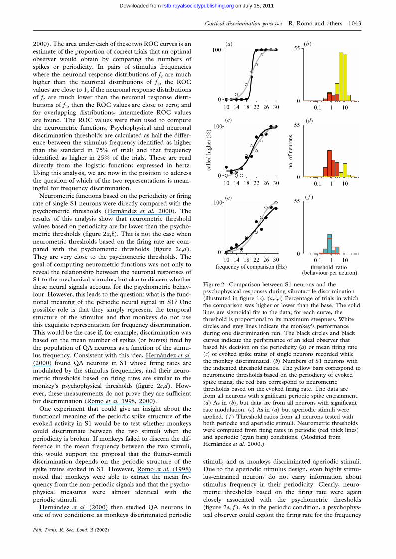
2000). The area under each of these two ROC curves is anestimate of the proportion of correct trials that an optimalobserver would obtain by comparing the numbers ofspikes or periodicity. In pairs of stimulus frequencieswhere the neuronal response distributions of f2 are muchhigher than the neuronal distributions of f1, the ROCvalues are close to 1; if the neuronal response distributionsof f2 are much lower than the neuronal response distri-butions of f1, then the ROC values are close to zero; andfor overlapping distributions, intermediate ROC valuesare found. The ROC values were then used to computethe neurometric functions. Psychophysical and neuronaldiscrimination thresholds are calculated as half the differ-ence between the stimulus frequency identified as higherthan the standard in 75% of trials and that frequencyidentified as higher in 25% of the trials. These are readdirectly from the logistic functions expressed in hertz.Using this analysis, we are now in the position to addressthe question of which of the two representations is mean-ingful for frequency discrimination.
Neurometric functions based on the periodicity or firingrate of single S1 neurons were directly compared with thepsychometric thresholds (Hernandez et al. 2000). Theresults of this analysis show that neurometric thresholdvalues based on periodicity are far lower than the psycho-metric thresholds (figure 2a,b). This is not the case whenneurometric thresholds based on the firing rate are com-pared with the psychometric thresholds (figure 2c,d).They are very close to the psychometric thresholds. Thegoal of computing neurometric functions was not only toreveal the relationship between the neuronal responses ofS1 to the mechanical stimulus, but also to discern whetherthese neural signals account for the psychometric behav-iour. However, this leads to the question: what is the func-tional meaning of the periodic neural signal in S1? Onepossible role is that they simply represent the temporalstructure of the stimulus and that monkeys do not usethis exquisite representation for frequency discrimination.This would be the case if, for example, discrimination wasbased on the mean number of spikes (or bursts) fired bythe population of QA neurons as a function of the stimu-lus frequency. Consistent with this idea, Hernandez et al.(2000) found QA neurons in S1 whose firing rates aremodulated by the stimulus frequencies, and their neuro-metric thresholds based on firing rates are similar to themonkey’s psychophysical thresholds (figure 2c,d). How-ever, these measurements do not prove they are sufficientfor discrimination (Romo et al. 1998, 2000).
One experiment that could give an insight about thefunctional meaning of the periodic spike structure of theevoked activity in S1 would be to test whether monkeyscould discriminate between the two stimuli when theperiodicity is broken. If monkeys failed to discern the dif-ference in the mean frequency between the two stimuli,this would support the proposal that the flutter-stimulidiscrimination depends on the periodic structure of thespike trains evoked in S1. However, Romo et al. (1998)noted that monkeys were able to extract the mean fre-quency from the non-periodic signals and that the psycho-physical measures were almost identical with theperiodic stimuli.
Hernandez et al. (2000) then studied QA neurons inone of two conditions: as monkeys discriminated periodic
Phil. Trans. R. Soc. Lond. B (2002)
10 14 18 22 26 300
100
10 14 18 22 26 300
100
cal
led
high
er (
%)
10 14 18 22 26 300
100
frequency of comparison (Hz)
0.1 1 100
55
0.1 1 100
55
no. o
f ne
uron
s
0.1 1 100
55
threshold ratio (behaviour per neuron)
(a)
(c)
(e)
(b )
(d)
( f )
Figure 2. Comparison between S1 neurons and thepsychophysical responses during vibrotactile discrimination(illustrated in figure 1c). (a,c,e) Percentage of trials in whichthe comparison was higher or lower than the base. The solidlines are sigmoidal fits to the data; for each curve, thethreshold is proportional to its maximum steepness. Whitecircles and grey lines indicate the monkey’s performanceduring one discrimination run. The black circles and blackcurves indicate the performance of an ideal observer thatbased his decision on the periodicity (a) or mean firing rate(c) of evoked spike trains of single neurons recorded whilethe monkey discriminated. (b) Numbers of S1 neurons withthe indicated threshold ratios. The yellow bars correspond toneurometric thresholds based on the periodicity of evokedspike trains; the red bars correspond to neurometricthresholds based on the evoked firing rate. The data arefrom all neurons with significant periodic spike entrainment.(d) As in (b), but data are from all neurons with significantrate modulation. (e) As in (a) but aperiodic stimuli wereapplied. ( f ) Threshold ratios from all neurons tested withboth periodic and aperiodic stimuli. Neurometric thresholdswere computed from firing rates in periodic (red thick lines)and aperiodic (cyan bars) conditions. (Modified fromHernandez et al. 2000.)
stimuli; and as monkeys discriminated aperiodic stimuli.Due to the aperiodic stimulus design, even highly stimu-lus-entrained neurons do not carry information aboutstimulus frequency in their periodicity. Clearly, neuro-metric thresholds based on the firing rate were againclosely associated with the psychometric thresholds(figure 2e, f ). As in the periodic condition, a psychophys-ical observer could exploit the firing rate for the frequency
on July 15, 2011rstb.royalsocietypublishing.orgDownloaded from
1044 R. Romo and others Cortical discrimination processes
discrimination of aperiodic stimuli. These results indicatethat an observer could solve this task with a precision simi-lar to that of a monkey, based only on the firing rate pro-duced during the stimulus periods.
(d) Probing the flutter coding by microstimulationof S1
Unequivocal proof that the activity of a localizedcortical neuronal population provides sufficient basis fora specific cognitive function has not been obtained.Neurophysiological studies often reveal close associationsbetween neuronal activity and sensory events, but thendoes such activity have an impact on perception and sub-sequent behaviour? We typically assume this to be so, butthis is hard to verify. Intracortical microstimulation hasprovided the most compelling evidence to date of a causallink between the activity of localized populations of neu-rons and specific cognitive functions (Britten & Wezel1998; Salzman et al. 1990; Romo et al. 1998, 2000). Elec-trical microstimulation directly activates small cluster ofneurons, and has been shown to bias a monkey’s choiceduring the decision stage of an ongoing perceptual task(Seidemann et al. 1998; Gold & Shadlen 2000). A con-venient model that can be used to answer this question isthe flutter sensation, for which humans and monkeys havesimilar discrimination thresholds (Hernandez et al. 1997;Mountcastle et al. 1990). During the vibrotactile-discrimination task, subjects pay attention to the fre-quency of the first (base) stimulus, store a trace of it dur-ing the delay period between the two stimuli and comparethe stored trace with the frequency of the second(comparison) stimulus. This task, therefore, contains anumber of cognitive processes, such as stimulus detection,working memory, discrimination between the two stimuliand decision making. These cognitive processes should beinitiated by the evoked neuronal activity in S1 (Romo &Salinas 1999, 2001). As reviewed in § 2b, the QA circuitof S1 distributes the representation of the flutter stimulito more central structures anatomically linked to it to solvethis task. Romo et al. (1998) used intracorticalmicrostimulation in S1 to manipulate the neural code forflutter discrimination.
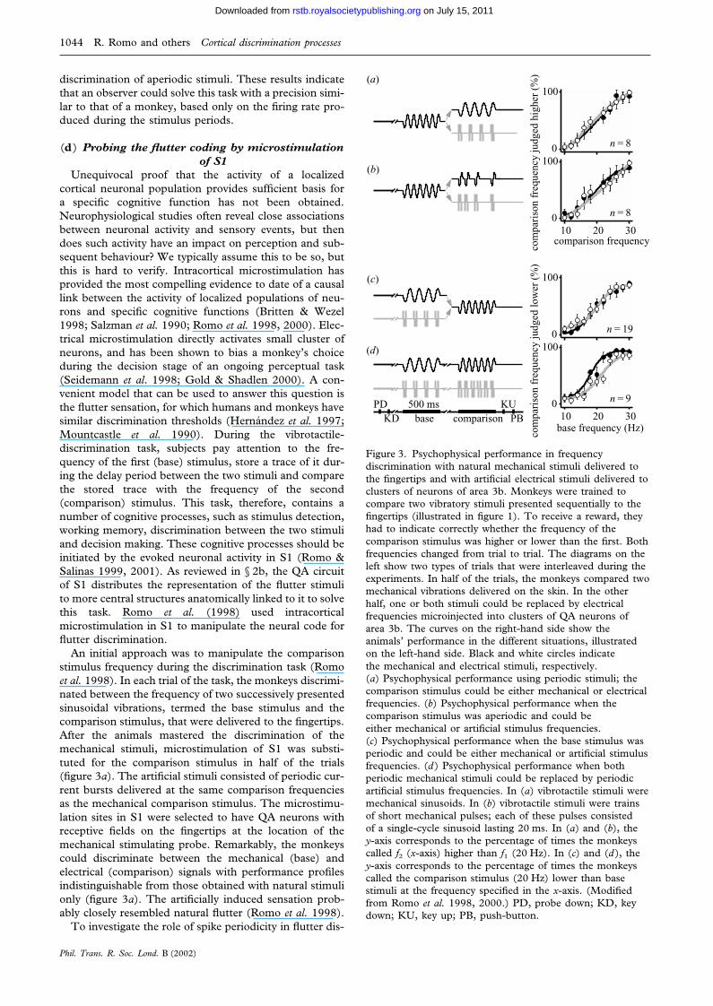
An initial approach was to manipulate the comparisonstimulus frequency during the discrimination task (Romoet al. 1998). In each trial of the task, the monkeys discrimi-nated between the frequency of two successively presentedsinusoidal vibrations, termed the base stimulus and thecomparison stimulus, that were delivered to the fingertips.After the animals mastered the discrimination of themechanical stimuli, microstimulation of S1 was substi-tuted for the comparison stimulus in half of the trials(figure 3a). The artificial stimuli consisted of periodic cur-rent bursts delivered at the same comparison frequenciesas the mechanical comparison stimulus. The microstimu-lation sites in S1 were selected to have QA neurons withreceptive fields on the fingertips at the location of themechanical stimulating probe. Remarkably, the monkeyscould discriminate between the mechanical (base) andelectrical (comparison) signals with performance profilesindistinguishable from those obtained with natural stimulionly (figure 3a). The artificially induced sensation prob-ably closely resembled natural flutter (Romo et al. 1998).
To investigate the role of spike periodicity in flutter dis-
Phil. Trans. R. Soc. Lond. B (2002)
10 20 300
100
n = 9
com
pari
son
freq
uenc
y ju
dged
low
er (
%)
base frequency (Hz)
0
100
n = 19
10 20 300
100
n = 8
com
pari
son
freq
uenc
y ju
dged
hig
her
(%)
comparison frequency
0
100
n = 8
base comparisonPD
KDKU
PB500 ms
(a)
(b)
(c)
(d)
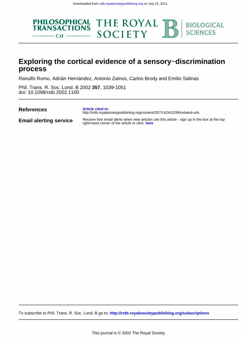
Figure 3. Psychophysical performance in frequencydiscrimination with natural mechanical stimuli delivered tothe fingertips and with artificial electrical stimuli delivered toclusters of neurons of area 3b. Monkeys were trained tocompare two vibratory stimuli presented sequentially to thefingertips (illustrated in figure 1). To receive a reward, theyhad to indicate correctly whether the frequency of thecomparison stimulus was higher or lower than the first. Bothfrequencies changed from trial to trial. The diagrams on theleft show two types of trials that were interleaved during theexperiments. In half of the trials, the monkeys compared twomechanical vibrations delivered on the skin. In the otherhalf, one or both stimuli could be replaced by electricalfrequencies microinjected into clusters of QA neurons ofarea 3b. The curves on the right-hand side show theanimals’ performance in the different situations, illustratedon the left-hand side. Black and white circles indicatethe mechanical and electrical stimuli, respectively.(a) Psychophysical performance using periodic stimuli; thecomparison stimulus could be either mechanical or electricalfrequencies. (b) Psychophysical performance when thecomparison stimulus was aperiodic and could beeither mechanical or artificial stimulus frequencies.(c) Psychophysical performance when the base stimulus wasperiodic and could be either mechanical or artificial stimulusfrequencies. (d ) Psychophysical performance when bothperiodic mechanical stimuli could be replaced by periodicartificial stimulus frequencies. In (a) vibrotactile stimuli weremechanical sinusoids. In (b) vibrotactile stimuli were trainsof short mechanical pulses; each of these pulses consistedof a single-cycle sinusoid lasting 20 ms. In (a) and (b), they-axis corresponds to the percentage of times the monkeyscalled f2 (x-axis) higher than f1 (20 Hz). In (c) and (d ), they-axis corresponds to the percentage of times the monkeyscalled the comparison stimulus (20 Hz) lower than basestimuli at the frequency specified in the x-axis. (Modifiedfrom Romo et al. 1998, 2000.) PD, probe down; KD, keydown; KU, key up; PB, push-button.
on July 15, 2011rstb.royalsocietypublishing.orgDownloaded from

Cortical discrimination processes R. Romo and others 1045
crimination, aperiodic microstimulation patterns were alsoapplied in the QA neurons of S1 (Romo et al. 1998). Thesame mean frequencies were also used in this condition—20 Hz still corresponded to 10 current bursts delivered in500 ms—but the current bursts were separated by randomtime intervals. The monkeys had to compare the base andcomparison frequencies just as before, and microstimu-lation and mechanical stimulation trials were again inter-leaved. From the very beginning, the animals were able todiscriminate between the aperiodic signals with practicallythe same performance level as that reached with natural,periodic vibrations (figure 3b). Periodic and aperiodicstimuli are, of course, different in the time course of thestimulating pulses, but the neural codes for flutter fre-quency underlying the discriminations performed by themonkeys might be the same for both. If so, the resultmight imply that spike periodicity does not play a func-tional role in our monkey’s performance of the frequencydiscrimination task.
Due to the design of this task, comparison of f2 is madeagainst the memory trace of the first stimulus. Romo et al.(2000) wondered whether, in addition to using artificialstimuli during the decision-making stage of the task, mon-keys could store and use a quantitative trace of an electri-cal stimulus delivered to the QA neurons in S1 in placeof the first mechanical stimulus. They also wonderedwhether monkeys could perform the entire task on thebasis of purely artificial stimuli. This would demonstratethat the activation of QA neurons was sufficient to initiatethe entire cognitive process involved in the task.
Again, the mixed mechanical–microstimulation proto-col was used, in which microstimulation trials were ran-domly intermixed with standard, purely mechanical, trials(Romo et al. 2000). The frequency pairs and eventsequence were the same in both the mechanical andmicrostimulation trials, except that in the microstimu-lation trials the first or both mechanical stimuli werereplaced by trains of current pulses injected in the S1 anddelivered at the frequency of the mechanical stimulus theywere replacing. The design of the stimulus set allowed theexploration of the working-memory component of the taskand the determination of the discrimination thresholds.
Psychophysical performance with the electricalmicrostimulation patterns in S1, at the mechanical basestimulus frequencies they were replacing, was similar tothose measured with the mechanical stimulus (figure 3c).These results show that monkeys were able to memorizethe base artificial stimulus frequency and make quantitat-ive comparisons of f2 against the trace left by the artificialstimulus. As for replacing the comparison stimulus withelectrical patterns, monkeys could not reach the usuallevel of performance when clusters of SA neurons weremicrostimulated; nor could they discriminate whenmicrostimulation patterns were made at the borderbetween the QA and SA clusters (Romo et al. 2000).These control experiments tell us about the specificity ofthe QA circuit of S1 in flutter discrimination. Finally, inmost sessions in which the two mechanical stimuli werereplaced by microstimulated patterns, monkeys were ableto reach discrimination levels close to those measuredwhen mechanical stimuli were delivered to their fingertips(figure 3d). This indicates that microstimulation elicitsquantitative memorizable and discriminable percepts, and
Phil. Trans. R. Soc. Lond. B (2002)
shows that activation of the QA circuit of S1 is sufficientto initiate the entire subsequent neural process associatedwith flutter discrimination (Romo et al. 2000).
In flutter discrimination, the first stimulus has to bedetected and memorized. Comparison of the secondstimulus is made against the trace left by the first stimulus,and a decision is then projected to the motor apparatusto indicate discrimination. Accurate performance of thetask can be consistent only with induction of a sensorypercept during both stimulus periods. The reviewedresults indicate that the whole sequence of events thatleads to discrimination could be initiated by artificialstimulus patterns injected into the QA circuit of S1. Thus,the neural activity produced by either the natural or theartificial stimulus can be used as the basis for sensory dis-crimination by a psychophysical observer. The results tellus also that periodicity does not play a functional role inour monkey’s performance of the frequency discrimi-nation. Psychophysical performance with periodic oraperiodic electrical patterns injected in S1 can be discrimi-nated similarly to when they are delivered to the fingertips.
5. CODING OF VIBROTACTILE STIMULI INCORTICAL AREAS CENTRAL TO S1
The results reviewed here are the basis for exploring thesomatosensory network central to S1. This is an importantenterprise, considering that S1 is only one of many brainstructures that participate in somatosensory perception.But, in the flutter task, what is the neuronal representationof flutter stimuli in structures that are central to S1?Assuming that it is periodicity, do S2 neurons representflutter stimuli in the same format? What is the neural cor-relate for flutter discrimination in structures central to S1?An obvious candidate to explore these questions is S2.S1 is strongly connected with S2 (Burton et al. 1995;Krubitzer et al. 1995). This central somatosensory regionbelongs to the ventral stream (Mishkin 1979; Murray &Mishkin 1984).
(a) Coding of flutter stimuli in S2Unlike the majority of S1 neurons, very few S2 neurons
are periodically entrained by the flutter stimuli (Salinas etal. 2000). There are basically three groups of neuronalresponses during the stimulus periods: the first groupincreases the firing rate as a function of the stimulus fre-quency; the second group decreases the firing rate as afunction of increasing stimulus frequency; and the thirdgroup responds but is not modulated as a function of thestimulus frequency. According to this, there is a dramaticchange in the flutter representation from S1 to S2.Clearly, the most interesting responses in S2 are thosewhich modulate their firing rate as a function of the stimu-lus frequency. These responses are affected by the ani-mal’s state (Salinas et al. 2000). These responses are moreprominent during the discrimination task than when thesame stimuli are delivered in non-working conditions.These distinct populations operate simultaneously in S2and should produce a computation that is useful for fre-quency discrimination in an analogous manner to thatreported in central visual areas such as the middle tem-poral cortex (Britten et al. 1992). Finally, an importantresult obtained in S2 neurons is that many of them retain
on July 15, 2011rstb.royalsocietypublishing.orgDownloaded from
1046 R. Romo and others Cortical discrimination processes
0
70
2
4
6
834 Hz
10 Hz
0 1 2 30
120
0 1 2 30
80
time (s)
0
80firi
ng r
ate
0
90
0
600
60
0
40neur
ons
(a)
(c)
(e)
(g)
(b)
(d )
( f )
(h)
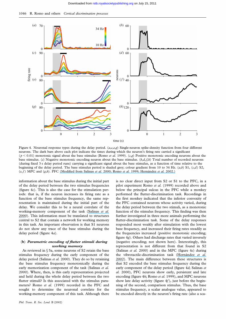
Figure 4. Neuronal response types during the delay period. (a,c,e,g) Single-neuron spike-density function from four differentneurons. The dark bars above each plot indicate the times during which the neuron’s firing rate carried a significant(p � 0.01) monotonic signal about the base stimulus (Romo et al. 1999). (c,g) Positive monotonic encoding neurons about thebase stimulus. (e) Negative monotonic encoding neuron about the base stimulus. (b,d, f,h) Total number of recorded neurons(during fixed 3 s delay period runs) carrying a significant signal about the base stimulus, as a function of time relative to thebeginning of the delay period. The base stimulus period is shaded grey; colour gradient from 10 to 34 Hz. (a,b) S1, (c,d) S2,(e, f ) MPC and (g,h). PFC (Modified from Salinas et al. 2000; Romo et al. 1999; Hernandez et al. 2002.)
information about the base stimulus during the initial partof the delay period between the two stimulus frequencies(figure 4c). This is also the case for the stimulation per-iods: that is, if the neuron increases its firing rate as afunction of the base stimulus frequency, the same rep-resentation is maintained during the initial part of thedelay. We consider this to be a neural correlate of theworking-memory component of the task (Salinas et al.2000). This information must be translated to structurescentral to S2 that contain a network for working memoryin this task. An important observation is that S1 neuronsdo not show any trace of the base stimulus during thedelay period (figure 4a).
(b) Parametric encoding of flutter stimuli duringworking memory
As reviewed in § 5a, some neurons of S2 retain the basestimulus frequency during the early component of thedelay period (Salinas et al. 2000). They do so by retainingthe base stimulus frequency monotonically during theearly memorization component of the task (Salinas et al.2000). Where, then, is this early representation projectedand held during the whole delay period between the twoflutter stimuli? Is this associated with the stimulus para-meters? Romo et al. (1999) recorded in the PFC andsought to determine the neuronal correlate for theworking-memory component of this task. Although there
Phil. Trans. R. Soc. Lond. B (2002)
is no clear direct input from S2 or S1 to the PFC, in apilot experiment Romo et al. (1999) recorded above andbelow the principal sulcus in the PFC while a monkeyperformed the flutter-discrimination task. Recordings inthe first monkey indicated that the inferior convexity ofthe PFC contained neurons whose activity varied, duringthe delay period between the two stimuli, as a monotonicfunction of the stimulus frequency. This finding was thenfurther investigated in three more animals performing theflutter-discrimination task. Some of the delay responsesresponded most weakly after stimulation with the lowestbase frequency, and increased their firing rates steadily asthe frequencies increased (positive monotonic encoding;figure 4g). Others had discharge rates that varied inversely(negative encoding; not shown here). Interestingly, thisrepresentation is not different from that found in S2(Salinas et al. 2000) and in the MPC (figure 4e) duringthe vibrotactile-discrimination task (Hernandez et al.2002). The main difference between these structures isthat S2 encoded the base stimulus frequency during theearly component of the delay period (figure 4d; Salinas etal. 2000), PFC neurons show early, persistent and lateencoding (figure 4h; Romo et al. 1999), and MPC neuronsshow late delay activity (figure 4f ), just before the begin-ning of the second, comparison stimulus. Thus, the basestimulus frequency, a scalar analogue value, appeared tobe encoded directly in the neuron’s firing rate (also a sca-
on July 15, 2011rstb.royalsocietypublishing.orgDownloaded from
Cortical discrimination processes R. Romo and others 1047
_3 0 3 _3
0
3
–3 0 3 –3
0
3
a2
_2 0 2 _2
0
2
0.0s
0.2s 0.5s
–6 0 6 –6
0
6
–3 0 3 –3
0
3
0.0 s
0.2s
0.5s
–3 0 3 –3
0
3
a1
(a) (b)
(c) (d )
(e) ( f )
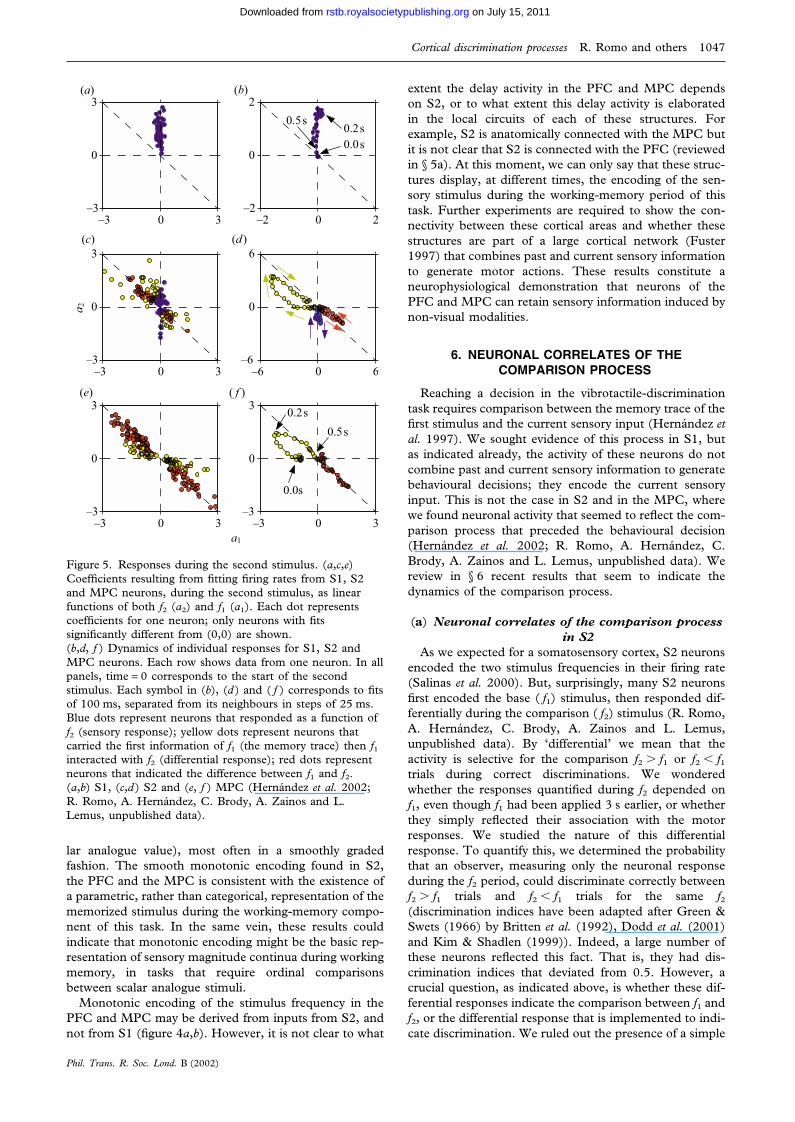
Figure 5. Responses during the second stimulus. (a,c,e)Coefficients resulting from fitting firing rates from S1, S2and MPC neurons, during the second stimulus, as linearfunctions of both f2 (a2) and f1 (a1). Each dot representscoefficients for one neuron; only neurons with fitssignificantly different from (0,0) are shown.(b,d, f ) Dynamics of individual responses for S1, S2 andMPC neurons. Each row shows data from one neuron. In allpanels, time = 0 corresponds to the start of the secondstimulus. Each symbol in (b), (d ) and ( f ) corresponds to fitsof 100 ms, separated from its neighbours in steps of 25 ms.Blue dots represent neurons that responded as a function off2 (sensory response); yellow dots represent neurons thatcarried the first information of f1 (the memory trace) then f1interacted with f2 (differential response); red dots representneurons that indicated the difference between f1 and f2.(a,b) S1, (c,d ) S2 and (e, f ) MPC (Hernandez et al. 2002;R. Romo, A. Hernandez, C. Brody, A. Zainos and L.Lemus, unpublished data).
lar analogue value), most often in a smoothly gradedfashion. The smooth monotonic encoding found in S2,the PFC and the MPC is consistent with the existence ofa parametric, rather than categorical, representation of thememorized stimulus during the working-memory compo-nent of this task. In the same vein, these results couldindicate that monotonic encoding might be the basic rep-resentation of sensory magnitude continua during workingmemory, in tasks that require ordinal comparisonsbetween scalar analogue stimuli.
Monotonic encoding of the stimulus frequency in thePFC and MPC may be derived from inputs from S2, andnot from S1 (figure 4a,b). However, it is not clear to what
Phil. Trans. R. Soc. Lond. B (2002)
extent the delay activity in the PFC and MPC dependson S2, or to what extent this delay activity is elaboratedin the local circuits of each of these structures. Forexample, S2 is anatomically connected with the MPC butit is not clear that S2 is connected with the PFC (reviewedin § 5a). At this moment, we can only say that these struc-tures display, at different times, the encoding of the sen-sory stimulus during the working-memory period of thistask. Further experiments are required to show the con-nectivity between these cortical areas and whether thesestructures are part of a large cortical network (Fuster1997) that combines past and current sensory informationto generate motor actions. These results constitute aneurophysiological demonstration that neurons of thePFC and MPC can retain sensory information induced bynon-visual modalities.
6. NEURONAL CORRELATES OF THECOMPARISON PROCESS
Reaching a decision in the vibrotactile-discriminationtask requires comparison between the memory trace of thefirst stimulus and the current sensory input (Hernandez etal. 1997). We sought evidence of this process in S1, butas indicated already, the activity of these neurons do notcombine past and current sensory information to generatebehavioural decisions; they encode the current sensoryinput. This is not the case in S2 and in the MPC, wherewe found neuronal activity that seemed to reflect the com-parison process that preceded the behavioural decision(Hernandez et al. 2002; R. Romo, A. Hernandez, C.Brody, A. Zainos and L. Lemus, unpublished data). Wereview in § 6 recent results that seem to indicate thedynamics of the comparison process.
(a) Neuronal correlates of the comparison processin S2
As we expected for a somatosensory cortex, S2 neuronsencoded the two stimulus frequencies in their firing rate(Salinas et al. 2000). But, surprisingly, many S2 neuronsfirst encoded the base ( f1) stimulus, then responded dif-ferentially during the comparison ( f2) stimulus (R. Romo,A. Hernandez, C. Brody, A. Zainos and L. Lemus,unpublished data). By ‘differential’ we mean that theactivity is selective for the comparison f2 � f1 or f2 � f1trials during correct discriminations. We wonderedwhether the responses quantified during f2 depended onf1, even though f1 had been applied 3 s earlier, or whetherthey simply reflected their association with the motorresponses. We studied the nature of this differentialresponse. To quantify this, we determined the probabilitythat an observer, measuring only the neuronal responseduring the f2 period, could discriminate correctly betweenf2 � f1 trials and f2 � f1 trials for the same f2(discrimination indices have been adapted after Green &Swets (1966) by Britten et al. (1992), Dodd et al. (2001)and Kim & Shadlen (1999)). Indeed, a large number ofthese neurons reflected this fact. That is, they had dis-crimination indices that deviated from 0.5. However, acrucial question, as indicated above, is whether these dif-ferential responses indicate the comparison between f1 andf2, or the differential response that is implemented to indi-cate discrimination. We ruled out the presence of a simple
on July 15, 2011rstb.royalsocietypublishing.orgDownloaded from

1048 R. Romo and others Cortical discrimination processes
differential motor activity associated with the push-buttonpresses (figure 1a) by testing these S2 neurons in a controltask where the same vibrotactile stimuli were used, butanimals had to follow a visual cue to produce the motorresponse. In this condition, all neurons reduced the devi-ation from 0.5, indicating that the differential activityobserved during the comparison period depends on theactual computation between f1 and f2 and does not reflecta purely motor response aimed to press one of the twopush-buttons.
If the discharges during the comparison period are theproduct of the interaction between f1 and f2, then the traceof f1 and the current f2 could be observed during the com-parison period before the discharges indicated the motorresponses. To further quantify the interaction between f1and f2 during the comparison period and beyond it, weused a multivariate regression analysis (Draper & Smith1981). We fit the activity of each differential response overthe periods before, during and after the comparison per-iod, as a linear function of both f1 and f2. The responses,which in principle could be an arbitrary function of bothf1 and f2, were reasonably well approximated by a generallinear fit to both f1 and f2 as follows:
firing rate = a1 × f1 � a2 × f2 � constant.
In this formula, the coefficients a1 and a2 serve as directmeasurements of firing rate dependence on f1 and f2,respectively. Three lines are of particular importance inthese fits. Points that fall on the a1 = 0 axis representresponses that are a function of f2 (the sensory evidenceof f2; blue dots in figure 5). Points that fall on the a2 = 0axis represent responses that are a function of f1 (thememory trace of f1; yellow dots in figure 5). And pointsthat fall on the a1 = �a2 line represent responses that arefunctions of the difference between f1 and f2 (red dots infigure 5). This last consideration is of particular impor-tance because, in this task, correct behaviour depends onthe sign of the difference between f1 and f2.
The analysis revealed the contributions of f1 and f2 dur-ing the comparison period for S2 neurons (figure 5c,d; fora comparison see in figure 5a,b the responses of S1 neu-rons during the f2 period). Interestingly, when they areplotted as a function of the evolution of the comparisonprocess, it is clearly observed that some neurons evolvefrom coding the sensory stimuli (which could be f1 or f2;see three examples in figure 5d: blue dots indicate a sen-sory response; yellow dots indicate that the neurons carryinformation of f1 then show the difference between f1 andf2; red dots indicate a purely differential response) to adifferential response that is consistent with the motor out-put. Indeed, the analysis of the error trials indicated thatthe differential response correlated with the behaviouralchoice; that is, the selection of the push-button.
These results are important because they show that anearly sensory area shows not only the representation of thecurrent sensory input ( f2; blue dots in figure 5c,d), butalso the representation of the sensory referent ( f1; yellowdots in figure 5c,d) which is stored in the working mem-ory. These two processes are important ingredients for theresulting differential responses between f1 and f2 (reddots in figure 5c,d) that correlate with the behaviouraldecisions. As S2 neurons do not store information aboutthe f1 stimulus during the later part of the delay period,
Phil. Trans. R. Soc. Lond. B (2002)
the comparison process in S2 could be made between theinput from S1 that provides information on the currentstimulus and an input from the frontal cortical areas thatcarries information on the base stimulus during the laterpart of the delay period. However, more experiments areneeded to show whether this is so. The comparison pro-cess is reported by a voluntary motor action, and anatom-ical studies have shown that S2 projects to the motor areasof the frontal lobe (reviewed in § 6a). We explored thepossibility that the motor areas reflect the behaviouraldecision during the vibrotactile-discrimination task.
(b) Neuronal correlates of the comparison processin the MPC
As indicated in § 6a, anatomical studies in monkeyshave shown that S1 and S2 are serially connected (Pons etal. 1987, 1992; Burton & Fabri 1995; Burton et al. 1995;Krubitzer et al. 1995), and that one of the major outputsfrom S2 leads to the motor areas of the frontal lobe(Jones & Powell 1969; Pandya & Kuypers 1969; Jones etal. 1978; Jurgens 1984; Luppino et al. 1993). If we con-sider a serial processing model, in principle S2 could pro-cess the S1 representation of the vibrotactile stimuli andtransmit its output to the motor cortices. As indicated in§ 6a, S2 neurons show a transformation of the S1 vibro-tactile representation (Salinas et al. 2000) and appear toreflect activity associated with the comparison of the twostimuli (R. Romo, A. Hernandez, C. Brody, A. Zainos andL. Lemus, unpublished data). The question that arises iswhether there is a truly clear distinction between thoseareas presumably dedicated to sensory processing andthose traditionally viewed as motor areas. There are twopossibilities. First, the motor areas could process a fullyformed decision signal in order to generate an appropriateset of motor commands. In this case the information andprocesses used before reaching a decision should bemostly absent from motor cortical activity. Second, themotor areas could participate more actively in the forma-tion of a behavioural decision, in which case they shouldreflect details about the sensory inputs regardless of themotor outcome. We tested these two possibilities by rec-ording single neurons in the MPC while monkeys per-formed the vibrotactile-discrimination task (Hernandez etal. 2002).
The responses of single neurons in the MPC wereextremely informative of the sequence of the discrimi-nation process. For example, during the base stimulus,some of the neurons had graded responses as a functionof f1 and displayed a trace of it at the end of the delayperiod between the two stimuli. Interestingly, during thecomparison period these neurons showed informationabout f1 or f2 and then reflected in their activities the dif-ference between the two stimuli (figure 5e, f ). This differ-ential activity was correlated with the motor response onlyduring the discrimination task; these neurons lost their dif-ferential responses when tested in the light-instruction taskmentioned above.
As for the S2 neurons, we studied the dynamics of thecomparison process, and sought evidence of whether thisprocess is due to an interaction between the past ( f1)information and the current sensory stimulus ( f2). A sub-population of the differential neurons displayed infor-mation about f1 (the memory trace; yellow dots in figure
on July 15, 2011rstb.royalsocietypublishing.orgDownloaded from

Cortical discrimination processes R. Romo and others 1049
5e, f; in figure 5e the dots represent individual neurons;figure 5f details the response profiles of a neuron) duringthe comparison period and their responses evolved to adifferential activity that corresponded to an interactionbetween the f1 and f2 stimuli (red dots in figure 5e, f; infigure 5e the dots represent individual neurons; in figure 5fthe response profile of a neuron is given). These responsescould be confined to the comparison period or be pro-longed to the reaction and movement time periods of thebehavioural motor responses. Thus, what is typicallyobserved in the MPC during the comparison period is thatinitially some neurons encode f1 or f2, and later these andother units encode the difference between f1 and f2.
The results indicate that activity in the MPC reflectsmany aspects of the vibrotactile-discrimination task, notjust the motor component. Distinct subpopulations ofMPC neurons are activated during the period where thecomparison between stimuli presumably takes place, andtheir activities during the end of the comparison processare consistent with the behavioural decision. This processappears to precede activity in M1 during the behaviouralresponse associated with this sensory-discrimination task(Mountcastle et al. 1992; R. Romo, A. Hernandez, C.Brody, A. Zainos and L. Lemus, unpublished data).
7. CONCLUDING REMARKS
The vibrotactile-discrimination task requires perceivinga stimulus, storing it in working memory, combining thestored trace with current sensory input and producing adecision that is communicated to the motor apparatus.This temporal sequence is reflected in the activity of someneuronal populations of different cortical areas of the par-ietal and frontal lobes. Our results indicate that neuronscentral to S1 do not simply wait for a signal-encodingdecision, but instead participate in almost every step ofits generation by integrating working-memory and sensoryinputs. Similar processes may occur in other discrimi-nation tasks that require comparison between sensorystimuli. Finally, an important question which needs to beaddressed in this and in similar tasks is how the corticalneuronal circuits interact to produce a comparisonbetween the current sensory input and those sensory rep-resentations stored in working or in long-term memory.Revealing this neural process must be key to understand-ing this finest brain operation that leads to behaviouraldecisions.
The research of R.R. was supported in part by an InternationalResearch Scholars Award from the Howard Hughes MedicalInstitute and from grants from the Millennium Science Initiat-ive, CONACyT and DGAPA-UNAM. The authors thank S.Mendez and L. Lemus for technical assistance.
REFERENCES
Britten, K. H. & Wezel, R. J. 1998 Electrical microstimulationof cortical MST biases heading perception in monkeys. Nat-ure Neurosci. 1, 59–63.
Britten, K. H., Shadlen, M. N., Newsome, W. T. & Movshon,J. A. 1992 The analysis of visual motion: a comparison ofneuronal and psychophysical performance. J. Neurosci. 12,4745–4765.
Burton, H. & Fabri, M. 1995 Ipsilateral intracortical connec-
Phil. Trans. R. Soc. Lond. B (2002)
tions of physiologically defined cutaneous representations inareas 3b and 1 of macaque monkeys. J. Comp. Neurol. 355,508–538.
Burton, H., Fabri, M. & Alloway, K. 1995 Cortical areaswithin the lateral sulcus connected to cutaneous represen-tations in areas 3b and 1: a revisited interpretation of thesecond somatosensory area in macaque monkeys. J. Comp.Neurol. 355, 539–562.
Burton, H., Sinclair, R. J., Hong, S. Y., Pruett, J. R. & Wang,K. C. 1997 Tactile-spatial and cross-modal attention effectsin the second somatosensory and 7b cortical areas of rhesusmonkeys. Somatosens. Mot. Res. 14, 237–267.
Carmichael, S. T. & Price, J. L. 1995 Sensory and premotorconnections of orbital and medial prefrontal cortex ofmacaque monkeys. J. Comp. Neurol. 363, 642–664.
Cavada, C. & Goldman-Rakic, P. S. 1989 The posterior par-ietal cortex in rhesus monkeys: I. Parcellation of areas basedon distinctive limbic and sensory corticocortical connec-tions. J. Comp. Neurol. 287, 393–421.
Darian-Smith, I. 1984 The sense of touch: performance andperipheral neural processes. In Handbook of physiology: sectionI. The nervous system, vol. III. Sensory processes, part 2 (ed. J.M. Brookhart & V. B. Mountcastle), pp. 739–788. Bethesda,MD: American Physiological Society.
Dodd, J. V., Krug, K., Cumming, B. G. & Parker, A. J. 2001Perceptually bistable three-dimensional figures evoke highchoice probabilities in cortical area MT. J. Neurosci. 21,4809–4821.
Draper, N. & Smith, H. 1981 Applied regression analysis. 2ndedn. New York: Wiley.
Fuster, J. M. 1997 Network memory. Trends Neurosci. 20,451–459.
Godshalk, M., Lemon, R. B., Kuypers, H. G. & Ronday, H. K.1984 Cortical afferents and efferents of monkey postarcuatearea: an anatomical and electrophysiological study. Exp.Brain Res. 56, 410–424.
Gold, J. I. & Shadlen, M. N. 2000 Representation of a percep-tual decision in developing oculomotor commands. Nature404, 390–394.
Green, D. M. & Swets, J. A. 1966 Signal detection theory andpsychophysics. New York: Wiley.
Hernandez, H., Salinas, E., Garcia, R. & Romo, R. 1997 Dis-crimination in the sense of flutter: new psychophysicalmeasurements in monkeys. J. Neurosci. 17, 6391–6400.
Hernandez, A., Zainos, A. & Romo, R. 2000 Neuronal corre-lates of sensory discrimination in the somatosensory cortex.Proc. Natl Acad. Sci. USA 97, 6091–6096.
Hernandez, A., Zainos, A. & Romo, R. 2002 Temporal evol-ution of a decision-making process in medial premotor cor-tex. Neuron 33, 959–972.
Horwitz, G. D. & Newsome, W. T. 1999 Separate signals fortarget selection and movement specification in the superiorcolliculus. Science 284, 1158–1161.
Hsiao, S. S., Johnson, K. O. & O’Shaughnessy, D. M. 1993Effects of selective attention of spatial form processing inmonkey primary and secondary somatosensory cortex. J.Neurophysiol. 70, 444–447.
Jiang, W., Tremblay, F. & Chapman, C. E. 1997 Neuronalencoding of texture changes in the primary and in the sec-ondary somatosensory cortical areas of monkeys during pass-ive texture discrimination. J. Neurophysiol. 77, 1656–1662.
Johnson, K. O. & Hsiao, S. S. 1992 Neural mechanisms of tac-tual form and texture perception. A. Rev. Neurosci. 15,227–250.
Jones, E. G. & Powell, T. P. S. 1969 Connexions of thesomatic sensory cortex of the rhesus monkey. I. Ipsilateralcortical connexions. Brain 92, 477–502.
Jones, E. G., Coulter, J. D. & Hendry, S. H. 1978 Intracorticalconnectivity of architectonic fields in the somatic sensory,
on July 15, 2011rstb.royalsocietypublishing.orgDownloaded from
1050 R. Romo and others Cortical discrimination processes
motor and parietal cortex of monkeys. J. Comp. Neurol. 181,291–347.
Jurgens, U. 1984 The efferent and afferent connections of thesupplementary motor area. Brain Res. 300, 63–81.
Kaas, J. H., Nelson, R. J., Sur, M., Lin, C. S. & Merzenich,M. M. 1979 Multiple representations of the body within theprimary somatosensory cortex of primates. Science 204,521–523.
Kim, J. N. & Shadlen, M. N. 1999 Neuronal correlates of adecision in the dorsolateral prefrontal cortex of the macaque.Nature Neurosci. 2, 176–185.
Krubitzer, L., Clarey, J., Tweendale, R., Elston, G. & Calford,M. A. 1995 A redefinition of somatosensory areas in the lat-eral sulcus of macaque monkeys. J. Neurosci. 15, 3821–3839.
LaMotte, R. H. & Mountcastle, V. B. 1975 Capacities ofhumans and monkeys to discriminate between vibratorystimuli of different frequency and amplitude: correlationbetween neural events and psychophysical measurements. J.Neurophysiol. 38, 539–559.
Leichnetz, G. R. 1989 Afferent and efferent connections of thedorsolateral precentral gyrus (area 4, hand/arm region) inthe macaque monkey, with comparisons to area 8. J. Comp.Neurol. 254, 460–492.
Luppino, G., Matelli, M., Camarda, R. M. & Rizzolatti, G. M.1993 Cortico-cortical connections of area F3 (SMA-proper)and area F6 (Pre-SMA) in the macaque monkey. J. Comp.Neurol. 338, 114–140.
Macefield, G., Gandevia, S. C. & Burke, D. 1990 Perceptualresponses to microstimulation of single afferents innervatingjoints, muscles and skin of the human hand. J. Physiol. 429,113–129.
Merchant, H., Zainos, A., Hernandez, A., Salinas, E. & Romo,R. 1997 Functional properties of primate putamen neuronsduring the categorization of tactile stimuli. J. Neurophysiol.77, 1132–1154.
Mishkin, M. 1979 Analogous neural models for tactual andvisual learning. Neurophychologia 17, 139–151.
Mountcastle, V. B. 1957 Modality and topographic propertiesof single neurons of cat’s somatic sensory cortex. J. Neuro-physiol. 20, 408–434.
Mountcastle, V. B., Talbot, W. H., Sakata, H. & Hyvarinen,J. 1969 Cortical neuronal mechanisms in flutter-vibrationstudied in unanesthetized monkeys. Neuronal periodicityand frequency discrimination. J. Neurophysiol. 32, 452–484.
Mountcastle, V. B., LaMotte, R. H. & Carli, G. 1972 Detec-tion thresholds for stimuli in humans and monkeys: com-parison with thresholds events in mechanoreceptive afferentnerve fibers innervating the monkey hand. J. Neurophysiol.25, 122–136.
Mountcastle, V. B., Steinmetz, M. A. & Romo, R. 1990 Fre-quency discrimination in the sense of flutter: psychophysicalmeasurements correlated with postcentral events in behavingmonkeys. J. Neurosci. 10, 3032–3044.
Mountcastle, V. B., Atluri, P. P. & Romo, R. 1992 Selectiveoutput-discriminative signals in the motor cortex of wakingmonkeys. Cerebr. Cortex 2, 277–294.
Murray, E. A. & Mishkin, M. 1984 Relative contributions ofSII and area 5 to tactile discrimination in monkeys. Behav.Brain Res. 11, 67–83.
Nelson, R. J., Sur, M., Felleman, D. J. & Kaas, J. H. 1980Representations of the body surface in the postcentral par-ietal cortex of Macaca fascicularis. J. Comp. Neurol. 192,611–643.
Newsome, W. T., Britten, K. H. & Movshon, J. A. 1989 Neu-ronal correlates of a perceptual decision. Nature 341, 52–54.
Ochoa, J. & Torebjork, E. 1983 Sensations evoked by intraneu-ral microstimulation of single mechanoreceptor unitsinnervating the human hand. J. Physiol. 42, 633–654.
Phil. Trans. R. Soc. Lond. B (2002)
Pandya, D. N. & Kuypers, H. G. 1969 Cortico-cortical con-nections in the rhesus monkey. Brain Res. 13, 13–36.
Pearson, R. C. & Powell, T. P. S. 1985 The projection of pri-mary somatic sensory cortex upon area 5 in the monkey.Brain Res. 356, 89–107.
Phillips, J. R. & Johnson, K. O. 1981 Tactile spatial resolution.II. Neural representations of bars, edges, and gratings inmonkey primary afferents. J. Neurophysiol. 46, 1192–1203.
Pons, T. P., Garraghty, P. E., Friedman, D. P. & Mishkin, M.1987 Physiological evidence for serial processing in somato-sensory cortex. Science 237, 417–420.
Pons, T. P., Garraghty, P. E. & Mishkin, M. 1992 Serial andparallel processing of tactual information in somatosensorycortex of rhesus monkeys. J. Neurophysiol. 68, 518–527.
Powell, T. P. S. & Mountcastle, V. B. 1959 Some aspects ofthe functional organization of the cortex of the postcentralgyrus of the monkey: a correlation of findings obtained in asingle unit analysis with cytoarchitecture. Bull. Johns HopkinsHosp. 105, 133–162.
Preuss, T. M. & Goldman-Rakic, P. S. 1989 Connections ofthe ventral granular frontal cortex of macaques with perisylv-ian premotor and somatosensory areas: anatomical evidencefor somatic representation in primate frontal association cor-tex. J. Comp. Neurol. 282, 293–316.
Recanzone, G. H., Merzenich, M. M. & Schreiner, C. E. 1992Changes in the distributed temporal response properties ofSI cortical neurons reflect improvements in performance ona temporally based tactile discrimination task. J. Neurophy-siol. 67, 1071–1091.
Romo, R. & Salinas, E. 1999 Sensing and deciding in thesomatosensory system. Curr. Opin. Neurobiol 9, 487–493.
Romo, R. & Salinas, E. 2001 Touch and go: decision-makingmechanisms in somatosensation. A. Rev. Neurosci. 24,107–137.
Romo, R., Ruiz, S., Crespo, P., Zainos, A. & Merchant, H.1993 Representation of tactile signals in primate supplemen-tary motor area. J. Neurophysiol. 70, 2690–2694.
Romo, R., Merchant, H., Zainos, A. & Hernandez, A. 1996Categorization of somaesthetic stimuli: sensorimotor per-formance and neuronal activity in primary somatic sensorycortex of awake monkeys. NeuroReport 7, 1273–1279.
Romo, R., Merchant, H., Zainos, A. & Hernandez, A. 1997Categorical perception of somesthetic stimuli: psychophys-ical measurements correlated with neuronal events in pri-mate medial premotor cortex. Cerebr. Cortex 7, 317–326.
Romo, R., Hernandez, A., Zainos, A. & Salinas, E. 1998 Som-atosensory discrimination based on cortical microstimu-lation. Nature 392, 387–390.
Romo, R., Brody, C. D., Hernandez, A. & Lemus, L. 1999Neuronal correlates of parametric working memory in theprefrontal cortex. Nature 339, 470–473.
Romo, R., Hernandez, A., Zainos, A., Brody, C. D. & Lemus,L. 2000 Sensing without touching: psychophysical perform-ance based on cortical microstimulation. Neuron 26, 273–278.
Salinas, E. & Romo, R. 1998 Conversion of sensory signalsinto motor commands in primary motor cortex. J. Neurosci.18, 499–511.
Salinas, E., Hernandez, A., Zainos, A. & Romo, R. 2000Periodicity and firing rate as candidate neural codes for thefrequency of vibrotactile stimuli. J. Neurosci. 20, 5503–5515.
Salzman, D., Britten, K. H. & Newsome, W. T. 1990 Corticalmicrostimulation influences perceptual judgements ofmotion direction. Nature 346, 174–177.
Seidemann, E., Zohary, E. & Newsome, W. T. 1998 Temporalgaiting of neural signals during performance of a visual dis-crimination task. Nature 394, 72–75.
Shadlen, M. N. & Newsome, W. T. 1994 Noise, neural codesand cortical organization. Curr. Opin. Neurobiol. 4, 569–579.
on July 15, 2011rstb.royalsocietypublishing.orgDownloaded from

Cortical discrimination processes R. Romo and others 1051
Shadlen, M. N. & Newsome, W. T. 2001 Neural basis of aperceptual decision in the parietal cortex (area LIP) of therhesus monkey. J. Neurophysiol. 86, 1916–1936.
Shanks, M. F., Person, R. C. & Powell, T. P. S. 1985 The ipsi-lateral cortico-cortical connexions between the cytoarchitec-tonic subdivisions of the primary somatic sensory cortex inthe monkey. Brain Res. 356, 67–88.
Sinclair, R. J. & Burton, H. 1993 Neuronal activity in thesecond somatosensory cortex of monkeys (Macaca mulatta)during active touch of gratings. J. Neurophysiol. 70, 331–350.
Singer, W. & Gray, C. M. 1995 Visual feature integration andthe temporal correlation hypothesis. A. Rev. Neurosci. 18,555–586.
Sur, M., Wall, J. T. & Kaas, J. H. 1984 Modular distributionof neurons with slowly adapting and rapidly adaptingresponses in area 3b of somatosensory cortex in monkeys.J. Neurophysiol. 51, 724–744.
Talbot, W. H., Darian-Smith, I., Kornhuber, H. H. &Mountcastle, V. B. 1968 The sense of flutter-vibration: com-parison of the human capacity response patterns of mech-anoreceptive afferents from the monkey hand. J.Neurophysiol. 31, 301–334.
Tokuno, H. & Tanji, J. 1993 Input organization of distal andproximal forelimb areas in the monkey primary motor cor-tex: retrograde double labeling study. J. Comp. Neurol. 333,199–209.
Vallbo, A. B. 1995 Single-afferent neurons and somatic sen-sation in humans. In The cognitive neurosciences (ed. M. S.Gazzaniga), pp. 237–252. Cambridge, MA: MIT Press.
Phil. Trans. R. Soc. Lond. B (2002)
Vallbo, A. B. & Johansson, R. S. 1984 Properties of cutaneousmechanoreceptors in the human hand related to touch sen-sations. Hum. Neurobiol. 3, 3–14.
Vogels, R. & Orban, G. A. 1990 How well do response changesof striate neurons signal differences in orientations: a studyin the discriminating monkey. J. Neurosci. 10, 3543–3558.
Werner, G. 1980 The study of sensation in physiology. InMedical physiology, vol. 1 (ed. V. B. Mountcastle), pp. 605–628. St Louis, MO: Mosby.
Werner, G. & Mountcastle, V. B. 1965 Neural activity inmechanoreceptive cutaneous afferents: stimulus-responserelations, Weber functions, and information transmission. J.Neurophysiol. 28, 459–497.
GLOSSARY
M1: primary motor cortexMPC: medial premotor cortexPC: Pacinian receptor organPFC: prefrontal cortexPM: premotorQA: Meissner receptor organROC: receiver operating characteristicS1: primary somatosensory cortexS2: secondary somatosensory cortexSA-I: Merkel organSA-II: Ruffini organs
on July 15, 2011rstb.royalsocietypublishing.orgDownloaded from