EXPLORING ROBIN SEQUENCE - Nederlandse Vereniging ...
192
EXPLORING ROBIN SEQUENCE Manouk van Lieshout
-
Upload
khangminh22 -
Category
Documents
-
view
4 -
download
0
Transcript of EXPLORING ROBIN SEQUENCE - Nederlandse Vereniging ...
EXPLORING ROBIN SEQUENCE
Manouk van Lieshout

Van Lieshout, M.J.S. ‘Exploring Robin Sequence’
Cover design: Iliana Boshoven-Gkini - www.agilecolor.comThesis layout and printing by: Ridderprint BV - www.ridderprint.nlISBN: 978-94-6299-693-9
Printing of this thesis has been financially supported by the Erasmus University Rotterdam.
Copyright © M.J.S. van Lieshout, 2017, Rotterdam, the NetherlandsAll rights reserved. No parts of this thesis may be reproduced, stored in a retrieval system, or transmitted in any form or by any means without permission of the author or when appropriate, the corresponding journals

Exploring Robin SequenceVerkenning van Robin Sequentie
Proefschrift
ter verkrijging van de graad van doctor aan deErasmus Universiteit Rotterdam
op gezag van de rector magnificus
Prof.dr. H.A.P. Pols
en volgens besluit van het College voor Promoties.
De openbare verdediging zal plaatsvinden opwoensdag 20 september 2017 om 09.30 uur
door
Manouk Ji Sook van Lieshoutgeboren te Seoul, Korea

PROMOTIECOMMISSIE
Promotoren: Prof.dr. E.B. Wolvius Prof.dr. I.M.J. Mathijssen
Overige leden: Prof.dr. J.de Lange Prof.dr. M. De Hoog Prof.dr. R.J. Baatenburg de Jong
Copromotoren: Dr. K.F.M. Joosten Dr. M.J. Koudstaal

TABLE OF CONTENTS
INTRODUCTION
Chapter I: General introduction 9
Chapter II: Robin Sequence, A European survey on current 37 practice patterns
Chapter III: Non-surgical and surgical interventions for airway 55 obstruction in children with Robin Sequence
AIRWAY OBSTRUCTION
Chapter IV: Unravelling Robin Sequence: Considerations 79 of diagnosis and treatment
Chapter V: Management and outcomes of obstructive sleep 95 apnea in children with Robin Sequence, a cross-sectional study
Chapter VI: Respiratory distress following palatal closure 111 in children with Robin Sequence
QUALITY OF LIFE
Chapter VII: Quality of life in children with Robin Sequence 129
GENERAL DISCUSSION AND SUMMARY
Chapter VIII: General discussion 149
Chapter IX: Summary / Nederlandse samenvatting 169
APPENDICES
About the author 181List of publications 183Ph.D. portfolio 185Words of gratitude / Dankwoord 189


INTRODUCTION


General introduction
1

11 |
Cha
pter
1
General introduction |
BACKGROUND
“I have never seen babies live for more than 16 to 18 months who presented hypoplasia such that the lower maxilla was pushed more than 1 cm behind the upper.’’ – Pierre Robin, 1934
Dr. Pierre Robin was the first to describe neonates who presented with a small mandible, which led to backwards placement of the tongue and finally resulted in obstruction of the airway. This sequence of events, with the small mandible as inciting anomaly, became known as Robin Sequence (RS).
RS (OMIM 261800) is a rare, congenital facial condition occurring in about 1 in 5,600 to 1 in 30,000 newborns.1-5 The condition is classically characterized by underdevelopment of the mandible (mandibular hypoplasia), backwards placement of the tongue (glossoptosis) and airway obstruction. RS can occur isolated, or in combination with other anomalies or syndromes. A cleft palate is present in 80-90% of the RS cases.4,6-8
RS is a challenging disease, comprising of a phenotypically, highly diverse group in which the airway obstruction ranges from very mild to life threatening. Mortality rates for children with RS range from 0.0- 26.0%, a number that may even be higher for children with additional anomalies or a syndrome.9-11 The following part of the introduction will discuss the background and several clinical aspects of RS. At the end of the introduction the aims and outline of this thesis are presented.
HISTORY
The association between a small mandible and cleft palate was made for the first time around 1900.12 However, it was not until 1923 that the description of RS as we know it today was published.13,14 It was the French professor of stomatology, Pierre Robin (1867-1950) who took a particular interest in cases of neonates who presented with airway obstruction, ‘backward and downward fall of the base of the tongue’ and ‘dysmorphic atresia of the mandible’. He also introduced the term ‘glossoptosis’. Robin wrote detailed reports on feeding difficulties, cyanosis, pulmonary complications and treatment methods. In a 30-year period, he published approximately 20 articles on this disorder.
One of Robin’s articles describes the use of a prosthetic appliance to reposition the mandible.14 However, this was not the first published treatment method. In as early

| 12
| Chapter 1
as 1902, Shukowsky was the first to use surgical adhesion of the tongue to the lower lip.15 In the years that followed, a wide range of treatment methods were introduced leading to diversity in management across the world.
Thanks to Robin, the condition as we know it today, was acknowledged. However, even though RS has been recognized and studied for over 100 years, so far, no international consensus with regards to management has been reached. Notably, in the last 40 years, a number of changes took place in terms of the name of ‘Pierre Robin’s’ condition. The name switched from Pierre Robin syndrome to Robin anomalad and then to Robin malformation complex and eventually Robin Sequence.16 In this thesis, we opted to use the term ‘Robin Sequence’. If a syndrome or associated anomalies are present, this will be referred to as non-isolated RS. If no syndrome or associated anomalies are present, this will be referred to as isolated RS.
ETIOLOGY AND PATHOGENESIS
Management of RS is challenging, as it is causally heterogenous and pathogenetically variable. Unfortunately, the exact mechanisms are yet to be elucidated.17-20 Multiple theories have been suggested on the cause of RS, most of them based on the arrest of mandibular development.16 It is thought that due to the arrest of mandibular development, the tongue is kept high in the oral cavity, preventing the closure of palatal shelves. These are the three most well-known theories.
The neuromuscular abnormalities theoryThis theory states that isolated RS originates from an inhibited intra-uterine mandibular motion, which prevents the tongue from migrating inferiorly out of the palatal shelves. This can occur due to prenatal abnormalities in the pharyngolaryngeal and/or glossopharyngeal tone or any other neuromuscular abnormality that would inhibit mandibular motion.20,21
Deformational theory According to this theory, mandibular hypoplasia is secondary to a mechanical problem that restricts mandibular growth, such as the intrauterine position of the embryo, crowding from multiple births or the presence of cervical hemivertebrae.18,21 In these cases, the mandible is intrinsically normal, but is deformed by external factors. The mandible may show catch-up growth after birth, after the mechanical problem is removed.18 This theory applies mostly to patients with an isolated RS.

13 |
Cha
pter
1
General introduction |
Malformational theoryIf RS is ‘malformational’, there is primary failure of mandibular growth caused by a genetic defect.21 In these cases with an inherent growth defect, catch-up growth of the mandible is not likely to occur.18 To understand this theory, it is important to know more of the developmental biology and the genetics of RS. In early development, the mandible originates from the first branchial arch. Both the mesenchymal and connective tissue components are derived from the cranial neural crest cells. After that, a number of pathways lead to the formation of the first mandibular skeletal element: the cartilaginous rod ‘Meckel’s cartilage’. Meckel’s cartilage is one of the most important drivers of early mandibular outgrowth.
Development of (Meckel’s) cartilage is dependent of the SOX9 gene and loss of SOX9 leads to failure of cartilage development. Previous animal studies showed that loss of SOX9 solely in neural crest cells, result in short RS-like mandibles. Furthermore, mandibular outgrowth is a pre-requisite for the morphogenesis of other structures such as the tongue and palate.22 SOX9 is the most described gene, which is involved in the development of RS. SOX9 was first identified in children with campomelic dysplasia who also presented with the triad of mandibular hypoplasia, cleft palate and airway obstruction (SOX 9, 17a24,3).1 Other genes, which may play an important part in the development of RS are GAD67, PVRL1, KCNJ2, KCNJ16, MAP2K6 en SH3BP2.2,3,4
Mutations in these genes lead to a variety of syndromes. The most common syndromes that can occur with RS are Stickler syndrome, 22a11.2 deletion syndrome/velocardiofacial syndrome and Marshall syndrome.23-25 So far, more than 40 different syndromes with RS have been described.19,24,26 Since in about 40-60% a known syndrome is present, genetic testing and counselling is an important part of the RS work-up. Confirmation of genetic defect might have an impact on management decisions.
Definition“This obstruction of the oral pharynx by the lowering of the tongue, let’s call it glossoptosis… “ – Pierre Robin, 1923
Classically RS was described in neonates who presented with a small mandible, which led to backwards placement of the tongue, which resulted in obstruction of the airway. Nowadays, one of the greatest challenges in RS research still lies

| 14
| Chapter 1
within the lack of a uniform definition of RS. Surveys executed among American paediatric otolaryngology fellowship programs, members of the American Cleft Palate-Craniofacial Association and members of Dutch and Belgium cleft palate teams all reported a widespread lack in uniformity in definition.27-31 For example, although Pierre Robin’s original description of the condition did not include cleft palate, this is frequently mentioned as part of the RS definition in literature and clinical practice.9,24,32,33 As long as the aetiology of RS is not completely understood, it remains debated whether this should be an obligatory feature. In this thesis, RS was defined as the triad of mandibular hypoplasia, glossoptosis and airway obstruction. The paragraphs below discuss the separate items.
MandibleSeveral terms are used to describe the mandibular anomaly such as micrognathia (small mandible), mandibular hypoplasia (underdevelopment of the mandible), retrognathia (posterior placed mandible) and/or microretrognathia (posterior placed small mandible). There is a clear semantic difference between the terms. However, at an infant age it is difficult to distinguish between the different forms. In this research and thesis, no distinction was made and we opted to consistently use mandibular hypoplasia. Although, attempts have been made to objectify mandibular hypoplasia, at the moment the diagnosis of mandibular hypoplasia is mostly subjective.34
TongueGlossoptosis is a distinct feature of children with RS, but one not well defined. The severity of glossoptosis and the degree of airway obstruction varies from case to case. There is currently no gold standard to diagnose glossoptosis.
Endoscopy is a helpful tool in order to visualize the position of the tongue and to identify other levels of obstruction. Sher et al. classified four types of airway obstruction based on nasopharyngoscopy results in 33 children with craniofacial anomalies. Type I of this classification describes true glossoptosis or posterior displacement of the tongue against the posterior pharyngeal wall. In Sher type 2, the tongue compresses the soft palates against the posterior pharyngeal wall, in Sher type 3 there is a medial opposition of the lateral pharyngeal walls and Sher type 4 involves sphincteric constriction of the pharynx.
15 |
Cha
pter
1
General introduction |
In a study by Marques et al., a Sher type I was found in 80.0% of 62 RS children.8 In this same study, it was proposed to further categorize Sher type I into mild, moderate and severe glossoptosis. However, no correlation was established between the severity of glossoptosis and the degree of clinical manifestations.8 More recently, the reliability of awake Flexible Fiberoptic Laryngoscopy in diagnosing glossoptosis was examined, offering promising results.35 Besides endoscopy, radiographic studies are available, but these are considered of limited value.
Airway obstructionAirway obstruction is another important feature of RS, but no widely accepted definition exists for children with RS. The spectrum of airway obstruction is broad with variation in type, severity and level.
PRENATAL DIAGNOSIS
Early recognition of RS is highly important since newborns may present with severe, in some cases life threatening respiratory distress. In these cases immediate intervention may be indicated. Ideally, recognition of mandibular hypoplasia would take place in the prenatal phase, allowing pre-arranged specialized care directly after birth. Indeed, prenatal imaging techniques, such as ultrasonography and MRI play an increasing role in early diagnosis. However, so far detection rates are still low due to difficulties in imaging. In the literature, the range for detection varies between 7 and 22%.24,36,37
In general, fetal mandibular hypoplasia remains the best ‘call sign’ for suspecting RS, despite a reported low specificity and even though it may only be possible to clearly document mandibular hypoplasia until the third trimester.38,39 To objectify mandibular hypoplasia in utero, a few methods are available such as the jaw-index and the inferior facial angle which both have a high sensitivity and specificity.40-42 After mandibular hypoplasia is suspected, one should further assess an abnormal position of the tongue or cleft palate. Other important sonographic findings include the presence of a polyhydramnion or additional anomalies.40,43
Fortunately, knowledge is improving, thereby holding a significant promise for prenatal detection of mandibular hypoplasia and its relation with airway problems.40-43 Future research should be focused on determining normal mandibular growth values, optimizing imaging techniques and the significance of associated findings (e.g. polyhydramnios or additional anomalies). Furthermore, establishing

| 16
| Chapter 1
the link between prenatal findings and the severity of postnatal problems will be essential.
AIRWAY OBSTRUCTION
The airway obstruction in children with RS is primarily caused by posterior placement of the tongue into the hypopharynx. However, other mechanisms may also be involved such as disproportion of the tongue and the mandible, neuromuscular impairment of the genioglossus and other parapharyngeal muscles and/or increased negative oesophageal pressure drawing the tongue in the hypopharynx.44 Airway obstruction in children with RS may also be caused by movement of the pharyngeal walls leading to narrowing of the oropharynx.45 Finally, (secondary) structural airway anomalies such as laryngomalacia, tracheomalacia and subglottic narrowing may also play a role.46,47
The severity of the airway obstruction in children varies from mild to severe, or in some cases life threatening. The presentation of a child with a severe form of RS is obvious and consists of choking sounds while trying to breathe, episodes of apnea and cyanosis and increased efforts of the respiratory muscles. Children with a more mild type of RS maintain an adequate airway when awake or crying, but may have obstruction when they fall asleep or exacerbate with oral feeding.48,49
Upper airway obstruction occuring during sleep, is also referred to as Sleep-Disordered Breathing (SDB). SDB is not a distinct disease, but rather a syndrome of upper airway dysfunction during sleep characterized by snoring and/or increased respiratory effort secondary to enhanced upper airway resistance and pharyngeal collapsibility.48,49 Obstructive SDB includes a spectrum of clinical entities with variable severity of intermittent upper airway obstruction during sleep i.e. primary snoring (PS), upper airway resistance syndrome (UARS) and obstructive sleep apnea (OSA).
The focus in this thesis will be on OSA. OSA is one of the clinical manifestations of upper airway obstruction. OSA is a ‘disorder of breathing during sleep characterized by prolonged partial upper airway obstruction and/or intermitted complete obstruction (obstructive apnea) that disrupts normal ventilation during sleep and normal sleep patterns’.50 The reported prevalence of OSA in children with RS based on polysomnography (PSG), the gold standard to diagnose OSA, ranges between 46-100% depending on used criteria (table 1).51-56 Accurate diagnosis of OSA is important since it is associated with considerable comorbidity such as failure

17 |
Cha
pter
1
General introduction |
to thrive, recurrent infections, pulmonary hypertension, cor pulmonale and sudden death if left untreated.57,58
Table 1. Reported OSA prevalence in literature based on PSG results
Age at PSG evaluation Prevalence Severity
Bull et al., 1990 56 N/A 18/21 (86.0%) N/AGilhooly et al., 1993 52 Newborn infants 6/13 (46.2%) Not definedWilson et al., 2000 54 N/A, but infant age 10/11 (90.1%) N/ABravo et al., 2005 51 N/A 31/52 (59.6%) N/AAnderson et al., 2011 53 48 days
(range 7-214 days)11/13 (84.6%) Mean oAHI 33.5
(range 0 tot 85.7)Daniel et al., 2012 81 N/A 39/39 (100%) 10/39 mild/moderate OSA
(AHI ≤10). 29/39 severe OSA (AHI ≤10)
Mac Lean et al., 2012 55 N/A 8/8 (100.0%) Mean mOAHI: 45.2 (±8.6) All infants had an oMAHI > 3 events/hour.
Khayat et al., 2017 60 0.8 year (± 0.3) 22/46 (46.8%) 10/22 mild, 3/22 moderate and 9/22 severe
A few studies examined the course of OSA with time. Classically it was thought infants with RS would develop airway obstruction directly after birth. However, two studies with a small sample size (n=11 and n=15) suggested that absence of OSA on the initial PSG in the neonatal period does not guarantee the obstruction will not develop at a later age.52,54 In one study, up to 70% of the infants with RS did not present with obstructive airway pathology until 24-51 days of age.54 In addition, another study also did not find a decrease in OSA severity with advancing age.59
Due to the complexity of the airway obstruction and the potential for secondary difficulties a thorough diagnostic airway evaluation is needed, preferably soon after birth. With exception of those that present with life threatening respiratory distress requiring immediate intervention, the diagnostic process for most children starts with clinical observation. Clinical symptoms of OSA include habitual snoring, laboured or cessation of breathing during sleep, disturbed or restless sleep, cyanosis, irritability and excessive daytime sleepiness.61,62 Furthermore, a structured clinical assessment may be helpful. In this assessment, the sleeping infant is cradled upright and then lowered into a supine positioning while looking for signs of respiratory distress.63,64 If there is no sign of obstruction, this procedure is repeated while bottle-feeding.

| 18
| Chapter 1
Another helpful tool to illustrate the type of airway obstruction is the jaw thrust manoeuvre, in which the lower jaw (including the tongue) is pulled upwards. Improvement of respiratory distress after the jaw thrust suggests the obstruction is localized at the base of the tongue. Other clinical manifestations of airway obstruction include feeding difficulties, poor weight gain and poor sucking.
Besides clinical observation, a number of other objective diagnostic tools are available to assess the airway obstruction and quantify the degree of respiratory distress. For example, with regard to OSA history, standardized questionnaires such as the Brouillette, OSA-18 and the paediatric sleep questionnaire may be helpful. However, its use has not yet extensively been described in children with RS. Fibre optic nasopharyngoscopy/bronchoscopy is also an accepted diagnostic method to establish the type, level and severity of airway obstruction.65 Finally, in order to detect more severe respiratory compromise, assessing hypoxemia and hypercapnia by continuous oxygen saturation monitoring and blood gas carbon dioxide qualifications are also important and relatively simple methods.64,66 Other methods, which may be used in a setting in which PSG is not readily available include plain films, sleep fluoroscopy, computed tomography, cine MRI, awake laryngoscopy and drug-induced sleep endoscopy.67 However, reports of these techniques specifically in patients with RS are scarce.
PSG is currently considered as the gold standard to establish the presence and quantification of OSA and to identify any potential aspect of central apnea contributing to the infants’ respiratory compromise.68 PSG consists of simultaneous recording of multiple physiological parameters related to sleep and wakefulness such as the nasal airflow, respiratory effort and CO2 levels. In some settings, video or nursing staff observation may also be available. Evaluation by PSG can take place at home or in the hospital. Although more research is needed, so far, reasonably good results have been reported of the use of portable monitoring devices at home in the diagnosis of OSA in children.69,70
For the scoring of the respiratory events, the American Academy of Sleep Medicine provides guidelines.71 The number of mixed, obstructive and central apneas and hypopneas and desaturations per hour of total sleep time can be summarized in the apnea-hypopnea index (AHI), obstructive apnea-hypopnea index (oAHI), mixed obstructive apnea-hypopnea index (moAHI), central apnea-index (CAI) or oxygen desaturation index (ODI). In general, having a value >1 for one of these parameters is considered necessary for a diagnosis of OSA or CSA. However, various cut-off

19 |
Cha
pter
1
General introduction |
values to define mild, moderate and severe OSA have been reported in literature.72,73 In this thesis, the grading system according to Guilleminault was used defining OSA as oAHI ≥1 per hour. In case of OSA, patients can be further divided into those with mild OSA (oAHI ≥1 and <5), moderate OSA (oAHI ≥5 and <24) or severe OSA (oAHI ≥25).
Little is known about the presence of central sleep apnea (CSA), but a recent study suggests the prevalence of CSA may be significantly higher in the RS population compared to the general paediatric population.59
Even though PSG is considered the gold standard to diagnose OSA and CSA, it was recently shown that only one in five studies reported use of PSG in RS infants.65 Also, uniform criteria to classify the respiratory distress in children with OSA are lacking. Furthermore, due to lack of in-hospital equipment or access, PSG is therefore currently not used in the standard diagnostic work-up of the airway in children with RS.
TREATMENT OF THE AIRWAY OBSTRUCTION
The airway obstruction in children with RS ranges from very mild to life threatening. In literature, mortality rates up to 26.0% have been reported.9,10 The goal of treatment is temporarily or definitive relieve of the obstruction, thereby aiding adequate growth and development. Airway, feeding and growth are intimately interlinked to each other.
Although about 70% of children can be managed conservatively, other children with RS are in need for treatment other than prone positioning.10,66 Children with a non-isolated RS seem to require more aggressive management than those with isolated RS.25,46,74 In literature a number of non-surgical and surgical interventions have been described, but evidence remains scarce and there is widespread lack of consensus amongst clinicians. Furthermore, although several treatment plans have been proposed, there is currently no widely accepted treatment algorithm.18,19,75
Except for prone positioning and mandibular distraction, all techniques will be more extensively discussed in chapter II.
Non-surgical treatmentProne positioning is the most widely accepted and oldest technique in the treatment of RS. Robin already recommended feeding and positioning in prone in the late

| 20
| Chapter 1
twenties (1926, Robin). By positioning the infant prone, the tongue falls forward and relieves the obstruction. Sleeping in prone positioning increases the risk for Sudden Infant Death Syndrome (SIDS). However, to our knowledge no cases of RS and SIDS have been reported in literature.
Nasopharyngeal airway (NPA) is another popular non-surgical treatment method. An NPA is placed through the nose and positioned at the distal end of the larynx after bypassing the tongue base. The NPA permits the child to breathe through the tube, but may also break the seal between the posterior placed tongue base and the pharynx wall. The mean duration of an NPA varies in literature from 44 days – 9 months. Success rates range from 67-100%.54,73,76-79
Continuous positive airway pressure (CPAP) and non-invasive positive pressure (NIPP) ventilation both apply positive pressure during a breathing cycle, thereby maintaining the airway patency. Even though, these techniques were already used in craniofacial patients by Guilleminault in 1986, only a few authors reported the use of CPAP or NIPP in patients with RS and OSA.51-54,66,80-84 Most of these cases concerned older RS patients, with exception of two articles that described the successful of CPAP in RS neonates and infants with improvement of breathing pattern, decreased respiratory effort and improved PSG results.80 In these groups, the mean duration of home ventilation therapy was 16 months and four weeks to four months. In another study of five patients with RS, clinical improvement with non-invasive ventilation was also reported.85
A final non-surgical measure is an orthodontic appliance. Already in 1967, Pielou described the use of an acrylic plastic obturator with an extension posteriorly beyond the distal border of the palate, thereby preventing the tongue to fall back in the pharynx.86 In the years that followed, only a few more reports on oral appliances were published. Recently a number of positive results from the Tuebingen group in Germany were published on the use of a pre-epiglottic baton plate (PEBP).87-89 This PEBP was characterized by a velar extension shifting the base of the tongue forward resulting in a widened hypopharyngeal space.
Surgical treatmentIn 1911 Shukowsky was the first to suggest to suture the tongue to the lower lip to resolve the glossoptosis (1911 Shukowsky). This was one of the first descriptions of the glossopexy technique. Later, glossopexy would appear in many different forms including approximations to the lip, the mandible and the hyoid. The tongue to lip

21 |
Cha
pter
1
General introduction |
adhesion (TLA) has now become the surgical treatment of choice in many centers. By suturing the tongue to the inside of the lower lip, the tongue is maintained in a forward position. After a number of months, when the airway deems safe, the tongue is released. Success rates of TLA vary between 70-95%.75,90-99
Subperiosteal release of the floor of the mouth (SPRFM) is another surgical technique, which was first described by Delorme in 1989. SPRFM is based on the theory that the musculature of the floor of the mouth is under an increased tension creating the glossoptosis.100 The glossoptosis can be relieved via a submental incision, followed by a subperiosteal dissection of the medial side of the mandible, allowing the tongue base to fall down into the floor of the mouth. Only a few manuscripts have been published on this technique with success rates ranging from 50-84%.101-103
A small number of reports describe mandibular traction, a technique introduced in 1937 by Callister (1937 Callister).104,105 By mandibular traction, the tongue and mandible are forced in a forward position, slowly lengthening, creating a larger oropharyngeal space and relieving the glossoptosis. In a 2009 article from Germany, the authors report mandibular traction is the treatment of choice for severe airway obstruction.106 In this clinic a mandibular traction system is used consisting of an acrylic plate fixed around the mandible with wires.
Mandibular distraction osteogenesis seems currently the most popular surgical technique. In MDO, surgical osteotomies are followed by gradually lengthening of the mandible. This mandibular advancement brings forward the insertions of the muscles of the floor of the mouth and consequently the tongue base thereby relieving the airway obstruction. In MDO different devices are available and many different approaches and techniques have been described.107-110 In several centers, MDO is the surgical treatment of choice. So far, good results have been reported in literature in terms of relieve of the airway obstruction or achieving successful decannulation.111,112 Several studies also described the successful use of MDO in young children aged below three months old as a way to avoid a tracheostomy and its associated morbidity.112-114
However, MDO has a few disadvantages. First, it only offers gradual improvement of the airway obstruction (which may not always be an option for severe RS cases needing immediate airway support). Secondly, since MDO is a relatively new practice, the long-term effects on mandibular growth are still unknown. Finally, each device and each approach seem to have its own specific limitations. The

| 22
| Chapter 1
main disadvantages of extra-oral devices are scarring and the physical presence of the device and related problems.107 Disadvantages of intra-oral devices include scarring (if an extra-oral approach is used), a more difficult vector control, the risk on a post-operative open bite and the need for a second general anesthetic upon removal.107,108 In general MDO carries the additional risk of complications such as local infections of the skin, nerve damage, dental complications and device failure or migration.109,110
The use of a tracheostomy is considered to be the safest therapy in those infants with a severe, and in some cases life-threatening airway obstruction. In a tracheostomy, a tube is placed directly into the trachea thereby bypassing the upper airway obstruction. In a recent survey among the American Cleft Palate-Craniofacial Association members tracheostomy was in only 16% the preferred primary surgical treatment option.115
FEEDING DIFFICULTIES
After airway obstruction, feeding difficulties are considered the second most important challenge. Feeding difficulties may be caused by a combination of factors. For example, insufficient energy levels may exist for the action of eating, due to the amount of energy used to breathe against an obstructed airway.116 Backwards placement of the tongue causes obstruction of the oral cavity. Also, if present, cleft of the palate results in inability to build up sufficient negative intraoral pressure for suction. Finally, in some cases motor dysfunction may be involved. Recent studies show oesophageal manometric anomalies (such as incomplete relaxation of the lower oesophageal sphincter, oesophageal dyskinesia and lower oesophageal sphincter hypertonia) in 50-100% of the children with mandibular hypoplasia.117,118 The authors suggest these anomalies are caused by dysregulation of the swallowing center in the brainstem.117
Neonates with feeding difficulties are at risk for growth problems, which is often referred to as ‘failure to thrive’. Failure to thrive is thought to occur in about half of the cleft palate patients (and in 100% of severe congenital mandibular hypoplasia patients).116 There is currently no consensus on diagnosing the feeding difficulties in patients with RS, but assessment of the feeding difficulties is highly important. Nutritional support is often necessary and this can consist of high caloric drinks, feeding facility techniques, specialist bottles such as a Habermann bottle, nasogastric tube (NGT) and/or gastric feeding tube (G-tube).66,119 About 38-62% of the children

23 |
Cha
pter
1
General introduction |
with RS will eventually need tube feeding.120,121 The presence of gastroesophageal reflux (GER) can exaggerate both feeding and breathing difficulties by compromising swallowing mechanism.121
CLEFT PALATE
A cleft palate is present in about 80-90% of the children with RS.4,6-8 In most cases the cleft is complete, wide and U-shaped and in the minority complete or incomplete, narrow and V-shaped. 8 The majority of RS patients has a Veau cleft type II, although presence of solely a Veau cleft I is also commonly described.122 It is suggested that patients with RS have more extensive clefting than those with an isolated cleft palate.123
Following cleft palate repair, children with RS are at risk to develop airway-related complications. In small case series post-operative airway complications such as stridor, postoperative respiratory distress, difficulties on intubation, intubation or a tracheostomy are reported with incidences ranging from 24% to 31%.124-127 Children with RS have a more restricted oropharyngeal space and are therefore more likely to develop respiratory distress in case of lingual swelling caused by, for example, a tongue retractor.128 In order to minimize the chance on these types of airway complications, some authors recommend postponing palatal repair in children with RS.129 However, this is in conflict with the tendency to close early in order to facilitate normal speech development.
MANDIBULAR GROWTH
So far, a number of objective measurement methods have been described to objectively monitor mandibular size and growth such as the jaw-index, the maxilla-mandibular discrepancy, lateral cephalograms or CT-scans, each with its specific advantages and disadvantages.34,41,130-136 Other objective measurement methods may include cone beam CT, and (3D) photography. But, currently there is no gold standard to objectively monitor mandibular size and growth.
It is often stated that the airway obstruction improves with age as the neuromuscular coordination becomes better and the mandible grows.63 Interestingly, some authors specifically mention the presence of so-called catch-up growth in their RS study population. With catch-up growth, it is postulated that the mandible develops relatively faster than the rest of facial structures. However, whether this catch up

| 24
| Chapter 1
growth really takes place and in which patients it occurs, is unclear. One study states that there are two distinct groups. In the first group the underlying cause of RS is a mechanical problem that restricts mandibular growth.18 When this restraint is removed, catch-up growth takes place. In the second group there is an internal growth defect. These patients will not demonstrate catch-up growth.18,137
In general, studies in favour for catch-up growth seem in minority and state that catch-up growth usually would occur when the child is between 6 months and 2 years old.33,34,138,139 Most of these studies are relatively old. More recent studies conclude there is no catch-up growth of the mandible.135,140-147 In other words, the mandible remains small. Unfortunately, most of these studies have a small sample size and only focus on a certain age group. Since the presence of catch-up growth may influence the choice for a certain treatment, more research to this phenomenon is needed.
COGNITIVE AND FUNCTIONAL DEVELOPMENT
OSA may influence the cognitive development in children with RS. This is due to the intermittent hypoxia and/or sleep disturbance associated with OSA.148 Not much is known about the cognitive development in children with RS. A few studies observe cognitive impairment, but knowledge regarding the prevalence, severity and precise cause is limited.7,149,150
Closely related is satisfaction with their facial appearance, psychosocial status and health-related quality of life (QOL). Both are considered important health outcomes.151,152 Only one study focused specifically on these psychosocial problems in children with RS.150 It was found that there were no significant differences concerning self-concept, emotional or behavior problems compared to healthy peers.
Speech and hearingAs stated previously, a cleft palate occurs in about 80-90 % of patients with RS.4,6,7 Following palatal closure, children are at risk to develop a velopharyngeal insufficiency (VPI). VPI diminishes oral volume and is therefore associated with articulation errors and abnormal nasal air emission. Studies regarding speech outcomes specifically in RS patients are limited. Hoffman and Lehmann reported a satisfactory or good outcome of speech after palatal closure in two thirds of the RS patients.153,154 The study of De Buys Roessingh, published in 2008, found that even after intensive speech therapy only 55% of the 38 RS patients achieved a good

25 |
Cha
pter
1
General introduction |
speech outcome, defined as a type 1 or type ½ Borel-Maisonny Score.155 A number of studies reported differences in speech outcome between isolated and non-isolated RS patients.156,157
So far, little attention has been given to the hearing status of children with RS. Studies report an incidence of hearing loss between 46% and 60%.66,157-159 Gould et al. compared the incidence and degree of hearing loss between children with isolated cleft palate and RS, but no significant difference was found.159 However, the study of Handzic revealed an incidence of hearing loss of 73-83% in children with RS, which was significantly more than the isolated cleft palate group that showed an incidence of 58- 59%.160 Hearing loss in RS patients was also significantly more severe. The majority of RS patients had a moderate (25-40dB) or severe hearing loss (>40dB). Although hearing improved over time in the isolated cleft palate group was this not the case in the RS patients. Hearing loss in RS patients was most often conductive and bilateral without middle ear or inner ear malformations. The ears of the RS patients with hearing loss revealed middle ear effusion. The authors advised ventilation tubes at the same time as the palatoplasty.
OUTLINE AND AIMS OF THIS THESIS
This thesis aims to provide better understanding of RS with regards to diagnosis, treatment and quality of life.
Current State of Evidence and Patterns of PracticeIn order to get a better understanding of the differences that currently exist within Europe, a survey study in Chapter II is presented. This study aims to answer questions such as: ‘How do clinicians define RS?’ ‘Is PSG commonly used?’ ‘What are the preferred treatment methods?’ On basis of this and a few other survey- and literature studies, notions on management seem to differ highly from clinic to clinic. However, so far no study systematically categorized and summarized the available evidence on treatment of the airway obstruction in RS. This is the focus of Chapter III of this thesis.
Airway ObstructionThe next three chapters focus on obstructive sleep apnea in children with RS. In Chapter IV an overview is given of the experience of our clinic with diagnosis and treatment. Chapter V describes the prevalence and course of obstructive sleep apnea

| 26
| Chapter 1
in children with RS above the age of one year old. In this chapter an attempt is made to identify patients at risk for continuing or re-developing respiratory distress. Chapter VI specifically focuses on the prevalence, severity and duration of respiratory distress following palatoplasty in children with RS and evaluates perioperative management. As mentioned previously, OSA may influence the cognitive development in children with RS due to the intermittent hypoxia and/or sleep disturbance.
Quality of LifeIn Chapter VII a closer look is taken at the health-related quality of life in children with RS based on questionnaire studies and its relation to the child’s respiratory status.
Discussion & Summary In Chapter VIII and Chapter IX the most important findings will be summarized and discussed against the background of published literature. Furthermore, clinical implications and future directions will be considered.
27 |
Cha
pter
1
General introduction |
LIST OF REFERENCES1. Bush PG, Williams AJ. Incidence of the
Robin Anomalad (Pierre Robin syndrome). Br J Plast Surg. 1983;36(4):434-437.
2. Tolarova MM, Cervenka J. Classification and birth prevalence of orofacial clefts. Am J Med Genet. 1998;75(2):126-137.
3. Printzlau A, Andersen M. Pierre Robin Sequence in Denmark: A Retrospective Population-Based Epidemiological Study. Cleft Palate-Craniofac J. 2004;41(1):47-52.
4. Vatlach S, Maas C, Poets CF. Birth prevalence and initial treatment of Robin sequence in Germany: a prospective epidemiologic study. Orphanet J Rare Dis. 2014;9:9.
5. Paes EC, van Nunen DP, Basart H, et al. Birth prevalence of Robin sequence in the Netherlands from 2000-2010: A retrospective population-based study in a large Dutch cohort and review of the literature. Am J Med Genet A. 2015.
6. Costa MA, Murage KP, Tholpady SS, Flores RL. Airway compromise following palatoplasty in Robin sequence: improving safety and predictability. Plast Reconst Surg. 2014;134(6):937e-945e.
7. Caouette-Laberge L, Bayet B, Larocque Y. The Pierre Robin sequence: review of 125 cases and evolution of treatment modalities. Plast Reconstr Surg. 1994;93(5):934-942.
8. Marques IL, De Sousa TV, Carneiro AF, Peres SPDBA, Barbieri MA, Bettiol H. Robin sequence: A single treatment protocol. J Pediatr. 2005;81(1):14-22.
9. Butow KW, Hoogendijk CF, Zwahlen RA. Pierre Robin sequence: appearances and 25 years of experience with an innovative treatment protocol. J Pediatr Surg. 2009;44(11):2112-2118.
10. van den Elzen AP, Semmekrot BA, Bongers EM, Huygen PL, Marres HA. Diagnosis and treatment of the Pierre Robin sequence: results of a retrospective clinical study and review of the literature. Eur J Pediatr. 2001;160(1):47-53.
11. Costa MA, Tu MM, Murage KP, Tholpady SS, Engle WA, Flores RL. Robin sequence: mortality, causes of death, and clinical outcomes. Plast Reconstr Surg. 2014;134(4):738-745.
12. Eley RC, Farber, S. Hypoplasia of the mandible (micrognathy) as a cause of cyanotic attacks in the newly-born infant: report of four cases. Amer. J. Dis. Child. 1930;39(1167-1175).
13. Robin P. Backward lowering of the root of the tongue causing respiratory disturbance. Bull. Acad. med. 1923;89:37-41.
14. Paletta CE, Dehghan K, Hutchinson RL, Klaw BA. A fall of the base of the tongue considered as a new cause of nasopharyngeal respiratory impairment: Pierre Robin sequence, a translation. Plast Reconstr Surgy. 1994;93(6):1301-1303.
15. St-Hilaire H, Buchbinder D. Maxillofacial pathology and management of Pierre Robin sequence. Otolaryngol Clin North Am. 2000;33(6):1241-1256, vi.
16. Sadewitz VL. Robin sequence: changes in thinking leading to changes in patient care. Cleft Palate Craniofac J. 1992;29(3):246-253.
17. Cohen MM, Jr. Robin sequences and complexes: causal heterogeneity and pathogenetic/phenotypic variability. Am J Med Genet. 1999;84(4):311-315.
18. Mackay DR. Controversies in the diagnosis and management of the Robin sequence. J Craniofac Surg. 2011;22(2):415-420.

| 28
| Chapter 1
19. Evans KN, Sie KC, Hopper RA, Glass RP, Hing AV, Cunningham ML. Robin sequence: from diagnosis to development of an effective management plan. Pediatrics. 2011;127(5):936-948.
20. Abadie V, Morisseau-Durand MP, Beyler C, Manach Y, Couly G. Brainstem dysfunction: A possible neuroembryological pathogenesis of isolated Pierre Robin sequence. Eur J Pediatr. 2002;161(5):275-280.
21. Sadewitz VL. Robin sequence: Changes in thinking leading to changes in patient care. Cleft Palate-Craniofac J. 1992;29(3):246-253.
22. Stewart K, Uetani N, Hendriks W, Tremblay ML, Bouchard M. Inactivation of LAR family phosphatase genes Ptprs and Ptprf causes craniofacial malformations resembling Pierre-Robin sequence. Development. 2013;140(16):3413-3422.
23. Shprintzen RJ. The implications of the diagnosis of Robin sequence. Cleft Palate Craniofac J. 1992;29(3):205-209.
24. Holder-Espinasse M, Abadie V, Cormier-Daire V, et al. Pierre Robin sequence: a series of 117 consecutive cases. J Pediatr. 2001;139(4):588-590.
25. Izumi K, Konczal LL, Mitchell AL, Jones MC. Underlying genetic diagnosis of pierre robin sequence: Retrospective chart review at Two Children’s Hospitals and a systematic literature review. J Pediatr. 2012;160(4):645-650.e642.
26. Izumi K, Konczal LL, Mitchell AL, Jones MC. Underlying genetic diagnosis of Pierre Robin sequence: retrospective chart review at two children’s hospitals and a systematic literature review. J Pediatr. 2012;160(4):645-650 e642.
27. Myer Iii CM, Reed JM, Cotton RT, Wlllging JP, Shott SR. Airway management in Pierre Robin sequence. Otolaryngol Head Neck Surg. 1998;118(5):630-635.
28. Breugem CC, Courtemanche DJ. Robin sequence: Clearing nosologic confusion. Cleft Palate-Craniofac J. 2010;47(2):197-200.
29. Breugem CC, Mink van der Molen AB. What is ‘Pierre Robin sequence’? J Plast Reconstr Aesthetic Surg. 2009;62(12):1555-1558.
30. Collins B, Powitzky R, Robledo C, Rose C, Glade R. Airway Management in Pierre Robin Sequence: Patterns of Practice. Cleft Palate Craniofac J. 2013.
31. Basart H, Kruisinga FH, Breugem CC, Don Griot JP, Hennekam RC, Van der Horst CM. Will the right Robin patient rise, please? Definitions and criteria during management of Robin sequence patients in the Netherlands and Belgium. J Craniomaxillofac Surg. 2015;43(1):92-96.
32. Tan TY, Kilpatrick N, Farlie PG. Developmental and genetic perspectives on Pierre Robin sequence. Am J Med Genet C Semin Med Genet. 2013;163C(4):295-305.
33. Figueroa AA, Glupker TJ, Fitz MG, BeGole EA. Mandible, tongue, and airway in Pierre Robin sequence: a longitudinal cephalometric study. Cleft Palate Craniofac J. 1991;28(4):425-434.
34. Pruzansky S, Richmond JB. Growth of mandible in infants with micrognathia; clinical implications. AMA Am J Dis Child. 1954;88(1):29-42.
35. Basart H, Konig AM, Bretschneider JH, et al. Awake Flexible Fiberoptic Laryngoscopy to diagnose glossoptosis in Robin Sequence patients. Clin Otolaryngol. 2015.
36. Offerdal K, Jebens N, Syvertsen T, Blaas HG, Johansen OJ, Eik-Nes SH. Prenatal ultrasound detection of facial clefts: a prospective study of 49,314 deliveries in a non-selected population in Norway. Ultrasound Obstet Gynecol. 2008;31(6):639-646.

29 |
Cha
pter
1
General introduction |
37. Maarse W, Berge SJ, Pistorius L, et al. Diagnostic accuracy of transabdominal ultrasound in detecting prenatal cleft lip and palate: a systematic review. Ultrasound Obstet Gynecol. 2010;35(4):495-502.
38. Lind K, Aubry MC, Belarbi N, et al. Prenatal diagnosis of Pierre Robin Sequence: accuracy and ability to predict phenotype and functional severity. Prenat Diagn. 2015;35(9):853-858.
39. Pilu G, Romero R, Reece EA, Jeanty P, Hobbins JC. The prenatal diagnosis of Robin anomalad. Am J Obstet Gynecol. 1986;154(3):630-632.
40. Paladini D. Fetal micrognathia: almost always an ominous finding. Ultrasound Obstet Gynecol. 2010;35(4):377-384.
41. Paladini D, Morra T, Teodoro A, Lamberti A, Tremolaterra F, Martinelli P. Objective diagnosis of micrognathia in the fetus: the jaw index. Obstet Gynecol. 1999;93(3):382-386.
42. Rotten D, Levaillant JM, Martinez H, Ducou le Pointe H, Vicaut E. The fetal mandible: a 2D and 3D sonographic approach to the diagnosis of retrognathia and micrognathia. Ultrasound Obstet Gynecol. 2002;19(2):122-130.
43. Hsieh YY, Chang CC, Tsai HD, Yang TC, Lee CC, Tsai CH. The prenatal diagnosis of Pierre-Robin sequence. Prenat Diagn. 1999;19(6):567-569.
44. Argamaso RV. Glossopexy for upper airway obstruction in Robin sequence. Cleft Palate-Craniofac J. 1992;29(3):232-238.
45. Sher AE. Mechanisms of airway obstruction in Robin sequence: Implications for treatment. Cleft Palate-Craniofac J. 1992;29(3):224-231.
46. Cruz MJ, Kerschner JE, Beste DJ, Conley SF. Pierre Robin sequence: Secondary respiratory difficulties and intrinsic feeding abnormalities. Laryngoscope. 1999;109(10):1632-1636.
47. Knapp K, Powitzky R, Digoy P. Subglottic stenosis: another challenge for intubation and potential mechanism of airway obstruction in Pierre Robin Sequence. Int J Pediatr Otorhinolaryngol. 2011;75(9):1075-1077.
48. Kaditis A, Kheirandish-Gozal L, Gozal D. Algorithm for the diagnosis and treatment of pediatric OSA: a proposal of two pediatric sleep centers. Sleep medicine. 2012;13(3):217-227.
49. Katz ES, D’Ambrosio CM. Pathophysiology of pediatric obstructive sleep apnea. Proceedings of the American Thoracic Society. 2008;5(2):253-262.
50. Standards and indications for cardiopulmonary sleep studies in children. American Thoracic Society. Am J Respir Crit Care Med. 1996;153(2):866-878.
51. Bravo G, Ysunza A, Arrieta J, Pamplona MC. Videonasopharyngoscopy is useful for identifying children with Pierre Robin sequence and severe obstructive sleep apnea. Int J Pediatr Otorhinolaryngol. 2005;69(1):27-33.
52. Gilhooly JT, Smith JD, Howell LL, Deschaine BL, Richey SL. Bedside polysomnography as an adjunct in the management of infants with Robin sequence. Plast Reconstr Surg. 1993;92(1):23-27.
53. Anderson IC, Sedaghat AR, McGinley BM, Redett RJ, Boss EF, Ishman SL. Prevalence and severity of obstructive sleep apnea and snoring in infants with Pierre Robin sequence. Cleft Palate Craniofac J. 2011;48(5):614-618.
54. Wilson AC, Moore DJ, Moore MH, Martin AJ, Staugas RE, Kennedy JD. Late presentation of upper airway obstruction in Pierre Robin sequence. Arch Dis Child. 2000;83(5):435-438.

| 30
| Chapter 1
55. MacLean JE, Fitzsimons D, Fitzgerald DA, Awaters K. The spectrum of sleep-disordered breathing symptoms and respiratory events in infants with cleft lip and/or palate. Arch Dis Child. 2012;97(12):1058-1063.
56. Bull MJ, Givan DC, Sadove AM, Bixler D, Hearn D. Improved outcome in Pierre Robin sequence: Effect of multidisciplinary evaluation and management. Pediatrics. 1990;86(2):294-301.
57. Nixon GM. Sleep. 8: paediatric obstructive sleep apnoea. Thorax. 2005.
58. Spicuzza L. Paediatric sleep apnoe: early onset of the syndrome? Sleep Med. Rev. . 2009.
59. Lee JJ, Thottam PJ, Ford MD, Jabbour N. Characteristics of sleep apnea in infants with Pierre-Robin sequence: Is there improvement with advancing age? Int J Pediatr Otorhinolaryngol. 2015.
60. Khayat A, Bin-Hassan S, Al-Saleh S. Polysomnographic findings in infants with Pierre Robin sequence. Ann Thorac Med. 2017;12(1):25-29.
61. Ward SL, Marcus CL. Obstructive sleep apnea in infants and young children. J Clin Neurophysiol. 1996;13(3):198-207.
62. Chan J, Edman JC, Koltai PJ. Obstructive sleep apnea in children. Am Fam Physician. 2004;69(5):1147-1154.
63. de Buys Roessingh AS, Herzog G, Hohlfeld J. Respiratory distress in Pierre Robin: successful use of pharyngeal tube. J Pediatr Surg. 2007;42(9):1495-1499.
64. Wagener S, Rayatt SS, Tatman AJ, Gornall P, Slator R. Management of infants with Pierre Robin sequence. Cleft Palate Craniofac J. 2003;40(2):180-185.
65. Reddy VS. Evaluation of upper airway obstruction in infants with Pierre Robin sequence and the role of polysomnography - Review of current evidence. Paediatr Respir Rev. 2015.
66. Glynn F, Fitzgerald D, Earley MJ, Rowley H. Pierre Robin sequence: An institutional experience in the multidisciplinary management of airway, feeding and serous otitis media challenges. Int J Pediatr Otorhinolaryngol. 2011;75(9):1152-1155.
67. Manickam PV, Shott SR, Boss EF, et al. Systematic review of site of obstruction identification and non-CPAP treatment options for children with persistent pediatric obstructive sleep apnea. Laryngoscope. 2015.
68. Section on Pediatric Pulmonology SoOSASAAoP. Clinical practice guideline: diagnosis and management of childhood obstructive sleep apnea syndrome. Pediatrics. 2002;109(4):704-712.
69. Alonso-Alvarez ML, Teran-Santos J, Ordax Carbajo E, et al. Reliability of home respiratory polygraphy for the diagnosis of sleep apnea in children. Chest. 2015;147(4):1020-1028.
70. Tan HL, Kheirandish-Gozal L, Gozal D. Pediatric Home Sleep Apnea Testing: Slowly Getting There! Chest. 2015;148(6):1382-1395.
71. Berry RB, Budhiraja R, Gottlieb DJ, et al. Rules for scoring respiratory events in sleep: update of the 2007 AASM Manual for the Scoring of Sleep and Associated Events. Deliberations of the Sleep Apnea Definitions Task Force of the American Academy of Sleep Medicine. J Clin Sleep Med. 2012;8(5):597-619.
72. Van Den Elzen APM, Semmekrot BA, Bongers EMHF, Huygen PLM, Marres HAM. Diagnosis and treatment of the Pierre Robin sequence: Results of a retrospective clinical study and review of the literature. Eur J Pediatr. 2001;160(1):47-53.
73. Abel F, Bajaj Y, Wyatt M, Wallis C. The successful use of the nasopharyngeal airway in Pierre Robin sequence: An 11-year experience. Arch Dis Child. 2012;97(4):331-334.

31 |
Cha
pter
1
General introduction |
74. Tomaski SM, Zalzal GH, Saal HM. Airway obstruction in the Pierre Robin sequence. Laryngoscope. 1995;105(2):111-114.
75. Kirschner RE, Low DW, Randall P, et al. Surgical airway management in Pierre Robin sequence: Is there a role for tongue-lip adhesion? Cleft Palate-Craniofac J. 2003;40(1):13-18.
76. Chang AB, Masters IB, Williams GR, Harris M, O’Neil MC. A modified nasopharyngeal tube to relieve high upper airway obstruction. Pediatr Pulmonol. 2000;29(4):299-306.
77. Wagener S, Rayatt SS, Tatman AJ, Gornall P, Slator R. Management of infants with Pierre Robin sequence. Cleft Palate-Craniofac J. 2003;40(2):180-185.
78. Parhizkar N, Saltzman B, Grote K, et al. Nasopharyngeal airway for management of airway obstruction in infants with micrognathia. Cleft Palate-Craniofac J. 2011;48(4):478-482.
79. Anderson KD, Cole A, Chuo CB, Slator R. Home management of upper airway obstruction in Pierre Robin Sequence using a nasopharyngeal airway. Cleft Palate-Craniofac J. 2007;44(3):269-273.
80. Leboulanger N, Picard A, Soupre V, et al. Physiologic and clinical benefits of noninvasive ventilation in infants with Pierre Robin sequence. Pediatrics. 2010;126(5):e1056-e1063.
81. Daniel M, Bailey S, Walker K, et al. Airway, feeding and growth in infants with Robin sequence and sleep apnoea. Int J Pediatr Otorhinolaryngol. 2013.
82. Deegan PC, McGlone B, McNicholas WT. Treatment of Robin sequence with nasal CPAP. J Laryngol Otol. 1995;109(4):328-330.
83. Miller SDW, Glynn SF, Kiely JL, McNicholas WT. The role of nasal CPAP in obstructive sleep apnoea syndrome due to mandibular hypoplasia. Respirology. 2010;15(2):377-379.
84. Amaddeo A, Abadie V, Chalouhi C, et al. Continuous Positive Airway Pressure for Upper Airway Obstruction in Infants with Pierre Robin Sequence. Plast Reconstr Surg. 2016;137(2):609-612.
85. Girbal IC, Goncalves C, Nunes T, et al. Non-invasive ventilation in complex obstructive sleep apnea--a 15-year experience of a pediatric tertiary center. Rev Port Pneumol. 2014;20(3):146-151.
86. Pielou WD. Non-surgical management of Pierre Robin syndrome. Arch Dis Child. 1967;42(221):20-23.
87. Bacher M, Sautermeister J, Urschitz MS, Buchenau W, Arand J, Poets CF. An oral appliance with velar extension for treatment of obstructive sleep apnea in infants with Pierre Robin sequence. Cleft Palate Craniofac J. 2011;48(3):331-336.
88. Von Bodman A, Buchenau W, Bacher M, Arand J, Urschitz MS, Poets CF. Treatment of infants with Pierre Robin Sequence using a new oral appliance. Wien Klin Wochenschr. 2003;115(24):871-873.
89. Buchenau W, Urschitz MS, Sautermeister J, et al. A randomized clinical trial of a new orthodontic appliance to improve upper airway obstruction in infants with Pierre Robin sequence. J Pediatr. 2007;151(2):145-149.
90. Cumberbatch EP, Robinson CA. Treatment of Gonococcal Infection by Diathermy. Br Med J. 1923;2(3263):54-56.
91. Cohn EJ, Berggren RE. Studies in the Physical Chemistry of the Proteins : Iii. The Relation between the Amino Acid Composition of Casein and Its Capacity to Combine with Base. J Gen Physiol. 1924;7(1):45-79.
92. Hoffman W. Outcome of tongue-lip plication in patients with severe Pierre Robin sequence. J Craniofac Surg. 2003;14(5):602-608.

| 32
| Chapter 1
93. Denny AD, Amm CA, Schaefer RB. Outcomes of tongue-lip adhesion for neonatal respiratory distress caused by Pierre Robin sequence. J Craniofac Surg. 2004;15(5):819-823.
94. Huang F, Lo LJ, Chen YR, Yang JC, Niu CK, Chung MY. Tongue-lip adhesion in the management of Pierre Robin sequence with airway obstruction: Technique and outcome. Chang Gung Med J. 2005;28(2):90-96.
95. Cozzi F, Totonelli G, Frediani S, Zani A, Spagnol L, Cozzi DA. The effect of glossopexy on weight velocity in infants with Pierre Robin syndrome. J Pediatr Surg. 2008;43(2):296-298.
96. Bijnen CL, Don Griot PJW, Mulder WJ, Haumann TJ, Van Hagen AJ. Tongue-lip adhesion in the treatment of Pierre Robin sequence. J Craniofac Surg. 2009;20(2):315-320.
97. Rogers GF, Murthy AS, LaBrie RA, Mulliken JB. The GILLS score: part I. Patient selection for tongue-lip adhesion in Robin sequence. Plast Reconstr Surg. 2011;128(1):243-251.
98. Abramowicz S, Bacic JD, Mulliken JB, Rogers GF. Validation of the GILLS score for tongue-lip adhesion in robin sequence patients. J Craniofac Surg. 2012;23(2):382-386.
99. Mann RJ, Neaman KC, Hill B, Bajnrauh R, Martin MD. A novel technique for performing a tongue-lip adhesion - The tongue suspension technique. Cleft Palate-Craniofac J. 2012;49(1):27-31.
100. Delorme RP, Larocque Y, Caouette-Laberge L. Innovative surgical approach for the Pierre Robin anomalad: Subperiosteal release of the floor of the mouth musculature. Plast Reconstr Surg. 1989;83(6):960-966.
101. Breugem CC, Olesen PR, Fitzpatrick DG, Courtemanche DJ. Subperiosteal release of the floor of the mouth in airway management in Pierre Robin sequence. J Craniofac Surg. 2008;19(3):609-615.
102. Caouette-Laberge L, Borsuk DE, Bortoluzzi PA. Subperiosteal release of the floor of the mouth to correct airway obstruction in Pierre Robin sequence: Review of 31 cases. Cleft Palate-Craniofac J. 2012;49(1):14-20.
103. Siddique S, Haupert M, Rozelle A. Subperiosteal release of the floor of the mouth musculature in two cases of Pierre Robin sequence. Ear Nose Throat J. 2000;79(10):816-819.
104. Easter B, Wood C, Eppley BL, Sadove AM. Mandibular traction system for adjunctive management of airway insufficiency in infants with Pierre Robin malformation sequence. Am J Occup Ther. 1991;45(10):941-943.
105. Wada T, Ishi T, Sugai T. Mandibular traction for relieving respiratory distress in the Pierre Robin anomaly. A case report. J Maxillofac Surg. 1983;11(4):187-190.
106. Pradel W, Lauer G, Dinger J, Eckelt U. Mandibular Traction-An Alternative Treatment in Infants With Pierre Robin Sequence. J Oral Maxillofac Surg. 2009;67(10):2232-2237.
107. Genecov DG, Barcelo CR, Steinberg D, Trone T, Sperry E. Clinical experience with the application of distraction osteogenesis for airway obstruction. J Craniofac Surg. 2009;20(8 SUPPL. 2):1817-1821.
108. Pluijmers BI, Koudstaal MJ, Wolvius EB, Van Der Wal KGH. Custom-made intraoral mandibular distraction as treatment for neonatal airway obstruction. Int J Oral Maxillofac Surg. 2012;41(2):186-191.

33 |
Cha
pter
1
General introduction |
109. Paes EC, Mink van der Molen AB, Muradin MS, et al. A systematic review on the outcome of mandibular distraction osteogenesis in infants suffering Robin sequence. Clin Oral Investig. 2013;17(8):1807-1820.
110. Hong P. A clinical narrative review of mandibular distraction osteogenesis in neonates with Pierre Robin sequence. Int J Pediatr Otorhinolaryngol. 2011;75(8):985-991.
111. Sidman JD, Sampson D, Templeton B. Distraction osteogenesis of the mandible for airway obstruction in children. Laryngoscope. 2001;111(7):1137-1146.
112. Ow ATC, Cheung LK. Meta-analysis of mandibular distraction osteogenesis: Clinical applications and functional outcomes. Plast Reconstr Surg. 2008;121(3):54e-69e.
113. Denny A, Kalantarian B. Mandibular distraction in neonates: A strategy to avoid tracheostomy. Plast Reconstr Surg. 2002;109(3):896-904.
114. Scott AR, Tibesar RJ, Lander TA, Sampson DE, Sidman JD. Mandibular distraction osteogenesis in infants younger than 3 months. Arch Facial Plast Surg. 2011;13(3):173-179.
115. Collins B, Powitzky R, Robledo C, Rose C, Glade R. Airway management in pierre robin sequence: patterns of practice. Cleft Palate Craniofac J. 2014;51(3):283-289.
116. Pandya AN, Boorman JG. Failure to thrive in babies with cleft lip and palate. Br J Plast Surg. 2001;54(6):471-475.
117. Baujat G, Faure C, Zaouche A, Viarme F, Couly G, Abadie V. Oroesophageal motor disorders in Pierre Robin syndrome. J Pediatr Gastroenterol Nutr. 2001;32(3):297-302.
118. Baudon JJ, Renault F, Goutet JM, et al. Motor dysfunction of the upper digestive tract in Pierre Robin sequence as assessed by sucking-swallowing electromyography and esophageal manometry. J Pediatr. 2002;140(6):719-723.
119. Marques IL, Prado-Oliveira R, Leiriao VH, Jorge JC, de Souza L. Clinical and fiberoptic endoscopic evaluation of swallowing in Robin sequence treated with nasopharyngeal intubation: the importance of feeding facilitating techniques. Cleft Palate Craniofac J. 2010;47(5):523-529.
120. Li HY, Lo LJ, Chen KS, Wong KS, Chang KP. Robin sequence: review of treatment modalities for airway obstruction in 110 cases. Int J Pediatr Otorhinolaryngol. 2002;65(1):45-51.
121. Evans AK, Rahbar R, Rogers GF, Mulliken JB, Volk MS. Robin sequence: a retrospective review of 115 patients. Int J Pediatr Otorhinolaryngol. 2006;70(6):973-980.
122. Patel KB, Sullivan SR, Murthy AS, Marrinan E, Mulliken JB. Speech outcome after palatal repair in nonsyndromic versus syndromic Robin sequence. Plast Reconstr Surg. 2012;130(4):578e-585e.
123. Godbout A, Leclerc JE, Arteau-Gauthier I, Leclerc LD. Isolated versus pierre robin sequence cleft palates: are they different? Cleft Palate Craniofac J. 2014;51(4):406-411.
124. Costa M, Murage K, Tholpady S, Havlik R, Flores R. Airway compromise following cleft palate repair in robin sequence: improving safety and predictability. Plast Reconstr Surg. 2014;134(4 Suppl 1):6-7.
125. Antony AK, Sloan GM. Airway obstruction following palatoplasty: analysis of 247 consecutive operations. Cleft Palate Craniofac J. 2002;39(2):145-148.

| 34
| Chapter 1
126. Dell’Oste C, Savron F, Pelizzo G, Sarti A. Acute airway obstruction in an infant with Pierre Robin syndrome after palatoplasty. Acta Anaesthesiol Scand. 2004;48(6):787-789.
127. Reilly J, Russell, C., Gibson, N., Moores, T., Ray, A., Devlin, M. & Wynne, D. Sleep disordered breathing patterns in children presenting for cleft palate repair. Pierre Robin Sequence is associated with pre-and postperative respiratory compromise. . Poster Abstract presented at Britisch Academic Conference in Otolaryngology Posters; 2012.
128. Junghaenel S, Keller T, Mischkowski R, et al. Massive macroglossia after palatoplasty: case report and review of the literature. Eur J Pediatr. 2012;171(3):433-437.
129. Henriksson TG, Skoog VT. Identification of children at high anaesthetic risk at the time of primary palatoplasty. Scand J Plast Reconstr Surg Hand Surg. 2001;35(2):177-182.
130. Figueroa AA, Glupker TJ, Fitz MG, BeGole EA. Mandible, tongue, and airway in Pierre Robin sequence: a longitudinal cephalometric study. Cleft Palate Craniofac J. 1991;28(4):425-434.
131. Laitinen SH, Ranta RE. Cephalometric measurements in patients with Pierre Robin syndrome and isolated cleft palate. Scand J Plast Reconstr Surg Hand Surg. 1992;26(2):177-183.
132. Daskalogiannakis J, Ross RB, Tompson BD. The mandibular catch-up growth controversy in Pierre Robin sequence. Am J Orthod Dentofacial Orthop. 2001;120(3):280-285.
133. Lu DW, Shi B, Wang HJ, Zheng Q. The comparative study of craniofacial structural characteristic of individuals with different types of cleft palate. Ann Plast Surg. 2007;59(4):382-387.
134. Suri S, Ross RB, Tompson BD. Craniofacial morphology and adolescent facial growth in Pierre Robin sequence. Am J Orthod Dentofacial Orthop. 2010;137(6):763-774.
135. Vegter F, Hage JJ, Mulder JW. Pierre Robin syndrome: mandibular growth during the first year of life. Ann Plast Surg. 1999;42(2):154-157.
136. van der Haven I, Mulder JW, van der Wal KG, Hage JJ, de Lange-de Klerk ES, Haumann TJ. The jaw index: new guide defining micrognathia in newborns. Cleft Palate Craniofac J. 1997;34(3):240-241.
137. Long R. Growth patterns in and the growth effects of treatment in the pierre robin sequence. Presented at: ACPA Pre-Conference Symposium, The Pierre Robin Sequence: Diagnostic and Treatment Challenges for the Cleft Team. Scottsdale, AZ; April 20, 2009. 2009.
138. Pruzansky S. Not all dwarfed mandibles are alike. Birth Defects Orig Artic Ser. 1969;5:120-129.
139. Schaefer RB, Stadler JA, 3rd, Gosain AK. To distract or not to distract: an algorithm for airway management in isolated Pierre Robin sequence. Plast Reconstr Surg. 2004;113(4):1113-1125.
140. Laitinen SH, Ranta RE. Cephalometric measurements in patients with Pierre Robin syndrome and isolated cleft palate. Scand J Plast Reconstr Surg Hand Surg. 1992;26(2):177-183.
141. Daskalogiannakis J, Ross RB, Tompson BD. The mandibular catch-up growth controversy in Pierre Robin sequence. Am J Orthod Dentofacial Orthop. 2001;120(3):280-285.
142. Hermann NV, Kreiborg S, Darvann TA, Jensen BL, Dahl E, Bolund S. Early craniofacial morphology and growth in children with nonsyndromic Robin Sequence. Cleft Palate Craniofac J. 2003;40(2):131-143.

35 |
Cha
pter
1
General introduction |
143. Hermann NV, Kreiborg S, Darvann TA, Jensen BL, Dahl E, Bolund S. Craniofacial morphology and growth comparisons in children with Robin Sequence, isolated cleft palate, and unilateral complete cleft lip and palate. Cleft Palate Craniofac J. 2003;40(4):373-396.
144. Eriksen J, Hermann NV, Darvann TA, Kreiborg S. Early postnatal development of the mandible in children with isolated cleft palate and children with nonsyndromic Robin sequence. Cleft Palate Craniofac J. 2006;43(2):160-167.
145. Krimmel M, Kluba S, Breidt M, et al. Three-dimensional assessment of facial development in children with Pierre Robin sequence. J Craniofac Surg. 2009;20(6):2055-2060.
146. Suri S, Ross RB, Tompson BD. Craniofacial morphology and adolescent facial growth in Pierre Robin sequence. Am J Orthod Dentofacial Orthop. 2010;137(6):763-774.
147. Ozawa T, Lorenzoni D, Franca de Oliveira L, Silva Filho O. Facial Profile Evaluation of Isolated Pierre Robin Sequence. Cleft Palate Craniofac J. 2010.
148. Bass JL, Corwin M, Gozal D, et al. The effect of chronic or intermittent hypoxia on cognition in childhood: a review of the evidence. Pediatrics. 2004;114(3):805-816.
149. Kapp-Simon KA, Krueckeberg S. Mental development in infants with cleft lip and/or palate. Cleft Palate Craniofac J. 2000;37(1):65-70.
150. Drescher FD, Jotzo M, Goelz R, Meyer TD, Bacher M, Poets CF. Cognitive and psychosocial development of children with Pierre Robin sequence. Acta Paediatr. 2008;97(5):653-656.
151. Bonomi AE, Patrick DL, Bushnell DM, Martin M. Validation of the United States’ version of the World Health Organization Quality of Life (WHOQOL) instrument. J Clin Epidemiol. 2000;53(1):1-12.
152. Urden LD. Patient satisfaction measurement: current issues and implications. Lippincotts Case Manag. 2002;7(5):194-200.
153. Lehman JA, Fishman JR, Neiman GS. Treatment of cleft palate associated with Robin sequence: appraisal of risk factors. Cleft Palate Craniofac J. 1995;32(1):25-29.
154. Hoffman S, Kahn S, Seitchik M. Late problems in the management of the pierre robin syndrome. Plast Reconstr Surg. 1965;35:504-511.
155. de Buys Roessingh AS, Herzog G, Cherpillod J, Trichet-Zbinden C, Hohlfeld J. Speech prognosis and need of pharyngeal flap for non syndromic vs syndromic Pierre Robin Sequence. J Pediatr Surg. 2008;43(4):668-674.
156. Witt PD, Myckatyn T, Marsh JL, Grames LM, Dowton SB. Need for velopharyngeal management following palatoplasty: an outcome analysis of syndromic and nonsyndromic patients with Robin sequence. Plast Reconstr Surg. 1997;99(6):1522-1529; discussion 1530-1524.
157. Pasyayan HM, Lewis MB. Clinical experience with the Robin sequence. Cleft Palate J. 1984;21(4):270-276.
158. Williams AJ, Williams MA, Walker CA, Bush PG. The Robin anomalad (Pierre Robin syndrome)--a follow up study. Arch Dis Child. 1981;56(9):663-668.
159. Gould HJ. Audiologic findings in Pierre Robin sequence. Ear Hear. 1989;10(3):211-213.
160. Handzic J, Bagatin M, Subotic R, Cuk V. Hearing levels in Pierre Robin syndrome. Cleft Palate Craniofac J. 1995;32(1):30-36.


Robin Sequence: A European survey on current practice patterns
Manouk J.S. van LieshoutKoen F.M. JoostenIrene M.J. MathijssenMaarten J. KoudstaalHans L.J. HoeveMarc P. van der SchroeffEppo B. Wolvius
Published in the Journal of Craniomaxillofacial Surgery 2015 Oct;43(8):1626-31. doi: 10.1016/j.jcms.2015.07.008. PMID: 26315273
Supplementary material can be found at http://www.sciencedirect.com/science/article/pii/S10105182150023462

| 38
| Chapter 2
ABSTRACT
To provide an overview of current practice patterns with regard to Robin sequence (RS) patients in Europe, a survey was conducted among European clinicians. This online survey consisted of different sections assessing characteristics of the respondent and clinic, definition, diagnosis, treatment, and follow-up. In total, surveys from 101 different European clinics were included in the analysis, and 56 different RS definitions were returned. The majority (72%) of the respondents used a sleep study system to determine the severity of the airway obstruction. A total of 63% used flexible endoscopy and 16% used rigid endoscopy in the diagnostic process. Treatment of the airway obstruction differed considerably between the different countries. Prone positioning for mild airway obstruction was the treatment modality used most often (63%). When prone positioning was not successful, a nasopharyngeal airway was used (62%). Surgical therapies varied considerably among countries. For severe obstruction, mandibular distraction was performed most frequently. Three-quarters of the respondents noted the presence of catch-up growth in their patient population. This first European survey study on definition and management of RS shows that there are considerable differences within Europe. Therefore, we would encourage the establishment of national (and international) guidelines to optimize RS patient care.
39 |
Cha
pter
2
Robin Sequence, A European survey |
INTRODUCTION
Robin Sequence (RS) is a condition classically characterized by micrognathia, glossoptosis and airway obstruction.1 Since this description in the original monograph of Pierre Robin, several authors have used modified definitions.2,3 RS has an estimated incidence of about 1 in 8000 to 1 in 20,000 newborns depending on the criteria used to define RS.4-7 It can be divided into those with an isolated and those with a non-isolated condition, the latter being present in about 40% of the cases. Several problems, such as airway obstruction and feeding difficulties may occur in children with RS. In severe RS cases this may require long-lasting admission to the paediatric intensive care unit.
Frequently, authors have described that care of children with RS should be multidisciplinary, with non-surgical and surgical disciplines involved, but there seems to be no consensus on the diagnostic work-up and treatment.8,9 Recent European literature describes a wide range of diagnostic and treatment modalities. We conducted a survey to provide an overview of current practice patterns of RS within Europe and to provide a springboard for future discussion. To our knowledge, this is the first European survey to focus on RS.
MATERIAL AND METHODS
For this cross-sectional study, an online survey was constructed by the author panel, which was followed by pilot testing among eight local clinicians who were acquainted with RS. After further refinement, the final survey consisted of 23–44 questions, depending on the answers given (Supplementary Material). Both multiple choice questions and open questions were included. Most questions were formulated in such a way that the respondent was asked to give a response for his or her clinic, assuming that definition and management were uniform within the clinic. The survey could be accessed in a secure survey environment (Lime Survey Version 1.91 + Erasmus Medical Center).
In total, 655 persons in the European network existing through contacts of the Cleft Center Rotterdam and the Dutch Craniofacial Center were invited. This network includes pediatricians, otolaryngologists, plastic surgeons, oral and maxillofacial surgeons, and nurse practitioners. All received an e-mail message with an online link to the survey. If the e-mailed person was not involved in care of children with RS, we asked them to send the e-mail address of the person who was involved in their clinic. This person was then invited. After the initial survey distribution we send out

| 40
| Chapter 2
a maximum of two reminders to increase response rate. The survey period was from June 2013 until November 2013. The survey was available only in English.
Both complete and incomplete surveys were included in the analysis with the exception of surveys that were considered unreliable by all authors for various reasons (e.g., only completion of the respondent characteristics). If more than one survey per clinic was filled out, we included only the first submitted survey in the main analysis. Statistical analysis was performed using IBM SPSS version 20 (SPSS Inc., Chicago, IL, USA). We performed only descriptive statistical calculations. For this survey study, we obtained approval of the Medical Ethics Commission (MEC-2014-242) of the Erasmus Medical Center.
RESULTS
Characteristics of the respondentsA total of 138 responses were collected (a response rate of 21%). Twelve surveys were excluded from the main analysis because they originated at clinics that had already returned a survey, and 25 surveys were excluded because only the respondent’s characteristics were filled out. In total, 101 surveys from 24 European countries were included in the main analysis.
The response between countries was variable. There were five countries with more than five respondents: the United Kingdom (n = 26), Germany (n = 12), the Netherlands (n = 11), France (n = 8), and Sweden (n = 6). The other countries had fewer than 5 respondents: Ireland, Italy, Portugal and Spain had four respondents; Belgium, Romania, and Switzerland had three; Hungary had two; and Austria, Croatia, Denmark, Estonia, Finland, Greece, Latvia, Lithuania, Norway, Poland, and Turkey had one respondent.
The large majority of the respondents (72%) were specialists in the field of oral and maxillofacial surgery, plastic surgery, or otorhinolaryngology. Other fields included pediatrics (15%) and cleft nurse, orthodontist, or pulmonologist (10%). Three respondents did not answer on this item. Respondents worked mainly in university hospitals (70%) but also in general hospitals (21%), private practices (4%), or another type of clinic (6%). In most of the respondents’ clinics, a specialized multidisciplinary cleft and/or craniofacial team was present: 35% had a cleft team, 6% had a craniofacial team and 45% had both a cleft team and a craniofacial team. Twelve percent did not have a cleft team or craniofacial team, and 3% did not

41 |
Cha
pter
2
Robin Sequence, A European survey |
answer on this question. The number of new RS children who were seen annually in the respondents clinic were more than five children (33%), five to 10 children (42%), 10 to 15 children (11%), and more than 15 children (11%).
Definition In total, 56 different combinations of features necessary for a diagnosis of RS were returned (Table 1). About one in three respondents distinguished micrognathia, retrognathia, and mandibular hypoplasia. In the comments, some respondents noted that micrognathia is a small mandible (size), that retrognathia is a normal-sized, backwards-placed mandible (position), and that mandibular hypoplasia is the same as micrognathia. Others regarded the different terms as a grade of severity, with micrognathia being the most severe form. However, several respondents noted that the use of a certain term does not influence their management.
Table 1. Definition of RS
Features obligatory for a diagnosis of RS
Mandibular hypoplasia 39 (39%) Retrognathia 52 (52%) Micrognathia 65 (65%) Cleft palate 75 (75%) Glossoptosis 65 (65%) Macroglossia 7 (7%) Clinical airway obstruction 47 (47%) Airway obstruction proven by a diagnostic modality 11 (11%)
Feeding difficulties 19 (19%) Other 2 (2%)Distinction retrognathia, micrognathia and mandibular hypoplasia Yes 35 (35%) No 65 (66%)Distinction between isolated and non-isolated RS Yes 54 (54%) No 47 (47%)Most often mentioned combinations of features obligatory for a diagnosis of RS* N=101 Mandibular hypoplasia and/or retrognathia and/or micrognathia and glossoptosis
and cleft palate25 (25%)
Mandibular hypoplasia and/or retrognathia and/or micrognathia and cleft palate 12 (12%) Mandibular hypoplasia and/or retrognathia and/or micrognathia and clinical
airway obstruction and glossoptosis and cleft palate10 (10%)
*Mandibular hypoplasia, retrognathia and micrognathia have been pooled together in this table.

| 42
| Chapter 2
A cleft palate was considered an obligatory feature for a diagnosis of RS by 96% in the United Kingdom, 88% in France, 67% in Sweden, 58% in Germany, and 55% in the Netherlands. A clear distinction between isolated and non-isolated RS was made by roughly half of the respondents. Twenty-one of these respondents noted in the comments that this distinction influenced their management approach.
Diagnosis Mandibular size was mainly assessed by clinical sight (Table 2). About one in three of the respondents used other diagnostic modalities to assess the mandible and airway such as X-ray, three-dimensional computed tomography, magnetic resonance imaging, esophageal pressure recording, sleep endoscopy, and blood-gas analysis. For additional screening, genetic analysis, hearing tests, gastroscopy, echocardiography and electrocardiography were used.
About three-quarters of respondents used a sleep study system. Flexible endoscopies were performed far more frequently than rigid endoscopies (63% vs 16%). Respondents considered pre-surgical assessment, severe airway obstruction, or suspicion of other airway anomalies to be the main indications for a rigid endoscopy.
Treatment More than half of the clinics (56%) indicated that they had a treatment algorithm for RS. Prone positioning was the treatment modality used most often, with mild airway obstruction mentioned as the main indication (Table 3). Considerable differences were noticed in treatment preferences between countries (Fig. 1 and Fig. 2). The absolute minimum age at which surgical intervention was performed was 1 month (median = 90 months, range = 0–150 months). For mandibular traction, the absolute minimum patient age was 0.5 months (median = 4 months, range = 0–6 months) and for mandibular distraction 1.5 months (median = 32 months, range 0–36 months).
Mandibular catch-up growthIn all, 9% of the respondents did not observe catch-up growth in their patients. However, the majority of respondents did not catch-up growth: 9% in about <25% of cases, 42% in about 25%–75% of cases, 20% in more than 75% of cases, and 7% in all cases.

43 |
Cha
pter
2
Robin Sequence, A European survey |
Table 2. Diagnosis
Diagnosis N = 101
Determination of abnormal mandible by: Clinical sight 89 (89%) Alveolar overjet 24 (24%) Jaw-index 7 (7%) Measurements on an x-ray 11 (11%) Measurements on a Cone Beam-CT 2 (2%) Measurements on a CT-scan 18 (18%) Other (e.g. depending on the individual case) 7 (7%)Use of a sleep study system to determine the severity of the airway obstruction Yes 72 (72%) No 25 (25%) If yes, which sleep study system: Type I* 40 (56%) Only on indication performed 19 Always performed 20 Other 1 Type II* 9 (13%) Only on indication performed 4 Always performed 5 Type III* 9 (13%) Only on indication performed 2 Always performed 7 Type IV* 24 (33%) Only on indication performed 13 Always performed 11Use of flexible and/or rigid endoscopy to determine the level of the airway obstruction Yes, flexible endoscopy 63 (63%) Only on indication performed 41 Always performed 22 Yes, rigid endoscopy 16 (16%) Only on indication performed 15 Always performed 1Use of other diagnostic modalities Yes 36 (36%) No 61 (60%) No answer 4 (4%)
*Type I: in laboratory attended polysomnography with minimum seven channels. Type II: portable unattended polysomnography with min. 7 channels. Type III: portable unattended ‘dedicated sleep apnea diagnosis’ polysomnography with 4-6 channels. Type IV: continuous recording of 1 or 2 channels, such as oximetry and/or airflow.

| 44
| Chapter 2
Clinical caseThe clinical case that we presented involved an RS neonate with severe respiratory distress: ‘You are called to the Department of Neonatology. A boy is born: an otherwise healthy non-syndromal newborn, but he has a severely underdeveloped mandible, backwards placement of the tongue and obvious severe respiratory distress. The respiratory distress is clinically characterized by heavy respiratory effort, saturation dips below 70%, and there are a low mean saturation, and a rising CO2 level. Prone positioning does not help, nor do conservative measures such as a nasopharyngeal tube placement, CPAP, or biPAP, and oxygen supplementation will not generate acceptable oxygen saturation levels. It was not possible to bring the larynx entrance into view by fiber-optic endoscopy. What would you do, treatment wise?’ Five different answer options were provided: intubate and plan short-term surgery (34%), perform tracheostomy (13%), perform tracheostomy and plan long-term surgery (21%), or plan short-term surgery (2%). Of the respondents, 18% chose other responses (referral, plate therapy, intubation followed by re-evaluation), and 13% of the respondents did not answer this question. Again, considerable differences were found between countries in treatment preference (Fig. 3).
Table 3. Used treatment methods. NPA = nasopharyngeal airway, CPAP = continuous positive airway pressure
Used treatment methods N=101 Most often mentioned indications
Prone positioning 63 (63% Mild airway obstruction. Nasopharyngeal airway 62 (62%) When prone positioning is not successful Palatal plate 20 (20%) When a cleft palate is present Oxygen supplementation 37 (37%) Desaturations or low mean oxygen saturation CPAP 45 (45%) Moderate to severe airway obstruction. Failure
of previous non-surgical therapy Tongue lip adhesion 18 (18%) Moderate to severe airway obstruction. Failure
of previous therapy. Subperiosteal release of the floor of the mouth
3 (3%) Failure of previous therapy (e.g. NPA)
Mandibular traction 4 (4%) Persisting severe airway obstruction Mandibular distraction 33 (33%) Persisting severe airway obstruction.
Avoidance of tracheostomy. Intubation 30 (30%) Severe airway obstruction, respiratory
emergency Tracheostomy 50 (50%) Severe (life threatening) airway obstruction.
No other solution. Other 8 (8%)

45 |
Cha
pter
2
Robin Sequence, A European survey |
0
10
20
30
40
50
60
70
80
90
100
Prone positioning Nasopharyngeal tube
Continuous positive airway pressure
Palatal plate Oxygen Intubation
Perc
enta
ge
of
clin
ics
with
in a
co
un
try
usi
ng t
his
tre
atm
ent
Overview of non-surgical treatment per country
Mean*
France (8)
Germany (12)
Netherlands (12)
Sweden (6)
UK (26)
Belgium (3)
Finland (1)
Hungary (2)
Ireland (4)
Italy (4)
Turkey (1)
Figure 1. Overview of non-surgical treatment per country, calculated for countries with more than five participating clinics and/or for countries with one or more clinics who saw more than 10 new Robin Sequence patients per year.*Mean of the percentages of the respondents within all countries using this particular surgical treatment
0102030405060708090
100
Tongue lip adhesion Subperiosteal release of the floor of the mouth
Mandibular traction Mandibular distraction TracheostomyPerc
enta
ge o
f clin
ics
with
in a
co
untry
usi
ng th
is tr
eatm
ent
Overview of surgical treatment per country
Mean*
France (8)
Germany (12)
Netherlands (12)
Sweden (6)
UK (26)
Belgium (3)
Finland (1)
Hungary (2)
Ireland (4)
Italy (4)
Turkey (1)
Figure 2. Overview of surgical treatment per country, calculated for countries with more than five participating clinics and/or for countries with one or more clinics who saw more than 10 new Robin Sequence patients per year. *Mean of the percentages of the respondents within all countries using this particular treatment

| 46
| Chapter 2
0
10
20
30
40
50
60
Intubation andplan short-term
surgery
Tracheostomy Tracheostomy andplan long-term
surgery
Plan short-termsurgery
Other No response
Perc
enta
ge
of
the
resp
on
den
ts w
ithin
a c
ou
ntr
y (>
5 r
esp
on
den
ts)
choosi
ng t
his
tre
atm
ent
Overview of treatments in the clinical case
Mean*
France
Germany
Netherlands
Sweden
UK
Figure 3. Overview of treatments in the clinical case calculated for countries with more than five respondents *Mean of the percentages of the respondents within all countries using this particular surgical treatment
DISCUSSION
Since the first original description of RS by Pierre Robin in 1923, both definition and management of RS have been a topic of debate. Previous surveys on this condition executed among American paediatric otolaryngology fellowship programs, members of the American Cleft Palate-Craniofacial Association and members of Dutch and Belgium cleft palate teams all found a widespread lack in uniformity.2,3,10-12 This first European survey on both the definition and management confirms these previous findings by revealing a large variety in current practice patterns within European clinics.
For example, our study demonstrated a wide range of features considered necessary for a diagnosis of RS by respondents. Pierre Robin’s original description included micrognathia, glossoptosis, and airway obstruction.1 However, this original description was not mentioned in the three most often-mentioned combination of features in this survey. Interestingly, cleft palate was reported as an obligatory feature in 75% of the respondents, although this was not part of the original description.
There are currently no objective criteria for the various RS features, complicating the establishment of a strict definition. Definitions of the features differed among

47 |
Cha
pter
2
Robin Sequence, A European survey |
respondents. For example, some respondents noted that micrognathia, retrognathia, and/or mandibular hypoplasia are different entities, whereas others questioned this. Uniformity in the RS definition is obviously indispensable for good clinical practice, surveillance systems, and research purposes. Therefore, we encourage international discussion and dialog in order to reach a consensus on this important matter.
To determine the size and/or position of the mandible in the RS child, a number of measurement methods have been described in literature, such as the jaw index, plain x-rays, and computed tomography scans.13-17 In this survey, the majority of the respondents relied on clinical sight in the determination of an (ab)normal size and/or position of the mandible.
There is still lack of proof on the presence of the so-called catch-up of the mandible occurring in children with RS. Most research from the last decade has indicated there is no catch-up growth.18-20 Interestingly, and in contrast, three-quarters of the respondents answered yes when we asked if they noticed catch-up growth in their patients. The reason why there is discrepancy between previous research and the answers of the respondents is unclear, and should be elucidated in future research.
The prevalence of obstructive sleep apnea (OSA) in children with RS is considerably high, with reported prevalence rates between 46% and 100%, depending on the criteria used.21-24 Since it is associated with considerable morbidity, diagnosis of OSA is important.23,25 To diagnose OSA, a type I sleep study is the gold standard.26 Even though our survey results suggest that sleep studies are well integrated in RS patient care, with 72% of the respondents reporting use of some type of sleep study, only about half of the respondents reported using a type I sleep study system, which may lead to an underestimation of the obstructive pathology.
To determine the level, type and severity of the airway obstruction an endoscopy can be another important diagnostic tool. Remarkably, only 16% reported the use of rigid endoscopy, whereas associated anomalies of the airway such as laryngomalacia, tracheomalacia, and subglottic obstructions are frequently seen in RS patients.8
Our survey showed that only about half of the respondents make a distinction between isolated and non-isolated RS. Children with non-isolated RS are believed to be more frequently in need of tube feeding, to have a higher prevalence of failure to thrive, to have more delay in development and growth, and to be more often in need for surgical intervention.27-32 About 1 in 5 respondents noted that this distinction also

| 48
| Chapter 2
influences management. Distinguishing between non-isolated and isolated RS may be relevant when informing parents on the treatment and outcome.
Concerning treatment of the airway obstruction, numerous treatment strategies have been proposed. Although most clinicians agree that prone positioning is the treatment of choice for mild RS cases, debate exists over treatment of moderate and severe cases in which prone positioning is not sufficient. In our survey, about two-thirds of the respondents used prone positioning, making it the most frequently used procedure. However, it raises the question of what the other third of the respondent uses as first-line treatment. This question cannot be answered from these survey results.
A nasopharyngeal airway (NPA) was used by more than half of the respondents. Continuous positive airway pressure (CPAP) and other types of non-invasive respiratory support were used by 45% of the respondents. It was unclear whether these were mainly used in older RS patients, since reports in the literature on CPAP in RS seem mostly to concern older patients.24,33,34 Our survey found that oral appliances (specifically, a palatal plate) are currently not often used in the European clinics participating in this study, with an exception of a majority of German centers. In 2007, a randomized clinical trial compared a new orthodontic appliance to a conventional orthodontic appliance and found a significant difference in the decrease of the apnea index.35
Mandibular distraction is performed by one in three respondents in this survey in cases of severe obstruction. The minimum age reported for mandibular distraction was a median of 1.5 months, which correspondents to several publications describing mandibular distraction at an early age.36-38 Tongue lip adhesion (TLA) is the only technique that made it through the evolution of the glossopexy techniques and remains a commonly used surgical option. Surgical techniques that were reported less frequently were subperiosteal release of the floor of the mouth and mandibular traction.
In the clinical case presented, we described an RS newborn in severe respiratory distress. Based on the information given, one-third of respondents opted for intubation and short-term surgery (which type of surgery was not specified). About another third of the respondents chose to perform a tracheostomy. A tracheostomy entails a long-term commitment in which reported mean times to decannulation vary from 2 to 8 years.39-41 Responses to our clinical case showed a widespread variety

49 |
Cha
pter
2
Robin Sequence, A European survey |
of treatment choices, confirming the differences between the European clinics with regard to type of treatment and timing.
This study has several limitations. Although, we attempted to include as many European clinics as possible, we were not able to reach all of them, which resulted in a less representative sample. Due to the limited sample size, it is not possible to draw conclusions by country. In Fig. 1 and Fig. 2, we included, in addition countries with more than 5 respondents, also countries with clinics who saw more than 10 new RS patients per year. Although this complicates comparisons between countries, these clinics are likely to be experienced in RS care. Also, it is likely that clinicians who have a special interest in RS were more eager to respond, leading to additional bias. In addition, some questions in the survey may not have been clearly formulated. It was, for example, unclear whether we meant a semantic or clinical distinction between isolated and non-isolated RS, leading to variability in the type of response. The survey was available only in English, which may have influenced the interpretation of questions. Although we intended to have the respondents answer the questions for their clinics, this may not always have been the case. Strengths of the survey include that it is the first European survey on RS with more than 100 respondents. The respondents’ countries are well spread across Europe. Also, we have addressed most of the practical issues that one comes across during the management of the RS patient.
CONCLUSION
Despite several limitations, this survey study provides insight into current RS management throughout Europe. The results confirm that there are considerable differences in both definition and management of RS between the European clinics. We encourage establishment of national (and international) guidelines to optimize patient care. To establish these guidelines, more high-level evidence papers on RS will be indispensable. Furthermore, we would like to encourage clinics to publish their management algorithms to offer clinicians better insights into the various strategies. Finally, our survey findings may help lead to consensus concerning RS management

| 50
| Chapter 2
LIST OF REFERENCES1 Robin, P. A fall of the base of the
tongue considered as new cause of nasopharyngeal impairment. Bull Acad Natl Med 89, 37 (1923).
2 Breugem, C. C. & Courtemanche, D. J. Robin sequence: Clearing nosologic confusion. Cleft Palate-Craniofac J 47, 197-200, doi:10.1597/08-061.1 (2010).
3 Breugem, C. C. & Mink van der Molen, A. B. What is ‘Pierre Robin sequence’? J Plast Reconstr Aesthetic Surg 62, 1555-1558, doi:10.1016/j.bjps.2008.06.050 (2009).
4 Bush, P. G. & Williams, A. J. Incidence of the Robin Anomalad (Pierre Robin syndrome). Br J Plast Surg 36, 434-437, doi:0007-1226(83)90123-6 [pii] (1983).
5 Printzlau, A. & Andersen, M. Pierre Robin Sequence in Denmark: A Retrospective Population-Based Epidemiological Study. Cleft Palate-Craniofac J 41, 47-52, doi:10.1597/02-055 (2004).
6 Tolarova, M. M. & Cervenka, J. Classification and birth prevalence of orofacial clefts. Am J Med Genet 75, 126-137, doi:10.1002/(sici)1096-8628(19980113)75:2<126: :a id -ajmg2>3.0.co;2-r (1998).
7 Vatlach, S., Maas, C. & Poets, C. F. Birth prevalence and initial treatment of Robin sequence in Germany: a prospective epidemiologic study. Orphanet J Rare Dis 9, 9, doi:1750-1172-9-9 [pii]10.1186/1750-1172-9-9 (2014).
8 Mackay, D. R. Controversies in the diagnosis and management of the Robin sequence. J Craniofac Surg 22, 415-420, doi:10.1097/SCS.0b013e3182074799 (2011).
9 Whitaker, I. S., Koron, S., Oliver, D. W. & Jani, P. Effective management of the airway in the Pierre Robin syndrome using a modified nasopharyngeal tube and pulse oximetry. Br J Oral Maxillofac Surg 41, 272-274, doi:10.1016/s0266-4356(03)00100-1 (2003).
10 Myer Iii, C. M., Reed, J. M., Cotton, R. T., Wlllging, J. P. & Shott, S. R. Airway management in Pierre Robin sequence. Otolaryngol Head Neck Surg 118, 630-635, doi:10.1016/s0194-5998(98)70232-3 (1998).
11 Collins, B., Powitzky, R., Robledo, C., Rose, C. & Glade, R. Airway Management in Pierre Robin Sequence: Patterns of Practice. Cleft Palate Craniofac J, doi:10.1597/12-214 (2013).
12 Basart, H. et al. Will the right Robin patient rise, please? Definitions and criteria during management of Robin sequence patients in the Netherlands and Belgium. J Craniomaxillofac Surg 43, 92-96, doi:S1010-5182(14)00292-3 [pi i ]10.1016/j. jcms.2014.10.015 (2015).
13 Figueroa, A. A., Glupker, T. J., Fitz, M. G. & BeGole, E. A. Mandible, tongue, and airway in Pierre Robin sequence: a longitudinal cephalometric study. Cleft Palate Craniofac J 28, 425-434 (1991).
14 Laitinen, S. H. & Ranta, R. E. Cephalometric measurements in patients with Pierre Robin syndrome and isolated cleft palate. Scand J Plast Reconstr Surg Hand SURG 26, 177-183 (1992).
15 Daskalogiannakis, J., Ross, R. B. & Tompson, B. D. The mandibular catch-up growth controversy in Pierre Robin sequence. Am J Orthod Dentofacial Orthop 120, 280-285 (2001).
16 Vegter, F., Hage, J. J. & Mulder, J. W. Pierre Robin syndrome: mandibular growth during the first year of life. Ann Plast Surg 42, 154-157 (1999).

51 |
Cha
pter
2
Robin Sequence, A European survey |
17 van der Haven, I. et al. The jaw index: new guide defining micrognathia in newborns. Cleft Palate Craniofac J 34, 240-241, doi:10.1597/1545-1569(1997)034<0240:TJINGD>2.3.CO;2 (1997).
18 Ozawa, T. O., Lorenzoni, D. C., de Oliveira, L. G. & da Silva Filho, O. G. Facial profile evaluation of isolated Pierre Robin sequence. Cleft Palate Craniofac J 49, 546-552, doi:10.1597/10-020 (2012).
19 Eriksen, J., Hermann, N. V., Darvann, T. A. & Kreiborg, S. Early postnatal development of the mandible in children with isolated cleft palate and children with nonsyndromic Robin sequence. Cleft Palate Craniofac J 43, 160-167, doi:04-113 [pii]10.1597/04-113.1 (2006).
20 Hermann, N. V. et al. Early craniofacial morphology and growth in children with nonsyndromic Robin Sequence. Cleft Palate Craniofac J 40, 131-143, doi:10.1597/1545-1569(2003)040<0131:ECMAGI>2.0.CO;2 (2003).
21 Bravo, G., Ysunza, A., Arrieta, J. & Pamplona, M. C. Videonasopharyngoscopy is useful for identifying children with Pierre Robin sequence and severe obstructive sleep apnea. Int J Pediatr Otorhinolaryngol 69, 27-33, doi:S0165-5876(04)00244-7 [pii]10.1016/j.ijporl.2004.07.009 (2005).
22 Gilhooly, J. T., Smith, J. D., Howell, L. L., Deschaine, B. L. & Richey, S. L. Bedside polysomnography as an adjunct in the management of infants with Robin sequence. Plast Reconstr Surg 92, 23-27 (1993).
23 Anderson, I. C. et al. Prevalence and severity of obstructive sleep apnea and snoring in infants with Pierre Robin sequence. Cleft Palate Craniofac J 48, 614-618, doi:10.1597/10-100 (2011).
24 Wilson, A. C. et al. Late presentation of upper airway obstruction in Pierre Robin sequence. Arch Dis Child 83, 435-438 (2000).
25 Spicuzza, L., Leonardi, S. & La Rosa, M. Pediatric sleep apnea: early onset of the ‘syndrome’? Sleep Med Rev 13, 111-122, doi:S1087-0792(08)00079-8 [pi i ]10.1016/j.smrv.2008.07.001 (2009).
26 Marcus, C. L. et al. Diagnosis and management of childhood obstructive sleep apnea syndrome. Pediatrics 130, e714-755, doi:peds.2012-1672 [pii]10.1542/peds.2012-1672 (2012).
27 Kirschner, R. E. et al. Surgical airway management in Pierre Robin sequence: Is there a role for tongue-lip adhesion? Cleft Palate-Craniofac J 40, 13-18, doi:10.1597/1545-1569(2003)040<0013:samipr>2.0.co;2 (2003).
28 Smith, M. C. & Senders, C. W. Prognosis of airway obstruction and feeding difficulty in the Robin sequence. Int J Pediatr Otorhinolaryngol 70, 319-324, doi:S0165-5876(05)00336-8 [pii]10.1016/j.ijporl.2005.07.003 (2006).
29 Bull, M. J., Givan, D. C., Sadove, A. M., Bixler, D. & Hearn, D. Improved outcome in Pierre Robin sequence: Effect of multidisciplinary evaluation and management. Pediatrics 86, 294-301 (1990).

| 52
| Chapter 2
30 Marques, I. L. et al. Clinical experience with infants with Robin sequence: A prospective study. Cleft Palate-Craniofac J 38, 171-178, doi:10.1597/1545-1569(2001)038<0171:cewiwr>2.0.co;2 (2001).
31 Perkins, J. A., Sie, K. C. Y., Milczuk, H. & Richardson, M. A. Airway management in children with craniofacial anomalies. Cleft Palate-Craniofac J 34, 135-140, doi:10.1597/1545-1569(1997)034<0135:amicwc>2.3.co;2 (1997).
32 Patel, K. B., Sullivan, S. R., Murthy, A. S., Marrinan, E. & Mulliken, J. B. Speech outcome after palatal repair in nonsyndromic versus syndromic Robin sequence. Plast Reconstr Surg 130, 578e-585e (2012).
33 Deegan, P. C., McGlone, B. & McNicholas, W. T. Treatment of Robin sequence with nasal CPAP. J Laryngol Otol 109, 328-330 (1995).
34 Miller, S. D. W., Glynn, S. F., Kiely, J. L. & McNicholas, W. T. The role of nasal CPAP in obstructive sleep apnoea syndrome due to mandibular hypoplasia. Respirology 15, 377-379, doi:10.1111/j.1440-1843.2009.01681.x (2010).
35 Buchenau, W. et al. A randomized clinical trial of a new orthodontic appliance to improve upper airway obstruction in infants with Pierre Robin sequence. J Pediatr 151, 145-149 (2007).
36 Cascone, P. et al. Fast and early mandibular osteodistraction (FEMOD) in severe Pierre Robin Sequence. J Craniomaxillofac Surg 42, 1364-1370, doi:S1010-5182(14)00107-3 [pi i ]10.1016/j. jcms.2014.03.027 (2014).
37 Scott, A. R., Tibesar, R. J., Lander, T. A., Sampson, D. E. & Sidman, J. D. Mandibular distraction osteogenesis in infants younger than 3 months. Arch Facial Plast Surg 13, 173-179, doi:archfacial.2010.114 [pii]10.1001/archfacial.2010.114 (2011).
38 Wittenborn, W., Panchal, J., Marsh, J. L., Sekar, K. C. & Gurley, J. Neonatal distraction surgery for micrognathia reduces obstructive apnea and the need for tracheotomy. J Craniofac Surg 15, 623-630, doi:00001665-200407000-00018 [pii] (2004).
39 Han, K. D., Seruya, M., Oh, A. K., Zalzal, G. H. & Preciado, D. A. “Natural” decannulation in patients with robin sequence and severe airway obstruction. Ann Otol Rhinol Laryngol 121, 44-50 (2012).
40 Demke, J. et al. Parental perceptions and morbidity: Tracheostomy and Pierre Robin sequence. Int J Pediatr Otorhinolaryngol 72, 1509-1516, doi:10.1016/j.ijporl.2008.07.002 (2008).
41 Tomaski, S. M., Zalzal, G. H. & Saal, H. M. Airway obstruction in the Pierre Robin sequence. Laryngoscope 105, 111-114, doi:10.1288/00005537-199502000-00001 (1995).


Non-surgical and surgical interventions for airway obstruction in children with Robin Sequence
Manouk J.S. van LieshoutKoen F.M. JoostenIrene M.J. MathijssenMaarten J. KoudstaalEppo B. WolviusMarc P. van der Schroeff
Published in the J Craniomaxillofac Surg. 2016 Dec;44(12):1871-1879. doi: 10.1016/j jcms.2016.06.021. Epub 2016 Jul 2. PMID: 27876376 3

| 56
| Chapter 3
SUMMARY
There is widespread lack of consensus regarding treatment of airway obstruction in children with Robin Sequence. This study aimed to systematically summarize outcomes of non-surgical and surgical options to treat airway obstruction in children with Robin Sequence. The authors searched the Medline, EMBASE and CENTRAL databases. Studies primarily on mandibular distraction were excluded. Study quality was appraised with the Methodological Index for Non-Randomized Studies (MINORS) score. Forty-eight studies were included, of which 45 studies had a retrospective non-comparative set up, two studies had a prospective design and one study was a clinical trial. The mean MINORS score was 7.3 (range 3-10). The rates of successful relief of the airway obstruction (SRoAO) were: not available for orthodontic appliance (2 studies, n = 24), 67-100% for nasopharyngeal airway (6 studies, n = 126); 100 % for non-invasive respiratory support (2 studies, n = 12); 70-96% for tongue-lip adhesion (11 studies, n = 277); 50-84% for subperiosteal release of the floor of the mouth (2 studies, n = 47); 100% for mandibular traction (3 studies, n = 133); 100% for tracheostomy (1 study, n = 25). The complication rate ranged from zero to 55%. Although SRoAO rates seemed comparable, high-level evidence remains scarce. Future research should include description of the definition, treatment indication, and objective outcomes.

57 |
Cha
pter
3
Interventions in children with Robin Sequence |
INTRODUCTION
Robin Sequence (RS) is a congenital facial condition occurring in 1 in 8,500 to 1 in 30,000 newborns.1-3 The French stomatologist Pierre Robin originally defined RS in 1923 as a triad of mandibular hypoplasia, glossoptosis and airway obstruction. Some clinicians also include cleft palate as part of the definition. However, there is no clear, unanimous definition of RS.
The main problems in RS include airway obstruction and feeding difficulties, both occurring with varying degree of severity. In this review we focus on airway obstruction. Airway obstruction may vary from virtually non-existing to apneas, increased activity of breathing muscles, failure to thrive, cyanosis and ultimately respiratory insufficiency. Patients with RS are frequently diagnosed with obstructive sleep apnea (OSA), which in turn is associated with considerable morbidity. The prevalence of OSA has been reported to be between 46 and 100 % in children with RS.4-7 The current gold standard to diagnose OSA is a nocturnal polysomnography.8
A number of treatment options are available to treat airway obstruction in RS, but there is currently no widely accepted guideline or treatment algorithm. Most clinicians agree that prone positioning is the treatment of choice for mild cases, but a large variety of treatment options exist for cases in which prone positioning fails. There is an obvious need for a more evidence-based approach to treatment of children with RS.
The aim of our study was to systematically summarize outcomes of non-surgical and surgical interventions for airway obstruction in children with RS based on effectiveness and safety. This review intends to inform clinicians about the current state of evidence in literature and to highlight research gaps, thereby functioning as a guide in the set-up of future clinical studies.
MATERIAL AND METHODS
The Preferred Reporting Items for Systematic Reviews and Meta-Analysis (PRISMA) statement was adhered to as much as possible in the preparation of this review. No approval was necessary by an institutional review boards due to the nature of this study.

| 58
| Chapter 3
Search strategyA detailed systematic review protocol was prepared by all authors. The review was conducted using detailed search and extraction methods for the MEDLINE, EMBASE and CENTRAL databases aimed at studies published after January 1st 2000. The reference list of included studies was checked for additional eligible studies.
Eligibility criteria
Studies were considered eligible for inclusion if: 1. Study participants had a diagnosis of RS; 2. Study participants were below the age 18 years old; 3. Studies had more than 5 participants; 4. Studies had a focus on non-surgical and/or surgical intervention(s) to manage the airway obstruction; 5. Studies contained original data on treatment outcomes; 6. The study was published in English.
The diagnosis of RS was author-defined to avoid excluding relevant studies. Given the ongoing debate on specific, more or less obligatory features of RS, all definitions were accepted. Children with a diagnosis of mandibular hypoplasia and airway obstruction were also considered to have RS. Children with both isolated and non-isolated forms of RS were included.
Since studies specifically on mandibular distraction in children with RS already have been extensively covered in reviews by Ow, Bookman and Paes, we decided to exclude articles solely on mandibular distraction.9-11
Study quality was appraised with the Methodological Index for Non-Randomized Studies (MINORS) and the Oxford Centre for Evidence-Based Medicine (CEBM) scale.12 MINORS consists of a 12-item checklist. The first eight items focus specifically on non-comparative studies. Each item is scored 0 (not reported), 1 (reported, but inadequate) or 2 (reported and/or adequate). The maximum score is 16 for non-comparative studies and 24 for comparative studies. Primary outcomes included successful relief of the airway obstruction without necessity for further treatment (SROAO), the obstructive apnea hypopnea index (oAHI) and mortality (not disease specific). Secondary outcomes included side effects, complications and improvement of oxygen saturation.

59 |
Cha
pter
3
Interventions in children with Robin Sequence |
Selection of studies
Initially, all papers were independently examined on titles and abstracts by two authors (MvL and MvdS). Afterward, the full text manuscript was assessed for eligibility on basis of the defined criteria by the same authors. Any disagreements were resolved by discussion between the two review authors and if needed by involvement of another author of our review group.
Data extraction and quality appraisal
Data extraction of the manuscripts was performed independently by two authors (MvL and MvdS) using a customized data collection form.
RESULTS
Forty-eight studies were included in the qualitative synthesis. All studies except Buchenau et al. were Oxford CEBM Level type IV. We did not find any studies that focused specifically on prone positioning. The mean MINORS score was 7.3 (range 3–10). Reported outcome measures differed and included: clinical signs of airway obstruction, overnight polysomnography outcomes (oAHI, mixed-obstructive apnea index (mOAI), central apnea index (CAI), oxygen desaturation index (ODI), capillary blood pH, CO2 pressure), weight velocity, body weight, oxygen saturation, growth, (in-patient) hospital stay, complication rate, need for additional surgery, need for tracheostomy, questionnaires on satisfaction, maxilla-mandibular discrepancy and death. Eleven studies mentioned the use of polysomnography in their clinic, but specific data were not always available (Fig. 1).
Orthodontic appliance (table 1) (Two studies with 24 patients in total)13,14
Two studies of the same group on the use of an orthodontic appliance were found. In a prospective observational study and a randomized clinical trial, the study group of the Tuebingen Hospital in Germany described the use of an intraoral orthodontic appliance with velar extension shifting the tongue anteriorly, thereby widening the hypopharyngeal space.13,14 In the study of Buchenau et al. in 90% of the children in the pre-epiglottic plate group an improvement of mOAI was observed, compared to only 36% of infants in the control group who received a conventional appliance. In the study of Bacher et al. a significant decrease in mean mOAI was noted at the three-month follow-up.

| 60
| Chapter 3
Literature search 21st January 2015Embase
('pierre robin syndrome'/de OR (((pierre* OR syndrome* OR sequence* OR anomalad*) NEAR/3 robin*) OR micrognati* OR brachygnati* OR retrognati* OR ((small* OR underdevelop* OR hypoplas*) NEAR/6 ('lower jaw' OR mandibul*))):ab,ti) AND (therapy/exp OR surgery/exp OR 'cohort analysis'/de
OR (therap* OR surg* OR operat* OR treat* OR cohort* OR remed* OR relief OR reliev* OR ameliorate*):ab,ti)
Medline (pierre robin sequence/ OR (((pierre* OR syndrome* OR sequence* OR anomalad*) ADJ3 robin*) OR
micrognati* OR brachygnati* OR retrognati* OR ((small* OR underdevelop* OR hypoplas*) ADJ6 (lower jaw* OR mandibul*))).ab,ti.) AND (therapy/exp OR therapy.xs. OR exp Surgical Procedures,
Operative/ OR exp cohort Studies/ OR (therap* OR surg* OR operat* OR treat* OR cohort* OR remed* OR relief OR reliev* OR ameliorate*).ab,ti.)
2183 records screened (after duplicates removed)
122 full text articles assessed for eligibility
48 studies included in the qualitative synthesis
2061 records excluded
74 records excluded0 records added from the references
Figure 1. Flow diagram of literature search and selection process.
Nasopharyngeal airway (table 2) (Six studies with 126 patients in total)5,15 16 17 18,19
Techniques differed in the six available studies, but in general a nasopharyngeal airway was created by modifying an endotracheal tube and position of the distal end of the tube on top of the larynx, bypassing the tongue base. The nasopharyngeal airway permits the child to breathe through the tube, and may break the seal between the posterior placed tongue base and the pharynx wall. The mean duration of the use of a nasopharyngeal airway ranged from 44 days to 8 months. The SROAO rates ranged from 67 to 100%. In the study of Wagener four complications were reported: three patients developed a chest infection and one patient developed right nostril stenosis.19
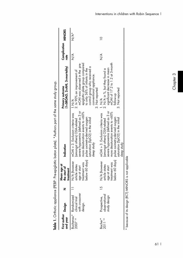
61 |
Cha
pter
3
Interventions in children with Robin Sequence |
Tabl
e 1.
Orth
otic
app
lianc
e (P
EBP:
Pre
-epi
glot
tic b
aton
pla
te).
*Aut
hors
par
t of t
he s
ame
study
gro
up.
Firs
t aut
hor
and
year
Desi
gnN
Mea
n ag
e at
th
e st
art o
f tre
atm
ent
Indi
catio
nPr
imar
y ou
tcom
e
(1=S
ROA
O, 2
=AH
I, 3=
mor
talit
y)Co
mpl
icat
ion
rate
MIN
ORS
Buch
enau
* 20
07 14
Rand
omiz
ed
clin
ical
tria
l w
ith c
ross
over
de
sign.
11N
/A (h
owev
er
artic
le s
tate
s ag
e at
sta
rt tre
atm
ent w
as
belo
w 6
0 da
ys)
mO
AI >
3. E
xclu
sion
crite
ria w
as
(am
ongs
t oth
ers)
OSA
-rela
ted
seve
re h
ypox
emia
(defi
ned
as 3
or
mor
e de
satu
ratio
n ev
ents
to <
60%
pu
lse o
xim
etry
-der
ived
oxy
gen
satu
ratio
n (S
pO2)
in th
e in
itial
sle
ep s
tudy
1.N
/A2.
In 9
0% a
n im
prov
emen
t of
mO
AI w
as o
bser
ved
in th
e p
re-
epig
lotti
c pl
ate
grou
p co
mpa
red
to o
nly
36%
of i
nfan
ts in
the
cont
rol g
roup
who
rece
ived
a
conv
entio
nal a
pplia
nce.
3. N
ot re
porte
d
N/A
N/A
*
Bach
er*
2011
13Pr
ospe
ctiv
e,
obse
rvat
iona
l stu
dy d
esig
n
15N
/A (h
owev
er
artic
le s
tate
s ag
e at
sta
rt tre
atm
ent w
as
belo
w 6
0 da
ys)
mO
AI >
3. E
xclu
sion
crite
ria w
as
(am
ongs
t oth
ers)
OSA
-rela
ted
seve
re h
ypox
emia
(defi
ned
as 3
or
mor
e de
satu
ratio
n ev
ents
to <
60%
pu
lse o
xim
etry
-der
ived
oxy
gen
satu
ratio
n (S
pO2)
in th
e in
itial
sle
ep s
tudy
1. N
/A2.
N/A
in %
, but
they
foun
d a
signi
fican
t dec
reas
e in
mea
n
mO
AI o
f 17.
2 to
1.2
at 3
-mon
th
follo
w-u
p3.
Not
repo
rted
N/A
10
* be
caus
e of
its
desig
n (R
CT)
MIN
ORS
is n
ot a
pplic
able

| 62
| Chapter 3
Tabl
e 2.
Nas
opha
ryng
eal a
irway
Firs
t aut
hor
and
year
Desi
gnN
Mea
n ag
e at
the
star
t of t
reat
men
tIn
dica
tion
Prim
ary
outc
ome
(1
=SRO
AO
, 2=A
HI,
3=m
orta
lity
Com
plic
atio
n ra
teM
INO
RS
Cha
ng
2000
15Re
trosp
ectiv
e stu
dy d
esig
n6
N/A
N/A
1. 1
00%
(al
thou
gh 4
requ
ired
supp
lem
enta
l oxy
gen.
M
ean
follo
w-u
p du
ratio
n no
t re
porte
d)2.
N/A
3. N
ot re
porte
d
Dur
ing
the
initi
al
perio
d 2
child
ren
had
regu
rgita
tion
of fe
edin
g
4
Wils
on
2000
5Re
trosp
ectiv
e stu
dy d
esig
n7 (la
te
obstr
uctio
n)
N/A
Diff
ered
, but
in g
ener
al p
oor
wei
gh g
ain
and
desa
tura
tions
to
80%
1. 6
7% (M
ean
follo
w-u
p du
ratio
n no
t rep
orte
d.)
2. N
/A3.
Not
repo
rted
Non
e re
porte
d3
Wag
ener
20
03 19
Cas
e se
ries
20N
/AU
nsat
isfac
tory
oxy
gen
satu
ratio
n an
d w
eigh
t gai
n (m
oder
ate
obstr
uctio
n) o
r in
case
of s
ever
e ai
rway
obs
truct
ion
at re
st
1. 1
00%
(Fol
low
-up
dura
tion
not r
epor
ted
)2.
N/A
3. N
ot re
porte
d
25%
7
And
erso
n 20
07 16
Retro
spec
tive
study
des
ign
13N
/A b
ut re
porte
d to
be
clos
e to
th
e m
edia
n ag
e of
adm
issio
n (6
day
s, ra
nge
1-12
2 da
ys)
Uns
atisf
acto
ry o
xyge
n sa
tura
tion
and
wei
ght g
ain
(mod
erat
e ob
struc
tion)
or i
n ca
se o
f sev
ere
airw
ay o
bstru
ctio
n at
rest.
1. 1
00%
(Mea
n fo
llow
-up
dura
tion
not r
epor
ted.
)2.
N/A
3. N
one
repo
rted
Non
e re
porte
d5
Parh
izka
r 20
16 17
Retro
spec
tive
case
ser
ies
18N
/A (n
o sp
ecifi
c da
ta fo
r RS
avai
labl
e)
AWO
on
basis
of g
loss
opto
sis1.
N/A
(no
spec
ific
data
for R
S av
aila
ble)
2. N
/A3.
Unk
now
n fo
r RS
poop
ulat
ion
Non
e re
porte
d5
Abe
l 201
2(o
ok in
m
ix) 18
Retro
spec
tive
study
des
ign
63N
/A (V
arie
d fro
m
1-33
0 da
ys)
Slee
p stu
dy s
how
ing
mod
erat
e of
sev
ere
UA
O (a
ccor
ding
to
Nix
on’s
crite
ria).
mod
erat
e U
AO
for a
set
of a
t lea
st th
ree
clus
ters
of
des
atur
atio
ns w
ith a
t lea
st 3
dips
bel
ow 8
5% (b
ut n
ot b
elow
80
%) a
nd s
ever
eU
AO
for a
set
of a
t lea
st th
ree
clus
ters
of d
esat
urat
ions
with
at
leas
t 3 d
ips
belo
w 8
0%.
1. 8
1,8%
with
a m
edia
n fo
llow
-up
perio
d of
12
mon
ths
(rang
e 2-
30 m
onth
s)2.
N/A
3. N
one
Non
e re
porte
d6

63 |
Cha
pter
3
Interventions in children with Robin Sequence |
Non-invasive respiratory support (table 3) (Two studies with 12 patients in total)20,21
Only two studies were found on non-invasive respiratory support.20 Non-invasive respiratory support includes continuous positive airway pressure (CPAP) and non-invasive positive pressure (NIPP) ventilation. By applying positive pressure during a breathing cycle, the airway patency is maintained. Leboulanger described the use of home ventilation with a custom-molded mask. The mean duration of the home ventilation therapy was 16 months. In this study an SROAO rate of 100% was found. In a large study on non-invasive ventilation by Girbal et al., five patients with RS were described.21 All started non-invasive ventilation at a young age (median 1 month with an interquartile range between 0 and 2 months). Although specific data on RS patients were missing, the study reported non-invasive ventilation led to clinical improvement in all cases and mostly minor complications were reported in 14.7% of the cases including local skin irritation, skin breakdown, conjunctivitis and slight facial deformation.
Tongue-lip adhesion (table 4) (Eleven studies with 277 patients in total)22-32 Tongue-lip adhesion is a technique first popularized by Douglas in 1946 in which the tongue is sutured to the lower lip in order to advance the tongue. In a second procedure, when the airway is deemed safe, the tongue is released. The eleven studies used different tongue-lip adhesion techniques including those described by Douglas, Argamosa and Routhledge or modified forms. The mean age of release of the tongue-lip plication ranged from 9 to 14.8 months. SROAO rates ranged from 70 to 95%. Non-respiratory related complications included dehiscence and abscesses. Respiratory-related complications included edema, stridor, apnea and a tracheostomy. According to the study of Mann et al., scarring of the lip was insignificant and scarring on the tongue modest. There was no need for scar revisions in any studies. Some studies reported considerable differences in management between non-syndromal and syndromal RS children. For example, Rogers et al. found a higher need for preoperative intubation, more average days of intubation, a longer length of intensive care unit and hospital stay and a higher incidence of reintubation.30 In the study of Kirschner et al. management by tracheostomy was more frequently required in patients with RS with multiple anomaly syndromes.25 Cozzi et al. examined differences in mean body weight or weight velocity percentiles, but did not find significant differences between isolated or non-isolated RS patients.28

| 64
| Chapter 3
Tabl
e 3.
Con
tinuo
us p
ositi
ve a
irway
pre
ssur
e (C
PAP)
and
non
-inva
sive
posit
ive
pres
sure
(NIP
P) v
entil
atio
n
Firs
t aut
hor
and
year
Desi
gnN
Mea
n ag
e at
the
star
t of t
reat
men
tIn
dica
tion
Prim
ary
outc
ome
(1
=SRO
AO
, 2=A
HI,
3=m
orta
lity
Com
plic
atio
n ra
teM
INO
RS
Lebo
ulan
ger
2010
20Re
trosp
ectiv
e stu
dy d
esig
n7
Med
ian
age
of 2
m
onth
s
(rang
e 1-
10
mon
ths)
Sym
ptom
s of
sev
ere
uppe
r airw
ayob
struc
tion
(dys
pnea
, stri
dor,
ches
t re
tract
ions
, lou
d br
eath
ing,
or f
ailu
reto
thriv
e) th
at w
as re
spon
sible
for
alve
olar
hypo
vent
ilatio
n du
ring
sleep
,de
fined
on
the
basis
of h
yper
capn
ia(tr
ansc
utan
eous
car
bon
diox
ide
pres
sure
[Ptc
CO
2] o
f _50
mm
Hg
for _
10co
nsec
utiv
e m
inut
es a
nd/o
r _10
% o
fsle
ep ti
me)
des
pite
pos
ition
ing
mea
sure
san
d ex
clus
ive
naso
gastr
ic tu
befe
edin
g.
1. 1
00%
(Mea
n fo
llow
-up
dura
tion
not r
epor
ted.
)2.
N/A
3. N
one
Non
e re
porte
d7
Girb
al 2
014
21Re
trosp
ectiv
e stu
dy d
esig
n5
Med
ian
age
1 m
onth
(In
terq
uarti
le
rang
e 0-
2)
Com
plex
OSA
1.N
/A2.
N/A
3. N
/A
Repo
rted,
bu
t no
rate
av
aila
ble
7
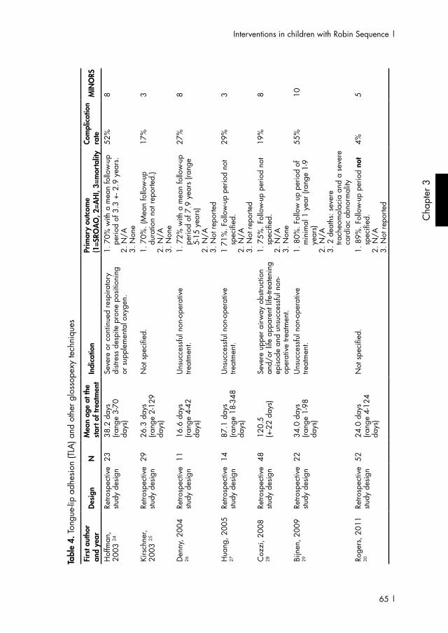
65 |
Cha
pter
3
Interventions in children with Robin Sequence |
Tabl
e 4.
Ton
gue-
lip a
dhes
ion
(TLA
) and
oth
er g
loss
opex
y te
chni
ques
Firs
t aut
hor
and
year
Desi
gnN
Mea
n ag
e at
the
star
t of t
reat
men
t In
dica
tion
Prim
ary
outc
ome
(1
=SRO
AO
, 2=A
HI,
3=m
orta
lity
Com
plic
atio
n ra
teM
INO
RS
Hof
fman
, 20
03 24
Retro
spec
tive
study
des
ign
2338
.2 d
ays
(ra
nge
3-70
da
ys)
Seve
re o
r con
tinue
d re
spira
tory
di
stres
s de
spite
pro
ne p
ositi
onin
g or
sup
plem
enta
l oxy
gen.
1. 7
0% w
ith a
mea
n fo
llow
-up
perio
d of
3.3
+- 2
.9 y
ears
.2.
N/A
3. N
one
52%
8
Kirs
chne
r, 20
03 25
Retro
spec
tive
study
des
ign
2926
.3 d
ays
(ra
nge
2-12
9 da
ys)
Not
spe
cifie
d.
1. 7
0%. (
Mea
n fo
llow
-up
dura
tion
not r
epor
ted.
)2.
N/A
3. N
one
17%
3
Den
ny, 2
004
26Re
trosp
ectiv
e stu
dy d
esig
n11
16.6
day
s
(rang
e 4-
42
days
)
Uns
ucce
ssfu
l non
-ope
rativ
e tre
atm
ent.
1.
72%
with
a m
ean
follo
w-u
p pe
riod
of 7
.9 y
ears
(ran
ge
5-15
yea
rs)
2. N
/A3.
Not
repo
rted
27%
8
Hua
ng, 2
005
27Re
trosp
ectiv
e stu
dy d
esig
n14
87.1
day
s
(rang
e 18
-348
da
ys)
Uns
ucce
ssfu
l non
-ope
rativ
e tre
atm
ent.
1
71%
. Fol
low
-up
perio
d no
t sp
ecifi
ed.
2. N
/A3.
Not
repo
rted
29%
3
Coz
zi, 2
008
28Re
trosp
ectiv
e stu
dy d
esig
n48
120.
5 (+
-22
days
)Se
vere
upp
er a
irway
obs
truct
ion
and/
or li
fe a
ppar
ent l
ife-tr
eate
ning
ep
isode
and
uns
ucce
ssfu
l non
-op
erat
ive
treat
men
t.
1. 7
5%. F
ollo
w-u
p pe
riod
not
spec
ified
.2.
N/A
3. N
one
19%
8
Bijn
en, 2
009
29Re
trosp
ectiv
e stu
dy d
esig
n22
34.0
day
s
(rang
e 1-
98
days
)
Uns
ucce
ssfu
l non
-ope
rativ
e tre
atm
ent.
1.
80%
. Fol
low
up
perio
d of
m
inim
al 1
yea
r (ra
nge
1-9
year
s)2.
N/A
3. 2
dea
ths:
sev
ere
trach
eom
alac
ia a
nd a
sev
ere
card
iac
abno
rmal
ity
55%
10
Roge
rs, 2
011
30Re
trosp
ectiv
e stu
dy d
esig
n52
24.0
day
s
(rang
e 4-
124
da
ys)
Not
spe
cifie
d.1.
89%
. Fol
low
-up
perio
d no
t sp
ecifi
ed.
2. N
/A3.
Not
repo
rted
4%5

| 66
| Chapter 3
Tabl
e 4.
Ton
gue-
lip a
dhes
ion
(TLA
) and
oth
er g
loss
opex
y te
chni
ques
(Con
tinue
d)
Firs
t aut
hor
and
year
Desi
gnN
Mea
n ag
e at
the
star
t of t
reat
men
tIn
dica
tion
Prim
ary
outc
ome
(1
=SRO
AO
, 2=A
HI,
3=m
orta
lity
Com
plic
atio
n ra
teM
INO
RS
Abr
amow
icz,
20
12 63
Retro
spec
tive
study
des
ign
2218
.4 d
ays
(ra
nge
3-45
da
ys)
Not
spe
cifie
d.1.
90%
. Fol
low
-up
perio
d no
t sp
ecifi
ed.
2. N
/A3.
Not
repo
rted
N/A
8
Man
n, 2
012
32Re
trosp
ectiv
e stu
dy d
esig
n22
13.9
day
s
(rang
e un
know
n)Ty
pe 1
or 2
airw
ay o
bstru
ctio
ns,
verifi
ed v
ia b
ronc
hosc
opy,
who
ex
perie
nced
des
atur
atio
ns w
hen
supi
ne o
r eat
ing
but r
emai
ned
stabl
e w
hen
plac
ed p
rone
.
1. 9
5%, o
ne c
ase
requ
ired
a tra
cheo
stom
y. A
vera
ge
follo
w-u
p du
ratio
n w
as 8
ye
ars.
2. N
/A3.
Not
repo
rted
15%
3
Seda
ghat
20
12 22
Retro
spec
tive
study
des
ign
829
day
s
(rang
e 15
-56
days
)
Uns
ucce
ssfu
l tre
atm
ent b
y co
nser
vativ
e m
easu
res
such
as
pron
e po
sitio
ning
and
sup
plem
enta
l na
soga
stric
tube
feed
ing.
1. 8
8%2.
Mea
n pr
e-op
erat
ive
oAH
I pr
eope
rativ
e 52
.6, p
ost-
oper
ativ
e oA
HI 1
8.1.
7/8
sh
owed
impr
ovem
ent.
In o
nly
3/8
ther
e w
as re
solu
tion
of
oAH
I bel
ow 5
. 3.
Not
repo
rted
Non
e re
porte
d9
Mok
al 2
014
23Re
trosp
ectiv
e stu
dy d
esig
n26
9.7
days
(ra
nge
3-30
da
ys)
Airw
ay c
ompr
omise
, diffi
culty
fe
edin
g, p
oor w
eigh
t gai
n an
d fa
ilure
of c
onse
rvat
ive
treat
men
t to
relie
ve s
ympt
oms.
1. 9
6% w
ith a
mea
n fo
llow
-up
perio
d of
98
mon
ths
(rang
e 9
mon
ths
– 15
yea
rs)
2. N
/A3.
Not
repo
rted
Non
e5

67 |
Cha
pter
3
Interventions in children with Robin Sequence |
Subperiosteal release of the floor of the mouth (table 5) (Two studies with 47 patients in total)33,34
Two studies were included on subperiosteal release of the floor of the mouth. The procedure is based on the theory that the muscular insertion of the tongue on the mandible is under increased tension creating the glossoptosis and elevation of the tip of the tongue.35 Both Breugem et al. and Caouette-Laberge used the technique according to Delorme.33,34 In the study of Breugem et al. children remained intubated for one week postoperatively to allow for weight gain, swelling of the floor of the mouth to subside and the endotracheal tube to splint the tongue in a forward direction. In this study a SROAO rate of 50% was found, considerably lower than the 84% SROAO rate as reported by Caouette-Laberge.
Mandibular traction (table 6) (Three studies with 133 patients in total)36-39
Three studies on mandibular traction were included.36-38 During mandibular traction the tongue and mandible are forced in a forward position, while slowly lengthening the mandible and the soft tissues and thus creating a larger oropharyngeal space and relief of the airway obstruction. Mandibular traction was described in a group of eight RS children by Pradel et al., for a period of two to five weeks using 50 – 200 gram weights.36 Traction was stopped when the infant no longer showed signs of airway obstruction anymore, and blood gas analysis showed normal values during observation. Baciliero et al. performed mandibular traction surgery according to the Stellmach & Schettler technique.37 Traction was discontinued on basis of oxygen saturation values and the degree of correction of mandibular deficiency. The oxygen saturation values had to be stable and >96% in room conditions without traction for 72 hours. The maxilla-mandibular discrepancy had to be less than three millimeters. The mean period of traction treatment was 44 days. Dong et al. followed the same surgical technique as Baciliero. 38 In this study of seven patients the mean duration of traction was 26.6 days. All studies reported a SROAO rate of 100%. In the study of Baciliero et al. complications were reported in 25% of patients including transient infection at the site of the wires. In three patients loss of one of the wires was reported. No evident scars were reported. In the study of Dong et al. no severe complications were reported, but there was note of minor complications such as increased oral secretion after surgery.

| 68
| Chapter 3
Tabl
e 5.
Sub
perio
steal
rele
ase
of th
e flo
or o
f the
mou
th
Firs
t aut
hor
and
year
Desi
gnN
Mea
n ag
e at
th
e st
art o
f tre
atm
ent
Indi
catio
nPr
imar
y ou
tcom
e
(1=S
ROA
O, 2
=AH
I, 3=
mor
talit
yCo
mpl
icat
ion
rate
MIN
ORS
Breu
gem
20
08 33
Retro
spec
tive
study
des
ign
1415
wee
ks
(rang
e 1
– 68
w
eeks
)
Failu
re o
f tra
ditio
nal m
anag
emen
t suc
h as
pr
one
posit
ioni
ng a
nd n
asop
hary
ngea
l in
tuba
tion
and
indi
cato
rs o
f con
tinue
d re
spira
tory
dist
ress
suc
h as
des
atur
atio
ns,
elev
ated
car
bon
diox
ide
leve
ls, c
ardi
ac
abno
rmal
ities
and
failu
re to
thriv
e
1. 5
0%2.
N/A
3. N
ot re
porte
d
Non
e re
porte
d6
Cao
uette
-La
berg
e 20
12 34
Retro
spec
tive
study
des
ign
3133
day
s
(rang
e 3-
188
days
)
Failu
re o
f con
serv
ativ
e m
easu
res
as v
entra
l po
sitio
ning
or a
nas
opha
ryng
eal a
irway
1. 8
4%
2. e
arly
pos
tope
rativ
e re
cord
ings
m
ean
AH
I of 1
7.4,
com
pare
d to
ean
46.
5 pr
eope
rativ
ely.
3.
One
dea
th. C
ircum
stanc
es
uncl
ear.
Non
e re
porte
d7
Tabl
e 6.
Man
dibu
lar t
ract
ion
Firs
t aut
hor
and
year
Desi
gnN
Mea
n ag
e of
th
e st
art o
f tre
atm
ent
Indi
catio
nPr
imar
y ou
tcom
e
(1=S
ROA
O, 2
=AH
I, 3=
mor
talit
yCo
mpl
icat
ion
rate
MIN
ORS
Prad
el
2009
36Re
trosp
ectiv
e stu
dy d
esig
n8
Not
repo
rted
Failu
re o
f con
serv
ativ
e m
anag
emen
t (la
tera
l or p
rone
pos
ition
ing,
acr
ylic
pla
te)
faile
d an
d re
sulte
d in
hyp
erca
pnia
> 60
m
m H
g, a
cido
sis P
H <
7.2
and
pur
e ox
ygen
atio
n <8
5%
1. 1
00%
with
a fo
llow
-up
perio
d of
11
year
s.2.
N/A
3. O
ne p
atie
nt d
ied
of a
n as
pira
tion
pneu
mon
ia
Non
e re
porte
d5
Baci
liero
20
11 37
Retro
spec
tive
study
des
ign
118
22 d
ays
(ra
nge
2-64
da
ys)
Neo
nate
s w
ho h
ad e
xper
ienc
ed e
ven
a sin
gle
episo
de o
f des
atur
atio
n or
re
spira
tory
obs
truct
ion,
and
bab
ies
with
fe
edin
g di
fficu
lties
.
1. 1
00%
2. N
/A3.
Non
e re
porte
d
25%
6
Don
g 20
14 38
Pros
pect
ive
study
des
ign
713
.7 d
ays
Surg
ery
was
per
form
ed if
the
mea
ntra
nscu
tane
ous
oxyg
en s
atur
atio
n w
as
less
than
90%
afte
r a 2
4-h
mon
itorin
g pe
riod
in th
e la
tera
l/pr
one
posit
ion,
or
if th
e tra
nscu
tane
ous
oxyg
en s
atur
atio
n de
crea
sed
cont
inuo
usly
due
to d
yspn
ea
durin
g th
e m
onito
ring
perio
d an
d m
anua
l in
terv
entio
n/re
scue
was
requ
ired.
1. 1
00%
2. N
/A3.
Not
repo
rted
No
maj
or
com
plic
atio
ns
repo
rter.
6

69 |
Cha
pter
3
Interventions in children with Robin Sequence |
Mandibular distraction9-11
As stated in the method section, mandibular distraction in children was already elaborated by a number of review studies. Therefore, publications solely on this topic were excluded from our analysis.9-11
Tracheostomy (table 7) (One study with 25 patients in total)40
One study reported specifically on the use of a tracheostomy, a technique usually performed in patients with a life-threatening airway obstruction who are in need of immediate treatment. 40 Han et al. found the median time to ‘natural’ decannulation was 97 months without further surgical intervention. Patients with syndromic RS had a significantly longer median time to decannulation than did those with isolated RS. The mean follow-up was 4 years and a SROAO rate of 100% was found. Complications were noted in 52% of patients such as tracheïtis, pneumonia, wound breakdown, wound infection and hematoma. One patient died due to a tracheostomy tube occlusion early in the study period. Long term-outcomes such as developmental delay, organ system dysfunction and death were also recorded.
Studies describing a mix of treatments (21 studies)41-62 Twenty-one studies reported on multiple techniques simultaneously in case series of children with RS in their institution. Most studies reported successful treatment of the airway obstruction by ‘prone positioning only’ in the majority of RS patients.41-43,52 In a number of studies a difference in children with an isolated RS and children with non-isolated RS was reported. 41,42 For example, the study of Marques et al. reported on the clinical course of 62 patients during the first six months of life and found prone positioning treatment or a nasopharyngeal airway to be the definitive treatment in 75,8% in children with probable isolated RS and in 52% of the cases of syndromes or other malformations. In contrast some studies did not find such a difference. A large study by Evans et al. among 115 RS patients showed no statistical difference between syndromic and non-syndromic patients regarding operative treatment. 48 In this study 44% of patients in total underwent any form of operative treatment. Handley et al. reported a significant difference in need for a surgical intervention between children with and without cleft palate. Handley et al. also identified a number of factors that may predict need for a definitive airway intervention.59
Supplement 1 shows the ratio between the MINORS score, type of treatment and SROAO success percentage.

| 70
| Chapter 3
Table 7. Tracheostomy
First author and year Design N
Mean age of the start of treatment
IndicationPrimary outcome (1=SROAO, 2=AHI, 3=mortality
Complication rate MINORS
Han 2012 40
Retrospective study design
25 N/A Not specifically specified
1. 100% with a mean follow-up of 4 years.
2. N/A3. 2 children died.
52% 7
DISCUSSION
The total number of studies with original data on treatment in RS was low, as well as the mean number of patients participating. Although we found two prospective studies and one clinical trial, most studies had a retrospective non-comparative character. To appreciate methodological quality differences between CEBM level IV studies, the MINORS score was applied. The mean MINORS (range 3–10) score was 7.3 out of 16 (non-comparative studies). The large majority of scores ranged between 5 and 8. Therefore, unfortunately the MINOR scores did not provide much guidance in differentiating between outcomes of studies. Low scores were especially found on items 3 (prospective collection of data) and 5 (unbiased evaluation of endpoints). These scores indicate there is still a lot to gain in reporting outcome for rare diseases using case series. We advocate the usage of objective descriptions of indication and the pre-treatment airway status. Only 11 out of 48 studies reported use of polysomnography, the gold standard to diagnose OSA. All other studies used fairly subjective measures or none at all for means of indication.
In most studies there seemed to have been a stepwise treatment approach, in which the indication for a certain treatment is failure of another. This makes it difficult to generalize results to the entire RS population due to selection bias. Generating a treatment protocol on the basis of these studies for any newborn child with syndromic or non-syndromic RS is therefore difficult. Nonetheless, there seems to be agreement that prone positioning, although not substantiated by specific scientific evidence, is the first step in the treatment cascade with exception of RS cases with acute life-threatening respiratory distress. Remarkably, we did not find any notions on the relation between prone positioning in children with RS and the possibly increased risk on SIDS.

71 |
Cha
pter
3
Interventions in children with Robin Sequence |
As in all conditions, preference should be given to a treatment which is most effective and least invasive. Therefore, when prone positioning fails, other non-surgical therapies may be applied, such as a nasopharyngeal airway, non-invasive respiratory support or an orthodontic plate. All these measures give temporary support to the airway. The included studies in this review showed similar success rates for SROAO with few complications. One can argue that the choice of any of these non-surgical does not matter, since results on outcomes seemed almost the same.
When prone positioning or other non-surgical therapies fail to relieve airway symptoms, a multitude of surgical options is available. On the basis of this review, given the quality of studies and the impossibility of a fair comparison (due to incompatible outcome reporting and selection bias), one cannot warrant a consensus recommendation for clinical practice. In reviews conducted on mandibular distraction, levels of evidence and effectivity appeared to be similar to our findings.9-11 Therefore, local circumstances and experience of the practitioner or clinic, rightfully guide treatment decisions.
Finally, there is an ongoing discussion regarding what the natural course of RS is. Since most of the described measures give temporary airway support and probably do not have much (or any) long-term influence on the anatomical or physiological situation after the treatment has been stopped, this implies presence of a natural improvement of the airway obstruction in time. Some studies found that more permanent invasive surgical measures were more often needed in children with non-isolated RS, suggesting the natural improvement in airway dimensions may be less in these children. Unfortunately, in most studies data on long-term outcomes, when the child has overcome the critical first years were missing.
During the preparation of this systematic review, we noted a few recurring complicating factors one comes across quite often while doing research in children with RS, such as variety in RS definition, the heterogeneity of the RS population and lack of a reliable and uniform outcome measures. In this review we used SROAO as our primary endpoint, but this was not always clearly mentioned in the articles and therefore sometimes interpreted by the researcher from the text. However, SROAO was available across all studies and therefore enabled us to compare outcome. Also, absence of need for further treatment does not necessarily mean that OSA is absent. To investigate this, post-operative PSG studies are needed.

| 72
| Chapter 3
LIST OF REFERENCES1. Bush PG. Incidence of the Robin Anomolad.
Br J Plast Surg. 1983;36(4):434-437 2. Tolarova M, Harris J. Reduced recurrence
of orofacial clefts after periconceptional supplementation with high-dose folic acid and multivitamins. Teratology. 1995;51(2):71-78.
3. Printzlau A, Andersen M. Pierre Robin Sequence in Denmark: A Retrospective Population-Based Epidemiological Study. Cleft Palate-Craniofac J. 2004;41(1):47-52.
4. Gilhooly JT, Smith JD, Howell LL, Deschaine BL, Richey SL. Bedside polysomnography as an adjunct in the management of infants with Robin sequence. Plast Reconstr Surg. 1993;92(1):23-27.
5. Wilson AC, Moore DJ, Moore MH, Martin AJ, Staugas REM, Kennedy JD. Late presentation of upper airway obstruction in Pierre Robin sequence. Arch Dis Child. 2000;83(5):435-438.
6. Bravo G, Ysunza A, Arrieta J, Pamplona MC. Videonasopharyngoscopy is useful for identifying children with Pierre Robin sequence and severe obstructive sleep apnea. Int J Pediatr Otorhinolaryngol. 2005;69(1):27-33.
7. Anderson IC, Sedaghat AR, McGinley BM, Redett RJ, Boss EF, Ishman SL. Prevalence and severity of obstructive sleep apnea and snoring in infants with Pierre Robin sequence. Cleft Palate Craniofac J. 2011;48(5):614-618.
8. Section on Pediatric Pulmonology SoOSASAAoP. Clinical practice guideline: diagnosis and management of childhood obstructive sleep apnea syndrome. Pediatrics. 2002;109(4):704-712.
9. Ow ATC, Cheung LK. Meta-analysis of mandibular distraction osteogenesis: Clinical applications and functional outcomes. Plast Reconstr Surg. 2008;121(3):54e-69e.
10. Bookman LB, Melton KR, Pan BS, et al. Neonates with tongue-based airway obstruction: A systematic review. Otolaryngol Head Neck Surg. 2012;146(1):8-18.
11. Paes EC, Mink van der Molen AB, Muradin MS, et al. A systematic review on the outcome of mandibular distraction osteogenesis in infants suffering Robin sequence. Clin Oral Investig. 2013;17(8):1807-1820.
12. Slim K, Nini E, Forestier D, Kwiatkowski F, Panis Y, Chipponi J. Methodological index for non-randomized studies (minors): development and validation of a new instrument. ANZ J Surg. 2003;73(9):712-716.
13. Bacher M, Sautermeister J, Urschitz MS, Buchenau W, Arand J, Poets CF. An oral appliance with velar extension for treatment of obstructive sleep apnea in infants with Pierre Robin sequence. Cleft Palate-Craniofac J. 2011;48(3):331-336.
14. Buchenau W, Urschitz MS, Sautermeister J, et al. A randomized clinical trial of a new orthodontic appliance to improve upper airway obstruction in infants with Pierre Robin sequence. J pediatr. 2007;151(2):145-149.
15. Chang AB, Masters IB, Williams GR, Harris M, O’Neil MC. A modified nasopharyngeal tube to relieve high upper airway obstruction. Pediatr Pulmonol. 2000;29(4):299-306.
16. Anderson KD, Cole A, Chuo CB, Slator R. Home management of upper airway obstruction in Pierre Robin Sequence using a nasopharyngeal airway. Cleft Palate-Craniofac J. 2007;44(3):269-273.
17. Parhizkar N, Saltzman B, Grote K, et al. Nasopharyngeal airway for management of airway obstruction in infants with micrognathia. Cleft Palate-Craniofac J. 2011;48(4):478-482.

73 |
Cha
pter
3
Interventions in children with Robin Sequence |
18. Abel F, Bajaj Y, Wyatt M, Wallis C. The successful use of the nasopharyngeal airway in Pierre Robin sequence: An 11-year experience. Arch Dis Child. 2012;97(4):331-334.
19. Wagener S, Rayatt SS, Tatman AJ, Gornall P, Slator R. Management of infants with Pierre Robin sequence. Cleft Palate-Craniofac J. 2003;40(2):180-185.
20. Leboulanger N, Picard A, Soupre V, et al. Physiologic and clinical benefits of noninvasive ventilation in infants with Pierre Robin sequence. Pediatrics. 2010;126(5):e1056-e1063.
21. Girbal IC, Goncalves C, Nunes T, et al. Non-invasive ventilation in complex obstructive sleep apnea - A 15-year experience of a pediatric tertiary center. Rev Port Pneumol. 2014;20(3):146-151.
22. Sedaghat AR, Anderson ICW, McGinley BM, Rossberg MI, Redett RJ, Ishman SL. Characterization of Obstructive Sleep Apnea Before and After Tongue-Lip Adhesion in Children With Micrognathia. Cleft Palate-Craniofacial Journal. 2012;49(1):21-26.
23. Mokal NJ, Desai MF, Sawant P. Reinventing the technique of tongue-lip adhesion in Pierre Robin sequence. J Plast Reconstr Aesthetic Surg. 2014;67(3):415-417.
24. Hoffman W. Outcome of tongue-lip plication in patients with severe Pierre Robin sequence. J Craniofac Surg. 2003;14(5):602-608.
25. Kirschner RE, Low DW, Randall P, et al. Surgical airway management in Pierre Robin sequence: Is there a role for tongue-lip adhesion? Cleft Palate-Craniofac J. 2003;40(1):13-18.
26. Denny AD, Amm CA, Schaefer RB. Outcomes of tongue-lip adhesion for neonatal respiratory distress caused by Pierre Robin sequence. J Craniofac Surg. 2004;15(5):819-823.
27. Huang F, Lo LJ, Chen YR, Yang JC, Niu CK, Chung MY. Tongue-lip adhesion in the management of Pierre Robin sequence with airway obstruction: Technique and outcome. Chang Gung Med J. 2005;28(2):90-96.
28. Cozzi F, Totonelli G, Frediani S, Zani A, Spagnol L, Cozzi DA. The effect of glossopexy on weight velocity in infants with Pierre Robin syndrome. J Pediatr Surg. 2008;43(2):296-298.
29. Bijnen CL, Don Griot PJW, Mulder WJ, Haumann TJ, Van Hagen AJ. Tongue-lip adhesion in the treatment of Pierre Robin sequence. J Craniofac Surg. 2009;20(2):315-320.
30. Rogers GF, Murthy AS, LaBrie RA, Mulliken JB. The GILLS score: part I. Patient selection for tongue-lip adhesion in Robin sequence. Plast Reconstr Surg. 2011;128(1):243-251.
31. Abramowicz S, Bacic JD, Mulliken JB, Rogers GF. Validation of the GILLS score for tongue-lip adhesion in robin sequence patients. J Craniofac Surg. 2012;23(2):382-386.
32. Mann RJ, Neaman KC, Hill B, Bajnrauh R, Martin MD. A novel technique for performing a tongue-lip adhesion - The tongue suspension technique. Cleft Palate-Craniofac J. 2012;49(1):27-31.
33. Breugem CC, Olesen PR, Fitzpatrick DG, Courtemanche DJ. Subperiosteal release of the floor of the mouth in airway management in Pierre Robin sequence. J Craniofac Surg. 2008;19(3):609-615.
34. Caouette-Laberge L, Borsuk DE, Bortoluzzi PA. Subperiosteal release of the floor of the mouth to correct airway obstruction in Pierre Robin sequence: Review of 31 cases. Cleft Palate-Craniofac J. 2012;49(1):14-20.

| 74
| Chapter 3
35. Epois V. [Anatomy and development of the facial skeleton in labiomaxillopalatal clefts] Anatomie et evolution du squelette facial dans les fentes labio-maxillo-palatines. Chir Pediatr. 1983;24(4-5):240-246.
36. Pradel W, Lauer G, Dinger J, Eckelt U. Mandibular Traction-An Alternative Treatment in Infants With Pierre Robin Sequence. J Oral Maxillofac Surg. 2009;67(10):2232-2237.
37. Baciliero U, Spanio Di Spilimbergo S, Riga M, Padula E. Respiratory distress in Pierre Robin sequence: An experience with mandible traction by wires. Int J Oral Maxillofac Surg. 2011;40(5):464-470.
38. Dong CB, Zheng S, Shen C, Li H. Mandible traction with wires for the treatment of upper airway obstruction caused by Pierre Robin sequence in Chinese infants: Preliminary findings. J Cranio-Maxillofac Surg. 2014;42(7):1122-1127.
39. Robinson BL. The Need of Monographic Activity in American Botanical Taxonomy. Science. 1923;57(1472):307-311.
40. Han KD, Seruya M, Oh AK, Zalzal GH, Preciado DA. “Natural” decannulation in patients with robin sequence and severe airway obstruction. Ann Otol Rhinol Laryngol. 2012;121(1):44-50.
41. Marques IL, De Sousa TV, Carneiro AF, Barbieri MA, Bettiol H, Gutierrez MRP. Clinical experience with infants with Robin sequence: A prospective study. Cleft Palate-Craniofac J. 2001;38(2):171-178.
42. Van Den Elzen APM, Semmekrot BA, Bongers EMHF, Huygen PLM, Marres HAM. Diagnosis and treatment of the Pierre Robin sequence: Results of a retrospective clinical study and review of the literature. Eur J Pediatr. 2001;160(1):47-53.
43. Li HY, Lo LJ, Chen KS, Wong KS, Chang KP. Robin sequence: Review of treatment modalities for airway obstruction in 110 cases. Int J Pediatr Otorhinolaryngol. 2002;65(1):45-51.
44. Butow KW, Hoogendijk CF, Zwahlen RA. Pierre Robin sequence: appearances and 25 years of experience with an innovative treatment protocol. J Pediatr Surg. 2009;44(11):2112-2118.
45. Chowchuen B, Jenwitheesuk K, Chowchuen P, Prathanee B. Pierre Robin sequence: challenges in the evaluation, management and the role of early distraction osteogenesis. J Med Assoc Thai. 2011;94 Suppl 6:S91-99.
46. Daniel M, Bailey S, Walker K, et al. Airway, feeding and growth in infants with Robin sequence and sleep apnoea. Int J Pediatr Otorhinolaryngol. 2013.
47. de Buys Roessingh AS, Herzog G, Hohlfeld J. Respiratory distress in Pierre Robin: successful use of pharyngeal tube. J Pediatr Surg. 2007;42(9):1495-1499.
48. Evans AK, Rahbar R, Rogers GF, Mulliken JB, Volk MS. Robin sequence: A retrospective review of 115 patients. Int J Pediatr Otorhinolaryngol. 2006;70(6):973-980.
49. Genecov DG, Barcelo CR, Steinberg D, Trone T, Sperry E. Clinical experience with the application of distraction osteogenesis for airway obstruction. J Craniofac Surg. 2009;20(8 SUPPL. 2):1817-1821.
50. Glynn F, Fitzgerald D, Earley MJ, Rowley H. Pierre Robin sequence: An institutional experience in the multidisciplinary management of airway, feeding and serous otitis media challenges. Int J Pediatr Otorhinolaryngol. 2011;75(9):1152-1155.
51. Gozu A, Genc B, Palabiyik M, et al. Airway management in neonates with Pierre Robin sequence. Turk J Pediatr. 2010;52(2):167-172.
52. Hamdi M, Brutus JP, De Mey A. Clinical experience with the Pierre Robin sequence. Eur J Plast Surg. 2004;26(8):401-405.

75 |
Cha
pter
3
Interventions in children with Robin Sequence |
53. Lin SY, Halbower AC, Tunkel DE, Vanderkolk C. Relief of upper airway obstruction with mandibular distraction surgery: Long-term quantitative results in young children. Arch Otolaryngol Head Neck Surg. 2006;132(4):437-441.
54. Meyer AC, Lidsky ME, Sampson DE, Lander TA, Liu M, Sidman JD. Airway interventions in children with Pierre Robin Sequence. Otolaryngol Head Neck Surg. 2008;138(6):782-787.
55. Schaefer RB, Stadler Iii JA, Gosain AK. To distract or not to distract: An algorithm for airway management in isolated Pierre Robin sequence. Plast Reconstr Surg. 2004;113(4):1113-1125.
56. Smith MC, Senders CW. Prognosis of airway obstruction and feeding difficulty in the Robin sequence. Int J Pediatr Otorhinolaryngol. 2006;70(2):319-324.
57. Filip C, Feragen KB, Lemvik JS, et al. Multidisciplinary Aspects of 104 Patients With Pierre Robin Sequence. Cleft Palate Craniofac J. 2015.
58. Flores RL, Tholpady SS, Sati S, et al. The surgical correction of Pierre Robin sequence: mandibular distraction osteogenesis versus tongue-lip adhesion. PLAST RECONSTR SURG. 2014;133(6):1433-1439.
59. Handley SC, Mader NS, Sidman JD, Scott AR. Predicting surgical intervention for airway obstruction in micrognathic infants. Otolaryngol Head Neck Surg. 2013;148(5):847-851.
60. Maas C, Poets CF. Initial treatment and early weight gain of children with Robin Sequence in Germany: A prospective epidemiological study. Arch Dis Child Fetal Neonatal Ed. 2014.
61. Papoff P, Guelfi G, Cicchetti R, et al. Outcomes after tongue-lip adhesion or mandibular distraction osteogenesis in infants with Pierre Robin sequence and severe airway obstruction. Int J Oral Maxillofac Surg. 2013;42(11):1418-1423.
62. Van Lieshout MJS, Joosten KFM, Hoeve HLJ, Mathijssen IMJ, Koudstaal MJ, Wolvius EB. Unravelling Robin sequence: Considerations of diagnosis and treatment. Laryngoscope. 2014;124(5):E203-E209.
63. Abramowicz S, Bacic JD, Mulliken JB, Rogers GF. Validation of the GILLS score for tongue-lip adhesion in Robin sequence patients. J CRANIOFAC SURG. 2012;23(2):382-386.

| 76
| Chapter 3
SUPPLEMENTARY MATERIAL
60
65
70
75
80
85
90
95
100
0 2 4 6 8
Type of treatment
SRoAO percentages of treatments on basis of minor scores
MINORS score 1-4
MINORS score 5-8
MINORS score 9-12
MINORS score 13-16
* Appliance NPA CPAP TLA SPRFM MT Tracheostomy
n=13
n=104
n=7
n=30 n=182
n=65
n=44
n=25
Supplementary material 1. Success percentage SRoAO of the different treatments on basis of MINORS score (classified in MINORS score 1-4, 5-8, 9-12 and 13-16) and total number of patients. *Applicance=oral appliance, NPA=nasopharyngeal airway, TLA=Tongue Lip Adhesion, SPRFM=subperiosteal release of the floor of the mouth, MT=mandibular traction

AIRWAY OBSTRUCTION


Unravelling Robin Sequence: Considerations of diagnosis and treatment
Manouk J.S. van LieshoutKoen F.M. JoostenHans L.J. HoeveIrene M.J. MathijssenMaarten J. KoudstaalEppo B. Wolvius 4Published in Laryngoscope. 2014 May;124(5):E203-9. doi: 10.1002/lary.24437. Epub 2013 Nov 15. PMID: 24115087

| 80
| Chapter 4
ABSTRACT
Objectives/HypothesisThe airway management of children with Robin sequence is controversial. This study provides an overview of a single-center experience with the diagnosis and treatment of 59 children with Robin sequence.
Study DesignRetrospective cohort study.
MethodsWe conducted a retrospective cohort study of 59 children (<1 year old) with Robin sequence managed between 2000 and 2010. Robin sequence was defined as the presence of mandibular hypoplasia and clinical signs of airway obstruction. Data were collected on demographic characteristics, the presence of a syndrome, the perinatal period, and the management of airway and nutritional problems.
ResultsEighteen children (31%) needed respiratory support because of severe respiratory distress, and a sleep study found obstructive apneas in another eight children who had been managed by prone positioning and/or monitoring. In the isolated group significantly fewer children needed respiratory support compared to the non-isolated group. After the age of 1 year, 10% of the Robin sequence cohort was still in need of treatment for obstructive symptoms. Almost half (47%) needed temporary nutritional support.
ConclusionsThe prevalence of respiratory distress in children with Robin sequence is high. In most children, treatment with prone positioning was sufficient to relieve the airway obstruction. Successful treatment with prone positioning was significantly more often seen in children with an isolated Robin sequence. About one-third of all Robin sequence children needed respiratory support in the neonatal and/or infant period. However, in childhood, only 10% of the total Robin sequence cohort was still dependent on respiratory support.

81 |
Cha
pter
4
Unravelling Robin Sequence |
INTRODUCTION
Robin Sequence (RS) is a congenital disorder occurring in about 1 in 8,500 to 1 in 20,000 newborns.1-3 As the name implies, RS is considered a sequence in which a single developmental defect results in a chain of secondary anomalies.4 The primary developmental defect is believed to be underdevelopment of the mandible (mandibular hypoplasia).5 By displacing the tongue posteriorly (glossoptosis), this can result in airway obstruction and feeding difficulties.
Despite growing attention to RS and its clinical implications in recent decades, many aspects of the condition remain unclear, and management of children with RS is controversial. In addition to reviewing our experience regarding diagnosis and treatment of the airway obstruction in a retrospective cohort of 59 children, this article reflects on our approach. Because there are few large retrospective cohort studies, this study may aid understanding of this challenging condition.
MATERIALS AND METHODS
A retrospective cohort study was undertaken of all children with RS who were born between 2000 and 2010 and treated at Erasmus Medical Center–Sophia’s Children Hospital Rotterdam. We defined RS as the presence of mandibular hypoplasia and clinical signs of airway obstruction (e.g., snoring and increased effort in breathing) observed by one (or more) member(s) of the cleft palate team or craniofacial team. The first author (m.j.s.v.l.) also double-checked all diagnoses on the basis of patient files. We identified 66 children with RS.
Fifty-nine of these children had their first consultation under the age of 1 year. The other seven children were older than 1 year (range, 374–1,739 days) and were referred to our center after various treatments (both surgical and nonsurgical) elsewhere. Six of the seven cases had a clinical syndrome or additional anomalies. To minimize the bias of these older children who had already received extensive treatment in other clinics, and also because of incomplete data, we excluded these children from analysis.
The data collected included demographic characteristics and information on the perinatal period (average birth weight; number of children born with a low birth weight, defined as a birth weight <2,500 g; pregnancy duration; and the number of children born prematurely, defined as a birth that took place before 37 weeks of gestation). We also collected data concerning associated anomalies, syndromes,

| 82
| Chapter 4
and other comorbidities. All children with a cleft palate were screened and assessed by a clinical geneticist to rule out or confirm a syndrome diagnosis. Those without cleft palate were checked by the geneticist if there was a clinical suspicion (i.e., familiar presence of mandibular hypoplasia or presence of other congenital malformations).
We also noted the presence of airway problems and their management in our clinic. Regarding treatment, we made a distinction between prone positioning, respiratory support (e.g., a nasopharyngeal airway [NPA], continuous positive airway pressure [CPAP], and/or oxygen supplementation) or surgical measures (e.g., a tracheostomy followed by mandibular distraction). Furthermore, we gathered data on the children’s nutritional status.
If available, we noted the outcomes of the endoscopy of the upper airway and the sleep study (also called polysomnography). A single experienced ear, nose, and throat surgeon in the operation theater performed endoscopies of the upper airway. The endoscopy took place while the child was under general anesthesia and in supine position. Findings were reported on basis of anatomical location. The Cormack-Lehane score was also determined.
In all children who underwent an overnight clinical sleep study in our sleep laboratory, the diagnosis of obstructive sleep apnea (OSA) and its severity was established using the criteria set by Guilleminault, in which the obstructive apnea-hypopnea index (oAHI) was used.6 The oAHI was calculated by dividing the total number of obstructive apneas and hypopneas by the total sleep time. An oAHI <1 was considered normal, 1 to 5 as mild OSA, 5 to 24 as moderate OSA, and a score >24 as severe OSA.
To compare diagnosis and treatment between isolated RS and non-isolated RS, the Pearson χ test was used. A P value of <.05 was considered to be statistically significant. For the other data, descriptive statistics were used. Analyses were performed with SPSS 20.0 for Windows (SPSS, Inc., Chicago, IL).
RESULTS
PopulationThere were 30 (51%) males and 29 (49%) females. Fifty-one of the 59 children had a cleft palate. The median age at the first consultation in our center was 14 days (range, 0–349 days). Three children in our cohort died: one due to the complications

83 |
Cha
pter
4
Unravelling Robin Sequence |
of a neuroblastoma stage IV at 6 years of age; one who had been diagnosed with a chromosomal disorder, trisomy 9, and died after withdrawal of treatment 7 days after birth; and one who died at the age of 6 years due to an aspiration pneumonia combined with severe epileptic episodes and psychomotor retardation.
Perinatal PeriodInformation on birth weight and pregnancy duration was available in 53 children. The average birth weight was 3,168 g (range, 995–4,295 g). Seven children were born with a low birth weight. The average pregnancy duration was 38.8 weeks (range, 30.1–42.1 weeks). Nine children were born prematurely.
Associated Syndromes and Other ComorbiditiesTable 1 presents the syndromes and anomalies identified in more detail. Eight RS children had a clinical syndrome (14%). Fifteen (25%) had additional anomalies, but no clinical syndrome could be identified. The largest group comprised 36 (61%) isolated RS children.
Table 1. Type of RS
Type of RS* N (%) Specified
I-RS 36 (61)
S-RS 8 (14) 2 (25%) Treacher Collins Syndrome2 (25%) Stickler Syndrome1 (17%) Nager Syndrome1 (8%) Miller Syndrome1 (8%) Chromosome 11 duplication, 12(q23,3:q24,3) deletion1 (8%) Trisomia 19
AA-RS 15 (25) E.g. hypertelorism, microtia, long and/or broad philtrum, frontal bossing, psychomotor retardation, ear malformations and hand malformations.
* I-RS = isolated RS S-RS= syndromal RS AA-RS= associated anomalies RS
Diagnosis of the Upper Airway ObstructionForty-two out of 59 children underwent one or more clinical overnight sleep study. In 26 of them, the first clinical sleep study took place before the age of 1 year and before upper airway-related surgical intervention (e.g., tracheostomy, mandibular distraction, nasal surgery, adenoidectomy and/or adenotonsillectomy). The median

| 84
| Chapter 4
age at the first sleep study was 47 days (range, 5–348 days). Seven children (27%) had a normal breathing pattern, three (12%) had an immature breathing pattern, two (8%) had an upper airway resistance syndrome, and 14 (54%) had OSA. The severity of OSA was mild in three children, moderate in three children, and severe in eight children. In four children, no sleep study was performed because these children needed a tracheostomy within 1 week after birth due to severe upper airway obstruction. Although these children did not have a formal assessment, they could be classified as having severe OSA.
An endoscopy of the upper airway was done in 12 children. The median age at this first endoscopy was 24 days. The main indications for endoscopy were intubation (four cases), removal of an endotracheal tube (one case), and evaluation of a clinical airway obstruction (eight cases). The Cormack-Lehane score was 4 in six of these children, 3 in four children, 2 in one child, and 1 in one child. In one child, the Cormack-Lehane score was unclear. The high Cormack-Lehane scores in most of these children were caused by a large and retropositioned base of the tongue against the posterior pharyngeal wall. A tracheomalacia was reported in two children. Ten of these 12 children needed respiratory support and/or a tracheostomy (followed by distraction).
TreatmentTreatment of the airway obstruction consisted of prone positioning, respiratory support (NPA, CPAP, and/or oxygen supplementation) or a tracheostomy followed by mandibular distraction. Figure 1 gives an overview of the treatment of the 59 RS children in their neonatal period (0–4 weeks old), in their infant period (4 weeks–1 year old), and in the follow-up period until the time at cross-section. The end of an arrow indicates treatment was successful with sufficient relief of the airway obstruction (no clinical obstructive signs and no desaturation events), and that there was no further need for treatment.
Thus, treatment was evaluated at three different periods. Treatment in the neonatal period consisted of prone positioning in 47 children, respiratory support in eight, and a tracheostomy in four. At an infant age, prone positioning and/or saturation monitoring were still used successfully in 41 of the 48 children in whom no respiratory support was necessary. One child in this cohort died at the age of 6 years as previously described.

85 |
Cha
pter
4
Unravelling Robin Sequence |
NE
ON
AT
AL
PE
RIO
D<2
8da
ys
INF
AN
TP
ER
IOD
<1
year
CH
ILD
HO
OD
PE
RIO
D>
1ye
ar
CH
ILD
HO
OD
PE
RIO
D>1
year
O2
=Isolated R S =P rone P ositioning =End of treatment
=Non-isolated R S =R espiratory support =Follow-up period without relapse
=Death =Tracheostomy
=Mandibular distraction
59 R S children
47 children
P P and/or monitorR espiratory support
8 children
Tracheostomy
4 children
O2
O2/
NP
T
NP
T/C
PA
P
O2/
CP
AP
Intu
batio
n
death
Typ
esu
ppor
tunk
now
n
TR
AC
PP
and/
orm
onito
rT
RA
C
PP
and/
orm
onito
rN
PT
PP
and/
orm
onito
rC
PA
P
PP
and/
orm
onito
rO
2
PP
and/
orm
onito
rO
2
PP
and/
orm
onito
rC
PA
P
O2
death
Typ
esu
ppor
tunk
now
nO
2
O2
Intu
batio
n
Intu
batio
n
Intu
batio
n
Intu
batio
n
TR
AC
TR
AC
TR
AC
TR
AC
TR
AC
No furtherrespiratory
supportneccessary
NINIII I NININI NI I NI NI II NININI NI30 x I
11 x NI
I
NI
death
P S G: in 8children OS A
was found
MDMD MD MD MD
MD
MD
MD
CP
AP
11,8 11,1 7,6 4,4 0,0 5,1 5,7 4,3 9,9 3,5 ? 6,5 6,1 9,3 4,6 8,7 9,3 7,6
Age
atcr
oss-
sect
ion
(yea
rs)
One childdied
Per
iods
ofO
2P
Pan
d/or
mon
itor
MD
Figure 1. Treatment of children with Robin Sequence in the neonatal period, the infant period and during childhood
When they grew older, six of the 47 children who had been initially managed by prone positioning and/or saturation monitoring in the neonatal period needed additional respiratory support and/or surgery. Five children were managed by CPAP, NPA, and/or oxygen, whereas one child underwent mandibular distraction.
One of the eight children who had started respiratory support in the neonatal period died after 7 days as previously described. Respiratory support could be stopped in two children, and in one child it was continued. Four children needed surgical

| 86
| Chapter 4
intervention with a tracheostomy and/or mandibular distraction. Of the four children who were tracheotomized soon after birth, all but one got a mandibular distraction. Only six out of 59 (10%) children still needed treatment in childhood. Three of these six patients needed long-term respiratory support, indicating a 5% chance of persistent obstructive airway pathology. Although the figure shows that OSA was present in eight children, these children still could be managed conservatively.
Nutritional StatusTwenty-eight children (47%) needed nutritional support for their feeding difficulties in the form of nasogastric tube feeding (25 children) or a gastrostomy tube (three children of whom two were syndromic cases and one was isolated).
Table 2. Differences in diagnostic characteristics between children with isolated and non-isolated RS
Isolated RS*(36 children)
Non-isolated RS**(23 children) P-value
Mean age at first consultation Presence of cleft palateEndoscopy of the upper airway Endoscopy performed Cormack score 4 Cormack score 3 Cormack score 2 Cormack score 1 Cormack score unclearPolysomnography (sleep study) PSG performed PSG: below the age of one year PSG: OSA presence PSG: mild OSA PSG: moderate OSA PSG: severe OSA FeedingPresence of feeding difficulties needing nutritional support
33 days33 (92%)
4 (11%)4
21 (58%)14 7 (50%)124
18 (50%)
62 days18 (78%)
8 (35%)23111
16 (70%)12 7 (58%)214
10 (43%)
n.s.
0,028 (κ² = 4,853, df = 1)
n.s.
n.s.
* Isolated RS includes children with I-RS** Non-isolated RS: includes children with S-RS and AA-RS

87 |
Cha
pter
4
Unravelling Robin Sequence |
Differences Between Children With Isolated RS and Non-isolated RSTable 2 shows the differences in diagnosis between children with isolated RS and children with non-isolated RS. The non-isolated group comprised RS children with a recognizable syndrome and also RS children with additional anomalies. In Table 3, differences are shown in treatment of the airway obstruction with respect to the different time periods. Two children with non-isolated RS died in the neonatal period and were therefore excluded from the analysis in the infant period and childhood. Some children had multiple treatments and therefore switched between treatment groups. Table 3 shows that treatment of the isolated group was markedly different from that of the non-isolated group. Those with isolated RS being treated significantly more often with prone positioning than children with non-isolated RS in all time periods. Furthermore, non-isolated RS children were significantly more often in need of respiratory support, which remained necessary after their first year of life.
Table 3. Differences in treatment of children with RS in the neonatal period (<28 days), the infant period (< 1 year) and childhood (>1 year until time at cross section)*
Isolated RS Non-isolated P-value
Neonatal periodProne positioning Respiratory supportTracheostomy and/or
mandibular distraction
n=3632 / 36 (89%)1 / 36 (3%)3 / 36 (9%)
n=2315 / 23 (65%)7 / 23 (30%)1 / 23 (4%)
0.028 (κ² = 4,853, df = 1)0.002 (κ² = 9,159, df = 1)n.s.
Infant periodProne positioning Respiratory supportTracheostomy and/or
mandibular distraction
n=3630 / 36 (83%)1 / 36 (3%)5 / 36 (14%)
n=2112 / 21 (57%)6 / 21 (29%)3 / 21 (14%)
0.030 (κ² = 4,692, df = 1)0.004 (κ² = 8,191, df = 1)n.s.
ChildhoodProne positioning Respiratory supportTracheostomy and/or
mandibular distraction
n=3636 / 36 (100%)0 / 36 (0%)0 / 36 (0%)
n=2115 / 21 (71%)5 / 21 (24%)1 / 21 (5%)
0.001 (κ² = 11,496, df = 1)0.002 (κ² = 9,396, df = 1)n.s
* See also figure 1
DISCUSSION
Providing an overview of the diagnosis and treatment of 59 children with RS, this retrospective cohort study shows that most RS children could be treated conservatively by prone positioning. About one-third of the RS children needed respiratory support in the neonatal period and/or infant period. However, in childhood only 10% of the
| 88
| Chapter 4
total RS group still needed respiratory support. In the other patients no deterioration occurred during follow-up. Treatment differed between the isolated and non-isolated RS cases. Isolated RS cases were treated significantly more often with prone positioning as compared to non-isolated RS cases.
The definition of RS is under constant debate. A questionnaire study on this subject executed among 66 health professionals at the annual Dutch Cleft Palate Meeting resulted in 29 different sets of requirements necessary for defining RS.7 As described in the Materials and Methods section, we decided to include children with mandibular hypoplasia and clinical signs of airway obstruction experienced by a health care professional. Glossoptosis was not a strict inclusion criterion for this study, because the presence of glossoptosis was not always reported in the patient charts, probably due to its subjective nature. To our knowledge there currently is neither for mandibular hypoplasia nor for glossoptosis an objective standard with reference values available. Cleft palate was not an inclusion criterion, because it is not necessarily part of the sequence of events occurring in RS.
Although not a strict criterion, the majority (86%) of the population had a cleft palate. According to our protocol, cleft palate repair takes place around the age of 9 months. Because we know palate repair in children with RS can induce severe respiratory distress postoperatively, a diagnostic workup focused on this particular feature is warranted. In our clinic, RS patients get a preoperative sleep study with a palatal plate that imitates the postoperative situation. If this imitated closure results in increased desaturation events, an increase in carbon dioxide, and/or obstructive apneas, the repair will be postponed.
In this study the prevalence of respiratory distress among RS children was high. Eighteen of the 59 children needed respiratory support, and in another eight children OSA was found by a sleep study. Previous studies have shown that OSA prevalence rates range from 46% to 100%, in which this prevalence discrepancy is presumably caused by differences in sleep study criteria, available patient population, and variety in the RS definition.8-12
The question is when children should be referred to a specialized tertiary center for additional diagnosis and treatment. In our clinic referral was often late, with a median age at the first visit of 14 days. In the large majority of children, respiratory symptoms can be relieved by prone positioning, but in this study a small number of cases who were initially treated by prone positioning seemed to develop a need

89 |
Cha
pter
4
Unravelling Robin Sequence |
for respiratory support at a later age, or the airway obstruction may not have been clinically apparent.13 However, whether we should routinely screen all children by an overnight sleep study for early OSA detection is questionable. We advocate referral to a specialized center for all RS children who continue to have respiratory distress despite prone positioning or who have persistent feeding difficulties. Follow-up sleep studies are performed to monitor the course of the obstructive problems or in case of a new clinical suspicion. Sleep studies at childhood are currently not routinely performed but only for the above-mentioned reasons. Overnight ambulatory sleep studies may play an increasingly important role in the diagnosis of OSA in patients with RS.
Another important diagnostic tool besides a sleep study is endoscopy of the upper airway. This study shows that an endoscopy was done primarily in children with a more severe form of RS (i.e., children in whom prone positioning was not sufficient to relieve the airway obstruction). A retropositioned and/or large base of the tongue was not reported in all children who underwent an endoscopy, suggesting that the level of airway obstruction may have been different in those cases. In two non-isolated RS children tracheomalacia was found. As associated anomalies of the airway such as laryngomalacia, tracheomalacia, and subglottic obstructions are commonly seen, this further emphasizes the value of endoscopy in determining the level and severity of the airway obstruction, in detecting additional airway anomalies, and in choosing a suitable treatment.14
Regarding treatment, we found in our series, as in others, that most children could be managed successfully with prone positioning alone.15-17 A natural improvement of the airway obstruction is often observed, and therefore prone positioning is only needed temporarily. The prone positioning therapy can be stopped when the child starts to roll over and the parents do not report obstructive events.
When prone positioning fails there is no consensus regarding the indication, timing, and choice of treatment.18 A number of non-surgical and surgical options have been described including NPA, CPAP, oxygen supplementation, tongue lip adhesion, subperiostal release of the floor of the mouth, tracheostomy, mandibular traction, and mandibular distraction.15,19-22 Comparative studies on the efficacy of these treatments are scarce, because the highly heterogeneous phenotype and the variety of definitions of RS makes it difficult, if not impossible, to compare the studies reliably.23

| 90
| Chapter 4
In our center, decisions about treatment are mainly made on the basis of the clinical consequences of the airway obstruction and the feeding difficulties in the patient. Surgical treatment of the airway obstruction is located at the far end of the treatment algorithm. Mandibular distraction is only indicated in children who continue to have severe respiratory distress and is preferably carried out beyond the neonatal period. At a very young age, the bone is too soft to accept adequate screw fixation and/or pin fixation for the positioning of the distractors. Therefore, to overcome the critical period before the distraction, children with severe respiratory distress and failure of conservative measures are tracheotomized shortly after birth. In this retrospective cohort mandibular distraction was carried out in eight severe cases. Respiratory support could be stopped shortly after the distraction in all but one patient.
Most children in our cohort did not need treatment after the age of 1 year. Even in those who did not receive surgical treatment, there seemed to be sufficient clinical relief of the airway obstruction. This indicates that most children may overcome the obstructive problem in time through natural growth (or maybe even catch-up growth) of the mandible. There is little evidence that surgery has a considerably better outcome than nonsurgical therapy. Furthermore, surgery inevitably brings the risks associated with anesthesia and with the procedure itself.
A notable finding was that significantly more isolated RS children had a good response on prone positioning in comparison to the non-isolated RS children. In addition, we found that isolated RS children tend to need respiratory support only in the neonatal and infant period, whereas non-isolated RS children seem to need this support beyond their first year of life (i.e., childhood). We advocate a nonsurgical approach for the isolated RS patient. Previous studies have stressed the importance of distinguishing between non-isolated RS and isolated RS. These comparative studies reported that non-isolated RS children have a higher prevalence of failure to thrive, more frequent delay in development and growth, more need of surgical intervention, more failure of specific surgical procedures, and more frequent need of tube feeding.24-29 Although a distinction between isolated and non-isolated RS children does not have direct influence on clinical management, it may offer both the clinician and the children’s family more insights in the course and the prognosis of the airway obstruction and/or feeding difficulties.
Besides airway obstruction, feeding difficulties are the most important problem in children with RS. Although this was not the primary focus of this article, we found that almost half of the RS children needed nutritional support. Unfortunately, reasons
91 |
Cha
pter
4
Unravelling Robin Sequence |
to start nutritional support were not systematically reported in the patients’ charts. However, in general, nutritional support is considered if the child does not reach his or her daily intake necessary to maintain normal growth. This can be due to disturbance of the suction by the glossoptosis and cleft palate, by insufficient energy levels for feeding due to the large amount of energy used to breathe against an obstructed airway, and/or by swallowing difficulties. It also partly depends on the feeding skills and compliance of the parents. Because feeding difficulties can lead to failure to thrive, it is important to monitor growth closely and if necessary start nutritional support support.30
One of the strengths of this study is that it is one of the few to report on the prevalence of respiratory distress and to establish differences in treatment between the isolated and non-isolated forms of RS. It is also a relatively large group compared to other cohort studies. The general limitations of this study include the retrospective design and the bias of an academic setting, where the RS population may be more severe. In some cases the definition of RS used in the studies referred to above differed from our definition of RS. This makes the comparison of outcomes less reliable.
CONCLUSION
The prevalence of respiratory distress in children with RS is high. Most isolated RS children can be successfully managed by prone positioning. Few RS children are in need of respiratory support at childhood. To identify subgroups at risk for additional respiratory support and surgery, prospective longitudinal studies in which RS children are carefully monitored are mandatory. These will allow parents of RS children to be informed accurately about the course and prognosis of the disease.

| 92
| Chapter 4
LIST OF REFERENCES1 Tolarova, M. M. & Cervenka, J.
Classification and birth prevalence of orofacial clefts. Am J Med Genet 75, 126-137, doi:10.1002/(sici)1096-8628 (19980113)75 :2<126: :a id -ajmg2>3.0.co;2-r (1998).
2 Bush, P. G. & Williams, A. J. Incidence of the Robin Anomalad (Pierre Robin syndrome). Br J Plast Surg 36, 434-437, doi:0007-1226(83)90123-6 [pii] (1983).
3 Printzlau, A. & Andersen, M. Pierre Robin sequence in Denmark: a retrospective population-based epidemiological study. Cleft Palate Craniofac J 41, 47-52, doi:02-055 [pii]10.1597/02-055 (2004).
4 Spranger, J. et al. Errors of morphogenesis: concepts and terms. Recommendations of an international working group. J Pediatr 100, 160-165 (1982).
5 Carey, J. C., Fineman, R. M. & Ziter, F. A. The Robin sequence as a consequence of malformation, dysplasia, and neuromuscular syndromes. J Pediatr 101, 858-864 (1982).
6 Guilleminault, C., Pelayo, R., Clerk, A., Leger, D. & Bocian, R. C. Home nasal continuous positive airway pressure in infants with sleep-disordered breathing. J Pediatr 127, 905-912, doi:S0022-3476(95)70026-9 [pii] (1995).
7 Breugem, C. C. & Courtemanche, D. J. Robin sequence: clearing nosologic confusion. Cleft Palate Craniofac J 47, 197-200, doi:08-061 [pii]10.1597/08-061.1 (2010).
8 Bravo, G., Ysunza, A., Arrieta, J. & Pamplona, M. C. Videonasopharyngoscopy is useful for identifying children with Pierre Robin sequence and severe obstructive sleep apnea. Int J Pediatr Otorhinolaryngol 69, 27-33 (2005).
9 Gilhooly, J. T., Smith, J. D., Howell, L. L., Deschaine, B. L. & Richey, S. L. Bedside polysomnography as an adjunct in the management of infants with Robin sequence. Plast Reconstr Surg 92, 23-27 (1993).
10 Anderson, I. C. et al. Prevalence and severity of obstructive sleep apnea and snoring in infants with pierre robin sequence. Cleft Palate Craniofac J 48, 614-618, doi:10.1597/10-100 (2011).
11 Wilson, A. C. et al. Late presentation of upper airway obstruction in Pierre Robin sequence. Arch Dis Child 83, 435-438 (2000).
12 Daniel, M. et al. Airway, feeding and growth in infants with Robin sequence and sleep apnoea. Int J Pediatr Otorhinolaryngol 77, 499-503, doi:S0165-5876(12)00721-5 [pii]10.1016/j.i jporl.2012.12.019 (2013).
13 Richards, W. & Ferdman, R. M. Prolonged morbidity due to delays in the diagnosis and treatment of obstructive sleep apnea in children. Clin Pediatr (Phila) 39, 103-108 (2000).
14 Cruz, M. J., Kerschner, J. E., Beste, D. J. & Conley, S. F. Pierre Robin sequences: secondary respiratory difficulties and intrinsic feeding abnormalities. Laryngoscope 109, 1632-1636, doi:10.1097/00005537-199910000-00016 (1999).
15 Li, H. Y., Lo, L. J., Chen, K. S., Wong, K. S. & Chang, K. P. Robin sequence: Review of treatment modalities for airway obstruction in 110 cases. Int J Pediatr Otorhinolaryngol 65, 45-51, doi:10.1016/s0165-5876(02)00131-3 (2002).

93 |
Cha
pter
4
Unravelling Robin Sequence |
16 Glynn, F., Fitzgerald, D., Earley, M. J. & Rowley, H. Pierre Robin sequence: An institutional experience in the multidisciplinary management of airway, feeding and serous otitis media challenges. Int J Pediatr Otorhinolaryngol 75, 1152-1155, doi:10.1016/j.ijporl.2011.06.009 (2011).
17 van den Elzen, A. P., Semmekrot, B. A., Bongers, E. M., Huygen, P. L. & Marres, H. A. Diagnosis and treatment of the Pierre Robin sequence: results of a retrospective clinical study and review of the literature. Eur J Pediatr 160, 47-53 (2001).
18 Mackay, D. R. Controversies in the diagnosis and management of the Robin sequence. J Craniofac Surg 22, 415-420, doi:10.1097/SCS.0b013e3182074799 (2011).
19 Denny, A. & Amm, C. New technique for airway correction in neonates with severe Pierre Robin sequence. J Pediatr 147, 97-101, doi:10.1016/j.jpeds.2005.02.018 (2005).
20 Denny, A. D. Distraction osteogenesis in Pierre Robin neonates with airway obstruction. Clin Plast Surg 31, 221-229, doi:10.1016/s0094-1298(03)00131-7 (2004).
21 Huang, F. et al. Tongue-lip adhesion in the management of Pierre Robin sequence with airway obstruction: Technique and outcome. Chang Gung Med J 28, 90-96 (2005).
22 Wagener, S., Rayatt, S. S., Tatman, A. J., Gornall, P. & Slator, R. Management of infants with Pierre Robin sequence. Cleft Palate Craniofac J 40, 180-185, doi:10.1597/1545-1569(2003)040<0180:MOIWPR>2.0.CO;2 (2003).
23 Bookman, L. B. et al. Neonates with tongue-based airway obstruction: A systematic review. Otolaryngol Head Neck Surg 146, 8-18, doi:10.1177/0194599811421598 (2012).
24 Kirschner, R. E. et al. Surgical airway management in Pierre Robin sequence: is there a role for tongue-lip adhesion? Cleft Palate Craniofac J 40, 13-18, doi:10.1597/1545-1569(2003)040<0013:SAMIPR>2.0.CO;2 (2003).
25 Smith, M. C. & Senders, C. W. Prognosis of airway obstruction and feeding difficulty in the Robin sequence. Int J Pediatr Otorhinolaryngol 70, 319-324, doi:10.1016/j.i jporl.2005.07.003 (2006).
26 Bull, M. J., Givan, D. C., Sadove, A. M., Bixler, D. & Hearn, D. Improved outcome in Pierre Robin sequence: Effect of multidisciplinary evaluation and management. Pediatrics 86, 294-301 (1990).
27 Marques, I. L. et al. Clinical experience with infants with Robin sequence: a prospective study. Cleft Palate Craniofac J 38, 171-178, doi:10.1597/1545-1569(2001)038<0171:CEWIWR>2.0.CO;2 (2001).
28 Perkins, J. A., Sie, K. C. Y., Milczuk, H. & Richardson, M. A. Airway management in children with craniofacial anomalies. Cleft Palate-Craniofacial Journal 34, 135-140 (1997).
29 Patel, K. B., Sullivan, S. R., Murthy, A. S., Marrinan, E. & Mulliken, J. B. Speech outcome after palatal repair in nonsyndromic versus syndromic Robin sequence. Plast Reconstr Surg 130, 578e-585e (2012).
30 Evans, A. K., Rahbar, R., Rogers, G. F., Mulliken, J. B. & Volk, M. S. Robin sequence: A retrospective review of 115 patients. Int J Pediatr Otorhinolaryngol 70, 973-980, doi:10.1016/j.ijporl.2005.10.016 (2006).


Management and outcomes of obstructive sleep apnea in children with Robin Sequence, a cross-sectional study
Manouk J.S. van LieshoutKoen F.M. JoostenMaarten J. KoudstaalMarc P. van der SchroeffKarolijn DulferIrene M.J. MathijssenEppo B. Wolvius 5Published in Clin Oral Investig. 2016 Nov 26. [Epub ahead of print] PMID: 27889866

| 96
| Chapter 5
ABSTRACT
ObjectiveThe objective of this cross-sectional study is to assess the prevalence, course, and management of obstructive sleep apnea (OSA) in children with Robin sequence (RS) aged 1–18 years.
Materials and methodsA cross-sectional study was conducted in 63 children aged 1 to18 years with RS. Patient data were collected on baseline characteristics and management. OSA was evaluated by polysomnography.
ResultsSixty-three children with RS were included (median age 8.0 years) and divided into two groups based on the initial treatment: prone positioning or respiratory support. Respiratory support was more often indicated in children with a non-isolated RS (p < 0.05). At cross section, in the prone positioning group (n = 32), one child was diagnosed with OSA. In the respiratory support group (n = 31), 13 children (42 %) had respiratory problems of whom 10 needed respiratory support.
ConclusionsBetween the age of 1 and 18 years, almost one out of four children with RS still has respiratory problems. Children with RS, who can be treated with prone positioning only as an infant, are not likely to develop obstructive airway problems at a later age. In contrast, children who need respiratory support early after birth are at risk of continuing or re-developing OSA after the age of 1 year.
Clinical relevanceThis study shows that those who need respiratory support at an early age need careful monitoring until adulthood.

97 |
Cha
pter
5
Obstructive sleep apnea in children with Robin Sequence|
INTRODUCTION
Robin Sequence (RS) is a congenital facial condition occurring in 1 in 5600 to 1 in 30,000 newborns.1-5 The condition is classically characterized by an underdeveloped mandible (mandibular hypoplasia), backward displacement of the tongue (glossoptosis), and airway obstruction. In 80-90% of the RS cases, a cleft palate is present.4,6,7
Children with RS are at risk of developing obstructive sleep apnea (OSA).8 OSA is characterized by prolonged partial upper airway obstruction and/or intermittent complete airway obstruction, disrupting the child’s sleeping pattern.9 Leaving OSA untreated may result in serious morbid consequences on the cardiovascular system, the metabolic system and neurocognitive and behavioral functioning.9 To establish the presence of OSA, polysomnography (PSG) is currently considered the gold standard.10
The prevalence of respiratory problems in infants with RS is considerably high with reported OSA prevalence rates between 46 and 100% depending on the criteria used.8,11-14 However, to our knowledge, no follow-up studies on OSA have been conducted in children with RS beyond the infant period.
The aim of this cross-sectional study was to examine the prevalence, course and management of OSA in children over the age of 1 year old with RS.
MATERIAL AND METHODS
A cross-sectional study was carried out among children with RS aged above 1 year combined with retrospective data collection from the patients’ chart. Inclusion was based on historical data of the Dutch Craniofacial Center, Erasmus Medical Center-Sophia’s Children Hospital. Children were considered suitable for inclusion if they had been diagnosed with RS, in this study defined as the presence of mandibular hypoplasia and airway obstruction, and were aged between 1 and 18 years old.15,16 The ethical committee of the Erasmus Medical Center (MEC-2012-048) approved the study. For all participating children, parents and/or children (if above 12 years old) provided written-informed consent. As primary treatment providers, no approval of the ethical committee was necessary for the data collection of baseline characteristics of the non-participants. Inclusion and study visits took place between November 2012 and July 2015.

| 98
| Chapter 5
During the study visit, participants underwent PSG at home or in the hospital. Furthermore, a retrospective chart review was performed on the initial treatment of airway obstruction from birth on until cross-section. Based on these retrospective data children were divided into two groups: those who initially had been treated with prone positioning and those who initially had been treated with respiratory support. Interventions of airway obstruction, which were performed in other centers, were also taken into account in this review. PSG results and treatment history were analyzed for these two groups.
PolysomnographyIn this study, ambulant sleep studies (level III, with data recorded by the Embletta portable diagnostic system) and clinical sleep studies in the hospital (level I, i.e., attended PSG including medical and technical support) were done. During the sleep studies, a variety of cardiorespiratory variables were assessed, including nasal airflow, chest and abdominal wall motion, and arterial oxygen saturation. Data were analyzed using Somnologica for Embletta software 3.3 ENU (Medcare Flags, Reykjavik, Iceland) for ambulant studies and Shell+ BrainRT Software Suite Version 2.0 (O.S.G., Rumst, Belgium) for clinical studies.
For analysis, we aimed for a total sleep time (TST) of at least 360 min, free of artifact. Summary statistics and events were scored according to the updated rules for scoring respiratory events by the American Academy of Sleep Medicine (AASM).17 An obstructive event was defined as a reduction in nasal airflow of ≥90 % (apnea) or 30–90 % (hypopnea) for the duration of at least two breaths, in the presence of thoracic and abdominal breathing movement. A hypopnea was only included if it was associated with a subsequent SpO2 desaturation of at least 3 % from baseline or with an arousal. Central apnea/hypopnea meets the same criteria as its obstructive counterpart, only without the presence of thoracic and abdominal breathing movement. A mixed apnea is a combination of a central apnea and an obstructive apnea. The obstructive apnea–hypopnea index (oAHI) was calculated by adding the number of obstructive apneas, mixed apneas, and obstructive hypopneas with SpO2 desaturation, divided by the TST; OSA was defined as an oAHI ≥1 per hour. An oAHI ≥1 and <5 was defined as mild OSA, between ≥5 and <25 as moderate OSA, and ≥25 as severe OSA.18

99 |
Cha
pter
5
Obstructive sleep apnea in children with Robin Sequence|
Statistical analysisTo assess whether the participant group was not significantly different from the non-participant group, baseline characteristics were compared using Pearson’s chi-squared tests and independent Student t tests. In order to determine the mean age at time of cross section in the group of non-participants, we used the date halfway our study inclusion period as date of cross section. A p value of <0.05 was considered to be statistically significant. Analyses were performed with SPSS 20.0 for Windows (SPSS, Inc. Chicago, IL).
RESULTS
Baseline characteristicsIn total, 111 children with RS were eligible for inclusion of whom 63 (57 %) RS patients and their parents gave informed consent (Fig. 1). For 48 children, consent was not obtained due to various reasons. In order to assess whether the study sample was representative for the RS population in our hospital, in Table 1, baseline characteristics of the study participants are compared with the non-participants. No significant differences were found for mean age at cross section, sex, presence of a syndrome or additional anomalies, presence of a cleft palate, and initial treatment of airway obstruction.
Table 1. Baseline characteristics of the study participants (n=63) vs. non-participants (n=48). IQR=Interquartile range.
Participants (63)
Non-participants (n=48) P-value
Median age in years at cross-section [IQR] 8.0 [4.0-12.0] 9.0 [6.0-13.0] p=0.57
Sex FemaleMale
31 (49.2%)32 (50.8%)
21 (43.8%)27 (56.3%)
p=0.57
Presence of asyndrome or additional anomalies
Yes, additional anomaliesYes, syndromeNo
19 (30.2%)7 (11.1%)37 (58,7%)
14 (29.2%)13 (27.1%)21 (43.8%)
p=0.08
Presence of a cleft palate
YesNo
58 (92.1%)5 (7.9%)
40 (83.3)8 (16.7%)
p=0.16
Treatment of airway obstruction
Prone positioning onlyNon-surgical treatmentSurgical treatment
32 (50.8%)19 (30.2%)12 (19.0%)
29 (60.4%)8 (16.7%)11 (22.9%)
p=0.19

| 100
| Chapter 5
n=18
Robin Sequence patients n=111
Prone positioning only n=61
42 x I, 15 x AA, 4 x S
Respiratory support n=49
(16 x I, 17 x AA, 16 x S)
Death within a few hoursn=1
HISTORY
n=32(23 x I, 7 x AA, 2
x S)
n=31(14 x I, 12x AA, 5
x S )PARTICIPANTS
(n=63
AIRWAY STATUS ATCROSS-SECTION
(n=63)
OSA found at PSG without treatment
n=62x I, 3x AA, 1x S
Non-surgical respiratory
supportn=3
1x I, 1x AA, 1 x S
Tracheostomyn=5
1x I, 2x AA, 2x S
No treatment. No OSA at PSG
n=4933 x I, 13 x AA, 3 x S
n=1n=3 n=5n= 5n=31
Re-start non-surgical respiratory support (CPAP) shortly after
PSG. n=2 1x I, 1x S
n= 2
Figure 1. Treatment overview of children with RS. I = isolated, AA = associated anomalies, S = proven syndrome
Five children were deceased but were nonetheless included in the calculations of the non-participation group. In two of these cases, the cause of death was respiratory-related: one child with various comorbidities died at the age of 1 year due to severe respiratory insufficiency following aspiration and another child died within hours after birth due to severe obstruction of the upper airway and no option for a tracheostomy.
Thirty-one females and 32 males participated in the study. Of these 63 children, 37 children had an isolated RS, while 26 children had additional anomalies (n = 19) or a syndrome (n = 7); 2 children were diagnosed with Stickler syndrome, 1 child with Nager syndrome, 1 child with Shprintzen–Goldberg syndrome, 1 child with acampomelic dysplasia, 1 child with a FOXC2 mutation, and 1 child with a MFDM mutation. A variety of conditions were reported in the group with associated

101 |
Cha
pter
5
Obstructive sleep apnea in children with Robin Sequence|
anomalies such as psychomotor retardation, hip dysplasia, or facial anomalies, but not with a proven syndrome. Five children had RS without a cleft palate.
The mean pregnancy duration was 38.5 weeks. Eight children were born pre-term (<37 weeks). The mean birth weight was 3137 g. Three families reported occurrence of mandibular hypoplasia in the family.
Feeding difficulties were reported in 60 out of 63 (95.2 %) children. Twenty-six children out of 63 (41.3 %) needed a temporary feeding tube and seven children (11.1 %) a percutaneous endoscopic gastrostomy tube.
Prior to cross-section: Initial management of obstructive sleep apnea Figure 2 shows an overview of initial management (including the findings at cross section). Initially, 32 children (48.5 %) were treated with prone positioning and 31 children were in need of respiratory support, which consisted of non-surgical respiratory support such as a nasopharyngeal tube, continuous positive airway pressure (CPAP), and oxygen therapy, or surgical measures such as tracheostomy and mandibular distraction osteogenesis with or without tracheostomy at the time of distraction. Children with additional anomalies or a syndrome (n = 26) were in need of respiratory support significantly more often compared to those with an isolated RS (65.4 vs. 37.8 %, p < 0.05).
At cross-sectionThe median age in years at cross section was 8.0 years (IQR 4.0–12.0). Thirty-one out of 63 (49.2 %) children were female. Thirty-seven children (58.7 %) had an isolated RS. A cleft palate was present in 58 (92.1 %) children.
At cross-section: Assessment of obstructive sleep apnea by polysomnographyFrom the 63 children in this study, in 19 no PSG result was available, in 3 the parents refused, in 10 PSG failed due to logistic reasons, and in 6 PSG was not indicative of OSA because of a tracheostomy (n = 5) or CPAP (n = 1). These last six cases were classified as having OSA without formal assessment. At the end, 44 PSG studies (n = 37 ambulatory and n = 7 clinical) were available for analysis: 23 PSG studies in the group initially managed with prone positioning (n = 32) and 21 PSG studies in the group initially managed with respiratory support (n = 31).

| 102
| Chapter 5
Non-surgical respiratory support n=11Oxygen therapy, CPAP, nasopharyngeal tube
(Range of end treatment: day 1 until 12 mo old)
No OSA(n=18)
OSA at PSG(n=5)
With 2 patients re-starting CPAP
shortly after cross-section
Non-surgicalrespiratory support
(n=3)
Tracheostomy(n=5)
Treatment free interval: 9,6yrs (range 0-17.4 yrs)
9.8 yrs(mean, range0.7-18.0 yrs)
Surgical respiratory supportn=7
MDO+T x 5, T x 1, MDO x 1 (Range of end treatment: day 1 until 12 mo old) Treatment free interval: 8,6 yrs
(range 0.7-15.8 yrs)
11.2 yrs(range 6.1-
18.0 yrs)
CPAP(Duration: a few days after
birth)1.5 yrs
History
NPT(Duration:6 mo, until 6
mo old6.8 yrs
O2(Duration: a few days
after birth
16.0 yrs
O2(Duration: from birth until now
16.4 yrs
NPT(Duration: 4 mo, from 1
moth old until 5 mo)
CPAP(from 9 yrs until now) 11.9 yrs
Tracheostomy (from 2 mo old until now), with MDO at age 2 mo and 7.4 yrs) 7.7 yrs
Tracheostomy(from 1.5 mo old until now) 1.8 yrs
Tracheostomy(from 2 days old until now, with MDO at age 6 mo and 3.7 yrs) 11.9 yrs
NPT & CPAP(Duration: 2 mo, from birth
until 2 mo old)
Tracheostomy(from 2 mo old until now) 4.0 yrs
O2(Duration:unknown
Tracheostomy and oxygen(from 5 mo old until now) 3.8 yrs
Prone positioning onlyn=31
No OSA(n=31)
Cross-section Age Presence of OSA*
7.1 yrs(mean, range
1-17.0 yrs)
Prone positioning onlyn=1 6 yrs
History
OSA at PSG(n=1)
CPAP(Duration:6 mo, from 1
year until 1,5 year)9.3 yrsCPAP
(from 8 yrs until now)
Re-start CPAP
Re-start CPAP
Study participantstreated by pronepositioning only
n=32
Study participantswho
received respiratorysupport n=31
Cross-section Age Presence of OSA*
CPAPDuration: 6,5 yrs: from 2,5 yrs until 9
yrs old with MDO at age 9 yrs16.0 yrs
Tracheostomy and afterwards MDO at 3 wks old.After that oxygen supplementation until 6 yrs old. 6.0 yrs
Figure 2. Treatment overview of study participants (n=63) including the (mean) follow-up duration and age at cross-section. MDO=mandibular distraction osteogenesis, T=tracheostomy, yrs=age in years, mo=age in months. *Presence of OSA was based on PSG results (n=44). If no PSG were available, presence of OSA was based on the need for treatment

103 |
Cha
pter
5
Obstructive sleep apnea in children with Robin Sequence|
OSA was detected in six children who did not receive OSA treatment at the time of cross section. Besides these six children, OSA was confirmed in two other children who already received respiratory support for OSA but a PSG was done without this support as part of routine clinical evaluation. Table 2 further elaborates on these cases.
Table 2. Overview of cases in whom OSA was found during PSG (n=44)
Case number; isolated or non-isolated, age at cross-section
History
PSG at cross-section
oAHI ODI
Case 1, isolated,6.0 yrs
Treated by prone positioning in the neonatal period and no complaints afterwards. After PSG treated with nasal corticosteroids because of mild-moderate OSA
3.7 2.8
Case 2, associated anomalies, 1.5 yrs
PSG for follow-up purposes and a wait-and-see policy was set. This child was already known to have OSA and hypoventilation.
6.7 9.5
Case 3, syndromal, 6.8 yrs
A nasopharyngeal tube shortly after birth for 5 months. After PSG, CPAP was re-started because of moderate OSA.
17.0 20.9
Case 4, isolated, 16.4 yrs
A few days of oxygen therapy at birth and afterwards treated with CPAP for 3 months. At the age of 16, severe complaints of OSA and re-start of CPAP shortly after PSG.
50.0 26.7
Case 5, associated anomalies, 16.0 yrs
CPAP for 6,5 years until the age of 9 years old, when mandibular distraction osteogenesis was performed. Based upon the PSG results, a wait-and-see policy was set.
3.2 0.7
Case 6, associated anomalies, 6.0 yrs
Mandibular distraction osteogenesis and decannulation at the age of 3 months and oxygen supplementation until the age of 6 years. Based upon the PSG results, a wait-and-see policy was set.
7.3 8.9
Case 7, isolated, 9.3 yrs
Six months of CPAP treatment at the age of 1 year and re-start of CPAP at the age of 8 years. At home PSG without CPAP.
3.0 0.2
Case 8, syndromal, 16.0 yrs
Oxygen therapy since birth, at home PSG without oxygen. 7.0 3.7
At cross-section: Airway management Out of the total group of 63 children, 14 (22 %) received either OSA treatment and/or showed OSA during PSG at cross section.
In the cohort of the children (n = 32, mean age at cross section 7.1 ± 4.5 years) who were initially treated with prone positioning, in one child mild OSA was found with PSG, but further treatment was not necessary. Thirty-one out of 32 (96.9 %) children were free of OSA treatment at the time of cross section.

| 104
| Chapter 5
In the cohort of children (n = 31, mean age at cross section 9.4 ± 5.8 years) who were initially treated by respiratory support, 13 children (42 %) had respiratory problems; 3 children were still in need of non-surgical respiratory support (CPAP or oxygen therapy) (age range at cross section 9.3–16.0 years), and 5 children were dependent on a tracheostomy (age range at cross section 1.8–7.7 years). In five children, OSA was found (age range 1.5–16.4 years), and in two of these children a re-start of respiratory support (CPAP) was indicated shortly after PSG (Table 2). Eighteen out of 31 (58.1 %) children were free of OSA or respiratory support at the time of cross section.
DISCUSSION
To our knowledge, this is the first long-term follow-up study investigating obstructive airway problems in children with RS between 1 and 18 years of age. At cross section, 22 % of the children with RS still had respiratory problems. Those who had a history of only prone positioning were not likely to develop a significant airway obstruction at a later age. However, children who needed respiratory support early after birth were 13 times more likely to be diagnosed with OSA at a later age or to remain dependent on or re-develop a need for respiratory support.
In this study, children were divided into two groups on the basis of their initial airway management. About half of the children initially needed respiratory support for which different respiratory support modalities were used. In eight children, mandibular distraction osteogenesis had been performed. At cross section, in six of these children mandibular distraction osteogenesis was successful, although in two cases mild OSA was diagnosed with PSG but no further treatment was necessary. Two children still needed a tracheostomy at the time of the study; however, follow-up after the mandibular distraction osteogenesis was short. Also, two children who had received non-surgical respiratory support shortly after birth were still dependent on a tracheostomy.
Interestingly, four children became again in need of non-surgical respiratory support at an older age, in two of them as a result of this study. In a recent retrospective study of Lee et al., it was shown that their RS infant population, who were followed to 1 year of age, did not show significant decreases in AHI, oAHI, and central apnea index.19 Unfortunately, other studies with follow-up on OSA in RS patients are so far lacking. On the basis of this study, one might conclude that the group of children who initially needed respiratory support might benefit from more careful monitoring

105 |
Cha
pter
5
Obstructive sleep apnea in children with Robin Sequence|
until adulthood.
Remarkably, of those who were still in need of respiratory support, six children had been diagnosed with associated anomalies or a syndrome and only two with an isolated RS. In daily practice (data not shown), many children with an associated anomaly or a syndrome have an even smaller mandible than those with an isolated RS. It is speculated that in children with RS, with an intrinsic tissue deficiency as primary cause, there is impaired growth of the mandible.20 This may also explain why this group of children needed respiratory support more often. In contrast, in the case of isolated RS there might be catch-up growth from an initial deformation.21 However, there is an ongoing discussion about the concept of accelerated growth of the mandible. Previous studies showed a “partial mandibular catch-up growth” and an increase (3.5 times its original size) in airway dimension in the first 2 years of life. Additionally, an increase in upper airway dimensions in longitudinal cephalograms of children with RS from childhood to adulthood was noticed.22,23 Remarkably, the depth of the oropharyngeal airway was an exception to this. In contrast, other studies did not observe this acceleration of mandibular growth.24-26 Since the upper airway is a three-dimensional and complex dynamic altering space, one may question whether the method of analyzing two-dimensional radiographs (cephalogram) to determine alterations of airway space is the most appropriate one.
Ultimately, flexible fiberoptic laryngoscopy (FFL) of the upper airway should determine the extent of the upper airway obstruction, but validated scoring systems that objectify the obstruction of the airway are lacking. A recent attempt to score glossoptosis in RS patients using awake FFL was not successful, due to disappointing inter- and intra-agreement in the analysis of awake FFL videos of RS patients compared to non-RS patients.27 Another complicating factor is that the degree of micrognathia does not seem to correlate well with the degree of airway compromise and the higher Cormack-Lehane grades diagnosed with laryngoscopy. 28
LimitationsOnly about half the RS population participated, resulting in a small sample size. Of some non-participants, there were no data available on the further course of the obstructive problems. Nonetheless, a flowchart was created based on the latest available data in the patient charts. In Table 1, it is shown that our sample appears to be representative of the total RS population in our clinic.

| 106
| Chapter 5
Table 1. Baseline characteristics of the study participants (n=63) vs. non-participants (n=48). IQR=Interquartile range.
Participants (63)
Non-participants (n=48) P-value
Median age in years at cross-section [IQR] 8.0 [4.0-12.0] 9.0 [6.0-13.0] p=0.57Sex Female
Male31 (49.2%)32 (50.8%)
21 (43.8%)27 (56.3%)
p=0.57
Presence of asyndrome or additional anomalies
Yes, additional anomaliesYes, syndromeNo
19 (30.2%)7 (11.1%)37 (58,7%)
14 (29.2%)13 (27.1%)21 (43.8%)
p=0.08
Presence of a cleft palate YesNo
58 (92.1%)5 (7.9%)
40 (83.3)8 (16.7%)
p=0.16
Treatment of airway obstruction
Prone positioning onlyNon-surgical treatmentSurgical treatment
32 (50.8%)19 (30.2%)12 (19.0%)
29 (60.4%)8 (16.7%)11 (22.9%)
p=0.19
A PSG or appropriate PSG results were not available in all participants. If there was no PSG result available at cross section, the presence of OSA was determined by the child’s current need for respiratory support. Because of this, results from this study should be interpreted with caution.
Furthermore, our inclusion was based on historical data, which is another limitation of this study. Especially, in the group who was initially treated by prone positioning, one may argue whether these children are “true” Robin sequence patients. What was the indication to start prone positioning? What was the level of airway obstruction? However, just the mere fact that these children were given a prone positioning advice despite knowing the risks such as sudden infant death syndrome suggests that the clinical issues must have been substantial.
ConclusionThis is the largest cross-sectional study on OSA in children with RS to date. Half of the RS population had been treated with prone positioning, while the other half needed respiratory support. Children with RS, who were treated by prone positioning as an infant, appear to have a very low risk on obstructive pathology at a later age indicating some natural improvement. Children who needed respiratory support continued or re-developed dependence on respiratory support at a later age. Considering the potential long-term effects of untreated OSA, children with RS in which the airway obstruction cannot be managed with prone positioning only require close follow-up beyond the infant period preferably using PSG.

107 |
Cha
pter
5
Obstructive sleep apnea in children with Robin Sequence|
LIST OF REFERENCES1 Bush, P. G. & Williams, A. J. Incidence of the
Robin Anomalad (Pierre Robin syndrome). Br J Plast Surg 36, 434-437, doi:0007-1226(83)90123-6 [pii] (1983).
2 Tolarova, M. M. & Cervenka, J. Classification and birth prevalence of orofacial clefts. Am J Med Genet 75, 126-137, doi:10.1002/(sici)1096-8628 (19980113 )75 :2<126 : : a i d -ajmg2>3.0.co;2-r (1998).
3 Printzlau, A. & Andersen, M. Pierre Robin Sequence in Denmark: A Retrospective Population-Based Epidemiological Study. Cleft Palate-Craniofac J 41, 47-52, doi:10.1597/02-055 (2004).
4 Vatlach, S., Maas, C. & Poets, C. F. Birth prevalence and initial treatment of Robin sequence in Germany: a prospective epidemiologic study. Orphanet J Rare Dis 9, 9, doi:1750-1172-9-9 [pii]10.1186/1750-1172-9-9 (2014).
5 Paes, E. C. et al. Birth prevalence of Robin sequence in the Netherlands from 2000-2010: A retrospective population-based study in a large Dutch cohort and review of the literature. Am J Med Genet A, doi:10.1002/ajmg.a.37150 (2015).
6 Costa, M. A., Murage, K. P., Tholpady, S. S. & Flores, R. L. Airway compromise following palatoplasty in Robin sequence: improving safety and predictability. Plast Reconstr Surg 134, 937e-945e, doi:10.1097/PRS.000000000000072900006534-201412000-00030 [pii] (2014).
7 Caouette-Laberge, L., Bayet, B. & Larocque, Y. The Pierre Robin sequence: Review of 125 cases and evolution of treatment modalities. Plast Reconstr Surg 93, 934-942 (1994).
8 Anderson, I. C. et al. Prevalence and severity of obstructive sleep apnea and snoring in infants with Pierre Robin sequence. Cleft Palate Craniofac J 48, 614-618, doi:10.1597/10-100 (2011).
9 Gozal, D., Hakim, F. & Kheirandish-Gozal, L. Chemoreceptors, baroreceptors, and autonomic deregulation in children with obstructive sleep apnea. Respir Physiol Neurobiol 185, 177-185, doi:S1569-9048(12)00248-0 [pii]10.1016/j.resp.2012.08.019 (2013).
10 Marcus, C. L. et al. Diagnosis and management of childhood obstructive sleep apnea syndrome. Pediatrics 130, e714-755, doi:peds.2012-1672 [pii]10.1542/peds.2012-1672 (2012).
11 Bravo, G., Ysunza, A., Arrieta, J. & Pamplona, M. C. Videonasopharyngoscopy is useful for identifying children with Pierre Robin sequence and severe obstructive sleep apnea. Int J Pediatr Otorhinolaryngol 69, 27-33 (2005).
12 Gilhooly, J. T., Smith, J. D., Howell, L. L., Deschaine, B. L. & Richey, S. L. Bedside polysomnography as an adjunct in the management of infants with Robin sequence. Plast Reconstr Surg 92, 23-27 (1993).
13 Wilson, A. C. et al. Late presentation of upper airway obstruction in Pierre Robin sequence. Arch Dis Child 83, 435-438, doi:10.1136/adc.83.5.435 (2000).
14 Daniel, M. et al. Airway, feeding and growth in infants with Robin sequence and sleep apnoea. Int J Pediatr Otorhinolaryngol (2013).
15 van Lieshout, M. J. et al. Unravelling Robin sequence: considerations of diagnosis and treatment. Laryngoscope 124, E203-209, doi:10.1002/lary.24437 (2014).
16 Paes, E. C. et al. A pragmatic approach to infants with Robin sequence: a retrospective cohort study and presence of a treatment algorithm. Clin Oral Investig, doi:10.1007/s00784-015-1407-6 (2015).

| 108
| Chapter 5
17 Berry, R. B. et al. Rules for scoring respiratory events in sleep: update of the 2007 AASM Manual for the Scoring of Sleep and Associated Events. Deliberations of the Sleep Apnea Definitions Task Force of the American Academy of Sleep Medicine. J Clin Sleep Med 8, 597-619, doi:10.5664/jcsm.2172 (2012).
18 Guilleminault, C., Pelayo, R., Clerk, A., Leger, D. & Bocian, R. C. Home nasal continuous positive airway pressure in infants with sleep-disordered breathing. J Pediatr 127, 905-912, doi:S0022-3476(95)70026-9 [pii] (1995).
19 Lee, J. J., Thottam, P. J., Ford, M. D. & Jabbour, N. Characteristics of sleep apnea in infants with Pierre-Robin sequence: Is there improvement with advancing age? Int J Pediatr Otorhinolaryngol, d o i : S 0 1 6 5 - 5 8 7 6 ( 1 5 ) 0 0 4 6 1 - 9 [pi i ]10.1016/j. i jpor l .2015.09.014 (2015).
20 Cohen, M. M., Jr. Robin sequences and complexes: causal heterogeneity and pathogenetic/phenotypic variability. Am J Med Genet 84, 311-315, doi:10.1002/(SICI)1096-8628 (19990604 )84 :4<311 : :A ID -AJMG1>3.0.CO;2-9 [pii] (1999).
21 Mackay, D. R. Controversies in the diagnosis and management of the Robin sequence. J Craniofac Surg 22, 415-420, doi:10.1097/SCS.0b013e3182074799 (2011).
22 Figueroa, A. A., Glupker, T. J., Fitz, M. G. & BeGole, E. A. Mandible, tongue, and airway in Pierre Robin sequence: a longitudinal cephalometric study. Cleft Palate Craniofac J 28, 425-434 (1991).
23 Staudt, C. B., Gnoinski, W. M. & Peltomaki, T. Upper airway changes in Pierre Robin sequence from childhood to adulthood. Orthod Craniofac Res 16, 202-213, doi:10.1111/ocr.12019 (2013).
24 Vegter, F., Hage, J. J. & Mulder, J. W. Pierre Robin syndrome: mandibular growth during the first year of life. Ann Plast Surg 42, 154-157 (1999).
25 Suri, S., Ross, R. B. & Tompson, B. D. Craniofacial morphology and adolescent facial growth in Pierre Robin sequence. Am J Orthod Dentofacial Orthop 137, 763-774, doi:10.1016/j.ajodo.2008.07.020 (2010).
26 Hermann, N. V. et al. Early craniofacial morphology and growth in children with nonsyndromic Robin Sequence. Cleft Palate-Craniofac J 40, 131-143, doi:10.1597/1545-1569(2003)040<0131:ecmagi>2.0.co;2 (2003).
27 Basart, H. et al. Awake Flexible Fiberoptic Laryngoscopy to diagnose glossoptosis in Robin Sequence patients. Clin Otolaryngol, doi:10.1111/coa.12552 (2015).
28 Hong, P. & Kearns, D. Airway characteristics of infants with Pierre Robin sequence who undergo mandibular distraction osteogenesis. Ear Nose Throat J 94, E25-29 (2015).


Respiratory distress following palatal closure in children with Robin Sequence
Manouk J.S. van LieshoutIlje E. VosholKoen F.M. JoostenMarc P. van der SchroeffIrene M.J. MathijssenMaarten J. KoudstaalEppo B. Wolvius
Published in Cleft Palate Craniofac J. 2016 Mar;53(2):203-9. doi: 10.1597/14-250. Epub 2015 Jun 23. PMID: 26101809 6

| 112
| Chapter 6
ABSTRACT
Objective The aim of this study is to assess prevalence, severity, and duration of respiratory distress following palatoplasty in children with Robin sequence and to evaluate perioperative management.
Design Retrospective chart review study. Data were collected for patients who were born between 2009 and 2012 and underwent palatoplasty in the Sophia Children’s Hospital-Erasmus Medical Center.
Results Of the 75 patients with cleft palate, 30 with Robin sequence and a control group of 45 cleft without Robin sequence underwent palatoplasty. Prior to closure, 26 of 30 patients with Robin sequence had been treated by prone positioning, and four needed additional treatment. The mean age at closure was 12.4 months for patients with Robin sequence and 10.9 months for patients without Robin sequence (P = .05). On the basis of the results of preoperative polysomnography with palatal plate, closure was postponed in two patients with Robin sequence. In the Robin sequence group, eight of the 30 patients developed postoperative respiratory distress within 48 hours and one patient, after 7 days; whereas none within the non-Robin sequence group developed respiratory distress. In all nine cases of Robin sequence the obstructive problems resolved within a few days, with four children requiring a temporary nasopharyngeal tube. There were no significant differences between preoperative polysomnography results of the nine patients with Robin sequence who developed postoperative respiratory distress compared with those patients with Robin sequence who did not.
Conclusion Despite delayed closure compared with children without Robin sequence, 30% of the children with Robin sequence developed respiratory distress following palatoplasty, which resolved within a few days. This study emphasizes the need for close perioperative monitoring of patients with Robin sequence who undergo palatoplasty.

113 |
Cha
pter
6
Respiratory distress following palatal closure in children with Robin Sequence|
INTRODUCTION
Robin Sequence (RS) is a congenital facial condition occurring in 1 in 8500 to 1 in 30,000 newborns.1-4 The condition is classically characterized by an underdeveloped mandible (mandibular hypoplasia), backward placement of the tongue (glossoptosis), and airway obstruction.5 In 80 to 90% of the RS cases, a cleft palate is present.4,6,7
Children with RS are prone to develop airway-related complications after cleft palate repair such as stridor or postoperative respiratory distress, difficulty on intubation or a tracheostomy.8-11 In small series of studies on children, an incidence of airway-related complications has been reported ranging from 24% to 31%.8,9 Hence, there is an obvious need for a clear perioperative management plan. So far, limited research has been done regarding respiratory distress after cleft palate repair in children with RS. The aim of this study is to evaluate our perioperative management in a large cohort of children with RS and to assess prevalence, severity and duration of respiratory distress following palatoplasty (primary closure of the palate).
METHODS
Patients with RS (RS group) and a control group of patients withoutRS (non-RS group)with a cleft palate who were born between 2009 and 2012 were included in this retrospective chart review study. All patients underwent a palatoplasty in the Sophia Children’s Hospital—Erasmus MC and did not have an additional cleft of the lip and/or jaw. Robin sequence was defined as presence of mandibular hypoplasia and airway obstruction needing treatment. Children with a submucous cleft, those with a tracheostomy, and children with an unrepaired cleft palate (at the date of our analysis, June 2014) were excluded from the main analysis. Data were collected from the medical records on demographics, the presence of a syndrome, the characteristics of the cleft palate, preoperative airway obstruction, polysomnography (PSG), the duration of the surgery, the length of the postoperative intensive care stay, the postoperative clinical course (e.g., presence of airway obstruction, necessity of airway support), and the situation at the outpatient clinic 3 weeks postoperatively. This study was approved by the institutional medical ethics review board (MEC-2014-559).

| 114
| Chapter 6
PolysomnographyPrior to palatal surgery, a PSG was routinely performed in children with RS. This PSG was done with a removable custom-made palatal plate in place that simulates the palatal anatomy after closure. This plate, made of synthetic resin, was fabricated a few days before PSG by the orthodontic department. The aim of this PSG was to assess the possibly resulting airway obstruction (Fig. 1).
Figure 1. Custom-made palatal plate made of synthetic resin
The PSG was performed overnight in our pediatric sleep laboratory. Scoring of the PSG was done according 2012 AASM criteria. Obstructive apnea was defined as absence of airflow for more than two breaths during baseline breathing and ongoing respiratory effort throughout the period of absent airflow. Obstructive hypopnea was defined as absence of airflow for more than two breaths, decrease in nasal airflow of >30%, snoring or paroxysmal breathing, and a saturation drop >3% or an arousal. To assess the presence of obstructive sleep apnea (OSA), the obstructive apnea-hypopnea index (oAHI) was used, combining the number of obstructive apneas and hypopneas per hour. Obstructive sleep apnea was defined as an oAHI >1 per hour. Central apnea was defined as a decrease in nasal airflow of >90%, no breathing effort, and absence of airflow for more than two breaths with an arousal or desaturation >3% or with a duration of more than 20 seconds. The number of pathological central apneas was indexed (pCAI) to the duration of sleep (i.e., episodes per hour of sleep). The oxygen saturation was also indexed, resulting in the oxygen desaturation index (ODI). A pCAI and an ODI >1 was defined as abnormal. All PSG results were reanalyzed by a research nurse who is specialized in PSG analysis.
115 |
Cha
pter
6
Respiratory distress following palatal closure in children with Robin Sequence|
PalatoplastyPalatoplasty (primary closure of the cleft palate) was performed according to our cleft palate protocol. According to protocol, palatoplasty preferably takes place around the age of 9 to 10 months. In our clinic, palatoplasty is done mostly using the Von Langenbeck technique. Patients with a cleft palate were routinely admitted on the day of surgery and discharged the day after surgery. All cleft palate repairs were executed by one of three pediatric plastic surgeons.
Statistical AnalysisStatistical analysis was performed using SPSS, version 21.0 for Windows (IBM Corp., Armonk, NY). Descriptive statistics were computed with continuous data and reported as the mean or median, depending on normality. Due to the small sample size, normality could not always be tested. For comparison of the PSG outcomes we used a Mann-Whitney U test or a Student’s t test and to compare the RS group and the non-RS group we used a Pearson chi-square test. A two-tailed P value <.05 was set as the significance criteria for the analysis.
RESULTS
Between January 2009 and December 2012, a total of 80 children were included. Three children (all of the RS group) had a tracheostomy and were excluded: One child had palatoplasty at the age of 14 months and still had a tracheostomy at the time of analysis (age 37 months), another child had palatoplasty complicated by a dehiscence at the age of 19 months and still had a tracheostomy at the time of analysis (age 37 months), and the third child had palatoplasty at the age of 18 months still had a tracheostomy at the time of analysis (age 39 months). Two other children had a submucous cleft and were also excluded for further analysis.
A total of 75 children were included in the main analysis (Fig. 2).The RS group comprised 30 children with RS, and the remaining 45 constituted the non-RS group. Two children were referred at an older age. These two children were excluded from analyses of the evaluation of the mean age at the time of repair and of the palatal repair between 8 and 11 months.

| 116
| Chapter 6
75children had cleftpalate repair between
2009-2012
45x non-RS children 30 RS group30 x RS children
2x pre-operativepolysomnography
without a palatal plate
28x pre-operativepolysomnography with
a palatal plate
2 x postponement
2x positive advice afterthe second
polysomnography
26x positive advice forpalatal closure
Palatal closure(mean age=12.4mô)
9 x post-operativerespiratory distress in
varying degree(all RS children)
No post-operativerespiratory distress
21x RS children45x non-RS children
Palatal closure(mean age=10.9mô)
Figure 2. Flowchart of the 75 children with follow-up
The Non-RS GroupFive of the 45 children in the non-RS group had an additional syndrome or had congenital anomalies other than RS (including Axenfield-Rieger syndrome, Goldenhar syndrome, van der Woude syndrome, a 22q11.2 deletion; one child had multiple congenital anomalies) (Table 1). The mean age at cleft palate repair in the non-RS group was 10.9 months. In five cases there were non– airway related complications such as dehiscence (n = 3), hemorrhage (n = 1), and a fistula (n = 1). In one child without RS, a preoperative PSG was done for complaints of OSA. None in the non-

117 |
Cha
pter
6
Respiratory distress following palatal closure in children with Robin Sequence|
RS group developed respiratory distress following palatoplasty. Mean follow-up in the non-RS group was 37.2 months.
Table 1. Comparison of the non-RS group and the RS group
Non-RS-group(45)
RS-group(30) p-value
Presence of a syndrome 5 (11%) 7 (23%) n.s.
Pre-operative PSG 1 (2%) 30 (100%)
Mean age at the time of repair in months* 10.8 ± 2.1 12.4 ±2.5 0.05
Palatal repair between 8 and 11 months* 25 (58%) 9 (30%) n.s.
Non-airway related complications 5 (11%) 5 (14%) n.s.Non-elective post-operative high care or intensive care admission 0 (0%) 4 (11%) n.s.
Post-operative airway obstruction < 48 hours 0 (0)%) 9 (30%) <0.01RS = Robin Sequence, PSG = Polysomnography*Two cases of late referral were excluded from this analysis
The RS-groupSeven of the 30 children in the RS group had an additional syndrome or congenital anomalies other than RS, including Treacher-Collins syndrome (n=1), Stickler syndrome (n=2), Marshall syndrome (n=1) and multiple congenital anomalies (n=3). Initial treatment included prone positioning (n=17), prone positioning and saturation monitoring (n=9), a nasopharyngeal tube (n=2), oxygen supplementation (n=1) and mandibular distraction in another clinic (n=1). At the time of surgery, all children were free of airway-related treatment.
All 30 children had pre-operative PSG (Table 2). Seventeen children had a baseline PSG at an age younger than 3 months. In seven of these patients, OSA was found: three mild, three moderate and one severe. Of the 30 children, 28 children also had a pre-operative PSG with a custom-made palatal plate in place. Two of the 30 did not have a pre-operative PSG with palatal plate for logistic reasons.
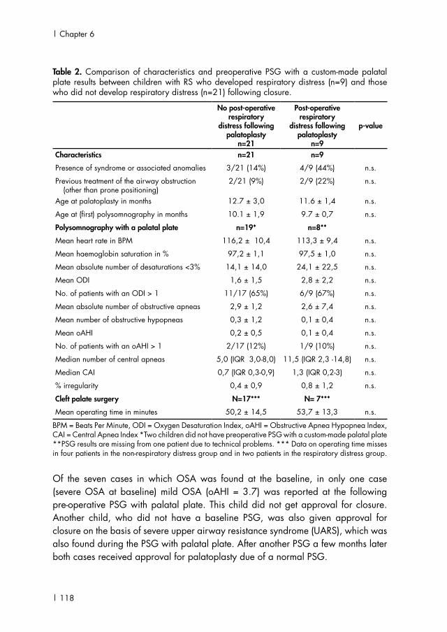
| 118
| Chapter 6
Table 2. Comparison of characteristics and preoperative PSG with a custom-made palatal plate results between children with RS who developed respiratory distress (n=9) and those who did not develop respiratory distress (n=21) following closure.
No post-operative respiratory
distress following palatoplasty
n=21
Post-operative respiratory
distress following palatoplasty
n=9
p-value
Characteristics n=21 n=9
Presence of syndrome or associated anomalies 3/21 (14%) 4/9 (44%) n.s.
Previous treatment of the airway obstruction (other than prone positioning)
2/21 (9%) 2/9 (22%) n.s.
Age at palatoplasty in months 12.7 ± 3,0 11.6 ± 1,4 n.s.
Age at (first) polysomnography in months 10.1 ± 1,9 9.7 ± 0,7 n.s.
Polysomnography with a palatal plate n=19* n=8**
Mean heart rate in BPM 116,2 ± 10,4 113,3 ± 9,4 n.s.
Mean haemoglobin saturation in % 97,2 ± 1,1 97,5 ± 1,0 n.s.
Mean absolute number of desaturations <3% 14,1 ± 14,0 24,1 ± 22,5 n.s.
Mean ODI 1,6 ± 1,5 2,8 ± 2,2 n.s.
No. of patients with an ODI > 1 11/17 (65%) 6/9 (67%) n.s.
Mean absolute number of obstructive apneas 2,9 ± 1,2 2,6 ± 7,4 n.s.
Mean number of obstructive hypopneas 0,3 ± 1,2 0,1 ± 0,4 n.s.
Mean oAHI 0,2 ± 0,5 0,1 ± 0,4 n.s.
No. of patients with an oAHI > 1 2/17 (12%) 1/9 (10%) n.s.
Median number of central apneas 5,0 (IQR 3,0-8,0) 11,5 (IQR 2,3 -14,8) n.s.
Median CAI 0,7 (IQR 0,3-0,9) 1,3 (IQR 0,2-3) n.s.
% irregularity 0,4 ± 0,9 0,8 ± 1,2 n.s.
Cleft palate surgery N=17*** N= 7***
Mean operating time in minutes 50,2 ± 14,5 53,7 ± 13,3 n.s.
BPM = Beats Per Minute, ODI = Oxygen Desaturation Index, oAHI = Obstructive Apnea Hypopnea Index, CAI = Central Apnea Index *Two children did not have preoperative PSG with a custom-made palatal plate **PSG results are missing from one patient due to technical problems. *** Data on operating time misses in four patients in the non-respiratory distress group and in two patients in the respiratory distress group.
Of the seven cases in which OSA was found at the baseline, in only one case (severe OSA at baseline) mild OSA (oAHI = 3.7) was reported at the following pre-operative PSG with palatal plate. This child did not get approval for closure. Another child, who did not have a baseline PSG, was also given approval for closure on the basis of severe upper airway resistance syndrome (UARS), which was also found during the PSG with palatal plate. After another PSG a few months later both cases received approval for palatoplasty due of a normal PSG.

119 |
Cha
pter
6
Respiratory distress following palatal closure in children with Robin Sequence|
In the RS group, the mean age at cleft palate repair was 12.4 months. Patients underwent closure according to the Veau-Wardill-Kilner technique (n = 3), Perko technique (n = 2), or von Langenbeck technique (n = 25). No significant differences were found with regard to postoperative obstructive airway symptoms between the different closure techniques. In five cases there were non–airway-related complications including dehiscence (n = 1) and fistula (n = 4). Eleven patients were postoperatively admitted to the pediatric high-care unit or intensive care unit.
Nine children of the RS group developed respiratory distress to varying degrees following palatoplasty. Eight of the nine children developed respiratory distress within 48 hours after surgery. One child developed respiratory distress after a week. The mean follow-up in the RS-group was 41.2 months.
Comparison of the non-RS group to the RS group The mean age at the time of repair was significantly later in the RS group compared to the non-RS group (12.4 versus 10.9 months) (Table 1). In the non-RS group none developed respiratory distress following palatoplasty, whereas, in the RS group a significantly higher number of children (30%) developed respiratory distress.
Comparison of the RS children who developed respiratory distress to those who did not Regarding the presence of a syndrome or a history of a previous treatment other than prone positioning, no significant differences were found between the children who developed respiratory distress following palatal closure and those who did not (Table 2). Furthermore, no significant differences were found in the results of the pre-operative PSG with a custom-made palatal plate between the groups.
Overview of cases who developed respiratory distress
Case 1
Syndromal RS; Preoperative PSG With Palatal Plate at 10 Months; Palatoplasty at 13 Months. Directly after extubation, this patient needed an NPT for one day. The patient was admitted to the intensive care unit (ICU) for 1 day and 1 day to the general children’s ward. Following discharge, no respiratory support was needed. At the check-up (3 weeks postoperatively) at the outpatient clinic no respiratory problems were reported.
| 120
| Chapter 6
Case 2
Syndromal RS (Treacher Collins Syndrome); Preoperative PSG With Palatal Plate at 11 Months; Palatoplasty at 13 Months. This patient had been treated by mandibular distraction in another clinic at the age of 16 months. The patient developed respiratory distress directly after extubation and needed an NPT for 2 days. The NPT could be removed under dexamethasone. After spending 2 days at the ICU, the patient was discharged with a saturation monitor. At the check-up at the outpatient clinic (3 weeks postoperatively) it was reported that patient’s sleeping behavior was unchanged compared with before the palatoplasty.
Case 3
Syndromal RS; Preoperative PSG With Palatal Plate at 10 Months; Palatoplasty at 11 Months. This patient needed reintubation within 7 minutes after extubation. Thirty minutes later an NPT was placed. The NPT was accidentally removed and the patient was reintubated. In the first night postoperatively there was another accidental detubation; afterward the saturation remained stable and the airway remained free. The patient was discharged without respiratory support. At the check-up (4 weeks later) at the outpatient clinic no snoring or apneas were reported.
Case 4
Syndromal RS; Preoperative PSG With Palatal Plate at 10 Months; Palatoplasty at 12 Months. This patient needed an NPT at the ward for 1 day following palatoplasty. The patient was discharged without respiratory support. At the check-up at the outpatient clinic no apneas or snoring were reported.
Case 5
Nonsyndromal RS; Preoperative PSG With Palatal Plate at 10 Months; Palatoplasty at 14 Months. The patient was discharged after 1 day without respiratory support. At the check-up at the outpatient clinic (4 weeks postoperatively), the parents reported the patient had suffered heavy apneas in the first few days that improved spontaneously. At the second check-up (5 months later), there was no report of snoring or heavy breathing when sleeping on the back. No apneas were present. A follow-up PSG was done 9 months after the palatoplasty and no OSA was found.

121 |
Cha
pter
6
Respiratory distress following palatal closure in children with Robin Sequence|
Case 6
Nonsyndromal RS; Preoperative PSG With Palatal Plate at 9 Months; Palatoplasty at 10 Months. Following closure, this patient had a stridorous breathing pattern the first few days with snoring and saturation drops. The patient was admitted for 3 days to the general children’s ward. At the check-up at the outpatient clinic (3.5 weeks postoperatively) there was no report of snoring or apneas.
Case 7
Nonsyndromal RS; Preoperative PSG With Palatal Plate at 10 Months; Palatoplasty at 11 Months. This patient was discharged after one night with a monitor and had saturation drops in the first few days. At the check-up (3 weeks postoperatively) at the outpatient clinic spontaneous improvement was reported. The patient snored but did not have any apnea or saturation drops. A follow-up PSG was done 3 months after palatoplasty and no OSA was found.
Case 8
Nonsyndromal RS; Preoperative PSG With Palatal Plate at 9 Months; Palatoplasty at 10 Months. This patient returned at day 2 after surgery with fast heavy breathing and snoring when lying on the back. Instruction for prone-positioning was given, and at the follow-up (3 weeks postoperatively) no sleeping problems were reported. At the second follow-up (2 months postoperatively) no snoring was reported.
Case 9
Nonsyndromal RS; Baseline PSG Showing Moderate OSA; Preoperative PSG With Palatal Plate at 9 Months; Palatoplasty at 11 Months. No breathing problems were reported directly postoperatively. However, the patient returned at day 7 after surgery with snoring and sleeping problems. The patient also had a cold. At day 10 after surgery a PSG was done showing moderate OSA, after which the patient was admitted to the ICU for 2 days. The patient was treated with nasal corticosteroids and received prone-positioning instructions. At the check-up at the outpatient clinic (3 weeks postoperatively) no sleeping problems were reported. At the second follow-up (2 months postoperatively), there were also no sleeping problems was reported.

| 122
| Chapter 6
DISCUSSION
This study showed that children with RS are at risk to develop respiratory distress in varying degree following palatoplasty. In nine out of 30 RS (30%) cases, respiratory distress occurred in spite of our presurgical screening protocol; this is similar to the prevalences reported in literature.8,9 The prevalence of respiratory distress in the RS group was significantly higher compared with the non-RS group.
Preoperatively we routinely performed screening by PSG with a custom-made palatal plate in patients with RS. The aim of a PSG is to detect obstructive pathology or other sleep disordered breathing patterns. In our clinic, cleft palate closure is usually scheduled around 9 to 10 months, but in two cases closure was postponed because preoperative PSG with palatal plate showed OSA and UARS. A second PSG a few months later showed a normal breathing pattern, which suggests that further growth had overcome the respiratory problems. Both cases received approval for closure and were closed without post-operative problems. It can be assumed that postponed closure in these two patients has avoided severe postoperative respiratory distress. In the other 26 patients, no obstructive pathology was seen in the PSG.
Concerning timing of the palatal repair there is a conflict in children with RS between minimizing the chance of post-operative respiratory distress and facilitating normal speech development. Henriksson et al. recommend postponing the palatoplasty until 12 to 18 months in children at risk for conditons such as Robin Sequence.12 Their study showed that the risk of (postoperative) anaesthetic complications such as hypoxia was higher above the age of 1 year. In our clinic we intend to close the palate around the age of 9 to 10 months. In this study the mean age at cleft palate repair was higher in both groups. This was mainly due to logistical issues and late referrals. The mean age of repair was 12.4 months in the RS group, which was considerably later than the non-RS group. This delay might be due to a tendency of the surgeon to close RS patients later in order to benefit from growth.
The occurrence of respiratory distress was significantly higher in the RS compared with the non-RS group. In the RS group a prevalence of 30% was reported. Notably, a recent similar study by Costa et al. reported a prevalence rate of 7.7% in the RS group (74 patients) and no difference in the airway complication rate between the RS group and the non-RS group.8 These findings however, may be explained by the different criteria applied to define post-operative respiratory distress or complications.

123 |
Cha
pter
6
Respiratory distress following palatal closure in children with Robin Sequence|
In our study, respiratory distress developed in the majority of cases within 48 hours after palatoplasty, which is in accordance with reported cases in the literature.9 In all our cases the respiratory distress resolved quickly and therefore may most likely be explained by lingual and palatal swelling, which usually has its peak within 48 hours after palatoplasty.13 Postoperative lingual swelling is presumably caused by the pressure applied by the tongue retractor or by the patient’s position during the surgery impairing the venous and lymphatic drainage, resulting in edema.13 Children with RS tend to have a more restricted oropharyngeal space and are therefore more likely to develop distress due to lingual swelling. During palatoplasty it is recommended by some surgeons to release the tongue retractor every hour for 5 minutes and to avoid an extreme Trendelenburg positioning to minimize the risk of lingual swelling.13,14
It is interesting that we did not find a strong correlation between the severity of the airway obstruction in the early postnatal period and the respiratory distress following palatoplasty. This is in contrast to previous studies.8,15 However, although not significant, we did notice a possible association with the presence of congenital anomalies or syndromes. Costa et al. also reported that the presence of cardiac anomalies, gastrointestinal anomalies, lower airway anomalies and syndromic diagnosis/genetic anomaly were associated with post-operative intubation including both the RS group and the non-group in this analysis.8 Children with RS and the presence of congenital anomalies and/or a syndrome seem to be more prone to develop postoperative respiratory distress.
The question remains whether or not patients should be routinely admitted to the ICU postoperatively. In some clinics it is recommended for children with RS to spend the first postoperative 24 hours in the pediatric intensive care unit.10 14 In our clinic we arrange an ICU stay postoperatively and instruct the caretakers to monitor the child’s breathing when at home. Patients without RS remain for one night at the children’s ward.
As a result of the retrospective character of the study, it has several limitations. We were not able to assess the value of the PSG with palatal plate due to the lack of a control group. Furthermore, the later age of closure might have influenced outcomes, and therefore made it less reliable to compare the RS-group with the non-RS group. The strength of this study is that the pre-operative screening with PSG offers an objective assessment of the airway obstruction.

| 124
| Chapter 6
CONCLUSION
Thirty percent of the children with RS developed respiratory distress following palatoplasty, in spite of delayed closure compared with non-RS children. This respiratory distress resolved within a few days. Preoperative PSG with a palatal plate is valuable in detecting obstructive pathology preoperatively and seems helpful in preselecting cases at risk for severe post-operative respiratory distress. Close postoperative monitoring following palatal closure is warranted.

125 |
Cha
pter
6
Respiratory distress following palatal closure in children with Robin Sequence|
LIST OF REFERENCES1 Bush, P. G. & Williams, A. J. Incidence of the
Robin Anomalad (Pierre Robin syndrome). Br J Plast Surg 36, 434-437, doi:0007-1226(83)90123-6 [pii] (1983).
2 Tolarova, M. M. & Cervenka, J. Classification and birth prevalence of orofacial clefts. Am J Med Genet 75, 126-137, doi:10.1002/(sici)1096-8628 (19980113 )75 :2<126 : : a i d -ajmg2>3.0.co;2-r (1998).
3 Printzlau, A. & Andersen, M. Pierre Robin Sequence in Denmark: A Retrospective Population-Based Epidemiological Study. Cleft Palate-Craniofac J 41, 47-52, doi:10.1597/02-055 (2004).
4 Vatlach, S., Maas, C. & Poets, C. F. Birth prevalence and initial treatment of Robin sequence in Germany: a prospective epidemiologic study. Orphanet J Rare Dis 9, 9, doi:1750-1172-9-9 [pii]10.1186/1750-1172-9-9 (2014).
5 Robin, P. A fall of the base of the tongue considered as new cause of nasopharyngeal impairment. Bull Acad Natl Med 89, 37 (1923).
6 Costa, M. A. et al. Robin sequence: mortality, causes of death, and clinical outcomes. Plast Reconstr Surg 134, 738-745 (2014).
7 Caouette-Laberge, L., Bayet, B. & Larocque, Y. The Pierre Robin sequence: Review of 125 cases and evolution of treatment modalities. Plast Reconstr Surg 93, 934-942 (1994).
8 Costa, M., Murage, K., Tholpady, S., Havlik, R. & Flores, R. Airway compromise following cleft palate repair in robin sequence: improving safety and predictability. Plast Reconstr Surg 134, 6-7, doi:10.1097/01.prs.0000455321.61126.44 00006534-201410001-00010 [pii] (2014).
9 Antony, A. K. & Sloan, G. M. Airway obstruction following palatoplasty: analysis of 247 consecutive operations. Cleft Palate Craniofac J 39, 145-148, doi:10.1597/1545-1569(2002)039<0145:AOFPAO>2.0.CO;2 (2002).
10 Dell’Oste, C., Savron, F., Pelizzo, G. & Sarti, A. Acute airway obstruction in an infant with Pierre Robin syndrome after palatoplasty. Acta Anaesthesiol Scand 48, 787-789, doi:10.1111/j.0001-5172.2004.00407.x (2004).
11 Reilly, J., Russell, C., Gibson, N., Moores, T., Ray, A., Devlin, M. & Wynne, D. in Britisch Academic Conference in Otolaryngology Posters (2012).
12 Henriksson, T. G. & Skoog, V. T. Identification of children at high anaesthetic risk at the time of primary palatoplasty. Scand J Plast Reconstr Surg Hand Surg 35, 177-182, doi:10.1080/028443101300165318 (2001).
13 Junghaenel, S. et al. Massive macroglossia after palatoplasty: case report and review of the literature. Eur J Pediatr 171, 433-437, doi:10.1007/s00431-011-1567-6 (2012).
14 Gupta, R., Chhabra, B., Mahajan, R. K. & Nandini. Macroglossia following palatoplasty causing upper airway obstruction: case report. J Oral Maxillofac SurG 59, 940-941, doi:S0278239101444600 [pii] (2001).
15 Thieme, V., Selzer, G., Gunther, L., Rustemeyer, J. & Bremerich, A. [Pierre Robin sequence: postoperative complications following cleft palate surgery. A retrospective study covering 25 years] Pierre-Robin-Sequenz -- postoperative Komplikationen nach Gaumenspaltverschluss. Eine retrospektive Studie uber 25 Jahre. Mund Kiefer Gesichtschir 9, 306-311, do i :10.1007/s10006 -005 -0637 -1 (2005).


QUALITY OF LIFE


Quality of life in children with Robin Sequence
Karolijn DulferManouk J.S. van LieshoutMarc P. van der SchroeffMaarten J. KoudstaalIrene M.J. MathijssenEppo B. WolviusKoen F.M. Joosten
Published in Int J Pediatr Otorhinolaryngol. 2016 Jul;86:98-103. doi: 10.1016/j.ijporl.2016.04.030. Epub 2016 Apr 27. PMID: 272605917

| 130
| Chapter 7
ABSTRACT
IntroductionParents may play an important role during the management of children with Robin Sequence (RS). However, so far only one study has been done on both parent-reported health-related quality of life (HRQoL) and obstructive sleep apnea (OSA) symptoms in children with RS.
MethodsOverall, 63 children with RS, aged 1 and 18, were included in this cross-sectional study. Fifty-three parents of children with RS with a median age of 8.9 [IQR 5.1-12.7] completed questionnaires on HRQoL (OSA-18) and symptoms of OSA (the Brouillette score) in their child with RS. Ten children between 12 and 18 years filled out the self-reported HRQoL questionnaire OSA-12.
ResultsAt cross-section, 10 children still had respiratory problems. Overall, parents of children with RS reported a lower HRQoL in their child compared with parents in the general population. Parents of children with RS who still had respiratory problems, i.e. OSA or airway support, reported significantly worse OSA-18 scores compared with parents of RS children without OSA. Children with RS themselves (n = 10) reported less physical suffering and less emotional distress on the OSA-12 compared with children in the norm population. Parental perceptions of OSA-related symptoms were accurate, except for the incidence of snoring. In assessing snoring, the multidimensional OSA-18 sleep domain was more informative.
ConclusionsParents of children with RS reported a lower HRQoL in their child compared with parents in the general population. Parental perceptions of health and HRQoL in children with RS might have an additional value to recognize and treat respiratory problems.

131 |
Cha
pter
7
Quality of life in children with Robin Sequence |
INTRODUCTION
Robin Sequence (RS) is characterized by a sequence of clinical events with mandibular hypoplasia as the inciting anomaly. Due to the mandibular hypoplasia the tongue becomes posteriorly displaced (glossoptosis) and obstructs the airway. RS occurs in about 1 in 5600–30,000 newborns.1-5 Patients with RS also present a cleft palate in 80–90% of the cases.4,6,7 Children with RS are at high risk of developing obstructive sleep apnea (OSA), with reported OSA prevalence rates between 46 and 100%, depending on the criteria used. 8-12 When RS is untreated, airway and feeding problems may be sustained, resulting in a range of morbidities, e.g. failure to thrive, cyanosis, cerebral hypoxia, and even death.
Health-related quality of life (HRQoL) has become an important measure throughout the diagnosis and management of children with obstructive sleep disordered breathing.13 RQoL is considered to be a multidimensional concept relying on the patient’s subjective evaluation of physical, social, and emotional aspects of a patient’s well-being, that are relevant to health and/or illness. HRQoL questionnaires can be categorized into generic versus disease-specific, focusing on disease symptoms per se versus on the subjective evaluation of these symptoms, and self-reported versus proxy-reported HRQoL, for example a parent that assesses the HRQoL in his/her child.14-15
Pediatric OSA has been associated with an impaired HRQoL assessed with both generic and disease-specific questionnaires (Child Health Questionnaire and OSA-18).16 In a mixed group of 79 children with OSA, the OSA-18 score was found to be an important tool to identify the impact of OSA on children with OSA and their families. However, in relation to PSG findings, the sensitivity and the negative predictive value (NPV) of OSA-18 for OSA were low. However, the positive predictive value (PPV) was high. Therefore, it was recommended that OSA-18 be used as a quality-of-life indicator and not as a reliable substitute for PSG.17, 18 In an RCT regarding early-adenotonsillectomy versus watchful-waiting, children with OSA significantly improved their PSG scores after early-adenotonsillectomy. In addition, parents also reported improvements in HRQoL (e.g. assessed with the OSA-18) in their child with OSA after early-adenotonsillectomy.16, 19, 20
Little is known about the impact of RS on HRQoL in children. Therefore, the aim of this study was to perform a cross-sectional study in children who were born with RS in order to assess parent-reported HRQOL and parent-reported symptoms of OSA in

| 132
| Chapter 7
their child with RS. Besides, these outcomes were related to the current respiratory status of the child.
METHODS
All children, aged between 1 and 18 years, with RS who were treated at the Dutch Craniofacial Center, Erasmus MC – Sophia Children’s Hospital, between 2012 and 2015 were eligible for this cross-sectional study. RS was defined as the presence of mandibular hypoplasia and airway obstruction.21, 22
Assessment procedureThe ethics committee review board of the Erasmus MC (MEC-2012-048) approved the research protocol. All eligible patients and/or parents were approached in a standardized way through a patient information letter. Written informed consent was obtained from all parents and patients above 12 years.
If possible, children underwent polysomnography (PSG) in the hospital or at home. Data regarding medical history were collected from the patients’ medical record. For psychological assessment, parents were asked to fill out the Brouillette score, a parent-report of symptoms of OSA in their child, and the parent-reported Obstructive Sleep Apnea survey-18 (OSA-18), a questionnaire in which parents report on the impact of OSA in their child with RS. Children between 12 and 18 years old filled out the self-reported Obstructive Sleep Apnea survey-12 (OSA-12) regarding the impact of OSA on them.
PolysomnographyPSG was done either in an ambulant sleep study at home; level III, with data recorded by the Embletta Portable Diagnostic system, or in a clinical sleep study in the hospital; level I, i.e. attended PSG including medical and technical support. During these sleep studies a variety of cardiorespiratory variables were assessed, including nasal airflow, chest and abdominal wall motion, and arterial oxygen saturation. Data were analyzed using Somnologica for Embletta software 3.3 ENU (Medcare Flage, Reykjavik, Iceland) for ambulant studies, and Shell + BrainRT Software Suite Version 2.0 (O.S.G. Rumst, Belgium) for clinical studies.
For analysis, we aimed for a total sleep time (TST) of at least 360 minutes, free of artifact. The scoring for respiratory events and calculation of the obstructive apnea–

133 |
Cha
pter
7
Quality of life in children with Robin Sequence |
hypopnea index (oAHI) were done as described by Spruijt et al.23 OSA was defined as an oAHI ≤ 1 per hour.24
2.3 The Brouillette ScoreParents were asked to fill out the Brouillette score, a questionnaire to screen for the presence of OSA. 25 The score was calculated using the following formula: 1.42D + 1.41A + 0.72S – 3.83. D stands for difficulty in breathing, A for apnea, and S for snoring. For D and S, caregivers could choose between the options never = 0, sometimes = 1, often = 2, and always = 3. For A, caregivers could score 0 if no apneas occur or 1 if they do. A Brouillette score of >3.5 predicts the presence of OSA, a Brouillette score between −1 and 3.5 is suggestive of OSA, and a score <−1 predicts the absence of OSA. In analyses, parent-reported OSA was defined as a Brouillette score ≥ −1.
Parent-reported Obstructive Sleep Apnea-18 (OSA-18) The Dutch version of the parent-reported disease-specific quality of life questionnaire OSA-18 was used to assess parental perceptions of the impact of OSA on the child with RS. 26 The OSA-18 is the most widely used HRQoL questionnaire in pediatric OSA. 16 The Dutch version has been validated in 459 parents of healthy children and in 119 parents of children with syndromic craniosynostosis.26 The OSA-18 consists of 18 age-independent items grouped into five domains: sleep disturbance, physical suffering, emotional distress, daytime problems, and caregiver concerns. Parents were asked to report how often during the previous 4 weeks their child has had specific symptoms, using a response scale from 1 (never) to 7 (always). The total OSA-18 score ranges from 18 to 126, with a higher score indicating a worse outcome. Scores less than 60 suggest a small impact on health-related quality of life, scores between 60 and 80 suggest a moderate impact, and scores above 80 suggest a large impact. The parent-reported OSA-18 scores of the impact of OSA in children with RS were compared with those of 459 parents in the general population.26
Child-reported Obstructive Sleep Apnea-12 (OSA-12)Children aged 12–18 completed a comparable OSA questionnaire, the self-reported OSA-12, to assess the impact of OSA on them. Children were asked to report how often during the previous 4 weeks they have had specific symptoms, using a response scale from 1 (never) to 7 (always). This questionnaire consists of 12 questions, comparable to those of the OSA-18 with the following exceptions: 2

| 134
| Chapter 7
questions of the sleep disturbance domain and the total caregiver concerns domain were excluded. The total OSA-12 score ranges from 12 to 84. The OSA-12 has been validated in n = 162 children from the general population and in n = 29 children with craniosynostosis. OSA-12 scores of children with RS were compared with those of 162 children, aged 12–18 years, in the general population.26
Visual Analogue Scale (VAS)In addition, parents and children were asked to indicate the child’s HRQoL on a visual analogue scale (VAS). The VAS consists of a Likert scale from 0 to 10, which was adjusted from the EQ-5D.27 Scores ranged from 0, worst imaginable HRQoL, to 10, best imaginable HRQoL. A higher score indicated a better HRQoL.
Statistical analysis To compare baseline characteristics between participants and non-participants, Pearson’s Chi Square tests for dichotomous data and non-parametric Mann–Whitney U tests for continuous non-normally distributed data were used. In order to determine the median age at the time of cross-section in the group of non-participating patients we used the date halfway our study inclusion period as the date of cross-section.
To compare the mean OSA-18 results of the RS population with the mean in the general population, sample T-tests were used. Spearman correlation was calculated between OSA-18 and the visual analogue scale. Median OSA-18 scores were compared between three groups, divided for treatment history and the presence of OSA/currently receiving airway support, using Kruskal–Wallis H tests. A p-value of <0.05 was considered statistically significant. Analyses were performed with SPSS 20.0 for Windows (SPSS, Inc. Chicago, IL).
RESULTS
Baseline characteristicsIn total, 111 children with RS were eligible for inclusion of which 63 (57%) parents of RS children gave informed consent (Fig. 1). Forty-eight children did not consent for this study with various reasons. Ten children did not have complete data on OSA-18, VAS, and Brouillette score and were therefore excluded from analyses.
Baseline characteristics of the participants with complete data (n = 53) were compared with those of non-participants (n = 48) and participants without complete

135 |
Cha
pter
7
Quality of life in children with Robin Sequence |
data (n = 10) (Table 1). No significant differences were found for median age at follow-up, gender, presence of a syndrome or additional anomalies, presence of a cleft palate, or initial treatment of airway obstruction.Fifty-three RS children, median age 8.9 years [IQR 5.1–12.7], had complete data of which 27 were male, 33 had an isolated RS, 15 had additional anomalies, and 4 had a syndrome (Table 1). Four children had RS without a cleft palate. Of these 53 children, 27 were previously treated in the prone position and 26 were previously treated with respiratory support, of which 18 with non-surgical treatment and 8 with surgical treatment.
Screened on eligibillity (n=111)
Enrolled in the study (n=63)
Declined to participate (n=48)
Complete cases on OSA-18, VAS, Brouillette score included in analyses (n=53)
Previously treated with prone positioning Previously treated with respiratory support
No OSA-18 questionnaire (n=8)No Visual Analogue Scale (n=5)
No Brouillette score (n=5)
Without OSA (n=26) With OSA (n=1) Without OSA (n=17) With OSA* (n=9)
Figure 1. Participants’ flow-chart

| 136
| Chapter 7
Table 1. Baseline demographics of participants with complete data (n=53) versus non-participants or participants without complete data (n=58)
Participants (n=53)
Non-participants (n=58) P-value
Median age in years [IQR] 1 8.9 [5.1-12.7] 8.5 [5.0-13.7] 0.86
Gender, n (%) Female 26 (49) 26 (45) 0.10
Male 27 (51) 32 (55)Presence of syndrome or additional anomalies, n (%)
Yes, anomalies 15 (28) 18 (31) 0.14
Yes, syndrome 6 (12) 14 (24)
No 32 (60) 26 (45)Presence of a cleft palate, n (%)
Yes 49 (93) 49 (84) 0.19
No 4 (7) 9 (16)Treatment of airway obstruction, n(%) 2
Prone position only 27 (51) 34 (59) 0.06
Non-surgical treatment 18 (34) 8 (14)
Surgical treatment 8 (15) 15 (27)
1. IQR=Inter quartile range.2. In n=1 non-participant, information regarding treatment of airway obstruction is missing.
Respiratory outcome Respiratory data, available in 53 children with RS, were derived from 38 PSGs, 31 (82%) ambulatory at home and 7 (18%) clinically (in-house). In 15 children no PSG was done; parents refused (n = 3), PSG failed due to logistic reasons (n = 9), or PSG was not possible because of a tracheotomy (n = 3). Respiratory data of these 15 children were interpreted on the basis of the current respiratory support.
In total 10 children still had respiratory problems: in 5 children OSA was found at PSG, defined as an oAHI ≥ 1 per hour, 2 children needed non-surgical respiratory support, and in 3 children a tracheostomy was still present (Fig. 1). Of those 5 children in which OSA was newly detected, 4 were treated with respiratory support in the past, and 1 child was treated in the prone position at birth and re-developed mild OSA during follow-up.
Health-related quality of life: OSA-18 and OSA-12 Compared with the norm population, parents of children with RS (n = 53) reported significantly worse scores in their child on the following OSA-18 domains: sleep disturbance, physical suffering, caregiver concerns, and the total OSA-18 score (Table 2) compared with parents in the general population. Furthermore, a worse
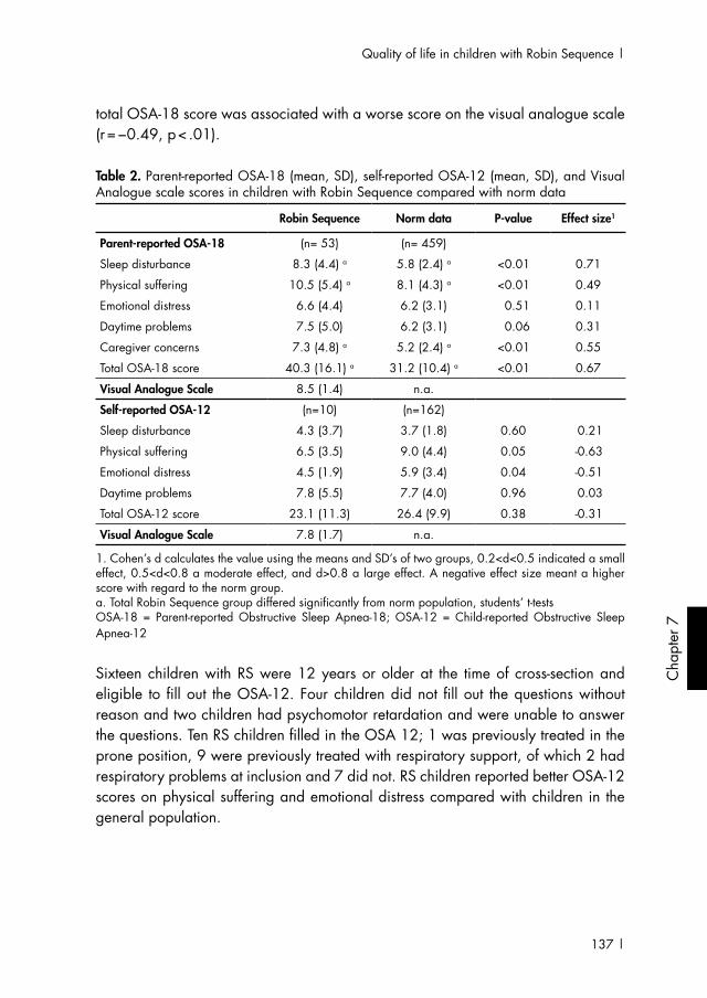
137 |
Cha
pter
7
Quality of life in children with Robin Sequence |
total OSA-18 score was associated with a worse score on the visual analogue scale (r = −0.49, p < .01).
Table 2. Parent-reported OSA-18 (mean, SD), self-reported OSA-12 (mean, SD), and Visual Analogue scale scores in children with Robin Sequence compared with norm data
Robin Sequence Norm data P-value Effect size1
Parent-reported OSA-18 (n= 53) (n= 459)
Sleep disturbance 8.3 (4.4) a 5.8 (2.4) a <0.01 0.71
Physical suffering 10.5 (5.4) a 8.1 (4.3) a <0.01 0.49
Emotional distress 6.6 (4.4) 6.2 (3.1) 0.51 0.11
Daytime problems 7.5 (5.0) 6.2 (3.1) 0.06 0.31
Caregiver concerns 7.3 (4.8) a 5.2 (2.4) a <0.01 0.55
Total OSA-18 score 40.3 (16.1) a 31.2 (10.4) a <0.01 0.67
Visual Analogue Scale 8.5 (1.4) n.a.
Self-reported OSA-12 (n=10) (n=162)
Sleep disturbance 4.3 (3.7) 3.7 (1.8) 0.60 0.21
Physical suffering 6.5 (3.5) 9.0 (4.4) 0.05 -0.63
Emotional distress 4.5 (1.9) 5.9 (3.4) 0.04 -0.51
Daytime problems 7.8 (5.5) 7.7 (4.0) 0.96 0.03
Total OSA-12 score 23.1 (11.3) 26.4 (9.9) 0.38 -0.31
Visual Analogue Scale 7.8 (1.7) n.a.
1. Cohen’s d calculates the value using the means and SD’s of two groups, 0.2<d<0.5 indicated a small effect, 0.5<d<0.8 a moderate effect, and d>0.8 a large effect. A negative effect size meant a higher score with regard to the norm group.a. Total Robin Sequence group differed significantly from norm population, students’ t-testsOSA-18 = Parent-reported Obstructive Sleep Apnea-18; OSA-12 = Child-reported Obstructive Sleep Apnea-12
Sixteen children with RS were 12 years or older at the time of cross-section and eligible to fill out the OSA-12. Four children did not fill out the questions without reason and two children had psychomotor retardation and were unable to answer the questions. Ten RS children filled in the OSA 12; 1 was previously treated in the prone position, 9 were previously treated with respiratory support, of which 2 had respiratory problems at inclusion and 7 did not. RS children reported better OSA-12 scores on physical suffering and emotional distress compared with children in the general population.

| 138
| Chapter 7
Table 3: Parent-reported OSA-18, Visual Analogue Scale, and Brouillette Score (median [IQR]) in children with Robin Sequence divided in treatment history with and without current respiratory problems
Treatment History
Prone position1 Received respiratory support
No-OSA, n=26 No-OSA, n=17 OSA/respiratory problems2 n=9
OSA-18 scales, Mdn [IQR]
Sleep disturbance 6.5 [4.0-9.1] a 7.0 [5.5-9.7] 10.0 [4.3-17.0] a
Physical suffering 8.5 [4.0-13.3] 11.0 [5.0-17.0] 13.0 [9.0-15.5]
Emotional distress 4.0 [3.0-7.0] 6.0 [3.0-9.5] 7.0 [3.5-12.5]
Daytime problems 4.5 [3.0 – 8.0] b 6.0 [3.5-9.0] b 15.0 [7.5-18.0] b
Caregiver concerns 4.0 [4.0-7.3] b 6.0 [4.0-7.0] b 13.0 [6.5-19.5] b
Total OSA-18 score 33.5 [22.0-41.5] b 37.0 [31.0-47.5] b 59.0 [44.5-71.5] b
Visual Analogue Scale, Mdn [IQR] 9.0 [8.0-9.7] b 9.0 [8.0-9.9] b 7.5 [5.8-9.0] b
Brouillette Score, n (%)
Brouillette score > -1 2 (8) c 3 (18) c 6 (67) c
≤ -1 24 (92) c 14 (82) c 3 (33) c
Difficulty in breathing yes 5 (19) d 3 (18) d 6 (67) d
no 21 (81) d 14 (82) d 3 (33) d
Apnea yes 3 (12) e 3 (18) e 6 (67) e
no 23 (88) e 14 (82) e 3 (33) e
Snoring yes 19 (73) 14 (82) 7 (78)
no 7 (27) 3 (18) 2 (22)
1. One RS child treated with prone position developed mild OSA. This child is excluded from analyses.2. Children with OSA or airway support: OSA found at PSG without treatment n=4, non-surgical respiratory support n=2, and tracheostomy n=3a. Post-hoc analysis: Children treated with prone position only differed significantly from children who received respiratory support/developed OSA, Mann-Whitney U tests, p<.03b. Children treated with prone position only and children who received respiratory support/did not develop OSA differed significantly from children who received respiratory support/developed OSA, Mann-Whitney U tests, p<.02c, e. The observed counts differed significantly from the expected counts under the assumption of no association, Chi-square comparison p<.01d. The observed counts differed significantly from the expected counts under the assumption of no association, Chi-square comparison p<.02d. IQR = inter quartile range; Mdn = median; OSA = obstructive sleep apnea; OSA-18 = Parent-reported Obstructive Sleep Apnea-18; OSA-12 = Child-reported Obstructive Sleep Apnea-12

139 |
Cha
pter
7
Quality of life in children with Robin Sequence |
Table 3: Parent-reported OSA-18, Visual Analogue Scale, and Brouillette Score (median [IQR]) in children with Robin Sequence divided in treatment history with and without current respiratory problems
Treatment History
Prone position1 Received respiratory support
No-OSA, n=26 No-OSA, n=17 OSA/respiratory problems2 n=9
OSA-18 scales, Mdn [IQR]
Sleep disturbance 6.5 [4.0-9.1] a 7.0 [5.5-9.7] 10.0 [4.3-17.0] a
Physical suffering 8.5 [4.0-13.3] 11.0 [5.0-17.0] 13.0 [9.0-15.5]
Emotional distress 4.0 [3.0-7.0] 6.0 [3.0-9.5] 7.0 [3.5-12.5]
Daytime problems 4.5 [3.0 – 8.0] b 6.0 [3.5-9.0] b 15.0 [7.5-18.0] b
Caregiver concerns 4.0 [4.0-7.3] b 6.0 [4.0-7.0] b 13.0 [6.5-19.5] b
Total OSA-18 score 33.5 [22.0-41.5] b 37.0 [31.0-47.5] b 59.0 [44.5-71.5] b
Visual Analogue Scale, Mdn [IQR] 9.0 [8.0-9.7] b 9.0 [8.0-9.9] b 7.5 [5.8-9.0] b
Brouillette Score, n (%)
Brouillette score > -1 2 (8) c 3 (18) c 6 (67) c
≤ -1 24 (92) c 14 (82) c 3 (33) c
Difficulty in breathing yes 5 (19) d 3 (18) d 6 (67) d
no 21 (81) d 14 (82) d 3 (33) d
Apnea yes 3 (12) e 3 (18) e 6 (67) e
no 23 (88) e 14 (82) e 3 (33) e
Snoring yes 19 (73) 14 (82) 7 (78)
no 7 (27) 3 (18) 2 (22)
1. One RS child treated with prone position developed mild OSA. This child is excluded from analyses.2. Children with OSA or airway support: OSA found at PSG without treatment n=4, non-surgical respiratory support n=2, and tracheostomy n=3a. Post-hoc analysis: Children treated with prone position only differed significantly from children who received respiratory support/developed OSA, Mann-Whitney U tests, p<.03b. Children treated with prone position only and children who received respiratory support/did not develop OSA differed significantly from children who received respiratory support/developed OSA, Mann-Whitney U tests, p<.02c, e. The observed counts differed significantly from the expected counts under the assumption of no association, Chi-square comparison p<.01d. The observed counts differed significantly from the expected counts under the assumption of no association, Chi-square comparison p<.02d. IQR = inter quartile range; Mdn = median; OSA = obstructive sleep apnea; OSA-18 = Parent-reported Obstructive Sleep Apnea-18; OSA-12 = Child-reported Obstructive Sleep Apnea-12

| 140
| Chapter 7
Parent-reported HRQoL and parent-reported symptoms of OSA divided for treatment history with and without current respiratory problemsDivided into treatment history, parents of children with RS who initially received respiratory support and who developed OSA during follow-up/who still received airway support (n = 9) reported significantly worse scores on daytime problems, caregiver concerns, total OSA-18, and on the VAS compared with parents whose children had no current respiratory problems (respectively n = 26 and n = 17; Table 3). Besides, they more frequently reported a Brouillette total score >−1, difficulty in breathing, and apnea in their child with RS. Parents of RS children who were initially treated with respiratory support and who developed OSA during follow-up/who still received airway support reported a worse score on sleep disturbance compared with parents of RS children initially treated in the prone position.
Within this RS group of children with current respiratory problems, those parents of children with a tracheostomy (n = 3) reported better scores on sleep disturbance and caregiver concerns compared with those with OSA or other respiratory support. Besides, these 3 parents reported a total Brouillette score lower than −1 and no symptoms of difficulty in breathing, apnea, or snoring.
DISCUSSION
Health-related quality of life (HRQoL) has become an important outcome throughout the stepwise approach to the diagnosis and management of children with obstructive sleep disordered breathing.13
While little is known about the lifelong impact on HRQoL in children born with RS, there are a number of studies on generic and disease-specific HRQoL outcomes in children who suffered from pediatric OSA.16 In these studies, pediatric OSA has been significantly associated with a lower HRQoL compared with healthy peers. Overall, in our follow-up of children with RS, parents also reported a lower HRQoL in their child with RS compared with parents in the general population. These worse disease-specific OSA-18 scores were correlated with a lower VAS score. The VAS is a single-item general HRQoL instrument that is easy to administer. Therefore, this VAS score may be a valuable, informative addition to disease-specific HRQoL questionnaires such as the OSA-18.28

141 |
Cha
pter
7
Quality of life in children with Robin Sequence |
So far, HRQoL was reported in only one other study in which parents of 21 children with RS reported on HRQoL in their child after mandibular distraction osteogenesis (mean age at surgery 54 days). All children with RS demonstrated improved airway outcomes post-MDO; there were no significant post-operative complications. The GCBI (Glascow Children’s Benefit Inventory) questionnaire was used as a generic instrument to detect changes in health status following a surgical intervention. Parents reported an overall benefit in HRQoL in all domains in their child with RS, especially in the physical domain.29
Interestingly in our study, although the majority of the children did not have OSA, parents still reported significantly higher scores in their child with RS on the following domains: sleep disturbance, physical suffering, and caregiver concerns compared with parents in the general population. There were no differences in HRQoL scores between children previously treated in the prone position or those previously treated with respiratory support. Parents of children with RS who still received respiratory support or who had OSA reported significantly worse HRQoL scores on sleep domain, daytime problems, caregiver concerns, and on total HRQoL. These worse OSA-18 scores were comparable with the OSA-18 scores reported by parents of children with OSA before they underwent surgery.16 In contrast, the parent-reported OSA-18 scores of RS children without OSA or respiratory problems seemed to be comparable to those of healthy children.26
Parents of RS children who still received respiratory support or who had OSA also reported a higher Brouillette score more frequently. However, within this group of children with current respiratory problems, those parents of children with a tracheostomy were the exception. They reported less problems regarding breathing, apnea, and snoring in their child, and they also reported a better HRQoL in their child. This is rather obvious since the airway of these children was secured through a cannula. As a result, the OSA-18 questionnaire seemed to be a helpful tool to recognize OSA-related symptoms and one might speculate that treatment in this group of children may not have been optimal.
Regarding children with RS themselves, in our study they reported better OSA-12 scores on physical suffering, such as having frequent colds or nasal secretion, and on emotional distress, such as less mood change or anger fit, or aggressive/hyperactive behavior, compared with children in the general population. Since we only had complete data on 10 children and 8 of them had no OSA or respiratory problems at that moment, we should handle this result with caution. Especially since

| 142
| Chapter 7
in a large study in chronically ill children overall HRQoL was impaired compared with healthy children.30 Our findings should be confirmed in a larger sample of children with RS.
Interestingly, although few children with RS in this study were diagnosed with OSA, parents reported a high prevalence (around 70–80% in all three groups) of snoring in their child with RS regardless of treatment history or current treatment. Remarkably, parents reported a high incidence of snoring on the Brouillette score, whereas on the OSA-18 questionnaire they did not report more sleep disturbance or snoring. This could be explained by the way the occurrence of snoring was assessed. In the Brouillette score, the presence of snoring was assessed in a one-dimensional manner, scored with yes/no, whereas in the OSA-18, snoring was assessed in a more multidimensional manner, which may have been more informative.
LimitationsSince not all eligible patients agreed to participate in the current study, the results might have been influenced by selection bias. Besides, not all participants had complete data on HRQoL. On the other hand, participants and non-participants/participants without complete data did not differ as to the distribution of median age at follow-up, gender, presence of a syndrome or additional anomalies, presence of a cleft palate, or initial treatment of airway obstruction. Furthermore, the participants were divided into sub-groups, based on treatment history and current OSA status/treatment. Although sub-group analyses were performed non-parametrically, these may have suffered from small sample sizes. Finally, respiratory data were derived from PSG assessments or, when missing, were based on current respiratory support. The findings regarding the differences between children with and without current respiratory problems should therefore be confirmed in another study with more complete data and a larger sample size.
CONCLUSIONS
Health-related quality of life (HRQoL) is an important outcome throughout the diagnosis and management of children with Robin Sequence (RS). In this cross-sectional study of 53 children with RS, parents reported a lower HRQoL in their child compared with parents in the general population. Especially parents of children with RS who were previously treated with respiratory support and who re-developed OSA or received current airway support reported lower HRQoL scores. Overall, parental perceptions regarding treatment in their child with RS could have an impact

143 |
Cha
pter
7
Quality of life in children with Robin Sequence |
on HRQoL and the well being of the child. Children themselves reported better OSA-12 scores on physical suffering and on emotional distress.
As to recommendations, parents should be seen as important stakeholders in the management of their child with RS.31 Since parents “know their child best”, parental perceptions of health and HRQoL are important and informative with regard to treatment decisions. In this study, parents’ scores of HRQoL in their child were associated with the presence of OSA/respiratory support. Therefore, it is recommended not only to assess impairments in functional health status during outpatients’ consultations, but also to screen parents’ assessment of HRQoL problems in their children using multidimensional, generic as well as disease-specific instruments. These HRQoL assessments may provide valuable information regarding the child’s functioning and may contain predictive value for the child’s health development.

| 144
| Chapter 7
LIST OF REFERENCES1. Gupta, R., Chhabra, B., Mahajan,
R. K. & Nandini. Macroglossia following palatoplasty causing upper airway obstruction: case report. J Oral Maxillofac SurG 59, 940-941, doi:S0278239101444600 [pii] (2001).
2. Bush PC, Williams AG. Incidence of the Robin Anomalad (Pierre Robin syndrome). Br J Plast Surg; 36. (1983): 434-437.
3. Tolarova MM, Cervenka J. Classification and birth prevalence of orofacial clefts. Am J Med Genet; 75. (1998): 126-137.
4. Printzlau A, Andersen M. Pierre Robin Sequence in Denmark: A Retrospective Population-Based Epidemiological Study. Cleft Palate-Craniofac J; 41. (2004): 47-52.
5. Vatlach S, Maas C, Poets CF. Birth prevalence and initial treatment of Robin sequence in Germany: a prospective epidemiologic study. Orphanet J Rare Dis; 9. (2014): 9.
6. Paes EC, van Nunen DP, H Basart, JP Don Griot, JM van Hagen, CM van der Horst, MJ van denBoogaard, CC Breugem. Birth prevalence of Robin sequence in the Netherlands from 2000-2010: A retrospective population-based study in a large Dutch cohort and review of the literature. Am J Med Genet A. (2015).
7. MA Costa, KP Murage, SS Tholpady, RL Flores. Airway compromise following palatoplasty in Robin sequence: improving safety and predictability. PLAST RECONSTR SURG; 134. (2014): 937e-945e.
8. L Caouette-Laberge, B Bayet, Y Larocque. The Pierre Robin sequence: Review of 12cases and evolution of treatment modalities. PLAST RECONSTR SURG; 93. (1994): 934-942.
9. IC Anderson, AR Sedaghat, BM McGinley, RJ Redett, EF Boss, SL Ishman. Prevalence and severity of obstructive sleep apnea and snoring in infants with Pierre Robin sequence. Cleft Palate Craniofac J; 48. (2011): 614-618.
10. G Bravo, A Ysunza, J Arrieta, MC Pamplona. Videonasopharyngoscopy is useful for identifying children with Pierre Robin sequence and severe obstructive sleep apnea. INT J PEDIATR OTORHINOLARYNGOL; 69. (2005): 27-33.
11. JT Gilhooly, JD Smith, LL Howell, BL Deschaine, SL Richey. Bedside polysomnography as an adjunct in the management of infants with Robin sequence. PLAST RECONSTR SURG; 92. (1993): 23-27.
12. AC Wilson, DJ Moore, MH Moore, AJ Martin, REM Staugas, JD Kennedy. Late presentation of upper airway obstruction in Pierre Robin sequence. Arch Dis Child; 83. (2000): 435-438.
13. M Daniel, S Bailey, K Walker, R Hensley, C Kol-Castro, N Badawi, A Cheng, K Waters. Airway, feeding and growth in infants with Robin sequence and sleep apnoea. Int J Pediatr Otorhinolaryngol. (2013).
14. AG Kaditis, ML Alonso Alvarez, A Boudewyns, EI Alexopoulos, R Ersu, K Joosten, H Larramona, S Miano, I Narang, H Trang, M Tsaoussoglou, N Vandenbussche, MP Villa, D Van Waardenburg, S Weber, S Verhulst. Obstructive sleep disordered breathing in 2-18 year-old children: diagnosis and management. Eur Respir J. (2015).
15. M Solans, S Pane, MD Estrada, V Serra-Sutton, S Berra, M Herdman, J Alonso, L Rajmil. Health-related quality of life measurement in children and adolescents: a systematic review of generic and disease-specific instruments. Value Health; 11. (2008): 742-764.

145 |
Cha
pter
7
Quality of life in children with Robin Sequence |
16. H Raat, AD Mohangoo, MA Grootenhuis. Pediatric health-related quality of life questionnaires in clinical trials. Curr Opin Allergy Clin Immunol; 6. (2006): 180-185.
17. CM Baldassari, RB Mitchell, C Schubert, EF Rudnick. Pediatric obstructive sleep apnea and quality of life: a meta-analysis. Otolaryngol Head Neck Surg; 138. (2008): 265-273.
18. SL Ishman, CJ Yang, AP Cohen, JR Benke, JK Meinzen-Derr, RM Anderson, ME Madden, ME Tabangin. Is the OSA-18 predictive of obstructive sleep apnea: comparison to polysomnography. Laryngoscope; 125. (2015): 1491-1495.
19. E Constantin, TL Tewfik, RT Brouillette. Can the OSA-18 quality-of-life questionnaire detect obstructive sleep apnea in children? Pediatrics; 125. (2010): e162-168.
20. CL Marcus, RH Moore, CL Rosen, B Giordani, SL Garetz, HG Taylor, RB Mitchell, R Amin, ES Katz, R Arens, S Paruthi, H Muzumdar, D Gozal, NH Thomas, J Ware, D Beebe, K Snyder, L Elden, RC Sprecher, P Willging, D Jones, JP Bent, T Hoban, RD Chervin, SS Ellenberg, S Redline, T Childhood Adenotonsillectomy. A randomized trial of adenotonsillectomy for childhood sleep apnea. N Engl J Med; 368. (2013): 2366-2376.
21. RP Venekamp, BJ Hearne, D Chandrasekharan, H Blackshaw, J Lim, AG Schilder. Tonsillectomy or adenotonsillectomy versus non-surgical management for obstructive sleep-disordered breathing in children. Cochrane Database Syst Rev; 10. (2015): CD011165.
22. MJ van Lieshout, KF Joosten, HL Hoeve, IM Mathijssen, MJ Koudstaal, EB Wolvius. Unravelling Robin sequence: considerations of diagnosis and treatment. LARYNGOSCOPE; 124. (2014): E203-209.
23. EC Paes, DP van Nunen, L Speleman, MS Muradin, B Smarius, M Kon, AB van der Molen, TL Niers, ES Veldhoen, CC Breugem. A pragmatic approach to infants with Robin sequence: a retrospective cohort study and presence of a treatment algorithm. Clin Oral Investig. (2015).
24. B Spruijt, KF Joosten, C Driessen, D Rizopoulos, NC Naus, MP van der Schroeff, EB Wolvius, ML van Veelen, RC Tasker, IM Mathijssen. Algorithm for the Management of Intracranial Hypertension in Children with Syndromic Craniosynostosis. Plast Reconstr Surg; 136. (2015): 331-340.
25. C Guilleminault, R Pelayo, A Clerk, D Leger, RC Bocian. Home nasal continuous positive airway pressure in infants with sleep-disordered breathing. J PEDIATR; 127. (1995): 905-912.
26. R Brouilette, D Hanson, R David, L Klemka, A Szatkowski, S Fernbach, C Hunt. A diagnostic approach to suspected obstructive sleep apnea in children. J Pediatr; 105. (1984): 10-14.
27. N Bannink, M Maliepaard, H Raat, KF Joosten, IM Mathijssen. Reliability and validity of the obstructive sleep apnea-18 survey in healthy children and children with syndromic craniosynostosis. J Dev Behav Pediatr; 32. (2011): 27-33.
28. K Burstrom, A Bartonek, EW Brostrom, S Sun, AC Egmar. EQ-5D-Y as a health-related quality of life measure in children and adolescents with functional disability in Sweden: testing feasibility and validity. Acta Paediatr; 103. (2014): 426-435.

| 146
| Chapter 7
29. U Ravens-Sieberer, N Wille, X Badia, G Bonsel, K Burstrom, G Cavrini, N Devlin, AC Egmar, N Gusi, M Herdman, J Jelsma, P Kind, PR Olivares, L Scalone, W Greiner. Feasibility, reliability, and validity of the EQ-5D-Y: results from a multinational study. Qual Life Res; 19. (2010): 887-897.
30. P Hong, M McNeil, DB Kearns, AE Magit. Mandibular distraction osteogenesis in children with Pierre Robin sequence: impact on health-related quality of life. Int J Pediatr Otorhinolaryngol; 76. (2012): 1159-1163.
31. JW Varni, CA Limbers, TM Burwinkle. Impaired health-related quality of life in children and adolescents with chronic conditions: a comparative analysis of 10 disease clusters and 33 disease categories/severities utilizing the PedsQL 4.0 Generic Core Scales. Health Qual Life Outcomes; 5. (2007): 43.
32. J Demke, M Bassim, MR Patel, S Dean, R Rahbar, JA van Aalst, A Drake. Parental perceptions and morbidity: tracheostomy and Pierre Robin sequence. Int J Pediatr Otorhinolaryngol; 72. (2008): 1509-1516.

GENERAL DISCUSSION AND SUMMARY


General discussion
8


151 |
Cha
pter
8
General discussion |
INTRODUCTION
The aim of this thesis is to improve knowledge with regard to diagnosis, treatment and quality of life in children with Robin Sequence (RS). Within this general discussion, the main findings of our studies will be discussed against the background of the literature. Furthermore, clinical recommendations and suggestions for future research will be provided.
What is Robin Sequence? RS remains a diagnostic and therapeutic challenge, partially due to the lack of a standardized definition. An online survey among 101 different clinics (chapter II) showed that 56 different combinations of clinical features were used to define RS. Many clinicians considered cleft palate an essential feature. This large variety in definition was in accordance with findings of other (survey) studies.1-5 It is needless to say that a uniform and strict definition of RS is crucial for both care and research.
In this thesis, Pierre Robin’s original set of features (mandibular hypoplasia, glossoptosis and airway obstruction) was used to define RS and select patients.6 However, applying this triad as the basis for an RS diagnosis still did not provide us with clear criteria for eligibility of patients, as strict criteria for these individual features were not available. Also, our inclusion had been based on historical data. Especially in the group that was initially treated with prone positioning, one may question whether these children were ‘true’ RS patients, as no objective measurements of obstructive breathing had been done.
First, the three individual features that are part of the original RS definition, will be further discussed below.
Defining and diagnosing mandibular hypoplasiaMandibular hypoplasia is the hallmark of the RS phenotype. Previous studies have shown significant differences in e.g. mandibular body length and height, ramus-to-body ratio and chin thickness in children with RS compared to controls.7-9 In daily practice (no data), sometimes neonates with a seemingly small lower jaw without any other symptoms are observed. This group of patients might receive an inaccurate diagnosis of RS at birth when the clinician notes the hypoplastic lower jaw, but does not assess the airway status.10 Especially in RS children with additional anomalies (non-isolated RS), mandibular hypoplasia is often distinctly present, but unfortunately objective data are lacking.

| 152
| Chapter 8
Quantifying mandibular hypoplasia is not standard practice. A few methods, such as jaw-index, cephalograms, cone beam computed tomography scanning, three-dimensional photography, have been suggested, but there is currently no widely accepted standard measurement method available.11 Suggested measurement methods have considerable disadvantages and are often invasive, time consuming and/or not yet validated. Yet, a method to quantify and monitor mandibular hypoplasia is helpful in order to identify a child with RS and essential in order to establish the correlation between the severity of mandibular hypoplasia and respiratory distress and to examine growth. As long as normative data sets on mandibular size and position are unavailable, a diagnosis of mandibular hypoplasia cannot be established. Therefore, there is currently little added clinical value of a diagnosis of mandibular hypoplasia.
Defining and diagnosing glossoptosisGlossoptosis is defined as displacement of the tongue base into the oro- and hypopharynx, obstructing the retroglossal airway. The severity of glossoptosis and resultant upper airway obstruction varies. Unfortunately, like mandibular hypoplasia, there is currently no standardized method available to diagnose glossoptosis, and we experienced that glossoptosis was not reported in a consistent way or even not reported at all in the patient files (chapter IV).
Glossoptosis can be visualized by intra-oral inspection, but often an endoscopy is needed, as this allows better visualization of the position of the tongue in the oral cavity and oropharynx. Endoscopy also allows screening for other airway anomalies. So far, a number of classifications exists to describe the severity and type of obstruction during endoscopy. Bravo et al. came up with a classification with four grades of obstruction: no, mild, moderate and severe, as found with video-nasopharyngoscopy for the evaluation of children with RS. This classification took the amount of tongue base protrusion in account, as well as other factors such as adenoid hypertrophy, tonsillar hypertrophy and the obstruction by the lateral pharyngeal walls. Sher et al. reported four different patterns of airway obstruction in children with a craniofacial anomaly based on nasopharyngoscopy of which type I of Sher’s classification describes true glossoptosis or posterior displacement of the tongue against the posterior pharyngeal wall.12 An endoscopy can be done while the patient is awake or during sleep (e.g. light sedation). So far, only one study examined the clinical applicability of awake flexible fiberoptic laryngoscopy in diagnosing glossoptosis in children with RS. 13 This study showed that, solely used, this tool was insufficient for judging glossoptosis based on intra- and interrater

153 |
Cha
pter
8
General discussion |
agreements. Other options {Lee, 2016 #3909}to quantify tongue size and position include MRI or CT-generated measurements of the airway. Fairly recent, five CT measures were selected in a composite score, including tongue length and tongue position relative to palate, predicting the need for tracheostomy with a sensitivity rate of 86% in children with RS.9 Obviously, CT-scanning has radiation as its main disadvantage. In conclusion, there is no reliable, non-invasive tool to diagnose glossoptosis in children with RS. Although successful relieve of the airway obstruction by prone position strongly suggests involvement of the tongue base, this again is quite subjective. Strict criteria to define glossoptosis are important in order to be considered a crucial element for a diagnosis of RS.
Defining and diagnosing airway obstruction The final and most important feature of RS is airway obstruction. Airway obstruction in children with RS is primarily caused by glossoptosis, but other mechanisms may influence the child’s respiratory status as well, such as disproportion of the tongue and the mandible, neuromuscular impairment of the genioglossus and/or other parapharyngeal muscles.14
During sleep, the airway obstruction may lead to obstructive sleep apnea (OSA).15 Ongoing airway obstruction can disrupt growth by a number of mechanism such as increased energy expenditure associated with respiratory distress, chronic hypoxic exposures triggering cellular and metabolic mechanism diverting energy away from growth pathways, sleep fragmentation etc.16 Timely diagnosis and treatment of the airway obstruction is therefore of great importance. The presentation of a child with RS depends on the severity of the airway obstruction. The child with severe RS presents itself with clear symptoms such as choking sounds while trying to breathe, and additional episodes of apnea and/or cyanosis will be evident. There is increased respiratory effort with suprasternal and sternal retraction and activation of accessory muscles. In less severe cases, the child will have only an obstruction during sleep, the child will have to wake up to maintain the airway. Also, the obstruction can exacerbate during feeding. Additional airway-impacted manifestations include feeding difficulties, aspiration, gastro-esophageal reflux and failure to thrive. Our multidisciplinary team experienced that clinical observation is one of the cornerstones in the diagnosis of airway obstruction in a child with RS, which, if needed, will lead to prompt further screening.

| 154
| Chapter 8
During the clinical assessment of respiratory distress, several factors should be taken into account: 1) Do respiratory symptoms occur only during sleep or also when the child is awake?2) In which position do these symptoms occur, supine and/or prone?3) Do these symptoms occur during agitation, feeding or without a specific cause?
Standardized questionnaires such as the Brouillette Score, OSA-18 and other paediatric sleep questionnaires may be helpful in assessment of the airway obstruction. The use of the Brouillette Score as a clinical tool seems questionable, as it shows a poor sensitivity and specificity for the prediction of polysomnography (PSG) results.17,18
Endoscopy is a useful tool in the evaluation of airway obstruction and is mainly used to determine the level and type of obstruction. Of note, awake flexible fiberoptic laryngoscopy has been found to be unreliable to diagnose glossoptosis, and struggling and muscular tension during the procedure may not provide a representative interpretation of the obstruction.13 Sleep endoscopy with light sedation provides information about the airway status during the ‘critical moment’ e.g. while the child is asleep and signs of airway obstruction are most likely and most severe. Recently, the reliability of a new scoring system, the Sleep Endoscopy Rating Scale (SERS) was tested in children undergoing drug-induced sleep endoscopy for OSA.19 The SERS total score correlated significantly with OSA severity based on PSG results. Although SERS was used in children undergoing (adeno)tonsillectomy, it can also be applied in other high-risk OSA populations such as children with a mandibular hypoplasia or midface hypoplasia, although SERS has not yet been validated for this particular group. So far, only one study looked specifically at sleep endoscopy in children with RS, showing that the degree of obstruction during sleep endoscopy correlates with the severity of clinical symptoms.20
Endoscopy also allows for screening for other airway anomalies. In our retrospective study (chapter IV), we found two cases of tracheomalacia out of 12 endoscopies. In a retrospective study of Cruz, in seven of 47 RS patients, a diagnosis of laryngomalacia, tracheomalacia and/or bronchomalacia was found.21 This relatively high prevalence of other anomalies, highlights the importance of complete and thorough airway evaluation, especially for adequate treatment planning.
To diagnose OSA, PSG is regarded to be the gold standard. PSG quantifies the airway obstruction and also identifies other respiratory features such as central apnea.

155 |
Cha
pter
8
General discussion |
A three-step approach was introduced to decide which infants with RS need a PSG:22
1) If no respiratory symptoms in the supine position during sleep and wakefulness are observed by an experienced clinician over an adequate duration of time, PSG may not be mandatory.
2) If any signs of respiratory distress are present in a patient with RS despite prone positioning, referral to a specialized center with experience in airway assessments including PSG will optimize timely care.
3) If severe obstructive breathing indicates acute respiratory support is present, urgent treatment should be started, and PSG may not be feasible.
Other reasons to perform PSG in children with RS include:4) A high clinical suspicion for the development of OSA following palatal closure
(advice is to perform PSG with palatal plate)5) Monitoring the course of obstructive problems (advice is to perform PSG every
3-6 months)6) Assessing the outcome of surgical treatments aiming to relieve airway obstruction.
Diagnostic thresholds for OSA in infants and young children remain debated, and various cut-off values have been reported in RS.23,24 For example, many authors used only the number and depth of desaturations to describe the severity of respiratory distress, and for those using PSG, inconsistent criteria were applied. In this thesis, PSG recordings were scored according to the 2007 AASM manual update.25
In this thesis, a PSG was performed in the minority of children of the retrospective cohort (Chapter IV), as this was not part of standard protocol. Routine PSG in all children with RS soon after birth is not necessary, since in the majority of infants, prone position will relieve symptoms and PSG results will have little or no impact on management. Although the standard in diagnosing OSA is level 1 PSG in the hospital, ambulatory PSG is also an option. In the cross-sectional study (chapter V), both ambulatory and clinical PSG were used. Advantages of ambulatory PSG include especially sleeping in a trusted environment, and no need for hospital admission. However, in ten out of 54 PSG studies, all done ambulatory, the recordings failed. Ambulatory PSG proved to be difficult due to absence of signals, a too short period of registration of signals or technical failure. Several studies have evaluated the use of ambulatory sleep studies in children, but results and recommendations vary.26-28 Another option to assess the airway obstruction at home would be oxymetry. Recently, good results were reported for nocturnal oxymetry in screening for pediatric OSA.29

| 156
| Chapter 8
Following palatal closure, small series reported airway-related complication rates between 24%-31%, which prompted us to investigate the utility of a PSG with a custom-made palatal plate prior to cleft palate repair (chapter VI).30-33 In this study with 30 RS patients and a control group of 45 non-RS cleft palate patients, we found a prevalence of postoperative respiratory distress of 30% in the RS group compared to 0% in the non-RS cleft group. In all cases the obstructive problems were mild and resolved within a few days, with four children requiring a temporary nasopharyngeal tube. In this study, closure had been postponed in two patients with RS based on the results of pre-operative PSG with palatal plate. Following a second PSG, in both cases three months later at the age of 13 and 14 months, a normal breathing pattern was found. Both cases were closed without post-operative problems. It can be assumed that postponed closure in these two patients avoided postoperative respiratory distress. Despite preoperative screening with PSG, the prevalence of postoperative respiratory distress was high and therefore, postoperative monitoring is warranted. Although preoperative screening with a palatal plate with a PSG might be useful to select patients at risk, it remains questionable whether this routine screening is indicated in all cases. Costs of the palatal plate, PSG and related hospital stay are relatively high and the respiratory distress is shown to be mild in most cases.
Occurence and natural course of airway obstructionThe reported prevalence of OSA in children with RS ranges between 46-100% depending on used criteria, although one might argue that all children by definition should suffer from OSA.15,34-38 In the majority of RS patients, there is a spontaneous improvement of OSA symptoms with time (chapter IV). This natural improvement of OSA in children with RS, may be due to mandibular growth or neurodevelopmental changes (e.g. improved autonomic control). More likely is it a combination of these factors.
In some RS cases, the obstructive problems continue into infancy and sometimes even worsen with time. The retrospective study (chapter IV) showed that 10% of children, who were treated by prone positioning in the neonatal period (<28 days old), were in need for respiratory support at a later age. This ‘late’ need for respiratory support may be related to anatomical changes (for example the mandible-airway relationship) or, especially in preschool-aged cases, to tonsil and adenoid size.
In the cross-sectional study (chapter V), performed in children over 1 year of age, an OSA prevalence of 22% was found. Of these, 12% had respiratory support and
157 |
Cha
pter
8
General discussion |
in the additional 10%, OSA was found by PSG. This study showed that obstructive airway problems can be a problem beyond infancy. So far, few other studies have looked at the course of OSA in RS. Despite the common assumption that children with RS develop airway obstruction directly after birth, two studies suggest that absence of OSA on the initial PSG in the neonatal period does not guarantee obstruction will not develop at a later age.35,36 In one of these studies, seven out of ten RS patients had presented with airway obstruction between 24 and 51 days of age. 12 Both studies had very small sample sizes (n=11 and n=15). In another recent retrospective study of Lee et al. it was shown that their RS population, who were followed from birth to one year of age did not show significant decrease in AHI, oAHI, and central apnea index.39 Interestingly, this study also reported a high prevalence of central sleep apnea (55.8%) in their cohort, indicating that RS may include derangements to the central nervous system’s respiratory control. However, it should be noted that central apneas are common during the neonatal period. Other studies on the course of OSA (or central sleep apnea) in RS patients are lacking thus far.
Taking this all into account, the above mentioned literature and our studies presented in Chapter IV and V, imply the obstruction is dynamic and continues (or re-develops) in a considerable number of cases into infancy and childhood.
Risk factors for re-developing or continuing airway obstructionIn our cross-sectional study (chapter V), we have shown that those who have a history of being treated for respiratory distress only with prone positioning as an infant, have a low risk of obstructive pathology at a later age (1-18 years old). In contrast, children who need respiratory support in their first year, are 13 times more likely to be diagnosed with OSA at a later age. The clinical relevance of these studies is that the group of children who initially need respiratory support requires more careful monitoring until adulthood.
Both in the retrospective study (chapter IV) and the cross-sectional study (chapter V) it was shown that respiratory support was more often indicated in children with non-isolated RS. Therefore, in general, these are also the children who are prone to have obstructive problems at a later age. Some theories state that in children with a non-isolated RS, ‘catch-up mandibular growth’ is less likely to occur. Although, it is not possible to correlate this directly, it will be interesting to examine the need for respiratory support at a later age in children with non-isolated RS, in relation to mandibular growth.

| 158
| Chapter 8
These results emphasize the importance of distinguishing between non-isolated RS and isolated RS. In the retrospective cohort (chapter IV), 39% of the children were diagnosed as non-isolated RS. However, it should be noted that this percentage may have been under-reported, considering several other studies reported a higher percentage of non-isolated RS due to a more rigorous genetic workup including chromosomal microarray.40 Recently, a study reported high incidence of cardiac (30.9%) and central nervous system anomalies (25.4%) associated with increased mortality, prompting the authors to include cardiac and cranial ultrasonography as part of the initial assessment of infants with RS.41 In contrast, in the isolated RS patient cohort, there were no deaths during the 11-year study period and the diagnosis of isolated RS was even positively associated with survival. In any case, a genetic diagnosis offers both the clinician and the children’s family more insight in the course and the prognosis.38,42-46 Not all anomalies have yet become apparent shortly after birth, which can result in a delay in genetic diagnosis. This emphasizes the importance of gene research specifically targeting this patient population.
Treatment of the airway obstructionThe goal of treatment in RS is clear: temporarily or definitive relieve of the obstruction aiding adequate growth and development. Preference should be given to an individual and specific treatment, which is most effective and least invasive per patient.
Both the survey (chapter II) and the systematic review (chapter III) showed that a wide range of treatment modalities exists to manage airway obstruction in children with RS. There seems to be overall agreement that prone positioning is the first choice of treatment in mild cases. In our retrospective series (chapter IV) it was found, as in other series, that about 70% of the children could be adequately managed with prone positioning.47,48 If needed, the severity of the airway obstruction can be temporarily monitored with a pulse oximeter at home. However, if the child shows persistent respiratory distress despite prone position, it is less clear which therapy should be used. In our center non-surgical treatment is the treatment of choice. A number of non-surgical respiratory support modalities exist, such as nasopharyngeal tube, non-invasive ventilation, Optiflow or oxygen supplementation. These measures only provide temporary support of the airway, suggesting a natural improvement of the airway obstruction with time. Still, other centers may prefer surgical therapy, such as tongue-lip adhesion, subperiosteal release of the floor of the mouth (SPRFM), mandibular traction or MDO. In general, children with a non-isolated RS require more aggressive management than those with isolated RS, which was also confirmed by our retrospective study (chapter IV).21,49,50 This suggests that the natural improvement

159 |
Cha
pter
8
General discussion |
might be less in these children. Concerning timing of treatment, Lidsky et al. reported that early airway intervention reduced the need for gastrostomy tube placement.51 With regard to feeding status, this emphasizes the need for timely diagnosis and intervention of OSA.
The systematic review (chapter III) showed that evidence on which treatment provided the best outcome was scarce and the quality of included studies was low. Indications for surgical treatment were often not clearly described and/or varied between centers. However, in general they involved failure of non-surgical therapies. On basis of the available studies, we noticed that success and complication rates of the different treatment seemed comparable. However, due to the lack of a uniform definition and outcome, a true comparison of techniques was not possible. MDO remains the most often reported surgical treatment in RS. A recent review comparing PSG outcome of surgical procedures (MDO versus tongue-lip adhesion versus SPRMF), found MDO to be the most effective technique.52 Several centers reported highly successful application of MDO in young children below the age of 3 months.53-56 These studies reported successful decannulation in almost all cases. Flores et al. reported that an MDO age <30 days was associated with a more favorable MDO outcome.57 In general, the presence of neuromuscular anomalies seemed to be predictive for failure of MDO treatment.57 In this study, the bone quality was not mentioned as a predictor. In our clinic, we found that it is important to discriminate between children with an isolated or non-isolated RS, of which children with the latter form generally present with more severe mandibular hypoplasia, airway obstruction and other co-morbidity. Still, this group can often be successfully managed by NPT or CPAP, preventing the need for tracheostomy and MDO early after birth. We experienced one case in which MDO failed in a neonatal non-isolated RS case with poor bone quality due to low volume of vitamin D. MDO should always be considered if a child needs to be tracheostomized in case of life-threatening airway obstruction.
To come to a final decision whether to treat a child with RS or not, the physical exam, gas exchange abnormalities, feeding problems, growth and the PSG results should be taken into account. Additionally, endoscopy might be useful. Based on this thesis and clinical experience, the following treatment algorithm is proposed (figure 1). The algorithm starts with prone positioning as first-line treatment. After this, results are discussed in a multidisciplinary setting. In case of moderate or severe airway obstruction, other treatment modalities are applied and evaluated over time. In case of severe respiratory distress immediate intubation or a tracheostomy might be necessary.

| 160
| Chapter 8
NewbornRobin Sequence
patient
Severe (life-threatening)airway obstruction
Clinical observationProne posititioning trial*
Unsuccessful
Moderate airwayobstruction
Intubation
Mild airway obstruction
Successful
Continue pronepositioning andre-evaluation
1. Nasopharyngeal tube(2. Non-invasive
respiratory support , e.g.CPAP , NIV, high flow
nasal cannula)
Evaluation by endoscopy
Unsuccessful
Consider tracheostomyor mandibular distraction
osteogenesis
Re -evaluation byassessing decannulation
possibility and/or bypolysomnography
Extubation not possible
Continue treatment andre-evaluation withpolysomnography
Successful
Long dependence
Unsuccessful
Extubationpossible
Polysomnography
In case of cleftpalate, consider
polysomnographywith imitated closure
Presence of respiratorydistress ; postpone repair
and schedule anotherpolysomnography
In case of a history ofrespiratory support ,biannual screeningby e.g. OSA-18 andVAS until adulthood
Suspected respiratorydistress : schedule apolysomnography
Figure 1: Proposed algorithm ‘The Rotterdam Approach’ outlining evaluation and treatment of the airway obstruction in children with RS. Successful is defined as acceptable oxygen saturation and carbon dioxide level and no symptoms of airway obstruction. NG tube = nasogastric tube, G tube = gastrostomy tube, PP = prone positioning, NPT = nasopharyngeal tube, CPAP = continuous positive airway pressure, NIV = non-invasive ventilation, MDO = mandibular distraction osteogenesis, PSG = polysomnography, OSA-18 = obstructive sleep apnea questionnaire, VAS = visual analogue scale.
161 |
Cha
pter
8
General discussion |
Feeding difficulties Feeding difficulties in children with RS can have multiple causes: presence of a cleft palate, insufficient energy levels due to the amount of energy used to breathe with an obstructed airway, obstruction of the oral cavity by the tongue, motor dysfunction or dysregulation of the swallowing center in the brainstem.58-60 The presence of gastroesophageal reflux (GER) can exaggerate both feeding and breathing difficulties by compromising swallowing mechanism.11
In our retrospective cohort, almost half (47%) of the RS children needed temporary nutritional support (e.g. nasogastric tube or a gastric tube). This percentage is similar to other studies and is highest in children with a non-isolated RS.11,61 Feeding difficulties can lead to failure to thrive, which has been associated with long-term deficits in childhood height and weight, cognitive and academic performance, and behaviour.62-64 Early recognition of feeding difficulties is therefore imperative, together with the start of nutritional support.11 Notably, a study of Thouvenin et al. showed that in children with RS with initial severe functional disorders (both feeding and respiratory) a long-term developmental outcome was within the normal range.65 They concluded that their relatively invasive treatments (e.g. long-term tube feeding, gastrostomy, tracheostomy), had no adverse effects, but even seem to protect the cognitive potential of the RS children. Feeding difficulties tend to improve with time.59,65 Baujat reported a spontaneous improvement in oroesophageal motility in children with RS after the age of 1 year.59 Since feeding difficulties are common in children with RS, and together with the presence of OSA can exacerbate growth failure, we recommend early assessment of feeding by a speech therapist.
Quality of lifeIn chapter VII, a study was performed in order to assess parent-reported and self-reported health-related quality of life (HRQoL) and to relate these outcomes with the current respiratory status in children with RS. We found that caregivers reported a lower HRQoL in their RS child, compared with parents in the general population, more specifically for the domains of sleep disturbance, physical suffering, and caregiver concerns. These worse disease-specific OSA-18 scores were also correlated with a lower VAS score. Caregivers of a child with RS who still had respiratory problems reported the worse HRQoL scores on sleep disturbance domain, daytime problems, caregiver concerns, and on total HRQoL. In addition, these parents often scored a higher Brouillette score, suggesting that there was still respiratory distress. One might speculate that treatment in this group of children has not been optimal.

| 162
| Chapter 8
So far, only a few other studies have looked at HRQoL specifically in children with RS. One Dutch study of 102 RS children aged between 0-18 years reported comparable HRQoL outcomes between RS children and a norm population based on a number of questionnaires.66 When comparing HRQoL in isolated RS children to non-isolated RS children, no differences were found. However, parental distress was higher in the non-isolated RS group. Another study examined HRQoL in 21 RS children after mandibular distraction osteogenesis (MDO). This study did also not find any differences between isolated and non-isolated RS children.67 Since questionnaires are simple, non-invasive and easy-to-administer tools, we recommend biannual screening by OSA-18 and VAS in patients who have a history of respiratory support.
Multidisciplinary team approachConsidering the diversity in challenges in caring for a child with RS, a multidisciplinary team approach is highly desirable, especially in those with a non-isolated form of RS. Preferably the multidisciplinary team would consist of at least the following disciplines (with their main focus): paediatrician or paediatric intensive care specialist (clinical assessment, non-surgical treatment, feeding and growth), plastic surgeon (surgical management), oral and maxillofacial surgeon (surgical management), otolaryngologist (assessment of the upper airway by endoscopy, tonsillectomy), geneticist (genetic testing for syndromes), speech and language therapist (speech development, swallowing, feeding difficulties), dietitian (feeding difficulties, growth) and a nurse practitioner (overview, primary contact with caregiver). Since about 80% of the children with RS have a cleft palate, a cleft team would serve best as designated multidisciplinary team to follow these patients on a regular basis. Over the past 20 years, the Erasmus MC-Sophia’s Children Hospital treated over 125 children with RS.
Future perspectives Consensus about the RS definition is a priority. The lack of a clear definition of RS contributed, together with the heterogeneity of the RS population and lack of reliable and uniform outcome measures, to the complexity of all research projects, and especially the inability to compare patients. It will be important to examine and quantify RS key features individually. Promising, new techniques to determine mandibular size include stereo-photogrammetry (specifically handheld devices) to further assist in investigating the controversial ‘catch-up growth’ phenomenon. Glossoptosis can be further investigated with modalities such as cine MRI for size/location and possibly electromyography to assess the muscular tone. Refinement

163 |
Cha
pter
8
General discussion |
of modalities such as cine MRI and drug-induced sleep endoscopy are necessary to assess the site and type of airway obstruction. With regard to PSG, clear cut-off values have to be developed, not only to classify cardiorespiratory variables, but also sleep quality variables. New developments with regard to the technology of ambulatory PSG and oximetry have been reported including the use of smartphones, wireless recording and video monitoring at home. More research will also be needed to establish the feasibility of PSG with a palatal plate in RS patients with as main question: In what percentage of children post-operative respiratory distress is avoided due to PSG with palatal plate?
Understanding the correlation between the genotype and the clinical manifestations is helpful. At present there is no consensus regarding the genetic workup of patients with RS. However, genetic counseling is important for management and follow-up, especially for syndromic forms of RS. A small number of studies suggest high rates of associated anomalies (e.g. cardiac / central nervous system) and more research to the exact type mechanism is needed. Insights in the etiology and pathogenesis might be helpful in predicting the chance on natural improvement of the obstruction and improve understanding of other mechanisms. Knowledge of the other contributing prognostic factors (e.g. those who predict the chance of more severe airway obstruction) can be used to further improve and fine-tune every step in the algorithm of RS management. Finally, one of the most important issues to address in future studies will be the influence of RS on growth, (psychosocial) development and quality of life. For example, it will be interesting to assess the long-term consequences of having obstructive breathing problems in childhood. What is the effect of pediatric intensive care admittance, respiratory and/or feeding support on cognitive and academic development, and growth and development? And, if there is a delay in (cognitive) development, is this the result from the airway obstruction?
Overall, cross-sectional and prospective longitudinal studies are necessary to establish the long-term effects of treatment, to assess the course of the airway obstruction and to identify risk factors. To increase the sample size of the study population, national and international collaboration will be necessary, for example through the use of global outcome sets such as the International Consortium for Health Outcomes Measurement (ICHOM). Treatment and follow-up of children with RS should take place in a multidisciplinary setting (such as a cleft team), although the pediatrician may initiate the first line treatment. This thesis is a further step forward towards a better understanding of RS.

| 164
| Chapter 8
LIST OF REFERENCES1. Myer Iii CM, Reed JM, Cotton RT, Wlllging
JP, Shott SR. Airway management in Pierre Robin sequence. Otolaryngol Head Neck Surg 1998; 118(5): 630-5.
2. Breugem CC, Courtemanche DJ. Robin sequence: Clearing nosologic confusion. Cleft Palate-Craniofac J 2010; 47(2): 197-200.
3. Breugem CC, Mink van der Molen AB. What is ‘Pierre Robin sequence’? J Plast Reconstr Aesthetic Surg 2009; 62(12): 1555-8.
4. Collins B, Powitzky R, Robledo C, Rose C, Glade R. Airway Management in Pierre Robin Sequence: Patterns of Practice. Cleft Palate Craniofac J 2013.
5. Basart H, Kruisinga FH, Breugem CC, Don Griot JP, Hennekam RC, Van der Horst CM. Will the right Robin patient rise, please? Definitions and criteria during management of Robin sequence patients in the Netherlands and Belgium. J Craniomaxillofac Surg 2015; 43(1): 92-6.
6. Robin P. Backward lowering of the root of the tongue causing respiratory disturbance. Bull Acad med 1923; 89: 37-41.
7. Suri S, Ross RB, Tompson BD. Craniofacial morphology and adolescent facial growth in Pierre Robin sequence. Am J Orthod Dentofacial Orthop 2010; 137(6): 763-74.
8. Rogers GF, Lim AA, Mulliken JB, Padwa BL. Effect of a syndromic diagnosis on mandibular size and sagittal position in Robin sequence. J Oral Maxillofac Surg 2009; 67(11): 2323-31.
9. Lee VS, Evans KN, Perez FA, Oron AP, Perkins JA. Upper Airway Computed Tomography Measures and Receipt of Tracheotomy in Infants With Robin Sequence. JAMA Otolaryngol Head Neck Surg 2016; 142(8): 750-7.
10. Singh DJ, Bartlett SP. Congenital mandibular hypoplasia: Analysis and classification. J Craniofac Surg 2005; 16(2): 291-300.
11. Evans AK, Rahbar R, Rogers GF, Mulliken JB, Volk MS. Robin sequence: a retrospective review of 115 patients. Int J Pediatr Otorhinolaryngol 2006; 70(6): 973-80.
12. Sher AE. Mechanisms of airway obstruction in Robin sequence: implications for treatment. Cleft Palate Craniofac J 1992; 29(3): 224-31.
13. Basart H, Konig AM, Bretschneider JH, et al. Awake Flexible Fiberoptic Laryngoscopy to diagnose glossoptosis in Robin Sequence patients. Clin Otolaryngol 2015.
14. Argamaso RV. Glossopexy for upper airway obstruction in Robin sequence. Cleft Palate-Craniofac J 1992; 29(3): 232-8.
15. Anderson IC, Sedaghat AR, McGinley BM, Redett RJ, Boss EF, Ishman SL. Prevalence and severity of obstructive sleep apnea and snoring in infants with Pierre Robin sequence. Cleft Palate Craniofac J 2011; 48(5): 614-8.
16. Daniel M, Bailey S, Walker K, et al. Airway, feeding and growth in infants with Robin sequence and sleep apnoea. Int J Pediatr Otorhinolaryngol 2013.
17. Schechter MS, Section on Pediatric Pulmonology SoOSAS. Technical report: diagnosis and management of childhood obstructive sleep apnea syndrome. Pediatrics 2002; 109(4): e69.
18. Plomp RG, Joosten KF, Wolvius EB, et al. Screening for obstructive sleep apnea in Treacher-Collins syndrome. Laryngoscope 2012; 122(4): 930-4.
19. Lam DJ, Weaver EM, Macarthur CJ, et al. Assessment of pediatric obstructive sleep apnea using a drug-induced sleep endoscopy rating scale. Laryngoscope 2016; 126(6): 1492-8.

165 |
Cha
pter
8
General discussion |
20. Manica D, Schweiger C, Sekine L, et al. Severity of clinical manifestations and laryngeal exposure difficulty predicted by glossoptosis endoscopic grades in Robin sequence patients. Int J Pediatr Otorhinolaryngol 2016; 90: 270-5.
21. Cruz MJ, Kerschner JE, Beste DJ, Conley SF. Pierre Robin sequence: Secondary respiratory difficulties and intrinsic feeding abnormalities. Laryngoscope 1999; 109(10): 1632-6.
22. Breugem CC, Evans KN, Poets CF, et al. Best Practices for the Diagnosis and Evaluation of Infants With Robin Sequence: A Clinical Consensus Report. JAMA Pediatr 2016; 170(9): 894-902.
23. Van Den Elzen APM, Semmekrot BA, Bongers EMHF, Huygen PLM, Marres HAM. Diagnosis and treatment of the Pierre Robin sequence: Results of a retrospective clinical study and review of the literature. Eur J Pediatr 2001; 160(1): 47-53.
24. Abel F, Bajaj Y, Wyatt M, Wallis C. The successful use of the nasopharyngeal airway in Pierre Robin sequence: An 11-year experience. Arch Dis Child 2012; 97(4): 331-4.
25. Berry RB, Budhiraja R, Gottlieb DJ, et al. Rules for scoring respiratory events in sleep: update of the 2007 AASM Manual for the Scoring of Sleep and Associated Events. Deliberations of the Sleep Apnea Definitions Task Force of the American Academy of Sleep Medicine. J Clin Sleep Med 2012; 8(5): 597-619.
26. Bannink N, Mathijssen IM, Joosten KF. Use of ambulatory polysomnography in children with syndromic craniosynostosis. J Craniofac Surg 2010; 21(5): 1365-8.
27. Brouillette RT, Jacob SV, Morielli A, et al. There’s no place like home: evaluation of obstructive sleep apnea in the child’s home. Pediatr Pulmonol Suppl 1995; 11: 86-8.
28. Aurora RN, Zak RS, Karippot A, et al. Practice parameters for the respiratory indications for polysomnography in children. Sleep 2011; 34(3): 379-88.
29. Kaditis A, Kheirandish-Gozal L, Gozal D. Pediatric OSAS: Oximetry can provide answers when polysomnography is not available. Sleep Med Rev 2016; 27: 96-105.
30. Costa M, Murage K, Tholpady S, Havlik R, Flores R. Airway compromise following cleft palate repair in robin sequence: improving safety and predictability. Plast Reconstr Surg 2014; 134(4 Suppl 1): 6-7.
31. Antony AK, Sloan GM. Airway obstruction following palatoplasty: analysis of 247 consecutive operations. Cleft Palate Craniofac J 2002; 39(2): 145-8.
32. Dell’Oste C, Savron F, Pelizzo G, Sarti A. Acute airway obstruction in an infant with Pierre Robin syndrome after palatoplasty. Acta Anaesthesiol Scand 2004; 48(6): 787-9.
33. Reilly J, Russell, C., Gibson, N., Moores, T., Ray, A., Devlin, M. & Wynne, D. Sleep disordered breathing patterns in children presenting for cleft palate repair. Pierre Robin Sequence is associated with pre-and postperative respiratory compromise. . Britisch Academic Conference in Otolaryngology Posters; 2012.
34. Bravo G, Ysunza A, Arrieta J, Pamplona MC. Videonasopharyngoscopy is useful for identifying children with Pierre Robin sequence and severe obstructive sleep apnea. int j pediatr otorhinolaryngol 2005; 69(1): 27-33.
35. Gilhooly JT, Smith JD, Howell LL, Deschaine BL, Richey SL. Bedside polysomnography as an adjunct in the management of infants with Robin sequence. Plast Reconstr Surg 1993; 92(1): 23-7.
| 166
| Chapter 8
36. Wilson AC, Moore DJ, Moore MH, Martin AJ, Staugas RE, Kennedy JD. Late presentation of upper airway obstruction in Pierre Robin sequence. Arch Dis Child 2000; 83(5): 435-8.
37. MacLean JE, Fitzsimons D, Fitzgerald DA, Awaters K. The spectrum of sleep-disordered breathing symptoms and respiratory events in infants with cleft lip and/or palate. Arch Dis Child 2012; 97(12): 1058-63.
38. Bull MJ, Givan DC, Sadove AM, Bixler D, Hearn D. Improved outcome in Pierre Robin sequence: Effect of multidisciplinary evaluation and management. Pediatrics 1990; 86(2): 294-301.
39. Lee JJ, Thottam PJ, Ford MD, Jabbour N. Characteristics of sleep apnea in infants with Pierre-Robin sequence: Is there improvement with advancing age? Int J Pediatr Otorhinolaryngol 2015.
40. Gomez-Ospina N, Bernstein JA. Clinical, cytogenetic, and molecular outcomes in a series of 66 patients with Pierre Robin sequence and literature review: 22q11.2 deletion is less common than other chromosomal anomalies. Am J Med Genet A 2016.
41. Costa MA, Tu MM, Murage KP, Tholpady SS, Engle WA, Flores RL. Robin sequence: mortality, causes of death, and clinical outcomes. Plast Reconstr Surg 2014; 134(4): 738-45.
42. Kirschner RE, Low DW, Randall P, et al. Surgical airway management in Pierre Robin sequence: is there a role for tongue-lip adhesion? Cleft Palate Craniofac J 2003; 40(1): 13-8.
43. Smith MC, Senders CW. Prognosis of airway obstruction and feeding difficulty in the Robin sequence. Int J Pediatr Otorhinolaryngol 2006; 70(2): 319-24.
44. Marques IL, de Sousa TV, Carneiro AF, Barbieri MA, Bettiol H, Gutierrez MR. Clinical experience with infants with Robin sequence: a prospective study. Cleft Palate Craniofac J 2001; 38(2): 171-8.
45. Perkins JA, Sie KCY, Milczuk H, Richardson MA. Airway management in children with craniofacial anomalies. Cleft Palate-Craniofacial Journal 1997; 34(2): 135-40.
46. Patel KB, Sullivan SR, Murthy AS, Marrinan E, Mulliken JB. Speech outcome after palatal repair in nonsyndromic versus syndromic Robin sequence. Plast Reconstr Surg 2012; 130(4): 578e-85e.
47. van den Elzen AP, Semmekrot BA, Bongers EM, Huygen PL, Marres HA. Diagnosis and treatment of the Pierre Robin sequence: results of a retrospective clinical study and review of the literature. Eur J Pediatr 2001; 160(1): 47-53.
48. Glynn F, Fitzgerald D, Earley MJ, Rowley H. Pierre Robin sequence: An institutional experience in the multidisciplinary management of airway, feeding and serous otitis media challenges. Int J Pediatr Otorhinolaryngol 2011; 75(9): 1152-5.
49. Tomaski SM, Zalzal GH, Saal HM. Airway obstruction in the Pierre Robin sequence. Laryngoscope 1995; 105(2): 111-4.
50. Izumi K, Konczal LL, Mitchell AL, Jones MC. Underlying genetic diagnosis of pierre robin sequence: Retrospective chart review at Two Children’s Hospitals and a systematic literature review. J Pediatr 2012; 160(4): 645-50.e2.
51. Lidsky ME, Lander TA, Sidman JD. Resolving feeding difficulties with early airway intervention in Pierre Robin Sequence. Laryngoscope 2008; 118(1): 120-3.
52. Almajed A, Viezel-Mathieu A, Gilardino MS, Flores RL, Tholpady SS, Cote A. Outcome Following Surgical Interventions for Micrognathia in Infants with Pierre Robin Sequence: A Systematic Review of the Literature. Cleft Palate Craniofac J 2016.

167 |
Cha
pter
8
General discussion |
53. Denny A, Kalantarian B. Mandibular distraction in neonates: A strategy to avoid tracheostomy. Plast Reconstr Surg 2002; 109(3): 896-904.
54. Scott AR, Tibesar RJ, Lander TA, Sampson DE, Sidman JD. Mandibular distraction osteogenesis in infants younger than 3 months. Arch Facial Plast Surg 2011; 13(3): 173-9.
55. Ow ATC, Cheung LK. Meta-analysis of mandibular distraction osteogenesis: Clinical applications and functional outcomes. Plast Reconstr Surg 2008; 121(3): 54e-69e.
56. Schoemann MB, Burstein FD, Bakthavachalam S, Williams JK. Immediate mandibular distraction in mandibular hypoplasia and upper airway obstruction. J Craniofac Surg 2012; 23(7 Suppl 1): 1981-4.
57. Flores RL, Greathouse ST, Costa M, Tahiri Y, Soleimani T, Tholpady SS. Defining failure and its predictors in mandibular distraction for Robin sequence. J Craniomaxillofac Surg 2015.
58. Pandya AN, Boorman JG. Failure to thrive in babies with cleft lip and palate. Br J Plast Surg 2001; 54(6): 471-5.
59. Baujat G, Faure C, Zaouche A, Viarme F, Couly G, Abadie V. Oroesophageal motor disorders in Pierre Robin syndrome. J Pediatr Gastroenterol Nutr 2001; 32(3): 297-302.
60. Baudon JJ, Renault F, Goutet JM, et al. Motor dysfunction of the upper digestive tract in Pierre Robin sequence as assessed by sucking-swallowing electromyography and esophageal manometry. J Pediatr 2002; 140(6): 719-23.
61. Li HY, Lo LJ, Chen KS, Wong KS, Chang KP. Robin sequence: Review of treatment modalities for airway obstruction in 110 cases. Int J Pediatr Otorhinolaryngol 2002; 65(1): 45-51.
62. Bithoney WG, McJunkin J, Michalek J, Snyder J, Egan H, Epstein D. The effect of a multidisciplinary team approach on weight gain in nonorganic failure-to-thrive children. J Dev Behav Pediatr 1991; 12(4): 254-8.
63. Drotar D, Sturm L. Prediction of intellectual development in young children with early histories of nonorganic failure-to-thrive. J Pediatr Psychol 1988; 13(2): 281-96.
64. Reif S, Beler B, Villa Y, Spirer Z. Long-term follow-up and outcome of infants with non-organic failure to thrive. Isr J Med Sci 1995; 31(8): 483-9.
65. Thouvenin B, Djadi-Prat J, Chalouhi C, et al. Developmental outcome in Pierre Robin sequence: a longitudinal and prospective study of a consecutive series of severe phenotypes. Am J Med Genet A 2013; 161A(2): 312-9.
66. Basart H, van Oers HA, Paes EC, et al. Health-related quality of life in children with Robin sequence. Am J Med Genet A 2016.
67. Hong P, McNeil M, Kearns DB, Magit AE. Mandibular distraction osteogenesis in children with Pierre Robin sequence: impact on health-related quality of life. Int J Pediatr Otorhinolaryngol 2012; 76(8): 1159-63.


Summary Nederlandse samenvatting
9

171 |
Summary |
Cha
pter
9
SUMMARY
Robin Sequence (RS) is a congenital malformation, classically characterized by the triad of mandibular hypoplasia, glossoptosis and airway obstruction. This dissertation aims to explore RS in depth with a focus on current evidence and patterns of practice, airway obstruction and quality of life. The chapters of this dissertation are summarized below. General background and history of RS are discussed in Chapter I. This chapter also provides the aims and outline of this thesis. In order to get a better understanding of the current status of RS management, the following two chapters consist of a survey and systematic review. Chapter II starts with an overview of current patterns of practice with regard to RS patients in Europe through an online survey conducted among 101 practitioners from different European clinics. This survey confirmed that considerable differences exist in both definition and management between European clinics. From the survey, 56 different RS definitions were returned. Used diagnostic tools and therapies chosen varied largely amongst clinics. However, there seemed to be consensus that prone positioning is the treatment of choice for children with mild airway obstruction. Chapter III describes the results of a literature study on outcomes of non-surgical and surgical options to treat airway obstruction in children with RS. This review revealed that the total number of publications with original data on treatment in RS was low, as well as the mean number of included patients per study. Moreover, most studies had a retrospective, non-comparative set-up and the mean MINORS score, an index to measure the methodological quality of non-randomized studies, was only 7.3 (out of a maximum of 16). In general, both success- and complication rates of included studies were comparable. However, since studies were hampered by inadequate power and design and results could not be reliably compared due to the use of different outcome measures and definitions, conclusions about feasibility and effectiveness of treatments should be interpreted with caution. Both chapter II and III conclude that there is lack of a universally accepted definition and of objective outcome measures. There is need of evidence-based, standardized and universally accepted guidelines to further improve and develop RS care and research. Hence, establishing these guidelines should be a priority.
The next three chapters focus on airway obstruction in children with RS. Chapter IV entails a retrospective overview of a single center experience with diagnosis and treatment in 59 children with RS. The prevalence of respiratory distress in this cohort was high. Eighteen of the 59 children needed respiratory support, and in another eight children obstructive sleep apnea (OSA) was diagnosed using polysomnography (PSG). In childhood, 10% of the total RS cohort was still dependent on respiratory

| 172
| Chapter 9
support. In general, it was found that most children could be managed successfully by temporary prone positioning alone. Treatment differed between isolated and non-isolated RS cases. Isolated RS cases were significantly more often successfully treated with prone positioning as compared to non-isolated RS cases, who were more often in need of respiratory support. With regard to feeding difficulties, in this cohort, 28 children (47%) needed nutritional support in the form of nasogastric tube feeding (25 children) or a gastrostomy tube (three children of whom two were syndromic cases and one was isolated). After getting an idea of the occurrence and type of problems in children with RS, we continue to Chapter V, which revealed the results of the largest cross-sectional study on obstructive sleep apnea in children with RS to date. In this study, 63 children with RS aged between 1 and 18 were included and divided into two groups based on the initial treatment (<1 year): prone positioning or respiratory support. It was found that almost one out of four children had respiratory problems. Children treated with only prone positioning as an infant, were not likely to develop obstructive airway problems at a later age. In contrast, children who needed respiratory support early after birth were 13 times more likely to be diagnosed with obstructive sleep apnea at a later age or to remain dependent on or, in four cases, re-develop a need for respiratory support. The results of this study strongly suggest that children with RS who need respiratory support at an early stage (<1 year) need careful follow-up, preferably using PSG, until adulthood. Because palatal closure can exacerbate airway obstruction in children with RS, Chapter VI, examined specific airway-related complications following palatoplasty. Retrospectively, 30 children with RS were compared to a control group of 45 children with a cleft palate, but without RS. In the children with RS, a PSG was routinely performed prior to palatal surgery. This PSG was done with a removable custom-made palatal plate in place that simulates the repaired palate. In two cases, closure was postponed because this PSG showed OSA and upper airway resistance syndrome. It can be assumed that postponed closure in these two patients has avoided severe postoperative respiratory distress. In the other 28 patients, no obstructive pathology was seen in the PSG. Still, in the RS group, eight of the 30 patients developed postoperative respiratory distress within 48 hours and one patient after 7 days; whereas none within the non-RS group developed respiratory distress. In all nine cases of RS the obstructive problems resolved within a few days, with four children requiring a temporary nasopharyngeal tube. Preoperative PSG with a palatal plate seems helpful in preselecting cases at risk for severe postoperative respiratory distress, but more research is needed. The high incidence of postoperative respiratory distress warrants close monitoring after palatal closure by clinical observation and oximetry.

173 |
Summary |
Cha
pter
9
Chapter VII takes a closer look at the quality of life in children with RS. In this chapter, parent-reported health-related quality of life (HRQoL) was investigated amongst the same cross-sectional study cohort mentioned in Chapter V. Parents completed questionnaires on HRQoL (OSA-18) and symptoms of OSA (the Brouillette Score). Furthermore, children aged between 12 and 18 years filled out the self-reported HRQoL questionnaire OSA-12. Parents reported a significantly lower HRQoL in their child with RS compared with parents in the general population. Especially parents of children with RS who were previously treated with respiratory support and who re-developed OSA or received current airway support reported lower HRQoL scores. Remarkably, children with RS themselves reported less physical suffering and less emotional distress on the OSA-12 compared with children in the norm population. Parental perceptions of OSA-related symptoms were accurate, except for the presence of snoring. In assessing snoring, the multidimensional OSA-18 sleep domain was more informative. We suggest that parents should be seen as important stakeholders in the management of their child with RS and parental perceptions of health and HRQoL are important and informative with regard to treatment decisions. Therefore, it is recommended not only to assess impairments in functional health status during consultations, but also to screen parents’ assessment of HRQoL problems in their children using multidimensional, generic as well as disease-specific instruments. The final chapter, Chapter VIII, discusses the major findings of aforementioned chapters in a broader perspective. Furthermore, this chapter provides clinical recommendations and suggestions for future research possibilities.


175 |
Cha
pter
9
Nederlandse samenvatting |
NEDERLANDSE SAMENVATTING
Robin Sequentie (RS) is een aangeboren afwijking, die traditioneel wordt gekenmerkt door een trias van mandibulaire hypoplasie, glossoptosis en luchtwegobstructie. Dit proefschrift heeft als doel om RS te verkennen met een focus op huidig wetenschappelijk bewijs en praktijkvoering, luchtwegobstructie en kwaliteit van leven. De hoofdstukken van dit proefschrift zijn hieronder samengevat.
De algemene achtergrond en geschiedenis van RS worden uiteengezet in Hoofdstuk I. Dit hoofdstuk bespreekt tevens de doelen en de opbouw van het proefschrift. De hierop volgende twee hoofdstukken bevatten de resultaten van een vragenlijst en een systematische review, welke beogen meer inzicht te geven in de huidige stand van zaken met betrekking tot het management van RS. Hoofdstuk II begint met een overzicht van de klinische praktijkvoering bij patiënten met RS in Europa, middels een online vragenlijst studie onder 101 Europese behandelaars. De resultaten van deze studie bevestigen dat er aanzienlijke verschillen bestaan in zowel definitie als management tussen de Europese klinieken. Zo kwamen er 56 verschillende definities van RS naar voren uit de responses. De gebruikte diagnostiek en behandelopties varieerden enorm tussen de klinieken. Echter, er leek wel consensus te zijn dat buikligging de behandeling van keuze is voor kinderen met een milde luchtwegobstructie. Hoofdstuk III bevat de resultaten van een literatuurstudie over de uitkomsten van niet-chirurgische en chirurgische behandelopties voor luchtwegobstructie in kinderen met RS. Deze studie laat zien dat het totaal aantal publicaties met originele data van de verscheidene behandelopties laag is, evenals het gemiddelde aantal geïncludeerde patiënten per studie. De meeste studies hadden bovendien een retrospectieve, niet-vergelijkende opzet en de gemiddelde MINORS score, een index om de methodologische kwaliteit van niet-gerandomiseerde studies te meten, was slechts 7.3 (uit een maximaal haalbare score van 16). In het algemeen waren succes- en complicatie percentages van de verschillende behandelingen vergelijkbaar. Echter, aangezien de uitkomsten van de studies worden beperkt door inadequate power en opzet en tevens de resultaten niet goed vergelijkbaar zijn door verschillende uitkomstmaten en gebruikte definities, dienen conclusies met betrekking tot bruikbaarheid en effectiviteit van de verschillende behandelingen met voorzichtigheid te worden geïnterpreteerd. Zowel in hoofdstuk II als hoofdstuk III werd geconcludeerd dat er gebrek is aan een universeel geaccepteerde definitie van RS en objectieve uitkomstmaten. Er is behoefte aan evidence-based, gestandaardiseerde en universeel geaccepteerde richtlijnen om verdere zorg en onderzoek rondom kinderen met RS te verbeteren. Om deze reden vormt het opstellen van deze richtlijnen dan ook een prioriteit.

| 176
| Chapter 9
De volgende drie hoofdstukken richten zich op luchtwegobstructie in kinderen met RS. Hoofdstuk IV bevat een retrospectief overzicht van de ervaringen van een behandelcentrum met de diagnose en behandeling in een cohort van 59 kinderen met RS. In dit cohort was het voorkomen van luchtwegobstructie hoog. Achttien van de 59 kinderen hadden luchtwegondersteuning nodig, en in acht andere kinderen werd obstructief slaap apnoe (OSA) gediagnosticeerd middels polysomnografie (PSG). Op kinderleeftijd (1-18 jaar), was 10% van het totale RS cohort afhankelijk van luchtwegondersteuning. Echter, de meerderheid van de kinderen kon succesvol behandeld worden met enkel buikligging. De behandeling tussen kinderen met een geïsoleerde RS en niet-geïsoleerde RS verschilde significant, waarbij kinderen met een niet-geïsoleerde RS vaker luchtwegondersteuning nodig hadden. Met betrekking tot voedingsproblemen, waren er in dit cohort 28 kinderen (47%) die voedingsondersteuning nodig hadden middels een neusmaagsonde (25 kinderen) of een gastrostomie (drie kinderen waarvan twee kinderen RS met een syndroom hadden en een kind geïsoleerde RS). Nu we een idee hebben van het voorkomen en het type luchtwegobstructie, gaan we door naar Hoofdstuk V, waarin de resultaten van de tot nog toe grootste cross-sectionele studie over OSA in kinderen met RS worden besproken. In deze studie zijn 63 kinderen met RS in een leeftijd tussen de 1 en 18 jaar geïncludeerd en ingedeeld in twee groepen gebaseerde op initiële behandeling (jonger dan 1 jaar): buikligging of luchtwegondersteuning. In totaal bleek een op de vier kinderen ouder dan 1 jaar nog luchtwegobstructie te hebben. Bij kinderen die initieel enkel met buikligging waren behandeld, was de kans op het ontwikkelen van luchtwegobstructie op latere leeftijd klein. Dit stond in contrast met kinderen die initieel een vorm van luchtwegondersteuning nodig hadden. Deze kinderen hadden 13x zo veel kans om ook op latere leeftijd met een luchtwegobstructie gediagnosticeerd te worden, waarbij bij enkele kinderen opnieuw behoefte was aan luchtwegondersteuning. De resultaten van deze studie benadrukken dat kinderen die op jonge leeftijd (<1 jaar) een vorm van luchtwegondersteuning nodig hebben, behoefte hebben aan gedegen follow-up, bij voorkeur middels PSG, tot volwassenheid. Aangezien sluiting van het palatum bij kinderen met RS kan leiden tot luchtwegobstructie, werd dit in Hoofdstuk VI nader onderzocht. In een retrospectieve studieopzet werden 30 kinderen met RS en een palatoschisis, vergeleken met een controle groep van 45 kinderen zonder RS, maar met een palatoschisis. In de kinderen met RS werd een PSG verricht alvorens palatumsluiting met een op-maat-gemaakt, verwijderbaar palatumplaatje dat de postoperatieve anatomie nabootst. In twee gevallen werd de sluiting uitgesteld omdat de PSG, OSA en weerstandsproblematiek vertoonde. Het is aannemelijk dat er in deze twee gevallen ernstige postoperatieve luchtwegobstructie is voorkomen. In de

177 |
Cha
pter
9
Nederlandse samenvatting |
andere 28 patiënten werden er geen kenmerken gezien van een luchtwegobstructie op PSG. Toch ontwikkelden er in de RS groep, acht van 30 patiënten postoperatief ademhalingsproblemen binnen 48 uur, en een patiënt na 7 dagen. Bij de controle groep zonder RS ontwikkelde geen enkele patiënt ademhalingsproblemen na de sluiting. Bij alle negen RS patiënten waren de ademhalingsproblemen mild en binnen enkele dagen verdwenen, waarbij vier kinderen een tijdelijke nasopharyngeale tube nodig hadden. Preoperatieve screening met PSG en palatumplaatje lijkt nuttig in het selecteren van patiënten met een risico op ernstige postoperatieve ademhalingsproblemen, maar er is behoefte aan meer onderzoek. Aangezien het voorkomen van ademhalingsproblemen na palatumsluiting hoog is, is postoperatieve klinische observatie en zuurstofmeting geïndiceerd in kinderen met RS.
Hoofdstuk VII richt zich op de kwaliteit van leven in kinderen met RS. In dit hoofdstuk wordt de door ouders gerapporteerde gezondheid gerelateerde kwaliteit van leven (HRQoL) onderzocht in hetzelfde studiecohort als genoemd in hoofdstuk V. Ouders vulden vragenlijsten over HRQoL (OSA-18) en OSA symptomen (Brouillette Score). Tevens vulden kinderen ouder dan 12 jaar ook een zelf-gerapporteerde HRQoL vragenlijst in (OSA-12). Ouders rapporteerde een significant lagere HRQoL in hun kind met RS, in vergelijking tot ouders in de algemene bevolking. Met name kinderen met RS die eerder behandeld werden met luchtwegondersteuning en die opnieuw klachten ontwikkelden of die op dat moment nog luchtwegondersteuning nodig hadden, rapporteerden de laagste scores. Opvallend gaven kinderen met RS minder lichamelijk lijden en emotionele stress op in vergelijking tot hun gezonde peers op de OSA-12 vragenlijst. De perceptie van ouders van OSA-gerelateerde symptomen was accuraat, met uitzondering van snurken. Voor de analyse van snurken was het multidimensionale OSA-18 slaap domein informatiever. Ouders zouden gezien moeten worden als belangrijke betrokkenen bij het management van een kind met RS. De perceptie van ouders over gezondheid en HRQoL zijn belangrijk en informatief met betrekking tot behandelkeuzes. Om die reden bevelen we aan om tijdens consulten niet alleen aandacht te besteden aan functionele klachten, maar ook aan HRQoL middels multidimensionale, generieke en ziekte-specifieke meetinstrumenten. In het laatste hoofdstuk, Hoofdstuk VIII, worden de belangrijkste bevindingen van de hiervoor genoemde hoofdstukken in een breder perspectief bediscussieerd. Ook worden er in dit hoofdstuk klinische aanbevelingen en suggesties voor toekomstig onderzoek gegeven.


APPENDICES


181 |
About the author|
ABOUT THE AUTHOR
Manouk Ji Sook van Lieshout was born on February 2nd 1986 in Seoul, Korea. At the age of three months, she was adopted by her Dutch parents. She graduated from the Emmauscollege in Rotterdam in 2004, after which she continued to study medicine at the Erasmus University. During medical school, she developed a passion for travel and exploring the world. This passion led to several multiple-months trips in Asia and medical internships in
Kenya, Surinam and Thailand. Later, she would found an adventure travel website which quickly gained an audience. As a student, Manouk was involved in a number of research projects. For her graduation research, she spent five months in the Craniofacial Unit of the John Radcliffe Hospital in Oxford, the United Kingdom. After graduating her ‘Artsexamen’ cum laude in 2011, she was given the opportunity to start her Ph.D. research trajectory on Robin Sequence (Prof.dr. Eppo B. Wolvius, Prof.dr. Irene M.J. Mathijssen, Dr. Koen F.M. Joosten, Dr. Maarten J. Koudstaal). In 2016, she took a half-year off to fulfil a lifelong dream: backpacking Southern Africa and the Silk Road. She currently works at the Department of Surgery of the Amsterdam Medical Center.


183 |
List of publications|
LIST OF PUBLICATIONS
1. M.J.S. van Lieshout, K.F.M. Joosten, M.J. Koudstaal, M.P. van der Schroeff, K. Dulfer, I.M.J. Mathijssen, E.B. Wolvius. Management and outcomes of obstructive sleep apnea in children with Robin Sequence, a cross-sectional study. Clin. Oral Investigations. 2017 Jul; 21(6): 1971-1978.
2. M.J.S. van Lieshout, M.P. van der Schroeff, K.F.M. Joosten, I.M.J. Mathijssen, M.J. Koudstaal, E.B. Wolvius. Non-surgical and surgical interventions for airway obstruction in children with Robin Sequence. J Craniomaxillofac Surg. 2016 Dec; 44(12);1871-1879
3. K. Dulfer, M.J.S. van Lieshout, M.P. van der Schroeff, M.J. Koudstaal, I.M.J. Mathijssen, E.B. Wolvius, K.F.M. Joosten. Quality of life in children with Robin Sequence. Int J Pediatr Otorhinolaryngol. 2016 Jul;86:98-103.
4. R.G. Plomp, M.J.S. van Lieshout, K.F.M. Joosten, E.B. Wolvius, M.P. van der Schroeff, S.L. Vernel, R.M.L. Poublon, I.M.J. Mathijssen. Treacher Collins syndrome: a systematic review on evidence-based treatment and recommendations. Plast Reconstr Surg. 2016 Jan;137(1):191-204.
5. M.J.S. van Lieshout, K.F.M. Joosten, M.P. van der Schroeff, I.M.J. Mathijssen, M.J. Koudstaal, E.B. Wolvius.Robin Sequence: A European survey on current practice patterns. J Craniomaxillofac Surg. 2015 Oct;43(8):1626-31.
6. M.J.S. van Lieshout, K.F.M. Joosten, M.P. van der Schroeff, I.M.J. Mathijssen, M.J. Koudstaal, E.B. Wolvius. Respiratory distress following cleft palate repair in children with Robin Sequence. Cleft Palate Craniofac J. 2015 Mar;53(2):203-9.
7. M.J.S. van Lieshout, K.F.M. Joosten, L.J. Hoeve, I.M.J. Mathijssen, M.J. Koudstaal, E.B. Wolvius. Unravelling Robin Sequence: considerations of diagnosis and treatment. Laryngoscope. 2014 May;124(5):E203-9.
8. R.G. Plomp, S.L. Versnel, M.J.S. van Lieshout, R.M. Poublon, I.M.J. Mathijssen. Long-term assessment of facial features and functions needing more attention in treatment of Treacher Collins syndrome. J Plast Reconstr Aesthet Surg. 2013 Aug;66(8):e217-26.


185 |
Ph.D. Portfolio|
PORTFOLIO
Summary of PhD training and teaching activitiesName PhD student: Manouk Ji Sook van Lieshout PhD period: 2011-2017Department: Oral and Maxillofacial Surgery, Erasmus Medical Center Promotors: Prof.dr. E.B. Wolvius, Dept. of Oral and Maxillofacial Surgery Prof.dr. I.M.J. Mathijssen, Dept. of Plastic and Reconstructive Surgery Co-promotors: Dr. K.F.M. Joosten, Dept. of Pediatric Intensive Care Dr. M.J. Koudstaal, Dept. of Oral and Maxillofacial Surgery
PhD training
Year WorkloadGeneral courses (ECTS)Basis Course Regulations and Organization 2011/2016 0.3 (BROK) + renewalIntegrity course 2013 0.3Biomedical English Writing and Communication 2012 4 Introduction to Clinical Research (NIHES) 2012 0.7Endnote, Pubmed and Open Clinica 2012 0.3
In-depth courses
Microsurgery training 2011-2015 10Developing a Cochrane Systematic Review 2013 0.7 of InterventionsCoping with groups for educators 2013 0.1Sleep Course ‘Lage Landen’ for clinicians 2013 0.9 University Hospital Antwerp, Belgium
Workshops
Nerve reconstruction 2012/2014 0.4Tendon reconstruction 2013 0.2Local transposition flaps 2013 0.2ENT head and neck dissection course 2012 1

| 186
| Ph.D. Portfolio
Presentations
Conference presentations
Diversity of topics within the Erasmus MC 2011-2015 2 European Society of Craniofacial Surgery, 2014 1 Paris, FranceEuropean Society of Respiratory Science, 2014 2 Tallinn, EstoniaSophia Onderzoeksdag, Rotterdam 2014 1Robin Sequence meeting, Utrecht 2014 1European Society of Pediatric Neonatal 2014 1 Intensive CareSophia Onderzoeksdag, Rotterdam 2013 1 X World Congress in Sleep Apnea, Rome, Italy 2013 2
Conference attendance
NVVH conference, Veldhoven 2017 1Wound conference, Rotterdam 2015 1 NVPC conferences 2011-2014 1.5Esser courses 2012-2014 1.5Orbital pathology symposium, Rotterdam 2012 1Pediatric Oral and Maxillofacial Surgery, Amsterdam 2011 1 Teaching and lecturingTutor of first year medical students 2012 1VO workshop hand anatomy teaching assistant 2013 0.4Teacher at several (inter)national microsurgery courses 2013-2015 2Supervising student research 2012-2014 6
OthersContribution to the organization of the annual 2012-2014 1 social program of the Department of Plastic and Reconstructive and Hand Surgery Organization committee of the 22nd Esser course 2014 2 ‘What’s New in Breast Reconstruction?’ Organization committee of the 21st Esser course 2013 2 ‘Wide Awake, a live-surgery event’

187 |
Ph.D. Portfolio|
Organization committee of the 20th Esser course 2013 2 ‘Neuropathic Pain’ Organization committee of the 19th Esser course 2012 2 ‘To the base of the thumb, the CMC1 joint’ Organization committee of the annual 2012 2 Erasmus MC PhD day


189 |
Dankwoord / Words of gratitude |
DANKWOORD / WORDS OF GRATITUDE
Het is af! Uiteraard was dit proefschrift niet tot stand gekomen zonder de hulp en steun van vele anderen. Graag wil ik een aantal mensen in het bijzonder bedanken.
Mijn promotoren Prof.dr. Eppo B. Wolvius, Prof.dr. Irene M.J. Mathijssen en copromotoren Dr. Maarten J. Koudstaal en Dr. Koen F.M. Joosten, enorm veel dank voor jullie vertrouwen, ondersteuning en geduld! Ik vond het een eer om met jullie samen te werken en heb veel van jullie en dit onderzoekstraject geleerd. Heel veel succes met alle projecten die lopen en nog gaan komen! Eppo, net als met hardlopen, ben jij ook in je werk, ambitieus en snel. Ik heb veel bewondering voor jouw onuitputtelijke energie! Dank voor die laatste duwtjes in de rug. Irene, ik vind het bijzonder hoe jij de zorg voor kinderen met een craniofaciale aandoening op de kaart hebt gezet. Jouw toewijding is een absolute inspiratie! Dank voor jouw scherpe commentaren. Maarten, je wist altijd binnen no-time de kern van een vraagstuk naar boven te halen en ook nog eens een goede oplossing te bedenken! Bovendien kan jij als geen ander studenten enthousiast maken voor MKA en de craniofaciale chirurgie, mooi om te zien! Veel dank voor je tijd, humor en betrokkenheid! Koen, door jouw enthousiasme voor het vak en de wetenschap en aandachtige en kritische blik op de studies, vond ik het een plezier om met je samen te werken. De deur stond bovendien altijd open wat ook fijn was. Dank hiervoor.
Patiënten en ouders, veel dank voor jullie deelname!!
Geachte leden van de leescommissie: Prof.dr. Jan de Lange, Prof.dr. Rob J. Baatenburg de Jong en Prof.dr. Matthijs de Hoog, hartelijk dank voor uw bereidheid dit proefschrift op zijn wetenschappelijke waarde te beoordelen. Ik vind het een absolute eer.
Geachte leden van de grote commissie: Prof.dr. J.P.R. van Merkesteyn, Prof.dr. J.C. de Jongste en Prof.dr. J.A. Hazelzet, veel dank voor het plaatsnemen in mijn grote commissie. Jullie kennis vanuit diverse achtergronden geeft wetenschappelijke verdieping aan de commissie.
Dr. Marc van der Schroeff, Dr. Hans J. Hoeve en Dr. Karolijn Dulfer, Marc, altijd fijn om bij jou even binnen te kunnen lopen voor het bespreken van een artikel of voor een praatje! Dank voor jouw (methodologische) input en vanzelfsprekend voor je hulp bij het afronden van de systematische review. Karolijn en Hans, dank voor de plezierige samenwerking!

| 190
| Dankwoord / Words of gratitude
Hansje Bredero-Boelhouwer, Mariska van Veen, Linda Corel, Sandra Dimmendaal en Mirjam Zondag, zeer veel dank voor de bijdrage aan het onderzoek en jullie gezelligheid. Met een aantal van jullie ben ik naar Tallinn geweest, hier kijk ik met veel plezier op terug!!
Ineke Hekking, ‘de afdelingspsycholoog’, de microchirurgie sessies waren superleuk. Het kantoortje van jou en Esther Breure-Fijneman voelde als een thuis binnen het Erasmus MC! Ik vond het altijd heerlijk om met jullie over van alles en nog wat te kletsen onder het genot van een kop thee. Hopelijk tot snel!
Collega’s van de afdeling Plastische Chirurgie in het Erasmus MC, dank voor de gezelligheid tijdens congressen, borrels, stranddagen en skireisjes! Prof.dr. Steven E.R. Hovius, dank voor de gastvrijheid op uw afdeling. Carin, nog vele mooie reizen toegewenst! Robbin, veel succes met je onderzoek!
Onderzoekers, assistenten en staf van de afdeling MKA in het Erasmus MC, dank voor de samenwerking en de plezierige tijd. Linda, succes met de laatste loodjes! Sandra, dank voor je gezelligheid en ondersteuning!
Staf en medewerkers van het Hyperbaar Geneeskundig Centrum in Rijswijk, ik heb het enorm naar mijn zin gehad bij jullie. Dank daarvoor!
Chirurgen en assistenten van het Albert Schweitzer ziekenhuis, wat een topperiode heb ik bij jullie gehad! Ik heb enorm veel geleerd en daarnaast ook nog eens genoten van de borrels, feestjes en een super skireis! Hopelijk snel weer een keer hamertje tik! Dr. Peter W. Plaisier en Dr. Jurgen A.M. Avontuur, dank voor uw steun en tips!
Chirurgen, assistenten en onderzoekers in het AMC, dank voor een warm welkom met direct al uitnodigingen voor BBQ’s, chirurgencup en borrels! Ik kijk er naar uit om met jullie samen te werken de komende tijd!!
To all my travel friends around the world, thank you for the adventures and for making my travels even more unforgettable! I wish you all the best, and may we meet again someday!
Ik bof met deze vrienden en vriendinnen om mij heen! Dank voor de ontelbaar gezellige momenten in de afgelopen jaren: Robert, Robbert, Jeffrey, Maurice, Kevin, Benno (Habari!), Bruce, Vincent, Anne, Marrit, Leontien, Marjolein, Marieke, Tara, Jacqueline, Bianca en Anika.

191 |
Dankwoord / Words of gratitude |
Ashvin, Maykel, Andrew, Cindy, Marlies, Ralph, Julian: Met jullie is het altijd een (tropisch) feestje! Dank voor de vele mooie verhalen en inside jokes! Ik hoop nog vele jaren van onze (whatsapp) groep te kunnen genieten, als Andrew mij tenminste niet weer verwijderd ;)!
Floris, al maatjes sinds groep 5 op de basisschool, hopelijk volgen er nog vele jaren!
Lieve Charlotte en Jannet, wat een heerlijke tijd hebben we gehad in ’t Nest met onze sfeervolle roze muren! Lot, leuk (en handig) dat je ook zo veel verschillende interesses hebt! Van museumbezoekjes tot avontuurlijke reizen en Castlefest, het is altijd gezellig samen! Petze, ik vind het superfijn om jou als vriendinnetje te hebben. Ik waardeer je zorgzaamheid en droge humor en mooi om te zien hoe jij alles op orde hebt! Ik wens jullie veel succes als huisarts en cardioloog, jullie worden sowieso toppers! Lieve Sas, als medepromovendus wist je me altijd weer te motiveren, hield mijn tijdschema in de gaten, bood je een luisterend oor en had altijd goede adviezen (die helaas niet altijd opgevolgd werden)! Dank voor jouw betrokkenheid en natuurlijk gezelligheid! Wat hebben we al veel meegemaakt! Hopelijk nog ontelbare etentjes, reisjes en festivalletjes samen!
Navin, ondanks dat verhaal met die hond en kip in Suriname zijn we nog steeds een onafscheidelijk duo. Zoals we vroeger samen in de bibliotheek gingen leren voor het eindexamen, zaten we nu samen in het EMC te werken aan onze promotie! Jouw positiviteit en energie werken gelukkig altijd aanstekelijk! Veel succes met de opleiding tot kinderarts en alvast heel veel geluk voor jou en je lieve vrouw Britt! En mocht er een moment zijn: Sihanoukville wacht op ons!
Ome Pé, een mooie traditie onze etentjes in Eindhoven! Ik vind het elke keer weer hartstikke gezellig! Veel dank voor alles!
Lieve pap en mam, dank voor jullie onvoorwaardelijke steun en interesse bij alles wat ik onderneem. Ik vind het heerlijk om thuis te komen in Soest en gezellig dat jullie ook regelmatig in Rotterdam of Amsterdam langskomen. Ik hou van jullie!
Manouk