EXOMORPHOLOGY AND INTERNAL STRUCTURE OF CYPSELAS OF SEVEN SPECIES OF THE TRIBE CARDUEAE (ASTERACEAE)
11
Transcript of EXOMORPHOLOGY AND INTERNAL STRUCTURE OF CYPSELAS OF SEVEN SPECIES OF THE TRIBE CARDUEAE (ASTERACEAE)
J. Indian bot. Soc. ISSN-0019-4468 Vol. 93 (1& 2) 2014: 50-59
EXOMORPHOLOGY AND INTERNAL STRUCTURE OF CYPSELAS OF SEVEN SPECIES! OF THE TRIBE CARDUEAE
(COMPOSITAE)
BIDYUT KUMAR JANA AND SOBHAN KR. MUKHERJEE Taxonomy and Biosystematics Laboratory, Department of Botany, University of
Kalyani, Kalyani-741235, Nadia, West Bengal, India.
Cypselar morpho-anatomy is very helpful for die separation of taxa, when flowering condition is unavailable i.e. fruiting condition is available. For this purpose, cypselas of seven species belonging to five genera (Arctium lappa, Cirsium arvense, Cirsium candelabrum, Cirsium heleni-odes, Mantisaka salmantica, Saussurea fastuosa and Xeranthemum annuum) of the tribe Caidueae have been investigated. The important mor-phological features are die presence of surface structure, pappus bristles, styiopodia, carpopodia with cellular orientation etc. Except in the cyp-selas of Mantisalca salmantica and Arctium lappa, the remaining 5 studied cypselas are homomorphic. In the cypsela of Xeranthemum annu-um, suffice is pubescent, whereas in remaining 6 studied cypselas, surface is rough and glabrous. In the cypsela of Arctium lappa, Cirsium arvense, Cirsium heleniodes and Cirsium candelabrum, styiopodia are prominent and enlarge than die remaining studied cypselas where they are inconspicuous. Carpopodia are the basal, meristematic zone of cypselas. hi the cypaela of Cirsium arvense, Arctium lappa, Mantisaka salman-tica and Cirsium candelabrum, pseudocarpopodia are present, whereas in remaining studied cypselas carpopodial cells are arranged from 1 layer (Cirsium heleniodes, Xeranthemum annuum) to 2 layers (Saussurea fastuosa). Pappus structures help in the dispersal of cypselas. hi the cypsela of Saussurea fastuosa, Cirsium heleniodes and Cirsium candelabrum, plumose type of pappus bristles, are present, whereas in the cypsela of Manti-saka salmantica baibettate pappus bristles are present In the cypsela of Xeranthemum annum, scaly pappus is present. Among the studied cypselas, in Arctium lappa and Cirsium arvense, pappus are absent Anatomically, the mesocarpic region is very interesting than other layer of pericarp. In the cypsela of Saussurea fastuosa, Cirsium heleniodes and Xeranthemum annuum, mesocarpic regions are heterogeneously devel-oped than the other studied cypselas where mesocarpic regions are homogenously developed. In the cypsela of Arctium lappa, crystal is present in the mesocarpic region but in six studied cypsdas crystals are absent Presence of secretary duct is also an important taxonomic character and which is present in die mesocarpic region of die cypsela of Mdntisalca salmantica and Cirsium candelabrum. In remaining five studied cypselas, secretary ducts are absent. In the cypsela of Cirsium candelabrum, internal to the testal region vellicular cavity is present, whereas in die cypsela of Xeranthemum annuum, vellicular cavity is present within the mesocarpic region. In the cypsela of Saussurea fastuosa and Xeran-themum annuum, testal layers are made up of round-elongated, uni-seriately arranged, parenchyma cells, whereas in rest studied cypselas, testal layers are made up of vertically arranged, thick-walled, palisade parenchyma cells.
Key Words: Cypselar exomocphology and internal structures, Caidueae, Compositae
The broadly defined tribe Cardueae consists of four subtribes ( Bentham, 1873), which are Echinop-sidinae, Carlinae, Carduinae and Centaureinae. Bremer (1996) has recognized the tribe Cardueae as subfamily Carduoideae. The exomorphology and internal structure of cypselas of this tribe has a great importance for the better classification of taxa. Lavialle (1912) has studied the cypselar fea-tures of about 15 genera and 83 species of this tribe. He pointed out the importance of crystals and secretary ducts which are present in the mes-ocarpic region of cypselas of this tribe. Presence of calcium oxalate crystals has been reported by Hanausek (1911) and Singh and Pandey (1984) etc. in this tribe. Importance of cypselar exomorpholo-gy and internal structure of cypselas in this tribe has been reported by authors like Mukherjee (2000) Fourment and Rouzet (1957), Carlquist (1958), Chauhan (1972), Jana and Mukherjee (2012), Zarembo and Boyko(2008). According to Roth (1977) not only the exomorphology of cyp-
selas but also the internal characters are also helpful for better taxonomic treatment. The objec-tive of the present papa: is to show the cypselar features of this tribe in detail and to show the relationship on the basis of present observation.
MATERIALS AND METHODS
Mature, dry, identified cypselas were collected from 4 Herbaria of the world and 1 specimen from Siklrim, by first author, which are given in the Table-1.
For morphological observation, cypselas were sof-tened by NaOH (2 % ) solution and after that they were stained by using safranin-light green combination (4% ) and finally observed under sim-ple dissecting microscope ( Meopta, Praha, 65301, made in Czechoslovakia) and stereo biocular dis-secting microscope (Olympus, made in Tokyo, Japan). For anatomical observation, hand sections were done by sharp razor blade from the middle
Received on August 24,2013 Accepted on February 6,2014
SL No. Name of Taxa Source
1 Arctium lappa L. Humboldt- Universitat zu Berlin. Institut fur Biologie, Spezielle Botanik u. Arboretum, Berlin, Germany. Collection Number- 704.
2 Cirsium arvense (L.) Scop. Conservatoire Et Jardin Botaniques Geneve- Geneva, Switzerland Col-lection Number-107.
3 Cirsium candelabrum Griseb. Botanic Garden and Museum of die University of Copenhagen (Natural History Museum of Denmark). Collection Number- 268 S2000-0876 A W Kit Tan 24698.
4 Cirsium heleniodes (L.)Hill Botanischer Garten der Universitat Zurich. Collection Number- CHOZ -20031567
5 Mantisalca salmantica (L.) Brig. Et casill.
Botanic Garden and Museum of die University of Copenhagen (Natural History Museum of Denmark). Collection Number- 389 E 2867-B001 A G
6 Saussurea fastuosa (Decne.) Sch. Bip.
Sikkim. Collection Number- 52.
7 Xeranthemum anmtum L. Botanischer Garten der Universitat Zurich. Collection Number- XXOZ 20040897
BIDYUT KUMAR JANA AND SOBHAN KR MUKHERJEE
Table-1: List of taxa studied and their sources.
part of cypselas and finally the good sections were taken for anatomical observation under com-pound light microscope (Metzer).
OBSERVATIONS AND DISCUSSION
Arctium lappa Morphology ( Fig 1 A-E) Cypsela heteromorphic. Ray cypsela 5 mm x 2 mm, creamy brown, blotched, narrow oblanceolate, slight-ly curved, upper part truncate whereas lower part narrow. Disk cypsela 6 mm x 2 mm, offwhite, blotched, straight, upper part truncate, whereas lower part tapered. Ellipsoidal in cross sectional configura-tion. Surface rough and glabrous containing 14-18 ribs, alternating with furrows. Furrows wider than the ribs. Pappus absent. At the upper part of cypsela, sty-lopodium present; enlarge, cyiindric. At the basal re-gion of cypsela, carpopodium present, narrower than the base, quadrangular. Carpopodial cells are not clearly distinct from the remaining part of cypselas.
Anatomy (Fig 2 A )
Cypsela elliptic in cross section. Ribs present; 14-18 in number, conspicuous. Cypselar wall 0.04 mm and 0.06 mm wide at ribs and furrow region re-spectively. Pericarp thick, differentiated into epi-
carp, mesocarp and endocarp. Epicarp uni-seriate, thin - walled, triangular - quadrangular, compactely arranged, parenchyma cells provided with cuticle. Internal to the epicarp, mesocarp present; made up of thin - walled, rounded, compactely arranged, paren-chyma cells containing crystals. Internal to the meso-carp, endocarp present; thin walled, parenchymatous, uniseriately arranged. Testa attached with cypselar wall, approximately 0.04 mm thick, made up of thick-walled, vertically placed, palisade parenchyma cells. Endosperm persists in mature cypsela, uniseri-ate, thick-walled, parenchymatous. Mature embryo occupies a major part of cypsela; cotyledons 2 in number, arranged oblique to the axis of cypsela, con-taining 6 resin ducts (3 ducts in each cotyledon ).
Cirsium arvense Morphology (Fig 1 F-H ) Cypsela homomorphic, 3 mm x 0.5 mm, black brown, obovate, slightly curved, upper part truncate whereas lower part tapered, ellipsoidal in cross sectional con-figuration. Surface glabrous, ribs absent. Pappus ab-sent. At the upper portion of cypsela, stylcpodium present, enlarge, dome shape, free. At the basal re-gion of cypsela, carpopodium present, narrow than the base, irregular ring like. Carpopodial cells not clearly distinguish from die remaining part of cypsela
52
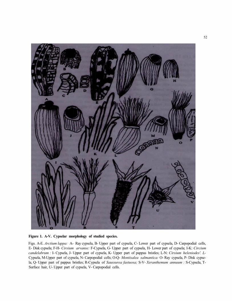
Figure 1. A-V. Cypselar morphology of studied species.
Figs. A-E. Arctium lappa: A- Ray cypsela, B- Upper part of cypsela, C- Lower part of cypsela, D- Carpopodial cells, E- Disk cypsela; F-H- Cirsium arvanse: F-Cypsela, G- Upper part of cypsela, H- Lower part of cypsela; I-K: Circium candelabrum : I- Cypsela, J- Upper part of cypsela, K- Upper part of pappus bristles; L-N: Cirsium heleniodes'. L-Cypsela, M-Upper part of cypsela, N- Carpopodial cells; O-Q- Montisalea salmantica: O- Ray cypsela, P- Disk cypse-la, Q- Upper part of pappus bristles; R-Cypsela of Saussurea fastuosa; S-V- Xeranthemum annuum : S-Cypsela, T-Surface hair, U- Upper part of cypsela, V- Carpopodial cells.
BIDYUT KUMAR J AN A AND SOBHAN KR. MUKHERJEE 53
i.e. pseudocarpopodium.
Anatomy (Fig 2 B)
Cypsela elliptic in cross section. Ribs absent. Pericarp thin, on an average 0.02 mm, differentiated in to epi-carp and mesocarp. Epicarp uniseriate, made up of thin walled, compactely arranged, rectangular, paren-chyma cells, provided with cuticle. Internal to the epicarp, mesocarp present; homogenous, made up of thick-walled, rounded, compactely arranged, paren-chyma cells. Testa attached with cypselar wall, ap-proximately 0.02 mm thick, made up of thick-walled, elongated, vertically oriented, palisade parenchyma cells, uniseriately arranged. Endosperm persists in mature cypsela, biseriate, outer layer made up of crusted layer of parenchyma cells whereas inner layer made up of thick walled, barrel shape parenchyma cells. Mature embryo occupies a major part of the cypsela; cotyledons two in number, arranged at right angle to the axis of cypsela, containing 6 resin ducts ( 3 ducts in each cotyledon ).
Cirsium candelabrum Morphology (Fig 11-K ) Cypsela homomorphic, 4 mm x 1 mm, brownish, oblong, straight, margin entire, upper part truncate whereas lower part slightly tapered. Surface glabrous. Ribs absent. Stylopodium prominent, enlarge, dome shape. Pappus homomorphic, represented by 20-29, unequally arranged, plumose type of pappus bristles, brownish, arranged in single whorl. At the basal re-gion of cypsela, carpopodium present, symmetric, biconvex. Carpopodial cells not clearly distinct from the remaining part of cypsela i.e. pseudocarpopodi-um
Anatomy (Fig 2C)
Cypsela elliptic in cross sectional configuration. Ribs absent. Pericarp thick, on an average 0.02 mm wide, differentiated into two zones- epicarp and mesocarp. Epicarp uniseriate, made up of thin walled, cubical, parenchyma cells, provided with cuticle. Internal to the epicarp, mesocarp present; made up of thick walled, rounded, compactely arranged, parenchyma cells, containing secretary duct. At the basal region of
secretary duct, vascular trace present. Testa attached with cypselar wall, approximately 0.029 mm thick, uniseriate, made up of thick walled, vertically placed, palisade parenchyma cells. Internal to the testal re-gion, vellicular cavity present. Endosperm persists in mature cypsela; biseriate. Outer layer made up of thick walled, rectangular, parenchyma cells with vas-cular trace whereas inner layer made up of thick walled, barrel shaped, compactely arranged, paren-chyma cells. Mature embryo occupies a major part of the cypsela; cotyledons two in number, arranged oblique to the axis of cypsela, containing 10 resin ducts ( 5 ducts in each cotyledon).
Cirsium heleniodes Morphology ( Fig I L-N ) Cypsela homomorphic, 24 mm x 1 mm with pappus, 6 mm x 1 mm without pappus, light brown, oblanceo-late, curved, margin entire, upper part truncate where as lower part compressed, ellipsoidal in cross section-al configuration. Surface rough and glabrous contain-ing 6 ribs, alternating with furrow. Furrows wider than ribs. At the upper part of cypsela, stylopodium present, enlarge, cylindric. Pappus homomorphic, represented by 18-22, unequally arranged, white, plu-mose type of pappus bristles. At the basal region of cypsela, carpopodium present, narrow than the base, biconvex, symmetric. Carpopodial cells with thick-walled, medium, not pitted, arranged in single row.
Anatomy ( Fig 2 D )
Cypsela elliptic in cross section. Ribs present; 6 in number, inconspicuous. Cypselar wall 0.19 mm and 0.14 mm wide at ribs and furrow region respectively. Pericarp thick, differentiated into two zones- epicarp and mesocarp. Epicarp uniseriate, made up of thin walled, rectangular, compactely arranged, parenchy-ma cells, provided with cuticle. Internal to the epi-carp, mesocarp present, heterogenous, made up of thin walled, circular, compactely arranged parenchy-ma cells and thickwalled, circular, compactely ar-ranged, sclerenchyma cells containing vascular trace. Testa attached with cypselar wall, approximately 0.02 mm thick, made up of vertically placed, thick-walled, palisade parenchyma cells, uniseriately ar-ranged. Endosperm persists in mature cypselas, unise-

EXOMORPHOLOGY AND INTERNAL. 54
riate, made up of tfaiek walled, horizontally placed, parenchyma cells. Mature embryo occupies a major part of cypsela, cotyledons 2 in number, arranged oblique to the axis of cypsela, containing 6 resin ducts ( 3 ducts in each cotyledon).
Mantisalca salmantica Morphology ( Fig 1 O-Q )
Cypsela heteromorphic. Ray cypsela 5 mm x 1 mm including pappus, 3 mm x 1 mm excluding pappus, yellow brown, oblanceolate, straight, upper part trun-cate, whereas lower part slightly tapered with entire margin. Disk cypsela 3 mm x 0.5 mm, light yellow, ob lanceolate, straight, middle part slightly swollen, tapered at both aids. Ellipsoidal in cross sectional configuration. Surface glabrous.At die upper part of cypsela, stylopodium present, inconspicuously devel-op, fully immersed into the nectar. Pappus homomor-phic, represented by 19-24, unequally arranged, bar-bellate setose type of pappus bristles, white yellow, arranged in 3 rows. Carpopodium basal in position, symmetric, biconvex. Carpopodial cells not clearly observed from the remaining part of cypsela i.e. pseu-docarpopodia.
Anatomy ( Fig 2 E )
Cypsela elliptic in cross section. Ribs absent. Pericarp thick, approximately 0.02 mm, differentiated into three zones- epicarp, mesocarp and endocarp. Epicarp uniseriate, made up of thin walled, rectangular, com-pactely arranged, parenchyma cells, provided with cuticle. Interna] to the epicarp, mesocarp present, made up of thick walled, rounded, compactely ar-ranged, parenchyma cells, with secretary duct. Inter-nal to the mesocarpic region, endocarp present, paren-chymatous, uniseriately arranged. Testa attached with cypselar wall, approximately 0.06 mm thick, made up of, vertically arranged, thickwalled, palisade pa-renchyma cells. Endosperm persists in mature cypse-la, uniseriate, made up of thick walled, barrel shaped, parenchyma cells. Mature embryo occupies a major part of cypsela; cotyledons 2 in number, arranged oblique to the axis of cypsela, containing 16 resin ducts (8 ducts in each cotyledon).
Saussurea fastuosa Morphology ( Fig 1 R)
Cypsela homomorphic, 15 mm x 1.5 mm including pappus, 6 mm x 1.5 mm excluding pappus, black brown, linear, straight, upper part truncate whereas lower part tapered Irregular in cross sectional config-uration Surface rough and glabrous, containing 10 ribs, alternating with furrow. Furrows wider than the ribs. At the upper portion of cypsela, stylopodium present; inconspicuously arranged, hilly immersed in the nectar. Pappus represented by 10-17, unequally arranged, plumose type of pappus bristles, light yel-low in colour. At the basal region of cypsela, car-popodium present, narrow than the base, irregular ring like. Carpopodial cells with thick walled, biseri-ately arranged
Anatomy ( Fig2F)
Cypsela irregular in cross section Ribs present, 10 in number, conspicuous. Cypselar wall 0.05 mm and 0.01 mm wide at rib and furrow region respectively. Pericarp thick, differentiated in to two zones-epicarp and mesocarp. Epicarp uniseriate, made up of thin walled, rectangular, compactely arranged, parenchy-ma cells provided with cuticle. Internal to epicarp, mesocarp present; heterogenous, made up of thin walled, circular, compactely arranged, parenchyma cells and thick walled, circular, compactely arranged, sclerenchyma cells. Testa attached with cypselar wall, approximately 0.014 mm thick, made up of thick walled, round to square, compactely arranged, paren-chyma cells, uniseriately arranged. Endosperm per-sists in mature cypsela, biseriate. Outer cells wide-barrel shaped and inner cells narrow-barrel shaped. Cells of both the layers thich walled, parenchyma-tous, compactely arranged. Mature embryo occupies a major part of the cypsela. Cotyledons 2 in number. Cotyledonary resin ducts are not clearly distinguish.
Xeranthemum annuum Morphology ( Fig 1 S-V)
Cypsela homomorphic, 6 mm x 0.5 mm including pappus, 4 mm x 0.5 mm excluding pappus, black brown, oblanceolate, straight, upper part truncate
BIDYUT KUMAR J AN A AND SOBHAN KR. MUKHERJEE 55
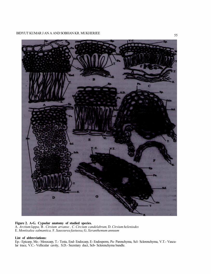
Figure 2. A-G. Cypselar anatomy of studied species. A. Arctium lappa, B . Cirsium arvanse , C. Circium candelabrum, D. Cirsium heleniodes E. Montisalea salmantica, F. Saussurea fastuosa, G. Xeranthemum annuum
List of abbreviations: Ep.- Epicarp, Me.- Mesocarp, T.- Testa, End- Endocarp, E- Endosperm, Pa- Parenchyma, Scl- Sclerenchyma, V.T.- Vascu-lar trace, V.C.- Vellicular cavity, S.D.- Secretary duct, Scb- Sclerenchyma bundle.
EXOMORPHOLOGY AND INTERNAL. 56
whereas lower part tapered, entire margin. Ellipsoi-dal in cross sectional configuration. Surface pubes-cent. Surface hair ascending-inclined in orientation with the surface, made up of body and basal cells. The tip portion of body cells with bifurcation and ar-ranged in different plain. Surface containing 16-18 ribs, alternating with furrows. Furrows wider than ribs. The distance between two ribs 0.17 mm. At the upper portion of cypsela, stylopodium present, incon-spicuous, fully immersed into the nectar. Pappus rep-resented by 4 scarious, subulate, apically pinnate scale, equal. At the basal region of cypsela, carpopo-dium present, narrower than the base, asymmetric, irregular ring like. Carpopodial cells with thick walled, rectangular, not pitted, arranged in single row.
Anatomy (Fig2 G)
Cypsela elliptic in cros section Ribs present; 16-18 in number, inconspicuous. Cypselar wall 0.09 mm and 0.03 mm wide at ribs and furrow region respectively. Pericarp thick, differentiated into two zones-epicarp and mesocarp. Epicarp uniseriate, made up of round to ovoid, thin walled, parenchyma cells, provided with cuticle. Internal to the epicarp, mesocarp pre-sent, made up of round to ovoid, thick walled, paren-chyma cells and thick walled, pentangular, scleren-chyma cells containing vascular bundle near the ribs region. Within the mesocarpic region, vellicular cavi-ty present. Testa attached with cypselar wall, approxi-mately 0.008 mm thick, uniseriate, made up of, thick walled, cubical, parenchyma cells. Endosperm per-sists in mature cypsela, biseriate. Outer layer made up of, thick walled, rectangular, compactely arranged, parenchyma cells and inner layer made up of thick walled, barrel shaped, parenchyma cells. Mature em-bryo occupies a major part of the cypsela; cotyledons two in number, arranged at right angle to the axis of cypselar wall, containing 18 resin ducts ( 9 ducts in each cotyledon).
Cypselar Morphology
Shape of cypselas are usually narrow oblanceolate - linear- narrow oblong-obovate. Cypsela with nar-row oblong is also reported by Mukheijee (2000)
in Carlina and Echinops of this tribe. Except in the cypsela of Xeranthemum annuum, the re-maining studied cypselas are with rough and gla-brous surface, whereas in the cypsela of Xeranthe-mum annuum, surface is pubescent, containing twin hairs. Twin hair is also reported in some other species ( Centaurea cyanus) of this tribe ( Mukher-jee 2000, Mukherjee and Nordenstam2012). At the upper portion of cypselas, styiopodia are present. Among the studied cypselas, in Xeranthemum annuum, Saussurea fastuosa and Mantisalca sal-mantica styiopodia are inconspicuously developed but in remaining 4 studied cypselas, styiopodia are very prominent and enlarged. Mukherjee (2005) and Wetter (1983) have studied the stylopodial fea-tures in some Asteraceae and they have men-tioned the different structures of styiopodia in Asteraceae. Present observation regarding the stylo-podial features are clearly indicated with the stud-ies of Mukhetjee (2005) and Wetter (1983). At the basal region of cypselas, carpopodia are present. Carpopodia are usually regular or irregular ring like structure and there are usually symmetric or some time asymmetric in nature ( Cirsium arven-se, Cirsium canadelabrum, Mantisalca salmantica). Diameter of carpopodia may be as wide as the base of cypsela or narrower than the base of cypsela as in Arctium lappa, Cirsium arvense etc. Carpopodium is a meristematic zone with variable layers of cellular orientation. In the cypsela of Cirsium arvense, Arctium lappa, Mantisalca sal-mantica and Cirsium candelabrum, pseudocarpopo-dia are present i.e. carpopodial cells are not dis-tinguishable from other epicarpic cells of cyp-selas, whereas in remaining studied cypselas, car-popodial cells are arranged from 1 layer (Cirsium heleniodes, Xeranthemum annuum ) to 2 layers {Saussurea fastuosa). Carpopodial structures of some genera ( Echinops, Centaurea ) of this tribe have been reported byHaque and Godward(1984). Mukherjee and Nordenstam (2004), have indicated that majority members of this tribe possess strongly asymmetric ring like carpopodia. In the cypsela of Saussurea fastuosa, Cirsium heleniodes and Cirsium candelabrum, plumose type of pappus bristles are present, while the cypsela of Manti-
BIDYUT KUMAR JAN A AND SOBHAN KR. MUKHERJEE
salca salmantica pappus bristles are barbellate type. Only in the cypsela of Xeranthemum an-num, scaly pappus has been observed. Among the studied cypselas, Arctium lappa and Cirsium arv-ense lacks pappus bristles. Pappus is the main or-gan for the dispersal of cypselas. Dittrich ( 1977) has contributed regarding the pappus structures of the tribe Cardueae.
Cypselar Anatomy
Anatomically, all the cypselas are elliptic to irreg-ular in cross sectiona. Among the studied cyp-selas, pericarpic region is differentiated into 2-3 zones. Within the pericarpic region, the mesocarpic region is more interesting than the other layers. In the cypsela of Saussurea fastuos, Cirsium heleni-odes and Xeranthemum annuum, mesocarpic re-gions are heterogeneously developed rather than the other studied cypselas, where mesocarpic re-gions are homogenously developed. In die cypsela of Arctium lappa, crystals are present in the mes-ocarpic region but in remaining 6 studied cyp-selas, crystals are absent. Zarembo and Boyko (2008) have reported the presence of crystals in the inner layer of pericarp and testa. They had also mentioned that the crystals are formed by calcium oxalate and have complex inner organiza-tion. Dormer ( 1961, 1962) has studied the crystal structure in the ovaries of certain Compositae ( Centaurea, Cirsium and others), whereas Mukher-jee and Nordenstam (2010), have studied the crys-tal structure in Compositae and they have men-tioned the presence of crystals only in Arctium lappa among the studied taxa of this tribe. Taxonom-ically, secretary ducts have great importance in the tribe Cardueae (Dittrich, 1977). Among the studied cypselas Mantisalca salmantica and Cirsium can-delabrum secretary ducts are present which are absent in five other studied taxa. Secretary ducts are also absent in some genera Amberboa (Pers.) Less., Yunquea Skottsb. in this tribe as has been reported by Chauhan (1972), Carlquist (1958). On the basis of the structure of testal epidermal cells, the studied species could be divided into 2 cate-gories:
S7
I. Testal epidermal cells are palisade like in ap-pearance. e.g. Arctium lappa , Cirsium arvanse , Cirsium candelabrum , Cirsium heleniodes , Manti-salca salmantica and Saussurea fastuosa.
II. Testal epidermal cells are cuboidal parenchy-ma cells, e.g. Xeranthemum annuum
In Arctium lappa, Cirsium heleniodes and Man-tisalca salmantica endosperm layer is uniseriately develop , whereas in remaining 4 studied cypselas, endosperm layers are biseriately oriented. Biseri-ately development of endosperm layer have been reported by Singh and Pandey(1984) in some spe-cies ( Carthamus baeticus and Saussurea hetero-mella ) of this tribe. Number of resin ducts of each cotyledon is more or less fixed and is one of the diacritical features of cypselas in Composi-tae. Each cotyledon has 3 (Cirsium heleniodes, Cirsium arvense, Arctium lappa), 5 (Cirsium cande-labrum), %{Mantisalca salmantica), or 9 (Xeranthemum annuum) resin ducts but in Saus-surea fastuosa, resin duct is not clearly visible. Variable type of resin ducts have also been re-ported by Mukherjee (2000) in the same tribe.
From this study, it can be concluded that mor-pho-anatomical features of cypselas are equally valuable as other taxonomic parameters, which are employed for proper identification of taxa along with other floral and vegetative characters in the tribe Cardueae.
Key to the studied species
la. Pappus absent; 3 resin ducts in each cotyle-don (2)
2a. Mesocarp with calcium oxalate crys-tals Arctium lappa
2b. Mesocarp without calcium oxalate crys-tals Cirsium arvense
lb. Pappus present; resin ducts are either not clearly observed or varies from 3^5-8 in each cotyledon (3)
EXOMORPHOLOGY AND INTERNAL. 58
3a. Carpopodial cells not clearly distinguishable from the epidermal cells, i.e. pseudocarpopodia; mesocarp homogenous; cotyledonary resin ducts varies from 5-8 (4)
4a. Stylopodium prominent; cotyledonary resin ducts 5 in number... Cirsium candelabrum
4b. Stylopodium inconspicuous; cotyledonary resin ducts 8 in number Mantisalca salman-tica
3b. Carpopodial cells distinct from epidermal cells of epicarp; pappus may be either plumose or scaly type; mesocarp heterogenous; cotyledonary resin ducts either not clearly observed or varys f rom 3-8 in each cotyle-don (5)
5a. Pappus plumose type (6)
6a. Carpopodial cells arranged in 1 lay-er Cirsium heleniodes
6b. Carpopodial cells arranged in 2 lay-ers.... Saussurea fastuosa
5b. Pappus scaly type Xeranthemum annuum
We are thankful to Prof. (Dr.) Hans V. Hansen, Curator of the Herbarium, Botanic Garden and Museum of the University of Copenhagen, Den-mark (DK) ; Dr. Peter Enz, Curator, Botanischer Garten der Universitat Zurich, Zurich, Switzerland (Z) ; Dr. Thomas JanBen, Curator of Sp&th-Arboretum, Humboldt-Universitat zu Berlin, Ber-lin,Germany (BHU) and Curator of Conservatoire Et Jardin Botaniques Geneve, Geneva, Switzerland (G) for sending mature seeds for this study.
REFERENCES
Bentham G 1873 Compositae. In: Genera Planta-rum,eds. Bentham G and Hooker JD, Londini Reeve and Co., London. 163-533.
Bremer K 1996 Major clades and grades of the Aster-aceae. In: Proceedings of the International Com-
positae Conference, eds. Hind DJN and Beentje HJ Royal Botanic Garden, Kew.1-7.
Carlquist S 1958 Anatomy and systematic position of Centaurodendron and Yunque ( Compositae). Brit-tonia 10 78-93.
Chauhan YS 1972 Effect of gamma rays and 2,4-D on the structure and development of seed coat and fruit wall of Carthamus tinctorius L. PI Sci 4 33-36.
Dittrich M 1977 Cynareae- systematic review. In: The Biology and Chemistry of the Compositae, eds. Heywood VH, Harborne JB and Turner BL Aca-demic Press, London. 999-1038.
Dormer KJ 1961 The crystals in the ovaries of certain Compositae. Annals of Botany 25 241-254.
Dormer KJ 1962 The taxonomic significance of crystal forms in Centaurea. New Phytologist 61 32 -35.
FourmentPS andRouzetM 1957 Fruit structure of Galactites tomentosa Monech. Bull Soc Hist Nat Afr Nord 48 307-317.
HanausekTF 1911 Uber das Pericarp und das Per-ica rpsekret der Gattung Carthamus. Berichte der Deutschen Botanischen Gesellschafi 29 13-18.
Haque MZ and Godward MBE 1984 New records of the carpopodium in Compositae and its taxo-nomic use. Bot Jour Linnean Soc 89 321-340.
J ana BK and Mukherjee SK 2012 Diversity of cyp-selar features and their taxonomic significance in three species of the tribe Cardueae of Asteraceae. In: Diversity and Conservation of Plants and Tradi-tional Knowledge 249-257.
Lavialle P 1912 Researches sur le development de 1' overie en fruit chezles Composees. Ann Sci Nat BotSer 9(15)39-151.
Mukherjee SK 2000 Comparative morpho-anatomical studies of cypselas of some members of the tribe Cardueae (Asteraceae) by LM and
BIDYUT KUMAR JANA AND SOBHAN KR. MUKHERJEE
SEM. J. Indian bot Soc 79 43-52.
Mukheijee SK 2005 Comparative studies of stylo-podium in some Asteraceae. In: Plant Taxonomy. Advances and Relevance, eds. Pandey AK, Jun W and Dogra JW CBS Publishers and Distributors, New Delhi. 493-503.
Mukherjee SK and Nordenstam B 2004 Diversity of carpopodial structure in the Asteraceae and its tax-onomic significance. Comp. Newsl. 41 29-41.
Mukheijee SK and Nordenstam B 2010 Distribu-tion of calcium oxalate crystals in the cypselar walls in some members of the Compositae and their taxonomic significance. Comp. Newsl. 48 63-88.
Mukherjee SK and Nordenstam B 2012. Diversity of
59
trichomes from mature cypselar surface of some taxa from the basal tribes of Compositae. Comp. Newsl. 50 78 - 125.
Roth I 1977 Fruits of Angiosperms, Handbuch der Pflanzenanatomie.Band X, Teil 1. Berlin, Gebruder Borntraeger.
Singh RP and Pandey AK 1984 Development and structure of seeds and fruits in Compositae -Cynareae. Phytomorphology 34 1-10.
Wetter MA 1983 Micromorphological characters and generic delimitation of some new world Sene-cioneae (Asteraceae). Brittonia 35 1 - 22.
Zarembo EV and Boyko EV 2008 Carpology of some east Asian Cardueae (Asteraceae). Anales del Jardin Botanico de Madrid 65 (1) 129-134.
1