Evolution of an arsenal structural and functional diversification of the venom system in the...
32
Evolution of an Arsenal STRUCTURAL AND FUNCTIONAL DIVERSIFICATION OF THE VENOM SYSTEM IN THE ADVANCED SNAKES (CAENOPHIDIA)* Bryan G. Fry‡§, Holger Scheib¶, Louise van der Weerd**, Bruce Young‡‡, Judith McNaughtan§§, S. F. Ryan Ramjan‡, Nicolas Vidal¶¶, Robert E. Poelmann**, and Janette A. Norman‡ Venom is a key innovation underlying the evolution of advanced snakes (Caenophidia). Despite this, very little is known about venom system structural diversification, toxin recruitment event timings, or toxin molecular evolu- tion. A multidisciplinary approach was used to examine the diversification of the venom system and associated toxins across the full range of the 100 million-year-old advanced snake clade with a particular emphasis upon families that have not secondarily evolved a front-fanged venom system (80% of the 2500 species). Analysis of cDNA libraries revealed complex venom transcriptomes containing multiple toxin types including three finger tox- ins, cobra venom factor, cysteine-rich secretory protein, hyaluronidase, kallikrein, kunitz, lectin, matrix metallopro- tease, phospholipase A 2 , snake venom metalloprotease/a disintegrin and metalloprotease, and waprin. High levels of sequence diversity were observed, including mutations in structural and functional residues, changes in cysteine spacing, and major deletions/truncations. Morphological analysis comprising gross dissection, histology, and mag- netic resonance imaging also demonstrated extensive modification of the venom system architecture in non- front-fanged snakes in contrast to the conserved struc- ture of the venom system within the independently evolved front-fanged elapid or viperid snakes. Further, a reduction in the size and complexity of the venom system was observed in species in which constriction has been secondarily evolved as the preferred method of prey cap- ture or dietary preference has switched from live prey to eggs or to slugs/snails. Investigation of the timing of toxin recruitment events across the entire advanced snake ra- diation indicates that the evolution of advanced venom systems in three front-fanged lineages is associated with recruitment of new toxin types or explosive diversification of existing toxin types. These results support the role of venom as a key evolutionary innovation in the diversifi- cation of advanced snakes and identify a potential role for non-front-fanged venom toxins as a rich source for lead compounds for drug design and development. Molecular & Cellular Proteomics 7:215–246, 2008. It has only become evident recently that venom in snakes is a basal characteristic and that the three front-fanged venom delivery system architectures are each independent second- ary derivations (1– 4). In earlier schemes, the “colubrid,” or “non-front-fanged,” snakes were seen as a monophyletic transitional group to the presumably advanced front-fanged lineages Atractaspis, Elapidae, and Viperidae (e.g. Kardong (5)), and all front-fanged snakes were assumed to share a common ancestor. Resolution of higher order relationships revealed not only the colubrid snakes to be paraphyletic but the front-fanged snakes to be polyphyletic with viperids being one of the earliest advanced snake radiations and elapids only recently derived (1, 6). Previous studies indicate significant morphological varia- tion in the venom gland (7–9) and dentition of snakes (10). The fang may or may not be enlarged and can range from a solid tooth with or without grooving to an enclosed canaliculate channel as present in elapids and viperids (10). Few com- parative studies of the caenophidian venom system have been performed, although West (11, 12) and Sarkar (13) described several different structural and topological fea- tures. A distinction was attempted between the venom glands of the front-fanged and non-front-fanged snakes with the glands in most non-front-fanged snakes termed “Duvernoy’s glands” (7). Similarly, very little has been revealed about the composi- tion of venoms from advanced snake lineages other than the three clades with high pressure, hollow front-fang venom delivery systems (Atractaspis, elapids, and viperids). Despite accounting for the majority of the families (Fig. 1 and Ref. 6) and 1900 of the 2500 advanced snake species, the multiple non-front-fanged families have received scant attention. Only a few studies have been undertaken, and even fewer have sequenced or bioactivity-tested individual toxins (2, 14 –18). From the ‡Department of Biochemistry and Molecular Biology, Bio21 Molecular Science and Biotechnology Institute and §§Depart- ment of Oral Medicine and Surgery, School of Dental Science, Uni- versity of Melbourne, Parkville, Victoria 3010, Australia, ¶SBC Lab AG, Seebu ¨ elstrasse 26, 8185 Winkel, Switzerland, **Molecular Imag- ing Laboratories Leiden, MRI Facility, Departments of Anatomy and Radiology, 2300 RC, Leiden, The Netherlands, ‡‡Department of Bi- ology, Washburn University, Topeka, Kansas 66621, ¶¶UMR 7138, De ´ partement Syste ´ matique et Evolution, Muse ´ um National d’Histoire Naturelle, CP 26, 57 rue Cuvier, 75005 Paris, France, and Molecular Biology, Museum Victoria, G. P. O. Box 666, Melbourne, Victoria 3001, Australia Received, March 2, 2007, and in revised form, September 10, 2007 Published, MCP Papers in Press, September 17, 2007, DOI 10.1074/mcp.M700094-MCP200 Research © 2008 by The American Society for Biochemistry and Molecular Biology, Inc. Molecular & Cellular Proteomics 7.2 215 This paper is available on line at http://www.mcponline.org at CNRS on February 7, 2008 www.mcponline.org Downloaded from
Transcript of Evolution of an arsenal structural and functional diversification of the venom system in the...

Evolution of an ArsenalSTRUCTURAL AND FUNCTIONAL DIVERSIFICATION OF THE VENOM SYSTEM IN THE ADVANCED SNAKES(CAENOPHIDIA)*
Bryan G. Fry‡§, Holger Scheib¶�, Louise van der Weerd**, Bruce Young‡‡,Judith McNaughtan§§, S. F. Ryan Ramjan‡, Nicolas Vidal¶¶, Robert E. Poelmann**,and Janette A. Norman‡��
Venom is a key innovation underlying the evolution ofadvanced snakes (Caenophidia). Despite this, very little isknown about venom system structural diversification,toxin recruitment event timings, or toxin molecular evolu-tion. A multidisciplinary approach was used to examinethe diversification of the venom system and associatedtoxins across the full range of the �100 million-year-oldadvanced snake clade with a particular emphasis uponfamilies that have not secondarily evolved a front-fangedvenom system (�80% of the 2500 species). Analysis ofcDNA libraries revealed complex venom transcriptomescontaining multiple toxin types including three finger tox-ins, cobra venom factor, cysteine-rich secretory protein,hyaluronidase, kallikrein, kunitz, lectin, matrix metallopro-tease, phospholipase A2, snake venom metalloprotease/adisintegrin and metalloprotease, and waprin. High levelsof sequence diversity were observed, including mutationsin structural and functional residues, changes in cysteinespacing, and major deletions/truncations. Morphologicalanalysis comprising gross dissection, histology, and mag-netic resonance imaging also demonstrated extensivemodification of the venom system architecture in non-front-fanged snakes in contrast to the conserved struc-ture of the venom system within the independentlyevolved front-fanged elapid or viperid snakes. Further, areduction in the size and complexity of the venom systemwas observed in species in which constriction has beensecondarily evolved as the preferred method of prey cap-ture or dietary preference has switched from live prey toeggs or to slugs/snails. Investigation of the timing of toxinrecruitment events across the entire advanced snake ra-diation indicates that the evolution of advanced venom
systems in three front-fanged lineages is associated withrecruitment of new toxin types or explosive diversificationof existing toxin types. These results support the role ofvenom as a key evolutionary innovation in the diversifi-cation of advanced snakes and identify a potential rolefor non-front-fanged venom toxins as a rich source forlead compounds for drug design and development.Molecular & Cellular Proteomics 7:215–246, 2008.
It has only become evident recently that venom in snakes isa basal characteristic and that the three front-fanged venomdelivery system architectures are each independent second-ary derivations (1–4). In earlier schemes, the “colubrid,” or“non-front-fanged,” snakes were seen as a monophyletictransitional group to the presumably advanced front-fangedlineages Atractaspis, Elapidae, and Viperidae (e.g. Kardong(5)), and all front-fanged snakes were assumed to share acommon ancestor. Resolution of higher order relationshipsrevealed not only the colubrid snakes to be paraphyletic butthe front-fanged snakes to be polyphyletic with viperids beingone of the earliest advanced snake radiations and elapids onlyrecently derived (1, 6).
Previous studies indicate significant morphological varia-tion in the venom gland (7–9) and dentition of snakes (10). Thefang may or may not be enlarged and can range from a solidtooth with or without grooving to an enclosed canaliculatechannel as present in elapids and viperids (10). Few com-parative studies of the caenophidian venom system havebeen performed, although West (11, 12) and Sarkar (13)described several different structural and topological fea-tures. A distinction was attempted between the venomglands of the front-fanged and non-front-fanged snakeswith the glands in most non-front-fanged snakes termed“Duvernoy’s glands” (7).
Similarly, very little has been revealed about the composi-tion of venoms from advanced snake lineages other than thethree clades with high pressure, hollow front-fang venomdelivery systems (Atractaspis, elapids, and viperids). Despiteaccounting for the majority of the families (Fig. 1 and Ref. 6)and �1900 of the 2500 advanced snake species, the multiplenon-front-fanged families have received scant attention. Onlya few studies have been undertaken, and even fewer havesequenced or bioactivity-tested individual toxins (2, 14–18).
From the ‡Department of Biochemistry and Molecular Biology,Bio21 Molecular Science and Biotechnology Institute and §§Depart-ment of Oral Medicine and Surgery, School of Dental Science, Uni-versity of Melbourne, Parkville, Victoria 3010, Australia, ¶SBC LabAG, Seebuelstrasse 26, 8185 Winkel, Switzerland, **Molecular Imag-ing Laboratories Leiden, MRI Facility, Departments of Anatomy andRadiology, 2300 RC, Leiden, The Netherlands, ‡‡Department of Bi-ology, Washburn University, Topeka, Kansas 66621, ¶¶UMR 7138,Departement Systematique et Evolution, Museum National d’HistoireNaturelle, CP 26, 57 rue Cuvier, 75005 Paris, France, and ��MolecularBiology, Museum Victoria, G. P. O. Box 666, Melbourne, Victoria3001, Australia
Received, March 2, 2007, and in revised form, September 10, 2007Published, MCP Papers in Press, September 17, 2007, DOI
10.1074/mcp.M700094-MCP200
Research
© 2008 by The American Society for Biochemistry and Molecular Biology, Inc. Molecular & Cellular Proteomics 7.2 215This paper is available on line at http://www.mcponline.org
at CN
RS
on February 7, 2008
ww
w.m
cponline.orgD
ownloaded from

Studies of the first full-length toxins from non-front-fangedsnakes were revealing, particularly the isolation and charac-terization of a potently neurotoxic three-finger toxin (3FTx)1
from the colubrid snake Coelognathus radiatus (2). This toxintype had long been considered the hallmark of elapid venomsand had been the subject of intense study (19). Follow-up
molecular phylogenetic studies demonstrated the shared or-igin of 3FTx and other toxin types across the entire advancedsnake radiation (3).
In view of the homology of the venom toxins and toxin-secreting glands of all advanced snakes, the fact that Duver-noy’s glands represented a primitive condition and that thederived glands of the front-fanged snakes were independentlyevolved from these, the distinction between Duvernoy’sglands and venom glands was revealed to be an artificial onethat impeded the understanding of the evolution of the venomapparatus of snakes. For this reason, the term Duvernoy’sgland was abandoned, and the term “venom gland” was usedfor the toxin-secreting oral glands of all snakes regardless of
1 The abbreviations used are: 3FTx, three-finger toxin; CRISP, cys-teine-rich secretory protein; SVMP, snake venom metalloprotease;ADAM, a disintegrin and metalloprotease; BLAST, Basic Local Align-ment Search Tool; 3D, three-dimensional; SPDBV, Swiss-PdbViewer;MRI, magnetic resonance imaging; T, tesla; CVF, cobra venom factor;MMP, matrix metalloprotease; PLA2, phospholipase A2; CRD, cys-teine-rich domain.
FIG. 1. Cladogram of evolutionary relationships of advanced snakes (1, 6, 58–61) showing relative timing of toxin recruitment eventsand derivations of the venom system. MRI images are shown for representatives. Acn, Acetylcholine esterase; LAO, L-amino oxidase; C3B,FAMC3B cytokine; CNP-BPP, c-type natriuretic peptide-bradykinin-potentiating peptide; GrTx, glycine-rich toxin; Hya, hyaluronidase; RAP,renin-like aspartic protease; VEGF, vascular endothelial growth factor.
Evolution of an Arsenal
216 Molecular & Cellular Proteomics 7.2
at CN
RS
on February 7, 2008
ww
w.m
cponline.orgD
ownloaded from

the degree of anatomical specialization in the venom deliveryapparatus (14).
It has been shown previously that snake venoms evolve viaa process by which a gene encoding for a normal bodyprotein, typically one involved in key regulatory processes orbioactivity, is duplicated, and the copy is selectively ex-pressed in the venom gland (20). The newly created toxin typeevolves via the birth-and-death model of protein evolution inwhich a toxin multigene family is created by further geneduplication events followed by the deletion of some copiesand conversion of others to non-functional copies or pseudo-genes (19).
In addition to gene duplication, mutation is an importantprocess that generates a tremendous diversity of venom tox-ins within these multigene families. The newly created toxinmultigene families preserve the molecular scaffold of the an-cestral protein but modify key functional residues at the tips ofloops to acquire a myriad of newly derived activities (19, 20).Other mutations can include the selective expression of aparticular domain, such as the expression of the disintegrindomain from snake venom metalloprotease, ADAM-type(SVMP/ADAM) toxins in viperid venoms (21). These toxinshave an unusual combination of precise specificity and ex-treme potency, characteristics that make them particularlyamenable for use as investigational ligands or as leads fordrug design and development (22–24).
In this study we used a multidisciplinary approach to (a)characterize the venom transcriptomes of representative cae-nophidian snakes, (b) determine the timing of toxin recruit-ment events and patterns of toxin diversification, and (c)characterize changes in the venom delivery architecture.From comparison with the more widely studied elapids andviperids, we determined whether there are significant struc-tural or functional differences in the evolution of the venomsystem and their associated toxins in the poorly characterizednon-front-fanged caenophidian snakes.
MATERIALS AND METHODS
Species Studied and Molecular Phylogeny of Advanced Snakes—Atotal of 107 species representing each of the major lineages of ad-vanced snakes were included in the study and examined using one ormore of the following techniques: clone sequencing of cDNA libraries,gross dissection and examination of dentition, histology of venomglands and ducts, and magnetic resonance imaging (see “Appendix I”for details). In most cases different individuals were used as a sourceof material for each of these analyses.
cDNA Library Construction and Analysis—RNA was isolated fromvenom glands of 13 species spanning the full taxonomical diversity(10 non-front-fanged snakes, one elapid, and two viperids; see “Ap-pendix I”) using the Qiagen RNeasy Midi kit with subsequent selectionof mRNAs using the Oligotex Midi kit. cDNA libraries were con-structed using the Clontech Creator SMART cDNA Library Construc-tion kit and transformed into One Shot Electrocompetent GeneHogs(Invitrogen) as described previously (4). Isolation and sequencing ofinserts was undertaken at the Australian Genome Research Facilityusing BDTv3.1 chemistry with electrophoretic separation on anAB330xl. Up to 384 colonies were sequenced per library, inserts were
screened for vector sequences, and those parts were removed priorto analysis and identification. Toxin sequences were identified byhomology of the translated cDNA sequences with previously charac-terized toxins using a BLAST search of the Swiss-Prot protein data-base (www.expasy.org/tools/blast/).
Molecular Modeling of Toxins—3D models for caenophidian toxinswere generated based on the assumption that homologous proteinsshare similar 3D structures (25). In other words, the three-dimensionalstructure of a target protein can be modeled if its sequence is ho-mologous to at least one template protein whose 3D structure hasbeen determined experimentally by applying either x-ray or NMRtechniques (26). In this work aligning the protein sequences of targetand template(s) was carried out in SPDBV (27), and the initial align-ments were refined manually. From these alignments 3D models werebuilt directly in SPDBV applying the “Build Preliminary Model” option,which is disabled in the currently distributed public version of thesoftware. Loops were built by scanning a database of known loopstructures using the same software, and suitable specimens wereselected after visual inspection. The enthalpy of the resulting modelswas minimized applying two times 200 steps of Steepest Descentminimization. Finally the quality of each model structure was as-sessed in iMolTalk (28), and a Van der Waals surface was calculatedin MolMol (29). In MolMol electrostatic potentials were calculatedapplying the “simplecharge” command and mapped on the modelstructure surface. The families of venom proteins were analyzed bysuperimposing the structures in SPDBV (27), and conserved andvariable structural regions were identified. In the molecular modelingof representative proteins, blue surface areas indicate positivecharges, red surface areas indicate negative charges, and modelpairs show sides of the protein rotated by 180°.2
Molecular Phylogeny of Toxin Sequences—Molecular phylogeneticanalyses of toxin transcripts were conducted using the translatedamino acid sequences. Comparative sequences from other venom-ous reptiles and outgroups were obtained through BLAST searching(www.expasy.org/tools/blast/) using representative toxin sequences.To minimize confusion, all sequences obtained in this study arereferred to by their GenBankTM accession numbers (www.ncbi.nlm-.nih.gov/sites/entrez?db�Nucleotide), and sequences from previousstudies are referred to by their UniProt/Swiss-Prot accession num-bers (www.expasy.org/cgi-bin/sprot-search-ful). Resultant sequencesets were aligned using the program ClustalX followed by visualinspection for errors. When presented as sequence alignments, theleader sequence is shown in lowercase, the prepro region is under-lined, cysteines are highlighted in black, and functional residues are inbold. Datasets were analyzed using Bayesian inference implementedon MrBayes, version 3.0b4. The analysis was performed by running aminimum of 1 � 106 generations in four chains and saving every 100thtree. The log likelihood score of each saved tree was plotted againstthe number of generations to establish the point at which the loglikelihood scores of the analysis reached their asymptote, and theposterior probabilities for clades were established by constructing amajority rule consensus tree for all trees generated after the comple-tion of the burn-in phase.3
Gross Dissection and Analysis of Dentition—Gross dissection wasperformed on freshly euthanized and formalin-fixed specimens todocument the relative size and position of the venom gland andassociated skeletal musculature. Features of the maxillary dentitionwere scored using data from a previous study (10) in which fivemorphological states were defined: 1) smooth surface and no en-
2 Homology model coordinates can be obtained from H. Scheib.E-mail: [email protected].
3 Sequence alignments can be obtained from B. G. Fry. E-mail:[email protected].
Evolution of an Arsenal
Molecular & Cellular Proteomics 7.2 217
at CN
RS
on February 7, 2008
ww
w.m
cponline.orgD
ownloaded from
closed venom canal, 2) no enclosed venom canal and surface withshallow furrow, 3) deep groove running the majority of the length ofthe tooth, 4) deep groove present but restricted to less than half thelength of the tooth, and 5) enclosed venom canal.
Histological Analysis—Histological sections were prepared fromthe intact head and the excised venom delivery system. Whole headswere removed, and a cut was made to the underside to allow fastpenetration of the fixative (10% neutral buffered formalin). After aminimum of 2 days excess tissue was removed, and specimens wereimmersed in Kristensen’s decalcification solution and placed on arotor for up to 3 weeks (depending on the size of the head). Beforeprocessing the heads were bisected longitudinally for cutting trans-versely, at 3 �m, in two separate blocks. The processing schedulewas: 10% formalin, 2 h; absolute ethanol, 4 � 1 h; Histolene, 3 � 1 h;paraffin wax, 2 � 90 min. The sections were taken every 100 �m, andmatching sections were stained with periodic acid-Schiff stain andMasson’s trichrome stain. In other specimens, the venom gland,venom duct, and the adjacent bony and muscular tissue were excisedand placed in decalcifying solution (Cal-Ex, Fisher) for 72–168 h. Eachsample was dehydrated and cleared through a progressive ethanolseries and Cyto-Sol (Fisher) prior to embedding in Paraplast (Fisher).Serial sections were cut at 10–12 �m. All species were sectioned inthe frontal plane; when available, the contralateral venom deliverysystem was sectioned either parasagittally or transversely. Sectionswere stained using a variant of Van Gieson’s stain, which providesclear distinction between connective tissue, muscle, and epithelium,or with hematoxylin and eosin.
Magnetic Resonance Imaging—Magnetic resonance imaging (MRI)was used to examine the three-dimensional shape and internal anat-omy of the venom glands. Formalin-ethanol-fixed heads were firstsubmersed in Fomblin (Solvay Solexis) to prevent air artifacts. De-pending on head size, imaging was performed on either 9.4-T (small/medium) or 17.6-T (large) vertical 89-mm-bore systems (Bruker Bio-Spin, Rheinstetten, Germany) with a Bruker Micro2.5 gradient systemof 1 T/m and transmit/receive birdcage radiofrequency coil with di-ameter of 10–30 mm. Bruker ParaVision 3.0 software was used for
image acquisition. Anatomical images were acquired using a 3Dgradient echo sequence. The field of view and matrix were varied tofit the individual samples, resulting in voxel sizes between (40)3 and(70)3 mm3. Imaging parameters were: echo time � 8 ms; repetitiontime � 40 ms; flip angle, 20°; four to eight averages; total scan timebetween 3 and 9 h per sample, depending on size and resolution.Image segmentation of the glands was performed manually in Amira4.1 (Mercury Computer Systems Inc.), and 3D surface renderingswere generated for all species.
RESULTS
Isolation and Characterization of Venom Toxin Transcripts—Analysis of venom gland cDNA from non-front-fanged snakelibraries revealed the presence of transcripts with homologyto previously characterized venom toxins from front-fangedsnakes and venomous lizards (helodermatids and varanids)(Tables I and II). Transcripts sequenced were 3FTx (Figs. 2and 3), C3/cobra venom factor (CVF) (Fig. 4), cysteine-rich se-cretory protein (CRISP) (Figs. 5 and 6), hyaluronidase (Fig. 7),kallikrein (Figs. 8 and 9), kunitz (Figs. 10 and 11), lectin (Figs. 12and 13), matrix metalloprotease (MMP) (Fig. 14), phospholipaseA2 (PLA2) Type IB (Fig. 15), SVMP/ADAM (Figs. 16, 17, and 18),and waprin (Fig. 19). Transcripts of five of these toxin types werealso recovered from the cDNA libraries of the representativeelapid Oxyuranus microlepidotus (3FTx, CRISP, kunitz, and wa-prin) and the viperid Causus rhombeatus (kallikrein).
Alignment of the translated amino acid sequences revealedextensive variation in the molecular structure of the transcriptsfor most toxin types. There was less amino acid sequencedivergence in C3/CVF, CRISP, hyaluronidase, kunitz, PLA2
(Type IB), and MMP toxins than in the much more variable
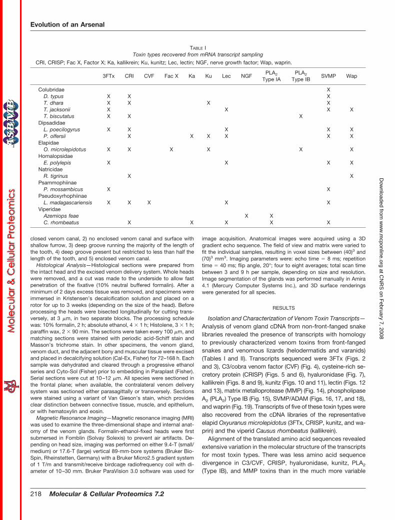
TABLE IToxin types recovered from mRNA transcript sampling
CRI, CRISP; Fac X, Factor X; Ka, kallikrein; Ku, kunitz; Lec, lectin; NGF, nerve growth factor; Wap, waprin.
3FTx CRI CVF Fac X Ka Ku Lec NGFPLA2
Type IAPLA2
Type IBSVMP Wap
Colubridae XD. typus X X XT. dhara X X X XT. jacksonii X X X XT. biscutatus X X X
DipsadidaeL. poecilogyrus X X X X XP. olfersii X X X X X X
ElapidaeO. microlepidotus X X X X X X
HomalopsidaeE. polylepis X X X X
NatricidaeR. tigrinus X X
PsammophiinaeP. mossambicus X X
PseudoxyrhophiinaeL. madagascariensis X X X X X
ViperidaeAzemiops feae X XC. rhombeatus X X X X X
Evolution of an Arsenal
218 Molecular & Cellular Proteomics 7.2
at CN
RS
on February 7, 2008
ww
w.m
cponline.orgD
ownloaded from
3FTx, kallikrein, lectin, SVMP/ADAM, and waprin toxins. Formost toxin types multiple transcripts containing significantmolecular variations were also isolated from individual cDNAlibraries; this is a characteristic previously attributed to acceler-ated diversification in these toxin multigene families (19). cDNAsequencing revealed numerous transcripts in the venom glandsof non-front-fanged snakes that preserve the key amino acidsessential for a particular bioactivity and potentially represent themRNA precursors of functional proteins.
Reflective of sequence variation of proteins in the CRISPfamily, its members have been found to interact with differenttarget proteins, i.e. cyclic nucleotide-gated ion channels aswell as L-type Ca2� and BKCa K� channels (30–32). Bindingto the respective channels is speculated to be a generalproperty of CRISP and was attributed to a predominantlyhydrophobic cavity involving residues from both domain PR-1and cysteine-rich domain (CRD), whereas specific channelblock supposedly is taking place with residues of CRD (30–35). Six amino acids were identified for the calcium channelblocker Triflin (Protein Data Bank code 1WVR (30)) to bind toion channels (numbering according to Triflin): Glu-44, Tyr-54,Ser-65, Tyr-125 (all four in PR-1), Thr-184, and Arg-185 (bothin CRD). An additional four amino acids were conserved andpostulated to affect ion channel function of this neurotoxin.They are all located in CRD: Phe-189, Leu-195, Tyr-205, andPhe-215. However, this work reveals that none of these 10positions is conserved throughout the CRISP toxin family.
Even more so our in silico studies could not confirm sug-gested functional motifs, namely EX2F and DVF, at least as ageneral principle of CRISP. Differences in CRISP function oftoxic venom proteins and the non-toxic representative frommouse are subtle and likely to be found in CRD, i.e. Leu-220and Leu-230 (numbering according to mouse; Fig. 5). Thepromiscuous susceptibility of CRISP toward different ionchannels is reflected in their high degree of sequence varia-tion. In the absence of obvious sequence and structure dif-ferences between toxic and non-toxic specimens (Figs. 5 and6) it will be necessary to acquire further data on the functionof the CRISP family members to identify the functionally dis-criminating amino acids.
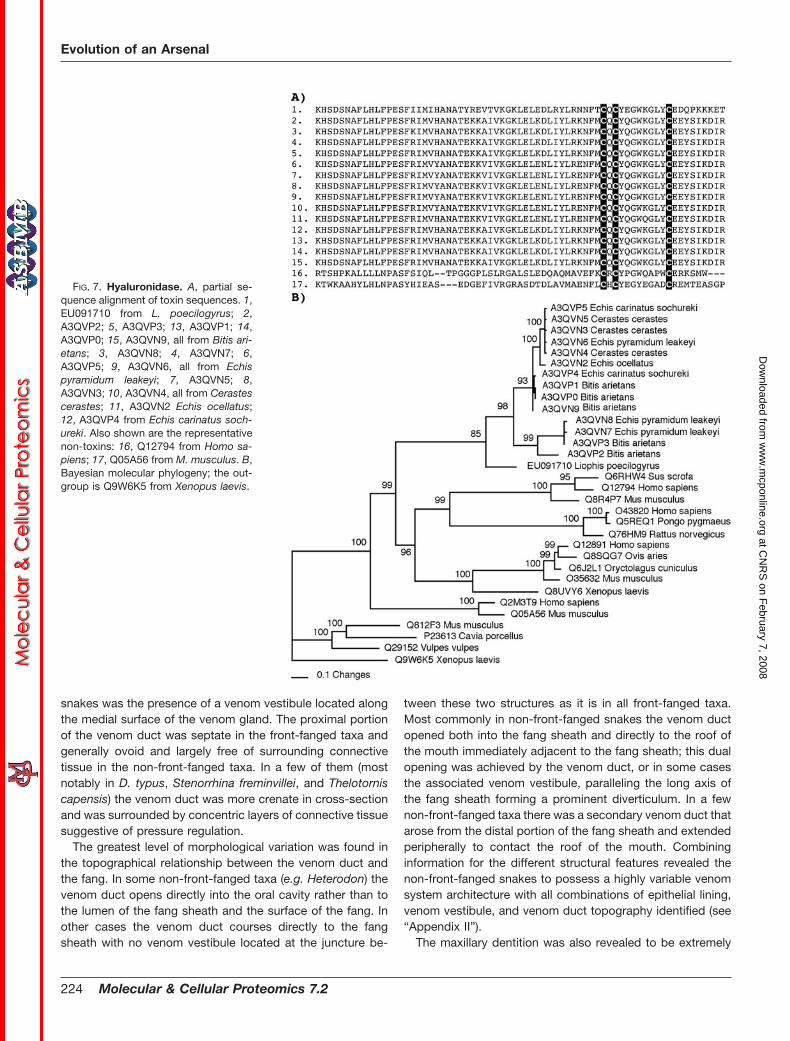
The hyaluronidase transcript sequenced from Liophis po-ecilogyrus showed significant sequence similarity to thosesequenced from viperid venoms (36), and the snake se-quences all formed a clade (Fig. 7). Kallikrein transcripts iso-lated from Philodryas olfersii retained residues essential forthe potent hypotensive action mediated by the liberation ofbradykinin from kininogen (Fig. 8).
Kunitz-type toxins belong to the superfamily of bovine pan-creatic trypsin-like inhibitors. Although they share the sameoverall 3D fold, their antitrypsin activity may vary. It has beenlong known that the residue in the so-called P1 site of thetrypsin inhibitor is positively charged (arginine, lysine, or his-tidine), whereas this residue in a chymotrypsin inhibitor usu-ally is large and hydrophobic (leucine, phenylalanine, tyrosine,
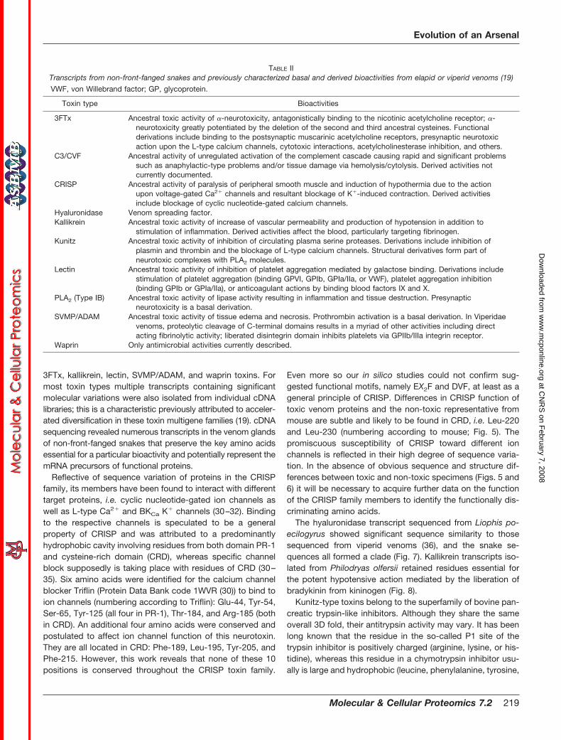
TABLE IITranscripts from non-front-fanged snakes and previously characterized basal and derived bioactivities from elapid or viperid venoms (19)
VWF, von Willebrand factor; GP, glycoprotein.
Toxin type Bioactivities
3FTx Ancestral toxic activity of �-neurotoxicity, antagonistically binding to the nicotinic acetylcholine receptor; �-neurotoxicity greatly potentiated by the deletion of the second and third ancestral cysteines. Functionalderivations include binding to the postsynaptic muscarinic acetylcholine receptors, presynaptic neurotoxicaction upon the L-type calcium channels, cytotoxic interactions, acetylcholinesterase inhibition, and others.
C3/CVF Ancestral activity of unregulated activation of the complement cascade causing rapid and significant problemssuch as anaphylactic-type problems and/or tissue damage via hemolysis/cytolysis. Derived activities notcurrently documented.
CRISP Ancestral activity of paralysis of peripheral smooth muscle and induction of hypothermia due to the actionupon voltage-gated Ca2� channels and resultant blockage of K�-induced contraction. Derived activitiesinclude blockage of cyclic nucleotide-gated calcium channels.
Hyaluronidase Venom spreading factor.Kallikrein Ancestral toxic activity of increase of vascular permeability and production of hypotension in addition to
stimulation of inflammation. Derived activities affect the blood, particularly targeting fibrinogen.Kunitz Ancestral toxic activity of inhibition of circulating plasma serine proteases. Derivations include inhibition of
plasmin and thrombin and the blockage of L-type calcium channels. Structural derivatives form part ofneurotoxic complexes with PLA2 molecules.
Lectin Ancestral toxic activity of inhibition of platelet aggregation mediated by galactose binding. Derivations includestimulation of platelet aggregation (binding GPVI, GPIb, GPIa/IIa, or VWF), platelet aggregation inhibition(binding GPIb or GPIa/IIa), or anticoagulant actions by binding blood factors IX and X.
PLA2 (Type IB) Ancestral toxic activity of lipase activity resulting in inflammation and tissue destruction. Presynapticneurotoxicity is a basal derivation.
SVMP/ADAM Ancestral toxic activity of tissue edema and necrosis. Prothrombin activation is a basal derivation. In Viperidaevenoms, proteolytic cleavage of C-terminal domains results in a myriad of other activities including directacting fibrinolytic activity; liberated disintegrin domain inhibits platelets via GPIIb/IIIa integrin receptor.
Waprin Only antimicrobial activities currently described.
Evolution of an Arsenal
Molecular & Cellular Proteomics 7.2 219
at CN
RS
on February 7, 2008
ww
w.m
cponline.orgD
ownloaded from

or asparagine) (37). From the multiple sequence alignment pre-sented in Fig. 10 it can be concluded that kunitz-type toxinsfrom P. olfersii, Ophiophagus hannah, Naja naja, and Bungarusmulticinctus are likely to inhibit chymotrypsin, whereas the oth-ers putatively are trypsin inhibitors. Our 3D modeling effortsshowed that the kunitz toxins vary, i.e. in the length of their C-but also their N-terminal tails (Figs. 10 and 11). The N-terminaltail is held in place by formation of two disulfide bonds involvingthe first and sixth as well as the second and fourth cysteines.The inhibitory residues are solvent-exposed and reside in a longsurface loop immediately after the second cysteine, which indi-cates that the position of these residues is rather conservedamong kunitz toxins (Figs. 10 and 11). Therefore, changes inpolarity and charge are expected to affect the physicochemicalproperties of this region.
Within the lectin toxins, variation of a key tripeptide motifhas been shown to have significant impact upon functionality(38) with EPN conferring mannose binding ability, whereasQPD confers galactose binding. In this study, both were ob-tained as well as new variants of this motif containing EAP,QAP, and LTD (Figs. 12 and 13). The EPN motif appears to bebasal, whereas the QPD motif is an early emerging variant, andthe other variants evolved subsequently at different times duringthe evolution of the animals themselves. The viper venom-specific heterodimeric forms have lost this motif entirely.
A novel MMP toxin had been reported previously from thevenom of the lethal natricid snake Rhabdophis tigrinus (39).However, only a very small fragment was obtained so theMMP-subtype relationship remained unclear. In this study, afull-length MMP was obtained from the L. poecilogyrus library
FIG. 2. Sequence alignment of rep-resentative 3FTx. Species shown are E.polylepis (1, EU029668), P. mossambi-cus (2, EU029669), L. poecilogyrus (3,EU029670; 6, EU029672; 7, EU029673),T. dhara (4, EU029671), D. typus (5,EU029674; 9, EU036636; 17, EU029681;19, EU029683), T. jacksonii (8, EU036635;18, EU029682; 20, EU029684; 21,EU029685), T. biscutatus (10, EU029675;12, EU029677; 13, EU029678), Leioheter-odon madagascariensis (11, EU029676),T. dhara (14, EU029686; 15, EU029679;16, EU029680), C. radiatus (22, P83490),Dendroaspis jamesonii (23, P25682),from B. multicinctus (24, Q9PW19; 25,Q9YGJ0), Bungarus candidus (26,P81783), and Naja sputatrix (27,Q9W7I3). Also included is the represent-ative non-toxin peptide brain �-neu-ropeptide (28, Q9WVC2) from Musmusculus.
Evolution of an Arsenal
220 Molecular & Cellular Proteomics 7.2
at CN
RS
on February 7, 2008
ww
w.m
cponline.orgD
ownloaded from

and was shown to be closest to MMP2 (Fig. 14) rather thanshowing the MMP9 relationship hypothesized in the previousR. tigrinus study.
A Type IB PLA2 transcript was isolated from Trimorphodonbiscutatus that retained not only the ancestral pancreatic loopbut also the catalytic diad residues histidine and aspartate as
well as two conserved tyrosine residues that together withstructural water form the catalytic center (Fig. 15). This toxin ismost likely responsible for conferring the presynaptic neuro-toxicity observed for this venom (40). The electrostatic sur-faces of PLA2 IB clearly indicate the overall similarity of thefive toxins studied both in terms of size and shape as well as
FIG. 4. Molecular evolution of C3/CVF toxins. A, partial sequence align-ment of the representative toxin formsshown from top to bottom: Q2XXR5 fromL. madagascariensis, Q49HM6 from Aus-trelaps superbus, Q01833 from N. naja,and Q91132 from Naja kaouthia as wellas the non-toxin forms P23667 from Xe-nopus laevis and Q90633 from Gallusgallus. B, Bayesian molecular phyloge-netic analysis of representative toxin andnon-toxin body forms. Outgroups arethe non-toxin sequences P23667 X. lae-vis and Q90633 G. gallus.
FIG. 3. Bayesian molecular phylog-eny of 3FTx. The outgroup is the non-toxic brain �-neuropeptide (Q9WVC2)from M. musculus. The ribbon model ofEU029668 E. polylepis shows �-strandsin yellow.
Evolution of an Arsenal
Molecular & Cellular Proteomics 7.2 221
at CN
RS
on February 7, 2008
ww
w.m
cponline.orgD
ownloaded from

with regard to the physicochemical properties. It can be con-cluded that all PLA2 IB toxin specimens exhibit very similarneurotoxic function.
The SVMP/ADAM sequences were shown to be of the PIIItype, consistent with previously reported fragments fromDispholidus typus (41). Numerous transcripts were also recov-ered with significant variations including changes in the numberand spacing of cysteine residues and large scale deletions.Caenophidian 3FTx, lectin, MMP, SVMP, and waprin transcriptswere all characterized by changes in ancestral cysteines inaddition to the evolution of new cysteines (Figs. 2, 12, 16, 17,and 19). In contrast, transcripts of the caenophidian C3/CVF,CRISP, hyaluronidase, kallikrein, kunitz, and PLA2 Type IB tox-ins preserved the ancestral cysteine numbers and spacing
(Figs. 4, 5, 8, 10, and 15). Large deletions were detected intranscripts of the lectin and SVMP/ADAM toxins. Multiple tran-scripts of a deleted form of the lectin toxin were isolated fromEnhydris polylepis (Fig. 12) in which a large stretch of residuesincluding an ancestral cysteine is deleted, leaving a free cys-teine, potentially facilitating dimerization. Another lectin toxinversion from the E. polylepis cDNA (again for which multipletranscripts were obtained) had a significant change in sequenceto the second half of the protein as a consequence of a frame-shift mutation. The resulting new transcript contains overall fourcysteine bonds and therefore may fold into a stable, bioactivemolecule. A major truncation was detected in transcripts of theSVMP/ADAM toxins isolated from Psammophis mossambicus(Fig. 16). These transcripts consisted solely of the propeptide
FIG. 5. Sequence alignment of rep-resentative full-length (unless other-wise indicated) CRISP toxins. 1,Q2XXQ5 from D. typus; 2, partial se-quence Q2XXP5 from T. dhara; 3, partialsequence Q2XXP4 from T. biscutatus; 4,partial sequence Q2XXP7 from P. olfer-sii; 5, partial sequence Q2XXQ0 from L.poecilogyrus; 6, Q2XXQ3 from E. polyl-epis; 7, partial sequence Q2XXQ1 fromL. madagascariensis; 8, Q2XXP9 from O.microlepidotus; 9, Q3SB03 from Hoplo-cephalus stephensii; 10, Q3SB05 fromP. textilis; 11, Q8UW11 from L. curtus;12, Q8AVA3 from Pseudechis porphyria-cus; 13, Q8AVA4 from Pseudechis aus-tralis; 14, Q8JI38 from Laticauda semi-fasciata; 15, Q8JI39 from Trimeresurusflavoviridis; 16, P79845 from P. mucros-quamatus; 17, Q8JGT9 from R. tigrinus;18, Q2XXP1 from Varanus varius; 19,Q91055 from H. horridum; 20, the non-toxin representative Q91XA3 from M.musculus.
Evolution of an Arsenal
222 Molecular & Cellular Proteomics 7.2
at CN
RS
on February 7, 2008
ww
w.m
cponline.orgD
ownloaded from
region normally post-translationally cleaved from the functionalenzymatic region, and some isoforms had evolved new cys-teines within this domain.
Structural Variation of the Venom System—The combina-tion of MRI, histological analysis, gross dissection, and ex-amination of dentition revealed extensive variations in therelative size of the venom gland and lumen and variation in thecourse of the venom duct (Figs. 1, 20, and 21 and see “Ap-pendix II”). Large venom glands, similar in size to those ofsome elapids, were found in representatives from each of thedifferent non-front-fanged families, such as the colubridsnake Telescopus dhara, the homalopsid snake Cerberus ryn-chops, the psammophiine snake P. mossambicus, and thedipsadid snake Helicops leopardinus. The lethal colubridsnake D. typus had the most robust glands of all non-front-fanged snakes studied. In contrast, the venom glands of twocolubrid snakes (Pituophis guttatus and Dasypeltis scabra)were found to be greatly atrophied as was that of the pareatid
Pareas carinatus. The course of the venom ducts ranged fromnearly craniad in the elapid and viperid to varying degrees ofmedial or craniomedial in the non-front-fanged species.
In histological analyses the serous (protein-secreting) sec-tion of the venom gland was isolated and easily distinguishedfrom the mucus-secreting supralabial glands of all species. Inthe majority of the taxa examined the venom duct was linedwith a combination of stratified squamous and mucoid cells.The relative amount of mucoid cells was far more variablewithin the non-front-fanged snakes; mucoid cells were absentin some taxa (e.g. Diadophis punctatus). In Atractaspis bibro-nii the radially arranged secretory tubules have a mucoidsection at their opening into the central lumen. Irrespective ofthe epithelial lining, the majority of the taxa examined had alocalized expansion of the venom duct, termed a venomvestibule. A venom vestibule was located adjacent to the fangsheath of all front-fanged species as well as many non-front-fanged snakes. However, unique among the non-front-fanged
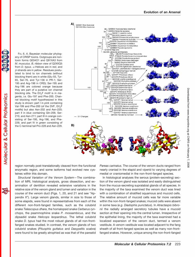
FIG. 6. A, Bayesian molecular phylog-eny of CRISP toxins. Outgroups are non-toxin forms Q034O1 and Q91XA3 fromM. musculus. B, ribbon view of Q2XXQ5from D. typus. �-Helices are in red, and�-strands are in yellow. Residues postu-lated to bind to ion channels (withoutblocking them) are in white (Glu-55, Tyr-65, Ser-76, and Tyr-136 in PR-1; Ser-195 and Arg-196 in CRD); Ser-195 andArg-196 are colored orange becausethey are part of a putative ion channelblocking site. The EX2F motif is in ma-genta, i.e. Glu-197 and Phe-200. Chan-nel blocking motif hypothesized in thisstudy is shown: part I in pink containingVal-199 and Phe-200 (of the DVF, EX2Fmotifs) but also Asn-202 and Asn-220;part II in blue containing Gln-209, Ser-210, and Asn-211; part III in orange con-sisting of Ser-195, Arg-196, and Phe-226; and part IV in green consisting ofthe C-terminal tail Pro-228 and Asn-229.
Evolution of an Arsenal
Molecular & Cellular Proteomics 7.2 223
at CN
RS
on February 7, 2008
ww
w.m
cponline.orgD
ownloaded from
snakes was the presence of a venom vestibule located alongthe medial surface of the venom gland. The proximal portionof the venom duct was septate in the front-fanged taxa andgenerally ovoid and largely free of surrounding connectivetissue in the non-front-fanged taxa. In a few of them (mostnotably in D. typus, Stenorrhina freminvillei, and Thelotorniscapensis) the venom duct was more crenate in cross-sectionand was surrounded by concentric layers of connective tissuesuggestive of pressure regulation.
The greatest level of morphological variation was found inthe topographical relationship between the venom duct andthe fang. In some non-front-fanged taxa (e.g. Heterodon) thevenom duct opens directly into the oral cavity rather than tothe lumen of the fang sheath and the surface of the fang. Inother cases the venom duct courses directly to the fangsheath with no venom vestibule located at the juncture be-
tween these two structures as it is in all front-fanged taxa.Most commonly in non-front-fanged snakes the venom ductopened both into the fang sheath and directly to the roof ofthe mouth immediately adjacent to the fang sheath; this dualopening was achieved by the venom duct, or in some casesthe associated venom vestibule, paralleling the long axis ofthe fang sheath forming a prominent diverticulum. In a fewnon-front-fanged taxa there was a secondary venom duct thatarose from the distal portion of the fang sheath and extendedperipherally to contact the roof of the mouth. Combininginformation for the different structural features revealed thenon-front-fanged snakes to possess a highly variable venomsystem architecture with all combinations of epithelial lining,venom vestibule, and venom duct topography identified (see“Appendix II”).
The maxillary dentition was also revealed to be extremely
FIG. 7. Hyaluronidase. A, partial se-quence alignment of toxin sequences. 1,EU091710 from L. poecilogyrus; 2,A3QVP2; 5, A3QVP3; 13, A3QVP1; 14,A3QVP0; 15, A3QVN9, all from Bitis ari-etans; 3, A3QVN8; 4, A3QVN7; 6,A3QVP5; 9, A3QVN6, all from Echispyramidum leakeyi; 7, A3QVN5; 8,A3QVN3; 10, A3QVN4, all from Cerastescerastes; 11, A3QVN2 Echis ocellatus;12, A3QVP4 from Echis carinatus soch-ureki. Also shown are the representativenon-toxins: 16, Q12794 from Homo sa-piens; 17, Q05A56 from M. musculus. B,Bayesian molecular phylogeny; the out-group is Q9W6K5 from Xenopus laevis.
Evolution of an Arsenal
224 Molecular & Cellular Proteomics 7.2
at CN
RS
on February 7, 2008
ww
w.m
cponline.orgD
ownloaded from

variable among the non-front-fanged snakes, ranging fromsolid smooth fangs (type 1) to grooved fangs of varying depthand length (types 2–4) (Table III). Tooth size was also variablewith significant enlargement occurring on multiple independ-ent occasions. Particularly enlarged teeth were observed inrepresentatives from all families, such as Dispholidus andOligodon in Colubridae, Tomodon and Waglerophis in Dipsa-didae, Macropisthodon in Natricidae, Malpolon and Rham-phiophis in Psammophiinae, and Homalopsis in Homalopsi-dae. In contrast, Atractaspis plus all elapids and viperidspossessed type 5 dentition with an enclosed venom canal,and relative tooth size was also much less variable.
DISCUSSION
Molecular Characterization of Caenophidia Venoms—Com-plex venoms are a feature of the well studied elapids, viperids,and Atractaspis and have also recently been shown to be afeature of the helodermatid and varanid venomous lizards (4).In this study, multiple toxin types were sequenced from allnon-front-fanged snake cDNA libraries, indicating the com-mon presence of complex venom transcriptomes containingmultiple bioactive components. For hyaluronidase, kunitz, andwaprin these were the first sequences, whether protein ornucleotide, obtained for non-front-fanged species. For 3FTx,CRISP, kallikrein, lectin, MMP, PLA2, and SVMP/ADAM, onlya very few proteins had been previously even partially se-
quenced from non-front-fanged species. For 3FTx, CRISP,lectin, and kallikrein, taxonomically limited cDNA sequenceswere previously reported, but comprehensive comparisonshad not been undertaken.
In the sequencing surveys undertaken in this study, tran-scripts of 3FTx, CRISP, and SVMP/ADAM were the mostphylogenetically widespread dominantly secreted toxin types,occurring in at least seven of the 10 non-front-fanged snakesexamined. Other phylogenetically widespread dominant toxintranscripts were lectin and waprin. Identification of 3FTx tran-scripts as a common component of caenophidian venom isconsistent with previous studies showing active expression ofpotent 3FTx in a range of non-front-fanged snakes (2, 14, 40).Similarly for the other toxin types, the congruence with pre-vious LC/MS data on the same species provides additionalevidence of active expression of bioactive proteins in thevenom glands of non-front-fanged snakes (14). Multiple tran-scripts of the majority of toxins were recovered from individualcDNA libraries; this pattern is consistent with accelerateddiversification in toxin multigene families as observed in elap-ids and viperids (19). Numerous transcripts were recoveredwith significant variations including changes in key functionalresidues, changes in the number and spacing of cysteineresidues, and large scale deletions. These modifications rep-resent potential neofunctionalization (evolution of novel bio-activities). These results support previous suggestions that
FIG. 8. Sequence alignment of therepresentative full-length (unless oth-erwise indicated) kallikrein toxins. 1,partial sequence Q2XXM2 from P. olfer-sii; 2, Q5MCS0 from L. curtus; 3, Q6T6S7from Bitis gabonica; 4, P81824; 5,Q9PTU8, both from Bothrops jararaca;6, P33589 from Lachesis muta; 7,Q2XXN0 from V. varius; 8, P43685 fromH. horridum; 9, the non-toxin represent-ative Q9POG3 from H. sapiens.
Evolution of an Arsenal
Molecular & Cellular Proteomics 7.2 225
at CN
RS
on February 7, 2008
ww
w.m
cponline.orgD
ownloaded from

the venom system is a basal characteristic of the advancedsnakes (3, 4) and also previous crude venom studies (14, 16,17). This has important implications for understanding theevolution and ecology of the advanced snake radiation andidentifies venoms from non-front-fanged snakes as an impor-tant bioresource.
Despite their relative presence, these toxin types in differentlineages should not be considered the only classes present ina particular venom. This caveat is due to the limited sequenc-ing that detected only the major toxin transcripts for eachspecies. Other types may be present at lower expressionlevels. It is likely that more detailed exploration of their ven-oms will reveal the presence of additional toxin types. This isdemonstrated by this study recovering only 3FTx, CRISP,factor X, kunitz, and Type IB PLA2 from O. microlepidotus butnot other toxin types previously sequenced from this speciessuch as factor V, natriuretic peptides, or nerve growth factor.
Toxin Structure-Function Relationships—Structure-functionrelationships of a number of the toxin types sequenced in thisstudy have been well characterized in venoms from snakes(Atractaspis, elapids, or viperids) or helodermatid lizards (TableII) (20). This includes information on the key amino acid residuesnecessary for conferring toxicity (e.g. CRISP proteins (30–35)),the role of cysteine spacing in determining folding structure andtoxicity, and a demonstrated role for novel mutations such aslarge scale deletions in conferring new bioactivities.
CRISP toxin transcripts were present in the majority of thevenom glands examined. Previous analysis of CRISP toxinsequences from the venom of the natricid snake R. tigrinusshowed that it lacked the EX2F motif thought to be responsi-ble for the smooth muscle paralytic effect as well as theKX6KR motif hypothesized to be essential for the inhibition ofcyclic nucleotide-gated calcium channels. Subsequent bioac-tivity testing confirmed that R. tigrinus CRISP toxin indeedlacked these activities (31, 32, 42, 43), but it was unclearwhether this was representative of CRISP toxins from thevenoms of other non-front-fanged snakes. In this study, formscontaining the EX2F motif were present in some venoms (Figs.5 and 6). Translation and expression of these encoded proteinswould thus likely induce smooth muscle paralysis. It is notablethat CRISP toxins are particularly rich in the venoms of speciesthat are reptilian feeders (14) such as T. dhara and T. biscutatus.It may be that the toxic hypothermic effect is useful in slowingdown the movement of exothermic prey. Studies on this aspect
FIG. 9. Bayesian molecular phylogeny of kallikrein toxins. Out-groups are non-toxin forms Q9GOG3 and Q9UBX7 from H. sapiens.
FIG. 10. Sequence alignment ofkunitz toxins with functional residues(protease-inhibiting reactive bond)shown in bold and the leader se-quence shown in lowercase. 1,EU029687 from T. dhara; 2, EU029688from P. olfersii; 3, P24541 from Eristoco-phis macmahonii; 4, P82966 from O.hannah; 5, P19859 from N. naja; 6,P00981 from Dendroaspis polylepis; 7,Q7LZE4 from O. scutellatus; 8, Q90WA0from P. textilis; 9, P00989 from B. mul-ticinctus; 10, the representative non-toxin P04815 from Bos taurus.
Evolution of an Arsenal
226 Molecular & Cellular Proteomics 7.2
at CN
RS
on February 7, 2008
ww
w.m
cponline.orgD
ownloaded from

may shed significant light into the evolutionary pressures drivingthe molecular diversification of this toxin type.
Cation-permeable channels (sodium-, potassium-, and cal-cium-selective channels as well as cyclic nucleotide-gatedchannels, which are unselective for monovalent Na� and K�
but also allow transfer of Ca2� cations) often contain morethan one binding site for channel blockers, one generallywithin the transmembrane bundle to bind e.g. anesthetics andone or more at their extracellular side that interact with toxins.A most effective channel block occurs when the ion perme-ation pore is directly occluded by a toxin from the extracellularside. This pore in many cases is bevelled, narrowing toward aselectivity filter (i.e. in potassium, calcium, and sodium chan-
nels). To attract positively charged ions this pore containsboth acidic and aromatic residues. Therefore, extracellularchannel blockers often contain positive charges, i.e. guani-dinium groups as in arginine, which are flanked by H-bonddonors, like in conotoxins and dendrotoxins. Due to theirsize, venom proteins can affect channel gating only from theextracellular side. We postulate that to occlude the channelpore these proteins require a 3D structure motif that (i) isaccessible to interact with an ion channel, (ii) consists of one(ideally positively) charged residue to interact with thecharged amino acids inside the ion permeation pore, (iii)exhibits one or more H-bond donor(s) in vicinity to thecharged residue, and (iv) possibly contains an aromatic
FIG. 11. A, Bayesian molecular phy-logeny of kunitz toxins. The outgroup isthe non-toxin P04815 from B. taurus. B,ribbon view of EU029687 from T. dhara.�-Helices are in red, �-strands are inyellow, and the functional residue Ala-18is shown in green.
Evolution of an Arsenal
Molecular & Cellular Proteomics 7.2 227
at CN
RS
on February 7, 2008
ww
w.m
cponline.orgD
ownloaded from

amino acid to form either �-� or cation-� interactions withthe channel pore.
Structural studies revealed that CRISP is a two-domainprotein of conserved fold consisting of a pathogenesis-relatedPR-1 and a CRD, which are linked together via a flexible hinge(30, 44). Although significantly smaller than PR-1, the CRDspans the C-terminal six of a total of 16 conserved cysteines.The resulting three disulfide bonds stabilize the CRD fold andleave only marginal room for movement in this domain. It iswidely assumed that hydrophobic residues of a concave cleftincluding both the PR-1 and the cysteine-rich domain wouldbind the CRISP to the ion channel (30), whereas actual chan-nel block is hypothesized to involve amino acids of CRD only.The fact that CRISP toxins block a variety of ion channels mayindicate that different residues located at different positions ofCRD are responsible for occluding the extracellular entry tothe channel. This hypothesis is supported by the observationthat CRD can move relative to the PR-1 domain, which mayeven lead to exposure of residues that in the experimentalx-ray structures (Protein Data Bank codes 1RC9 (44) and1XX5 (31)) are found at the interface to PR-1. Yet the aminoacids actually blocking ion channels remained unidentified to
date and were mainly postulated from multiple sequence align-ments. To our understanding, the cysteine-rich domain containsthree sites, which may putatively block ion channels: (i) the loopbetween �11 and �7 spanning eight residues between the firstand second cysteine in CRD, (ii) the loop connecting �7 and �8containing up to six amino acids, and (iii) the C-terminal tailfollowing the sixth conserved CRD cysteine.
A hypothesized functional EX2F motif (34, 35) was foundthree to six residues C-terminal to the first CRD cysteine. Inthis study we could not confirm this motif to be conserved andthus functional throughout the CRISP family. However, thewidespread taxonomical presence is suggestive that if thismotif is not basal it is at least early emerging. Whereas thearomatic residue was conserved (either phenylalanine or ty-rosine), the glutamate occurred in only five of 16 sequences.At the same position five asparagines, one glutamine, and twolysines were found, but interestingly no aspartate was found.In contrast, the C-terminal neighbor to glutamate in position�3 of the first CRD cysteine is highly conserved exhibitingonly aspartate (nine occurrences) and asparagine (11 occur-rences). This position corresponds to the first residue in theDVF motif that has been identified to selectively interact with
FIG. 12. Sequence comparison ofrepresentative lectin toxins. The homo-meric forms shown are: 1, EU029691; 2,EU029689; 3, EU091713, all from E. polyl-epis; 4, EU029699 from L. madagascar-iensis; 5, EU029697; 6, EU029702, bothfrom L. poecilogyrus; 7, EU029700 fromP. olfersii; 8, EU029696 from T. jacksonii;9, Q90WI6 from B. multicinctus; 10, Mi-crurus corallinus sequence publishedbut not database-curated (62); 11,Q6TRS6 from Bothrops jararacussu; and12, Q6T7B7 from B. gabonica. Repre-sentative heterodimeric forms (�- and�-chains, respectively) shown are: 13,Q8JIV6; and 14, Q8JIV7A, both from D.acutus. Also included is the representa-tive non-toxin form: 15, P83300 fromAnser anser.
Evolution of an Arsenal
228 Molecular & Cellular Proteomics 7.2
at CN
RS
on February 7, 2008
ww
w.m
cponline.orgD
ownloaded from

L-type calcium channels (35). It ranges from position 4 to 6after the first CRD cysteine. This motif was first identified in�-conotoxin TxVII (45) and was later found in the venomprotein ablomin (35). Our structural studies showed that theside chain of the aspartate/asparagine in DVF was orientedtoward the core of CRD forming several H-bonds with thethird and most C-terminal a-helix �9. The second and thirdresidues of this motif are located at the surface of CRD withtheir side chains pointing toward the solvent. Yet valine wasfound only three times in this study, whereas in 14 instancesthis residue was charged (four times lysine and 10 timesglutamate and aspartate). The third residue of this motif waslarge and hydrophobic and, with the exception of the se-quence Q3SB05 from Pseudonaja textilis, aromatic. We hy-pothesize that these two residues may dock to ion channelsbecause they satisfy, in most cases, the four criteria postu-lated earlier: (i) spatial accessibility, (ii) a charged amino acid,(iii) nearby H-bond donors, and (iv) an aromatic residue inspatial proximity to the charged amino acid. Exceptions tocriterion ii are several hydrophobic or polar residues found inD. typus, L. poecilogyrus, E. polylepis, Lapemis curtus, Pro-tobothrops mucrosquamatus, and Heloderma horridum. Inter-estingly potential H-bond donors are available in positions �1and �2 of the fourth CRD cysteine (Fig. 5).
Alternatively, interactions with L-type calcium channels mayinvolve one of the following motifs. (i) The loop tip betweenhelices �7 and �8 may be involved. Here the first residue ofthe loop is either polar (Gln or Asn) or charged (Lys or Glu)followed by a mostly polar (Asn, Ser, or Thr) and an eitherpolar (Ser, Asn, Thr, or His) or charged amino acid (Lys or Asp)just before the third conserved cysteine. This region containscharged residues in many toxins and is structurally exposed.However, it lacks an aromatic amino acid in its vicinity. (ii) Theresidues in position �1 or �2 of the first and in �1 of the fifthconserved CRD cysteine may be involved. The former waspostulated to be involved in cyclic nucleotide-gated channelblock and often contains a basic amino acid (see below andFig. 5); the latter is predominantly phenylalanine. This sub-structure is also accessible and is held in place by a nearbydisulfide bond. (iii) Positions �1, �2, and �3 after the sixthCRD cysteine may be involved. This part of the C terminus ismostly positively charged and polar. It is relatively flexible andaccessible and may therefore interact with L-type calciumchannels.
Morita and co-workers (32, 33, 35) proposed one or tworesidues immediately C-terminal of the first conserved CRDcysteine to be critical for interaction with cyclic nucleotide-gated ion channels. Their work revealed that the C-terminal
FIG. 13. Bayesian molecular phylogeny of lectin toxins. The outgroup is the non-toxin P83300 from A. anser. *, M. corallinus sequencepublished but not database-curated (62).
Evolution of an Arsenal
Molecular & Cellular Proteomics 7.2 229
at CN
RS
on February 7, 2008
ww
w.m
cponline.orgD
ownloaded from

domains of pseudechetoxin and pseudecin, which are verysimilar to CRD, were identical but for two residues. The KRmotif in pseudechetoxin made this protein more susceptibleto cyclic nucleotide-gated block than the NY of pseudecin. Inthis work, seven of 19 specimens had a positively chargedresidue in either position �1 or �2 of the first cysteine (Fig. 5).For these specimens, we therefore postulate some activity toblock cyclic nucleotide-gated channels.
Modification of structural residues was also evident in nu-merous caenophidian venom toxin transcripts. It has beenshown that changes in the spacing of ancestral cysteines, theoccurrence of newly evolved cysteines, and modifications instructural residues flanking the cysteines can alter the three-dimensional structure of the molecule and thus the residuescontributing to the surface chemistry with consequences forbioactivity (20). Such modifications can produce toxin variants
that have specificities or potencies differing radically frompreviously characterized forms, including the potential emer-gence of neofunctionalizations.
Most of the 3FTxs sequenced in this study retained theancestral 10-cysteine arrangement (19) (Fig. 2) as had beenshown previously for the �-neurotoxic 3FTx from the venomof the colubrid C. radiatus (2). However, the conservation ofthe ancestral cysteines does not preclude bioactivities otherthan �-neurotoxicity as typified by a form from the venom ofthe colubrid snake Boiga dendrophila that retained the ances-tral cysteines yet was only weakly �-neurotoxic but had anewly derived presynaptic mode of neurotoxicity (16). As thesequences obtained in this study were even more divergent,including the evolution of alternate cysteines, it is quite likelythat a multiplicity of novel activities has been derived.
FIG. 14. Matrix metalloprotease. A, partial sequence alignment ofEU091709 from L. poecilogyrus (1), Q9DE15 from G. gallus (2),A2VCV4 from X. laevis (3), Q98TC6 from Cyprinus carpio (4), P50757from Oryctolagus cuniculus (5), and Q9TUL8 from Equus caballus (6).B, Bayesian molecular phylogeny of lectin toxins. The outgroup is thenon-toxin P52176 from B. taurus.
FIG. 15. PLA2 (Type IB) analyses. A, sequence alignment of rep-resentative toxins. 1, EU029703 from T. biscutatus; 2, P80966 from O.hannah; 3, Q9PSN5 from Notechis scutatus; 4, Q9W7J3 from P.textilis; 5, the non-toxin representative Q8JFB2 from L. semifasciatapancreas. B, Bayesian molecular phylogeny; the outgroup is Q8JFB2from L. semifasciata liver. The ribbon model of TRI002F09 from T.biscutatus shows the conserved catalytic residues (63). �-Helices arein red, and �-strands are in yellow.
Evolution of an Arsenal
230 Molecular & Cellular Proteomics 7.2
at CN
RS
on February 7, 2008
ww
w.m
cponline.orgD
ownloaded from

In addition to variations in functional residues and cys-teines, mutants were also sequenced with frameshifts, trun-cations, and putative exon deletions. Novel transcripts of theSVMP/ADAM toxin type were isolated from P. mossambicusthat were comprised solely of the propeptide domain (Fig. 16).Although forms of SVMP/ADAM have been identified previ-ously that selectively express a particular domain (e.g. com-prised solely of the disintegrin domain (19)) this is the first timethat the prepro domain has been discovered as being selec-tively expressed as the sole domain. The putative exon dele-tion in the lectin toxin variant from E. polylepis not only re-moved a large stretch of residues but also one of the ancestralcysteines, leaving a free cysteine, potentially facilitatingdimerization (Fig. 12). Similarly the putative frameshift in thelectin toxin variant from E. polylepis produced transcripts witheven numbers of cysteines, and therefore these sequencesmay be able to fold to produce stable frameworks (Fig. 12).Thus, genomic mutations in both the caenophidian SVMP/ADAM and lectin toxins have the potential to produce func-tional proteins with significantly altered bioactivities.
Venom toxins from non-front-fanged snakes may alsoprove useful for investigations of the structure-function rela-tionships of the normal body proteins from which they werederived (20). In some cases, much more is known about thetoxic forms than the ancestral body homologues. For exam-ple, the normal body forms of the CRISP proteins are poorlycharacterized with virtually nothing known of their bioactivi-ties. Even the activities of forms that may play important roles,such as those with high expression levels in specific tissue ororgans, remain unknown. The basal activity of the CRISPvenom protein is likely to be relaxation of peripheral smoothmuscle, such as helothermine from H. horridum (46), and mayindicate a tissue-specific role for the ancestral normal bodyprotein. The kallikrein toxins are an example of where anancestral body action (liberation of bradykinin from kininogen)is preserved, but some toxin isoforms have derived additionalactivities (e.g. cleavage of fibrinogen). Similarly the two inde-pendently recruited types of PLA2 proteins in snake venomshave derived activities that the normal body form lacks (e.g.antiplatelet toxicity, myotoxicity, and neurotoxicity). Consist-
FIG. 16. N-terminal sequence align-ment of SVMP/ADAM toxins. The partialsequences from non-front-fanged ad-vanced snakes are: 1, EU029705 from P.olfersii; and 2, EU029707 from L. poecilo-gyrus. The full-length unique truncatedforms are: 3, EU029708; 4, EU029713; 5,EU029717; and 6, EU029727, all from P.mossambicus. The representative viperidvenom sequences are: 7, O42138 fromAgkistrodon contortrix laticinctus; and 8,Q4VM08 from Macrovipera lebetina. 9,the representative elapid sequenceQ8JGN1 from Naja mossambica. 10, therepresentative atractaspidid venom formQ9PT48 from A. engaddensis. The rep-resentative non-toxin forms are: 11,Q9UKQ2 (ADAM28 type); and 12,Q9H2U9 (ADAM7 type) from H. sapiens.
Evolution of an Arsenal
Molecular & Cellular Proteomics 7.2 231
at CN
RS
on February 7, 2008
ww
w.m
cponline.orgD
ownloaded from

ent with this, the toxic forms contain a positively chargedhotspot on the surface that is lacking in the ancestral form fromthe pancreas (Fig. 15). The venom proteins therefore representexquisite natural “knock-out” studies of tremendous usefulnessin elucidating the structure-function relationships of the non-toxin, body homologues of physiological importance. The rela-tive assignment of conserved/variable functional residuesshould not be considered definitive as not all sites/proteins arewell characterized, and other transcripts/toxin types may con-tain functional residues that confer ancestral or novel bioactiv-ities, especially for the toxin types not listed above.
Timing of Toxin Recruitment Events—The phylogenetic dis-tribution of venom toxins in the Toxicofera (venomous squa-mates) provides insights into the timing of toxin recruitmentevents (Fig. 1). The majority of snake venom toxins (14 of 27)
are either shared with the two other Toxicofera lineages (Igua-nia and Anguimorpha) (7 of 27) or occur near the base of theCaenophidia snake clade (7 of 27). The independent evolu-tions of advanced front-fang architectures in Atractaspis andViperidae are linked with recruitments of new toxin types (Fig.1). In the viperids, two new toxin types, c-type natriureticpeptide-bradykinin-potentiating peptide and PLA2 (Type IIA),are closely linked to the evolution of advanced venom deliverysystems and ambush feeding, whereas four other toxin typeswere evolved later on (renin-like aspartic protease, cytokineFAM3B, glycine-rich toxin, and waglerin). The evolution ofadvanced venom architecture in viperids also coincides withsignificant molecular diversification of two existing toxin types(SVMP/ADAM and kallikrein). The evolution of sarafotoxins inAtractaspis is also closely timed with the evolution of ad-
FIG. 17. C-terminal sequence align-ment of SVMP/ADAM toxins. The partialsequences from non-front-fanged snakesare: 1, EU029734; 2, EU029736; 3,EU029737, all from D. typus; 4, EU029735from T. jacksonii; 5, EU029738; 6,EU029739, both from L. madagascarien-sis; and 7, EU036637 from T. dhara. Thetypical viperid venom forms are: 8,O42138 from A. contortrix laticinctus; and9, Q4VM08 from M. lebetina. 10, the rep-resentative truncated disintegrin formQ6T6T2 from B. gabonica. 11, the repre-sentative elapid sequence Q8JGN1 fromN. mossambica. 12, the representativeatractaspidid venom form Q9PT48 fromA. engaddensis. The representative non-toxin forms are: 13, Q9UKQ2 (ADAM28representative); and 14, Q9H2U9 (ADAM7representative) from H. sapiens.
Evolution of an Arsenal
232 Molecular & Cellular Proteomics 7.2
at CN
RS
on February 7, 2008
ww
w.m
cponline.orgD
ownloaded from

vanced front fangs, but whether as a cause or an effect(occurring before or after) the evolution of hollow fangs re-mains to be elucidated. In the elapids, the only two additionaltoxin types currently known do not coincide with developmentof a high pressure front-fanged venom system with the hole-punching, short stubby fangs useful for penetrating toughreptile scales. Instead the evolution of new venom systemsappears to also be linked to the explosive diversification ofexisting toxin types: 3FTx and PLA2 (Type IB) toxins in theelapids with numerous new bioactivities developed for eachtoxin type. The only documented newly evolved toxin types arefound within the rapidly radiated Australian elapid snakes. Oneevolved at the base of this clade (factor X), and one evolved inthe common ancestor of Pseudonaja and Oxyuranus (factor V).
The very long, derived glands of C. rhombeatus do notappear to be linked to the evolution of new toxin types eitherwith the dominant toxin types being CRISP, kallikrein, Type IIAPLA2, and SVMP/ADAM (including RGD disintegrins). WithinAtractaspis the venom differences between long and shortglanded species is unclear. It is also unknown whether thelong glands of some Calliophis species are linked to signifi-cant changes in venom composition.
The early recruitment of a LYNX/SLUR-like gene to form the3FTx toxin multigene family was one of the most significantdevelopments in snake venom evolution, priming the Cae-nophidia for extensive diversification. Indeed, venoms from
non-front-fanged species have been shown to be just as toxicas well characterized extremely potent elapid venoms (40). Aunique nuance of this potent and fast acting toxin type is that,unlike most other toxins, the lethality is almost the samewhether injected subcutaneously, intramuscularly, or evenintravenously (19), and therefore delivery just under the skin isas efficient as straight into the bloodstream. The highestexpression levels of this toxin type in the non-front-fangedsnake families are in the gracile, fast moving forms that haveonly slightly or moderately enlarged rear maxillary teeth andpredate upon fast moving, soft, thin skinned, non-dangerousprey items such as geckos and frogs (14). In contrast, the longfanged D. typus is an arboreal snake that includes birds as amajor prey item, and the venom is rich in forms of the SVMP/ADAM toxins that are potently prothrombin-activating. Thefish-eating aquatic homalopsid snakes, which also have largemaxillary dentition, favor hemotoxic lectin toxins as well asADAM toxins. The elapid snakes, which puncture tough reptileskin with their short, strong hollow front fangs, also typicallyexpress 3FTx transcripts as the dominant mRNA species.However, they favor the derived forms that lack the secondand third ancestral cysteines.
Within the elapids additional toxin types were evolved lateron but also with tremendous impact, such as factor X at thebase of the Australian elapid clade and factor V in the com-mon ancestor of Pseudonaja/Oxyuranus. The recruitment of
FIG. 18. Bayesian molecular phylog-eny of SVMP/ADAM toxins. The out-group is the non-toxin ADAM28 se-quence Q9UKQ2 from H. sapiens. *, apublished but uncurated sequence fromMicropechis ikaheka (64).
Evolution of an Arsenal
Molecular & Cellular Proteomics 7.2 233
at CN
RS
on February 7, 2008
ww
w.m
cponline.orgD
ownloaded from

factor X and factor V in the Australian clade of elapids wouldhave also tremendously aided prey capture with only a smallamount of these very active enzymes needed to cause severedisruption of blood chemistry. However, the activity of factorX toxins is rate-limited by the requirement for cofactors, par-ticularly the essential need to form a 1:1 complex with factorV (47). Thus, the recruitment of factor V into the venom of theOxyuranus/Pseudonaja clade resulted in a virtually complete,perfect toxin complex, greatly increasing the relative toxicityof the venoms and thus significantly aiding in the prey cap-turing ability. Subsequent to the recruitment of factor V, theOxyuranus/Pseudonaja common ancestor split into two verydifferent forms despite the two genera remaining very closelygenetically related.
The Oxyuranus species are very large snakes with ex-
tremely long fangs and high venom yields. Unlike most Aus-tralian snakes, which often feed on reptiles, these snakes feedexclusively on mammalian prey including large rats andbandicoots, dangerous prey items capable of an adept de-fense potentially causing serious injury to the snake preda-tors. The snakes successfully feed on such dangerous prey byminimizing prey contact time through the use of a snap-release form of striking along with a tendency for multiplestrikes, thus overwhelming the prey items with copiousamounts of the extraordinarily potent venom delivered deepinto the tissues several times. A limitation of this form of preycapture is that the snakes need to be very warm to success-fully use this energetically costly form of prey capture. Con-sequently their habitat is limited only to the tropical north ofAustralia and in New Guinea (Oxyuranus scutellatus) and the
FIG. 19. A, sequence comparison ofrepresentative waprin toxins from non-front-fanged advanced snakes (1,EU029745 from E. polylepis; 2, EU029744from L. poecilogyrus; 3, EU029743; 4,EU029746; 5, EU029742, all from P. olf-ersii; 6, EU029741 from R. tigrinus; 7,EU029740 from T. jacksonii) and fromfront-fanged advanced snakes (8, P60589from Naja nigricollis; 9, P83952 from O.microlepidotus) and of the representativenon-toxin form (10, P19957 from H. sapi-ens). B, Bayesian molecular phylogeny ofwaprin toxins. The outgroup is the non-toxin P83300 from A. anser. The ribbonmodel of EU029740 from T. jacksoniishows �-strands in yellow.
Evolution of an Arsenal
234 Molecular & Cellular Proteomics 7.2
at CN
RS
on February 7, 2008
ww
w.m
cponline.orgD
ownloaded from

baking heat of the channel country in the Australian outback(O. microlepidotus).
In contrast, the Pseudonaja species have taken a very dif-ferent evolutionary path, taking advantage of possessing suchtoxic venom to greatly minimize the amount of biologicalenergy expended in venom production. Pseudonaja speciesare smaller snakes with very short fangs and much smallervenom yields, utilizing constriction to hold non-dangerousprey items (such as reptiles) in place while envenomating. Asthese snakes do not expend as much energy in prey capture,they do not have to be as warmed up as the Oxyuranusspecies. These snakes consequently have taken advantage oftheir extremely toxic venom to become one of the mostadaptable and successful snake types of the entire Australiancontinent.
Another conspicuous finding of this study was the resolu-tion of the lectin toxins into a single clade with homomericforms not reciprocally monophyletic in relation to the het-erodimeric form found exclusively in viperid venoms (Fig. 13).The homomeric and the heterodimeric lectin forms had beenconsidered previously to be the result of two separate toxinrecruitment events (3) or the result of a single recruitmentevent (18). However, reconstructing the evolutionary historyhad been hampered by the scarcity of homomeric sequences.
FIG. 20. Transverse histology of Masson’s trichrome-stainedsections showing the relative size of venom glands (proportionalto head size) for H. leopardinus (A), D. typus (B), Homalopsisbuccata (C), P. guttatus (D), and D. scabra (E). VG, venom gland;SG, supralabial gland.
FIG. 21. Histology using modifiedVan Gieson’s stain. A, frontal sectionthrough Ahaetulla nasuta showing thestructural differences between the su-pralabial gland and venom gland. B,transverse section showing a proximal todistal transition in the epithelial lining ofthe venom duct of Psammophisschokari. C, frontal section showing therelatively small venom vestibule of D.punctatus located adjacent to the fang.D, frontal section through R. tigrinusshowing the venom vestibule locatedimmediately adjacent to the venomgland. E, frontal section through thevenom duct of T. capensis showing theconcentric rings of surrounding connec-tive tissue. F, transverse section throughthe venom duct of Coluber constrictorshowing the venom duct opening to theoral cavity. G, frontal section throughPsammodynastes pulverulentus show-ing the direct course of the venom ductfrom the venom gland to the fangsheath. H, frontal section through thefang sheath and diverticulum of Malpo-lon monspessulanus. I, frontal sectionthrough Oligodon smithii. showing theaccessory venom duct extending awayfrom the fang sheath. D, venom duct; F,fang; O, oral cavity; S, fang sheath; SG,supralabial gland; VG, venom gland; V,venom vestibule.
Evolution of an Arsenal
Molecular & Cellular Proteomics 7.2 235
at CN
RS
on February 7, 2008
ww
w.m
cponline.orgD
ownloaded from

In this study, we sequenced numerous lectin toxin transcriptsfrom four additional non-front-fanged snake families. Thisallowed for a more robust reconstruction of the molecularevolutionary history and thus the confirmation that all forms ofthe snake venom lectin toxins are the result of a single toxinrecruitment event (Fig. 1) and that the viperid venom het-erodimeric forms arose from within the homomeric basal form(Fig. 13). This study also allowed for the inference that theEPN mannose-binding motif is basal whereas the QPD galac-tose-binding variant was an early emerging derivation thatpredated the viperid radiation.
Derivations of the Venom System—Taub (7) described ex-tensive morphological variation within the venom gland (his“Duvernoy’s” gland) of non-front-fanged snakes. The presentstudy demonstrates that similar morphological variation existsin the venom duct as well as in the topographic relationshipbetween the venom duct and the fang sheath (see “AppendixI”). These morphological variations do not assort along estab-lished evolutionary lines, nor do they correlate well with theattributes of the venoms detailed in this study. This is incontrast to the front-fanged Elapidae and Viperidae families,which each have a well defined, characteristic venom appa-ratus at the familial level with some minor variations (Fig. 1),such as the size of venom glands and connections of thecompressor and other head muscles. The same is true for thefront-fanged genus Atractaspis but not for the non-front-fanged snakes now included in the Atractaspidinae subfamily,which were shown in this study to be quite variable.
The mechanics of venom injection in front-fanged snakesrelies on pressurization of the venom delivery system (48–51).The colubrid snake D. typus is the only species with wellcharacterized rudimentary compressor musculature (52), al-though there is evidence that this has been convergentlyevolved in the lamprophiine snake Mehelya capensis (52) andin the non-front-fanged atractaspidine Brachyophis revoili(53). The general absence of these features from the venomdelivery system of non-front-fanged snakes suggests that asubstantially different mechanism(s) is present within thisgroup. The limited skeletal muscle contact with the venom
gland in non-front-fanged snakes (54), the relatively weak wallof the venom duct, and the presence of a venom vestibulewould all oppose the pressurization of the venom deliverysystem (but see Jansen and Foehring (55)). The multiple to-pographic relationships between the venom duct and thefang, including taxa in which the venom duct opens to the roofof the mouth rather than the fang sheath, clearly indicate thatdeformation of the fang sheath need not be an important partof venom delivery in non-front-fanged snakes.
Interestingly the colubrid family also contained a clade inwhich the venom gland was greatly atrophied, the American“rat snake” clade of colubrid snakes, as typified by P. guttatus(Figs. 1 and 20). The American rat snakes have secondarilyevolved a new form of prey capture (powerful constriction)and prey preference (rodents). Subsequently the gland hasbecome greatly atrophied. Similarly the African D. scabra,which feeds exclusively on bird eggs, also has greatly atro-phied venom glands (Fig. 20). This is paralleled in the seasnake Aipysurus eydouxii that, subsequent to switching fromfeeding on fish to feeding exclusively on fish eggs, has greatlyatrophied venom glands (Fig. 1) and almost entirely lost fangsaccompanied by significant accumulation of deleterious mu-tations to the toxins still transcribed (56, 57). Similarly thelizard egg-eating Brachyurophis species of Australian terres-trial elapids also have significant reduction of the venomsystem. Finally the other few lineages displaying an absenceor reduction of the glands are malacophagous (Asianpareatids and some American dipsadids) (1).
Changes in gland size were not restricted to atrophying butalso included spectacular lengthening. In each of the front-fanged clades, tremendous elongation has occurred inde-pendently: Atractaspis engaddensis, Atractaspis microlepi-dota, Atractaspis micropholis, and Atractaspis scortecci in theAtractaspinae, Calliophis intestinalis and Calliophis bivirgata inthe Elapidae, and Causus resimus and C. rhombeatus in theViperidae. The elongations extend to a quarter of the bodylength. The biological advantage gained by these elongationsremains to be elucidated. In at least the case of Causus thechange in gland length was not accompanied by a significant
TABLE IIISummary of venom dentition variation in genera studied (for full species list see �Appendix III�)
Smooth surface and no enclosed venom canal Colubridae: Coluber, Dasypeltis, Lycodon, Masticophis, Oligodon, Pituophis,Philothamnus, Spalerosophis. Dipsadidae: Diadophis, Heterodon,Hydrodynastes, Hypsiglena. Lamprophiinae: Lamprophis, Lycophidion.Natricidae: Macropisthodon, Nerodia, Rhabdophis.
No enclosed venom canal and surface withshallow furrow
Dipsadidae: Erythrolamprus. Natricidae: Amphiesma.
Deep groove present but restricted to less thanhalf the length of the tooth
Colubridae: Ahaetulla, Chrysopelea, Dispholidus, Trimorphodon. Dipsadidae:Coniophanes, Leptodeira, Oxyrhopus, Pseudoboa, Tachymenis, Tomodon.Homalopsidae: Cerberus, Enhydris, Erpeton. Lamprophiinae:Psammodynastes. Psammophiinae: Psammophis. Pseudoxyrhophiinae:Madagascarophis.
Deep groove running the majority of the lengthof the tooth
Atractaspidinae: Amblyodipsas, Aparallactus. Colubridae: Boiga, Chionactis,Crotaphopeltis, Oxybelis, Stenorrhina, Thelotornis. Dipsadidae: Philodryas.Psammophiinae: Malpolon, Psammophylax.
Enclosed venom canal Atractaspidinae: Atractaspis only. Elapidae: ubiquitous. Viperidae: ubiquitous.
Evolution of an Arsenal
236 Molecular & Cellular Proteomics 7.2
at CN
RS
on February 7, 2008
ww
w.m
cponline.orgD
ownloaded from

shift in venom profile or recruitment of new toxin types. In-stead, having typical viperid venom composition with themajor toxin types sequenced (SVMP, kallikrein, and PLA2
(Type IIA)) in this study being consistent with molecularweights obtained in a previous LC/MS study by us (14). Itremains to be investigated whether there are significantvenom compositional differences between long and shortglanded forms within the Atractaspis and Calliophis genera.
Snakes of Potential Medical Concern—This study has im-portant implications in regard to human medicine by providingnew information regarding snake venom composition andthus potential clinical effects of envenomation. It also high-lights the potential danger of snakes regarded as of trivialconcern based solely upon the lack of severe bites, but insome cases the rarity of reports is due to the rarity of thesnake in captivity or encounters in the wild.
This study has shown that the venom transcriptome ofThrasops jacksonii is very similar to the extremely potentvenom of the very closely related (and proven lethal) D. typus,particularly the abundance of PIII-type SVMP/ADAM toxins.The lethality of D. typus venom is the result of high expressionlevels of a potent prothrombin-activating form of the SVMP/ADAM toxin type. In the clinical pathology, the prothrombinactivation causes consumptive coagulopathy, resulting in thedisappearance of fibrinogen from the blood and the elevatedpresence of d-dimer products with the ultimate net result ofincoagulable blood. Predation by T. jacksonii on live mamma-lian prey results in rapid death in a manner identical to thatproduced by D. typus: profound disruption of hemostasismanifested by rapid and copious bleeding from the mouthand nostrils.4 Clearly potent venom toxins are actively ex-pressed in this species. We advise exotic animal keepers to
treat this species with the utmost caution until studies of theaction of T. jacksonii venom upon the hemostatic system areundertaken, including the relative neutralization by D. typusantivenom. In at least this case, absence of evidence shouldnot be taken as evidence of absence.
Conclusion—Collectively these findings strongly supportthat there has been extensive evolutionary modification of thevenom system in the advanced snake radiation (Caenophidia).All variables appear to change independently ranging from thebiochemical variation and specialization of the venoms to thedentition and glandular morphology. The long evolutionaryhistory of the advanced snake radiation (�100 million years)has resulted in extensive diversification of the venom systemand shows how little we really know about venom macroevo-lution and the potential role in the ecology/evolution of theanimals themselves. Furthermore the toxin types identifiedhere should not be regarded as the only toxins present inthose venoms. Additional experimental efforts will certainlyreveal new isoforms of these toxins, and some classes willalmost certainly be confirmed as evolving earlier than cur-rently appreciated. Further entirely new toxin types may yet bediscovered in any one of the poorly studied snake lineages.Our major findings are of a multidisciplinary nature with usesranging from evolutionary biology to protein evolution andhighlight the underutilization of snake venoms as biore-sources in drug design and development.
Acknowledgments—The Naturalis Museum, Leiden, The Nether-lands generously loaned us preserved specimens for some of mag-netic resonance imaging studies. H. S. is grateful to GlaxoSmithKlinefor an exclusive version of SPDBV. We are deeply grateful to Profes-sor Elazar Kochva for patience in answering our many questions andfor very helpful comments on the manuscript. We also thank thevarious wildlife departments for the issuing of the relevant scientificcollecting permits.4 B. G. Fry, personal observations.
Evolution of an Arsenal
Molecular & Cellular Proteomics 7.2 237
at CN
RS
on February 7, 2008
ww
w.m
cponline.orgD
ownloaded from

APPENDIX ISpecies and mode examined
C, cDNA library; G, gross dissection; D, dentition; H, histology; M, magnetic resonance imaging.
Evolution of an Arsenal
238 Molecular & Cellular Proteomics 7.2
at CN
RS
on February 7, 2008
ww
w.m
cponline.orgD
ownloaded from

APPENDIX I—continued
Evolution of an Arsenal
Molecular & Cellular Proteomics 7.2 239
at CN
RS
on February 7, 2008
ww
w.m
cponline.orgD
ownloaded from

APPENDIX I—continued
Evolution of an Arsenal
240 Molecular & Cellular Proteomics 7.2
at CN
RS
on February 7, 2008
ww
w.m
cponline.orgD
ownloaded from

APPENDIX IIStructural variation
Epithelium: 1, no mucus; 2, isolated mucoid cells or patches; 3, prominent mucoid region or gland. Contour: 1, relatively large ovate duct;2, duct diameter reduced, surrounded by extensive circular connective tissue; 3, internal partitions to venom duct. Vestibule: 1, no vestibule;2, vestibule present adjacent to venom gland; 3, vestibule present adjacent to fang sheath; 4, vestibule in contact with oral cavity. Topographyof venom duct opening: 1, venom duct opens only to oral cavity; 2, venom duct opens only to fang sheath; 3, venom duct opens to both oralcavity and fang sheath.
Evolution of an Arsenal
Molecular & Cellular Proteomics 7.2 241
at CN
RS
on February 7, 2008
ww
w.m
cponline.orgD
ownloaded from
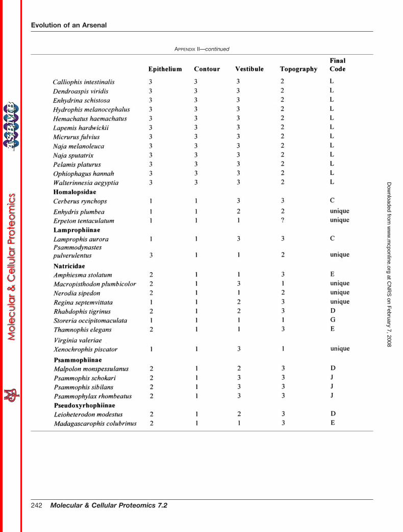
APPENDIX II—continued
Evolution of an Arsenal
242 Molecular & Cellular Proteomics 7.2
at CN
RS
on February 7, 2008
ww
w.m
cponline.orgD
ownloaded from

APPENDIX II—continued
Evolution of an Arsenal
Molecular & Cellular Proteomics 7.2 243
at CN
RS
on February 7, 2008
ww
w.m
cponline.orgD
ownloaded from

APPENDIX IIIDentitional morphology
Evolution of an Arsenal
244 Molecular & Cellular Proteomics 7.2
at CN
RS
on February 7, 2008
ww
w.m
cponline.orgD
ownloaded from

* This work was supported by grants (to B. G. F.) from the Austra-lian Academy of Science, Australian French Association for Scienceand Technology, Australia and Pacific Science Foundation, CASSFoundation, Ian Potter Foundation, International Human FrontiersScience Program Organisation, Netherlands Organisation for Scien-tific Research, and University of Melbourne (Faculty of Medicine andDepartment of Biochemistry) and a Department of Innovation, Indus-try and Regional Development Victoria Fellowship; by Australian Re-search Council Grant DP0451663 (to B. G. F. and J. A. N.); and by anAustralian Government Department of Education, Science and Train-ing/EGIDE International Science Linkages grant (to B. G. F., J. A. N.,and N. V.). The costs of publication of this article were defrayed in partby the payment of page charges. This article must therefore be herebymarked “advertisement” in accordance with 18 U.S.C. Section 1734solely to indicate this fact.
The nucleotide sequence(s) reported in this paper has been sub-mitted to the GenBankTMEBI Data Bank with accession number(s)EU029668 –EU029765, EU036635–EU036637, and EU091709 –EU091713.
§ To whom correspondence should be addressed. E-mail:[email protected].
� Present address: Nestle Research Center, Vers-chez-les-Blanc,P. O. Box 44, 1000 Lausanne, Switzerland.
REFERENCES
1. Vidal, N. (2002) Colubroid systematics: evidence for an early appearance ofthe venom apparatus followed by extensive evolutionary tinkering. J.Toxicol. Toxin Res. 21, 21–41
2. Fry, B. G., Lumsden, N., Wuster, W., Wickramaratna, J., Hodgson, W. C.,and Kini, R. M. (2003) Isolation of a neurotoxin (�-colubritoxin) from a‘non-venomous’ colubrid: evidence for early origin of venom in snakes. J.Mol. Evol. 57, 446–452
3. Fry, B. G., and Wuster, W. (2004) Assembling an arsenal: origin and evo-lution of the snake venom proteome inferred from phylogenetic analysisof toxin sequences. Mol. Biol. Evol. 21, 870–883
4. Fry, B. G., Vidal, N., Norman, J. A., Vonk, F. J., Scheib, H., Ramjan, R.,Kuruppu, S., Fung, K., Hedges, S. B., Richardson, M. K., Hodgson,W. C., Ignjatovic, V., Summerhayes, R., and Kochva, E. (2006) Earlyevolution of the venom system in lizards and snakes. Nature 439,509–632
5. Kardong, K. (1980) Evolutionary patterns in advanced snakes. Am. Zool. 20,269–282
6. Vidal, N., Delmas, A. S., David, P., Cruaud, C., Couloux, A., and Hedges,S. B. (2007) The phylogeny and classification of caenophidian snakesinferred from seven nuclear protein-coding genes. C. R. Biol. 330,182–187
7. Taub, A. M. (1967) Comparative histological studies on Duvernoy’s gland ofcolubrid snakes. Bull. Am. Mus. Nat. Hist. 138, 1–50
8. Gabe, M., and Saint Girons, H. (1969) Histological data on the salivaryglands of the Lepidosauria. Mem. Mus. Natn. Hist. Nat. Paris 58, 1–112
9. Kochva, E. (1978) Oral glands of the Reptilia, in Biology of the Reptilia(Gans, C., and Gans, K. A., eds) Vol. 8, pp. 43–161, Academic Press,New York
10. Young, B., and Kardong, K. (1996) Dentitional surface features in snakes(Reptilia: Serpentes). Amphib.-Reptilia 17, 261–276
11. West, G. S. (1895) On the buccal glands and teeth of certain poisonoussnakes. Proc. Zool. Soc. Lond. 1895, 195–206
12. West, G. (1898) On the history of the salivary buccal and harderian glandsof the colubroidae with notes on their tooth succession and the relation-ships of the poison ducts. J. Linn. Soc. Lond. Zool. 26, 517–526
13. Sarkar, S. C. (1923) A comparative study of the buccal glands and teeth ofthe opisthoglypha, and a discussion on the evolution of the order fromaglypha. Proc. Zool. Soc. Lond. 1923, 295–322
14. Fry, B. G., Wuster, W., Ramjan, S. F. R., Jackson, T., Martelli, P., and Kini,R. M. (2003) LC/MS (liquid chromatography, mass spectrometry) analy-sis of Colubroidea snake venoms: evolutionary and toxinological impli-cations. Rapid Commun. Mass Spectrom. 17, 2047–2062
15. Huang, P., and Mackessy, S. P. (2004) Biochemical characterization ofphospholipase A2 (trimorphin) from the venom of the Sonoran Lyre
Snake Trimorphodon biscutatus lambda (family Colubridae). Toxicon 44,27–36
16. Lumsden, N. G., Fry, B. G., Ventura, S., Kini, R. M., and Hodgson, W. C.(2005) Pharmacological characterisation of a neurotoxin from the venomof Boiga dendrophila (Mangrove snake). Toxicon 45, 329–334
17. Lumsden, N. G., Banerjee, Y., Kini, R. M., Kuruppu, S., and Hodgson, W. C.(2007) Isolation and characterization of rufoxin, a novel protein exhibitingneurotoxicity from venom of the psammophiine Rhamphiophis oxyrhyn-chus (Rufous beaded snake). Neuropharmacology 52, 1065–1070
18. Ching, A. T., Rocha, M. M., Paes Leme, A. F., Pimenta, D. C., de Fatima, M.,Furtado, D., Serrano, S. M. T., Ho, P. L., and Junqueira de Azevedo,I. L. M. (2006) Some aspects of the venom proteome of the Colubridaesnake Philodryas olfersii revealed from a Duvernoy’s (venom) gland tran-scriptome. FEBS Lett. 580, 4417–4422; Correction (2006) FEBS Lett.580, 5122–5123
19. Fry, B. G., Wuster, W., Kini, R. M., Brusic, V., Khan, A., Venkataraman, D.,and Rooney, A. P. (2003) Molecular evolution of elapid snake venomthree finger toxins. J. Mol. Evol. 57, 110–129
20. Fry, B. G. (2005) From genome to ‘venome’: molecular origin and evolutionof the snake venom proteome inferred from phylogenetic analysis oftoxin sequences and related body proteins. Genome Res. 15, 403–420
21. Nei, M., Gu, X., and Sitnikova, T. (1997) Evolution by the birth-and-deathprocess in multigene families of the vertebrate immune system. Proc.Natl. Acad. Sci. U. S. A. 94, 7799–7806
22. Harvey, A. L., Bradley, K. N., Cochran, S. A., Rowan, E. G., Pratt, J. A.,Quillfeldt, J. A., and Jerusalinsky, D. A. (1998) What can toxins tell us fordrug discovery? Toxicon 36, 1635–1640
23. Menez, A. (1998) Functional architectures of animal toxins: a clue to drugdesign? Toxicon 36, 1557–1572
24. Cushman, D. W., Ondetti, M. A., Gordon, E. M., Natarajan, S., Karanewsky,D. S., Krapcho, J., and Petrillo, E. W., Jr. (1987) Rational design andbiochemical utility of specific inhibitors of angiotensin-converting en-zyme. J. Cardiovasc. Pharmacol. 10, S17–30
25. Chothia, C., and Lesk, A. M. (1986) The relation between the divergence ofsequence and structure in proteins. EMBO J. 5, 823–826
26. Greer, J. (1981) Comparative model-building of the mammalian serineproteases. J. Mol. Biol. 153, 1027–1042
27. Guex, N., and Peitsch, M. C. (1997) SWISS-MODEL and the Swiss-Pdb-Viewer: an environment for comparative protein modeling. Electrophore-sis 18, 2714–2723
28. Diemand, A. V., and Scheib, H. (2004) iMolTalk: an interactive, internet-based protein structure analysis server. Nucleic Acids Res. 32,W512–W516
29. Koradi, R., Billeter, M., and Wuthrich, K. (1996) MOLMOL: a program fordisplay and analysis of macromolecular structures. J. Mol. Graph. 14,51–55
30. Shikamoto, Y., Suto, K., Yamazaki, Y., Morita, T., and Mizuno, H. (2005)Crystal structure of a CRISP family Ca2�-channel blocker derived fromsnake venom. J. Mol. Biol. 350, 735–743
31. Wang, J., Shen, B., Guo, M., Lou, X., Duan, Y., Cheng, X. P., Teng, M., Niu,L., Liu, Q., Huang, Q., and Hao, Q. (2005) Blocking effect and crystalstructure of natrin toxin, a cysteine-rich secretory protein from Naja atravenom that targets BKCa channel. Biochemistry 44, 10145–10152
32. Yamazaki, Y., Brown, R. L., and Morita, T. (2002) Purification and cloning oftoxins from elapid venoms that target cyclic nucleotide-gated ion chan-nels. Biochemistry 41, 11331–11337
33. Yamazaki, Y., and Morita, T. (2004) Structure and function of snake venomcysteine-rich secretory proteins. Toxicon 44, 227–231
34. Yamazaki, Y., Koike, H., Sugiyama, Y., Motoyoshi, K., Wada, T., HishinumaS., Mita, M., and Morita, T. (2002) Cloning and characterization of novelsnake venom proteins that block smooth muscle contraction. Eur. J. Bio-chem. 269, 2708–2715
35. Yamazaki, Y., Hyodo, F., and Morita, T. (2003) Wide distribution of cysteine-rich secretory proteins in snake venoms: isolation and cloning of novelsnake venom cysteine-rich secretory proteins. Arch. Biochem. Biophys.412, 133–141
36. Harrison, R. A., Ibison, F., Wilbraham, D., and Wagstaff, S. C. (2007)Identification of cDNAs encoding viper venom hyaluronidases: cross-generic sequence conservation of full-length and unusually short varianttranscripts. Gene (Amst.) 392, 22–33
37. Chen, C., Hsu, C. H., Su, N. Y., Lin, Y. C., Chiou, S. H., and Wu, S. H. (2001)
Evolution of an Arsenal
Molecular & Cellular Proteomics 7.2 245
at CN
RS
on February 7, 2008
ww
w.m
cponline.orgD
ownloaded from

Solution structure of a kunitz-type chymotrypsin inhibitor isolated fromthe elapid snake Bungarus fasciatus. J. Biol. Chem. 276, 45079–45087
38. Drickamer, K. (1992) Engineering galactose-binding activity into a C-typemannose-binding protein. Nature 360, 183–186
39. Komori, K., Konishi, M., Maruta, Y., Toriba, M., Sakai, A., Matsuda, A., Hori,T., Nakatani, M., Minamino, N., and Akizawa, T. (2006 May) Character-ization of a novel metalloproteinase in Duvernoy’s gland of Rhabdophistigrinus tigrinus. J. Toxicol. Sci. 31, 157–168
40. Lumsden, N. G., Fry, B. G., Kini, R. M., and Hodgson, W. C. (2004) In vitroneuromuscular activity of ‘colubrid’ snake venoms: clinical and evolu-tionary implications. Toxicon 43, 819–827
41. Kamiguti, A. S., Theakston, R. D., Sherman, N., and Fox, J. W. (2000) Massspectrophotometric evidence for P-III/P-IV metalloproteinases in thevenom of the Boomslang (Dispholidus typus). Toxicon 38, 1613–1620.
42. Brown, R. L., Lynch, L. L., Haley, T. L., and Arsanjani, R. (2003) Pseudeche-toxin binds to the pore turret of cyclic nucleotide-gated ion channels.J. Gen. Physiol. 122, 749–760
43. Brown, R. L., Haley, T. L., West, K. A., and Crabb, J. W. (1999) Pseudeche-toxin: a peptide blocker of cyclic nucleotide-gated ion channels. Proc.Natl. Acad. Sci. U. S. A. 96, 754–759
44. Guo, M., Teng, M., Niu, L., Liu, Q., Huang, Q., and Hao, Q. (2005) Crystalstructure of the cysteine-rich secretory protein stecrisp reveals that thecysteine-rich domain has a K� channel inhibitor-like fold. J. Biol. Chem.280, 12405–12412
45. Kobayashi, K., Sasaki, T., Sato, K., and Kohno, T. (2000) Three-dimensionalsolution structure of �-conotoxin TxVII, an L-type calcium channelblocker. Biochemistry 39, 14761–14767
46. Mochca-Morales, J., Martin, B. M., and Possani, L. S. (1990) Isolation andcharacterization of helothermine, a novel toxin from Heloderma horridumhorridum (Mexican beaded lizard) venom. Toxicon 28, 299–309
47. Kini, R. M., Rao, V. S., and Joseph, J. S. (2001) Procoagulant proteins fromsnake venoms. Haemostasis 31, 218–224
48. Rosenberg, H. (1967) Histology, histochemistry and emptying mechanismof the venom gland of some elapid snakes. J. Morphol. 122, 133–156
49. Young, B., Blair, M., Zahn, K., and Marvin, J. (2001) Mechanics of venomexpulsion in Crotalus, with special reference to the role of the fangsheath. Anat. Rec. 264, 415–426
50. Young, B. A., Lee, C. E., and Daley, K. M. (2002) Do snakes meter venom?Biosciences 52, 1121–1126
51. Young, B. A., Dunlap, K., Koenig, K., and Singer, M. (2004) The buccalbuckle: the functional morphology of venom spitting in cobras. J. Exp.Biol. 207, 3483–3494
52. Kochva, E., and Wollberg, M. (1970) The salivary glands of Aparallactinae
(Colubridae) and the venom glands of Elaps (Elapidae) in relation to thetaxonomic status of this genus. Zool. J. Linn. Soc. 49, 217–224
53. Underwood, G., and Kochva, E. (1993) On the affinities of the burrowingasps Atractaspis (Serpentes: Atractaspididae). Zool. J. Linn. Soc. 107,3–64
54. Haas, G. (1973) Muscles of the jaw and associated structures in theRhyncocephalia and Squamata, in Biology of the Reptilia (Gans, C., andParsons, T., eds) Vol. 4, pp. 285–490, Academic Press, New York
55. Jansen, W., and Foehring, R. (1983) The mechanism of venom secretionfrom Duvernoy’s gland of the snake Thamnophis sirtalis (Serpentes:Colubridae). J. Morphol. 175, 271–277
56. Li, M., Fry, B. G., and Kini, R. M. (2005) Eggs only diet: the shift in preferredprey by the Marbled sea snake (Aipysurus eydouxii) resulting in a loss ofpostsynaptic neurotoxicity. J. Mol. Evol. 60, 81–89
57. Li, M, Fry, B. G., and Kini, R. M. (2005) Putting the brakes on snake venomevolution: the unique molecular evolutionary patterns of Aipysurus ey-douxii (Marbled sea snake) phospholipase A2 toxins. Mol. Biol. Evol. 22,934–941
58. Keogh, J. S., Shine, R., and Donnellan, S. (1998) Phylogenetic relationshipsof terrestrial Australo-Papuan elapid snakes (subfamily Hydrophiinae)based on cytochrome b and 16S rRNA sequences. Mol. Phylogenet.Evol. 10, 67–81
59. Vidal, N., Kindl, S. G., Wong, A., and Hedges, S. B. (2000) Phylogeneticrelationships of xenodontine snakes inferred from 12S and 16S riboso-mal RNA sequences. Mol. Phylogenet. Evol. 14, 389–402
60. Vidal, N., and Hedges, S. B. (2002) Higher-level relationships of caenophid-ian snakes inferred from four nuclear and mitochondrial genes. C. R. Biol.325, 987–995
61. Lawson, R., Slowinski, J. B., Crother, B. I., and Burbrink, F. T. (2005)Phylogeny of the Colubroidea (Serpentes): new evidence from mitochon-drial and nuclear genes. Mol. Phylogenet. Evol. 37, 581–601
62. Ho, P. L., Soares, M. B., Yamane, T., and Raw, I. (1995) Reverse biologyapplied to Micrurus corallinus, a South American coral snake. J. Toxicol.Toxin Rev. 14, 327–337
63. Rouault, M., Rash, L. S., Escoubas, P., Boilard, E., Bollinger, J., Lomonte,B., Maurin, T., Guillaume, C., Canaan, S., Deregnaucourt, C., Schrevel,J., Doglio, A., Gutierrez, J. M., Lazdunski, M., Gelb, M. H., and Lambeau,G. (2006) Neurotoxicity and other pharmacological activities of the snakevenom phospholipase A2 OS2: the N-terminal region is more importantthan enzymatic activity. Biochemistry 45, 5800–5816
64. Gao, R., Kini, R. M., and Gopalakrishnakone, P. (2002) A novel prothrombinactivator from the venom of Micropechis ikaheka: isolation and charac-terisation. Arch. Biochem. Biophys. 408, 87–92
Evolution of an Arsenal
246 Molecular & Cellular Proteomics 7.2
at CN
RS
on February 7, 2008
ww
w.m
cponline.orgD
ownloaded from