The Concept of Number - Ernst Cassirer (Portuguese translation)
Upload
gadermpathCategory
view
0download
0
1.
Volume 4, Supplement 3, November 2013 ISSN: 2081-9390 p. 583 - 665 DOI: 10.7241/ourdIssue online since Tuesday, November 12, 2013
NASZA DERMATOLOGIA Online www.odermatol.com
OUR DERMATOLOGY Online
Supplement 3 / 2013
„AUTOIMMUNE BULLOUS DISEASE”Issue in Memorial:Dr Ernst H. Beutner, (August 27, 1923 – June 10, 2013)

Editor in Chief: Piotr Brzeziński, MD Ph.DAddress:ul. Braille’a 50B, 76200 Słupsk, Polandtel. 48 692121516, fax. 48 598151829e-mail: [email protected]
Associate Editor:Ass. Prof. Viktoryia Kazlouskaya (USA)
Editorial Pages
e-ISSN: 2081-9390
Quarterly published since 01/06/2010 years
Our Dermatol Online
www.odermatol.com
Publisher:Our Dermatology OnlineAddress:ul. Andersa 5/8, 76200 Słupsk, Polandtel. 48 692121516, fax. 48 598151829e-mail: [email protected]
Previous website: issue 1.2010 www.ndermatol.like.pl since issue 2.2010 to issue 3.2011 www.odermatol.like.pl since issue 4.2011 www.odermatol.com
Previous shortcut: since issue 1.2010 to issue 3.2011 N Dermatol Online since issue 4.2011 Our Dermatol Online
Open access journal:This is an open access journal which means that all content is freely available without charge to the user or his/her institution. Users are allowed to read, download, copy, distribute, print, search, or link to the fullor texts of the articles in this journal without asking prior permission
from the publisher or the author. Our Dermatology Online is a international journal that publishes original contributions in the field of
dermatology, including papers on biochemistry, morphology and immunology of the skin.The journal is among the few not related to dermatological associations or belonging to respective societies which guarantees complete
independence. Offers a platform for review articles in areas of interest for dermatologists. OurDermatologyOnline offers article in English as well as in other languages.
This is in accordance with the BOAI definition of open access.
www.odermatol.com
Indexed in:Universal Impact Factor for year 2012 is = 0.7319
system of opinion of scientific periodicals INDEX COPERNICUS (6,70)(Academic Search) EBSCO
(Academic Search Premier) EBSCOMNiSW (kbn)-Ministerstwo Nauki i Szkolnictwa Wyższego (4.00)
DOAJ (Directory of Open Acces Journals)
Geneva Foundation for Medical Education and Research (GFMER), Google Scholar, Open J-Gate, NewJour, International Committee of Medical Journal Editors (ICMJE), Genamics JournalSeek, Hinari,
Bielefeld Academic Search Engine (BASE), WorldCat, e-journal, WorldWideScience.org, National Science Library, LibSearch, Sciencegate, Virtual Science Library (VSL), Wanfang Data, COnnecting REpositories (CORE),
CAB Abstracts, Global Health, Journal Indexed in Directory of Research Journals Indexing, OAIster: The Open Access Initiative, OAJSE - Open Access Journals Search Engine, Scirus

CONTENTS
E d i t o r i a l P a g e s
E d i t o r i a l A r t i c l e
► Ana Maria Abreu VelezErnst H. Beutner, (August 27, 1923 – June 10, 2013) 583
O r i g i n a l A r t i c l e s
► Ana Maria Abreu Velez, Paul B. Googe, Jr., Michael S. HowardImmunohistochemistry versus immunofluoresence in the diagnosis of autoimmune blistering diseases 585
► Ana Maria Abreu Velez, Ana Maria Roselino, Michael S. HowardMast cells, Mast/Stem Cell Growth Factor receptor (c-kit/cd117) and IgE may be integral to the pathogenesis of endemic pemphigus foliaceus 596
► Aline Bicalho Matias, Ana Maria Ferreira RoselinoPemphigus: a disease stamped in the skin 601
► Ana Maria Abreu Velez, Paul B. Googe, Jr., Michael S. HowardIn situ immune response evaluation via immunohistochemistry in skin biopsies from patients affected by autoimmune blistering diseases 606
► Ana Maria Abreu Velez, Bruce R Smoller, Mihael S. HowardRouleaux, autoaglutinitation of erythrocytes associated with a pinkish material resembling fibrin-like agregate material in the in skin biopsies from patients with autoimmune blistering diseases 613
► Aline Bicalho Matias, Ana Maria Ferreira RoselinoPemphigus and psychological stress: a review of the literature 616
C a s e R e p o r t s
► Tatsuhiko Mori, Toshiyuki YamamotoVancomycin-induced linear IgA bullous dermatosis with isomorphic response 619
► Ana Maria Abreu Velez, Juliana Calle-Isaza, Michael S. HowardA case of bullous pemphigoid with immunoreactivty to blood vessels and sweat glands 621
► Toshiyuki YamamotoPostherpetic oral ulcers misdiagnosed as pemphigus in a patient with rheumatoid arthritis under bucillamine therapy 625
► Ana Maria Abreu Velez, Jorge Oliver, Michael S. HowardImmunohistochemistry studies in a case of dermatitis herpetiformis demonstrate complex patterns of reactivity 627
R e v i e w s A r t i c l e s
► Ana Maria Abreu Velez, Juliana Calle, Michael S. HowardAutoimmune epidermal blistering diseases 631
► Ana Maria Abreu Velez, Daniel Alberto Vasquez-Hincapie, Michael S. HowardAutoimmune basement membrane and subepidermal blistering diseases 647
S h o r t R e p o r t
► Sunderamoorthy Srinivasan, Ramamoorthy MathumathyScoring systems in bullous dermatoses 663
582 © Our Dermatol Online. Suppl. 3.2013
Editorial ArticleDOI: 10.7241/ourd.20134.147
Our Dermatol Online. 2013; 4(Suppl. 3): 583-584 Date of submission: 05.08.2013 / acceptance: 21.08.2013
www.odermatol.com
Cite this article: Ana Maria Abreu Velez: Ernst H. Beutner, (August 27, 1923 – June 10, 2013). Our Dermatol Online. 2013; 4(Suppl.3): 583-584.
ERNST H. BEUTNER, (AUGUST 27, 1923 – JUNE 10, 2013)
Ana Maria Abreu Velez
Georgia Dermatopathology Associates, Atlanta, Georgia, USA
Corresponding author: Ana Maria Abreu Velez, MD PhD [email protected]
Source of Support: Georgia Dermatopathology
Associates, Atlanta, Georgia, USA (MSH, AMAV).
Competing Interests: None
SirI write to honor our memory of Ernst H. Beutner, Ph.D., a
distinguished leader in dermatology and immunology; a mentor, gentleman, husband and father, an outstanding supervisor and true light in this world for many years.I do not write to characterize his outstanding scientific achievements; these are well known. For those desiring to learn more about his scientific legacy, please refer to the links provided below.When I first began to communicate professionally with Dr. Beutner, I was a physician scientist from outside the United States, working to medically and scientifically characterize a new form of endemic pemphigus foliaceus. Specifically, the disease is geographically centered in the area of El Bagre, Colombia, South America (El Bagre EPF). Because my work represented an attempt to characterize a new disease, many of my requests for funding, as well as my early scientific results were not easily accepted. I contacted Dr. Beutner, a well established international expert in autoimmune bullous diseases; I then sent some patient biopsies and a summary of my initial work to him at Beutner Laboratories in Buffalo, New York, USA. One day, I received a return call from Dr. Beutner, telling me that he wanted to review further data on El Bagre EPF and meet in the future. Indeed, in one of the next meetings of the Society for Investigative Dermatology, a senior gentleman approached me without a name badge; he had white hair, deep blue eyes and an amazing smile. He asked me to explain my poster to him, and asked many excellent scientific questions. He told me one beautiful, truly funny joke. I noticed that some people were taking photographs of this gentleman, and asked someone why this was happening. I was then informed that he was indeed Dr. Beutner. I remember being absolutely amazed that such a humble, funny man could also be such an amazing scientific genius! Dr. Beutner encouraged me by sharing how he went through similar experiences seeking funding and publishing success when he attained groundbreaking immunodermatology discoveries with Dr. Robert Jordon and others. He related that small journals initially published much of the work, because these journals judged the work itself and were not afraid to challenge the prevailing scientific dogma and paradigms. He
reminded me that Christopher Columbus was once on the verge of a crew mutiny because he had put absolute trust in his studies of earth and star movements. He also reminded me of Sir Isaac Newton under the apple tree, where a simple observation changed our understanding of gravitational forces. He provided many other examples. I listened to his wisdom.When we met by telephone or in person, Dr. Beutner never asked me how many indexed publications I had attained, how many professional committees I had served on and so forth. He consistently asked the same initial questions, specifically: 1) How are you?; 2) How is your daughter?; and 3) How are your patients?. These questions reminded me of his priorities and values, ie, people in first place, and science and medicine to serve them in a respectful second place.In July, 2010 at Beutner Laboratories we sat together comparing immunofluorescence and immunohistochemistry findings in autoimmune bullous diseases (Fig. 1). He listened and analyzed my slides. I recall presenting some different findings in selected diseases to those previously described by him, and demonstrating my data. He never stated that I was “wrong”. He would listen and observe each slide carefully. He would think deeply and encourage me to keep working hard on the data.
Figure 1. Dr Ernest H. Beutner, the Father of Immunodermatology of Boytner labs, Buffalo, NY (July) with Dr Ana Maria Abreu-velez.
© Our Dermatol Online. Suppl. 3.2013 583

I saw a man at an advanced age, working long hours on our research project and demonstrating a phenomenal level of interest and commitment. Dr. Beutner’s last words to me were to the effect that “I pass the torch on to you”. I recall telling him, “l am not you, Dr. Beutner; but I can assure you that I will do my best to continue your work and honor your legacy with other colleagues. We will try our best to serve future generations, and especially to help the patients”. I believe all members of our profession now have Dr. Beutner’s torch, and thus should try our best to be honest and ethical scientists and laboratory physicians. As Dr. Beutner and I discussed, we all believe we might know the truth about a given biologic process or disease, but other findings will emerge over time and God will certainly have the last word. Given history, it is pretentious to believe otherwise. Great scientists know that natural system phenomology is consistent in revealing truth, but only when we observe carefully and honestly.
In summary, my sincere thanks to Dr. Beutner’s family and to all at Beutner Laboratories for allowing all of us to share his life. It is rare to have the opportunity to learn from, and to know such a wonderful giant of a man. I believe I can speak for all who knew him in stating that we will always miss him, and deeply respect him.
Websites:http://www.buffalonews.com/20130613/ernst_h_beutner_renowned_dermatology_researcher.htmlhttp://wiki.verkata.com/en/wiki/Wikipedia_talk:Articles_for_creation/Ernst_H._Beutner
BEUTNER, JORDAN SHARE 2000 DERMATOLOGY FOUNDATION DISCOVERY AWARD and hundreds more sites, publications books etc
Copyright by Ana Maria Abreu Velez. This is an open access article distributed under the terms of the Creative Commons Attribution License, which permits unrestricted use, distribution, and reproduction in any medium, provided the original author and source are credited.
584 © Our Dermatol Online. Suppl. 3.2013

Original ArticleDOI: 10.7241/ourd.20134.148
Our Dermatol Online. 2013; 4(Suppl. 3): 585-595 Date of submission: 10.06.2013 / acceptance: 05.08.2013
AbstractIntroduction: In patients with autoimmune skin blistering diseases (ABDs), the diagnostic gold standard has classically been direct and indirect immunofluorescence (DIF and IIF), despite inherent technical problems of autofluorescence. Aim: We sought to overcome autofluorescence issues and compare the reliability of immunofluorescence versus immunohistochemistry (IHC) staining in the diagnoses of these diseases.Methods: We tested via IHC for anti-human IgG, IgM, IgA, IgD, IgE, Kappa light chains, Lambda light chains, Complement/C3c, Complement/C1q, Complement/C3d, albumin and fibrinogen in 30 patients affected by a new variant of endemic pemphigus foliaceus in El Bagre, Colombia (El Bagre-EPF), and 30 control biopsies from the endemic area. We also tested archival biopsies from patients with ABDs whose diagnoses were made clinically, histopathologically and by DIF/IIF studies from 2 independent dermatopathology laboratories in the USA. Specifically, we tested 34 patients with bullous pemphigoid (BP), 18 with pemphigus vulgaris (PV), 8 with pemphigus foliaceus (PF), 14 with dermatitis herpetiformis (DH) and 30 control skin samples from plastic esthetic surgery reduction surgeries. Results: The diagnostic correlation between IHC and DIF-IIF was almost 98% in most cases. IHC revealed evidence of autofluorescence around dermal blood vessels, dermal eccrine glands and neurovascular packages feeding skin appendices in ABDs; this autofluorescence may represent a non-specific immune response. Strong patterns of positivity were seen also in endothelial-mesenchymal cell junction-like structures, as well as between dermal fibrohistiocytic cells. In PV, we noted strong reactivity to neurovascular packages supplying sebaceous glands, as well as apocrine glands with edematous changes. Conclusions: We suggest that IHC is as reliable as DIF or IIF for the diagnosis of ABDs; our findings further suggest that what has previously been considered DIF/IIF autofluorescence background may be of relevance in ABDs. Our discovery of reactivity against edematous dermal apocrine glands may be related to the fact that PV has a vegetant form, with lesions present in anatomic areas where these glands exist.
Key words: autoimmune blistering skin diseases; autofluorescence; immunohistochemistry
Abbreviations and acronyms: Bullous pemphigoid (BP), immunohistochemistry (IHC), direct and indirect immunofluorescence (DIF, IIF), hematoxylin and eosin (H & E), basement membrane zone (BMZ), intercellular staining between keratinocytes (ICS), pemphigus vulgaris (PV), autoimmune blistering skin disease (ABD), fogo selvagem (FS), endemic pemphigus foliaceus in El Bagre, Colombia (El Bagre-EPF), dermatitis herpetiformis (DH).
Practical learning:
· IHC may be as reliable as DIF or IIF for the diagnosis of ABDs. Running positive and negative controls is recommended, utilizing paraffin blocks of similar ages to the patient cases.
· IHC reveals that ABDs may present more antigenic molecules than are classically recognized.· IHC cannot replace antibody titers, or salt split skin techniques in combination with IIF.
www.odermatol.com
Cite this article: Ana Maria Abreu Velez, Paul B. Googe, Jr., Michael S. Howard: Immunohistochemistry versus immunofluoresence in the diagnosis of autoimmune blistering diseases. Our Dermatol Online. 2013; 4(Suppl.3): 585-595.
IMMUNOHISTOCHEMISTRY VERSUS IMMUNOFLUORESENCE IN THE DIAGNOSIS OF AUTOIMMUNE BLISTERING DISEASES
Ana Maria Abreu Velez1, Paul B. Googe, Jr.2, Michael S. Howard1
1Georgia Dermatopathology Associates, Atlanta, Georgia, USA2Knoxville Dermatopathology Laboratory, Knoxville, Tennessee, USA
Corresponding author: Ana Maria Abreu Velez, MD PhD [email protected]
Source of Support: Georgia Dermatopathology
Associates, Atlanta, Georgia, USA (MSH, AMAV), Mineros SA,
Medellin and Embassy of Japan in Colombia, Medellin (AMAV), and
Knoxville Dermatopathology Laboratory, Knoxville, Tennessee,
USA (PBG).Competing Interests:
None
© Our Dermatol Online. Suppl. 3.2013 585

IntroductionThe techniques of direct and indirect immunofluorescence
(DIF and IIF) are of proven value in confirming the presence of immunoglobulins, complement, and fibrinogen; in turn, these findings contribute to the diagnosis of multiple autoimmune skin diseases [1-4]. In classic ABD immunofluoresence testing, a single fluorophore (fluorescein isothiocyanate; FITC) has been utilized, and it has been accepted that background autofluorescence exists [5]. Further, it is assumed that diagnostic DIF/IIF reactivity in ABD patients will vary depending on concomitant administration of therapeutic immunosupressor agents [6].In addition, correlation of serum antibodies with disease severity in pemphigus and bullous pemphigoid (BP) via IIF is widely utilized [8,9]. Because correlating paraffin block biopsies are available in most dermatological services, we attempted to compare the diagnostic results obtained from DIF, IIF and immunohistochemistry (IHC) in several ABDs.
Materials and MethodsSubjects of studyWe tested 30 biopsies from patients affected by endemic pemphigus foliaceus in El Bagre, Colombia (El Bagre-EPF); diagnostic criteria were followed as previously described [8-10]. We also tested skin biopsies from 30 controls from the El Bagre EPF endemic area, and 30 additional control skin samples from cosmetic surgery patients in the USA, taken from the chest and/or abdomen. Biopsies were initially fixed in 10% buffered formalin, then embedded in paraffin and cut at 4 micron thicknesses. The tissue was then submitted for hematoxylin and eosin (H&E) and IHC staining. We also tested ABD cases from archival files of two private, board certified dermatopathology laboratories in the USA. Our patients were diagnosed clinically by the referring physicians, by H&E staining, and by DIF and IIF. We did not record the age of the biopsies, nor if the patients were taking immunosuppressive therapeutic medications at the time of the biopsy. We evaluated 34 biopsies from bullous pemphigoid (BP) patients, 4 from patients with pemphigus vulgaris (PV), 8 from patients with sporadic pemphigus foliaceus (PF), and 14 from patients with dermatitis herpetiformis (DH). For all of the El Bagre area patients and controls we obtained written consents, as well as Institutional Review Board permission. The archival biopsies were IRB exempt due to the lack of patient identifiers.
Quantificaton of staining intensity to obtain precise data on IHC parametersWe utilized the following algorithm: area of positive signal divided by the area studied. The staining intensity of these antibodies was also evaluated qualitatively by two independent observers. We utilized the following traditional categories to classify reactivity: intercellular staining between keratinocytes (ICS) and basement membrane staining (BMZ). In addition to these patterns, we added the following: upper dermal blood vessel perivascular staining (UVS), neurovascular staining around skin appendageal structures (NVS), endothelial-mesenchymal cell junction-like staining (EMCJ), dermal cell junction staining (DCS) and peritelocyte staining (ATS) [11]. Our IHC staining
was performed as previously described before [7-10]. For IHC, all antibodies utilized were obtained from Dako(Carpinteria, California, USA). A summary of the antibodies utilized, their dilutions, catalogue numbers and methods of antigen retrieval show in Supplementary Table I.
Statistical methodsDifferences in staining intensity and positivity were tested using a GraphPad Software statistical analysis system, and employing Student’s t-test. We considered a statistical significance to be present with p values of 0.05 or less, assuming a normal distribution of the samples.
ResultIn most of the ABDs, our results followed established DIF
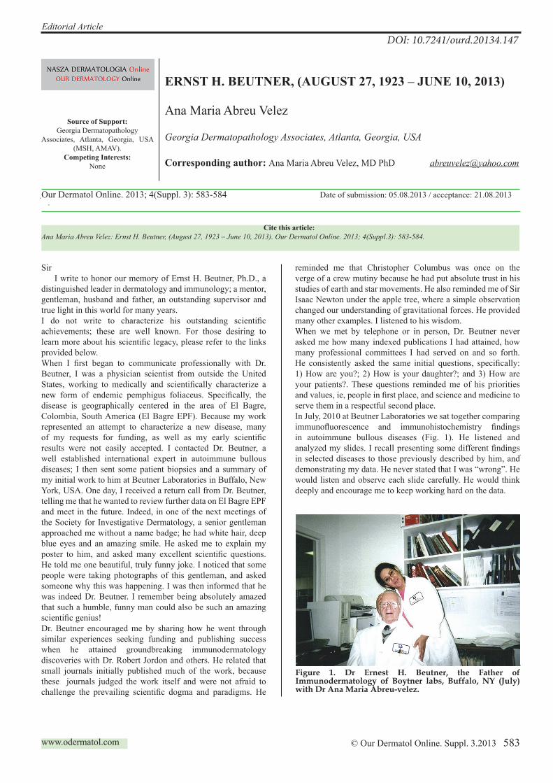
and IIF patterns with statistical significance in comparison to controls run with similar markers (p values of less than 0.05). Our summarized results shown in Table I. In addition to the classical patterns of reactivity appreciated by DIF and IIF, multiple additional patterns of positivity were seen. The most common one featured positive staining around the upper dermal blood vessels in the ABD patients, again with statistical significance in comparison to controls (p = 0.0001). IgG, IgM, Complement/C3c, Complement/C1q and fibrinogen were the most common positive markers detected in this pattern. Most of the ABDs were positive in this pattern for more than 3 markers at a time. In BP, we also observed strong perivascular positivity in the intermediate and deep dermis with similar markers as those described above (Fig. 1-4). In BP, PV, El Bagre-EPF and PF, we noted strong positivity to neurovascular structures feeding skin appendageal structures, especially in eccrine glands and pilosebaceous units. In PV, this phenomenon was also observed in neurovascular packages supplying and surrounding dermal apocrine glands; on H&E review, these glands were also noted to be edematous with acantholysis-like features (p<0.05) (Fig. 1-4). We classified our findings as negative (-), weakly positive (+), positive (+++) and strongly positive (++++).Positivity was also observed in most ABDs in what seemed to be cell junction-like structures between endothelial cells in the dermis and the surrounding mesenchymal extracellular matrix (p<0.05) (Fig. 1-4). The degree of positivity varied depending of the size of the vessels and their relative deepness in the dermis (Tabl. I). In BP, such those that seems to be in the junctions between the dermal cells junctions with stronger positivity in the middle of the dermis. The normal controls did not show this staining, with exception of deposits of IgG, IgM and albumin in the dermis; however, this staining was weaker and without any specific pattern. In several active clinical cases of El Bagre-EPF, we also noted positive staining to some kind of cell junction-like structures in piloerector muscles with both IgG and IgM.Notably, the reactivity against the apocrine glands and the H&E alterations of edema and acanthoytic-like changes may be related with the fact that PV has a vegetant clinical form, with lesions anatomically present in areas where these glands predominate.In Table I and Figures 1 through 4, we summarize our primary results.
586 © Our Dermatol Online. Suppl. 3.2013

Ant
ibod
yB
P n
=34
PV n
=14
PF n
=4D
H n
=10
El B
agre
-EPF
n=3
0C
ontr
ols
from
E
nd
emic
ar
ean=
30
Skin
pl
astic
Surg
ery
cont
rols
n=
15Ig
APo
sitiv
e ar
ound
som
e de
rmal
bl
ood
vess
els.
Som
e ep
ider
mal
ke
ratin
ocyt
es sh
owed
som
e cy
topl
asm
ic st
aini
ng in
seve
ral
biop
sies
(+).
Som
e po
sitiv
e ce
lls d
ebris
insi
de
the
blis
ter.
Som
e po
sitiv
e bl
ood
vess
els i
n th
e up
per d
erm
is a
nd
som
e in
divi
dual
cel
ls in
the
uppe
r de
rmis
. Als
o po
sitiv
e on
blo
od
vess
els a
roun
d th
e ec
crin
e gl
ands
an
d on
ves
sels
of t
he se
ptae
of
subc
utan
eous
adi
pose
tiss
ue.
Som
e sm
all d
erm
al
bloo
dves
sels
pos
itive
. A
lso,
pos
itive
on
som
e sm
all b
lood
ves
sels
arou
nd
eccr
ine
swea
t gla
nds (
+).
Posi
tive
belo
w
sube
pide
rmal
blis
ters
, m
ainl
y in
the
papi
llary
de
rmis
(+++
). Po
sitiv
e in
som
e lin
ear a
reas
in
the
epid
erm
al st
ratu
m
corn
eum
(++)
. Als
o po
sitiv
e in
som
e ar
eas o
f th
e in
trace
llula
r mat
rix
bund
les (
++).
Som
e po
sitiv
ity in
neu
rova
scul
ar
pack
ages
feed
ing
seba
ceou
s gla
nds (
++).
Som
e pa
tient
s with
pos
itive
stai
ning
aro
und
uppe
r and
inte
rmed
iate
der
mal
blo
od v
esse
ls
(++)
.
Neg
ativ
e.N
egat
ive.
IgG
BM
Z lin
ear p
ositi
vity
on
roof
s an
d flo
ors o
f the
blis
ters
, with
m
ore
posi
tivity
on
the
roof
s (3
4/34
). So
me
area
s of t
he
epid
erm
is sh
owed
pe
ricyt
opla
smic
stai
ning
in
kera
tinoc
ytes
(22/
34).
So
met
imes
pos
itive
per
inuc
lear
st
aini
ng a
s wel
l. Po
sitiv
e st
aini
ng a
lso
note
d ar
ound
so
me
smal
l der
mal
blo
od
vess
els.
Som
e ar
eas o
f the
pa
pilla
ry d
erm
is e
xtra
cellu
lar
mat
rix a
nd u
pper
der
mis
sh
owed
reac
tivity
(+++
). Se
vera
l fibr
obla
stoi
d ce
lls
posi
tive
thro
ugh
the
entir
e de
rmis
. Man
y de
ep n
erve
s po
sitiv
e in
the
epin
euria
.
ICS,
som
e up
per a
nd a
roun
d se
vera
l neu
rova
scul
ar v
esse
ls.
Posi
tive
arou
nd so
me
vess
els
arou
nd th
e sw
eat g
land
s upp
er
derm
is a
nd sm
all v
esse
ls in
the
sept
aes o
f the
fatty
tiss
ue (+
+).
Posi
tive
som
e ex
trace
llual
r mat
rix
fiber
s esp
ecia
lly in
the
uppe
r and
in
term
edia
te d
erm
is. P
ositi
ve a
lso
som
e sm
all v
esse
ls a
roun
d th
e sw
eat g
land
s are
a (+
).
ICS
betw
een
epid
erm
al
kera
tinoc
ytes
, mos
tly in
up
per l
ayer
s. Po
sitiv
e st
aini
ng o
f som
e sm
all
uppe
r der
mal
blo
od
vess
els.
Posi
tive
stai
ning
ar
ound
som
e de
rmal
blo
od
vess
els a
nd e
ccrin
e gl
ands
(+
+).
Sim
ilar d
istri
butio
n as
IgA
; als
o in
seve
ral
vess
els.
Posi
tive
in se
vera
l cas
es in
epi
derm
al st
ratu
m
corn
eum
(++)
. Als
o, so
me
intra
cyto
plas
mic
po
sitiv
e st
aini
ng w
ithin
epi
derm
al
kera
tinoc
ytes
. Pos
itive
ICS,
mai
nly
in
epid
erm
al st
ratu
m g
ranu
losu
m in
acu
te a
nd
rela
psin
g ca
ses.
Posi
tive
in se
vera
l cas
es o
n up
per d
erm
al b
lood
ves
sels
(++)
. Chr
onic
cas
es
dem
onst
rate
d so
me
posi
tive
stai
ning
aga
inst
m
esen
chym
al-e
ndot
helia
l cel
l jun
ctio
n-lik
e st
ruct
ures
in th
e de
rmis
, as w
ell a
s aro
und
som
e de
rmal
ecc
rine
glan
ds.
Neg
ativ
e.N
egat
ive.
IgM
Som
e ar
eas o
f IC
S-lik
e st
aini
ng
and
som
e ar
eas o
f pe
ricyt
opla
smic
stai
ning
in
epi
derm
al k
erat
inoc
ytes
(2
1/34
). A
lso,
pos
itive
stai
ning
ar
ound
upp
er b
lood
ves
sels
and
th
e de
rmal
ext
race
llula
r mat
rix
(+++
). Th
e pa
ttern
of t
his
imm
unog
lobu
lin is
ver
y si
mila
r to
that
seen
with
IgG
.
Som
e ep
ider
mal
subc
orne
al
reac
tivity
in se
vera
l are
as. F
ocal
ep
ider
mal
ICS
stai
ning
is n
oted
in
seve
ral s
pots
. Pos
itive
stai
ning
al
so n
oted
aro
und
seve
ral d
erm
al
bloo
d ve
ssel
s in
the
derm
is,
arou
nd so
me
conn
ectiv
e tis
sue
and
som
e de
ep n
euro
vasc
ular
tiss
ue
arou
nd e
ccrin
e sw
eat g
land
s, se
bace
ous g
land
s and
adi
pose
se
ptae
(+++
).
Posi
tive
ICS
betw
een
epid
erm
al k
erat
inoc
ytes
, m
ostly
in u
pper
laye
rs a
nd
posi
tive
arou
nd so
me
smal
l up
per d
erm
al b
lood
ve
ssel
s. A
lso
posi
tive
arou
nd so
me
bloo
d ve
ssel
s ar
ound
ecc
rine
glan
ds
(++)
. Po
sitiv
e ar
ound
se
bace
ous a
nd e
ccrin
e gl
and
neur
ovas
cula
r su
pplie
s.
Sim
ilar d
istri
butio
n as
w
ith Ig
A. I
n so
me
biop
sies
, som
e in
terc
ellu
lar k
erat
inoc
yte
stai
ning
was
obs
erve
d.
Als
o, so
me
rein
forc
emen
t w
as n
oted
aro
und
hair
folli
cles
.
Som
e ca
ses d
ispl
ayed
spot
ty p
ositi
ve st
aini
ng
in th
e ep
ider
mal
cor
neal
laye
r. So
me
epid
erm
al
kera
tinoc
ytic
ICS
in se
vera
l cas
es (+
+). S
ome
epid
erm
al k
erat
inoc
yte
spot
ty p
eric
ytop
lasm
ic
posi
tive
stai
ning
in in
seve
ral a
reas
of t
he
epid
erm
is, a
nd so
me
BM
Z st
aini
ng. I
n m
ost
chro
nic
case
s, po
sitiv
e st
aini
ng in
a b
and-
like
dist
ribut
ion
in th
e up
per d
erm
is a
nd/o
r in
term
edia
te d
erm
is, i
nclu
ding
on
bloo
d ve
ssel
s (+
+). R
einf
orce
men
t of t
he m
esen
chym
al-
endo
thel
ial c
ell j
unct
ion-
like
stru
ctur
es a
nd
cells
and
telo
cyte
- lik
e st
ruct
ures
als
o se
en.
Neg
ativ
eN
egat
ive
Tabl
e I.
Cel
l pop
ulat
ions
and
mar
kers
in le
sion
al s
kin
from
mul
tiple
aut
oim
mun
e sk
in d
isea
ses.
© Our Dermatol Online. Suppl. 3.2013 587

588 © Our Dermatol Online. Suppl. 3.2013
DiscussionThe purpose of our study was the immunophenotypic
characterization of inflammatory cells and the expression of adhesion molecules in lesional and perilesional skin of patients with ABD. ABD have been classically defined as B cell-mediated diseases due to the presence of both 1) spontaneously appearing, intraepidermal clinical blisters after injection of human sera into neonatal mice, and 2) epidermal-specific autoantibodies whose serum titers classically correlate with
clinical activity and disease severity as demonstrated by IIF and ELISA [1-4,20]. Our in situ results document the involvement of other immune cells in ABD lesional skin, indicating that other immune system components may be important in these diseases [21-27]. In our study, we did not attempt to demonstrate a precise pathogenic role of these molecules; however, further investigation is warranted in this area.
Ant
ibod
yB
P n
=34
PV n
=14
PF n
=4D
H n
=10
El B
agre
-EPF
n=3
0C
ontr
ols
from
E
nd
emic
ar
ean=
30
Skin
pl
astic
Surg
ery
cont
rols
n=
15Ig
ELi
near
on
both
floo
rs a
nd
roof
s of b
liste
rs in
man
y ca
ses
(23/
34).
Posi
tive
(+++
) in
tracy
topl
asm
ic, p
erin
ucle
ar
stai
ning
in so
me
epid
erm
al
kera
tinoc
ytes
.(26/
34).
Posi
tive
stai
ning
on
som
e in
divi
dual
ce
lls in
der
mis
, and
foca
lly
arou
nd u
pper
der
mal
blo
od
vess
els (
++).
Posi
tive
nucl
ear a
nd fo
cal
cyto
plas
mic
stai
n in
g in
epi
derm
al
kera
tinoc
ytes
. Als
o, p
ositi
ve o
n se
vera
l lar
ge c
ells
in th
e up
per
derm
al in
flam
mat
ory
infil
trate
. Po
sitiv
e st
aini
ng o
f som
e ve
ssel
s ar
ound
ecc
rine
glan
ds. (
++).
Posi
tive
nucl
ear a
nd fo
cal
cyto
plas
mic
stai
ning
in
epid
erm
al k
erat
inoc
ytes
A
lso
posi
tive
stai
ning
on
seve
ral l
arge
cel
ls in
the
uppe
r der
mal
infla
mm
ator
y in
filtra
te. P
ositi
ve st
aini
ng
of so
me
vess
els a
roun
d th
e ec
crin
e gl
ands
(++)
.
Mos
t cas
es w
ere
nega
tive.
So
me
posi
tive
stai
ning
in th
e up
per d
erm
al in
flam
mat
ory
infil
trate
. Tw
o ca
ses w
ere
posi
tive
in se
bace
ous g
land
s, in
pla
smac
ytoi
d ce
lls in
the
derm
is a
nd a
roun
d ec
crin
e gl
ands
.
Seve
ral c
ases
, bot
h ac
ute
and
chro
nic,
di
spla
yed
posi
tive
stai
ning
on
indi
vidu
al
cells
, mai
nly
arou
nd th
e up
per d
erm
al
bloo
d ve
ssel
s (++
). M
esen
chym
al-
endo
thel
ial c
ell j
unct
ion-
like
stru
ctur
es a
nd te
locy
te-li
ke st
ruct
ure
posi
tive
stai
ning
was
als
o no
ted,
as w
ell
as a
roun
d so
me
derm
al e
ccrin
e gl
ands
.
Neg
ativ
e.N
egat
ive.
IgD
Posi
tive
linea
r sta
inin
g in
se
vera
l bio
psie
s alo
ng th
e B
MZ,
mos
tly o
n bl
iste
r floo
rs.
Posi
tive
on se
vera
l fib
robl
asto
id c
ells
in th
e de
rmis
. Pos
itive
aro
und
seve
ral
derm
al b
lood
ves
sels
; sup
er-
ficia
l, in
term
edia
te a
nd d
eep
(+++
) (20
/34)
.
Som
e po
sitiv
e st
aini
ng o
n in
divi
dual
larg
e ce
lls in
the
uppe
r de
rmal
per
ivas
cula
r infl
amm
ator
y in
filtra
te, a
nd o
n so
me
of th
e up
per
derm
al b
lood
ves
sels
(+).
Som
e
epid
erm
al IC
S po
sitiv
ity a
nd so
me
posi
tive
stai
ning
on
cells
insi
de th
e bl
iste
rs.
Posi
tive
in se
vera
l upp
er
derm
al sm
all b
lood
ve
ssel
s, an
d on
som
e bl
ood
vess
els a
roun
d ec
crin
e gl
ands
. Som
e sc
atte
red
stai
ning
aro
und
very
ac
tivel
y in
flam
mat
ory
epid
erm
al b
liste
rs (+
).
Seve
ral c
ases
follo
wed
the
sam
e pa
ttern
as I
gA,
incl
udin
g th
e po
sitiv
ity in
the
derm
al p
apill
ae, a
nd b
lood
ve
ssel
s. Tw
o ca
ses w
ere
posi
tive
in th
e se
bace
ous
glan
ds, i
n pl
asm
acyt
oid
cells
in
the
derm
is a
nd a
roun
d th
e ec
crin
e gl
ands
.
Posi
tive
stai
ning
pat
tern
follo
wed
the
dist
ribut
ion
of th
e st
rong
er
imm
unog
lobu
lins,
incl
udin
g so
me
epid
erm
al k
erat
inoc
ytic
intra
cyto
plas
mic
po
sitiv
e st
aini
ng, a
nd so
me
stai
ning
on
uppe
r der
mal
blo
od v
esse
ls. I
n so
me
case
s, so
me
spot
ty p
ositi
vity
alo
ng th
e B
MZ
and
som
e st
aini
ng a
roun
d se
lect
ed
eccr
ine
glan
d du
cts.
Neg
ativ
e.N
egat
ive.
Com
plem
ent/
C3c
Posi
tive
linea
r dep
osits
at
blis
ter s
plits
, prim
arily
on
blis
ter r
oofs
but
als
o so
me
on
blis
ter fl
oors
(+++
) in
34/3
4 ca
ses.
Als
o ar
ound
seve
ral
smal
l and
larg
e de
rmal
ne
urov
ascu
lar p
acka
ges (
+++)
. Po
sitiv
e st
aini
ng a
lso
pres
ent
arou
nd e
ccrin
e du
cts a
nd B
MZ
of e
ccrin
e du
cts,
as w
ell a
s on
bloo
d ve
ssel
s aro
und
the
hair
folli
cles
(++)
. Som
e re
activ
ity
also
seen
in th
e up
per
epid
erm
al c
orne
al la
yer (
+).
Epid
erm
al IC
S, a
nd st
aini
ng o
n so
me
uppe
r der
mal
blo
od v
esse
ls
and
seve
ral d
erm
al n
euro
vasc
ular
pa
ckag
es. S
ome
posi
tivity
in fo
cal
area
s of t
he e
pide
rmal
cor
neal
la
yer.
Als
o po
sitiv
e st
aini
ng o
n so
me
fibro
blas
toid
cel
ls in
the
derm
is. P
ositi
ve fo
cal s
tain
ing
on
BM
Zs o
f the
seba
ceou
s gla
nds
and
on th
eir n
euro
vasc
ular
supp
ly
pack
ages
. Pos
itive
stai
ning
on
som
e sm
all b
lood
ves
sels
in th
e de
ep c
onne
ctiv
e tis
sue
(++)
.
Posi
tive
stai
ning
on
seve
ral
bloo
d ve
ssel
s in
the
uppe
r an
d in
term
edia
te d
erm
al
plex
us. P
ositi
ve in
som
e sm
all b
lood
ves
sels
in th
e de
ep c
onne
ctiv
e tis
sue.
Po
sitiv
e ar
ound
ne
urov
ascu
lar p
acka
ges
of d
erm
al se
bace
ous a
nd
eccr
ine
glan
ds (+
+).
Posi
tive
depo
sits
in th
e up
per
and
low
er d
erm
al ti
ssue
(+
++).
Posi
tivity
als
o no
ted
in th
e ep
ider
mal
cor
neal
la
yer.
Som
e ep
ider
mal
ke
ratin
ocyt
e IC
S an
d/or
cy
topl
asm
ic st
aini
ng.
Stai
ning
in th
e ex
trace
llula
r m
atrix
and
aro
und
eccr
ine
glan
ds a
nd d
ucts
. Mul
tiple
fib
robl
asto
id c
ells
wer
e po
sitiv
e in
the
derm
is.
Som
e sp
otty
pos
itive
stai
ning
on
the
epid
erm
al c
orne
al la
yer.
Som
e ep
ider
mal
pos
itive
ICS
in se
vera
l cas
es
(++)
. BM
Z st
aini
ng a
lso
in se
vera
l cas
es
(++)
. Po
sitiv
e st
aini
ng in
upp
er d
erm
is
and
on n
euro
vasc
ular
pac
kage
s of a
ll sk
in
appe
ndag
eal s
truct
ures
(++)
. Pos
itive
st
aini
ng in
mul
tiple
cas
es o
n m
esen
chym
al-e
ndot
helia
l cel
l ju
nctio
n-lik
e st
ruct
ures
, and
on
telo
cyte
-like
stru
ctur
es.
Neg
ativ
e.N
egat
ive.
Com
plem
ent/
C3d
Posi
tive
arou
nd se
vera
l sm
all
and
larg
e de
rmal
blo
od
vess
els.
Posi
tive
linea
r BM
Z st
aini
ng o
n bl
iste
r floo
rs a
nd
roof
s (++
+).
Posi
tive
arou
nd se
vera
l upp
er
derm
al b
lood
ves
sels
. Som
e ep
ider
mal
ICS,
pos
itive
in fo
cal
area
s (++
). Po
sitiv
e st
aini
ng in
side
th
e bl
iste
rs. P
ositi
ve st
aini
ng
arou
nd n
euro
vasc
ular
supp
lies o
f pi
lose
bace
ous g
land
s uni
ts. S
ome
extra
cellu
lar m
atrix
stai
ning
, po
sitiv
e in
the
inte
rmed
iate
de
rmis
.
Posi
tive
arou
nd se
vera
l up
per d
erm
al b
lood
ve
ssel
s. So
me
epid
erm
al
ICS,
pos
itive
in fe
w a
reas
(+
+). P
ositi
ve st
aini
ng
arou
nd d
erm
al se
bace
ous
and
eccr
ine
glan
d ne
urov
ascu
lar s
uppl
ies.
In so
me
biop
sies
, sta
inin
g fo
llow
ed th
e pa
ttern
of
posi
tivity
of I
gA, a
lthou
gh
with
wea
ker i
nten
sity
.
Posi
tive
in th
e m
ajor
ity o
f the
cas
es in
m
ost v
esse
ls in
der
mis
.N
egat
ive.
Neg
ativ
e.
Tabl
e I.
Cel
l pop
ulat
ions
and
mar
kers
in le
sion
al s
kin
from
mul
tiple
aut
oim
mun
e sk
in d
isea
ses
(con
tinue
d).

© Our Dermatol Online. Suppl. 3.2013 589
Ant
ibod
yB
P n
=34
PV n
=14
PF n
=4D
H n
=10
El B
agre
-EPF
n=3
0C
ontr
ols
from
E
nd
emic
ar
ean=
30
Skin
pl
astic
Surg
ery
con
trol
s n=
15
Com
plem
ent/
C1q
Posi
tive
linea
r BM
Z st
aini
ng
on b
oth
side
s of t
he b
liste
rs,
but p
rimar
ily o
n bl
iste
r roo
fs.
Posi
tive
stai
ning
on
foca
l ex
trace
llula
r mat
rix fi
bers
, in
bund
les i
n th
e up
per,
inte
rmed
iate
and
dee
p de
rmis
(+
+). A
lso
posi
tive
derm
al
stai
ning
on
the
uppe
r ne
urov
ascu
lar p
lexu
s (++
). So
me
foca
l are
as o
f the
ep
ider
mis
with
cyt
opla
smic
st
aini
ng o
f the
ker
atin
ocyt
es.
Posi
tive
stai
ning
on
blis
ter fl
oors
, an
d so
me
insi
de b
liste
rs. S
ome
stai
ning
in th
e up
per d
erm
al
extra
cellu
lar m
atrix
. Pos
itive
stai
ning
on
der
mal
blo
od v
esse
ls, s
ebac
e-ou
s gla
nds n
euro
vasc
ular
supp
ly
pack
ages
(++)
. Sev
eral
BM
Zs o
f hai
r fo
llicl
es a
nd se
bace
ous g
land
s wer
e al
so p
atch
y po
sitiv
e. P
ositi
ve
stai
ning
aro
und
derm
al e
ccrin
e gl
ands
and
duc
ts.
Posi
tive
stai
ning
aro
und
pilo
seba
ceou
s uni
ts (+
+). P
ositi
ve
stai
ning
aro
und
seve
ral u
pper
de
rmal
blo
od v
esse
ls. S
ome
foca
l ep
ider
mal
ICS
posi
tive
stai
ning
(+
+). P
ositi
ve st
aini
ng a
roun
d de
r-m
al se
bace
ous a
nd e
ccrin
e gl
and
neur
ovas
cula
r sup
plie
s.
Posi
tive
stai
ning
at t
he
sube
pide
rmal
blis
ter fl
oor,
and
in th
e pa
pilla
ry d
erm
al a
reas
(+
++).
Som
e fo
cal e
pide
rmal
co
rnea
l lay
er re
activ
ity. T
he
derm
al e
xtra
cellu
lar m
atrix
was
ve
ry re
activ
e, a
ccen
tuat
ed in
bu
ndle
d gr
oups
(+++
). Po
sitiv
e st
aini
ng a
roun
d so
me
derm
al
neur
ovas
cula
r pac
kage
s. In
su
mm
ary,
the
stai
ning
pat
tern
fo
llow
ed a
sim
ilar p
atte
rn to
th
at o
f IgA
.
Posi
tive
seve
ral e
xtra
cellu
lar
mat
rix fi
bers
, mos
tly
inte
rmed
iate
one
s. Po
sitiv
e ce
lls-ju
nctio
n lik
e of
the
pilo
erec
tor m
uscl
e. P
ositi
ve
arou
nd sw
eat g
land
s ves
sels
.
Neg
ativ
e.N
egat
ive.
Kap
pa li
ght
chai
nsPo
sitiv
e st
aini
ng o
n bl
iste
r flo
ors a
nd ro
ofs,
but m
ore
prom
inen
t on
the
floor
s (++
). Po
sitiv
e st
aini
ng in
mos
t are
as
of th
e de
rmis
(++)
.
Posi
tive
epid
erm
al IC
S, o
n up
per
derm
al b
lood
ves
sels
and
aro
und
eccr
ine
duct
s (++
).
Posi
tive
stai
ning
in th
e ep
ider
mal
co
rnea
l lay
er. P
ositi
ve
epid
erm
al IC
S, a
nd p
ositi
ve
stai
ning
on
uppe
r der
mal
blo
od
vess
els a
nd a
roun
d de
rmal
ecc
rine
duct
s (++
). Po
sitiv
e st
aini
ng
arou
nd th
e se
bace
ous a
nd sw
eat
glan
d ne
urov
ascu
lar s
uppl
ies.
Sim
ilar d
istri
butio
n as
IgA
.Fo
llow
s sam
e pa
ttern
than
IgG
an
d Ig
M c
ombi
ned.
Neg
ativ
e.N
egat
ive.
Lam
bda
light
ch
ains
Posi
tive
stai
ning
on
blis
ter
floor
s and
roof
s, bu
t mor
e pr
omin
ent o
n th
e flo
ors (
++).
Posi
tive
stai
ning
in m
ost a
reas
of
the
derm
is (+
+).
Posi
tive
epid
erm
al IC
S, o
n up
per
derm
al b
lood
ves
sels
and
aro
und
eccr
ine
duct
s. (+
+).
Posi
tive
in th
e ep
ider
mal
cor
neal
la
yer.
Posi
tive
epid
erm
al IC
S, a
nd
posi
tive
stai
ning
on
uppe
r der
mal
bl
ood
vess
els a
nd a
roun
d de
rmal
ec
crin
e du
cts (
++).
Posi
tive
stai
ning
aro
und
the
seba
ceou
s an
d sw
eat g
land
neu
rova
scul
ar
supp
lies.
Sim
ilar d
istri
butio
n as
IgA
.Fo
llow
s sam
e pa
ttern
than
IgG
an
d Ig
M c
ombi
ned.
Neg
ativ
e.N
egat
ive.
Tabl
e I.
Cel
l pop
ulat
ions
and
mar
kers
in le
sion
al s
kin
from
mul
tiple
aut
oim
mun
e sk
in d
isea
ses
(con
tinue
d).

Ant
ibod
yB
P n
=34
PV n
=14
PF n
=4D
H n
=10
El B
agre
-EPF
n=3
0C
ontr
ols
from
E
nd
emic
ar
ean=
30
Skin
pl
astic
Surg
ery
cont
rols
n=
15Fi
brin
ogen
Posi
tive
stai
ning
in th
e ep
ider
mal
cor
neal
laye
r, IC
S in
ep
ider
mal
stra
tum
spin
osum
, on
upp
er a
nd in
term
edia
te
derm
al b
lood
ves
sels
and
ar
ound
der
mal
ecc
rine
glan
ds
Posi
tive,
stro
ng b
and-
like
stai
ning
thro
ugho
ut th
e pa
pilla
ry d
erm
is (+
++).
Als
o,
som
e po
sitiv
ity in
the
deep
de
rmal
ext
race
llula
r mat
rix.
Posi
tive
stai
ning
aro
und
smal
l de
rmal
blo
od v
esse
ls, i
nclu
ding
th
ose
asso
ciat
ed w
ith e
ccrin
e gl
ands
. Ess
entia
lly, v
ery
sim
ilar
to Ig
G.
Posi
tive
epid
erm
al IC
S in
stra
tum
sp
inos
um. S
ome
posi
tive
stai
ning
in
side
dis
ease
blis
ters
. Pos
itive
st
aini
ng o
n se
vera
l sm
all b
lood
ve
ssel
s in
the
derm
is, a
nd a
roun
d so
me
derm
al c
onne
ctiv
e tis
sues
. Po
sitiv
e st
aini
ng in
the
epid
erm
al
subc
orne
al a
rea.
Pos
itive
stai
ning
on
som
e de
ep d
erm
al la
rge
nerv
es
arou
nd th
e ec
crin
e gl
ands
Pos
itive
st
rong
ban
d-lik
e st
aini
ng in
the
derm
al e
xtra
cellu
lar m
atrix
in se
vera
l ar
eas,
and
on se
vera
l der
mal
neu
ro-
vasc
ular
ple
xus s
truct
ures
(++)
and
de
ep d
erm
al, s
mal
l blo
od v
esse
ls.
Posi
tive
stai
ning
aro
und
derm
al
eccr
ine
glan
ds. P
ositi
ve st
aini
ng a
lso
note
d ar
ound
som
e su
bcut
aneo
us
adip
ose
tissu
e se
ptae
. Pos
itive
st
aini
ng o
n th
e B
MZs
in so
me
area
s of
the
seba
ceou
s gla
nds.
Posi
tive
stai
ning
in th
e ep
ider
mal
cor
neal
laye
r, ep
ider
mal
ICS,
on
uppe
r and
in
term
edia
te d
erm
al b
lood
ve
ssel
s and
aro
und
derm
al
eccr
ine
glan
ds P
ositi
ve st
rong
ba
nd-li
ke st
aini
ng th
roug
hout
th
e pa
pilla
ry d
erm
is (+
++).
Posi
tive
stai
ning
aro
und
derm
al se
bace
ous a
nd e
ccrin
e gl
and
neur
ovas
cula
r sup
plie
s.
Sim
ilar p
ositi
ve st
aini
ng
dist
ribut
ion
as Ig
A. P
ositi
ve
stai
ning
on
neur
ovas
cula
r su
pply
stru
ctur
es o
f der
mal
se
bace
ous g
land
s (++
).
Som
e sp
otty
pos
itive
stai
ning
on
the
epid
erm
al c
orne
al la
yer
in se
vera
l cas
es (+
). So
me
BM
Z st
aini
ng. P
ositi
ve st
aini
ng o
n up
per
derm
al b
lood
ves
sels
(++)
. Pos
itive
st
aini
ng o
n de
rmal
mes
ench
ymal
- en
doth
elia
l cel
l jun
ctio
n-lik
e st
ruct
ures
and
telo
cyte
-like
st
ruct
ures
. Pos
itive
stai
ning
on
cell-
-junc
tion
like
stru
ctur
es in
der
mal
pi
loer
ecto
r mus
cles
. Pos
itive
sta-
inin
g ar
ound
der
mal
blo
od v
esse
ls
supp
lyin
g de
rmal
ecc
rine
glan
ds
Neg
ativ
e.N
egat
ive.
Alb
umin
Posi
tive,
stro
ng b
and-
like
stai
ning
not
ed in
the
papi
llary
de
rmis
and
aro
und
derm
al
vess
els a
nd e
ccrin
e gl
ands
(+
+++)
. Als
o, p
ositi
ve st
aini
ng
on th
e de
ep d
erm
al
extra
cellu
lar m
atrix
.
Posi
tive
stai
ning
on
the
epid
erm
al
corn
eal l
ayer
. Pos
itive
epi
derm
al
ICS,
and
upp
er a
nd in
term
edia
te
derm
al b
lood
ves
sel s
tain
ing
and
arou
nd d
erm
al e
ccrin
e gl
ands
and
on
e pe
riecc
rine
larg
e ne
rve.
Po
sitiv
e, st
rong
ban
d-lik
e st
aini
ng
thro
ugho
ut th
e pa
pilla
ry a
nd
inte
rmed
iate
der
mis
, sug
gest
ing
a co
mpa
rtmen
taliz
atio
n of
the
over
all
imm
une
resp
onse
(+++
).
Posi
tive
stai
ning
on
the
epid
erm
al c
orne
al la
yer.
Posi
tive
epid
erm
al IC
S, a
nd
posi
tive
stai
ning
on
uppe
r and
in
term
edia
te d
erm
al b
lood
ve
ssel
s and
aro
und
derm
al
eccr
ine
glan
ds P
ositi
ve, s
trong
ba
nd-li
ke st
aini
ng th
roug
hout
th
e pa
pilla
ry d
erm
is (+
+++)
. Po
sitiv
e st
aini
ng a
roun
d de
rmal
seba
ceou
s and
swea
t gl
and
neur
ovas
cula
r sup
plie
s.
Posi
tive
stro
ng b
and-
like
stai
ning
in th
e pa
pilla
ry
derm
is (+
++).
Posi
tive
stai
ning
in so
me
area
s of t
he
epid
erm
al c
orne
al la
yer,
and
on th
e de
rmal
ext
race
llula
r m
atrix
(+++
). So
me
trace
st
aini
ng in
the
epid
erm
is
betw
een
kera
tinoc
ytes
, and
so
me
kera
tinoc
ytic
cyt
opla
-sm
ic st
aini
ng. F
ollo
wed
a
sim
ilar p
atte
rn a
s IgA
, but
w
ith st
rong
er p
ositi
vity
.
Follo
wed
the
sam
e pa
ttern
as
fibrin
ogen
Non
- spe
ci-
fic st
aini
ng
was
pre
sent
in
the
der-
mis
.
Neg
ativ
e.
Tabl
e I.
Cel
l pop
ulat
ions
and
mar
kers
in le
sion
al s
kin
from
mul
tiple
aut
oim
mun
e sk
in d
isea
ses
(con
tinue
d).
590 © Our Dermatol Online. Suppl. 3.2013

Figure 1. a. Classic H&E staining of a DH case, demonstrating a subepidermal blister (black arrow). b IHC, demonstrating positive staining with anti-human IgA antibodies in the blister, (brown staining; red arrow), and weaker, punctate staining in the upper dermis (brown staining; blue arrow). c. DIF of the same patient as in a and b, utilizing FITC conjugated anti-human IgA and showing positive “snow on the mountains” staining in the blister and around upper dermal blood vessels (green staining; yellow arrow); epidermal keratinocyte nuclei were counterstained with Dapi (light blue). d. Classic H&E image of a BP case; note the eosinophils within the upper dermis, subjacent to a disease blister (red arrow). e. Case of BP, with IHC positive linear staining for Complement/C3 at the BMZ (dark staining; red arrows). f. Same BP case as in e, with positive linear IHC staining for IgM at the BMZ. g. BP case, with positive punctate IHC staining for IgE below a disease blister in the dermis(brown staining; red arrow). h. BP case, with positive linear IHC staining for IgG on both sides of a disease blister (brown staining; red arrows). i. The same BP case as in h, highlighting DIF positive staining of FITC conjugated IgG on both sides of a disease blister (green-yellow staining; white arrows). j. A classic case of PV, with classic H&E “tombstone” acantholytic keratinocytes along the epidermal basaloid layer (red arrow). k. A case of PV, demonstrating a positive IHC “chicken wire” pattern of ICS with IgG between epidermal keratinocytes (brown staining; yellow arrow). l. Positive IHC staining for IgG in an epidermal blister in a case of PF (brown staining; black arrow). Please also note the focal epidermal corneal staining.
Cat No. Antibodies (all from Dako) Working dilution Retrieval method (High, Low or Proteinase K(PK))
AR0423 Polyclonal rabbit anti-human IgG Flex ready to use High
IR153 Polyclonal rabbit anti-human IgM Flex ready to use High
IR510 Polyclonal rabbit anti-human IgA. Flex ready to use High
IR517 Polyclonal rabbit anti-human IgD Flex ready to use Low
A0094 Polyclonal rabbit anti-human IgE 1:800 High
A0062 Polyclonal rabbit anti-human Complement/C3c 1:1000 None
A0063 Polyclonal rabbit anti-human Complement/C3d 1:400. PK
A0136 Polyclonal rabbit Complement /C1q 1:100. PK
A0001 Polyclonal rabbit anti-human albumin. 1:13,000 None
A0080 Polyclonal rabbit anti-human fibrinogen 1:1000 PK
IR506 Polyclonal rabbit anti-human kappa light chains Flex ready to use High
IR507 Polyclonal rabbit anti-human lambda light chains Flex ready to use High
Table I - Supplemental. Antibodies utilized, with their respective working parameters.
© Our Dermatol Online. Suppl. 3.2013 591

592 © Our Dermatol Online. Suppl. 3.2013
Figure 2. Atypical IHC staining patterns seen in ABDs. a. Complement/C3c positive IHC staining in the central dermis in a patient with BP, in ATS and/or EMCJ patterns (black arrows). b. Positive IHC staining for Complement/C3d in a BP patient, highlighting positive staining in several upper and intermediate dermal blood vessels (brown staining; black arrows). c. Many BP cases also demonstrated positive IHC IgD staining, in both ATS and EMCJ patterns (brown staining; blue arrows). d. Many BP cases displayed positive, punctate IHC staining for IgD along the BMZ(brown staining; red arrows), but also in the dermis in an EMCJ pattern (brown staining; green arrows). e. BP case, displaying positive IHC staining in a mesenchymal-endothelial junction pattern (MES) with anti-human IgD (brown staining; red arrows). f. In a BP patient biopsy, we utilized IHC vimentin staining to highlight compartmentalization of the inflammatory process. Note the positive linear staining at the BMZ in the floor of a disease blister (brown staining; red arrow), as well as the inflammation and structural reorganization of dermal blood vessels (brown staining; black arrows).
Figure 3. a. A PV case, demonstrating positive IHC staining for fibrinogen (note the strong dark brown staining inside an epidermal blister). In addition, note the positive staining around upper dermal inflamed blood vessels, and some parts of the extracellular matrix; this staining pattern is commonly seen in immunofluorescence and has been traditionally interpreted as autofluorescence of the vessels and dermal matrix fibers. Here, IHC is not a fluorescent method, and the observation of the identical pattern raises the possibility that the DIF staining may be due to real antigenicity not previously characterized. b. Same case of PV, highlighting fibrinogen reactivity in the blister (brown staining; blue arrow); also note the positive staining in the upper dermal neurovascular plexus (brown staining; black arrows). c. In a PF case, we noted positive IHC staining for fibrinogen in both subepidermal and subcorneal blister areas (brown staining; blue arrows), and additional staining around upper dermal blood vessels(brown staining; red arrows). d. In most ABDs, strong positive IHC staining was noted to small and intermediate sized dermal blood vessels, in this case with IgG (brown staining; blue arrow). e. Positive IHC staining for IgG around the neurovascular supply package of a sebaceous gland in a PV case (brown staining; blue arrows). f. Most ABDs also stained positive via IHC for Complement/C3, Complement/C3d, fibrinogen and sometimes IgG around dermal eccrine glands and their ducts; an example of a positive stain is illustrated (brown staining; blue arrows). g. Also, in most ABDs the neurovascular packages supplying skin appendageal structures showed positive staining with the markers documented in f. Sometimes, deep nerves also stained positive, as in this case of PV with fibrinogen (brown staining; blue arrow). h. Positive IHC staining with IgG in a BP patient, directed against either telocytes and/or the EMCJ (brown staining; blue arrow). i. A PV case, staining positive via IHC in a hair follicle for Complement/C3c (brown staining; red arrow). Note also some adjacent blood vessels with positive staining (brown staining; black arrow).

© Our Dermatol Online. Suppl. 3.2013 593
Figure 4. a. A PV case, demonstrating positive IHC staining with an ICS pattern and anti-Complement/C3c antibody between keratinocytes (brown staining; red arrow) as well as around upper dermal blood vessels (brown staining; black arrow)(400X). b. Same case as in a, demonstrating positive staining with Complment/ C3c to neurovascular packages feeding a sebaceous gland (brown staining; black arrow)(400X). c. A BP case, demonstrating positive staining with linear deposits of complement/C3c around a subepidermal blister (brown staining; black arrow) and also in the upper dermis (brown staining; black arrow)(200X). d. A PV case, demonstrating positive staining with IgA against small vessels in the upper dermis brown stain (brown staining; black arrows)(400x). e. A PV case H&E, demonstrating edematous and acantholytic-like changes in apocrine gland cells (400X)(black arrow). f. A PV case, demonstrating positive IHC staining of a neurovascular package around an apocrine glands using anti-human fibrinogen (400X).
DiscussionDIF and IIF have been classically used for the diagnostic
of ABDs; the salt split skin IIF technique was also developed to help to differentiate ABDs. IIF has been the gold standard to determine autoantibody titers that correlate with disease severity, further validated by ELISA testing [1-5]. DIF and IIF of the skin detect several autofluorescence molecules, including molecules in the extracellular matrix, blood vessels, and pigments like lipofucsin, melanin, collagen, indolamine, tryptophan, tyrosine, pyridoxine, folic acid, retinol, collagen, cholecalciferol, riboflavin and NAD(P)H [12]. The majority of these molecules fluoresce in the same UV range as FITC. In contradistinction, IHC techniques often recognize well established internal positive control staining patterns on recognized anatomic structures.In our study, we were able to see an “edge effect” in DIF and IIF of non-specific reinforced staining around the edges of the biopsies. We were also able to determine that all the skin biopsies should be run with a control biopsy of similar age as the biopsy to be tested, especially when using older archival biopsies.Besides the classic staining patterns seen in DIF and IIF and detected by IHC with similar specificity and sensitivity (such ICS and/or BMZ staining), we also observed other patterns that we describe as non-classic patterns. These patterns include positivity to the vessels of the dermal neurovascular packages feeding skin appendageal structures, and positivity to junctions between endothelial cells and the surrounding mesenchymal tissue. Other patterns included positivity to cell junctions within the dermis, and a pattern that we described as a telocyte-like pattern [11]. We didn’t find these patterns in the controls, with
the exception of some even, nonspecific staining of IgG and IgM within the dermis.Other authors have reported studies utilizing IHCs in ABD diagnosis, with similar results to ours in the classical patterns [14-17]. Our non-classic patterns of positivity have been also documented in DIF and IIF, especially when using multicolor and confocal microscopy (not routinely utilized in most immnodermatopathology laboratories). In theory and based on other studies, these autofluoresence molecules should not be detected by IHC [10,17-21]. As noted, besides the classical patterns of reactivity we noticed additional patterns such as UVS. The increased reactivity of dermal blood vessels and/or molecules involved in the transit of inflammatory markers through the dermal blood vessels has been previously shown to possibly play roles in ABDs [19-21].The reactivity of patients with ABDs to dermal blood vessels and/or endothelial cells with strong inflammatory and immune activation markers, including autoantigens to plakophilins 3 and 4 (present in dermal blood vessels) has been also previously reported [23-29].Some authors have shown DH to have evidence of endothelial cell activation in the skin, and systemic manifestations of the ongoing inflammation associated with the mucosal immune response. Endothelial cell activation may play a critical role in the development of skin lesions in patients with DH [27]. Other authors investigated the expression of vascular permeability factor (VPF), that plays an important role in increased vascular permeability and angiogenesis in three bullous diseases; these diseases feature subepidermal blister formation characterized by hyperpermeable dermal microvessels and pronounced papillary dermal edema [28].

The expression of VPF mRNA was strongly up-regulated in the lesional epidermis of BP (n = 3), erythema multiforme (n = 3), and DH (n = 4) as detected by in situ hybridization studies. Epidermal labeling was particularly pronounced over blisters, but strong expression was also noted in areas of the epidermis adjacent to dermal inflammatory infiltrates distant from the blisters. Moreover, the VPF receptors were upregulated in endothelial cells in superficial dermal microvessels. High levels of VPF were detected in blister fluids obtained from patients with BP. The findings described by these authors strongly suggest that VPF plays an important role in the induction of increased microvascular permeability in bullous diseases; leading to papillary edema, fibrin deposition and blister formation in these disorders [28]. Therefore, the positive staining to dermal blood vessels and neurovascular supply packages may be detecting secondary antigens.Previous authors also reported that soluble E-selectin (sE-selectin), an isoform of the cell membrane protein E-selectin (an adhesion molecule synthesized only by endothelial cells) is significantly increased in sera of patients with BP and PV [29]. Other authors recently investigated endothelial cell activation via gene profiling in patients affected by DH (e.g. SELL, SELE genes) that code for cell surface components, specifically members of a family of adhesion/homing receptors that play important roles in lymphocyte-endothelial cell interactions. The gene activation was increased, as well as neutrophil extravasation. The SELL/SELE coded molecules are composed of multiple domains: one homologous to lectins, one to epidermal growth factor, and two to the consensus repeat units found in Complement C3/C4-binding proteins [30]. Our findings of positive reactivity in dermal blood vessels, especially in the upper dermis and neurovascular skin appendices are suggestive of possible antigenicity for these structures in ABDs. Finally, recent studies have shown positive reactivity to eccrine and sebaceous glands in ABDs utilizing IIF and DIF [31-35].In regard to the positive patterns seen in the dermis(possibly associated with cell junctions and/or telocytes), we were able to recently demonstrate activation of both multiple proteases and protease inhibitors following the same patterns of positivity we are describing here [36]. Electron microscopy studies using antibodies and colocalization will help to see if these non-classical patterns of reactivity seen in ABDs are artifacts, or possibly associated with pathological effects in these diseases.
AcknowledgementsJonathan S. Jones, HT (ASCP) at Georgia Dermatopathology
Associates for excellent technical assistance.
REFERENCES
1. Beutner EH, Hale WL, Nisengard RJ, Chorzelski TP, Holubar K: In Immunopathology of the Skin: Labeled Antibody Studies. EH Beutner, TP Chorzelski, SF Bean, RE Jordan, eds. Stroudsburg, Pennsylvania: Dowden, Hutchinson, and Ross, 1973:213-7.2. Jablonska S, Beutner EH, Michel B: Uses for immunofluorescence tests of skin and sera; utilization of immunofluorescence in the diagnosis of bullous diseases, lupus erythematosus, and certain other dermatoses. Arch Dermatol. 1975;111:371-81.3. Mihai S, Sitaru C: Immunopathology and molecular diagnosis of autoimmune bullous diseases. J Cell Mol Med. 2007;11:462-81.
4. Cotell S, Robinson ND, Chan LS: Autoimmune blistering skin diseases. Am J Emerg Med. 2000;18:288-99.5. Beutner EH, Prigenzy LS, Hale W, Leme CA: Immunofluorescence studies of autoantibodies to intercellular areas of epithelia in Brazilian pemphigus foliaceus. Proc Soc Exp Biol Med. 1968;127:81-6.6. Lever WF, Schaumburg-Lever G: Immunosuppressants and prednisone in pemphigus vulgaris: therapeutic results obtained in 63 patients between 1961 and 1975. Arch Dermatol. 1977;113:1236-41.7. Abrèu-Velez AM, Hashimoto T, Bollag WB, Tobón Arroyave S, Abrèu-Velez CE, Londoño ML, et al: A unique form of endemic pemphigus in Northern Colombia. J Am Ac Dermatol. 2003;49:599-608.8. Howard MS, Yepes MM, Maldonado-Estrada JG, Villa-Robles E, Jaramillo A, Botero JH, et al: Broad histopathologic patterns of non-glabrous skin and glabrous skin from patients with a new variant of endemic pemphigus foliaceus-part 1. J Cutan Pathol. 2010;37:222-30.9. Abrèu-Velez AM, Beutner EH, Montoya F, Bollag WB, Hashimoto T: Analyses of autoantigens in a new form of endemic pemphigus foliaceus in Colombia. J Am Acad Dermatol. 2003;49:609-14.10. Abreu-Velez AM, Howard MS, Yi H, Gao W, Hashimoto T, Grossniklaus HE: Neural system antigens are recognized by autoantibodies from patients affected by a new variant of endemic pemphigus foliaceus in Colombia. Clin Immunol. 2011;31:356-68.11. Ceafalan L, Gherghiceanu M, Popescu LM, Simionescu O: Telocytes in human skin--are they involved in skin regeneration? J Cell Mol Med. 2012;16:1405-20.12. Monici M: Cell and tissue autofluorescence research and diagnostic applications. Biotechnol Annu Rev. 2005;11:227-56.13. Lai A, Fat RF, Cormane RH, van Furth R: An immunohistopathological study on the synthesis of immunoglobulins and complement in normal and pathological skin and the adjacent mucous membranes. Br J Dermatol. 1974;90:123-36. 14. Asghar SS, van Joost T, Cormane RH: Comparison of immunofluorescence and immunoperoxidase techniques for detection of tissue antigen. Arch Dermatol Forsch. 1973;248:99-108.15. Chandler W, Zone J, Florell S: C4d immunohistochemical stain is a sensitive method to confirm immunoreactant deposition in formalin-fixed paraffin-embedded tissue in bullous pemphigoid. J Cutan Pathol. 2009;36:655-9.16. Magro CM, Dyrsen ME: The use of C3d and C4d immunohistochemistry on formalin-fixed tissue as a diagnostic adjunct in the assessment of inflammatory skin disease. J Am Acad Dermatol. 2008;59:822-33.17. Zhang X, Hyjek E, Soltani K, Petronic-Rosic V, Shea CR: Immunohistochemistry for Immunoglobulin G4 on Paraffin Sections for the diagnosis of Pemphigus. Arch Pathol Lab Med. 2012;136:1402–7.18. Abreu Velez AM, Yi H, Girard JG, Zhe J, Ramírez MD, Arias LF, et al: Dermatitis herpetiformis bodies and autoantibodies to noncutaneous organs and mitochondria in dermatitis herpetiformis. Our Dermatol Online. 2012; 3: 283-91.19. Abreu Velez, AM, Brown VM, Howard MS: Cytotoxic and antigen presenting cells present and non-basement membrane zone pathology in a case of bullous pemphigoid. Our Dermatol Online, 2012;3:93-9.20. Abreu Velez AM, Zhe J, Howard MS, Dudley SC: Heart immunoreactivity in patients affected by a new variant of endemic pemphigus foliaceus in El-Bagre, Colombia, South America. J Clin Immunol. 2011;31:985-97.21. Abreu Velez AM, Howard MS, Hashimoto T: Palm tissue displaying a polyclonal autoimmune response in patients affected by a new variant of endemic pemphigus foliaceus in Colombia, South America. Eur J Dermatol. 2010;20:74-81.
594 © Our Dermatol Online. Suppl. 3.2013

22. Abreu-Velez AM, Howard MS, Hashimoto K, Hashimoto T: Autoantibodies to sweat glands detected by different methods in serum and in tissue from patients affected by a new variant of endemic pemphigus foliaceus. Arch Dermatol Res. 2009;301:711-8.23. Macadam RF, Vetters JM, Saikia NK: A search for microtubular inclusions in endothelial cells in a variety of skin diseases. Br J Dermatol. 1975;92:175-82.24. Ameglio F, D’Auria L, Cordiali-Fei P, Mussi A, Valenzano L, D’Agosto G, et al: Bullous pemphigoid and pemphigus vulgaris: correlated behaviour of serum VEGF, sE-selectin and TNF-alpha levels. J Biol Regul Homeost Agents. 1997;11:148-53.25. Karashima T, Hachisuka H, Okubo K, Sasai Y: Epidermal keratinocytes of bullous pemphigoid express intercellular adhesion molecule-1 (ICAM-1). J Dermatol. 1992;19:82-6.26. Lambert J, Bracke S, van Roy F, Pas HH, Bonné S, De Schepper S: Br J Dermatol. Serum plakophilin-3 autoreactivity in paraneoplastic pemphigus. 2010;163:630-2.27. Hall RP, Takeuchi F, Benbenisty KM, Streilein RD: Cutaneous endothelial cell activation in normal skin of patients with dermatitis herpetiformis associated with increased serum levels of IL-8, sE-Selectin, and TNF-alpha. J Invest Dermatol. 2006;126:1331-7.28. Brown LF, Harrist TJ, Yeo KT, Ståhle-Bäckdahl M, Jackman RW, Berse B, et al: Increased expression of vascular permeability factor (vascular endothelial growth factor) in bullous pemphigoid, dermatitis herpetiformis, and erythema multiforme. J Invest Dermatol. 1995;104:744-9.29. Ameglio F, D’Auria L, Cordiali-Fei P, Mussi A, Valenzano L, D’Agosto G, et al: Bullous pemphigoid and pemphigus vulgaris: correlated behaviour of serum VEGF, sE-selectin and TNF-alpha levels. J Biol Regul Homeost Agents. 1997;11:148-53.
30. Dolcino M, Cozzani E, Riva S; Parodi A, Tinazzi E, Lunardi C, et al: Gene expression profiling in dermatitis herpetiformis skin lesions. Clin Dev Immunol. 2012;2012:198956. 31. Lehman JS, Camilleri MJ: Diagnostic utility of direct immunofluorescence findings around hair follicles and sweat glands in immunobullous disease. J Cutan Pathol. 2013;40:230-5.32. Abreu Velez AM, Howard MS, Smoller BR: Antibodies to pilosebaceous units, in a new variant of pemphigus foliaceus. Eur J Dermatol. 2011;21:371-5.33. Abreu-Velez AM, Smith JG Jr, Howard MS: IgG/IgE bullous pemphigoid with CD45 lymphocytic reactivity to dermal blood vessels, nerves and eccrine sweat glands. N Am J Med Sci. 2010;2:540-3.34. Lehman JS, Camilleri MJ: Diagnostic utility of direct immunofluorescence findings around hair follicles and sweat glands in immunobullous disease. J Cutan Pathol. 2013;40:230-5.35. Barnadas MA, Curell R, Alomar A, Gelpí C: Paraneoplastic pemphigus with negative direct immunofluorescence in epidermis or mucosa but positive findings in adnexal structures. J Cutan Pathol. 2009;36:34-8.36. Abreu Velez AM, Googe PB, Jiao Z, Duque-Ramírez M, Arias LF, Howard MS: Tissue inhibitor of metalloproteinase 1, matrix metalloproteinase 9, α-1 antitrypsin, metallothionein and urokinase type plasminogen activator receptor in skin biopsies from patients affected by autoimmune blistering diseases. Our Dermatol Online. 2013;4:275-80.
Copyright by Ana Maria Abreu Velez, et al. This is an open access article distributed under the terms of the Creative Commons Attribution License, which permits unrestricted use, distribution, and reproduction in any medium, provided the original author and source are credited.
© Our Dermatol Online. Suppl. 3.2013 595

Original ArticleDOI: 10.7241/ourd.20133.149
Our Dermatol Online. 2013; 4(Suppl. 3): 596-600 Date of submission: 28.05.2013 / acceptance: 06.08.2013
AbstractIntroduction: Pemphigus foliaceus (PF) is endemic in some South American countries, especially in Colombia and Brazil; in Brazil, it is also known as fogo selvagem (FS). We aimed to study the presence of mast cells and the expression of the mast/stem cell growth factor receptor (c-kit/CD117) in PF skin biopsies, as well as the role of IgE in the disease pathogenesis.Methods: Forty-four skin biopsies from patients affected by endemic PF (EPF) (30 patients from El Bagre, Colombia, and 14 from the northeastern region of São Paulo State, Brazil), 48 control biopsies from Colombian and Brazilian endemic areas, and additional control biopsies from none endemic areas in Colombia and the USA non were studied. Immunohistochemistry (IHC) was performed to evaluate skin biopsies with anti-mast cell tryptase (MCT), anti-c-kit and anti-IgE antibodies. We also searched for serum IgE in 30 EPF and 30 non-atopic controls from the El Bagre region via ELISA. In our El Bagre patients and controls, we also searched for IgE in skin samples by direct immunofluorescence. Results: In 100% of the EPF biopsies, MCT, c-kit and IgE were identified with stronger expression relative to control biopsies, especially in the inflammatory infiltrates around upper dermal blood vessels and dermal eccrine glands. IgE staining was positive along the BMZ in some EPF skin samples. The DIF results confirmed a linear deposition of IgE at the BMZ. Increased IgE serum levels were also noted in PF patients relative to controls.. Conclusions: In patients with EPF, the observed increased expression of MCT, c-kit and IgE in lesional skin, associated with higher serum IgE levels may indicate possible IgE participation in the antigenic response.
Key words: autoimmunity; c-kit, immunoglobulin E; mast cell tryptase; endemic pemphigus; autoimmune blistering skin diseases
Abbreviations and acronyms: Endemic pemphigus foliaceus (EPF), El-Bagre endemic pemphigus foliaceus (El Bagre-EPF), direct and indi-rect immunofluorescence (DIF and IIF), fogo selvagem (FS), immunohistochemistry (IHC), mast cell tryptase (MCT), immunoglobulin E (IgE), mast/stem cell growth factor receptor (c-kit/CD117).
Capsule Summary:
· MCT, c-kit and IgE expression in lesional skin is higher in PF patients relative to controls.
· Serum IgE is increased in PF patients relative to controls.· IgE may participate in the PF antigenic response.
www.odermatol.com
Cite this article: Ana Maria Abreu Velez, Ana Maria Roselino, Michael S. Howard: IMast cells, Mast/Stem Cell Growth Factor receptor (c-kit/cd117) and IgE may be integral to the pathogenesis of endemic pemphigus foliaceus. Our Dermatol Online. 2013; 4(Suppl.3): 596-600.
MAST CELLS, MAST/STEM CELL GROWTH FACTOR RECEPTOR (C-KIT/CD117) AND IGE MAY BE INTEGRAL TO THE PATHOGENESIS OF ENDEMIC PEMPHIGUS FOLIACEUS
Ana Maria Abreu Velez1, Ana Maria Roselino2, Michael S. Howard1
1Georgia Dermatopathology Associates, Atlanta, Georgia, USA2Molecular Biology Laboratory, Division of Dermatology, School of Medicine of Ribeirão Preto, University of São Paulo, São Paulo, Brazil
Corresponding author: Ana Maria Abreu Velez, MD PhD [email protected]
Source of Support: Georgia Dermatopathology
Associates, Atlanta, Georgia, USA (MSH, AMAV); Mineros de
Antioquia, Medellin, Colombia, SA (AMAV); the University of
Antioquia, Medellin, Colombia (AMAV); and the Embassy of Japan in Colombia (AMAV). This work was
further supported by FAEPA, Clinical Hospital, School of Medicine of Ribeirão Preto, University of São
Paulo, São Paulo, Brazil (AMR). Competing Interests:
None
596 © Our Dermatol Online. Suppl. 3.2013
Introduction
Endemic pemphigus foliaceus (EPF) is an autoimmune
blistering skin disease presenting in well-defined geographic regions [1-7]. The roles of IgG autoantibodies and complement in lesional EPF skin has been previously documented [1-7].

Based on the fact that EPF occurs in tropical areas, and that previous authors have reported high levels of IgE in serum samples of patients affected by EPF in Brazil and Colombia [8,9], we attempted to confirm the presence of IgE in lesional skin with the purpose of confirming a potential role of IgE in EPF pathogenesis. In addition, we investigated whether mast cells and mast/stem cell growth factor receptor (c-kit/CD117) expression in lesional skin could be associated with EPF autoimmunity. Given previously documented descriptions of IgE in bullous pemphigoid [10,11], we also included skin biopsies from patients with other blistering diseases for comparison. Thus, we tested for 1) mast cell tryptase (MCT) and c-kit/CD117 expression by using immunohistochemistry (IHC), and for 2) IgE via IHC and direct immunofluorescence (DIF) in lesional skin of patients affected by EPF, and in the control biopsies.
Materials and MethodsWe tested 44 paraffinized skin biopsies from patients affected
by EPF (30 patients from El Bagre, Colombia, and 14 from the northeastern region of São Paulo State, Brazil). All EPF patients fulfilled the established criteria for PF in Colombia [4,5] and in Brazil [2,3]. As controls, we utilized 30 skin biopsies from healthy individuals from the El Bagre endemic area (without any history of an atopic disorder). In addition, we tested 3 normal skin biopsies from Brazil, and 15 plastic surgery patients with abdominal or breast reductions from the USA or Colombian non-endemic regions. Our EPF and endemic area control biopsies were taken predominantly from areas on the chest, arms or face. A medication history was also taken, and all cases and controls were required to have had no immunosuppressant medications for at least 4 weeks. The same criteria were utilized for our serological studies. From all these patients we obtained written consents, as well as Institutional Review Board permission.
Immunohistochemistry in paraffinized skin samplesWe utilized monoclonal and polyclonal antibodies from Dako (Carpinteria, California, USA). Specifically, we tested for monoclonal mouse anti-human MCT, catalogue number M7052, dilution 1:250; for polyclonal rabbit anti-human CD117/c-kit, catalogue number A4502, dilution 1:200; and for polyclonal rabbit anti-human IgE, catalogue number A0094, dilution 1:750 as previously described [13]. For our IHC testing, we utilized a dual endogenous peroxidase blockage, with the addition of an Envision dual link (to assist in chromogen adherence). We further utilized the chromogen 3,3-diaminobenzidine (DAB), and counterstained with hematoxylin. The samples were run in a Dako Autostainer Universal Staining System. Positive and negative controls were consistently performed.
Quantitative digital morphometry of staining intensity to obtain precise data on IHC parametersThe staining intensity of all antibodies was evaluated qualitatively by two independent observers, as well as via a semiquantitative approach by an automated computer image analysis system designed to quantify IHC staining in hematoxylin counterstained histologic sections. Slides were scanned with a ScanScope CS system (Aperio Technologies, Vista, California, USA), utilizing brightfield imaging at 200× and 400× magnifications. We then calculated the area of positive signal, divided by the studied area. The intensity of the staining was then classified on a scale
from 0 to 4, where 0 represented negative staining and 4 the strongest staining.
Detection of serum levels of IgEWe tested for serum IgE levels in 30 El Bagre-PF patients and in 30 control individuals from the El Bagre EPF endemic area with a commercial ELISA kit (AlaSTAT Total IgE kit, Diagnostic Products Corporation, Los Angeles, California, USA). We followed the kit instructions, and the positive cutoff level was 138 IU (international units) [8].
Direct immunofluorescence (DIF)For DIF, 4 µm thick skin cryosections were partially fixed with paraformaldehyde, rinsed in phosphate-buffered saline (PBS, pH 6.8), incubated with fluorescein isothiocyanate (FITC) in a solubilization buffer (PBS with 0.5% Triton X-100 Octylphenolpoly (ethyleneglycolether) and then rinsed again. After blocking with PBS with 0.01%-Tween 20 (Polysorbate 20) and 0.5% bovine serum albumin (BSA), the sections were incubated with antiserum for one hour. Goat FITC conjugated anti-human IgE (Epsilon-chain) antiserum was obtained from Vector Laboratories (Burlingame, California, USA). The slides were then counterstained with 4′,6-diamidino-2-phenylindole (DAPI) (Pierce, Rockford, Illinois, USA), washed, coverslipped and dried overnight at 4°C.
Statistical methodsDifferential serum IgE levels between EPF and control samples were analyzed by the Mann-Whitney test (GraphPad Software, San Diego, California, USA). We considered p values ≤0.05 to be statistically significant.
ResultIHC resultsIn our study, we found that MCT and c-kit were Overexpressed, primarily in perivascular infiltrates surrounding the upper dermal neurovascular plexus, as well as around dermal blood vessels supplying sebaceous and eccrine glands. As expected, normal skin demonstrated rare mast cells around these blood vessels. Interestingly, positive IgE staining was noted along the BMZ in some EPF biopsies. In eccrine gland cells, deposits of IgE were predominantly cytoplasmic, and were noted in 70% of the EPF cases. Four controls from the endemic area also stained positive for IgE, in a similar pattern as the EPF cases. Significantly, the EPF cases demonstrated stronger MCT and IgE expression than controls from the endemic and non-endemic areas. In our EPF biopsies, we demonstrated positive epidermal staining in multiple patterns utilizing the anti-IgE antibody. Some staining was intracytoplasmic, and some nuclear; most of the cases (75%) exhibited a combination of these patterns. IgE displayed positive staining within sebaceous gland sebocytes, within the base and isthmus of hair follicular units and within hair follicles. In addition to these patterns, positive staining was also noted between epidermal keratinocytes in approximately sixty (60) per cent of the EPF cases. The controls did not demonstrate these epidermal staining patterns. We also observed strong staining positivity with anti-IgE in several cases on epidermal supracorneal and subcorneal debris. Further, our MCT, c-kit and IgE staining displayed similar distributions; in several cases, the staining was seen subjacent to the basement membrane zone (BMZ) of the epidermis, as well as under the BMZs of dermal eccrine and sebaceous glands.
© Our Dermatol Online. Suppl. 3.2013 597

598 © Our Dermatol Online. Suppl. 3.2013
The MCT, c-kit and IgE deposits were also seen surrounding dermal eccrine glands, blood vessels and nerves.
In Figure 1, we show our IHC staining data for MCT, c-kit and IgE expression, and DIF with FITC conjugated anti-IgE in representative skin biopsies from EPF patients.In Figure 2, we shouw our IgE levels detected by ELISA testing,
with increased levels in the EPF group relative to controls (p<0.0001).Table I summarizes our primary results using IHC, and the patterns of reactivity and intensity of staining of the MCT, c-kit and IgE expression in EPF and in controls. Again, the MCT, c-kit and IgE expression in PF patients displayed similar patterns and intensity.
Figure 1. IHC staining with anti-human MCT, IgE and c-kit, and DIF staining with FITC conjugated anti-human IgE in representative skin biopsies from EPF patients. a. IHC staining showing strong expression of MCT around upper dermal vessels (brown staining; red arrow). b. DIF showing focal linear staining at the dermal-epidermal junction with FITC conjugated IgE (green staining; red arrow). c. IHC staining with anti-IgE displays brown staining in the papillary dermis (brown staining; red arrows), as well as around the upper dermal blood vessels (brown staining; black arrow). d. IHC staining with anti-IgE shows positive staining around a hair follicle unit (brown staining; blue arrows). e. DIF with FITC conjugated IgE displaying a common pattern seen in most of the EPF cases; specifically, the presence of individual large cells (green staining; red arrow) in the dermis. f. IHC staining with c-kit/CD117 positive staining around eccrine ducts (brown staining; red arrow).
Skin biopsies MCT c-kit/CD117 IgE Finding conclusions
Brazilian EPF (n=14) 100% (4+) staining around both the dermal neurovascular plexus, and blood vessels feeding dermal sebaceous and eccrine sweat glands
80% positive staining 80% (4+) staining around both the dermal neurovascular plexus, and blood vessels feeding dermal sebaceous and eccrine glands
All samples showed high expression compared with the physiological baseline
Colombian/El Bagre EPF (n=30)
100% positive (4+) staining around the dermal neurovascular plexus, and blood vessels feeding dermal sebaceous and eccrine glands
80% positive (3+) staining around the dermal neurovascular plexus, and blood vessels feeding dermal sebaceous and eccrine glands
80% positive (4+) around the dermal neurovascular plexus, and blood vessels feeding dermal sebaceous and eccrine glands
All samples showed high expression compared with the physiological baseline
Controls from the endemic area of Colombian/El Bagre EPF (n=30)
4/30 (3+), 7/30 (2+), and all others baseline physiological staining (1+)
4/30 (3+), 6/30 (2+) and all others baseline physiological staining (1+)
2/30 (2+), and all others baseline physiological staining (1+)
Increased positivity relative to plastic surgery controls and/or Brazilian control samples
Normal controls from Brazil (n=3)
Base line physiological staining in all samples (1+)
None None All samples showed baseline physiological staining compared to the patients
Normal controls from plastic surgery reductions in Colombia and the USA (n=15)
Baseline physiological staining in all samples (1+)
None None All samples showed base line physiological staining compared to the patients
Table I. Mast cell tryptase, c-kit/CD117 and IgE immunohistochemistry(IHC) staining results in skin biopsies from endemic pemphigus foliaceus (EPF) patients and controls
Figure 2. IgE levels detected by ELISA in EPF and control groups. The boxes represent 25 to 75 quartile ranges of values, and the horizontal lines inside the boxes the respective medians (p<0.0001).

© Our Dermatol Online. Suppl. 3.2013 599
DiscussionIgE plays a central role in Type I hypersensitivity. In response
to an allergen/antigen, binding of IgE to Fcε receptors occurs on mast cells and basophils, which in turn induces signaling and leads to mast cell degranulation, immune mediator release and expression of c-kit (CD117) [14]. Mast cells reside in most tissues, including the skin, and are occasionally found around dermal blood vessels. We found stronger MCT expression in patients with EPF that in our control group. Previous authors have also demonstrated increased MCT staining in skin biopsies of patients with bullous pemphigoid [15-17]. Mast cells and basophils have several intramembranous receptors for the Fc portion of IgE (FC€RI). Aggregation of two or more of those FC€RI receptors with antigen induces cross-linking between IgE molecules. The resultant IgE-receptor activation induces complex signal transduction, leading to the release of mediators from mast cells and basophils. Pre-formed mediators from mast cell granules are released immediately, and include histamine, neutral proteases and a small number of cytokines and proteoglycans. Mast cell responsiveness can be also increased by complement; in EPF, complement plays an important role [17]. Based on our findings, we speculate that MCT, c-kit and IgE may contribute to dermal blood vessel vasodilatation, with increased permeability and leukocyte extravasation. We also speculate that in EPF, the aggregation of two or more of those FC€RI receptors with cross-linking of receptor-bound IgE molecules leads to receptor activation and complex signal transduction, thus leading to the release of mediators from mast cells and basophils. Serum IgE levels were higher in our EPF patients in comparison with controls, and this also has been shown in other autoimmune blistering diseases [17-20]. More recently, in 2013 Kalantari-Dehaghi et al. reported interesting experiments which confirm mitochondrial damage by autoantibodies in pemphigus vulgaris (PV) [21]. Moreover, in 2011 Zhang et al. investigated the role of mitochondrial dynamics in degranulation of human cultured mast cells during activation by IgE/antigen and substance P, showing that degranulation is accompanied by mitochondrial translocation from a perinuclear location to exocytosis sites. Mitochondrial translocation was also evident in skin mast cells from atopic dermatitis patients [22]. Since both forms of pemphigus (PF and PV) are endemic in Brazil [23], we speculate that common pathogenetic mechanisms may exist for both disorders. Thus, this important evidence [21,22] leads us to speculate regarding mast cells and IgE in EPF, and a mitochondrial role in the pathogenesis of this disorder. Further, it is important to note that in pemphigus and bullous pemphigoid IgG4 autoantibodies predominate, and that IgE versus IgG4 production can be differentially regulated by IL-10. Finally, timing studies (featuring acute versus chronic cases) in EPF may help to address the increased antigenic response evidenced by 1) stronger local immune expression as well as 2) the stronger seric response relative to controls.
REFERENCES
1. Diaz L, Sampaio S, Rivitti E, Martins CR, Cunha PR, Lombardi C, et al: Endemic pemphigus foliaceus (fogo selvagem). Clinical features and immunopathology. J Am Acad Dermatol. 1989;20:657-9.
2. Chiossi MP, Costa RS, Roselino AM: Dermal dendritic cell number correlates with serum autoantibody titers in Brazilian pemphigus foliaceus patients. Braz J Med Biol Res. 2004;37:337-41.3. Chiossi MP, Roselino AM: Endemic Pemphigus foliaceus („Fogo selvagem”): a series from the Northeastern region of the State of São Paulo, Brazil, 1973-1998. Rev Inst Med Trop Sao Paulo. 2001;43:59-62.4. Abréu-Vélez, AM, Beutner EH, Montoya F, Bollag WB, Hashimoto T: Analyses of autoantigens in a new form of endemic pemphigus foliaceus in Colombia. J Am Acad Dermatol. 2003;49:609-14.5. Abreu-Velez, A, Hashimoto T, Bollag W, Tobón Arroyave S, Abreu-Velez CE, Londoño ML, et al: A unique form of endemic pemphigus in Northern Colombia. J Am Acad Dermatol. 2003;49:599-08.6. Ramos W, Chacon GR, Galarza C, Gutierrez EL, Smith ME, Ortega-Loayza AG: Endemic pemphigus in the Peruvian Amazon: epidemiology and risk factors for the development of complications during treatment. An Bras Dermatol. 2012;87:838-45.7. Morini J, Jomaa B, Gorgi Y, Saguem MH, Nouira R, Roujeau JC, et al. Pemphigus foliaceus in young women. An endemic focus in the Sousse area of Tunisia. Arch Dermatol. 1993;129:69-73.8. Abréu-Vélez AM, Warfvinge G, Herrera WL, Abréu-Vélez CE, Montoya MF, Hardy DM, et al: Detection of mercury and other undetermined materials in skin biopsies of endemic pemphigus foliaceus. Am J Dermatopathol. 2003;25:384-91.9. Figueiredo AM, Antunes L, Patrus OA: Hipersensibilidade cutânea mediada por IgE em pacientes com pênfigo endêmico (Fogo selvagem). (Immediate skin hypersensitivity mediated by IgE in patients with fogo selvagem). An Bras Dermatol. 1988;6:47–9.10. Fairley JA, Burnett CT, Fu CL, Larson DL, Fleming MG, Giudice GJ: A pathogenic role for IgE in autoimmunity: bullous pemphigoid IgE reproduces the early phase of lesion development in human skin grafted to nu/nu mice. J. Invest. Dermatol. 2007;127:2605–11.11. Dresow SK, Sitaru C, Recke A, Oostingh GJ, Zillikens D, Gibbs BF: IgE autoantibodies against the intracellular domain of BP180. Br J Dermatol. 2009;160:429-32.12. Howard MS, Yepes MM, Maldonado JG, Villa E, Jaramillo A, Botero J, et al: Broad histopathologic patterns of non-glabrous skin and glabrous skin from patients with a new variant of endemic pemphigus foliaceus (part 1). J. Cutan Pathol. 2010;37:222-30.13. Abreu-Vélez AM, Javier Patiño P, Montoya F, Bollag WB: The tryptic cleavage product of the mature form of the bovine desmoglein 1 ectodomain is one of the antigen moieties immunoprecipitated by all sera from symptomatic patients affected by a new variant of endemic pemphigus. Eur J Dermatol. 2003;13:359-66.14. Eady RA, Cowen T, Marshall TF, Plummer V, Greaves MW: Mast cell population density, blood vessel density and histamine content in normal human skin. Br J Dermatol. 1979;100:623.15. Wintroub BU, Mihm MC Jr, Goetzl EJ, Soter NA, Austen KF: Morphologic and functional evidence for release of mast-cell products in bullous pemphigoid. N Engl J Med. 1978; 23;298:417-21.16. Borrego L, Maynard B, Peterson EA, George T, Iglesias L, Peters MS, et al: Deposition of eosinophil granule proteins precedes blister formation in bullous pemphigoid comparison with neutrophil and mast cell granule proteins. Am J Pathol. 1996;148:897-909.17. Klapholz L, Kupietzky A, Weinrauch L, Shalit M, Okon E: Increased numbers of mast cells in pemphigus vulgaris skin lesions. A histochemical study. Acta Derm Venereol. 1991;71:269–71.18. Bruns GR, Ablin RJ, Guinan PD: Serum immunoglobulin E in pemphigus. J Invest Dermatol. 1978;71:217–8.19. Nagel A, Lang A, Engel D, Podstawa E, Hunzelmann N, de Pita O et al: Clinical activity of pemphigus vulgaris relates to IgE autoantibodies against desmoglein 3. Clin Immunol. 2010;134:320-30.

600 © Our Dermatol Online. Suppl. 3.2013
20. Qian Y, Prisayanh P, Andraca E, Qaqish BF, Aoki V, Hans-Filhio G, et al: IgE, IgM, and IgG4 anti-desmoglein 1 autoantibody profile in endemic pemphigus foliaceus (fogo selvagem). J Invest Dermatol. 2011;131:985-7.21. Kalantari-Dehaghi M, Chen Y, Deng W, Chernyavsky A, Marchenko S, Wang PH, et al: Mechanisms of mitochondrial damage in keratinocytes by pemphigus vulgaris. J Biol Chem. 2013;288:16916-25.22. Zhang B, Alysandratos K-D, Angelidou A, Asadi S, Sismanopoulos N, Delivanis D-A, et al: Human mast cell degranulation and preformed TNF secretion require mitochondrial translocation to exocytosis sites: Relevance to atopic dermatitis. Allergy Clin Immunol. 2011;127:1522–31.
23. Gonçalves GA, Brito MM, Salathiel AM, Ferraz TS, Alves D, Roselino AM: Incidence of pemphigus vulgaris exceeds that of pemphigus foliaceus in a region where pemphigus foliaceus is endemic: analysis of a 21-year historical series. An Bras Dermatol. 2011;86:1109-12.
Copyright by Ana Maria Abreu Velez, et al. This is an open access article distributed under the terms of the Creative Commons Attribution License, which permits unrestricted use, distribution, and reproduction in any medium, provided the original author and source are credited.

Original ArticleDOI: 10.7241/ourd.20134.150
Our Dermatol Online. 2013; 4(Suppl. 3): 601-605 Date of submission: 08.09.2013 / acceptance: 13.10.2013
AbstractIntroduction: Pemphigus are autoimmune blistering diseases that affect the skin and mucous membranes. The blisters characteristics of pemphigus tend to break, causing painful erosions that easily bleed. This study aimed to identify the experiences involved in the triggering of pemphigus and how patients face the illness and treatment. Material and Methods: The study included 31 patients diagnosed with pemphigus foliaceus and vulgaris, under a standardized pulsetherapy treatment. Data collection was through semi-structured interviews, which were audio-recorded, transcribed and analyzed using a qualitative approach. Results: During the journey in search of the phenomenon, were defined two thematic categories, subdivided into eleven subcategories. Months before the triggering of the illness, patients experienced feelings of losses, familiar conflicts and concerns; also showed an experience permeated by heartache and disappointments. After the first signs of pemphigus, patients experienced a long journey until the correct diagnose; worsening of the lesions after the communication of the diagnosis; feelings of isolation, shame and prejudgment; interruptions of the future plans; lack of information about the disease and treatment; difficult adherence to the pulsetherapy and the appearance of new lesions or worse by stressful events. Discussion: It might be observed that the disease is not just a biological deviation, but also a social deviance, which explicit the need to adapt to the new reality of the disease and face the isolation, prejudgment and shame of living with a stigmatizing disease.
Key words: pemphigus foliaceus; pemphigus vulgaris; psychological factors; psychological interview
www.odermatol.com
Cite this article: Aline Bicalho Matias, Ana Maria Ferreira Roselino: Pemphigus: a disease stamped in the skin. Our Dermatol Online. 2013; 4(Suppl.3): 601-605.
PEMPHIGUS: A DISEASE STAMPED IN THE SKIN
Aline Bicalho Matias, Ana Maria Ferreira Roselino
School of Medicine of Ribeirão Preto, University of São Paulo, São Paulo, Brazil
Corresponding author: M.Sc. Aline Bicalho Matias [email protected]
Source of Support: FAPESP (Fundação de Apoio à
Pesquisa do Estado de São Paulo), Process number: 2011/03230-1.
Competing Interests: None
IntroductionThe term pemphigus, from the greek pemphix (blister),
describes a group of autoimmune blistering diseases involving the skin and mucous membranes, the histologically characteristic is the formation of acantholytic intraepidermal blister and the deposition of IgG autoantibodies on the surface of epidermal keratinocytes. Two main types of pemphigus are stands out: pemphigus vulgaris (PV), in which acantholysis at the basal layer; and pemphigus foliaceus (PF) with subcorneal acantholysis of the granular layer [1]. Pemphigus foliaceus (PF) affects only the skin, and the autoantibodies recognize desmoglein 1, causing surface blisters [2]. It can be sporadic, known as Cazenave Pemphigus, with worldwide distribution, or endemic, known as “Fogo Selvagem”.Pemphigus vulgaris (PV) affects the skin and mucous membranes, the autoantibodies are directed against desmoglein 1 and 3, respectively. The desmoglein 3 is more expressive in the lower layers of the epidermis and in the mucous membranes, the blisters formed are strained and more severe [3]. Its distribution is worldwide, occurs similarly in both sexes, has a peak incidence in the fourth to sixth decade of life and can affect
any age group. In the northeast region of the state of São Paulo (Brazil), the PV has become more frequent, compared to PF, affecting younger patients [4].Although the morphology of pemphigus is well established, the etiology for the loss of immune tolerance is still under investigation [5]. The blisters and vesicles characteristics of pemphigus tend to break, causing painful erosions or ulcers that easily bleed. In the treatment is common to use glucocorticoids, due to its anti-inflammatory and immunosuppressive proprieties. Although effective in controlling the disease, glucocorticoids can lead to side effects, changing the organic metabolism, which, in turn, can cause accumulation of centripetal fat, reduce the tolerance to carbohydrates, vascular and skin fragility, muscle weakness, hypertension, osteoporosis and increased susceptibility to infections [6].Thus, there are significant changes in the quality of life of patient, including the absence from work and social life, by the side effects of medications such as the characteristics of the disease [7].
© Our Dermatol Online. Suppl. 3.2013 601

In addition, the skin is considered the largest organ of the human body, sensorial receptors base, responsible for capturing stimuli of cold, heat, touch, pressure and pain and it is responsible for the physiological functions of organic defense, thermal regulation, control of blood flow and gas exchange [8]. As a sensorial organ, it is fundamental in the socialization processes throughout life and it is an important organ of communication, responsive to a variety of emotional stimuli and can affect body image and self-esteem [9]. Through the skin, affects, feelings and conflicts are expressed. It also demonstrates the internal and external organs and plays an important symbolic role of protection, delimiting the self and not self, between the inner world and the outer [10,11]. Such notes have raised questions about how patients experience their illness, in which moment of life noted the first symptoms and the consequences of illness and treatment in your life. This study aimed to identify the experiences involved in the triggering of pemphigus and how patients face the illness and treatment.
Materials and MethodsIn compliance with Resolution No. 196/96, of the Brazilian
National Board of Health, regarding research involving human subjects, this study was approved by the Ethics Committee in Research of the Hospital of the Faculty of Medicine of Ribeirão Preto, University of São Paulo (HC-FMRP-USP, process number 503/2011).The objectives of the study and the conditions of professional secrecy were informed in advance, and the individuals who freely agreed with the work signed the consent form.
ParticipantsThe study included 31 patients, 20 women and 11 men, diagnosed with pemphigus foliaceus (17 participants) and pemphigus vulgaris (14 participants), with a mean of 4.5
years of symptoms, under a standardized pulsetherapy with dexamethasone and/or cyclophosphamide in the Dermatology division of HC-FMRP-USP. InstrumentsData collection was through semi-structured interviews, conducted individually, in face to face situation, following two guiding questions previously defined (How did you noticed something different in your body? How is having a disease such as pemphigus?), allowing openness to new questions that arose during the interview. The interviews were audio-recorded, with the authorization of the participant. Subsequently, the interviews were transcribed, the material was read and analyzed using a qualitative approach to identifying the concepts, beliefs, values, motivations and attitudes of participants. A thematic content analysis was used as a method [12,13].To capture the meanings of the experiences, of the perceptions that humans have of their own experiences, attributing meanings followed by feelings [14], the steps proposed by Valle [15] were followed: Search information provided by the subject; Comprehensive analysis of the descriptions, by reading the material, seeking global understanding; Careful reading of the material in order to define the units of meaning; The agreements and disagreements among the units of meaning gave rise to the themes; Description of the structure of the studied phenomenon.During the journey in search of the phenomenon, were defined, initially, two categories of analysis, subdivided into twenty sub-categories, which were presented to three judges with college degrees, two psychologists and a medical doctor, which compared each category with the other, confirming or regrouping them according to their contextual equivalence. Finally, the final cast was composed of two thematic categories, Events Prior to Illness and Existing-in-world with Pemphigus, subdivided into eleven subcategories (Tabl. I).
602 © Our Dermatol Online. Suppl. 3.2013
Categories SubcategoriesEvents Prior to Illness Losses
Familiar ConflictsConcernsHeartache/ Disappointments
Existing-in-world with Pemphigus The journey to diagnoseWorsening of the lesions after the communication of the diagnosisIsolation / Shame / PrejudgmentInterruption of plansA lack of information about the disease and treatmentThe difficult adherence to the pulsetherapy
Table I. Categories and Subcategories.

© Our Dermatol Online. Suppl. 3.2013 603
Methods1. Events Prior to IllnessThe perception of the first lesions was associated with marked and significant events in the lives of patients, mainly related to loss events, family conflict, concerns and heartache/ disappointments. The impact and intensity that these events represent in their lives suggest how the events were challenging of their psychological resources to deal with them [16].
Losses: Beyond the the concrete death, the symbolic deathI guess I never had losses, had always won. Left one, then the other and then the other [children went to college] and I have been getting ... alone and I don`t think I knew how to manage
these things. I can say it was a drop of water. (Female, 50 years, PF).
Family conflictsFamily conflicts, between husband and wife and between parents and children/grandchildren were also a recurring theme among the patients interviewed.
My husband cheated on me, but I suffered silently, not vented, just cried, spoke to no one (...). Soon it [pemphigus] has begun,
a month as well. (Female, 33 years, PF).
Concerns I discovered I was pregnant, it was intended (...). I got my
baby, soon got cold and he got sick (...). I was afraid to let him in hospital, he had only 21 days. After 15 days my bronchitis
attacked and gave bronchus pneumonia. I thought I was going to die, I was bad. I improved. After two months gave me this
[pemphigus]. (Female, 32 years, PF).
Heartache/ Disappointments Patients also showed an experience permeated by heartache and disappointments, mainly related to the relationship with a aggressive father.
When I was 2 months my mother dropped my dad and I do not know her. My brother stayed with her, I also do not know him.
Then I lived with my father and came a time he took advantage of me, I had no one for me. (...) I feel like a rejected person.
(Female, 43 years, PF).
2. Existing-in-world with PemphigusThe illness causes an accidental crisis in the existence and constitutes a subjective phenomenon, complex, multidetermined, rarely anticipated and experienced in different ways, suffering cultural and environmental influence. The culture in which the being is inserted influences the perception, reaction and communication of the disease [17].Perceptions and interpretations of the world and the existence occur exclusively in terms of an experiential understanding of being-in-the-world, in which the body has a central role, since it is through this body that the being-in-the-world is revealed. It is the bodily experience that conceptions of health and mental illness are perceived and created.
The journey to diagnosePatients underwent several medical professionals until receive the diagnostic of pemphigus and be referred for appropriate treatment.Seemed thrush, tonsillitis, doctors spoke it was some resistance problem. (...) A doctor began to suspect of lupus, leukemia and
started making more complex tests. (...) The doctor said it was SIDA (...) They started to treat gastritis.(Female, 21years, PV).
The worsening of the lesions after the communication of the diagnosePatients realized a quantitative and qualitative worsening of lesions after confirmation and communication of the diagnostic of pemphigus.They confirmed that it was the Pemphigus that it took the entire
body. (Female, 63 years, PV).
Isolation/ Shame/ Prejudgment Experience of isolation feelings, shame and prejudgment were observed. A skin disease, severe and chronic, carries symbolic associations that can influence how a person identifies their illness and the behavior of others. Thus, it is observed that pemphigus is not just a biological deviation, but also a social deviance.
It was that thing, small town, people scared. Departed. I suffered a lot of prejudgment. (Female, 54 years, PV).
Interruption of plans It seems that our lives finish. It closes in that little world and
just (...) I already said that I do not make plans as I used to do. I think my life stopped, it is standing there. (Female, 46 years,
PF).
Appearance of new lesions or worse by stressful eventsThe reporting of appearance of new lesions by the experience of stressful events was frequently observed. I had the graduation from my older son and when it was closer
I got worse. I talked to him I was going to ruin his album and he said ‚Mom I want you.’. (Female, 50 years, PF).
Be very happy it [pemphigus] attacks, be very sad it attack, all too much. (Female, 32 years, PV).
A lack of information about the disease and treatmentA significant gap in the knowledge that patients have about pemphigus and treatment they are subjected to, as well as a difficulty in understanding the information given by medical professionals were observed. So, that’s what I was told, that it is a chemotherapy [the pulse],
but I do not have cancer. (Female, 33 years, PF).
This pemphigus is a fungal infection that we handle. (...) it had to treat on the remedy base to cure. (Male, 49 years, PF).
The difficult adherence to pulsetherapyWe observed a close relationship between adherence to treatment and effective communication between doctor and patient.
The pulsetherapy is the worst, as I was reluctant to do it! I was not improving with medication and doctors asked to start the pulse. (...) In the first consultation I sat beside a lady and
a girl and I heard the girl mention that I also had pemphigus. The lady started crying and told my mom ‚Do not let her do
the pulse, my daughter died doing it’ (...) I understood that the pulse have killed her and it created a huge resistance. (...). The first impression was that the pulse would kill me. I tried a long time with home remedies and there was no improvement and I did not accept the pulse, but there came a time that I could not
resist anymore. (Female, 21 years, PV).

DiscussionAll patients showed stressful life events, striking and
significant preceding the triggering of pemphigus, events related to losses, family conflicts and concerns. It was also identified the presence of former stressful life events that marked the history of life of these patients, mainly related to hurts and disappointments.With the clinical scene installed, patients experienced a real journey in search of answers and appropriate diagnostic Pemphigus, especially the PV to affect the mucous membranes, is considered a serious autoimmune disease due to following a clinical concern when not diagnosed and treated in its early stage [18]. Nevertheless, due to the low incidence, the time between the first symptoms and the diagnosis can be long.Our way of being, in general, does not provide for the sick. And a skin disease, chronic, stigmatized and of an aggressive treatment represents a disruption in the order to exist. There have been many reports of the interruption of future plans, changes in body image, in the social role and in lifestyle. Abrupt onset and surrounded by striking and significant stressful life events, pemphigus had its evolution permeated by feelings of shame, isolation and prejudgment, making the arduous path of adaptation to the reality of illness and treatment.The analysis of the material obtained in interviews allowed to unveil people of various ages caught by the involvement of a stigmatizing disease, having as one of its consequences the absence of socializing (isolation), revealing the need to spare the enjoyment of others, the social gaze, aesthetic of failure, of the suffering duplicate that they believes they have both in the disease and in the shame of the other’s gaze full of strangeness, curiosity and compassion.Being-in-world with other beings is a ontological fundamental constitution of the human being and the involvement by a chronic illness carries symbolic associations and can influence on how the person identifies their illness and the behavior of others and the easy visibility of a disease skin, such as pemphigus, increases the probability of stigmatization [19].Thus, the disease is not just a biological deviation, but also a social deviance, which explicit the need to adapt to the new reality of the disease and face the isolation, prejudgment and the shame of living with a stigmatizing disease. Planning the life, the human being, in general, sees a promising future. However, inserted into a different reality, permeated by the destruction of his vanity, autonomy, confidence and significant changes in your body image, might feel defeated before the world [20].Dominated by pemphigus patients expressed their anguish against their existential conditions, impeding them from being authors of their own history, given to this feeling of imminent possibility of being in shock. It is like if the patient with pemphigus decreed his „death prior” a form of „social death”, as if seeking a hiding to protect him from an attack on his estimates, and thereby surrenders to a process of withdrawal of life, that might means, the approach of death. The absence of future perspective and the inability to project themselves in this future, to anticipate your image at a time to come, make the present moment empty [21]. The news of a chronic disease such as pemphigus, mobilizes the need to adapt to the new reality, to the social stigma and the implications of the clinical chronicity, factors that can produce overload, conflicts, feelings of disbelief, loss of control and fear
[22,23].Silva, Castoldi and Kijner [10] suggest that the impact of the diagnostic, the disfiguring appearance and the chronicity of some skin disease might be stressful events and bring serious damages to the quality of life of the individual. Patients suffering from skin diseases experience feelings of inadequacy and stigma forward the current demands of aesthetics. And this feeling of inadequacy and discrimination raises dissatisfaction with himself, and might be focus of stress [24].Associated to the impact of diagnostic was observed a gap in the patients’ knowledge about their disease and treatment, contributing to difficult adherence to the treatment for some patients. According to some reports they postpone the agreement to start pulsetherapy because of the lack of information or of misunderstanding of the information received.According to Kübler-Ross [25], there are five stages involved in receive and elaboration of bad news. At first, patients experience the initial shock and denial, goes through moments of anger, bargaining, depression, when finally can come to accept the new condition.Upon to the initial shock, at the diagnostic moment, the way, the language of communication and not open to the clarification of doubts might lead to misinterpretation of diagnosis, prolonging the stages involved in the elaboration and acceptance of the new reality.The standard pulsetherapy with dexamethasone and/or cyclophosphamide consist in the administration of high doses of drugs during short periods of time in monthly hospitalizations. It is chosen as treatment when the exclusive use of daily doses of corticosteroids is not effective in controlling the disease or carries severe side effects, such as Metabolic Syndrome.Although the treatment has its efficacy increased, the pulse, in long term, might lead side effects such as sterility by cyclophosphamide, increased blood pressure and weight gain by dexamethasone. Impacted by news of the necessity of this new treatment modality it was observed difficulty in understanding the benefits of therapy, complicating treatment adherence. Adherence that can be defined as the active collaboration between patient and medical team, in cooperative work, with the objective of achieving therapeutic success and can be expressed as the patient’s behavior corresponds to the opinion, information or carefully, following instructions for proper treatment [26]. Thus, it can be noted an intimate relationship between adherence and empathetic relationship and effective communication between medical team and patients. Afraid to constantly question your doctor about diagnosis and treatment the patient keep in silence [27]. Evidencing, therefore, the urgent need to develop more open communication between doctors and patients, enabling higher quality in the relationship.In this direction, in the context of public health, highlights the crucial importance to realize the integrality of the individual in yours bio psychosocial and spiritual context, with the central focus in the improvement of their quality of life. Aiming the multidisciplinary care and taking into account the inter-relationship between mind and body, Rolland [28] points out that the treatment of a chronic illness must go beyond strategies related to the biological understanding of the disease, psychosocial issues should also be considered, as well as family involvement in illness. In the treatment of chronic diseases, the isolated understanding of the case is inefficient, making it necessary the interface of the knowledge.
604 © Our Dermatol Online. Suppl. 3.2013

© Our Dermatol Online. Suppl. 3.2013 605
Acording to Souza et al. [29], psychotherapy, combined with medical care, might help patients with chronic skin diseases changing your posture and attitudes of in stressful situations, reflecting positively on their quality of life, and consequently of their skin. According to the authors, psychological assistance focused on the identification and management of stressful life events, especially those of intern origin, might enable a new way of symbolizing in which the skin is no longer the vehicle of expression of suffering.
ConclusionsJust like the skin, one of the major function of the immune
system is to distinguish self from the non-self [10,11,30,31]. The epidermal intercellular adhesion plays a vital role. When the mechanism of self-tolerance is broken, breaking occurs due to the binding of autoantibodies to the epidermal self-antigens, resulting in the formation of blisters [32]. Thus, pemphigus, an autoimmune disease with antibodies directed against specific proteins, specifically affecting cells of the epidermis, resulting in a disease that is expressed in the skin leads us to reflect on the existential meaning of this disease for the patient affected by it: a disease in which the body’s own components become to identified as aggressors to the immune system. Moreover, it has broken the limitation between the inner world and the outer, between self and non-self. The organism does not recognize and destroy the body’s own structures and the individual loses some of its identification through the disfigurement resulting from chronic disease.Pemphigus mobilizes the need to adapt to the new reality, to the social stigma and the implications of the clinical chronicity. The time after diagnosis and the journey of living with the disease involve a relearning of the meaning of life and an appropriation of consciousness to accept the possibility and the need to maintain hope and the desire to strengthen the development of its existence, creating favorable conditions for treatment.
REFERENCES
1. Grando SA: Pemphigus autoimmunity: Hypotheses and realities. Autoimmunity. 2012; 45:7–35.2. Cunha PR, Barraviera SRCS: Dermatoses bolhosas auto-imunes. An Bras. Dermatol. 2009; 84:111-24.3. Rocha-Alvarez R, Ortega-Loayza AG, Friedman H, Campbell I, Aoki V, Rivitti EA, et al: Endemic Pemphigus Vulgaris. Arch Dermatol. 2007;143:895-9. 4. Gonçalves GAP, Cumming Brito MM, Salathiel AM, Ferraz TS, Alves D, Ferreira Roselino AM: Incidência do pênfigo vulgar ultrapassa a do pênfigo foliáceo em região endêmica para pênfigo foliáceo: análise de série histórica de 21 anos. An Bras Dermatol. 2011;86:1109-12.5.Kalantari-Dehaghi M, Molina DM, Farhadieh M, Morrow WJ, Liang X, Felgner PL, et al: New targets of pemphigus vulgaris antibodies identified by protein array technology. Exp Dermatol. 2011;20:154–6.6. da Cunha DF, da Cunha SF, Monteiro JP, Ferreira TP, dos Santos JA, Furtado RA, et al: Nutritional evaluation of pemphigus foliaceus patients on long term glucocorticoid therapy. Rev Inst Med Trop. 2000;42:23-6.7. Timóteo RP, Marques LS, Bertoncello D: Intervenção fisioterapêutica promove melhora na qualidade de vida de indivíduos com pênfigo. Revista da Sociedade Brasileira de Medicina Tropical. 2010;43:580-3.
8. Anzieu D: O eu-pele. São Paulo(SP): Casa do Psicólogo, 1989.9. Picardi A, Abeni D: Stressful Life Events and Skin Diseases: Disentangling Evidence from Myth. Psychother Psychosom. 2001;70:118-36.10. Silva AK, Castoldi L, Kijner LG: A pele expressando o afeto: uma intervenção grupal com pacientes portadores de psicodermatoses. Contextos Clínicos. 2011;4:53-63.11. Muller MC: Psicossomática: uma visão simbólica do vitiligo. São Paulo: Editora Vetor, 2005.12. Minayo MCS: O desafio do conhecimento: Pesquisa qualitativa em saúde. São Paulo(SP): Hucitec; 1994.13. Triviños ANS: A pesquisa qualitativa em educação. São Paulo(SP): Atlas; 1992. 14. Forghieri Y: Fenomenologia do existir de uma professora universitária (Tese - Livre-Docência). São Paulo: Instituto de Psicologia - Universidade de São Paulo, 1991. 15. Valle E: Câncer infantil: compreender e agir. Campinas(SP): Psy; 1997.16. Lazaru RS, Folkman S: Stress, appraisal, and coping. New York(NY): Springer, 1984.17. Lustosa MA: A família do paciente internado. Rev SBPH. 2007;10:3-8.18. Kavusi S, Daneshpazhooh M, Farahani F, Abedini R, Lajevardi V, Chams-Davatchi C: Outcome of pemphigus vulgaris. J Eur Acad Dermatol Venereol. 2008;22:5804. 19. Barankin B, Dekoven J: Psychosocial effect of common skin diseases. Canadian Family Physician. 2002;48:712-6.20. Leite Almeida CS, Donini Silva JD, Silva VA, Pagliarini Waidman MA. Qualidade de vida sob a ótica de pessoas em tratamento antineoplásico: uma análise fenomenológica. Rev Eletr Enf. [On line]. 2011;13:250-8. 21. Birman J: Futuro de todos nós: temporalidade, memória e terceira idade em psicanálise [w:] Terceira idade: um envelhecimento digno para o cidadão do futuro. Veras R [res]. Rio de Janeiro(RJ): Relume-Dumará; 1995.22. Pereira MAO, Pereira Júnior A: Transtorno mental: dificuldades enfrentadas pela família. Rev Esc Enferm USP. 2003;37:92-100.23. Lipp MEN: Mecanismos neuropsicofisiológicos do stress: teorias e aplicações clínicas. São Paulo(SP): Casa do Psicólogo, 201024. Silva JDT, Muller MC: Uma integração teórica entre psicossomática, stress e doenças crônicas de pele. Estud. Psicol.(Campinas). 2007;24:247-56.25. Kubler-Ross E: Sobre a Morte e o Morrer. São Paulo(SP): Martins Fontes, 1992.26. Drotar D: Promoting adherence to medical treatment in chronic childhood illness: concepts, methods, and interventions. Nova York(NY): Lawrence Erlbaum, 2000.27. Petean EBL, Pina Neto JM: Investigações em aconselhamento genético: impacto da primeira notícia: a reação dos pais à deficiência. Medicina. 1998;31:288-95.28. Rolland JS: Doença crônica e o ciclo de vida familiar. [w:] As mudanças no ciclo de vida familiar: uma estrutura para terapia familiar. Carter B, Mcgoldrick M (red). Porto Alegre: Artes Médicas, 1995. 29. Souza APFS, Carvalho FT, Rocha KB, Lages, MN, Calvetti PU, Castoldi L: Associação de eventos estressores ao surgimento ou agravamento de vitiligo e psoríase. PSICO. 2005;36:167-74. 30. Abbas AK, Lichtman AH, Pillai S: Cellular and Molecular Immunology. Philadelphia: Elsevier/Saunders, 2012.31. Volich RM: Psicossomática: de Hipócrates à Psicanálise. São Paulo: Casa do Psicólogo, 2000.32. Aoki V, Huang MH, Périgo AM, Fukumori LM, Maruta CW, Santi CG, et al: Endemic pemphigus foliaceus (fogo selvagem) and pemphigus vulgaris: immunoglobulin G heterogeneity detected by indirect immunofluorescence. Rev. Hosp. Clin. 2004;59:251-6.
Copyright by Aline Bicalho Matias, et al. This is an open access article distributed under the terms of the Creative Commons Attribution License, which permits unrestricted use, distribution, and reproduction in any medium, provided the original author and source are credited.

Original ArticleDOI: 10.7241/ourd.20134.151
Our Dermatol Online. 2013; 4(Suppl. 3): 606-612 Date of submission: 03.06.2013 / acceptance: 01.08.2013
AbstractIntroduction: The in situ immune response in skin biopsies from patients affected by autoimmune skin blistering diseases (ABD) is not well characterized. Aim: Our investigation attempts to immunophenotype cells in lesional skin in several ABD, utilizing immunohistochemistry (ICH).Methods: We tested by IHC for CD4, CD8, CD19, CD20, CD45, CD56/NCAM, PAX-5, granzyme B, myeloperoxidase, neutrophil elastase, LAT and ZAP-70 in patients affected by ABD. We tested 30 patients with endemic pemphigus foliaceus (EPF), 15 controls from the EPF endemic area, and 15 biopsies from healthy controls from the USA. We also tested archival biopsies from patients with selected ABD, including 30 patients with bullous pemphigoid, 20 with pemphigus vulgaris, 8 with pemphigus foliaceus and 14 with dermatitis herpetiformis. Results: We found a predominantly CD8 positive/CD45 positive T cell infiltrate in all ABD. Our skin biopsies demonstrated consistently positive staining for myeloperoxidase, but negative staining for neutrophil elastase. Most ABD biopsies displayed negative staining for CD4 and B cell markers; natural killer cell markers were also rarely seen. ZAP-70 and LAT were frequently detected. In El Bagre-EPF, a significant fragmentation of T cells in lesional skin was noted, as well as autoreactivity to lymph nodes. Conclusions: The documented T cell and myeloperoxidase staining are indicative of the role of T lymphocytes and neutrophils in lesional biopsies in patients with ABD, in addition to previously documented deposition of B cells, immunoglobulins and complement in situ. In El Bagre-EPF, T cells could also target lymph nodes; however, further studies are needed to confirm this possibility.
Key words: autoimmune blistering skin diseases; B lymphocytes; T lymphocytes; CD4; CD8; CD45
Abbreviations and acronyms: Bullous pemphigoid (BP), immunohistochemistry (IHC), direct and indirect immunofluorescence (DIF and IIF), hematoxylin and eosin (H&E), basement membrane zone (BMZ), intercellular staining between keratinocytes (ICS), pemphigus vulgaris (PV), cicatricial pemphigoid (CP), autoimmune blistering skin diseases (ABD), fogo selvagem (FS), endemic pemphigus foliaceus in El-Bagre, Colombia (El Bagre EPF), linker for activation of T cells (LAT), T cell antigen receptor zeta chain (ZAP-70), B cell specific activator proteins (BSAPs).
Highlights:
· Our study suggests that a CD3/CD8/CD45 positive T cell response and neutrophils may play significant roles in these diseases.
· The positivity of T cell activation markers such as LAT and ZAP-70 in lesional skin favors their role in ABD.· El Bagre-EPF demonstrates fragmented T cells in situ, with concomitant immune reactivity to lymph nodes that warrants further studies.
www.odermatol.com
Cite this article: Ana Maria Abreu Velez, Paul B. Googe, Jr., Michael S. Howard: In situ immune response evaluation via immunohistochemistry in skin biopsies from patients affected by autoimmune blistering diseases. Our Dermatol Online. 2013; 4(Suppl.3): 606-612.
IN SITU IMMUNE RESPONSE EVALUATION VIA IMMUNOHISTOCHEMISTRY IN SKIN BIOPSIES FROM PATIENTS AFFECTED BY AUTOIMMUNE BLISTERING DISEASES
Ana Maria Abreu Velez1, Paul B. Googe, Jr.2, Michael S. Howard1
1Georgia Dermatopathology Associates, Atlanta, Georgia, USA2Knoxville Dermatopathology Laboratory, Knoxville, Tennessee, U.S.A.
Corresponding author: Ana Maria Abreu Velez, MD PhD [email protected]
Source of Support: Georgia Dermatopathology
Associates, Atlanta, Georgia, USA (MSH, AMAV).
Competing Interests: None
IntroductionMultiple theories and studies have been proposed regarding
the pathophysiology of cutaneous autoimmune blistering skin diseases (ABD); most favor B cell mediated processes, given autoantibodies and complement deposits in the skin, and
the known correlation between titers of autoantibodies and the clinical severity of the diseases [1-5]. Additional studies have been performed on animal models, transferring human autoantibodies and inducing temporary blisters.
606 © Our Dermatol Online. Suppl. 3.2013

However, the injected animals used are often neonatal, and have thin skin; moreover, no animal model reproduces the clinical chronicity noted in most human ABD [1-5]. Notably, a few reports have shown that pregnant mothers with pemphigus can transfer disease autoantibodies to their fetus, and that the fetus/newborn in turn develops a self-resolving presentation of the disease [6]. It is known that healthy relatives of patients with ABD may possess disease antibodies, but not develop clinical disease. It has further been documented that people who live in the endemic areas may possess disease autoantibodies without clinical disease, suggesting that other immune responses and/or factors are necessary for the development of the clinical disease [7].Previous research on ABD has primarily focused on the humoral immune response, but little attention has been given to the function of the cell-mediated immune response and cellular elements of the tissue reaction in lesions of ABD. The present investigation aims to characterize the immune infiltrate in various ABD, considering that patients affected by ABD often display complications related to non-B cell autoimmunity (e.g, verrucae, tuberculosis, strongyloidisis, entamoebiasis, nocardiosis, hydatodosis, herpes, varicella zoster, etc.) [8-14]. Thus, we decided to study the in situ immune response by performing immunohistochemistry (IHC) on ABD lesional skin biopsies.
Materials and MethodsSubjects of studyWe tested 30 biopsies from patients affected by EPF in El Bagre, Colombia, South America (El Bagre-EPF) as previously described and 30 normal controls from the endemic area [15-20]. We also utilized 30 control skin biopsies from cosmetic surgery patients in the USA, taken from the chest and/or abdomen. Biopsies were fixed in 10% buffered formalin, then embedded in paraffin and cut at 4 micron thicknesses. The tissue was then submitted for H&E and IHC staining. In addition, we also tested biopsies from the archival files of two private, board certified dermatopathology laboratories in the USA, from patients who were not taking immunosuppressive therapeutic medications at the time of biopsy. We evaluated 30 biopsies from bullous pemphigoid (BP) patients, 20 from patients with pemphigus vulgaris (PV), 30 from patients with El Bagre-EPF, 8 patient biopsies with pemphigus foliaceus (PF) and 14 from patients with dermatitis herpetiformis (DH). For all of the El Bagre area patients and controls we obtained written consents, as well as Institutional Review Board permission from the local hospital. The archival biopsies were IRB exempt due to the lack of patient identifiers.
IHC stainingThe staining intensity of the antibodies was also evaluated qualitatively by two independent observers, we use the scale of no staining (negative), one to 3 cells stained positive per 200x microscopic field (+), 3 to 7 (++), 7 to 13 (+++) and 14 or more (++++). For IHC, we utilized the following antibodies from Dako (Carpinteria, California, USA): monoclonal mouse anti-human CD3 (Clone F7.2.38), monoclonal mouse anti-human CD4 (Clone 4B12), monoclonal mouse anti-human CD5, monoclonal mouse anti-human CD8 (Clone C8/144B), monoclonal mouse anti-human CD19 (Clone LE-CD19) , monoclonal mouse anti-human CD20cy (Clone L26), monoclonal mouse anti-human
CD45 (Clone 2B11 + PD7/26), monoclonal mouse anti-human CD56/NCAM (Clone 123C3), monoclonal mouse anti-human neutrophil elastase (Clone NP57), monoclonal mouse anti-human B-cell-specific activator protein/BSAP (Clone DAK-Pax5; also known as Pax-5, a transcription factor expressed in B cells), polyclonal rabbit anti-human myeloperoxidase, monoclonal mouse anti-human granzyme B, monoclonal mouse anti-human linker for activation of T cells/LAT (Clone LAT-1; expressed on T cells without restriction to any T-cell subpopulation), monoclonal mouse anti-human T-cell antigen receptor zeta chain/ZAP-70 (Clone 2F3.2). The ZAP-70 antibody reacts with ZAP-70 expressed in T cells, natural killer cells and pro/pre B cells, but not in normal mature B cells, monoclonal mouse anti-human myeloid/histiocyte antigen (clone MAC 387). The myeloid/histiocyte antibody reacts with a human cytoplasmic antigen (L1-antigen or calprotectin) which contains two different subunits (L1H and L1L). The protein is a member of the S100 family, and the subunits in this context are titled S100A8 and S100A9. It is expressed on granulocytes, monocytes, tissue histiocytes, squamous mucosal epithelia, and reactive epidermis. The antibody is useful for demonstrating tissue histiocytes in tissue sections from malignant lymphomas, and for detecting lymphoid neoplasms of histiocytic origin. Our IHC staining was performed as previously described [15]. A positive and negative control sample was used with all experiments.Immunofluorescence studies on lymph nodes were performed as previously described, utilizing rat, mouse, chipmunk and beef tissue as antigen sources [15].
Statistical methodsDifferences in staining positivity and intensity were tested using a GraphPad Software statistical analysis system, and employing Student’s t-test. We considered a statistical significance to be present with a p value of 0.05 or less, assuming a normal distribution of the samples.
ResultThe semiquantitative analysis of the cell population revealed
a predominance of CD3/CD8/CD45 positive T lymphocytes in the tissue response of perilesional and lesional skin of the majority of the ABD. Ninety-eight percent (98%) of the patients with ABD demonstrated negative staining for CD4 (Tabl. I). Ninety-five percent (95%) stained positively for CD3, CD8 and CD45, as well as T cell activation markers such LAT and ZAP-70 (p<0.05). These cells and/or markers were predominantly positive in superficial dermal infiltrates, located around the upper dermal neurovascular plexus. No significant differences were seen between the different ABD in regard to positivity in these markers, however, in the BP, PV, PF, El Bagre-EPF and DH biopsies, the infiltrate was also noted within eccrine sweat glands. In the El Bagre-EPF, PV and PF cases, the infiltrate was also noted in neurovascular areas of hair follicular units (Fig. 1, 2). Ninety-eight percent (98%) of the ABD stained negative for natural killer cell and related markers such granzyme B and PAX-5. Ninety-five percent (95%) of the ABD stained negative for CD19, CD20 and BSAP (Tabl I). Finally, LAT and ZAP-70 were positive in all of the 58 consolidated cases of PV, PF and El Bagre-EPF, within the upper dermal inflammatory infiltrate and also within the epidermis. These markers were negative in the control cases (Fig. 1, 2).
© Our Dermatol Online. Suppl. 3.2013 607
608 © Our Dermatol Online. Suppl. 3.2013
Seventy percent of the El Bagre-EPF patients demonstrated fragmented CD3/CD45 T cells in lesional skin; this positive finding was not seen in controls from the El Bagre EPF endemic area, nor in the plastic surgery control samples.
Myeloperoxidase staining displayed consistent positivity in 97% of the ABD cases; in contradistinction, neutrophil elastase was predominantly negative (p<0.05%) (Fig. 1, 2, 3).
Antibody BP n=34 PV n=14 PF n=4 DH n=10 El Bagre-EPF n=30
Controls fromEndemic arean= 30
Controls/plasticsurgery
n=15CD45 Positive in
the upper inflammatory infiltrate mostly around the vessels (++++). Also some cells positive around the sweat ducts (30/34) (++).
Positive at the upper inflammatory infiltrate the upper dermis, Some also positive around the sweat ducts and around neurovascular bundles (++). Positive around neurovascular supplies of the sebaceous glands (13/14).
Positive at the upper inflammatory infiltrate the upper dermis, and around some sweat ducts and around neurovascular bundles (++). (4/4).
Positive infiltrate in the upper inflammatory infiltrate (+++) in 8/10. Also positive around the sweat glands in 3/10.
Positive in the upper dermis (++++). Also positive around the sweat glands (8/10).
Negative Negative
CD8 Positive in the upper dermal inflammatory infiltrate (+++) (27/34). Positive also around the sweat ducts (++).
Very strong infiltrate in all the upper dermis and some keratinocytes and eccrine ducts (+++). (10/14)
Positive in the upper dermal inflammatory infiltrate. Positive also around the ec-crine ducts (++).(4/4). Positive in the keratynocytes
Very strong infiltrate in all the upper dermis and some keratynocytes (++) (8/10).
Positive at the inflammatory infiltrate in the upper vessels and some eccrine and also around pilosebaceous units glands (++) (26/30).
Negative Negative
CD3 Positive upper inflammatory infiltrate (+++) (30/34).
Very strong infiltrate in all the upper vessels and also around pilosebaceous units (++) (4/4).
Positive at the inflammatory infiltrate in the upper vessels and some eccrine and also around pilosebaceous units glands (++) (8/10).
Positive around the inflammatory infiltrate of the base of the neurovascular package of the sebaceous glands (++).
Positive at the inflammatory infiltrate in the upper vessels and some eccrine and also around pilosebaceous units glands (++) (28/30).
Negative Negative
CD4 Negative. Only one biopsy positive in the upper inflammatory infiltrate especially around the vessels
Negative Negative Negative Negative
CD19 Negative Negative Negative Negative Negative Negative Negative
CD20 Negative 2/14 few spare cells in the upper dermis.
2/4 some cells positive in the hair follicles.
2/10 some cells positive in the hair follicles.
Some few biopsies have some few positive cells in the intermediate vessels of the dermis. Most cases were negative.
Negative Negative
Table I. Cell populations and markers in lesional skin from multiple autoimmune skin diseases.

© Our Dermatol Online. Suppl. 3.2013 609
Antibody BP n=34 PV n=14 PF n=4 DH n=10 El Bagre-EPF n=30
Controls fromEndemic arean= 30
Controls/plasticsurgery
n=15Granzyme B Negative Negative Negative 2/10 (++)
around the upper neurovascular plexus.
Negative Negative Negative
Myeloperoxsidase Positive inside the blister, and in the cells of the upper inflammatory infiltrate (+++) (30/34). Positive pattern of reactivity around the BMZ like linear.
Multiple positive cells in the upper inflammatory infiltrate (+++) and also some in the lower epidermis (10/14).
Some positive cells in the upper inflammatory infiltrate (++) (3/4).
Positive in the blister and in all the inflammatory infiltrate (++++) in 9/10, and also around the eccrine ducts (++). Positive also several cells in the dermis.
Very positive in the blister and in all the inflammatory infiltrate in 28/30.
Negative Negative
Neutrophil elastase
Positive weak in 10/34 upper inflammatory cells and or in the blister
Positive weak in 6/14 upper inflammatory cells and or in the blister
Negative Positive weak in 6/10 upper inflammatory cells and or in the blister
Weak positive in 10/30 upper inflammatory cells
Negative Negative
LAT Positive in most biopsies all the inflammatory infiltrate around the upper vessel (30/34) (+++).
Positive in all the inflammatory infiltrate around the upper vessel in 4/14 (+++). Positive the epidermal cells in 12/14
Positive in all the inflammatory infiltrate around the upper vessel (+++). Positive the epidermal cells in 4/4.
Positive in 3/10 in the upper inflammatory cells.
Positive in all the inflammatory infiltrate around the upper vessel (+++) in 27/30. Some positivity of the eccrine ducts. Positive the epidermal cells in 28/30.
Negative
ZAP-70 Positive in all the inflammatory infiltrate around the upper vessel in 30/34 (+++). Positive around the inflammatory in 6/34
Positive in all the inflammatory infiltrate around the upper vessel (+++) 14/14. Positive the epidermal cells in 12/14. Also positive in the eccrine glands and ducts in 8/14.
Positive in all the inflammatory infiltrate around the upper vessel (+++). Positive the epidermal cells in 4/4.
Positive around some upper vessels and some sweat glands in 6/10.
Positive in all the inflammatory infiltrate around the upper vessel (+++) in 27/30. Some positivity of the eccrine ducts. Positive the epidermal cells in 28/30.
Negative Negative
B-cell specific activator protein /BSAP
Negative. Very few positive cells in the hair follicle steam area.
Negative. Negative. Negative Negative Negative
CD56/NCAM Negative Negative Negative Negative Negative Negative Negative
Table I. Cell populations and markers in lesional skin from multiple autoimmune skin diseases (continued).
DiscussionThe purpose of our study was the immunophenotypic
characterization of inflammatory cells and the expression of adhesion molecules in lesional and perilesional skin of patients with ABD. ABD have been classically defined as B cell-mediated diseases due to the presence of both 1) spontaneously appearing, intraepidermal clinical blisters after injection of human sera into neonatal mice, and 2) epidermal-specific autoantibodies whose serum titers classically correlate with
clinical activity and disease severity as demonstrated by IIF and ELISA [1-4,20]. Our in situ results document the involvement of other immune cells in ABD lesional skin, indicating that other immune system components may be important in these diseases [21-27]. In our study, we did not attempt to demonstrate a precise pathogenic role of these molecules; however, further investigation is warranted in this area.

610 © Our Dermatol Online. Suppl. 3.2013
Figure 1. a. BP case with positive CD8 staining around an upper dermal blood vessel (brown staining; red arrow) (400X) and in b, the same case, with positive staining around a hair follicle and the periphery of an adjacent sebaceous gland. c. Same case as a and b, with positive CD8 staining inside the blister (brown staining; red arrow) as well as in the subjacent dermis (brown staining; black arrows). In d, another BP case with similar findings as in c; however, also note additional linear staining for CD68 under the blister (brown staining; green arrow), in the upper dermal vessels and inflammatory infiltrate (black arrow) and inside the blister (red arrow).
Figure 2. a. A PV case, with positive CD8 staining in dermal papillae and around upper dermal blood vessels (brown staining; red arrows) (200X). b. A PV case, demonstrating positive LAT staining where the blister is forming (brown staining; red arrow), and in dermal neurovascular packages and sweat gland ducts (brown staining; black arrows). c. A BP case, demonstrating positive staining for CD45 under the BMZ and in the upper dermal infiltrate (brown staining; red arrows)(40X). d. A PV case with positive staining for ZAP-70 inside and around a sebaceous gland (brown staining; red arrows) (400X).
Figure 3. a. A PV case, with positive staining for ZAP-70 inside dermal eccrine glands (brown staining; red arrows) (400X). b. H&E staining of the patient biopsy in a revealed edematous sweat glands (black arrows; 200X). c. A BP case, with positive staining for myeloperoxidase in the blister and in the upper dermal perivascular infiltrate (brown staining; black arrows) (200X). d. IIF utilizing rat lymph node tissue, showing positive staining with El Bagre-EPF serum and FITC conjugated anti-human IgG antibody (yellow staining; red arrows). Note the accentuation around three lymph node capsules and trabeculae, and in some interior foci (200X). e. Similar to d, but with positive IgG staining in lymph nodes perfectly colocalizing with Texas red conjugated p0071 antibody(orange staining; red arrows) (200X). f. Similar to e, but in this case higher magnification and demonstrating further perfect colocalization of IgG staining with Texas red conjugated ARVCF antibody(orange staining; red arrows) (400X). In e and f, also note that connective tissue nuclei were counterstained with Dapi (light blue).

Previous studies tested 30 fogo selvagem (FS) patients and 30 controls for B and T lymphocytes in the peripheral blood. The total T lymphocyte count and the T cell functional count were significantly lower in the FS patients. No FS patients were receiving immunosuppressive therapy when evaluated [21-23]. Previous authors have found some alteration in the T cell immune response, and/or alterations in T cell numbers detected by several methods including FACS analysis in ABD [21-28]. One group of authors have shown that T cells are required for the production of blister inducing autoantibodies in experimental epidermolysis bullosa acquisita [24]. Another study with untreated BP patients compared to controls has shown low CD4+, CD25 bright+, FOXP3+ cells were significantly reduced in BP [25]. However, a similar study displayed different results [26]. Our data provides evidence of a T cell-mediated role in ABD; however, our findings do not contradict the role that B lymphocytes play in these diseases, as demonstrated by others [27-29].In contradistinction to recently published data, our findings did not support a significant role for natural killer cells, with a few exceptions noting the presence of these cells in situ in a few clinically active cases of DH [30]. We also were unable to demonstrate strong staining with neutrophil elastase, as shown by others [31], but instead we noted positive staining with myeloperoxidase in all of the ABD. Both enzymes are found in the same granules of neutrophils; specific enzymologic studies could further confirm our findings. Other authors have speculated that cell-mediated cytotoxic reactions are probably enhancing proteolytic activity at the site of bullous eruptions [23].Since few studies have addressed the in situ immune response in ABD, we performed our IHC studies as a pilot study. Our next study will further investigate the pathogenic role of the CD8-positive T cell infiltrate found in lesional biopsies.The fact that pemphigus is considered an autoantibody-mediated disease does not necessarily mean that B cells have to be present in the skin of pemphigus patients, as shown in our results. Moreover, the fact that many T lymphocytes are detected in lesional skin does not contradict the important role of autoreactive B cells in ABD. Limited information is available regarding how CD3/CD8/CD45 positive T cells might contribute to the pathogenesis of different ABD; however, our positive LAT and ZAP-70 markers indicate that some activation is occurring in situ in lesional skin. In clinical context, our study suggests that people affected by ABD are likely to have infections requiring a T cell, and not only a B cell response [8-14,31,32]. Also, many modulators of the T cell response seem to help in controlling ABD. Current literature data from other diseases notes that a CD8+ T-cell deficiency is a feature of many chronic autoimmune diseases, and is also found in Epstein Barr virus infection and low vitamin D [33]. Our findings may also be consistent with the fact that several T cell-target immunosuppressors such cyclosporine, azathioprine, mycophenolate mofetil, intravenous immunoglobulin, rituximab and pentoxifylline are effective adjuvants in treating ABD [34]. The humoral aspects of the autoimmune responses in ABD have been extensively studied in the past; however, our studies and more recent evidence is showing that 1) diverse cellular interactions ultimately resulting in the formation of autoantibodies and 2) the involvement of autoreactive T cells in these diseases are also important in the immune response [35]. Taking into account superinfections with
viruses [36] and parasitic diseases in ABD, our data encourage the study of T cells interacting with B cells and dendritic antigen presenting cells in ABD. Finally, it has also been recently shown that when utilizing a functional classification of the differentially expressed genes (DEGs) in DH, data show both a B- and T-cell immune response (LAG3, TRAF5, DPP4, and NT5E) as suggested by our results.
ConclusionsAnalyzing the previous literature and assessing our current
findings, we believe that our observed T cell immune response (primarily CD8 positive) could play an important role in the immune response in situ in patients with ABD. Our findings thus warrant extended studies with larger sample sizes to address these questions, aimed at both confirming the T cell immune response and further characterizing its nature utilizing activation studies with multiple antigens.
AcknowledgementsJonathan S. Jones, HT (ASCP) at Georgia Dermatopathology
Associates for technical assistance. We also thank the patients and personnel of the Hospital Nuestra Senora del Carmen in El Bagre, Colombia; the Mayor of El Bagre, the Secretary of Health in El Bagre and officials at Mineros, SA that facilitated our work.
REFERENCES
1. Mihai S, Sitaru C: Immunopathology and molecular diagnosis of autoimmune bullous diseases. J Cell Mol Med. 2007;11:462-1.2. Beutner EH, Jordan RE: Demonstrations of skin antibodies in sera of pemphigus vulgaris patients by indirect immunofluorescence staining. Proc Soc Exp Biol Med. 1964;117:505-7.3. Jordan RE, Trift Shausher CT, Schroeter AL: Direct immunofluorescence studies of pemphigus and bullous pemphigoid. Arch Dermatol. 197;103:486-9.4. Beutner EH, Wood GW, Chorzleski TP, Leme CA, Bier OG: Producao de lesoes semlhantes as do penfigo foliaceo pela injecao intradermica, em coelhos e macacos, de soros de doentes com titulo elevado de autoanticorpo. “Development of lesions resembling those of pemphigus foliaceus after intradermo-injection of sera from patients with high titers of autoantibodies in monkeys and in rabbits”. Mem Inst Butanta. 1971;35:79-94.5. Holm TL, Markholst H: Confirmation of a disease model of pemphigus vulgaris: characterization and correlation between disease parameters in 90 mice. Exp Dermatol. 2010;19:158-65.6. Rocha-Alvarez R, Friedman H, Campbell IT, Souza-Aguiar L, Martins-Castro R, Diaz LA: Pregnant women with endemic pemphigus foliaceus (Fogo Selvagem) give birth to disease-free babies. J Invest Dermatol. 1992;99:78-2.7. Castro RM, Chozelsky T, Jablonska S, Marquart Jr A: Antiepithelial antibodies in healthy people living in an endemic area of South American pemphigus foliaceus (fogo selvage). Castellania. 1976;4:111-2.8. Martín FJ, Pérez-Bernal AM, Camacho F: Pemphigus vulgaris and disseminated nocardiosis. J Eur Acad Dermatol Venereol. 2000;14:416-89. Markitziu A, Pisanty S: Pemphigus vulgaris after infection by Epstein-Barr virus. Int J Dermatol. 1993;32:917-8. 10. Martins-Castro R, Proença N, de Salles-Gomes LF: On the association of some dermatoses with South American pemphigus foliaceus. Int J Dermatol. 1974;13:271-5.
© Our Dermatol Online. Suppl. 3.2013 611

11. Palleschi GM, Falcos D, Giacomelli A, Caproni M: Kaposi’s varicelliform eruption in pemphigus foliaceus. Int J Dermatol. 1996;35:809-10.12. Abreu Velez AM, Smoller BR, Gao W, Grossniklaus HE, Jiao Z, Arias LF: Varicella-zoster virus (VZV) and alpha 1 antitrypsin: a fatal outcome in a patient affected by endemic pemphigus foliaceus. Int J Dermatol. 2012;51:809-16. 13. Bozikov V, Dzebro S, Seidl K, Dominis M, Zambal Z, Skrabalo Z: Fatal „overwhelming” strongyloidiasis in an immunosuppressed patient]. Lijec Vjesn. 1996;118:23-6.14. Alaibac M, Berti E, Chizzolini C, Fineschi S, Marzano AV, Pigozzi B: A.Role of cellular immunity in the pathogenesis of autoimmune skin diseases. Clin Exp Rheumatol. 2006;24:14-9.15. Howard MS, Yepes MM, Maldonado-Estrada JG, Villa-Robles E, Jaramillo A, Botero JH: Broad histopathologic patterns of non-glabrous skin and glabrous skin from patients with a new variant of endemic pemphigus foliaceus-part 1. J Cutan Pathol. 2010;37:222-30.16. Abrèu-Velez AM, Hashimoto T, Bollag WB, Tobón Arroyave S, Abrèu-Velez CE, Londoño ML, et al: A unique form of endemic pemphigus in Northern Colombia. J Am Ac Dermatol. 2003;4:609-4.17. Abrèu-Velez AM, Beutner EH, Montoya F, Bollag WB, Hashimoto T: Analyses of autoantigens in a new form of endemic pemphigus foliaceus in Colombia. J Am Acad Dermatol. 2003;49:609-4.18. Hisamatsu Y, Abreu Velez AM, Amagai M, Ogawa MM, Kanzaki T, Hashimoto T: Comparative study of autoantigen profile between Colombian and Brazilian types of endemic pemphigus foliaceus by various biochemical and molecular biological techniques. J Dermatol Sci. 2003;32:33-1.19. Abréu-Vélez AM, Javier Patiño P, Montoya F, Bollag WB: The tryptic cleavage product of the mature form of the bovine desmoglein 1 ectodomain is one of the antigen moieties immunoprecipitated by all sera from symptomatic patients affected by a new variant of endemic pemphigus. Eur J Dermatol. 2003;13:359-6.20. Abréu-Vélez AM, Yepes MM, Patiño PJ, Bollag WB, Montoya F Sr: A sensitive and restricted enzyme-linked immunosorbent assay for detecting a heterogeneous antibody population in serum from people suffering from a new variant of endemic pemphigus. Arch Dermatol Res. 2004;295:434-41.21. Castro LCM: Imunidade cellular no penfigo foliaceo. Sao Paulo, 1097. Dissertacao de Maestrado Facultade de medicina da Univerversidade de Sao Paulo).22. Guerra H deA, Atahualpa PR, Guerra MVN: T and B cells in South American pemphigus foliaceus. Clin Exp Immunol. 1976;23:477-80.23. de Leme Abreu C: Penfigo foliaceo brasileiro. Visao atual de sua patogenia em face dos recentes estudos immunologicos. Rev Ass Med Bras.1972;19:71-4.24. Sitaru AG, Sesarman A, Mihai S, Chiriac MT, Zillikens D, Hultman P: T cells are required for the production of blister-inducing autoantibodies in experimental epidermolysis bullosa acquisita. J Immunol. 2010;184:1596-3.
25. Quaglino P, Antiga E, Comessatti A, Caproni M, Nardò T, Ponti R, et al: Circulating CD4+ CD25brightFOXP3+ regulatory T-cells are significantly reduced in bullous pemphigoid patients. Arch Dermatol Res. 2012;304:639-45.26. Rensing-Ehl A, Gaus B, Bruckner-Tuderman L, Martin SF: Frequency, function and CLA expression of CD4+CD25+FOXP3+ regulatory T cells in bullous pemphigoid. Exp Dermatol. 2007;16:13-21.27. Hertl M: Humoral and cellular autoimmunity in autoimmune bullous skin disorders. Int Arch Allergy Immunol. 2000;122:91-100.28. Grando SA, Glukhenky BT, Drannik GN, Kostromin AP, Boiko YYa, Senyuk OF: Autoreactive cytotoxic T lymphocytes in pemphigus and pemphigoid. Autoimmunity. 1989;3:247-60.29. Hunyadi J, Dobozy A, Kenderessy AS: Evidence for cell-mediated autoimmunity in patients with pemphigus vulgaris and bullous pemphigoid. Arch Dermatol Res. 1979;265:9-14.30. Zakka LR, Fradkov E, Keskin DB, Tabansky I, Stern JN, Ahmed AR: The role of natural killer cells in autoimmune blistering diseases. Autoimmunity. 2012;45:44-54.31. Verraes S, Hornebeck W, Polette M, Borradori L, Bernard P: Respective contribution of neutrophil elastase and matrix metalloproteinase 9 in the degradation of BP180 (type XVII collagen) in human bullous pemphigoid. J Invest Dermatol. 2001;117:1091-6.31. Wang BY, Krishnan S, Isenberg HD: Mortality associated with concurrent strongyloidosis and cytomegalovirus infection in a patient on steroid therapy. Mt Sinai J Med. 1999;66:128-32. 32. Sagi L, Baum S, Agmon-Levin N, Sherer Y, Katz BS, Barzilai O, et al: Autoimmune bullous diseases the spectrum of infectious agent antibodies and review of the literature. Autoimmun Rev. 2011;10:527-35.33. Pender MP: CD8+ T-Cell Deficiency, Epstein-Barr Virus Infection, Vitamin D Deficiency, and Steps to Autoimmunity: A Unifying Hypothesis. Autoimmune Dis. 2012;2012:189096. doi: 10.1155/2012/189096.34. Schiavo AL, Puca RV, Ruocco V, Ruocco E: Adjuvant drugs in autoimmune bullous diseases, efficacy versus safety: Facts and controversies. Clin Dermatol. 2010;28:337-43. 35. Oostingh GJ, Sitaru C, Kromminga A, Dormann D, Zillikens D: Autoreactive T cell responses in pemphigus and pemphigoid. Autoimmun Rev. 2002;1:267-72.36. Chiu HY, Chang CY, Hsiao CH, Wang LF: Concurrent cytomegalovirus and herpes simplex virus infection in pemphigus vulgaris treated with rituximab and prednisolone. Acta Derm Venereol. 2013;27:200-1.37. Dolcino M, Cozzani E, Riva S; Parodi A, Tinazzi E, Lunardi C, et al:Gene expression profiling in dermatitis herpetiformis skin lesions. Clin Dev Immunol. 2012;2012:198956.
Copyright by Ana Maria Abreu Velez, et al. This is an open access article distributed under the terms of the Creative Commons Attribution License, which permits unrestricted use, distribution, and reproduction in any medium, provided the original author and source are credited.
612 © Our Dermatol Online. Suppl. 3.2013

Original ArticleDOI: 10.7241/ourd.20134.152
Our Dermatol Online. 2013; 4(Suppl. 3): 613-615 Date of submission: 21.10.2013 / acceptance: 02.11.2013
AbstractIntroduction: Autoimmune bullous skin diseases (ABDs) represent a heterogeneous group of disorders of the skin and mucosa; these disorders are commonly associated with deposits of immunoglobulins, complement, and fibrinogen, usually directed against distinct adhesion molecules. Methods: We utilized hematoxylin and eosin (H & E) stained tissues sections to evaluate for the presence of rouleaux in lesional skin biopsies of patients affected by ABDs including patients with endemic and nonendemic pemphigus foliaceus, bullous pemphigoid (BP), pemphigus vulgaris (PV), dermatitis herpetiformis (DH), and a group of controls taken from routine biopsies seen in our practice.Results: Most autoimmune bullous skin diseases biopsies showed rouleaux formation within and around post-capillary venules in the superficial vascular plexus in association with a pinkish brush-like material that resembles fibrin or other amorphous eosinophilic material. Discussion: We document that rouleaux and the pinkish aggregates are present in within biopsies taken from lesional skin in the majority of patients with ABDs and speculate that this maybe as result of the exocytosis of inflammatory cells, antibodies that form when exposed to the extracellular matrix which is already edematous in most ABDs. In addition red blood cells in the presence of plasma proteins or other macromolecules may form aggregates. Further studies are needed.
Key words: Autoimmune blistering skin diseases; rouleax; fibrin; red blood cells
Abbreviations and acronyms: Red blood cells (RBCs), autoimmune bullous diseases (ABDs), bullous pemphigoid (BP), immunohistochemi-stry (IHC), direct and indirect immunofluorescence (DIF, IIF), hematoxylin and eosin (H & E), pemphigus vulgaris (PV), autoimmune blistering skin diseases (ABD), dermatitis herpetiformis (DH), endemic pemphigus foliaceus (EPF), with linear IgA (LAD).
www.odermatol.com
Cite this article: Ana Maria Abreu Velez, Bruce R Smoller, Mihael S. Howard: Rouleaux, autoaglutinitation of erythrocytes associated with a pinkish material resembling fibrin-like agregate material in the in skin biopsies from patients with autoimmune blistering diseases. Our Dermatol Online. 2013; 4(Suppl.3): 613-615.
ROULEAUX, AUTOAGLUTINITATION OF ERYTHROCYTES ASSOCIATED WITH A PINKISH MATERIAL RESEMBLING FIBRIN-LIKE AGREGATE MATERIAL IN THE IN SKIN BIOPSIES FROM PATIENTS WITH AUTOIMMUNE BLISTERING DISEASES
Ana Maria Abreu Velez1, Bruce R Smoller2, Michael S. Howard1
1Georgia Dermatopathology Associates, Atlanta, Georgia, USA2Hamilton Medical Center Pathology, Dalton, Georgia, USA
Corresponding author: Ana Maria Abreu Velez, MD PhD [email protected]
Source of Support: Georgia Dermatopathology
Associates, Atlanta, Georgia, USA Competing Interests:
None
IntroductionAutoimmune bullous diseases (ABDs) are bullous
dermatoses of the skin and mucosae characterized by the presence of tissue-bound and circulating antibodies directed against specific target antigens. These diseases comprise two main subgroups, i.e, subepidermal autoimmune bullous disorders and intraepidermal ones such as the pemphigus family [1,2]. It is known that in the majority of ABDs the interstitial fluid volume is increased as determined by the sodium thiocyanate method. Further, this finding can be seen in the upper dermis with hematoxylin and eosin (H & E) [3,4]. We have noticed the presence of rouleaux in red blood cells (RBCs) with H & E stains and evaluated skin biopsies from diverse ABDs in order
to assess the prevalence of this finding in a small series.
MethodsSubjects of studyWe examined skin biopsies from patients who fulfilled clinical, histopathological and direct immunofluoresence criteria for ABDs. DIF, in brief, was performed on frozen biopsies kept at minus 20 degrees Celsius that were cut at five micron thickness each. DIF was performed utilizing the antibodies against immunoreactants including IgG, IgA, IgM, IgD, IgE, complement/C1q, complement/C3, complement/C4, Kappa light chains, Lambda light chains, albumin and fibrinogen as previously described [5,6]
© Our Dermatol Online. Suppl. 3.2013 613

We examined 30 biopsies from patients affected by endemic pemphigus foliaceus (EPF) and 20 skin biopsies from normal controls (patients without EPF) from the EPF endemic area (NCEA). We also utilized 30 control skin biopsies from healthy plastic surgery reduction patients, taken from the chest and/or abdomen normal human skin in the local population (south eastern USA) (NHS). Biopsies were fixed in 10% buffered formalin, then embedded in paraffin and cut at 4 micron thicknesses. The tissue was then stained with H & E. All patients were having primary diagnostic biopsies, and were not taking immunosuppressive therapeutic medications at the time of biopsy. We evaluated 20 biopsies from bullous pemphigoid (BP) patients, 20 from patients with pemphigus vulgaris (PV), eight patient biopsies with pemphigus foliaceus (PF), 12 from patients with dermatitis herpetiformis (DH) and one with linear
IgA (LAD). The archival biopsies were IRB exempt due to the lack of patient identifiers.
ResultRouleaux, erythrocytes and some type of autoagglutinitation
of RBCs and the presence of a pinkish brush-like material that resembles fibrinoid aggregates were positive in 24/30 biopsies from EPF, in 17/20 patients with BP, in 16/20 in patients with PV, in 10/12 patients with DH, and in the only LAD patient. 30/30 NHS and 15/15 NCEA normal controls skin biopsies failed to demonstrate the finding. The rouleaux, the aggregated RBCs and the pinkish material were uniformly seen under the blisters in most ABDs, and or under the inflamed vessel were close to the blisters (Fig. 1, 2).
614 © Our Dermatol Online. Suppl. 3.2013
Figure 1 and 2. Shows representative H & E stain in the different ABDs showing the presence of rouleux, individuals and agglutinned erythrocytes with the pinkish material in the upper dermis close to the blisters black arrows (20X).
DiscussionIn several ABDs, it has been reported that the number
of erythrocytes, hematocrit and the amount of hemoglobin decreases in patients with advanced autoimmune bullous diseases, mainly in the pemphigus group [4,7]. Rouleaux are a linear arrangement of red blood cells (RBC) sometimes known as having a „coinstack” configuration. This phenomenon has been associated with increased fibrinogen, globulins, or paraproteins [8-10]. The serum of patients with ABDs is rich in circulating autoantibodies as well as fibrinogen and these are usually deposited in lesional skin in most ABDs patients [3]. Roulex also occur in acute and chronic inflammatory disorders, Waldenstrom’s macroglobulinemia and multiple myeloma [2-5]. RBCs in the presence of plasma proteins or other macromolecules may form aggregates, normally in rouleaux formations, which are dispersed with increasing blood flow [8-10]. Based on our findings of rouleaux, the pinkish material, the
dilatation and/or alteration of the vessels proximal to blisters in association with the presence of inflammation, we believe that the formation of rouleax results from flow and permeability alterations. It is also possible that the endothelial cells lose some of their strict regulation due to the adjacent tissue damage, especially beneath the blisters. All these changes may result in the sudden release of inflammatory cells, RBCs, fibronectine, fibrin and other materials that get mixed with some of the acantholytic cells, cells altering that microbalance. The plasma proteins in various ABDs have been described to be altered [11]. This may produce alterations in the blood flow, and may result in rouleaux formation, with or without autoagglutination, clumpled with lysed cells and extravasation of molecules. Electron microscopy studies demonstrated that in most ABDs the blood vessels show alterations indicative of an outward passage of fluid and the adjacent dermal, and edema separating collagen fibers of the collagen bundles [12,13].
© Our Dermatol Online. Suppl. 3.2013 615
The presence of erythrocytes outside the upper vessels and under and or inside the blister could not be attributed solely to procedural trauma caused by the biopsy. If this were the case we would see them in the periphery of the biopsy and in the cases examined, these findings were in the central portions of the biopsy specimens. To the best of our knowledge, this is the first report of rouleaux/and /or autoagglutination in the series of lesional skin biopsies taken from patients with ABDs. The significance of these findings is a topic for a more intense and thorough investigation.
REFERENCES
1. Daneshpazhooh M, Chams-Davatchi C, Payandemehr P, Nassiri S, Valikhani M, Safai-Naraghi Z: Spectrum of autoimmune bullous diseases in Iran: a 10-year review. Int J Dermatol. 2012;51:35-41. 2. Beutner EH, Jordon RE, Chorzelski TP: The immunopathology of pemphigus and bullous pemphigoid. J Invest Dermatol. 1989;92(4 Suppl):166S.3. Grandall LA, Jr, Anderson MX: Estimation of the state of hydration of the body by the amount of water available for the solution of sodium thiocyanate. Am J Digest Dis Nutrition. 1934;1:126.4. Kandhari KC, Pasricha JS: A study of proteins and electrolytes of serum and blister fluid in pemphigus. J Invest Dermatol. 1965;44:246-51.
5. Abreu-Velez AM, Klein AD, Howard MS: Skin appendageal immune reactivity in a case of cutaneous lupus. Our Dermatol Online. 2011;2:175-80.6. Abreu Velez AM, Jackson B, Howard MS: Deposition of immunoreactants in a cutaneous allergic drug reaction. North Am J Med Sci. 2009;1:180-3. 7. Fisher I: Pemphigus vulgaris. A clinical and laboratory study. AMA Arch Derm Syphilol. 1952;66:49-58.8. Barshtein GD Wajnblum D, Yedgar S: Kinetics of linear rouleaux formation studied by visual monitoring of red cell dynamic organization. Biophys J. 2000;78:2470–4. 9. Samsel RW, Perelson AS: Kinetics of rouleau formation. II. Reversible reactions. Biophys J. 1984;45:805-24.10. Stoltz JF, Donner M: Erythrocyte aggregation: experimental approaches and clinical implications. Int Angiol. 1987;6:193-201.11. Lever WF, Schultz EL, Hurley NA: Plasma proteins in various diseases of the skin; electrophoretic studies. AMA Arch Derm Syphilol. 1951;63:702-28.12. Charles A: Electron microscopic observations on pemphigoid. Brit J Dermat.1960;72:439. 13. Wilgram GF, Caulfield JB, Lever WF: An electron microscopic study of acantholysis in pemphigus vulgaris. J Invest Dermatol. 1961;36:373-82.
Copyright by Ana Maria Abreu Velez, et al. This is an open access article distributed under the terms of the Creative Commons Attribution License, which permits unrestricted use, distribution, and reproduction in any medium, provided the original author and source are credited.

Original ArticleDOI: 10.7241/ourd.20134.153
Our Dermatol Online. 2013; 4(Suppl. 3): 616-618 Date of submission: 02.09.2013 / acceptance: 09.10.2013
AbstractIntroduction: Psychological stress has been associated with the course of several autoimmune skin diseases and reported a possible factor in triggering and aggravating for pemphigus in predisposed patients. Aim: The aim of this study is to present an upgrade of the scientific literature on the relation between pemphigus and psychological stress. Methods: To assure a comprehensive investigation, we have performed searches on LILACS, MedLine, PEPSIC, PubMed, SCOPUS and Web of Science databases. The terms used were pemphigus, psychological stress and psychological distress. We have selected works published on journals indexed in different online databases, without distinction as to language and date of the studies. Results and Discussion: Initially, 22 works had fulfilled the selection criteria. After discarding publications which deviated from the subject, 9 works were selected for analysis. Among the selected articles, one was a theoretical review, five case studies, two case-control studies and one documental analysis. Publications discuss the importance of recognizing the influence of exogenous factors, such as psychological stress, on the development and evolution of pemphigus, since the health condition of the patients can be improved through the recognition, validation and treatment of their psychological issues, associating psychological assistance to the immunosuppressive treatment. Conclusion: The report of stressor events by patients at the Dermatology Clinical is frequently observed, however, the relation between psychological stress and the development or aggravation of pemphigus is a recent subject among researchers of the field.
Key words: pemphigus foliaceus; pemphigus vulgaris; stress, psychological; review literature
www.odermatol.com
Cite this article: Aline Bicalho Matias, Ana Maria Ferreira Roselino: Pemphigus and psychological stress: a review of the literature. Our Dermatol Online. 2013; 4(Suppl.3): 616-618.
PEMPHIGUS AND PSYCHOLOGICAL STRESS: A REVIEW OF THE LITERATURE
Aline Bicalho Matias, Ana Maria Ferreira Roselino
School of Medicine of Ribeirão Preto, University of São Paulo, São Paulo, Brazil
Corresponding author: M.Sc. Aline Bicalho Matias [email protected]
Source of Support: FAPESP (Fundação de Apoio à
Pesquisa do Estado de São Paulo), Process number: 2011/03230-1.
Competing Interests: None
IntroductionPemphigus represents blistering autoimmune diseases,
defined by IgG antibody deposits in the keratinocyte membranes of the epidermis, leading to acantholysis with consequent generation of blisters. While the morphology is well defined, its etiopathogenesis remains a subject of research [1]. Literature shows two mainly clinical forms of manifestations for pemphigus. Pemphigus foliaceus (PF), endemic in some countries, only affects the skin, and the autoantibodies primarily target the desmoglein 1, a cadherin-type protein constituent of the desmosomes, responsible for the adherence of keratinocytes [2,3]. Pemphigus vulgaris (PV) affects the skin and mucosae, and the autoantibodies linked to both desmoglein 1 and 3, respectively. Desmoglein 3 prevails in the lower layers of epidermis and in the mucosae [4]. Brazilian and international medical-scientific production on pemphigus has increased significantly in recent years. Approximately 1.213 articles were published on the subject during the last 5 years; 258 in 2012 and 143 until august of 2013, according to the Virtual Health Library – BVS (http://regional.bvsalud.org). The growth of the scientific knowledge has yielded visibility to the subject. On the other hand, the
bigger exposure has revealed some gaps in the studies being performed. In spite of the growing number of publications, several aspects have been relatively under considered by science, especially regarding the influence of emotional aspects on the triggering and evolution of pemphigus. A bibliographical search in BVS demonstrated the shortage of studies regarding the relation between psychological factors and pemphigus, especially in the case of stress, in spite of the psychological stress has been associated to the development of autoimmune diseases and can be one of the causes for its aggravation [5]. In 1965, Perry and Brunsting [6] reported that the symptoms of PF were aggravated due to emotional issues of the patient. On their turn, Brenner and Bar-Nathan [7] described two patients who developed PV under great emotional stress.Considering those information, the aim of the present study is to present an upgrade of the scientific literature related to pemphigus, focusing on its relation to psychological stress, highlighting the profile of the works published in the most respectable sources in the field and discussing the trends indicated by such publications.
616 © Our Dermatol Online. Suppl. 3.2013

MethodsIn order to achieve the proposed objective, the literature
review was made by combining terms related to pemphigus and psychological stress, as the scope of this study triggers works on the relation amongst stress and the development and evolution of pemphigus. To assure a comprehensive search, we have used the LILACS, MedLine, PEPSIC, PubMed, SCOPUS and Web of Science databases. Such databases were chosen to give visibility to the scientific production of Brazilian and worldwide studies. With the uniterm pemphigus we have localized 312 registered works in the LILACS, 6.899 in MedLine, 8.369 in PubMed, 10.191 in SCOPUS and 10.351 in Web of Science databases. In PEPSIC database were not found any work. Given the objective of this review, the keywords psychological stress and psychological distress were added to pemphigus uniterm.After the initial gathering of the publications indexed in these databases, their abstracts were read and analyzed according to inclusion/exclusion criteria. Selected works were fully accessed and analyzed in accordance with the article’s scope. The analysis was made in order to retrieve the contributions provided by the selected articles. As inclusion criterion, only works published in indexed journals from different online databases were selected, taking into consideration the fact that the systems available for bibliographical identification are very important resources for scientific disclosure, regardless the idiom, and without limitations as to the date of publication of the studies. As exclusion criterion, theses, dissertations, reviews, books and book chapters were excluded, as they do not undergo the evaluation process of peer review. Those publications which deviate from the studied subject were discarded.
Result and DiscussionBased on the search terms used, 22 works were compiled,
until august 2013. In the MedLine and PubMed databases 15 works were found. In LILACS only one work was localized, which was discarded because it focused the coexistence of pemphigus and lichen in the oral mucosa. No works were found on the subject in the PEPSIC database. The 6 works found in Web of Science were also found in the other databases, except for one work, which was discarded because it focused quality of life. In the SCOPUS database we found 17 works, among which 12 were also found in PubMed and MedLine. With the inclusion/exclusion criteria, the number of selected articles was reduced to 9. The remaining works were discarded because their subjects deviated from what was proposed for this study, such as quality of life, environmental and occupational aspects, lichen, pediatric pemphigus, psoriasis, herpes, exposure to mercury, general skin diseases without mention to pemphigus, aggravation of the lesions caused by heat and pain syndrome. Regarding the idiom, all the 9 selected publications are in English. As to the country where the study was held, we noticed the predominance of five countries: Israel (3), France (2), Italy (2), Croatia (1) and Egypt (1).Concerning the specific characteristics of each study, we have found articles related to theoretical review [8], case reports [9-13], case-control studies [14,15] and documental analysis [16]. The first study found during the previous decade discusses the case of a 68-year-old Ukrainian woman diagnosed with PV. Three months after her arrival in Israel, she began to present lesions on her skin and oral mucosa. The psychological evaluation
discovered great sensibility and worry about her daughter and granddaughter, who had stayed in Ukraine. Even treated with drugs, lesions worsened for three weeks. With the beginning of treatment with psychiatric drugs, the patient presented an improvement of her mental health condition and became less anxious; then the clearance of the lesions occurred. This case report indicates the importance of recognizing the influence that psychological factors have on the PV pathogenesis among people with genetic predisposition to the condition [13]. Four years later, Cremniter et al. [9] pointed that, despite the genetic predisposition to pemphigus, the occurrence of cases in the same family is rare, suggesting that other aspects may be important in the disease’s pathogenesis. The study deals with the investigation of stressful life events and personality disorders on 13 patients without previous treatment for pemphigus; 2 of those patients were diagnosed with PF and 11 with PV. Authors researched the impact of stressful life events which took place up to one year before the symptoms of pemphigus first appeared, the presence of personality disorders and symptoms of anxiety or depression. It was thus verified the presence of stressor events in 12 patients and some sort of personality disorder in 11 of them, what suggest that the presence of stressor events and personality disorders is probably not just a coincidence, as well as the potential development of pemphigus related to emotional stress.Adding to the studies of Cremmiter et al, Morell-Dubois et al. [12], in an epidemiologic study with 10 participants, found evidence of the role of stress in the development of pemphigus, even when there is no personality disorder, and that stressful life events can aggravate or cause pemphigus. The bibliographical review article [8], found in this search, discusses the data available in the literature from 1966 to 1998 about the role of stressful life events on the development or aggravation of skin conditions. In this review there is a paragraph related to pemphigus, in which the studies of Perry and Brunsting [6], Brenner and Bar-Nathan [7] and Cremniter et al [9] are presented and it is highlighted the lack of controlled studies as well as the small bibliographic production on the subject.In another case report, published in 2004, it was presented a 58-year-old woman who lived under emotional stress since her childhood, in Poland, during the Holocaust. The first symptoms of PV appeared during the Gulf War, one month after the beginning of the treatment against tuberculosis with rifampicin. The authors of the study revealed that after the removal of the need for rifampicin by pulmonary lobotomy the pemphigus lesions reduced, except for the periods when the patient was under emotional stress. Such findings suggest the combination of factors in the development of the disease [10].Šustić et al. [16] analyzed the files of patients from a hospital in Eastern Croatia in order to evaluate the epidemiology of blistering diseases through 10-year periods, from 1986 to 1990 and from 1990 to 1996, and to assess the effect of prolonged exposure to traumatic events during the Croatian War on the prevalence and incidence of the blistering diseases acquired.In their findings, 45 patients developed some sort of blistering disease, 19 during the first analyzed period and 26 during the second, being PV the most common type. This indicates a larger incidence of blistering diseases during and immediately after the war. Therefore, prolonged psychological stress might be involved in the development of blistering diseases in the area studied.
© Our Dermatol Online. Suppl. 3.2013 617

618 © Our Dermatol Online. Suppl. 3.2013
More recently, the study performed by Mazzotti et al. [11] presented the relation between psychological stress and dysfunctional investments on appearance, using interviews and self-applicable questionnaires about dysfunctional investments on appearance, and anxiety and nosocomial depression, among 78 patients with diagnosed PV and PF. Those patients who presented psychological stress also presented higher levels of dysfunctional investments on the appearance. However, the transversal nature of the study did not allow for the identification of the direction of such association, being impossible to affirm that a dysfunctional investment on the appearance causes psychological stress in patients of pemphigus or if the opposite occurs. The bibliographical research resulted in a recent case-control Egyptian study. Ragab et al. [14] evaluated the serum TNF-alpha levels in 10 patients with diagnosed PV, 4 with PF and 7 healthy individuals, correlating those levels with a history of stress. Patients of pemphigus were treated and observed for two months. The authors found higher serum TNF-alpha in PV patients compared to both PF patients and the control group. Four patients in the PV group presented aggravation of the condition and, among them, three suffered from high psychological stress one month before the development of the disease. It can be concluded that emotional stress is a factor which affects the prognostic of the disease, and it would be possible to suggest that the pre-treatment evaluation of serum levels of TNF-alpha in patients of pemphigus can be a guide to the prognostic and the selection of the appropriate treatment regimen.The other case-control study [15] was performed with patients with diagnosed PV, and as control group were collected data of patients with diagnosed psoriasis. The study was not able to confirm a relation between the development or aggravation of PV and stress. However, the limitations of the study are widely discussed: its low number of samples, 17 patients, and the non-conformity of the questionnaire used for the evaluation of patients with different skin diseases.
ConclusionThe publications we have found focus mainly on reports
of cases and the potential relation to previous experiences of stressor life events; only two studies used control groups [14,15]. Uncontrolled studies should be evaluated with precaution, as only controlled studies can produce strict enough evidence of the role of stressor events on the development or aggravation of skin diseases [8].The selected articles deal with the importance of genetic predisposition to pemphigus. However, they point to the influence of exogenous factors on the pathogenesis of the disease, suggesting a combination of factors in the development of the disease. The publications also discuss the importance of recognizing the influence of psychological stress on the development and evolution of pemphigus, since the health condition of the patients can be improved through the recognition, validation and treatment of their psychological issues, associating psychological assistance to the immunosuppressive treatment [11,12,14].The analysis of the literature review revealed that the relation
between psychological stress and the development or aggravation of pemphigus is a recent subject among researchers of the field and the report of stressor events by patients at the Clinic of Dermatology is frequently observed. Although pemphigus is endemic in Brazil [2], with cases being reported since the beginning of the 20th century [17], we have not found Brazilian publications on the relation between pemphigus and psychological factors. It is therefore possible to observe a gap in the Brazilian scientific knowledge about the subject. And the need for more studies on the subject should be emphasized.
REFERENCES
1. Stanley JR, Amagai M: Pemphigus, bullous impetigo, and the Staphylococcal scalded-skin syndrome. N Engl J Med. 2006;355:1800-10.2. Chiossi MP, Roselino AM: Endemic Pemphigus foliaceus („Fogo selvagem”): a series from the Northeastern region of the State of São Paulo, Brazil, 1973-1998. Rev Inst Med Trop São Paulo. 2001;43:59-62.3. Cianchini GMD, Corona R, Frezzolini A, Ruffeli M, Didoni B, Puddu P: Treatment of Severe Pemphigus With Rituximab. Arch Dermatol. 2007;143:1033-8.4. Rocha-Alvarez R, Ortega-Loayza AG, Friedman H, Campbell I, Aoki V, Rivitti E A, et al: Endemic Pemphigus Vulgaris. Arch Dermatol. 2007;143:895-9.5. Shoenfeld Y, Zandman-Goddard G, Stojanovich L, Cutolo M, Amital H, Levy Y, et al: The Mosaic of Autoimmunity: Hormonal and Environmental Factors Involved in Autoimmune Diseases. Isr Med Assoc J. 2008;10:8-12.6. Perry HO, Brunsting LA: Pemphigus Foliaceus. Arch Dermatol. 1965;91:10-23. 7. Brenner S, Bar-Nathan EA: Pemphigus vulgaris triggered by emotional stress. J Am Acad Dermatol. 1984;11:524-5.8. Picardi A, Abeni D: Stressful Life Events and Skin Diseases: Disentangling Evidence from Myth. Acta Psychother Psychosom. 2001;70:118-36.9. Cremniter D, Baudin M, Roujeau JC, Prost C, Consoli SG, Francés C et al: Stressful Life Events as Potential Triggers of Pemphigus. Arch Dermatol. 1998;134:1486-7.10. Goldberg I, Ingber A, Brenner S: Pemphigus vulgaris triggered by rifampin and emotional stress. Skinmed. 2004;3:294.11. Mazzotti E, Mozzetta A, Antinone V, Alfani S, Cianchini G, Abeni D: Psychological distress and investment in one’s appearance in patients with pemphigus. JEADV. 2011;25:285–9.12. Morell-Dubois S, Carpentier O, Cottencin O, Queyrel V, Hachulla E, Hatron PY, et al.: Stressful Life Events and Pemphigus. Dermatology. 2008;16:104–8.13. Tamir A, Ophir J, Brenner S: Pemphigus vulgaris triggered by emotional stress. Dermatology. 1994;189:210.14. Ragab N, Abdallah M, El-Gohary E, Elewa R: Stress and serum TNF-alpha levels may predict disease outcome in patients with pemphigus: a preliminary study. Cutis. 2011;87:189-94.15. Srebrnik A, Brenner S: Pemphigus vulgaris and Stress: A Psychological Evaluation of 17 Cases. Dermatol Psychosomat. 2001;2:191-4.16. Šustić N, Ručević I, Barišić-Druško V: Epidemiology of Acquired Bullous Diseases in Eastern Croatia: A Retrospective Prewar to Postwar Study. Acta Dermatovenerol Croat. 2005;13:228-32.17. Lindenberg A: Contribuição para o estudo da etiologia do pemphigus. Arq Dermatol Syphil. 1937;1:116-35.
Copyright by Aline Bicalho Matias, et al. This is an open access article distributed under the terms of the Creative Commons Attribution License, which permits unrestricted use, distribution, and reproduction in any medium, provided the original author and source are credited.

Case ReportDOI: 10.7241/ourd.20134.154
Our Dermatol Online. 2013; 4(Suppl. 3): 619-620 Date of submission: 24.07.2013 / acceptance: 16.09.2013
AbstractLinear IgA bullous dermatosis (LABD) is occasionally induced by certain drugs, of which vancomycin is the most common. We hereindescribe a case of vancomycin-induced LABD on the trunk and extremities. Our case was unique in which tense bulla was induced on the oldoperation scars. A 92-year-old man developed diffuse erythema and bullas on his trunk and extremities. Also, blister formation was observedon the operation scar on the abdomen. A biopsy specimen showed subepidermal split with neutrophilic and lymphocytic infiltration in theupper dermis. Direct immunofluorescence showed a linear IgA deposition at the basement membrane zone. His skin conditions were improvedby stoppage of vancomycin and topical corticosteroids. We should know about the occurrence of LABD in patients under vancomycintreatment.
Key words: drug; vancomycin; linear IgA bullous dermatosis; isomorphic response; Koebner phenomenon
www.odermatol.com
Cite this article: Tatsuhiko Mori, Toshiyuki Yamamoto: Vancomycin-induced linear IgA bullous dermatosis with isomorphic response. Our Dermatol Online. 2013; 4(Suppl.3): 619-620.
VANCOMYCIN-INDUCED LINEAR IGA BULLOUS DERMATOSIS WITH ISOMORPHIC RESPONSE
Tatsuhiko Mori, Toshiyuki Yamamoto
Department of Dermatology, Fukushima Medical University, Fukushima, Japan
Corresponding author: Prof. Toshiyuki Yamamoto [email protected]
IntroductionLinear IgA bullous dermatosis (LABD) is a rare
autoimmune subepidermal bullous disease, characterized by linear IgA deposition at the basement membrane zone. LABD is occasionally induced by certain drugs, of which vancomycin is the most common. Vancomycin-induced LABD shows various clinical features, such as dermatitis herpetiformis, erythema multiforme, toxic epidermal necrolysis, and erythroderma [1]. We herein report a case of vancomycin-induced LABD with isomorphic response.
Case ReportA 92-year-old Japanese man was referred to dermatology,
for the diffuse erythematous eruption on the trunk. He was hospitalized for the treatment of cerebral infarction, and treated with vancomycin for methiillin-resistant Staphylococcus aureus (MRSA) infection. Oral vancomycin was initially started, which however was not sufficiently effective. Two weeks later, intravenous vancomycin (1 g/day) was started. On the 12th day, erythema and vesicles appeared on his trunk and extremities, and spread on the whole body. On physical examination, erythema and tense bullae were widely recognized on the trunk and extremities. Also, tense blisters were localized to the old operation scars on the abdomen (Fig. 1). A biopsy specimen showed a subepidermal bulla containing abundant neutrophils
and lymphocytes (Fig. 2). Direct immunofluorescence studies showed linear deposition of IgA along the basement membrane (Fig. 3), whereas negative for IgM, IgG and C3. Vancomycin infusion was stopped and the patient treated with topical corticosteroid ointment. The eruption improved within 3 weeks.
Source of Support: Nil
Competing Interests: None
Figure 1. Multiple tense blisters on the operation scars(arrow), as well as erythema spreading on the trunk.
© Our Dermatol Online. Suppl. 3.2013 619

DiscussionLABD can be induced by various kinds of drugs such as
vancomycin, rifampicin, penicillin, cephalosporin, amoxicillin, cefuroxime, captopril, lithium, metronidazole, trimetropin sulfametoxazol, furosemide, atorvastatin, amiodarone, diclofenac, and amlodipine. Vancomycin is the most common drug, however, the pathomechanisms of drug-induced LABD are still unknown. Several possible antigens have been detected by immunoblotting, such as 97-kD, 120-kD, 180-kD, 230-kD, 250-kD, and 285-kD proteins [2]. Certain drugs may elicit autoimmune responses, which lead to the break of self-tolerance to native antigens [3]. Isomorphic response (Koebner phenomenon) means the occurrence of new, unrelated disorders on previously healed sites [4]. Bullous lesions in autoimmune bullous dermatosis, especially bullous pemphigoid, occasionally develop on the operation scars or peri-stomal areas. To date, only limited cases of LABD showing isomorphic response have been reported [5], which developed exclusively where adhesives were previously applied. Our case developed tense bullae in the heart operation scar on the abdomen, as well as spreading on the trunk. Although vesiculo-bullous lesions of drug-induced LABD occur on the trunk and extremities in the majority of cases, a case involved only the palms has rarely been reported [6]. Traumatized epidermis may express antigens or expose new epitopes.Usually, vancomycin-induced LABD causes blisters between 1 day to 2 weeks after use. In our case, vancomycin was first administered orally for 2 weeks, followed by intravenous
administration. On the 12th days, widespread erythema and bullae were induced on the trunk and extremities. Discontinuance of vancomycin with topical corticosteroids rapidly improved the skin condition within 3 weeks. We should keep in mind the use of vancomycin, if we are consulted for inpatients with a number of blisters.
REFERENCES
1. Aso Y, Oyama N, Nakamura-Wakatsuki T, Yamamoto T, Yoneda M: Possible dual immunoreactivity for laminin-332 and type VII collagen in a case with vancomycin-induced linear IgA bullous dermatosis manifesting as erythroderma. Clin Exp Dermatol Res. 2012;3:1.2. Fortuna G, Salas-Alanis JC, Guidetti E, Marinkovich MP: A critical reappraisal of the current data on drug-induced linear immunoglobulin A bullous dermatosis: a real and separate nosological entity? J Am Acad Dermatol. 2012;66:988-94.3. Guide SV, Marinkovich MP: Linear IgA bullous dermatosis. Clin Dermatol. 2001;19:719-27.4. Ruocco V, Ruocco E, Ghersetich I, Bianchi B, Lotti T: Isotopic response after herpesvirus infection: an update. J Am Acad Dermatol. 2002;46:90-4.5. McDonald HC, Pandya AG: Drug-induced linear IgA bullous dermatosis demonstrating the isomorphic phenomenon. J Am Acad Dermatol. 2010;62:897-8.6. Walsh SN, Kerchner K, Sangüeza OP: Localized palmar vancomycin-induced linear IgA bullous dermatosis occurring at supratherapeutic levels. Arch Dermatol. 2009;145:603.
Figure 3. Direct immunofluorescence showing linear IgAdeposition at the basement membrane zone.
Copyright by Tatsuhiko Mori, et al. This is an open access article distributed under the terms of the Creative Commons Attribution License, which permits unrestricted use, distribution, and reproduction in any medium, provided the original author and source are credited.
Figure 2. Subepidermal blister with numerous neutrophilsand lymphocytes in the upper dermis.
620 © Our Dermatol Online. Suppl. 3.2013
Case ReportDOI: 10.7241/ourd.20134.155
Our Dermatol Online. 2013; 4(Suppl. 3): 621-624 Date of submission: 02.10.2013 / acceptance: 25.10.2013
AbstractIntroduction: Bullous pemphigoid (BP) is one of the most prevalent autoimmune blistering diseases, and believed to be mediated by autoantibodies and complement. The disorder is categorized by the development of urticarial plaques surmounted by subepidermal blisters, and the deposition of immunoglobulins and complement at the basement membrane zone (BMZ) of the skin. Case Report: A 70-year-old male Caucasian patient was evaluated for a four day history of multiple itchy, erythematous blisters on his abdomen. Biopsies for hematoxylin and eosin (H&E) examination, immunohistochemistry (IHC) and direct immunofluorescence (DIF) analyses were performed. Results: The H&E biopsy demonstrated a subepidermal blister, with partial re-epithelialization of the blister floor. Within the blister lumen numerous neutrophils and eosinophils, and occasional lymphocytes were observed. Within the dermis, dilated superficial blood vessels with a mild, perivascular infiltrate of lymphocytes, histiocytes and eosinophils were seen; mild perivascular leukocytoclastic debris was also noted. A periodic acid Schiff (PAS) special stain demonstrated positive staining along the BMZ, and around selected dermal blood vessels and sweat glands. DIF revealed linear deposits of IgG, Complement/C3 and fibrinogen at the BMZ, and around selected dermal blood vessels and sweat glands. By IHC, positive staining for CD8 and CD45, and occasional CD4 positivity was seen on dermal lymphocytes. These lymphocytes were present surrounding selected dermal blood vessels and eccrine sweat glands. Conclusions: The patient displayed immunoreactivity to the BMZ, and also to dermal blood vessels and eccrine glands in his immune response. Similar immune responses would be of interest in immunologic studies of BP patients.
Key words: bullous pemphigoid; blood vessels; sweat glands; autoantibodies
Abbreviations and acronyms: Bullous pemphigoid (BP), immunohistochemistry (IHC), direct and indirect immunofluorescence (DIF and IIF), hematoxylin and eosin (H&E), basement membrane zone (BMZ), intercellular staining between epidermal keratinocytes (ICS).
www.odermatol.com
Cite this article: Ana Maria Abreu Velez, Juliana Calle-Isaza, Michael S. Howard: A case of bullous pemphigoid with immunoreactivty to blood vessels and sweat glands. Our Dermatol Online. 2013; 4(Suppl.3): 621-624.
A CASE OF BULLOUS PEMPHIGOID WITH IMMUNOREACTIVTY TO BLOOD VESSELS AND SWEAT GLANDS
Ana Maria Abreu Velez1, Juliana Calle-Isaza2, Michael S. Howard1
1Georgia Dermatopathology Associates, Atlanta, Georgia, USA2Hospital General de Medellin, Medellin, Colombia, S.A
Corresponding author: Ana Maria Abreu Velez, MD PhD [email protected]
Source of Support: Georgia Dermatopathology
Associates, Atlanta, Georgia, USA (MSH, AMAV).
Competing Interests: None
IntroductionPatients with bullous pemphigoid (BP) have autoantibodies
binding to specific antigens in the basement membrane zone (BMZ), causing detachment of the entire epidermis from the dermis [1]. The autoantibodies directed against the BMZ can be visualized by both direct and indirect immunofluorescence (DIF, IIF) [2-4]. Eosinophils, neutrophils, and mast cells have all been implicated in the pathogenesis of BP [5-7]. It is also believed that the autoantibodies in turn activate complement and other inflammatory mediators, causing mast cell degranulation and migration of eosinophils, neutrophils
and antigen presenting cells to the BMZ. These events then lead to splitting at the dermal/epidermal junctional zone [1]. Specifically, BMZ splitting is thought to occur secondary to secretion of inflammatory cytokines and proteases.Clinically, bullous pemphigoid (BP) is a rare skin disorder that causes tense, fluid-filled blisters on the lower abdomen, upper thighs, armpits and abdomen. BP is most common in people older than age 60 [1,2]. Treatment frequently includes corticosteroids such as prednisone, and other drugs that suppress the immune system. BP can be life-threatening, especially for older people who are already in poor health.
© Our Dermatol Online. Suppl. 3.2013 621

Case Report A 70 year old male patient was referred with a four day
eruption of multiple, severely pruritic, tense bullae with erythematous bases concentrated on the abdomen (Fig. 1a). Skin biopsies for hematoxylin and eosin (H&E), direct and indirect immunofluorescence (DIF and IIF) and immunohistochemistry (IHC) review were performed. In addition, IIF with 0.1 M sodium chloride salt split skin was requested.
MethodsDIFIn brief, our DIF was performed utilizing skin cryosections, incubated with multiple fluorescein isothiocyanate (FITC)-conjugated secondary antibodies as previously described [8,9]. The secondary antibodies were of rabbit origin, and included a) anti-human IgG, b) anti-human IgA, c) anti-human IgM, d) anti-human fibrinogen, and e) anti-human albumin (all with at 1:20 to 1:40, anti-human C3C and C4 FITCI conjugates obtained from Dako (Carpinteria, California, USA)). We also utilized FITC conjugated secondary antibodies of goat origin, including a) anti-human IgE (Vector Laboratories, Bridgeport, New Jersey, USA) and b) anti-human complement/C1q and IgD FITCI conjugated (Southern Biotech, Birmingham, Alabama, USA). The DIF slides were counterstained with 4’,6-diamidino-2-phenylindole (Dapi) (Pierce, Rockford, Illinois, USA) washed, coverslipped, and dried overnight at 4oC. The NACl split skin as performed as previously described [8,9]. IHC staining was performed using anti-human antibodies to IgG, IgM, IgE, Complement/C3c, fibrinogen, CD4, CD8, CD45, kappa light chains, lambda light chains and myeloid/histiocyte antigen (Clone MAC 387). Our IHC antibodies were all obtained
from Dako. Our IHC staining was performed utilizing a Dako automatized dual endogenous flex system, and following Dako technical instructions as previously described [8,9].
ResultExamination of the H&E tissue sections demonstrated a
tense subepidermal blister. The edge of the blister demonstrated degranulating eosinophils, close to the epidermal BMZ. Within the dermis, a mild, superficial, perivascular infiltrate of lymphocytes, histiocytes, eosinophils was identified. The vessels in the upper dermis were dilated. A PAS special stain was reviewed; the positive control stained appropriately. The PAS special stain revealed no fungal organisms, and focally increased staining around dermal blood vessels. DIF displayed the following results: IgG (+++, linear BMZ); IgA (+, focal linear BMZ); IgM (+, Focal dermal perivascular cells); IgD(-); IgE (-); complement/C1q (+ focal dermal perivascular cells); complement/C3 (+++, linear BMZ); complement/C4 (-); albumin (+, focal epidermal cell junctions and focal linear BMZ) and Fibrinogen(+, focal BMZ and diffuse deep dermal). IIF showed similar results as DIF, and IgG titers of 1:280. Salt split skin/IIF studies revealed that the primary autoreactivity was on the blister roof; however, focal staining was also noted on the blister floor. Finally, by IHC the dermal blood vessels stained positively for IgG (++), IgM (+++), IgE (+), fibrinogen (+++) and complement/C3(++). Lymphocytes surrounding these blood vessels stained positively for CD4(+), CD8(++) and CD45(+++). The dermal eccrine sweat glands and ducts stained positive for IgM(+++) and lambda light chains (++). In Figures 1 and 2, we highlight our most significant H&E, DIF and IHC results.
622 © Our Dermatol Online. Suppl. 3.2013
Figure 1. a. A representative clinical blister(black arrow). b. A representative H&E section, demonstrating a subepidermal blister (black arrow) (100X). c. DIF positive stain with FITC conjugated fibrinogen, present in a linear pattern at the BMZ (green staining; black arrow). Also note the focal positive staining around upper dermal blood vessels and within the epidermis. d. Positive IHC staining for IgE around an upper dermal vessels (red/purple staining; black arrow). e. Positive IHC staining for IgG around small upper dermal blood vessels, (brown staining; red arrows). f. Positive IHC staining for IgM around small upper dermal blood vessels (brown staining; red arrows). The blue arrow highlights additional linear staining on the floor of the BP blister.

© Our Dermatol Online. Suppl. 3.2013 623
Figure 2. a. Positive DIF staining with FITC conjugated fibrinogen against a blood vessel in the dermis (green/white staining; red arrow)(400X). b. Same as a, but adding a Dapi counterstain to highlight blood vessel endothelial cell nuclei (blue staining) (400X). c. IHC positive staining for IgM inside a BP blister lumen, as well as along the BMZ(brown staining; red arrow) (100x). d. IHC positive staining for IgG on a dermal blood vessel interior surface (brown staining; red arrow). e. DIF, showing positive staining with FITC conjugated Complement/C3c on a dermal eccrine gland coil (green staining; red arrow). f. IHC positive staining with myeloid/histiocyte antigen antibody around an eccrine sweat duct (brown staining; red arrow).
DiscussionBullous pemphigoid is a subepidermal bullous dermatosis
resulting in a pathologic disruption between basaloid layer of the epidermis and the dermis, and thus causing formation of tense clinical blisters [1]. Many previous studies have documented an increase in blood vessel permeability in BP [5-7]. It is known that the human cutaneous BMZ contains multiple components, including BPAGI (230kD) and BPAGII (180 kDa; Collagen Type XVII) proteins; Type I, IV and VII collagens, alpha6 and beta4 integrins, laminins 1, 5 and 6, entactin/nidogen, heparan sulfate proteoglycans and microfibrils. The dermal blood vessels also contain diverse molecules, including laminin 1, Type IV collagen, and heparan sulfate proteoglycans. Thus, it is possible to hypothesize that any of these antigens in the skin and dermal blood vessels could be immune targets in BP. We previously reported a different case of BP, having autoantibodies to dermal blood vessels and sweat glands [10]. It has also been reported that Collagen XVII is expressed in podocytes of the renal glomerular barrier [11]. We have also previously observed strong activity of several proteases and protease inhibitors in the dermal blood vessels in BP, as well as in other autoimmune blistering diseases [12].We were able to demonstrate a direct correlation of our DIF and IIF/salt split skin positive findings with positive PAS staining in dermal blood vessels. Notably, vascular dilatation and perivascular dermal infiltrates have been previously documented in BP. Recently, we reported that soluble E-selectin (sE-selectin; an isoform of cell membrane E-selectin, an adhesion molecule synthesized only by endothelial cells), is significantly increased in sera of patients with BP and pemphigus vulgaris. We also reported that collagen XVII (a transmembrane molecule known
to be required for epithelial adhesion) is expressed in podocytes of normal human and mouse renal tissue, as well as in endothelial cells of the glomerular filtration barrier. Immunoelectron microscopy has revealed that the collagen XVII is specifically localized in the foot processes of the podocytes, and within the glomerular basement membrane [12]. Multiple authors have previously reported augmentation of chemokines, cytokines and ICAM/CD54 in BP; in addition, increased expression of vascular permeability factors including integrins and selectins in BP has been documented [13-23].We conclude that our findings of 1) abundant IHC CD45 positive lymphocytes around the dermal blood vessels and eccrine glands and 2) positive autoantibody staining observed by DIF and IIF in these areas warrant additional investigation in BP cases.
Acknowledgement The study was supported by funding from Georgia
Dermatopathology Associates, Atlanta, Georgia, USA.
REFERENCES
1. Walsh SR, Hogg D, Mydlarski PR: Bullous pemphigoid: From bench to bedside. Drugs 2005;65:905-26.2. Jordon RE, Sams WM Jr, Beutner EH: Complement immunofluorescent staining in bullous pemphigoid. J Lab Clin Med. 1969;74:548-56.3. Jordon RE, Beutner EH, Witebsky E, Blumental G, Hale WL, Lever WF: Basement zone antibodies in bullous pemphigoid. JAMA. 1967;29:751-6.

4. Gammon WR, Kowalewski C, Chorzelski TP, Kumar V, Briggaman RA, Beutner EH: Direct immunofluorescence studies of sodium chloride-separated skin in the differential diagnosis of bullous pemphigoid and epidermolysis bullosa acquisita. J Am Acad Dermatol. 1990;22:664-70.5. Macvicar DN, Graham JH, Burgoon CF Jr: Dermatitis herpetiformis, erythema multiforme and bullous pemphigoid: a comparative histopathological and histochemical study. J Invest Dermatol. 1963;41:289-300.6. Lever WF: Pemphigus and pemphigoid. Charles. Thomas, Springfield 1965.7. Lever WF: Differential diagnosis of pemphigus vulgaris, bullous pemphigoid and dermatitis herpetiformis. Med Klin. 1967;62:1173-6.8. Abreu Velez AM, Howard MS: Neural reactivity detected by immunofluorescence in a patient with a localized blistering disease. Our Dermatol Online. 2013;4:91-4.9. Abreu Velez AM, Brown VM, Howard MS: Cytotoxic and antigen presenting cells present and non-basement membrane zone pathology in a case of bullous pemphigoid. Our Dermatol Online. 2012;3:93-9.10. Abreu Velez AM, Smith JG Jr, Howard MS: IgG/IgE bullous pemphigoid with CD45 lymphocytic reactivity to dermal blood vessels, nerves and eccrine sweat glands. North Am J Med Sci. 2010;2:540-43.11. Hurskainen T, Moilanen J, Sormunen R, Franzke CW, Soininen R, Loeffek S, et al: Transmembrane collagen XVII is a novel component of the glomerular filtration barrier. Cell Tissue Res. 2012;348:579-88.12. Abreu Velez AM, Yepes-Naranjo MM, Avila IC, Londoño ML, Googe PB, Velásquez-Velez JE, et al: Tissue inhibitor of metalloproteinase 1, Matrix metalloproteinase 9, αlpha-1 antitrypsin, metallothionein and urokinase type plasminogen activator receptor in skin biopsies from patients affected by autoimmune blistering diseases. Our Dermatol Online. 2013; 4: 275-80.13. Ameglio F, D’Auria L, Cordiali-Fei P, Mussi A, Valenzano L, D’Agosto G, et al: Bullous pemphigoid and pemphigus vulgaris: correlated behaviour of serum VEGF, sE-selectin and TNF-alpha levels. J Biol Regul Homeost Agents. 1997;11:148-53.
14. Günther C, Wozel G, Meurer M, Pfeiffer C: Up-regulation of CCL11 and CCL26 is associated with activated eosinophils in bullous pemphigoid. Clin Exp Immunol. 2011;166:145-53.15. Miyagaki T, Sugaya M, Kagami S, Nakashima H, Ishiura N, Watanabe R, et al: Increased CCL1 levels in the sera and blister fluid of patients with bullous pemphigoid. J Dermatol Sci. 2009;54:45-47.16. Zebrowska A, Sysa-Jedrzejowska A, Wagrowska-Danilewicz M, Joss-Wichman E, Erkiert-Polguj A, Waszczykowska E: Expression of selected integrins and selectins in bullous pemphigoid. Mediators Inflamm. 2007;2007:31051.17. Borrego L, Maynard B, Peterson EA, George T, Iglesias L, Peters MS, et al: Deposition of eosinophil granule proteins precedes blister formation in bullous pemphigoid. Comparison with neutrophil and mast cell granule proteins. Am J Pathol. 1996;148:897-909.18. D’Auria L, Bonifati C, Mussi A, Ameglio F; Monitoring of sE-selectin serum levels in three different dermatoses. Clin Ter. 1998;149:49-52.19. Dahlman-Ghozlan K, Ortonne JP, Heilborn JD, Stephansson E: Altered tissue expression pattern of cell adhesion molecules, ICAM-1, E-selectin and VCAM-1, in bullous pemphigoid during methotrexate therapy. Exp Dermatol. 2004;13:65-9.20. D’Auria L, Cordiali Fei P, Pietravalle M, Ferraro C, Mastroianni A, Bonifati C, et al: The serum levels of sE-selectin are increased in patients with bullous pemphigoid or pemphigus vulgaris. Correlation with the number of skin lesions and recovery after corsticosteroid therapy. Br J Dermatol.1997;137:59-64.21. Dahlman-Ghozlan K, Heilborn JD, Stephansson E: Circulating levels of soluble E-selectin, ICAM-1 and VCAM-1 in bullous pemphigoid during low-dose methotrexate therapy. A prospective study. Exp Dermatol. 2000;9:336-40.22. D’Auria L, Cordiali Fei P, Ameglio F: Cytokines and bullous pemphigoid. Eur Cytokine Netw. 1999;10:123-34. 23. Brown LF, Harrist TJ, Yeo KT, Ståhle-Bäckdahl M, Jackman RW, Berse B, et al: Increased expression of vascular permeability factor (vascular endothelial growth factor) in bullous pemphigoid, dermatitis herpetiformis, and erythema multiforme. J Invest Dermatol. 1995;104:744-9.
Copyright by Ana Maria Abreu Velez, et al. This is an open access article distributed under the terms of the Creative Commons Attribution License, which permits unrestricted use, distribution, and reproduction in any medium, provided the original author and source are credited.
624 © Our Dermatol Online. Suppl. 3.2013

Case ReportDOI: 10.7241/ourd.20134.156
Our Dermatol Online. 2013; 4(Suppl. 3): 625-626 Date of submission: 25.09.2013 / acceptance: 20.10.2013
AbstractAutoimmune bullous disease is sometimes seen in patients with rheumatoid arthritis (RA). In addition, pemphigus can be induced by certain disease modifying anti-rheumatic drugs (DMARDs) for RA, such as thiol compounds. Antibodies against desmogleins are occasionally detected in the sera of drug-induced pemphigus patients. We herein describe a case which showed ulceration following herpes zoster in the oral cavity of a patient with RA under treatment with bucillamine. The patient was misdiagnosed with pemphigus in another clinic, because of mucous membrane lesions and positive circulating levels of anti-desmoglein-1 IgG. Clinicians should know that circulating antibodies against desmogleins can be detected, although at low titers, in the sera of patients under therapies with certain drugs.
Key words: rheumatoid arthritis; bucillamine; pemphigus; desmoglein-1
www.odermatol.com
Cite this article: Toshiyuki Yamamoto: Postherpetic oral ulcers misdiagnosed as pemphigus in a patient with rheumatoid arthritis under bucillamine therapy. Our Dermatol Online. 2013; 4(Suppl.3): 625-626.
POSTHERPETIC ORAL ULCERS MISDIAGNOSED AS PEMPHIGUS IN A PATIENT WITH RHEUMATOID ARTHRITIS UNDER BUCILLAMINE THERAPY
Toshiyuki Yamamoto
Department of Dermatology, Fukushima Medical University, Fukushima, Japan
Corresponding author: Prof. Toshiyuki Yamamoto [email protected]
IntroductionDrug-induced pemphigus is well known, with thiol
compounds and amide containing drugs being the most common causative drugs. Circulating antibodies against desmoglein-1 are occasionally detected in the sera of patients with drug-induced pemphigus. On the other hand, thiol compounds induce the production of anti-desmoglein antibodies in the sera, even if the skin and mucous membrane lesions are absent. Herein, we report a case of rheumatoid arthritis (RA) under therapy with bucillamine, a thiol compound, which was misdiagnosed as pemphigus due to the presence of oral ulcers and serum anti-desmoglein-1 levels.
Case ReportA 66-year-old female was suffering from RA for 20 years.
She had been treated with oral bucillamine (200mg per day) for 5 years. One month prior to the initial visit to our department, painful vesicles and erythematous lesions appeared on the right side of her face. She presented to a nearby dermatology clinic, where she was diagnosed with herpes zoster and prescribed oral anti-viral tablets (Valaciclovir 3000mg per day for 7 days). Thereafter, she visited the dental department complaining of painful ulcers in the upper hard palate. After examination, she was referred to our department under suspicion of pemphigus, because she was positive for anti-desmoglein-1. On physical
examination, there were a few ulcers in the upper hard palate in the oral cavity (Fig. 1). A biopsy specimen revealed subepidermal blisters with inflammatory cell infiltrates in the dermis (Fig. 2). Examination by direct immunofluorescence (DIF) staining was not carried out. Laboratory examination showed that titers of IgG antibodies to desmoglein-1 were 25 (normal index <14), whereas those to desmoglein-3 were within normal range. The ulcers on the hard palate healed spontaneously within one month.
DiscussionDrug-induced pemphigus is well known, and amongst disease
modifying anti-rheumatic drugs (DMARDs), D-penicillamine is a representative drug that induces pemphigus [1-3]. Although the mechanism of D-penicillamine-induced pemphigus is still obscure, it has been suggested that epidermal cell surface proteins acquire new antigens reacting with a thiol group (-SH) contained in the drugs [4]. In addition, in vitro studies showed that those drugs have the potential to induce acantholysis of keratinocytes [5]. Other than D-penicillamine, captopril, enalapril, and bucillamine are also thiol drugs. Bucillamine is an analogue of D-penicillamine, and thus its mode of action is similar to that of D-penicillamine. So far, bucillamine-induced pemphigus is rare, and only several cases have been reported [6-8].
Source of Support: Nil
Competing Interests: None
© Our Dermatol Online. Suppl. 3.2013 625

Our patient developed herpes zoster at the V3 region, with mucous membrane lesions in the oral cavity. After treatment with anti-viral drugs, ulceration remained. Biopsy was carried out at another clinic, but direct immunofluorescence study was not examined. Laboratory examination showed low but positive titers of desmoglein-1 by ELISA. Desmoglein-1 is targeted in pemphigus foliaceus, while desmoglein-3 is targeted in pemphigus vulgaris. It has been shown that circulating desmoglein-specific antibodies are detected in patients with drug-induced pemphigus [9]. Withdrawal of the drugs resulted in rapid decline of anti-desmoglein titers by ELISA, suggesting that thiol-drugs are important in not only induction but also maintenance of the autoantibodies [10]. In addition, the thiol compounds contribute to gaining autoantibodies [11]. It is important to remind that serum levels of desmoglein-1 become positive in patients under bucillamine therapy, even without features of pemphigus.
REFERENCES
1. Yung CW, Hambrick GW Jr: D-penicillamine-induced pemphigus syndrome. J Am Acad Dermatol. 1982;6:317-324.2. Shapiro M, Jimenez S, Werth VP: Pemphigus vulgaris induced by D-penicillamine therapy in a patient with systemic sclerosis. J Am Acad Dermatol. 2000;42:297-9.3. Brenner S, Goldberg I: Drug-induced pemphigus. Clin Dermatol. 2011;29:455-7.
4. Korman NJ, Eyre RW, Zone J, Stanley JR: Drug-induced pemphigus: autoantibodies directed against the pemphigus antigen complexes are present in penicillamine and captopril-induced pemphigus. J Invest Dermatol. 1991;96:273-6.5. De Dobbeleer G, Godfrine S, Gourdain JM, De Graef C, Heenen M: In vitro acantholysis induced by D-penicillamine, captopril, and piroxicam on dead de-epidermized dermis. J Cutan Pathol. 1992;19:181-6.6. Kishimoto K, Iwatsuki K, Akiba H, Motoki Y, Kaneko F: Subcorneal pustular dermatitis-type IgA pemphigus induced by thiol drugs. Eur J Dermatol. 2001;11:41-4.7. Hur J-W, Lee C-W, Yoo D-H: Bucillamine-induced pemphigus vulgaris in a patient with rheumatoid arthritis and polymyositis overlap syndrome. J Korean Med Sci. 2006;21:585-7.8. Fujita H, Iguchi M, Watanabe R, Asahina A: Pemphigus foliaceus induced by bucillamine. Eur J Dermatol. 2007;17:98-9.9. Feng S, Zhou W, Zhang J, Jin P: Analysis of 6 cases of drug-induced pemphigus. Eur J Dermatol. 2011;21:696-9.10. Nagao K, Tanikawa A, Yamamoto N, Amagai M: Decline of anti-desmoglein 1 IgG ELISA scores by withdrawal of D-penicillamine in drug-induced pemphigus foliaceus. Clin Exp Dermatol. 2005;30:43-5.11. Yamamoto T, Takata-Michigami M, Hisamatsu Y, Yamamoto T, Hamada T, Fujii K, et al: A prospective analysis of anti-desmoglein antibody profiles in patients with rheumatoid arthritis treated with thiol compounds. J Dermatol Sci. 2010;59:170-5.
Figure 2. Histological features showing subepidermal blisters and cellular infiltrates in the dermis.
Copyright by Toshiyuki Yamamoto. This is an open access article distributed under the terms of the Creative Commons Attribution License, which permits unrestricted use, distribution, and reproduction in any medium, provided the original author and source are credited.
Figure 1. Ulceration in the hard palate (arrow).
626 © Our Dermatol Online. Suppl. 3.2013

Case ReportDOI: 10.7241/ourd.20134.157
Our Dermatol Online. 2013; 4(Suppl. 3): 627-630 Date of submission: 06.10.2013 / acceptance: 10.25.2013
AbstractIntroduction: Dermatitis herpetiformis (DH) is an autoimmune, clinically pleomorphic, papulovesicular disorder sometimes associated with celiac disease and gluten sensitivity. DH is categorized by subepidermal vesicles and bullae on hematoxylin and eosin (H&E) staining, and with immunoglobulin A deposits present along the dermal papillary tips on direct immunofluorescence (DIF). Case Report: We describe a 50 year old female that presented with sudden onset pruritus and clinical blisters, predominantly on extensor areas of the extremities. Biopsies for H&E examination, as well as immunohistochemistry (IHC) and DIF analysis were performed. Results: H&E examination demonstrated subepidermal blistering; within the blister lumen, numerous neutrophils were present, with occasional eosinophils and lymphocytes also seen. DIF examination revealed linear deposits of IgA along the epidermal basement membrane zone, associated with other immunoglobulins and complement. IHC examination showed similar patterns of reactivity to IgA, and also to other immunoreactants. Cells positive for CD1a were present within the blisters, correlating with S-100 staining. Cells staining positive for CD8, CD45 and occasionally CD4 and Granzyme B were seen not only in the blister lumens, but also around neurovascular packages under the blisters. Finally, CD2 positive cells were found around the upper dermal blood vessels. Discussion: Focal DIF linear IgA deposition is the classic hallmark diagnostic finding in DH. However, it is possible that genetic susceptibility and environmental triggers also play a crucial role in the pathogenesis, often acting via cellular pathways exhibiting disease-associated polymorphisms. In tolerance breakthrough, the initiating antigen presenting cells likely lead to immune system cell differentiation, and activation of adaptive immunity.
Key words: dermatitis herpetiformis; immunohistochemistry; autoimmunity
Abbreviations and acronyms: Dermatitis herpetiformis (DH), immunohistochemistry (IHC), direct and indirect immunofluorescence (DIF and IIF), hematoxylin and eosin (H&E), basement membrane zone (BMZ).
www.odermatol.com
Cite this article: Ana Maria Abreu Velez, Jorge Oliver, Michael S. Howard. Howard: Immunohistochemistry studies in a case of dermatitis herpetiformis demonstrate complex patterns of reactivity. Our Dermatol Online. 2013; 4(Suppl.3): 627-630.
IMMUNOHISTOCHEMISTRY STUDIES IN A CASE OF DERMATITIS HERPETIFORMIS DEMONSTRATE COMPLEX PATTERNS OF REACTIVITY
Ana Maria Abreu Velez1,3, Jorge Oliver2, Michael S. Howard1
1Georgia Dermatopathology Associates, Atlanta, Georgia, USA2Hamilton Medical Center Pathology, Dalton, Georgia, USA2Section of Dermatology, University of Antioquia, Medellin, Colombia, South America
Corresponding author: Ana Maria Abreu Velez, MD PhD [email protected]
Source of Support: Georgia Dermatopathology
Associates, Atlanta, Georgia, USA (MSH, AMAV).
Competing Interests: None
IntroductionDermatitis herpetiformis (DH) was initially described by
Louis A. Duhring, M.D. (1845-1913) [1]. DH is a rare disease characterized by inflammatory pustules, usually grouped on extensor surfaces. Previously, groups of researchers have reported autoantibodies deposited along the dermal papillary tip basement membrane zones (BMZ). Other groups have reported autoantibodies to tissue transglutaminase and epidermal transglutaminase, as well as anti-endomysial antibodies [2-5]. In some clinical cases, a gluten-free diet may ameliorate the disease. The treatments of choice are dapsone and sulfones [6,7].
Clinical, histopathologic and direct immunofluorescence (DIF) studies have been used in the diagnosis of this disease. DIF studies have demonstrated that the main autoreactivity in DH is found along the dermal papillary tip BMZ, with IgA present in a focal linear pattern of “snow on the mountain tops”, this concept has prevailed over time [2,3]. We aim to report multiple other positive immunoreactants in our case in addition to IgA.
Case ReportA 50 year old female presented with sudden onset of pruritus
and clinical blisters, initially located on the extremities.
© Our Dermatol Online. Suppl. 3.2013 627

These lesions then extended to the rest of the body, associated with diffuse patches of erythema containing microvesiculation. The lesions initially prevailed on extensor areas of the extremities, although some vesicles were seen on the back. Biopsies for hematoxylin and eosin (H&E), immunohistochemistry (IHC) and direct immunofluorescence (DIF) analysis were performed as previously described [8-12].
ResultDIFIn brief, skin cryosections were prepared, and incubated with multiple fluorescein isothiocyanate (FITC) conjugated antibodies as previously reported [8-12].
IHCParaffin-embedded sections (3-4 micron thickness) were used for routine H&E staining and for immunohistochemistry. IHC was performed utilizing a Dako (Carpinteria, California, U.S.A.) EnVision detection system and the immunoperoxidase method. The following Dako primary monoclonal antibodies were utilized: CD1a, CD2, CD4, CD8, CD45, IgG, IgM, IgD, IgE, Complement/C1q, Complement/C3, Complement/C4, myeloid/histiocyte antigen, S-100, Granzyme B, kappa light chains, lambda light chains, fibrinogen and albumin. We also utilized Factor XIIIa antibodies, obtained from Biocare Medical, Concord, California, U.S.A. Our IHC studies were performed as previously described [8-12].
Microscopic DescriptionExamination of the H&E tissue sections demonstrated a subepidermal blistering disorder. Specifically, the epidermis
displayed mild, diffuse spongiosis; subepidermal blistering was present, with numerous neutrophils noted within the blister lumens. Occasional luminal eosinophils were also present. The papillary dermis contained a mild, superficial, perivascular infiltrate of lymphocytes, histiocytes and neutrophils; eosinophils were rare. We also observed the presence of multiple erythrocytes displaying a “rouleaux process” in dermal blood vessels subjacent to the blisters (Fig. 1). No epidermal dyskeratosis or acantholysis was noted.DIF demonstrated focal, linear IgA deposition on the dermal papillary tips. However, we also found deposits of IgG, IgM, IgD, Kappa light chains, Lambda light chains, Complement/C1q, Complement/C3 and fibrinogen in a diffuse pattern; specifically, these immunoreactants were noted in linear deposits along the BMZ, and within the upper dermis (Fig. 1). By IHC, the patterns of positivity were also not restricted to the tips of the dermal papillae, but also present in the upper dermis (Fig. 1, 2). In addition to this reactivity, we also were able to see a clear neurovascular reactivity around dermal eccrine sweat ducts with the same immunoreactants. We observed diffuse positivity with CD8 and CD45 in these areas; focal CD4 and Granzyme B positivity were also appreciated (Fig. 1, 2). CD2 positive cells were found around most of the upper dermal blood vessels subjacent to the blisters, with some positive cells present in the blister lumens.CD1a and S100 stains were also positive above the blisters in the epidermis (Fig. 1); the myeloid/histiocyte antigen staining was exclusively located in papillary dermal areas (Fig. 1). Finally, Factor XIIIa positive staining was accentuated around most of the upper dermal blood vessels.
628 © Our Dermatol Online. Suppl. 3.2013
Figure 1. a. A representative clinical lesion of a DH patient; small blisters on an erythematous base (black arrow), note some are grouped. b. Representative H&E section, showing a subepidermal blister with a luminal inflammatory infiltrate (black arrow) (100x). c. IHC, demonstrating IgG epidermal keratinocytic pericytoplasmic, intracytoplasmic and nuclear staining (brown staining; black arrow). d. Same as c, but in this case highlighting IgG staining at the BMZ (brown staining; black arrow) and additional staining in the blister lumen (200x). e. IHC positive IgG staining of the upper dermal neurovascular plexus (brown staining; black arrow). f. IHC positive IgE staining against the same neurovascular plexus as in e(brown staining; black arrow). g. IHC, highlighting positive IgD staining in several keratinocytes above a subepidermal blister, and within the blister (brown staining; black arrow). h. DIF, demonstrating positive staining with FITC conjugated IgA antibody (green staining; red arrow) (200x). i. IHC, demonstrating positive staining for myeloid/histiocyte antigen around a dermal papillary tip blood vessel (brown staining; black arrow).

© Our Dermatol Online. Suppl. 3.2013 629
Figure 2. a. Positive IHC staining for CD1a, present in the epidermis above a subepidermal blister (black arrows, brown stain). b. IHC CD45 positive staining, accentuated around upper and lower dermal blood vessels (brown staining; black arrow)(100x). c. CD4 positive IHC staining in a dermal neurovascular area (brown staining; black arrow). d. CD8 positive IHC staining in a region spanning the upper and lower dermal neurovascular packages (brown staining; black arrow). e. Positive IHC staining for Granzyme B, present in focal cells around a dermal neurovascular package (brown staining; black arrow). f. Positive S-100 IHC staining on cells within the epidermis above a subepidermal blisters (brown staining; black arrow).
DiscussionCurrently, the diagnosis of DH is based on clinical and
histopathologic data, as well as DIF findings of focal, linear deposits of IgA in the dermal papillary tip BMZs. In our case we noted these traditional DIF findings, as well as evidence of other immunoglobulins and complement components as immunoreactants. In addition, we were able to document several types of antigen presenting cells present around the blistering areas. The additional immune response cells included T lymphocytes. IgA class switching has been previously reported to occur via both T lymphocyte dependent and T lymphocyte independent pathways, further, the IgA antibody response targets both pathogenic and commensal microorganisms. As described in the literature, we found the H&E histologic presence of a neutrophilic infiltrate in blister lumens near the basement membrane zone (BMZ). We further noted the presence of dermal blood vessel erythrocytes in rouleaux formation; we suggest that increased viscosity could be present in thin blood vessels of the upper dermis. Finally, the significance in our case of abundant CD2, CD8 and CD45 staining (and in a few areas, some CD4 and Granzyme B positive cells) around the upper dermal blood dermal vessels and selected neurovascular packages remains unknown. In DH, the documented loss of tolerance seen in spontaneous human autoimmunity was traditionally believed to be restricted to autoantibody responses produced by B lymphocytes. We have previously reported other cases where T lymphocytes and antigen presenting cells are abundant in lesional skin from DH patients [13]. IHC analysis has been demonstrated to be of value in studying DH cases [14]. Other groups have also described a complex autoimmunity in DH; this autoimmunity may vary among patients, especially in the presence of celiac disease, lymphoproliferative disorders, other systemic autoimmune disorders and/or endomysial antibodies [15].Based on our current knowledge of immunology and autoimmune responses, several mechanisms may be at play in DH; possibilities include 1) loss of tolerance by T cells,
2) T lymphocyte/B lymphocyte discordance, 3) aberrant B lymphocyte receptor mediated feedback, 4) molecular mimicry, 5) idiotypic cross reactions, 6) epitope modification and 7) cryptic epitope exposure. In one endemic form of pemphigus foliaceus, we have previously documented a complex immune response involving multiple immunoglobulins and complement components [16].We suggest that in DH, a substitution of the heavy chain constant regions of IgM and IgD with that of IgG, IgA or IgE could occur. Such immunoglobulin class switching would endow antibodies with novel effectors functions, thus enhancing the ability of the immune system to effectively perform a complex immune response. The presence of such class switching is suggested by studies on other diseases [17]. More DH cases are needed for further studies, in order to address these etiologic possibilities.
Acknowledgement Jonathan S. Jones, HT (ASCP), at Georgia Dermatopathology
Associates for excellent technical assistance.
REFERENCES
1. Duhring LA: Dermatitis herpetiformis. JAMA. 1983;250:212-6.2. Zone JJ, Meyer LJ, Petersen MJ: Deposition of granular IgA relative to clinical lesions in dermatitis herpetiformis. Arch Dermatol. 1996;132:912-8.3. Dieterich W, Laag E, Bruckner-Tuderman L, Reunala T, Kárpáti S, Zágoni T, et al: Antibodies to tissue transglutaminase as serologic markers in patients with dermatitis herpetiformis. J Invest Dermatol. 1999;113:133-6. 4. Preisz K, Sárdy M, Horváth A, Kárpáti S: Immunoglobulin, complement and epidermal transglutaminase deposition in the cutaneous vessels in dermatitis herpetiformis. J Eur Acad Dermatol Venereol. 2005;19:74-9.5. Sárdy M, Kárpáti S, Merkl B, Paulsson M, Smyth N: Epidermal transglutaminase (TGase 3) is the autoantigen of dermatitis herpetiformis. J Exp Med. 2002;195:747-7.

6. Frödin T, Gotthard R, Hed J, Molin L, Norrby K, Walan A: Gluten-free diet for dermatitis herpetiformis: the long-term effect on cutaneous, immunological and jejunal manifestations. Acta Derm Venereol. 1981;61:405-411.7. Mendes FB, Hissa-Elian A, Abreu MA, Gonçalves VS: Review: dermatitis herpetiformis. An Bras Dermatol. 2013;88:594-9.8. Abreu-Velez AM, Howard MS, Yi H, Gao W, Hashimoto T: Grossniklaus HE. Neural system antigens are recognized by autoantibodies from patients affected by a new variant of endemic pemphigus foliaceus in Colombia. J Clin Immunol. 2011;31:356-68.9. Abreu Velez AM, Yi H, Googe PB Jr, Mihm MC Jr, Howard MS: Autoantibodies to melanocytes and characterization of melanophages in patients affected by a new variant of endemic pemphigus foliaceus. J Cutan Pathol. 2011;38:710-19.10. Abreu Velez AM, Howard MS, Brzezinski P: Immunofluorescence in multiple tissues utilizing serum from a patient affected by systemic lupus erythematosus. Our Dermatol Online. 2012;3:36-42.11. Abreu Velez AM, Howard MS, Hashimoto K, Hashimoto T: Autoantibodies to sweat glands detected by different methods in serum and in tissue from patients affected by a new variant of endemic pemphigus foliaceus. Arch Dermatol Res. 2009;301:711-8.12. Abreu-Velez AM, Yepes-Naranjo MM, Avila IC, Londoño ML, Googe PB, Velásquez-Velez JE, et al: Tissue inhibitor of metalloproteinase 1, matrix metalloproteinase 9, αlpha-1 antitrypsin, metallothionein and urokinase type plasminogen activator receptor in skin biopsies from patients affected by autoimmune blistering diseases. Our Dermatol Online. 2013;4:275-80.
13. Abreu Velez AM, Yi H, Girard JG, Zhe J, Duque-Ramírez M, Arias LF, et al: Dermatitis herpetiformis bodies and autoantibodies to noncutaneous organs and mitochondria in dermatitis herpetiformis. Our Dermatol Online. 2012;3:283-91.14. Pfaltz K, Mertz K, Rose C, Scheidegger P, Pfaltz M, Kempf W: C3d immunohistochemistry on formalin-fixed tissue is a valuable tool in the diagnosis of bullous pemphigoid of the skin. J Cutan Pathol. 2010;37:654-8. 15. Alonso-Llamazares J, Gibson LE, Rogers RS III: Clinical, pathologic, and immunopathologic features of dermatitis herpetiformis: review of the Mayo Clinic experience. Int J Dermatol. 2007;46:910-9.16. Selmi C: Autoimmunity in 2012. Clin Rev Allergy Immunol. 2013;45:290-301. 17. Cerutti A, Qiao X, He B: Plasmacytoid dendritic cells and the regulation of immunoglobulin heavy chain class switching. Immunol Cell Biol. 2005;83:554-62.
Copyright by Ana Maria Abreu Velez, et al. This is an open access article distributed under the terms of the Creative Commons Attribution License, which permits unrestricted use, distribution, and reproduction in any medium, provided the original author and source are credited.
630 © Our Dermatol Online. Suppl. 3.2013

Review ArticleDOI: 10.7241/ourd.20134.158
Our Dermatol Online. 2013; 4(Suppl. 3): 631-646 Date of submission: 25.07.2013 / acceptance: 02.09.2013
AbstractAutoimmune bullous skin diseases (ABDs) are uncommon, potentially fatal diseases of skin and mucous membranes which are associated with deposits of autoantibodies and complement against distinct molecules of the epidermis and dermal/epidermal basement membrane zone (BMZ). These autoantibodies lead to a loss in skin molecular integrity, which manifests clinically as formation of blisters or erosions. In pemphigus vulgaris, loss of adhesion occurs within the epidermis. The pioneering work of Ernst H. Beutner, Ph.D. and Robert E. Jordon, M.D. confirmed the autoimmune nature of these diseases. Walter F. Lever, M.D. contributed significantly to our understanding of the histopathologic features of these diseases. Walter Lever, M.D. and Ken Hashimoto, M.D. contributed electron microscopic studies of these diseases, especially in pemphigus vulgaris and bullous pemphigoid. In bullous pemphigoid (BP), linear IgA bullous dermatosis, epidermolysis bullosa acquisita (EBA) and dermatitis herpetiformis (DH), loss of adhesion takes place within or underneath the BMZ. Classic EBA demonstrates extensive skin fragility; DH is commonly associated with gluten-sensitive enteropathy, and manifests clinically with pruritic papulovesicles on the extensor surfaces of the extremities and the lumbosacral area. The clinical spectrum of bullous pemphigoid includes tense blisters, urticarial plaques, and prurigo-like eczematous lesions. Pemphigoid gestationis mostly occurs during the last trimester of pregnancy, and mucous membrane pemphigoid primarily involves the oral mucosa and conjunctivae and leads to scarring. Linear IgA bullous dermatosis manifests with tense blisters in a „cluster of jewels”-like pattern in childhood (chronic bullous disease of childhood) and is more clinically heterogeneous in adulthood. Many of the autoantigens in these disorders are known and have been well characterized. ABDs may be influenced by both genetic and exogenous factors. The diagnoses of ABDs is based on histology of lesional skin and direct immunofluorescence (DIF) of perilesional skin, as well as by serologic confirmation of autoantibodies by indirect immunofluorescence (IIF) and recombinant autoantigens. The titers of the autoantibodies may correlate with the disease severity, and can be measured by indirect immunofluorescence and by ELISA testing. Therapeutically, systemic treatment with glucocorticoids is combined with immunosuppressive adjuvants which allow for fast reduction of systemic steroids. A prospective clinical trial in pemphigus showed that adjuvant treatment with azathioprine, mycophenolate mofetil and cyclophosphamide led to a significant reduction of the cumulative dose of systemic steroids until complete clinical remission was achieved. In bullous pemphigoid, topical treatment with clobetasol can help to accomplish a clinical remission without the major side effects seen with systemic steroids. Also, therapeutic depletion of B lymphocytes by rituximab has considerably improved the overall prognosis of pemphigus. Nurses and other paramedical personal caring for patients with these disorders play a critical role in improving the quality of life of the patients and their families. The patients need to be continuously evaluated to avoid secondary infections, especially if they have long term immuosupressive treatment.
Key words: autoimmune blistering skin diseases; bullous pemphigoid (BP); cicatricial pemphigoid (CP); basement membrane zone (BMZ); pemphigus vulgaris (PV); dermatitis herpetiformis (DH)
Abbreviations and acronyms: Bullous pemphigoid (BP), immunohistochemistry (IHC), direct and indirect immunofluorescence (DIF and IIF), hematoxylin and eosin (H&E), antibody (Ab), basement membrane zone (BMZ), intercellular staining between epidermal keratinocytes (ICS), pemphigus vulgaris (PV), cicatricial pemphigoid (CP), subcorneal pustular dermatosis (SCPD), dermatitis herpetiformis (DH), sodium dodecyl sulfate (SDS), SDS polyacrylamide gel electrophoresis(SDS-PAGE), autoimmune blistering skin diseases (ABDs), fogo selvagem (FS), endemic pemphigus foliaceus in El-Bagre, Colombia (El Bagre-EPF), desmogleins 1 and 3 (Dsg1 and Dsg3), nonkeratinizing squamous metaplasia (SM).
www.odermatol.com
Cite this article: Ana Maria Abreu Velez, Juliana Calle, Michael S. Howard: Autoimmune epidermal blistering diseases. Our Dermatol Online. 2013; 4(Suppl.3): 631-646.
AUTOIMMUNE EPIDERMAL BLISTERING DISEASES
Ana Maria Abreu Velez1, Juliana Calle2, Michael S. Howard1
1Georgia Dermatopathology Associates, Atlanta, Georgia, USA2Department of Dermatology, University of Antioquia, Medellin, Colombia, SA.
Corresponding author: Ana Maria Abreu Velez, MD PhD [email protected]
Source of Support: Georgia Dermatopathology
Associates, Atlanta, Georgia, USA (MSH, AMAV).
Competing Interests: None
© Our Dermatol Online. Suppl. 3.2013 631

IntroductionAutoimmune bullous skin diseases (ABDs) are uncommon,
potentially fatal diseases of skin and mucous membranes which are associated with deposits of autoantibodies and complement against distinct molecules of the epidermis and dermal/epidermal basement membrane zone (BMZ). These autoantibodies lead to a loss in skin molecular integrity, which manifests clinically as formation of blisters or erosions. In pemphigus vulgaris, loss of adhesion occurs within the epidermis. The pioneering work of Ernst H. Beutner, Ph.D. and Robert E. Jordon, M.D. confirmed the autoimmune nature of these diseases. Walter F. Lever, M.D. contributed significantly to our understanding of the histopathologic features of these diseases. Walter Lever, M.D. and Ken Hashimoto, M.D. contributed electron microscopic studies of these diseases, especially in pemphigus vulgaris and bullous pemphigoid. In bullous pemphigoid (BP), linear IgA bullous dermatosis, epidermolysis bullosa acquisita (EBA) and dermatitis herpetiformis (DH), loss of adhesion takes place within or underneath the BMZ. Classic EBA demonstrates extensive skin fragility; DH is commonly associated with gluten-sensitive enteropathy, and manifests clinically with pruritic papulovesicles on the extensor surfaces of the extremities and the lumbosacral area. The clinical spectrum of bullous pemphigoid includes tense blisters, urticarial plaques, and prurigo-like eczematous lesions. Pemphigoid gestationis mostly occurs during the last trimester of pregnancy, and mucous membrane pemphigoid primarily involves the oral mucosa and conjunctivae and leads to scarring. Linear IgA bullous dermatosis manifests with tense blisters in a „cluster of jewels”-like pattern in childhood (chronic bullous disease of childhood) and is more clinically heterogeneous in adulthood. Many of the autoantigens in these disorders are known and have been well characterized. ABDs may be influenced by both genetic and exogenous factors. The diagnoses of ABDs is based on histology of lesional skin and direct immunofluorescence (DIF) of perilesional skin, as well as by serologic confirmation of autoantibodies by indirect immunofluorescence (IIF) and recombinant autoantigens. The titers of the autoantibodies may correlate with the disease severity, and can be measured by indirect immunofluorescence and by ELISA testing. Therapeutically, systemic treatment with glucocorticoids is combined with immunosuppressive adjuvants which allow for fast reduction of systemic steroids. A prospective clinical trial in pemphigus showed that adjuvant treatment with azathioprine, mycophenolate mofetil and cyclophosphamide led to a significant reduction of the cumulative dose of systemic steroids until complete clinical remission was achieved. In bullous pemphigoid, topical treatment with clobetasol can help to accomplish a clinical remission without the major side effects seen with systemic steroids. Also, therapeutic depletion of B lymphocytes by rituximab has considerably improved the overall prognosis of pemphigus. Nurses and other paramedical personal caring for patients with these disorders play a critical role in improving the quality of life of the patients and their families. The patients need to be continuously evaluated to avoid secondary infections, especially if they have long term immuosupressive treatment.
Skin autoimmune bullous diseasesIn 1881, Auspitz coined the term „acantholysis”, and claimed it to be one of the early disease hallmarks of pemphigus. In 1884, Duhring distinguished dermatitis herpetiformis
from pemphigus. In 1943, Civatte gave a histologic review of the process of acantholysis, greatly contributing to our understanding of cutaneous bullous diseases [1]. He further observed that acantholysis was absent in dermatitis herpetiformis and erythema multiforme. In 1949, Tzanck and Aron Brunetire demonstrated that scrapings from the floor of pemphigus vulgaris blisters showed typical acantholytic cells (large round epidermal cells which had no intercellular bridges remaining) [2]. Their cutaneous cytologic testing was based on Civatte’s finding of degenerated epidermal cells in the bullae of pemphigus. In 1953, Lever distinguished bullous pemphigoid from many other blistering diseases by showing that the bullae of pemphigoid were subepidermal, and that acantholysis was absent [3,4]. In 1959, steroids were introduced for the treatment of pemphigus vulgaris and bullous pemphigoid, with a significant reduction in morbidity and mortality.Current concepts of skin autoimmune bullous diseases (ABDs) also advanced around 1965 by confirmation of the autoimmune nature of these diseases, precise histologic characterizations of the level of the blisters and through electron microscopic analysis [3-21] . ABDs are uncommon disorders, and may be chronic and/or lethal. These diseases should be recognized by dermatologists, pediatricians, general physicians, ophthalmologists, pathologists, dermatopathologists, otolaryngologists, dentists, and obstetricians/gynecologists.As previously noted, the current classification of ABD blisters, as well as the precise location of deposits of the autoantibodies and complement was pioneered by Drs Walter Lever, Ken Hashimoto, Ernst Beutner and Robert Jordon [12,13]. Those ABDs that have deposits of autoantibodies and/or complement located within the epidermis belong largely to the pemphigus disease group. In contrast, ABDs whose autoantibodies and/or complement are classically directed sub-epidermally are classified in the pemphigoid group [9-20]. Most of the autoantigens in these diseases have been characterized at the molecular level; however, the precise mechanisms that trigger production of the disease autoantibodies are not known. The clinical, immunologic and epidemiologic spectrum of these diseases is broad [1-20]. In general, therapy is directed at controlling downstream disease events rather than controlling initial pathologic processes.
Epidermal autoimmune diseasesPemphigusThe term pemphigus defines a group of autoimmune interepithelial blistering diseases that are characterized by loss of normal cell-cell adhesion (acantholysis), and by the presence of pathogenic (predominantly IgG) autoantibodies reacting against epithelial adhesion molecules. Pemphigus is further divided in two major subtypes: pemphigus vulgaris (PV) and pemphigus foliaceus (PF). However, several other disorders such as IgA pemphigus, IgE pemphigus, pemphigus herpetiformis, drug induced pemphigus, Senear Usher syndrome and endemic pemphigus foliaceus exist; further, some researchers have described possible endemic forms of PV [1-2,9-10,12-13,16-18]. A less common form of pemphigus is the paraneoplastic form (PNP) [22]. PNP is characterized by autoantibodies which immuoprecipitate a group of peptides synthesized by keratinocytes, including BP230, desmoplakin, envoplakin and other 170 and 190 kDa antigens. (Tabl. I).
632 © Our Dermatol Online. Suppl. 3.2013

© Our Dermatol Online. Suppl. 3.2013 633
Hereditary intraepidermal blistering diseases
Bacterial and viral intraepidermal blistering diseases
Other intraepidermal blsitering diseases
Vesicants, sunlight and others
Bullous congenital ichthyosiform erythoderma
Bullous impetigo, staphlococcal scalded skin syndrome/SSSS.
Miliaria crystallina Compounds including pyrimidine, alkyl sulfides, arsenic, organic mercury and organic isothiocyanates.
Bullous ichthyosis of Siemens Herpesvirus infections, including herpes viruses 1 and 2, and varicella zoster.
Friction blisters. Sun and ultraviolet radiation.
Incontinentia pigmentii (vesicular stage)
Hand foot and mouth disease. Hydroa vacciniforme. Cantharidin, ricin, methoxalem, podophyllin, toxins from snake and other animal bites, thermal blisters
Table I. Diseases or conditions with intraepidermal blisters, excluding autoinmune skin diseases.
Pemphigus vulgaris (PV)(OMIM 169610) PV is a rare vesiculobullous condition which accounts for a large number of the cases of pemphigus [1,2]. Its incidence is approximately 0.1–1.0 per 100,000/year [1,2, 9-10]. PV is the common form of pemphigus; the prevalence of PV is high in regions with a predominant Jewish population. PV is a potentially lethal autoimmune blistering disorder, involving both skin and mucous membranes [3-10]. A slight female predominance has been reported in the disease. The mean age of onset is 50-60 years; however, the range is broad, and disease onset in older individuals and in children has been documented. The Nikolsky clinical sign is usually present in patients with active blistering; specifically, firm, lateral sliding pressure fractures a normal-appearing epidermis, producing an erosion. The Nikolsky sign is not specific for pemphigus vulgaris, and is also found in other active cutaneous blistering diseases. The PV diagnosis is confirmed by the following criteria: typical clinical lesions, a histologic demonstration of loss of adhesion in the epidermis with intraepidermal blister formation and acantholysis; and demonstration of IgG antibodies bound to the cell surfaces of affected keratinocytes. As noted, patients with pemphigus vulgaris commonly suffer from mucosal involvement; the mucosal involvement may precede cutaneous manifestations by months, or may represent the sole manifestation of the disease. In the mid- to late 1980s, the target antigens of pemphigus were characterized by immunochemical methods, including immunoprecipitation (IP) and immunoblotting (IB). Further characterization occurred in the 1990s, by isolation of the cDNA for pemphigus antigens; these studies demonstrated that the target antigens in pemphigus are desmogleins. Patients with PV possess antibodies directed against desmoglein 3, a 130 kDa transmembrane glycoprotein, and expressed only in the stratified squamous epithelia; the target protein belongs to the desmosomal cadherin family of cell adhesion molecules [23-26]. Further, a heterogeneous autoantibody population has been encountered in many PV sera, including to Dsg1 [23-30]. Dsg1 is a calcium-dependent cell adhesion molecule. Autoantibodies against plaque related keratiocyte proteins, including periplakin, envoplakin and desmoplakin dominate in mucosal PV, and also in dogs [32,33]. New techniques, such as antibody profiling by proteomic tehniques have been used for testing additional putative PV antigens. A recent study tested 264 pemphigus sera and 138 normal control sera on a multiplexed protein array platform; this platform contained 701 human genes encompassing many known keratinocyte cell surface molecules and members of protein families targeted by non-organ specific PV antibodies [34]. The top 10 antigens recognized by the majority of the test patient’s sera were proteins encoded by the desmocollin 1 and 3 (DSC1, DSC3),
ATP2C1, PKP3, CHRM3, COL21A1, ANXA8L1, CD88 and CHRNE genes. The most common combinations of target antigens included 1) at least one of the adhesion molecules DSC1, DSC3 or PKP3 and/or 2) the acetylcholine receptor CHRM3 or CHRNE with or without 3) the MHC class II antigen DRA. The authors sorted the data based on a ratio of patient to control frequencies of antigen recognition that exceeded 1:10. The most common antigens were molecules encoded by the CD33, GP1BA, CHRND, SLC36A4, CD1B, CD32, CDH8, CDH9, PMP22 and HLA-E genes, as well as mitochondrial proteins encoded by the NDUFS1, CYB5B, SOD2, PDHA1 and FH genes. The highest specificity combinations for PV were autoantibodies to the calcium pump encoded by ATP2C1 with a C5a receptor, plus DSC1, DSC3 or HLA-DRA [34]. A second group of authors had previously reported similar testing using protein array technology, and described positivity towards with 16 antigens; most of these were cell-surface proteins such as CD2, CD31, CD33, CD36, CD37, CD40, CD54, CD66c and CD84 molecules, nicotinamide/nicotinic acid mononucleotide adenylyltransferase, immunoglobulin heavy chain constant region gamma 2 and others. [35]. Overall, the sensitivity of these investigative techniques seems to be high; however, the specificity needs further evaluation.
PV affects mucosaeIn the mouth, extensive painful erosions may result in decreased food and drink intake. Involvement of the throat can produce hoarseness, and difficulty swallowing. The esophagus, conjunctiva, nasal mucosa, vagina, penis, anus and labia may also be involved. Often the presenting lesions of PV are oral lesions, which may lead to periodonitis (plaque-induced gum inflammation); these lesions should be recognized and treated. The treatment should be coordinated with orthodontists, periodontists and oral surgeons as warranted. .Disease associated gum plaque hygiene is critical in preventing periodontal infection in PV patients. These patients should be informed about the risk of periodontitis and other oral and laryngeal esophageal complications, and encouraged to pursue long-term periodontal follow up. One of the more important differential diagnoses of oral PV is drug induced oral lesions. Although less common than those affecting the skin, adverse drug reactions involving the mouth are quite frequent. A high index of suspicion assists in the diagnosis of these drug reactions, as they may mimic other disease states such as oral aphtae, erythema multiforme, xerostomia, lichen planus and pemphigus [36-39]. Other reactions such as gingival hyperplasia secondary to the administration of phenytoins, nifedipine or cyclosporine are well known and clinically characteristic.

634 © Our Dermatol Online. Suppl. 3.2013
Behcet’s disease may also lie in the diferential diagnosis. ‚The pathogenic mechanisms of oral reactions to drug administration are similar to those causing adverse drug reactions in the skin. To diagnose oral adverse drug reactions, a clinical interview is necessary including a detailed drug history and identification of any nonprescription and herbal medicines.The lower urinary tract also seems to be involved in patients with ABDs. In one study, fourteen patients diagnosed with ABDs underwent video-recorded urethrocystoscopy: 9 patients (7 women and 2 men) with PV, 4 patients (2 women and 2 men) with BP and 1 female patient with mucous membrane pemphigoid. None of the 14 patients complained of lower urinary tract symptoms. The urethrocystoscopy disclosed characteristic lower urinary tract lesions in almost every patient with ABDs (13 of 14 patients; 93%). Two partially overlapping pathologic patterns prevailed: 1) nonkeratinizing squamous metaplasia (SM), found in 64% of the patients, including 2 of 4 men; and 2) mucosal inflammation of the bladder base/trigone that extended-especially among male patients-to the proximal urethra (64% of the patients). SM prevailed among patients with pemphigus vulgaris; the inflammatory lesions among patients with BP, and involvement of proximal urethra among male patients. The authors presented urethrocystoscopic evidence that inflammatory urothelial lesions of the bladder and proximal urethra and/or with nonkeratinizing SM of the trigone area are almost invariable findings in patients with ABDs. Studies focusing on the pathophysiology of bladder lesions in these patients may contribute to a better understanding of both the pathology of bullous skin diseases, and the pathobiology of urinary bladder urothelium [40].Conjuctival lesions have been also associated with PV. Most commonly, the disease begins in the oral cavity and spreads to other areas including the conjunctiva and eyelids. True ocular bulbar involvement is rare, and likely underdiagnosed. Some of the ocular findings have included conjunctivitis and marginal eyelid erosion. In addition, a diffuse conjunctival hyperemia with an area of bulbar conjunctival erosion has been documented in the anterior segment of the eye. As with the oral manifestations of systemic PV, ocular findings may imply severe disease and require a multidisciplinary approach [41,42]. In clinical similarity, rectal lesions and toxic dilatation of the colon have been also described in patients with PV [43,44]. The histopathologic hallmark of PV is a suprabasilar epidermal acantholytic blister; the roof is formed by most of the epidermis, possibly as result of transudative fluids accumulating between epidermal keratinocytes and the basement membrane zone (BMZ). These conditions produce a suprabasal epidermal split, forming a blister. Histopathology demonstrates an intraepidermal blister. The earliest histologic changes consist of intercellular edema, with loss of intercellular attachments in the basal layer. Suprabasal epidermal keratinocytes then separate from the basal layer cells to form clefts and blisters. Basal cells are separated from one another and stand like a row of tombstones on the floor of the blister, but they remain attached to the basement membrane zone. Blisters contain some acantholytic cells. The associated dermal infiltrate is found mainly in perivascular areas, and classically consists of lymphocytes, neutrophils and few eosinophils. Histopathology can help differentiate pemphigus vulgaris from pemphigus foliaceus, which demonstrates a more superficial epidermal cleavage; often the dermal papillae are edematous, with dilatation of blood vessels and extravasation of inflamamtory cells including neutrophils; a variable amount of
serous infiltration of the dermis is noted. A Tzanck preparation is a smear taken from the base of a blister or oral erosion, which may contain acantholytic cells. Histologic blistering is preceded by eosinophilic spongiosis in some patients. In 1952, Dr. Director reported for the first time the characteristic “row of tombstones” that represents one the best histopathological features of this disease [45-46]. The PV diagnosis is then confirmed by the presence of IgG deposition on the surfaces of epidermal keratynocytes by direct immunofluorescence evaluation. Complement component C3, IgM, and IgA are not detected as often than IgG, and their staining is less intense.
Genetic factorsPredisposition to pemphigus is linked to genetic factors [47-54].Certain major histocompatibility complex (MHC) genes have been associated with PV. These include human leukocyte antigen (HLA) class I and class II molecules, in particular alleles of HLA-DR4 (DRB1*0402) and HLA-DRw6 (DQB1*0503). Non-classical HLA-E alleles, known to mediate natural killer cells and CD8 positive T-cell activity have also been also associated with PV [55].Besides the genetic predisposing factors that have been associated with PV, stressful life events have been found to possibly play a role in triggering and/or worsening of pemphigus [56,57]. Thus psychological care is included in management of these patients [57]. One study searched for triggering factors, including occupational exposures and personal habits in the etiopathogenesis of PV. The authors found that occupational exposure to pesticides was significantly higher in patients with pemphigus (14.8%) than in controls (5.4%); patients with pemphigus were exposed to pesticides three times more often than were healthy subjects [58]. Other studies have shown exposure to chemicals and pesticides and long-term sun exposure to be possible triggering factors in PV [59]. Sun exposure and high temperatures have been implicated in increasing the severity of the disease.
Fluorescent antibody technique Beutner and Jordon provided the earliest data pertinent to this technique, with the demonstration of antiepithelial autoantibodies in the sera of patients with pemphigus and bullous pemphigoid. The basic theory of the fluorescent antibody technique is simple. Antigenic material present in the tissue (e.g. in the cells, at cell margins, or at the basement membrane zone) will react specifically with its related antibody. The immunopathologic reaction then results in deposits of minute amounts of disease specific antibodies over the areas of a tissue section where the antigen is present. When correlating anti-antibody antibodies have previously been marked with fluorescent markers, microdeposits of these correlating fluorescent antibodies are visible under a fluorescent microscope [9-12].
Indirect immunofluorescence testing (IIF)In indirect immunofluorescence (IIF) testing, is utilized to obtain titers of disease autoantibodies. The serum autoantibody itself not labelled; a fluorescent marker is carried by a second antibody, having specificity for the pathologic autoantibody immunoglobulin which has been fixed to antigen(s) in a normal human skin substrate. The indirect immunofluorescent test is thus more sensitive, and results in a brighter staining than the direct test, because it is capable of attaching more fluorescent label per antigenic site.

© Our Dermatol Online. Suppl. 3.2013 635
For testing, sera are serially diluted with isotonic saline, starting with a 1:10 dilution. The autoantibody titer is considered the highest dilution of patient serum which gives a positive immunofluorescent test. A testing dilution of labelled antihuman gamma globulin is also utilized; this fluorescent labelled antibody is commercially available. It is usually prepared by injecting suitable animals with purified human gamma globulins. After a time interval the hyperimmune animal blood is collected. The generated animal antibody is then extracted, and chemically combined with a fluorescent dye. To perform IIF, the substrate is prepared by quick-freezing in liquid nitrogen, without chemical fixation which would change the immunologic reactivity of the substrate antigenic material. Four micron sections are cut in a cryostat and placed on a glass slide. The tissue section is first washed thoroughly, and then one or two drops of a dilution of untreated patient serum are placed on the sections; the sections are then incubated at room temperature in a moist chamber for at least 30 minutes. Excess patient serum is removed by washing with phosphate-buffered saline. The rinsed tissue section is then covered with one or two drops of fluorescent chromogen-tagged antihuman testing immunoglobulin, and again incubated and washed. The sections are then microscopically examined for fluorescence [9-12].
Direct immunofluorescence testing (DIF)In contradistinction to the above, in the direct immunofluorescent test, the labelled testing antibody reacts directly with patient skin tissue as substrate. The labelled testing antibody may be derived from the patient or from other sources. To perform DIF, the patient’s skin section is overlaid with one or two drops of a dilution of labelled testing antiserum directed against any disease antibodies attached in the patient skin substrate. Thus, the distinguishing difference between IIF and DIF is the position of the labelled testing antibody during the test. In DIF, the labelled testing antibody is attached to the patient disease autoantibodies, located on the patient skin. In IIF, the labelled testing antibody is attached to patient disease antibodies which have been bound to a normal human skin substrate [9-12]. For the best methods of handling the biopsies, deciding the sites of the biopsies and processing the serum, please consult Immunopathology of the skin. E.H.Beuter,T.P. Chozelski, S.F. Bean and R.E Dowden, Editors.
ImmunoblottingThe Western blot, or protein immunoblot (IB) is a widely accepted analytic technique used to detect specific proteins in a sample of tissue homogenate or extract. The method was used to discover many of the pemphigus antigens. It utilizes gel electrophoresis to separate native proteins by their three dimensional structures, or denatured proteins by the length of their polypeptides. The proteins are then transferred onto a membrane (typically nitrocellulose or PVDF), where they are stained with antibodies specific to the target protein. Initially, the samples can be obtained from whole tissue, or from cell culture. In pemphigus investigations, bovine snouts, skin and tongue have been common antigen sources. In brief, the solid tissues are first broken down mechanically using a blender (for larger volumes), a homogenizer (smaller volumes) or by sonication (sound). Cells may also be broken open by one of these mechanical methods. Detergents, salts, and buffers are also used to improve cell lysis, and to solubilize proteins. Protease and phosphatase inhibitors are often added to prevent
the digestion of the sample by its own enzymes. The proteins of the samples are then separated via gel electrophoresis. The separation of proteins may occur utilizing their isoelectric points, their electric charges, their molecular weights, or by a combination of these methods. Thus, gel electrophoresis is a very useful way to identify proteins and is a common technique used in research laboratories where cutaneous blistering diseases are studied. The most common type of gel electrophoresis uses polyacrylamide gels and buffers including sodium dodecyl sulfate (SDS-PAGE; SDS polyacrylamide gel electrophoresis). The samples are loaded into wells in the gel with one or more lanes to add molecular weight standards. When voltage is applied along the gel, the proteins migrate within the gel and separate into bands within each lane. The separated proteins are then transferred from the gel onto a membrane made of nitrocellulose or polyvinylidene difluoride (PVDF). PVDF is technically better for protein sequencing studies. The transfer onto the membranes is termed electroblotting, and uses an electric current to pull proteins from the gel onto the PVDF or nitrocellulose. The transfer of the proteins from the gel onto the membrane can be confirmed by staining the membrane with Coomassie Brilliant Blue or Ponceau S dyes. Next, the membranes are blocked and test antibodies (ie, the patient’s serum) are added to the membranes. Additionally, IB antibody detection methods require the usage of secondary antibodies directed against human antigens linked to biotin, or to a reporter enzyme such as alkaline phosphatase or horseradish peroxidase. Most commonly, a horseradish peroxidase-linked secondary antibody is used to cleave a chemiluminescent agent, and the reaction product produces luminescence in proportion to the amount of protein. Finally, a sensitive sheet of photographic film is placed against the membrane, and exposure to the light from the reaction creates an image of the antibodies bound to the IB. Alternatively, a radioactive signal may be utilized to quantify the amount of protein. Finally, a cheaper but less sensitive approach utilizes a 4-chloronaphthol staining with 1% hydrogen peroxide; reaction of the peroxide radicals with 4-chloronaphthol then produces a dark purple stain that can be photographed without using sensitive photographic film, and/or submitted to colorimetric analysis [24,26-30].
Enzyme linked immunosorbent assay (ELISA)Detecting autoantibodies in PV and PF sera is possible using ELISA testing. ELISA testing may utilize recombinant forms of Dsg3 and Dsg1, and conformational and linear epitopes obtained from bovine snout tissue as an antigen source. Notably, disease activity correlates with ELISA antibody titers in most patients [60-63]. Multiple ELISA assays are commercially available; however, the initial ELISAs did have some problems with grey zone positivity and with non-pathogenic anti-desmoglein 3 antibodies found in selected PV patient’s sera [64]. ELISA improvements have thus been made; an ethylenediaminetetraacetic acid (EDTA)-treated ELISA was found to detect only non-pathogenic anti-Dsg3 antibodies directed against non-calcium (Ca++)-dependent PV epitopes [62]. Also, meta-analyses have been performed to estimate the diagnostic accuracy of ELISA to detect anti-BP180 and anti-Dsg3 autoantibodies in the diagnosis of autoimmune blistering skin diseases. A large review was conducted of Medline English written articles published between 1994 and 2011, reporting data on the sensitivity and specificity of ELISA diagnostic tests.

Reviewed articles were then evaluated according to the quality of the statistical methods used to calculate diagnostic accuracy (ie, definitions of cutoff value, use of ROC curves, and selection of control cases). The meta-analysis was performed using a summary ROC (SROC) curve and a random-effect model to independently combine sensitivity and specificity across studies. The results of the meta-analysis demonstrated that ELISA tests for anti-BP180 and anti-Dsg3 autoantibodies have high sensitivity and specificity for BP and PV respectively, and can be confidently used in daily laboratory practice for the initial diagnosis of autoimmune blistering skin diseases [63] (Fig. 1).
Experimental animal studiesExperimental animal studies on pemphigus were published years ago in non-English languages; thus, many were not indexed in Medline. The investigators injected antibodies with or without adjuvants into multiple animals, including mice and rabbits; these studies demonstrated temporary generation of autoantibodies and blisters, resembling the human disease in selected clinical, histopathologic and immunopathologic features. In the late 1980s and early 1990s, pemphigus autoantibodies were shown to have pathogenic activity in the blister induction in skin organ culture systems. The first reported studies in the English medical literature improved the animal model data, including findings of intercellular antibodies in all forms of pemphigus, and further studies on antigen isolation and animal immunization [60-64]. These advanced animal models confirmed pathogenic activity in the induction of blister formation in skin organ culture systems, as well as via passive transfer of patient IgG into neonatal animals. Further, multiple animal models have been created from these initial models; most of them use neonatal animals with thin skin, and none of these models have successfully demonstrated the chronicity of human pemphigus [65]. Finally, many animal models have been reported with null or transgenic mice that include complex immunological manipulations; in some cases, it is then difficult to distinguish pathologic findings, and what data is genuinely of scientific value [66-67].
Differential diagnosis of acantholysis. Multiple acantholytic cutaneous blistering diseases may resemble pemphigus. In brief, our differential includes Darier’s disease, familial benign pemphigus/Grover’s disease, Hailey-Hailey disease, viral vesicles and D-penicillamine associated related pemphigus-like lesions. Acantholysis may also be observed in actinic keratoses, squamous cell carcinomas, warty dyskeratomas and in focal areas of other tumors [7].
Pemphigus vegetans/pyostomatitis vegetansIn most of the current dermatologic lietarure, pemphigus vegetans is characterized as a variant of PV, with lesional predilection of the axillae, groin, pubis, umbilical, submammary and perianal regions (ie, intertriginous areas) Dr Hallopeau initially described pemphigus vegetans in 1889 as pyodermitis vegetante, and believed it to represent a new clinicopathologic entity. In the same year, he changed his opinion to state that his‚ pyodermite vegetante’ was in fact a suppurative variant of pemphigus vegetans [68,69]. Some of the contemporary dermatologists believed that these lesions described by Hallopeau (and subsequently by Hatzell, Jamieson, Fordyce and Gottheil) should be titled dermatitis vegetants or dermatitis herpetiformis. The lesions can present with or without oral compromise, and with or without abdomial sequelae such as ulcerative colitis, abdomial pain, diarrhea or proctitis. Some authors have highlighted the chronic nature of the disease, and the presence of diarrhea and other bowel involvement; these signs often precede the oral and skin manifestations. Primary oral presentations of pemphigus vegetans should also be recognized; two types are clinically documented: (a) The Neumann form, where flaccid blisters soon lead to areas of denudation, and then papillomatosis and hyperkeratosis. The buccal cavity is not often affected, but the vermilion borders of the lips often demonstrating vegetations and fissures, with secondary infections resulting in a characteristic appearance and odor. Some of the oral lesions show soft hyperplastic folds of mucosa, characterized by small miliary abscesses within superficial erosions (b). The less common clinical form, described by Hallopeau as pyodermite vegetante seems to affects younger patients and seems to have a better prognosis [68-76]. When oral lesions are present, there may be extensive areas of buccal denudation and the oral surface may become granular. Lesions are initially vesicles and pustules rather than bullae. In the oral cavity, these lesions may present as an erosive stomatitis. The vermilion borders of the lips often show a vegetating hyperplasia, with hypertrophic, fissured plaques exhibiting oozing and crusting; occasionally, the lesions can extend onto the nasal alae. Further, some cases of pemphigus vegetans classified as “pyostomatitis vegetans” have primarily involved the oral cavity. Since many of these patients have not been characterized with immunopathological testing, properly characterizing cases of pemphigus vegetans of Newman or Hallopeau may be difficult. Thus, upon analysis of their complete clinical, histologic, and immunofluorescent findings, some cases of pyostomatitis vegetans may need to be re-classified as oral manifestations of either pyoderma vegetans of Hallopeau, pemphigus vegetans of Hallopeau, or pemphigus vegetans of Neumann, although exact nosologic definitions of these diseases as separate entities are still controversial [68-76]. Histopathologic examination of patient’s intertriginous area lesions has revealed papillomatosis and acantholysis, as well as suprabasilar clefting with acantholytic cells.
636 © Our Dermatol Online. Suppl. 3.2013
Figure 1. Comparison for the presence of autoantibodies against pemphigus foliaceus antigens (s), detected by this ELISA.

© Our Dermatol Online. Suppl. 3.2013 637
The presence of microabscesses containing eosinophils can occasionally be seen. By direct immunofluorescence (DIF) examination, deposits of immunoglobulin IgG and complement/C3 have been documented in some cases in the intercellular spaces of the epidermis. In the few cases has have been describe with finger nail lesions, histologic onycholysis and sterile pustules were noted with or without subungual bullous lesions. Selected patients have been treated with oral prednisone (80 mg/day); after twelve months of follow-up care the vegetating lesions disappeared completely, leaving hyperpigmentation. Rapid recurrence of lesions has also been observed when corticosteroids were discontinued; reinstitution of this therapy was followed by their regression. As noted, the current literature highlights some differences between pemphigus vegetans of Hallopeau and pemphigus vegetans of Neumann. The differences between the two subsets center on their clinical presentation and course. Patients with the Hallopeau type often have a relatively benign disease requiring lower doses of systemic corticosteroids, and usually have a prolonged remission. Patients with the Neumann type have a course similar to pemphigus vulgaris; they need higher doses of systemic corticosteroids, and often have relapses and remissions. The histologic findings in the vegetating lesions are similar in both types [68-76]. The immunopathologic features of both types are indistinguishable, and similar to pemphigus vulgaris.H&E staining usually confirms acantholysis, papillomatosis, a few dysplastic keratinocytes and intraepidermal clefting with luminal acantholytic cells; in addition, neutrophils and eosinophils are often found in the epidermis. A dermal perivascular, mixed inflammatory cell infiltrate of eosinophils, plasma cells, lymphocytes, and histiocytes is often confirmed. DIF studies classically reveals deposits of IgG and C3 in the intercellular spaces of the epidermis, and IIF reveals circulating anti-epithelial IgG antibodies. The skin lesions classically completely clear following treatment with systemic corticosteroids and azathioprine. Some authors have also reported the presence of intercellular deposits of IgA or IgM [77,78]. Other clinical differential diagnoses are plaques of tuberculosis, deep fungal infections, skin carcinomas or tertiary syphilis.
Pemphigus foliaceus (PF)The clinical features of PF were originally described by Dr Pierre Louis Alphée Cazenave in 1844, as a disease producing superficial cutaneous blisters and erosions. Dr. Cazanave described this non-endemic disease that occurs worldwide, with a low incidence [79]. PF is a rare skin disease, highlighted by the formation of flaccid bullae which then rupture, resulting in an inflamed, excoriated and fissured skin surface. It was originally thought that this disease does not affect the mucosae, but microscopic and immunopathological investigations have indeed demonstrated mucosal changes. PF is caused by autoantibodies directed against cell surface antigens on keratinocytes; these cells then lose their cellular adhesion properties, and separate from one another to form vesicles and bullae within the epidermis. Differences in the particular antigens targeted by the antibodies, and the distribution of these antigens in the different regions of the body and in differential layers of the epidermis then result in variable clinical manifestations of the disease. PF is diagnosed based on its clinical manifestations (flaccid blisters and erosions on skin), histologic findings
(epidermal acantholysis), and immunologic abnormalities (circulating and tissue-fixed antibodies against keratinocytic cell surface antigens) [80-87]. Clinical PF lesions predominate in seborrheic areas (e.g, head, middle chest and umbilicus). The more common clinical lesions of PF are erythematous, confluent erosions with greasy crusts and peripheral collarette scales. The histopathology of early pemphigus foliaceus lesions demonstrates early vacuoles in the upper epidermal spinous and granular cell layers. Coalescence of these vacuoles results in the formation of acantholytic focio, which then progress to blister formation. Occasionally, epidermal lyperplasia is noted. Eroded lesions lacking the epidermal upper layers are commonly seen beause the blisters are so delicate that they rupture, either as a consequence of the skin biopsy itself or due to other mild trauma. Acantholytic keratinocytes are also frequently seen. A superficial dermal, perivascular and interstitial infiltrate of lymphocytes, eosinophils and lymphohisticyts is also commonly noted. Epidermal spongiosis and papillary dermal edema are often seen. In rare cases, neutrophilic spongiosis can be seen. An epidermal serum scale crust is often visualized. Similar to PV, the clinical and histologic features are not sufficient to make a certain diagnosis of PF. It is thus necessary to perform DIF and/or IIF, and/or ELISA testing to Dsg1/Dsg3. DIF classically confirms intercellular staining between epidermal keratynocytes (ICS), with deposits of IgG(mainly IgG4) and complement/C3; however, additional studies are confirming that as in PV, the immunoglobulins and complement deposits may be more polyclonal than previously believed. Since the blisters are superficial and easy to rupture, sporadic clinical lesions are similar of those seen in endemic pemphigus foliaceus (EPF) [45]. PF affects equally both sexes, and commonly presents in the fourth and fifth decades; desmoglein 1 (Dsg1) has been confirmed as one of the primary disease antigens; however, desmoplakin, periplakin and envoplakin have been also confirmed as disease antigens. PF may be exacerbated by sunlight and other ultraviolet radiation exposure, as described for PV [88, 89]. Another immunologic phenomenon that has been described is antigen switching between PV and PF; however, many of these reports have not been confirmed. Burns should to be considered in the clinical differential diagnosis of PF; burns may further produce an immunologic epidermal intercellular staining between keratinocytes(ICS) [90]. Desmoglein 1 is also a major autoantigen in cases of PH, suggesting that cases of both pemphigus erythematosus and pemphigus herpetiformis may be clinical variants of PF. The mechanism of induction of acantholysis by autoantibodies may involve phosphorylation of intracellular proteins associated with desmosomes. Complement activation does not appear to play a pathogenic role in PV. Finally, PF seems to have multiple variants: 1). Senear-Usher, or seborrheic; 2) Cazanave, or sporadic; and 3) endemic pemphigus foliaceus (EPF). Some authorities have also considered adding 4) pemphigus herpetiformis as a subvariant (Fig. 2).
Endemic pemphigus foliaceus (EPF)In Brazil, this disorder was historically called fogo selvagem (FS), which in Portuguese means “wild fire”. Later, Beutner, et. al., demonstrated that patients with PF exhibit anti-epidermal autoantibodies, which may be detected by DIF as well as IIF [19]. In PF, the most common immune response is directed against Dsg1 [91]. The predominant pathogenically relevant autoantibodies belong to the IgG subclass, and are more specifically IgG4 antibodies [80-110].
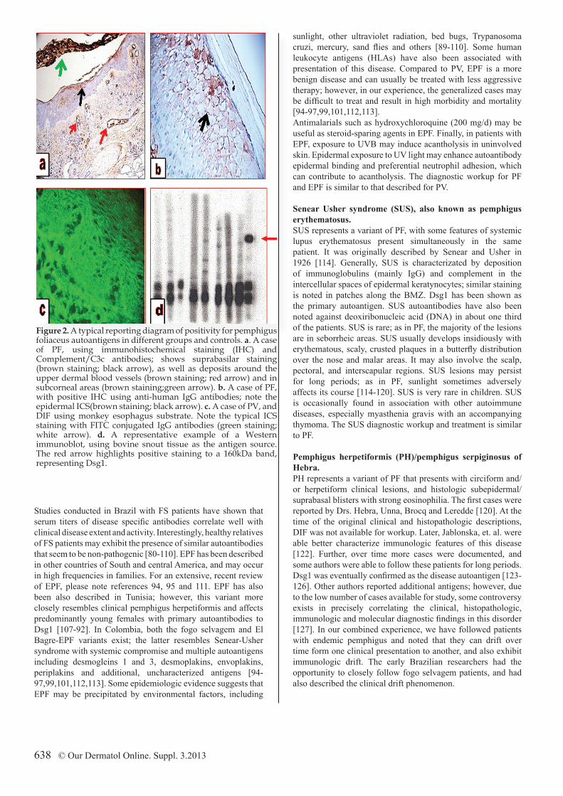
638 © Our Dermatol Online. Suppl. 3.2013
Studies conducted in Brazil with FS patients have shown that serum titers of disease specific antibodies correlate well with clinical disease extent and activity. Interestingly, healthy relatives of FS patients may exhibit the presence of similar autoantibodies that seem to be non-pathogenic [80-110]. EPF has been described in other countries of South and central America, and may occur in high frequencies in families. For an extensive, recent review of EPF, please note references 94, 95 and 111. EPF has also been also described in Tunisia; however, this variant more closely resembles clinical pemphigus herpetiformis and affects predominantly young females with primary autoantibodies to Dsg1 [107-92]. In Colombia, both the fogo selvagem and El Bagre-EPF variants exist; the latter resembles Senear-Usher syndrome with systemic compromise and multiple autoantigens including desmogleins 1 and 3, desmoplakins, envoplakins, periplakins and additional, uncharacterized antigens [94-97,99,101,112,113]. Some epidemiologic evidence suggests that EPF may be precipitated by environmental factors, including
sunlight, other ultraviolet radiation, bed bugs, Trypanosoma cruzi, mercury, sand flies and others [89-110]. Some human leukocyte antigens (HLAs) have also been associated with presentation of this disease. Compared to PV, EPF is a more benign disease and can usually be treated with less aggressive therapy; however, in our experience, the generalized cases may be difficult to treat and result in high morbidity and mortality [94-97,99,101,112,113].Antimalarials such as hydroxychloroquine (200 mg/d) may be useful as steroid-sparing agents in EPF. Finally, in patients with EPF, exposure to UVB may induce acantholysis in uninvolved skin. Epidermal exposure to UV light may enhance autoantibody epidermal binding and preferential neutrophil adhesion, which can contribute to acantholysis. The diagnostic workup for PF and EPF is similar to that described for PV.
Senear Usher syndrome (SUS), also known as pemphigus erythematosus. SUS represents a variant of PF, with some features of systemic lupus erythematosus present simultaneously in the same patient. It was originally described by Senear and Usher in 1926 [114]. Generally, SUS is characterizated by deposition of immunoglobulins (mainly IgG) and complement in the intercellular spaces of epidermal keratynocytes; similar staining is noted in patches along the BMZ. Dsg1 has been shown as the primary autoantigen. SUS autoantibodies have also been noted against deoxiribonucleic acid (DNA) in about one third of the patients. SUS is rare; as in PF, the majority of the lesions are in seborrheic areas. SUS usually develops insidiously with erythematous, scaly, crusted plaques in a butterfly distribution over the nose and malar areas. It may also involve the scalp, pectoral, and interscapular regions. SUS lesions may persist for long periods; as in PF, sunlight sometimes adversely affects its course [114-120]. SUS is very rare in children. SUS is occasionally found in association with other autoimmune diseases, especially myasthenia gravis with an accompanying thymoma. The SUS diagnostic workup and treatment is similar to PF.
Pemphigus herpetiformis (PH)/pemphigus serpiginosus of Hebra. PH represents a variant of PF that presents with circiform and/or herpetiform clinical lesions, and histologic subepidermal/suprabasal blisters with strong eosinophilia. The first cases were reported by Drs. Hebra, Unna, Brocq and Leredde [120]. At the time of the original clinical and histopathologic descriptions, DIF was not available for workup. Later, Jablonska, et. al. were able better characterize immunologic features of this disease [122]. Further, over time more cases were documented, and some authors were able to follow these patients for long periods. Dsg1 was eventually confirmed as the disease autoantigen [123-126]. Other authors reported additional antigens; however, due to the low number of cases available for study, some controversy exists in precisely correlating the clinical, histopathologic, immunologic and molecular diagnostic findings in this disorder [127]. In our combined experience, we have followed patients with endemic pemphigus and noted that they can drift over time form one clinical presentation to another, and also exhibit immunologic drift. The early Brazilian researchers had the opportunity to closely follow fogo selvagem patients, and had also described the clinical drift phenomenon.
Figure 2. A typical reporting diagram of positivity for pemphigus foliaceus autoantigens in different groups and controls. a. A case of PF, using immunohistochemical staining (IHC) and Complement/C3c antibodies; shows suprabasilar staining (brown staining; black arrow), as well as deposits around the upper dermal blood vessels (brown staining; red arrow) and in subcorneal areas (brown staining;green arrow). b. A case of PF, with positive IHC using anti-human IgG antibodies; note the epidermal ICS(brown staining; black arrow). c. A case of PV, and DIF using monkey esophagus substrate. Note the typical ICS staining with FITC conjugated IgG antibodies (green staining; white arrow). d. A representative example of a Western immunoblot, using bovine snout tissue as the antigen source. The red arrow highlights positive staining to a 160kDa band, representing Dsg1.

In PH, itching is common; some initial histopathologic changes we have noted in our PH patients include eczema-like epidermal spongiosis with eosinophilia, and possible subepidermal and/or subcorneal bullae. DIF in our cases showed consistent intercellular IgG staining; IIF on monkey esophagus substrate confirmed this finding in several cases. About half of our patients responded to therapy with sulfones and prednisone, and only one patient responded to sulfones alone. Half of our patients required either combined therapy with prednisone and cyclophosphamide, or with higher doses of prednisone. In our consecutive relapse patients, nine patients retained the pattern of pemphigus herpetiformis; in the others, lesions mostly resembled pemphigus foliaceus. Our most common clinical differential diagnoses included diseases with grouped vesicles such as dyshidrosis, nummular dermatitis, herpes simplex, and herpes zoster; diseases with grouped vesicobullae such as dermatitis herpetiformis, herpes gestationis, subacute lupus erythematosus(bullous variant); and diseases with grouped pustules such as pustular psoriasis, dermatitis continua of Hallopeau, and impetigo herpetiformis.
IgA pemphigus/subcorneal pustular dermatosis (Sneddon-Wilkinson disease). The subcorneal pustular dermatosis of Sneddon and Wilkinson belongs to the heterogeneous group of neutrophilic dermatoses. Sneddon Wilkinson disease is a chronic, sterile pustular eruption and first described by Sneddon and Wilkinson in 1956 [128]. The disease represents a unique disorder, and is characterized by a superficial pustular eruption. The disease pustules are flaccid and aseptic. They develop predominantly on the trunk, groin, axillae and submammary areas. Sneddon Wilkinson disease is often clinically chronic and benign. However, some scattered reports had shown associations with other conditions, including lymphoproliferative and myeloproliferative diseases (monoclonal gammopathies, such as IgA paraproteinemias) and other malignancies. Dapsone is the treatment of choice to control the skin manifestations. The nosologic value of Sneddon Wilkinson disease is still debated, especially vis-a-vis IgA pemphigus, which often is successfully treated with the same dosages of dapsone. Most patients respond well to dapsone, although in rare instances they require etretinate. Several studies studied vesiculobullous lesions that resembled pemphigus foliaceus clinically and histopathologically; however, some displayed intercellular IgA, but not IgG, in the epidermis by direct immunofluorescence. Similar histologic and immunofluorescence findings have been reported in eight other cases. In our cases, no circulating IgA or IgG intercellular antibodies could be detected; in four of the eight previously noted cases, IgA antibodies demonstrated epidermal intercellular staining, similar to a pemphigus pattern [128-148]. A different classification would suggest that the IgA pemphigus cases reported to date fall into one of two groups; specifically, 1) an IgA pemphigus foliaceus group, and 2) an IgA pemphigus of intraepidermal neutrophilic type (ie, Sneddon-Wilkinson disease), which seems to be clinically less common. In our experience, we have encountered three cases of intercellular IgA dermatosis. Somehow, it seems that the world has often merged IgA pemphigus and subcorneal pustular dermatosis (Sneddon-Wilkinson disease) and many confusing reports group them together. Notably, before immunopathologic studies were available, dermatologists and dermatopathologists identified many of these cases as Sneddon-Wilkinson disease. Later,
when the immunopathologic studies become more available it seems that many authors mixed both diseases [149-153]. Some cases of PF have autoantibodies to IgA, in addition to IgG, or IgM (personal experiences). Additionally, a few cases of PF have IgA ICS, but without characteristic neutrophilic features of Sneddon-Wilkinson disease. Further, some autoantigens such as desmocollins and Dsg1 have been named as putative autoantigens by the same groups of authors. Clearly, many of these cases differ from classic Sneddon-Wilkinson disease. Indeed, we recently reported a typical case of Sneddon-Wilkinson disease, in which biopsies for H&E, DIF and immunohistochemistry (IHC) analysis were performed. The H&E staining demonstrated typical features of Sneddon-Wilkinson disease, including some damage to dermal pilosebaceous units subjacent to the subcorneal blistering process. By DIF, FITC conjugated IgE, IgA and fibrinogen were observed in an epidermal pericytoplasmic and perinuclear pattern, with several additional foci within the epidermal stratum corneum (++). Other findings included IgM (+, intercellular epidermal stratum spinosum); IgD (+/-, focal BMZ cytoid bodies; complement/C1q (-); complement/C3 (+, roof of subcorneal pustules); albumin (+, intercellular epidermal stratum spinosum); and fibrinogen (++, focally within papillary dermal tip areas, focally within the superficial corneal layer, and surrounding some upper and intermediate dermal blood vessels). The blister lumens were positive for IgG, IgA, IgM, IgE and fibrinogen. DIF also revealed strong deposits of the immunoreactants IgG, IgM, fibrinogen and complement/C3, present in a shaggy pattern within the subcorneal disease areas; in focal, areas of the basement membrane junction and in focal pericytoplasmic areas of epidermal keratinocytes. IHC revealed strong positivity to HLA-DPDQDR, mast cell tryptase, CD68, and ZAP-70 in the subcorneal luminal inflammatory infiltrate, and surrounding dermal blood vessels. Myeloperoxidase staining was also positive in these areas [154] (Tabl. II).
Drug induced pemphigusDrugs and radiotherapy are important causes of pemphigus. The diagnosis of drug-induced pemphigus may be difficult. Patients have often been exposed to multiple drugs, and some drugs may have a prolonged latency period between exposure and the onset of disease. Some of the most frequently associated medications include D-penicillamine, captopril, clavulinic acid, amoxicillin, imiquimoid, lisinopril and rifampin [155-161]. With patients taking multiple medications, drug-drug interactions may represent a triggering factor. Food supplements, non-prescription medications and topical medications should always be considered. Patients who suffer drug-induced pemphigus usually present with a clinical picture of PF, and less frequently PV [155-162]. The DIF and IIF usually show deposits of predominantly IgG ICS; some reports of antigenicity against epidermal keratinocyte cytoplasms exist. The disease is often transient and resolves shortly after the drug has been discontinued, especially in those patients that lack evidence of circulating antibodies. Skin biopsies of drug-induced and idiopathic pemphigus were reviewed by five dermatologists, with no clinical data available about the patients [158]. The sections were assessed for the presence of epidermal spongiosis with eosinophils, vacuolar degeneration, acantholysis and cleavage level. Using the criteria, the reviewers were unable to correctly identify a clinical case of drug-induced pemphigus.
© Our Dermatol Online. Suppl. 3.2013 639

640 © Our Dermatol Online. Suppl. 3.2013
Thus, it is advisable to consider drug etiology in every case of newly diagnosed pemphigus, as histologic features cannot reliably differentiate between drug-associated and idiopathic disease [158]. Therapy consists of cessation of the target medication.
Intercellular IgE pemphigus (¨IgE pemphigus¨)IgG4 and IgE are present in most autoimmune skin diseases. However, it is not clear that an IgG4 or IgE ICS pemphigus nosologically exists [163-170]. In many conditions such as PV, PF, EPF and El-Bagre-EPF, IgE ICS autoantibodies have been documented. It is further not presently clear if the IgE antibodies are elevated as result of isotype switching, and/or are possibly recognizing specific antigen sites. Notably, increased serum IgE levels are occasionally found in patients with pemphigus. The IgE elevation is often simultaneously present with lesional deposition of eosinophils. In fogo selvagem, anti-Dsg1 antibodies have been associated with disease onest; the same is true with Dsg3-specific IgE and IgG4 autoantibodies in PV [163-170]. Thus, more cases of IgE pemphigus are needed to adequately investigate these questions.
Paraneoplastic pemphigus (PNP)PNP is a recently described blistering disorder that arises exclusively in the context of a neoplasma (most commonly a non-Hodgkin’s lymphoma) has polymorphous skin lesions. These lesions display clinical and histologic features of both erythema multiforme and PV. Other neoplasms associated with PNP include cases of CD20-positive follicular lymphomas, chronic lymphocytic leukemia, thymoma, hepatocellular carcinoma, malignant fibrous histiocytoma, inflammatory myofibroblastic tumor, follicular dendritic cell sarcoma, Waldenström’s macroglobulinemia, B-cell lymphocytic leukemia/lymphoma,
uterine carcinoma and Castleman’s disease. When associated with a malignancy, the course of the disease is usually fatal. If the associated neoplasm is benign and it is removed, the PNP lesions may respond to treatment with corticosteroids and resolve completely. Rarely, PNP lesions may resemble BP, or present as a lichenoid tissue reaction. In addition to the polymorphous skin lesions, patients may develop prominent, painful mucous membrane ulcerations. Involvement of internal organs, such as pulmonary and gastrointestinal tracts, may also be observed. Histologically, skin and mucosal lesions typically present intraepithelial cleavages with suprabasal acantholysis and interface changes featuring necrotic and apoptotic keratinocytes. DIF studies reveal intraepidermal and/or BMZ deposition of IgG and/or C3 complement component, whereas by IIF PNP sera contain autoantibodies binding to stratified, complex and simple epithelia, as well as to the myocardium of the heart. Gallbladder positivity may also be noted in PNP [171-186]. Sera of patients with PNP will immunoprecipitate Dsg1, Dsg3, desmoplakin I (250 kD), bullous pemphigoid antigen I (230 kd), desmoplakin II (210 kD), envoplakin (210 kD), periplakin (190 kD), plectin (500 kD) and a 170-kD protein, that has been reported as alpha-2-macroglobulin-like-protein 1, a broad range protease inhibitor expressed in stratified epithelia and other tissue damaged in PNP. PNP is characterized by the presence of autoantibodies directed against components of the ICS, as well as against the BMZ [171-180]. In one study, patients with PNP presented initially with isolated oral erosions that were undistinguishable from those seen in PV patients, and 27% had histologic findings of only suprabasal acantholysis. The association with a lymphoproliferative disorder, diagnostic IIF labeling of rat bladder, and immunoblotting recognition of envoplakin and/or periplakin represented sensitive and specific features leading to the diagnosis of PNP.
Disease Variants DIF Target antigen/s Differential diagnoses
Pemphigus foliaceus Cazanave’s, Senear-Usher and EPF. ICS with IgG, IgM, IgG, kappa, lambda and Complement/C3 and C1q, mainly in the upper epidermis.
Dsg1, Dsg3, envoplakin, periplakin and desmoplakins.
Impetigo, dermatophytes, burns, acantholytic actinic keratosis, Staphylococcal scalded skin syndrome/SSSS, acute generalized exantemous pustulosis/AGEP.
Pemphigus vulgaris Hallopeau, Neumann, and pyostomatitis vegetans with or without bowel involvement.
IgG and Complement/C3 ICS, and fibrinogen mainly in basilar epidermis.
Dsg 1 and 3, DSC1, DSC3, ATP2C1, acetylcholine.
Viral infections, Hailey- Hailey, Grover’s, friction, Darier’s, disease, burns, aphtous stomatitis, gingivostomatitis, erythema multiforme, erosive lichen planus, blastomycosis, candidiasis, botroymcosis, axillary granular parakeratosis.
Paraneoplastic pemphigus
Unknown. Mainly IgG and Complement/C3 ICS; also BMZ.
Envoplakins, periplakins, Dsg1, Dsg3, BP180 and 230, desmoplakins plectins, α2-macroglobulin-like protein 1.
Erythema multiforme, erosive lichen planus, herpetic lesions, staphyloccal scalded skin syndrome/SSSS.
Drug-induced pemphigus Pemphigus vulgaris and pemphigus foliaceus-like.
IgG and Complement/C3 ICS, mainly in the upper epidermis.
Dsg1, Dsg3 and other uncharacterized antigens.
Halogenoderma, erythema multiforme,Stevens-Johnson.
IgA Pemphigus Subcorneal and other. IgA (ICS and/or cytoplasmic), IgM and fibrinogen.
Dsg 3 and desmocollins.
Infantile acropustulosis (subcorneal), erythem toxico neonatorum, miliaria.
IgE Pemphigus Unknown. ICS IgE. Dsg1 and Dsg3. Any of the above
Table II. Descriptions of autoinmune cutaneous epidermal bullous diseases, including blisters, respective target antigens, and autoimmune disease differential diagnoses.

Recently, it has also been reported that alemtuzumab is effective against severe chronic lymphocytic leukemia-associated PNP [187]. In these patients, the presence of blisters with histologic and immunofluorescence evidence of pemphigus have been demonstrated [188,189]. Neonatal pemphigus is caused by transfer of maternal IgG autoantibodies against Dsg3 or Dsg1 to the neonate through the placenta when the mother is affected by one of the pertinent diseases. Neonatal pemphigus is clinically characterized by transient, flaccid blisters and erosions on the skin, and rarely on the mucous membranes [188,189]. Neonatal pemphigus vulgaris has never been reported to persist beyond the neonatal period and progress to adult disease. Usually neonatal pemphigus lesions are self limiting, and resolve within two weeks. Neonatal pemphigus needs to be differentiated from the “pemphigus neonatorum” of the distant past medical literature, referring to babies with Staphylococcal skin disease [200].
Pemphigus in animalsAnimals, especially dogs, cats and horses can develop PF, PV, and drug induced pemphigus [201-204].
TreatmentPrednisone is the most common therapy for autoinmune
cutaneous blistering diseases, and the initial dose is based on clinical stage of the patient. Generally, 1-2 mg/kg/day are utilizad, supplemented with broad range antiparasitic medicines, calcium supplements, topical corticosteroid cream and sun protection. Sometimes, antibiotic prophylaxis to prevent wound infection is recommended. Several immunosuppressive agents including cyclophosphamide, azathioprine, methotrexate, cyclosporine and gold have been utilized in therapy; for severe cases, plasmapheresis, photopheresis and high-dose intravenous gammmaglobulins have also been used [204-220]. Cloroquine is also used as a coadjuvant. One initial large study on the treatment of 84 patients with PV was conducted between 1961 and 1982 by Dr Walter Lever [204]. Over time, the study showed that 47 of the 84 patients were free of lesions, and not receiving treatment. Twenty-two of the patients had been without lesions and treatment for more than five years. There had been no disease- or treatment-related fatalities since 1976. Since the Lever study, other therapeutic modalities had been added; these include combined treatment with small, alternating day doses of prednisone, plus a daily dose of a different immunosuppressant agent (usually azathioprine). In mild, relatively stable cases, the combined form of treatment may be given initially. Thus, in addition to prednisone monotherapy, prednisone combined with other immunosupressors (“adjuvants”), ie, azathioprine (2 mg/kg/d), cyclosporine (2.5-3 mg/kg/d), cyclophosphamide (1.1-1.5 mg/kg/d), mycophenolate mofetil (2 g/d), mycophenolate sodium have show good results in pemphigus treatment [204-220]. Many combinations of prednisone and adjuvants have been utilized. In many cases, superinfections represent the most dangerous complications of the therapy. Thus, if the patient needs to be hospitalized, testing for tuberculosis and parasitic diseases should be conducted before any treatment. If strong immunosuppression will be given, the patient needs to be hospitalized for this reason alone. In these cases, admission tests such as EKG, chest radiography, kidney and liver electrolytes and a CBC need to be performed. If the admitting physician is suspicious of any superinfection on the skin, cultures need to be taken before initiation of therapy. Biopsies should be taken
for H&E studies and for DIF studies in Michel’s medium. In addition, serum for IIF, immunoblotting and ELISA testing should be obtained. The patient needs to be bathing if possible, cleaning the flexural areas; the mouth needs to be cleaned and colleagues such as ENT, oral surgery, urology and others may be consulted to treat the patients. If the patients have severe oral lesions, a nutritionist should be consulted for appropriate protein control in the diet. Additional therapies such as high-dose intravenous immunoglobulin, rituximab and immunoadsorption are used for refractory patients. Some of these therapies may require 4 weeks to be effective.Appropriate nursing care is also important with immunosupression therapy. The application of topical steroids over a wide area is a safe and effective treatment for thesegroup of diseases, but again the physicians and nurses should monitor the amount of skin that is affected because these patients can become toxic easily. Finally, the therapy of pemphigus with oral corticosteroids has well documented side effects, including systemic infections, high blood sugar, loss of bone density, caries, osteoporosis, thromboses and gastrointestinal ulcers.
REFERENCES
1. Civatte A, Tzanck A, Dobkevitch S: Ann Dermatol Syphiligr (Paris). 1946;6:486.2. Teanok A, Aron-Brunetire R: Arch Bel q Derm Syph. 1949;5:276.3. Lever WF: Pemphigus and pemphigoid: a review of the advances made since 1964. J Am Acad Dermatol. 1979;1:1-31.4. Lever WF: Pemphigus and pemphigoid. Charles C. Thomas, Springfield, 1965.5. Robin AC, Brown G: Lecture notes on Dermatology. Blackwell Scientific Ltd, England, 7ed, Lecture notes series, 1996.6. Moss C: Dermatology and the new genetics (eds) Blackwell Science, England Ltd, 19957. Anderson TF, Voorhees JJ: Thiers BH, Dobson RL (eds): Pathogenesis of skin disease. New York, Churchill Livingstone, 1996.8. Hertl M, Niedermeier A, Borradori L: Autoimmune bullous skin disorders. Ther Umsch. 2010;67:465-82.9. Beutner EH, Jordon RE: Demonstration of skin antibodies in sera of pemphigus vulgaris patients by indirect immunofluorescent staining. Proc Soc Exp Biol Med. 1964;117:505-10. 10. Beutner EH, Lever WF, Witebsky E, Jordon R, Chertock B: Autoantibodies in pemphigus vulgaris: response to an intercellular substance of epidermis. JAMA. 1965;192:682-8.11. Jordon RE, Beutner EH, Witebsky E, Blumental G, Hale WL, Lever WF: Basement zone antibodies in bullous pemphigoid. JAMA. 1967;29:751-6.12. Lever WF: Differential diagnosis of pemphigus vulgaris, bullous pemphigoid and dermatitis herpetiformis. Med Klin. 1967;62:1173-6.13. Lever WF: Medicine (Baltimore). Pemphigus. 1953;32:1-123.14. Wilgram GF, Caufield JB, Lever WF: Electron microscopic studies in skin diseases with acantholysis (pemphigus vulgaris, pemphigus familiaris benignus chronicus, darier’s disease. Dermatol Wochenschr. 1964;147:281-92.15. Wilgram GF, Caulfield JB, Lever WF: An electron microscopic study of acantholysis in pemphigus vulgaris. J Invest Dermatol. 1961;36:373-82.16. Chorzelski TP, Von Weiss JF, Lever WF: Clinical significance of autoantibodies in pemphigus. Arch Dermatol. 1966;93:570-6. 17. Hashimoto K, Lever WF: An ultrastructural study of cell junctions in pemphigus vulgaris. Arch Dermatol. 1970;101:287-98.
© Our Dermatol Online. Suppl. 3.2013 641

18. Hashimoto K, Lever WF: An electron microscopic study on pemphigus vulgaris of the mouth and the skin with special reference to the intercellular cement. J Invest Dermatol. 1967;48:540-52. 19. Beutner EH, Prigenzi LS, Hale W, Leme Cde A, Bier OG: Immunofluorescent studies of autoantibodies to intercellular areas of epithelia in Brazilian pemphigus foliaceus. Proc Soc Exp Biol Med. 1968;127:81-6.20. Chorzelski TP, Jablonska S, Jarzabek-Chorzelska M, Beutner EH, Nisengard RJ, Kumar V, et al: Reliability of indirect immunofluorescence testing. Arch Dermatol. 1980;116:1228-9.21. Jordon RE, Sams WM Jr, Beutner EH: Complement immunofluorescent staining in bullous pemphigoid. J Lab Clin Med. 1969;74:548-56.22. Camisa C, Helm TN: Paraneoplastic pemphigus is a distinct neoplasia-induced autoimmune disease. Arch Dermatol. 1993;129:883-6.23. Amagai M, Klaus-Kovtun V, Stanley JR: Autoantibodies against a novel epithelial cadherin in pemphigus vulgaris, a disease of cell adhesion. Cell. 1991;67:869-77.24. Kárpáti S, Amagai M, Prussick R, Stanley JR: Pemphigus vulgaris antigen is a desmosomal desmoglein. Dermatology. 1994;189 Suppl 1:24-6.25. Kárpáti S, Amagai M, Prussick R, Cehrs K, Stanley JR: Pemphigus vulgaris antigen, a desmoglein type of cadherin, is localized within keratinocyte desmosomes. J Cell Biol. 1993;122:409-15.26. Emery DJ, Diaz LA, Fairley JA. Lopez A, Taylor AF, Guidice GJ: Pemphigus foliaceus and pemphigus vulgaris autoantibodies react with the extracellular domain of desmoglein 1. J Invest Dermatol. 1995;104:323-8.27. Stanley JR, Koulu L, Thivolet C: Distiction between epidermal antigens binding pemphigus vulgaris and pemphigus foliaceus utoantibodies. J Clin Invest. 1984;74:313-20. 28. Amagai M, Hashimoto T, Shimizu N: Absorption of pathogenic autoantibodies by the extracellular domain of pemphigus vulgaris antigen (Dsg3) produced by baculovirus. J Clin Invest. 1994;94:59-67. 29. Stanley JR, Yaar M, Hawley-Nelson P, and Katz SI: Pemphigus antibodies identify a cell surface glycoprotein synthesized by human and mouse keratinocytes. J Clin Invest 1982; 70:281-8.30. Hashimoto T, Ogawa MM, Konohana A, and Nishikawa T: Detection of pemphigus vulgaris and pemphigus foliaceus antigens by immunoblot analysis using different antigen sources. J Invest Dermatol. 1990; 94:327-31. 31. Koch PJ, Walsh MJ, Schmelz M, Goldschmidt MD, Zimbelmann R, et al: Identification of desmoglein, a constitutive desmosomal glycoprotein, as a member of the cadherin family of cell adhesion molecules. Eur J Cell Biol. 1990; 53:1-12.32. Mimouni D, Foedinger D, Kouba DJ, Orlow SJ, Rappersberger K, Sciubba JJ, et al: Mucosal dominant pemphigus vulgaris with anti-desmoplakin autoantibodies. J Am Acad Dermatol. 2004;51:62-7.33. Olivry T, Alhaidari Z, Ghohestani RF: Anti-plakin and desmoglein autoantibodies in a dog with pemphigus vulgaris. Vet Pathol. 2000;37:496-9.34. Kalantari-Dehaghi M, Anhalt GJ, Camilleri MJ, Chernyavsky AI, Chun S, Felgner PL, et al: Pemphigus vulgaris autoantibody profiling by proteomic technique. PLoS One. 2013;8:e57587.35. Kalantari-Dehaghi M, Molina DM, Farhadieh M, Morrow WJ, Liang X, Felgner PL, et al: New targets of pemphigus vulgaris antibodies identified by protein array technology. Exp Dermatol. 2011;20:154-6.36. Pradeep AR, Manojkumar ST, Arjun R: Pemphigus vulgaris with significant periodontal findings: a case report. J Calif Dent Assoc. 2010;38:343-6.37. Mignogna MD, Lo Muzio L, Bucci E: Clinical features of gingival pemphigus vulgaris. J Clin Periodontol. 2001;28:489-93.38. Samy LL, Girgis IH, Wasef SA: Pharyngeal and laryngeal pemphigus. J Laryngol Otol. 1968;82:111-21.
39. Durovic E, Sak M: Contribution to the incidence of pemphigus vulgaris and its manifestations on the mouth mucosa.Cesk Otolaryngol. 1969;18:35-8. 40. Tsiriopoulos I, Kiorpelidou D, Gaitanis G, Sofikitis N, Bassukas ID: Lower urinary tract involvement in patients with newly diagnosed autoimmune bullous dermatoses: an urethrocystoscopic study. Am J Med Sci. 2010;340:109-13.41. Brackley R, Pagani JM: Conjunctival erosions associated with pemphigus vulgaris. Optom Vis Sci. 2011;88:1010-3.42. Bianciotto C, Herreras Cantalapiedra JM, Alvarez MA, Méndez Díaz MC: Conjunctival blistering associated with pemphigus vulgaris: report of a case. Arch Soc Esp Oftalmol. 2005;80:365-8.43. Epstein JH, Feigen GM, Epstein NN: Pemphigus vulgaris with lesions of the rectal mucosa. AMA Arch Derm. 1958;78:36-8.44. Stone DD: Rectal lesions and toxic dilatation of the colon in a case of pemphigus vulgaris. Am J Dig Dis. 1971;16:163-6.45. Lever WF, Schaumburg-Lever. Histopathology of the skin. 1975. Lippincott, Philadelphia.46. Director W: Pemphigus vugaris; a clinicopathologic study. AMA Arch Derm Syphilol. 1952;65:155-69.47. Firooz A, Mazhar A, Ahmed AR: Prevalence of autoimmune diseases in the family members of patients with pemphigus vulgaris. J Am Acad Dermatol. 1994;31:434-7. 48. Ahmed AR, Wagner R, Khatri K, Notani G, Awdeh Z, Alper CA, et al: Major histocompatibility complex haplotypes and class II genes in non-Jewish patients with pemphigus vulgaris. Proc Natl Acad Sci U S A. 1991;88:5056-60.49. Lombardi ML, Mercuro O, Ruocco V, Lo Schiavo A, Lombari V, Guerrera V, et al.: Common human leukocyte antigen alleles in pemphigus vulgaris and pemphigus foliaceus Italian patients. J Invest Dermatol. 1999;113:107-10.50. Reohr PB, Mangklabruks A, Janiga AM, DeGroot LJ, Benjasuratwong Y, Soltani K: Pemphigus vulgaris in siblings: HLA-DR4 and HLA-DQw3 and susceptibility to pemphigus. J Am Acad Dermatol.1992;27:189-93. 51. Slomov E, Loewenthal R, Goldberg I, Korostishevsky M, Brenner S, Gazit E: Pemphigus vulgaris in Jewish patients is associated with HLA-A region genes: mapping by microsatellite markers. Hum Immunol. 2003;64:771-9.52. Szafer F, Brautbar C, Tzfoni E, Frankel G, Sherman L, Cohen I, et al: Detection of disease-specific restriction fragment length polymorphisms in pemphigus vulgaris linked to the DQw1 and DQw3 alleles of the HLA-D region. Proc Natl Acad Sci USA. 1987;84:6542-5. 53. Gazit E, Slomov Y, Goldberg I, Brenner S, Loewenthal R: HLA-G is associated with pemphigus vulgaris in Jewish patients. Hum Immunol. 2004;65:39-46.54. Koc CK, Sallakci N, Akman-Karakaş A, Alpsoy E, Yegin O: Human leukocyte antigens class I and class II in patients with pemphigus in southern Turkey. Int J Dermatol. 2013;52:53-8.55. Bhanusali DG, Sachdev A, Rahmanian A, Gerlach JA, Tong JC, Seiffert-Sinha K, et al: HLA-E*0103X is associated with susceptibility to Pemphigus vulgaris. Exp Dermatol. 2013;22:108-12.56. Marzano AV, Tourlaki A, Merlo V, Spinelli D, Venegoni L, Crosti C: Herpes simplex virus infection and pemphigus. Int J Immunopathol Pharmacol. 2009;22:781-6.57. Morell-Dubois S, Carpentier O, Cottencin O, Queyrel V, Hachulla E, Hatron PY, et al: Stressful life events and pemphigus.Dermatology. 2008;216:104-8. 58. Valikhani M, Kavusi S, Chams-Davatchi C, Daneshpazhooh M, Barzegari M, Ghiasi M, et al: Pemphigus and associated environmental factors: a case-control study. Clin Exp Dermatol. 2007;32:256-60.59. Wohl Y, Brenner S: Pemphigus in Israel--an epidemiologic analysis of cases in search of risk factors. Isr Med Assoc J. 2003;5:410-2.
642 © Our Dermatol Online. Suppl. 3.2013
60. Abréu Vélez AM, Yepes MM, Patiño PJ, Bollag WB, Montoya F: A cost-effective, sensitive and specific enzyme-linked immunosorbent assay useful for detecting a heterogeneous antibody population in sera from people suffering a new variant of endemic pemphigus. Arch Dermatol Res. 2004;295:434-41.61. Ide A, Hashimoto T, Amagai M, Tanaka M, Nishikawa T: Detection of autoantibodies against bullous pemphigoid and pemphigus antigens by an enzyme-linked immunosorbent assay using the bacterial recombinant proteins. Exp Dermatol. 1995;4:112-6.62. Kamiya K, Aoyama Y, Shirafuji Y, Hamada T, Morizane S, Fujii K, et al: Detection of antibodies against the non-calcium-dependent epitopes of desmoglein 3 in pemphigus vulgaris and their pathogenic significance. Br J Dermatol. 2012;167:252-61.63. Kamiya K, Aoyama Y, Shirafuji Y, Hamada T, Morizane S, Fujii K, et al: A higher correlation of the antibody activities against the calcium-dependent epitopes of desmoglein 3 quantified by ethylenediaminetetraacetic acid-treated enzyme-linked immunosorbent assay with clinical disease activities of pemphigus vulgaris. J Dermatol Sci. 2013;70:190-5. 60. Grob PJ, Inderbitzin TM: Experimental production in rabbits of antiepithelial autoantibodies. J Invest Dermatol. 1967;49:637-41. 61. Beutner EH, Chorzelski TP, Jarzabek M, Wood GW, Leme Cde A, Bier O: Studies in immunodermatology. I. Passive induction of intraepidermal clefts in rabbits by transfer of sera from Brazilian Pemphigus foliaceus patients. Int Arch Allergy Appl Immunol. 1972;42:545-55. 62. Ablin RJ, Beutner EH: Precipitation of epithelia antigen(s) with pemphigus-like autoantibodies of rabbit and human pemphigus autoantibodies. Experientia. 1968;24:1055.63. Wood GW, Beutner EH, Chorzelski TP: Studies in immunodermatology. II. Production of pemphigus-like lesions by intradermal injection of monkeys with Brazilian pemphigus foliaceus sera. Int Arch Allergy Appl Immunol. 1972;42:556-64. 64. Sabolinski ML, Beutner EH, Krasny S, Kumar V, Huang J, Chorzelski TP, et al: Substrate specificity of anti-epithelial antibodies of pemphigus vulgaris and pemphigus foliaceus sera in immunofluorescence tests on monkey and guinea pig esophagus sections. J Invest Dermatol. 1987;88:545-9.65. Anhalt GJ, Labib R, Voorhees JJ, Beals TF, Diaz LA: Induction of pemphigus in neonatal mice by passive transfer of IgG from patients with the disease. N Engl J Med. 1982;306:1189-96.66. Hata T, Nishifuji K, Shimoda K, Sasaki T, Yamada T, Nishikawa T, et al: Transgenic rescue of desmoglein 3 null mice with desmoglein 1 to develop a syngeneic mouse model for pemphigus vulgaris. J Dermatol Sci. 2011;63:33-9.67. Yokoyama T, Matsuda S, Takae Y, Wada N, Nishikawa T, Amagai M, et al: Antigen-independent development of Foxp3+ regulatory T cells suppressing autoantibody production in experimental pemphigus vulgaris. Int Immunol. 2011;23:365-73. 68. Hallopeau: Pyodermitis vegetante. iHre Beziehungen zur dermatitis herpetiformis und dem pemphigus vegetants. Archiv F Derm U Syph. Bd XLIV 1898:289. 69. Hallopeau: Zweitc Mittheilung ueber „Pyodermite Vegetante.” (Suppurative Form der Ncumann’schen Krankheit. Archiv F Derm U Syph. Bd XLV, 1898:323.70. Roeckl H: [On pyoderma vegetans hallopeau as a benign form of pemphigus vegetans Neumann with some remarks on pyostomatitis vegetans mccarthy]. Arch Klin Exp Dermatol. 1964;218:574-82. 71. Nelson CG, Apisarnthanarax P, Bean SF, Mullins JF: Pemphigus vegetans of Hallopeau: immunofluorescent studies. Arch Dermatol. 1977;113:942-5.72. Leroy D, Lebrun J, Maillard V, Mandard JC, Deschamps P: Pemphigus vegetans, a clinical type of chronic pustular dermatitis of Hallopeau. Ann Dermatol Venereol. 1982;109:549-55.73. Newman BA, Feldman F: Pemphigus vegetans: involution with cortisone therapy. AMA Arch Derm Syphilol. 1951;64:105.
74. Neumann HA, Faber WR: Pyodermite végétante of Hallopeau. Immunofluorescence studies performed in an early disease stage. Arch Dermatol. 1980;116:1169-71.75. Brinkmeier T, Frosch PJ: Pyodermatitis-pyostomatitis vegetans: a clinical course of two decades with response to cyclosporine and low-dose prednisolone. Acta Derm Venereol. 2001; 81:134-6.76. Hansen LS, Silverman S Jr, Daniels TE: The differential diagnosis of pyostomatitis vegetans and its relation to bowel disease. Oral Surg Oral Med Oral Pathol. 1983;55:363-73.77. Wolz MM, Camilleri MJ, McEvoy MT, Bruce AJ: Pemphigus vegetans variant of IgA pemphigus, a variant of IgA pemphigus and other autoimmune blistering disorders. Am J Dermatopathol. 2013;35:e53-6. 78. Ahmed AR, Blose DA: Pemphigus vegetans. Neumann type and Hallopeau type. Int J Dermatol. 1984;23:135-41.79. Cazanave P: Pemphigus chronique, general;forme rare de pemphigus foliace; mort; autopsie, altation du foie. Ann Maladies Peau Syph. 1844;1:208-10.80. Castro RM, Proença NG: Similarities and differences between Brazilian wild fire and pemphigus foliaceus Cazenave. Hautarzt. 1982;33:574-7.81. Kessler JB: Pemphigus foliaceus. JAMA. 1913;61:102-5.82. Bilet S, Faucher F, Crickx B, Chosidow O, Belaïch S, Grossin M, et al: Cazenave’s pemphigus foliaceus: therapeutic difficulties. Aspects of transient acanthosis nigricans during healing. Ann Dermatol Venereol. 1988;115:1140-2.83. Kormann N: Pemphigus. J Am Acad Dermatol. 1988;18:1219-38.84. Bystryn JC, Rudolph JL: Pemphigus. Lancet. 2005;366:61-73.85. Kazerounian S, Mahoney MG, Uitto J, Aho S: Envoplakin and periplakin, the paraneoplastic pemphigus antigens, are also recognized by pemphigus foliaceus autoantibodies. J Invest Dermatol. 2000;115:505-7.86. Jiao D, Bystryn JC: Antibodies to desmoplakin in a patient with pemphigus foliaceous. J Eur Acad Dermatol Venereol. 1998;11:169-72.87. Ioannides D, Hytiroglou P, Phelps RG, Bystryn JC: Regional variation in the expression of pemphigus foliaceus, pemphigus erythematosus, and pemphigus vulgaris antigens in human skin. J Invest Dermatol.1991;96:159-61.88. Igawa K, Matsunaga T, Nishioka K: Involvement of UV-irradiation in pemphigus foliaceus. J Eur Acad Dermatol Venereol. 2004;18:216-7.89. Kano Y, Shimosegawa M, Mizukawa Y, Shiohara T: Pemphigus foliaceus induced by exposure to sunlight. Report of a case and analysis of photochallenge-induced lesions. Dermatology. 2000;201:132-28.90. Triftshauser C, Wypych J, Cloutier LC, Beutner EH: Flourescent antibody studies of pemphigus-like antibodies in burn sera. Ann N Y Acad Sci. 1971;177:227-33.91. Castro RM, Proenca NG: Semelhancas e diferencas entre o fogo selvagem e o penfigo foliaceo de Cazanave. Similarities and differences between South American pemphigus foliaceus and cazanaves pemphigus foliaceus. An Bras Dermatol. 1983;53:137-9. 92. Bastuji G, Souissi R, Blum L, Turki H, Nouira R, Jomaa B, et al: Comparative epidemiology of pemphigus in Tunisia and France: unusual incidence of pemphigus foliaceus in young Tunisian woman. J Invest Dermatol.1995;104:302-5.93. Hans-Filho G, dos Santos, Katayama JH, Aoki V, Rivitti EA, Sampaio SAP, et al: An active focus of high prevalence of Fogo Selvagem on an amerindian reservations in Brazil. J Invest Dermatol. 1996;107:68-75.94. Abreu Velez AM, Messias-Reason LJ, Howard MS, Roselino AM: Endemic pemphigus foliaceus over a century: part 1 North Am J Med Sci. 2010:2:51-9.95. Abreu Velez AM, Roselino AM, Howard MS, Messias-Reason LJ: Endemic pemphigus foliaceus over a century: part 2. North Am J Med Sci. 2010:2:114-25.
© Our Dermatol Online. Suppl. 3.2013 643

96. Abréu Vélez AM, Abréu Vélez CE, Patiño PJ, Montoya-Maya F, Bollag BW: The tryptic cleavage product of the mature form of the bovine desmoglein 1 ectodomain is one of the antigen moieties immunoprecipitated by all sera from symptomatic patients affected by a new variant of endemic pemphigus. Eur Journal Dermatol. 2003;359-66.97. Abreu Velez AM, Villa-Robles E, Howard MS: A new variant of endemic pemphigus foliaceus in El-Bagre, Colombia: the Hardy-Weinberg-Castle law and linked short tandem repeats. North Am J Med Sci. 2009;1:169-79. 98. Hisamatsu Y, Abreu Velez AM, Amagai M, Ogawa MM, Kanzaki T, Hashimoto T: Comparative study of autoantigen profile between Colombian and Brazilian types of endemic pemphigus foliaceus by various biochemical and molecular biological techniques. J Dermatol Sci. 2003;32:33-41.99. Abréu Vélez AM, Hashimoto T, Tobón S, Londoño ML, Montoya F, Bollag WB, et al: A unique form of endemic pemphigus in Northern Colombia. J Am Acad Dermatol. 2003;4:609-14. 100. Cerna M, Fernandez-Vina M, Friedman H, Moraes JR, Diaz LA, Stastny P: Genetic markers for susceptibility to endemic Brazilian pemphigus foliaceus (fogo selvagem) in Xavante indians. Tissue Antigens. 1993;42:138-40.101. Abreu Velez AM, Howard MS, Gao W, Grossniklaus HE, Jiao Zhe, Arias LF, et al: Varicella-zoster virus (VZV), and alpha 1 antitrypsin: a fatal outcome in a patient affected by endemic pemphigus. Int J Dermatol. 2012;51:809-16.102. Gonçalves GA, Brito MM, Salathiel AM, Ferraz TS, Alves D, Roselino AM: Incidence of pemphigus vulgaris exceeds that of pemphigus foliaceus in a region where pemphigus foliaceus is endemic: analysis of a 21-year historical series. An Bras Dermatol. 2011;86:1109-12. 103. Abida O, Mahfoudh N, Kammoun A, Gaddour L, Hakim F, Toumi A, et al: Polymorphisms of HLA microsatellite marker in Tunisian pemphigus foliaceus. Hum Immunol. 2013;74:104-9.104. Assumpção TC, Eaton DP, Pham VM, Francischetti IM, Aoki V, Hans-Filho G, et al: An insight into the sialotranscriptome of Triatoma matogrossensis, a kissing bug associated with fogo selvagem in South America. Am J Trop Med Hyg. 2012;86:1005-14.105. Qian Y, Jeong JS, Maldonado M, Valenzuela JG, Gomes R, Teixeira C, et al: Brazilian pemphigus foliaceus anti-desmoglein 1 autoantibodies cross-react with sand fly salivary LJM11 antigen. J Immunol. 2012;189:1535-59. 106. Ribeiro JM, Valenzuela JG, Pham VM, Kleeman L, Barbian KD, Favreau AJ, et al: An insight into the sialotranscriptome of Simulium nigrimanum, a black fly associated with fogo selvagem in South America. Am J Trop Med Hyg. 2010;82:1060-75.107. Kallel Sellami M, Ben Ayed M, Mouquet H, Drouot L, Zitouni M, Mokni M, et al: Anti-desmoglein 1 antibodies in Tunisian healthy subjects: arguments for the role of environmental factors in the occurrence of Tunisian pemphigus foliaceus. Clin Exp Immunol. 2004;137:195-200.108. Cunha PR, Bystryn JC, Medeiros EP, de Oliveira JR: Sensitivity of indirect immunofluorescence and ELISA in detecting intercellular antibodies in endemic pemphigus foliaceus (Fogo Selvagem). Int J Dermatol. 2006;45:914-8.109. Cordel N, Maire C, le Gilbert D, Courville P, Tressières B: Afro-Caribbean pemphigus: epidemiological data from a 5-year prospective study on the island of Guadeloupe (French West Indies). Int J Dermatol. 2013;52:1357-60.110. Ortega-Loayza AG, Ramos W, Gutierrez EL, Jimenez G, Rojas I, Galarza C: Endemic pemphigus foliaceus in the Peruvian Amazon. Clin Exp Dermatol. 201338:594-600.111. Robledo MA. Chronic methyl mercury poisoning may trigger endemic pemphigus foliaceus „fogo selvagem”. Med Hypotheses. 2012;78:60-66.
112. Abreu-Velez AM, Howard MS, Jiao Z, Gao W, Yi H, Grossniklaus HE, et al: Cardiac autoantibodies from patients affected by a new variant of endemic pemphigus foliaceus in Colombia, South America. J Clin Immunol. 2011;31:985-97. 113. Abreu Velez AM, Yi H, Gao W, Smoller BR, Grossniklaus HE, Howard MS: Antibodies to pilosebaceous units along their neurovascular supply routes in a new variant of endemic pemphigus foliaceus in Colombia, South America. Eur J Dermatol. 2011;21:371-5. 114. Senear FE, Usher B: An unusual trype of pemphigus combining features of lupus erythematosus. Arch Dermatol.1926;13:761-81.115. Chorzelski T, Jablosnka S, Blaszcyk M: immunopathological investigation in the Senear-Usher syndrome (coexistance of pemphigus and lupus erythermatosus). Br J Derm; 1968;80:217.116. Maize JC, Green D, Provost TT: Pemphigus foliaceus: a case with serologic features of Senear-Usher syndrome and other autoimmune abnormalities. J Am Acad Dermatol.1982;7:736-41.117. Pérez-Pérez ME, Avalos-Díaz E, Herrera-Esparza R: Autoantibodies in senear-usher syndrome: cross-reactivity or multiple autoimmunity? Autoimmune Dis. 2012;2012:296214. 118. Orfanos CE, Gartmann H. Mahrle G: Pathogenesis of pemphigus erythematosus. Transformation of a chronic discoid erythematosus in a pemphigus erythematosus (Senear-Usher). Arch Dermatol Forsch. 1971;240:317-33.119. Steffen C, Thomas D: The men behind the eponym: Francis E. Senear, Barney Usher, and the Senear-Usher syndrome. Am J Dermatopathol. 2003;25:432-6.120. Jordon RE: An unusual type of pemphigus combining features of lupus erythematosus. by Senear and Usher, June 1926. Commentary: Pemphigus erythematosus, a unique member of the pemphigus group. Arch Dermatol.1982;118:723-42.121. Bolgert M, Levy G, Mikol C: Eosinophilia in Duhring-Brocq disease and pemphigus.Bull Soc Fr Dermatol Syphiligr. 1953;60:134-5.121. Ehlers G, Krause W: On the clinical picture of pemphigus serpiginosus HEBRA. Z Haut Geschlechtskr. 1968;43:85-90. 122. Jabloska S, Chorlezelski TP, Beutner EH, Chorzelska JF: Herpetiform pemphigus, a variable pattern of pemphigus. 1975. Int J Dermatol.14:353-9.123. Maciejowska E, Jablonska S, Chorzelski T: Is pemphigus herpetiformis an entity? Int J Dermatol. 1987;26:571-7.124. Jonquieres ED, Bonafina OA: Herpetiforme pemphigus. Med Cutan Ibero Lat Am. 1976;4:265-70.125. Binet O, Girard J, Beltzer-Garelly E, Avram A, Césarini JP, Aron-Brunetière R: Pemphigus herpetiformis (eosinophilic spongiosis). Dermatologica. 1979;159:151-9.124. Ishii K, Amagai M, Komai A, Ebihara T, Chorzelski TP, Jablonska S, et al: Desmoglein 1 and desmoglein 3 are the target autoantigens in herpetiform pemphigus. Arch Dermatol. 1999;135:943-7.125. Lever WF: Inflammatory skin diseases and hypereosinophilia. Z Hautkr. 1987;62:766-8, 771-2.126. Rebora A: Shape and configuration of skin lesions: grouped herpetiform. Clin Dermatol. 2011;29:509-10. 127. Ohata C, Higashi Y, Yamagami J, Koga H, Ishii N, Kanekura T, et al: Coexistence of pemphigus herpetiformis with IgG Antibodies to desmocollin 1 and pemphigoid with IgG antibodies to BP180 C-terminal domain and Laminin γ2. JAMA Dermatol. 2013;149:502-4. 128. Sneddon IB, Wilkinson DS: Subcorneal pustular dermatosis. Br J Dermatol. 1956;68:385-94. 129. Huff JC, Golitz LE, Kunke KS: Intraepidermal neutrophilic IgA dermatosis. N Engl J Med. 1985;313:1643-5.130. Varigos GA: Subcorneal pustulosis with IgA abnormalities in serum and small bowel mucosa: case report. Australas J Dermatol. 1979;20:75-7.
644 © Our Dermatol Online. Suppl. 3.2013

131. Launay F, Albès B, Bayle P, Carrière M, Lamant L, Bazex J: Sneddon-Wilkinson disease. Four cases report. Rev Med Interne. 2004;25:154-9.132. Kuan YZ, Chiou HT, Chang HC, Chan HL, Kuo TT: Intraepidermal neutrophilic IgA dermatosis. J Am Acad Dermatol. 1990;22:917-9.133. Wallach D, Foldès C, Cottenot F: Subcorneal pustulosis, superficial acantholysis and monoclonal IgA. Ann Dermatol Venereol. 1982;109:959-63.144. Cheng S, Edmonds E, Ben-Gashir M, Yu RC: Subcorneal pustular dermatosis: 50 years on. Clin Exp Dermatol. 2008;33:229-33. 145. Dmochowski M, Hashimoto T, Chidgey MA, Yue KK, Wilkinson RW, Nishikawa T, et al: Demonstration of antibodies to bovine desmocollin isoforms in certain pemphigus sera. Br J Dermatol. 1995;133:519-25.146. Hashimoto T, Kiyokawa C, Mori O, Miyasato M, Chidgey MA, Garrod DR, et al: Human desmocollin 1 (Dsc1) is an autoantigen for the subcorneal pustular dermatosis type of IgA pemphigus. J Invest Dermatol. 1997;109:127-31.147. Tsuruta D, Ishii N, Hamada T, Ohyama B, Fukuda S, Koga H, et al: IgA pemphigus. Clin Dermatol. 2011;29:437-42.148. Düker I, Schaller J, Rose C, Zillikens D, Hashimoto T, Kunze J: Subcorneal pustular dermatosis-type IgA pemphigus with autoantibodies to desmocollins 1, 2, and 3. Arch Dermatol. 2009;145:1159-62.149. Kárpáti S, Amagai M, Liu WL, Dmochowski M, Hashimoto T, Horváth A: Identification of desmoglein 1 as autoantigen in a patient with intraepidermal neutrophilic IgA dermatosis type of IgA pemphigus. Exp Dermatol. 2000;9:224-8.150. Ongenae KC, Temmerman LJ, Vermander F, Naeyaert JM: Intercellular IgA dermatosis. Eur J Dermatol. 1999;9:85-94.152.Wallach D: Intraepidermal IgA pustulosis. J Am Acad Dermatol. 1992;27:993-1000. 153. Hashimoto T, Komai A, Futei Y, Nishikawa T, Amagai M: Detection of IgA autoantibodies to desmogleins by an enzyme-linked immunosorbent assay: the presence of new minor subtypes of IgApemphigus. Arch Dermatol. 2001;137:735–8.153. Ebihara T, Hashimoto T, Iwatsuki K, Takigawa M, Ando M, Ohkawara A, et al: Autoantigens for IgA anti-intercellular antibodies of intercellular IgA vesiculopustular dermatosis. J Invest Dermatol. 1991;97:742-5.154. Abreu Velez AM, Smith JG Jr, Howard MS: Subcorneal pustular dermatosis an immnohisto-pathological perspective. Int J Clin Exp Pathol. 2011;4:526-9. 155. Korman NJ, Eyre RW, Zone J, Stanley JR: Drug induced pemphigus: autoantibodies directed against the pemphigus antigen complexes are present in penicillamine and captopril-induced pemphigus. J Invest Dermatol.1991;96:273-6.156. Feng S, Zhou W, Zhang J, Jin P: Analysis of 6 cases of drug-induced pemphigus. Eur J Dermatol. 2011;21:696-9.157. Brenner S, Goldberg I: Drug-induced pemphigus.Clin Dermatol. 2011;29:455-77. 158. Landau M, Brenner S: Histopathologic findings in drug-induced pemphigus. Am J Dermatopathol. 1997;19:411-4.159. Tang X, Zhang X: Drug-induced pemphigus after six years of treatment with phenytoin and carbamazepine. Int J Dermatol. 2012;51:485-6. 160. Feng S, Zhou W, Zhang J, Jin P: Analysis of 6 cases of drug-induced pemphigus. Eur J Dermatol. 2011;21:696-9.161. Brenner S, Goldberg I: Drug-induced pemphigus. Clin Dermatol. 2011;29:455-7. 162. Cozzani E, Rosa GM, Drosera M, Intra C, Barsotti A, Parodi A: ACE inhibitors can induce circulating antibodies directed to antigens of the superficial epidermal cells. Arch Dermatol Res. 2011;303:327-32.
163. Qian Y, Prisayanh P, Andraca E, Qaqish BF, Aoki V, Hans-Filhio G, et al: Cooperative Group on Fogo Selvagem Research. IgE, IgM, and IgG4 anti-desmoglein 1 autoantibody profile in endemic pemphigus foliaceus (fogo selvagem). J Invest Dermatol. 2011;131:985-7.167. Bruns GR, Ablin RJ: IgE in pemphigus. Arch Dermatol. 1977;113:1461.168. Nagel A, Lang A, Engel D, Podstawa E, Hunzelmann N, de Pita O, et al: Clinical activity of pemphigus vulgaris relates to IgE autoantibodies against desmoglein 3. Clin Immunol. 2009;134:320–30.169. Abreu-Velez AM, Howard MS, Hashimoto T, Grossniklaus HE: Human eyelid meibomian glands and tarsal muscle are recognized by autoantibodies from patients affected by a new variant of endemic pemphigus foliaceus in El-Bagre, Colombia, South America. J Am Acad Dermatol. 2010;62:437-47.170. Nagel A, Lang A, Engel D, Podstawa E, Hunzelmann N, de Pita O, et al: Clinical activity of pemphigus vulgaris relates to IgE autoantibodies against desmoglein 3. Clin Immunol. 2010;134:320-30.171. Anhalt GJ, Kim SC, Stanley JR, Korman NJ, Jabs DA, Cory M, et al: Paraneoplastic pemphigus. An autoimmune mucocutaneous disease associated with neoplasia. N Engl J Med. 1990;323:1729-35.172. Bystryn J-C, Hodak E, Gao S-Q, Chuba JV, Amorosi EL: A paraneoplastic mixed bullous skin disease associated with anti-skin antibodies and a B-cell lymphoma. Arch Dermatol 1993;129:870-5.173. Kiyokawa C: Envoplakin and periplakin are the paraneoplastic pemphigus antigens. Kurume Med J. 1999;46:71-8.174. Oursler JR, Labib RS, Ariss-Abdo L, Burke T, O’Keefe EJ, Anhalt GJ. Human autoantibodies against desmoplakins in paraneoplastic pemphigus. J Clin Invest 1992; 89:1775-82.175. Abreu Velez AM, Howard MS: Diagnosis and treatment of cutaneous paraneoplastic disorders. Dermatol Ther. 2010;23:662–75.176. Prakken JR, Woerdeman MJ: Paraneoplastic pemphigus and Pemphigus Vulgaris. Ned Tijdschr Geneeskd. 1954;30;98:3127-35.177. Plewig G, Jansen T, Jungblut RM, Röher HD: Castleman tumor, lichen ruber and pemphigus vulgaris: paraneoplastic association of immunological diseases? Hautarzt. 1990;41:662-70.178. Panteleeva GA: Paraneoplastic bullous dermatoses. Vestn Dermatol Venerol. 1990;50-2.179. Belsan I, Jirásek L, Kratochvílová M, Bohut V: Pemphigus vulgaris as a paraneoplastic manifestation of bronchogenic carcinoma (author’s transl)]. Cesk Dermatol. 1980;55:314-7. 180. Bohatka L, Alfody G: Oral manifestations of paraneoplastic syndrome. Fogorv Sz. 1976;69:264-8.181. Morandi GA, Panerai A, Bongi G: Paraneoplastic syndromes. 3. Clinical contribution to the knowledge of paraneoplastic pemphigoid]. Riv Crit Clin Med. 1964;64:498-508.182. Kim SC, Kwon YD, Lee IJ, Chang SN, Lee TG: R. Envoplakin is a component of the antigen complex in paraneoplastic pemphigus. J Invest Dermatol. 1997abs;108:581.183. Kim SC, Kwon YD, Lee IJ, Chang SN, Lee TG: cDNA cloning of the 210-kDa paraneoplastic pemphigus antigen reveals that envoplakin is a component of the antigen complex. J Invest Dermatol. 1997;109:365-9.184. Proby C, Fujii Y, Owaribe K, Nishikawa T, Amagai M: Human autoantibodies against HD1/plectin in paraneoplastic pemphigus. J Invest Dermatol.1999;112:153-6.185. Mahoney MG, Aho S, Uitto J, Stanley JR: The members of the plakin family of proteins recognized by paraneoplastic pemphigus antibodies include periplakin. J Invest Dermatol. 1998;111:308-13.186. Schepens I, Jaunin F, Begre N, Läderach U, Marcus K, Hashimoto T, et al: The protease inhibitor alpha-2-macroglobulin-like-1 is the p170 antigen recognized by paraneoplastic pemphigus autoantibodies in human.PLoS One. 2010;5:e12250.
© Our Dermatol Online. Suppl. 3.2013 645

187. Bech R, Baumgartner-Nielsen J, Peterslund NA, Steiniche T, Deleuran M, d’Amore F: Alemtuzumab (alz) is effective against severe chronic lymphocytic leukemia-associated paraneoplastic pemphigus (pnp). Br J Dermatol. 2013;169:469-72.188. Grunwald MH, Zamora E, Avinoach I, Holcberg G, Azaz B, Katz M, et al: Pemphigus neonatorum. Pediatr Dermatol. 1993;10:169-70.189. Panko J, Florell SR, Hadley J, Zone J, Leiferman K, Vanderhooft S: Neonatal pemphigus in an infant born to a mother with serologic evidence of both pemphigus vulgaris and gestational pemphigoid. J Am Acad Dermatol. 2009;60:1057-62. 200. Dancer SJ, Poston SM, East J, Simmons NA, Noble WC: An outbreak of pemphigus neonatorum. J Infect. 1990;20:73-82.201. Winfield LD, White SD, Affolter VK, Renier AC, Dawson D, Olivry T, et al: Pemphigus vulgaris in a Welsh pony stallion: case report and demonstration of antidesmoglein autoantibodies. Vet Dermatol. 2013;24:269-e60.202. Medleau L, Dawe DL, Scott DW: Complement immunofluorescence in sera of dogs with pemphigus foliaceus. Am J Vet Res. 1987;48:486-7.203. Scott DW: Pemphigus vegetans in a dog. Cornell Vet. 1977;67:374-84.204. Oberkirchner U, Linder KE, Dunston S, Bizikova P, Olivry T: Metaflumizone-amitraz (Promeris)-associated pustular acantholytic dermatitis in 22 dogs: evidence suggests contact drug-triggered pemphigus foliaceus. Vet Dermatol. 2011;22:436-48.204. Lever WF, Schaumburg-Lever G: Treatment of pemphigus vulgaris. Results obtained in 84 patients between 1961 and 1982. Arch Dermatol. 1984;120:44-7.205. Lapidoth M, David M, Ben-Amitai D, Katzenelson V, Lustig S, Sandbank M: The efficacy of combined treatment with prednisone and cyclosporine in patients with pemphigus: preliminary study. J Am Acad Dermatol. 1988;30:752-7.206. Green MG, Bystryn JC: Effect of intravenous immunoglobulin therapy on serum levels of IgG1 and IgG4 antidesmoglein 1 and antidesmoglein 3 antibodies in pemphigus vulgaris. Arch Dermatol. 2008;144:1621-4. 207. Tan-Lim R, Bystryn JC: Effect of plasmapheresis therapy on circulating levels of pemphigus antibodies. J Am Acad Dermatol. 1990;22:35-40.
208. Director W, Rogachefsky H: Pemphigus folicaceus: response to corticotropin (ACTH) and cortisone; appearance of acanthotic lesions. AMA Arch Derm Syphilol. 1953;68:469-70.209. lszewska M, Kolacinska-Strasz Z, Sulej J, Labecka H, Cwikla J, Natorska U, et al. Efficacy and safety of cyclophosphamide, azathioprine, and cyclosporine (ciclosporin) as adjuvant drugs in pemphigus vulgaris. Am J Clin Dermatol. 2007;8:85-92.210. Bystryn JC, Steinman NM: The adjuvant therapy in pemphigus. Arch Dermatol. 1996; 132:203-12.211. Macdonal C: Immunomodulatory and cytotoxic agents in dermatology. Marcel Dekker Inc (Eds). Blackwell Scientific Ltd, 1996.212. Basset N, Guillot B, Michel B, Meynadier J, Guilhou JJ: Dapsone as initial treatment in superficial pemphigus. Arch Dermatol. 1987;123:783-5.213. Tappeiner G, Steiner A: High-dosage intravenous gamma globulin: therapeutic failure in pemphigus and pemphigoid. J Am Acad Dermatol. 1989;20:684-5.214. Rook AH, Heald PW, Nahass GT: Treatment of autoimmune disease with extracorporal photochemotherapy: pemphigus vulgaris: preliminary report. Yale J Biol Med.1989;62:647-52.215. Beckers RCY, Brand A, Vermeer BJ, Boom BW: Adjuvant high-dose intravenous gammaglobulin in the treatment of pemphigus and bullous pemphigoid: experience in six patients. Br J Dermatol.1996;133:289-93.216. Harman KE, Albert S, Black MM: Guidelines for the management of pemphigus vulgaris. Br J Dermatol. 2003;149:926–37. 217. Wojnarowska F, Kirtschig G, Highet AS, Venning VA, Khumalo NP.: Guidelines for the management of bullous pemphigoid. Br J Dermatol. 2002;147:214–21. 218. Pasricha JS, Thanzama J, Khan UK: Intermittent high-dose dexamethason-cyclophosphamide therapy for pemphigus. Br J Dermatol. 1988;119:73-7.219. Pandya AG, Sontheimer RD: Treatment of pemphigus vulgaris with pulse intravenous cyclophosphamide. Arch Dermatol. 1992;128:1626-30.220. Hymes SR, Jordon RE: Pemphigus foliaceus: use of antimalarial agents as adjuvant therapy. Arch Dermatol. 1992;128:1462-4.
Copyright by Ana Maria Abreu Velez, et al. This is an open access article distributed under the terms of the Creative Commons Attribution License, which permits unrestricted use, distribution, and reproduction in any medium, provided the original author and source are credited.
646 © Our Dermatol Online. Suppl. 3.2013

Review ArticleDOI: 10.7241/ourd.20134.159
Our Dermatol Online. 2013; 4(Suppl. 3): 647-662 Date of submission: 23.10.2013 / acceptance: 04.11.2013
AbstractAutoimmune mucocutaneous blistering diseases (ABDs) represent a group of conditions that manifest with blisters on the skin and/or mucous membranes. Bullous pemphigoid (BP) is the most common autoimmune mucocutaneous blistering disease. In BP, the location of the blisters is subepidermal and the oral involvement is rare. Variants of BP have been described, including pemphigoid vegetans; however, this disease is not completely characterized. The majority of ABDs have blisters and/or vesicles, that are often pruritic, and manifest autoantibodies to diverse proteins. These proteins include 1) hemidesmosomal plaque proteins(ie, BP230, plectins), 2) transmembrane proteins such as BP180 and α6β4-integrin, which are connected via laminin 332 to type VII collagen and 3) currently uncharacterized 105 kDa and 200 kDa molecules. Other ABDs include drug-induced linear IgA disease, bullous systemic lupus erythematosus (BSLE), dermatitis herpetiformis (DH), cicatricial pemphigoid (CP; also termed mucous membrane pemphigoid), lichen planus pemphigoides (LPP), pemphigoid gestationis (PG), herpes gestationis(HG), chronic bullous dermatosis of childhood (CBDC) and the localized forms of CP, such as Brunsting-Perry pemphigoid. The diagnosis of ABDs requires clinical data; skin biopsies (in 10% buffered formalin) for hematoxylin and eosin (H&E) examination and skin biopsies(in Michel’s transport medium) for direct immunofluorescence (DIF). In many ABDs, the histopathologic findings demonstrate a subepidermal vesicle or bulla with a luminal inflammatory infiltrate of neutrophils, eosinophils and/or lymphocytes. In many ABDs, an extensive perivascular and interstitial inflammatory infiltrate is also noted subjacent to the blister in the upper dermis. Normal skin adjacent to an ABD plaque is often excellent for DIF results. Many ABD biopsies reveal autoantibody deposition at the lesional basement membrane zone (BMZ); IgG, IgM, IgA, other immunoglobulins, complement components and fibrinogen may be detected. Indirect immunofluorescence (IIF) yields antibody titer data; the titers usually correlate with disease activity and with ELISA. Linear epitopes are commonly studied by using an immunoblotting (IB) assay. Topical and systemic corticosteroids remain as mainstays of therapy in ABDs; however, multiple other immunosupressors and/or “steroid sparing agents” such as azathioprine have been demonstrated to be of therapeutic value. In the IgA mediated dermatoses, dapsone is often helpful; in addition, liver and blood testing (including G6PD levels) is indicated. The prognosis depends on each case; rapid diagnosis avoids complications and assists in maintaining a good quality of life for each patient.
Key words: bullous pemphigoid; antigens; mucous membrane pemphigoid
Abbreviations and acronyms: Autoimmune mucocutaneous blistering diseases (ABDs), bullous pemphigoid (BP), mucous membrane pemphigoid (MMP), chronic bullous dermatosis of childhood (CBDC), adult linear IgA bullous dermatosis (LAD), lichen planus pemphigoides (LPP), immunohistochemistry (IHC), direct and indirect immunofluorescence (DIF and IIF), hematoxylin and eosin (H&E), antibodies (Abs), basement membrane zone (BMZ), intercellular staining between epidermal keratinocytes (ICS), pemphigus vulgaris (PV), cicatricial pemphigo-id (CP), dermatitis herpetiformis (DH), sodium dodecyl sulfate (SDS), SDS-PAGE (SDS polyacrylamide gel electrophoresis), bullous systemic lupus erythematosus (BSLE); bullous pemphigoid antigens II(180 kDa) and I(230 kDa)(BP180 and BP230), epidermolysis bullosa simplex (EBS), intravenous immunoglobulin (IVIG).
www.odermatol.com
Cite this article: Ana Maria Abreu Velez, Daniel Alberto Vasquez-Hincapie, Michael S. Howard: Autoimmune basement membrane and subepidermal blistering diseases. Our Dermatol Online. 2013; 4(Suppl.3): 647-662.
AUTOIMMUNE BASEMENT MEMBRANE AND SUBEPIDERMAL BLISTERING DISEASES
Ana Maria Abreu Velez1,2, Daniel Alberto Vasquez-Hincapie3, Michael S. Howard2
1Georgia Dermatopathology Associates, Atlanta, Georgia, USA2Department of Dermatology, University of Antioquia, Medellin, Colombia, SA.3Pecet. Tropical Disease Program, University of Antioquia, Medellin, Colombia, SA.
Corresponding author: Ana Maria Abreu Velez, MD PhD [email protected]
Source of Support: None
Competing Interests: None
IntroductionSubepidermal autoimmune blistersTraditionally, the classification of subepidermal blistering diseases has been based on their patterns of inflammation [1-
3]. However, some overlap occurs between the traditional categories, especially with subepidermal vesiculobullous diseases in which neutrophils or eosinophils represent the predominant infiltrating cell [1-3].
© Our Dermatol Online. Suppl. 3.2013 647

Special techniques, including electron microscopy, immunoelectron microscopy, immunoblotting, direct immunofluorescence (DIF), indirect immunofluorescence (IIF), IIF/salt split skin, and immunohistochemistry (IHC) has allowed many of the subepidermal blistering diseases to be characterized not only histologically on the basis of the anatomic split level within the BMZ, but also immunologically [4-11]. Further, additional information helps to exclude viral blistering diseases, bullous allergic drug reactions and genodermatoses; this information may be provided via viral serology, viral culture, a history of previous intake of medications (including nonprescription medications) and possible genetic evaluation.
Bullous pemphigoid (BP)Bullous pemphigoid is the most common autoimmune skin blistering disease (ABD) in the adult population in developed countries, with an estimated incidence that varies from 0.2 to 3 cases per 100,000 inhabitants per year to 1 in 40,000 [12-14]. In undeveloped countries, it seems to be the second more common ABD; however, few pertinent epidemiologic studies have been published, except in Singapore [15-17]. A sex predisposition has not been clearly demonstrated. The course of this disease can be acute, chronic, or relapsing. BP usually manifests in the sixth or seventh decade of life; however, cases have been observed at other ages, including in children [1-3]. The most common clinical presentation includes scattered urticarial papules and plaques on the trunk, arms, and legs early in the disease. The blisters are often large, tense and located on an erythematous base. Sometimes the blisters are not clinically apparent, due to previous rupture. Acral sites may be involved. The most important symptom related by the patients is pruritus, which may be severe; in contradistinction, pemphigus patients often report a burning sensation [1-3,14]. In some patients, the pruritus seems not to be present. Overall, skin lesions are symmetrically distributed, the most common sites are consistently the flexor surfaces of the extremities, the groin, the axillae and the lower abdomen. The lesions generally heal without scarring, but in rare cases milia formation has been described. In some cases, BP is misdiagnosed as urticaria. Specifically, the disease may initially present as tense vesicles and bullae change on an urticarial base. The bullae can reach a size of several centimeters before rupturing [14]. Blisters in the oral cavity are rare, may compromise the oropharynx and are classically non-scarring. Pemphigus vegetans, described as a variant of BP, is discussed below. Occasionally, BP may be induced by medications such as furosemide, captopril, and penicillin; however, BP needs to be immunologically differentiated from blistering allergic drug eruptions [3]. The physiopathologic aspects of BP include cutaneous deposition of autoantibodies, complement, fibrinogen, albumin and other products of proteases; this deposition results in the disruption of adhesive interactions between epidermal basal layer keratinocytes and the cutaneous basement membrane zone (BMZ). Initial dermatopathology studies in BP were led by Walter F. Lever, M.D. On light microscopy, H&E staining classically reveals a subepidermal blister under an intact epidermis. When clinical blisters arise on erythematous skin, there is often a prominent cellular infiltrate in the papillary dermis consisting of numerous eosinophils, lymphocytes and neutrophils. Papillary dermal microabscesses of neutrophils and eosinophils are present in about 20% of BP cases [3]. The dermal infiltrate in clinically non-inflamed skin lesions is sparse, perivascular, and primarily composed of lymphocytes and histiocytes. Multiple
autoantibodies are directed to components of the BMZ. Initial studies demonstrating the autoimmune nature of BP were led by Ernst Beutner, Ph.D. and Robert Jordon, M.D. [3,5]. These investigators demonstrated the increased diagnostic value of DIF biopsies taken from perilesional blister areas. Serologic studies including indirect immunofluorescence (IIF) can help to confirm the diagnosis, utilizing antigen sources such as normal human skin or monkey esophagus (Fig. 1). The DIF biopsies should be performed at the same time as the cutaneous H&E biopsies. In H&E biopsies, histopathologic findings may vary depending if the biopsy was taken from lesional, lesional border or nonlesional skin [1-4]. In BP, classic DIF of perilesional skin shows deposits of IgG and complement component C3 at the BMZ (Fig. 1). The IgG and C3 deposits are present in a continuous, fine linear pattern along the BMZ. In BP, linear deposits of IgG and C3 are observed in nearly 100% and approximately 90% of the cases, respectively. IgG deposits belong mainly to the IgG4 subclass, and, to a lesser degree, to IgG1 [5]. Other immunoreactant classes are less frequently observed with a similar linear pattern of deposition and usually in association with IgG; these classes include IgM, IgA, IgD, IgE fibrinogen, Complement/C1q and Complement/C3 [19-21]. Using IIF-NaCl split skin, autoantibodies are present predominantly on the blister roof in 90% of BP cases (Fig. 1). IIF is usually performed on normal human or monkey esophagus substrate skin; the skin substrate separates through the lamina lucida on incubation in 1.0 M NaCl. Via non-salt split IIF, the majority of pertinent ABD sera produce an indistinguishable pattern of linear immunofluorescence at dilutions of 1:10 or higher. On salt split skin IIF, these same antibodies bind to either the blister roof (epidermal pattern), blister floor (dermal pattern), or both the roof and floor (combined pattern). The binding patterns have been described in comparison with normal controls. Sera from patients with clinical and histologic features of epidermolysis bullosa acquisita (EBA) show a predominant dermal pattern. However, some sera from patients with BP and EBA show a combined pattern. Indirect immunoelectron microscopy of selected sera show antibodies producing the epidermal and combined patterns are anti-lamina lucida antibodies, and those producing the dermal pattern were anti-sublamina densa antibodies [22-24]. These results show that indirect immunofluorescence on salt split skin is a dependable method for differentiating bullous diseases with anti-lamina lucida versus anti-sublamina densa antibodies, and that differentiating between these antibodies is essential for accurate diagnosis in some patients. The results also suggest that BP anti-lamina lucida antibodies may have more than one antigenic specificity.The autoantibodies detected in sera from patients with BP have been reported to primarily bind to two hemidesmosomal proteins initially detected by immunoblotting (IB) and cDNA cloning as a 180-kD antigen (BPAG II; BP180; Collagen Type XVII), and a 230-kD antigen (BPAGI; BP230. BP230 is a plakin protein family member that promotes the association of hemidesmosomes with keratin intermediate filaments. BP180 is a type II transmembrane collagen that is associated with hemidesmosome anchoring filament complexes, and is believed to harbor all or a portion of the primary pathogenic epitope responsible for the initiation of BP. The extracellular domain of BP180 contains 15 interrupted collagenous domains. Rotary shadowing studies of purified BP180 reveal its intracytoplasmic region to be a globular head, and its ectodomain as a central rod joined to a flexible tail.
648 © Our Dermatol Online. Suppl. 3.2013

© Our Dermatol Online. Suppl. 3.2013 649
Figure 1 a. DIF of a case of EBA, demonstrating positive antibodies to human FITC conjugated Complement/C3 along the BMZ (green staining; white arrow) (400X). The nuclei epidermal keratinocytes were counterstained with Topro 3 (red staining). b. Same case as in a, with PAS positive staining along the BMZ (pink staining; black arrow) (200X). c. A case of BP, with positive IHC staining for IgE at the BMZ (brown staining; black arrow). d. A BP case, with NaCl salt split skin/IIF and positive staining on the blister roof for FITC conjugated anti-human IgG (yellow staining; white arrow). The nuclei of epidermal keratinocytes were counterstained with Dapi (blue staining).
Immunoelectron microscopy studies indicate that BP180 spans the lamina lucida, and inserts into the lamina densa. BP180 is targeted by autoantibodies from patients with BP, pemphigoid gestationis, cicatricial pemphigoid (CP) and linear IgA dermatosis (LAD) [7-10,25] (Tabl. I).Enzyme-linked immunosorbent assays (ELISA) for BP180 and BP230 (MCW2 and MCW1, respectively) were developed by a BP research group at the Medical College of Wisconsin. Following further modifications using a fragment named NC16A, these assays are now commercially available. A recent study has shown that the BP180 ELISA is specific for the immunodominant NC16A domain of the BPAGII protein; however, the ELISA is also exclusive of other parts of the NC16A domain [29]. The NC16A finding is consistent with the immunology concept that all conformational epitopes at least carry at least one or more linear epitopes.Electron microscopy studies on non-inflamed skin lesions from BP patients reveal that dermal-epidermal cleavage occurs within the dermal-epidermal junction, i.e., through the lamina lucida [30-33].In regard to BP antigen(s), epitope mapping studies of recombinant proteins have formerly shown that autoantibodies from most patients with BP bind a determinant within the sixteenth non-collagenous domain of BP180 (i.e., the portion of its ectodomain that is positioned adjacent to plasma membranes of basal keratinocytes). It also have been shown in some experiments using passive transfer of experimental IgG (developed against the murine homolog of this determinant to neonatal BALB/c mice) produces clinical, histologic, and immunopathologic alterations with similarities to those seen in patients with BP patients. However, no animal or cell culture study has been able shown to reproduce the chronicity of BP [34]. Authors have reported multiple animal models with genetic manipulations; however, many of these studies lack proper controls. Thus, it is difficult to correlate these models to BP in vivo because most animal models lesions only persist for few days, and not reflective the chronic nature of this disease.
In contrast to pemphigus, BP is often a self-limited disease; thus, it may be sufficient to treat the patient symptomatically for a limited period. In general, relapse episodes are not common; systemic corticosteroids represent the most common therapy for generalized BP [33]. Specifically, BP therapy primarily consists of administration of topical and systemic corticosteroids. Topical corticosteroids present less adverse effects compared to systemic steroids [35]. The systemic dosage classically ranges between 0.5 and 1 mg Prednisone/kg/d [36]. The dose of prednisone can be tapered slowly over a period of several months to a maintenance dose of a total of 5 to 10 mg/day. Corticosteroids are often combined with other immunosuppressants in recalcitrant cases; hidroelectrolytic disorders often result from these treatments, especially in the elderly [37]. Because BP affects many senior patients suffering from other medical problems, systemic corticosteriod complications may be severe in these cases. Localized BP can be treated with topical corticosteroids [35-37]. However, the personnel treating these patients need to be aware of secondary cutaneous complicatons (e.g, erysipelas, lymphangitis, sepsis, phlegmons, cutaneous fistulas, atrophy and purpura) in patients treated with topical corticosteroids [38]. Dapsone and sulfonamides, either alone in combination with topical or systemic corticosteroids may also be effective [39]. Specifically, dapsone at a dose of approximately 100 mg/day is initiated at the same time as prednisone. The addition of dapsone often accelerates disease control, and thus allows a faster prednisone taper. Other researchers have describe the use of oral tetracycline, or a combination of tetracycline and niacinamide as successful treatments for BP. Cyclosporine, intravenous immunoglobulin (IVIG), azathioprine, rituximab, and plasmapheresis have all been proposed as additional treatments [40-44] (Tabl. I).Frequent bacterial, fungal and viral cultures of cutaneous erosions, catheters, and serum cultures will allow detection of secondary infections arising as a result of immunosuppressive therapy. If positive results are found, appropriate therapy should be quickly initiated.

650 © Our Dermatol Online. Suppl. 3.2013
In Table I, we present a summary of the more common immunosuppressive agents, their metabolites, their serum half lives and common complications. Previous reports have addressed morbidity and mortality in BP. In a private hospital in Wisconsin, thirty-eight new patients were identified and complete follow-up data were obtained on 37 of the patients. Patients were followed for a minimum of 1 year, or until the time of death. The mean duration of follow-up was 20 months. A Kaplan-Meier analysis of the population indicated a 1 year survival rate of 88.96%, with a 95% confidence interval of 75.6% to 94.2%. The survival rate was considerably higher than that recently reported in several studies from Europe (29%-41% one year mortality). The authors reported that although the age at onset and co-morbidities of our patients were similar to those in the European studies, the rate of hospitalization of our patients was much lower than that of patients from Europe (1.5 days per patient, vs. 11-25 days per patient). The study suggests that differences in practice patterns may be an important factor in the reduced mortality rate in US BP patients compared to those in Europe [45].Previously, BP has been associated with significant morbidity and mortality rates. In one study, the authors retrospectively studied 94 patients with BP in a Chinese tertiary medical center between 2005 and 2010, to evaluate treatment and prognostic factors for mortality. Cerebrovascular diseases (42.55%) and hypertension (39.36%) represented the most common pre-existing conditions. Cardiopathy, diabetes and psoriasis pre-existed in 24.47%, 22.34% and 5.32% of patients, respectively [46]. Eighty of 94 patients were treated by systemic corticosteroid, specifically prednisone 0.3 mg/kg to 1.5 mg/kg daily. Patients were followed up for a minimum of 1 year or until the time of death. The mean duration of follow-up was 32 months. Kaplan-Meier analysis demonstrated a 1 year survival probability of 76.6% (standard error 4.4%), with a 95% confidence interval (68.04% to 85.16%). Multivariate analysis revealed that increased age, bedridden condition, presence of cerebrovascular diseases at diagnosis, pre-existing cardiopathy and low serum albumin level were associated with an elevated 1 year mortality rate [47]. In a second study, authors in Latin America obtained similar results as those previously described for developing countries [48]. An association has been suggested between neurologic disorders and BP, since Type XVII collagen (BPAGII; BP180) is a component of hemidesmosomes, which connect epidermal basal layer keratinocytes to the underlying basement membrane. In addition, Type collagen XVII has been recently demonstrated in human brain neurons by in situ hybridization.Skin biopsies for hematoxylin and eosin (H&E) staining and direct immunofluorescence (DIF) are needed to confirm a BP diagnosis. Histopathologic findings of early lesions show subepidermal vesicles, with luminal infiltrates of eosinophils and subjacent, upper dermal, perivascular infiltrates of eosinophils and neutrophils with dilation of infiltrate associated blood vessels [1-3]. The cutaneous H&E biopsy should be placed in 10% buffered formalin, and will produce excellent archival material; however, formalin can form precipitates at high temperatures, and freezing temperatures may produce tissue artifacts. Normal skin adjacent to a BP urticarial plaque is ideal for a DIF biopsy. Almost all biopsies reveal linear Complement/C3 deposition at the basement membrane zone (BMZ), and 80% of biopsies will also show linear immunoglobulin (IgG) deposition at the BMZ [4-6] (Fig. 1). Biopsies for DIF should be placed in Michel’s transport media for processing to avoid false negative results; however, when Michel’s medium is not available the biopsy
may be sent in saline solution. Several independent research studies have reported mixed results in regards to increased sensitivity and specificity of ELISA testing, in comparison to other diagnostic techniques for BP [49]. Occasionally, BP has been described to be induced by medications such as furosemide, captopril, and penicillin.Author’s note: We recommend carefully differentiating BP from with medication-induced blistering diseases; the medication-induced bullous dermatoses represent a more common presentation of bullae in elderly patients, especially in the USA. In our own immunodermatology practice, blisters caused by medications are much more common than BP
Mucous membrane pemphigoid (MMP), or cicatrical pemphigoid (CP)CP represents a subepidermal blistering disease, that characteristically presents in the sixth or seventh decade of life. The female-to-male ratio is about 2: 1 [50]. The term cicatricial pemphigoid (CP) has been used for more than 70 years, and originated with pioneers in the study of cutaneous autoimmune bullous diseases [50-60]. However, at the First International Consensus on Mucous Membrane Pemphigoid in 2002 [57], the name for the disease was recommended to be changed to MMP. CP mainly involves oral mucosa, other mucous membranes, and rarely the skin. Gingival involvement is frequent. In a case of desquamative gingivitis, the clip sign suggests the diagnosis of CP. CP is an autoimmune vesiculobullous disease, distinguished clinically by its predilection for oral and ocular mucous membranes and a tendency for the lesions to scar. Considerable heterogeneity exists in terms of age at presentation and the clinical pattern of disease. In rare cases, CP may present in children and adolescents. The mouth is the most frequent site of presentation, and is eventually involved in 85% of cases. There are erosions, irregular ulcers, and vesiculobullous lesions. Ocular involvement is also common, and corneal and conjunctival scarring may lead to blindness. A CP diagnosis is established by clinical, histological and DIF examination. On DIF, linear deposition of IgG and/or IgA are present along the BMZ of biopsies [50-60]. In some reports, autoantibodies to plectin have also been identified. CP differs from BP in that individual lesions heal with scarring that can be deforming, altering vision and/or genitourinary functionality. As in pemphigus vulgaris, any mucosal site can be affected. Involvement of the nasal mucosa can subsequently lead to strictures in the oral and esophageal areas. If the patient is coughing or has a rough voice on initial presentation, possible blistering in the pharynx and larynx should be suspected. An extensive workup should be performed, including consultation with an ENT specialist and a voice therapist. Involvement of the conjunctivae (which manifests clinically as conjunctivitis and xerosis) may result in scarring causing symblepharon, entropion, and later trichiasis [58-60]. Progressive scarring may then lead to blindness. H&E studies show similar results to BP, with subepidermal blisters containing edema fluid, fibrin and inflammatory cells. A dermal perivascular lymphohistiocytic infiltrate with occasional plasma cells and neutrophils can be seen. In general, fewer eosinophils are appreciated than in BP. Conjunctival squamous metaplasia with foci of hyperkeratosis and parakeratosis, accompanied by goblet cell depletion and conjunctival vesicles or bulla is rare. By IIF, the patients usually have low titers of circulating antibodies; IIF on NaCl split skin shows antibodies on the blister floor. By electron microscopy, CP antibodies are located in the lamina lucida.

© Our Dermatol Online. Suppl. 3.2013 651
An interesting recent study investigated the levels of matrix metalloproteinases (MMPs), myeloperoxidase (MPO) and tissue inhibitor of metalloproteinase-1 (TIMP-1) in ocular tears of patients with Stevens Johnson syndrome (SJS) and ocular cicatricial pemphigoid (OCP). The authors performed a prospective, non-interventional cohort study with four SJS patients (7 eyes), 19 OCP patients (37 eyes) and 20 healthy controls who underwent phacoemulsification (40 eyes). The authors evaluated tear washes collected from all patients; these washes were analyzed for levels of MMP-2, MMP-3, MMP-7, MMP-8, MMP-9, MMP-12, MPO, and TIMP-1 using a multianalyte bead-based ELISA test. Total MMP activity was also determined, using a fluorometric assay. Correlation studies were performed comparing specific analytes within study groups. The authors reported that MMP-8, MMP-9, and MPO levels were significantly elevated in SJS and OCP tears (SJS>OCP) in comparison to controls. MMP activity was highest in SJS patients, whereas OCP patients and controls displayed lower activities(which were similar to each other). The TIMP-1 levels were decreased in SJS and OCP patients when compared to controls, with levels in OCP patients attaining significance. The MMP-8 to TIMP-1 and MMP-9 to TIMP-1 ratios were markedly elevated in SJS and OCP tears (SJS>OCP) when compared to controls. Across all study groups, MMP-9 levels correlated strongly with MMP-8 and MPO levels, and MMP-8 correlated with MPO, but did not reach significance in SJS patients. The authors also reported that there was no significant relationship between MMP-7 and MPO [61] (Tabl. I). In CP, involvement of the genital and anal mucosa is common. Persistent blistering and scarring of the vaginal mucosa can result in stenosis, which may be discovered and also interfere with screening pelvic examinations [50]. Anal involvement manifests as localized pain and bleeding, which can lead to stenosis if left untreated. Cutaneous tense blisters (similar to those seen in BP) occur in only 20% of CP patients. Healing occurs with pink, atrophic scarring. In CP, multiple manifestations of the disease exist depending on which protein in the BMZ is the primary antigen involved. Around 90% of patients with CP have been described to have oral erosions. If the oral mucosa is the single mucosal site involved in CP, the condition may be called oral pemphigoid. If the conjuctival mucosa is the only site of CP clinical presentation, the disorder is termed ocular pemphigoid [50-60].Anti-epiligrin subtype: Another type of CP, termed antiepiligrin cicatricial pemphigoid, occurs when antibodies are formed against laminins 5, 332 and 6 in the basement membrane. This disease is uncommon, and primarily affects mucous membranes but also the skin. The disease is associated with mortality from treatment with systemic immunosuppressive drugs. In this subset, there is an association with solid organ malignancies. Biopsies for H&E staining and DIF should be performed to confirm the diagnosis. Antiendomysial antibodies can be documented, using monkey esophagus as the antigen source for the IIF. Histologically, the findings are almost identical to BP [54,55]. A subepidermal vesicle is seen, with an inflammatory infiltrate of neutrophils and eosinophils in the upper dermis [50-53]. Scarring may be seen in the upper dermis. DIF of perilesional mucosa reveals linear IgG and Complement/C3 deposition at the BMZ in 90 to 95% of patients. Because there are low amounts of circulating antibodies, IIF testing is not generally helpful. The diagnostic findings of CP are summarized in Table I.
Patients with localized oral involvement often respond to topical clobetasol gel, or intralesional triamcinolone (5 to 10 mg/mL, injected sublesionally every 3 weeks as needed). Patients with multiple mucosal sites should be treated with systemic therapy such as dapsone or prednisone. If the lesions are unresponsive, an additional immunosupressor should be utilized in combination with prednisone. In some patients, mycophenolate mofetil is less myelosuppressive and hepatotoxic than corticosteroids. Due to a superior safety profile, mycophenolate mofetil or enteric-coated mycophenolate sodium may gradually replace azathioprine as the first-line adjuvant of choice in the treatment of moderate to severe autoimmune CP. Cyclophosphamide still has a place in the treatment of severe relapsing CP; continuous oral cyclophosphamide provides optimal immunosuppression, but it also produces the highest cumulative dose. Therefore, pulsed cyclophosphamide regimens have been developed and are useful in severe forms of CP. Because of the low incidence and prevalence of these diseases, few randomized controlled studies have compared the efficacy and safety of immunosuppressants such cyclophosphamide with newer treatment options such as rituximab and immunoapheresis. In addition, few studies have been conducted to define optimal dose ranges and optimal durations of immunosuppressive treatments at different stages of CP. We encourage the multidisciplinary collaboration necessary for the diagnosis and proper treatment of difficult cases of CP. Systemic adjuvant immunosuppressive therapy is necessary for patients with progressive disease. In spite of the advances in available immunosuppressive medications and biologics, scarring is a significant complication in many cases [59,62] (Tabl. I). Surgical intervention in general does not cure the disease, though in some occasions with severe sequels, it may be necessary for restoring function and humanizing quality of life. BP alone and bullous lupus rarely involve the oral mucosa. Author’s note: Early, strong immunosupressive treatment is advised in CP patients, due to potential scarring damage to the eyes, nasal mucosa and genito-urinary tract. Systemic steroids in combination with immunosuppressive agents such as methotrexate, mycophenalate mofetil, azathioprine or cyclophosphamide are recommended in severe cases.
Localized cicatricial pemphigoidLocalized CP, also known as Brunsting–Perry pemphigoid, is characterized by the occurrence of one or more scarring, plaque-like lesions, usually on the head and neck; the disease is characterized by a lack of involvement of mucous membranes, including on prolonged follow-up. The temple is the most frequent site of presentation, lesions have occurred in tissue transplanted to the site of a pre-existing lesion. The exact relationship of this condition to CP is speculative, although they are thought to be closely related diseases [63,64]. Rare cases may progress to a generalized disease that heals with scarring; however, although the second disorder that could be CP, there is no mucous membrane involvement. In DIF and IIF studies, the most commonly described findings are linear deposits of IgG(but not IgM or IgA) at the epidermal-dermal junction. In few patients, linear Complement/C3 deposition at the BMZ is noted; a few patients also exhibit circulating anti-BMZ antibodies [65,66]. In some cases, DIF has been reported as negative. In other cases the putative antigen(s) have been described to be IgG autoantibodies to laminin-332, BP230 and desmoplakins I and II; and in further cases to a 290 kDa molecule [67,68].

652 © Our Dermatol Online. Suppl. 3.2013
Electron microscopy studies have been shown a subepidermal separation below the basal lamina, and the basal lamina and anchoring fibrils preserved and attached to the intact epidermis along the blister roof [69]. Many authors in the 1980s and 90s considered Brunsting-Perry disease to be a variant of CP. These findings support the concept that localized CP, CP, and disseminated CP are in fact closely related diseases, and may explain the occurrence of scar formation in localized CP [70]. Due to the low incidence of Brunsting-Perry disease, has been difficult to definitively categorize the disorder; in addition, early BP may present with an individual blister, and may be thus improperly categorized as Brunsting-Perry disease.
Pemphigoid vegetansPemphigoid vegetans is a rare disease exhibiting clinical similarity to pemphigus vegetans, but with histological and immunopathological features of BP. Only few cases have been reported; the relationship of pemphigoid vegetans to BP thus is not clear. Clinically, pemphigoid vegetans classically presents with multiple, well-circumscribed, erythematous, erosive and vegetating plaques in the axillae, inframammary areas and neck [71-73]. Microscopic examination reveals epidermal hyperplasia, dermal/epidermal junctional separation, and prominent dermal eosinophilia [74]. By DIF, perilesional skin demonstrates linear deposits of IgG at the BMZ, primarily of the IgG4 subclass. On salt split skin IIF, these antibodies are present on the blister roof of normal human skin [75,76]. By immunoblotting (IB) studies, the 230 kD BPAGI antigen is one of the disease antigens in pemphigoid vegetans [76]. Some authors have suggested that pemphigoid vegetans is best classified as a BP variant, after describing a 57 year old man with intertriginous vegetating plaques. The histologic examination and DIF of a biopsy specimen were identical to those of BP. IB studies and IIF of salt-split skin were negative [75,76]. Direct immunoelectron microscopy was consistent with BP [77]. Based on a limited experience, previous authors have reported that pemphigoid vegetans patients seem to improve with tetracycline and/or corticosteroids [78] (Tabl. I).
Pemphigoid gestationis (PG)Pemphigoid gestationis (also known as herpes gestationis (HG)) is a rare, pruritic, vesiculobullous dermatosis of the late pregnancy and puerperium (post-delivery) periods [79-82]. The most common age of presentation is between 20 and 40 years. It accounts for less than 5% of the dermatoses of pregnancy. The onset of the disease is usually in the second or third trimester of pregnancy with the development of papules and urticarial plaques, initially localized periumbilically and extending towards the thighs and/or extremities. The presence of circinate plaques is not unusual [79]. PG needs to be suspected in presentations of vesicles on extensor surfaces of the elbows, knees or buttocks. Clinically, PG is characterized by severe pruritus. Mucosal lesions are uncommon; untreated lesions may persist through the pregnancy, but classically break down following delivery over several days or weeks [80]. DIF of perilesional skin is helpful in the diagnosis, and the results are similar to those described for BP. The electron microscopy findings seem to be present throughout the entire lamina lucida and the basal cell plasma membrane appeared to be accentuated. The most remarkable ultrastructural changes in normal-appearing skin were the destruction of the basal cell membranes on the dermal side, localized cytoplasmic dissolution, and intracellular edema unaccompanied by inflammatory cells (Tabl. I). Early,
nonvesicular lesions showed basal cell degeneration and dermal inflammatory cells. Necrosis and loss of basal cells occurred in the next stage which resulted in microvesicles in which collagen or a well-preserved basal lamina formed the vesicle base. In the later blister stage, the basal lamina was usually lost. It is suggested that damage of basal cell membranes on their dermal side leads to the destruction of basal cells with the subsequent protrusion of epidermal and junctional substances into the dermis. This may result in inflammatory cell infiltration and blister formation. A study in Finland during 2002 to 2011 tested a group of 12 PG pregnancies, evaluating clinical outcome and morphologic and functional placental data [82]. The authors showed that the placental to birth weight ratio was abnormal in half of the pregnancies. In the same study, the authors showed that the PG placentas displayed detachment of basement membranes and undeveloped hemidesmosomes [83]. The authors also reported that ultrasound evaluations of placental function prior to delivery were normal in all but one pregnancy. The authors reported that three (25%) neonates were delivered preterm after 35 gestational weeks, and one pregnancy was complicated by pre-eclampsia and severe fetal growth restriction. The authors reported that the neonatal outcome was uneventful in every PG case [82]. Overall, in pregnancies complicated by PG, slight alterations in ultrastructural morphology of the placental basement membrane have been detected, but umbilical artery Doppler evaluation has indicated no functional placental changes [83]. Thus, placental studies should be further pursued in patients affected by PG. In regard to the autoantigens, it has been shown that PG and BP autoantibodies react with common epitope sites on the extracellular domain of the BP180/BPAGII antigen [83,84]. In some cases, autoantibodies against placental and dermal collagen Type XVII have been also reported (Tabl. I). Therapeutically, in mild cases of PG topical corticosteroids and antihistamines (to alleviate pruritus) may be sufficient. However, PG treatment should ideally be coordinated between a nurse, pediatric neonatologist, dermatologist and obstetrician [85-86]. Topical corticosteroids are indicated for pregnant women with skin conditions, but their safety in pregnancy is not fully understood. A recent study reassuringly demonstrated no association of maternal topical corticosteroid exposure with orofacial clefting, preterm delivery, fetal death, low Apgar scores or mode of delivery [86]. Given all available evidence, risk of low birth weight seems to correlate with the total quantity of topical corticosteroid exposure. Due to the possibility that the mother of a baby in gestation may need systemic steroids, risks of prematurity and fetal growth restriction should be monitored. In severe prenatal PG cases and during postpartum exacerbations, prednisone (20-40 mg/d) may be needed in addition to topical steroids [85-86]. The main differential diagnosis is pruritic urticarial papules and plaques of pregnancy (PUPPP), an illness that classically begins in abdominal striae in late first pregnancies, and may present negative findings via DIF and IIF.
Dermatitis herpetiformis (DH)Dermatitis herpetiformis is an uncommon, chronic, polymorphous pruritic skin disease with subepidermal blistering. DH was first described by Louis A. Duhring, M.D. in 1884 [87]. A male predilection exists; lesions generally present in the fourth decade, although DH has been described in patients from 2 to 90 years of age [88]. DH is most common in Caucasians of northern European descent [89].

© Our Dermatol Online. Suppl. 3.2013 653
Cutaneous manifestations include grouped papulovesicles on an erythematous base, sometimes with excoriations and crusts [89,89]. A symmetric distribution, with lesions on the extensor surfaces of the elbows and knees, back, scalp (often posterior hairline) and buttocks is frequent; however, lesions may sometimes be present on other parts of the body. DH is commonly associated with other autoimmune diseases such as vitiligo, primary biliary cirrhosis, Hashimoto’s thyroiditis, Sjögren’s syndrome, rheumatoid arthritis, sarcoidosis, lupus erythematosus, type I diabetes and pernicious anemia, among others [90,91]. DH is also commonly associated with celiac disease [92]. Gluten diet allergy typically presents as celiac disease, a common, chronic small intestinal disease. Although DH is highly associated with celiac disease, the gastroenterological symptoms in DH patients are generally mild or not present [92,93]. To confirm the diagnosis of DH, a skin biopsy should be obtained for H&E examination. DIF studies are also indicated, as are serum tests for anti-endomysial and transglutaminase antibodies. Anti-tissue transglutaminase or transglutaminase 2 IgA enzyme-linked immunosorbent assay (ELISA) tests are reported to be of good specificity and sensitivity [94]. The H & E biopsy should be taken from an area near an active DH lesion; the biopsy classically demonstrates a neutrophilic infiltrate within the dermal papillae, and small, punctate subepidermal blisters with luminal neutrophils. Fibrin deposition and leukocytoclasis in the dermal papillary tips are common in DH [95]. DIF classically reveals granular immunoglobulin A (IgA) deposits at the tops of the dermal papillary tips, likened to “snow on the mountain tops”. The DH pattern is distinctly different from the DIF pattern seen in linear IgA bullous dermatosis [96]. More complex patterns of immoreactivity have been described via DIF, IIF and immunohistochemistry in DH patients [97] (Tabl. I). The autoantigen or autoantigens of DH remain obscure, although some authors have suggested that these antigens are transglutaminases [98-100], or part of the BP180/BPAGII antigen. In regards to treatment, a gluten free diet may be helpful; however only less than 30% of the patients with DH present with celiac disease. Dapsone is the gold standard treatment for patients with DH; glucose-6-phosphate dehydrogenase levels should be reviewed before initiating dapsone treatment. Before starting dapsone, it is also recommended to perform a baseline complete blood count (CBC) with differential; renal and liver function tests and urinalysis should be performed. Subsequent monitoring of the CBC should be performed weekly for 1 month, then every other week for 1 month and then every 3 to 4 months. The blood studies are needed to rule out hemolytic anemia and hypersensitivity reactions to dapsone. Other adverse side effects of dapsone include leukopenia, agranulocytosis, cutaneous drug reactions, liver abnormalities, peripheral neuropathy, nephrotic syndrome and pulmonary abnormalities. Documented effective dapsone dosages are from 25 to 400 mg per day, with an average dose of 100 mg per day. Dapsone inhibits neutrophil chemotaxis. Because hemolytic anemia and methemoglobinemia represent well documented side effects, patients need to be educated to recognize these complications. In case of these reactions, it is important to stop the dapsone. The therapy should be stopped if white blood cell count falls below 4000 cells/dl. Signs of peripheral motor neuropathy should be assessed on physical exam. Liver and renal function tests should be evaluated every 3 months, or if symptoms of dysfunction are noted [102-103]. Alternative treatments for DH in patients intolerant to dapsone
are sulfasalazine and sulfapyridine, although these agents are not always available. Appropriate doses of 2 to 4 g/day of sulfasalazine, or 1 to 2 g/day of sulfapyridine have been documented [102]. If DH is not controlled sufficiently by these agents, antihistamines and oral steroids may be added (Tabl. I).
Linear IgA bullous dermatosis (LAD)LAD disease has presented classification challenges. According to some, LAD has two variants. The first variant presents in children as chronic bullous dermatosis of childhood (CBDC); the second variant presents in adults, is either associated with medication allergies or of idiopathic etiology and termed adult linear IgA bullous dermatosis (LAD). Some researchers have postulated these two variants since similar DIF linear deposits of IgA at the BMZ are found in each variant. However, clinically and epidemiologically they seem to be two discrete nosologic entities. The variants demonstrate a bimodal age predilection, with CBDC occurring in children between 6 months and 10 years of age, and rarely persisting after puberty. LAD mainly affects adults over the age of 60 years. Implicated LAD drugs include antibiotics, antihypertensives, and nonsteroidal anti-inflammatory agents (Tabl. I). Vancomycin is the most commonly implicated drug [104-108]. In addition, LAD associations with lymphoproliferative disorders, infections, ulcerative colitis and systemic lupus erythematosus have been described [109-111]. The majority of the reported cases have been induced with medication intake. In addition to the skin, mucosal surfaces with stratified squamous epithelium may also be affected. The incidence of LAD has varied in different studies from 0.22 to 2.3 cases per million per year. Usually LAD patients have bullous and erosive lesions on the trunk and extremities, appearing after taking the medications. The lesions appear as clear or hemorrhagic vesicles or bullae, with an erythematous or urticarial base. The blisters are classically tense, vary in size, and may form annular or circular patterns. In children, CBDC lesions are often localized to the lower abdomen, perineal area, and inner thighs. The face, hands, and feet are not commonly affected. In adults, LAD mainly affects the extensor surfaces, trunk, buttocks, and face. Mucous membranes may be involved; in these cases, the mouth and eyes are most commonly affected. CBDC has been known in the past as juvenile dermatitis herpetiformis, juvenile pemphigoid, and linear IgA disease of childhood. CBDC is characterized by an abrupt onset in the first decade of life and large bullae. CBDC is frequently misdiagnosed as bullous impetigo [104-108]. CBDC lesions may present in characteristic “crown of jewels” or “string of pearls” distributions. Histologic H&E sections of LAD classically reveals a subepithelial blister, with a predominance of luminal neutrophils. Neutrophils are also present in the upper dermis; these may form papillary dermal microabscesses and leukocytoclasis. The DIF findings feature linear deposits of IgA at the BMZ, and may also include IgG and Complement/C3 in a linear pattern at the BMZ. Note that the LAD DIF IgA deposition differs from DH, in that the IgA deposits in DH are primarily granular and located at the dermal papillary tips [104-108] (Tabl. I). Interestingly, some researchers have postulated that LAD and DH represent variable expressions of the same disease; both variants would theoretically share an identical target antigen (a 97 kDa or 285 kDa antigen in the upper lamina lucida) [105]. However, such an antigen association has not been definitively proven.

654 © Our Dermatol Online. Suppl. 3.2013
The LAD antigen was originally identified as a 97 kDa peptide; however, some studies have also shown LAD reactivity to BP180. Anti-laminin 5 mAbs also localize to the blister floor in LAD. Some authors have also tried to describe a nosologic association of LAD and BP. Specifically, the hypothesis cites DIF results in each disease showing deposits of IgG and IgA to multiple similar molecules; in addition, immunoblotting results show IgG and IgA antibodies to subunits of laminin-332, Type VII collagen, laminin-γ1, BP230/BPAGI and BP180/BPAGII recombinant proteins. However, these reports require careful analysis, and further confirmation [112,113]. CBDC is often a self-limited disease; most patients enter remission within 2 years. However, full treatment of CBDC and LAD require identification of any offending drug(s) or agent(s), and immediate withdrawal. Especially in LAD, such actions alone may result in resolution of skin lesions within days to weeks. Finally, both LAD and DH are usually responsive to dapsone; alternatively, sulfapyridine or sulfamethoxypyridazine are also effective (Tabl. I). In LAD, severe cases may also require oral steroids [114-115].
Subepidermal autoimmune bullous diseases with antibodies to 105- or 200-kDa BMZ proteinsIn recent years, two new pemphigoid-like diseases have been postulated. Specifically, the first disorder would exhibit autoantibodies to a 105-kDa lamina lucida protein, and be known as deep lamina lucida pemphigoid; the second disorder would exhibit autoantibodies to a BMZ protein of 200 kDa [116-117]. According to the authors, patients with these conditions responded well to systemic corticosteroids. We further describe these entities in the next two paragraphs.
Deep lamina lucida (anti-p105) pemphigoidAnti-p200 pemphigoid: Anti-p200 pemphigoid represents a unique subepidermal blistering disease. The disease presents with clinical similarity to BP, but without scarring. In DIF, the disease resembles DH or LAD, with additional linear deposits of IgG and complement/C3 along the BMZ [117]. Disseminated small blisters and erosions are often present. Palmoplantar involvement has been also described. Large, tense bullae may also be the dominant lesion [118]. Anti-p200 pemphigoid is associated with psoriasis in some patients. The antibodies are usually of the IgG4 subclass, and directed against a 200 kDa protein in the lower lamina lucida which is distinct from either laminin 5 or Type VII collagen. A recent study found that the autoantigen in this condition is a non-collagenous glycoprotein, and is synthesized by keratinocytes and fibroblasts; the autoantigen is distinct from nidogen-2 (Tabl. I). Another report described a subepidermal bullous disease with clinical features of BP and erythema multiforme, and non-scarring mucous membrane involvement. The immune response was directed against a 200 kDa protein at the BMZ, and the disease was responsive to both tetracycline and niacinamide [117-119]. Because few cases have been described of these anti-p105 and anti-p200 pemphigoid diseases, caution is recommended when interpreting these results (Tabl. I).
Epidermolysis bullosa acquisita (EBA)EBA is a chronic, autoimmune, subepidermal bullous disease with clinical features similar to the genetic form of dystrophic epidermolysis bullosa [120-123]. EBA has been differentiated from other bullous diseases on the basis of distinctive clinical and histologic and immunodermatologic features, establishing
diagnostic criteria for the disease. Specifically, these include 1) clinical lesions resembling dystrophic epidermolysis bullosa, 2) adult onset of the disease, 3) a negative family history of dystrophic epidermolysis bullosa, and 4) exclusion of other bullous diseases [124]. EBA is characterized by subepidermal blisters and autoantibodies to Type VII collagen, the major component of BMZ anchoring fibrils. Indeed, additional evidence from mouse models supports a pathogenic role of autoantibodies against Type VII collagen in EBA. Type VII collagen is unique to stratified squamous epithelium; it consists of 3 identical α chains, each with a molecular weight of 290 kDa. The amino terminus of Type VII collagen contains a large, globular non-collagenous domain termed NC1; a small noncollagenous domain termed NC2 lies at the carboxyl terminus. Anchoring fibrils are formed as a consequence of an antiparallel alignment of individual Type VII collagen molecules, that subsequently unite via disulfide bonds within the NC2 tails. Once a tail-to-tail dimer is formed, the NC2 domain is proteolytically cleaved, leaving a long, thread-like macromolecule characterized by a central rod with large, globular NC1 head domains at each end. Type VII collagen dimers then aggregate laterally to form anchoring fibrils. Once considered a diagnosis of exclusion from a group of heterogeneous blistering disorders, EBA is now recognized as a polymorphic, yet distinct, subepidermal blistering disease [125,126]. Ocular involvement in EBA should not be confused with drug induced pemphigoid (pseudo–ocular cicatricial pemphigoid), which is self-limiting, and usually develops after long term use of glaucoma medication. Of several proposed subclassifications, one includes 3 different skin manifestations of the disease: 1) a non-inflammatory form of EBA, affecting trauma-prone areas of the skin, 2) a generalized inflammatory blistering eruption, and 3) a CP-like disease, mainly affecting mucous membranes. A second subclassification divides EBA into two main clinical types: 1) mechanobullous and 2) inflammatory EBA. Mechanobullous EBA, referred to as classic EBA, presents with skin fragility, blisters and dystrophic changes on trauma-prone areas. Inflammatory EBA resembles other autoimmune subepidermal bullous diseases. Further subclassifications of EBA include patients with the dermolytic, or noninflammatory variant of EBA; these patients have trauma induced blisters and erosions on none inflamed skin, atrophic scars, milia, nail dystrophy, and/or oral erosions. Other inflammatory EBA patients have widespread inflammatory blisters that mimic those seen in patients with BP. Some patients may transition from the inflammatory to the dermolytic form of the disease [127-129]. All forms of EBA are difficult to treat, and treatment is often unsatisfactory. EBA is typically chronic; many patients also have underlying inflammatory bowel disease Systemic corticosteroids alone, or in combination with azathioprine or cyclophosphamide may not be effective in controlling the disease. Some intractable cases of EBA have successfully been treated with intravenous immunoglobulin or rituximab. Some patients may respond to dapsone or cyclosporine; a small number of patients have also been successfully treated with colchicine or extracorporeal photopheresis [124] (Tabl. I). Direct immunofluorescence of perilesional skin revealed linear BMZ deposition of IgG and Complement/C3. ELISA testing detects anti-COL7 autoantibodies, confirming the diagnosis of EBA [130] (Fig. 1). An ELISA for EBA has correlated disease activity with positivity [131]. Extracutaneous EBA manifestations include ocular, oral mucosal, esophageal, anal, vaginal, tracheal and laryngeal lesions.

© Our Dermatol Online. Suppl. 3.2013 655
Ocular involvement in EBA predominantly presents with scaring, and resembles lesions observed in patients with MMP. Rarely, laryngeal involvement may cause hoarseness, impaired phonation, loss of voice, and may lead to irreversible respiratory distress with esophageal strictures. Patients may not be able to swallow food and thus require endoscopic esophageal dilations, which may have to be repeated if disease activity cannot be controlled [132-134].In multiple cases, EBA has also been associated other diseases such subacute cutaneous systemic lupus erythematosus, diabetes mellitus, cryoglobulinemia, ulcerative colitis, Crohn’s disease and psoriasis. Interestingly, regarding the association of EBA with psoriasis, in the 4 patients described so far, EBA presented subsequent to psoriasis [132-134]. However, most of these findings are anecdotal reports, establishing no definitive pathogenic interaction of psoriasis and EBA. The main differential diagnosis of EBA is the dystrophic epidermolysis bullosa (DEB) and other genetic forms of DEB. DEB is due to a genetic defect in the gene encoding Type VII collagen; as previously noted, this molecule represents a primary component of anchoring fibrils, structures that attach the epidermis and its underlying BMZ to the papillary dermis. Thus, DEB patients have decreased normal functioning anchoring fibrils, due to structural defects [135-136]. EBA patients have a similar functional problem; however, their decrease in functionality is due to an abnormality in their immune system, in which they produce anti-Type VII collagen antibodies that attack the anchoring fibrils.
Bullous systemic lupus erythematosus (BSLE)BSLE represents a rare blistering disease, presenting in patients who 1) meet American College of Rheumatology (ACR) criteria for systemic lupus erythematosus (SLE) and 2) displaying clinical lesional blisters. In addition, BSLE may present in patients with a vesiculobullous eruption, who do not fulfill complete ACR SLE clinical diagnostic criteria for SLE [137-138]. BSLE displays a clinical spectrum including herpetiform vesicles, large hemorrhagic bullae, and a disseminated urticarial, erythematous eruption associated with tense blisters, erosions, and crusting. Sometimes the lesions are limited to sun-exposed areas of the body, and can be triggered by sun exposure [139]. BSLE may also present with renal and or other clinical abnormalities [138]. BSLE skin biopsies typically demonstrate subepidermal blistering with a prominent neutrophilic luminal infiltrate. By DIF, BSLE usually presents with linear BMZ deposits of IgG; however, other immunoreactants such as IgA, IgM, and Complement/C3 may be also seen at the BMZ and accentuated on the blister floor [140]. The best antigen most clearly associated with this disease is Type VII Collagen; by electron microscopy, the antibodies are located under the BMZ lamina densa. In the limited cases found in the literature, patients responded better to dapsone than corticosteroids [141]. The differential diagnosis includes lichen planus pemphigoides (LPP), and bullous allergic drug eruptions (Tabl. I).
Lichen planus pemphigoides (LPP)LPP, a rare skin disorder, has been generally considered to represent a mixture of the clinical, histopathologic and
immunologic patterns of lichen planus (LP) and BP [142-144]. LPP is characterized by the development of tense blisters, often located on the extremities, in a patient with lichen planus. LPP is predominantly idiopathic; however, in rare cases it has been associated with drug administration. One pertinent example was development of the disease after the use of captopril [145].
Histologic changes include a mild, perivascular infiltrate of lymphocytes; sometimes, there are a few eosinophils and neutrophils beneath the blister and a lichenoid, lymphohistiocytic infiltrate at the BMZ. Occasionally, Civatte bodies are present in the basalilar epidermis at the margins of the blister. LPP needs to be distinguished from vesiculobullous dermatomyositis, in which a subepidermal blister occurs with a weak infiltrate of lymphocytes in the upper dermis. Interface changes are usually very mild in this condition, in contrast to LPP [142-144]. If the bullae develop in papules of lichen planus in contradistinction to uninvolved skin, there is usually a much heavier dermal and lichenoid infiltrate (Tabl. I). One study represented a clinicopathological study of nine cases of LPP, including immunofluorescence, ultrastructural and immuno-electronmicroscopic observations. In LPP, DIF classically demonstrates IgG and Complement/C3 along the BMZ and on the Civatte bodies [145]. In this study, DIF did indeed reveal immunologic characteristics of LP in skin and mucosal lesions, with deposits of IgG and Complement/C3, usually on blister roofs; an ELISA to BP180/BPAGII was also positive. The main differential diagnosis of LPP is LP. LPP serum has further been documented to react with a novel epitope within the C-terminal NC16A domain of BP180/BPAGII [146]. In cases of LPP resistant to steroid treatment, other immunosupressors may be added.
Conclusions and personal notesIn ABDs, the blisters are produced via a variety of pathologic
mechanisms; the most common mechanism involves the presence of autoantibodies that activate complement, proteases and other secondary cell signaling processes. The clinical presentation of each disease seems to depend on the level within the skin that the blister cleavage occurs. The proper diagnosis of autoimmune blistering diseases requires skin biopsy for H&E review, as well as specialized testing techniques including direct and indirect immunofluorescence, salt split skin, immunoblotting, ELISA, immunoprecipitation and electron microscopy. In patients with significant skin involvement, morbidity and mortality is often associated with inadequate therapy, lack of electrolytic balance control and secondary infections with bacteria, parasites and viruses. Treatment should include family support and education regarding chronic disease care. In addition, we recommend caution when interpreting pertinent medical literature. Specifically, recently suggested subvariants of an ABD may not be validated over time. Further, only a few laboratories provide full expertise for the workup of ABDs. ABDs should at least be investigated at the clinical, histologic and immunopathologic levels. In addition, advanced studies may be needed in molecular biology and electron microscopy, and should be performed when clinically indicated.

Bullous Pemphigoid· Usually elderly patients (over 70 years).· Most commonly affected areas are the trunk and extremities; head and mucous membranes are seldom affected.· Blisters present on erythematous skin; sometimes chronic urticaria-like lesions are present without blistering. · Pruritus is common, and lesions heal without scars.H&E: Subepidermal bullae are present. Vacuolar degeneration of the epidermal basaloid layer may be noted. Eosinophils are present within the blister lumens, and also present within a superficial dermal infiltrate. The base of the blister re-epithelializes quickly and the blister may then appear intraepidermal.DIF: Normal skin adjacent to a lesion is ideal for DIF. Classic findings include linear IgG and Complement/C3 at the BMZ. However, recent studies have also shown positive staining of dermal blood vessels, neurovascular packages and eccrine sweat glands.Salt split skin/IIF: Positive staining is usually present along the blister roof; however, in some cases positive staining may be noted on both the blister roof and floor.Antigens via immunoblotting: BP230/BPAGI, BP180/BPAGII, desmoplakins.ELISA: Commercially available against BP180 NC16A, and BP230Electron microscopy: Ultrastructural alterations of superficial epidermal keratinocytes have been described using scanning electron microscopy.Treatment: In localized lesions such as the Brusting-Perry variant, topical corticosteroids are the treatment of choice, supplemented by antihistamines. If the lesions progress, addition of systemic corticosteroids usually provides fast and effective control. Initial doses of oral prednisone are recommended at 0.5 to 1.0 mg/kg per day. The dose of prednisone can be tapered gradually over a period of months to a maintenance dose between 5 and 10 mg/day. If lesions are intractable to prednisone at 10 mg/day, an immunosuppressive agent such as azathioprine or mycophenolate is warranted. If lesions are refractory to conventional therapy, try intravenous immunoglobulin or rituximab are recommended. For moist cutaneous erosions, topical soaks with aluminum acetate (Domeboro®) for 10 minutes three times a day are often helpful.Differential diagnosis: Bullous amyloidosis, dermatitis herpetiformis, EBA, disease lineal by IgA, chronic bullous disease of childhood, erythema multiforme and pemphigus, cicatricial pemphigoid, toxic epidermal necrolysis, allergic contact dermatitis.
Mucous Membrane Pemphigoid· Commonly affects mucous membranes of the mouth, pharynx, larynx, esophagus, conjuctiva, genital areas and anus; skin involvement is less frequent.· Disease has a tendency to scar and form strictures; conjunctival involvement may lead to blindness.· Initial lesions may be blisters on the skin, and erosions on the mucous membranes. Erosions are more common than vesicles on mucous membranes.H&E: Subepidermal bullae and vesicles with a chronic dermal inflammatory infiltrate. Dermal proliferation of capillaries and associated granulation tissue, with subsequent fibrosis and scarring. The dermal infiltrate contains neutrophils; later eosinophils appear. Sometimes vacuolar changes can be seen along the BMZ.DIF: Linear IgG and Complement/C3 deposition at the BMZ. IIF on NaCl salt split skin shows antibodies on the blister floor.Antigens via immunoblotting: BP-180 kDa carboxyl domain, laminin-5, laminin 332, α6β4-integrin.ELISAs: BP180 NC16A, BP230.Electron microscopy: The antibodies are localized in the lamina lucida.Treatment: Topical steroids and intralesional steroids injected soon after diagnosis may prevent scarring. Patients with localized oral, ocular or genital involvement often respond to topical Clobetasol® gel or intralesional triamcinolone(at a dose of 5 to 10 mg/ml, injected sublesionally every 3 weeks as required). If these treatments are not successful, systemic steroid therapy may be of value. If then refractory to prednisone at 10 mg/day, an immunosuppressive agent such as azathioprine or mycophenolate is recommended. If lesions are refractory to conventional therapy, IVIg or rituximab may be helpful. To aid in healing moist cutaneous erosions, topical soaks with aluminum acetate (Domeboro®) for 10 minutes three times a day are often helpful.Differential diagnosis: Candidiasis, lichen planus, Behcet’s disease.
Pemphigoid Gestationis· A rare disease, usually occurring in the third trimester of pregnancy or postpartum period.· Commonly, lesions are located in the periumbilical region, on extremities and on the trunk.· The lesions are primarily vesicles, with some elevated erythematous plaques that resemble urticarial lesions.· Pruritus is common.H&E: Pemphigoid gestationis resembles BP, with additional necrotic keratinocytes and prominent edema of the dermal papillae. DIF: Linear BMZ deposition of IgG and Complement/C3 may be found, as well as the HG factor (indirect IIF/Complement). Antigens via immunoblotting: BP 180 kDa (posible).ELISAs: BP180 NC16A, BP230.
Table I. Summary of subepidermal autoimmune blistering diseases.
656 © Our Dermatol Online. Suppl. 3.2013

Electron microscopy: Immuno-electron microscopy using a multistep peroxidase antiperoxidase method revealed the in vivo deposition of IgG at the basal plasma cell membrane that extended into the lamina lucida. It also showed the marked degenerative and necrotic changes of the basal cells in the involved areas of skin. It appears that at the histological as well as at the ultrastructural level, the blister of HG results from degenerative changes in the basal cells and is initially located in the epidermis.Treatment: Topical steroid creams can be used in mild cases if only a limited area of skin is affected. Even if the rash is quite extensive using a strong steroid cream may be worthwhile before steroid tablets are given. Oral antihistamines (only those suitable for use during pregnancy) can be used to relieve itching. Treatment for more severe disease (with blistering) is usually with high doses of steroid tablets to get the disease under control rapidly. This needs careful monitoring and should involve the obstetricians and paediatricians as well as the dermatologists, to look after the health of both mother and baby.
Dermatitis Herpetiformis· Usually affects extensor surfaces, with predilection for elbows, knees, sacral region, buttocks, chest, scalp and around the hair line. In rare cases, generalized lesions are seen.· The primary lesions consist of small papules, vesicles and excoriations; the excoriations are often secondary to scratching of lesions. · Significant lesional pruritus is present.H&E: Neutrophils below the epidermal basement membrane, and papillary dermal edema; subepidermal small, punctate clefts or vesicles containing primarily neutrophils.DIF: Usually granular deposits of IgA, present at tips of the dermal papillae along the BMZ. Recently, deposits of other immunoglobulins and complement have been documented in the dermal papillary tips as well as around dermal papillary blood vessels.Antigens: Possible transglutaminases.ELISA: BP 180.Electron microscopy: Abnormal collagen fibers in the dermis; specifically, many misshapen fibrils are noted with frayed ends and associated amorphous material. The BMZ basal lamina shows considerable alterations. In some regions, it may be completely obliterated. When present, it has demonstrated breaks, thickening, and increases in electron density.Treatment: In patients that have associated celiac disease and/or gluten intolerance, a gluten free diet is suggested. However, due to the difficulty of maintaining the diet, well documented gastrointestinal pathologic alterations are encouraged before prescribing this diet. Dapsone is the treatment of choice; if an alternate therapy is needed, sulfapyridin is recommended.Differential diagnosis: Eczema, atopic dermatitis, papular urticaria, neurotoxic excoriations, bullous pemphigoid and pemphigoid gestationis.
LAD/CBDCLesions appear as clear or hemorrhagic vesicles or bullae, with an erythematous or urticarial base. The blisters are usually tense, vary in size, and may form annular or circular patterns. In children, lesions are often localized to the lower abdomen, perineal area, and inner thighs.H&E: Classically, a subepidermal bulla with neutrophils. In addition, neutrophils are present in the upper dermis; these neutrophils may form papillary microabscesses, associated with fibrin and leukocytoclasis.DIF: Mainly linear deposits of IgA at the BMZ.Antigens via immunoblotting: A possible 97 kDa (BP 180-like) antigen. Also, possible soluble ectodomain of BP180 (LAD-1).ELISA: Tissue transglutaminase ELISA has been partially successful.Treatment: Removal of any eliciting medication; additional dapsone of sulfapyridin. Some patients require low-dose prednisolone therapy to suppress blister formation.Differential diagnosis: Dermatitis herpetirformis, bullous pemphigoid, epidermolysis bullosa acquisita, cicatricial pemphigoid, bullous lupus erythematosus, lichen planus, toxic epidermal necrolysis.
Epidermolysis Bullosa AcquisitaBlisters often form following pressure, rubbing or trauma.H&E: A subepidermal blister, with a clean separation between the epidermis and dermis.DIF: Mainly linear deposits of IgG and Complement/C3 at the BMZ. On salt split skin/IIF, immunreactants are located on blister floor.Antigens via immunoblotting: 290 kDa (Type VII collagen).Differential diagnosis: Dermatitis herpetirformis, bullous pemphigoid, cicatricial pemphigoid, bullous lupus erythematosus, lichen planus, toxic epidermal necrolysis.ELISA: Anti-COL7 autoantibodies NC1.Electron microscopy: Localization of immune deposits by inmunoelectron microscopy is the “gold standard” for diagnosis. Immune deposits are found within the sub-lamina densa zone of the cutaneous BMZ.Treatment: Proper nutrition, avoiding trauma and Domeboro for macerated lesions. Antibiotic baths, steroids, and other immunosupressors as needed. Good dental hygiene.
Table I. Summary of subepidermal autoimmune blistering diseases (continued).
© Our Dermatol Online. Suppl. 3.2013 657

Bullous Lupus ErythematosusLesions vary in appearance from herpetiform vesicles to large hemorrhagic bullae. A spectrum of lesions may be present simultaneously, featuring an urticarial, erythematous eruption associated with tense blisters, erosions and crustingH&E: Subepidermal blistering with a prominent neutrophilic infiltrate.DIF: Linear BMZ IgG antibodies are most common, present on the blister floor on salt split skin/IIF. In addition, similar deposits of IgA, IgM and Complement/C3 can be found.Antigens via immunoblotting: Disease is often associated with Type VII collagen.ELISA: Positive in some cases to BP180.Electron microscopy: It has been shown that the basement membrane split is below the BMZ lamina densa.Treatment: Patients respond better to dapsone than to corticosteroids.Differential diagnosis: Bullous pemphigoid, cicatricial pemphigoid, LAD/CBDC, lichen planus pemphigoid, epidermolysis bullosa acquisita, bullous drug eruptions.
Lichen Planus PemphigoidCharacterized by development of tense blisters, often located on the extremities, in a patient with lichen planus.H&E: Subepidermal blistering. Dermal histologic changes include a mild perivascular infiltrate of lymphocytes and histiocytes; sometimes, there may be a few eosinophils and neutrophils beneath the blister, and a lichenoid infiltrate under the BMZ.DIF: Immunological characteristics of lichen planus, with deposits of IgG and Complement/C3 along the BMZ and Civatte bodies.Antigens via immunoblotting: BP180, and additional 200 and 220 kDa antigens.ELISAs: BP180 NC16A, BP230Treatment: Corticosteroids; if unsuccessful, other immunosuppressors may be added.Electron microscopy: The LPP antigen seems to localize similarly to BP antigens, but is different from the EBA antigen.Differential diagnosis: Lichen planus, bullous lichen planus, bullous lupus erythematosus.
Table I. Summary of subepidermal autoimmune blistering diseases (continued).
REFERENCES
1. Lever WF. Pemphigus and pemphigoid. Charles C. Thomas, Springfield,1965.2. Lever WF. Pemphigus and pemphigoid: a review of the advances made since 1964. J Am Acad Dermatol, 1979;1:1-31.3. Lever WF. Differential diagnosis of pemphigus vulgaris, bullous pemphigoid and dermatitis herpetiformis. Med Klin. 1967;62:1173-6.4. Chorzelski TP, Jablonska S, Jarzabek-Chorzelska M, Beutner EH, Nisengard RJ, Kumar V, et al. Reliability of indirect immunofluorescence testing. Arch Dermatol. 1980;116:1228-9.5. Jordon RE, Sams WM Jr, Beutner EH. Complement immunofluorescent staining in bullous pemphigoid. J Lab Clin Med. 1969;74:548-56.6. Jordon RE, Beutner EH, Witebsky E, Blumental G, Hale WL, Lever WF. Basement zone antibodies in bullous pemphigoid. JAMA. 1967; 29:751-6.7. Stanley JR, Tanaka T, Mueller S, Klaus-Kovtun V, Roop D. Isolation of a complementary DNA for bullous pemphigoid antigen by use of patients’ autoantibodies. J Clin Invest. 1988;82:1864-70.8. Stanley JR, Hawley-Nelson P, Yuspa SH, Shevach EM, Katz SI. Characterization of bullous pemphigoid antigen: a unique basement membrane protein of stratified epithelia. Cell.1981;24:897-903.9. Labib RS, Anhalt GJ, Patel HP, Mutasim DF, Diaz LA. Molecular heterogeneity of the bullous pemphigoid antigens as detected by immunoblotting. J Immunol.1986;136:1231-5.10. Giudice GJ, Emery DJ, Diaz LA. Cloning and primary structural analysis of the bullous pemphigoid autoantigen, BP180. J Invest Dermatol. 1992;99:243-50.
11. Giudice GJ, Wilske KC, Anhalt GJ, Fairley JA, Taylor AF, Emery DJ, et al. Development of an ELISA to detect anti-BP180 autoantibodies in bullous pemphigoid and herpes gestationis. J Invest Dermatol. 1994;102:878-81.12. Bernard P, Vaillant L, Labeille B. Incidence and distribution of subepidermal autoimmune bullous skin disease in three French regions. Arch Dermatol.1995;31:48-52.13. Zillikens D, Wever S, Roth A, Weidenthaler-Barth B, Hashimoto T, Bricker EB. Incidence of autoimmune subepidermal blistering diseases in a region of central Germany. Arch Dermatol. 1995;131:957-8.14. Korman N. Bullous pemphigoid. J Am Acad Dermatol. 1987;16,907-24.15. Wong SN, Chua SH. Spectrum of subepidermal immunobullous disorders seen at the National Skin Centre, Singapore: a 2-year review. Br J Dermatol. 2002;147:476-80.16. Daneshpazhooh M, Chams-Davatchi C, Payandemehr P, Nassiri S, Valikhani M, Safai-Naraghi Z. Spectrum of autoimmune bullous diseases in Iran: a 10-year review. Int J Dermatol. 2012;51:35-41.17. Zaraa I, Kerkeni N, Ishak F, Zribi H, El Euch D, Mokni M, et al. Spectrum of autoimmune blistering dermatoses in Tunisia: an 11-year study and a review of the literature. Int J Dermatol. 2011;50:939-944.18. Nanda A, Dvorak R, Al-Saeed K, Al-Sabah H, Alsaleh QA. Spectrum of autoimmune bullous diseases in Kuwait. Int J Dermatol. 2004;43:876-81.19. Abreu Velez, AM, Brown VM, Howard MS. Cytotoxic and antigen presenting cells present and non-basement membrane zone pathology in a case of bullous pemphigoid. Our Dermatol Online. 2012;3:93-9.
658 © Our Dermatol Online. Suppl. 3.2013

20. Abreu Velez AM, Girald JG, Howard MS. IgG bullous pemphigoid with antibodies to IgD, dermal blood vessels, eccrine glands and the endomysium of monkey esophagus. Our Dermatol Online. 2011;2:48-51.21. Abreu-Velez AM, Smith JG Jr, Howard MS. IgG/IgE bullous pemphigoid with CD45 lymphocytic reactivity to dermal blood vessels, nerves and eccrine sweat glands North Am J Med Sci. 2010;2:538-41.22. Gammon WR, Briggaman RA, Inman AO 3rd, Queen LL, Wheeler CE. Differentiating anti-lamina lucida and anti-sublamina densa anti-BMZ antibodies by indirect immunofluorescence on 1.0 M sodium chloride-separated skin. J Invest Dermatol. 1984;82:139-44.23. Gammon WR, Kowaleski C, Chorzelski TP, Kumar V, Briggaman RA, Beutner EH. Direct immunofluorescestudies of sodium chloride-separated skin in the differential diagnosis of bullous pemphigoid and epidermolysis bullosa acquisita. J Am Acad Dermatol. 1990;22,664-70.24. De Jong MC, Bruins S, Heeres K, Jonkman MF, Nieboer C, Boorsma DM, et al. Bullous pemphigoid and epydermolysis bullosa acquisita acquisita: differentiation by fluorescence overlay antigen mapping. Arch Dermatol. 1996;132:151-7.25. Zillikens D, Rose PA, Balding SD, Liu Z, Olague-Marchan M, Diaz LA, et al. Tight clustering of extracellular BP180 epitopes recognized by bullous pemphigoid autoantibodies. J Invest Dermatol. 1997;109:573-9.26. Zillikens D, Mascaro JM, Rose PA, Liu Z, Ewing SM, Caux F, et al. highly sensitive enzyme-linked immunosorbent assay for the detection of circulating anti-BP180 autoantibodies in patients with bullous pemphigoid. J Invest Dermatol. 1997;109:679-83.27. Döpp R, Schmidt E, Chimanovitch I, Leverkus M, Bröcker EB, Zillikens D. IgG4 and IgE are the major immunoglobulins targeting the NC16A domain of BP180 in Bullous pemphigoid: serum levels of these immunoglobulins reflect disease activity. J Am Acad Dermatol. 2000;42:577-83.28. Mariotti F, Grosso F, Terracina M, Ruffelli M, Cordiali-Fei P, Sera F, et al. Development of a novel ELISA system for detection of anti-BP180 IgG and characterization of autoantibody profile in bullous pemphigoid patients. Br J Dermatol. 2004;151:1004-10.29. Fairley JA, Bream M, Fullenkamp C, Syrbu S, Chen M, Messingham KN. Missing the target: characterization of bullous pemphigoid patients who are negative using the BP180 enzyme-linked immunosorbant assay. J Am Acad Dermatol. 2013;68:395-403. 30. Schaumburg-Lever G, Rule A, Schmidt-Ullrich R, Lever WF. Ultrastructural localization of in vivo bound immunoglobulins in bullous pemphigoid--a preliminary report. J Invest Dermatol. 1975;64:47-9.31. Ishikura T.[A comparative ultrastructural study of blister formation in bullous pemphigoid, dermatitis herpetiformis and erythema multiforme. Nihon Hifuka Gakkai Zasshi. 1974;84:453-472.32. Schaumburg-Lever G, Orfanos CE, Lever WP. Electron microscopic study of bullous pemphigoid. Arch Dermatol. 1972;106:662-7. 33. Lever WF. Differential diagnosis of pemphigus vulgaris, bullous pemphigoid and dermatitis herpetiformis. Med Klin. 1967;62:1173-6.34. Walsh SR, Hogg D, Mydlarski PR. Bullous pemphigoid: From bench to bedside. Drugs 2005;65:905-26.35. Spivey J, Nye AM. Bullous pemphigoid: corticosteroid treatment and adverse effects in long-term care patients. Consult Pharm. 2013;28:455-6236. Westerhof W. Treatment of bullous pemphigoid with topical clobetasol propionate. J Am Acad Dermatol. 1989;20:458-61.37. Stockman A, Beele H, Vanderhaeghen Y, Naeyaert JM. Topical
class I corticosteroids in 10 patients with bullous pemphigoid: correlation of the outcome with the severity degree of the disease and review of the literature. J Eur Acad Dermatol Venereol. 2004;18:164-8.38. Boughrara Z, Ingen-Housz-Oro S, Legrand P, Duong TA, Roujeau JC. Cutaneous infections in bullous pemphigoid patients treated with topical corticosteroids. Ann Dermatol Venereol. 2010;137:345-51.39. Venning VA, Millard PR, Wojnarowska F. Dapsone as first line therapy for bullous pemphigoid. Br J Dermatol. 1989;120:83-92.40. Schmidt E, Hunzelmann N, Zillikens D, Bröcker EB, Goebeler M. Rituximab in refractory autoimmune bullous diseases. Clin Exp Dermatol 2006;31:503-8.41. Gual A, Iranzo P, Mascaró Jr JM. Treatment of bullous pemphigoid with low-dose oral cyclophosphamide: a case series of 20 patients. J Eur Acad Dermatol Venereol. 2013 Apr 13. doi: 10.1111/jdv.12155.48. Lehman JS, Khunger M, Lohse CM. Infection in autoimmune bullous diseases: A retrospective comparative study. J Dermatol. 2013;40:613-9.49. Yang B, Wang C, Chen S, Chen X, Lu X, Tian H, et al. Evaluation of the combination of BP180-NC16a enzyme-linked immunosorbent assay and BP230 enzyme-linked immunosorbent assay in the diagnosis of bullous pemphigoid. Indian J Dermatol Venereol Leprol. 2012;78:722-7. 50. Foster CS. Cicatricial pemphigoid. Tr AM Ophth Soc. 1986;84:527-663.51. Balding SD, Prost C, Diaz LA, Bernard P, Bedane C, Aberdam D, et al. Cicatricial pemphigoid antibodies react with multiple sites on the BP180 extracellular domain. J Invest Dermatol. 1996;106:141-6.52. Vaillant L. Bullous autoimmune diseases of the oral mucosa. Rev Stomatol Chir Maxillofac. 1999;100:230-9.53. McCuin JB, Hanlon T, Mutasim DF. Autoimmune bullous diseases: Diagnosis and management. Dermatol Nurs. 2006;18:20-5.54. Caux FA, Giudice GJ, Diaz LA, Fairley JA. Cicatricial pemphigoid. J Geriatr Dermatol. 1996;4:42-6.55. Axt M, Wever S, Bayer G, Bogdan S, Hashimoto T, Bricker EB, et. Cicatricial pemphigoid-a problem to treat. Hautarzt. 1995;46:620-7.56. Hisamatsu Y, Nishiyama T, Amano S, Matsui C, Ghohestani R, Hashimoto T. Usefulness of immunoblotting using purified laminin 5 in the diagnosis of anti-laminin 5 cicatricial pemphigoid. J Dermatol Sci. 2003;33:113-9.57. Chan LS, Ahmed AR, Anhalt GJ, Bernauer W, Cooper KD, Elder MJ, et al. The first international consensus on mucous membrane pemphigoid: definition, diagnostic criteria, pathogenic factors, medical treatment, and prognostic indicators. Arch Dermatol. 2002;18:370-9.48. Lehman JS, Khunger M, Lohse CM. Infection in autoimmune bullous diseases: A retrospective comparative study. J Dermatol. 2013;40:613-9.49. Yang B, Wang C, Chen S, Chen X, Lu X, Tian H, et al. Evaluation of the combination of BP180-NC16a enzyme-linked immunosorbent assay and BP230 enzyme-linked immunosorbent assay in the diagnosis of bullous pemphigoid. Indian J Dermatol Venereol Leprol. 2012;78:722-7. 50. Foster CS. Cicatricial pemphigoid. Tr AM Ophth Soc. 1986;84:527-663.51. Balding SD, Prost C, Diaz LA, Bernard P, Bedane C, Aberdam D, et al. Cicatricial pemphigoid antibodies react with multiple sites on the BP180 extracellular domain. J Invest Dermatol. 1996;106:141-6.52. Vaillant L. Bullous autoimmune diseases of the oral mucosa. Rev Stomatol Chir Maxillofac. 1999;100:230-9.53. McCuin JB, Hanlon T, Mutasim DF. Autoimmune bullous diseases: Diagnosis and management. Dermatol Nurs. 2006;18:20-5.54. Caux FA, Giudice GJ, Diaz LA, Fairley JA. Cicatricial pemphigoid. J Geriatr Dermatol. 1996;4:42-6.
© Our Dermatol Online. Suppl. 3.2013 659

660 © Our Dermatol Online. Suppl. 3.2013
55. Axt M, Wever S, Bayer G, Bogdan S, Hashimoto T, Bricker EB, et. Cicatricial pemphigoid-a problem to treat. Hautarzt. 1995;46:620-7.56. Hisamatsu Y, Nishiyama T, Amano S, Matsui C, Ghohestani R, Hashimoto T. Usefulness of immunoblotting using purified laminin 5 in the diagnosis of anti-laminin 5 cicatricial pemphigoid. J Dermatol Sci. 2003;33:113-9.57. Chan LS, Ahmed AR, Anhalt GJ, Bernauer W, Cooper KD, Elder MJ, et al. The first international consensus on mucous membrane pemphigoid: definition, diagnostic criteria, pathogenic factors, medical treatment, and prognostic indicators. Arch Dermatol. 2002;18:370-9.48. Lehman JS, Khunger M, Lohse CM. Infection in autoimmune bullous diseases: A retrospective comparative study. J Dermatol. 2013;40:613-9.49. Yang B, Wang C, Chen S, Chen X, Lu X, Tian H, et al. Evaluation of the combination of BP180-NC16a enzyme-linked immunosorbent assay and BP230 enzyme-linked immunosorbent assay in the diagnosis of bullous pemphigoid. Indian J Dermatol Venereol Leprol. 2012;78:722-7. 50. Foster CS. Cicatricial pemphigoid. Tr AM Ophth Soc. 1986;84:527-663.51. Balding SD, Prost C, Diaz LA, Bernard P, Bedane C, Aberdam D, et al. Cicatricial pemphigoid antibodies react with multiple sites on the BP180 extracellular domain. J Invest Dermatol. 1996;106:141-6.52. Vaillant L. Bullous autoimmune diseases of the oral mucosa. Rev Stomatol Chir Maxillofac. 1999;100:230-9.53. McCuin JB, Hanlon T, Mutasim DF. Autoimmune bullous diseases: Diagnosis and management. Dermatol Nurs. 2006;18:20-5.54. Caux FA, Giudice GJ, Diaz LA, Fairley JA. Cicatricial pemphigoid. J Geriatr Dermatol. 1996;4:42-6.55. Axt M, Wever S, Bayer G, Bogdan S, Hashimoto T, Bricker EB, et. Cicatricial pemphigoid-a problem to treat. Hautarzt. 1995;46:620-7.56. Hisamatsu Y, Nishiyama T, Amano S, Matsui C, Ghohestani R, Hashimoto T. Usefulness of immunoblotting using purified laminin 5 in the diagnosis of anti-laminin 5 cicatricial pemphigoid. J Dermatol Sci. 2003;33:113-9.57. Chan LS, Ahmed AR, Anhalt GJ, Bernauer W, Cooper KD, Elder MJ, et al. The first international consensus on mucous membrane pemphigoid: definition, diagnostic criteria, pathogenic factors, medical treatment, and prognostic indicators. Arch Dermatol. 2002;18:370-962. Sacher C, Hunzelmann N. Cicatricial pemphigoid (mucous membrane pemphigoid): current and emerging therapeutic approaches. Am J Clin Dermatol. 2005;6:93-103.63. Kurzhals G, Maciejewski W, Agathos M, Meurer M, Mily H, Breit R. Localized cicatricial bullous pemphigoid of the Brunsting-Perry type. Hautarzt. 1993;44:110-3. 64. Michel B, Bean SF, Chorzelski T, Fedele CF. Cicatricial pemphigoid of Brunsting-Perry. Immunofluorescent studies. Arch Dermatol. 1977;113:1403-5.65. Jacoby WD Jr, Bartholome CW, Ramchand SC, Simmons EB, Burnham TK. Cicatricial pemphigoid (Brunsting-Perry type). Case report and immunofluorescence findings. Arch Dermatol. 1978;114:779-81. 66. Jakubowicz K, Gruca S. Cicatricial pemphigoid of the Brunsting-Perry type (case report). Przegl Dermatol. 1979;66:411-4. 67. Minato H, Ishii N, Fukuda S, Wakasa T, Wakasa K, Sogame R, et al. Heterogeneity of Brunsting-Perry type pemphigoid: a case showing blister formation at the lamina lucida, immune deposition beneath the lamina densa and autoantibodies against the 290-kD polypeptide along the lamina densa. J Dermatol. 2011;38:887-92. 68. Fukuda S, Tsuruta D, Uchiyama M, Mitsuhashi Y, Kobayashi H, Ishikawa T, et al. Brunsting-Perry type pemphigoid with IgG autoantibodies to laminin-332, BP230 and desmoplakins I/II.Br J Dermatol. 2011;165:433-5.
69. Kurzhals G, Stolz W, Maciejewski W, Karpati S, Meurer M, Breit R. Localized cicatricial pemphigoid of the Brunsting-Perry type with transition into disseminated cicatricial pemphigoid. Report of a case proved by preembedding immunogold electron microscopy. Arch Dermatol. 1995;131:580-5.70. Leenutaphong V, von Kries R, Plewig G. Localized cicatricial pemphigoid (Brunsting-Perry): electron microscopic study. J Am Acad Dermatol. 1989;21:1089-93.71. Winkelmann RK, Su WP. Pemphigoid vegetans.Arch Dermatol. 1979;115:446-448.72. Wolff K, Rappersberger K, Steiner A, Konrad K. Vegetating cicatricial pemphigoid. A new subset of the cicatricial pemphigoid spectrum. Arch Dermatol Res. 1987;279Suppl:S30-7.73. Ueda Y, Nashiro K, Seki Y, Otsuka F, Tamaki K, Ishibashi Y. Pemphigoid vegetans.Br J Dermatol. 1989;120:449-53.74. Kim J, Chavel S, Girardi M, McNiff JM. Pemphigoid vegetans: a case report and review of the literature. J Cutan Pathol. 2008;35:1144-7.75. Chan LS, Dorman MA, Agha A, Suzuki T, Cooper KD, Hashimoto K. Pemphigoid vegetans represents a bullous pemphigoid variant. Patient’s IgG autoantibodies identify the major bullous pemphigoid antigen. J Am Acad Dermatol. 1993;28:331-5.76. Ahn BK, Kim SC. Pyodermatitis-pyostomatitis vegetans with circulating autoantibodies to bullous pemphigoid antigen 230. J Am Acad Dermatol. 2004;50:785-8.77. Delpuget-Bertin N, Bernard P, Bedane C, Boulinguez S, Garnier A, Bonnetblanc JM. Pemphigoid vegetans. An immunoelectron microscopic study. Ann Dermatol Venereol. 1997;124:467-9.78. Ogasawara M, Matsuda S, Nishioka K, Asagami C. Pemphigoid vegetans. J Am Acad Dermatol. 1994;30:649-0.79. Jordon RE, Heine KG, Tappeiner G, Bushkell LL, Provost TT. The immunopathology of herpes gestationis. Immunofluorescence studies and characterization of „HG factor”. J Clin Invest. 1976;57:1426-31.80. Shornick JK. Herpes gestationis. J Am Acad Dermatol.1987;17:539-56.81. Huilaja L, Mäkikallio K, Tasanen K. Duodecim. Gestational pemphigoid. 2013;129:731-8.82. Huilaja L, Mäkikallo K, Sormunen R, Lohi J, Hurskainen T, Tasanen K. Gestational pemphigoid: placental morphology and function. Acta Derm Venereol. 2013;93:33-8.83. Diaz LA, Ratrie III H, Saunders WS, Futamura S, Squiquera HL, Anhalt GJ, et al. Isolation of a human epidermal cDNA corresponding to the 180-kD autoantigen recognized by bullous pemphigoid and herpes gestationis sera. Immunolocalization of this protein to the hemidesmosome. J. Clin. Invest. 1990;86:1088-94.84. Giudice GJ, Emery DJ, Zelickson BD, Anhalt GJ, Liu Z, Diaz LA. Bullous pemphigoid and herpes gestationis autoantibodies recognize a common non-collagen’s site on the BP180 ectodomain. J Immunol. 1993;151:5742-50. 85. Braunstein I, Werth V.Treatment of dermatologic connective tissue disease and autoimmune blistering disorders in pregnancy. Dermatol Ther. 2013;26:354-63. 86. Chi CC, Wang SH, Mayon-White R, Wojnarowska F. Pregnancy Outcomes after maternal exposure to topical corticosteroids: A UK Population-Based Cohort Study. JAMA Dermatol. 2013 Sep 4. doi: 10.1001/jamadermatol.2013.5768.87. Roth MM. Pregnancy dermatoses: diagnosis, management, and controversies. Am J Clin Dermatol. 2011;12:25-41.88. Duhring LA. Dermatitis herpetiformis. JAMA 1983;250:212-6.89. Bolotin D, Petronic-Rosic V. Dermatitis herpetiformis. Part I. Epidemiology, pathogenesis, and clinical presentation. J Am Acad Dermatol. 2011;64:1017-24.90. Reunala T, Collin P. Diseases associated with dermatitis herpetiformis. Br J Dermatol. 1997;136:315-8.91. Junkins-Hopkins JM. Dermatitis herpetiformis: pearls and pitfalls in diagnosis and management. J Am Acad Dermatol. 2010;63:526-8.

© Our Dermatol Online. Suppl. 3.2013 661
92. Nakajima K. Recent advances in dermatitis herpetiformis. Clin Dev Immunol. 2012;2012:914162.93. Ohata C, Ishii N, Hamada T, Shimomura Y, Niizeki H, Dainichi Tet al. Distinct characteristics in Japanese dermatitis herpetiformis: a review of all 91 Japanese patients over the last 35 years. Clin Dev Immunol. 2012;2012:562168. 94. Borroni G, Biagi F, Ciocca O, Vassallo C, Carugno A, Cananzi R, et al. IgA anti-epidermal transglutaminase autoantibodies: a sensible and sensitive marker for diagnosis of dermatitis herpetiformis in adult patients. J Eur Acad Dermatol Venereol. 2013;27:836-41.95. Blenkinsopp WK, Haffenden GP, Fry L, Leonard JN. Histology of linear IgA disease, dermatitis herpetiformis, and bullous pemphigoid Am J Dermatopathol. 1983;5:547-54.96. Pohla-Gubo G, Hintner H. Direct and indirect immunofluorescence for the diagnosis of bullous autoimmune diseases. Dermatol Clin. 2011;29:365-72.97. Abreu Velez AM, Hong Yi, Julia Griffin G, Zhe J, Duque-Ramírez M, Arias LF, et al. Dermatitis herpetiformis bodies and autoantibodies to noncutaneous organs and mitochondria in dermatitis herpetiformis. Our Dermatol Online. 2012;3:283-91.98. Lytton SD, Antiga E, Pfeiffer S, Matthias T, Szaflarska-Poplawska A, Ulaganathan VK, et al. Neo-epitope tissue transglutaminase autoantibodies as a biomarker of the gluten sensitive skin disease-dermatitis herpetiformis. Clin Chim Acta. 2013;415:346-9.99. Zone JJ, Schmidt LA, Taylor TB, Hull CM, Sotiriou MC, Jaskowski TD, et al. Dermatitis herpetiformis sera or goat anti-transglutaminase-3 transferred to human skin-grafted mice mimics dermatitis herpetiformis immunopathology. J Immunol. 2011;186:4474-80.100. Sardy M, Karpati S, Merkl B, Paulsson M, Smyth N. Epidermal transglutaminase (TGase 3) is the autoantigen of dermatitis herpetiformis. J Exp Med. 2002;195:747–57.101. Frödin T, Gotthard R, Hed J, Molin L, Norrby K, Walan A. Gluten-free diet for dermatitis herpetiformis: the long-term effect on cutaneous, immunological and jejunal manifestations. Acta Derm Venereol. 1981;61:405-11.102. Cardones AR, Hall RP 3rd. Management of dermatitis herpetiformis. Immunol Allergy Clin North Am. 2012;32:275-81.103. Zhu YI, Stiller MJ. Dapsone and sulfones in dermatology: overview and update. J Am Acad Dermatol. 2001;45:420-34.104. Fortuna G, Salas-Alanis JC, Guidetti E, Marinkovich MP. A critical reappraisal of the current data on drug-induced linear immunoglobulin A bullous dermatosis: a real and separate nosological entity? J Am Acad Dermatol. 2012;66:988.105. Chen S, Mattei P, Fischer M, Gay JD, Milner SM, Price LA.Linear IgA bullous dermatosis. Eplasty. 2013;13:ic49.106. Wojnarowska F, Marsden RA, Bhogal B, Black MM. Chronic bullous disease of childhood, childhood cicatricial pemphigoid, and linear IgA disease of adults. A comparative study demonstrating clinical and immunopathological overlap. J Am Acad Dermatol. 1988;19:792-805.107.Guide SV, Marinokovich MP. Linear IgA bullous dermatosis. Clin Dermatol. 2001;19:719–27.108. Jabłońska S, Chorzelski TP, Rosinska D, Maciejowska E. Linear IgA bullous dermatosis of childhood (chronic bullous dermatosis of childhood) Clin Dermatol. 1991;9:393–401.109. Navi D, Michael DJ, Fazel N. Drug-induced linear IgA bullous dermatosis. Dermatol Online J. 2006;12:12.110. Schroeder D, Saada D, Rafaa M, Ingen-Housz-Oro S, Valeyrie-Allanore L, Sigal ML. Verapamil-induced linear IgA disease mimicking toxic epidermal necrolysis. Ann Dermatol Venereol. 2011;138:302-6. 111.Usmani N, Baxter KF, Child JA, Sheehan-Dare R. Linear IgA disease in association with chronic lymphocytic leukaemia. Br J Dermatol. 2004;151:710–1.112. Chan LS, Traczyk T, Taylor TB, Eramo LR, Woodley DT, Zone JJ. Linear IgA bullous dermatosis. Characterization of a subset
of patients with concurrent IgA and IgG anti-basement membrane autoantibodies. Arch Dermatol. 1995;131:1432-7.113. Sakaguchi M, Bito T, Oda Y, Kikusawa A, Nishigori C, Munetsugu T, et al. Three Cases of Linear IgA/IgG Bullous Dermatosis Showing IgA and IgG Reactivity With Multiple Antigens, Particularly Laminin-332. JAMA Dermatol. 2013 Sep 4. doi: 10.1001/jamadermatol.2013.5691114. Wojnarowska F. Linear IgA dapsone responsive bullous dermatosis. J R Soc Med. 1980;73:371–3.115. Chaffins ML, Collison D, Fivenson DP. Treatment of pemphigus and linear IgA dermatosis with nicotinamide and tetracycline: a review of 13 cases. J Am Acad Dermatol. 1993;28:998-1000. 116. Chan LS, Fine JD, Briggaman RA, Woodley DT, Hammerberg C, Drugge RJ, et al. Identification and partial characterization of a novel 105-kDalton lower lamina lucida autoantigen associated with a novel immune-mediated subepidermal blistering disease. J Invest Dermatol. 1993;101:262-7.117. Zillikens D, Kawahara Y, Ishiko A, Shimizu H, Mayer J, Rank CV, et al. A novel subepidermal blistering disease with autoantibodies to a 200-kDa antigen of the basement membrane zone. J Invest Dermatol. 1996;106:1333-6. 118. Watanabe M, Tsunoda T, Tagami H. A subepidermal blistering dermatosis associated with coexistent IgG and IgA anti-dermal basement membrane zone antibodies; demonstration of IgG antibodies reactive against a 200-kDa dermal antigen. Eur J Dermatol. 2002;12:603.119. Vafia K, Groth S, Beckmann T, Hirose M, Dworschak J, Recke A, et al. Pathogenicity of autoantibodies in anti-p200 pemphigoid. PLoS One. 2012;7:e41769.120. Chan LS, Chen M, Woodley DT. Epidermolysis bullosa acquisita in the elderly: clinical manifestation, diagnosis, and therapy. J Geriatr Dermatol. 1996; 4:47-52.121. Woodley DT, Briggaman RA, O’Keefe EJ, Inman AO, Queen LL, Gammon WR. Identification of the skin basement-membrane autoantigen in epidermolysis bullosa acquisita. N Engl J Med. 1984;310:1007–13.122. Caux F. Epidermolysis bullosa acquisita. Presse Med. 2010;39:1081-8. 123. Zillikens D, Erhard E, Prost C, Hashimoto T, Nishikawa T, Br`cker EB. Inflammatory epidermolysis bullosa acquisita. Hautarzt. 1994;45:166-70. 124. Ludwig RJ. Clinical presentation, pathogenesis, diagnosis, and treatment of epidermolysis bullosa acquisita. ISRN Dermatol. 2013;2013:812029. 125. Woodley DT, Remington J, Chen M. Autoimmunity to type VII collagen: epidermolysis bullosa acquisita. Clin Rev Allergy Immunol. 2007;33:78-84.126. Chen M, Kim GH, Prakash L, Woodley DT. Epidermolysis bullosa acquisita: autoimmunity to anchoring fibril collagen. Autoimmunity. 2012;45:91–101.127. Taniuchi K, Inaoki M, Nishimura Y, Mori T, Takehara K. Nonscarring inflammatory epidermolysis bullosa acquisita with esophageal involvement and linear IgG deposits. J Am Acad Dermatol. 1997;36:320–2.128. Kim JH, Kim YH, Kim S-C. Epidermolysis bullosa acquisita: a retrospective clinical analysis of 30 cases. Acta Derm Venereol. 2011;91:307–12.129. Gupta R, Woodley DT, Chen M. Epidermolysis bullosa acquisita. Clin Dermatol. 2012;30:60–9.130. Buijsrogge JJA, Diercks GFH, Pas HH, Jonkman MF. The many faces of epidermolysis bullosa acquisita after serration pattern analysis by direct immunofluorescence microscopy. Br J Dermatol. 2011;165:92–8.131. Komorowski L, Müller R, Vorobyev A, Probst C, Recke A, Jonkman MF, et al. Sensitive and specific assays for routine serological diagnosis of epidermolysis bullosa acquisita. J Am Acad Dermatol. 2013;68:e89-95.

662 © Our Dermatol Online. Suppl. 3.2013
132. Camara A, Bécherel PA, Bussel A, Lagrange S, Chosidow O, Joly P, et al. Resistant epidermolysis bullous acquisita with severe ocular involvement: successful extracorporeal photochemotherapy. Ann Dermatol Venereol. 1999;126:612–5.133. Harman KE, Whittam LR, Wakelin SH, Black MM. Severe, refractory epidermolysis bullosa acquisita complicated by an oesophageal stricture responding to intravenous immune globulin. Br J Dermatol. 1998;139:1126–7.134. Hester JE, Arnstein DP, Woodley D. Laryngeal manifestations of epidermolysis bullosa acquisita. Arch Otolaryngol Head Neck Surg. 1995;121:1042–4.135. Gamobrg NP, Sjolund E. Epidermolysis bullosa simplex localisata associated with anodontia, hair and nail disorders: a new syndrome, Acta Derm Venearol (Stockh). 1985;65:526-30. 136. Aberdam D, Galliano MF, Vailly. Hertiz’s Junctional epidermolysis bullosa is linked to mutations in the gene (LAMC2) for the gamma 2 subunit of nicein/kalinin (Laminin-5). Nature Genet. 1994;6:299-304.137. Fujimoto W, Hamada T, Yamada J, Matsuura H, Iwatsuki K. Bullous systemic lupus erythematosus as an initial manifestation of SLE. J Dermatol. 2005;32:1021-7.138. Sebaratnam DF, Murrell DF. Bullous systemic lupus erythematosus. Dermatol Clin. 2011;29:649-53. 139. Nasongkhla P, Pratchyapruit W, Tagami H. Bullous systemic lupus erythematosus induced by UVB: report a case. J Med Assoc Thai. 2012;95:969-73.
140. Yung A, Oakley A. Bullous systemic lupus erythematosus. Australas J Dermatol. 2000;41:234-7.141. Ludgate MW, Greig DE. Bullous systemic lupus erythematosus responding to dapsone. Australas J Dermatol. 2008;49:91-3. 142. Stingl G, Holubar K. Coexistence of lichen planus and bullous pemphigoid. A immunopathological study. Br J Dermatol. 1975;93:313-20.143. Saurat JH, Guinepain MT, Didierjean L, Sohier J, Puissant A. Coexistence of lichen planus and bullous pemphigoid (an immunofluorescence study of a „lichen pemphigoides”). Ann Dermatol Venereol. 1977;104:368-74.144. Willsteed E, Bhogal BS, Das AK, Wojnarowska F, Black MM, McKee PH. Lichen planus pemphigoides: a clinicopathological study of nine cases. Histopathology. 1991;19:147-54.145. Ben Salem C, Chenguel L, Ghariani N, Denguezli M, Hmouda H, Bouraoui K. Captopril-induced lichen planus pemphigoides. Pharmacoepidemiol Drug Saf. 2008;17:722-4.146. Zillikens D, Caux F, Mascaro JM, Wesselmann U, Schmidt E, Prost C, et al. Autoantibodies in lichen planus pemphigoides react with a novel epitope within the C-terminal NC16A domain of BP180. J Invest Dermatol. 1999;113:117-21.
Copyright by Ana Maria Abreu Velez, et al. This is an open access article distributed under the terms of the Creative Commons Attribution License, which permits unrestricted use, distribution, and reproduction in any medium, provided the original author and source are credited.

Short ReportDOI: 10.7241/ourd.20134.160
Our Dermatol Online. 2013; 4(Suppl. 3): 663-665 Date of submission: 01.08.2013 / acceptance: 21.08.2013
www.odermatol.com
Cite this article: Sunderamoorthy Srinivasan, Ramamoorthy Mathumathy: Scoring systems in bullous dermatoses. Our Dermatol Online. 2013; 4(Suppl.3): 663-665.
SCORING SYSTEMS IN BULLOUS DERMATOSES
Sunderamoorthy Srinivasan, Ramamoorthy Mathumathy
Chettinad Hospital & Research Institute, Kelambakkam, Chennai, India
Corresponding authors: Prof. Sunderamoorthy Srinivasan [email protected] Dr. R. Mathumathy [email protected]
SirThese group of disorders are chronic blistering auto immune
disorders characterized histologically by intra epidermal/sub epidermal blister formation. They have circulatory auto antibodies against distinct adhesion molecules of epidermis and BMZ. They are potentially life threatening, characterized by widespread blisters and erosions of skin and mucosa. Pemphigus being clinically heterogenous group of disorder, comprehensive scoring systems are needed to assess the severity of disease for effective monitoring of treatment. Pemphigus Disease Area Index (Tabl. I) integrates cutaneous with mucosal disease in well-defined anatomical locations, assesses number and sizes of lesions and also scores post-inflammatory hyperpigmentation of resolving lesions. Numerous scoring systems (Tabl. II-IV) have been described in literature at different times to score disease activity of pemphigus.
SCORING SYSTEMS
ABSIS(Auto immune bullous skin disorder intensity score)
SKIN INVOLVEMENT2 parameters* The extent of affected area (calculated by rule of nine)* Quality of skin lesions
% of BSA X weighing factorWeighing factor1.5- for erosive,exudative lesions, blisters, positive Nikolsky sign.1 - for erosive, dry lesions0.5- re epithelialized lesions0-150 pointsCompare scores from initial scores with treatment to monitor disease activity.
ORAL INVOLVEMENT1st Mucosal score-extent of lesions (0-11)
1- upper gingival mucosa2-lower gingival mucosa3-upper lip mucosa4-lower lip mucosa5-left buccal mucosa6-right buccal mucosa7-tongue8-floor of mouth9-hard palate10-soft palate11-pharynx
Rating0-no lesion1-presence of lesion
2nd Mucosal involvement- severity of lesion (0-45)
1 point-if pain/bleeding always occurred0.5 point-pain/bleeding occurred sometimes0 point-no pain
Types of food -Water -1*xSoup -2*xYoghurt -3*xCustard -4*xSmashed potatoes/scrambled egg -5*xBaked fish -6*xWhite bread -7*xApple/raw carrot -8*xWhole grain bread -9*x
Source of Support: Nil
Competing Interests: None
© Our Dermatol Online. Suppl. 3.2013 663

664 © Our Dermatol Online. Suppl. 3.2013
Skin Activity Damage
PDAI-skin
Anatomical location
Erosion/blister or new erythema
Number of lesions, if<3
Post-inflamatory hyperpigmentation or erythema from resolving lesions
0 = Absent1 = 1-3 lesions, up to one?2cm in any diameter, none >6cm2 = 2-3 lesions at laest two >2cm, none >6cm3 = >3 lesions, none >6cm5 = >3 lesions, and/or at least one >6cm10 = >3 lesions, and/or at least one >16cm or entire area
0 = Absent1 = Present
EarsNoseRest of faceNeckChestAndomenBack/buttocksArmsHandsLegsFeetGenitalsTotal skin /120 /12
PDAI-skin
Scalp Erosion/blister or new erythema Number of lesions, if<3 Post-inflamatory hyperpigmentation or erythema from resolving lesions
Total scalp
0 = Absent1 = In one quadrat2 = Two quadrats3 = Three quadrats4 = Whole skull10 = At least one lesion ?6cm
0 = Absent1 = Present
/10 /1
PDAI-mucous membranes
Anatomical location
Erosion/blister0 = Absent1 = 1 lesion2 = 2-3 lesions5 = >3 lesions or 2 lesions >2cm10 = entire area
Number of lesions, if<3
EyesNoseBuccal mucosaHard palateSoft palateUpper gingivaLower gingivaTongueFloor of mouthLabial mucosaPosterior pharynxAnogenitalTotal mucosa
/120
Table I. PDAI (Pemphigus disease activity index).

© Our Dermatol Online. Suppl. 3.2013 665
Severity Antibody Titer LevelOral Skin
0 = Nil 0 = Nil Dsg1 and Dsg3 levels as assessed by ELISA
1 = <3 Erosions 1 = <5 Erosions
2 = 3-4 Erosions 2 = 5-20 Erosions
3 = >10 Erosions 3 = >20 Erosions
Table II. Harman’s Pemphigus Grading.
Oral score
0 - No mucosal involvement1 - Minimal disease (only bucal mucosa, labiogingival, lingual, palatal, pharyngeal)2 - Moderate disease (bucal and labiogingival, lingual, palatal or pharyngeal)3 - Severe disease (extensive oral erosions, i.e., >3 mucosal sites affected)
Skin score
0 - Quescent disease1 - Minimal disease (<10% BSA)2 - Moderate disease (11-30% BSA)3 - Severe disease (>30% BSA)
Table III. Kumar’s Scoring System.
Severity Cutaneus involvement Mucosal involvement
Mild (1+) 10% BSA, able to carry daily routine without discomfort or localisation to oral mucosa only
Only localised to buccal mucosa, no difficulty in swallowing or chewing
Moderate (2+) 10-25% BSA along with oral mucosal involvement, able to carry out daily routine with discomfort
Buccal and gingivolabial mucosal involvement, difficulty in solid food intake
Severe (3+) 25-50% BSA along with oral mucosal involvement, unable to carry out daily routine
Extensive oral mucosal involvement, difficulty in semisolid food inake
Extensive (4+) 50% BSA along with mucosal involvement, bedridden or has complications
Extensive oral mucosal involvement, other mucosal involvement, difficulty in swallowing liquids also (unable to take anything orally)
Table IV. Mahajan’s Scoring System.
Copyright by Sunderamoorthy Srinivasan, et al. This is an open access article distributed under the terms of the Creative Commons Attribution License, which permits unrestricted use, distribution, and reproduction in any medium, provided the original author and source are credited.
Copyright © 2022 FDOKUMEN