Endophytic fungi found in association with Smallanthus sonchifolius (Asteraceae) as resourceful...
10
142 Journal of Basic Microbiology 2009, 49, 142 – 151 © 2009 WILEY-VCH Verlag GmbH & Co. KGaA, Weinheim www.jbm-journal.com Full Paper Endophytic fungi found in association with Smallanthus sonchifolius (Asteraceae) as resourceful producers of cytotoxic bioactive natural products Margareth B. C. Gallo 1 , Fernanda O. Chagas 1 , Marília O. Almeida 1 , Cláudia C. Macedo 1 , Bruno C. Cavalcanti 2 , Francisco W. A. Barros 2 , Manoel O. de Moraes 2 , Letícia V. Costa-Lotufo 2 , Cláudia Pessoa 2 , Jairo K. Bastos 1 and Mônica T. Pupo 1 1 Faculdade de Ciências Farmacêuticas de Ribeirão Preto, Universidade de São Paulo, Ribeirão Preto, SP, Brazil 2 Departamento de Fisiologia e Farmacologia, Universidade Federal do Ceará, Fortaleza, CE, Brazil Smallanthus sonchifolius is a traditional Andean plant which has been cultured mainly in Brazil, Japan and New Zealand due to its medicinal properties. A study of the endophytic fungi associated to the plant was carried out in order to characterize new cytotoxic agents. Thirty two fungal strains were isolated and submitted to cultivation and extraction producing 186 extracts. Of these, 12% displayed moderate to high cytotoxic activities and were considered promising anticancer compound sources. The ethyl acetate fractions of Nigrospora sphaerica and Phoma betae liquid fermentations contained the synergistic compounds 8-hydroxy-6-methoxy-3- methylisocoumarin and (22E,24R)-ergosta-4,6,8(14),22-tetraen-3-one which are potential com- pounds for drug discovery. Another isolated compound, pimara-7,15-dien-3-β-ol diterpene is being characterized for the first time through a detailed spectroscopic analysis including GC/MS, homo- and hetero-nuclear correlated NMR experiments (HMQC, HMBC, COSY and NOEdiff) along with its optical rotation. Keywords: Nigrospora sphaerica / Phoma betae / Yacon / Aphidicolin / Isocoumarin Received: March 19, 2008; accepted: June 30, 2008 DOI 10.1002/jobm.200800093 Introduction * The Asteraceae Smallanthus sonchifolius (Poepp. and Endl.) H. Robinson, a commonly employed Andean-originated medicinal plant, has antidiabetic and anti-inflamma- tory effects. Some unique substances like the anti- microbial melampolide-type sesquiterpene lactones [1 – 3] are present in the plant. Its tuberous roots, rich in enzymes and fructose derivatives (FOS, GF 2 -9), but deficient in starch, are indicated and consumed as healthy diet foods and prebiotics, specially in South America, New Zealand and Japan [4 – 7]. The plant is also a special and unusual biological niche for fungal colonization. Correspondence: Dr. Mônica T. Pupo, Faculdade de Ciências Farma- cêuticas de Ribeirão Preto, USP, Avenida do Café, s/n, 14040-903, Ribeirão Preto, SP, Brazil E-mail: [email protected] Phone: +55 16 36024710 Fax: +55 16 36024178 Interactions involving plants and fungi are quite complex. They may have qualitatively different rela- tionships with a single host plant, from parasitism to mutualism, and/or interact with other fungi inside the same plant. As mutualists, endophytic fungi are con- sidered innocuous to the host plant. They live in a reciprocally balanced stable state that disadvantages none of the partners, including the absence of external manifestation of diseases in the host [8]. Associated fungi may produce substances with specific functions inside and outside the host plant in which they nor- mally reside. Thus, several bioactive natural products have been isolated from endophytic fungi showing a range of effects, for instance antimicrobial and anti- cancer [9]. As part of the bioprospecting program in endophytic fungi associated to therapeutic Asteraceae [10 – 13], it was considered that S. sonchifolius, commonly named Yacon, could host endophytic fungi able to produce
Transcript of Endophytic fungi found in association with Smallanthus sonchifolius (Asteraceae) as resourceful...

142 Journal of Basic Microbiology 2009, 49, 142–151
© 2009 WILEY-VCH Verlag GmbH & Co. KGaA, Weinheim www.jbm-journal.com
Full Paper
Endophytic fungi found in association with Smallanthus sonchifolius (Asteraceae) as resourceful producers of cytotoxic bioactive natural products
Margareth B. C. Gallo1, Fernanda O. Chagas1, Marília O. Almeida1, Cláudia C. Macedo1, Bruno C. Cavalcanti2, Francisco W. A. Barros2, Manoel O. de Moraes2, Letícia V. Costa-Lotufo2, Cláudia Pessoa2, Jairo K. Bastos1 and Mônica T. Pupo1
1 Faculdade de Ciências Farmacêuticas de Ribeirão Preto, Universidade de São Paulo, Ribeirão Preto, SP, Brazil
2 Departamento de Fisiologia e Farmacologia, Universidade Federal do Ceará, Fortaleza, CE, Brazil
Smallanthus sonchifolius is a traditional Andean plant which has been cultured mainly in Brazil,
Japan and New Zealand due to its medicinal properties. A study of the endophytic fungi
associated to the plant was carried out in order to characterize new cytotoxic agents. Thirty
two fungal strains were isolated and submitted to cultivation and extraction producing
186 extracts. Of these, 12% displayed moderate to high cytotoxic activities and were considered
promising anticancer compound sources. The ethyl acetate fractions of Nigrospora sphaerica and
Phoma betae liquid fermentations contained the synergistic compounds 8-hydroxy-6-methoxy-3-
methylisocoumarin and (22E,24R)-ergosta-4,6,8(14),22-tetraen-3-one which are potential com-
pounds for drug discovery. Another isolated compound, pimara-7,15-dien-3-β-ol diterpene is
being characterized for the first time through a detailed spectroscopic analysis including
GC/MS, homo- and hetero-nuclear correlated NMR experiments (HMQC, HMBC, COSY and
NOEdiff) along with its optical rotation.
Keywords: Nigrospora sphaerica / Phoma betae / Yacon / Aphidicolin / Isocoumarin
Received: March 19, 2008; accepted: June 30, 2008
DOI 10.1002/jobm.200800093
Introduction*
The Asteraceae Smallanthus sonchifolius (Poepp. and Endl.)
H. Robinson, a commonly employed Andean-originated
medicinal plant, has antidiabetic and anti-inflamma-
tory effects. Some unique substances like the anti-
microbial melampolide-type sesquiterpene lactones
[1–3] are present in the plant. Its tuberous roots, rich
in enzymes and fructose derivatives (FOS, GF2-9), but
deficient in starch, are indicated and consumed as
healthy diet foods and prebiotics, specially in South
America, New Zealand and Japan [4–7]. The plant is
also a special and unusual biological niche for fungal
colonization.
Correspondence: Dr. Mônica T. Pupo, Faculdade de Ciências Farma-cêuticas de Ribeirão Preto, USP, Avenida do Café, s/n, 14040-903, Ribeirão Preto, SP, Brazil E-mail: [email protected] Phone: +55 16 36024710 Fax: +55 16 36024178
Interactions involving plants and fungi are quite
complex. They may have qualitatively different rela-
tionships with a single host plant, from parasitism to
mutualism, and/or interact with other fungi inside the
same plant. As mutualists, endophytic fungi are con-
sidered innocuous to the host plant. They live in a
reciprocally balanced stable state that disadvantages
none of the partners, including the absence of external
manifestation of diseases in the host [8]. Associated
fungi may produce substances with specific functions
inside and outside the host plant in which they nor-
mally reside. Thus, several bioactive natural products
have been isolated from endophytic fungi showing a
range of effects, for instance antimicrobial and anti-
cancer [9].
As part of the bioprospecting program in endophytic
fungi associated to therapeutic Asteraceae [10–13], it
was considered that S. sonchifolius, commonly named
Yacon, could host endophytic fungi able to produce

Journal of Basic Microbiology 2009, 49, 142–151 Production of new cytotoxic agents 143
© 2009 WILEY-VCH Verlag GmbH & Co. KGaA, Weinheim www.jbm-journal.com
bioactive secondary metabolites for pharmaceutical
and biotechnological purposes. Using this approach,
32 endophytes have been isolated and characterized
yielding several cytotoxic extracts and compounds.
Some of the compounds isolated in this research are
being reported for the first time as secondary metabo-
lites produced by the studied fungi. The relevance of
the cytotoxic outcome in the fungi and host plant asso-
ciations is discussed.
Materials and methods
Isolation, identification and preservation of fungal strains Yacon leaves, roots and stems were collected in Febru-
ary 2006, in the city of Ribeirão Preto (S 21°11.933′
NO 47°46.699′), State of São Paulo, Brazil. A voucher
specimen was deposited at the herbarium of the Fac-
ulty of Philosophy, Sciences and Humanities of Ribeirão
Preto campus, University of São Paulo (FFCLRP-USP)
Ribeirão Preto, Brazil.
The collected plant material was washed with run-
ning tap water and dried with absorbent paper. Surface
sterilization procedures for epiphytic microbes were
carried out by sequentially submerging the plant mate-
rial in 70% aqueous ethanol (2.5 min), 2.5% aqueous
sodium hypochlorite (30, 60 or 90 min), 70% aqueous
ethanol (1 min) and sterile distilled water (1 min). The
borders of the sterilized segments were discarded and
four tiny pieces each about 5 mm in length were ex-
cised with a sterilized scalpel from the remaining plant
tissue. The excised pieces from each tissue type were
plated on triplicate Petri dishes containing PDA me-
dium supplemented with pentabiotic (100 μg/ml) and
incubated initially at 30 °C for 48 h, followed by 72 h at
23 °C. The effectiveness of the sterilization procedure
was verified by plating aliquots of the final water rinse
on the same medium used for the endophyte cultures.
Each grown fungus from plant tissues was repeatedly
subcultured on PDA antibiotic-free medium until pure
cultures were obtained.
Conidia producing strains were identified at the De-
partment of Mycology, Center of Biological Sciences
of the Federal University of Pernambuco, PE, Brazil.
Sporulating fungi were stored at 10 °C in tubes contain-
ing silica gel (6 to 12 mesh, grade 40, desiccant
activated). The isolates, deposited at the Laboratory of
Industrial Enzymology, Faculty of Pharmaceutical
Sciences of Ribeirão Preto, University of São Paulo
(FCFRP-USP), have been maintained by serial transfers
onto PDA slants at 4 °C.
Cultivation and extraction procedures Two culture media, Czapek and rice, were selected for
cultivation and screening of biological activities of the
isolated fungi, respectively, in liquid and solid cultures.
Liquid cultures were conducted in two steps (Fig. 1).
First, the silica or slant stored fungi were inoculated
onto PDA-containing medium in Petri dishes and incu-
bated at 30 °C for 7 d. After this period, aqueous sus-
pensions obtained either with fungal conidia or mycelia
were transferred to 1 l Erlenmeyer flasks containing
200 ml of seed medium [14] and incubated at 30 °C for
48 h in a rotatory shaker (120 rpm). As a second step,
this resulting pre-culture was filtered on a Büchner
filter through gauze or filter paper and the obtained
mycelia was transferred to a 2 l Erlenmeyer flask con-
taining 400 ml of Czapek medium [15] and incubated at
the same pre-culture conditions for 20 d. This proce-
dure was carried out for a small scale extract produc-
tion for bioprospection. Fifteen Erlenmeyer flasks were
used for a large scale extract production, aiming to
isolate secondary metabolites. Fungi in the 20 day cul-
tures were killed by addition of 50 ml of methanol
(MeOH), the suspension was vacuum-filtered and the
filtrate fractionated by liquid-liquid partition with
ethyl acetate (EtOAc). The resulting organic layer was
evaporated under reduced pressure to produce the
ethyl acetate fraction of liquid cultures (EaL) and a
residue (RL), which was lyophilized. The mycelial mass
collected from the filter was soaked overnight in EtOH,
followed by filtration and solvent evaporation to pro-
Figure 1. Schematic outline of endophytic fungi cultivation and extraction procedures (experimental details in text “Cultivation and extraction procedures”).
144 M. B. C. Gallo et al. Journal of Basic Microbiology 2009, 49, 142–151
© 2009 WILEY-VCH Verlag GmbH & Co. KGaA, Weinheim www.jbm-journal.com
duce the mycelial ethanolic extract (EtM). An Erlen-
meyer flask containing seed medium without inoculum
was submitted to the same preculture conditions. The
resulting solid filtrate was transferred to Czapek me-
dium and treated in the same way for control purposes.
For the solid culture (Fig. 1) 500 ml Erlenmeyer flasks
containing 90 g of parboiled rice in 90 ml distilled wa-
ter per flask, previously twice autoclaved at 120 °C for
40 min, were employed. For a small scale preparation,
one flask sufficed, but thirty flasks were needed for
large scale extract preparations. Agar plugs (about
2 × 2 cm) cut from the 7 day-old original cultures on
PDA agar were used for inoculation. One flask, without
inoculum, was kept for control use. After 20 days incu-
bation in a BOD (Biological Oxygen Demand) device at
30 °C, EtOH was added to each flask and the contents
were allowed to stand overnight at room temperature.
The EtOH was filtered and evaporated under reduced
pressure yielding the ethanol extract (EtS), which was
submitted to the previously described liquid-liquid
partition affording the ethyl acetate fraction (EaS) and
the residual fraction (RS).
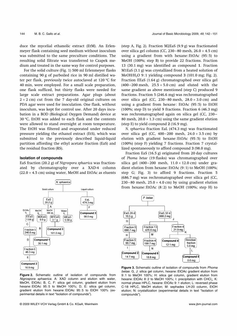
Isolation of compounds EaS fraction (20.2 g) of Nigrospora sphaerica was fraction-
ated by chromatography over a XAD-4 column
(22.0 × 4.5 cm) using water, MeOH and EtOAc as eluents
Figure 2. Schematic outline of isolation of compounds from Nigrospora sphaerica. A: XAD column and elution with water, MeOH, EtOAc; B, C, F: silica gel column, gradient elution from hexane :EtOAc 95 :5 to MeOH 100%; D, E: silica gel column, gradient elution from hexane :EtOAc 95 :5 to EtOH 100% (ex-perimental details in text “Isolation of compounds”).
(step A, Fig. 2). Fraction M2EaS (9.9 g) was fractionated
over silica gel column (CC, 230–80 mesh, 26.0 × 4.5 cm)
using a gradient from with hexane:EtOAc (95:5) to
MeOH (100%; step B) to provide 22 fractions. Fraction
13 (30.1 mg) was identified as compound 1. Fraction
M1EaS (3.1 g) was crystallized from a heated solution of
MeOH/H2O 9:1 yielding compound 3 (101.0 mg; Fig. 2).
Fraction EEaS (1.64 g) chromatographed over silica gel
(400–200 mesh, 25.5 × 5.0 cm) and eluted with the
same gradient as above mentioned (step C) produced 9
fractions. Fraction 5 (246.6 mg) was rechromatographed
over silica gel (CC, 230–80 mesh, 28.0 × 3.0 cm) and
using a gradient from hexane: EtOAc (95:5) to EtOH
(100%; step D) to yield 9 fractions. Fraction 6 (46.3 mg)
was rechromatographed again on silica gel (CC, 230–
80 mesh, 20.0 × 1.3 cm) using the same gradient elution
(step E) to yield compound 2 (16.9 mg).
N. sphaerica fraction EaL (474.3 mg) was fractionated
over silica gel (CC, 400–200 mesh, 24.0 × 3.5 cm) by
elution with gradient hexane:EtOAc (95:5) to EtOH
(100%) (step F) yielding 7 fractions. Fraction 7 crystal-
lized spontaneously to afford compound 3 (98.0 mg).
Fraction EaS (16.5 g) originated from 20 day cultures
of Phoma betae (19 flasks) was chromatographed over
silica gel (400–200 mesh, 11.0 × 12.0 cm) under gra-
dient elution from hexane:EtOAc (9:1) to MeOH (100%;
step G; Fig. 3) to afford 9 fractions. Fraction 5
(686.7 mg) was rechromatographed over silica gel (CC,
230–80 mesh, 25.0 × 4.0 cm) by using gradient elution
from hexane:EtOAc (8:2) to MeOH (100%; step H) to
Figure 3. Schematic outline of isolation of compounds from Phoma betae. G, J: silica gel column, hexane :EtOAc gradient elution from 9 :1 to MeOH 100%; H: silica gel column, gradient elution from hexane :EtOAc 8 :2 to MeOH 100%; I: precipitation with CHCl3; K: normal phase HPLC, hexane :EtOAc 9 :1 elution; L: reversed phase C-18 HPLC, MeOH elution; M: sephadex LH-20 column, EtOH elution; N: crystallization (experimental details in text “Isolation of compounds”).

Journal of Basic Microbiology 2009, 49, 142–151 Production of new cytotoxic agents 145
© 2009 WILEY-VCH Verlag GmbH & Co. KGaA, Weinheim www.jbm-journal.com
afford 12 fractions. Compound 2 (14.7 mg) was precipi-
tated from fraction 5 (99.7 mg) using CHCl3 (step I).
Fraction EaS (888.2 mg) originated from 32 day
cultures of P. betae (1 flask, fungus sporulation time)
was chromatographed over silica gel (400–200 mesh,
27.0 × 3.0 cm) using gradient elution with hexane:
EtOAc (9:1) to MeOH (100%; step J, Fig. 3) to provide
13 fractions (Pb-1 to 13). Fraction 4 (Pb-4, 21.9 mg) was
submitted to HPLC using an isocratic solvent system
containing hexane:EtOAc (9:1) delivered to the a semi-
prep Nucleosil 100–7 column (250 × 10 mm I.D.) at a
flow rate of 1.5 ml min–1, and monitored at 254 nm
detection wavelength at 25 °C (step K). Compound 4
(12.1 mg) was collected at 41 min. The residual fraction
(5.0 mg) collected at 45 min was resubmitted to HPLC
employing a prep C18 Shimadzu column (250 × 20 mm
I.D.) at a flow rate of 2.5 ml min–1, using MeOH as elu-
ent, and monitored at 254 nm (step L). Compound 5
(0.6 mg) was collected at 28 min. Fraction 13 (Pb-13,
426.8 mg) was chromatographed over sephadex LH-20
column (60.0 × 2.5 cm) using EtOH as eluent (step M) to
afford 7 fractions. Fraction 4 crystallized spontaneously
to yield compound 6 (18.8 mg; step N).
Spectroscopic data of compound 1 Pimara-7,15-dien-3β-ol (1): white powder; molecular
formula: C20H32O; [α]D
26 -3.7° (c 2.15, CHCl3); EI/MS m/z (rel.
int.): 288 (30), 273 (60), 255 (100), 187 (38), 145 (35), 119
(55), 107 (70), 91 (55), 80 (48), 55 (42); 1H NMR (500 MHz,
CDCl3): δ 5.69 (1H, dd, J = 10.8 and 18.0 Hz, H-15), 5.10
(1H, m, H-7), 4.82 (1H, dd, J = 1.5 and 18.0 Hz, H-16a),
4.79 (1H, dd, J = 1.5 and 10.4 Hz, H-16b), 3.13 (1H, dd,
J = 4.4 and 11.6 Hz, H-3), 0.90 (3H, s, Me-18), 0.89 (3H, s,
Me-17), 0.88 (3H, s, Me-19), 0.73 (3H, s, Me-20); 13C NMR
(125 MHz, CDCl3): δ 39.2 (C-1), 29.1 (C-2), 79.0 (C-3), 37.1
(C-4), 52.8 (C-5 and 9), 35.8 (C-6), 113.2 (C-7), 38.9 (C-10
and 12), 20.9 (C-11), 35.2 (C-13), 44.1 (C-14), 145.4 (C-15),
110.8 (C-16), 28.1 (C-17), 35.6 (C-18), 21.6 (C-19), 15.4
(C-20), C-8 was not observed.
Spectroscopic and chromatographic equipment 1D and 2D nuclear magnetic resonance (NMR) spectra
were recorded in deuterated solvents (CDCl3, acetone-d6
or MeOH-d4) on Bruker spectrometers (DRX-400 or DRX-
500), working at 400 or 500 MHz for 1H, and at 100 or
125 MHz for 13C. Tetramethylsilane (TMS) was used as
internal reference. Mass spectra were performed in
a high-resolution eletrospray Micromass Quattro LC
spectrometer (HR/ESI/MS) or in a Shimadzu GC/EI/MS
OP2010 employing an AOC-20i auto-injector and a DBS-
MS (30 m × 0.25 mm × 0.25 μm) column. The high per-
formance liquid chromatography (HPLC) system con-
sisted of a Shimadzu LC-6AD pump, and a SPD-20A UV-
vis detector with a CBM-20A interface. Data acquisition
was carried out on LC Solution software version 1.1.
Tumor cell cultures and cytotoxic bioassay Human tumor cell lines MDA-MB435 (breast), HCT-8
(colon), SF295 (brain) and HL-60 (promyelocytic leuke-
mia) were obtained from National Cancer Institute
(Bethesda, MD, USA). All cell lines were cultured in
RPMI1640 medium, supplemented with 10% fetal bo-
vine serum and 1% antibiotics (penicillin and strepto-
mycin). Cultures were maintained in a humidified in-
cubator at 37 °C and 5% CO2 atmosphere. The cytotoxic
effect was assessed using the MTT-dye reduction assay
for cell viability described by Mosmann [16] according
to an established protocol [17]. Briefly, tumor cells were
placed in 96-well plates at the following densities:
0.7 × 10⁵ (HCT-8), 0.6 × 10⁵ (SF295), 0.3 × 10⁶ (HL-60) and
0.1 × 10⁶ (MDA-MB435) cells/ml. Extracts and fractions
were tested in a single concentration (100 μg/ml) and
pure substances in a range between 0.048 and 25 μg/ml
during 72 h. Control groups received the same amount
of vehicle and doxorubicin (Doxolem®, Zodiac Produtos
Farmacêuticos S/A, Brazil), which was used as a positive
control. Growth inhibition rates were quantified as the
percentage of control absorbance by the reduced dye at
550 nm in accordance with the following equation:
Inhibition rate = [(OD control well – OD treated well)
× (OD control well)–1] × 100 .
Each sample was tested in two independent experi-
ments performed in triplicates. The results consisted of
the average value for each experimental unit. The IC50
values and their 95% confidence intervals were ob-
tained by non linear regression using the GraphPad
Prism program (Intuitive Software for Science, San
Diego, CA). An activity scale was utilized to appraise the
cytotoxic potential of the tested samples: inactive sam-
ples (I), samples with low activity (LA, cell growth inhi-
bition between 1–50%), moderated activity (MA, cell
growth inhibition between 50–75%), and high activity
(HA, cell growth inhibition between 75–100%).
Results and discussion
Isolation and identification of fungal strains Altogether 32 species of fungi were isolated and labeled
using the plant name initial letters and a number, for
example, SS29 (Table 1). The results indicated an un-
even distribution of endophytic fungi within S. sonchi-

146 M. B. C. Gallo et al. Journal of Basic Microbiology 2009, 49, 142–151
© 2009 WILEY-VCH Verlag GmbH & Co. KGaA, Weinheim www.jbm-journal.com
Table 1. Endophytic fungi occurrence in Smallanthus sonchifolius tissues.
Tissue ocurrence Fungal strain Taxonomic identification
Leaves Stems Roots
SS13 Papulaspora immersa Hotson P A P SS16 Preussia minima (Auersw.) von Arx. P A A SS20 NI P A A SS21 NI P P P SS22 Mycelia sterile P A A SS26 NI P A A SS29 Aspergillus aculeatus Lizuka P A A SS32 Glomerella cingulata (Stoneman) Spaulding et Schrenk P A A SS33 Drechslera ravenelli (Curt.) Subram. et Jain. P A A SS36 Glomerella sp. P P A SS40 Colletotrichum sp. A P A SS42 Curvularia lunata (Wakker) Boedijn var. aerea
(Bat., Lima et Vasconcelos) M.B. Ellis A P P
SS43 NI A P A SS44 NI A P A SS46 Fusarium oxysporum A A P SS50 Fusarium oxysporum A A P SS55 NI A A P SS62 NI A A P SS65 NG A P P SS67 Nigrospora sphaerica (Sacc.) E. W. Mason A P P SS68 NI A A P SS70 Glomerella sp. A A P SS73 NI A P A SS74 Mycelia sterile A P A SS75 NG A P A SS76 Aspergillus fumigatus Fresenius A P A SS77 Alternaria tenuissima (Kunze) Wiltshire A P A SS78 Mycelia sterile P A A SS79 NI P A A SS82 Aspergillus niger van Tieghen A P A SS83 NI A P A SS84 Phoma betae Frank A P A
NI = not identified. NG = the fungus did not grow. A = absent. P = present.
folius tissues possibly demonstrating tissue specificity.
Hence, it was possible to isolate eleven, six and nine
species from stems, roots and leaves, respectively. A
single species, SS21, displayed a systemic colonization
and five other species occurred simultaneously in two
tissues. Species SS13 was recovered from leaves and
roots, SS36 grew from leaves and stems, while SS42,
SS65 and SS67 were isolated from both stems and roots.
Some strains did not sporulate or grow in the culture
conditions employed. They remain unidentified and
were named sterile mycelia. Isolates that sporulated
were identified by traditional taxonomical methods
based on morphological features as established in
Table 1.
The isolated genera Drechslera, Fusarium, Phoma, Alter-
naria, Curvularia, Nigrospora, Glomerella and Aspergilus
have been previously reported as plant pathogens in a
range of hosts [18–20]. They also have proven to be
endophytes in several plant species [21–26] strengthen-
ing the argument that disease is an exception in the
plant-microbe interaction, which could be more prop-
erly regarded as an unbalanced status in a symbiosis
[27]. Genus Papulaspora is a well-known competitive soil
mold in the culture of mushrooms [28, 29] and it has
just been recently reported as an endophyte in a few
species as Vitex negundo [24], Manilkara bidentata [30],
Taxus chinensis [31], Amomum siamense [32] and Solanum
tuberosum [25]. Preussia, however, is recognized either as
a soil or a coprophilous genus [33, 34] but has never
been mentioned before as an endophyte.
Cytotoxic activity of extracts From the 32 fungal isolates, 186 extracts were obtained
and tested against three tumor cell lines. EaL fractions
had the most pronounced cytotoxic effects together
with EaS fractions (Table 2), whereas fractions EtM, RL
and RS were practically inactive (data not shown). The
exceptions were RLSS33, RLSS75 and RLSS84, which

Journal of Basic Microbiology 2009, 49, 142–151 Production of new cytotoxic agents 147
© 2009 WILEY-VCH Verlag GmbH & Co. KGaA, Weinheim www.jbm-journal.com
Table 2. Cytotoxic activity of the ethyl acetate fractions from liquid (EaL) and solid (EaS) cultures of the endophytic fungi isolated from Smallanthus sonchifolius.
Cell growth inhibition (%) Fungus
HCT-8 MDA-MB435 SF295
EaL fraction EaS fraction EaL fraction EaS fraction EaL fraction EaS fraction
SS13 100.0 (HA) 54.5 (MA) 95.2 (HA) 81.3 (HA) 98.1 (HA) 34.2 SS16 32.0 1.6 31.3 25.7 35.7 4.1 SS20 NC 10.3 NC 11.0 NC 4.1 SS21 27.2 I 37.0 I 23.3 10.9 SS22 100.0 (HA) I 100.0 (HA) 11.8 100.0 (HA) I SS26 31.9 19.9 17.1 15.9 41.2 24.9 SS29 40.1 68.0 (MA) 60.6 (MA) 27.9 33.1 62.9 (MA) SS32 56.6 (MA) 33.7 20.0 33.0 47.7 15.1 SS33 86.7 (HA) 3.6 71.2 (MA) I 84.5 (HA) I SS36 52.4 (MA) 35.3 38.7 44.7 49.0 19.7 SS40 3.6 I 10.7 I 12.0 I SS42 30.9 41.0 36.3 52.0 (MA) 21.2 27.1 SS43 33.9 54.2 (MA) 49.2 59.4 (MA) 25.0 54.5 (MA) SS44 62.7 (MA) 23.3 50.7 (MA) 12.1 48.1 3.2 SS46 85.6 (HA) 13.0 83.2 (HA) 30.3 85.8 (HA) 9.7 SS50 30.2 I 57.9 (MA) I 12.3 I SS55 55.3 (MA) I I I 14.2 I SS62 99.8 (HA) 27.2 51.4 (MA) 32.2 67.5 (MA) 31.8 SS65 NC 1.4 NC 10.8 NC 14.2 SS67 79.2 (HA) 86.2 (HA) 48.2 41.9 73.0 (MA) 82.2 (HA) SS68 39.2 55.2 (MA) 36.4 23.3 26.9 54.4 (MA) SS70 22.1 32.8 32.1 41.7 18.5 18.9 SS73 28.4 I 6.6 I 9.1 I SS74 24.6 83.9 (HA) I 81.0 (HA) 8.4 71.3 (MA) SS75 19.5 22.2 41.0 4.9 7.4 28.1 SS76 100.0 (HA) 48.9 42.6 30.4 52.7 (MA) 40.3 SS77 94.7 (HA) 72.9 (MA) 82.0 (HA) 53.0 (MA) 78.3 (HA) 69.2 (MA) SS78 4.9 I 19.3 3.3 6.4 I SS79 26.2 5.1 I 12.4 8.4 I SS82 87.6 (HA) 9.1 45.7 1.4 68.2 (MA) I SS83 49.3 8.8 27.7 32.5 51.0 (MA) 16.5 SS84 100.0 (HA) 12.4 92.4 (HA) 10.1 95.6 (HA) 15.0 control 25.9 2.2 34.6 20.1 25.1 4.7
displayed moderate to high activities against colon and
breast tumor cells; RSSS65 and RSSS78 inhibited about
50% of colon cells proliferation and EtMSS13, EtMSS67
and EtMSS76 were efficient inhibitors of HCT-8 tumor
cells growth. It is noteworthy that only ethyl acetate
fractions from both culture types of P. immersa (SS13),
N. sphaerica (SS67) and A. tenuissima (SS77) produced good
results on all tested cells indicating that production
of bioactive molecules by these fungi does not depend
on the culture medium employed. These observations
emphasize the significance of these species in future
biotechnology studies.
Cytotoxic effects and ecological importance of isolated compounds N. sphaerica (SS67) and P. betae (SS84) were chosen for
large scale cultures and isolation of bioactive com-
pounds due to the cytotoxicity of their extracts.
Three substances were isolated from the N. sphaerica
EaS fraction (SS67, Fig. 4): compound 1 was identified as
pimara-7,15-dien-3β-ol, compound 2 as ergosterol per-
oxide and compound 3 as aphidicolin. Spectral data of
compound 2 were in agreement with literature data
[35], while compound 1, recently detected in a plant
essential oil, was not characterized by the authors [36].
This is the first report of compound 1 obtained from a
fungal source and having its structure completely elu-
cidated by 1H and 13C NMR, HMQC, HMBC, MS and
NOEdiff spectral data. Both compounds 1 and 2 showed
weak cytotoxic activity against the tumor cell lines
tested (Table 3).
Compound 3, isolated from the EaL fraction of
N. sphaerica (SS67) had its structure established as the
diterpene aphidicolin (Fig. 4) on the basis of its NMR
data that are in agreement with reported data [37].
Aphidicolin showed a remarkable cytotoxic activity

148 M. B. C. Gallo et al. Journal of Basic Microbiology 2009, 49, 142–151
© 2009 WILEY-VCH Verlag GmbH & Co. KGaA, Weinheim www.jbm-journal.com
Figure 4. Structures of compounds isolated from Nigrospora sphaerica and Phoma betae. Pimara-7,15-dien-3β-ol (1); ergosterol peroxide (2); aphidicolin (3); (22E,24R)-ergosta-4,6,8(14),22-tetraen-3-one (4); 8-hydroxy-6-methoxy-3-methylisocoumarin (5); dimethyl terephthalate (6).
against all cell lines (Table 3), but its direct in vivo ad-
ministration is difficult due to its poor water solubility
and fast metabolization. Aphidicolin also may damage a
fragile site of the mammalian genome [38, 39]. How-
ever, it is a valuable tool to study eukaryotic DNA syn-
thesis and cell cycles.
P. betae (SS84) EaL fraction actually showed maximal
efficacy against all the three cancer cell lines and was
Table 3. Inhibitory effect on cultured cell growth of compounds isolated from Nigrospora sphaerica (NS) and Phoma betae (PB), endophytes in Smallanthus sonchifolius.
IC50 (µg/ml) [CI95]a Compound (Fungus)
HCT-8 MDA-MB435 SF295 HL-60
1 (NS) >25 >25 >25 22.60 [18,57–27,64] 2 (NS and PB) >25 >25 >25 >25 3 (NS) 0.05 [0.037–0.079] 0.20 [0.116–0.367] 0.16 [0.081–0.338] 0.09 [0.063–0.174] 4 (PB) 6.24 [4.96–7.85] 14.11 [11.87–16.78] 17.03 [11.40–25.43] 5.29 [4.31–6.50] 5 (PB) >25 >25 >25 >25 6 (PB) >25 >25 >25 >25 Doxorubicin 0.01 0.48 0.24 0.02
a 95% confidence interval.

Journal of Basic Microbiology 2009, 49, 142–151 Production of new cytotoxic agents 149
© 2009 WILEY-VCH Verlag GmbH & Co. KGaA, Weinheim www.jbm-journal.com
considered highly active (Table 2). In liquid potato-
sucrose medium, P. betae is reported to produce an as-
sortment of bioactive diterpenes [40, 41], including ap-
hidicolin (3), an specific inhibitor of DNA polymerase.
Hence, the low cytotoxic activity of SS84 EaS fraction
was intriguing (Table 2) and led us to investigate its
chemical composition. For this purpose the fungus was
cultivated for two different time periods: for 20 days
according to the general protocol and for 32 days leading
to sporulation. EaS fraction from 20 day cultures was
subjected to chromatographic procedures and the resul-
tant subfractions were monitored by GC/MS and 1H NMR
techniques. Several fatty acids were detected but only
compound 2 was isolated corroborating the initial low
activity. However, the EaS fraction resulting from 32 day
cultures yielded compound 6, which was identified as
dimethyl terephthalate (Fig. 4) [42], and a subfraction
that inhibited 100% the proliferation of tumor cell lines
tested. The subfraction 1H NMR spectrum revealed a
mixture of two substances in a 1:1 ratio, later separated
and identified as (22E,24R)-ergosta-4,6,8(14),22-tetraen-3-
one (compound 4) and 8-hydroxy-6-methoxy-3-methyl-
isocoumarin (compound 5) in accordance with literature
data (Fig. 4) [43, 44]. When compounds 4 and 5 were
tested alone they showed moderate and weak activities,
respectively (Table 3). The low cytotoxicity of compound
4 against other tumor cell lines had been previously
described [45, 46] but the results in this report suggest
that isocoumarin (compound 5) and ergosteroid (com-
pound 4) are acting synergistically. In fact, it has been
reported that an isocoumarin propionic acid derivative
has recently entered clinical trials as an anticancer and
anti-angiogenic agent with the property of potentiating
or improving either the therapeutic effects of some
chemotherapeutic drugs [47, 48] or radiotherapy [49].
Our results suggest that compounds 4 and 5 might be
considered as hits for drug development of a novel anti-
cancer combination therapy.
The amounts of pesticides employed in S. sonchifolius
cultivation are relatively small leading some authors to
suggest that sesquiterpene lactones [50] and kaurene
diterpenoids [51] occuring in plant leaves may act as
resistance factors. In fact, the endophytes P. betae and
N. sphaerica and their host have similar biosynthetic path-
ways [52, 53], and the fungal cytotoxic compounds may
provide extra protection for the plant (Table 1). N. spha-
erica is also known to cause both reduction of spore ger-
mination [54] and growth inhibition of some pathogenic
fungi, i.e, Fusarium oxysporum [55, 56], one of the isolated
endophytic species in Yacon roots (Table 1). Therefore,
maintenance of N. sphaerica and P. betae as endophytes
seems to be an ecological benefit to S. sonchifolius.
Acknowledgements
This study was funded by grants from the Fundação de
Amparo à Pesquisa do Estado de São Paulo (FAPESP)
within the Biota-Bioprospecta-FAPESP – The Biodiver-
sity Virtual Institute Program (www.biota.org.br), grant
n° 04/07935-6, and the Brazilian Government’s National
Council for Scientific and Technological Development
(CNPq). The authors thank Silvana França dos Santos
for technical assistance. Margareth B. C. Gallo acknowl-
edges FAPESP for the postdoctoral fellowship (grant n°
05/56259-6).
References
[1] Valentová, K., Truong, N.T., Moncion, A., Waziers, I. and Ulrichová, J., 2007. Induction of glucokinase mRNA by dietary phenolic compounds in rat liver cells in vitro. J. Agric. Food Chem., 55, 7726–7731.
[2] Schorr, K., Merfort, I. and Costa, F.B., 2007. A novel dimeric melampolide and further terpenoids from Smal-lanthus sonchifolius (Asteraceae) and the inhibition of the transcription factor NF-kappa B. Nat. Prod. Communic., 4, 367–374.
[3] Lin, F., Morifumi, H. and Kodama, O., 2003. Purification and identification of antimicrobial sesquiterpene lactones from Yacon (Smallanthus sonchifolius) leaves. Biosci. Bio-technol. Biochem., 67, 2154–2159.
[4] Neves, V.A. and Silva, M.A., 2007. Polyphenol oxidase from Yacon Roots (Smallanthus sonchifolius). J. Agric. Food Chem., 55, 2424–2430.
[5] Valentová, K., Lebeda, A., Doležalová, I., Jirovskỳ, D., Simonovska, B., Vovk, I., Kosina, P., Gasmanová, N., Dziechciarková, M. and Ulrichová, J., 2006. The biological and chemical variability of Yacon. J. Agric. Food Chem., 54, 1347–1352.
[6] Genta, S.B., Cabrera, W.M., Grau, A. and Sánchez, S.S., 2005. Subchronic 4-month oral toxicity study of dried Smallanthus sonchifolius (yacon) roots as a diet supplement in rats. Food Chem. Toxicol., 43, 1657–1665.
[7] Pedreschi, R., Campos, D., Noratto, G., Chirinos, R. and Cisneros-Zevallos, L., 2003. Andean yacon root (Smallan-thus sonchifolius Poepp. Endl.) fructooligosaccharides as a potential novel source of prebiotics. J. Agric. Food Chem., 51, 5278–5284.
[8] Gallo, M.B.C., Guimarães, D.O., Momesso, L.S. and Pupo, M.T., 2008. Natural Products from endophytic fungi. In: Saikai, R., Bezbaruah, R.L. and Bora, T.C. (eds.), Microbial Biotechnology, pp. 139–168. New India Publishing Agen-cy, New Delhi, India.
[9] Firáková, S., Šturdíková, M. and Múčková, M., 2007. Bio-active secondary metabolites produced by microorga- nisms associated with plants. Biologia, 62, 251–257.
[10] Guimarães, D.O., Borges, W.S., Kawano, C.Y., Ribeiro, P.H., Goldman, G.H., Nomizo, A., Thiemann, O.H., Lopes, N.P. and Pupo, M.T., 2008. Biological activities from ex-tracts of endophytic fungi isolated from Viguiera arenaria

150 M. B. C. Gallo et al. Journal of Basic Microbiology 2009, 49, 142–151
© 2009 WILEY-VCH Verlag GmbH & Co. KGaA, Weinheim www.jbm-journal.com
and Tithonia diversifolia. FEMS Immunol. Med. Microbiol., 52, 134–144.
[11] Borges, K.B., Borges, W.S., Pupo, M.T. and Bonato, P.S., 2008. Stereoselective analysis of thioridazine-2-sulfoxide and thioridazine-5-sulfoxide: An investigation of rac-thio-ridazine biotransformation by some endophytic fungi. J. Pharm. Biomed. Anal., 46, 945–952.
[12] Borges, K.B., Borges, W.S., Pupo, M.T. and Bonato, P.S., 2007. Endophytic fungi as models for the stereoselective biotransformation of thioridazide. Appl. Microbiol. Bio-technol., 77, 669–674.
[13] Borges, W.S. and Pupo, M.T., 2006. Novel anthraquinone derivatives produced by Phoma sorghina, an endophyte found in association with the medicinal plant Tithonia diversifolia (Asteraceae). J. Braz. Chem. Soc., 17, 929–934.
[14] Jackson, M., Karwowski, J.P., Humphrey, P.E., Kohl, W.L., Barlow, G.J. and Tanaka, S.K., 1993. Calbistrins, novel an-tifungal agents produced by Penicillium restrictum. 1. Pro-duction, taxonomy of the producing organism and biolo-gical activity. J. Antibiotics, 46, 34–38.
[15] Atlas, R.M., 1995. Handbook of Microbiological Media for the Examination of Food, 310 pp. CRC Press, Boca Raton.
[16] Mosman, T., 1983. Rapid colorimetric assay for cellular growth and survival: application to proliferation and cytotoxicity assays. J. Immunol. Methods, 65, 55–63.
[17] Pohlit, A.M., Tigre, R.F., Cavalcanti, B.C., Moraes, M.O., Costa- Lotufo, L.V., Moraes, M.E.A., Santos, E.V.M., Morais, S.K.R., Nunomura, S.M. and Pessoa, C., 2007. Cytotoxic fractions from the leaves of Tachia grandiflora. Pharm. Biol., 45, 429–433.
[18] Gannibal, P.B., Klemsdal, S.S. and Levitin, M.M., 2007. AFPL analysis of Russian Alternaria tenuissima populations from wheat kernels and other hosts. Eur. J. Plant Pathol., 119, 175–182.
[19] Chrysayi-Tokousbalides, M. and Kastanias, M.A., 2003. Cynodontin: a fungal metabolite with antifungal proper-ties. J. Agric. Food Chem., 51, 4920–4923.
[20] Garcia-Pajón, C.M. and Collado, I.G., 2003. Secondary metabolites isolated from Colletotrichum species. Nat. Prod. Rep., 20, 426–431.
[21] Gond, S.K., Verma, V.C., Kumar, A., Kumar, V. and Khar-war, R.N., 2007. Study of endophytic fungal community from different parts of Aegle marmelos Correae (Rutaceae) from Varanasi (India). World J. Microbiol. Biotechnol., 23, 1371–1375.
[22] Verma, V.C., Gond, S.K., Kumar, A., Kharvar, R.N. and Strobel, G., 2007. The endophytic mycoflora of bark, leaf, and stem tissues of Azadirachta indica A. Juss (Neem) from Varanasi (India). Microbial Ecol., 54, 119–125.
[23] White, I.R. and Backhouse, D., 2007. Comparison of fun-gal endophyte communities in the invasive panicoid grass Hyparrhenia hirta and the native grass Bothriochloa macra. Aust. J. Bot., 55, 178–185.
[24] Raviraja, N.S., Maria, G.L. and Sridhar, K.R., 2006. Anti-microbial evaluation of endophytic fungi inhabiting me-dicinal plants of the Western Ghats of India. Eng. Life Sci., 5, 515–520.
[25] O’Callaghan, M., Gerard, E.M., Waipara, N.W., Young, S.D., Glare, T.R., Barrell, P.J. and Conner, A.J., 2004. Mic-
robial communities of Solanum tuberosum and magainin-producing transgenic lines. Plant Soil, 266, 47–56.
[26] Liu, J.Y., Song, Y.C., Zhang, Z., Wang, L., Guo, Z.J., Zou, W.X. and Tan, R.X., 2004. Aspergillus fumigatus CY018, an endophytic fungus in Cynodon dactylon as a versatile producer of new and bioactive metabolites. J. Biotechnol., 114, 279–287.
[27] Kogel, K., Franken, P. and Hückelhoven, R., 2006. Endo-phyte or parasite – What decides? Curr. Opin. Plant Biol., 9, 358–363.
[28] Katyal, P., Khanna, P.K. and Kapoor, S., 2006. Toxicity of fungicides against Agaricus bisporus and its mycopatho-gens. J. Mycol. Plant Pathol., 36, 296–298.
[29] Fudo, R., Ando, T., Sato, S., Kameyama, T. and Yamanaka, S., 1997. Isolation of isoeugenitin as a fruiting body in-ducer for Stigmatella aurantiaca from a soil fungus Papu-laspora sp. Biosci. Biotechnol. Biochem., 61, 183–184.
[30] Bayman, P., Angulo-Sandoval, P., Báez-Ortiz, Z. and Lodge, D.J., 1998. Distribution and dispersal of Xylaria endo-phytes in two tree species in Puerto Rico. Mycol. Res., 102, 944–948.
[31] Hu, K., Tan, F., Tang, K., Zhu, S. and Wang, W., 2006. Isolation and screening of endophytic fungi synthesizing taxol from Taxus chinensis var. mairei. Ziran Kexueban, 31, 134–137.
[32] Bussaban, B., Lumyong, S., Lumyong, P., McKenzie, E.H.C. and Hyde, K.D., 2001. Endophytic fungi from Amomum siamense. Can. J. Microbiol., 47, 943–948.
[33] Weber, H.A. and Gloer, J.B., 1988. Interference competi- tion among natural fungal competitors: an antifungal metabolite from the coprophilous fungus Preussia fleisch-hakii. J. Nat. Prod., 51, 879–883.
[34] Johnson, J.H., Meyers, E., O’Sullivan, J., Phillipson, D.W., Robinson, G., Trejo, W.H. and Wells, J.S., 1989. Culpin, a novel hydroquinone antibiotic of fungal origin. J. Antibi-otics, 42, 1515–1517.
[35] Bok, J.W., Lermer, L., Chilton, J., Klingeman, H.G. and Towers, G.H.N., 1999. Antitumor sterols from the mycelia of Cordyceps sinensis. Phytochemistry, 51, 891–898.
[36] Tian, G., Li, B., Wang, W., Wang, C. and Gong, K., 2005. Components of the essential oil of Isidon henryi (hemsi) Kudo. Xibei Zhiwu Xuebao, 25, 2543–2548.
[37] Rizzo, C.J. and Smith, A.B., 1991. Aphidicolin synthetic studies: a stereocontrolled end game. J. Chem. Soc. Perkin Trans. I, 969–979.
[38] Palin, A.H., Critcher, R., Fitzgerald, D.J., Anderson, J.N. and Farr, C.J., 1998. Direct cloning and analysis of DNA sequences from a region of the Chinese hamster genome associated with aphidicolin-sensitivity fragility. J. Cell Sci., 111, 1623–1634.
[39] Michaelis, M., Zimmer, A., Handjou, N., Cinatl, J. and Cinatl, J.Jr., 2005. Increased systemic efficacy of aphidico-lin encapsulated in liposomes. Oncol. Rep., 13, 157–160.
[40] Oikawa, H., Toshima, H., Ohashi, S., König, W.A., Kenmo-ku, H. and Sassa, T., 2001. Diversity of diterpene hydro-carbons in fungus Phoma betae. Tetrahedron Lett., 42, 2329–2332.
[41] Ichihara, A., Oikawa, H., Hayashi, K., Hashimoto, M., Sakamura, S. and Sakai, R., 1984. 3-deoxyaphidicolin and

Journal of Basic Microbiology 2009, 49, 142–151 Production of new cytotoxic agents 151
© 2009 WILEY-VCH Verlag GmbH & Co. KGaA, Weinheim www.jbm-journal.com
aphidicolin analogues as phytotoxins from Phoma betae. Agric. Biol. Chem., 48, 1687–1689.
[42] Chenot, E., Bernardi, D., Comel, A. and Kirsh, G., 2007. Preparation of monoalkylterephthalates: an overview. Synth. Commun., 37, 483–490.
[43] Fujimoto, H., Nakamura, E., Okuyama, E. and Ishibashi, M., 2004. Six immunosuppressive features from an Asco-mycete, Zopfiella longicauda, found in a screening study monitored by immunomodulatory activity. Chem. Pharm. Bull., 52, 1005–1008.
[44] Kumagai, H., Amemiya, M., Naganawa, H., Sawa, T., Ishi-zuka, M. and Takeuchi, T., 1994. Biosynthesis of anti- tumor antibiotic, cytogenin. J. Antibiotics, 47, 440–446.
[45] Chen, J.-J., Chou, E.-T., Duh, C.-Y., Yang, S.-Z. and Chen, I.-S., 2006. New cytotoxic tetrahydrofuran- and dihydro-furan-type lignans from the stem bark of Beilschmiedia tsangii. Planta Med., 72, 351–357.
[46] Kwon, H.C., Zee, S.D., Cho, S.Y., Choi, S.U. and Lee, K.R., 2002. Cytotoxic ergosterols from Paecilomyces sp. J300. Arch. Pharmacol., Res., 25, 851–855.
[47] Yuan, H., Junker, B., Helquist, P. and Taylor, R.E., 2004. Synthesis of anti-angiogenic isocoumarins. Curr. Org. Chem., 1, 1–9.
[48] Agata, N., Nogi, H., Milhollen, M., Kharbanda, S. and Kufe, D.W., 2004. 2-(8-hydroxy-6-methoxy-1-oxo-1H-2-benzopy-ran-3-yl)propionic acid, a small molecule isocoumarin, potentiates dexamethasone-induced apoptosis of human multiple myeloma cells. Cancer Res., 64, 8512–8516.
[49] Salloum, R.M., Jaskowiak, N.T., Mauceri, H.J., Seetharam, S., Beckett, M.A., Koons, A.M., Hari, D.M., Gupta, V.K., Reimer, C., Kalluri, R., Posner, M.C., Hellman, S., Kufe, D.W. and Weichselbaum, R.R., 2000. NM-3, an isocouma-
rin, increases the antitumor effects of radiotherapy without toxicity. Cancer Res., 60, 6958–6963.
[50] Inoue, A., Tamogami, S., Kato, H., Nakazato, Y., Akiyama, M., Kodama, O., Akatsuka, T. and Hashidoko, Y., 1995. Antifungal melampolides from leaf extracts of Smallanthus sonchifolius. Phytochemistry, 39, 845–848.
[51] Kakuta, H., Seiki, T., Hashidoko, Y. and Mizutani, J., 1992. Ent-kaurenic acid and its related compounds from glan-dular trichome exudate and leaf extracts of Polymnia son-chifolia. Biosci. Biotech. Biochem., 56, 1562–1564.
[52] Oikawa, H., Nakamura, K., Toshima, H., Tayamasu, T. and Sassa, T., 2002. Proposed mechanism for the reaction ca-talyzed by a diterpene cyclase, aphidicolan-16-beta-ol syn-thase: experimental results on biomimetic cyclization and examination of the cyclization pathway by ab initio calcu-lations. J. Am. Chem. Soc., 124, 9145–9153.
[53] Yao, Q., 2007. Biosynthetic Studies of Fungal Diterpenes Antibiotics. Master Dissertation. Oregon State University. 203 pp.
[54] Perello, A., Simon, M.R. and Arambarri, A.M., 2002. Inter-actions between foliar pathogens and the saprophytic microflora of the wheat (Triticum aestivum L.) phylloplane. J. Phytophatol., 150, 232–243.
[55] Szewczuk, V., Kita, W., Jarosz, B., Truszkowska, W. and Siewinsk, A., 1991. Metabolites of Nigrospora – growth in-hibition of some phytopathogenic fungi by organic ex-tracts from Nigrospora orizae (Berkeley and Broome) Petch. J. Basic Microbiol., 31, 69–73.
[56] Kim, J.-C., Choi, G.J., Park, J.-H., Kim, H.T. and Cho, K.Y., 2001. Activity against plant pathogenic fungi of phomo-lactone isolated from Nigrospora sphaerica. Pest Manag. Sci., 57, 554–559.
((Funded by:
• Fundação de Amparo à Pesquisa do Estado de São
Paulo (FAPESP) within the Biota-Bioprospecta-FAPESP;
grant number: 05/56259-6
• The Biodiversity Virtual Institute Program
(www.biota.org.br); grant number: 04/07935-6
• Brazilian Government’s National Council for Scien-
tific and Technological Development (CNPq) ))