Embryonic skeletal anatomy of the sauropodomorph dinosaur Massospondylus from the Lower Jurassic of...
51
Embryonic skeletal anatomy of the sauropodomorph dinosaur Massospondylus from the Lower Jurassic of South Africa Journal: Journal of Vertebrate Paleontology Manuscript ID: JVP-2009-0201.R1 Manuscript Type: Article Date Submitted by the Author: 04-Jun-2010 Complete List of Authors: Reisz, Robert; University of Toronto Mississauga, Biology Evans, David; Royal Ontario Museum, Palaeobiology; University of Toronto, Ecology and Evolutionary Biology Sues, Hans-Dieter; National Museum of Natural History, Smithsonian Institution, ADRC Office Scott, Diane Key Words: fossil embryos, Early Jurassic, Sauropodomorpha, South Africa Society of Vertebrate Paleontology Journal of Vertebrate Paleontology: For Review Only
Transcript of Embryonic skeletal anatomy of the sauropodomorph dinosaur Massospondylus from the Lower Jurassic of...

Embryonic skeletal anatomy of the sauropodomorph
dinosaur Massospondylus from the Lower Jurassic of South
Africa
Journal: Journal of Vertebrate Paleontology
Manuscript ID: JVP-2009-0201.R1
Manuscript Type: Article
Date Submitted by the Author:
04-Jun-2010
Complete List of Authors: Reisz, Robert; University of Toronto Mississauga, Biology Evans, David; Royal Ontario Museum, Palaeobiology; University of Toronto, Ecology and Evolutionary Biology
Sues, Hans-Dieter; National Museum of Natural History, Smithsonian Institution, ADRC Office Scott, Diane
Key Words: fossil embryos, Early Jurassic, Sauropodomorpha, South Africa
Society of Vertebrate Paleontology
Journal of Vertebrate Paleontology: For Review Only

Embryonic skeletal anatomy of the sauropodomorph dinosaur Massospondylus from the
Lower Jurassic of South Africa
ROBERT R. REISZ1*
, DAVID C. EVANS2, HANS-DIETER SUES
3, and DIANE
SCOTT1
1Department of Biology, University of Toronto at Mississauga, Mississauga, Ontario,
L5L 1C6, Canada;
2Vertebrate Palaeontology, Royal Ontario Museum,100 Queen’s Park, Toronto, Ontario,
M5S 2C6, Canada, [email protected];
3Department of Paleobiology, National Museum of Natural History, Smithsonian
Institution, MRC 121, P.O. Box 37012,Washington, DC 20013-7012, USA,
*Corresponding author. E-mail: [email protected]
Page 1 of 50
Society of Vertebrate Paleontology
Journal of Vertebrate Paleontology: For Review Only
123456789101112131415161718192021222324252627282930313233343536373839404142434445464748495051525354555657585960

ABSTRACT� Two embryonic skeletons preserved inside thin-shelled eggs of a partially
preserved clutch from the Upper Elliot Formation (Lower Jurassic) of South Africa have
been attributed to the sauropodomorph dinosaur Massospondylus carinatus. A virtually
complete skeleton is exposed in right lateral view, with the slightly telescoped skull and
several cervical vertebrae extending beyond the eggshell. A second, partial skeleton has a
skull preserved in dorsal view. The embryos have proportionately very large skulls, with
the broad skull table formed by wide parietals and frontals. The wide posterolateral wing
of the frontal separates the postorbital from contact with the parietal. The embryos have
short rather than elongated cervical vertebrae with tall rather than low neural arches. The
large forelimbs are only slightly shorter than the hind limbs, which suggests an obligatory
quadrupedal posture for the hatchlings. This pattern may represent an ontogenetic
constraint related to the large size of the head and horizontally oriented neck. Similarities
between the embryonic and post-hatchling specimens include the slenderness of the
lower jaw and slight ventral curvature of the symphyseal portion of the dentary, the large
supraorbital process of the prefrontal, and the tall antorbital and infratemporal fenestrae.
There are 10 cervical, 14 dorsal, and three sacral vertebrae. The large distal claw-bearing
phalanx of manual digit 1 is longer than any other phalangeal element of either manus or
pes. The embryos of Massospondylus carinatus represent the oldest dinosaurian embryos
known to date.
Page 2 of 50
Society of Vertebrate Paleontology
Journal of Vertebrate Paleontology: For Review Only
123456789101112131415161718192021222324252627282930313233343536373839404142434445464748495051525354555657585960

INTRODUCTION
Basal sauropodomorph dinosaurs (‘prosauropods’) first appeared in the fossil
record in the early Late Triassic (Sereno, 1999; Galton and Upchurch, 2004; Martínez
and Alcober, 2009). They became the dominant large herbivores in Late Triassic and
Early Jurassic continental ecosystems. Among them, Massospondylus carinatus Owen,
1854 is known from numerous well-preserved specimens from many localities (Fig. 1) in
the Lower Jurassic Lower Elliot, Upper Elliot, and Clarens formations in South Africa
and Lesotho (Massospondylus Range Zone; Kitching and Raath, 1984), as well as from
the correlative Forest Sandstone in Zimbabwe (Cooper, 1981). It is represented by an
extensive growth series of well-preserved, articulated skeletons, with femoral lengths
ranging from 12.7 to 54 cm (specimens housed in the Bernard Price Institute for
Palaeontological Research at the University of Witwatersrand, in Johannesburg, South
Africa; BP/1/5253, BP/1/4934).
In 1976 the late James W. Kitching collected a block containing subspherical eggs
from a layer of reddish-brown, muddy siltstone that is part of the Upper Elliot Formation
(Lower Jurassic) in a roadcut at Rooidraai, in Golden Gate Highlands National Park,
South Africa. The block, BP/1/5347, preserves ten eggs (six of which are complete)
tightly clustered as part of a single clutch. All eggs are tilted slightly in one direction,
possibly because of modest crushing, each egg extending into the sediment and
noticeably beneath the neighboring egg. Kitching tentatively assigned these eggs to a
dinosaur, probably a prosauropod, in a brief preliminary report on the embryos that were
partially exposed (Kitching, 1979). This identification was subsequently questioned by
Zelenitsky and Modesto (2002).
Page 3 of 50
Society of Vertebrate Paleontology
Journal of Vertebrate Paleontology: For Review Only
123456789101112131415161718192021222324252627282930313233343536373839404142434445464748495051525354555657585960

New preparation on this partial cluster of eggs has revealed the presence of
articulated embryonic skeletons (Figs. 2–6) referable to a basal sauropodomorph,
probably Massospondylus carinatus (Reisz et al., 2005). Partial preparation of all the
eggs revealed that five of the ten eggs contain embryonic remains (Reisz et al., 2005).
Small sections of the eggshell were removed from each of the original six eggs, exposing
skeletal elements near the surface of the bone-bearing eggs, but only two embryos were
completely exposed during preparation. One of the eggs at the center of this clutch lacks
embryonic remains. During preparation, numerous shell fragments were discovered
around this egg, suggesting that the embryo may have hatched prior to fossilization of the
clutch. The remaining four eggs are too incomplete to yield embryonic remains.
The exposed articulated skeletons essentially fill the two eggs. The size of the
embryos, together with the advanced level of ossification indicated by the presence of the
well ossified stapes, vertebrae, the fourth trochanter on the femur, and metapodials,
suggests that the animals within these eggs were close to hatching. This contrasts with
embryos of Late Cretaceous titanosaurian sauropod from the Anacleto Formation in
Neuquén Province (Argentina) that have well-developed skulls but poorly ossified limb
elements and vertebrae, and were not close to hatching (Chiappe et al., 1998; Chiappe et
al., 2001). Computed Tomography scans of the eggs from Rooidraai failed to reveal the
delicate bones of the embryos, but established the presence of a highly reflective
envelope surrounding the skeletal remains inside the unprepared egg at the corner of the
clutch, indicating that the organic content of the eggs had settled to the bottom half of the
egg. A mineralized layer next to the bones is also present in the two fully prepared eggs.
Page 4 of 50
Society of Vertebrate Paleontology
Journal of Vertebrate Paleontology: For Review Only
123456789101112131415161718192021222324252627282930313233343536373839404142434445464748495051525354555657585960

It possibly represents remnants of either the egg membrane or extra-embryonic
membranes that enclosed the developing embryo.
These embryos represent the oldest dinosaurian embryos known to date. In view
of the rarity of articulated dinosaurian embryos and their potential significance for
understanding embryonic development and reproduction in early dinosaurs, this paper
documents the anatomy of these specimens in detail for the first time.
DESCRIPTION
Two skeletons have been fully exposed by mechanical preparation for
examination (Table 1) and description. They are referred to as BP/1/5347A-1 and
BP/1/5347A-2 on the larger block, and as BP/1/5347B-1 and BP/1/5347B-2 on the
counterpart blocks. The most complete skeleton, BP/1/5347A-1, is preserved curled-up
and still in articulation (Fig. 2); only the neural arches of its dorsal vertebrae and portions
of some ribs were lost during fossilization. Most of the embryonic skeleton is preserved
on BP/1/5347A, whereas only parts of the bones are present on the counterpart
BP/1/5347B as a result of the splitting process that originally exposed the embryonic
remains. Most of the embryonic skeleton is still contained within the egg on BP/1/5347A,
but preparation revealed four cervical vertebrae protruding beyond the perimeter of the
egg, and the skull is preserved at the end of the cervical series in an area covered with
eggshell fragments (Figs. 2, 3). It is impossible to determine if this position of the skull
was the result of an unsuccessful hatching or due to taphonomic changes, but we tend to
favor the latter interpretation because of the sharp, unnatural angle formed between two
successive cervical vertebrae and the telescoping of the snout. Some postmortem
Page 5 of 50
Society of Vertebrate Paleontology
Journal of Vertebrate Paleontology: For Review Only
123456789101112131415161718192021222324252627282930313233343536373839404142434445464748495051525354555657585960

displacement of dorsal vertebrae is evident just anterior to the pelvis, but otherwise the
skeleton has remained largely undisturbed. After the two skeletons had already been
prepared, the counterpart BP/1/5347B of the original block finally became available for
study. This allowed us to determine that little if any bone was lost because parts that were
missing on the main block were preserved on the counterpart (BP/1/5347B-1), including
dorsal vertebra 12.
The skull of the articulated skeleton (BP/1/5347A and B-1) is partially exposed in
right lateral view (Fig. 2–4). It has been slightly telescoped in the snout region, and the
anterior part of the mandible, the premaxilla, and the anterior end of the maxilla were
lost. We interpret this loss as at least partially due to preservation because a small bone
fragment is present in the area where the end of the snout would have been. Elements of
the skull roof and the right side of the orbital and temporal regions are exposed, with
parts of the palate visible through the orbit, and parts of the braincase protruding through
the temporal fenestrae and skull roof. Despite damage to the snout region, the rest of the
skull is preserved in nearly perfect condition, with even the delicate stapes remaining in
its original position medial to the quadrate.
In the second prepared egg (BP/1/5347A and B-2), the embryo was preserved
fairly evenly on the part and counterpart blocks. Most of its skull was split in two,
exposing both the dorsal and ventral surfaces of the skull roof. We were able to prepare
both part and counterpart (Fig. 5). The skeletal remains on BP/1/5347A were prepared
shortly after the original discovery and illustrated in a brief preliminary report (Kitching,
1979). Unfortunately its counterpart on BP/1/5347B suffered extensive damage caused
by past attempts to extract samples of embryonic bone using a circular sawblade.
Page 6 of 50
Society of Vertebrate Paleontology
Journal of Vertebrate Paleontology: For Review Only
123456789101112131415161718192021222324252627282930313233343536373839404142434445464748495051525354555657585960

However, important elements of the skeleton were still buried sufficiently deeply in the
siltstone matrix and thus escaped damage; they were subsequently exposed through
careful preparation. Preparation of the skull and other regions of the egg’s interior
revealed excellent preservation of bone in the previously concealed or uncut portions of
the egg. The skull is virtually complete on the part and counterpart blocks, with parts of
the skull roof preserved as positive and negative impressions. The positions of various
postcranial elements indicate that this skeleton was also preserved in a curled-up,
partially articulated condition.
The skull is preserved in dorsal view, showing the skull roof, parts of the orbital
region and right side of the palate, as well as portions of the snout. The mandibular rami
have shifted slightly forward, exposing the alveolar region and symphysis. There is a
small sliver between the second and third alveoli that might represent a partial tooth in
the right dentary, but we reject this interpretation because it is not in the right position for
a tooth; all other alveoli are empty. Two small, slender, elongate structures on the dorsal
process of the premaxilla resemble teeth, and may represent teeth that fell out after death.
An alternative interpretation of these two tooth-like structures is that they were egg
“teeth,” possibly used to break the eggshell open during hatching. The dorsal processes of
the premaxillae and nasals have been pushed slightly back relative to the rest of the
premaxillae and maxillae.
Despite proportional differences between the embryos and adults, the structure of
the embryonic skulls (Figs. 3–7) is entirely consistent with their referral to
Massospondylus. The posterior part of the maxilla forms a distinct, slender process that
underlies the jugal. As in juvenile and adult individuals of Massospondylus, this process
Page 7 of 50
Society of Vertebrate Paleontology
Journal of Vertebrate Paleontology: For Review Only
123456789101112131415161718192021222324252627282930313233343536373839404142434445464748495051525354555657585960

lacks teeth (Sues et al., 2004). The tall lacrimal is visible in dorsal view. A long
posterodorsal process of the prefrontal has been separated slightly from the frontal, but
the shape of this part of the bone is identical to that seen in other skulls of
Massospondylus. As in both juvenile and adult Massospondylus and other basal
sauropodomorphs, the anterior end of the dentary is slightly deflected ventrally, although
this deflection is less pronounced than in the adults, especially in the largest known
individual (BP/1/4934). A noteworthy feature of the embryonic skulls is the preservation
of nearly complete rings of scleral ossicles in the orbit. Although most of the known
skulls of Massospondylus preserve some of these elements in the orbital region or nearby
on the skull (Sues et al., 2004:fig.1), their presence in nearly perfect articulation in the
embryos is surprising. In addition, the rod-like stapes is still in its original position on one
of the embryonic skulls. It is well ossified, and its distal end is slightly expanded, as in
the adults.
As in basal sauropodomorphs, the antorbital fenestra is tall and approximately
triangular in outline (Galton and Upchurch, 2004). The external naris is large in the
embryo, but not as tall as the antorbital fenestra. Other notable features of the embryonic
skull include the wide, domed parietal with feeble emargination of the bone for the
supratemporal fenestra, the small size of the nasal, and the large size of the frontal and its
substantial contribution to the orbital margin. In contrast, the largest known skull of
Massospondylus (BP/1/4934) has a relatively short frontal, the nasal is the longest paired
element in the skull roof, and the parietals are strongly emarginated along the medial
edge of the fenestra. However, other, smaller specimens show intermediate stages
between the two extremes (Sues et al., 2004).
Page 8 of 50
Society of Vertebrate Paleontology
Journal of Vertebrate Paleontology: For Review Only
123456789101112131415161718192021222324252627282930313233343536373839404142434445464748495051525354555657585960

Comparisons in the following description focus on the post-hatchling cranial
growth series of Massospondylus described by Sues et al. (2004) and will highlight
changes during growth in addition to the unique features of the embryo. The antorbital
fenestra is tall and approximately triangular in outline. The external naris is large in the
embryo but not as tall as the antorbital fenestra. The supratemporal fenestra is longer than
wide, unlike its shape in adult Massospondylus where this fenestra is approximately as
wide as long (BP/1/4779). Unique to the embryo is the slight contribution of the frontal to
the anterior margin of the fenestra, and the absence of a contact between the parietal and
postorbital anteriorly. In the smallest known juvenile, BP/14376 (Sues et al., 2004:fig. 1)
the frontal has a slight emargination for the edge of the fenestra, but there is a narrow
contact between the postorbital and parietal. The infratemporal fenestra is hourglass-
shaped, as in adults, and bounded by the same bones.
Skull Roof
The premaxilla, observable only in BP/1/5347A-2 (Fig. 6A), forms the anterior
end of the snout and the anteroventral margin of the external naris. Its small size indicates
that the anterior end of the snout was narrow in dorsal view. The posterolateral process,
which overlaps the anterior ramus of the maxilla below the external naris, is not as well
developed as in larger specimens of Massospondylus (e.g., BPI/1/5241, BPI/1/4934), but
this may be due to breakage. As a result, the suture between the premaxilla and maxilla is
inclined posterodorsally, rather than being shaped like an inverted L in lateral view (Sues
et al., 2004). The presence of a subnarial foramen on the suture between the premaxilla
and maxilla in the embryos is uncertain, because the left premaxilla of BP/1/5347A-2 has
Page 9 of 50
Society of Vertebrate Paleontology
Journal of Vertebrate Paleontology: For Review Only
123456789101112131415161718192021222324252627282930313233343536373839404142434445464748495051525354555657585960

been displaced and overlies the anterior end of the maxilla. The dorsal (internarial)
processes curve posterodorsally, and appear in dorsal view as two thin straps that are
appressed at the midline and separate the anterior ends of the nasals. At the anteroventral
corner of the external naris there is a shallow fossa at the confluence of the dorsal process
and lateral ramus. The body of the premaxilla and its tooth-bearing margin are poorly
preserved, and the number of alveoli is unknown. Two small, slender, conical structures
with a waxy patina on the dorsal process of the premaxilla may represent teeth, but their
identity is uncertain. They are much smaller than the empty alveoli of the mandible, as is
another tooth-like structure that was exposed in cross-sectional view as an isolated
element on the counterpart block BP/1/5347B-2. However, in the latter case, the cross-
sectional view exposes a tiny pulp cavity, a dense, dentine-like core, and a very thin outer
layer that may be the enamel layer. If this interpretation is correct, these small teeth
would have been at a very early stage of development, and nowhere near a size to
approach eruption. Their postmortem displacement, while the rest of the skeleton has
remained largely in place, also supports this interpretation.
The triradiate maxilla (Figs. 4, 6A) comprises a dorsoventrally tall anterior
process, a dorsal process, and a straight posterior ramus. Below the external naris, the
maxilla is tall and has a dorsoventrally convex lateral surface. The dorsal surface of the
subnarial process and base of the dorsal process are transversely concave at the
posteroventral corner of the external naris. Just anterior to the base of the dorsal process,
a large neurovascular foramen, also present in this region on adult skulls of
Massospondylus (Sues et al., 2004), pierces the lateral surface along the margin of the
narial fossa. The posterodorsally directed dorsal process is set back approximately one-
Page 10 of 50
Society of Vertebrate Paleontology
Journal of Vertebrate Paleontology: For Review Only
123456789101112131415161718192021222324252627282930313233343536373839404142434445464748495051525354555657585960

third from the anterior end of the bone. It forms the posteroventral margin of the external
naris anteriorly and the anterior margin of the antorbital fenestra posteriorly. The surface
of the maxilla is gently concave adjacent to the antorbital fenestra to form the antorbital
fossa, which is particularly well demarcated at the base of the posterior ramus. The
margin of the inset medial lamina that defines the edge of the antorbital fenestra is close
and parallel to the rim of the antorbital fossa where preserved, as in Massospondylus and
Lufengosaurus but unlike Plateosaurus (Galton and Upchurch, 2004) and Aardonyx
(Yates et al., 2010), in which the flange is much broader. The posterior ramus of the
maxilla forms a straight, slender process that tapers beneath the jugal in lateral view. This
process, which terminates posteriorly just beyond the anterior margin of the orbit in the
embryo, extends past the ventral midpoint of the orbit in larger specimens of
Massosospondylus (Sues et al., 2004) as well as Lufengosaurus (Barrett et al., 2005). The
lateral surface of the body of the maxilla has several supralabial foramina. The long
alveolar margin preserves no erupted teeth.
The right nasal of BP/1/5347A-2 is almost complete (Fig. 6). Its total length is
less than 25% the length of the embryonic skull; its relatively smaller size than that of
large individuals of Massospondylus (in which the nasal is approximately half the skull
length, Sues et al., 2004) implies a positive allometry of the antorbital region of the skull
during growth. This is confirmed by the size of the nasal in juvenile skulls. The
anteromedial and anterolateral processes of the nasal diverge and form the posterodorsal
margin of the external naris. The anteromedial process overlaps the dorsal processes of
the premaxilla as it curves ventrally and tapers to a point anterior to the dorsal midpoint
of the naris. The triangular anterolateral process does not reach the lateral process of the
Page 11 of 50
Society of Vertebrate Paleontology
Journal of Vertebrate Paleontology: For Review Only
123456789101112131415161718192021222324252627282930313233343536373839404142434445464748495051525354555657585960

premaxilla. It is unclear whether the anterolateral process contributes to the margin of the
antorbital fenestra. The posterior end of the nasal is flattened at its contact with the
frontal on the skull roof. In dorsal view, the nasals are slightly separated posteriorly by an
anteromedial projection of the frontals.
The prefrontal forms the anterodorsal margin of the orbit. It articulates with the
lacrimal ventrally, and has a long posterodorsal process that extensively overlaps the
frontal on the skull roof. Nevertheless, in dorsal view (Figs. 3, 6), the posterodorsal
process is relatively shorter than in skulls of post-hatchling specimens of Massospondylus
(BP/1/4376, BP/1/5241), suggesting this portion of the prefrontal increases in relative
length through ontogeny, perhaps related to the increasing elongation of the snout. The
anteromedial suture with the nasal anterior to the frontal contact is long and well
developed.
The lacrimal (Figs. 3, 6A) is similar to that in adult Massospondylus, with a tall
ventral ramus and a short anterodorsal ramus. The anteriorly bowed ventral ramus forms
the anterior margin of the orbit and the posterior margin of the antorbital fenestra.
Ventrally, the lacrimal articulates with the jugal. The presence of a medial lamina just
above the contact with the jugal, which corresponds to the posteroventral region of the
antorbital fossa, is uncertain; if indeed present, it was likely small. The relative size of the
medial lamina increases in Massospondylus during growth (Sues et al., 2004). Dorsally,
the lacrimal is expanded and extends towards the dorsal apex of the maxilla, but it is
unclear whether they meet to exclude the nasal from the antorbital fenestra. The lacrimal
forms a small contribution to the skull roof lateral to the nasal in dorsal view. The
Page 12 of 50
Society of Vertebrate Paleontology
Journal of Vertebrate Paleontology: For Review Only
123456789101112131415161718192021222324252627282930313233343536373839404142434445464748495051525354555657585960

lacrimal is identical to that seen in other, larger skulls of Massospondylus and appears to
have changed little during ontogeny (Sues et al., 2004).
The frontal (Figs. 3, 6) is the largest bone of the embryonic skull. In dorsal and
lateral views, the frontal makes a large contribution to the dorsal rim of the orbit. This
feature is unchanged ontogenetically, as the degree of exposure of the frontal along the
orbit is similar to that in juvenile and adult specimens of Massospondylus (Sues et al.,
2004), but differs from the relatively small contribution to the orbital margin in
Lufengosaurus (Barrett et al., 2005). The total length of the frontal is more than three
times the transverse width of the bone at midlength of the orbit. In this respect, the
embryo more closely resembles the smallest known juvenile Massospondylus
(BP/1/4376) than larger specimens, in which the frontal is proportionately shorter. The
nasal and prefrontal overlap the anterior end of the frontal adjacent to the anterior end of
the orbit. Posteriorly, the anteromedial extension of the parietals results in a W-shaped
frontal-parietal contact in dorsal view. In larger specimens of Massospondylus this suture
is essentially straight, and, although the posterior margin of the frontal is slightly
emarginated by the supratemporal fenestra, the anterior margin of the opening is formed
mainly by the parietal and postorbital. The frontal is overlapped by the postorbital at the
posterodorsal corner of the orbit.
The large, domed parietals (Figs. 3, 6) are emarginated laterally along the margin
of the supratemporal fenestra. The lateral emargination in the embryo is substantially less
developed than in larger specimens of Massospondylus, in which the parietals form a
relatively narrow, stout bony separation between the two supratemporal fenestrae, best
seen in BP/1/5241 (Sues et al., 2004:fig. 7). In addition, the attachment area for the
Page 13 of 50
Society of Vertebrate Paleontology
Journal of Vertebrate Paleontology: For Review Only
123456789101112131415161718192021222324252627282930313233343536373839404142434445464748495051525354555657585960

adductor jaw musculature, which is extensively developed in adults, is also poorly
demarcated in the embryo. Anteriorly, the parietal does not meet the postorbital to
exclude the frontal from the margin of the supratemporal fenestra, as in larger specimens
of Massospondylus (BP/1/5241, BP1/1/4779). The parietals are depressed anteromedially
adjacent to the frontals, but there is no pineal foramen. Short posterolateral processes
contact the squamosals.
In lateral view, the postorbital is triradiate (Figs. 3, 6B). Its ventral process
separates the orbit from the infratemporal fenestra. The ventral half of this process forms
an extensive contact with the jugal. It curves distally and tapers to a point near the
midpoint of the ventral margin of the orbit. The short posterior process of the postorbital
articulates with the squamosal posteriorly and forms the bony bar separating the supra-
and infratemporal fenestrae. Dorsally, the postorbital becomes a thin sheet that extends
onto the skull roof where it extensively overlaps the frontal.
The jugal, excellently preserved in both embryos (Figs. 3, 6), is triradiate in
lateral view and forms the ventral margin of the orbit and the anteroventral corner of the
infratemporal fenestra. Its anterior process articulates with the lacrimal medially and
forms a long ventral suture with the maxilla below the anterior third of the orbit. In lateral
view, the anterior process tapers to a point ventral to the lacrimal and reaches the
antorbital fossa or fenestra but does not make a noticeable contribution to its margin. The
dorsal process of the jugal extends posteriorly at an angle of about 45° to the maxillary
ramus and forms the posteroventral half of the postorbital bar that separates the orbit
from the infratemporal fenestra. The short posterior process articulates with the
quadratojugal at the midpoint of the ventral margin of the infratemporal fenestra.
Page 14 of 50
Society of Vertebrate Paleontology
Journal of Vertebrate Paleontology: For Review Only
123456789101112131415161718192021222324252627282930313233343536373839404142434445464748495051525354555657585960

We interpret a small, slender piece of bone preserved close to the jugal and
partially covered by the quadrate in BP/1/5347A-1 (Fig. 3) as the anteroventral part of the
quadratojugal. The contact between the jugal and quadratojugal is relatively shorter and
different in structure from that in adult Massospondylus, in which the quadratojugal
tapers to a point below the posterior process of the jugal in lateral view.
The right quadrate is fully exposed in posterolateral view in BP/1/5347A-1 (Fig.
3), and the dorsal end of this bone is also preserved in BP/1/5347A and B-2 (Fig. 6). Its
small dorsal head fits into a ventral cotylus on the squamosal. In lateral view, the large,
anteriorly directed lateral wing progressively expands from the head to the point of the
quadratojugal articulation. Dorsal to the quadratojugal, the lateral wing articulates with
the elongate ventral process of the squamosal in all known specimens of Massospondylus
(Sues, et al., 2004), but the condition is unclear in the embryo because this portion of the
squamosal is not preserved. Along the contact with the quadratojugal, the lateral wing
becomes narrower toward the mandibular condyle. The mandibular condyle is expanded
in both the transverse and parasagittal planes. It is saddle-shaped and smoothly convex in
lateral view. The medial (pterygoid) wing of the quadrate is not exposed.
The squamosal (Figs. 3, 6B) forms the posterodorsal corner of the skull in lateral
aspect and contributes to the posterolateral and posterodorsal margins of the supra- and
infratemporal fenestrae, respectively. The ventral process of the squamosal, which is
typically well developed in prosauropods, is poorly exposed on both embryonic skulls.
Palate and Braincase
Page 15 of 50
Society of Vertebrate Paleontology
Journal of Vertebrate Paleontology: For Review Only
123456789101112131415161718192021222324252627282930313233343536373839404142434445464748495051525354555657585960

The palatine, exposed in dorsal view in both specimens, forms a large
anterolateral portion of the palate medial to the maxilla. The anterior, medial, and
posterior contacts of this bone cannot be identified in the embryonic skulls. The lateral
(maxillary) ramus of the palatine is exposed in BP/1/5347A-2 (Fig. 6A). It is expanded
anteroposteriorly where it contacts the medial surface of the maxilla below the orbit. The
bone is anteroposteriorly longer in adult specimens.
A large, irregularly shaped bone visible in dorsal view through the left orbit of the
BP/1/5347A-2 (Fig. 6) probably represents the pterygoid. It consists of a large posterior
plate and an elongate anterior palatal ramus for contact with the vomer and palatine as in
other prosauropods (Sues et al., 2004). The medial margin of the bone is slightly bowed
laterally as preserved. Posteriorly, the bases of the quadrate flange and the transverse
process extend at approximately right angles to one another, but their articular
relationships cannot be determined.
Other than for the bones of the skull roof, little information on the braincase is
available. None of the endochondral bones that make up the braincase is exposed. What
we tentatively identify as the cultriform process of the parabasisphenoid has been
displaced ventrally and is exposed between the mandibular rami in BP/1/5347A-1 (Fig.
3).
A right stapes is preserved in its original position in the skull of BP/1/5347A-1
(Fig. 4). Its distal end is slightly expanded, as in the adults; its proximal end is not
exposed. If correctly identified, the stapes appears to be more robust than that in larger
skulls of Massopondylus (Sues et al., 2004:fig. 4). Ossified scleral plates are also present
Page 16 of 50
Society of Vertebrate Paleontology
Journal of Vertebrate Paleontology: For Review Only
123456789101112131415161718192021222324252627282930313233343536373839404142434445464748495051525354555657585960

in both embryonic skulls (Figs. 3, 6A). BP/1/5347A-1 preserves a complete scleral ring
comprised of approximately eight thin bony plates.
Hyoid ossifications have not been identified in the embryonic material.
Mandible
A nearly complete mandible is preserved exposed in lateral view in BP/1/5347A-
1 (Fig. 3), whereas the lower jaws in the second skull, BP/1/5347A-2 (Fig. 6A) are
almost completely covered by the skull. However, they have shifted forward sufficiently
to expose the anterior portion of the alveolar rami in dorsal view. The prearticular,
articular, splenial, and coronoid are not visible in any of the specimens under study. As in
post-hatchling individuals, the mandible is approximately the same length as the cranium.
The anteroposteriorly elongate, oval external mandibular fenestra is formed by the
dentary, surangular, and angular. It is approximately 11 percent the length of the lower
jaw in the embryo and appears somewhat more elongate than in post-hatchling
individuals (BP/1/4376, BP/1/4779).
The dentary, the largest bone of the mandible, bears the distinct ridge on its
posterolateral surface; this feature is characteristic of most basal sauropodomorphs
(Galton and Upchurch, 2004) but is absent in Aardonyx (Yates et al., 2010). In lateral
view, the tooth-bearing ramus is slightly bowed dorsally and has subparallel dorsal and
ventral margins. A short edentulous region approximately equal to one alveolus in length
occurs at the anterior end of the dentary, another feature diagnostic for basal
sauropodomorphs (Galton and Upchurch, 2004). Posteriorly, the dentary bifurcates
around the external mandibular fenestra. The dorsal and ventral branches articulate with
Page 17 of 50
Society of Vertebrate Paleontology
Journal of Vertebrate Paleontology: For Review Only
123456789101112131415161718192021222324252627282930313233343536373839404142434445464748495051525354555657585960

the surangular and angular, respectively, and the region between them forms the anterior
margin of the opening.
The anterior four alveoli as exposed in BP/1/5347A-2 (Fig. 6A) are notably large
and long; each alveolus is approximately 1 mm in length, and the tooth-bearing ramus of
the dentary is approximately 10 mm long. Therefore, there were approximately 10–12
tooth positions in the dentary of the embryos, which is a lower count than but consistent
with the progressively increasing number of dentary teeth seen in later growth stages of
Massospondylus (Sues et al., 2004). A small fragment at the confluence of the second and
third alveolus of BP/1/5347A-2 has a "waxy" texture distinct from the dentary and may
represent a partial dentary tooth, although there are no serrations or other morphological
features that would confirm this identification. The remaining alveoli are empty.
The surangular is the second largest mandibular element and forms the dorsal
region of the mandibular ramus posterior to the dentary. The lateral surface bears a
horizontal ridge that is continuous with that on the dentary. Anteriorly, the surangular
overlaps the dentary and forms the dorsal margin of the external mandibular fenestra. In
lateral view, the dorsal margin of the surangular rises posteriorly from the anterior end of
the bone to a slightly convex coronoid eminence anterior to jaw joint. Ventrally, the
surangular contacts the angular along a subhorizontal suture that extends posteriorly from
the external mandibular fenestra.
The angular is a long, strap-like bone ventral to the surangular on the
posterolateral surface of the mandibular ramus. It forms the ventral margin of the external
mandibular fenestra and is overlapped by the dentary anteriorly. In lateral view, the
Page 18 of 50
Society of Vertebrate Paleontology
Journal of Vertebrate Paleontology: For Review Only
123456789101112131415161718192021222324252627282930313233343536373839404142434445464748495051525354555657585960

ventral margin of the angular curves gently upwards from its contact with the dentary to
the region below the jaw joint.
Postcranial Skeleton
The structure of the embryonic postcranial skeleton generally conforms to that in
more mature individuals of Massospondylus (Cooper, 1981), but differs dramatically in
its relative proportions, presumably in part related to constraints associated with
accommodating the developing embryo in a small egg.
Vertebral Column—There are eight exposed cervical vertebrae; very slender,
delicate, relatively short ribs are associated with all of them. The cervical vertebrae are
preserved in articulation; the atlas-axis complex could not be exposed without inflicting
damage to the skull. The cervical centra are not elongated relative to the dorsal centra,
but there is already extensive zygapophyseal contact between the vertebrae. Little
detailed information is available on the centra, except for basic measurements (Table 1).
The centra are slender, spool-shaped, and slightly shorter than the dorsal centra; there is
little variation in centrum length along the series. This is in marked contrast to the
condition seen in the adults (Cooper, 1981), where the cervical centra are greatly
elongated, with the mid-cervical centra being significantly longer than either the anterior
and posterior cervical centra. Juveniles (e.g., BP/1/4376) show an intermediate condition
between the embryos and the adults. The cervical neural arches are quite tall in the
embryos, possibly associated with the presence of the proportionately very large head. As
a result, the height of all cervical vertebrae is greater than their respective lengths. In
addition, the neural arches vary in height along the cervical series, becoming tallest on
Page 19 of 50
Society of Vertebrate Paleontology
Journal of Vertebrate Paleontology: For Review Only
123456789101112131415161718192021222324252627282930313233343536373839404142434445464748495051525354555657585960

the posterior cervical vertebrae, possibly even taller than in the dorsal vertebrae. This is
in marked contrast to the adult condition, where the cervical vertebrae are greatly
elongated, and the neural arches are low and anteroposteriorly long. The slender, well-
ossified cervical ribs are relatively short and thus differ from those in adult
Massospondylus, in which they extend for the length of at least two vertebrae (pers. obs.,
RRR). The last embryonic cervical rib is significantly longer than the others, being more
than twice as long the fourth cervical rib. The ribs are too delicate and small to preserve
the distinctive, anteriorly directed process seen in adults. The shape of the cervical
column indicates that the neck was held horizontally, as in adult Massospondylus and
other basal sauropodomorphs (Galton and Upchurch, 2004). The skeletal reconstruction
(Fig. 8) shows the long neck in maximum dorsiflexion, with the skull held far anteriorly.
Thirteen dorsal vertebrae are preserved in BP/1/5347A-1 (Figs. 2, 3), with most of
the ribs still in their original position. Damage in the posterior region of the series has
somewhat disrupted the ribs, but a tiny posterior left dorsal rib is preserved just anterior
to the pelvis. There is a gap within the dorsal series that corresponds to dorsal vertebra
12. The centrum of this vertebra is preserved, however, in BP/1/5347B-1, the result of the
original split of the egg-containing block into part and counterpart. This counterpart piece
was recently reexamined, once it has been returned from an extended loan, allowing for a
more complete reconstruction of the skeleton (Fig. 8), than was previously possible
(Reisz et al. 2005). As in juvenile and adult Massospondylus, there appears to be little if
any variation in centrum length along the series (Table 1). No useful information is
available about the shape and size of the dorsal neural spines. The first and third sacral
ribs are preserved. The rod-like first sacral rib extends to the anterior edge of the iliac
Page 20 of 50
Society of Vertebrate Paleontology
Journal of Vertebrate Paleontology: For Review Only
123456789101112131415161718192021222324252627282930313233343536373839404142434445464748495051525354555657585960
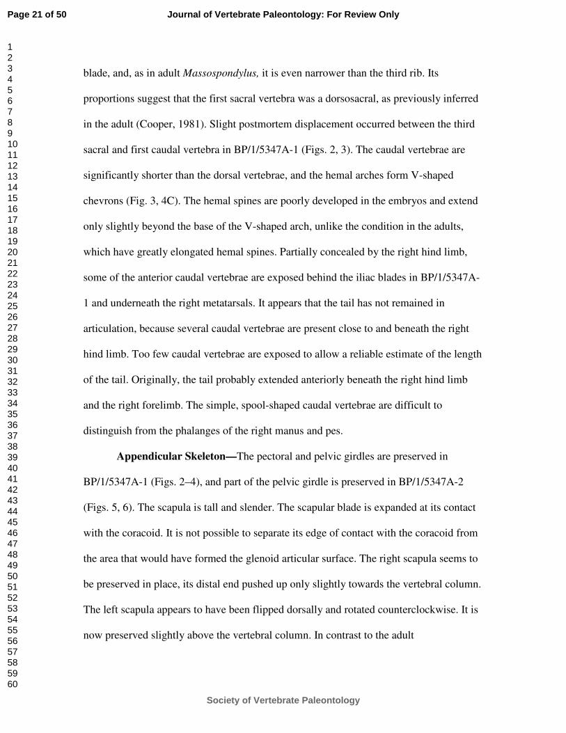
blade, and, as in adult Massospondylus, it is even narrower than the third rib. Its
proportions suggest that the first sacral vertebra was a dorsosacral, as previously inferred
in the adult (Cooper, 1981). Slight postmortem displacement occurred between the third
sacral and first caudal vertebra in BP/1/5347A-1 (Figs. 2, 3). The caudal vertebrae are
significantly shorter than the dorsal vertebrae, and the hemal arches form V-shaped
chevrons (Fig. 3, 4C). The hemal spines are poorly developed in the embryos and extend
only slightly beyond the base of the V-shaped arch, unlike the condition in the adults,
which have greatly elongated hemal spines. Partially concealed by the right hind limb,
some of the anterior caudal vertebrae are exposed behind the iliac blades in BP/1/5347A-
1 and underneath the right metatarsals. It appears that the tail has not remained in
articulation, because several caudal vertebrae are present close to and beneath the right
hind limb. Too few caudal vertebrae are exposed to allow a reliable estimate of the length
of the tail. Originally, the tail probably extended anteriorly beneath the right hind limb
and the right forelimb. The simple, spool-shaped caudal vertebrae are difficult to
distinguish from the phalanges of the right manus and pes.
Appendicular Skeleton—The pectoral and pelvic girdles are preserved in
BP/1/5347A-1 (Figs. 2–4), and part of the pelvic girdle is preserved in BP/1/5347A-2
(Figs. 5, 6). The scapula is tall and slender. The scapular blade is expanded at its contact
with the coracoid. It is not possible to separate its edge of contact with the coracoid from
the area that would have formed the glenoid articular surface. The right scapula seems to
be preserved in place, its distal end pushed up only slightly towards the vertebral column.
The left scapula appears to have been flipped dorsally and rotated counterclockwise. It is
now preserved slightly above the vertebral column. In contrast to the adult
Page 21 of 50
Society of Vertebrate Paleontology
Journal of Vertebrate Paleontology: For Review Only
123456789101112131415161718192021222324252627282930313233343536373839404142434445464748495051525354555657585960

Massospondylus but similar to the condition in the small juveniles, the dorsal portion of
the scapular blade is not expanded anteroposteriorly. Most of the right coracoid was lost,
and it is difficult to determine how much of it was ossified at the time of death. There
appear to be no ossified sternal elements, and there is no evidence of a clavicle. The
pelvic girdle is partially exposed in BP/1/5347A-1, with both ilia and ischia but only the
right pubis visible. The left iliac blade is also exposed in BP/1/5347A-2. Although the
iliac blade is already ossified, its supra-acetabular crest is not preserved and possibly was
not yet ossified. The iliac blade has a slender postacetabular process but a poorly
developed preacetabular process. The ischium and pubis, exposed in BP/1/5347A-1, are
short and slender, approximately one-half the length of the femur, and are much thinner
and less well ossified than either the scapula or the ilium. These pelvic proportions are
even more pronounced than in the holotype post-hatchling skeleton of Mussaurus
patagonicus, a basal sauropodomorph from the Upper Triassic El Tranquilo Formation of
Santa Cruz Province, Argentina (Bonaparte and Vince, 1979). In contrast, the ischium
and pubis are large and massive in the adults and exceed the ilium in size (Cooper, 1981).
The limbs are well preserved. All limb bones have thin bony walls and probably
had large cartilaginous internal cones in the expanded proximal and distal portions. This
is evident on the left tibia, where the internal surface of the shaft was exposed by
postmortem loss of bone along the distal half of the element. In addition, the external
surfaces of all proximal limb bones are strongly fluted near their proximal and distal
ends, indicating the presence of substantial cartilaginous “epiphyseal” caps. Despite the
thinness of the ossified portions of these elements, there is surprisingly little crushing.
Page 22 of 50
Society of Vertebrate Paleontology
Journal of Vertebrate Paleontology: For Review Only
123456789101112131415161718192021222324252627282930313233343536373839404142434445464748495051525354555657585960

The forelimbs are preserved in position on either side of the body. They comprise
both humeri, radii, and ulnae, as well as some elements of the manus (Table 1). The
proximal head of the humerus is larger than the distal end, probably due to the enormous
size of the deltopectoral crest. This is evident on the left humerus of BP/1/5347A-1 (Figs.
2–4), which has a the proximal head exposed in anteromedial view, with a small, ossified
crest projecting upwards, exposed right next to the edge of the shaft of the right humerus.
The distal end of the left humerus is almost completely covered by three thoracic ribs in
BP/1/5347A-1, but the distal end of the right humerus is exposed, showing a slight
depression that probably represents the intercondylar groove. Part of the humerus is also
exposed in BP/1/5347B-2, showing a well-ossified distal end of the bone (Figs. 5, 6B).
The proximal head of the ulna is wider than that of the radius, as in the adults, and lacks
an ossified olecranon, best seen in BP/1/5347B-2 (Fig. 6B). The radius and ulna are
preserved in a pronated position on the right side of BP/1/5347A-1 (Figs. 2–4), with the
slightly curved, slender radius crossing over the more massive, less curved ulna. They are
located beneath the right tibia and fibula, and only partly visible as illustrated. The left
radius and ulna are partly covered by ribs and the right pubis. Both radius and ulna are
significantly shorter and more slender than the fibula or tibia (Table 1). Parts of both
hands are preserved in BP/1/5347A-1 (Figs. 2–4). Only a single metacarpal of the left
manus is exposed, whereas the right manus is more complete but concealed by the distal
end of the tibia and right tarsus. No carpal elements are ossified in the embryos, and there
is a wide gap between the distal ends of the radius and ulna and the proximal ends of the
metacarpals in both skeletons. This lack of ossification is not unexpected since juvenile
individuals of Massospondylus usually have unossified carpals; for example, BPI 4376
Page 23 of 50
Society of Vertebrate Paleontology
Journal of Vertebrate Paleontology: For Review Only
123456789101112131415161718192021222324252627282930313233343536373839404142434445464748495051525354555657585960

preserves a complete manus with fully ossified metacarpals and phalanges but lacks any
carpals. In BP/1/5347A-1 (Figs. 2–4), the right manus, with two metacarpals, which are
approximately equal in size, and three associated phalanges can be readily identified. The
bones have flipped over at the level of the wrist, and have come to rest against the radius
and ulna. A relatively robust, wide element, slightly displaced, may be the first
metacarpal, but there are no phalangeal elements associated with it. If this identification
is correct, then the other metacarpals belong to digits II and III, respectively. The latter
two metacarpals are wider proximally than distally, and are closely appressed to each
other. The slightly more robust metacarpal has two proximal phalanges preserved in
articulation with it. The second, slightly more slender metacarpal has only one phalanx
preserved in articulation, but it is slightly longer than the other proximal phalanx in the
hand. All phalanges are shorter than the metacarpals. BP/1/5347B-2 (Figs. 5, 6B)
preserves a nearly complete right manus, which is separated slightly from the radius and
ulna by a gap that would have been occupied by cartilaginous carpal elements.
Identification of the ossified elements of the manus is therefore easier than in
BP/1/5347A-1. Four of the five metacarpals are ossified. The robust and short first
metacarpal is in articulation with a particularly robust proximal phalanx. This condition is
similar to that seen in juvenile and adult skeletons of Massospondylus. A very large,
elongated, and strongly curved ungual phalanx, the longest of the preserved phalanges, is
interpreted as the distal phalanx of the first digit, but has shifted slightly laterally from its
original articulated position. This ungual is also very large in all known post-hatchling
skeletons. Metacarpal II and III are similar in size, whereas metacarpal IV is slightly
shorter and more slender than the other two elements. They are all significantly longer
Page 24 of 50
Society of Vertebrate Paleontology
Journal of Vertebrate Paleontology: For Review Only
123456789101112131415161718192021222324252627282930313233343536373839404142434445464748495051525354555657585960
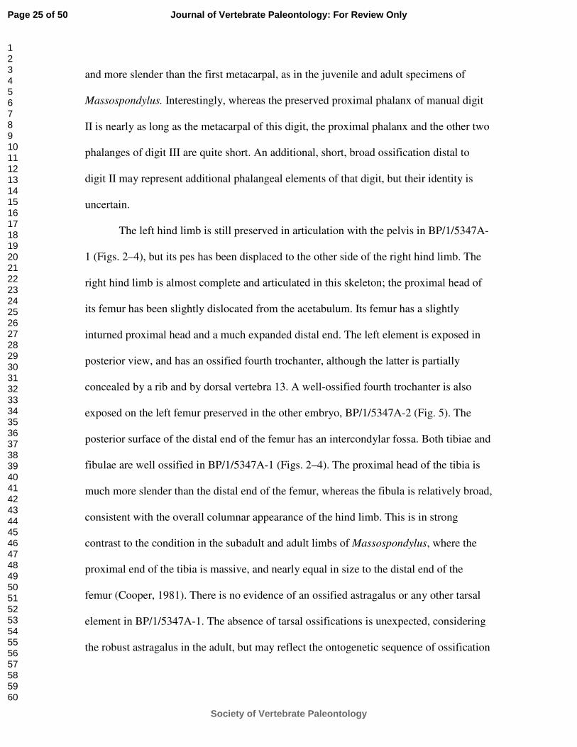
and more slender than the first metacarpal, as in the juvenile and adult specimens of
Massospondylus. Interestingly, whereas the preserved proximal phalanx of manual digit
II is nearly as long as the metacarpal of this digit, the proximal phalanx and the other two
phalanges of digit III are quite short. An additional, short, broad ossification distal to
digit II may represent additional phalangeal elements of that digit, but their identity is
uncertain.
The left hind limb is still preserved in articulation with the pelvis in BP/1/5347A-
1 (Figs. 2–4), but its pes has been displaced to the other side of the right hind limb. The
right hind limb is almost complete and articulated in this skeleton; the proximal head of
its femur has been slightly dislocated from the acetabulum. Its femur has a slightly
inturned proximal head and a much expanded distal end. The left element is exposed in
posterior view, and has an ossified fourth trochanter, although the latter is partially
concealed by a rib and by dorsal vertebra 13. A well-ossified fourth trochanter is also
exposed on the left femur preserved in the other embryo, BP/1/5347A-2 (Fig. 5). The
posterior surface of the distal end of the femur has an intercondylar fossa. Both tibiae and
fibulae are well ossified in BP/1/5347A-1 (Figs. 2–4). The proximal head of the tibia is
much more slender than the distal end of the femur, whereas the fibula is relatively broad,
consistent with the overall columnar appearance of the hind limb. This is in strong
contrast to the condition in the subadult and adult limbs of Massospondylus, where the
proximal end of the tibia is massive, and nearly equal in size to the distal end of the
femur (Cooper, 1981). There is no evidence of an ossified astragalus or any other tarsal
element in BP/1/5347A-1. The absence of tarsal ossifications is unexpected, considering
the robust astragalus in the adult, but may reflect the ontogenetic sequence of ossification
Page 25 of 50
Society of Vertebrate Paleontology
Journal of Vertebrate Paleontology: For Review Only
123456789101112131415161718192021222324252627282930313233343536373839404142434445464748495051525354555657585960

of the pes. The hind limb of the smallest known juvenile of Massospondylus carinatus
(BP/1/ 4376) has well-ossified limb bones, but the tarsus is surprisingly small. Only
metatarsals I–III are present in both hind limbs in BP/1/5347A-1 (Figs. 2–4). A proximal
phalanx articulates with metatarsal I but extends down into the matrix, and only part of its
proximal articular surface is visible. Metatarsal II does not have any phalanges in
articulation in BP/1/5347A-1, but metatarsal III is preserved with at least three phalanges
in partial articulation, including a terminal phalanx.
The forelimb is proportionately much longer in the embryo (Table 1) than in the
adult (Cooper, 1981), but this is consistent with the growth trajectory throughout
ontogeny (Reisz et al., 2005). All elements of the forelimb, even the metacarpals and
proximal phalanges of the manus, are proportionately longer than those in the adults.
DISCUSSION
Identity of the Embryos
The affinities of the eggs have been debated since the original report by Kitching
(1979); even their dinosaurian origin has been questioned (e.g., Zelenitsky cited in
Carpenter, 1999). Zelenitsky and Modesto (2002) argued that the peculiar microstructure
of the eggshell was the result of diagenetic alteration and thus taxonomically
uninformative. They noted that only the embryos themselves could provide secure
identification of the eggs. Numerous skeletal features of the embryos now provide clear
evidence that these individuals represent a sauropodomorph dinosaur (Reisz et al., 2005).
These include the first dentary tooth (or its alveolus) being set back from the anterior end
Page 26 of 50
Society of Vertebrate Paleontology
Journal of Vertebrate Paleontology: For Review Only
123456789101112131415161718192021222324252627282930313233343536373839404142434445464748495051525354555657585960

of the bone, presence of a lateral ridge on the dentary, anteroventral extension of the
infratemporal fenestra below the orbit, a horizontally extending rather than S-shaped
neck, posterior dorsal centra that are longer than tall, and the length of the humerus
exceeding 55% of that of the femur.
Taxonomic assignment of the embryo at a lower taxonomic level is more difficult
due to the generalized anatomy of embryos, the morphological subtleties that differentiate
some sauropodomorph taxa from the region, and the unstable nature of their taxonomy
(Yates and Kitching, 2003; Barrett, 2004, 2009; Galton and Upchurch, 2004; Barrett et.
al., 2007). Reisz et al. (2005) assigned the embryos to Massospondylus carinatus based
on a single autapomorphy for this taxon, greatest skull width exceeding skull height by at
least 10% (Sereno, 1999; Sues et al., 2004). In addition, this taxonomic assignment was
greatly strengthened by the tight fit of the embryonic materials with the growth trajectory
of Massospondylus carinatus (Reisz et al., 2005). Nevertheless, recent assessments of
sauropodomorphs from the Upper Elliot and Clarens formations reveal a much greater
diversity during the latest Triassic and Early Jurassic in southern Africa than previously
assumed (Yates, 2003; Yates and Kitching, 2003; Barrett, 2004, 2009; Galton and
Upchurch, 2004). Since the publication of Reisz et al. (2005), a new species of
Massospondylus, M. kaalae, has recently been named based on a single specimen with
distinctive jaw proportions (Barrett, 2009). The presence of several basal sauropods or
derived sauropodomorphs in the underlying Lower Elliot Formation (Antetonitrus, Yates
and Kitching, 2003; Melanorosaurus, Yates, 2007) even raised the possibility that basal
sauropods could be considered potential candidates for the identity of the embryos
although we found no apomorphies in support of this. Yates et al. (2010) recently
Page 27 of 50
Society of Vertebrate Paleontology
Journal of Vertebrate Paleontology: For Review Only
123456789101112131415161718192021222324252627282930313233343536373839404142434445464748495051525354555657585960

reported a new basal sauropodomorph, Aardonyx celestae, from the Upper Elliot
Formation. Aardonyx can be ruled out as the taxon to which the embryos can be referred
because the dentary lacks the lateral ridge, the premaxilla has a steeply ascending dorsal
process, and the maxilla has an extensive medial lamina (Yates et al., 2010).
The identification of the embryos to either of the two currently recognized species
of Massospondylus is difficult given the lack of ontogenetic data for the new species.
However, the genus-level identification of the embryos as Massospondylus remains
sound because one would expect the skull to be taller than wide in the embryos based on
the proportionately enormous size of the eye in early ontogenetic stages of most
tetrapods, yet their cranial proportions exhibit the diagnostic autapomorphy of
Massospondylus. In addition, Massospondylus is by far the most commonly found
sauropodomorph in the Upper Elliot Formation in southern Africa. Two as yet
uncollected subadult skeletons of Massospondylus are currently eroding out of the matrix
at the egg-producing locality, and several other skeletons have been identified within a
radius of 500 m from the site in Golden Gate Highlands National Park. A morphometric
analysis of skull growth in Massospondylus, currently in progress, may help resolve the
specific identity of the embryonic remains, but until then, we prefer to retain our original
identification of this material as M. carinatus (Reisz et al., 2005).
The Fossil Record of Dinosaur Embryos
The fossil record of non-avian dinosaurian embryos preserved within eggs is still
very limited. The specimens of Massospondylus reported by Reisz et al. (2005) and
described in detail in this paper represent the oldest example of dinosaurian embryos
Page 28 of 50
Society of Vertebrate Paleontology
Journal of Vertebrate Paleontology: For Review Only
123456789101112131415161718192021222324252627282930313233343536373839404142434445464748495051525354555657585960

found to date. They are also the oldest known example of terrestrial vertebrate embryos
in the fossil record. Remarkably, the small size of both the embryo and egg also set them
apart from similar finds of other dinosaurian taxa. This is particularly interesting because
the largest known adult specimen of Massospondylus carinatus had an estimated body
length of more than five meters.
Dinosaurian embryos and hatchlings (“babies”) have attracted a great deal of
scientific and popular attention (Carpenter, 1999). A stratigraphically older example of
dinosaurian hatchlings is a find of seven post-hatchling skeletons, all similar in size but in
various degrees of completeness, of the sauropodomorph dinosaur Mussaurus
patagonicus from the Upper Triassic (?Norian) El Tranquilo Formation of Santa Cruz
Province, Argentina (Bonaparte and Vince, 1979; Pol and Powell, 2007). The skull of the
holotype of Mussaurus patagonicus is about 30 mm long and the overall length of the
skeleton is approximately 20–25 cm. None of these skeletons was apparently preserved
inside an egg, and their size and level of ossification indicates that they are young, post-
hatchling individuals (Pol and Powell, 2007). Bonaparte and Vince (1979) and Pol and
Powell (2007) reported but did not describe or illustrate portions of two associated eggs
and eggshell.
Two specimens of juvenile sauropodomorph dinosaurs have been described from
the Lower Lufeng Formation of Yunnan Province, China. One is a small skull (less than
40 mm in length) with articulated mandible preserved in a nodule with an associated
vertebra. Originally interpreted as an early squamate and named Fulengia youngi by
Carroll and Galton (1977), Evans and Milner (1989) and Sereno (1991) demonstrated that
it is, in a fact, a juvenile sauropodomorph. The other is an incomplete, very small skull
Page 29 of 50
Society of Vertebrate Paleontology
Journal of Vertebrate Paleontology: For Review Only
123456789101112131415161718192021222324252627282930313233343536373839404142434445464748495051525354555657585960

(23 mm in length) that Yang (1982) designated as the holotype of a new “fabrosaurid”
ornithischian, Tawasaurus minor. Sereno (1991) restudied this specimen and concluded
that it probably represents a hatchling “prosauropod.” He noted that neither “Fulengia”
nor “Tawasaurus” displayed autapomorphies to support unequivocal referral to the
common large sauropodomorph Lufengosaurus huenei from the Lower Lufeng
Formation. Both specimens are much larger than we expect for basal sauropodomorph
embryos based on the specimens described here, and we consider them small post-
hatchling individuals.
The stratigraphically next oldest well-documented examples of dinosaurian
embryonic remains in and associated with eggs are from the Upper Jurassic
(Kimmeridgian-Tithonian) of Lourinhã, Portugal. Here, several well-preserved clutches
of eggs referable to the form genus Preprismatoolithus were found with many
fragmentary postcranial bones of embryos and/or hatchlings of an unidentified theropod
preserved in the matrix surrounding the eggs or, in at least one instance, within an egg
(Mateus et al., 1997:fig. 4). Based on the dimensions of the eggs, the total length of the
embryos was about 40 cm. The bones within the egg are very fragmentary and suggest a
relatively early stage of embryonic development.
From the more or less coeval Morrison Formation of the western United States,
embryonic remains of several dinosaurian taxa have been reported although in no
instance were these specimens found in eggs. Chure et al. (1994) described a partial
postcranial skeleton of a tiny dinosaur from the Brushy Basin Member in the Utah
portion of Dinosaur National Monument. They interpreted this specimen as an advanced
embryonic individual of the ornithopod Camptosaurus with an estimated overall length
Page 30 of 50
Society of Vertebrate Paleontology
Journal of Vertebrate Paleontology: For Review Only
123456789101112131415161718192021222324252627282930313233343536373839404142434445464748495051525354555657585960

of 24 cm. They interpreted the fact that the articular ends of the limb bones are unossified
as evidence for altricial behavior. Carpenter (1994) reported a partial skeleton including a
complete skull of a very small individual ("baby") of the ornithopod Dryosaurus from the
same horizon and general region. He also mentioned other occurrences of presumed
embryonic bones of this taxon. Again, the articular ends of the limb bones are poorly
developed. Finally, Britt and Naylor (1994) described a minute premaxilla of the
sauropod Camarasaurus from the Morrison Formation of the Dry Mesa Quarry in west-
central Colorado. They interpreted this bone as representing an embryonic individual and
noted the absence of erupted teeth.
Coombs (1982) described in detail two skulls and numerous postcranial bones of
possible hatchlings of the basal ceratopsian dinosaur Psittacosaurus from the Lower
Cretaceous Oshih Formation of Mongolia. He estimated the skull length of the smaller
specimen as 28 mm and the total length as about 23–27 cm.
There are now numerous examples of dinosaurian remains in eggs from Late
Cretaceous strata in Asia and in North and South America (see review by Carpenter
[1999]). The best-known examples include the embryo of an oviraptorid theropod in an
eroded egg of the Elongatoolithus type from the Djadokhta Formation (Campanian) of
Mongolia (Norell et al., 1994), embryos of titanosaurian sauropods in numerous eggs of
the form genus Megaloolithus from the Río Colorado Formation (Santonian) of Neuquén
Province, Argentina (Chiappe et al., 1998, 2001), embryos of the lambeosaurine
hadrosaurid Hypacrosaurus stebingeri in eggs of the form genus Sphaeroolithus from the
upper part of the Two Medicine Formation (Campanian) of Montana and Alberta (Horner
and Currie, 1994), extraordinarily preserved embryos of therizinosauroid theropods in
Page 31 of 50
Society of Vertebrate Paleontology
Journal of Vertebrate Paleontology: For Review Only
123456789101112131415161718192021222324252627282930313233343536373839404142434445464748495051525354555657585960

eggs from the Nanchao Formation (?Santonian-Campanian) of Henan Province, China
(Kundrát et al., 2008), and numerous embryonic bones referable to a variety of
dinosaurian taxa from the Bissekty Formation (Turonian) of Uzbekistan (Sues and
Averianov, in preparation).
In the absence of extensive comparative data for early development in non-avian
dinosaurs, it is difficult to assess the exact developmental stage of the two embryos of
Massospondylus carinatus described here. However, comparisons with remains from
dinosaurian eggs from geologically much younger sediments does support our previous
hypothesis that the level of ossification of these two embryos indicates that these
individuals were close to hatching. The embryonic remains found inside eggs of Late
Jurassic age and those of Late Cretaceous titanosaurian sauropods have well-developed
skulls, with teeth already formed, but the postcranial skeletons of these embryos show a
much earlier developmental stage than those of Massospondylus. The level of ossification
seen in the two embryos described here, along with the preservation of well-ossified
scleral plates and stapes, the preservation of much of the manus and pes, the presence of a
well-developed deltopectoral crest on the humerus and fourth trochanter on the femur all
support the hypothesis that these embryos were close to hatching. The best examples of
Late Cretaceous dinosaur embryos found inside eggs and similarly well ossified are those
of Hypacrosaurus stebingeri (Horner and Currie, 1994). Although their skeletal structure
indicates a comparable level of ossification, the embryos of Hypacrosaurus stebingeri are
significantly larger than those of Massospondylus carinatus, with a skull length of about
60 mm, and well-developed dentition in the maxillae and dentaries. Moreover, a well-
preserved embryonic skull of H. stebingeri already has two generations of teeth erupted
Page 32 of 50
Society of Vertebrate Paleontology
Journal of Vertebrate Paleontology: For Review Only
123456789101112131415161718192021222324252627282930313233343536373839404142434445464748495051525354555657585960

from several tooth positions and has started to develop occlusal surfaces (Horner and
Currie, 1994). Thus there is little doubt that these embryos were close to hatching. The
similarities in the levels of ossification of the postcranial skeleton between these two taxa
does support our hypothesis of the near-hatching condition of the two embryos of
Massospondylus carinatus, although neither preserves definite teeth. Comparisons with
sauropod and theropod embryos of clearly earlier developmental stages (Chiappe et al.,
2001; Mateus et al., 1997) than those of M. carinatus, with well-developed teeth,
militates against the use of the presence of teeth as an indicator of the developmental
stage in this dinosaur. The slightly larger skulls of the two presumed hatchlings of
sauropodomorph dinosaurs from the Lower Lufeng Formation (Lower Jurassic) already
have marginal teeth (Evans and Milner, 1989; Sereno, 1991).
The temporal and phylogenetic distance between these basal sauropodomorph
dinosaurs and their extant relatives, birds and crocodylians, makes direct comparisons
with their embryos difficult. Additional embryonic stages of Massospondylus carinatus
are required in order to gain a better understanding of the early ontogenetic development
of this taxon. Continuing work at the Rooidraai locality has already resulted in the
discovery of additional egg clusters that remain to be collected, prepared, and studied.
ACKNOWLEDGMENTS
We would like to thank Professor Bruce Rubidge, Director of the Bernard Price Institute
for Palaeontological Research at University of the Witwatersrand, and Drs. Michael
Raath and Adam Yates for their great assistance in this project. They have been generous
Page 33 of 50
Society of Vertebrate Paleontology
Journal of Vertebrate Paleontology: For Review Only
123456789101112131415161718192021222324252627282930313233343536373839404142434445464748495051525354555657585960

with their time, helping us with the loan of specimens, the logistics of mounting field
expeditions in South Africa, and their continued support over the years to the senior
author. We also thank the staff of the Golden Gate Highlands National Park, in particular
Mr. Johann Taljard, for their enthusiastic support of our work in that park. Funding for
this research has come from the National Science and Engineering Research Council of
Canada, the National Geographic Society, and PAST (South Africa).
Page 34 of 50
Society of Vertebrate Paleontology
Journal of Vertebrate Paleontology: For Review Only
123456789101112131415161718192021222324252627282930313233343536373839404142434445464748495051525354555657585960

LITERATURE CITED
Barrett P. M. 2009. A new basal sauropodomorph dinosaur from the Upper Elliot
Formation (Lower Jurassic) of South Africa. Journal of Vertebrate Paleontology
29:1032–1045.
Barrett, P. M. 2004. Sauropodomorph dinosaur diversity in the upper Elliot Formation
(Massospondylus range zone: Lower Jurassic) of South Africa. South African
Journal of Science 100:501–503.
Barrett, P. M., and A. M. Yates. 2006. New information on the palate and lower jaw of
Massospondylus (Dinosauria: Sauropodomorpha). Palaeontologia africana 41:123–
130
Barrett, P. M., P. Upchurch, X-Z. Zhou, and X-L. Wang. 2007. The skull of
Yunnanosaurus huangi Young, 1942 (Dinosauria: Prosauropoda) from the Lower
Lufeng Formation (Lower Jurassic) of Yunnan, China. Zoological Journal of the
Linnean Society 150:319–341.
Barrett, P. M., P. Upchurch, and X-L. Wang. 2005. Cranial osteology of Lufengosaurus
huenei Young (Dinosauria: Prosauropoda) from the Lower Jurassic of Yunnan,
People’s Republic of China. Journal of Vertebrate Paleontology 25:806–822.
Bonaparte, J. F., and M. Vince. 1979. El hallazgo del primer nido de dinosaurios
Triásicos, (Saurischia, Prosauropoda), Triásico superior de Patagonia, Argentina.
Ameghiniana 16:173–182.
Page 35 of 50
Society of Vertebrate Paleontology
Journal of Vertebrate Paleontology: For Review Only
123456789101112131415161718192021222324252627282930313233343536373839404142434445464748495051525354555657585960

Britt, B. B., and B. G. Naylor. 1994. An embryonic Camarasaurus (Dinosauria,
Sauropoda) from the Upper Jurassic Morrison Formation (Dry Mesa Quarry,
Colorado); pp. 256–264 in K. Carpenter, K. F. Hirsch, and J. R. Horner (eds.),
Dinosaur Eggs and Babies. Cambridge University Press, New York.
Carpenter, K. 1994. Baby Dryosaurus from the Upper Jurassic Morrison Formation of
Dinosaur National Monument; pp. 288–297 in K. Carpenter, K. F. Hirsch, and J. R.
Horner (eds.), Dinosaur Eggs and Babies. Cambridge University Press, New York,
Carpenter, K. 1999. Eggs, Nests, and Baby Dinosaurs. A Look at Dinosaur Reproduction.
Indiana University Press, Bloomington, 375 pp.
Carroll, R. L., and P. M. Galton. 1977. ‘Modern’ lizard from the Upper Triassic of China.
Nature 266:252–255.
Chiappe, L. M, R. A. Coria, L. Dingus, F. Jackson, A. Chinsamy, and M. Fox. 1998.
Sauropod embryos from the Late Cretaceous of Patagonia. Nature 396:258–261.
Chiappe, L. M., L. Salgado, and R. A. Coria. 2001. Embryonic skulls of titanosaurid
sauropod dinosaurs. Science 293:2444–2446.
Chure, D., C. Turner, and F. Peterson. 1994. An embryo of Camptosaurus from the
Morrison Formation (Jurassic, middle Tithonian) in Dinosaur National Monument,
Utah; pp. 298–311 in K. Carpenter, K. F. Hirsch, and J. R. Horner (eds.), Dinosaur
Eggs and Babies. Cambridge University Press, New York.
Coombs, W. P., Jr. 1982. Juvenile specimens of the ornithischian dinosaur
Psittacosaurus. Palaeontology 25:89–107.
Cooper, M. R. 1981. The prosauropod dinosaur Massospondylus carinatus Owen from
Zimbabwe: its biology, mode of life and phylogenetic significance. Occasional
Page 36 of 50
Society of Vertebrate Paleontology
Journal of Vertebrate Paleontology: For Review Only
123456789101112131415161718192021222324252627282930313233343536373839404142434445464748495051525354555657585960

Papers of the National Museums and Monuments of Rhodesia, Series B, Natural
Sciences 6:689–840.
Evans, S. E., and A. R. Milner. 1989. Fulengia, a supposed early lizard reinterpreted as a
prosauropod dinosaur. Palaeontology 32:223–230.
Galton, P. M., and P. Upchurch. 2004. Prosauropoda; pp. 232–258 in D. B. Weishampel,
P. Dodson, and H. Osmólska (eds.) The Dinosauria (Second Edition). University of
California Press, Berkeley.
Horner, J. R., and P. J. Currie. 1994. Embryonic and neonatal morphology and ontogeny
of a new species of Hypacrosaurus (Ornithischia, Lambeosauridae) from Montana
and Alberta; pp. 312–336 in K. Carpenter, K. F. Hirsch, and J. R. Horner (eds.)
Dinosaur Eggs and Babies. Cambridge University Press, New York.
Kitching, J. W. 1979. Preliminary report on a clutch of six dinosaurian eggs from the
Upper Triassic Elliot Formation, Northern Orange Free State. Palaeontologia
africana 22:41–45.
Kitching, J. W., and M. A. Raath. 1984. Fossils from the Elliot and Clarens Formations
(Karoo Sequence) of the northeastern Cape, Orange Free State and Lesotho, and a
suggested biozonation based on tetrapods. Palaeontologia africana 25:111–125.
Kundrát, M., A. R. I. Cruickshank, T. W. Manning, and J. Nudds. 2008. Embryos of
therizinosauroid theropods from the Upper Cretaceous of China: diagnosis and
analysis of ossification patterns. Acta Zoologica (Stockholm) 89:231–251.
Martínez, R. N., and O. Alcober. 2009. A basal sauropodomorph (Dinosauria: Saurischia)
from the Ischigualasto Formation (Triassic, Carnian) and the early evolution of
Sauropodomorpha. PLoS One 4:e4397.
Page 37 of 50
Society of Vertebrate Paleontology
Journal of Vertebrate Paleontology: For Review Only
123456789101112131415161718192021222324252627282930313233343536373839404142434445464748495051525354555657585960

Mateus, I., H. Mateus, M. T. Antunes, O. Mateus, P. Taquet, V. Ribeir, and G.
Manuppella. 1997. Covée, oeufs, et embryons d’un Dinosaure Théropode du
Jurassique supérieur de Lourinhã (Portugal). Comptes Rendus de l’Académie des
Sciences, Paris, Sciences de la Terre et des planètes 325:71–78.
Norell, M. A., J. M. Clark, D. Dashzeveg, R. Barsbold, L. M. Chiappe, A. Davidson, M.
C. McKenna, A. Perle, and M. J. Novacek. 1994. A theropod dinosaur embryo and
the affinities of the Flaming Cliffs dinosaur eggs. Science 266:779–782.
Pol, D., and J. E. Powell. 2007. Skull anatomy of Mussaurus patagonicus (Dinosauria:
Sauropodomorpha) from the Late Triassic of Patagonia. Historical Biology 19:
125–144.
Reisz, R. R., D. M. Scott, H.-D. Sues, D. C. Evans, and M. A. Raath. 2005. Embryos of
an Early Jurassic prosauropod dinosaur and their evolutionary significance. Science
309:761–764.
Sereno, P. C. 1991. Lesothosaurus, “fabrosaurids,” and the early evolution of
Ornithischia. Journal of Vertebrate Paleontology 11:168–197.
Sereno, P. C. 1999. The evolution of dinosaurs. Science 284:2137–2147.
Sues, H.-D., R. R. Reisz, S. Hinic, M. A. Raath. 2004. On the skull of Massospondylus
carinatus Owen, 1854 (Dinosauria: Sauropodomorpha) from the Elliot and Clarens
Formations (Lower Jurassic) of South Africa. Annals of Carnegie Museum 73:239–
257.
Yang, Z. 1982. [A new ornithopod from Lufeng, Yunnan.] In [Selected Works of Yang
Zhongjian.] Science Press, Beijing, 29–35. [In Chinese]
Page 38 of 50
Society of Vertebrate Paleontology
Journal of Vertebrate Paleontology: For Review Only
123456789101112131415161718192021222324252627282930313233343536373839404142434445464748495051525354555657585960

Yates, A. M. 2003. A definite prosauropod dinosaur from the Lower Elliot Formation
(Norian: Upper Triassic) of South Africa. Palaeontologia africana 39:63–68.
Yates, A. M. 2007. The first complete skull of the Triassic dinosaur Melanorosaurus
Haughton (Sauropodomorpha: Anchisauria). Special Papers in Palaeontology 77:9–
55.
Yates, A. M. and J. W. Kitching. 2003. The earliest known sauropod dinosaur and the
first steps towards sauropod locomotion. Proceedings of the Royal Society of
London B: Biological Sciences 270:1753–1758.
Yates, A. M., M. F. Bonnan, J. Neveling, A. Chinsamy, and M. G. Blackbeard. 2010. A
new transitional sauropodomorph dinosaur from the Early Jurassic of South Africa
and the evolution of sauropod feeding and quadrupedalism. Proceedings of the
Royal Society of London B: Biological Sciences 277:787–794.
Zelenitsky, D. K. and S. P. Modesto. 2002. Re-evaluation of the eggshell structure of
eggs containing embryos from the Lower Jurassic of South Africa. South African
Journal of Science 98:407–409.
Submitted January 12, 2010; accepted June 30, 2010.
Page 39 of 50
Society of Vertebrate Paleontology
Journal of Vertebrate Paleontology: For Review Only
123456789101112131415161718192021222324252627282930313233343536373839404142434445464748495051525354555657585960

FIGURE LEGENDS
FIGURE 1. Map of South Africa and Lesotho showing main localities that have yielded
skeletal remains of Massospondylus. The majority of these localities have sufficiently
complete skeletons to allow for their identification as those of M. carinatus. The asterisk
designates the Rooidraai locality, near the town of Clarens, in the Golden Gates
Highlands National Park, South Africa.
FIGURE 2. Photograph of skeleton of the first embryo of Massospondylus carinatus
(BP/1/5347A-1). White materials represent eggshell that remained around the embryonic
skeleton after they had been exposed. Scale equals 1cm.
FIGURE 3. Illustration of the first embryo of Massospondylus carinatus (BP/1/5347A-1),
showing details of the skull and skeleton in partial right lateral view. Scale equals1cm.
Abbreviations: an, angular; c, centra; c3–c10, cervical vertebrae; ca, caudal vertebra; ch,
hemal arch; co, coracoid; cp?, cultriform process?; d1–d14, dorsal vertebrae; de, dentary;
f, frontal; fe, femur; fi, fibula; h, humerus; il, ilium; is, ischium; j, jugal; l, lacrimal; m,
maxilla; mc1–mc4, metacarpals; mt1–mt3, metatarsals; n, nasal; p, parietal; pal,
palatine; ph, phalanges; pm, premaxilla; po, postorbital; prf, prefrontal; pt, pterygoid;
pu, pubis; q, quadrate; r, rib; ra, radius; s1–s3, sacral vertebrae; sc, scapula; scl, scleral
ring; sr1–sr3, sacral ribs; sq, squamosal; st, stapes; su, surangular; t, tibia; u, ulna.
Page 40 of 50
Society of Vertebrate Paleontology
Journal of Vertebrate Paleontology: For Review Only
123456789101112131415161718192021222324252627282930313233343536373839404142434445464748495051525354555657585960

FIGURE 4. Close-up photographs of the first embryo of Massospondylus carinatus
(BP/1/5347A-1), showing various parts of the articulated skeleton, including (A) shoulder
region, (B) skull, (C) pelvic region, and (D) limbs. Abbreviations as in Figure 3. Scale as
indicated.
FIGURE 5. Photograph of the skull and nearby elements of the skeleton in the second
embryo of Massospondylus carinatus, preserved on part and counterpart blocks, (A)
BP/1/5347A, and (B) BP/1/5347B. Scale equals 1cm.
FIGURE 6. Illustrations of skull and associated skeletal elements of second embryo of
Massospondylus carinatus preserved in part and counterpart blocks (A) BP/1/5347A and
(B) BP/1/5347B, showing details of the skull in dorsal view. Parts of the postcranial
skeleton are also shown. Scale equals 1cm. Abbreviations as in Figure 3.
FIGURE 7. Reconstruction of embryonic skull of Massospondylus carinatus in dorsal
and lateral views. The reconstructions do not show any teeth because there is no
conclusive evidence for any teeth being associated with this embryonic stage. Scale
equals 1cm.
FIGURE 8. Skeletal reconstruction of the embryonic Massospondylus carinatus in right
lateral view. Scale equals 1 cm.
Page 41 of 50
Society of Vertebrate Paleontology
Journal of Vertebrate Paleontology: For Review Only
123456789101112131415161718192021222324252627282930313233343536373839404142434445464748495051525354555657585960

TABLE 1. Table of measurements for embryonic skeleton of Massospondylus
(BP/1/5347A). All measurements (in millimeters) taken from the right side of the
specimen.
Element Measurement
Skull, reconstructed length 19.5
Quadrate, height 5.2
Middle cervical centrum, length 2.3
Middle dorsal centrum, length 2.4
Sacral centrum 1, length 2.5
Scapula, height 11.3
Humerus, length 9.5
Ulna, length 6.5
Iliac blade, length 6.5
Femur, length 11.5
Tibia, length 11
Fibula, length 11.4
MT II, length 5.1
Page 42 of 50
Society of Vertebrate Paleontology
Journal of Vertebrate Paleontology: For Review Only
123456789101112131415161718192021222324252627282930313233343536373839404142434445464748495051525354555657585960

FIGURE 1. Map of South Africa and Lesotho showing main localities that have yielded skeletal remains of Massospondylus 96x103mm (600 x 600 DPI)
Page 43 of 50
Society of Vertebrate Paleontology
Journal of Vertebrate Paleontology: For Review Only
123456789101112131415161718192021222324252627282930313233343536373839404142434445464748495051525354555657585960

FIGURE 2. Photograph of skeleton of articulated embryo, (BP/1/5347A-1). White materials represent eggshell that remained around the embryonic skeleton after they have been exposed.
Scale equals 1cm. 180x121mm (400 x 400 DPI)
Page 44 of 50
Society of Vertebrate Paleontology
Journal of Vertebrate Paleontology: For Review Only
123456789101112131415161718192021222324252627282930313233343536373839404142434445464748495051525354555657585960

FIGURE 3. Photograph of the skull and nearby elements of the skeleton in the second embryo, preserved on part and counterpart blocks, BP/1/5347A and BP/1/5347B. Scale equals 1cm.
107x64mm (600 x 600 DPI)
Page 45 of 50
Society of Vertebrate Paleontology
Journal of Vertebrate Paleontology: For Review Only
123456789101112131415161718192021222324252627282930313233343536373839404142434445464748495051525354555657585960

FIGURE 4. Close-up photographs of BP/1/5347A-1, showing various parts of the articulated skeleton, including (A) shoulder region, (B) skull, (C) pelvic region, and (D) limbs. Abbreviations as
in Figure 3. Scale as indicated. 180x247mm (400 x 400 DPI)
Page 46 of 50
Society of Vertebrate Paleontology
Journal of Vertebrate Paleontology: For Review Only
123456789101112131415161718192021222324252627282930313233343536373839404142434445464748495051525354555657585960

FIGURE 5. Photograph of the skull and nearby elements of the skeleton in the second embryo, preserved on part and counterpart blocks, BP/1/5347A and BP/1/5347B. Scale equals 1cm.
180x196mm (400 x 400 DPI)
Page 47 of 50
Society of Vertebrate Paleontology
Journal of Vertebrate Paleontology: For Review Only
123456789101112131415161718192021222324252627282930313233343536373839404142434445464748495051525354555657585960

FIGURE 6. Illustrations of skull and associated skeletal elements of second embryo of Massospondylus carinatus preserved in part and counterpart blocks (A) BP/1/5347A and (B)
BP/1/5347B, showing details of the skull in dorsal view. Parts of the postcranial skeleton are also shown.
75x31mm (600 x 600 DPI)
Page 48 of 50
Society of Vertebrate Paleontology
Journal of Vertebrate Paleontology: For Review Only
123456789101112131415161718192021222324252627282930313233343536373839404142434445464748495051525354555657585960

FIGURE 7. Reconstruction of embryonic skull of Massospondylus carinatus in dorsal and lateral views. Scale equals 1cm.
112x140mm (600 x 600 DPI)
Page 49 of 50
Society of Vertebrate Paleontology
Journal of Vertebrate Paleontology: For Review Only
123456789101112131415161718192021222324252627282930313233343536373839404142434445464748495051525354555657585960

FIGURE 8. Skeletal reconstruction of the embryonic Massospondylus carinatus in right lateral view. Scale equals 1 cm.
70x27mm (600 x 600 DPI)
Page 50 of 50
Society of Vertebrate Paleontology
Journal of Vertebrate Paleontology: For Review Only
123456789101112131415161718192021222324252627282930313233343536373839404142434445464748495051525354555657585960