El ácido úrico y la actividad de gammaglutamil transferasa se asocian a los índices de remodelado...
11
Original article Uric Acid and Gamma-glutamyl Transferase Activity Are Associated With Left Ventricular Remodeling Indices in Patients With Chronic Heart Failure Slavica Radovanovic, a Ana Savic-Radojevic, b Tatjana Pekmezovic, c Olivera Markovic, a,d Lidija Memon, a Svetlana Jelic, a,d Dragan Simic, d,e Tanja Radic, b Marija Pljesa-Ercegovac, b and Tatjana Simic b, * a Odeljenje Kardiologije, Klinicko-Bolnicki Centar Bezanijska Kosa, Belgrade, Serbia b Institut za Medicinsku i Klinicku Biohemiju, Medicinski Fakultet, Univerzitet u Beogradu, Belgrade, Serbia c Institut za Epidemiologiju, Medicinski Fakultet, Univerzitet u Beogradu, Belgrade, Serbia d Medicinski Fakultet, Univerzitet u Beogradu, Belgrade, Serbia e Klinika za Kardiovaskularne Bolesti, Klinicki Centar Srbije, Belgrade, Serbia Rev Esp Cardiol. 2014;67(8):632–642 Article history: Received 30 July 2013 Accepted 25 November 2013 Available online 29 March 2014 Keywords: Gamma-glutamyl transferase Uric acid Chronic heart failure Remodeling Flow mediated dilation Oxidative stress A B S T R A C T Introduction and objectives: Uric acid and gamma-glutamyl transferase are prognostic indicators in chronic heart failure. Nevertheless, the mechanism underlying the association between uric acid, gamma-glutamyl transferase, and chronic heart failure progression and prognosis remains largely unknown. Methods: The association of uric acid and gamma-glutamyl transferase with flow-mediated dilation and echocardiographic indices of cardiac remodeling was addressed in 120 patients with chronic ischemic heart failure. To determine the independent contribution of uric acid and gamma-glutamyl transferase to the flow-mediated dilation and echocardiographic indices of remodeling, a series of multiple linear regression models, based on traditional and nontraditional risk factors impacting upon these parameters, were constructed. Results: Uric acid, but not gamma-glutamyl transferase, was an independent predictor of flow-mediated dilation. Uric acid was associated with all the echocardiographic indices of left ventricular dysfunction tested in 3 multiple-regression models. Uric acid correlated with left ventricular end-systolic diameter, left ventricular end-diastolic diameter, left ventricular end-systolic volume, and left ventricular end- diastolic volume (r = 0.337; r = 0.340; r = 0.321; r = 0.294; P = .001, respectively). Gamma-glutamyl transferase was an independent predictor of left ventricular end-systolic volume and left ventricular end- diastolic volume, after adjustment for all variables. Gamma-glutamyl transferase correlated with left ventricular end-systolic diameter, left ventricular end-diastolic diameter, left ventricular end-systolic volume, and left ventricular end-diastolic volume (r = 0.238, P = .009; r = 0.219, P = .016; r = 0.359, P < .001; r = 0.369, P = .001, respectively). Conclusions: Serum uric acid and gamma-glutamyl transferase levels are associated with left ventricular remodeling in patients with chronic ischemic heart failure. ß 2013 Sociedad Espan ˜ola de Cardiologı ´a. Published by Elsevier Espan ˜a, S.L. All rights reserved. El a ´ cido u ´ rico y la actividad de gammaglutamil transferasa se asocian a los ı ´ndices de remodelado ventricular izquierdo en pacientes con insuficiencia cardiaca cro ´ nica Palabras clave: Gammaglutamil transferasa A ´ cido u ´ rico Insuficiencia cardiaca cro ´ nica Remodelado Dilatacio ´n mediada por flujo Estre ´s oxidativo R E S U M E N Introduccio ´n y objetivos: El a ´ cido u ´ rico y la gammaglutamil transferasa son indicadores prono ´ sticos en la insuficiencia cardiaca cro ´ nica. No obstante, el mecanismo subyacente a la asociacio ´n observada entre a ´ cido u ´ rico, gammaglutamil transferasa y progresio ´n y prono ´ stico de la insuficiencia cardiaca cro ´ nica sigue siendo en gran parte desconocido. Me ´todos: Se estudio ´ la asociacio ´n del a ´ cido u ´ rico y la gammaglutamil transferasa con la dilatacio ´n mediada por flujo y con los ı ´ndices ecocardiogra ´ ficos del remodelado cardiaco en 120 pacientes con insuficiencia cardiaca isque ´ mica cro ´ nica. Para determinar la contribucio ´n independiente del a ´ cido u ´ rico y la gammaglutamil transferasa en la dilatacio ´n mediada por flujo y en los ı ´ndices ecocardiogra ´ ficos del remodelado, se construyo ´ una serie de modelos de regresio ´n lineal mu ´ ltiple, basados en los factores de riesgo tradicionales y no tradicionales que influyen en estos para ´ metros. Resultados: El a ´ cido u ´ rico es un factor independiente predictivo de dilatacio ´n mediada por flujo, pero no la gammaglutamil transferasa. El a ´ cido u ´ rico se asocia a todos los ı ´ndices ecocardiogra ´ ficos de disfuncio ´n * Corresponding author: Institute of Medical and Clinical Biochemistry, Faculty of Medicine, University of Belgrade, Pasterova 2, 11000 Belgrade, Serbia. E-mail address: [email protected] (T. Simic). 1885-5857/$ – see front matter ß 2013 Sociedad Espan ˜ola de Cardiologı ´a. Published by Elsevier Espan ˜a, S.L. All rights reserved. http://dx.doi.org/10.1016/j.rec.2013.11.017
-
Upload
independent -
Category
Documents
-
view
2 -
download
0
Transcript of El ácido úrico y la actividad de gammaglutamil transferasa se asocian a los índices de remodelado...

Rev Esp Cardiol. 2014;67(8):632–642
Original article
Uric Acid and Gamma-glutamyl Transferase Activity Are AssociatedWith Left Ventricular Remodeling Indices in Patients With ChronicHeart Failure
Slavica Radovanovic,a Ana Savic-Radojevic,b Tatjana Pekmezovic,c Olivera Markovic,a,d Lidija Memon,a
Svetlana Jelic,a,d Dragan Simic,d,e Tanja Radic,b Marija Pljesa-Ercegovac,b and Tatjana Simicb,*a Odeljenje Kardiologije, Klinicko-Bolnicki Centar Bezanijska Kosa, Belgrade, Serbiab Institut za Medicinsku i Klinicku Biohemiju, Medicinski Fakultet, Univerzitet u Beogradu, Belgrade, Serbiac Institut za Epidemiologiju, Medicinski Fakultet, Univerzitet u Beogradu, Belgrade, Serbiad Medicinski Fakultet, Univerzitet u Beogradu, Belgrade, Serbiae Klinika za Kardiovaskularne Bolesti, Klinicki Centar Srbije, Belgrade, Serbia
Article history:
Received 30 July 2013
Accepted 25 November 2013
Available online 29 March 2014
Keywords:
Gamma-glutamyl transferase
Uric acid
Chronic heart failure
Remodeling
Flow mediated dilation
Oxidative stress
A B S T R A C T
Introduction and objectives: Uric acid and gamma-glutamyl transferase are prognostic indicators in
chronic heart failure. Nevertheless, the mechanism underlying the association between uric acid,
gamma-glutamyl transferase, and chronic heart failure progression and prognosis remains largely
unknown.
Methods: The association of uric acid and gamma-glutamyl transferase with flow-mediated dilation and
echocardiographic indices of cardiac remodeling was addressed in 120 patients with chronic ischemic
heart failure. To determine the independent contribution of uric acid and gamma-glutamyl transferase
to the flow-mediated dilation and echocardiographic indices of remodeling, a series of multiple linear
regression models, based on traditional and nontraditional risk factors impacting upon these parameters,
were constructed.
Results: Uric acid, but not gamma-glutamyl transferase, was an independent predictor of flow-mediated
dilation. Uric acid was associated with all the echocardiographic indices of left ventricular dysfunction
tested in 3 multiple-regression models. Uric acid correlated with left ventricular end-systolic diameter,
left ventricular end-diastolic diameter, left ventricular end-systolic volume, and left ventricular end-
diastolic volume (r = 0.337; r = 0.340; r = 0.321; r = 0.294; P = .001, respectively). Gamma-glutamyl
transferase was an independent predictor of left ventricular end-systolic volume and left ventricular end-
diastolic volume, after adjustment for all variables. Gamma-glutamyl transferase correlated with left
ventricular end-systolic diameter, left ventricular end-diastolic diameter, left ventricular end-systolic
volume, and left ventricular end-diastolic volume (r = 0.238, P = .009; r = 0.219, P = .016; r = 0.359, P < .001;
r = 0.369, P = .001, respectively).
Conclusions: Serum uric acid and gamma-glutamyl transferase levels are associated with left ventricular
remodeling in patients with chronic ischemic heart failure.
� 2013 Sociedad Espanola de Cardiologıa. Published by Elsevier Espana, S.L. All rights reserved.
El acido urico y la actividad de gammaglutamil transferasa se asocian a los ındicesde remodelado ventricular izquierdo en pacientes con insuficiencia cardiacacronica
Palabras clave:
Gammaglutamil transferasa
Acido urico
Insuficiencia cardiaca cronica
Remodelado
Dilatacion mediada por flujo
Estres oxidativo
R E S U M E N
Introduccion y objetivos: El acido urico y la gammaglutamil transferasa son indicadores pronosticos en la
insuficiencia cardiaca cronica. No obstante, el mecanismo subyacente a la asociacion observada entre
acido urico, gammaglutamil transferasa y progresion y pronostico de la insuficiencia cardiaca cronica
sigue siendo en gran parte desconocido.
Metodos: Se estudio la asociacion del acido urico y la gammaglutamil transferasa con la dilatacion
mediada por flujo y con los ındices ecocardiograficos del remodelado cardiaco en 120 pacientes con
insuficiencia cardiaca isquemica cronica. Para determinar la contribucion independiente del acido urico
y la gammaglutamil transferasa en la dilatacion mediada por flujo y en los ındices ecocardiograficos del
remodelado, se construyo una serie de modelos de regresion lineal multiple, basados en los factores de
riesgo tradicionales y no tradicionales que influyen en estos parametros.
Resultados: El acido urico es un factor independiente predictivo de dilatacion mediada por flujo, pero no
la gammaglutamil transferasa. El acido urico se asocia a todos los ındices ecocardiograficos de disfuncion
* Corresponding author: Institute of Medical and Clinical Biochemistry, Faculty of Medicine, University of Belgrade, Pasterova 2, 11000 Belgrade, Serbia.
E-mail address: [email protected] (T. Simic).
1885-5857/$ – see front matter � 2013 Sociedad Espanola de Cardiologıa. Published by Elsevier Espana, S.L. All rights reserved.
http://dx.doi.org/10.1016/j.rec.2013.11.017

S. Radovanovic et al. / Rev Esp Cardiol. 2014;67(8):632–642 633
ventricular izquierda evaluados en tres modelos de regresion multiple; tambien muestra correlacion con
los diametros telesistolico (r = 0,337) y telediastolico (r = 0,340) y los volumenes telesistolico (r = 0,321) y
telediastolicos (r = 0,294) del ventrıculo izquierdo (p = 0,001). La gammaglutamil transferasa es un factor
independiente predictivo de los volumenes telesistolico y telediastolico del ventrıculo izquierdo tras
introducir un ajuste por todas las variables. El gammaglutamil transferasa muestra correlacion con los
diametros telesistolico (r = 0,238; p = 0,009) y telediastolico (r = 0,219; p = 0,016) y los volumenes
telesistolico (r = 0,359; p < 0,001) y telediastolico (r = 0,369; p = 0,001) del ventrıculo izquierdo.
Conclusiones: El acido urico y la actividad de gammaglutamil transferasa se asocian a los ındices de
remodelado ventricular izquierdo en pacientes con insuficiencia cardiaca isquemica cronica.
� 2013 Sociedad Espanola de Cardiologıa. Publicado por Elsevier Espana, S.L. Todos los derechos reservados.
Abbreviations
CHF: chronic heart failure
FMD: flow-mediated vasodilation
GGT: gamma-glutamyl transferase
NYHA: New York Heart Association
UA: uric acid
INTRODUCTION
Chronic heart failure (CHF) is a highly prevalent syndrome allover the industrialized world and is associated with significantmorbidity and mortality. Several types of biomarkers reflectingneurohumoral activation, systemic inflammation, oxidative stress,metabolism and renal dysfunction, as well as anemia, have beenshown to be associated with disease severity and progression.1 Inaddition to brain natriuretic peptide, its derivatives andC-reactive protein, particular attention in CHF prognosis hasbeen paid to 2 inexpensive and easily accessible, highly sensitivelaboratory tests, namely, uric acid (UA) and gamma-glutamyltransferase (GGT) determination in plasma. Although elevatedplasma levels of UA and GGT are significantly associated withdisease severity, their prognostic significance is still controver-sial in CHF.2,3 Recent work by Poelzl et al4 have indicated amutual relationship between these biomarkers, since GGT levelswere also associated with higher levels of UA and C-reactiveprotein.4 Nevertheless, the mechanism underlying the associa-tion between UA, GGT and CHF progression and prognosisremains largely unknown.
Both GGT and the enzyme xanthine oxidase, one of the putativesources of elevated UA in CHF, are involved in free radicalproduction, followed by enhanced oxidation of biological macro-molecules. Free radicals and oxidative stress byproducts areimplicated in the key pathophysiological events in the course ofCHF progression–endothelial dysfunction and remodeling. Thus,free radicals produced by xanthine oxidase and in GGT-mediatedreactions may contribute to sequestration of nitric oxide and theresulting endothelial dysfunction in CHF. Endothelial function, asdetermined by the dilation of the brachial artery followingtransient occlusion (flow-mediated vasodilation [FMD]), is inver-sely correlated with serum UA levels in persons participantswithasymptomatic hyperuricemia associated with essential hyperten-sion,5 as well as in patients with chronic kidney disease.6
Conversely, reduction of UA with a xanthine oxidase inhibitorimproves endothelial function in persons participantswith
asymptomatic hyperuricemia, as well as in patients with CHF.7,8
Although endothelial dysfunction has been documented inperipheral and coronary arteries in CHF patients,9 the relationshipbetween UA and endothelial function has been investigated in only1 study, while data on the association between GGT activity andendothelial dysfunction are lacking. In addition, a growing body ofevidence suggests an important role of increased oxidative stressin adverse left ventricular remodeling after myocardial infarc-tion.10,11 Our previous study and the others12,13 have shown thatthe level of the oxidative stress byproduct, malondialdehyde,correlates with the degree of ventricular remodeling in CHFsecondary to myocardial infarction and represents an independentpredictor of death in these patients. Moreover, recent evidenceshows that hyperuricemia contributes to the pathogenesis ofmyocardial remodeling in experimental heart failure,14–16 Never-theless, there are no data on the role of UA or GGT in cardiacdysfunction in the clinical settings. We hypothesized that elevatedUA and upregulated GGT activity correlate with endothelialdysfunction and ventricular remodeling. Additionally, wehypothesized that potential association of these 2 laboratorymarkers with endothelial or ventricular dysfunction may bemediated by oxidative stress.
In this translational study, we addressed the association ofplasma GGT activity and UA level with FMD and echocardiographicindices of cardiac dysfunction in 120 patients with ischemic CHFand investigated whether these effects are mediated by enhancedoxidative stress.
METHODS
Study Group
This study enrolled 120 consecutively recruited CHF patientswith angiographically confirmed cardiovascular disease at theBezanijska Kosa Medical Center between 2008 and 2009. Thediagnoses of CHF were based on patient history, physicalexamination, electrocardiography, chest radiology, echocardio-graphy, and coronary angiography. The major inclusion criteriawere left ventricular ejection fraction < 45% and steady state ofCHF for a 4-week period with conventional pharmacologicaltreatment including diuretics, b-blockers, and angiotensin-con-verting enzyme inhibitors. Antioxidants and allopurinol wereexcluded in the previous 2 months. Acute events such asinfection, arrhythmia or discontinuation of therapy, whichcould precipitate manifestations of acute heart failure, werenot present in these patients. Regarding decompensation as anexclusion criterion, all New York Heart Association (NYHA)classes III and IV patients were on diuretics and dietary sodiumrestriction. Patients with severe comorbidity, renal failure, liverdisease, and severe disturbances in lung function, as well as those

S. Radovanovic et al. / Rev Esp Cardiol. 2014;67(8):632–642634
with autoimmune diseases, malignancy, or acute or chronicinflammation were excluded. The age- and sex-matched controlgroup consisted of 69 healthy persons, without no acute orchronic disease or symptoms related to the cardiovascularsystem. The study was approved by the Ethics Committee of theFaculty of Medicine of Belgrade University. All enrolled patientsgave their written informed consent.
Assessment of Cardiac Size and Function
Left ventricular ejection fraction, as well as left ventricular end-systolic and end-diastolic dimensions and volumes were deter-mined by 2-dimensional transthoracic echocardiography.17 Leftventricular end-systolic and end-diastolic volumes, as well asleft ventricular ejection fraction were estimated using the biplanemodified Simpson’s rule from apical 2- and 4-chamber views. Thedilated ventricle had end-diastolic dimensions � 5.8 cm. A leftventricular end-systolic volume of 33 mL to 68 mL (male) and18 mL to 65 mL (female) and a left ventricular end-diastolic volumeof 96 mL to 157 mL (male) and 59 mL to 138 mL (female) wereconsidered as normal values. In addition, Doppler ultrasound andM-mode examinations were carried out. Echocardiograms wereperformed by the same experienced sonographer using Vivid 7(GE Medical Systems).
Noninvasive Assessment of Flow-mediated Dilationof the Brachial Artery
Endothelium-dependent and -independent FMD was per-formed after echocardiographic assessment, using the 13.0 MHzlinear array transducer (Vivid 7, GE Medical Systems). After aresting period of 15 min in the supine position, the transducer wasplaced 4 cm to 5 cm above the elbow in the longitudinal section forthe measurement of the brachial artery basal diameter and flowvelocity. A sphygmomanometer cuff was placed on the upper armand inflated to 250 mmHg for 5 min, then deflated abruptly, andthe second scan was performed 60 s to 90 s later. After 10 min ofrest, sublingual nitroglycerin (5 mg) was administered and thebrachial artery was scanned within the next 5 min. Diametermeasurements were taken at the end of diastole and were calculatedat least 3 times. Endothelium-dependent and -independentvasodilations were defined as the percent change in diametercompared with baseline.
Laboratory Methods
Blood samples from CHF patients were taken during outpatientvisits. Serum UA, glucose, creatinine, urea, and lipid profile weredetermined using commercially available kits. Serum GGT activitywas measured at 37 8C on the day of blood collection by a modularP800 analyzer. The lower limit of detection was 3 U/L while theupper reference limit was set at 38 U/L for women and 65 U/L formen. Plasma malondialdehyde and glutathione peroxidase activitywere determined as previously described.12 Neurohormonal statuswas assessed from plasma brain natriuretic peptide levels, usingthe brain natriuretic peptide assay TriageW (Biosite Inc.; San Diego,California, United States).
Statistical Analysis
The difference between 2 arithmetic means was tested byanalysis of variance, while the difference between proportionswas estimated using the chi-square test. Pearson’s correlation
coefficient was used to determine the relationship betweeninvestigated variables.
To determine the independent contribution of UA and GGT tothe echocardiographic indices of remodeling and FMD, weconstructed a series of multiple linear regression models basedon traditional and nontraditional risk factors impacting upon theseparameters. All the factors considered as potentially physiologicallyrelevant for echocardiographic indices of remodeling and FMD wereintroduced into a standard multivariate linear regression analysis ina 3-step or 4-step procedure, using the enter method. In the first step(model 1), we evaluated the independent influence of UA or GGT,age, sex, body mass index, and smoking on echocardiographicindices of remodeling and FMD. In the second step (model 2), weperformed adjustments for age, sex, body mass index, smoking, thepresence of diabetes mellitus, and cholesterol. In the third step(model 3), we adjusted the model for the covariates in the secondstep, plus an additional adjustment for systolic and diastolic bloodpressure and creatinine. The fourth step (model 4) was theevaluation of the association between UA and GGT with FMD. Inthis fourth step, we adjusted the model for the covariates in the thirdstep, plus an additional adjustment for high-sensitivity C-reactiveprotein.
RESULTS
General Characteristics of Chronic Heart Failure Patients
The characteristics of patients and control participants parti-cipantsincluded in this study are shown in Table 1. Among CHFpatients, no significant differences between groups were observedin terms of age, body mass index, heart rate, and biochemicalprofile. The relationship between brain natriuretic peptide andNYHA also confirmed brain natriuretic peptide as a quantitativemarker of CHF (Table 1). The other clinical characteristics havealready been presented in our previous report.12 Left ventricularremodeling and endothelial dysfunction, as a pathophysiologicalmechanism of CHF progression, were analyzed. All the testedechocardiographic indices of left ventricular dysfunction weresignificantly higher in NYHA class III-IV patients than in healthyparticipants and NYHA class I-II patients. The degrees of endothe-lium-dependent and endothelium-independent vasodilatation(FMD) of the brachial artery get decreased with CHF progression(Table 1).
Uric Acid and Gamma-glutamyl Transferase Levels inChronic Heart Failure Patients
As shown in Table 1, UA levels were significantly elevated inNYHA class II-IV patients compared with healthy participantsparticipants(P < .001). There was a progressive increase of UA fromcontrols to the patients in the worst functional class, with the risebeing more pronounced in NYHA class IV (P < .001). A moderate,but still significant correlation was demonstrated for serum UAlevels with brain natriuretic peptide (r = –0.361; P = .001), as wellas with left ventricular ejection fraction (r = �0.335; P < .001),showing a clear relationship with the severity of myocardialdysfunction. Mean GGT activities in patients in NYHA classes I-IIdid not differ significantly from the values obtained in controls.However, GGT levels were higher in NYHA class III-IV patientscompared with controls (P < .01) as well as in comparison withNYHA class I-II (P < .01) (Table 1). GGT levels did not correlatewith brain natriuretic peptide (r = �0.035; P = .707), but acorrelation was found between GGT levels and left ventricularejection fraction (r = �0.259; P = .004).

Table 1Clinical Characteristics of the Study Group
Variable Controls, n = 69 NYHA
I (n = 11) II (n = 71) III (n = 27) IV (n = 11)
Age, mean (SD), y 58.4 (5.5) 57.9 (4.4) 57.7 (5.8) 62.1 (5.6) 61.6 (5.5)
Sex (men/female), no. 40/29 5/6 48/23 14/14 7/3
BMI, mean (SD), kg/m2 25.6 (3.5) 27.8 (3.9) 28.1 (5.1) 29.4 (4.4) 27.6 (3.4)
DM 0 3 (27.3) 28 (39.4) 10 (37.0) 6 (54.5)
HT 0 7 (63.6) 54 (76.1) 22 (81.5) 9 (81.8)
Smokers 26 (37.7) 4 (36.4) 23 (32.4) 12 (44.4) 5 (45.5)
LVEF, mean (SD), % 66.8 (3.9) 43.3 (2.8)a 38.7 (7.0)a,b 30.1 (7.4)a,b,c 21.7 (5.9)a,b,c,d
FMD, mean (SD), % 9.05 (5.42) 7.03 (4.94) 4.99 (5.24)a 4.15 (3.79)a 1.49 (1.80)a,b
Cholesterol, mean (SD), mmol/L 5.4 (1.2) 5.2 (1.2) 5.3 (1.2) 5.4 (1.3) 5.4 (1.5)
GFR, mean (SD), mL/min 86.2 (11.1) 77.3 (8.3) 83.8 (13.3) 84.4 (15.5) 75.8 (6.3)
Glucose, mean (SD), mmol/L 5.3 (1.3) 6.1 (1.0) 6.8 (2.1)a 7.2 (3.5)a 7.8 (3.3)a
Urea, mean (SD), mmol/L 5.3 (1.1) 5.9 (1.8) 6.6 (1.9)a 8.1 (3.2)a 8.7 (4.2)a
Creatinine, mean (SD), mmol/L 96.9 (12.8) 102.0 (14.9) 107.9 (17.4)a 112.5 (24.2)a 128.0 (27.6)a,b,c,d
BNP, mean (SD), pg/mL 13.23 (28.20) 76.86 (86.70) 116.60 (126.00)a 361.80 (221,90)a 877.40 (718.00)a
GGT, mean (SD), U/L 23.9 (42.7) 25.9 (18.6) 24.8 (19.3) 31.7 (20.2)a,e 37.18 (25.8)a,e
Uric acid, mean (SD), mmol/L 266.2 (64.9) 309.6 (66.8) 329.9 (19.3)a 381.7 (94.4)a,e 432.4 (106.5)a,b,c,d
BMI, body mass index; BNP, brain natriuretic peptide; DM, diabetes mellitus; FMD, flow-mediated vasodilation; GFR, glomerular filtration rate; GGT, gamma-glutamyl
transferase; HT, hypertension; LVEF, left ventricular ejection fraction; NYHA, New York Heart Association; SD, standard deviation.
Unless otherwise indicated, data are expressed as No. (%) or mean (standard deviation).a Statistically significant difference compared with controls (P < .05).b Statistically significant difference compared with New York Heart Association class I patients (P < .05).c Statistically significant difference compared with New York Heart Association class II patients (P < .05).d Statistically significant difference compared with New York Heart Association class III patients (P < .05).e Statistically significant difference compared with New York Heart Association class I-II patients (P < .01).
S. Radovanovic et al. / Rev Esp Cardiol. 2014;67(8):632–642 635
Association of Uric Acid With Flow-mediated Vasodilationin Chronic Heart Failure Patients
The association between UA and GGT activity and FMD ispresented in Table 2. In the stepwise multiple-regression analyses,UA was a statistically significant independent predictor of FMDin all 4 models tested (model 1: b = �0.205, P = .031; model 2:b = �0.212, P = .026; model 3: b = �0.254, P = .029; model 4:b = �0.266, P = .020) (Table 2). GGT was not a statisticallysignificant predictor of FMD (Table 2).
Association of Uric Acid and Gamma-glutamyl TransferaseActivity With Echocardiographic Indices of Left VentricularDysfunction
Stepwise multiple-regression analyses of the associationbetween UA level and left ventricular end-diastolic diameterrevealed that UA remained a statistically significant independentpredictor associated with left ventricular end-diastolic diameterin all 3 models (model 1: b = 0.324, P < .001; model 2: b = 0.327,P < .001; model 3: b = 0.340, P = .002) (Table 3). UA was also astatistically significant independent predictor of left ventricularend-systolic diameter after adjustment for all variables (model 1:b = 0.316, P = .001; model 2: b = 0.322, P < .001; model 3: b = 0.345,P = .002), as well as of left ventricular end-systolic volume (model1: b = 0.279, P = .002; model 2: b = 0.276, P = .002; model 3:b = 0.269, P = .003) (Table 3). In the stepwise multiple-regressionanalyses of the association between UA and left ventricular end-diastolic volume, UA remained a statistically significant indepen-dent predictor of left ventricular end-diastolic volume in model 1(b = 0.244, P = .005) and model 2 (b = 0.238, P = .006), but the
significance was lost in model 3 (b = 0.198, P = .059) (Table 3).The UA level correlated significantly with all echocardiographicindices of left ventricular dysfunction: left ventricular end-systolicand end-diastolic diameters, left ventricular end-systolic and end-diastolic volumes (r = 0.337, P < .001; r = 0.340, P < .001; r = 0.321,P < .001; r = 0.294, P = .001, respectively) (Figure 1).
Gamma-glutamyl transferase was a significant predictor of leftventricular end-diastolic and end-systolic diameters in unadjustedregression analyses (b = 0.219, P = .016; b = 0.238, P = .009;respectively), but after adjustments in 3 separate multiple-regression models, its effect on diameters was nonsignificant(Table 4). In the stepwise multiple-regression analyses of leftventricular end-diastolic volume, GGT was a statistically signifi-cant independent predictor that correlated with left ventricularend-diastolic volume in all 3 models (model 1: b = 0.217, P = .016;model 2: b = 0.221, P = .014; model 3: b = 0.191, P = .039) (Table 4).GGT was a statistically significant independent predictor of leftventricular end-systolic volume after adjustment for all variables(model 1: b = 0.278, P = .003; model 2: b = 0.280, P = .003; model 3:b = 0.232, P = .014) (Table 4). Moreover, GGT activity significantlycorrelated with left ventricular end-systolic and end-diastolicdiameters, left ventricular end-systolic and end-diastolic volumes(r = 0.238, P = .009; r = 0.219, P = .016; r = 0.359, P < .001; r = 0.369, P
= .001, respectively) (Figure 2).
Correlation of Uric Acid and Gamma-glutamyl TransferaseActivity With Biomarkers of Oxidative Damage
To assess whether an increased UA level and GGT activitymight be related to enhanced oxidative stress, we correlatedthese parameters with the plasma malondialdehyde level and

Table 2Multiple Linear Regression Models on Association of Flow-mediated Dilation With Uric Acid and Gamma-glutamyl Transferase
Unadjusted Model 1a Model 2b Model 3c Model 4d
b P b P b P b P b P
FMD
Uric acid –0.237 .009 –0.205 .031 –0.212 .026 –0.254 .029 –0.266 .020
Age –0.065 .509 –0.065 .512 –0.076 .471 –0.062 .552
Sex 0.079 .409 0.092 .340 0.111 .284 0.104 .309
BMI, kg/m2 –0.096 .292 –0.084 .364 –0.082 .395 –0.084 .377
Smoking 0.058 .544 0.072 .461 0.068 .494 0.063 .520
DM –0.118 .203 –0.115 .227 –0.125 .185
Cholesterol 0.025 .785 0.050 .595 0.038 .682
SBP 0.024 .887 –0.001 .994
DBP –0.048 .771 0.019 .910
Creatinine 0.081 .506 0.115 .344
hsCRP –0.197 .042
FMD
GGT –0.075 .416 –0.027 .788 –0.029 .769 –0.005 .960 –0.008 .941
Age –0.119 .227 –0.119 .231 –0.091 .406 –0.078 .469
Sex 0.099 .313 0.111 .259 0.089 .400 0.081 .438
BMI, kg/m2 –0.105 .284 –0.095 .337 –0.110 .286 –0.113 .270
Smoking 0.075 .445 0.088 .376 0.080 .428 0.076 .447
DM –0.103 .272 –0.096 .324 –0.104 .280
Cholesterol 0.035 .710 0.048 .621 0.037 .703
SBP 0.034 .843 0.011 .950
DBP –0.033 .844 0.030 .859
Creatinine –0.066 .536 –0.040 .704
hsCRP –0.185 .062
FMD, flow-mediated vasodilation; GGT, gamma-glutamyl transferase; BMI, body mass index; DM, diabetes mellitus; SBP, systolic blood pressure; DBP, diastolic blood
pressure; hs-CRP, high sensitivity-C-reactive protein.a Adjusted for age, sex, body mass index and smoking status.b Adjusted for the covariates in model 1 plus an additional adjustment for serum cholesterol and the presence of diabetes mellitus.c Adjusted for the covariates in model 2 plus an additional adjustment for systolic blood pressure, diastolic blood pressure and serum creatinine.d Adjusted for the covariates in model 3 plus an additional adjustment for high-sensitivity C-reactive protein.
S. Radovanovic et al. / Rev Esp Cardiol. 2014;67(8):632–642636
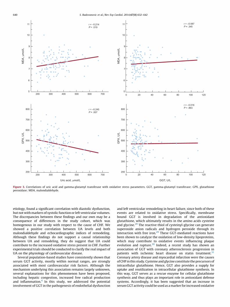
glutathione peroxidase activity. The UA level was significantlyassociated with both malondialdehyde concentration and glu-tathione peroxidase activity (r = 0.214, P = .019 and r = �0.245,P = .007, respectively). However, plasma GGT activity wasnot associated with the examined markers of oxidative stress(Figure 3).
DISCUSSION
Although endothelial dysfunction has been documented inperipheral and coronary arteries in CHF patients,9 only 1 studyin 38 mostly male participants with NYHA class II and III CHF hasfound an association between UA and endothelial function.Because the strict inclusion criteria (FMD < 8%) in this studycould impair the generalization of the results to a less severe CHFpopulation, we hypothesized that, in the early stages of CHF,increased serum UA levels, as an indicator of xanthine oxidaseactivation, could be used as a risk marker while, in the later phases,UA could act as an antioxidant, having a protective action onextracellular antioxidant enzymes and improving endothelialfunction. However, the results of the current study, which included3 times more participants with various stages of CHF, have shownan inverse correlation between UA and FMD, while the activity ofthe antioxidant enzyme glutathione peroxidase was inversely
correlated with UA. These results are biologically plausible, giventhat the main source of elevated UA in CHF is xanthine oxidase,the enzyme that produces superoxide or hydrogen peroxide asbyproducts of the terminal steps of purine metabolism in thepresence of hypoxia. Under these conditions, an increase insuperoxide may inactivate nitric oxide,18 suggesting an importantunderlying mechanism in the development of vascular endothelialdysfunction in CHF, which contributes to systemic vasoconstric-tion and increased cardiac loading.
Cardiac remodeling is an unfavorable prognostic factorassociated with myocardial hypertrophy, fibrosis, and ventriculardysfunction after myocardial infarction.19 The proposed mechan-ism causing a decrease in myocardial contractility is the celldamage produced by oxygen-free radicals, leading to peroxidationof membrane phospholipids, which can result in an increase inmembrane fluidity, increasing permeability and loss of membraneintegrity.20,21 In this study, we have shown that the UA levelcorrelates with the degree of myocardial dysfunction, based on alinear regression analysis of the association between UA level andthe echocardiographic indices of left ventricular remodeling. Thiseffect of UA on remodeling seems to be at least partially mediatedby enhanced lipid peroxidation, since UA correlated significantlywith the lipid peroxidation byproduct malondialdehyde andantioxidant glutathione peroxidase activity. Xanthine oxidaseexpression and activity were found to be markedly increased in the

Table 3Multiple Linear Regression Models on Association Between Uric Acid and Echocardiographic Indices of Remodeling
Unadjusted Model 1a Model 2b Model 3c
b P b P b P b P
LVEDD
Uric acid 0.340 <.001 0.324 <.001 0.327 <.001 0.340 .002
Sex �0.172 .055 �0.079 .397 �0.074 .457
Age �0.077 .405 �0.180 .048 �0.187 .055
BMI, kg/m2 0.183 .034 0.179 .042 0.202 .026
Smoking 0.069 .448 0.061 .505 0.073 .432
DM 0.057 .514 0.053 .548
Cholesterol �0.031 .719 �0.084 .345
SBP �0.168 .288
DBP 0.111 .474
Creatinine �0.038 .741
LVESD
Uric acid 0.337 <.001 0.316 .001 0.322 <.001 0.345 .002
Sex �0.118 .192 �0.023 .805 �0.014 .889
Age �0.028 .768 �0.123 .181 �0.134 .173
BMI, kg/m2 0.182 .038 0.172 .054 0.204 .026
Smoking 0.075 .411 0.070 .448 0.087 .354
DM 0.066 .457 0.062 .492
Cholesterol 0.025 .781 �0.046 .611
SBP �0.205 .201
DBP 0.103 .511
Creatinine �0.058 .614
LVEDV
Uric acid 0.289 .001 0.244 .005 0.238 .006 0.198 .059
Sex �0.253 .004 �0.039 .663 �0.041 .670
Age �0.033 .714 �0.252 .005 �0.237 .013
BMI, kg/m2 0.275 .001 0.285 .001 0.298 .001
Smoking �0.017 .849 �0.016 .860 �0.017 .852
DM �0.043 .607 �0.035 .689
Cholesterol �0.051 .547 �0.041 .632
SBP �0.124 .415
DBP 0.101 .503
Creatinine 0.071 .521
LVESV
Uric acid 0.321 <.001 0.279 .002 0.276 .002 0.269 .003
Sex �0.216 .016 0.002 .981 0.019 .842
Age 0.004 .968 �0.213 .019 �0.218 .018
BMI, kg/m2 0.198 .021 0.203 .021 0.219 .015
Smoking �0.024 .786 �0.022 .811 �0.021 .819
DM �0.031 .724 �0.016 .856
Cholesterol �0.007 .939 �0.013 .878
SBP �0.187 .233
DBP 0.128 .404
Creatinine 0.111 .417
BMI, body mass index; DM, diabetes mellitus; DBP, diastolic blood pressure; LVEDD, left ventricular end-diastolic diameter; LVEDV, left ventricular end-diastolic volume;
LVESD, left ventricular end-systolic diameter; LVESV, left ventricular end-systolic volume; SBP, systolic blood pressure.a Adjusted for age, sex, body mass index and smoking status.b Adjusted for the covariates in model 1 plus an additional adjustment for serum cholesterol and the presence of diabetes mellitus.c Adjusted for the covariates in model 2 plus an additional adjustment for systolic blood pressure, diastolic blood pressure and serum creatinine.
S. Radovanovic et al. / Rev Esp Cardiol. 2014;67(8):632–642 637
tissue 2 mm surrounding the infarct area22 in a mice model ofmyocardial infarction. In the heart, xanthine oxidase is localizedsolely in the capillary endothelium.23 Therefore, the UA generatedin hypoxic states originates from capillary endothelial cells, ratherthan from the myocardium,24 and hyperuricemia in heart failure
may reflect the metabolic effects of hypoxia on the microvascu-lature. The correlation between UA and left ventricular dysfunctionfound in the current study confirms the results of in vitro studiesthat have shown that reactive oxygen species production byxanthine oxidase leads to a depression of the excitation-contraction

r = 0.321P < .001
r = 0.337P < .001
r = 0.294P < .001
r = 0.340P < .001
50
0
150
100
250
200
350
400
300
50
0
150
100
250
200
350
400
300
3
2
4
5
6
7
8
3
2
4
5
6
7
8
200 300 400 500 600 700
200 300 400 500 600 700
200 300 400 500 600 700
200 300 400 500 600 700
Uric acid, µmol/L Uric acid, µmol/L
LVE
SV,
mL
LVE
DV,
mL
LVE
SD
, cm
LVE
DD
, cm
Figure 1. Correlations of uric acid with echocardiographic indices of remodeling. LVEDD, left ventricular end-diastolic dimension; LVEDV, left ventricular end-diastolic volume; LVESD, left ventricular end-systolic diameter; LVESV, left ventricular end-systolic volume.
r = 0.359P <. 001
r = 0.238P <. 009
r = 0.369P < .001
r = 0.219P < .016
00 0
50
150
100
250
200
350
300
400
3
2
4
5
6
7
8
0
50
150
100
250
200
350
300
400
3
2
4
5
6
7
8
20 40 60 80 100 120
20 40 60 80 100 120
20 40 60 80 100 120
20 40 60 80 100 120
GGT, U/L GGT, U/L
LVE
SV,
(mL)
LVE
SD
, cm
LVE
DV,
mL
LVE
DD
, cm
Figure 2. Correlations of gamma-glutamyl transferase with echocardiographic indices of remodeling. GGT, gamma-glutamyl transferase; LVEDD, left ventricularend-diastolic diameter; LVEDV, left ventricular end-diastolic volume; LVESD, left ventricular end-systolic diameter; LVESV, left ventricular end-systolic volume.
S. Radovanovic et al. / Rev Esp Cardiol. 2014;67(8):632–642638

Table 4Multiple Linear Regression Models on Association Between Gamma-glutamyl Transferase and Echocardiographic Indices of Remodeling
Unadjusted Model 1a Model 2b Model 3c
b P b P b P b P
LVEDD
GGT 0.219 .016 0.130 .176 0.133 .169 0.074 .453
Age 0.013 .889 0.009 .929 �0.046 .658
Sex �0.191 .044 �0.197 .040 �0.152 .132
BMI, kg/m2 0.169 .075 0.169 .080 0.218 .028
Smoking 0.042 .659 0.035 .716 0.054 .575
DM 0.035 .698 0.028 .759
Cholesterol �0.051 .576 �0.082 .372
SBP �0.174 .291
DBP 0.089 .584
Creatinine 0.146 .152
LVESD
GGT 0.238 .009 0.170 .079 0.170 .082 0.109 .276
Age 0.063 .507 0.065 .498 0.019 .859
Sex �0.130 .171 �0.135 .162 �0.096 .343
BMI, kg/m2 0.156 .102 0.150 .121 0.209 .036
Smoking 0.049 .608 0.044 .646 0.067 .492
DM 0.045 .625 0.037 .692
Cholesterol 0.003 .974 �0.046 .625
SBP �0.208 .212
DBP 0.079 .627
Creatinine 0.121 .238
LVEDV
GGT 0.340 <.001 0.217 .016 0.221 .014 0.191 .039
Age 0.043 .625 0.032 .719 �0.005 .955
Sex �0.249 .005 �0.246 .006 �0.206 .029
BMI, kg/m2 0.227 .011 0.240 .008 0.259 .005
Smoking �0.038 .665 �0.036 .682 �0.032 .718
DM �0.058 .493 �0.047 .586
Cholesterol �0.072 .398 �0.045 .596
SBP �0.113 .459
DBP 0.082 .583
Creatinine 0.148 .117
LVESV
GGT 0.359 <.001 0.278 .003 0.280 .003 0.232 .014
Age 0.092 .306 0.086 .344 0.032 .746
Sex �0.207 .022 �0.203 .026 �0.146 .124
BMI, kg/m2 0.135 .133 0.143 .117 0.177 .058
Smoking �0.049 .585 �0.046 .612 �0.037 .679
DM �0.047 .584 �0.035 .683
Cholesterol �0.032 .710 �0.009 .916
SBP �0.166 .285
DBP 0.091 .551
Creatinine 0.204 .034
BMI, body mass index; DM, diabetes mellitus; DBP, diastolic blood pressure, GGT, gamma-glutamyl transferase; LVEDD, left ventricular end-diastolic diameter; LVEDV, left
ventricular end-diastolic volume; LVESD, left ventricular end-systolic diameter; LVESV, left ventricular end-systolic volume; SBP, systolic blood pressure.a Adjusted for age, sex, body mass index and smoking status.b Adjusted for the covariates in model 1 plus an additional adjustment for serum cholesterol and the presence of diabetes mellitus.c Adjusted for the covariates in model 2 plus an additional adjustment for systolic blood pressure, diastolic blood pressure and serum creatinine.
S. Radovanovic et al. / Rev Esp Cardiol. 2014;67(8):632–642 639
coupling mechanism in cardiac muscle.25,26 These effects wouldinduce a decrease in cardiac contractility and in the rate of cardiacmuscle relaxation. In addition to xanthine oxidase, hyperuricemiaitself can also influence free radical production and myocardialremodeling. Chen et al16 have shown that hyperuricemia induced byoxonic acid stimulates myocardial superoxide production, resulting
in enhanced endothelin-1-induced ventricular remodeling ininfarcted rats. Furthermore, treatment of hyperuricemic rats withallopurinol and benzbromarone, UA-lowering agents, attenuatedremodeling after myocardial infarction. However, Cicoira et al27,who evaluated the effects of elevated UA levels on cardiac function in150 CHF patients resulting from dilated cardiomyopathy of diverse
11
10
9
8
7
6
5
11
10
9
8
7
6
5
800
700
600
500
r = –0.245P = .007
r = –0.214P = .019
r = –0.016P = .863
r = –0.087P = .345
400
300
200
800
700
600
500
400
300
200200 300 400 500
Uric acid, μmol/L GGT, U/L
MD
A, μ
mol
/L
MD
A, μ
mol
/L
GP
X, U
/L
GP
X, U
/L
600 700
200 300 400 500 600 700
0 20 40 60 80 100 120
0 20 40 60 80 100 120
Figure 3. Correlations of uric acid and gamma-glutamyl transferase with oxidative stress parameters. GGT, gamma-glutamyl transferase; GPX, glutathioneperoxidase; MDA, malondialdehyde.
S. Radovanovic et al. / Rev Esp Cardiol. 2014;67(8):632–642640
etiology, found a significant correlation with diastolic dysfunction,but not with markers of systolic function or left ventricular volumes.The discrepancies between these findings and our own may be aconsequence of differences in the study cohort, which washomogenous in our study with respect to the cause of CHF. Weshowed a positive correlation between UA levels and bothmalondialdehyde and echocardiographic indices of remodeling.Although these findings do not support a causal relationshipbetween UA and remodeling, they do suggest that UA couldcontribute to the increased oxidative stress present in CHF. Furtherexperimental trials should be conducted to clarify the real impact ofUA on the physiology of cardiovascular disease.
Several population-based studies have consistently shown thatserum GGT activity, mostly within normal ranges, are stronglyassociated with most cardiovascular risk factors. Although themechanism underlying this association remains largely unknown,several explanations for this phenomenon have been proposed,including hepatic congestion, increased free radical productionand inflammation.4 In this study, we addressed the potentialinvolvement of GGT in the pathogenesis of endothelial dysfunction
and left ventricular remodeling in heart failure, since both of theseevents are related to oxidative stress. Specifically, membranebound GGT is involved in degradation of the antioxidantglutathione, which ultimately results in the amino acids cysteineand glycine.28 The reactive thiol of cysteinyl-glycine can generatesuperoxide anion radicals and hydrogen peroxide through itsinteraction with free iron.29 These GGT-mediated reactions havebeen shown to catalyze the oxidation of low-density lipoproteins,which may contribute to oxidative events influencing plaqueevolution and rupture.30 Indeed, a recent study has shown anassociation of GGT with coronary atherosclerosis progression inpatients with ischemic heart disease on statin treatment.31
Coronary artery disease and myocardial infarction were the causesof CHF in this study. Cysteine and glycine constitute the precursors ofintracellular glutathione. Hence, GGT also provides a supply foruptake and reutilization in intracellular glutathione synthesis. Inthis way, GGT serves as a rescue enzyme for cellular glutathionesynthesis and thus plays an important role in antioxidant defensesystems. Accordingly, it has been suggested that an increase inserum GGT activity could be used as a marker for increased oxidative
S. Radovanovic et al. / Rev Esp Cardiol. 2014;67(8):632–642 641
stress in humans.32,33 Although the results of our study haveindicated a relationship between cardiac dysfunction and GGTactivity, it seems that it is not related to either increased lipidoxidation or impairment of antioxidant activity. Thus, in contrast toUA, serum GGT activity was not associated with the biomarkers ofoxidative stress in CHF. Therefore, it may be speculated that elevatedGGT in heart failure is only a part of the cholestatic profile oflaboratory elevations seen in these patients, secondary to hepaticcongestion. Nevertheless, the question of the mechanisms involvedin the association between GGT and cardiac dysfunction should beaddressed in future in vitro and animal studies.
The last decade has seen a significant advance in thepathophysiology of heart failure. As a result, many differentdiagnostic and prognostic markers have been proposed and fewhave shown clear clinical use.34,35 Among those, serum UA seemsto fulfill these criteria. First, accurate, repeated measurements ofUA are available to the clinician at a reasonable cost and with shortturnaround times; second, the determination of serum UAprovides information that is not already available from a carefulclinical assessment; and finally, knowing the measured UAconcentration should aid in medical decision-making. The currentstudy provides a moderate correlation between levels of UA andindices of remodeling. On the other hand, UA and other currentlyassessable markers of CHF lack cardiac specificity, and their levelscan be influenced by both systemic inflammatory and infectiveprocesses, which occur frequently in heart failure patients. Untilnow, several new biomarkers with a plausible biological linkwith heart failure pathophysiology have been identified. Amongthem, ST-2 and galectin share the ability to define the severity ofthe ongoing ventricular remodeling process.34,36 However, theirclinical use should be confirmed in large-scale studies.
Limitations
The limitations of this study include its cross-sectional designand rather small sample size, especially in certain NYHAsubgroups. The cross-sectional design of the study precludesdetermining a causal relationship and the prognostic value of thesedeterminations. Therefore, these findings warrant validationin further prospective studies with larger numbers of patients inadvanced NYHA classes in cardiac dysfunction.
CONCLUSIONS
In this study, we show that serum levels of UA and GGT activityare associated with echocardiographic indices of left ventricularremodeling in patients with CHF secondary to ischemic coronarydisease. Regarding endothelial function, only serum UA showed aninverse correlation with endothelium-dependent vasodilation inCHF. The effects of UA on endothelial function and left ventriculardysfunction may be at least partially explained by its relationshipwith oxidative stress markers such as malondialdehyde andglutathione peroxidase activity.
FUNDING
This work was supported by the grant 175052 from the SerbianMinistry of Education, Science and Technological Developmental.
CONFLICTS OF INTEREST
None declared.
REFERENCES
1. Colucci WS, Braunwald E. Pathophysiology of heart failure. Braunwald’s heartdisease. A textbook of cardiovascular medicine. 8th ed. Philadelphia: ElsevierSaunders; 2008.
2. Savarese G, Ferri C, Trimarco B, Rosano G, Dellegrottaglie S, Losco T, et al.Changes in serum uric acid levels and cardiovascular events: a meta-analysis.Nutr Metab Cardiovasc Dis. 2013;23:707–14.
3. Tamariz L, Harzand A, Palacio A, Verma S, Jones J, Hare J. Uric acid as a predictorof all-cause mortality in heart failure: a meta-analysis. Congest Heart Fail.2011;17:25–30.
4. Poelzl G, Eberl C, Achrainer H, Doerler J, Pachinger O, Frick M, et al. Prevalenceand prognostic significance of elevated gamma-glutamyltransferase in chronicheart failure. Circ Heart Fail. 2009;2:294–302.
5. Zoccali C, Maio R, Mallamaci F, Sesti G, Perticone F. Uric acid and endothelialdysfunction in essential hypertension. J Am Soc Nephrol. 2006;17:1466–71.
6. Kanbay M, Yilmaz MI, Sonmez A, Turgut F, Saglam M, Cakir E, et al. Serum uricacid level and endothelial dysfunction in patients with nondiabetic chronickidney disease. Am J Nephrol. 2011;33:298–304.
7. Mercuro G, Vitale C, Cerquetani E, Zoncu S, Deidda M, Fini M, et al. Effect ofhyperuricemia upon endothelial function in patients at increased cardiovas-cular risk. Am J Cardiol. 2004;94:932–5.
8. Farquharson CA, Butler R, Hill A, Belch JJ, Struthers AD. Allopurinol improvesendothelial dysfunction in chronic heart failure. Circulation. 2002;106:221–6.
9. Hornig B, Arakawa N, Kohler C, Drexler H, Vitamin C. improves endothelialfunction of conduit arteries in patients with chronic heart failure. Circulation.1998;97:363–8.
10. Sun Y. Oxidative stress and cardiac repair/remodeling following infarction. Am JMed Sci. 2007;334:197–205.
11. Zhang M, Shah AM. Role of reactive oxygen species in myocardial remodeling.Curr Heart Fail Rep. 2007;4:26–30.
12. Radovanovic S, Savic-Radojevic A, Pljesa-Ercegovac M, Djukic T, Suvakov S,Krotin M, et al. Markers of oxidative damage and enzyme activities as predictorsof morbidity and mortality in patients with chronic heart failure. J Card Fail.2012;18:493–501.
13. Parissis JT, Andreadou I, Markantonis SL, Bistola V, Louka A, Pyriochou A, et al.Effects of levosimendan on circulating markers of oxidative and nitrosative stressin patients with advanced heart failure. Atherosclerosis. 2007;195:e210–5.
14. Utsumi H, Takeshita A. Treatment with dimethylthiourea prevents left ven-tricular remodeling and failure after experimental myocardial infarction inmice: role of oxidative stress. Circ Res. 2000;87:392–8.
15. Lee TM, Lai PY, Chang NC. Effect of N-acetylcysteine on sympathetic reinnerva-tion in post-infarcted rat hearts. Cardiovasc Res. 2010;85:137–46.
16. Chen CC, Hsu YJ, Lee TM. Impact of elevated uric acid on ventricular remodelingin infarcted rats with experimental hyperuricemia. Am J Physiol Heart CircPhysiol. 2011;301:H1107–1.
17. Schiller NB, Shah PM, Crawford M, DeMaria A, Devereux R, Feigenbaum H, et al.Recommendations for quantitation of the left ventricle by two-dimensionalechocardiography. American Society of Echocardiography Committee onStandards, Subcommittee on Quantitation of Two-Dimensional Echocardio-grams. J Am Soc Echocardiogr. 1989;2:358–67.
18. Lynch SM, Frei B, Morrow JD, Roberts 2nd LJ, Xu A, Jackson T, et al. Vascularsuperoxide dismutase deficiency impairs endothelial vasodilator functionthrough direct inactivation of nitric oxide and increased lipid peroxidation.Arterioscler Thromb Vasc Biol. 1997;17:2975–81.
19. Weber KT, Anversa P, Armstrong PW, Brilla CG, Burnett Jr JC, Cruickshank JM,et al. Remodeling and reparation of the cardiovascular system. J Am Coll Cardiol.1992;20:3–16.
20. Freeman BA, Crapo JD. Biology of disease: free radicals and tissue injury. LabInvest. 1982;47:412–26.
21. Meerson FZ, Kagon VE, Kozlov YP, Bellina LM, Arkkhipenko YV. The role of lipidperoxidation in pathogenesis of ischemic damage and the antioxidant protec-tion of the heart. Basic Res Cardiol. 1982;77:465–85.
22. Engberding N, Spiekermann S, Schaefer A, Heineke A, Wiencke A, Muller M,et al. Allopurinol attenuates left ventricular remodeling and dysfunction afterexperimental myocardial infarction. A new action for an old drug? Circulation.2004;110:2175–9.
23. Jarasch E, Grund C, Bruder G, Heid HW, Keenan TW, Franke WW. Localization ofxanthine oxidase in mammary gland epithelium and capillary endothelium.Cell. 1981;25:67–82.
24. Nees S, Gerbes AL, Gerlach E, Staubesand J, Isolation. identification, and con-tinuous culture of coronary endothelial cells from guinea-pig hearts. Eur J CellBiol. 1981;24:287–97.
25. Prasad K, Kalra J, Chan WP, Chaudhary A. Effect of oxygen free radicals oncardiovascular function at organ cellular levels. Am Heart J. 1989;117:1196–202.
26. Hess ML, Okabe E, Kontos HA. Proton and free oxygen radical interaction withthe calcium transport system of cardiac sarcoplasmatic reticulum. J Mol CellCardiol. 1981;13:767–72.
27. Cicoira M, Zanolla L, Rossi A, Golia G, Franceschini L, Brighetti G, et al. Elevatedserum uric acid levels are associated with diastolic dysfunction in patients withdilated cardiomyopathy. Am Heart J. 2002;143:1107–11.
28. Whitfield JB. Gamma glutamyl transferase. Crit Rev Clin Lab Sci. 2001;38:263–355.

S. Radovanovic et al. / Rev Esp Cardiol. 2014;67(8):632–642642
29. Pompella A, Emdin M, Passino C, Paolicchi A. The significance of serum gamma-glutamyltransferase in cardiovascular diseases. Clin Chem Lab Med. 2004;42:1085–91.
30. Paolicchi A, Minotti G, Tonarelli P, Tongiani R, De Cesare D, Mezzetti A, et al.Gamma-glutamyl transpeptidase-dependent iron reduction and LDL oxidation—a potential mechanism in atherosclerosis. J Investig Med. 1999;47:151–60.
31. Niccoli G, Della Bona R, Cosentino N, D’Amario D, Belloni F, Conti MG, et al.Serum levels of g-glutamyltransferase and progression of coronary athero-sclerosis. Coron Artery Dis. 2013;24:40–7.
32. Lee DH, Blomhoff R, Jacobs Jr DR. Is serum gamma glutamyltransferase amarker of oxidative stress? Free Radic Res. 2004;38:535–9.
33. Rahman I, MacNee W. Oxidative stress regulation of glutathione in lunginflammation. Eur Respir J. 2000;16:534–54.
34. Hrynchyshyn N, Jourdain P, Desnos M, Diebold B, Funck F. Galectin-3: a newbiomarker for the diagnosis, analysis and prognosis of acute and chronic heartfailure. Arch Cardiovasc Dis. 2013;106:541–6.
35. Porcel JM. Utilization of B-type natriuretic peptide and NT-proBNP in thediagnosis of pleural effusions due to heart failure. Curr Opin Pulm Med.2011;17:215–9.
36. Shah RV, Januzzi Jr JL. ST2: a novel remodeling biomarker in acute and chronicheart failure. Curr Heart Fail Rep. 2010;7:9–14.