Process Intensification of Nicotinic Acid Production via ...
Upload
independentCategory
view
1download
0
ORIGINAL INVESTIGATION
Effects of TC-1734 (AZD3480), a selective neuronal nicotinicreceptor agonist, on cognitive performanceand the EEG of young healthy male volunteers
G. Dunbar & P. H. Boeijinga & A. Demazières &
C. Cisterni & R. Kuchibhatla & K. Wesnes & R. Luthringer
Received: 27 June 2006 /Accepted: 4 December 2006 / Published online: 16 January 2007# Springer-Verlag 2007
AbstractObjectives The aim of this study was to get insight into thecentral effects of TC-1734 (renamed AZD3480), a selectiveagonist at the neuronal nicotinic receptor of the α4β2subtype.Materials and methods Electroencephalography (EEG)techniques and computerized cognitive tests were per-formed in young, healthy male volunteers during twodouble-blind and placebo-controlled studies: a rising singledose crossover study (from 2 to 320 mg) and a risingrepeated dose study with a parallel group design (50, 100,and 200 mg).Results In contrast to acute administration, administrationof AZD3480 over 10 days produced statistically significantenhancement of several cognitive measures (attention andepisodic memory) compared to placebo. Regarding EEGdata, AZD3480 showed acceleration of the alpha centroidand of the alpha peak in the single-dose study. This EEGprofile of the acceleration type was confirmed in therepeated dose study on both day 1 and day 10, with thegreatest effect observed with the highest dose. The EEG
pattern shown for AZD3480 was consistent with thatpreviously described with other drugs known to improveattention and vigilance (including nicotine). In addition,subjects dosed with AZD3480 showed a statisticallysignificant increase in mismatch negativity (MMN) ampli-tude at 50 and 200 mg while reducing MMN latency(200 mg only), suggesting an improvement of pre-atten-tional mechanisms.Conclusion These early data in healthy subjects provideencouragement to consider development of AZD3480 as anovel agent for the treatment of cognitive decline in theelderly, including age-associated memory impairment and/ordementia of the Alzheimer’s type.
Keywords Electroencephalography . Neuronal nicotinicreceptors . Alzheimer .Memory . Attention
Introduction
A large body of evidence suggests that neuronal nicotinicreceptors (NNRs) play a key role in memory and cognitiveprocesses, whereas NNR loss is involved in the cognitivedeficits associated with aging and dementias, includingAlzheimer’s disease (AD; for review, see Perry et al. 2001;Auld et al. 2002; Hogg et al. 2003). Recently, neuroimagingstudies have shown a loss of NNRs—mainly thosecontaining the α4 subunit—in the hippocampus and in thetemporal and frontal cortex of patients with AD comparedto age-matched controls, together with significant correla-tions between cognitive dysfunction and the loss ofnicotinic receptor binding (Nordberg 2001; Hogg et al.2003). The role of the α4β2 NNR subtype in cognitivefunctioning was supported by preclinical behavioral studies.
Psychopharmacology (2007) 191:919–929DOI 10.1007/s00213-006-0675-x
P. H. Boeijinga :A. Demazières (*) : C. Cisterni : R. LuthringerFORENAP,27 rue du 4° RSM,68250 Rouffach, Francee-mail: [email protected]
G. Dunbar : R. KuchibhatlaTARGACEPT Inc.,200 East First Street Suite 300,Winston-Salem, NC 27101-4165, USA
K. WesnesCognitive Drug Research Ltd.,CDR House, Gatehampton Road,Goring-on-Thames RG8 0EN, UK

Indeed, mice lacking high-affinity β2 binding sites fornicotine showed impaired spatial learning (Zoli et al.1999), and hippocampal infusion of a selective α4β2antagonist induced working memory impairment, whichwas reversed by nicotine (Arthur and Levin 2002). Inclinical trials, nicotine, the prototype non-selective NNRagonist, improved performances of AD patients on learningand memory tasks, whereas mecamylamine, a non-specificnicotinic antagonist (above a low dose), produced theopposite effects (Newhouse et al. 2001). Acute transdermaladministration of ABT-418, a NNR agonist with highaffinity for the α4β2-receptor subtype, was also found toimprove performances of AD patients in learning andmemory tasks (Potter et al. 1999).
TC-1734, renamed AZD3480 (S-E-[4-(5-Isopropoxy-pyr-idin-3-yl)-1-methyl-but-3-enyl]-methyl-amine, also known asispronicline), is another NNR agonist with a high affinity andselectivity for the α4β2 receptor. Preclinical studies indicatedthat AZD3480 displayed neuroprotective properties, en-hanced learning, and memory processing in various animalmodels (e.g., object recognition test in mice, step-throughpassive avoidance test, and radial arm maze in rats) withoutinducing addictive-like behaviors and had a favorable toxicityprofile (Obinu et al. 2002; Gatto et al. 2004). Thus,AZD3480 might be a potential candidate for AD treatmentand other cognitive disorders in the elderly.
Phase 1 clinical trials have been completed. In the presentpaper, we report the effects of single and repeated doses ofAZD3480 on the electroencephalograph (EEG) and cognitiveperformance of healthy, young male volunteers. The reasonsfor choosing EEG to characterize the pharmacodynamicactivity of AZD3480 at an early stage of development weretwofold. Firstly, EEG is recognized as a highly sensitive toolto characterize drug effects on the central nervous system inhealthy volunteers (Saletu 1996). Secondly, EEG measureshave been used to some extent in the field of clinical ADresearch. Of primary interest, cognitive decline in Alz-heimer’s patients was found to correlate with EEG slowing(Prichep et al. 1994; Forstl et al. 1996; Besthorn et al. 1997;Rodriguez et al. 2002; Kowalski et al. 2001; Moretti et al.2004 for review), which was reversed by short-term treatmentwith acetylcholinesterase inhibitors (Jelic et al. 1998; Brassenand Adler 2003). Interestingly, in healthy volunteers,nicotine decreased slow wave activity and increased fastwave activity (Foulds et al. 1994; Knott et al. 1999;Lindgren et al. 1999), effects which are opposite to thoseobserved in AD (Knott et al. 2000). The validated CDRcomputerized test battery was used for assessment ofchanges in attention, learning, and memory, as this systemhas proven sensitive to drug-induced cognitive changes inhealthy subjects, including physostigmine, which belongs tothe same class of compounds as those currently used to treatAlzheimer’s disease (Wesnes et al. 1994).
Materials and methods
The two studies were conducted at a single center(FORENAP, France), after approval by an accreditedindependent ethics committee (CCPPRB of Strasbourg),and in accordance with the ethical principles of theDeclaration of Helsinki.
The preliminary pharmacodynamic evaluations reportedin the present paper were recorded in parallel to pharma-cokinetics and safety data, which were reported in aprevious paper (Dunbar et al. 2006a).
Participants
Non-smoking males in good health on the basis of medicalhistory, physical examination, cardiovascular examination,and routine laboratory tests were considered eligible forenrolment into these studies, after having given writteninformed consent. Subjects could not be included if theyhad a known or suspected history of alcohol or drug abuseor if they consumed excessive amounts of xanthine-containing beverages. No medication was allowed withinat least 2 weeks before admission (or 1 week for over-the-counter drugs). All subjects had a normal standard EEG atscreening.
Study designs and treatments
Both studies were conducted in a randomized, double blind,placebo-controlled fashion according to a two-period cross-over design (study A) and a parallel group design (study B).
Study A consisted of eight dose cohorts of six subjectseach, to test ascending single oral doses of AZD3480 (2, 4, 10,20, 40, 80, 160, 320 mg). Each subject remained in the clinicalunit for 5 days (day −1 to day 5) and received one dose ofAZD3480 and its matching placebo on day 1 and day 4 in theorder determined by their assigned randomized sequence.
Study B consisted of three dose cohorts, to test repeatedoral doses of AZD3480 (50, 100, and 200 mg) administeredonce daily for 10 days. In each dose cohort, six subjects wererandomized to receive AZD3480 and two subjects to receiveplacebo.
The subjects were abstinent from alcohol and caffeinethroughout their stay in the clinical unit for both studies.
Capsules containing AZD3480 (di-p-toluoyl-D-tartaricacid salt) or its placebo were swallowed with a glass ofwater around 9:00 A.M., subjects being in a fasting condition.
Cognitive measures
Different aspects of cognition were assessed by thecomputerized test battery developed by Cognitive DrugResearch (CDR, UK). Five factors were derived from the
920 Psychopharmacology (2007) 191:919–929

various individual parameters resulting from the differentcognitive tasks. Power of attention was derived from speedon the simple reaction time (SRT), the digit vigilance (DV),and choice reaction time (CRT). Continuity of attention wasderived from accuracy on the CRT, target detected and falsealarms on the DV, and average distance from target on thevisual tracking task. Quality of working memory wasderived from the sensitivity indices from the numeric andspatial working memory tasks (NWM and SWM). Qualityof episodic memory was derived from the parameters ofimmediate word recall (IWR), delayed word recall (DWR),and accuracy scores from the word recognition and picturerecognition tasks (WR and PR). Speed of memory wasderived from speed on the NWM, SWM, WR, and PR tasks.
Parallel forms of the tests were presented on each testingsession. All these tests and the techniques for calculatingthe different parameters have been described previously(see Wesnes et al. 2000 for details).
Training on the CDR system took place before the firststudy day to familiarize the subjects with the procedure, toovercome initial learning effects, and settle any anxietyaround the use of computer testing. On the study days, thecognitive test battery was administered 1 h before dosingand 1, 2, 4, 8, and 24 h post-dosing (day 1 and day 4 ofstudy A; day 1 and day 10 of study B). Additional timepoints (12 and 36 h post-dosing) were obtained duringstudy A for the 160- and 320-mg dose cohorts.
Statistical analyses All the analyses were performed onchanges from baseline at each time point. In study A, aseparate analysis was run for each dose cohort. Ananalysis of variance for repeated measures (RM-ANOVA,mixed procedure) was used to identify main effects fortreatment condition (placebo, AZD3480), time (timepoints), day (day 1, day 4) and “treatment × time”interaction. The estimate statement was used to compareplacebo to TC-1734 at each of the post-dosing timepoints. In study B, data from the three dose cohorts werepooled and analyzed by a RM-ANOVA (mixed proce-dure). Terms were fitted to the ANOVA for treatment (0,50, 100, 200 mg), time (the repeated assessments overdays 1 to 10), and the interaction between treatment andtime. Planned comparisons were made between placeboand each dose of AZD3480. The statistical level ofsignificance was fixed at p<0.05 (two-tailed).
Electroencephalography (EEG) procedures
All the EEG recordings and data acquisitions were done ona PC equipped with the HEM package (Notocord, Paris,France). Data analyses were done using NeuroScan®(Herndon, VA, USA).
Study A Spontaneous EEG recordings in resting conditionwere collected for 5 min, with eyes closed, from fourderivations (F4-T4, F3-T3, T4-O2, T3-O1) on day 1 andday 4 at the following times: before drug administration(double baseline) and 0.5, 1, 2, 4, 6, and 8 h post-dosing.Additional EEG recordings were collected at 1.5 h for dosecohorts from 2 to 80 mg and at 12, 24, and 36 h for dosecohorts 160 and 320 mg. The EEGs were digitized at256 Hz, and filter settings of 0.5–70 Hz were used. Afterremoval of artifacts by visual inspection, 2-s EEG epochsyielding 0.5 Hz frequency bins and averaged for therecording period were subjected to the fast Fouriertransformation algorithm for the computation of absoluteand relative power estimates in the following frequencybands: delta (0.5–3.5 Hz), theta (4–7.5 Hz), alpha 1 (8–9.5 Hz), alpha 2 (10–12.5 Hz), beta 1 (13–17.5 Hz), beta 2(18–20.5 Hz), and beta 3 (21–32 Hz). Additional parame-ters were calculated: mean frequency of the total EEG band(0.5–32 Hz), alpha centroid frequency, and alpha peakfrequency. Alpha slow-wave index (ASI = alpha/[delta +theta]); an indicator of overall arousal was also calculated(Matejcek 1980). An acceleration of the EEG was definedas an increase in alpha power with corresponding loss ofpower in the slow waves (delta, theta).
Statistical analyses All analyses were performed on themedian value of the four derivations calculated for eachspectral parameter. A global analysis on differencesfrom placebo was computed for comparisons of thedifferent doses of AZD3480 using the non-parametricKruskall–Wallis analysis of variance. An analysis onchanges from baseline was also computed for compar-isons of each dose to its matching placebo in each dosecohort using the Wilcoxon signed-rank test. Resultswere statistically significant at p<0.05 (trend towardssignificance at p<0.1).
Study B A 28-lead system was used to record event-related potentials (P300 and Mismatch Negativity orMMN) in addition to spontaneous EEG in vigilance-controlled and resting conditions (3 min each), on day 1and day 10 at the following times: before drug adminis-tration (double baseline) and 1, 2, 4, 6, and 8 h post-dosing. Additional measures were performed on day 2,before the second administration, and on day 11, 24 hafter the last administration. An ear-linked referenceelectrode and four artifact channels were also used (e.g.,detection of muscle activity, eye movements). EEG signalswere amplified, sampled, and subjected to the sameanalysis as described in study A. The same parameterswere also determined, except the alpha centroid and alphapeak frequencies. Frequency bands were expressed as thesquare root of the power.
Psychopharmacology (2007) 191:919–929 921

The P300 measures were obtained using an activeauditory “odd-ball” paradigm. According to this paradigm,the subjects are asked to attentively listen to a series offrequent tones (500 Hz) and rare tones (2,000 Hz), and tocount the latter. The tones were delivered through ear-phones bilaterally (85 dB, duration 120 ms). The two toneswere presented randomly with intertrial intervals rangingfrom 1 to 1.9 s and with frequent/rare tone ratio of 85/15%.For each EEG recording, 130 to 140 tones were deliveredto have around 20 rare tones. For each tone presentation,EEG waveforms were recorded with a sampling frequencyof 250 Hz and averaged to obtain 28 mean curves for eitherfrequent or rare tones separately. The filter settings were0.5–30 Hz or 0.5–70 Hz depending on the amount of EMGactivity. EEG recordings start 512 ms before the tone(baseline) and continued for 508 ms thereafter. The meanpositive amplitude, P300, located in the latency ranges232–352 ms, was calculated from the 28 scalp electrodes toobtain topographic maps.
The MMN measures were obtained using a passiveauditory “odd-ball” paradigm. According to this paradigm,the subjects had to listen to a series of frequent tones(1,000 Hz) and rare tones (1,050 Hz) while resting with theeyes opened. The tones were delivered through earphonesmonaurally (right ear, 85 dB, duration 60 ms). The toneswere presented randomly with intertrial intervals rangingfrom 680 to 720 ms and with frequent/rare tone ratio of85/15%. For each EEG recording, a block of 600 tones hasbeen presented. The recording procedures were the same asfor P300 acquisitions. MMN integrated amplitudes wereextracted to quantify the negative going deflection in thelatency range 135–265 ms for the difference in response torare and frequent stimuli. These were calculated using the28 electrodes to obtain topographic maps. The same, meanamplitude and mean latency of the second negative wavepeak (N2) and for the deflection in the latency range 76–116 ms (N100 following frequent tones) on the electrodeCz were analyzed qualitatively.
Statistical analyses Analyses of spontaneous EEG param-eters, P300 and MMN-integrated amplitude were performedaccording to the statistical decision tree (SDT) procedure(Dago et al. 1994). This non-parametric method based onWilcoxon tests allows testing baseline homogeneity first(predrug or PD, cf. x-axis of Fig. 1). Spectral data obtainedwith AZD3480 were compared to those obtained withplacebo at same time-points. To eliminate problems relatedto an eventual bias, data at a given time “t” weretransformed to changes from predrug values before enteringthe two-sample Wilcoxon calculation.
Statistical analyses were carried out on each electrodeseparately. We made decisions basically on this bivariatemodel (in analogy to planned comparisons for CDR tests).
To further reduce the influence of random effects due tointerindividual variability in parallel groups, drug-inducedmodifications could be evaluated with the software used byapplying analyses of within-day changes by matchedsample (univariate) statistical comparisons with respect topredrug assessments as well. Occasionally, other compar-isons of interest like pairwise differences between twoactive doses (e.g., highest versus lowest), or superpositionof day 10 and day 1 have been performed.
The statistical maps resulting from the SDT procedurewere coded according to the level of statistical trend(p<0.1) or significance (p<0.05 or p<0.01). To avoid falsepositives or type I error related to multiple testing, agrouping procedure was applied, i.e., having similar read-outs on at least five contiguous electrodes.
Results
Subjects
Forty-eight and twenty-four subjects completed study Aand study B in compliance with the protocol, respectively.Mean age varied from 25.3 to 33.5 years (range 18 to 45)and body mass index from 22.22 to 24.85 kg/m2 (range18.4 to 28.4) across the dose groups from both studies.
Cognitive tests
Overall, little shifts were detected in the different cognitivefactors tested after a single-dose administration ofAZD3480. Although a few statistical differences wereobserved between AZD3480 and placebo during study A,they were of small magnitude and largely due to randomfluctuation. Similarly, few significant differences wereobserved between AZD3480 and placebo after the firstadministration in study B. However, several individualmeasures and cognitive factors appeared improved after10 days of daily administration of AZD3480. Main“treatment” and/or “treatment × time” effects were detectedfor several individual parameters: DV (speed), NWM(speed), DWR (percent of correct words), and WR (speed).Multiple test comparisons showed a statistically significantimprovement of performance in the cohort dosed withAZD3480 200 mg on the DV task (speed) at all time points(Fig. 1). Multiple test comparisons for NWR and DWRfailed to find any significant changes mainly because oflarge random fluctuations. Although no main effects weredetected for two parameters (IWR percent of correct words,and PR sensitivity index), as well as for two factors derivedfrom the individual parameters (power of attention andquality of episodic memory), consistent statistically signif-
922 Psychopharmacology (2007) 191:919–929

IWR (word correctly recalled)
Picture recognition (sensitivity index) Power of attention (speed)
Quality of episodic memory
placebo 50 mg 100 mg 200 mg
-60-50-40-30-20-10
0102030405060
PD 1 h 2 h 4 h 8 h 24 htime (h)
placebo 50 mg 100 mg 200 mg
PD 1 h 2 h 4 h 8 h 24 htime (h)
placebo 50 mg 100 mg 200 mg
PD 1 h 2 h 4 h 8 h 24 htime (h)
placebo 50 mg 100 mg 200 mg
PD 1 h 2 h 4 h 8 h 24 htime (h)
placebo 50 mg 100 mg 200 mg
PD 1 h 2 h 4 h 8 h 24 htime (h)
chan
ges
from
base
line
(ms)
** **
**
*
(*)
Digit vigilance (speed)
-20-15-10-505
101520
chan
ges
from
base
line
(%)
##
#(#)
-0.5-0.4-0.3-0.2-0.1
00.10.20.30.40.5
chan
ges
from
base
line
(SI)
***
-80-60-40-20
020406080
100120140160ch
ange
sfr
omba
selin
e(m
s)
* **
*(*)
(*)
-80-60-40-20
0204060
chan
ges
from
base
line
#
* *
*
Fig. 1 Principal CDR tests showing significant changes by TC-1734compared to placebo on day 10. Results are expressed as means (withstandard error bars) on changes from baseline for placebo and eachdose of TC-1734. Asterisk stands for statistical differences between
dose 200 mg and placebo; number sign stands for statistical differencesbetween dose 100 mg and placebo. (*), (#) 0.05<p<0.1; *, #p<0.05,**p<0.01, (*)
Psychopharmacology (2007) 191:919–929 923
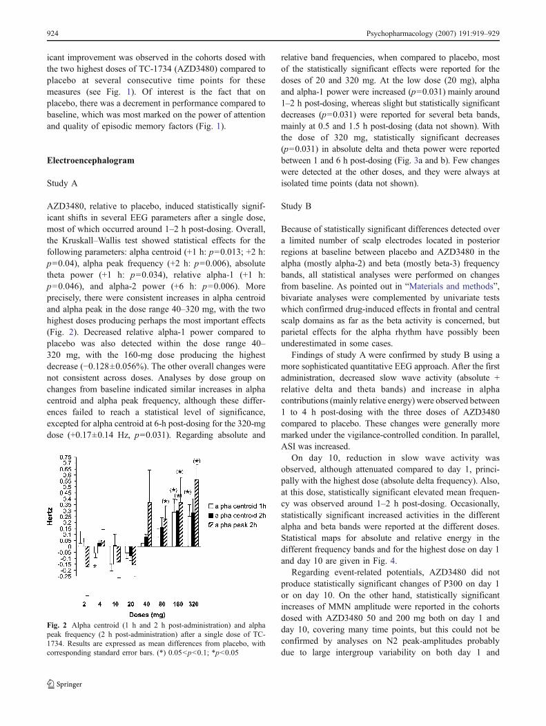
icant improvement was observed in the cohorts dosed withthe two highest doses of TC-1734 (AZD3480) compared toplacebo at several consecutive time points for thesemeasures (see Fig. 1). Of interest is the fact that onplacebo, there was a decrement in performance compared tobaseline, which was most marked on the power of attentionand quality of episodic memory factors (Fig. 1).
Electroencephalogram
Study A
AZD3480, relative to placebo, induced statistically signif-icant shifts in several EEG parameters after a single dose,most of which occurred around 1–2 h post-dosing. Overall,the Kruskall–Wallis test showed statistical effects for thefollowing parameters: alpha centroid (+1 h: p=0.013; +2 h:p=0.04), alpha peak frequency (+2 h: p=0.006), absolutetheta power (+1 h: p=0.034), relative alpha-1 (+1 h:p=0.046), and alpha-2 power (+6 h: p=0.006). Moreprecisely, there were consistent increases in alpha centroidand alpha peak in the dose range 40–320 mg, with the twohighest doses producing perhaps the most important effects(Fig. 2). Decreased relative alpha-1 power compared toplacebo was also detected within the dose range 40–320 mg, with the 160-mg dose producing the highestdecrease (−0.128±0.056%). The other overall changes werenot consistent across doses. Analyses by dose group onchanges from baseline indicated similar increases in alphacentroid and alpha peak frequency, although these differ-ences failed to reach a statistical level of significance,excepted for alpha centroid at 6-h post-dosing for the 320-mgdose (+0.17±0.14 Hz, p=0.031). Regarding absolute and
relative band frequencies, when compared to placebo, mostof the statistically significant effects were reported for thedoses of 20 and 320 mg. At the low dose (20 mg), alphaand alpha-1 power were increased (p=0.031) mainly around1–2 h post-dosing, whereas slight but statistically significantdecreases (p=0.031) were reported for several beta bands,mainly at 0.5 and 1.5 h post-dosing (data not shown). Withthe dose of 320 mg, statistically significant decreases(p=0.031) in absolute delta and theta power were reportedbetween 1 and 6 h post-dosing (Fig. 3a and b). Few changeswere detected at the other doses, and they were always atisolated time points (data not shown).
Study B
Because of statistically significant differences detected overa limited number of scalp electrodes located in posteriorregions at baseline between placebo and AZD3480 in thealpha (mostly alpha-2) and beta (mostly beta-3) frequencybands, all statistical analyses were performed on changesfrom baseline. As pointed out in “Materials and methods”,bivariate analyses were complemented by univariate testswhich confirmed drug-induced effects in frontal and centralscalp domains as far as the beta activity is concerned, butparietal effects for the alpha rhythm have possibly beenunderestimated in some cases.
Findings of study A were confirmed by study B using amore sophisticated quantitative EEG approach. After the firstadministration, decreased slow wave activity (absolute +relative delta and theta bands) and increase in alphacontributions (mainly relative energy) were observed between1 to 4 h post-dosing with the three doses of AZD3480compared to placebo. These changes were generally moremarked under the vigilance-controlled condition. In parallel,ASI was increased.
On day 10, reduction in slow wave activity wasobserved, although attenuated compared to day 1, princi-pally with the highest dose (absolute delta frequency). Also,at this dose, statistically significant elevated mean frequen-cy was observed around 1–2 h post-dosing. Occasionally,statistically significant increased activities in the differentalpha and beta bands were reported at the different doses.Statistical maps for absolute and relative energy in thedifferent frequency bands and for the highest dose on day 1and day 10 are given in Fig. 4.
Regarding event-related potentials, AZD3480 did notproduce statistically significant changes of P300 on day 1or on day 10. On the other hand, statistically significantincreases of MMN amplitude were reported in the cohortsdosed with AZD3480 50 and 200 mg both on day 1 andday 10, covering many time points, but this could not beconfirmed by analyses on N2 peak-amplitudes probablydue to large intergroup variability on both day 1 and
Fig. 2 Alpha centroid (1 h and 2 h post-administration) and alphapeak frequency (2 h post-administration) after a single dose of TC-1734. Results are expressed as mean differences from placebo, withcorresponding standard error bars. (*) 0.05<p<0.1; *p<0.05
924 Psychopharmacology (2007) 191:919–929

day 10, in particular on placebo (data not shown). For peak-latency measures, statistically significant changes werefound within the 200-mg dose only: Predrug values onday 10, as well as those 1 h after morning administra-tion, showed a shortening of more than 20 ms (paired ttest, p<0.01, one-tailed) compared to baseline on day 1(Fig. 5). There was no systematic modification for the N100component.
Discussion
AZD3480 (TC-1734) is a NNR agonist with high affinityand selectivity for the cerebral α4β2 receptor subtype, andit is considered as a potential new candidate for thetreatment of cognitive disorders in the elderly, includingage-associated memory impairment (AAMI) and dementia(see “Introduction”). The present paper reports the central
Fig. 4 Statistical maps (p val-ues for each of the 28 electrodes)of EEG changes over time forabsolute (A) and relative (R)magnitudes obtained in vigi-lance-controlled conditionrecordings, with TC-1734200 mg compared to placebo onday 1 and day 10. Increases anddecreases in power are indicatedby color changes in red andblue, respectively (orange andlight blue for p values com-prised between 0.1 and 0.05, redand blue for the range of 0.05and 0.01, dark red and dark bluelower than 0.01, cyan and yel-low, non-significant, SDT pro-cedure; Dago et al. 1994). Notethat results in the resting condi-tion were quite similar and, thus,not shown here
Absolute delta(TC-1734320 mg versus placebo)
-4
-3
-2
-1
0
1
2
3
4
0.5 1 2 4 6 8 12 24 36
time post administration (h)
mea
np
ow
er(µ
V2)
placebo TC-1734 320 mg
*
* *
placebo TC-1734 320 mg
Absolute theta(TC-1734320 mg versus placebo)
-7-6-5-4-3-2-10123
0.5 1 2 4 6 8 12 24 36
time post administration (h)
mea
np
ow
er(µ
V2)
**
*
a b
Fig. 3 Mean changes from baseline in absolute delta and theta EEG frequencies after a single administration of a high dose of TC-1734 (320 mg;a and b) compared to placebo. *p<0.05 (Wilcoxon tests): square, TC-1734; circles, placebo
Psychopharmacology (2007) 191:919–929 925

activity and cognitive effects of AZD3480 for the first timein man, as determined through EEG evaluations and acomputerized test battery during the first phase 1 clinicaltrials.
After administration of single doses between 2 to320 mg, AZD3480 did not induce relevant changes onany measure of the cognitive tests. However, after 10 daysof administration, cohorts dosed with AZD3480 at doses of100 and 200 mg improved cognitive performance on anumber of measures. Most of these effects occurred around1–2 h post-dose, which corresponds to the mean time forreaching maximal plasma concentration of AZD3480(Dunbar et al. 2006a). Interestingly, the drug/placebodifference on day 10 was due to both an improvement onAZD3480 and a decrement on placebo. Such a decline onplacebo could have been due to the fact that the volunteershad to stay confined in the clinical research facility for10 days. It is an unusual situation for a healthy, youngindividual to have to stay confined in a research unit formore than a week, and it is likely that changes in arousaloccurred, which may have prevented the volunteers fromperforming at optimal levels toward the end of the dosingperiod. In general, it is difficult to show enhancement ofcognitive performance in healthy, young volunteers due to a“ceiling effect”, as younger subjects perform nearlyoptimally. In the present study, there was no drug/placebodifference on day 1. However, confinement in the researchunit over the dosing period produced a cognitive decrementthat was reversed by study medication. These data providecompelling evidence that AZD3480 had a beneficial effecton cognition.
A further important point is that the drugs beneficialeffect was only seen after multiple (10 days) dosing. Thisreflects findings seen in an elderly volunteer study insubjects with AAMI in which improvement of cognitionwas seen after 3 weeks dosing (Dunbar et al. 2006b).
Considering the effect on cognition in relation to thetime of dosing, a statistically significant difference wasalready detected before drug administration on day 10 forthe digit vigilance task and power of attention factor score.This finding suggests that AZD3480 produced a long-lasting effect on attention, beyond the presence of thecompound in the plasma. Persistence of cognitive effects ofAZD3480 up to 18–24 h after its administration has alsobeen reported in mice (Gatto et al. 2004). This phenomenonhas been reviewed by Buccafusco et al. (Buccafusco et al.2005) who postulated a variety of possible mechanismsincluding long-lasting increase in intra-cellular calcium,activation of secondary messenger systems, elevated levelsof gene transcription, and enhanced neurotransmitterrelease.
The most striking effects of AZD3480 in healthyvolunteers consisted in improvement of attention andepisodic memory. Such cognitive improvements werepreviously reported with GTS-21, a compound acting asan agonist on the α-7 NNR subunit (Kitagawa et al. 2003).However, improvement of working memory with TC-1734(AZD3480) was less evident than with GTS-21, although astatistically significant main effect was found for the speedof working memory. As with AZD3480, the cognitiveenhancing effects of GTS-21 were seen only after repeatedadministration (Kitagawa et al. 2003).
In addition to neurocognitive tests, EEG findingsindicate that AZD3480 is a central active compound withfast brain penetration, as suggested by the time course ofthe EEG changes induced by doses in the range from 20 to320 mg. EEG change profile of AZD3480 was of theacceleration type (increased alpha centroid and alpha peak,decreased slow wave activity), with a maximum effectaround 1–2 h post administration (i.e., at the time of plasmapeak; see Dunbar et al. 2006a for details on pharmacoki-netic results). The EEG acceleration induced by AZD3480
Placebo, Day1
Placebo, Day10
TC-1734(200mg), Day 1
TC-1734(200mg), Day 10
Cz
-2.5
-2
-1.5
-1
-0.5
0
0.5
1
1.5
0 100 200 300
Fig. 5 Group averages for mis-match negativity 1 h after singleand repeated administration un-der placebo (adapted for n=5subjects) and following 200 mgof TC-1734 on day 1 andday 10. Note for the latter, ashortening of the peak latencycompared to placebo (peakaround 150 ms)
926 Psychopharmacology (2007) 191:919–929

in healthy subjects is opposite to the EEG profile of ADpatients, which is characterized by increased slow waveactivity and diminished activity in alpha and beta power(Knott et al. 2000). This type of EEG acceleration profile islargely considered as an index of increased arousal and wassimilar to that reported after acute administration ofnicotine (Knott et al. 1999) and nootropic drugs (Saletu etal. 2002). The attenuation of the “cortical arousal promot-ing” activity after repeated doses of AZD3480 suggestsdevelopment of EEG adaptation when cognitive enhancingeffects became evident. Early detection of “cortical arousal”by EEG might be predictive of cognitive enhancing effectsof the drug after chronic administration. This hypothesisneeds to be tested further in a targeted population.
As regards the effects on cognitive responses, none ofthe three doses have been capable to modify the P300.Scopolamine induces deficits characterized by reducedamplitudes and increased peak latencies (Meador et al.1989). It can be expected that cholinergic stimulation wouldenhance amplitudes (nonselective compounds: Dierks et al.1994; Münte et al. 1988, 1989), although for the auditorymodality, a role for nicotinic receptors is less evident(Houlihan et al. 1996). We cannot determine whether thelack of improvement is related to the small sample size or aceiling effect in the groups of healthy volunteers. It wouldbe of interest in future studies to verify whether AZD3480is able to reverse scopolamine-induced deteriorations in theauditory P300. On the other hand, increases in MMNamplitude were demonstrated both after single and repeatedadministration, but in a quasi dose-independent manner,with the highest dose producing the most important effects,whereas the 100-mg dose was without effect. In analogy tothe discussion for the P300, as scopolamine has beendemonstrated to induce a reversible deterioration in MMN(Pekkonen et al. 2001), one would expect that cholinergicactivation would do the opposite. It is a bit puzzling thatincreases have been observed after administration of 50 mgas well, casting some doubt on the amplitude measures.However, we found an improvement in peak-latencymeasures after sub-chronic treatment with AZD3480 forthe dose of 200 mg only, which favors the hypothesis for abeneficial effect in dementia, as Alzheimer patients areknown to have increased peak-latencies in the MMNparadigm (Pekkonen et al. 2000). The MMN response todeviant stimuli is thought to index a pre-attentionalorienting mechanism related to sensory memory (“echoic”memory, Näätänen et al. 1989). In summary, AZD3480 hada positive effect on the MMN paradigm, which mightpredict drug-induced functional improvement of pre-atten-tional mechanisms or some other basic aspect of highermental function in deficient patients. The hypothesis thatboth behavioral and ERP modifications are due to conver-gent putative underlying (acetylcholine-mediated) mecha-
nisms is supported by the literature. K. Wesnes et al.reported the existence of a direct relationship betweenchanges in visually evoked potentials produced by cigarettesmoking and the shortened reaction time to the experimen-tal targets in a sustained vigilance task (Edwards et al.1985). Effects of nicotine in AD patients in an auditoryparadigm (Engeland et al. 2002) suggest that in the presentstudy, the beneficial effects of AZD3480 on attention andthe improvements in MMN deviant discrimination speedwere related.
Several limitations of the study deserve attention. Firstly,these were exploratory studies and no control for multiplestatistical testing of EEG or CDR data was made. Secondly,the sample sizes were chosen according to the primarycriteria of the protocols, i.e., determination of the safety andpharmacokinetic profile of the drug (data reported in aseparate paper) and therefore might have been too small todetect significant pharmacodynamic effects. Indeed, largeinterindividual variability was observed for most of theEEG and CDR parameters. Lastly, a ceiling effect in younghealthy subjects was likely, especially after single doses, sothat a potential cognitive enhancing effect of AZD3480 wasdifficult to detect in this population. To explore this further,a study in healthy elderly volunteers was undertaken, as itis known that nicotinic receptor decline and cognitivechanges occur in normal aging (Gotti and Clementi 2004;Moore et al. 2006). A single 80-mg dose study in generallyhealthy elderly volunteers has now been completed, andresults support the long-lasting beneficial effects ofAZD3480 on episodic memory. A beneficial effect wasseen 36–48 h after dosing at a time when no medicationwas present in the plasma (Dunbar and Kuchibhatla 2006).
In conclusion, EEG and neurocognitive findings indicatethat AZD3480 (TC-1734) is a central active drug, with aclear cortical arousal effect, which characterizes compoundswith cognitive enhancing properties.
Acknowledgment This study was funded by Targacept Inc., themanufacturer of TC-1734.
References
Arthur D, Levin ED (2002) Chronic inhibition of α4β2 nicotinicreceptors in the ventral hippocampus of rats: impacts on memoryand nicotine response. Psychopharmacology 160:140–145
Auld DS, Kornecook TJ, Bastianetto S, Quirion R (2002) Alzheimer’sdisease and the basal forebrain cholinergic system: relations tobeta-amyloid peptides, cognition, and treatment strategies. ProgNeurobiol 68:209–245
Besthorn C, Zerfass R, Geiger-Kabisch C, Sattel H, Daniel S, Schreiter-Gasser U, Forstl H (1997) Discrimination of Alzheimer’s diseaseand normal aging by EEG data. Electroencephalogr ClinNeurophysiol 103(2):241–248
Brassen S, Adler G (2003) Short-term effects of acetylcholinesteraseinhibitor treatment on EEG and memory performance in
Psychopharmacology (2007) 191:919–929 927

Alzheimer patients: an open, controlled trial. Pharmacopsychiatry36(6):304–308
Buccafusco JJ, Letchworth SR, Bencherif M, Lippiello PM (2005)Long-lasting cognitive improvement with nicotinic receptoragonists: mechanisms of pharmacokinetic-pharmacodynamicdiscordance. Trends Pharmacol Sci 26:352–360
Dago KT, Luthringer R, Lengelle R, Rinaudo G, Macher JP (1994)Statistical decision tree: a tool for studying pharmaco-EEGeffects of CNS active drugs. Neuropsychobiology 29:91–96
Dierks T, Frölich L, Ihl R, Maurer K (1994) Event-related potentialsand psychopharmacology: cholinergic modulation of P300.Pharmacopsychiatry 27:72–74
Dunbar G, Kuchibhatla R (2006) Cognitive enhancement in man withispronicline, a nicotinic partial agonist. J Mol Neurosci 30:169–172
Dunbar G, Demazieres A, Monreal A, Cisterni C, Kuchibhatla R,Luthringer R (2006a) Pharmacokinetics and safety profile ofispronicline (TC-1734) a new neuronal nicotinic receptor partialagonist, in healthy young male volunters. J Clin Pharmacol 46(7):715–726
Dunbar G, Inglis F, Kuchibhatla R, Sharma T, TomlinsonM,Wamsley JR(2006b) Effect of ispronicline (TC-1734) a neuronal nicotinicreceptor partial agonist, in subjects with age associated memoryimpairment (AAMI). J Psychopharmacol (in press, August 4)
Edwards J, Wesnes K, Warburton DM, Gale A (1985) Evidence ofmore rapid stimulus evaluation following cigarette smoking.Addict Behav 10:113–126
Engeland C, Mahoney C, Mohr E, Ilivitsky V, Knott VJ (2002) Acutenicotine effects on auditory sensory memory in tacrine treated andnontreated patients with Alzheimer’s disease: an event-relatedpotential study. Pharmacol Biochem Behav 72(1–2):457–464
Forstl H, Sattel H, Besthorn C, Daniel S, Geiger-Kabisch C, HentschelF, Sarochan M, Zerfass R (1996) Longitudinal cognitive,electroencephalographic and morphological brain changes inageing and Alzheimer’s disease. Br J Psychiatry 168(3):280–286
Foulds J, McSorley K, Sneddon J, Feyerabend C, Jarvis MJ, RusselMA (1994) Effect of subcutaneous nicotine injections on EEGalpha frequency in non-smokers: a placebo-controlled pilot study.Psychopharmacology 115(1–2):163–166
Gotti C, Clementi F (2004) Neuronal nicotinic receptors: fromstructure to pathology. Prog Neurobiol 74:363–936
Gatto GJ, Bohme GA, Caldwell WS, Letchworth SR, Traina VM, ObinuMC, Laville M, Reibaud M, Pradier L, Dunbar G, Bencherif M(2004) TC-1734: an orally active neuronal nicotinic acetylcholinereceptor modulator with antidepressant, neuroprotective and long-lasting cognitive effects. CNS Drug Rev 10(2):147–166
Hogg RC, Raggenbass M, Bertrand D (2003) Nicotinic acetylcholinereceptors: from structure to brain function. Rev Physiol BiochemPharmacol 147:1–46
Houlihan ME, Pritchard WS, Robinson JH (1996) Faster P300latency after smoking in visual but not auditory oddball tasks.Psychopharmacology 123:231–238
Jelic V, Dierks T, Amberla K, Almkvist O, Winblad B, Nordberg A(1998) Longitudinal changes in quantitative EEG during long-termtacrine treatment of patients with Alzheimer’s disease. Neurosci Lett254:85–88
Kitagawa H, Takenouchi T, Azuma R, Wesnes KA, Kramer WG,Clody DE, Burnett AL (2003) Safety, pharmacokinetics, andeffects on cognitive function of multiple doses of GTS-21 inhealthy male volunteers. Neuropsychopharmacology 28:542–551
Knott V, Bosman M, Mahoney C, Ilivitsky V, Quirt K (1999)Transdermal nicotine: single dose effects on mood, EEG,performance, and event-related potentials. Pharmacol BiochemBehav 63(2):253–261
Knott V, Engeland C, Mohr E, Mahoney C, Llivitsky V (2000) Acutenicotine administration in Alzheimer’s disease: an exploratoryEEG study. Neuropsychobiology 41:210–220
Kowalski JW, Gawel M, Pfeffer A, Barcikowska M (2001) Thediagnosis value of EEG in Alzheimer disease. J Clin Neurophysiol18(6):570–575
Lindgren M, Molander L, Verbaan C, Lunell E, Rosén I (1999)Electroencephalograhic effects of intravenous nicotine—a dose-response study. Psychopharmacology 145:335–342
Matejcek M (1980) Cortical correlates of vigilance regulation andtheir use in evaluating the effects of treatment. Adv BiochemPsychopharmacol 23:339–348
Meador KJ, Loring DW, Davis HC, Sethi KD, Patel BR, Adams RJ etal (1989) Cholinergic and serotonergic effects on the P3 potentialand recent memory. J Clin Exp Neuropsychol 11(Suppl 2):252–260
Moore TL, Killiany RJ, Herndon JG, Rosene DL, Moss MB (2006)Executive system dysfunction occurs as early as middle-age inthe rhesus monkey. Neurobiol Aging 27(10):1484–1493
Moretti DV, Babiloni C, Binetti G, Cassetta E, Dal Forno G, FerrericF, Ferri R, Lanuzza B, Miniussi C, Nobili F, Rodriguez G,Salinari S, Rossini PM (2004) Individual analysis of EEGfrequency and band power in mild Alzheimer’s disease. ClinNeurophysiol 115:299–308
Münte TF, Heinze HJ, ScholzM, Künkel H (1988) Effects of a cholinergicnootropic (WEB 1881 FU) on event-related potentials recorded inincidental and intentional memory tasks. Neuropsychobiology19:158–168
Münte TF, Heinze HJ, Scholz MB, Bartusch SM, Dietrich DE (1989)Event-related potentials and visual spatial attention: influence ofa cholinergic drug. Neuropsychobiology 21:94–99
Näätänen R, Paavilainen P, Alho K, Reinikainen K, Sams M (1989)Do event-related potentials reveal the mechanism of the auditorysensory memory in the human brain? Neurosci Lett 98:217–221
Newhouse PA, Potter A, Kelton M, Corwin J (2001) Nicotinictreatment of Alzheimer’s disease. Biol Psychiatry 49:268–278
Nordberg A (2001) Nicotinic receptor in Alzheimer’s disease:therapeutic implications. Biol Psychiatry 49:200–210
Obinu MC, Reibaud M, Miquet JM, Pasquet M, Rooney T (2002)Brain-selective stimulation of nicotinic receptors by TC-1734enhances ACh transmission from frontoparietal cortex andmemory in rodents. Prog Neuropsychopharmacol Biol Psychiatry26:913–918
Pekkonen E, Jaaskelainen IP, Hietanen M, Huotilainen M, KaakkolaS, Näätänen R, Ilmoniemi RJ, Erkinjuntti T (2000) Mismatchnegativity in aging and in Alzheimer’s and Parkinson’s diseases.Audiol Neurootol 5:216–224
Pekkonen E, Hirvonen J, Jaaskelainen IP, Kaakkola S, Huttunen J (2001)Auditory sensory memory and the cholinergic system: implicationsfor Alzheimer’s disease. Neuroimage 14(2):376–382
Perry EK, Martin-Ruiz CM, Court JA (2001) Nicotinic receptorsubtypes in human brain related to aging and dementia. Alcohol24:63–68
Potter A, Corwin J, Lang J, Piasecki M, Lenox R, Newhouse PA (1999)Acute effects of the selective cholinergic channel activator (nicotinicagonist) ABT-418 in Alzhiemer’s disease. Psychopharmacology142:334–342
Prichep LS, John ER, Ferris SH, Reisberg B, Almas M, Alper K,Cancro R (1994) Quantitative EEG correlates of cognitivedeterioration in the elderly. Neurobiol Aging 15(1):85–90
Rodriguez G, Vitali P, De Leo C, De Carli F, Girtler N, Nobili F(2002) Quantitative EEG changes in Alzheimer patients duringlong-term donepezil therapy. Neuropsychobiology 46(1):49–56
Saletu B (1996) Pharmacodynamics and EEG. In: Krijzer F, HermannWM (eds) Advances in pharmaco-EEG. Practical and theoreticalconsiderations in preclinical and clinical studies. Berlin ZentraleUniversitätsdruckerer, pp 187–204
Saletu B, Anderer P, Saletu-Zylharz GM, Arnold O, Pascual-MarquiRD (2002) Classification and evaluation of the pharmacodynam-
928 Psychopharmacology (2007) 191:919–929

ics of psychotropic drugs by single-lead pharmaco-EEG, EEGmapping and tomography (LORETA). Methods Find Exp ClinPharmacol 24(suppl C):97–120
Wesnes KA, Simpson PM, Thompson S (1994) Cognition enhance-ment with physostigmine in young volunteers. J Psychopharmacol8(suppl):A19
Wesnes KA, Ward T, McGinty A, Petrini O (2000) The memoryenhancing effects of a Ginkgo biloba/Panax ginseng combination inhealthy middle-aged volunteers. Psychopharmacology 152:353–361
Zoli M, Picciotto MR, Ferrari R, Cocchi D, Changeux JP (1999)Increased neurodegeneration during aging in mice lacking high-affinity nicotine receptors. EMBO J 18(5):1235–1244
Psychopharmacology (2007) 191:919–929 929
Copyright © 2022 FDOKUMEN