Formulation aspects of biodegradable polymeric microspheres for antigen delivery

www.elsevier.com/locate/carbpol
Carbohydrate Polymers 66 (2006) 43–54
Effects of degree of deacetylation and cross-linking onphysical characteristics, swelling and release behavior
of chitosan microspheres
K.C. Gupta *, Fawzi Habeeb Jabrail
Polymer Research Laboratory, Department of Chemistry, Indian Institute of Technology, Roorkee-247 667, India
Received 25 September 2005; received in revised form 15 January 2006; accepted 15 February 2006Available online 12 May 2006
Abstract
The effect of N-deacetylation in chitosan was studied by preparing chitosan samples with 48%, 62%, and 75% degree of deacetylation(DDA). The degree of deacetylation in prepared samples of chitosan was estimated by infrared and potentiometric methods of analysis.The degree of deacetylation was further verified by a method based on elemental analysis. The degree of deacetylation obtained by thesemethods was found to be almost the same, hence providing support that the methods used in determination of degree of deacetylation inchitosan samples are complementary. The degree of cross-linking, swelling and controlled release characteristics of microspheres showeddependence on degree of deacetylation and cross-linking in chitosan. The microspheres prepared with 62% degree of deacetylation and6% (w/w) degree of cross-linking show an optimum degree of swelling, releasing 70% (w/w) of loaded centchroman in a controlled man-ner within a sustained period of 60 h. Varying the concentration of glutaraldehyde changed the degree of cross-linking in microspheres.The degree of deacetylation and cross-linking has controlled the loading and release profile of centchroman from prepared microspheres.The centchroman from microspheres was released in two stages. In the first stage, the centchroman was released in rapidly, in a burst,while in second stage the centchroman was released in a controlled manner. The centchroman release was diffusion controlled with zeroorder kinetics.� 2006 Elsevier Ltd. All rights reserved.
Keywords: Chitosan; Degree of deacetylation; Glutaraldehyde; Centchroman; Cross-linked density
1. Introduction
Chitosan is a potentially useful polysaccharide, whichis obtained by deacetylation of naturally occurring chitinderived from the wall of lower plants and skeletal ofarthropods and mollusks (Jeuniaux, Voss-Foucart, Pou-licek, & Bussers, 1989). Commercial grade chitin hasbeen deacetylated in the presence of alkali (Chang, Tsai,Lee, & Fu, 1997), or enzyme (Arake & Ito, 1975) in acontrolled manner, producing chitosan with differentdegree of deacetylation and physicochemical characterisi-
0144-8617/$ - see front matter � 2006 Elsevier Ltd. All rights reserved.
doi:10.1016/j.carbpol.2006.02.019
* Corresponding author. Tel: +91 1332 285325; fax: + 91 1332 273560.E-mail address: [email protected] (K.C. Gupta).
tics (Errington, Harding, Varum, & Illum, 1993; Varum,Ottoy, & Smidsrod, 2001). Hence, efforts were made toevaluate various molecular parameters of chitosan beforeusing it for specific applications (Mao, Troung-le, &Jans, 2001; Tarsi, Corbin, Pruzzo, & Muzzarelli, 1998).The non-toxic (Apsden, Mason, & Jones, 1997) andbio-adhesive nature of chitosan also supports its applica-tion as biomedical material (Mao et al., 2001) and carrierfor drug delivery devices. But investigations have indicat-ed that degree of deacetylation (Sang-Gyun et al., 2004)and molecular weight (Illum, Farraj, & Davis, 1994) ofchitosan have significantly affected the role of chitosanin therapeutic and intelligent drug delivery systems (Tha-charodi & Ruo, 1993). The degree of deacetylation alsocontrols degree of crystallinity and hydrophobicity in

44 K.C. Gupta, F.H. Jabrail / Carbohydrate Polymers 66 (2006) 43–54
chitosan due to variations in hydrophobic interactions.These hydrophobic interactions ultimately control theloading and release characteristics of chitosan matrices.The chitosan-based microspheres are able to encapsulatedrugs in aqueous solution without using organic solvents(Vander Lubben et al., 2003). The degree of deacetyla-tion of chitosan not only influences the properties ofchitosan but also controls the degree of cross-linking inchitosan in the presence of any suitable cross-linker.The higher the degree of deacetylation in chitosan, thehigher would be the degree of covalent cross-linking(Draget, 1996). The degree of deacetylation in chitosanhas been determined by NMR (Hiral, Odani, & Nakaj-ima, 1991), by the first derivative of UV-spectrophoto-metry (Tan, Khor, Tan, & Wong, 1998) and by X-raydiffraction (Zhang, Xue, Xue, Gao, & Zhang, 2005).But, infrared spectroscopy (Shigemasa, Matsuura,Sashiwa, & Saimoto, 1996), potentiometry (Broussignac,1968) and elemental analysis (Kasaai, Arul, & Charlet,2000) have been found to be easy and accurate incomparison to enzymatic (Nanjo, Katsumi, & Sakai,1991), ninhydrin (Tien, Lacroix, Ispas-Szabo, &Mircea-Alexandru, 2003) and circular dichroism (Don-ald, 1987).
The preparation of microspheres involved cross-linkingin chitosan with a molecule that has at least two reactivefunctional groups. The most common cross-linkers are glu-taraldehyde (Aly, 1998) and glyoxal (Patel & Amiji, 1996).Although glutaraldehyde is known to be toxic, its fate inhuman body is not fully explained (Beauchamp, Stclair,Fenell, Clarke, & Morgan, 1992). The glutaraldehydeallows direct reaction in aqueous media under mild condi-tions without adding any reducer (Cheng et al., 1998). Nat-urally, occurring genipin (Mi, Tan, Liang, & Sung, 2003)and oxalic acid (Hirano, Yamaguchi, Fukui, & Iwata,1990) have also been used as cross-linkers, but their bio-compatibility in human systems has not yet been assessedcompletely.
The degree of cross-linking in microspheres wasdependent on degree of deacetylation (Draget, 1996)but found to be independent of the molecular weight(Mi, Kuan, Shyu, Lee, & Chang, 2000) of chitosan.The concentration of glutaraldehyde (Donald, 1987; Miet al., 2000; Arguelles-monal, Gaycoolea, Peniche, &Higuera-Ciapra, 1998) and reaction temperature (Miet al., 2000) have also controlled the degree of cross-link-ing in chitosan microspheres. Thus, in the present inves-tigation, an attempt was made to prepare glutaraldehydecross-linked chitosan microspheres with different degreeof deacetylation and cross-linked density. This wasachieved by taking different concentrations of glutaralde-hyde and using chitosan with different degree of deacet-ylation, thus controlling the cross-linked density inmicrospheres. The degree of deacetylation was estimatedby different methods. Finally, prepared microsphereswere characterized for controlled release parameters forcentchroman.
2. Experimental
2.1. Chemicals used
The chitosan sample (a-chitosan, 48% DDA) wasobtained from Sigma–Aldrich Chemical Company, USAand was purified by dissolving in 2% (w/w) acetic acidand passing through a filter under pressure to removeundissolved fraction of chitosan. The filtrate was subse-quently precipitated in 1.0 M NaOH solution and driedat 30 �C under vacuum. The glutaraldehyde (25% w/w)solution, HCl, NaOH, KBr, acetic acid, and Rose Bengalwere analytical grades chemicals (Loba Chemie, India)and used without further purification. The centchromansample was received as gift sample from Torrin Pharma-ceuticals Ltd., Ahmedabad, India and used after purifica-tion and crystallization.
2.2. Molecular weight determination of chitosan
The molecular weight of chitosan samples was calculat-ed with the following equation (Khan, Peh, & C’ing, 2000)using intrinsic viscosity (g) of chitosan and chitosan–aceticacid interaction parameters k (1.81 · 10�3 cm3 g�1) anda (0.93) at 25 �C.
½g�25 �C ¼ kMav ðacetic acidÞ: ð1Þ
2.3. Deacetylation of chitosan
To obtain chitosan samples with different degree ofdeacetylation, 5.0 g chitosan sample with 48% degree ofdeacetylation was heated with 50 ml of 40% (w/w) solutionof sodium hydroxide at 80 �C for 4 h under reflux (Taka-nori, Keisuke, & Yoshio, 1976). Similarly, in a second setof experiments, the sample was refluxed for 8 h to obtainchitosan with higher degree of deacetylation. Finally, thealkali treated chitosan was removed and washed with hotand cold water to remove impurities.
2.4. Determination of degree of deacetylation
The chitosan samples obtained after treatment withalkali (40% w/w) were characterized for degree of deacety-lation by different methods. To determine the degree ofdeacetylation by potentiometric titration (Broussignac,1968), 0.025 g chitosan sample was dissolved in 25 ml of1.75 · 10�3 N HCl and excess HCl was back titrated with1.80 · 10�2 N NaOH solution using pH meter (46 CL,Toshniwal, India). The differential and integral titrationcurves were drawn between solution pH and volume ofalkali added, which produced an integral curve with twoinflexions. The differential volume (DV) of alkali betweenfirst and second neutralization point corresponds to theacid consumed by amino groups present in the chitosan.The degree of deacetylation has been calculated using fol-lowing equation

K.C. Gupta, F.H. Jabrail / Carbohydrate Polymers 66 (2006) 43–54 45
DDA ¼ 203Q1þ 42Q
� �� 100% and Q ¼ NDV
m: ð2Þ
where, m is weight of chitosan sample and N is strength ofNaOH used in titration.
The degree of deacetylation in alkali treated samples ofchitosan was also determined by an IR method (Takanoriet al., 1976). In this method, a thin film of chitosan was caston glass plate from a solution obtained by dissolving 0.5 gchitosan in 20 ml of 2% (w/w) acetic acid. The film thusobtained was washed with water to remove acetic acidand used to record an infrared spectrum via a Perkin-Elmer1600 FT-IR Spectrophotometer. The degree of deacetyla-tion was evaluated by recording absorbance at 1655 cm�1
for amide-I and at 3450 cm�1 for OH group in chitosan.The absorbance of chitosan was used to calculate thedegree of deacetylation (DDA) using following equation(Khan et al., 2000)
DDA ¼ ½1� ðA1655=A3450Þ=1:33� � 100�%; ð3Þwhere factor 1.33 represents the ratio of A1655/A3450 for ful-ly N-acetylated chitosan.
The degree of deacetylation in prepared samples ofchitosan was further verified by elemental analysis usinga Heraeus Carlo Ebra 1108 Elemental Analyzer. Consider-ing structural units of chitosan, the following derived rela-tions (Gupta & Jabrail, 2006) between weight percent ofelements (carbon and nitrogen) and degree of deacetylation(DDA) have been used
DDA ¼ 9600
364� W c þ 2400
� �� 100%; ð4Þ
DDA ¼ 1400
364� W N
� �� 100%; ð5Þ
where Wc and WN are the weight percent of carbon andnitrogen in the samples.
The degree of deacetylation determined by these rela-tionships was further verified using following relationshipgiven by Kasaai et al. (2000).
DDA ¼ 1� ðC=NÞ � 5:145
6:816� 5:145
� �� 100%; ð6Þ
where, C/N is the percent ratio of carbon and nitrogen inchitosan sample. The degree of deacetylation determinedby these methods has been finally compared and used toverify the methods applied for determining the degree ofdeacetylation in chitosan.
3. Preparation of cross-linked chitosan microspheres
The glutaraldehyde cross-linked chitosan microsphereswere prepared using chitosan of different degree ofdeacetylation and taking different concentrations of glu-taraldehyde. To prepare microspheres, a calculatedamount of chitosan (0.5 g) was dissolved in 200 ml of2% (w/w) acetic acid under vigorous stirring for about3 h at room temperature. The viscous solution of chitosan
thus obtained was blown through a nozzle as fine drop-lets into a vessel containing 250 ml methanolic solutionof NaOH (0.1 N) in which chitosan droplets were coacer-vated. After 30 min, the coacervated microspheres wereseparated by centrifuging the solution, and washing thesettled microspheres with water removed the excess alkalifrom the microspheres. These microspheres were subse-quently kept in 50 ml neutral solution of glutaraldehydeof known concentration (6% w/w). After 6 h, the micro-spheres were separated and vacuum dried at 30 �C afterwashing with hot and cold water. The microspheres usingchitosan of different degree of deacetylation also preparedby similar method and microspheres with different degreeof cross-linking were prepared taking different concentra-tions of glutaraldehyde in solution ranging from 2 to12% (w/w).
3.1. Size and morphology of chitosan microspheres
To determine the effect of size and morphology of chito-san microspheres on loading and release characteristics ofmicrospheres, the size of prepared microspheres was deter-mined by Scanning Electron Microscope (SEM) aftermounting microspheres on metal studs using double adhe-sive tapes and vacuum coating with gold. The size ofmicrospheres varied because of varying the degree ofcross-linking (Gupta & Jabrail, 2006), and degree ofdeacetylation; hence, SEM micrographs of microsphereswith different degree of cross-linking and degree of deacet-ylation were obtained. To predict the surface characteris-tics of microspheres, the size parameters were usedto calculate shape factor (S) using the following equation(Gonzalez-Rodriguez, Holgado, Sanchez-Lafuente, Rabas-co, & Fini, 2002):
S ¼ L2
4pA; ð7Þ
where L is the perimeter and A the surface area of preparedmicrospheres. The higher the value of S above 0.80, thehigher the surface roughness.
3.2. Degree of swelling (Sw) in cross-linked chitosan
microspheres
The degree of swelling of prepared microspheres withdifferent degree of deacetylation and cross-linking wasdetermined by keeping 100 mg microspheres in 20 mlphosphate buffer solution (pH 7) and recording variationsin their weight (Wt) in comparison to their initial weight(Wo). The percent degree of swelling is calculated usingfollowing equation as reported (Gupta & Ravikumar,2000)
Sw ¼W t � W o
W o
� �� 100%; ð8Þ
where Wt and Wo are the weights of microspheres at time t
and at zero time of swelling in microspheres, respectively.

Fig. 1. Potentiometric determination of degree of deacetylation inchitosan. [Chitosan] = 0.025 g in 25 ml HCl of 1.75 · 10�3 N,[NaOH] = 1.75 · 10�2 N. MV (Chitosan) = 1134 kg mol�1.
46 K.C. Gupta, F.H. Jabrail / Carbohydrate Polymers 66 (2006) 43–54
3.3. Surface hydrophobicity of the prepared microspheres
The glutaraldehyde cross-linking in chitosan micro-spheres produces surface hydrophobicity, which has beenestimated by determining the amount of hydrophobic dye(Rose Bengal) adsorbed per unit area of the microspheres.To determine the hydrophobicity, a fixed amount ofmicrospheres (0.1 g) of different size were kept separatelyin different vessels containing 10 ml Rose Bengal (0.1 M)for 2 h, and the amount of Rose Bengal adsorbed wasdetermined by recording the absorbance (kmax = 549 nm)of the solution after separating the microspheres. The par-tition quotient of the amount adsorbed to the initialamount of Rose Bengal taken was plotted against the sizeof the microspheres and the slope of resultant plot was usedas a measure of degree of hydrophobicity of the micro-spheres (William, Barron, Maria, & Remunan-Lopez,1998).
3.4. Loading of centchroman on microspheres
The loading of centchroman on prepared microsphereswas carried out by keeping 100 mg chitosan microspheresin 20 ml phosphate buffer solution (pH 5) containing aknown amount of centchroman. The amount of centchro-man in 20 ml loading solution was varied from 10 to100 mg and loading time for centchroman was kept at48 h. The amount of centchroman loaded on microsphereswas determined by recording the absorbance of the loadingsolution at kmax = 275 nm (after removing microspheresby filtration) using a Shimandzu UV-VIS-1601 PCspectrophotometer.
3.5. Release and diffusion coefficient of centchroman from
chitosan microspheres
To investigate the drug release behavior of preparedmicrospheres at 25 �C, microspheres loaded with 100 mgcentchroman were kept in 20 ml phosphate buffer solution(pH 7). The amount of centchroman released in solution atdifferent time intervals was estimated recording absorbanceat kmax=275 nm of filtered release medium (1 ml) withreplacement using Shimandzu UV-VIS-1601 PC spectro-photometer. The diffusion constant (D) of drug was deter-mined from the initial slope of the curve drawn betweenMt/Ma vs square root of release time (t), in which Ma
and Mt was taken as the amount of drug released at infinitetime and at time t from the microspheres.
4. Results and discussion
Although solubility of chitosan is influenced by itsmolecular weight, the extent of deacetylation in chitosanhas also influenced its solubility and reactivity in muchgreater way, especially relative to pure chitin and cellulose.The degree of deacetylation increases the number of freeamino groups, which modify hydrophobic interactions
with water and helps in controlling the degree of cross-link-ing in the presence of any type of cross-linkers. The molec-ular weight of untreated and sodium hydroxide treatedchitosan was determined viscometrically using Eq. (1),which was found to be 1134 kg mol�1. Since the molecularweight of alkali treated chitosan was found to be same asthat of untreated chitosan, hence giving an indication thatno degradation of chitosan has taken place during deacet-ylation of original sample on treatment with alkali.
4.1. Degree of deacetylation
The original sample of chitosan (MV ¼ 1134 kg mol�1Þwith 48% (w/w) degree of deacetylation (L DDA) exhibitedlow solubility in acetic acid (2% w/w) and other mineralacids like hydrochloric acid (Hizaji & Amiji, 2003). Howev-er, alkali treated samples were slightly more soluble in ace-tic acid, which indicated that these samples possess moreamino groups than untreated chitosan. The variation indegree of deacetylation in these samples has been deter-mined by applying different methods of analysis as givenbelow.
4.2. Potentiometric method
The degree of deacetylation in the original sample (LDDA) and samples treated with alkali for 4 h (M DDA)and 8 h (H DDA) was determined potentiometrically usingEq. (2). The differential volume (DV) between two neutral-ization points (Fig. 1) corresponds to the amount of aminogroups present in the chitosan (Broussignac, 1968). Thedegree of deacetylation in the original sample was found

K.C. Gupta, F.H. Jabrail / Carbohydrate Polymers 66 (2006) 43–54 47
to be 48% (w/w) and in samples treated for 4 and 8 h, thedegree of deacetylation was 61.8% and 74.6%, respectively.Thus, on the bases of degree of deacetylation, these chito-san samples have been categorized as low (L DDA), medi-um (M DDA) and high degree of deacetylation (H DDA)as shown in Table 2. These results have indicated thattreatment of chitosan with alkali has influenced the degreeof deacetylation. Since reaction temperature (Mi et al.,2000) affects the rate of deacetylation, deacetylation inchitosan samples was carried out at fixed temperature(80 �C).
4.3. Infrared method
The degree of deacetylation in chitosan samples was alsodetermined by an infrared technique using spectra recordedfor all of the chitosan samples applied in the potentiometrictitration method. The ratio of absorbance of amide-I at1655 cm�1 to that of hydroxyl group at 3450 cm�1 in chito-san (Fig. 2) depends upon the degree of deacetylation in thechitosan (Oyrton, Monteiro, & Claudio, 1999) Eq. (3). Theinfrared spectra of all three samples of chitosan are record-ed in transmittance mode. The transmittance for amide-Igroup (k = 1655 cm�1) in chitosan has shown an increasingtrend from samples-1 to 3. The chitosan with low, mediumand high degree of deacetylation has given percent trans-mittance (%T) as 31.1, 40.22, and 46.50, respectively, whichon fitting in Eq. (3) has given degree of deacetylation as48%, 62%, and 75% (w/w), respectively (Table 2). Thedegree of deacetylation calculated by infrared methodwas found to be very close to the values obtained by poten-tiometric titration method (Table 2).
Fig. 2. FT-IR spectrum of chitosan for determining the degree of deacetylati(3450 cm�1).
4.4. Elemental analysis
The degree of deacetylation in chitosan samples hasbeen determined further by elemental method Gupta andJabrail, 2006 using Eqs. (4) and (5) (Table 2). The weightpercent of carbon and nitrogen (Table 1) was used to cal-culate the degree of deacetylation using Eqs. (4) and (5),which was found to be in close agreement with the valuesobtained by the IR method (Table 2). The calculateddegree of deacetylation was found to be very close to thevalues as obtained by potentiometric and infrared methodsof analysis (Table 2). To verify the degree of deacetylationdetermined using Eqs. (4) and (5), the data were again fit-ted in Eq. (6) as suggested by Kasaai et al. (2000), whichgives degree of deacetylation as 47.57%, 61.92%, and74.73% (w/w). These values are similar to the values deter-mined by Eqs. (4) and (5) as determined by potentiometricand infrared methods of analysis (Table 2). These investi-gations for determination of degree of deacetylation haveclearly verified the methods used for determination ofdegree of deacetylation in chitosan samples. The degreeof deacetylation in these chitosan samples was thus deter-mined has been taken as 48%, 62%, and 75% (w/w) for fur-ther investigations, and categorized as L DDA (48% w/w),M DDA (62% w/w), and H DDA (75% w/w) chitosan (asshown in Table 2).
4.5. Cross-linked chitosan microspheres and their physical
characteristics
The data given in Table 3 clearly indicated that the sizeof microspheres was decreased from 110.0 to 27.28 lm on
on using transmittance ratio of amide-I (1655 cm�1) and hydroxyl group

Table 1Elemental analysis of chitosan samples with different degree of deacetylation
Samples of chitosan Weight percent of elements (Theoretical) Weight percent of elements (Experimental)
N C H N C H
Sample-1 (untreated) 8.01 48.35 6.89 8.03 48.36 6.90Sample-2 (treated for 4 h) 6.20 35.94 5.21 6.22 35.96 5.22Sample-3 (treated for 8 h) 5.12 28.57 4.21 5.13 28.56 4.22
Table 2Degree of deacetylation in treated chitosan determined by different methods
Samples of chitosan DDA potentiometricallyBroussignac (1968) (%)
DDA IR methodOyrton et al. (1999) (%)
DDA elemental methods (%)
Mi et al. (2000) Kasaai et al. (2000)
Sample-1 (L DDA) (untreated) 48.00 48.00 48.00 (47.90)a 47.57Sample-2 (M DDA) (treated for 4 h) 61.80 62.00 61.98 (61.80)a 61.92Sample-3 (HDDA) (treated for 8 h) 74.60 75.00 75.03 (74.97)a 74.73
a Values with Eq. (5).
Table 3Physical characteristics of chitosan microspheres prepared with different degree of cross-linking and degree of deacetylation
Types ofmicrospheres
DDA (%) Cross- linking (%) Size (/) (lm) Shapefactor (S)
Hydrophobicity(dye adsorbed) (ml/lm2)
Sw (%) D/10�11 cm2 s�1
1 48 0 112.0 0.915 0.037 295 1.4902 62 0 119.0 0.922 0.029 300 1.5793 75 0 132.0 0.926 0.026 322 1.6203 62 2 110.0 0.918 0.031 290 0.8084 62 4 76.74 0.872 0.038 270 0.1045 62 6 39.78 0.832 0.166 250 0.0246 62 12 27.28 0.714 0.220 150 0.0077 48 6 76.14 0.869 0.080 282 0.3348 75 6 18.75 0.687 0.195 213 0.003
48 K.C. Gupta, F.H. Jabrail / Carbohydrate Polymers 66 (2006) 43–54
increasing the degree of cross-linking from 2% to 12%(w/w). The non-cross-linked chitosan microspheres showa maximum size of 119.0 lm. The microspheres prepared6% degree of cross-linking using chitosan with differentdegree of deacetylation also show variations in their sizefrom 76.14 to 18.75 lm on varying the degree of deacetyla-tion from 48% w/w (L DDA) to 75% w/w (H DDA) in thechitosan (Table 3).
The degree of cross-linking also has an effect on themorphology of the microspheres, as is clear from theSEM micrographs of microspheres prepared with purechitosan (Fig. 3b) and cross-linked chitosan spheres(Fig. 3d). The microspheres prepared with pure chitosanwere large in size (119 lm) with rough surface (Fig. 3a).With cross-linking, the size of microspheres became small(39.78 lm) and the surface became smooth (Fig. 3c). Thedegree of cross-linking and deacetylation has also shownan effect on surface roughness, as evidenced by a changein the shape factor (S) of the microspheres calculated usingEq. (6) (Table 3). The high value of surface factor (S),greater than 0.80, is an indication of surface roughness;whereas, S equal or lower than 0.80 is an indication of asmooth surface. The microspheres prepared with purechitosan have shown a high value of shape factor (0.922)
indicating a high degree of roughness (Fig. 3a). In compar-ison to microspheres prepared at 6% (w/w) degree of cross-linking result in a low value of shape factor (0.832), whichis indicative of a smooth surface (Fig. 3c). The micro-spheres prepared with low degree of deacetylation (48%w/w DDA) have shown a rough surface (S = 0.869) incomparison to microspheres prepared with 75% DDA,which resulted in a low value of shape factor (0.687) atthe same degree of cross-linking (6% w/w). The effect ofthe degree of deacetylation and cross-linking has also beenobserved on the hydrophobicity of the microspheres (Wil-liam et al., 1998), as predicted on the basis of the valueof the partition quotient (Q). Q is determined by the ratioof the volume of hydrophobic dye (Rose Bengal) adsorbedper unit area of the microspheres to the initial volume ofRose Bengal taken in the vessel. The volume of Rose Ben-gal adsorbed per unit area of the microspheres preparedwith pure chitosan has been found to be low(0.029 ml lm�2) in comparison to cross-linked micro-spheres (0.166 ml lm�2) prepared at 6% (w/w) degree ofcross-linking (Table 3 and Fig. 4). The hydrophobicity inmicrospheres has shown an increasing trend on increasingthe degree of cross-linking. Although chitosan with highdegree of deacetylation (75% w/w H DDA) is hydrophilic

Fig. 3. Scanning electrons micrographs of pure (a and b) and cross-linked chitosan microspheres (c and d) for their size and morphology.
Fig. 4. Determination of hydrophobicity of pure chitosan and cross-linked chitosan microspheres by hydrophobic dye adsorption method.
K.C. Gupta, F.H. Jabrail / Carbohydrate Polymers 66 (2006) 43–54 49
(0.026 ml lm�2) in comparison to chitosan with low degree(48% w/w) deacetylation (0.037 ml lm�2) but order ofhydrophobicity has reversed on cross-linking the chitosanwith glutaraldehyde. The microspheres prepared with ahigh degree of deacetylation (75% w/w DDA) were morehydrophobic (0.195 ml lm�2) than with a low degree ofdeacetylation (48% w/w DDA), which adsorbed a low vol-ume of dye on microspheres (0.08 ml lm�2); hence, thesemicrospheres were more hydrophilic (Table 3). Thus,degree of cross-linking and deacetylation has shown
significant effect on size, shape, morphology, and hydro-phobicity of the microspheres. These characteristics ofmicrospheres would influence, ultimately, the parametersthat are important for loading and release of drug fromthe microspheres.
4.6. Degree of swelling (Sw) and diffusion coefficient (D)
The degree of swelling (Sw) in chitosan microspheresand diffusion coefficient (D) of drug in microspheres con-trolled the loading and release characteristics of preparedmicrospheres, hence these parameters were evaluated asgiven in Table 3. The microspheres prepared with purechitosan reveal a maximum degree of swelling of 300%(Table 3) in comparison to microspheres prepared with dif-ferent concentrations of glutaraldehyde, or with those fromchitosan with different degrees of deacetylation. The maxi-mum degree of swelling decreased to 150% on increasingthe concentration of glutaraldehyde from 2 to 12% (w/w)in microspheres (Table 3). The optimized microsphereswere prepared with a degree of cross-linking (6% w/w)and showed a maximum degree of swelling of 250% (w/w)using chitosan with 62% (w/w) degree of deacetylation(Table 3). However, microspheres from chitosan with75% (w/w) of DDA resulted in the lowest degree ofswelling (213% w/w) at the same degree of cross-linking(6% w/w), which was due to a high degree of cross-linkingwith available amino groups (Draget, 1996) in chitosanwith 75%w/w DDA. The variation in degree of cross-link-ing with concentration of glutaraldehyde and degree of

Fig. 5. Loading of centchroman on cross-linked microspheres prepared atdifferent concentrations of glutaraldehyde. Degree of deacetylation = 62%w/w. MV (Chitosan) = 1134 kg mol�1. Loading time = 48 h, Loadingmedia = 100 mg microspheres in 20 ml phosphate buffered solution ofcentchroman (pH 5), T = 25 �C.
50 K.C. Gupta, F.H. Jabrail / Carbohydrate Polymers 66 (2006) 43–54
deacetylation in chitosan has ultimately influenced thecompactness of matrices and its hydrophobicity. This likelycontrolled the degree of swelling (Sw) and diffusivity ofcentchroman in these microspheres, as evidenced by theobserved variations in the value of diffusion coefficient(D) determined as a function of degree of cross-linkingand degree of deacetylation in chitosan microspheres(Table 3).
4.7. Loading of centchroman on chitosan microspheres
The experimental data on physical characteristics ofcross-linked chitosan microspheres have clearly indicatedthat the degree of cross-linking and deacetylation have sig-nificantly influenced the physico-chemical properties of themicrospheres, hence the loading of centchroman in themicrospheres has been influenced significantly.
The loading of centchroman in microspheres was carriedout keeping 100 mg microspheres in 20 ml phosphate buf-fered solution (pH 5) of centchroman for 48 h. To verifythe effect of initial concentration of centchroman, the load-ing was carried by varying the concentration of centchro-man from 10 mg to 100 mg in 20 ml loading solution.The microspheres prepared with pure chitosan (Table 4and Fig. 5) resulted in a maximum loading (Lmax) of15 mg per 100 mg microspheres. At a 6% w/w degree ofcross-linking, loading has increased to 37.5 mg per100 mg microspheres. However, with a further increase indegree of cross-linking beyond 6% w/w, the loading ofcentchroman per 100 mg microspheres showed a decreas-ing trend (Table 4 and Fig. 5). At high degree of cross-link-ing (12% w/w), the extent of maximum loading ofcentchroman has decreased to 30 mg per 100 mg micro-spheres (Table 3, Fig. 5). This decreasing trend in maxi-mum loading (Lmax) for centchroman at high degree ofcross-linking (12% w/w) was due to the decrease in poresize and the increase in hydrophobic character of themicrospheres. This ultimately decreased the diffusivity ofcentchroman, as indicated from the diffusion constant (D)determined at high degree of cross-linking (12% w/w) inchitosan microspheres. The microspheres prepared at12% (w/w) cross-linking have shown low degree of swelling(150% w/w), which is responsible for the decrease in load-ing capacity of microspheres. The microspheres prepared at6% (w/w) degree of cross-linking have shown high loadingcapacity for centchroman (37.5 mg per 100 mg micro-
Table 4Loading and release characteristics of chitosan microspheres prepared with di
Types of microspheres DDA (%) Cross- linking (%) Loading Lmax (mg)
1 62 0 15.02 62 2 25.03 62 4 32.54 62 6 37.55 62 12 30.06 48 6 27.07 75 6 32.0
sphere). The loading of centchroman on microspheres pre-pared with chitosan of different degree of deacetylation wasalso evaluated (Table 4 and Fig. 6). These results haveclearly indicated that loading capacity of microsphereshas shown a dependence on degree of deacetylation inchitosan. The maximum loading capacity for centchromanwas increased on increasing the degree of deacetylationfrom 48% to 62% (w/w) (Table 4 and Fig. 6) but showna decreasing trend on further increasing the degree ofdeacetylation beyond 62% (w/w). The initial increase inmaximum loading capacity (Lmax) for centchroman wasdue to the formation of optimum size cross-links in chito-san, which facilitated the penetration and retention of drugin chitosan microspheres but on further increasing thedegree of deacetylation beyond 62% w/w, the size ofcross-links in microspheres was decreased to a minimum,which ultimately has decreased pore size and hydrophiliccharacter of microspheres. The decrease in pore size hasdecreased the loading capacity for centchroman (32.0 mgper 100 mg microspheres) due to the decrease in diffusivity
fferent degree of cross-linking and degree of deacetylation
Burst release (mg) Controlled release (mg) Controlled release time (h)
10.10 4.90 1012.40 12.60 2010.75 21.25 5011.10 26.40 6019.58 9.72 2019.25 7.79 1014.84 16.76 40

Fig. 6. Loading of centchroman on cross-linked microspheres preparedwith chitosan of different degree of deacetylation. Degree of cross-linking = 6% w/w, Loading time = 48 h. Loading media = 100 mg micro-spheres in 20 ml phosphate buffered solution of centchroman, MV
(chitosan) = 1134 kg mol�1, (pH 5), T = 25 �C.
Fig. 7. Release of centchroman from cross-linked microspheres preparedat different concentrations of glutaraldehyde. Degree of deacetyla-tion = 62% w/w MV (Chitosan)=1134 kg mol�1, release media = 100 mgdrug loaded microspheres in 20 ml phosphate buffered solution (pH 7).T = 25 �C.
K.C. Gupta, F.H. Jabrail / Carbohydrate Polymers 66 (2006) 43–54 51
of the drugs in microspheres. This data is shown in Table 4and Fig. 6 for the microspheres prepared with low degreeof deacetylation (48% w/w DDA) have also shown lowcapacity for loading (27 mg per 10 mg microspheres). Thistrend was due to the low degree of cross-linking and forma-tion of large size pores in the microspheres, which signifi-cantly decreased the retention capacity of microspheres(Table 4 and Fig. 6).
4.8. Release of centchroman from chitosan microspheres
The release profile of centchroman has indicated thatthe drug release was dependent on the degree of cross-link-ing in chitosan microspheres (Table 4 and Fig. 7). The totaldrug release from microspheres occurred in two steps. Inthe first step, the amount of centchroman released (duringthe first fixed time interval of 10 h) was variable, but in thesecond step of drug release, the amount of drug releasedwithin fixed time interval (10 h) was almost constant, asis clear from the trends shown in Table 4 and Fig. 7. Themicrospheres prepared with other variations have shownsimilar trends but variation in controlled and burst releasetime and amount of drug released in these steps wasnoticed. The microspheres prepared with pure chitosanhave shown a burst-release of 67.36% (w/w) within firststep of 30 h and 32% w/w of centchroman was releasedin controlled manner in second step of 10 h (Table 4 andFig. 7) but on increasing the degree of cross-linking, theamount and time of drug release in these steps was changedsignificantly (Table 4 and Fig. 7). The microspheres pre-pared at 6% (w/w) degree of cross-linking have shown a
burst-release of 29.6% (w/w) within a period 40 h and70% of centchroman was released in controlled mannerin second step of 60 h. But, on increasing the degree ofcross-linking beyond 6% (w/w), the amount of drugreleased in burst manner was 65% (w/w) within a pro-longed period of 70 h and small amount of centchroman(32.4% w/w) was released in controlled step of 20 h (Table4 and Fig. 7). The data shown in Table 4 and Fig. 7 havegiven a clear indication that microspheres with 6% (w/w)degree of cross-linking were capable of releasing a substan-tial amount of loaded centchroman (70% w/w) in a con-trolled manner within a sustained period of 60 h. This isin clear contrast to microspheres prepared with pure chito-san or with low and high degree of cross-linking with glu-taraldehyde. The analysis of drug release has indicatedthat, initially, the drug release has followed first orderkinetics and followed by zero order kinetics (Bezemeret al., 2000) in a controlled drug release step.
The release characteristics of chitosan microspheres withdifferent degree of deacetylation were evaluated by analyz-ing the release patterns of centchroman from these micro-spheres. The microspheres prepared with low degree ofdeacetylation (48% w/w DDA) have shown a burst releaseof 71% (w/w) within first release step of 40 h, with about28.8% (w/w) centchroman released in a controlled mannerwithin a period of 10 h (Table 4 and Fig. 8). The highburst-release (71% w/w) in these microspheres was due toa low degree of cross-linking in chitosan with 48% w/wDDA. On increasing the degree of deacetylation to 62%(w/w) (M DDA), the release pattern of centchroman wasimproved significantly. In these microspheres, 70% (w/w)
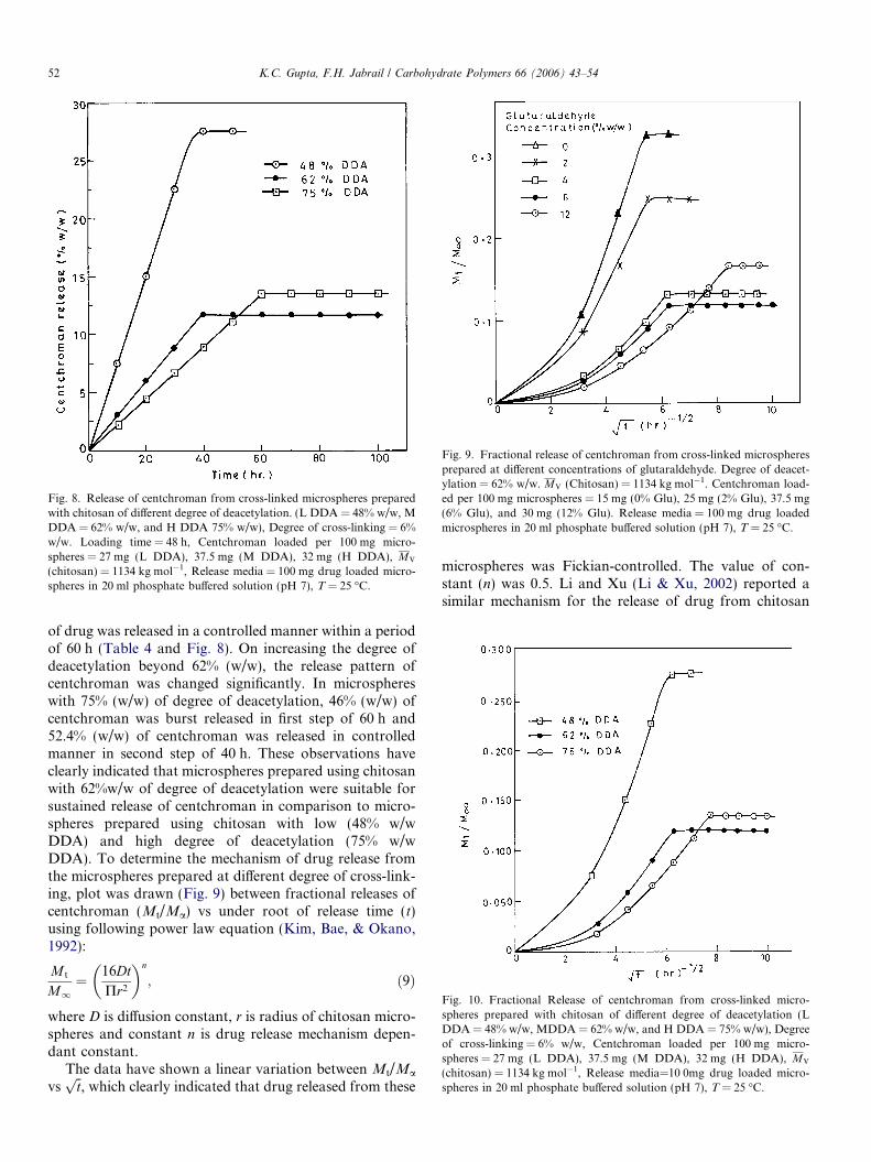
Fig. 8. Release of centchroman from cross-linked microspheres preparedwith chitosan of different degree of deacetylation. (L DDA = 48% w/w, MDDA = 62% w/w, and H DDA 75% w/w), Degree of cross-linking = 6%w/w. Loading time = 48 h, Centchroman loaded per 100 mg micro-spheres = 27 mg (L DDA), 37.5 mg (M DDA), 32 mg (H DDA), MV
(chitosan) = 1134 kg mol�1, Release media = 100 mg drug loaded micro-spheres in 20 ml phosphate buffered solution (pH 7), T = 25 �C.
Fig. 9. Fractional release of centchroman from cross-linked microspheresprepared at different concentrations of glutaraldehyde. Degree of deacet-ylation = 62% w/w. MV (Chitosan) = 1134 kg mol�1. Centchroman load-ed per 100 mg microspheres = 15 mg (0% Glu), 25 mg (2% Glu), 37.5 mg(6% Glu), and 30 mg (12% Glu). Release media = 100 mg drug loadedmicrospheres in 20 ml phosphate buffered solution (pH 7), T = 25 �C.
Fig. 10. Fractional Release of centchroman from cross-linked micro-spheres prepared with chitosan of different degree of deacetylation (LDDA = 48% w/w, MDDA = 62% w/w, and H DDA = 75% w/w), Degreeof cross-linking = 6% w/w, Centchroman loaded per 100 mg micro-spheres = 27 mg (L DDA), 37.5 mg (M DDA), 32 mg (H DDA), MV
(chitosan) = 1134 kg mol�1, Release media=10 0mg drug loaded micro-spheres in 20 ml phosphate buffered solution (pH 7), T = 25 �C.
52 K.C. Gupta, F.H. Jabrail / Carbohydrate Polymers 66 (2006) 43–54
of drug was released in a controlled manner within a periodof 60 h (Table 4 and Fig. 8). On increasing the degree ofdeacetylation beyond 62% (w/w), the release pattern ofcentchroman was changed significantly. In microsphereswith 75% (w/w) of degree of deacetylation, 46% (w/w) ofcentchroman was burst released in first step of 60 h and52.4% (w/w) of centchroman was released in controlledmanner in second step of 40 h. These observations haveclearly indicated that microspheres prepared using chitosanwith 62%w/w of degree of deacetylation were suitable forsustained release of centchroman in comparison to micro-spheres prepared using chitosan with low (48% w/wDDA) and high degree of deacetylation (75% w/wDDA). To determine the mechanism of drug release fromthe microspheres prepared at different degree of cross-link-ing, plot was drawn (Fig. 9) between fractional releases ofcentchroman (Mt/Ma) vs under root of release time (t)using following power law equation (Kim, Bae, & Okano,1992):
M t
M1¼ 16Dt
Pr2
� �n
; ð9Þ
where D is diffusion constant, r is radius of chitosan micro-spheres and constant n is drug release mechanism depen-dant constant.
The data have shown a linear variation between Mt/Ma
vsffiffitp
, which clearly indicated that drug released from these
microspheres was Fickian-controlled. The value of con-stant (n) was 0.5. Li and Xu (Li & Xu, 2002) reported asimilar mechanism for the release of drug from chitosan

K.C. Gupta, F.H. Jabrail / Carbohydrate Polymers 66 (2006) 43–54 53
gel. However, at later periods, the release mechanism ofcentchroman from microspheres became anomalous (Siep-mann & Peppas, 2004) due to structural variations inmicrospheres on swelling and degradation. The mechanismof drug release from microspheres with different degree ofdeacetylation was also evaluated on the basis of trendsshown by fractional releases of centchroman in the plotdrawn (Fig. 10) between Mt/Ma vs
ffiffitp
, which supporteda Fickian behavior. The trend of centchroman release frommicrospheres was linear in the beginning indicating a diffu-sion controlled mechanism of drug release but after struc-tural variations in the microspheres, the releasemechanism of centchroman became anomalous (Siepmann& Peppas, 2004) as found with microspheres obtained atdifferent degree of cross-linking (Fig. 9). From theobserved patterns of drug release from microspheres withdifferent degree of deacetylation (Fig. 10, Table 4), itbecame clear that the release of centchroman has followeda first order kinetics in the beginning but became zero orderafter equilibrium degree of swelling and burst-release ofcentcroman from these microspheres.
5. Conclusion
Microspheres for controlled delivery of centchromanwere prepared at different degree of cross-linking, usingchitosan with different degree of deacetylation. The pre-pared microspheres were characterized for degree of swell-ing and surface morphology. The loading and releasecharacteristics of microspheres were evaluated as a func-tion of degree of cross-linking and deacetylation. Themicrospheres prepared with low degree of deacetylation(48% w/w DDA) showed high burst-release of centchro-man (71% w/w) within a first step of drug release. About28.8%w/w of centchroman was released in a controlledmanner in second step of drug release of 10 h. However,microspheres prepared using chitosan with 62% (w/w)degree of deacetylation (M DDA) have shown significantimprovements in releasing centchroman in controlled man-ner within a period of 60 h. In contrast, microspheres pre-pared using chitosan with 75% (w/w) DDA showed poorcontrol characteristics. The effect of degree of cross-linkingon controlled characteristics of prepared microspheres wasstudied successfully. The microspheres prepared at 6% (w/w)degree of cross-linking have shown better characteristics incomparison to those prepared with low and high degree ofcross-linking. These investigations have clearly suggestedthat loading and release characteristics of chitosan micro-spheres were dependent on pore size and degree of hydro-phobicity produced in microspheres by cross-linking anddegree of acetylation.
Acknowledgements
Authors are thankful for I.I.T Roorkee, India, for pro-viding facilities to carryout these investigations. One of theauthors Mr. Fawzi Habeeb Jebrail is thankful to ICCR,
New Delhi, Govt. of India for awarding him a fellowshipunder cultural exchange programme.
References
Arake, Y., & Ito, E. (1975). A pathway of chitosan formation inmucorrouxii (enzymatic acetylation of chitin). European Journal of
Biochemistry, 55, 71–78.Aly, A. S. (1998). Self-dissolving chitosan-I: Preparation, characterization
and evaluation for drug delivery system. Angewandte Makromolekulare
Chemie, 259, 13–18.Apsden, T. J., Mason, J. D., & Jones, N. S. (1997). Chitosan as a nasal
delivery system, the effect of chitosan solutions on in vitro and in vivomucocilliary transport rates in human turbinals and volunteers.Journal of Pharmaceutical Science, 86, 509–513.
Arguelles-monal, W., Gaycoolea, F. M., Peniche, C., & Higuera-Ciapra, I.(1998). Rheological study of chitosan/glutaraldehyde chemical gelsystem. Polymer Gels Net Works, 6, 429–440.
Broussignac, P. (1968). Chitosan: A natural polymer not well known bythe industry. Chimistry Industry Genie Chimica, 99(9), 1241–1247.
Beauchamp, R. O., Stclair, M. B., Fenell, T. R., Clarke, D. O., & Morgan,K. T. (1992). A critical review of the toxicology of glutaraldehyde.Critical Reviews in Toxicology, 22, 145–174.
Bezemer, J. H., Rederma, R., Grijma, D. W., Dijkstra, P. J., Feijen, J., &van Butler SwijK, C. A. (2000). Zero order release of lysozyme frompolyethylene glycol/polybutylene terephthalate matrices. Journal of
Controlled Release, 64, 179–197.Chang, K. L. B., Tsai, G., Lee, J., & Fu, W. R. (1997). Heterogeneous N-
deacetylation of chitin in alkaline solution. Carbohydrate Research,
303, 327–332.Cheng, G. X., Liu, J., Zhao, R. Z., Yao, K. D., Sun, P. C., Men, A. J.,
et al. (1998). Studies on dynamic behavior of water in cross-linkedchitosan hydrogel. Journal of Applied Polymer Science, 67, 983–988.
Draget, K. I. (1996). Association phenomenon in highly acetylatedchitosan gel. Polymer Gels and Net Works, 4, 143–151.
Donald, A. (1987). pH and C.D. measurement on a fully deacetylatedchitosan; application to Cu (II) polymer interactions. International
Journal of Biomacromology, 9, 98–104.Errington, N., Harding, S. E., Varum, K. M., & Illum, L. (1993).
Hydrodynamic characterization of chitosan varying in degree ofacetylation. International Journal of Biology Macromology, 15, 113–117.
Gonzalez-Rodriguez, M. L., Holgado, M. A., Sanchez-Lafuente, C.,Rabasco, A. M., & Fini, A. (2002). Alginate/chitosan particulatesystems for sodium dichlofenac release. International Journal of
Pharmaceutics, 232, 225–234.Gupta, K. C., & Ravikumar, M. N. V. (2000). Preparation characteriza-
tion and release properties of pH-sensitive chitosan beads. Biomaterial,
21, 1115–1119.Gupta, K. C. & Jabrail, F. H. (2006). Studies on drug release character-
istics of cross-linked microspheres. Biomaterial, submitted forpublication.
Hiral, A., Odani, H., & Nakajima, A. (1991). Determination of degree ofdeacetylation of chitosan by 1H NMR spectrometry. Polymers
Bulletin, 26, 87–94.Hirano, S., Yamaguchi, R., Fukui, N., & Iwata, M. (1990). A chitosan
oxalate gel its conversion to an N-acetyl chitosan gel via a chitosan gel.Carbohydrate Research, 201, 145–149.
Hizaji, R., & Amiji, M. (2003). Chitosan based gastrointestinal deliverysystems. Journal of Control Release, 89(2), 151–165.
Illum, L., Farraj, N. F., & Davis, S. S. (1994). Chitosan as novel nasaldelivery system for peptides drugs. Pharmaceutical Research, 11,1186–1189.
Jeuniaux, C., Voss-Foucart, M. F., Poulicek, M., & Bussers, J. C. (1989).Sources of chitin, estimated from new data of chitin biomass andproduction. In G. Skjak-Break, T. Anthonson, & P. Sandsrod (Eds.),Chitin and chitosan (pp. 3–11). London/New York: Elsevier AppliedScience.

54 K.C. Gupta, F.H. Jabrail / Carbohydrate Polymers 66 (2006) 43–54
Kasaai, M. R., Arul, J., & Charlet, G. (2000). Intrinsic viscosity,molecular weight relationship for chitosan. Journal of Polymer Science:
Polymer Physics, 38, 2591–2598.Khan, T. A., Peh, K. K., & C’ing, H. S. (2000). Mechanical bioadhesive
strength and biological evaluation of chitosan films for wounddressing. Journal of Pharmaceutical and Pharmaceutical Science, 3(3),3003–3371.
Kim, S. W., Bae, Y. H., & Okano, T. (1992). Hydrogels swelling, drugloading, and release. Pharmaceutical Research, 9(3), 283–290.
Li, J., & Xu, Z. (2002). Physical characterization of a chitosan-basedhydrogel delivery system. Journal of Pharmaceutical Science, 91(7), 1669.
Mi, F. L., Tan, Y. C., Liang, H. F., & Sung, H. W. (2003). In vivobiocompability and degradability of a novel injectable chitosan basedimplants. Biomaterial, 23, 181–191.
Mi, F. L., Kuan, C. Y., Shyu, S. S., Lee, S. T., & Chang, S. F. (2000). Thestudy of gelation, kinetics and chain-relaxation property of glutaral-dehyde cross-linked chitosan gel and their effect on microspherespreparation and drug release. Carbohydrate Polymers, 41, 389–396.
Mao, H. Q., Troung-le, V. L., & Jans, K. A. (2001). Chitosan DNAparticle as gene carriers synthesis characterization and transfectionefficiency. Journal of Controlled Release, 70, 399–421.
Nanjo, F., Katsumi, R., & Sakai, K. (1991). Enzymatic method fordetermination of the degree of deacetylation of chitosan. Analytical
Biochemistry, 193(2), 164–167.Oyrton, A. C., Monteiro Jr, & Claudio, A. (1999). Some studies of cross-
linking chitosan–glutaraldehyde interaction in a homogeneous system.International Journal of Biomacromology, 26, 119–128.
Patel, V. R., & Amiji, M. M. (1996). Preparation and characterization offreeze-dried chitosan poly (ethylene oxide) hydrogels for site-specificantibiotic delivery in the stomach. Pharmaceutical Research, 13,588–593.
Shigemasa, Y., Matsuura, H., Sashiwa, H., & Saimoto, H. (1996).Evaluation of different absorbance ratios from infrared spectroscopyfor analyzing the degree of deacetylation in chitin. International
Journal of Biology and Macromology, 18(3), 237–242.
Sang-Gyun, K., Han-Sang, Y., Kim, S., Sang-Bong, S., Toshihiro, A., &Chong-Su, C. (2004). In vitro study of the immune stimulating activityof an anthrophic rhinitis; vaccine associated to chitosan microspheres.European Journal of Pharmaceutics and Biopharmaceutics, 58, 471–476.
Siepmann, J., & Peppas, M. A. (2004). Modeling of drug release fromdelivery system based on hydroxypropyl methyl cellulose. Advanced
Drug Delivery Reviews, 48, 139–157.Tarsi, R., Corbin, B., Pruzzo, C., & Muzzarelli, R. A. A. (1998). Effect of
low-molecular-weight chitosan on the adhesive properties of oralstreptococci. Oral Microbiology and Immunology, 13(4), 217–224.
Tan, S. C., Khor, E., Tan, T., & Wong, S. M. (1998). The degree ofdeacetylation of chitosan: advocating the first derivative UV-spectro-photometric method of determination. Talanta, 45, 713–719.
Takanori, S., Keisuke, K., & Yoshio, I. (1976). Studies on chitin, 2. Effectof deacetylation on solubility. Die Makromology Chemistry, 177(12),3589–3600.
Tien, C. L., Lacroix, M., Ispas-Szabo, P., & Mircea-Alexandru, M. (2003).N-acetylated chitosan ‘hydrophobic matrices for controlled drugrelease’. Journal of Controlled Release, 93(1), 1–13.
Thacharodi, D., & Ruo, K. P. (1993). Propranolol hydrochloride releasebehavior of cross-linked chitosan membranes. Journal of Chemical
Technology and Biotechnollogy, 58, 177–181.Vander Lubben, I. M., Kersten, G., Fretz, M. M., Beuvery, C., Verhoef, J.
C., & Juginder, H. E. (2003). Chitosan microparticles for mucosalvaccination against diphtheria, oral, and nasal efficiency studies inmice. Vaccine, 21, 1400–1408.
Varum, K. M., Ottoy, M. H., & Smidsrod, O. (2001). Acid hydrolysis ofchitosans. Carbohydrate Polymers, 46(1), 89–98.
William, O. R., Barron, M. K., Maria, J. A., & Remunan-Lopez, C.(1998). Investigation of a pMDI system containing chitosan micro-spheres and P134a. International Journal of Pharmaceutics, 174,209–222.
Zhang, Y., Xue, C., Xue, Y., Gao, R., & Zhang, X. (2005). Determinationof degree of deacetylation of chitin and chitosan by X-ray powderdiffraction. Carbohydrate Research, 340, 1914–1917.
Copyright © 2022 FDOKUMEN