Effects of cobalt and chromium ions at clinically equivalent concentrations after metal-on-metal hip...
7
Effects of cobalt and chromium ions at clinically equivalent concentrations after metal-on-metal hip replacement on human osteoblasts and osteoclasts: Implications for skeletal health Rebecca E. Andrews a, 1 , Karan M. Shah a, b, 1 , J. Mark Wilkinson a, b, ⁎, Alison Gartland a a The Mellanby Centre for Bone Research, Department of Human Metabolism, The University of Sheffield, Beech Hill Road, Sheffield, S10 2RX, UK b The National Institute for Health Research Biomedical Research Unit for Musculoskeletal Disease at the University of Sheffield and Sheffield Teaching Hospitals NHS Foundation Trust, Centre for Biomedical Research, Northern General Hospital, Herries Road, Sheffield, S5 7AU, UK abstract article info Article history: Received 19 May 2011 Accepted 5 June 2011 Available online 13 June 2011 Edited by: Thomas Einhorn Keywords: Metal-on-metal hip replacement Hip resurfacing Cobalt Chromium Human osteoblast Human osteoclast Metal-on-metal hip replacement (MOMHR) using large diameter bearings has become a popular alternative to conventional total hip arthroplasty, but is associated with elevated local tissue and circulating levels of chromium (Cr) and cobalt (Co) ions that may affect bone health. We examined the effects of acute and chronic exposure to these metals on human osteoblast and osteoclast formation and function over a clinically relevant concentration range previously reported in serum and within hip synovial fluid in patients after MOMHR. SaOS-2 cells were cultured with Co 2+ , Cr 3+ and Cr 6+ for 3 days after which an MTS assay was used to assess cell viability, for 13 days after which alkaline phosphatase and cell viability were assessed and for 21 days after which nodule formation was assessed. Monocytes were isolated from human peripheral blood and settled onto dentine disks then cultured with M-CSF and RANKL plus either Co 2+ , Cr 3+ or Cr 6+ ions for 21 days from day 0 or between days 14 and 21. Cells were fixed and stained for TRAP and osteoclast number and amount of resorption per dentine disk determined. Co 2+ and Cr 3+ did not affect osteoblast survival or function over the clinically equivalent concentration range, whilst Cr 6+ reduced osteoblast survival and function at concentrations within the clinically equivalent serum range after MOMHR (IC 50 =2.2 μM). In contrast, osteoclasts were more sensitive to metal ions exposure. At serum levels a mild stimulatory effect on resorption in forming osteoclasts was found for Co 2+ and Cr 3+ , whilst at higher serum and synovial equivalent concentrations, and with Cr 6+ , a reduction in cell number and resorption was observed. Co 2+ and Cr 6+ within the clinical range reduced cell number and resorption in mature osteoclasts. Our data suggest that metal ions at equivalent concentrations to those found in MOMHR affect bone cell health and may contribute to the observed bone-related complications of these prostheses. © 2011 Elsevier Inc. All rights reserved. Introduction In recent years total hip replacement using large diameter metal-on- metal bearings (MOMHR), either as a hip resurfacing procedure or using a stemmed femoral prosthesis, has become a common alternative to conventional total hip arthroplasty (THA) for the treatment of young and active arthritis patients because of advantages of lower volumetric wear and dislocation risk [1]. However, the clinical outcomes of hip replacement using these prostheses have been mixed. Data from the National Joint Register for England and Wales (2008) demonstrated a 3-year revision rate for hip resurfacing of 4.4% (95%CI 4.0 to 5.0) compared with 1.3% (1.2 to 1.4) for cemented THA (www.njrcentre.org.uk). The Australian Arthroplasty Register (1997 to 2005) also reported a higher 3-year revision rate for hip resurfacing versus THA (3.1% (2.7 to 3.6) versus 2.1% (1.9 to 2.5%) www.dmac.adelaide.edu.au/aoanjrr). The most common adverse events necessitating revision surgery after MOMHR include early periprosthetic fracture, osteolysis, failure of prosthesis osseo-integration resulting in aseptic loosening, unexplained pain, and inflammatory masses [2–7]. Circulating physiological levels of cobalt and chromium are normally b 0.25 μg/L (0.005 μM). Elevated levels of cobalt and chromium occur in both the hip synovial fluid and in peripheral blood after MOMHR. Whole blood concentrations of cobalt and chromium after MOMHR of up to 4.6 μM and 2.3 μM, respectively [8], and local hip synovial fluid levels of up to 30 μM and 25 μM, respectively, have been measured in-vivo [9]. Whilst circulating metal levels are usually highest over the first few months after implantation, persistent elevation occurs as late as 10 years after surgery [10]. Previous studies have shown that short-term exposure to these metal species may affect human osteoclast and osteoblast survival and function. High concentrations of cobalt 2+ (Co 2+ ), chromium 3+ (Cr 3+ ), and chromium 6+ (Cr 6+ ) ions is toxic to osteoblasts and reduces cell Bone 49 (2011) 717–723 ⁎ Corresponding author at: NIHR Biomedical Research Unit for Musculoskeletal Disease, Centre for Biomedical Research, Northern General Hospital, Herries Road, Sheffield, S5 7AU, UK. Fax: +44 114 2618775. E-mail address: j.m.wilkinson@sheffield.ac.uk (J.M. Wilkinson). 1 Equal contribution. 8756-3282/$ – see front matter © 2011 Elsevier Inc. All rights reserved. doi:10.1016/j.bone.2011.06.007 Contents lists available at ScienceDirect Bone journal homepage: www.elsevier.com/locate/bone
-
Upload
independent -
Category
Documents
-
view
0 -
download
0
Transcript of Effects of cobalt and chromium ions at clinically equivalent concentrations after metal-on-metal hip...

Bone 49 (2011) 717–723
Contents lists available at ScienceDirect
Bone
j ourna l homepage: www.e lsev ie r.com/ locate /bone
Effects of cobalt and chromium ions at clinically equivalent concentrations aftermetal-on-metal hip replacement on human osteoblasts and osteoclasts:Implications for skeletal health
Rebecca E. Andrews a,1, Karan M. Shah a,b,1, J. Mark Wilkinson a,b,⁎, Alison Gartland a
a The Mellanby Centre for Bone Research, Department of Human Metabolism, The University of Sheffield, Beech Hill Road, Sheffield, S10 2RX, UKb The National Institute for Health Research Biomedical Research Unit for Musculoskeletal Disease at the University of Sheffield and Sheffield Teaching Hospitals NHS Foundation Trust,Centre for Biomedical Research, Northern General Hospital, Herries Road, Sheffield, S5 7AU, UK
⁎ Corresponding author at: NIHR Biomedical ReseaDisease, Centre for Biomedical Research, Northern GeSheffield, S5 7AU, UK. Fax: +44 114 2618775.
E-mail address: [email protected] (J.M. W1 Equal contribution.
8756-3282/$ – see front matter © 2011 Elsevier Inc. Aldoi:10.1016/j.bone.2011.06.007
a b s t r a c t
a r t i c l e i n f oArticle history:Received 19 May 2011Accepted 5 June 2011Available online 13 June 2011
Edited by: Thomas Einhorn
Keywords:Metal-on-metal hip replacementHip resurfacingCobaltChromiumHuman osteoblastHuman osteoclast
Metal-on-metal hip replacement (MOMHR) using large diameter bearings has become a popular alternative toconventional total hip arthroplasty, but is associated with elevated local tissue and circulating levels of chromium(Cr) and cobalt (Co) ions that may affect bone health. We examined the effects of acute and chronic exposure tothese metals on human osteoblast and osteoclast formation and function over a clinically relevant concentrationrange previously reported in serum and within hip synovial fluid in patients after MOMHR. SaOS-2 cells wereculturedwith Co2+, Cr3+ and Cr6+ for 3 days after which anMTS assay was used to assess cell viability, for 13 daysafter which alkaline phosphatase and cell viabilitywere assessed and for 21 days afterwhich nodule formationwasassessed. Monocyteswere isolated from human peripheral blood and settled onto dentine disks then culturedwithM-CSF and RANKL plus either Co2+, Cr3+ or Cr6+ ions for 21 days fromday 0 or between days 14 and 21. Cellswerefixed and stained for TRAP and osteoclast number and amount of resorption per dentine disk determined. Co2+ andCr3+ did not affect osteoblast survival or function over the clinically equivalent concentration range, whilst Cr6+
reduced osteoblast survival and function at concentrations within the clinically equivalent serum range afterMOMHR(IC50=2.2 μM). In contrast, osteoclastsweremore sensitive tometal ions exposure. At serum levels amildstimulatory effect on resorption in forming osteoclasts was found for Co2+ and Cr3+, whilst at higher serum andsynovial equivalent concentrations, and with Cr6+, a reduction in cell number and resorption was observed. Co2+
and Cr6+within the clinical range reduced cell number and resorption inmature osteoclasts. Our data suggest thatmetal ions at equivalent concentrations to those found inMOMHR affect bone cell health andmay contribute to theobserved bone-related complications of these prostheses.
rch Unit for Musculoskeletalneral Hospital, Herries Road,
ilkinson).
l rights reserved.
© 2011 Elsevier Inc. All rights reserved.
Introduction
In recent years total hip replacement using large diameter metal-on-metal bearings (MOMHR), either as a hip resurfacing procedure or using astemmed femoral prosthesis, has become a common alternative toconventional total hip arthroplasty (THA) for the treatment of young andactive arthritis patients because of advantages of lower volumetric wearanddislocation risk [1].However, theclinical outcomesofhip replacementusing these prostheses have been mixed. Data from the National JointRegister for England and Wales (2008) demonstrated a 3-year revisionrate for hip resurfacing of 4.4% (95%CI 4.0 to 5.0) compared with 1.3%(1.2 to 1.4) for cemented THA (www.njrcentre.org.uk). The AustralianArthroplasty Register (1997 to 2005) also reported a higher 3-year
revision rate for hip resurfacing versus THA (3.1% (2.7 to 3.6) versus 2.1%(1.9 to 2.5%) www.dmac.adelaide.edu.au/aoanjrr). The most commonadverse events necessitating revision surgery after MOMHR include earlyperiprosthetic fracture, osteolysis, failure of prosthesis osseo-integrationresulting in aseptic loosening, unexplained pain, and inflammatorymasses [2–7].
Circulating physiological levels of cobalt and chromium are normallyb0.25 μg/L (0.005 μM). Elevated levels of cobalt and chromium occur inboth the hip synovialfluid and in peripheral blood afterMOMHR.Wholeblood concentrations of cobalt and chromium after MOMHR of up to4.6 μM and 2.3 μM, respectively [8], and local hip synovial fluid levels ofup to 30 μM and 25 μM, respectively, have been measured in-vivo [9].Whilst circulating metal levels are usually highest over the first fewmonths after implantation, persistent elevation occurs as late as 10 yearsafter surgery [10].
Previous studies have shown that short-term exposure to thesemetal species may affect human osteoclast and osteoblast survival andfunction. High concentrations of cobalt2+ (Co2+), chromium3+ (Cr3+),and chromium6+ (Cr6+) ions is toxic to osteoblasts and reduces cell

718 R.E. Andrews et al. / Bone 49 (2011) 717–723
activity in-vitro[11–13]. Fewdata are available on the effect of cobalt andchromium exposure on primary human osteoclasts, although Nicholsand Puleo showed short-term exposure to Co and Cr ions at sub-lethaldoses resulted in decreased resorptive activity in rat osteoclasts [14]. Incontrast, Rousselle et al. foundexposure of rabbit osteoclasts to Cr3+hadno effect on rabbit osteoclast function [15]. Sankaramanivel et al. haveshown that rats treated intraperitoneally with potassium dichromate(Cr6+) over 5 days led to accumulation of chromium in the femur, andwas associated with reduced systemic assays of alkaline phosphataseand tartrate-resistant acid phosphatase, suggesting an impact on bothbone formation and resorption [16]. However, the longer-term effect ofchronic exposure of both human osteoblasts and osteoclasts to theseions at clinically relevant concentrations, more akin to clinical exposureboth systemically and at the level of the hip joint, is unknown.
Wehypothesise that chronic exposure of local bone cells tometal ionsmay contribute to the clinical bone-related complications after MOMHR.The aims of this study were to investigate the effect of both short-termand chronic Co2+, Cr3+, and Cr6+ ion exposure at clinically relevantconcentrations after MOMHR on human osteoblast and osteoclastproliferation and function, and on mature primary human osteoclasts.A dose-ranging methodology was used including metal ion levelscovering the normal physiological range, through systemic levels foundafter MOMHR, to the high concentrations reported in hip joint synovialfluid aspirates after MOMHR.
Materials and methods
Metal ion preparation and validation of concentrations
Co2+ and Cr3+ were purchased as cobalt (II) chloride hexahydrateand Chromium (III) chloride hexahydrate from Sigma-Aldrich CompanyLtd, Gillingham, UK. Cr6+ was purchased as chromium (VI) oxide fromBDH, Lutterworth, UK. Stock solution for each metal ion at 0.2 M wasprepared in 50 ml of sterile water and stored at 4 °C prior to use. The0.2 M stock solutions were serially diluted in sterile distilled water togive aliquots of 100X theworking concentration range for the treatmentof cells. These were then diluted in Dulbecco's modified Eagle's medium(DMEM©GLUTAMAX™) supplementedwith0.5% FCSand1%penicillin–streptomycin (10000units penicillin, 10,000 μg/ml streptomycin),whichfrom here on will be referred to as vehicle. Control treatments wereprepared to contain 1% of distilled sterile water in vehicle to maintainconditions, referred to as 0 μM treatments.
Thefinalmetal ion concentrations in the test solutionswere confirmedusing flame-atomic absorbance spectroscopy. Co2+, Cr3+ and Cr6+
predictedversusmeasuredconcentrationshowedcloseagreement (linearregression, r2=1.00, 0.85 and0.98 for Co2+, Cr3+ andCr6+, respectively).
Osteoblast cell culture
Human SaOS-2 cells (a human osteosarcoma-derived osteoblast cellline) were cultured in T75 flasks containing Dulbecco's modified Eagle'smedium (DMEM© Glutamax™, Gibco® Invitrogen, Paisley, UK) supple-mented with 10% FCS, 100 IU/mL of penicillin and 100 μg/mL ofstreptomycin (Sigma, Poole, UK), hereafter termed complete DMEM.The cellsweremaintained at 37 °C in a humidified atmosphere of 95% airand 5% CO2 until required for assay.
Osteoblast viability and proliferation
SaOS-2 cellswere seeded into96-well plates at 5×10³ cells perwell in0.1 mL of complete DMEM and left to adhere overnight. The mediumwas then replaced with DMEM supplemented with 0.5% FCS and100 IU/mL of penicillin and 100 μg/mL of streptomycin (referred hereonin as vehicle)±metal ion treatments and incubated for 3 or 13 daysat 37 °C in a humidified atmosphere of 95% air and 5% CO2. Vehicle±treatmentswere replenishedevery3rdand4thdayconsecutively for cells
cultured for 13 days. At the end of the culture period a CellTiter 96®AQueous Non-Radioactive Cell Proliferation Assay was performed accord-ing to the manufacturer's instructions (Promega, Southampton, UK). Theassay utilises dehydrogenase enzymes found in metabolically active cellsto convert 3-(4,5-dimethylthiazol-2-yl)-5-(3-carboxymethoxyphenyl)-2-(4-sulfophenyl)-2H-tetrazolium, inner salt (MTS) into an aqueoussoluble formazan product. The absorbance of the formazan productproduced by the cells was read at 490 nm using a SpectraMax M5e
Microplate Reader (Molecular Devices, Sunnyvale, CA). Values wereexpressed as percentage response relative to vehicle.
Osteoblast synthetic function
Alkaline phosphatase (ALP) activitySaOS-2 cells were seeded into 24-well plates at 15×103 cells per
well in 1 mL of complete DMEM, left to adhere overnight and thenthe medium was replaced with vehicle±metal ion treatments andincubated for 13 days at 37 °C in a humidified atmosphere of 95% airand 5% CO2. The vehicle±treatments were replenished every 3rd or4th day. Cells were washed with PBS, lysed in nuclease-free water andfrozen at −80 °C following completion of culture. Cell lysates wereobtained after three freeze and thaw cycles. ALP activity was measuredusing p-nitrophenyl phosphate (pNPP) (Sigma) as the chromogenic ALPsubstrate in the presence of Mg2+ ions in a buffered solution. Theabsorbance was read at 405 nm using a SpectraMax M5e MicroplateReader. Values were expressed as percentage response relative tovehicle. DNA contentwas quantified usingQuant-iT™ PicoGreendsDNAAssay Kit (Invitrogen, Paisley, UK) according to the manufacturer'sinstructions. ALP activity was normalised to DNA content and ALP/DNAwas then expressed as percentage response relative to vehicle.
Osteoblast mineralisationOnce the SaOS-2 cells had reached confluency the cells were treated
with vehicle supplemented with 10-8 M dexamethasone and 50 μg/mlascorbic acid (referred to as osteogenicmedium)±metal ion treatment.Metal ion treatments in osteogenic mediumwere changed every 3rd or4th day. Two days prior to experiment end, 10 μL of 5 mM inorganicphosphate 4.2pHwasadded to the existing treatmentswithin eachwell.On day 21 cells were then washed once in PBS, fixed in 100% ethanol,rinsed in PBS and incubated in 40 mM alizarin red (pH 4.2; Sigma) for1 hour at room temperature. The cells were then washed extensively in95% ethanol and air-dried. The plateswere scanned on a high-resolutionflat-bed scanner. The percentage of each well stained with alizarin redwas quantified using Image J software (NIH: http://rsb.info.nih.gov/ij/)and expressed as percentage response relative to vehicle.
Human osteoclast culture
Osteoclasts were generated from human peripheral bloodmonocytestaken from healthy volunteers as previously described andwith researchethics committee approval [17]. Sterilisation of 6 mmdiameter coverslips(Richardson's of Leicester, Leicester,UK)wasperformedbybaking at 180°for 2 hours. Dentine disks (www.dentinedisks.com) were sonicated andsterilised by washing in 70% ethanol overnight. Venous blood wasobtained from healthy volunteers and separated using Histopaque®-1077 (Sigma). Themonocyte fractionwas collected, washed and then re-suspended in α-MEM Glutamax (Gibco® Invitrogen, Paisley, UK). Anappropriate volume of cell suspension containing 5×10⁵ cells was thenadded to pre-wetted coverslips or dentine disks in a 96-well plate. Cellswere incubated for a minimum of 1 hour to allow adherence to thedentine or glass surface. Non-adherent cells were subsequently washedaway with α-MEM. Adherent cells were incubated in 100 μL α-MEMGlutamax containing 10% FCS, 100 U/mL penicillin, 100 μg/mL strepto-mycin (Sigma) (referred to as completeα -MEM)andsupplementedwith25 ng/mLM-CSF, 30 ng/mL recombinant RANKL (Insight Biotechnology,Wembley, UK) at 37 °C in a humidified atmosphere of 93% air and 7% CO2
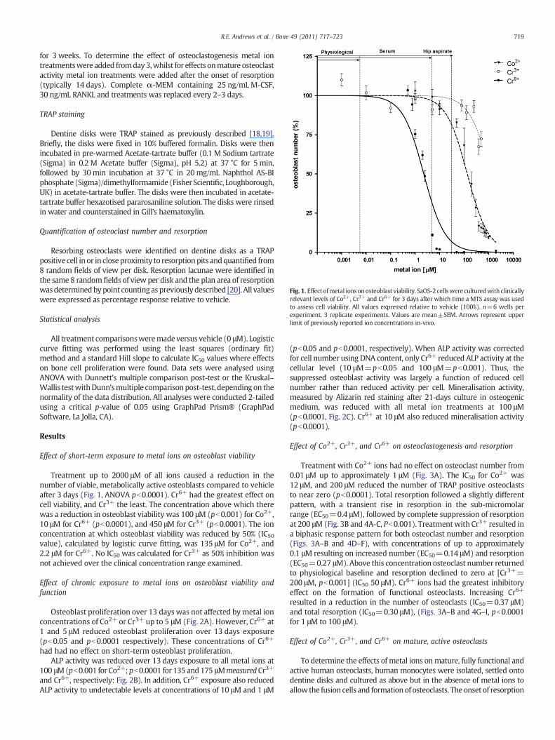
Fig. 1. Effect ofmetal ions onosteoblast viability. SaOS-2 cellswere culturedwith clinicallyrelevant levels of Co2+, Cr3+ and Cr6+ for 3 days after which time a MTS assay was usedto assess cell viability. All values expressed relative to vehicle (100%). n=6 wells perexperiment, 3 replicate experiments. Values are mean±SEM. Arrows represent upperlimit of previously reported ion concentrations in-vivo.
719R.E. Andrews et al. / Bone 49 (2011) 717–723
for 3 weeks. To determine the effect of osteoclastogenesis metal iontreatmentswere added fromday 3,whilst for effects onmature osteoclastactivity metal ion treatments were added after the onset of resorption(typically 14 days). Complete α-MEM containing 25 ng/mL M-CSF,30 ng/mL RANKL and treatments was replaced every 2–3 days.
TRAP staining
Dentine disks were TRAP stained as previously described [18,19].Briefly, the disks were fixed in 10% buffered formalin. Disks were thenincubated in pre-warmed Acetate-tartrate buffer (0.1 M Sodium tartrate(Sigma) in 0.2 M Acetate buffer (Sigma), pH 5.2) at 37 °C for 5 min,followed by 30 min incubation at 37 °C in 20 mg/mL Naphthol AS-BIphosphate (Sigma)/dimethylformamide (Fisher Scientific, Loughborough,UK) in acetate-tartrate buffer. The disks were then incubated in acetate-tartrate buffer hexazotised pararosaniline solution. The disks were rinsedin water and counterstained in Gill's haematoxylin.
Quantification of osteoclast number and resorption
Resorbing osteoclasts were identified on dentine disks as a TRAPpositive cell in or in close proximity to resorptionpits andquantified from8 random fields of view per disk. Resorption lacunae were identified inthe same 8 random fields of view per disk and the plan area of resorptionwasdeterminedbypoint counting as previouslydescribed [20]. All valueswere expressed as percentage response relative to vehicle.
Statistical analysis
All treatment comparisonsweremade versus vehicle (0 μM). Logisticcurve fitting was performed using the least squares (ordinary fit)method and a standard Hill slope to calculate IC50 values where effectson bone cell proliferation were found. Data sets were analysed usingANOVA with Dunnett's multiple comparison post-test or the Kruskal–Wallis testwithDunn'smultiple comparison post-test, depending on thenormality of the data distribution. All analyses were conducted 2-tailedusing a critical p-value of 0.05 using GraphPad Prism® (GraphPadSoftware, La Jolla, CA).
Results
Effect of short-term exposure to metal ions on osteoblast viability
Treatment up to 2000 μM of all ions caused a reduction in thenumber of viable, metabolically active osteoblasts compared to vehicleafter 3 days (Fig. 1, ANOVA pb0.0001). Cr6+ had the greatest effect oncell viability, and Cr3+ the least. The concentration above which therewas a reduction in osteoblast viability was 100 μM (pb0.001) for Co2+,10 μM for Cr6+ (pb0.0001), and 450 μM for Cr3+ (pb0.0001). The ionconcentration at which osteoblast viability was reduced by 50% (IC50value), calculated by logistic curve fitting, was 135 μM for Co2+, and2.2 μM for Cr6+. No IC50 was calculated for Cr3+ as 50% inhibition wasnot achieved over the clinical concentration range examined.
Effect of chronic exposure to metal ions on osteoblast viability andfunction
Osteoblast proliferation over 13 days was not affected bymetal ionconcentrations of Co2+ or Cr3+ up to 5 μM (Fig. 2A). However, Cr6+ at1 and 5 μM reduced osteoblast proliferation over 13 days exposure(pb0.05 and pb0.0001 respectively). These concentrations of Cr6+
had had no effect on short-term osteoblast proliferation.ALP activity was reduced over 13 days exposure to all metal ions at
100 μM(pb0.001 for Co2+; pb0.0001 for 135 and175 μMmeasuredCr3+
and Cr6+, respectively: Fig. 2B). In addition, Cr6+ exposure also reducedALP activity to undetectable levels at concentrations of 10 μM and 1 μM
(pb0.05 and pb0.0001, respectively). When ALP activity was correctedfor cell number using DNA content, only Cr6+ reduced ALP activity at thecellular level (10 μM=pb0.05 and 100 μM=pb0.001). Thus, thesuppressed osteoblast activity was largely a function of reduced cellnumber rather than reduced activity per cell. Mineralisation activity,measured by Alizarin red staining after 21-days culture in osteogenicmedium, was reduced with all metal ion treatments at 100 μM(pb0.0001, Fig. 2C). Cr6+ at 10 μM also reduced mineralisation activity(pb0.0001).
Effect of Co2+, Cr3+, and Cr6+ on osteoclastogenesis and resorption
Treatment with Co2+ ions had no effect on osteoclast number from0.01 μM up to approximately 1 μM (Fig. 3A). The IC50 for Co2+ was12 μM, and 200 μM reduced the number of TRAP positive osteoclaststo near zero (pb0.0001). Total resorption followed a slightly differentpattern, with a transient rise in resorption in the sub-micromolarrange (EC50=0.4 μM), followed by complete suppression of resorptionat 200 μM(Fig. 3B and 4A-C, Pb0.001). Treatmentwith Cr3+ resulted ina biphasic response pattern for both osteoclast number and resorption(Figs. 3A–B and 4D–F), with concentrations of up to approximately0.1 μM resulting on increased number (EC50=0.14 μM) and resorption(EC50=0.27 μM). Above this concentration osteoclast number returnedto physiological baseline and resorption declined to zero at [Cr3+=200 μM, pb0.001] (IC50 50 μM). Cr6+ ions had the greatest inhibitoryeffect on the formation of functional osteoclasts. Increasing Cr6+
resulted in a reduction in the number of osteoclasts (IC50=0.37 μM)and total resorption (IC50=0.30 μM), (Figs. 3A–B and 4G–I, pb0.0001for 1 μM to 100 μM).
Effect of Co2+, Cr3+, and Cr6+ on mature, active osteoclasts
To determine the effects of metal ions onmature, fully functional andactive human osteoclasts, human monocytes were isolated, settled ontodentine disks and cultured as above but in the absence of metal ions toallow the fusion cells and formationof osteoclasts. The onset of resorption

Fig. 2. Effect of metal ions on osteoblast function. SaOS-2 cells were cultured for 13 daysafter which time MTS assay was performed to assess proliferation (A) and ALP activitymeasured (B). Mineralisation was assessed in cells cultured in osteogenic medium in thepresence of metal ions for 3 weeks (C). n=3–6 wells per experiment, 3 replicateexperiments. apb0.05, bpb0.01, cpb0.001. (*spectroscopy measured concentrations were100 μM for Co2+, 125 μM for Cr3+, and 175 μM for Cr6+). Line within boxes indicatesmedian, boxes represent interquartile range, and whiskers are range.
720 R.E. Andrews et al. / Bone 49 (2011) 717–723
(an indicator of fully functional and active osteoclasts) was monitoreddaily from day 10 and once resorption had been detected (typically after14 days), the osteoclast culture medium was then replaced to include0.01 μM to 200 μM Co2+ and Cr3+ and 0.01 μM to 100 μM Cr6+ ions forthe last 7 days of culture. The pattern of response for Co2+ and Cr3+ wasdifferent to that seen for newly forming osteoclasts in that no transientincrease in cell number or activity was found, and that the inhibitoryeffects of all ions were seen at a lower ion concentration. Seven daystreatment with Co2+ ions ≥10 μM reduced mature osteoclast number(IC50=5.4 μM(pb0.001, Fig. 3C). Total amount of resorptionperdiskwasonly reduced at the high (200 μM) concentration (pb0.0001 andpb0.001, Figs. 3D and 4J–L). Treatment with Cr3+ ions reduced matureosteoclast number and resorption per disk, but only at the 200 μM dose(pb0.05, Figs. 3C–D and 4M–O, IC50 for osteoclast number=221 μMandIC50 for resorption per disk=77 μM). No trend towards increasedosteoclast number or resorption was seen for mature osteoclasts at thelower Cr3+ concentration range. Cr6+ ions had the greatest effect onosteoclast number and resorption. Cr6+ at concentration≥10 μMcauseda reduction in osteoclast number and resorption per disk (pb0.01 all
analyses, Figs. 3C–D and 4P–R, IC50 for osteoclast number=1.8 μM andIC50 for resorption per disk=3.9 μM).
Discussion
In this study we examined the effect of chronic exposure of humanosteoblast and primary human osteoclast cells to Co and Cr ions atconcentrations including the clinically equivalent range defined byprevious reports of measured metal levels in the serum and hipsynovial fluid taken from patients after MOMHR. We found that ionsof both metals affected osteoblast and osteoclast cell proliferation andfunction. These effects were greatest for Cr6+, then Co2+, with Cr3+
showing the least effect. The observed responses also varied withmetal ion concentration, cell type and cell maturity.
Our findings are consistent with in-vitro studies using animal cellsthat supra-physiological concentrations of cobalt and chromium ionsinduce apoptosis in human osteoblast-like cells in-vitro in a dose-dependent manner [12], and suppress osteoblast synthetic function[11,21,22]. Our dose-ranging data suggest that chronic exposure toCo2+ or Cr3+ do not have a major effect on cell viability or syntheticfunction at concentrations equivalent to those metal levels foundin serum or synovial fluid in patients after MOMHR, whilst exposureto Cr6+ has a profound effect on cell viability at concentrationsequivalent to the serum chromium range after MOMHR.
In contrast, our data suggest that an effect of Co and Cr on humanprimary osteoclasts occurs within the clinically observed concentra-tion range and varies with cell maturity. At systemic levels these ionsmay have a mild stimulatory effect on developing osteoclasts, but athigher concentrations and in mature osteoclasts their effect isinhibitory. The reason for this difference might be explained by thesubstrate resorbing activity of the exposed cell, as mature resorbingosteoclasts may accumulate more intracellular metal ions throughphagocytic activity versus developing osteoclasts, and thus demon-strate a greater toxic effect due to greater internalisation of the metal.In support of the increased resorption transient seen in the serumrange, Patntirapong et al. have shown that cobalt ions in solution orincorporated into calcium phosphate coated plastic at clinically-relevant concentrations increase murine osteoclast differentiationand resorption in-vitro[23]. Whilst cobalt ions do not localise to bone,chromium salts do have an affinity for bone [24], being trapped in thebone matrix, and thus levels in the bone microenvironment mayexceed those found in serum. Albrecht et al. have also suggested apossible indirect route for osteoclast activation in response to metalions, showing that exposure of human peripheral blood mononuclearcells to Co2+ ions in-vitro results in upregulation of IL-1α, IL-1β, andIL-6 expression, that may in turn increase osteoclast birth rate andresorption [25].
Differences in the cellular responses to Co2+, Cr3+, and Cr6+ arelikely complex, with several mechanisms operating. Co2+ and Cr6+
ion complexes are highly soluble and readily cross cell membranes viathe anion transporter, whilst Cr3+ complexes are less soluble atphysiological pH and cell membrane permeability to Cr3+ is low [26].These physico-chemical characteristics may explain, in part, the lowertoxicity of Cr3+ relative to the other ions to both osteoblasts andosteoclasts. The high toxicity of Cr6+ may be explained by its rapidtransport across cell membranes and subsequent reduction to Cr3+
within the cell by glutathione resulting in an increase in oxidativestress leading to cell death [27].
It is currently unclear which chromium species are released fromprosthesis surfaces after MOMHR. Metal ion release as a result ofcorrosion, distinct to that arising from the process of wear, has beenidentified as a significant contributor to systemic metal release afterMOMHR [7,28]. Merritt and Brown have shown that Cr6+ is releasedduring the corrosionof orthopaedic implants and is present systemicallyin-vivo [29]. However, De Flora et al. have observed that circulatingwhole blood has a capacity to sequester and reduce approximately

Fig. 3. Effect of metal ions on human osteoclasts. A–B) Osteoclast formation. Monocytes were isolated from human peripheral blood, settled onto dentine disks then culturedwithM-CSFand RANKL plus either Co2+, Cr3+ or Cr6+ ions for 21 days. C–D)Mature osteoclasts. Cells were culturedwithM-CSF and RANKL for 14 days prior to the addition ofmetal ions (C–D). Cellswere fixed and stained for TRAP with the number of osteoclasts and amount of resorption per dentine disk determined. n=5 wells per experiment, 3 replicate experiments. *pb0.05,**pb0.01, ***pb0.001. Logistic curve fitting was used to calculate the approximate EC50 and IC50 values.
721R.E. Andrews et al. / Bone 49 (2011) 717–723
200 mg of Cr6+/day [30], which is in excess of that released fromMOMHR bearings. Thus, bone cells in the prosthesis microenvironmentmay be subject to released Cr6+, and our data show that at clinicallyrelevant levels this would be highly toxic to local osteoblasts andosteoclasts. A recent speciation study of chromium complexes bymicrofocus x-ray spectroscopy using a synchrotron beam in retrievedtissues around failed MOMHR prostheses showed chromium is presentmainly as chromium(III) phosphate [31].However, asCr3+has poor cellmembrane permeability, its presencemay arguably be accounted for by
its entering the cell as Cr6+ then being reduced to Cr3+, and giving riseto the necrotic lesions for which the biopsies were taken.
Our observation of the toxicity of Co2+ to osteoclast cells atsynovial fluid levels and to osteoblasts at concentrations 3–5 timesthat found in local tissues after MOMHR may occur through a similarmechanism to that observed in previous studies of lung toxicology.High concentrations of Co2+ are thought to induce cell damage bystabilising hypoxia inducible factors (HIF) that bind to DNA andinitiate hypoxia-related gene expression and are normally degraded

Fig. 4.Metal ions and osteoclast number and dentine resorption. Peripheral blood mononuclear cells from healthy donors were cultured on dentine slices in recombinant M-CSF andRANKL-supplemented medium to generate multinucleated-osteoclasts. At day 21, dentine slices were fixed in 10% buffered formalin, stained for tartrate-resistant acid phosphatase(TRAP) and counterstained with Gill's haematoxylin. In “A,” the stars correspond to TRAP positive osteoclasts and the arrows point to the resorption pits. Metal ions were introducedin the culture at day 3 (for A–I) at the indicated concentrations. For J–R, metal ions were introduced once resorption was observed, generally day 14, indicating mature osteoclasts.Typical fields of view of cells following treatment; A–C and J–L treated with Co2+, D–F and M–O with Cr3+, G-I and P–R with Cr6+. Scale bar 200 μm.
722 R.E. Andrews et al. / Bone 49 (2011) 717–723
723R.E. Andrews et al. / Bone 49 (2011) 717–723
under normal oxygen tensions, resulting in HIF pathway activationand cellular apoptosis [32,33].
Our observations that Co and Cr ions at clinically identified levelsafter MOMHR have several clinical implications for local bone health.Suppressed osteoblast activity may explain early aseptic looseningas a failure of primary osseo-integration. In support of this concept,Long et al. have reported a 15% failure rate for the Durom acetabularprosthesis in 207 hips within 2 years following implantation [34].In all cases but 1 aseptic loosening of the prosthesis was the modeof failure, and in 13 prostheses examined in detail at retrieval, allshowed failure of osseo-integration of bone onto the fixation surface.Femoral neck narrowing has commonly been reported after MOMHRand may contribute to fracture risk [35]. It has been suggested thatnarrowing occurs as a result of elevated hydrostatic fluid pressures inthese patients, however, and alternative mechanism may be throughosteoclast activation at the bone surface due to elevated metal levels.In support of this increased osteoclast numbers have been identifiedhistologically on periosteal surfaces in fracture cases with femoralneck narrowing after MOMHR (Pat Campbell, personal communica-tion). At a systemic bone health level, our data suggest that metal ionsrelease may be sufficient to impact on osteoclast cell activity andnumber that in turn may affect bone mass and remodelling. The longterm implication of systemic metal release after MOMHR for systemicbone health remains to be elucidated.
Acknowledgments
The authors would like to thank the NIHR Biomedical Research Unitfor Musculoskeletal Diseases in Sheffield for supporting this study andCavendish Hip Foundation for funding to REA.
References
[1] McMinn D, Treacy R, Lin K, Pynsent P. Metal on metal surface replacement of thehip. Experience of the McMinn prosthesis. Clin Orthop Relat Res 1996:S89–98.
[2] Sibanda N, Copley LP, Lewsey JD, Borroff M, Gregg P, MacGregor AJ, et al. Revisionrates after primary hip and knee replacement in England between 2003 and 2006.PLoS Med 2008;5:e179.
[3] Willert HG, Buchhorn GH, Fayyazi A, Flury R, Windler M, Koster G, et al. Metal-on-metal bearings and hypersensitivity in patients with artificial hip joints. A clinicaland histomorphological study. J Bone Joint Surg 2005;87-A:28–36.
[4] Pandit H, Glyn-Jones S, Lardy-Smith P, Gundle R, Whitwell D, Gibbons CL, et al.Pseudotumours associated withmetal-on-metal hip resurfacings. J Bone Joint Surg2008;90-B:847–51.
[5] Steffen RT, Smith SR, Urban JP, McLardy-Smith P, Beard DJ, Gill HS, et al. The effectof hip resurfacing on oxygen concentration in the femoral head. J Bone Joint SurgBr 2005;87:1468–74.
[6] Berton C, Girard J, Krantz N, Migaud H. The Durom large diameter head acetabularcomponent: early results with a large-diameter metal-on-metal bearing. J BoneJoint Surg Br 2010;92:202–8.
[7] Haddad FS, Thakrar RR, Hart AJ, Skinner JA, Nargol AV, Nolan JF, et al. Metal-on-metalbearings: the evidence so far. J Bone Joint Surg Br 2011;93:572–9.
[8] Langton DJ, Jameson SS, Joyce TJ, Webb J, Nargol AV. The effect of component sizeand orientation on the concentrations of metal ions after resurfacing arthroplastyof the hip. J Bone Joint Surg Br 2008;90:1143–51.
[9] Kwon YM, Ostlere SJ, McLardy-Smith P, Athanasou NA, Gill HS, Murray DW.“Asymptomatic” pseudotumors after metal-on-metal hip resurfacing arthroplastyprevalence and metal ion study. J Arthroplasty 2011 Jun;26(4):511–8.
[10] Grubl A, Marker M, Brodner W, Giurea A, Heinze G, Meisinger V, et al. Long-termfollow-up of metal-on-metal total hip replacement. J Orthop Res 2007;25:841–8.
[11] Anissian L, Stark A, Dahlstrand H, Granberg B, Good V, Bucht E. Cobalt ionsinfluence proliferation and function of human osteoblast-like cells. Acta OrthopScand 2002;73:369–74.
[12] Fleury C, Petit A, Mwale F, Antoniou J, Zukor DJ, Tabrizian M, et al. Effect of cobaltand chromium ions on human MG-63 osteoblasts in vitro: morphology,cytotoxicity, and oxidative stress. Biomaterials 2006;27:3351–60.
[13] Wang JY, Wicklund BH, Gustilo RB, Tsukayama DT. Prosthetic metals interferewith the functions of human osteoblast cells in vitro. Clin Orthop Relat Res 1997:216–26.
[14] Nichols KG, Puleo DA. Effect of metal ions on the formation and function ofosteoclastic cells in vitro. J Biomed Mater Res 1997;35:265–71.
[15] Rousselle AV, Heymann D, Demais V, Charrier C, Passuti N, Basle MF. Influence ofmetal ion solutions on rabbit osteoclast activities in vitro. Histol Histopathol2002;17:1025–32.
[16] Sankaramanivel S, Jeyapriya R, Hemalatha D, Djody S, Arunakaran J, Srinivasan N.Effect of chromium on vertebrae, femur and calvaria of adult male rats. Hum ExpToxicol 2006;25:311–8.
[17] Matsuzaki K, Udagawa N, Takahashi N, Yamaguchi K, Yasuda H, Shima N, et al.Osteoclast differentiation factor (ODF) induces osteoclast-like cell formation inhuman peripheral blood mononuclear cell cultures. Biochem Biophys Res Commun1998;246:199–204.
[18] Heath DJ, Chantry AD, Buckle CH, Coulton L, Shaughnessy Jr JD, Evans HR, et al.Inhibiting Dickkopf-1 (Dkk1) removes suppression of bone formation and preventsthe development of osteolytic bone disease in multiple myeloma. J Bone Miner Res2009;24:425–36.
[19] van't Hof RJ, Tuinenburg-Bol Raap AC, Nijweide PJ. Induction of osteoclastcharacteristics in cultured avian blood monocytes; modulation by osteoblasts and1,25-(OH)2 vitamin D3. Int J Exp Pathol 1995;76:205–14.
[20] Walsh CA, Beresford JN, Birch MA, Boothroyd B, Gallagher JA. Application ofreflected light microscopy to identify and quantitate resorption by isolatedosteoclasts. J Bone Miner Res 1991;6:661–71.
[21] Fu J, Liang X, Chen Y, Tang L, Zhang QH, Dong Q. Oxidative stress as a component ofchromium-induced cytotoxicity in rat calvarial osteoblasts. Cell Biol Toxicol 2008;24:201–12.
[22] Ning J, Henderson C, Grant MH. The cytotoxicity of chromium in osteoblasts:effects on macromolecular synthesis. J Mater Sci Mater Med 2002;13:47–52.
[23] Patntirapong S, Habibovic P, Hauschka PV. Effects of soluble cobalt and cobaltincorporated into calcium phosphate layers on osteoclast differentiation andactivation. Biomaterials 2009;30:548–55.
[24] Stepensky D, Kleinberg L, Hoffman A. Bone as an effect compartment: models foruptake and release of drugs. Clin Pharmacokinet 2003;42:863–81.
[25] Jost-AlbrechtK,HofstetterW.Geneexpressionbyhumanmonocytes fromperipheralblood in response to exposure to metals. J Biomed Mater Res B 2006;76:449–55.
[26] Ramirez-Diaz MI, Diaz-Perez C, Vargas E, Riveros-Rosas H, Campos-Garcia J,Cervantes C.Mechanisms of bacterial resistance to chromiumcompounds. Biometals2008;21:321–32.
[27] Bagchi D, Stohs SJ, Downs BW, Bagchi M, Preuss HG. Cytotoxicity and oxidativemechanisms of different forms of chromium. Toxicology 2002;180:5–22.
[28] Yan Y, Neville A, Dowson D, Williams S, Fisher J. Electrochemical instrumentationof a hip simulator: a new tool for assessing the role of corrosion in metal-on-metalhip joints. Proc Inst Mech Eng H 2010;224:1267–73.
[29] Merritt K, Brown SA. Release of hexavalent chromium from corrosion of stainlesssteel and cobalt–chromium alloys. J Biomed Mater Res 1995;29:627–33.
[30] DeFlora S, CamoiranoA,BagnascoM, Bennicelli C, Corbett GE, KergerBD. Estimates ofthe chromium(VI) reducing capacity in human body compartments as a mechanismfor attenuating its potential toxicity and carcinogenicity. Carcinogenesis 1997;18:531–7.
[31] Hart AJ, Quinn PD, Sampson B, Sandison A, Atkinson KD, Skinner JA, et al. Thechemical form of metallic debris in tissues surrounding metal-on-metal hips withunexplained failure. Acta Biomater 2010;6:4439–46.
[32] Yuan Y, Beitner-Johnson D, Millhorn DE. Hypoxia-inducible factor 2alpha binds tocobalt in vitro. Biochem Biophys Res Commun 2001;288:849–54.
[33] Caltana L, Merelli A, Lazarowski A, Brusco A. Neuronal and glial alterations due tofocal cortical hypoxia induced by direct cobalt chloride (CoCl2) brain injection.Neurotox Res 2009;15:348–58.
[34] Long WT, Dastane M, Harris MJ, Wan Z, Dorr LD. Failure of the Durom Metasulacetabular component. Clin Orthop Relat Res 2010;468:400–5.
[35] Takamura KM, Yoon J, Ebramzadeh E, Campbell PA, Amstutz HC. Incidence andsignificance of femoral neck narrowing in the first 500 Conserve(R) Plus series of hipresurfacing cases: a clinical and histologic study. Orthop Clin North Am 2011;42:181–93 viii.