Effect of Animal Manure on the Incidence and Severity of Leaf ...
Upload
independentCategory
view
3download
0
Research ArticleEffects of Chlorophyll-Derived Efflux Pump InhibitorPheophorbide a and Pyropheophorbide a on Growth andMacrolide Antibiotic Resistance of Indicator and AnaerobicSwine Manure Bacteria
Mareike Kraatz,1 Terence R. Whitehead,1 Michael A. Cotta,1
Mark A. Berhow,1 and Mark A. Rasmussen2
1 National Center for Agricultural Utilization Research, ARS, USDA, 1815 N. University Street, Peoria, IL 61604, USA2 Leopold Center for Sustainable Agriculture, 209 Curtiss Hall, Iowa State University, Ames, IA 50011, USA
Correspondence should be addressed to Terence R. Whitehead; [email protected]
Received 21 October 2013; Revised 12 December 2013; Accepted 19 December 2013; Published 11 February 2014
Academic Editor: Federico Pea
Copyright © 2014 Mareike Kraatz et al.This is an open access article distributed under the Creative Commons Attribution License,which permits unrestricted use, distribution, and reproduction in any medium, provided the original work is properly cited.
Natural plant compounds, such as the chlorophyll a catabolites pheophorbide a (php) and pyropheophorbide a (pyp), are potentiallyactive in the gastrointestinal tracts and manure of livestock as antimicrobial resistance-modifying agents through inhibition ofbacterial efflux pumps. To investigate whether php, a known efflux pump inhibitor, and pyp influence bacterial resistance, wedetermined their long-term effects on the MICs of erythromycin for reference strains of clinically relevant indicator bacteria withmacrolide or multidrug resistance efflux pumps. Pyp reduced the final MIC endpoint for Staphylococcus (S.) aureus and Escherichia(E.) coli by up to 1536 and 1024𝜇g erythromycin mL−1 or 1.4- and 1.2-fold, respectively. Estimation of growth parameters of S.aureus revealed that pyp exerted an intrinsic inhibitory effect under anaerobic conditions and was synergistically active, therebypotentiating the effect of erythromycin and partially reversing high-level erythromycin resistance. Anaerobe colony counts of totaland erythromycin-resistant bacteria from stored swine manure samples tended to be lower in the presence of pyp. Tylosin, php,and pyp were not detectable by HPLC in the manure or medium. This is the first study showing that pyp affects growth and thelevel of sensitivity to erythromycin of S. aureus, E. coli, and anaerobic manure bacteria.
1. Introduction
Agricultural antimicrobial drug use is regarded amajor driverof one of today’s foremost global public health challenges:more frequent clinical antimicrobial treatment failures dueto resistant microorganisms [1–4]. In the U.S. swine andother livestock production, much of the use of antimicrobialsis nontherapeutic and/or occurs in the form of free-choicemedicated feeds and water [1, 2, 5]. This results in exposuresof the animals’ gastrointestinal tract and waste microbiota toinconsistent, often sublethal or subinhibitory concentrations[1, 6]. As even ultralow (≪MIC) antimicrobial concentrationscan confer a selective pressure towards the persistence ofresistance in microbial communities [7–12], induction of gutandwastemicrobial resistance is an inevitable collateral effectof oral antimicrobials in animal agriculture [13–16].
More than 335 million tons (dry weight) of manure,a valuable fertilizer, are produced by U.S. agriculture peryear [17]. Soil amendment with manure presents a sig-nificant route of transmission of antimicrobial resistancefrom livestock bacteria to human clinical pathogens [4, 18–24]. Of greatest concern in this context is the increasingprevalence of multidrug resistance (MDR), especially inGram-positive pathogens, such as Staphylococcus (S.) aureus,Streptococcus pneumonia, and enterococci [25, 26]. MDR isfrequently caused by bacterial efflux pumps that primarilyconfer broader, compound nonspecific functions unrelatedto antimicrobials and are ubiquitous among bacteria [22, 25,27–29]. MDR is a baseline resistance for the emergence offurther resistance mechanisms, and, due to its physiologicaldetermination, it naturally persists [29–32].
Hindawi Publishing CorporationInternational Journal of AntibioticsVolume 2014, Article ID 185068, 14 pageshttp://dx.doi.org/10.1155/2014/185068

2 International Journal of Antibiotics
Plants have recently been recognized as an importantsource for the discovery and development of compoundswithefflux pump inhibitor (EPI) activity [26, 33]. The pharmaco-logical inhibition of active efflux by adjuvant application ofphytogenic EPIs presents a promising strategy for the mitiga-tion of bacterial MDR [29, 31–34]; however, due to intrinsictoxicity among other factors, so far no EPI/antimicrobialdrug combination is used clinically [29, 35]. In this study,we aimed at investigating whether pheophorbide a (php) andpyropheophorbide a (pyp), catabolites of the major greenplant pigment chlorophyll a [36], would influence resistanceof clinically relevant indicator bacteria and anaerobic bacteriafrom stored swine manure to the macrolide antibioticserythromycin and tylosin.
Php and pyp can be ingested preformed in variousgreen foods and feedstuffs [37–41], or in humans swine andother nonruminant livestock species can be produced fromchlorophyll a or chlorophyllide a by acidity in the stomach(php) andmicrobial enzymes in the large intestine (php; pyp)(Figure 1) [36, 42–44]. Prior research further indicates thatdespite apparently undergoing an enterohepatic circulation[45–48], php and pyp aremostly excreted with no change andhence appear in a dietary concentration-dependent manneras the predominant chlorophyll catabolites in feces [43, 49,50]. The EPI activity of php was first deduced from itsberberine- and norfloxacin-potentiating, antimicrobial effectagainst S. aureus bearing theNorAMDRpump [51]. Later on,this effectwas extended to ciprofloxacin andother strains of S.aureus, S. epidermidis, Escherichia (E.) coli, and Pseudomonasaeruginosa [52]. An EPI activity of pyp has not been reportedat the time of writing.
Discovered in 1952 as the first macrolide antibiotic, eryth-romycin is now the representative of its class. Although itsapplication in human medicine has diminished over timedue to increased bacterial resistance [53], it is still an impor-tant alternative against human respiratory and food-borneinfections [31, 54–56]. In U.S. agriculture, erythromycin isapproved for in-feed application for swine, cattle, and poultry.It is also routinely used in the corn ethanol industry, entailingthe risk of inadvertent exposure of livestock to residues indistillers’ by-products, such as dried distillers’ grains withsolubles (DDGS) [57–59]. Erythromycin is a 14-memberedring macrolide and a narrow-spectrum antibiotic, as it iseffectively extruded by MDR efflux pumps in many Gram-negative bacteria [60]. In Gram-positive bacteria, efflux is themain mechanism of erythromycin resistance besides rRNAtarget modification [61].The related veterinary 16-memberedring macrolide tylosin can spur bacterial resistance to ery-thromycin [62–64]. It is estimated to be quantitatively thesecondmost common in-feed antimicrobial in the U.S. swineproduction [5] and is also approved for cattle and poultry[59].
2. Materials and Methods
2.1. Bacterial Reference Strains. S. aureus ATCC 29213, Ente-rococcus (Ent.) faecalisATCC29212, Salmonella (Sal.) entericaserovar Typhimurium ATCC 14028, and porcine E. coli
strains P286.10.99.C3 and P475.10.99.C3 were used for thedetermination of the effects of php and pyp on the MIC oferythromycin.The previously reportedMIC of erythromycin(𝜇gmL−1) was 0.25–0.5 (S. aureus), 2–256 (Ent. faecalis),128–>256 (Sal. Typhimurium), and >256 (E. coli) [65–67].The strains are best characterized as possessing the followingMDR and macrolide efflux pumps: NorA (S. aureus), EmeA(Ent. faecalis), AcrAB-TolC (S. Typhimurium), and Mef(B)(E. coli) [27, 67].
2.2. Serial Passage Selection for Induced Erythromycin Resis-tance. Cultures with induced resistance to erythromycinwere generated by serial passage of the parental (naıve)reference strains through progressively escalating doses oferythromycin. Starting at half the lower reported MIC, thestrains were repeatedly subcultured depending on growth in10mL volumes of Iso-Sensitest broth (ISB, Oxoid, Ontario,Canada)with erythromycin (E6376, Sigma-Aldrich, St. Louis,MO, USA) for two (Ent. faecalis) to eight (Sal. Typhimurium)weeks under static aerobic conditions at 37∘C. Next, thestrains were subcultured aerobically as well as anaerobi-cally in an anaerobic chamber (Coy Laboratories, AnnArbor, MI, USA) in a 96% carbon dioxide, 4% hydrogenatmosphere for another two (Ent. faecalis) to four (Sal.Typhimurium) weeks—until no further increase of the tol-erated erythromycin concentration was obtained by repeatedattempts.
2.3. MICs of Erythromycin over Time. The MICs of ery-thromycin were determined for the naıve and induced ref-erence strains under aerobic and anaerobic conditionsby using the Clinical and Laboratory Standards Institute-recommended broth macrodilution method [65].The antibi-otic range of the duplicate assays routinely comprisedfour and maximally ten concentrations of erythromycin inISB in doubling (0.125–1024𝜇gmL−1) or incremental (by512 𝜇gmL−1; 1024–5632 𝜇gmL−1) steps. Assays were supple-mented with either 0 (ethanol control), 0.5, or 50 𝜇g php orpyp (both from Frontier Scientific, Logan, UT, USA) mL−1.1 : 100 dilutions in ISB of 0.5 McFarland-equivalent aqueoussuspensions of cells grown overnight on antibiotic-free agarmedium (ISB + 8 g antibiotic-free agar-agar (Merck, Darm-stadt, Germany) L−1) were used as inocula. Incubation wasperformed for seven or 14 (induced S. aureus under anaerobicconditions) days protected from light and as described above.The MIC range defined as between the maximum toleratedconcentration (MTC, highest concentration of erythromycinwith visible growth in both duplicate assays) and the MICendpoint (lowest concentration of erythromycin with novisible growth in both duplicate assays) was recorded at 20 h,and then every 24 h. Experiments were repeated twice.
2.4. Growth of S. aureus. The effect of pyp on the growthkinetics of naıve and erythromycin-induced cultures of S.aureus was analyzed using a spectrophotometric method.Aliquots of overnight cultures in antibiotic-free ISB dilutedto approximately 107 CFUmL−1 [68] were inoculated intofive volumes of prewarmed ISB with 0, 0.0625 (naıve) or

International Journal of Antibiotics 3
Pyropheophorbide a Pheophorbide a Chlorophyllide a
PhytoporphyrinPheophorbide aChlorophyll a Pheophytin a
AcetatePhytol
Small intestine: hepatobiliary excretionPhytoporphyrin
Pyropheophorbide a?
Pheophorbide a
Stomach: acidity Large intestine: microbial enzymes
N
N N
NMg
OOOMeO2C
N
OOOMeO2C
N
HN
NH
N
OMeO2C
N
HN
NH
18 17
COOH
N
O
N
HN
NH
18 17
COOH
N
O
N
HN
NH
18 17
COOH
N
OMeO2C
N
HN
NH
18 17
COOH
N
O
N
HN
NH
18 17
COOH
N
O
N
HN
NH
18 17
COOH
N
N N
NMg
OMeO2CCOOH
N
N
O
HN
NH
MeO2CCOOH
1718
Mg2+
Figure 1: Enterohepatic metabolism of dietary chlorophyll a and its catabolic derivatives in humans and nonruminant livestock.
128
1024
128
0.5
1
64
1024
256
0.5
2
0
512
1024
1536
2048
2560
3072
3584
4096
4608
5120
E. coli P475.10.99.C3
E. coli P286.10.99.C3
S. ATCC 29213
Induced-anaerobicNaïve-anaerobic
Induced-aerobicNaïve-aerobic
Ent. faecalis ATCC 29212
Sal. Typhimurium ATCC 14028
Figure 2: Overall maximum tolerated concentration of ery-thromycin (𝜇gmL−1) and induction factor for reference strains ofindicator bacteria.
2048 (induced) 𝜇g erythromycin mL−1 and supplementedwith 0 (ethanol control), 0.5 or 50 𝜇g pyp mL−1. The assayswere incubated as described above, and the culture ODwas initially measured at 660 nm with a Beckman DU-600spectrophotometer (Beckman Coulter, Fullerton, CA, USA)and then measured every 60min over the entire incubationperiod (6.5 h and 9.5 h for naıve cultures under aerobic andanaerobic conditions, resp.) or every 120min between 24.5and 36.5 h of incubation (induced cultures under anaerobicconditions). The total yield (maximum OD−initial OD) wascalculated, as well as the average and maximum/minimumgeneration time, specific growth rate, and division rate [69].
All experiments were performed in duplicate and repeatedthree times. Results displayed in Figures 3, 4, and 5 are meansplus or minus standard deviations to show the variability andreproducibility of the data.
2.5. Collection and Preparation of Swine Manure Samples. Aswine farm near Peoria, IL, was used as the source of manurepit (slurry) samples. Samples from a concrete underfloormanure storage pit were collected at up to 2.4 meters (bottomof the pit) depth using sterile screw-capped 50mL plastictubes attached to a Tank Sampler (NASCO, Fort Atkinson,WI, USA). During the two-week sampling period, 875–668finisher pigs of 21–23 weeks of age that were fed a corn-soybean-DDGS-based diet were kept in two barns above themanure pit. The pit, with a full capacity volume of approx.1.8 × 10
6 L, had been emptied on day 31 of an “all outphase” of the farm’s all in-all out by building productionsystem. This 31st day was two days before the new pigs wereintroduced and 94, 101, and 108 days prior to the three days ofsampling. Samples were protected from light while returningto the laboratory (approx. 30min) and there immediatelytaken into a Coy anaerobic chamber. Approximately 25mLmanure was added to 2.5 g sterilized glass beads (NovagenColiRollers Plating Beads, Merck) in a sterile 50mL tube,vortexed gently for 10–20 s, and allowed to stand for 15min.Sample collection, preparation, and subsequent analyticalsteps were repeated twice.
2.6. Data on Zootechnical Additives andApplication of Antimi-crobials. Data concerning the use of feed additive premixes,
4 International Journal of Antibiotics
0.0625 0.125 0.25 0.5 1 2 4
Anaerobic
Aerobic
Anaerobic
Aerobic
Day 1Day 7
S. a
ureu
s ATC
C 29
213
Ent.
faec
alis
ATCC
292
12
All
pyp 0.5 + 50
EtOH 0.5 + 50; php 0.5
EtOH 0.5; pyp 0.5
EtOH 50; php 50; pyp 50
EtOH 0.5 + 50; php 0.5
php 0.5
php 50
php 50; pyp 0.5
pyp 50
(a)
16 32 64 128 256 512 1024 2048
Anaerobic
Anaerobic
Anaerobic
Aerobic
Aerobic
Aerobic
E. co
li P4
75.1
0.99
.C3
E. co
li P2
86.1
0.99
.C3
Sal.
Typh
imur
ium
A
TCC
140
28
Day 1Day 7
All
php 50; pyp 50
EtOH 0.5; php 0.5
EtOH 0.5
EtOH 50; php 50
EtOH 0.5; php 0.5; pyp 0.5 + 50
EtOH 50; php 0.5 + 50; pyp 0.5 + 50
EtOH 50; php 50; pyp 0.5 + 50
EtOH 0.5; php 0.5 + 50; pyp 0.5 + 50
EtOH 50
EtOH 0.5 + 50; php 0.5; pyp 0.5
(b)
512 1024 1536 2048 2560 3072 3584 4096 4608 5120 5632
Day 1Day 7 or 14 (S. aureus)
Anaerobic
Aerobic
Anaerobic
Aerobic
S. a
ureu
s ATC
C 29
213
Ent.
faec
alis
ATCC
292
12
All
EtOH 0.5; pyp 0.5
EtOH 0.5
EtOH 0.5; php 0.5
EtOH 50; php 50; pyp 50
EtOH 50; php 50
EtOH 50; php 50
php 0.5; pyp 0.5 + 50
pyp 50
pyp 50
php 0.5
(c)
1024 1536 2048 2560 3072 3584 4096 4608 5120 5632
EtOH 0.5; php 0.5
EtOH 0.5; php 0.5
EtOH 50; pyp 50
EtOH 50; php 50
EtOH 50; php 50
EtOH 50; php 50
pyp 50
pyp 50
EtOH 0.5; php 0.5
EtOH 50; php 50; pyp 50
pyp 0.5
EtOH 50; php 50; pyp 0.5 + 50
pyp 0.5
EtOH 0.5; php 0.5; pyp 0.5 + 50
EtOH 0.5; php 0.5; pyp 0.5
EtOH 0.5; php 0.5 + 50; pyp 0.5Anaerobic
Anaerobic
Aerobic
Aerobic
Anaerobic
Aerobic
E. co
li P4
75.1
0.99
.C3
E. co
li P2
86.1
0.99
.C3
Sal.
Typh
imur
ium
A
TCC
140
28
Day 1Day 7
(d)
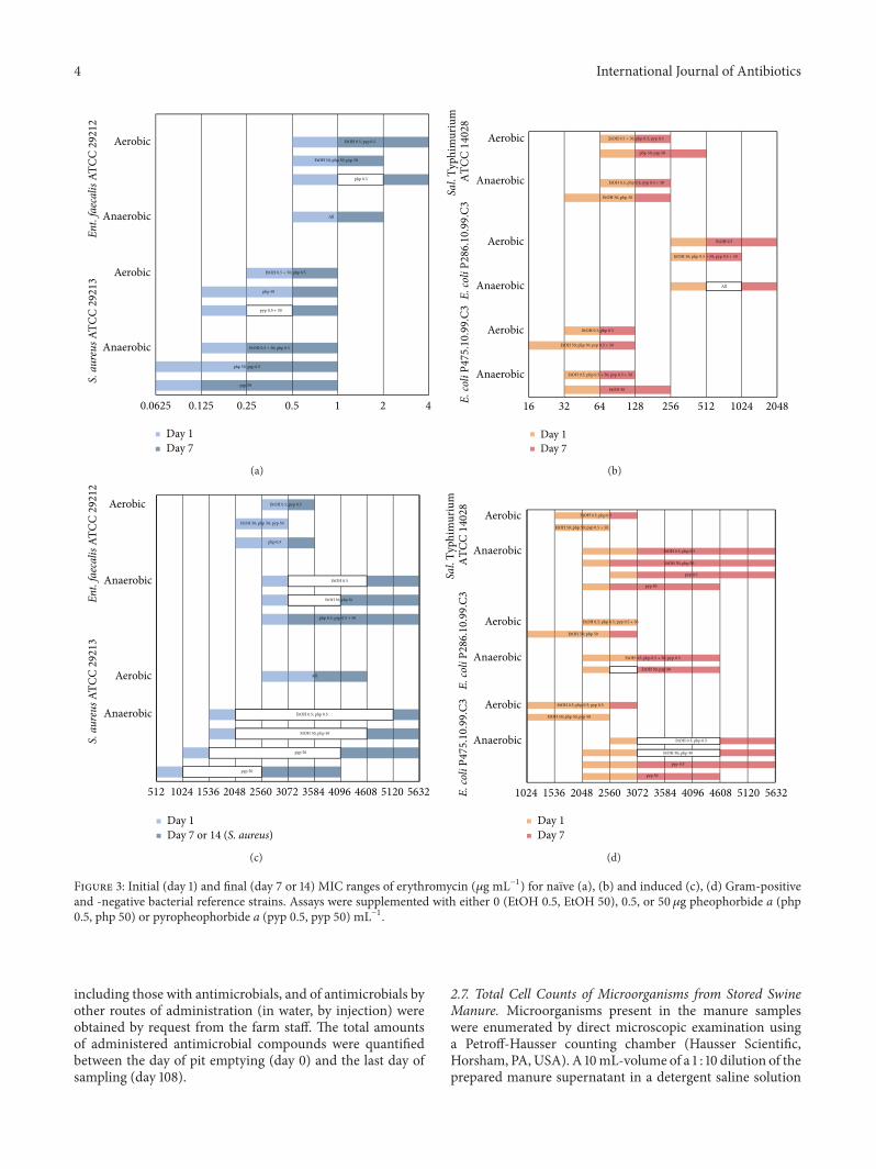
Figure 3: Initial (day 1) and final (day 7 or 14) MIC ranges of erythromycin (𝜇g mL−1) for naıve (a), (b) and induced (c), (d) Gram-positiveand -negative bacterial reference strains. Assays were supplemented with either 0 (EtOH 0.5, EtOH 50), 0.5, or 50𝜇g pheophorbide a (php0.5, php 50) or pyropheophorbide a (pyp 0.5, pyp 50) mL−1.
including those with antimicrobials, and of antimicrobials byother routes of administration (in water, by injection) wereobtained by request from the farm staff. The total amountsof administered antimicrobial compounds were quantifiedbetween the day of pit emptying (day 0) and the last day ofsampling (day 108).
2.7. Total Cell Counts of Microorganisms from Stored SwineManure. Microorganisms present in the manure sampleswere enumerated by direct microscopic examination usinga Petroff-Hausser counting chamber (Hausser Scientific,Horsham, PA,USA). A 10mL-volume of a 1 : 10 dilution of theprepared manure supernatant in a detergent saline solution

International Journal of Antibiotics 5
(9 g sodium chloride (NaCl) L−1, SDS and dibasic potassiumphosphate until foaming and pH 7.2–7.4, resp.) [70, 71] wasadded to 1 g of glass beads, suspended by gentle vortexingfor 10–20 s, and allowed to stand for 15min. Aliquots of thesupernatant were mixed 1 : 1 with 0.1M hydrogen chloride(HCl), and 2 𝜇L of the resulting mixture was used to fillthe counting chamber. At least 400 cells counted under anOlympus BX51 phase-contrast microscope (Olympus, CenterValley, PA, USA) at ×2000 magnification with oil immersionwere taken into account for the calculation of the number ofcells per mL of original sample [70].
2.8. Viable Cell Counts of Anaerobic Bacteria from StoredSwine Manure. To determine the numbers of anaerobicbacteria by viable plate counts, a 40mL volume of a 1 : 100dilution of the prepared manure supernatant in a sterile,anaerobic phosphate-buffered saline solution (PBS, 0.15MNaCl, 0.07M sodium phosphate, pH 7.0) was added to 2.5 gof sterilized glass beads in a sterile 50mL tube and vortexedgently for 10–20 s. Further serial dilutions in PBS wereperformed on this suspension, and aliquots were plated ontoanaerobically prepared [72] modified swine manure slurrymedium (Slurry medium [73] containing 50% (v/v) clarifiedswine manure slurry and supplemented with 1 g of porcinegastric mucin (M2378, Sigma-Aldrich) and 50mL swinefeed hydrolysate (from approx. 1 g swine feed, prepared asdescribed below) L−1). The nonselective, habitat-simulatingslurry medium has been proven to yield the highest viablecounts of anaerobic swine manure storage organisms, pre-dominantly Firmicutes bacteria [73]. Pyp (10 𝜇gmL−1) wasincluded individually and in combination with erythromycin(10 𝜇gmL−1) or tylosin (10 𝜇gmL−1, T-6134, Sigma-Aldrich)in some of the media. Plates were incubated anaerobicallyin a Coy anaerobic chamber at 37∘C, and growth on plateswas examined regularly for 4 weeks and numbers of coloniesenumerated.
2.9. Preparation of Hydrolyzed Swine Feed for Swine ManureSlurry Medium. A pepsin-pancreatin enzymatic hydrolysiswas performed on a sample of nonmedicated swine feedobtained from the swine farm used for manure sampling fol-lowing a previously described protocol [74, 75]. This pepsin-pancreatin enzymatic hydrolysis mimics the endogenous(host-derived) digestion in the porcine upper gastrointestinaltract. 1 g, aliquots of the fine grained feed were mixed with25mL of phosphate buffer solution (0.1M, pH 6.0) and10mL 0.2M HCl. The pH was adjusted to 2.0 with 1MHCl and 1M sodium hydroxide (NaOH), and 1mL of afreshly prepared porcine pepsin solution containing 25mgpepsin (516360, Calbiochem, San Diego, CA, USA) mL−1was added. The mixtures were transferred into 50mL glassserum bottles with rubber stoppers, and the crimp-sealedbottles were placed for 2 h in a gently rotating stainlesssteel beaker reactor system equipped with infrared heating(Labomat BFA-12 v200, Werner Mathis, Concord, NC, USA)set to 39∘C. After the pepsin hydrolysis, 10mL of phosphatebuffer solution (0.2M, pH 6.8) and 5mL 0.6M NaOH wereadded to the hydrolysis mixtures.The pHwas adjusted to 6.8,
and 1mL of a freshly prepared porcine pancreatin solutioncontaining 100mg pancreatin (P-1750, Sigma) mL−1 wasadded. The hydrolysis step using the Labomat reactor systemwas repeated at 39∘C for 4 h. Finally, the hydrolysates werefiltered through three layers of cheesecloth, and the filtratewas stored at −18∘C for later use.
2.10. Concentrations of Php, Pyp, and Tylosin in StoredSwine Manure, Hydrolyzed Feed, and Agar Medium. Php,pyp, and tylosin concentrations in the stored swine manure,hydrolyzed swine feed, and unsupplemented agar mediumwere determined on a wet weight basis using HPLC analysisof methanol extracts from dried samples. The concentrationsof erythromycin were not analyzed, as there had beenno history of erythromycin application on the farm usedfor manure sampling. To obtain dry materials and wetweight/dry weight ratios, the remaining manure sample andunautoclaved feed hydrolysate were spread out evenly in aplastic tray, weighed, and put in a convection oven (BlueM Electric Company, Blue Island, IL, USA) until they dry(approx. 48 h) at 40 and 60∘C, respectively. 2 agar platesof the unsupplemented medium were weighed and thenfreeze-dried in 50mL tubes with perforated lids using aLabconco Bulk Tray Dryer (Labconco, Kansas City, MO,USA) for approximately 48 h. After reweighing, the driedsamples were ground with mortar and pestle, and 3 × 0.3 ±0.01 g aliquots of the resulting powders were mixed with2mLHPLC grademethanol in 20mL disposable scintillationvials. The vials were capped, wrapped with sealing tape,sonicated for 30min in a Branson 2510 ultrasonic cleaner(Branson Ultrasonics, Danbury, CT, USA), and allowed tostand overnight at room temperature and to be protectedfrom light. Aliquots of the extraction supernatants werepassed through a 0.45 𝜇m nylon chromatography syringefilter (Fisher Thermo Scientific, Pittsburgh, PA, USA) into1.5mL screw-thread vials (SUN-Sri, Rockwood, TN, USA)for HPLC analysis for both pheophorbides, php and pyp, andtylosin.
HPLC analysis was conducted on a Shimadzu LC-20HPLC system (LC-20AT quaternary pump, DGU-20A5degasser, SIL-20A HT autosampler, and a SPDM20A photo-diode array detector, running under Shimadzu LCSolutionsversion 1.22 chromatography software, Shimadzu, Columbia,MD, USA). The column used was an Inertsil ODS-3 reversephase C-18 column (5 𝜇M, 250×4.6mm,Varian Lake Forrest,CA, USA). For php and pyp analysis, the initial conditionswere from 50% acetonitrile and 50% water with 0.025%trifluoroacetic acid (TFA) at a flow rate of 1mL per minute.The effluent was monitored at 410 nm on the photodiodearray (PDA) detector. After injection (typically 25 𝜇L), thecolumn was developed to 100% acetonitrile and 0.025% TFAin a linear gradient over 30 minutes. Standard curves basedon micrograms injected were prepared from a preparatorystandard of php from chlorophyll a (C5753, Sigma-Aldrich)[76] and pure standard of pyp purchased commercially fromFrontier Scientific. For tylosin analysis, the initial conditionswere 10% acetonitrile, 90% water, with 0.025% TFA at a flowrate of 1mL per minute. The effluent was monitored at 285

6 International Journal of Antibiotics
and 210 nm on the PDA detector. After injection (typically25 𝜇L), the column was held at the initial conditions for 5minutes and then developed to 100% acetonitrile and 0.025%TFA in a linear gradient over 30 additional minutes. Standardcurves based on 𝜇g tylosin injected were prepared fromcommercially obtained standard (Sigma-Aldrich).
3. Results
3.1. Serial Passage Selection for Induced Erythromycin Resis-tance. Figure 2 shows the MTCs of erythromycin for thenaıve strains and after serial passage selection in progressivelyincreasing erythromycin concentrations for 4 (Ent. faecalis)to 12 weeks (Sal. Typhimurium).The level of parental but notof induced resistancewas higher in theGram-negative strains(parental: 64–1024, induced: 2560–5120𝜇g erythromycinmL−1) than in the Gram-positives (parental: 0.5–2, induced:3072–5120𝜇gmL−1). For all strains, induction was higherunder anaerobic than under aerobic conditions, with themaximum factor being 10240 (S. aureus).
3.2. MICs of Erythromycin over Time. The most notableeffects on the MICs of erythromycin were observed in pyp-supplemented assays of S. aureus. The initial MIC rangeof naıve cultures of S. aureus was 2-fold lower both underaerobic (0.25–0.5 in controls versus 0.125–0.25 in assays withpyp at 0.5 and 50 𝜇gmL−1) and anaerobic (0.125–0.25 incontrol versus 0.0625–0.125 in assays with pyp at 50 𝜇gmL−1)conditions (Figure 3(a)). For induced cells under anaerobicconditions, the initial MIC range was reduced by 512 𝜇gerythromycin mL−1 or 1.3–1.5-fold (pyp at 0.5 𝜇gmL−1) to1024 𝜇g erythromycin mL−1 or 2-3-fold (pyp at 50 𝜇gmL−1)(Figure 3(c)). Using the higher concentration of pyp, thisinitial effect was stable over time (up to 14 days) with areduction of the finalMIC end point by 1536 𝜇g erythromycinmL−1 or 1.4-fold (Figure 3(c)). Pyp was more effective againstS. aureus under anaerobic conditions, and insofar as, in naıvecultures, the increase of MIC endpoints to the final value of 1occurred later (day 7) than under aerobic conditions (days3-4) (Figure 3(a), details not depicted), and insofar as nodifferences occurred on aerobically grown-induced cultures(Figure 3(c)). The effect of pyp (at 50 𝜇gmL−1) was less onE. coli P475.10.99.C3 (reduction of the final MIC endpoint ofinduced cultures by 1024 𝜇g erythromycin mL−1 or 1.2-foldunder anaerobic conditions, Figure 3(d)) and not significanton the other strains. MICs in php-supplemented assays wereunaffected or insignificantly different (Figures 3(a)–3(d)).
3.3. Growth of S. aureus. In general, results from the spec-trophotometric analysis of the growth kinetics of S. aureusconfirmed a concentration-dependent inhibitory effect ofpyp. In naıve cultures under aerobic conditions, the growth-retarding effect of 0.0625 𝜇g erythromycin mL−1 was syn-ergistically potentiated by 50𝜇g pyp mL−1, resulting in asignificantly longer generation time 𝑔 (minimum 𝑔 (𝑔min) =54.6 ± 3.7min, average 𝑔 (𝑔ave) = 71.1 ± 2.0min) andreduced growth rate 𝑘 (maximum 𝑘 (𝑘max) = 0.33 ± 0.02 h
−1,
average 𝑘 (𝑘ave) = 0.25 ± 0.01 h−1) compared with cultures
that contained only erythromycin (𝑔min, 𝑔ave, 𝑘max and𝑘ave = 49.3 ± 0.9min, 61.9 ± 1.6min, 0.37 ± 0.01 h−1 and0.29 ± 0.01 h−1, resp.) (Figures 4(a), 4(b), and 5(a)). Underanaerobic conditions, 0.5 and 50 𝜇g pyp mL−1 exerted anadditive to synergistic effect in combination with 0.0625 𝜇gerythromycin mL−1. Using the higher pyp concentrationsignificantly increased 𝑔min and 𝑔ave (from 63.7 ± 5.0 to73.1 ± 3.5min and 79.7 ± 2.1 to 85.9 ± 1.5min, respectively,Figure 4(a)) and reduced 𝑘max and 𝑘ave (from 0.29 ± 0.02to 0.25 ± 0.01 h−1 and 0.23 ± 0.01 to 0.21 ± 0.00 h−1, resp.,Figure 4(b)), as reflected by a lower total yield (0.84 ± 0.03versus 0.68 ± 0.08, Figures 4(c) and 5(b)). Furthermore,pyp at 50 𝜇gmL−1 exhibited an intrinsic growth-inhibitoryeffect, thereby leading to an increased 𝑔ave (from 74.1 ±1.3 to 79.4 ± 1.9min, Figure 4(a)) and reduced 𝑘ave (from0.24 ± 0.00 to 0.23 ± 0.01 h−1, Figure 4(b)) and total yield(from 1.07 ± 0.02 to 0.85 ± 0.02, Figures 4(c) and 5(b)).In erythromycin-induced cultures, the combinatory effectof 0.5 and 50 𝜇g pyp mL−1 was consistently synergistic andthus partially reversed high-level erythromycin resistance(Figure 5(c)). 𝑔min (319.8 ± 12.3min) and 𝑔ave (342.3 ±14.2min) were significantly longer and 𝑘max (0.06±0.00 h
−1),𝑘ave (0.05 ± 0.00 h−1), and the total yield (0.40 ± 0.06)significantly reduced in the presence of both 50 𝜇g pyp mL−1
and 2048𝜇g erythromycin mL−1 relative to cultures exposedto only erythromycin (𝑔min, 𝑔ave, 𝑘max, 𝑘ave, and total yield =273.7±6.0min, 299.0±6.0min, 0.07±0.00 h−1, 0.06±0.00 h−1and 0.59 ± 0.03, resp.) (Figures 4(a)–4(c), and 5(c)).
3.4. Data on Zootechnical Additives andApplication of Antimi-crobials. The antimicrobial compounds applied betweenthe day when the manure storage pit was emptied andthe last day of the sampling period were oxytetracyclinehydrochloride, lincomycin hydrochloride, tiamulin hydrogenfumarate, and penicillin G (benzylpenicillin) procaine inthe following decreasing quantities: 6.90 kg oxytetracyclinehydrochloride by feed, water, and injection, 5.82 kg lin-comycin hydrochloride by feed and injection, 0.31 kg tia-mulin hydrogen fumarate by feed and 0.15 kg penicillin Gprocaine by injection. There was no history of the use oferythromycin on the farm, and the last contamination ofthe manure pit with tylosin occurred 31 days before the pitemptying. Other applied zootechnical feed additives withantimicrobial activity were copper sulfate (4.93 kg) and zincoxide (67.77 kg). Besides antimicrobials, 78.47 kg of an algae-containing prebiotic feed additive (Intivate, Alltech, Ames,IA, USA) as well as 90.04 kg of MicroSource S, a direct-fedmicrobial feed supplement containing 1.47 × 10⁸ spores ofBacillus (B.) subtilis and B. licheniformis g−1 (DSM, Parsip-pany, NJ, USA) [77] were applied. Intivate had first beenintroduced 9 months and 10 days before the pit emptying,and the amount used during this time was 53.07 kg. Onlylincomycin hydrochloride (approx. 0.12 kg by injection) andMicroSource S (17.24 kg) were administered during the two-week sampling period.

International Journal of Antibiotics 7
Aerobic-naïve Anaerobic-naïve Anaerobic-induced
0306090
120150180210240270300330360
Min
imum
Aver
age
Min
imum
Aver
age
Min
imum
Aver
age
Gen
erat
ion
time (
min
)
EtOH 5𝜇L/mL (naıve) or 45𝜇L/mL (induced)pyp 50𝜇g/mL + EtOH 5𝜇L/mL (naıve) or 45𝜇L/mL (induced)ery 0.0625𝜇g/mL (naıve) or 2048𝜇g/mL (induced) + EtOH 5𝜇L/mL(naıve) or 45𝜇L/mL (induced)pyp 0.5 𝜇g/mL + ery 0.0625𝜇g/mL (naıve) or 2048𝜇g/mL(induced) + EtOH 5𝜇L/mL (naıve) or 45𝜇L/mL (induced)pyp 50𝜇g/mL + ery 0.0625𝜇g/mL (naıve) or 2048𝜇g/mL(induced) + EtOH 5𝜇L/mL (naıve) or 45𝜇L/mL (induced)
(a)
0.000.050.100.150.200.250.300.350.400.450.500.550.600.65
Aerobic-naïve Anaerobic-naïve Anaerobic-induced
Max
imum
Aver
age
Max
imum
Aver
age
Max
imum
Aver
age
EtOH 5𝜇L/mL (naıve) or 45𝜇L/mL (induced)pyp 50𝜇g/mL + EtOH 5𝜇L/mL (naıve) or 45𝜇L/mL (induced)ery 0.0625𝜇g/mL (naıve) or 2048𝜇g/mL (induced) + EtOH 5𝜇L/mL(naıve) or 45𝜇L/mL (induced)pyp 0.5 𝜇g/mL + ery 0.0625𝜇g/mL (naıve) or 2048𝜇g/mL(induced) + EtOH 5𝜇L/mL (naıve) or 45𝜇L/mL (induced)pyp 50𝜇g/mL + ery 0.0625𝜇g/mL (naıve) or 2048𝜇g/mL(induced) + EtOH 5𝜇L/mL (naıve) or 45𝜇L/mL (induced)
Gro
wth
rate
(h−1)
(b)
0.0
0.2
0.4
0.6
0.8
1.0
1.2
EtOH 5𝜇L/mL (naıve) or 45𝜇L/mL (induced)pyp 50𝜇g/mL + EtOH 5𝜇L/mL (naıve) or 45𝜇L/mL (induced)ery 0.0625𝜇g/mL (naıve) or 2048𝜇g/mL (induced) + EtOH 5𝜇L/mL(naıve) or 45𝜇L/mL (induced)pyp 0.5 𝜇g/mL + ery 0.0625𝜇g/mL (naıve) or 2048𝜇g/mL(induced) + EtOH 5𝜇L/mL (naıve) or 45𝜇L/mL (induced)pyp 50𝜇g/mL + ery 0.0625𝜇g/mL (naıve) or 2048𝜇g/mL(induced) + EtOH 5𝜇L/mL (naıve) or 45𝜇L/mL (induced)
Tota
l yie
ld (O
.D.660
nm)
Aerobic-naıve(6h)
Anaerobic-naıve(9h)
Anaerobic-induced(36h)
(c)
Figure 4: Effects of pyropheophorbide a (pyp, 0.5 and 50 𝜇gmL−1) alone and in combination with erythromycin (ery, 0.0625 and2048 𝜇gmL−1) on growth parameters ((a) generation time; (b) growth rate; (c) total yield) of naıve and erythromycin-induced S. aureusATCC 29213 under aerobic and anaerobic conditions. Values are averages and standard deviations from three repeated experiments.
Table 1: Culture counts of anaerobic bacteria from stored swine manure.
Viable counts: swine slurry medium∗
+H2O/EtOH +erythromycin +tylosin−pyp 1.26 × 10⁹ (±0.13 × 10⁹) 1.01 × 10⁹ (±0.30 × 10⁹) 1.07 × 10⁹ (±0.15 × 10⁹)+pyp 8.99 × 10⁸ (±1.69 × 10⁸) 8.90 × 10⁸ (±2.66 × 10⁸) 1.09 × 10⁹ (±0.22 × 10⁹)∗(CFUmL−1) on swine slurry medium supplemented with pyropheophorbide a (pyp, 10𝜇gmL−1) alone and in combination with erythromycin, tylosin(10 𝜇gmL−1, individually) or water and ethanol (H2O/EtOH, control). Values are averages and standard deviations from three repeated experiments withduplicates.

8 International Journal of Antibiotics
30 90 150 210 270 330 390Time (h)
0.0
0.2
0.4
0.6
0.8
1.0
1.2
EtOH 5𝜇L/mLpyp 50𝜇g/mL + EtOH 5𝜇L/mL
pyp 0.5 𝜇g/mL + ery 0.0625𝜇g/mL + EtOH 5𝜇L/mLg/mL + EtOH 5𝜇L/mLpyp 50𝜇g/mL + ery 0.0625𝜇
ery 0.0625𝜇g/mL + EtOH 5𝜇L/mL
O.D
. (660
nm)
(a)
0.5 1.5 2.5 3.5 4.5 5.5 6.5 7.5 8.5 9.5Time (h)
0.0
0.2
0.4
0.6
0.8
1.0
1.2
O.D
. (660
nm)
EtOH 5𝜇L/mLpyp 50𝜇g/mL + EtOH 5𝜇L/mL
pyp 0.5 𝜇g/mL + ery 0.0625𝜇g/mL + EtOH 5𝜇L/mLpyp 50𝜇g/mL + ery 0.0625𝜇
ery 0.0625𝜇g/mL + EtOH 5𝜇L/mL
g/mL + EtOH 5𝜇L/mL
(b)
0
0.2
0.4
0.6
0.8
0.5 24.5 26.5 28.5 30.5 32.5 34.5 36.5Time (h)
O.D
. (660
nm)
EtOH 45𝜇L/mLpyp 50𝜇g/mL + EtOH 45𝜇L/mLery 2048𝜇g/mL + EtOH 45𝜇L/mLpyp 0.5 𝜇g/mL + ery 2048𝜇g/mL + EtOH 45𝜇L/mL
45𝜇L/mLpyp 50𝜇g/mL + ery 2048𝜇g/mL + EtOH
(c)
Figure 5: Effects of pyropheophorbide a (pyp, 0.5 and 50 𝜇gmL−1) alone and in combination with erythromycin (ery, 0.0625 and2048 𝜇gmL−1) on growth curves of naıve (a), (b), and erythromycin-induced (c) S. aureus ATCC 29213 under aerobic (a) and anaerobic(b), (c) conditions. Values are averages and standard deviations from three repeated experiments.
3.5. Total Cell Counts of Microorganisms and Viable CellCounts of Anaerobic Bacteria from Stored Swine Manure.Directmicroscopic counts ofmicroorganisms in stored swinemanure were 5.49±0.41×10⁹mL−1, and the recovery rate onanaerobic swine slurry medium was about 23% (1.26± 0.13×10⁹ CFU, Table 1). About 80% (1.01 ± 0.30 × 10⁹ CFU) and
85% (1.07 ± 0.15 × 10⁹ CFU) of the organisms were capableof growing in erythromycin and tylosin containing media,respectively. Pyp exerted a significant intrinsic inhibitoryeffect which was greater (8.99 ± 1.69 × 10⁸ CFU) than theantibiotic effect of erythromycin or tylosin. CFU on mediasupplemented with both pyp and erythromycin tended tobe lower (8.90 ± 2.66 × 10⁸) than on those containing onlyerythromycin. This combinatorial effect was indifferent, thatis, equal to the effect of pyp alone. Pyp in combination withtylosin produced an antagonistic effect, resulting in increased
and equivalent counts (1.09 ± 0.22 × 10⁹ CFU) relative to thenumber of CFU on media containing only pyp and tylosin,respectively.
3.6. Concentrations of Php, Pyp, and Tylosin in Stored SwineManure, Hydrolyzed Feed, and Agar Medium. Php and pypconcentrations in stored swine manure samples and unsup-plemented swine slurry agar media were consistently belowthe general detection limit of 5𝜇g injected or ≤10 𝜇g g−1sample (wet weight). The concentration of tylosin or acompound with the same retention time in the unsupple-mented agar media was 56.3 ± 2.2 𝜇g g−1 wet weight, whereastylosin was not detected in the manure samples. SubsequentHPLC analysis of the hydrolyzed swine feed used for mediapreparation did not detect php, pyp, or tylosin and hence

International Journal of Antibiotics 9
ruled out that the feed accounted for the possible backgroundconcentration of tylosin in the media.
4. Discussion
In the course of the serial passage experiments, the overallMTCs of erythromycin for naıve cultures of S. aureus andEnt.faecalis were below the European Committee on Antimicro-bial Susceptibility Testing (EUCAST) epidemiological cut-off values of ≤1 and 4 𝜇gmL−1, respectively (Figure 2 andEUCAST [78, 79]).This was presumably due to the absence ofintrinsic resistance mechanisms and, because of their broadsubstrate specificities [80], downregulated basal expressionlevels of existing MDR efflux pumps, such as NorA inS. aureus and EmeA in E. faecalis. NorA and EmeA arehomologous to each other [27, 81]; however, while EmeA isknown to contribute to erythromycin resistance [61, 81], sucha function hasmerely been indicated forNorA [60, 82, 83]. Aswith other macrolides, erythromycin is classically consideredas bacteriostatic, but it has shown bactericidal activity againstS. aureus at higher drug/cell ratios [84–86]. Bactericidalantimicrobials at sublethal concentrations induce mutationalrather than adaptive resistance [10]. It is likely that theserial exposure of S. aureus to increasing concentrations oferythromycin ultimately resulted in the selection of cells bear-ing additive, high-level resistance-conferringmutations fromnaturally occurring subpopulations with, due to overexpres-sion of the regular cellular machinery, low-level phenotypicadaptive resistance [10, 29, 87, 88]. High-level resistance toerythromycin can be predominant among S. aureus strainsin individual studies (approx. 60% of strains with MIC >1024 𝜇gmL−1 [89]), but, generally it is rare (approx. 3% ofstrains withMICs≥ 512 𝜇gmL−1 according to EUCAST [78]).
For all facultatively anaerobic bacterial reference strainsexamined, the serial passage experiments led to higherMTCs under anaerobic (96% carbon dioxide, 4% hydrogenatmosphere) than under aerobic conditions (Figure 2). Sus-ceptibility testing of macrolides in the presence of carbondioxide can result in elevated resistance, because the carbondioxide decreases the pH of the medium and consequentlyerythromycin activity [90, 91]. However, in our experiments,the pH was controlled by sodium carbonate in the media(4 g L−1), and therefore the “pH effect” of incubation incarbon dioxide can be ruled out. Instead, reactive oxygenspecies (ROS) generated by aeration might account for theobserved differences between aerobic and anaerobic MTCs.ROS have antimicrobial activity and contribute to dysregu-lation primarily of MDR efflux pump expression by oxidativeinactivation of global regulator proteins [29, 32]. For example,in S. aureus, reduced aeration causes an increase of norBexpression and hence increased resistance to antimicrobialNorB substrates via an effect on MgrA, an oxidative stress-sensitive global regulator of 350 genes including norA andnorB [92–94]. Thirdly, efflux-mediated antimicrobial resis-tance is controlled by the metabolic condition of bacteria andcan be altered by a switch from an aerobic to an anaerobicmetabolism through the influence ofmetabolically integratedglobal regulators, different endogenous cellular metabolites,
such as ROS from aerobic respiration or anaerobic fermen-tation end products which often times are the natural pumpsubstrates, and an altered transmembrane electrochemicalproton gradient as energy source for secondary active trans-porters [27, 28, 32, 80]. Hence, it can be presumed thatthe higher level of induced resistance in S. aureus underanaerobic conditions was contingent on a switch from gly-colysis, the pentose phosphate pathway and ROS generatingtricarboxylic acid cycle under aerobiosis to anaerobic glucosefermentation andATPase-mediated proton efflux, generatinga greater motive force for erythromycin-proton antiporters[28, 80, 94], such as LmrS, MdeA, Mef(A), and conceivablyNorA [27, 32, 95–97].
In the brothmacrodilution assays, pyp exerted significanteffects on the MICs of erythromycin for E. coli P475.10.99.C3and especially S. aureus (Figure 3). Subsequent spectropho-tometric determination of growth parameters of S. aureusconfirmed that pyp in combination with erythromycin wasmost effective, that is, synergistic already at the lowerconcentration of 0.5𝜇gmL−1, in highly resistant culturesunder anaerobic conditions (Figures 4 and 5(c)) and revealedthat pyp was intrinsically inhibitory to anaerobic, naıvecultures (Figures 4 and 5(b)). As could be expected [8, 94],the growth rate in the absence and at a low concentration oferythromycin (0.0625𝜇gmL−1) (Figure 4(b)) was greatest inthe aerobic, naıve cultures and lowest for the highly resistantstrain—the latter effect being due to a “fitness cost” asentailed by most antimicrobial resistance mechanisms [88].The intrinsic growth inhibition of anaerobic, naıve culturesof S. aureus indicates that pyp, provided it equals php inits function as an EPI [51, 52], acts against a metabolicallyregulated MDR pump with broader, compound nonspecificfunctions unrelated to antimicrobials. It can be concludedthat this hypothetical MDR efflux pump is one other thanNorA, as there is no strong indication of erythromycin beinga NorA substrate [29, 60, 82, 83] and as we observed nosignificant effect of pyp against Ent. faecalis bearing the NorAhomologue EmeA (Figures 3(a) and 3(c)). The S. aureusgenome comprises at least 30 genes for putative drug trans-porters, and approximately 17 of these encode MDR effluxpumps, most of which are still unknown [98, 99] and twoof which, LmrS andMdeA, contribute to erythromycin resis-tance [27, 32, 97, 100] and are therefore possible candidatesfor inhibition by pyp. Pyp in combination with erythromycinonly partially reversed high-level resistance of S. aureus anddid not completely inhibit its growth (Figures 3(a), 3(c),and 5(c)). This indicates that the high-level resistance isdepended on the combined effects of more than one effluxpump and/or other resistance mechanisms [80]. There areat least 17 genes encoding for protein efflux systems formacrolides, lincosamides, and streptogramins [20]. Msr(A)andMef(A) arewell known to confer erythromycin resistancein strains of S. aureus [27, 95, 96, 101, 102]. The plasmid-borne Msr(A) transporter, even though mainly responsiblefor erythromycin efflux in S. aureus [103], is not present in thestrain used in our study, S. aureus ATCC 29213 [104]. Mef(A)is closely related to Mef(B), the erythromycin efflux pumpin E. coli P475.10.99.C3 [67], and as pyp decreased the final

10 International Journal of Antibiotics
MIC endpoint for induced cultures of this strain (Figures 3(b)and 3(d)), it can be concluded that Mef(A) is an additionalcandidate for inhibition by pyp in S. aureus.
The basal levels of resistance to erythromycin (approx.80%) and tylosin (approx. 85%) of anaerobic bacteria fromstored swine manure samples (Table 1) were considerablyhigher than those found earlier in manure samples from thesame farm (max. 21% and 32%) [73]. However, accordingto other previous studies [105, 106], background levels oferythromycin and macrolide resistance in nonmedicatedswine fecal andmanure bacterial communities can already beas high as approximately 40% and are enhanced further by theadministration of compounds with antimicrobial activities[62–64]. In our study, tylosin had last been used 31 daysand 4–4.5 months prior to the last pit emptying and days ofsampling, respectively, and consequently was not detected byHPLC in the manure. Also, it can be concluded that insteadof tylosin a different compound with the same retention timewas detected in the unsupplemented agar media, as both themanure and hydrolyzed swine feed could be ruled out as asource of contamination and as any tylosin present in themedia would, due to its heat lability [107], probably havebeen destroyed by autoclaving (121∘C, 20min). There hadbeen no history of the use of erythromycin on the farm,but a low concentration in the manure due to erythromycin-contaminated DDGS in the swine feed is possible and mighthave contributed to the observed high basal resistance level.More likely, however, the high levels of erythromycin andtylosin resistance were partly the outcome of the recentadministration of oxytetracycline, lincomycin, and the heavymetals copper and zinc, these compounds can indirectlyenhance antimicrobial incl. macrolide resistance throughcoselection [105, 108–113].
Pyp in combinationwith erythromycin (10𝜇gmL−1, indi-vidually) tended to reduce the number of erythromycin resis-tant bacteria cultured from stored swine manure (Table 1);however, this effect was indifferent and less significant thanthe synergistic effects observed on S. aureus (Figures 4and 5). The level of tylosin resistance remained unchangedwhen pyp and tylosin were combined (Table 1). Experimentswith S. aureus had shown that pyp acts in a concentration-dependent manner between 0.5 and 50 𝜇gmL−1 (Figures 3–5), and therefore it can be presumed that more significanteffects on macrolide resistance of manure bacteria can beachieved with higher concentrations of pyp. Alternatively, thelower effectiveness might be due to cross- or coresistancebetween pyp and the macrolide antibiotics.The developmentof bacterial resistance to EPIs themselves is a recognizedconcern [29]. As in the experiments with S. aureus (Figures4 and 5(b)), pyp was intrinsically inhibitory to the growth ofanaerobic manure bacteria (Table 1). It is not an uncommonfeature for an EPI from natural sources to possess a directantibacterial effect [26, 35]. For example, the aerobic MIC ofphp was found to vary between 4 𝜇gmL−1 (S. epidermidis)and 500 𝜇gmL−1 (S. aureusATCC 29213) [52], and in anotherstudy the death percentage of S. aureus ATCC 26923 in20𝜇g php mL−1 was 32% [114]. It is conceivable that, asdiscussed above for S. aureus, pyp exerted its intrinsic effect
on themanuremicrobiota through inhibition of aMDReffluxpump, such as LmrS, MdeA, and possibly NorA, as these andtheir homologues occur in B. subtilis, a major componentof MicroSource S, and are widespread among other lowmol percentage guanine-cytosine Gram-positive (phylumFirmicutes) bacteria [27, 81, 97, 100, 115–117], the predominantculturable anaerobic microorganisms from swine manure[73, 118, 119].
Neither php nor pyp was detectable by HPLC in theswine manure or corn-soybean-DDGS-based feed (withoutIntivate). These results confirm previous findings that thefecal concentrations of php and pyp are diet-dependent andrange between <1 and 180 𝜇g g−1 in feces (dry matter) fromswine and herbivore livestock with a low and high intakeof chlorophyll a, respectively [50]. Consequently, in order toachieve the effects indicated by our results (bacterial growthinhibition, potentiation of antimicrobials, and partial reversalof bacterial antimicrobial resistance) inside the gastroin-testinal tracts of swine and/or in manure storage pits, both“hotspot habitats” for the selection and spread of bacterialresistance genes [4, 16, 120], it is necessary to include greenplant feedstuffs, such as alfalfa and grass, or microalgae. Eventhough, in this study, the use of the algae-containing feedadditive Intivate was not sufficient, supplementation withmicroalgae is a suitable naturalmeans of obtaining significantconcentrations of php and pyp in swine manure. Microalgae,such as Chlorella vulgaris, which can be autochthonous toswine manure habitats [121, 122] presents a good sourceof chlorophyll a, php, and pyp [38, 40, 123] as well as avaluable substitute for conventional protein sources [123,124]. Their direct addition to swine waste lagoons, ponds,or other treatment bioreactors can be combined with thephotodegradative bioremediation of residual antimicrobialsubstances [125, 126].
5. Conclusions
To conclude, this is the first study showing that pyro-pheophorbide derived from chlorophyll affects growth andthe level of sensitivity to erythromycin of S. aureus, E. coli,and anaerobic manure bacteria. Addition of chlorophyll-containing plant material to animal diets has the potential toreduce antibiotic resistance in the animal gut and eventualstored manure.
Conflict of Interests
The authors declare that there is no conflict of interestsregarding the publication of this paper.
Acknowledgments
The authors thank Virve I. Enne, Centre for Immunologyand Infectious Disease at Barts and The London School ofMedicine and Dentistry, London, UK, for providing E. colistrains and the staff from the swine farm near Peoria, IL,for enabling sampling of swine stored manure and providing

International Journal of Antibiotics 11
swine feed and data on feed composition and application ofantimicrobials.
References
[1] E. K. Silbergeld, J. Graham, and L. B. Price, “Industrial food ani-mal production, antimicrobial resistance, and human health,”Annual Review of Public Health, vol. 29, pp. 151–169, 2008.
[2] G. Hansen, “Playing chicken with antibiotics: antibiotic use infood animals,” inProceedings of the 140thAPHAAnnualMeeting& Exposition, San Francisco, Calif, USA, 2012.
[3] T. F. Landers, B. Cohen, T. E. Wittum, and E. L. Larson, “Areview of antibiotic use in food animals: perspective, policy, andpotential,” Public Health Reports, vol. 127, no. 1, pp. 4–22, 2012.
[4] P. M. da Costa, L. Loureiro, and A. J. F. Matos, “Transfer ofmultidrug-resistant bacteria between intermingled ecologicalniches: the interface between humans, animals and the envi-ronment,” International Journal of Environmental Research andPublic Health, vol. 10, pp. 278–294, 2013.
[5] M. D. Apley, E. J. Bush, R. B. Morrison, R. S. Singer, andH. Snelson, “Use estimates of in-feed antimicrobials in swineproduction in the United States,” Foodborne Pathogens andDisease, vol. 9, no. 3, pp. 272–279, 2012.
[6] D. C. Love, M. F. Davis, A. Bassett, A. Gunther, and K.E. Nachman, “Dose imprecision and resistance: free-choicemedicated feeds in industrial food animal production in theUnited States,” Environmental Health Perspectives, vol. 119, no.3, pp. 279–283, 2011.
[7] F. Baquero, “Low-level antibacterial resistance: a gateway toclinical resistance,” Drug Resistance Updates, vol. 4, no. 2, pp.93–105, 2001.
[8] D. I. Andersson and D. Hughes, “Persistence of antibiotic resis-tance in bacterial populations,” FEMS Microbiology Reviews,vol. 35, no. 5, pp. 901–911, 2011.
[9] R. Canton and M.-I. Morosini, “Emergence and spread ofantibiotic resistance following exposure to antibiotics,” FEMSMicrobiology Reviews, vol. 35, no. 5, pp. 977–991, 2011.
[10] M. A. van der Horst, J. M. Schuurmans, M. C. Smid, B.B. Koenders, and B. H. ter Kuile, “De novo acquisition ofresistance to three antibiotics by escherichia coli,” MicrobialDrug Resistance, vol. 17, no. 2, pp. 141–147, 2011.
[11] D. I. Andersson and D. Hughes, “Evolution of antibioticresistance at non-lethal drug concentrations,” Drug ResistanceUpdates, vol. 15, pp. 162–172, 2012.
[12] R. Schmieder and R. Edwards, “Insights into antibiotic resis-tance through metagenomic approaches,” Future Microbiology,vol. 7, no. 1, pp. 73–89, 2012.
[13] N. Peak, C. W. Knapp, R. K. Yang et al., “Abundance of sixtetracycline resistance genes in wastewater lagoons at cattlefeedlots with different antibiotic use strategies,” EnvironmentalMicrobiology, vol. 9, no. 1, pp. 143–151, 2007.
[14] C. S. Holzel, K. Schwaiger, K. Harms et al., “Sewage sludgeand liquid pig manure as possible sources of antibiotic resistantbacteria,” Environmental Research, vol. 110, no. 4, pp. 318–326,2010.
[15] H. K. Allen, T. Looft, D. O. Bayles et al., “Antibiotics in feedinduce prophages in swine fecal microbiomes,”mBio, vol. 2, no.6, 2011.
[16] T. Looft, T. A. Johnson, H. K. Allen et al., “In-feed antibioticeffects on the swine intestinal microbiome,” Proceedings of the
National Academy of Sciences of the United States of America,vol. 109, no. 5, pp. 1691–1696, 2012.
[17] C. H. Bolster, “Factors controlling subsurface transport ofmanure-borne pathogens,” 2012, http://www.extension.org/pages/65647/microbes:-from-farm-to-public-risk.
[18] H. Heuer, H. Schmitt, and K. Smalla, “Antibiotic resistancegene spread due to manure application on agricultural fields,”Current Opinion in Microbiology, vol. 14, no. 3, pp. 236–243,2011.
[19] B. M. Marshall and S. B. Levy, “Food animals and antimicro-bials: impacts on human health,” Clinical Microbiology Reviews,vol. 24, no. 4, pp. 718–733, 2011.
[20] M. C. Roberts, “Environmental macrolide-lincosamide-strep-togramin and tetracycline resistant bacteria,” Frontiers inMicro-biology, vol. 2, pp. 1–8, 2011.
[21] K. J. Forsberg, A. Reyes, B. Wang et al., “The shared antibioticresistome of soil bacteria and human pathogens,” Science, vol.337, pp. 1107–1111, 2012.
[22] B. Gonzalez-Zorn and J. A. Escudero, “Ecology of antimicrobialresistance: humans, animals, food and environment,” Interna-tional Microbiology, vol. 15, pp. 101–109, 2012.
[23] M. Popowska, M. Rzeczycka, A. Miernik, A. Krawczyk-Balska,F. Walsh, and B. Duffy, “Influence of soil use on preva-lence of tetracycline,streptomycin, and erythromycin resistanceand associated resistance genes,” Antimicrobial Agents andChemotherapy, vol. 56, no. 3, pp. 1434–1443, 2012.
[24] T. O. Rahube and C. K. Yost, “Characterization of a mobile andmultiple resistance plasmid isolated from swine manure and itsdetection in soil after manure application,” Journal of AppliedMicrobiology, vol. 112, pp. 1123–1133, 2012.
[25] B. Marquez, “Bacterial efflux systems and efflux pumpsinhibitors,” Biochimie, vol. 87, no. 12, pp. 1137–1147, 2005.
[26] M. Stavri, L. J. V. Piddock, and S. Gibbons, “Bacterial effluxpump inhibitors from natural sources,” Journal of AntimicrobialChemotherapy, vol. 59, no. 6, pp. 1247–1260, 2007.
[27] K. Poole, “Efflux-mediated antimicrobial resistance,” Journal ofAntimicrobial Chemotherapy, vol. 56, no. 1, pp. 20–51, 2005.
[28] J. L. Martınez and F. Rojo, “Metabolic regulation of antibioticresistance,” FEMS Microbiology Reviews, vol. 35, no. 5, pp. 768–789, 2011.
[29] L. Fernandez and R. E. W. Hancock, “Adaptive and mutationalresistance: role of porins and efflux pumps in drug resistance,”Clinical Microbiology Reviews, vol. 25, pp. 661–681, 2012.
[30] T. A. Krulwich, O. Lewinson, E. Padan, and E. Bibi, “Dophysiological roles foster persistance of drug/multidrug-effluxtransporters? A case study,” Nature Reviews Microbiology, vol.3, no. 7, pp. 566–572, 2005.
[31] L. J. V. Piddock, “Multidrug-resistance efflux pumps—not justfor resistance,” Nature Reviews Microbiology, vol. 4, no. 8, pp.629–636, 2006.
[32] X.-Z. Li and H. Nikaido, “Efflux-mediated drug resistance inbacteria: an update,” Drugs, vol. 69, no. 12, pp. 1555–1623, 2009.
[33] A. C. Abreu, A. J. McBain, and M. Simoes, “Plants as sources ofnew antimicrobials and resistance-modifying agents,” NaturalProduct Reports, vol. 29, pp. 1007–1021, 2012.
[34] A. Ojeda-Sana, V. Repetto, and S. Moreno, “Carnosic acid isan efflux pumps modulator by dissipation of the membranepotential in Enterococcus faecalis and Staphylococcus aureus,”World Journal of Microbiology and Biotechnology, vol. 29, pp.137–144, 2013.
12 International Journal of Antibiotics
[35] J. G. Holler, S. B. Christensen, H.-C. Slotved et al., “Novelinhibitory activity of the Staphylococcus aureus NorA effluxpump by a kaempferol rhamnoside isolated from Persea lingueNees,” Journal of Antimicrobial Chemotherapy, vol. 67, no. 5, pp.1138–1144, 2012.
[36] L.Ma andD.Dolphin, “Themetabolites of dietary chlorophylls,”Phytochemistry, vol. 50, no. 2, pp. 195–202, 1999.
[37] M. Holden, “Chlorophyll degradation products in leaf proteinpreparations,” Journal of the Science of Food and Agriculture, vol.25, no. 11, pp. 1427–1432, 1974.
[38] Y. Takeda, S. Uchiyama, and Y. Saito, “High performance liquidchromatography of pheophorbide a and pyropheophorbide ain salted vegetables and chlorella,” Journal of the Food HygienicSociety of Japan, vol. 26, pp. 56–60, 1985.
[39] Y. Shioi, Y. Tatsumi, and K. Shimokawa, “Enzymatic degra-dation of chlorophyll in chenopodium album,” Plant and CellPhysiology, vol. 32, no. 1, pp. 87–93, 1991.
[40] H. Oshima, E. Ueno, I. Saito, and H. Matsumoto, “Develop-ment of a solid-phase extraction method for determination ofpheophorbide a and pyropheophorbide a in health foods byliquid chromatography,” Journal of AOAC International, vol. 87,no. 4, pp. 937–942, 2004.
[41] S. Aiamla-or, S. Kaewsuksaeng, M. Shigyo, and N. Yamauchi,“Impact of UV-B irradiation on chlorophyll degradation andchlorophyll-degrading enzyme activities in stored broccoli(Brassica oleracea L. Italica Group) florets,” Food Chemistry, vol.120, no. 3, pp. 645–651, 2010.
[42] M. G. Ferruzzi, M. L. Failla, and S. J. Schwartz, “Assessmentof degradation and intestinal cell uptake of carotenoids andchlorophyll derivatives from spinach puree using an in vitrodigestion andCaco-2 human cell model,” Journal of Agriculturaland Food Chemistry, vol. 49, no. 4, pp. 2082–2089, 2001.
[43] K. D. Ashby, J. Wen, P. Chowdhury, T. A. Casey, M. A. Ras-mussen, and J. W. Petrich, “Fluorescence of dietary porphyrinsas a basis for real-time detection of fecal contamination onmeat,” Journal of Agricultural and Food Chemistry, vol. 51, no.11, pp. 3502–3507, 2003.
[44] W. M. Campbell, G. S. Dombroski, I. Sharma, A. C. Partridge,and M. G. Collett, “Photodynamic chlorophyll a metabolites,including phytoporphyrin (phylloerythrin), in the blood ofphotosensitive livestock: overview and measurement,” NewZealand Veterinary Journal, vol. 58, no. 3, pp. 146–154, 2010.
[45] M. Aprahamian, S. Evrardl, P. Keller et al., “Distribution ofpheophorbide A in normal tissues and in an experimentalpancreatic cancer in rats,” Anti-Cancer Drug Design, vol. 8, no.2, pp. 101–114, 1993.
[46] J. W. Jonker, M. Buitelaar, E. Wagenaar et al., “The breastcancer resistance protein protects against a major chlorophyll-derived dietary phototoxin and protoporphyria,” Proceedings ofthe National Academy of Sciences of the United States of America,vol. 99, no. 24, pp. 15649–15654, 2002.
[47] R. W. Robey, K. Steadman, O. Polgar, and S. E. Bates, “ABCG2-mediated transport of photosensitizers: potential impact onphotodynamic therapy,” Cancer Biology andTherapy, vol. 4, no.2, pp. 187–194, 2005.
[48] M. Li, H. Yuan, N. Li et al., “Identification of interspeciesdifference in efflux transporters of hepatocytes from dog,rat, monkey and human,” European Journal of PharmaceuticalSciences, vol. 35, no. 1-2, pp. 114–126, 2008.
[49] M. R. F. Lee, V. J. Theobald, H. J. Ougham et al., “Natural faecalfluorophores and the potential of chlorophyll based markers to
optimise fluorescence as a real-time solution for the detectionof faecal contamination on carcasses,”Meat Science, vol. 86, no.4, pp. 966–975, 2010.
[50] C. A. Barnes, S. L. Rasmussen, J. W. Petrich, and M. A.Rasmussen, “Determination of the concentration of potentialefflux pump inhibitors, pheophorbide a and pyropheophorbidea, in the feces of animals by fluorescence spectroscopy,” Journalof Agricultural and Food Chemistry, vol. 60, pp. 10456–10460,2012.
[51] F. R. Stermitz, J. Tawara-Matsuda, P. Lorenz, P. Mueller,L. Zenewicz, and K. Lewis, “5’-methoxyhydnocarpin-D andPheophorbide A: Berberis species components that potentiateberberine growth inhibition of resistant Staphylococcus aureus,”Journal of Natural Products, vol. 63, no. 8, pp. 1146–1149, 2000.
[52] R.Musumeci, A. Speciale, R. Costanzo et al., “Berberis aetnensisC. Presl. extracts: antimicrobial properties and interaction withciprofloxacin,” International Journal of Antimicrobial Agents,vol. 22, no. 1, pp. 48–53, 2003.
[53] M. C. Roberts, “Update on macrolide-lincosamide-strep-togramin, ketolide, and oxazolidinone resistance genes,” FEMSMicrobiology Letters, vol. 282, no. 2, pp. 147–159, 2008.
[54] Compassion in World Farming, Antibiotics in animal farming,Public health and animal welfare, 2011, http://www.ciwf.org.uk/includes/documents/cm docs/2011/a/antibiotics in animalfarming.pdf.
[55] S. Dohne, R.Merle, A. V. Altrock et al., “Antibiotic susceptibilityof Salmonella, Campylobacter coli, and Campylobacter jejuniisolated from northern German fattening pigs,” Journal of FoodProtection, vol. 75, no. 10, pp. 1839–1845, 2012.
[56] H. Hao, Z. Yuan, Z. Shen et al., “Mutational and transcriptomicchanges involved in the development of macrolide resistance inCampylobacter jejuni,”Antimicrobial Agents and Chemotherapy,vol. 57, pp. 1369–1378, 2013.
[57] J. Olmstead, Institute for Agriculture and Trade Policy,Bugs in the system, How the FDA fails to regulate antibi-otics in ethanol production, 2012, http://www.iatp.org/files/2012 05 02 AntibioticsInEthanol JO 0.pdf.
[58] C. G. Shurson, D. M. Paulus, A. DiCostanzo et al., Are anti-biotics a concern in distiller’s co-products?, University of Min-nesota, 2012, http://www.ddgs.umn.edu/prod/groups/cfans/@pub/@cfans/@ddgs/documents/asset/cfans asset 414344.pdf.
[59] FDA, U.S. Food and Drug Administration, Animal Drugs @FDA, 2013, http://www.accessdata.fda.gov/scripts/animal-drugsatfda/.
[60] G. Tegos, F. R. Stermitz, O. Lomovskaya, and K. Lewis, “Mul-tidrug pump inhibitors uncover remarkable activity of plantantimicrobials,” Antimicrobial Agents and Chemotherapy, vol.46, no. 10, pp. 3133–3141, 2002.
[61] A. Aakra, H. Vebø, L. Snipen et al., “Transcriptional responseof Enterococcus faecalis V583 to erythromycin,” AntimicrobialAgents and Chemotherapy, vol. 49, no. 6, pp. 2246–2259, 2005.
[62] C. R. Jackson, P. J. Fedorka-Cray, J. B. Barrett, and S. R. Ladely,“Effects of tylosin use on erythromycin resistance in enterococciisolated from swine,” Applied and Environmental Microbiology,vol. 70, no. 7, pp. 4205–4210, 2004.
[63] M. E. Jacob, J. T. Fox, S. K. Narayanan, J. S. Drouillard, D.G. Renter, and T. G. Nagaraja, “Effects of feeding wet corndistillers grains with solubles with or without monensin andtylosin on the prevalence and antimicrobial susceptibilities offecal foodborne pathogenic and commensal bacteria in feedlotcattle,” Journal of Animal Science, vol. 86, no. 5, pp. 1182–1190,2008.

International Journal of Antibiotics 13
[64] D. B. Holman and M. R. Chenier, “Impact of subtherapeuticadministration of tylosin and chlortetracycline on antimicro-bial resistance in farrow-to-finish swine,” FEMS MicrobiologyEcology, vol. 85, no. 1, pp. 1–13, 2013.
[65] J.M.Andrews, “Determination ofminimum inhibitory concen-trations,” Journal of Antimicrobial Chemotherapy, vol. 48, no. 1,pp. 5–16, 2001.
[66] TOKU-E, Erythromycin (E-mycin, Ery-tab, Benzamycin), 2012,http://antibiotics.toku-e.com/antimicrobial 599.html.
[67] J. Liu, P. Keelan, P.M. Bennett, andV. I. Enne, “Characterizationof a novel macrolide efflux gene,mef (B), found linked to sul3 inporcine Escherichia coli,” Journal of Antimicrobial Chemother-apy, vol. 63, no. 3, pp. 423–426, 2009.
[68] R. Lindqvist, “Estimation of Staphylococcus aureus growthparameters from turbidity data: characterization of strain vari-ation and comparison of methods,” Applied and EnvironmentalMicrobiology, vol. 72, no. 7, pp. 4862–4870, 2006.
[69] M. T. Madigan, J. M. Martinko, P. V. Dunlap, and D. P.Clark, “Growth of bacterial populations,” in Brock Biology ofMicroorganisms, pp. 147–152, Pearson Education, San Francisco,Calif, USA, 2008.
[70] E. Bast, “Mikroskopische zellzahlung in einer zahlkammer,” inMikrobiologische Methoden: Eine Einfuhrung in GrundlegendeArbeitstechniken, pp. 280–285, Spektrum-AkademischerVerlag,2001, (German).
[71] K. P. Norris and E. O. Powell, “Improvements in determiningtotal counts of bacteria,” Journal of the Royal MicroscopicalSociety, vol. 80, pp. 107–119, 1961.
[72] M. P. Bryant, “Commentary on the Hungate technique forculture of anaerobic bacteria,” American Journal of ClinicalNutrition, vol. 25, no. 12, pp. 1324–1328, 1972.
[73] M. A. Cotta, T. R. Whitehead, and R. L. Zeltwanger, “Isolation,characterization and comparison of bacteria from swine faecesand manure storage pits,” Environmental Microbiology, vol. 5,no. 9, pp. 737–745, 2003.
[74] S. Boisen and J. A. Fernandez, “Prediction of the total tractdigestibility of energy in feedstuffs and pig diets by in vitroanalyses,” Animal Feed Science and Technology, vol. 68, no. 3-4,pp. 277–286, 1997.
[75] J. Bindelle, A. Buldgen, C. Boudry, and P. Leterme, “Effect ofinoculum and pepsin-pancreatin hydrolysis on fibre fermenta-tionmeasured by the gas production technique in pigs,”AnimalFeed Science and Technology, vol. 132, no. 1-2, pp. 111–122, 2007.
[76] J. L. Wickliff and S. Aronoff, “Degradation of chlorophyll ato pheophytin a, pheophorbide a, and pyrroporphine XV fortracer studies,” Analytical Biochemistry, vol. 6, no. 1, pp. 39–46,1963.
[77] Miller Publishing Co., S. MicroSource, Direct-fed Microbial,Enzyme & Forage Additive Compendium—Ninth Edition,2013, http://www.microbialcompendium.com.
[78] EUCAST, European Committee on Antimicrobial Suscep-tibility Testing, Erythromycin/Staphylococcus aureus, EUCASTMIC Distribution, Version 5.13, 2013, http://mic.eucast.org/Eucast2/regShow.jsp?Id=2642.
[79] EUCAST, European Committee on Antimicrobial Susceptibil-ity Testing, Erythromycin/Enterococcus faecalis, EUCASTMIC Distribution, Version 5.13, 2013, http://mic.eucast.org/Eucast2/regShow.jsp?Id=7183.
[80] J. L. Martinez, A. Fajardo, L. Garmendia et al., “A global view ofantibiotic resistance,” FEMS Microbiology Reviews, vol. 33, pp.44–65, 2009.
[81] B. M. Jonas, B. E. Murray, and G. M. Weinstock, “Charac-terization of emeA, a norA homolog and multidrug resistanceefflux pump, in Enterococcus faecalis,” Antimicrobial Agents andChemotherapy, vol. 45, no. 12, pp. 3574–3579, 2001.
[82] S. Gibbons, M. Oluwatuyi, and G. W. Kaatz, “A novel inhibitorof multidrug efflux pumps in Staphylococcus aureus,” Journal ofAntimicrobial Chemotherapy, vol. 51, no. 1, pp. 13–17, 2003.
[83] G. Belofsky,D. Percivill, K. Lewis, G. P. Tegos, and J. Ekart, “Phe-nolic metabolites of Dalea versicolor that enhance antibioticactivity against model pathogenic bacteria,” Journal of NaturalProducts, vol. 67, no. 3, pp. 481–484, 2004.
[84] L. Unger and A. Kisch, “Observations on bacteriostatic andbactericidal action of erythromycin,” Proceedings of the Societyfor Experimental Biology and Medicine, vol. 98, pp. 176–178,1958.
[85] F. Fraschini, P. C. Braga, and V. Copponi, “Bactericidal activityof erythromycin in the respiratory system,” Current MedicalResearch and Opinion, vol. 7, no. 7, pp. 429–439, 1981.
[86] G. A. Pankey and L. D. Sabath, “Clinical relevance of bacte-riostatic versus bactericidal mechanisms of action in the treat-ment of gram-positive bacterial infections,” Clinical InfectiousDiseases, vol. 38, no. 6, pp. 864–870, 2004.
[87] H. Nikaido, “Multidrug resistance in bacteria,” Annual Reviewof Biochemistry, vol. 78, pp. 119–146, 2009.
[88] J. L. Martınez, F. Baquero, and D. I. Andersson, “Beyond serialpassages: new methods for predicting the emergence of resis-tance to novel antibiotics,” Current Opinion in Pharmacology,vol. 11, no. 5, pp. 439–445, 2011.
[89] E. Piątkowska, J. Piątkowski, and A. Przondo-Mordarska, “Thestrongest resistance of Staphylococcus aureus to erythromycinis caused by decreasing uptake of the antibiotic into the cells,”Cellular andMolecular Biology Letters, vol. 17, pp. 633–645, 2012.
[90] E. J. C. Goldstein and V. L. Sutter, “Effect of carbon dioxide onerythromycin,”Antimicrobial Agents andChemotherapy, vol. 23,no. 2, pp. 325–327, 1983.
[91] L.M. Ednie,M. R. Jacobs, andP. C.Appelbaum, “Anti-anaerobicactivity of erythromycin, azithromycin and clarithromycin:effect of pH adjustment of media to compensate for pHshift caused by incubation in CO
2
,” Journal of AntimicrobialChemotherapy, vol. 41, no. 3, pp. 387–389, 1998.
[92] T. T. Luong, P. M. Dunman, E. Murphy, S. J. Projan, andC. Y. Lee, “Transcription profiling of the mgrA regulon inStaphylococcus aureus,” Journal of Bacteriology, vol. 188, no. 5,pp. 1899–1910, 2006.
[93] K. Poole, “Bacterial stress responses as determinants of antimi-crobial resistance,” Journal of Antimicrobial Chemotherapy, vol.67, pp. 2069–2089, 2012.
[94] Q. C. Truong-Bolduc, L. C. Hsing, R. Villet et al., “Reducedaeration affects the expression of the NorB efflux pump ofStaphylococcus aureus by posttranslational modification ofMgrA,” Journal of Bacteriology, vol. 194, no. 7, pp. 1823–1834,2012.
[95] R. Leclercq, “Mechanisms of resistance to macrolides and lin-cosamides: nature of the resistance elements and their clinicalimplications,”Clinical Infectious Diseases, vol. 34, no. 4, pp. 482–492, 2002.
[96] V. A. Luna, M. Heiken, K. Judge et al., “Distribution of mef (A)in gram-positive bacteria from healthy Portuguese children,”Antimicrobial Agents andChemotherapy, vol. 46, no. 8, pp. 2513–2517, 2002.

14 International Journal of Antibiotics
[97] J. L. Floyd, K. P. Smith, S. H. Kumar, J. T. Floyd, and M. F.Varela, “LmrS is amultidrug efflux pump of themajor facilitatorsuperfamily from Staphylococcus aureus,” Antimicrobial Agentsand Chemotherapy, vol. 54, no. 12, pp. 5406–5412, 2010.
[98] G. W. Kaatz, V. V. Moudgal, S. M. Seo, J. B. Hansen, and J.E. Kristiansen, “Phenylpiperidine selective serotonin reuptakeinhibitors interfere with multidrug efflux pump activity inStaphylococcus aureus,” International Journal of AntimicrobialAgents, vol. 22, no. 3, pp. 254–261, 2003.
[99] K. A. Hassan, R. A. Skurray, and M. H. Brown, “Active exportproteins mediating drug resistance in staphylococci,” Journal ofMolecular Microbiology and Biotechnology, vol. 12, no. 3-4, pp.180–196, 2007.
[100] J. Huang, P. W. O’Toole, W. Shen et al., “Novel chromosomallyencoded multidrug efflux transporter MdeA in Staphylococcusaureus,” Antimicrobial Agents and Chemotherapy, vol. 48, no. 3,pp. 909–917, 2004.
[101] J. I. Ross, E. A. Eady, J. H. Cove, and S. Baumberg, “Minimalfunctional system required for expression of erythromycinresistance bymsrA in Staphylococcus aureusRN4220,”Gene, vol.183, no. 1-2, pp. 143–148, 1996.
[102] K. Kadlec, A. T. Feßler, T. Hauschild, and S. Schwarz, “Noveland uncommon antimicrobial resistance genes in livestock-associated methicillin-resistant Staphylococcus aureus,” ClinicalMicrobiology and Infection, vol. 18, pp. 745–755, 2012.
[103] C. R. S. Teodoro, C. S. Mattos, F. S. Cavalcante, E. M. Pereira,and K. R. N. dos Santos, “Characterization of MLS
𝑏
resistanceamong Staphylococcus aureus and Staphylococcus epidermidisisolates carrying different SCCmec types,” Microbiology andImmunology, vol. 56, pp. 647–650, 2012.
[104] M. A. Argudın, B.-A. Tenhagen, A. Fetsch et al., “Virulenceand resistance determinants of German Staphylococcus aureusST398 isolates from nonhuman sources,” Applied and Environ-mental Microbiology, vol. 77, no. 9, pp. 3052–3060, 2011.
[105] Z. Zhou, L. Raskin, and J. L. Zilles, “Macrolide resistance inmicroorganisms at antimicrobial-free swine farms,”Applied andEnvironmentalMicrobiology, vol. 75, no. 18, pp. 5814–5820, 2009.
[106] M. Kalmokoff, L. M. Waddington, M. Thomas et al., “Continu-ous feeding of antimicrobial growth promoters to commercialswine during the growing/finishing phase does not mod-ify faecal community erythromycin resistance or communitystructure,” Journal of Applied Microbiology, vol. 110, no. 6, pp.1414–1425, 2011.
[107] Santa Cruz Biotechnology, Tylosin tartrate (CAS, 1405-54-5),Material Safety Data Sheet, 2013, http://datasheets.scbt.com/sc-204933.pdf.
[108] M. C. Roberts, “Resistance to macrolide, lincosamide, strep-togramin, ketolide, and oxazolidinone antibiotics,” MolecularBiotechnology, vol. 28, no. 1, pp. 47–62, 2004.
[109] C. Baker-Austin, M. S. Wright, R. Stepanauskas, and J. V.McArthur, “Co-selection of antibiotic and metal resistance,”Trends in Microbiology, vol. 14, no. 4, pp. 176–182, 2006.
[110] J. L. Martinez, M. B. Sanchez, L. Martınez-Solano et al., “Func-tional role of bacterial multidrug efflux pumps in microbialnatural ecosystems,” FEMS Microbiology Reviews, vol. 33, no. 2,pp. 430–449, 2009.
[111] J. Kluytmans and J. L. Murk, “Lincomycin and clindamycin,” inKucers’ the Use of Antibiotics: A Clinical Review of Antibacterial,Antifungal and Antiviral Drugs, L. M. Grayson, Ed., pp. 987–1007, CRC Press, Boca Raton, Fla, USA, 2010.
[112] C. S. Holzel, C. Muller, K. S. Harms et al., “Heavy metals inliquid pig manure in light of bacterial antimicrobial resistance,”Environmental Research, vol. 113, pp. 21–27, 2012.
[113] R. Leclercq, R. Canton, D. F. J. Brown et al., “EUCAST expertrules in antimicrobial susceptibility testing,” Clinical Microbiol-ogy and Infection, vol. 19, pp. 141–160, 2013.
[114] A. P. Gerola, A. Santana, P. B. Franca et al., “Effects of metal andthe phytyl chain on chlorophyll derivatives: physicochemicalevaluation for photodynamic inactivation of microorganisms,”Photochemistry and Photobiology, vol. 87, no. 4, pp. 884–894,2011.
[115] A. Bolotin, P. Wincker, S. Mauger et al., “The complete genomesequence of the lactic acid bacterium lactococcus lactis ssp. lactisIL1403,” Genome Research, vol. 11, no. 5, pp. 731–753, 2001.
[116] F. R. Stermitz, T. D. Beeson, P. J. Mueller, J.-F. Hsiang, andK. Lewis, “Staphylococcus aureus MDR efflux pump inhibitorsfromaBerberis and aMahonia (sensu strictu) species,”Biochem-ical Systematics and Ecology, vol. 29, no. 8, pp. 793–798, 2001.
[117] L. J. V. Piddock, “Clinically relevant chromosomally encodedmultidrug resistance efflux pumps in bacteria,” Clinical Micro-biology Reviews, vol. 19, no. 2, pp. 382–402, 2006.
[118] T. R. Whitehead and M. A. Cotta, “Characterisation and com-parison of microbial populations in swine faeces and manurestorage pits by 16S rDNA gene sequence analyses,” Anaerobe,vol. 7, no. 4, pp. 181–187, 2001.
[119] R. Snell-Castro, J.-J. Godon, J.-P. Delgenes, and P. Dabert,“Characterisation of the microbial diversity in a pig manurestorage pit using small subunit rDNA sequence analysis,” FEMSMicrobiology Ecology, vol. 52, no. 2, pp. 229–242, 2005.
[120] A. Tello, B. Austin, and T. C. Telfer, “Selective pressure ofantibiotic pollution on bacteria of importance to public health,”Environmental Health Perspectives, vol. 120, pp. 1100–1106, 2012.
[121] M. Wilson and J. A. Houghton, “Growth of algae on pigmanure,” Irish Journal of Agricultural Research, vol. 13, pp. 49–60, 1974.
[122] M. V. Jimenez-Perez, P. Sanchez-Castillo, O. Romera, D.Fernandez-Moreno, and C. Perez-Martınez, “Growth andnutrient removal in free and immobilized planktonic greenalgae isolated from pig manure,” Enzyme and Microbial Tech-nology, vol. 34, pp. 392–398, 2004.
[123] M. K. Garrett, J. J. Strain, and M. D. B. Allen, “Composition ofthe product of algal culture in the liquid phase of animal slurry,”Journal of the Science of Food and Agriculture, vol. 27, pp. 603–611, 1976.
[124] E.W. Becker, “Micro-algae as a source of protein,” BiotechnologyAdvances, vol. 25, no. 2, pp. 207–210, 2007.
[125] I. de Godos, R.Munoz, and B. Guieyssea, “Tetracycline removalduring wastewater treatment in high-rate algal ponds,” Journalof Hazardous Materials, vol. 229-230, pp. 446–449, 2012.
[126] E. M. Ferrero, I. de Godos, E. M. Rodrıguez, P. A. Garcıa-Encina, R. Munoz, and E. Becares, “Molecular characterizationof bacterial communities in algal-bacterial photobioreactorstreating piggery wastewaters,” Ecological Engineering, vol. 40,pp. 121–130, 2012.

Submit your manuscripts athttp://www.hindawi.com
PainResearch and TreatmentHindawi Publishing Corporationhttp://www.hindawi.com Volume 2014
The Scientific World JournalHindawi Publishing Corporation http://www.hindawi.com Volume 2014
Hindawi Publishing Corporationhttp://www.hindawi.com
Volume 2014
ToxinsJournal of
VaccinesJournal of
Hindawi Publishing Corporation http://www.hindawi.com Volume 2014
Hindawi Publishing Corporationhttp://www.hindawi.com Volume 2014
AntibioticsInternational Journal of
ToxicologyJournal of
Hindawi Publishing Corporationhttp://www.hindawi.com Volume 2014
StrokeResearch and TreatmentHindawi Publishing Corporationhttp://www.hindawi.com Volume 2014
Drug DeliveryJournal of
Hindawi Publishing Corporationhttp://www.hindawi.com Volume 2014
Hindawi Publishing Corporationhttp://www.hindawi.com Volume 2014
Advances in Pharmacological Sciences
Tropical MedicineJournal of
Hindawi Publishing Corporationhttp://www.hindawi.com Volume 2014
Medicinal ChemistryInternational Journal of
Hindawi Publishing Corporationhttp://www.hindawi.com Volume 2014
AddictionJournal of
Hindawi Publishing Corporationhttp://www.hindawi.com Volume 2014
Hindawi Publishing Corporationhttp://www.hindawi.com Volume 2014
BioMed Research International
Emergency Medicine InternationalHindawi Publishing Corporationhttp://www.hindawi.com Volume 2014
Hindawi Publishing Corporationhttp://www.hindawi.com Volume 2014
Autoimmune Diseases
Hindawi Publishing Corporationhttp://www.hindawi.com Volume 2014
Anesthesiology Research and Practice
ScientificaHindawi Publishing Corporationhttp://www.hindawi.com Volume 2014
Journal of
Hindawi Publishing Corporationhttp://www.hindawi.com Volume 2014
Pharmaceutics
Hindawi Publishing Corporationhttp://www.hindawi.com Volume 2014
MEDIATORSINFLAMMATION
of
Copyright © 2022 FDOKUMEN