EFFECTIVENESS OF NODOVENOUS SHUNTS IN FILARIAL ...
63
TAMIL NADU DR. MGR MEDICAL UNIVERSITY CHENNAI – TAMIL NADU EFFECTIVENESS OF NODOVENOUS SHUNTS IN FILARIAL LYMPHEDEMA OF LOWER LIMBS - AN EVALUATION - SUMMITTED FOR THE MCH DEGREE EXAMINATION (PLASTIC SURGERY) EXAMINATION IN AUGUST – 2007 THANJAVUR MEDICAL COLLEGE THANJAVUR
-
Upload
khangminh22 -
Category
Documents
-
view
3 -
download
0
Transcript of EFFECTIVENESS OF NODOVENOUS SHUNTS IN FILARIAL ...

TAMIL NADU DR. MGR MEDICAL UNIVERSITY
CHENNAI – TAMIL NADU
EFFECTIVENESS OF NODOVENOUS SHUNTS IN
FILARIAL LYMPHEDEMA OF LOWER LIMBS
- AN EVALUATION -
SUMMITTED FOR THE MCH DEGREE EXAMINATION
(PLASTIC SURGERY)
EXAMINATION IN AUGUST – 2007
THANJAVUR MEDICAL COLLEGE
THANJAVUR

CERTIFICATE
This is to certify that this dissertation entitled “EFFECTIVENESS ON
NODOVENOUS SHUNTS IN FILARIAL LYMPHEDEMA OF LOWER
LIMBS - AN EVALUATION” is the bonafide record work done by
Dr. K. Chandramohan, M.S., submitted as partial fulfillment for the
requirement of MCH Degree Examination in Plastic Surgery, August 2007.
Prof. Dr. Joseph Victor, M.S., MCh., DNB.,
Professor & Head of the Department,
Department of Plastic & Filarial Surgery,
Thanjavur Medical College,
Thanjavur.

1
1.ACKNOWLEDGEMENT
I am extremely thankful to the Dean Thanjavur medical college for
granting me permission to conduct the study.
Its an immense pleasure to acknowledge Prof Dr.Joseph Victor
MS.,Mch.,DNB, Professor and Head of the department of plastic,
reconstructive and filarial surgery, Thanjavur medical college who has given
me the topic for study and guided me with his everavailable help to carry out
the study.
I sincerely thank Prof.Dr.Olszewski w., the pioneer in nodo venous
shunt, for his advice and guidance who visits our centre every year for
research .
My sincere thanks to my assistant professors Dr.R.S.Subbaiyan MS.,
Mch.,FICS, Dr.V.Devasenan MS.,Mch, Dr.D.Rajkumar MS.,D.Orth.,Mch,
Dr.P.Ravindran MS.,Mch.,FICS. for their support and guidance.
My sincere thanks to co-postgraduates for their support and help.
I finally extend my heartfelt thanks to all the patients without whom
this study would not have been possible.

CONTENTS
1. Acknowledgement
2. Introduction
3. Clinical Features of filarial disease
4. Basic Sciences
5. Diagnosis and Management
6. Background of Study
7. Review of Literature
8. Lymphatico vein Anastomosis
9. Materials and Methods
10. Procedure
11. Methods
12. Observation
13. Results of the study
14. Discussion
15. Conclusion
16. Bibliography

2
2. INTRODUCTION
Definition
Filariasis is a name given for a group of diseases caused by nematodes
belonging to the family Filaridae which are transmitted by haemophagus
mosquito Culex fatigans.
In India the causative organism are Wucheria Bancrofti and Brugia
Malayi. About 250 million people live in the endemic area and about 32
million people are affected by it.
Among these 90% of infected people comprise asymptomatic carriers.
The management of the disease, apart from chemotherapy and surgery
includes preventive aspect of vector control through Vector Research and
Control Centre and through National Filarial Control programme launched
in 1955.

3
4

5
3.CLINICAL FEATURES OF FILARIAL DISEASE
Depends on the stage of infestation in human host.
Stage of Invasion
• Microfilarial larvae develops producing Esinophilia.
• Lymph Adenopathy.
Stage of Symptomless Carrier State
• Detected by night blood smear examinations.
Stage of Acute Manifestations
• Filarial fever
• Lymph adenitis
• Reversible lymph edema of various parts of the body and
Epididymo orchitis in males.

6
Stage of chronic Manifestations
Clinical features of limb edema which is irreversible, Elephantiasis of
leg, arm, genitals, hydrocele & chyluria.
Rare Manifestations
• Chylus ascites
• Chylo thorax
• Peri carditis
• Acute abdomen, Ileal perforation
• Inferior vena caval obstruction
• Synovitis
• Arthiris
• Neurological manifestations
• Chylous diarrhoea
• Chyluria
Pathogenesis of Lymphatic Filarial Disease in Man
In more intense and repeated attacks, microfilaria and mature worms
may travel in lymphatics to the nodes causing foreign protein allergic
inflammatory manifestation and temporary lymphatic obstruction which,

7
super added with bacterial infections may permanently damage the
lymphatic vessel wall and disturb histomechanical processes producing
lymph stasis and its complications.
LYMPH EDEMA
Definition
Lymph edema is a chronic condition involving the extremities, that is
characterised by accumulation of protein rich fluid within the intercellular
space of the sub-cutaneous tissue and skin.
CAUSES OF LYMPH EDEMA
Primary Lymph Edema
• Aplasia
• Hypoplasia
• Hyperplasia
Secondary lymph edema:
• Infection
• Filariasis
• LGV
• Tumour
• Irradiation
• Tuberculosis
8
5.BASIC SCIENCES
Anatomy of Leg Lymphatics
It consists of Epifascial and subfacial systems, lymph edema occurs mainly
in sub-cutaneous compartment which is normally drained by
• Dermal plexus
• Collecting channels
• Superficial lymphatic trunks
• The dermal plexus and the collecting channels constitute the Initial
lymphatics.
The dermal plexus consists of non-valved intradermal lymphatics and
valved deep dermal lymphatics.
The sub-fascial lymphatics or deep muscular lymphatics are separate
and communicate only under abnormal conditions with epifascial lymphatics
like proximal obstruction. This can be ascertained by doing
lymphangiograms.

9
In 1957 Kinmonth et al., introduced lymph angiograms. For
lymphangiography a soluble dye is injected into the web space of the toes,
which enters the dermal lymphatics and eventually outlines the superficial
medial and lateral trunks, which are identified through small incisions,
cannulated and injected with contrast material like Lipiodol.
Approximately 5 vessels are visually seen along the medial aspect of
the extremity, generally following the course of the great saphenous vein to
drain into the superficial inguinal nodes. A drainage system posterior to
lateral malleolus, paralleling the small saphenous vein can also be seen
along the lateral aspect of the leg which are 3 – 4 in number.
The superficial linguinal nodes drain into the iliac nodes.
In the arm, the superficial lymphatics course along the basilic vein on
the medial aspect and cephalic vein on the lateral aspect. They drain into the
axillary lymph nodes.
10
Physiology
The lymphatic system serves the following functions.
1. Drainage of macromolecular protein from capillaries.
2. Removal of foreign material and bacteria.
3. Transport of specific substances like vit. K, long chain fatty acids
from gastrointestinal tract.
4. Production of lymphocytes for cellular and humoral immunity.
The initial lymphatics fill up by
1. Open junctions
2. Hydrostatic pressure difference between capillaries and lymphatics.
3. Colloidal osmotic pressure.
Lymph contain globulin protein of 0.1 to 0.5 gm in a deciliter.
The lymph flow is normally slow and is brought about by the following
factors.
1. Hydrostatic pressure
2. Fluctuating intrathoracic and intraabdominal pressure.
3. Muscular activity of lymphangions, which are functional units of
lymphatics, under the influence of harmones and chemicals.
4. Single way valve system of collecting vessels.
5. Skeletal muscular activity.

11
Pathogenesis
In chronic lymphedema the protein content is as high as 5gm/dl.
Because of stasis of lymph and subsequent changes leading to inflammation,
fibrosis, hyperkeratinisation the skin loses its integrity. There is a back flow
of lymph into dermal plexus and in severe cases skin vesicles appear and
burst causing lymphorrhea.
Histopathology
1. Ingrowth of keratinocytes into dermis.
2. Presence of langerhan cells in Epidermis.
3. CL II antibodies in Epidermis.
4. Chronic inflammatory cells around lymph vessels.

12
6. DIAGNOSIS OF LYMPHEDEMA AND MANAGEMENT
Lymphedema can be primary or acquired
Primary lymphedema is due to developmental abnormality of lymphatics.
• Edema at birth – lymphedema congenital ( Aplasia )
• Edema at Middle age – lymphedema tarda
• Edema at adolescence – lymphdema precox
The Varicose type ( Hyperplasia ) is always due to lymphatic obstruction.
In secondary lymphedema or acquired form the pathologic site is the
regional lymph nodes.
Diagnosis of Filarial Lymphedema
1. History
2. Clinical features and findings
3. Lymph angiography
4. Fluorescent microlymph angiography
5. Lympho Scintigraphy, CT scan and ultrasound.
6. Study of lymph fluid
7. Nigh blood smear for microfilariae
8. Demonstration of microfilariae by Millipore technique
9. Tests to rule out TB.LGV etc.,
10. Cyto immunochemistry.

13
Differential Diagnosis
1. Edema due to pregnancy
2. Edema due to malignancy
3. Venous edema
4. Lipedema
5. Edema due to lymphadenopathy
6. Lymph angiomas
7. Iliac compression syndrome
( Lt. Iliac vein being compressed by Rt.Iliac artery)
Of all the diagnostic methods CT scan and lymphoscintigraphy ( using
Technitium 99 labelled Antimony) are the effective and non invasive
methods, detecting even incipient lymphedema both qualitatively and
quantitatively.
Lymph Angiography ( LAG ) an accurate single examination for
visualization of epifascial and sub-fascial lymphatics.
CT Scan shows enlarged honeycomb appearance of sub-cutis density
increased fascial blurring and muscle mass increase is visualized
differentiates from venous edema.
14
MANAGEMENT
Preoperative Management
1. Grading of the disease - measurements
2. Removal of foci of infections.
3. Long acting penicillin – once in 15 days.
4. Diethyl carbamazine 100 mg thrice daily for two days once in 15
days.
5. Leg elevation and rest.
6. Antifungal and antihistaminics.
Gradewise Management Schedule
Grade I Rest, elastic stockings, elevation of limb
Medical therapy
Pressure therapy with Intermittant pressure of 30 – 50 mm of Hg.
Manual lymphatic drainage.
NodoVenous shunts (+/-).
Grade II Elevation of limb, Elastic support.
Medical therapy
Pressure therapy ( cycled pneumatic pumps)
Manual lymphatic drainage
Nodo – Venous shunts.

15
Grade III Medical therapy, pressure therapy, elastic support, manual
drainage, Nodo venous shunt followed by Debulking surgery.
Grade IV Medical therapy, manual drainage.
Nodo- Venous shunt.
Debulking surgery.
Sculpturing of the nodules.
SSG for the raw areas.
Medical Therapy :
NON SPECIFIC DRUGS
1. Antibiotics.
2. Anti-inflammatory
3. Antihistaminics.
SPECIFIC DRUGS
1. Diethyl carbamazipine
Actions : Microfilaricidal and filaricidal
duration of action 12-21 days.
dose 5 to 6 mg /kg
adjuvant anti-inflammatory and antiplatelet action

16
2. Benzopyrones ( Coumarin)
Effective orally and topically
ACTION : It attracts macrophages to the edema site.
The macrophages lyse plasma protein thereby they pass back into
capillaries. 40 % to 25 % reduction occur in volume in 2 years and reduce
secondary infection and gives symptomatic relief.
Complex Decongestive Manual Physiotheraphy ( Winiwarter 1892)
1st Phase a) Hygiene measures.
b) Manual lymph drainage commencing on normal body
quadrant progressing to edema region.
c) Elastrocrepe bandages.
d) Remedial exercises.
Intermittant Pneumatic Compression ( Ventipress ) ( Olavi Airakinen)
Sequential and graded pumps are more efficient in increasing venous
and lymphatic outflow and reduction in edema and associated symptoms.
Heat and Bandage ( Zhang et al., 1984)
Warming the limb to 6o to 7oC above body temperature for 1 hour /
day for 3 – 4 days followed by elastic bandaging reduces lymphedema.

17
SURGICAL PROCEDURES IN LYMPH EDEMA
Physiological Procedures
1. To improve lymphatic transport capacity
Reconstruction of new lymph vessels with threads and tubes
(handles 1908).
2. To increase Drainage
a) Drainage through strips of fascia or though Tensor fascia Lata (
Martorell 1958)
b) Drainage through omental flaps( Dicks k 1935)
c) Drainage through skin flaps ( Rosanow 1912)
d) Drainage through Entero mesenteric bridging surgery (
Kinmonth 1932)
e) Drainage through peripheral lymphatico-venous anastamosis;
i) Lympho-nodo venous anastamosis. (Olzeweski 1966, S.Jamal 1981).
ii) Anastamosis between lymph collectors and veins ( Laine Howard
1963, Degni 1974)
iii) Bridging of localized obstruction of lymphatic drainage by
transplantation of lymph collectors ( Baumeister 1981) by
transplantation of veins ( Mandl 1981).

18
PROCEDURES TO REDUCE LOAD OF LYMPHATIC CLEARANCE
a. Resection of epifascial lymph edematous tissue.
I. Successive removal of ellipses of skin and sub-cutaneous tissue
and primary closure (Sistrunk 1918)
II. Excision and closure with local flaps ( Homan 1936)
b. Radical reduction of epifascial lymph space
i) Suction lipectomy of epifascial lymph space ( Teimourian
1987)
ii) Excision and closure with skin grafts ( Charles 1912 )
iii) Excision and closure with full thickness skin grafts ( Gibson
and Tough 1954)
Ligature of Arterial blood supply ( Carnochan 1854)
Post Operative Management:
IMMEDIATE :
1. Leg elevation and rest
2. Antibiotics
3. Antiinflammatory
4. Measurements

19
LATE :
1. Long acting penicillin once in 15 days.
2. Diethyl carbamazine 100 mg x 2days once in 15 days.
3. Elastrocrepe bandage / Stockings support while walking
or standing.
4. To sleep with leg elevated without bandage.
5. Local care of edematous limb Antifungal ointments.

20
7.BACKGROUND OF STUDY
Since Thanjavur is an endemic filarial area, filarial lymphedema of
lower limbs is one of the commonest problem we see in our out patient
Department.
We conduct filarial clinic on all mondays and treat around 4000
patients every year.
CENSUS OF FILARIAL PATIENTS TREATED AT OUR CENTRE FOR THE PAST FIVE YEARS
2002 2003 2004 2005 2006 TOTAL CASES 4770 5458 4774 5450 4863 NEW PATIENTS 404 513 456 425 256 OLD PATIENTS 4366 5945 4308 5025 4607 SURGERIES(TOTAL) 162 189 75 142 88 MAJOR 102 109 64 115 79 MINOR 60 80 11 27 9

21
Antibiotic prophylaxis and Diethyl carbamazine were given to all
patients once in 15 days.
We perform annually an average of 75 Nodovenous shunts and 30
cases of reduction annually.
All the patients are classified into four grades according to the history,
size of the limbs and skin changes.
GRADES OF LYMPHEDEMA:
Grade I : Pitting edema of lower limbs relieved by rest.
Grade II : Edema of the lower limbs not relieved by rest.
Grade III: Non-pitting edema of the lower limbs more than 5cm
of difference in circumference from normal limb in
anyone of the fixed points.
Grade IV: Non-pitting edema of lower limbs with
czema,nodule,ulceration,lymph vesicle,lymphorea.
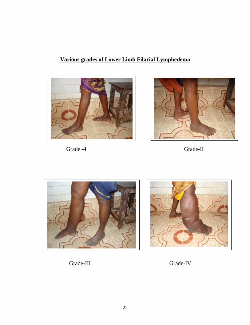
22
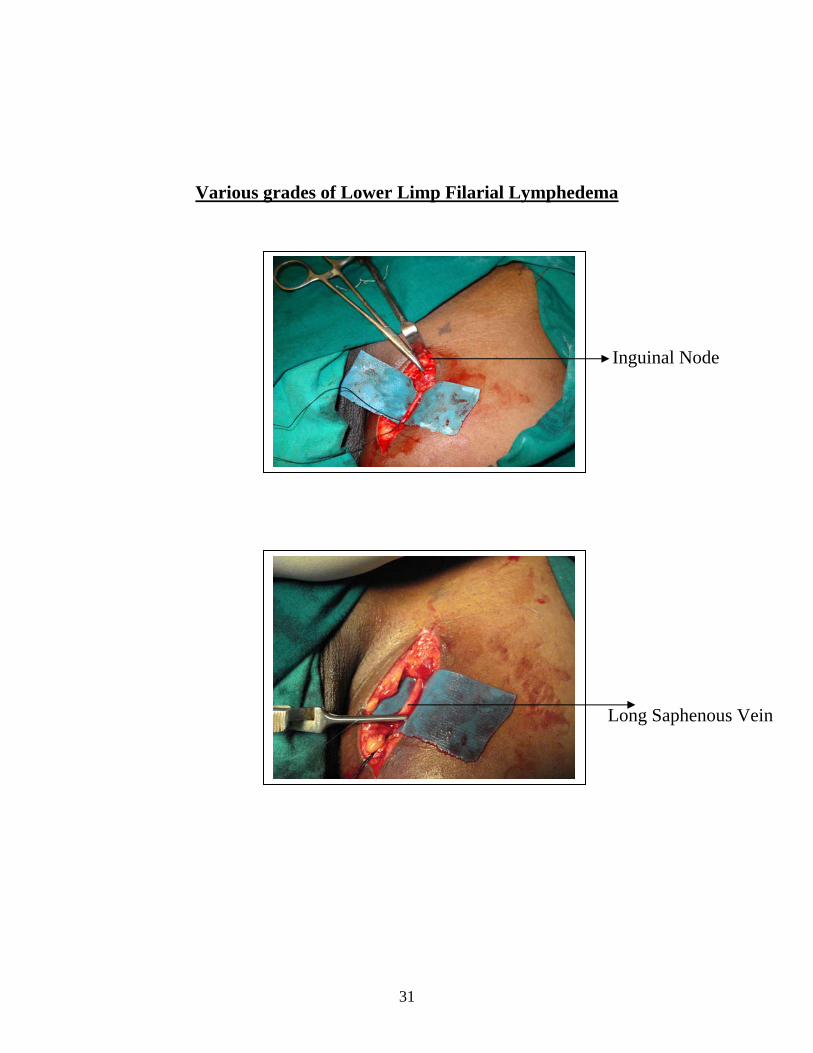
Various grades of Lower Limb Filarial Lymphedema
Grade –I Grade-II
Grade-III Grade-IV

23
For grade I and grade II patients in addition to penicillin and DEC
we perform nodovenous shunts in the affected limb.
For all grade III and IV patients we perform nodovenous shunts one
week prior to reduction surgery.

24
8. REVIEW OF LITERATURE
The task of the lymphedema surgeon is to restore the balance
quantitatively between the lymphatic load and transport capacity by
Reducing the lymphatic load and/or by increasing the lymphatic transport
capacity.
It would be preferable to operate during the latent phase and not
during manifest phase ie., during grade I and ii during which most of the
lymphatic system is destroyed.
Surgery should achieve a permanent and stable balance between
the lymphatic transport capacity and lymphatic load.
As a rule, increasing the lymphatic transport capacity by lympho
venous shunt does not completely quantitatively balance the lymphatic load.
The reduced transport capacity in primary and secondary lymphedema
leads to ‘Tissue poisoning’ by plasma proteins, further alteration of

25
lymphatic transport and filtration systems,changes in direction of lymph
flow and alteration of blood vascular system of the affected part.
During the period of clinical lymphedema, the lymph nodes gradually
start to harden and shrink.
Hence lymphonodovenous shunts should be performed early(grade I
and ii).
Because of failure to achieve permanent cure for filarial lymphedema
very few surgeons recommend surgery.
But in tropical climate the lymphedema load increases making the
lymphedematous skin situation more difficult to control.
Moreover the finesse of conservative therapy unfortunately may not
be available to many lymphedematous patients.
That is why in addition to conservative therapy, surgical approaches
are advocated.
Performing nodovenous in grade III and IV patients prior to surgery
makes tissue supple for reduction surgery, prevents increase in thigh
circumference and reduces adenolymphangitis also.
26
9.LYMPHATIC VEIN ANASTOMOSIS
When Edward and Kimmoth in 1969 performed lymphangiography on
a patient with bilateral lymphatic deficiency, but only one limb swollen, they
visualized LV shunt in the normal appearing limb.
Besides the formation of a collateral lymphatic circulation, the
opening of LVS is one of the natures method of byepassing a surgical block.
This natural phenomenon has stimulated others to create surgically
anastomosis between lymphatics in lymphedematous tissues and suitable
veins.
Olsweski first performed NV shunt in lymphedematous patients and
showed 70% patency for two years.
27
10.MATERIALS AND METHOD
This study was conducted in Thanjavur medical college during the
year 2005-2007.
A total of 100 patients were takenup for study which includes 60
females and 40 males.
All patients were screened for caries tooth,inguinal adenitis,hydrocele
and skin changes in lower limbs.
All patients were subjected to regular Antibiotic
prophylaxis,Compression garments and physical therapy for affected limbs.
All lymphedematous patients were classified to grades I – IV.
All grade I and II patients were grouped according to the presence or
absence of enlarged inguinal node.
Node positive patients were grouped for study.
Node negative patients were taken as control.
28
Grade I and II control group were subjected to compression and
physical therapy alone.
Remaining Grade I and II study group were subjected to compression
and physical therapy and nodovenous shunt.
Clinically leg volume and circumference at standard sites were
measured preoperatively ( at the time of admission) and at periodic intervals
postoperatively.
All grade III and IV patient were grouped randomly into two groups
(study and control group).
Grade III and IV study group were subjected to Nodovenous shunt
surgery,Reduction surgery and compression therapy .
Remaining Grade III and IV control group were subjected to
Reduction surgery, compression and physical therapy.
Clinically, thigh measurement at standard site were measured.
29
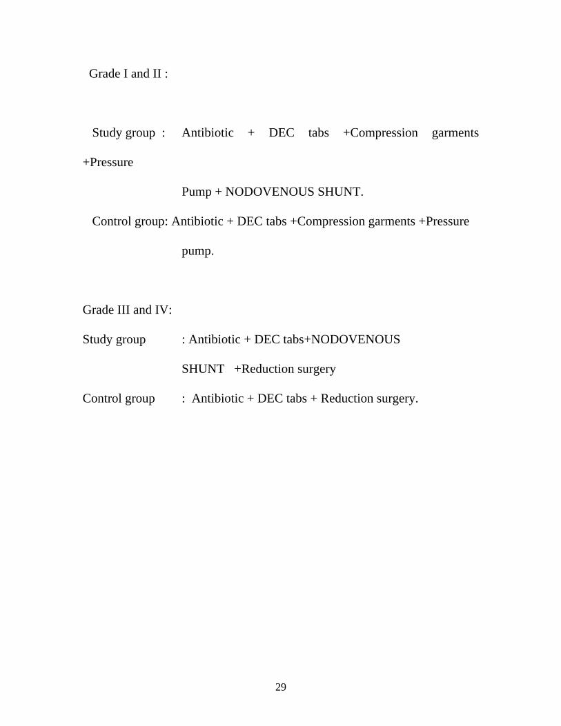
Grade I and II :
Study group : Antibiotic + DEC tabs +Compression garments
+Pressure
Pump + NODOVENOUS SHUNT.
Control group: Antibiotic + DEC tabs +Compression garments +Pressure
pump.
Grade III and IV:
Study group : Antibiotic + DEC tabs+NODOVENOUS
SHUNT +Reduction surgery
Control group : Antibiotic + DEC tabs + Reduction surgery.
30
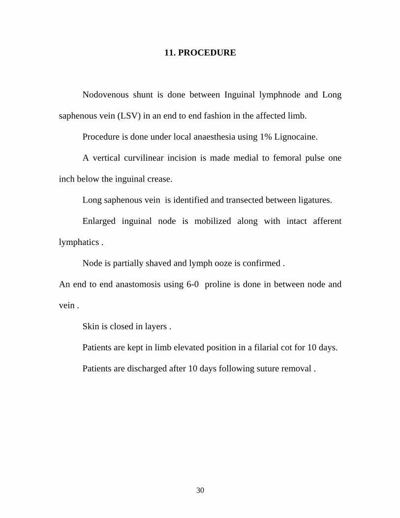
11. PROCEDURE
Nodovenous shunt is done between Inguinal lymphnode and Long
saphenous vein (LSV) in an end to end fashion in the affected limb.
Procedure is done under local anaesthesia using 1% Lignocaine.
A vertical curvilinear incision is made medial to femoral pulse one
inch below the inguinal crease.
Long saphenous vein is identified and transected between ligatures.
Enlarged inguinal node is mobilized along with intact afferent
lymphatics .
Node is partially shaved and lymph ooze is confirmed .
An end to end anastomosis using 6-0 proline is done in between node and
vein .
Skin is closed in layers .
Patients are kept in limb elevated position in a filarial cot for 10 days.
Patients are discharged after 10 days following suture removal .
31
Various grades of Lower Limp Filarial Lymphedema
Inguinal Node
Long Saphenous Vein

32
LSV Divided
Nodo Venous shunt

33
PATIENTS KEPT IN SPECIAL FILARIAL COTS

34
35

36
PRE OPERATIVE LYMPHOSCINTIGRAPHY
37
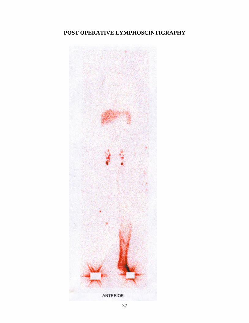
POST OPERATIVE LYMPHOSCINTIGRAPHY

38
12.METHODS
Patients were clinically evaluated using
a) Limb circumference measurement( size ).
b) volume measurement.
(A) LIMB CIRCUMFERENCE MEASUREMENT( SIZE ):
The lymphedematous limb is measured at standard sites at periodic
intervals .
The normal limb measurements at same sites are taken as reference .
SITE -I : 10 cm from tip of great toe proximally over dorsum of foot.
SITE -II : 12 cm proximal to the dorsoplantar skin junction of the foot in
the medial aspect.
SITE -III : 20 cm proximal to the dorsoplantar skin junction of the foot the
medial aspect.
SITE -IV : 30 cm proximal to the dorsoplantar skin junction of the foot the
medial aspect.

39
SITE V : 60cm proximal to the dorsoplantar skin junction of the foot the
medial aspect.
Measurements were done preoperatively once and postoperatively in
the frequency of 10 days ,3 months ,6 months and 1 year
( B ) LEG VOLUME MEASUREMENTS:
Volume reduction of the leg is measured by water displacement in a
special Drum.
The volume of water displaced by normal limb is taken as reference
value.
Volume displaced by affected limb is measured.
Difference between the two is approximately calculated as
lymphedema volume.
Measurements were done once preoperatively and at regular intervals
postoperatively in both limbs.
The difference between volume displacement by the diseased limb and
normal limb is recorded.

40
LEG VOLUME MEASURING DRUM
PNEUMATIC PRESSURE PUMP

41
13.OBSERVATION
General Characters of Study Group
100 patients of various grades of lower limb filarial lymphedema were
included in the study.
Among these 60 were grade I & II and another 40 were grade III &
IV.
30 patients of grade I & II with enlarged inguinal lymphnodes were
taken up for nodovenous shunt in addition to routine non surgical methods
were taken for study.
Another 30 patients of grade I and II without enlarged inguinal nodes
managed with non surgical methods were kept as control group.
20 patients of grade III & IV were taken up as study group and equal
number of 20 patints were taken as control group randomly.
42
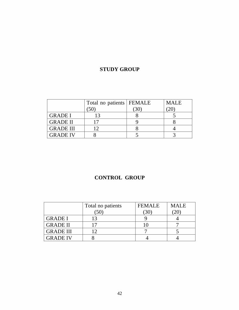
STUDY GROUP
Total no patients (50)
FEMALE (30)
MALE (20)
GRADE I 13 8 5 GRADE II 17 9 8 GRADE III 12 8 4 GRADE IV 8 5 3
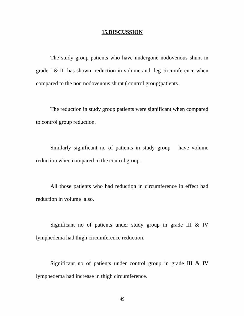
CONTROL GROUP
Total no patients (50)
FEMALE (30)
MALE (20)
GRADE I 13 9 4 GRADE II 17 10 7 GRADE III 12 7 5 GRADE IV 8 4 4

43
0
5
10
15
20
NO
OF
PATI
ENTS
MALE FEMALE
GRADE I AND II---STUDY GROUP
0
5
10
15
20
NO
OF
PATI
ENTS
FEMALE MALE
GRADE I AND II--CONTROL GROUP

44
02468
101214
NO
OF
PATI
ENTS
FEMALE MALE
GRADE III AND IV - STUDY GROUP
0
2
4
6
8
10
12
NO
OF
PATI
ENTS
FEMALE MALE
GRADE III AND IV -CONTROL GROUP

45
14. RESULTS OF THE STUDY
MEASUREMENTS IN GRADE I & II
In grade I & II patients there was reduction in size and volume in
both control and study group
In study group there was volume reduction in 17 patients
6 patients showed increase in volume measurement
7 patients showed no change in volume measurement
In control group there was volume reduction in 7 patients
19 patients showed increase in volume measurement
4 patients showed no change in volume measurement

46
DATA OF CHANGES IN VOLUME MEASUREMENT IN GROUP I AND II PATIENTS
CHANGES IN MEASUREMENT
STUDY GROUP (no 30)
CONTROL GROUP (no 30)
Decreased
17 7
Increased
6 19
No change
7 4
DATA OF CHANGES IN CIRCUMFERENCE MEASUREMENT AT THIGH MEASUREMENT IN GROUP III AND IV PATIENTS
CHANGES IN MEASUREMENT
STUDY GROUP (no 30)
CONTROL GROUP (no 30)
Decreased
13 2
Increased
7 17
No change
2 1

47
0
5
10
15
20
STUDY GROUP CONTROL GROUP
DATA OF CHANGES IN VOLUME AND CIRCUMFERENCE MEASUREMENT IN GROUP I AND II
Decreased Increased No change
0
5
10
15
20
STUDY GROUP CONTROL GROUP
DATA OF CHANGES IN CIRCUMFERENCE MEASUREMENT AT THIGH (60 cm) IN GRADE III AND IV
Decreased Increased No change

48
SIZE MEASURMENT IN GRADE I & II (20 cms)
In the study group 17 patient showed reduction in circumference
6 patient showed increase in circumference
7 patient showed no change in circumference
In the control group 7 patient showed reduction in circumference
19 patient showed increase in circumference
4 patient showed no change in circumference
SIZE MEASURMENT IN GRADE III & IV
In the study group 13 patients showed reduction in circumference
7 patientsshowed increase in circumference
2 patients showed no change in circumference
In the control group 2 patients showed reduction in circumference
17 patients showed increase in circumference
1 patients showed no change in circumference
49
15.DISCUSSION
The study group patients who have undergone nodovenous shunt in
grade I & II has shown reduction in volume and leg circumference when
compared to the non nodovenous shunt ( control group)patients.
The reduction in study group patients were significant when compared
to control group reduction.
Similarly significant no of patients in study group have volume
reduction when compared to the control group.
All those patients who had reduction in circumference in effect had
reduction in volume also.
Significant no of patients under study group in grade III & IV
lymphedema had thigh circumference reduction.
Significant no of patients under control group in grade III & IV
lymphedema had increase in thigh circumference.

50
16.CONCLUSION
The evaluation of results in control and study group shows volume
reduction in the study group who underwent nodovenous shunt in addition to
all other measures followed in control groups is very significant.
Filarial lymphedema is a common surgical problem around
Thanjavur.
In order to decompress the overloaded lymphatic system ,
lymphonodovenous shunts and drainage procedures are performed.
The primary pathology in filarial lymphedema being in lymph nodes
( regional inguinal ) and the stasis in distal level is more a secondary
effect.
By passing the lymph to the venous blood by nodovenous shunt
which done without microscope and which has produced consistant
reduction in all stages of filarial lymphedema , , Nodovenous seems to be
51
most appropriate surgical procedure suited for the magnitude of the problem
and resources available.
The initial reduction in thigh circumference of in grade III and IV is
good and the resultant skin laxity is utilized for the excisional procedures
followed here .

52
17.BIBLIOGRAPHY
1. Altofer j.,hedinger,c and clodius, :Light microscopic investingation of
extremities of dogs with experimentl chronic lymphostasis. Folia.Angiol,
25;141,1977.
2. Clodius,L:and Gibson T; Lymphedema.In Rob,c.,and
smith,R(Eds):Operative surgery 3rd editionButterworths,1979.
3. Edwards,J.M.,and kimonth,J.B:Lymphovenous shunts in man;Br m j
4;579.1969.
4. Olszewski,W:surgical lymphovenous shunts for treatment of filarial
lymphedema.In Clodius,L.(Ed):lymphedema,Stuttgart,Thieme,1977.
5. Olszewski,W.,and Neilubowicz,J;Surgical lymphaticovenous
communication in treatment of stasis. 43rd congress of polish.1966.
6. Yamada,Y.:Study on lymphatic venous anastomosis in lymphedema
.nagoya med. J.97:567,1984.
7. Jamal S,Lympho nodovenous shunt and treatment of filarial
elephantiasis.In weissleder,H Bartos,theClodius L,and Malek P(eds)
Progress in lymphology proceedings of the 7th international congress of
lymphology.

53
8. Evaluation of different surgical procedures in filarial lymphedema of
lower extremities. Indian medical journal.1991 may 89(5)127-9.Dhanapal.,
mukerjee.,Patna S.K.
9. Factors influencing response to LVS in Filarial
lymphedema.Natt.med.Jr.1999 Mar-Apr 12(2) ,55-8.
10. Homans J.The treatment of elephantiasis of the legs.N.Engl.J
med.215:1099,1936
11. Sedlacek .J: Lymphovenous shunt as supplementary treatmett of
elephantiasis of lower limbs.
12. Concesses document of the international society of lymphology executive
committee.Diagnosis and treatment of pheripheral lymphedema.In
proceedings of lymphology society, India 1st annual conference, Thanjavur
1996.
13. V.Fox,Romgnoli and Ribaldone, Progress in lymphology.Pg.437.
14. Cong.huang,ruquittu,ZongZhao liu, in plastic reconstructive surgery
76:77.
15. Mario Degmi and Pavel Nunes Progress in lymphology XI Pg 481.

54

WORK SHEET Gr. I& II
S.
No Name Grade
Circumference At 20 cm
[Leg]
Normal Limb
Affected Limb
10 Days
1 Month
6
Months
1 Year
Dif At
1 year Pre-Op Post - Op
1. Pattammal 63/F
II 26 30 28 30 32 35 5 cm
Volume 6.750 7250 69500 7100 7500 8200 +950ml
2. Sannasi 55/M
I 20 8 30 30 31 30 -
Volume 3300 3500 3200 3450 3500 3520 -
3. Danalakshmi 28/F
II 26 32 9 30 30 30 -2cm
Volume 3100 3700 3250 3100 3150 3150 -600ml
4. Manokaran 50/M
II 24 38 30 31 31 32 -6cm
Volume 3750 5100 3900 4000 4050 4000 -1100ml
5. Chinadurai 18/M
II 23 28 25 26 26 26 -2cm
Volume 4250 6100 4700 3000 5100 5100 -1000ml
6. Murugesan 40/M
II 22 27 23 29 30 30 +3cm
Volume 4300 8550 8400 8500 8400 9000 +450
7. Thulasiammal 55/F
I 22 27 21 22 20 21 -6cm
Volume 4300 6600 4700 4700 4900 5200 -600ml
8. Rajaraman 28/M
I 22.5 28 22 23 24 24 -4cm
Volume 4325 6925 5200 5000 5200 5500 -1425ml
9. Geetha 14/F
II 30 34 35 31 31 37 -2.5cm
Volume 4100 6200 5700 6010 6050 6100 -100ml
10. Md. Jinna 38/M
II 37.5 42 38 39 40 41 -1cm
Volume 7150 9900 7900 8200 8100 9000 -900ml
11. Renganayaki 29/F
I 27 32 28 29 30 20 -2cm
Volume 4750 5050 5000 5100 5100 5150 +100ml
12. Kumaravel 25/M
I 29 34 30 30 30 32 -3cm
Volume 5100 6700 5300 5200 5100 5700 -900
13.
Muniyammal 60/F
II 40 45 42 43 42.5 43 -3cm
Volume 6150 8100 6750 6900 6900 6800 1300
14.
Periasami 48/M
I 25 29 26 28 30 32 +3
Volume 4600 5300 4750 5730 5750 5900 +600
15.
Sampath 40/M
29 36 31 35 35 36 -
Volume 3900 4800 4100 4700 4750 4750 -50

S.
No Name Grade
Circumference At 20 cm
[Leg]
Normal Limb
Affected Limb
10 Days
1 Month
6 Months
1 Year Dif
At 1 year Pre-Op Post - Op
16. Gayathri 41/F
II 22 26 21 22 22 21 -5 Volume 4300 7100 4500 4650 4700 5025 -2100
17. Naina mohammed 26/M
I 26 31 27 29 29 29 -2 Volume 2750 3200 2900 3050 3150 3000 -200
18. Ansa mary 52/F
I 29 34 30 31 31 31 -3 Volume 3050 3900 3100 3150 3150 3100 -800
19. Fathima 40/F
I 30 34 31 36 38 38 +4 Volume 4150 6060 4550 4600 6250 6500 +500
20. Anil 24/M
II 27 31 28 30 29 29 -2 Volume 3750 4440 3900 3950 4100 4000 -400
21. Seetha 39/F
I 30 34 31 32 31 31 -3 Volume 4100 6200 4250 4300 4400 4450 -1750ml
22. Suseela 50/F
II 24 29 26 27 29 29 - Volume 30520 4700 4100 4250 4650 4650 -50
23. Malarkodi 38/F
I 27 28 25 26 22 27 -1 Volume 4250 4600 4200 4150 4100 4150 -450
24. Uma maheswari 45/F
II 25 29.5 27 28 31 32 +2.5 Volume 4650 6420 4900 6200 6700 6950 +1500
25. Sri devi 32/F
I 34 39 37 39 39 39 -- Volume 7000 9300 9100 9350 9300 9300 --
26. Kousalya 52/F
II 29.5 33 30 30 31 31 -2 Volume 6700 7550 6800 7100 7100 7150 -400
27. Paul nithyanandam 52/M
II 25.5 29 28 29 29 29 -- Volume 5500 6200 5950 5900 5100 6190 --
28. Roobini 18/F
I 25 28 24 26.5 26 27 -1.5
Volume 4500 4900 4200 5100 5050 5850 +950 29. Saithoon bee
60/F II 24 28 24 26 25 27 -1
Volume 4650 5250 4700 5000 5950 5150 -100 30.
Muthupandi 42/M
II 27 34 32 31 32 33 -1 Volume 4500 5900 5250 5300 5200 5200 -700

S.
No Name Grade
Circumference At 20 cm
[Leg]
Normal Limb
Affected Limb
10 Days
1 Month
6 Months
1 Year Dif
At 1 year Pre-Op Post - Op
31. Vellai samy 43/M
II 38 43 41 40 38 38 -5 Volume 6350 8050 7250 6900 6700 6700 -1350
32. Kandan 58 /M
I 30 35 32 33 35 35.4 -- Volume 3500 3950 3800 3900 3950 3900 --
33. Amirtham 32/F
II 26 30 30 27 28 27 -3 Volume 3100 3850 3450 3550 3600 3600 -250
34. Meena 42 / F
II 23 27.5 25 27 29 31 +3.5 Volume 4350 5700 5100 5750 5900 6050 +350
35. Kalarani 39/F
I 27 30 28 29 31 34 +4 Volume 4600 5300 4750 5700 5600 5800 +600
36. Mariya selvam 42 /F
II 23 28 26 28 27 28 -- Volume 4150 5800 5050 5800 5850 5750 -50
37. Banumathi 35 /F
I 27 30 29 31 32 32 +2 Volume 4160 4750 4250 4900 5100 5150 +400
38. Geetharani 39 /F
I 25 28 27 28 28 29 +1 Volume 4500 5100 4900 4950 5200 5350
39. Abdul samad 57 /M
II 30 34.5 30.5 32 34 34 +0.5 Volume 4450 5900 4550 5000 5100 5100 -800
40. Chinnaponnu 38 /F
II 38 41 40 42 41 43 +2 Volume 7200 9500 9100 9900 9800 10200 +700
41. Muthu 49 /M
II 26 30 27 28 28 28 -2 Volume 5500 6200 5700 5950 5950 5600 -200
42. Chandra 22 /F
II 25 28.5 27 28 30 32 +3.5 Volume 4500 5000 4700 5050 5700 5950 +950
43.
Govindaraj 50 /M
II 24 29 28 29 33 34 +5
Volume 4050 5250 4950 5300 5950 6100 +850 44.
Esther 38 /F
I 33 37 38 40 40 41 +3 Volume 4325 4750 4900 5150 5100 5200 +450
45.
Muthulaksmi 42 /F
II 31 35.5 30 31 33 33 -1.5 Volume 7150 8700 7050 7250 8100 8500 -200
S.
No Name Grade
Circumference At 20 cm
[Leg]
Normal Limb
Affected Limb
10 Days
1 Month
6 Months
1 Year Dif
At 1 year Pre-Op Post - Op
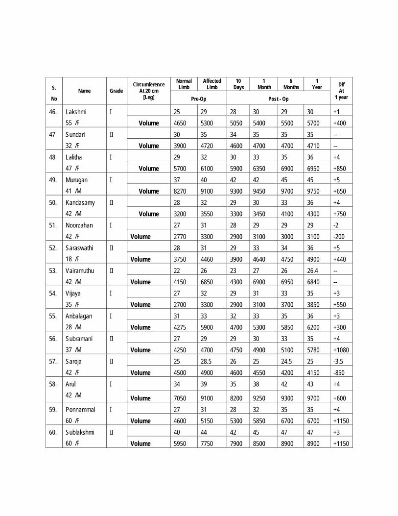
46. Lakshmi 55 /F
I 25 29 28 30 29 30 +1 Volume 4650 5300 5050 5400 5500 5700 +400
47
Sundari 32 /F
II 30 35 34 35 35 35 -- Volume 3900 4720 4600 4700 4700 4710 --
48 Lalitha 47 /F
I 29 32 30 33 35 36 +4 Volume 5700 6100 5900 6350 6900 6950 +850
49. Murugan 41 /M
I 37 40 42 42 45 45 +5 Volume 8270 9100 9300 9450 9700 9750 +650
50. Kandasamy 42 /M
II 28 32 29 30 33 36 +4 Volume 3200 3550 3300 3450 4100 4300 +750
51. Noorzahan 42 /F
I 27 31 28 29 29 29 -2 Volume 2770 3300 2900 3100 3000 3100 -200
52. Saraswathi 18 /F
II 28 31 29 33 34 36 +5 Volume 3750 4460 3900 4640 4750 4900 +440
53. Vairamuthu 42 /M
II 22 26 23 27 26 26.4 -- Volume 4150 6850 4300 6900 6950 6840 --
54. Vijaya 35 /F
I 27 32 29 31 33 35 +3 Volume 2700 3300 2900 3100 3700 3850 +550
55. Anbalagan 28 /M
I 31 33 32 33 35 36 +3 Volume 4275 5900 4700 5300 5850 6200 +300
56. Subramani 37 /M
II 27 29 29 30 33 35 +4 Volume 4250 4700 4750 4900 5100 5780 +1080
57. Saroja 42 /F
II 25 28.5 26 25 24.5 25 -3.5 Volume 4500 4900 4600 4550 4200 4150 -850
58.
Arul 42 /M
I 34 39 35 38 42 43 +4
Volume 7050 9100 8200 9250 9300 9700 +600 59.
Ponnammal 60 /F
I 27 31 28 32 35 35 +4 Volume 4600 5150 5300 5850 6700 6700 +1150
60.
Sublakshmi 60 /F
II 40 44 42 45 47 47 +3 Volume 5950 7750 7900 8500 8900 8900 +1150

CIRCUMFERENCE OF THIGH AT 60 CM IN GRADE III AND IV
STUDY GROUP
Sl no
Name Grade Normal limb
Affected Limb
10 Days
1 Month
6 Month
1 Year
Difference at 1 year
Pre op Post op 1 Arumugam 70/M IV 42 45 44 45 46 46 +1 2 Meenal 35/F III 38 43 40 39 39 39 -4 3 Balu 26/M III 31 43 42 45 46 46 +3 4 Annamalai 32/F IV 40 53 51 50 50 49 -4 5 Regina 65/F III 37 42 41 43 45 46 +4 6 Vasantha 55/M IV 37 39 37 36 38 38 -1 7 Gurumurthy 42/M III 48 50 50 51 53 53 +3 8 Jothi 38/F III 48 50 48 49 49 49 -1 9 Sugavanam 38/F IV 40 43 41 41 43 43 --
10 Sundari 70/F III 42 45 42 43 43 43 -2 11 Jayakumar 31/M IV 42 43 42 41 40 42 -1 12 Shanthakumari 42/F IV 38 40 39 40 41 41 +1 13 Balasubramanian
50/M III 44 46 41 42 43 43 -3
14 Bnanumathi 55/F III 55 63 59 60 58 60 -3 15 Akila 37/F III 45 48 46 47 48 47 -1.5 16 Sasikumari 32/F IV 38 40 40 39 38 40 -- 17 Lakshmi 48/F III 45 48 47 48 48 49 +1 18 Jayamary 60/F III 35 37 37 36 39 39 +2 19 Amsavalli 42/F III 42 45 43 42 44 44 -1 20 Murugadass 37/M IV 44 46 44 45 45 44 -1

CIRCUMFERENCE OF THIGH AT 60 CM IN GRADE III AND IV
STUDY GROUP
Sl no
Name Grade Normal limb
Affected Limb
10 Days
1 Month
6 Month
1 Year
Difference at 1 year
Pre op Post op
1 Pushpa 42/F III 52 59 58 60 61 63 +4
2 Arokia dass 58/M III 42 44 43 45 45 46 +2
3 Anusuya 36/F IV 34 38 36 38 41 41 +3
4 Ramalingam 67/M
IV 49 52 52 53 54 56 +4
5 Revathy 37/F III 37 42 40 42 43 45 +3
6 Sundarrajan 53/M III 47 49 48 49 51 51 +2
7 Abdul sathar 51/M
IV 44 48 45 44 45 46 +2
8 Pappathi 60/F III 41 46 45 46 47 48 +2
9 Dhanalakshmi 48/F
III 41 46 45 47 47 47 +1
10 Thulasiram 75/M III 45 48 47 47 48 48 ---
11 Sridhar 56/M III 45 46 46 42 47 48 +2
12 Rajkumar 43/M IV 44 50 45 45 45 46 -4
13 Parvatham 50/F IV 43 47 46 49 49 51 +4
14 Veerammal 21/F IV 38 43 40 42 43 43 --
15 Mumtaj begam 45/F
III 41 42 43 44 44 45 +2
16 Rengarajan 55/M IV 58 61 61 65 65 67 +6
17 Jamuna 38/f III 40 42 41 44 44 45 +3
18 Natarajan 42/M III 44 46 46 45 48 49 +3
19 Logambal 28/M IV 42 44 44 45 46 47 +3
20 Krishnaveni 32/F IV 38 40 39 40 41 42 +2