Effect of the herbicide Avans 330 SL on the liver pathomorphology of clinically healthy carp...
14
EFFECT OF THE HERBICIDE AVANS 330 SL ON THE LIVER PATHOMORPHOLOGY OF CLINICALLY HEALTHY CARP (CYPRINUS CARPIO L.) AND CARP INFECTED BY ICHTHYOPHTHIRIUS MULTIFILIIS Józef Szarek*, Izabella Babiñska*, Monika Truszczyñska*, Ryszard Kolman**, Andrzej K. Siwicki**, Ireneusz M. Kowalski*, Joanna Wojtacka*, Halina Kolman*, Tadeusz Banaszkiewicz*, Krystyna A. Skibniewska* *University of Warmia and Mazury in Olsztyn, Poland **The Stanis³aw Sakowicz Inland Fisheries Institute in Olsztyn, Poland ABSTRACT. This study was conducted on fingerlings of carp, Cyprinus carpio L., that were either clinically healthy or infected with a natural invasion of the ciliate, Ichthyophthirius multifiliis. The fish were exposed for 96 h to Avans 330 SL in a concentration of 2 mg trimethylosulfonium glyphosate l -1 water in order to establish its effect on the morphological pattern of the liver. It was demonstrated that the most frequent morphological deviations, particularly in fish infected with the protozoa and bathed in water with the addition of Avans, included regressive lesions, such as parenchymatous, vacuolar degeneration, and focal necrosis. Less often in these cases, and even less often in carp infected with I. multifiliis or only exposed to Avans, were disorders observed in the circulation in the form of hyperaemia and minor extravasations. Changes were noted in the ultrastructures of the mitochondria and the endoplasmic reticulum. In addition, the results obtained indicate that ciliate invasion intensified the occurrence of morphological lesions in the livers of carp that were exposed to Avans 330 SL in comparison to fish that were free of this infection. Key words: CARP (CYPRINUS CARPIO), ICHTHYOPHTHIRIUS MULTIFILIIS, LIVER, AVANS 330 SL, GLYPHOSATE, MORPHOLOGICAL LESIONS IN THE LIVER, LIVER ULTRASTRUCTURE INTRODUCTION As an important element of the aquatic ecosystem, fish are exposed to pollution caused by man-made compounds known as xenobiotics. Indeed, herbicides that are widely used in agriculture constitute the most frequent source of extensive water pollu- tion (Alberdi et al. 1996, Sopiñska et al. 1995, 2000, ¯elazny 2002). Their harmful Archives of Polish Fisheries Vol. 14 Fasc. 2 169-182 2006 Arch. Pol. Fish. CORRESPONDING AUTHOR: Prof. dr hab. Józef Szarek, Uniwersytet Warmiñsko-Mazurski, Wydzia³ Medycyny Weterynaryjnej, Katedra Weterynaryjnej Ochrony Zdrowia Publicznego, ul. Oczapowskiego 13, 10-719 Olsztyn; Tel./Fax: +48 89 523 32 52; e-mail: [email protected]
Transcript of Effect of the herbicide Avans 330 SL on the liver pathomorphology of clinically healthy carp...
EFFECT OF THE HERBICIDE AVANS 330 SL ON THE LIVER
PATHOMORPHOLOGY OF CLINICALLY HEALTHY
CARP (CYPRINUS CARPIO L.) AND CARP INFECTED
BY ICHTHYOPHTHIRIUS MULTIFILIIS
Józef Szarek*, Izabella Babiñska*, Monika Truszczyñska*, Ryszard Kolman**,
Andrzej K. Siwicki**, Ireneusz M. Kowalski*, Joanna Wojtacka*, Halina Kolman*,
Tadeusz Banaszkiewicz*, Krystyna A. Skibniewska*
*University of Warmia and Mazury in Olsztyn, Poland
**The Stanis³aw Sakowicz Inland Fisheries Institute in Olsztyn, Poland
ABSTRACT. This study was conducted on fingerlings of carp, Cyprinus carpio L., that were either
clinically healthy or infected with a natural invasion of the ciliate, Ichthyophthirius multifiliis. The fish
were exposed for 96 h to Avans 330 SL in a concentration of 2 mg trimethylosulfonium glyphosate l-1
water in order to establish its effect on the morphological pattern of the liver. It was demonstrated that
the most frequent morphological deviations, particularly in fish infected with the protozoa and bathed
in water with the addition of Avans, included regressive lesions, such as parenchymatous, vacuolar
degeneration, and focal necrosis. Less often in these cases, and even less often in carp infected with I.
multifiliis or only exposed to Avans, were disorders observed in the circulation in the form of
hyperaemia and minor extravasations. Changes were noted in the ultrastructures of the mitochondria
and the endoplasmic reticulum. In addition, the results obtained indicate that ciliate invasion
intensified the occurrence of morphological lesions in the livers of carp that were exposed to Avans 330
SL in comparison to fish that were free of this infection.
Key words: CARP (CYPRINUS CARPIO), ICHTHYOPHTHIRIUS MULTIFILIIS, LIVER, AVANS 330
SL, GLYPHOSATE, MORPHOLOGICAL LESIONS IN THE LIVER, LIVER ULTRASTRUCTURE
INTRODUCTION
As an important element of the aquatic ecosystem, fish are exposed to pollution
caused by man-made compounds known as xenobiotics. Indeed, herbicides that are
widely used in agriculture constitute the most frequent source of extensive water pollu-
tion (Alberdi et al. 1996, Sopiñska et al. 1995, 2000, ¯elazny 2002). Their harmful
Archives
of Polish FisheriesVol. 14 Fasc. 2 169-182 2006
Arch.
Pol. Fish.
CORRESPONDING AUTHOR: Prof. dr hab. Józef Szarek, Uniwersytet Warmiñsko-Mazurski,Wydzia³ Medycyny Weterynaryjnej, Katedra Weterynaryjnej Ochrony Zdrowia Publicznego, ul.Oczapowskiego 13, 10-719 Olsztyn; Tel./Fax: +48 89 523 32 52; e-mail: [email protected]
impact is intensified by the phenomenon of accumulation that occurs in living organ-
isms and in their habitats, as well as by the condensation of xenobiotics in the food
chain, which poses the greatest danger to the environment (Ghosh and Konar 1983,
Gluth et al. 1985). The amount of pesticide that accumulates depends not only on its
concentration in the water and exposure time but also on skin status; skin injuries,
such as those produced by parasites, increase accumulation.
The adverse effect of herbicides on the aquatic environment and its organisms is
demonstrated by the occurrence of pathological lesions and immunosuppression
(Neškoviæ et al. 1996, Sopiñska et al. 1995, Kolman et al 2003). On the one hand, the
increased herbicidal pollution of water and soil has been observed lately, which has
prompted new efforts to examine their effects on widely understood ecosystems, pri-
marily fields, forests and meadows, and less often on aquatic ecosystems (Dunier and
Siwicki 1993, Sopiñska et al. 2000). On the other hand, there are insufficient studies
concerning the pathomorphology of the internal organs of fish exposed to small
amounts of herbicides.
Research into glyphosate toxicity (the active substance in Avans 330 SL-AV) has
demonstrated its moderate toxicity in mammals and fish (toxicity class IV). However, it
can have a negative effect on the aquatic environment and can manifest its toxic charac-
teristics in an indirect way by becoming a component of the fauna and flora of aquatic
ecosystems (Alberdi et al. 1996, Neškoviæ et al. 1996). Studies that have focused on the
effects of glyphosate itself and have demonstrated its toxic effects on aquatic organisms
and fish are relatively few (and those that examine preparations containing it are even
rarer) (Neškoviæ et al. 1996, Raszka et al. 1998, Sopiñska et al. 2000, Szarek et al.
2000a, b). Some studies have found that glyphosate in the concentrations used in agri-
culture can have a toxic impact on hepatocytes and renal duct cells in fish (Studnicka
and Siwicki 1997, Szarek et al. 1997, 2000a, b).
Many factors causing disorders in the homeostasis of organisms (bacterial, viral, or
parasitic diseases) can result in the increased susceptibility of specimens to xenobiotics
and can intensify their effects. For some years in Poland there has been an increase in
the frequency of the occurrence of ichthyophthiriasis (ICH). This phenomenon has
been generated by limiting the scope and frequency of such maintenance works as
mowing and desludging ditches in the area of fishponds (Róg 2002). Ichthyophthiriasis
is caused by a parasite known as Ichthyophthirius multifiliis, also referred to as a cili-
170 J. SZAREK et al.
ate, and it is considered a typical “transport disease”. A mature form of the ciliate para-
sitizes almost all freshwater fish and feeds on their tissues. When there is significant
parasite density the invasion quickly spreads and reaches significant intensification
(Maki et al. 2001, Kinnunen et al. 2005).
The preceding data indicate that glyphosate has various, negative effects on living
organisms, including fish. The aim of the study was to define the effect of so-called toxi-
cally safe low level concentrations of glyphosate on the liver morphology in common
carp, Cyprinus carpio L., with healthy skin and in those injured by I. multifiliis.
MATERIAL AND METHODS
The research was conducted on carp juveniles weighing from 81 to 98 g. The fish
were obtained from two breeding centers: W¹sosze Fish Farm (central Poland) and the
Experimental Fish Farm of Freshwater Fishery in ¯abieniec, which is a branch of the
Stanis³aw Sakowicz Inland Fisheries Institute in Olsztyn, Poland (IFI Olsztyn; central
Poland). The fish were fed granulated feed (Aller 37/12) produced by Aller Aqua,
Poland. The feed chemical composition was 37% protein, 31% carbohydrate, 12% fat,
7% ash, and 4% fiber and its energetic value was 19.5 MJ kg-1. Prior to the experiment,
the carp were acclimated to laboratory conditions for two weeks and after this period
they were weighed.
The study was conducted as two separate experiments: A – on clinically healthy
carp free of I. multifiliis and B – on carp infected with the protozoa (permission from
Local Ethical Commission No. 31/N). The carp from group B did not exhibit any devia-
tion from the norm either immediately after transport or throughout the acclimatization
period. The histological skin lesions and the presence of parasites on the skin of the fish
suggested a ciliate invasion of medium intensity that had been present for a few days
(Szarek et al. 2006). In each experiment, the fish were divided into two equal groups (N
= 10): A1 and B1 – control groups, A2 and B2 – exposed to Avans 330 SL (AV) at a con-
centration of 2 mg trimethylosulfonium glyphosate l-1 of water. The examined prepara-
tion, in a dose calculated as per active substance, was thoroughly mixed with a small
amount of tap water and then introduced into the tanks where the fish had been placed.
The AV preparation used in the experiments was produced by Zeneca Agrochemicals
of Great Britain. The active substance is trimethylosulfonium glyphosate with
EFFECT OF THE HERBICIDE AVANS 330 SL ON THE LIVER... 171
a three-day half-life, 360 g l-1 of the preparation. While determining its concentration,
the following factors were taken into consideration: the presence of the examined active
substance in the bodies of the fish (Banaszkiewicz 2003); the results of studies that
indicated the so-called toxically safe herbicide level causes pathology in fish (Szarek et
al. 2000a, b); data reported by other authors (Ghosh and Konar 1983, Demael et al.
1990, Dunier and Siwicki 1993); direct penetration of glyphosate to water reservoirs,
including the shallow fishponds of fish populations (Aalbers 1996, Piska and Waghray
1997).
The fish were held in 200 l tanks under similar environmental conditions: water
temperature 18-19°C; oxygen level 7.5-8.0 mg.l-1; pH 7.5-8.5. Moreover, levels of total
ammonia nitrogen (TAN = NH4+-N + NH3-N) did not exceed 0.2 mg TAN l-1. The fish
were not fed for the duration of the experiment (i.e., 96 hours). The study also included
observations of carp behavior. After the carp had been bathed for 96 hours in the water
to which AV had been added, they were removed from the tanks and placed in 10 l
tanks containing water with the addition of Propiscin (a 0.2% etomidate solution manu-
factured by IFI Olsztyn) and anesthetized.
Macroscopic examinations were conducted directly after the fish had been sacri-
ficed. At the same time, liver sections were collected for ultrastructure assessment and
for microscopic analysis. The liver sections for microscopic analysis were fixed in 5%
neutralized formalin and after dehydration in a series of alcohols and acetone, they
were sealed in paraffin blocks. Microscopic fragments were stained with haematoxylin
and eosin (Bancroft and Cook 2000) and the PAS method according to McManus
(1948) was applied in order to establish the level and arrangement of polysaccharides.
The polysaccharide content in the liver was determined by taking into consideration
Pearse’s (1968) indications and the semiquantitative assessment provided by Szarek et
al. (1985). Cryostat fragments of heptopancreas were also stained with Sudan III, using
the Lillie Ashburn method in order to expose fat (Bancroft and Cook 2000). The liver
for the ultrastructural examination was fixed in glutaraldehyde in a phosphatic buffer
of pH 7.2 and sealed in Epon 812. Semi-thin sections were stained according to the
method of Levis and Knight (1977); the appropriate place for preparing ultrathin sec-
tions was determined under a light microscope. Structural analysis was conducted
using an Opton 900 PC electron microscope (Germany).
172 J. SZAREK et al.
RESULTS AND DISCUSSION
In experiment A and in the control group of experiment B (group B1), it was
established clinically that the carp behaved normally during the experiment and
responded distinctly to external stimuli. However, the fish infected with ciliates and
exposed to AV (B2) behaved normally only on day one of the experiment. On day two, the
liveliness and excitability of the carp in this group increased slightly (the fish exhibited
violent movements without any external stimuli), but on days three and four heaviness
was observed in their movements. The results of clinical observations and especially the
comparison of the behavior of the carp from groups A2 and B2 indicate that the intake of
AV increased due to injuries to the carp gills and skin. Macroscopic examinations of
fingerling from all groups revealed their correct morphological structure.
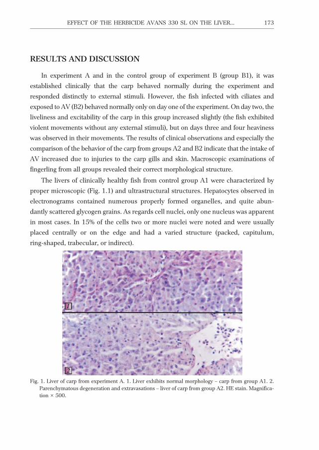
The livers of clinically healthy fish from control group A1 were characterized by
proper microscopic (Fig. 1.1) and ultrastructural structures. Hepatocytes observed in
electronograms contained numerous properly formed organelles, and quite abun-
dantly scattered glycogen grains. As regards cell nuclei, only one nucleus was apparent
in most cases. In 15% of the cells two or more nuclei were noted and were usually
placed centrally or on the edge and had a varied structure (packed, capitulum,
ring-shaped, trabecular, or indirect).
EFFECT OF THE HERBICIDE AVANS 330 SL ON THE LIVER... 173
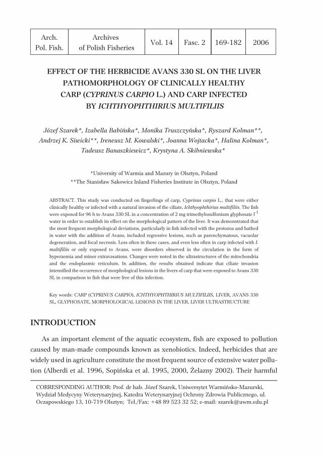
Fig. 1. Liver of carp from experiment A. 1. Liver exhibits normal morphology – carp from group A1. 2.Parenchymatous degeneration and extravasations – liver of carp from group A2. HE stain. Magnifica-tion × 500.
The microscopic examination of liver in carp from group A2 revealed that there was
parenchymatous degeneration at various intensification levels in the majority of the fish
(Fig. 1.2). In one case, parenchymatous degeneration was accompanied by vacuolar
degeneration. However, hyperaemia was noted more frequently and was usually
accompanied by blood extravasations covering small areas (Fig. 1.2). In most of the
fish, the cells of the endothelium of blood vessels and sinuses demonstrated a normal
structure, and only in two carp did they show swelling. Occasionally, there were
changes in behavior of the stellate cells, consisting of their proliferation or hypertrophy.
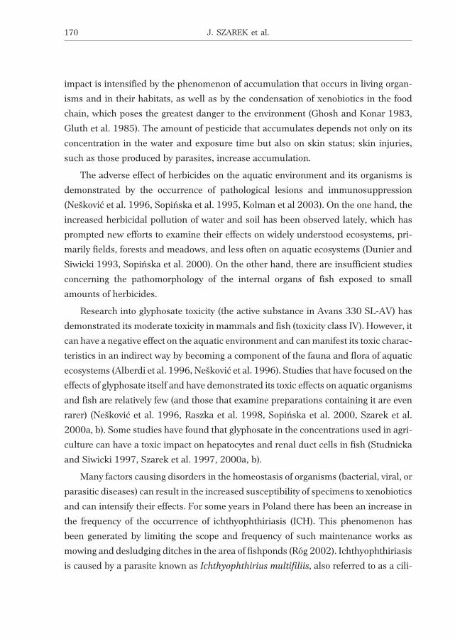
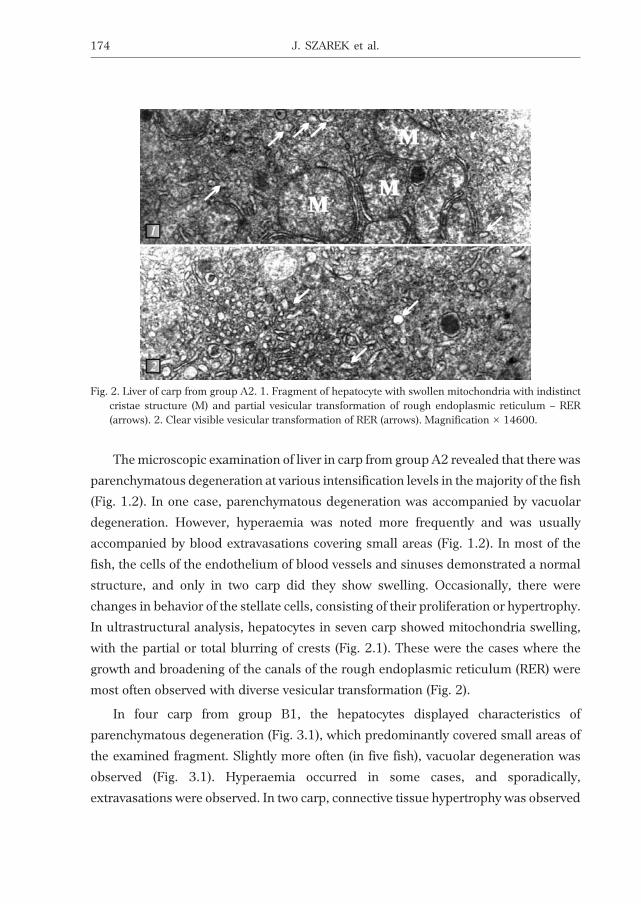
In ultrastructural analysis, hepatocytes in seven carp showed mitochondria swelling,
with the partial or total blurring of crests (Fig. 2.1). These were the cases where the
growth and broadening of the canals of the rough endoplasmic reticulum (RER) were
most often observed with diverse vesicular transformation (Fig. 2).
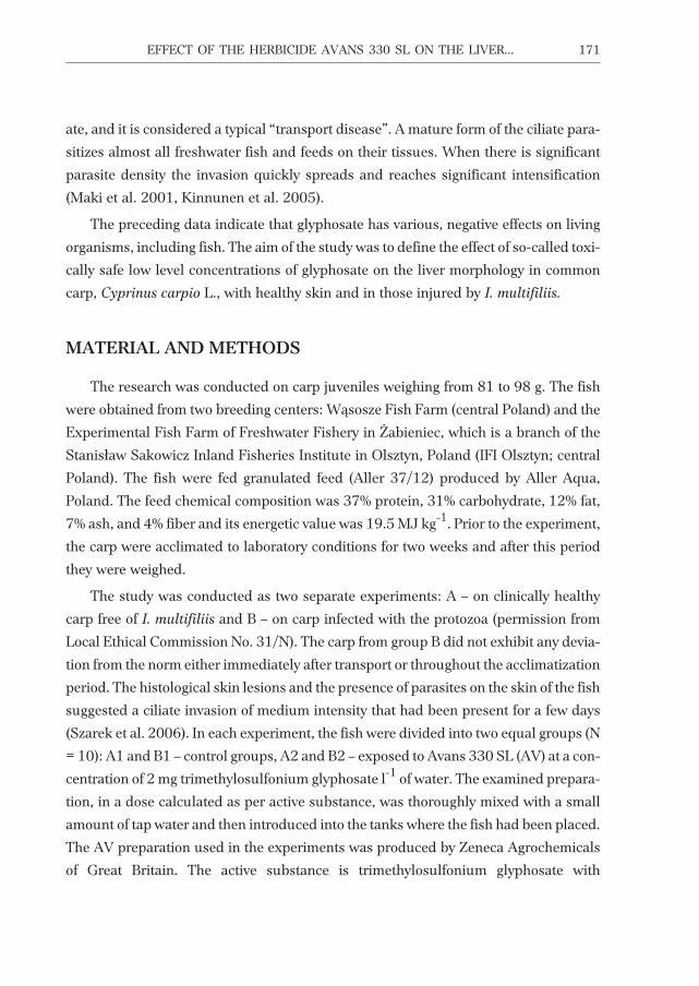
In four carp from group B1, the hepatocytes displayed characteristics of
parenchymatous degeneration (Fig. 3.1), which predominantly covered small areas of
the examined fragment. Slightly more often (in five fish), vacuolar degeneration was
observed (Fig. 3.1). Hyperaemia occurred in some cases, and sporadically,
extravasations were observed. In two carp, connective tissue hypertrophy was observed
174 J. SZAREK et al.
Fig. 2. Liver of carp from group A2. 1. Fragment of hepatocyte with swollen mitochondria with indistinctcristae structure (M) and partial vesicular transformation of rough endoplasmic reticulum – RER(arrows). 2. Clear visible vesicular transformation of RER (arrows). Magnification × 14600.
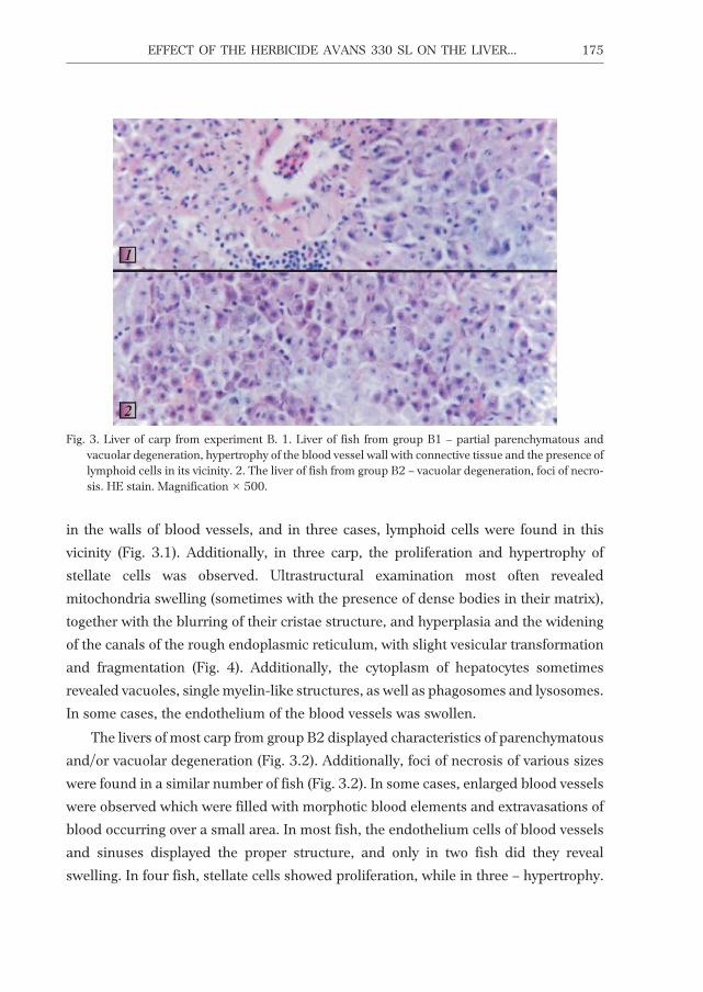
in the walls of blood vessels, and in three cases, lymphoid cells were found in this
vicinity (Fig. 3.1). Additionally, in three carp, the proliferation and hypertrophy of
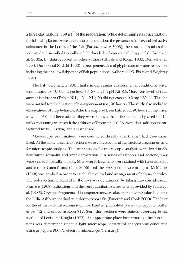
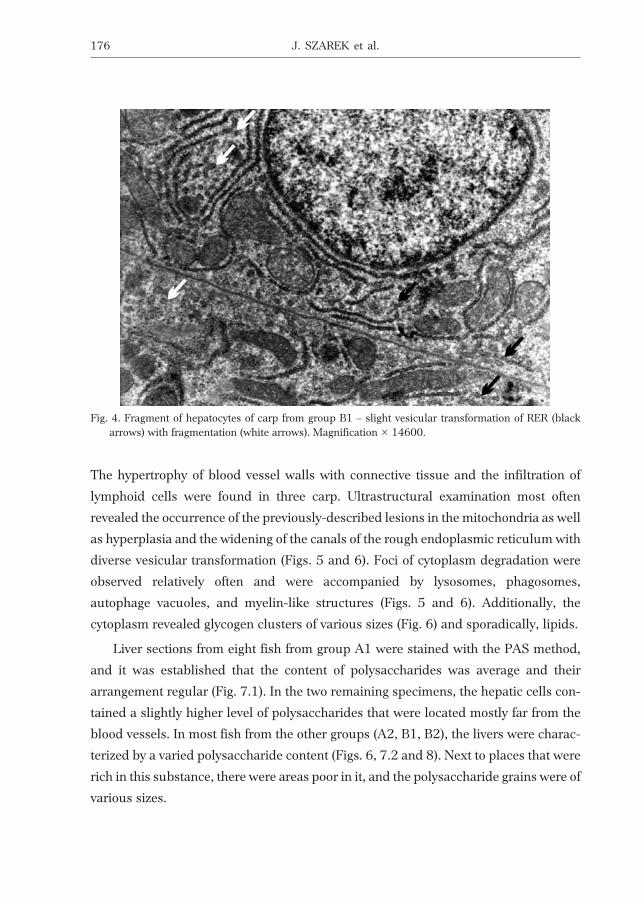
stellate cells was observed. Ultrastructural examination most often revealed
mitochondria swelling (sometimes with the presence of dense bodies in their matrix),
together with the blurring of their cristae structure, and hyperplasia and the widening
of the canals of the rough endoplasmic reticulum, with slight vesicular transformation
and fragmentation (Fig. 4). Additionally, the cytoplasm of hepatocytes sometimes
revealed vacuoles, single myelin-like structures, as well as phagosomes and lysosomes.
In some cases, the endothelium of the blood vessels was swollen.
The livers of most carp from group B2 displayed characteristics of parenchymatous
and/or vacuolar degeneration (Fig. 3.2). Additionally, foci of necrosis of various sizes
were found in a similar number of fish (Fig. 3.2). In some cases, enlarged blood vessels
were observed which were filled with morphotic blood elements and extravasations of
blood occurring over a small area. In most fish, the endothelium cells of blood vessels
and sinuses displayed the proper structure, and only in two fish did they reveal
swelling. In four fish, stellate cells showed proliferation, while in three – hypertrophy.
EFFECT OF THE HERBICIDE AVANS 330 SL ON THE LIVER... 175
Fig. 3. Liver of carp from experiment B. 1. Liver of fish from group B1 – partial parenchymatous andvacuolar degeneration, hypertrophy of the blood vessel wall with connective tissue and the presence oflymphoid cells in its vicinity. 2. The liver of fish from group B2 – vacuolar degeneration, foci of necro-sis. HE stain. Magnification × 500.
The hypertrophy of blood vessel walls with connective tissue and the infiltration of
lymphoid cells were found in three carp. Ultrastructural examination most often
revealed the occurrence of the previously-described lesions in the mitochondria as well
as hyperplasia and the widening of the canals of the rough endoplasmic reticulum with
diverse vesicular transformation (Figs. 5 and 6). Foci of cytoplasm degradation were
observed relatively often and were accompanied by lysosomes, phagosomes,
autophage vacuoles, and myelin-like structures (Figs. 5 and 6). Additionally, the
cytoplasm revealed glycogen clusters of various sizes (Fig. 6) and sporadically, lipids.
Liver sections from eight fish from group A1 were stained with the PAS method,
and it was established that the content of polysaccharides was average and their
arrangement regular (Fig. 7.1). In the two remaining specimens, the hepatic cells con-
tained a slightly higher level of polysaccharides that were located mostly far from the
blood vessels. In most fish from the other groups (A2, B1, B2), the livers were charac-
terized by a varied polysaccharide content (Figs. 6, 7.2 and 8). Next to places that were
rich in this substance, there were areas poor in it, and the polysaccharide grains were of
various sizes.
176 J. SZAREK et al.
Fig. 4. Fragment of hepatocytes of carp from group B1 – slight vesicular transformation of RER (blackarrows) with fragmentation (white arrows). Magnification × 14600.

Herbicides, like other toxic substances, can influence the processes that occur at
various biological levels ranging from cells to the tissues and organs, while their impact
EFFECT OF THE HERBICIDE AVANS 330 SL ON THE LIVER... 177
M
M
M
N
N
Fig. 5. Fragment of hepatocytes from carp from group B2 – vesicular transformation of RER (black arrows)with insignificant swelling of mitochondria (M) and foci of cytoplasm degradation (N) and lysosomes(white arrows). Magnification × 14600.
Gv
v
Fig. 6. Fragment of hepatocytes from carp from group B2 – autophage vacuoles (V), dense bodies in mito-chondria (white arrows) and glycogen clusters (G). Magnification × 25000.

178 J. SZAREK et al.
Fig. 7. Livers of carp from experiment A. 1. Liver of carp from group A1 – average content of polysaccha-rides with regular arrangement in hepatocytes and high content in blood vessel wall (arrows). 2. Liverof carp from group A2 – varied content of polysaccharides and high content in blood vessel wall(arrows), hyperaemia. Staining with the PAS method according to McManus (1948). Magnification ×500.
Fig. 8. Livers of carp from experiment B. 1. Liver of carp from group B1 – varied content of polysaccharides(arrows), hyperaemia. 2. Liver of carp from group B2 – low content of polysaccharides, hyperaemia.Staining with the PAS method according to McManus (1948). Magnification × 500.

level can range from affecting just individuals, whole populations, and even the envi-
ronment (Gluth et al. 1985, Neškoviæ et al. 1996, Piska and Waghray 1997). Therefore,
there are several methods that can be used to assess the major effects of various (as
regards their chemical composition) toxic compounds from the varied group of
xenobiotics that are herbicides. That is why the criteria for selecting proper
ecotoxicological biomarkers (i.e., biological indicators) are so important (Adams et al.
1996, Triebskorn et al. 1997). The carp that were used in the authors’ previous
research constitute good material for studying the effects of glyphosate on liver
pathomorphology.
Histological lesions are the final result of adverse biochemical and physiological
changes in the bodies of AV-exposed fish. As such, they can provide insight into the
mechanism of the toxic operations of these factors and indicate which organs are most
susceptible to their harmful impact. Numerous studies on xenobiotic toxicity, both in
lethal doses and at extremely low concentrations, conducted on various species of fish
have demonstrated changes in the circulation in the liver (hyperaemia and
extravasations) and hepatic damage that consists, not exclusively, of the vacuolization
of hepatocytes, necrosis, and lesions in the endoplasmic reticulum (Neškoviæ et al.
1996, Sopiñska et al. 2000, Szarek et al. 1997, 2000a). The vacuolization of
hepatocytes is a pathology that is noted quite commonly in breeding carp and is some-
times observed even in control groups and at low herbicide concentrations. These
lesions were observed in the authors’ previous research in both clinically healthy carp
and in fish infected with ciliates.
The references available provide only the morphological pattern of the organs of
clinically healthy fish that have been exposed to various herbicides. It must be empha-
sized that the authors’ previous research was the first to demonstrate how an invasion
of the protozoa results in increased susceptibility to glyphosate.
The results presented indicate that AV at a concentration of 2 mg of glyphosate l-1
water caused regressive lesions in the livers of carp fingerlings, occasional circulation
disorders, and sporadic progressive lesions. Under an electron microscope, the devia-
tions were mostly noted in the mitochondria and rough endoplasmic reticulum. More-
over, ultrastructural analysis has proved that Avans 330 SL produced microfoci of
necrosis in the liver and small droplets of lipids emerged only in carp infected with
I. multifiliis. The lesions were accompanied by the phenomenon of necrotic structure
EFFECT OF THE HERBICIDE AVANS 330 SL ON THE LIVER... 179
removal. Both these lesions and clinical observations revealed that natural infection
with ciliates permitted easier and increased herbicide access to the fish through injured
skin and gill epithelium.
ACKNOWLEDGEMENTS
The research was conducted within the framework of State Committee for Scientific
Research (KBN) grant No. 3 P04G 019 22. Financial support was obtained from the
European Social Fund and the national budget within the scope of the Integrated Oper-
ation Programme of Regional Development 2004-2006.
REFERENCES
Aalbers P. 1996 – Choice of treatments not dependent on effectiveness alone – Fruitteelt Den Hadg. 5:10–22.
Adams B.M., Lewis J.W., Andrews E.B. 1996 – Gill damage in the fresh-water fish Gnathonemus petersii
(family: Mormyridae) exposed to select pollutants: an ultrastructural study – Environ. Technol.17: 225–238.
Alberdi J.L., Saenz M.E., Di Marzio W.D., Tortorelli M.C. 1996 – Comparative acute toxicity of two herbi-cides, paraquat and glyphosate, to Daphnia magna and Daphnia spinulata – Bull. Environ.Contam. Toxicol. 57: 229–235.
Banaszkiewicz T. 2003 – Selected issues on ecotoxicology of chemical crop protection products – In:Chemical crop protection products (Ed.) T. Banaszkiewicz, Wyd. UWM Olsztyn: 59-89.
Bancroft J.D., Cook H.C. 2000 – Manual of histological techniques and their diagnostic application –Churchill Livingstone, Edinburgh, London, Madrid, Melbourne, New York, Tokyo.
Demael A., Dunier M., Siwicki A.K. 1990 – Some effects of Dichlorvos on carp metabolism – Comp. Bioch.Physiol. 95 C: 237–240.
Dunier A., Siwicki A.K. 1993 – Effects of pesticides and other organic pollutants in the aquatic environ-ment on immunity of fish. A review – Fish Shell. Immunol. 3: 423–438.
Ghosh T.K., Konar S.K. 1983 – Effects of some pesticides in mixture on fish, plankton and worm – Geobios10: 104-107.
Gluth G., Freitag D., Hanke W., Korte F. 1985 – Accumulation of pollutants in fish – Comp. Biochem.Physiol. C. 81(2): 273-277.
Kinnunen P.R., Rahkonen M., Liisa A. Keränen M., Suomalainen L.R., Mykrä H., Valtonen E.T. 2005 –Treatment of Ichthyophthiriasis after malachite green. I. Concrete tanks at salmonid farms – Dis.Aquatic Org. 64 (1): 69-76.
Kolman H., Terech-Majewska E., Kolman R., Szarek J., Œwi¹tecki A. 2003 – The ingestion of Aeromonas
salmonicida subsp. salmonicida by fish blood phagocytes in vitro under influence of herbicides –Acta Sci. Pol. Piscaria 2 (1): 123-130.
Levis P.R., Knight D.R. 1977 – Staining methods for sectioned material – North Holland Publishing Com-pany, Amsterdam, New York, Oxford: 75-78.
Maki J.L., Brown C.C., Dickerson H.W. 2001 – Occurrence of Ichthyophthirius multifiliis within theperitoneal cavities of infected channel catfish Ictalurus punctatus – Dis. Aquat. Org. 44(1): 41-45.
180 J. SZAREK et al.
McManus J.F.A. 1948 – Histological and histochemical uses of periodic acid – Stain. Technol. 23(3):99-108.
Neškoviæ N.K., Poleksiæ V., Elezoviæ I., Karan V., Budimir M. 1996 – Biochemical and histopathological ef-fects of glyphosate on carp Cyprinus carpio L. – Bull. Environ. Contam. Toxicol. 56: 295-302.
Pears E.A.G. 1968 – Histochemistry. Theoretical and applied – J. and A. Churchill (LTD), London.Piska M.B., Waghray S. 1997 – Toxic effects of Dimethoate on primary production of lake ecosystem –
Indian J. Environ. Health 33: 126-127.Raszka A., Nierzêbska E., Fochtman P. 1998 – Evaluation of the toxic effect of chemical plant protective
agents on aquaculture – Post. Ochr. Roœl. 38(2): 583-586 (in Polish).Róg T. 2002 – Current problems related to the state of health of the carp in fishpond conditions – In: Envi-
ronment and the state of health of the carp (Ed.) J. ¯elazny J., PIW, Pu³awy: 101-106.Sopiñska A., Grocho³a A., Niezgoda J. 2000 – Influence of water polluted with herbicide Roundup on fish
organism – Med. Wet. 56: 593-597 (in Polish).Sopiñska A., Lutnicka H., Guz L. 1995 – Activity of defensive processes in fish as the indicator of aquatic
environment pollution – Med. Wet. 51: 275-279.Studnicka M., Siwicki A.K. 1997 – Immunotoxical effect of selected pesticides on fish – Materials from the
Symposium: Aquatic environment pollution and the state of health of fish. Pu³awy 12-13.06.: 7-12.Szarek J., Babiñska I., Skibniewska K.A., Truszczyñska M., Kolman R., Siwicki A.K., Kowalski I.M.,
Wojtacka J., Kolman H., Banaszkiewicz T. 2006 – Effect of the herbicide Avans 330 SL andAzoprim 50 WP on skin pathomorphology of healthy and patient carp with Ichthyophthiriasis –Pol. J. Nat. Sci. 20(1): 453-462.
Szarek J., Fabczak J., Siwicki A.K., Andrzejewska A., Banaszkiewicz T. 1997 – Ultrastructural changes incarp liver and pancreatic tissue caused by the herbicide Roundup – Proc. 15 Meeting Europ. Soc.Vet. Pathol., Sassari-Alghero 16–19.09, Litotipografia Kalb, Cagliari: 180.
Szarek J., Piekut A., Kalinowska Z. 1985 – Experimental larval ascaridosis (Ascaris suum Goethe, 1782) inwhite mice. Histochemical studies on the liver – Acta Vet. Hung. 33(1-2): 25-32.
Szarek J., Siwicki A.K., Andrzejewska A., PrzeŸdziecka D., Fabczak J., Terech-Majewska E.,Banaszkiewicz T. 2000a – Effects of the herbicide RoundupTM on the ultrastructural pattern ofhepatocytes in carp (Cyprinus carpio) – Mar. Environ. Res. 50: 263-266.
Szarek J., Siwicki A.K., Andrzejewska A., PrzeŸdziecka D., Fabczak J., Terech-Majewska E.,Banaszkiewicz T. 2000b – Effect of atrazine (Azoprim 50 WP) and trimethylosulfoniumglyphosate (Avans 330 SL) on morphological changes in hepatopancreas of sturgeon (Acipenser
baeri) – Acta Pol. Toxicol. 8(1): 121-128.Triebskorn R., Koehler H.R., Honnen W., Schramm M., Adams S.M., Mueller E.F. 1997 – Induction of
heat shock proteins, changes in liver ultrastructure, and alterations of fish behaviour: are thesebiomarkers related and are they useful to reflect the state of pollution in the field? – J. Aquat. Ecos.Stress Recov. 6: 57-73.
¯elazny J. 2002 – Aquatic environment pollution and the state of health of the carp – In: Environment andthe state of health of the carp (Ed.) J. ¯elazny, PIW, Pu³awy: 15-24.
Received – 02 February 2006
Accepted – 08 June 2006
EFFECT OF THE HERBICIDE AVANS 330 SL ON THE LIVER... 181
STRESZCZENIE
WP£YW HERBICYDU AVANS 330 SL NA PATOMORFOLOGIÊ W¥TROBY KARPI
(CYPRINUS CARPIO L.), KLINICZNIE ZDROWYCH I KARPI ZARA¯ONYCH
ICHTHYOPHTHIRIUS MULTIFILIIS
Badania przeprowadzono na narybku karpia, Cyprinus carpio L., o masie cia³a 81-98 g w dwóch
oddzielnych doœwiadczeniach: A – karpie klinicznie zdrowe, wolne od orzêska, Ichthyophthirius multifiliis
i B – zara¿one tym pierwotniakiem (naturalna inwazja). W ka¿dym doœwiadczeniu ryby podzielono na
dwie grupy (n = 10): A1 i B1 – kontrolne, A2 i B2 – poddane dzia³aniu preparatu Avans 330 SL (AV) o stê-
¿eniu 2 mg trimethylosulfonium glyphosate na 1 l wody. AV, w stê¿eniu przeliczonym na substancjê
czynn¹, po dok³adnym wymieszaniu z ma³¹ iloœci¹ wody pitnej, wprowadzono do 200 l basenów, gdzie
umieszczono ryby na 96 h.
Stwierdzono, ¿e karpie zaka¿one orzêskiem i poddane dzia³aniu AV (grupa B2) zachowywa³y siê pra-
wid³owo tylko w pierwszym dniu eksperymentu. W drugim dniu ruchliwoœæ i pobudliwoœæ karpi z tej gru-
py nieco wzros³a, po czym w kolejnych dniach (3 i 4) obserwowano ociê¿a³oœæ w ich ruchach. Pozosta³e
ryby podczas doœwiadczeñ zachowywa³y siê normalnie.
Mikroskopowo wykazano, ¿e do najczêœciej wystêpuj¹cych morfologicznych odstêpstw od normy,
zw³aszcza u ryb zara¿onych orzêskiem i k¹panych w wodzie z dodatkiem AV, nale¿a³y zmiany wsteczne,
jak zwyrodnienie mi¹¿szowe (rys. 1.2), wodniczkowe (rys. 3) oraz martwica ogniskowa (rys. 3.2). Rzadziej
w tych przypadkach, a szczególnie rzadko u karpi chorych na Ichthyophthiriasis (ICH) lub tylko podda-
nych dzia³aniu AV, obserwowano zaburzenia w kr¹¿eniu (przekrwienie i drobne wynaczynienia) – rys. 1.2,
7.2, 8. Ultrastrukturalnie znajdowano zmiany w mitochondriach (obrzmienie – rys. 2.1, 5, zatarcie struk-
tury grzebieni – rys. 2.1, a niekiedy, g³ównie w grupie B2, obecnoœæ cia³ek gêstych – rys. 6) oraz w siateczce
sródplazmatycznej (rozrost i poszerzenie kana³ów szorstkiej siateczki sródplazmatycznej z jej niewielk¹
transformacj¹ pêcherzykow¹ – rys. 2, 4, 5, a czasem defragmentacj¹ – rys. 4). Stwierdzono te¿ mikroogni-
ska martwicze w cytoplazmie hepatocytów karpi z grupy B2 (rys. 5). Ponadto, uzyskane wyniki, a zw³asz-
cza rodzaj obserwowanych odstêpstw od normy i ich zasiêg, wskazuj¹, ¿e ICH nasila³a wystêpowanie
zmian morfologicznych w w¹trobie karpi znajduj¹cych siê pod wp³ywem AV w stosunku do ryb wolnych
od wymienionej choroby.
182 J. SZAREK et al.