Effect of fusaric acid and phytoanticipins on growth of rhizobacteria and Fusarium oxysporum
15
Effect of fusaric acid and phytoanticipins on growth of rhizobacteria and Fusarium oxysporum Blanca B. Landa, Juana M. Cachinero-Díaz, Philippe Lemanceau, Rafael M. Jiménez-Díaz, and Claude Alabouvette Abstract: Suppression of soilborne diseases by biocontrol agents involves complex interactions among biocontrol agents and the pathogen and between these microorganisms and the plant. In general, these interactions are not well character- ized. In this work, we studied (i) the diversity among strains of fluorescent Pseudomonas spp., Bacillus spp., and Paenibacillus sp. for their sensitivity to fusaric acid (FAc) and phytoanticipins from different host plants, (ii) the diversity of pathogenic and nonpathogenic Fusarium oxysporum isolates for their sensitivity to phytoanticipins, and (iii) the influ- ence of FAc on the production of pyoverdine by fluorescent Pseudomonas spp. tolerant to this compound. There was a great diversity in the response of the bacterial strains to FAc; however, as a group, Bacillus spp. and Paenibacillus macerans were much more sensitive to FAc than Pseudomonas spp. FAc also affected production of pyoverdine by FAc- tolerant Pseudomonas spp. strains. Phytoanticipins differed in their effects on microbial growth, and sensitivity to a phytoanticipin varied among bacterial and fungal strains. Biochanin A did not affect growth of bacteria, but coumarin in- hibited growth of Pseudomonas spp. strains and had no effect on Bacillus circulans and P. macerans. Conversely, tomatine inhibited growth of B. circulans and P. macerans. Biochanin A and tomatine inhibited growth of three patho- genic isolates of F. oxysporum but increased growth of three nonpathogenic F. oxysporum isolates. Coumarin inhibited growth of all pathogenic and nonpathogenic F. oxysporum isolates. These results are indicative of the complex interactions that can occur among plants, pathogens, and biological control agents in the rhizosphere and on the root surface. Also, these results may help to explain the low efficacy of some combinations of biocontrol agents, as well as the inconsistency in achieving disease suppression under field conditions. Key words: biocontrol, pyoverdines, fluorescent Pseudomonas spp., Bacillus spp., Paenibacillus spp., plant–microbe interactions. Landa et al. 985 Résumé : L’élimination des infections transmises par le sol par l’utilisation d’agents de biocontrôle suppose des interac- tions complexes entre les agents de biocontrôle et le pathogène et entre ces microorganismes et la plante-hôte. De façon générale, ces interactions ne sont pas bien caractérisées. Notre étude a porté sur : (i) la diversité parmi des souches de Pseudomonas spp. fluorescentes, de Bacillus spp. et de Paenibacillus sp. provenant de différentes plantes-hôtes quant à leur sensibilité à l’acide fusarique (FAc) et les phytoanticipines, (ii) les différences de sensibilité aux phytoanticipines d’isolats de Fusarium oxysporum pathogènes ou non-pathogènes et (iii) l’influence de FAc sur la production de pyover- dine par les Pseudomonas spp. fluorescents tolérants à ce composé. On a noté une grande différence dans la réponse des souches bactériennes à la FAc mais, comme groupes, les Bacillus spp. et Paenibacillus macerans étaient beaucoup plus sensibles à la FAc que les Pseudomonas spp. La FAc affectait aussi la production de pyoverdine chez les souches de Pseudomonas spp. tolérantes à la FAc. Les phytoanticipines avaient des effets différents sur la croissance microbienne et la sensibilité à ces phytoanticipines variait entre les souches bactériennes et fongiques. La biochanine A n’affectait pas la croissance des bactéries mais la coumarine inhibait la croissance des souches de Pseudomonas spp. et par contre n’avait pas d’effet sur Bacillus circulans et P. macerans. À l’inverse, la tomatine inhibait la croissance de B. circulans et de P. macerans. La biochanine A et la tomatine inhibaient la croissance de trois isolats pathogènes de F. oxysporum, mais favo- risaient la croissance de trois isolats de F. oxysporum non-pathogènes. La coumarine inhibait la croissance de tous les iso- lats de F. oxysporum pathogènes ou non-pathogènes. Les résultats de cette étude confirment la complexité des interactions qui peuvent survenir entre les plantes, les pathogènes et les agents de contrôle biologique dans la rhizosphère et à la sur- face des racines. Ces résultats pourraient aussi aider à expliquer la faible efficacité de certaines combinaisons d’agents de biocontrôle et le manque de réussite dans l’élimination des maladies dans les conditions retrouvées au champ. Can. J. Microbiol. 48: 971–985 (2002) DOI: 10.1139/W02-094 © 2002 NRC Canada 971 Received 3 May 2002. Revision received 8 October 2002. Accepted 15 October 2002. Published on the NRC Research Press Web site at http://cjm.nrc.ca on 29 November 2002. B.B. Landa and R.M. Jiménez-Díaz. 1 Instituto de Agricultura Sostenible, Consejo Superior de Investigaciones Científicas (CSIC), and Escuela Técnica Superior de Ingenieros Agrónomos y de Montes, Universidad de Córdoba, Apdo. 4084, 14080 Córdoba, Spain. J.M. Cachinero-Díaz, P. Lemanceau, and C. Alabouvette. Unité Mixte de Recherches (UMR) Biochimie, Biologie Cellulaire et Ecologie des Interactions Plantes–Microorganismes, Institut National de la Recherche Agronomique (INRA), 17 rue Sully, 21065 Dijon CEDEX, France.
Transcript of Effect of fusaric acid and phytoanticipins on growth of rhizobacteria and Fusarium oxysporum

Effect of fusaric acid and phytoanticipins ongrowth of rhizobacteria and Fusarium oxysporumBlanca B. Landa, Juana M. Cachinero-Díaz, Philippe Lemanceau,Rafael M. Jiménez-Díaz, and Claude Alabouvette
Abstract: Suppression of soilborne diseases by biocontrol agents involves complex interactions among biocontrol agentsand the pathogen and between these microorganisms and the plant. In general, these interactions are not well character-ized. In this work, we studied (i) the diversity among strains of fluorescent Pseudomonas spp., Bacillus spp., andPaenibacillus sp. for their sensitivity to fusaric acid (FAc) and phytoanticipins from different host plants, (ii) the diversityof pathogenic and nonpathogenic Fusarium oxysporum isolates for their sensitivity to phytoanticipins, and (iii) the influ-ence of FAc on the production of pyoverdine by fluorescent Pseudomonas spp. tolerant to this compound. There was agreat diversity in the response of the bacterial strains to FAc; however, as a group, Bacillus spp. and Paenibacillusmacerans were much more sensitive to FAc than Pseudomonas spp. FAc also affected production of pyoverdine by FAc-tolerant Pseudomonas spp. strains. Phytoanticipins differed in their effects on microbial growth, and sensitivity to aphytoanticipin varied among bacterial and fungal strains. Biochanin A did not affect growth of bacteria, but coumarin in-hibited growth of Pseudomonas spp. strains and had no effect on Bacillus circulans and P. macerans. Conversely,tomatine inhibited growth of B. circulans and P. macerans. Biochanin A and tomatine inhibited growth of three patho-genic isolates of F. oxysporum but increased growth of three nonpathogenic F. oxysporum isolates. Coumarin inhibitedgrowth of all pathogenic and nonpathogenic F. oxysporum isolates. These results are indicative of the complex interactionsthat can occur among plants, pathogens, and biological control agents in the rhizosphere and on the root surface. Also,these results may help to explain the low efficacy of some combinations of biocontrol agents, as well as the inconsistencyin achieving disease suppression under field conditions.
Key words: biocontrol, pyoverdines, fluorescent Pseudomonas spp., Bacillus spp., Paenibacillus spp., plant–microbeinteractions.
Landa et al. 985Résumé : L’élimination des infections transmises par le sol par l’utilisation d’agents de biocontrôle suppose des interac-tions complexes entre les agents de biocontrôle et le pathogène et entre ces microorganismes et la plante-hôte. De façongénérale, ces interactions ne sont pas bien caractérisées. Notre étude a porté sur : (i) la diversité parmi des souches dePseudomonas spp. fluorescentes, de Bacillus spp. et de Paenibacillus sp. provenant de différentes plantes-hôtes quant àleur sensibilité à l’acide fusarique (FAc) et les phytoanticipines, (ii) les différences de sensibilité aux phytoanticipinesd’isolats de Fusarium oxysporum pathogènes ou non-pathogènes et (iii) l’influence de FAc sur la production de pyover-dine par les Pseudomonas spp. fluorescents tolérants à ce composé. On a noté une grande différence dans la réponse dessouches bactériennes à la FAc mais, comme groupes, les Bacillus spp. et Paenibacillus macerans étaient beaucoup plussensibles à la FAc que les Pseudomonas spp. La FAc affectait aussi la production de pyoverdine chez les souches dePseudomonas spp. tolérantes à la FAc. Les phytoanticipines avaient des effets différents sur la croissance microbienne etla sensibilité à ces phytoanticipines variait entre les souches bactériennes et fongiques. La biochanine A n’affectait pas lacroissance des bactéries mais la coumarine inhibait la croissance des souches de Pseudomonas spp. et par contre n’avaitpas d’effet sur Bacillus circulans et P. macerans. À l’inverse, la tomatine inhibait la croissance de B. circulans et de P.macerans. La biochanine A et la tomatine inhibaient la croissance de trois isolats pathogènes de F. oxysporum, mais favo-risaient la croissance de trois isolats de F. oxysporum non-pathogènes. La coumarine inhibait la croissance de tous les iso-lats de F. oxysporum pathogènes ou non-pathogènes. Les résultats de cette étude confirment la complexité des interactionsqui peuvent survenir entre les plantes, les pathogènes et les agents de contrôle biologique dans la rhizosphère et à la sur-face des racines. Ces résultats pourraient aussi aider à expliquer la faible efficacité de certaines combinaisons d’agents debiocontrôle et le manque de réussite dans l’élimination des maladies dans les conditions retrouvées au champ.
Can. J. Microbiol. 48: 971–985 (2002) DOI: 10.1139/W02-094 © 2002 NRC Canada
971
Received 3 May 2002. Revision received 8 October 2002. Accepted 15 October 2002. Published on the NRC Research Press Website at http://cjm.nrc.ca on 29 November 2002.
B.B. Landa and R.M. Jiménez-Díaz.1 Instituto de Agricultura Sostenible, Consejo Superior de Investigaciones Científicas (CSIC),and Escuela Técnica Superior de Ingenieros Agrónomos y de Montes, Universidad de Córdoba, Apdo. 4084, 14080 Córdoba, Spain.J.M. Cachinero-Díaz, P. Lemanceau, and C. Alabouvette. Unité Mixte de Recherches (UMR) Biochimie, Biologie Cellulaire etEcologie des Interactions Plantes–Microorganismes, Institut National de la Recherche Agronomique (INRA), 17 rue Sully, 21065Dijon CEDEX, France.
I:\cjm\cjm48\4811\W02-094.vpTuesday, November 26, 2002 10:03:08 AM
Color profile: Generic CMYK printer profileComposite Default screen
Mots clés : biocontrôle, pyoverdines, Pseudomonas spp. fluorescents, Bacillus spp., Paenibacillus spp., interactionplante–microorganisme.
[Traduit par la Rédaction]
Introduction
Rhizobacteria (such as fluorescent Pseudomonas spp. andBacillus spp.) and fungi (such as nonpathogenic Fusariumoxysporum Schlechtend.:Fr.) are potential biocontrol agentsfor the suppression of soilborne diseases. Over the last de-cade, there have been many studies on the interactions be-tween microbes and plants, as well as among microbesthemselves; some of these are involved in disease suppres-sion, which enabled the identification of microbial and plantmetabolites playing a role in these complex interactions(Cook et al. 1995; Van Loon et al. 1998). Among thosemetabolites, much emphasis has been placed on pyoverdinesand phytoalexins. Pyoverdines are the siderophores producedby fluorescent pseudomonads. Several studies have stressedthe role played by pyoverdine-mediated iron competition inthe microbial antagonism performed by biocontrol strainsagainst some pathogens (Duijff et al. 1999; Lemanceau et al.1993; Loper and Buyer 1991). Also, pyoverdine is involved inthe systemic resistance induced by Pseudomonas fluorescensin tobacco (Nicotiana tabacum L.) (Maurhofer et al. 1994).Other studies have underlined the involvement of pyoverdine-mediated iron uptake in the ecological fitness of differentstrains of fluorescent pseudomonads (Höfte et al. 1992;Mirleau et al. 2000; Raaijmakers et al. 1995).
Phytoalexins are low molecular weight, antimicrobialcompounds that are both synthesized by and accumulated inplants after exposure to microorganisms (Dakora and Phil-lips 1996; VanEtten et al. 1994; Van Peer et al. 1991). Ex-cluded from this definition are low molecular weight,antimicrobial compounds that are present in plant tissueprior to microbial infection or that are produced after infec-tion solely from pre-existing constituents. These latterantimicrobial compounds are known as phytoanticipins(Dakora and Phillips 1996; VanEtten et al. 1994). Recently,the phytoalexin concept was expanded because manyisoflavonoids phytoalexins (the most widely studied class ofphytoalexins) were shown to serve as signal molecules dur-ing infection of plant roots by symbiotic microbes. Also,mutualistic bacteria and arbuscular mycorrhizal fungi wereshown to enhance the production or exudation of phyto-alexins (reviewed in Dakora and Phillips 1996). In addition,some biocontrol agents were shown to induce production ofphytoalexins. For example, inoculation of carnation (Dianthuscaryophylium L.) with a biocontrol strain of fluorescentpseudomonads accelerated accumulation of phytoalexins,which accounted for the systemic resistance induced by thebacteria (Van Peer et al. 1991). Similarly, inoculation of cu-cumber (Cucumis sativus L.) with Pseudomonas putida pro-tected the plant against Pythium aphanidermatum (Edson)Fitzp. as result of the systemic induction of phytoalexins(Ongena et al. 2000). Also, production of phytoalexins wasdemonstrated to increase after prior inoculation of chickpea(Cicer arietinum L.) seedlings with nonpathogenic isolatesof F. oxysporum (inducers), and this was correlated with a
delay on the onset of symptoms and reduction of Fusariumwilt development (Cachinero 1996; Cachinero et al. 2002).Phytoalexins and phytoanticipins produced by plants arelikely to be released from roots into soil or sloughed fromroot border cells (Dakora and Phillips 1996), and this mayinfluence the rhizospheric microflora. In fact, Stevenson etal. (1997), Cachinero (1996), and Cachinero et al. (2002)showed that phytoalexins produced by chickpea after rootinfection by F. oxysporum Schlechtend.:Fr. f.sp. ciceris(Padwick) Matuto & K. Sato, the Fusarium wilt agent, or bynonpathogenic F. oxysporum isolates were present in, orreleased from, the root.
Fusaric acid (FAc) is a toxic compound produced by plantpathogenic and nonpathogenic isolates of Fusarium spp. FAcproduced by pathogenic isolates in plants behaves as anonspecific phytotoxin that could contribute to wilt and rootrot diseases of various crops caused by F. oxysporum (e.g.,Chakrabarti and Basu Chaudhary 1980; Kern 1972; Remottiand Löfter 1996; Toyoda et al. 1988). Similarly, productionof FAc by soilborne nonpathogenic isolates of Fusariumspp. suggests that it may play a role in the interactions amongsoil- and rhizosphere-inhabiting microorganisms. Actually,FAc was shown to impair the metabolism of the biocontrolstrain P. fluorescens CHA0. In fact, FAc repressed bacterialsynthesis of the antibiotic 2,4-diacetylphloroglucinol, whichis a key factor of the microbial antagonism determined by thisstrain (Duffy and Défago 1997a; Schnider-Keel et al. 2000).
The four compounds referred to above, i.e., pyoverdines,phytoalexins, phytoanticipins, and FAc, may have a toxiceffect on the rhizosphere microflora, including biocontrolagents. These deleterious effects can influence both thepopulation density and activity of the biocontrol agents and,thus, impair their efficacy in disease suppression. To beefficient, biocontrol agents should optimally be tolerant totoxic compounds produced either by the soil microflora, suchas pyoverdine and FAc, or by the host plant, such as phyto-alexins.
The aims of this study were to assess (i) the diversityamong strains of fluorescent Pseudomonas spp., Bacillusspp., and Paenibacillus sp. for their sensitivity to FAc andphytoanticipins (biochanin A, coumarin, and tomatine),(ii) the diversity of F. oxysporum isolates for their sensitiv-ity to phytoanticipins (biochanin A, coumarin, and toma-tine), and (iii) the influence of FAc on the synthesis ofpyoverdine by fluorescent pseudomonads strains tolerant tothis compound.
Materials and methods
Bacterial isolates and inoculum productionThirty-four bacterial isolates, representing a range of spe-
cies comprising biocontrol agents were used in this study. Ofthese 34 isolates, 29 are species and biovars of fluorescentPseudomonas spp., 4 are Bacillus spp., and 1 is Paenibacillus
© 2002 NRC Canada
972 Can. J. Microbiol. Vol. 48, 2002
I:\cjm\cjm48\4811\W02-094.vpTuesday, November 26, 2002 10:03:08 AM
Color profile: Generic CMYK printer profileComposite Default screen

macerans (formerly Bacillus). The isolates were obtainedfrom rhizosphere and bulk soils sampled from a Fusariumwilt suppressive soil (10 isolates) located at Châteaurenard,France, as well as two Fusarium wilt conducive soils locatedat Dijon, France (12 isolates), and Santaella, Spain (8 iso-lates) (Alabouvette et al. 1987; Eparvier et al. 1991;Jiménez-Díaz et al. 1989; Landa et al. 1997). Pseudomonasputida WCS 358 is a reference strain kindly provided by Dr.P.A.H.M. Bakker (University of Utrecht, Netherlands). The34 bacterial isolates and their characteristics are listed in Ta-ble 1. Bacterial cells produced in Luria–Bertani (LB) brothwere stored in 25% glycerol at –80°C. Active cultures were
obtained by streaking bacteria from stock cultures onto po-tato dextrose agar (PDA; Difco Laboratories, Detroit, Mich.,U.S.A.) (Bacillus and Paenibacillus isolates) slants or onKing’s B (KB) agar (Pseudomonas isolates) slants and incu-bating at 28°C for 2 days.
Inocula were obtained by pipetting 5 mL of sterile dis-tilled water (SDW) onto KB agar or PDA slants and gentlyshaking them to wash cells free from the agar surface. Foreach isolate, 400 µL of the cell suspension was transferred to50 mL of succinate medium (SM), pH 7.0, (Meyer andAbdallah 1978) for Pseudomonas isolates or potato dextrosebroth (PDB, Difco Laboratories) for Bacillus spp. and
© 2002 NRC Canada
Landa et al. 973
Isolate* Source Location† Year
P. fluorescens bv. II C7 flax rhizoplane Châteaurenard 1986P. fluorescens bv. II C7R12‡ — — —P. putida Li — — —P. putida WCS 358§ potato rhizosphere — —P. fluorescens bv. VI A6 bean rhizosphere — —P. putida bv. A DS 131 soil Dijon 1992Pseudomonas (IT) DS 824¶ soil Dijon 1992P. putida bv. A DS 1026 soil Dijon 1992P. putida bv. A DLR 223 flax rhizosphere Dijon 1992P. putida bv. A DLR 228 flax rhizosphere Dijon 1992P. fluorescens bv. IV DLR 426 flax rhizosphere Dijon 1992P. fluorescens bv. IV DLRP 214 flax rhizoplane Dijon 1992P. fluorescens bv. IV DLE 411 J flax endophytic Dijon 1992P. putida bv. A DLE 3216 flax endophytic Dijon 1992P. chlororaphis bv. V DTR 133 tomato rhizosphere Dijon 1992P. fluorescens bv. IV DTR 335 tomato rhizosphere Dijon 1992P. putida bv. A DTRP 621 tomato rhizoplane Dijon 1992P. putida bv. A CS 111 soil Châteaurenard 1994P. putida bv. A CS 413 soil Châteaurenard 1994P. fluorescens bv. II CS 611 soil Châteaurenard 1994P. fluorescens bv. II CLR 711 flax rhizosphere Châteaurenard 1994P. fluorescens bv. II CLRP 812 flax rhizoplane Châteaurenard 1994P. fluorescens bv. II CLE 513 flax endophytic Châteaurenard 1994P. fluorescens bv. II CTR 212 tomato rhizosphere Châteaurenard 1994P. fluorescens bv. II CTR 1015 tomato rhizosphere Châteaurenard 1994P. fluorescens bv. II CTRP 112 tomato rhizoplane Châteaurenard 1994P. fluorescens bv. V RGAF 19 chickpea rhizosphere Santaella (A) 1993P. fluorescens bv. IV RG 22 chickpea rhizosphere Santaella (A) 1993P. fluorescens bv. II RG 26 chickpea rhizosphere Santaella (A) 1993B. circulans RGAF 6a chickpea rhizosphere Santaella (A) 1993B. circulans RGAF 6b chickpea rhizosphere Santaella (A) 1993B. megaterium RGAF 12 chickpea rhizosphere Santaella (A) 1993B. megaterium RGAF 51 chickpea rhizosphere Santaella (A) 1993Paenibacillus macerans RGAF 101 chickpea rhizosphere Santaella (B) 1993
Note: P., Pseudomonas; B., Bacillus.*Strains of Pseudomonas spp. had been previously characterized to species and biovars (Lemanceau et al.
1995), and those of Bacillus spp. and Paenibacillus spp. were characterized to species by Landa et al. (1997).†Soils from Châteaurenard and Dijon (France) are suppressive (Alabouvette et al. 1987) and conducive
(Lemanceau et al. 1995), respectively, to Fusarium wilt diseases. Soils A and B from Santaella (Spain) havebeen used for Fusarium wilt resistance screening for more than 15 years. Soil A is infested with different racesof F. oxysporum f.sp. ciceris. Soil B has been under barley monoculture for the same time period (Landa et al.1997).
‡Spontaneous rifampicin-resistant mutant of P. fluorescens bv. II C7 [21].§Pseudomonas putida WCS 358 was kindly provided by Dr. P.A.H.M. Bakker, University of Utrecht,
Netherlands.¶Intermediate type P. fluorescens–P. putida.
Table 1. Bacterial strains used in this study, listed by source, location, and year of isolation.
I:\cjm\cjm48\4811\W02-094.vpTuesday, November 26, 2002 10:03:09 AM
Color profile: Generic CMYK printer profileComposite Default screen
Paenibacillus isolates in 150-mL Erlenmeyer flasks. Cul-tures were incubated on a rotary shaker at 150 rpm and 25°Cfor 24 h. Cells were harvested and washed twice in SDW bycentrifugation (11 000 × g, 20 min) to remove residual me-tabolites and any traces of nutrients and resuspended in10 mL of SDW. The number of cells per millilitre was de-termined by measuring absorbance at 600 nm using standardcurves.
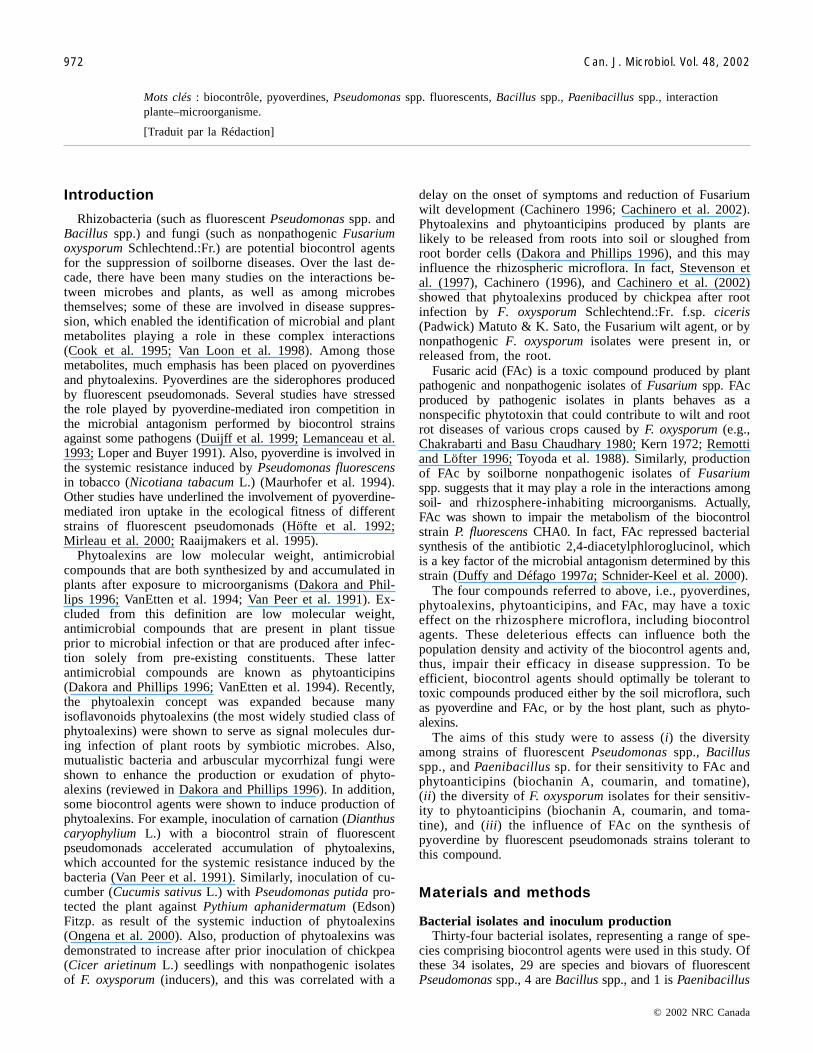
Fusarium oxysporum isolates and inoculum productionSix monoconidial isolates of F. oxysporum were used in
this study (Table 2). Of the six isolates, two (Fo 9009 and Fo90105) are nonpathogenic F. oxysporum shown effective inthe suppression of Fusarium wilt of chickpea (Hervás et al.1997, 1998), one (Fo 47) is nonpathogenic to tomato(Lycopersicum esculentum Mill.) (Alabouvette et al. 1987;Fuchs et al. 1999), two (F. oxysporum f.sp. ciceris Foc 7802and Foc 8012) are pathogenic to chickpea (Jiménez-Díaz etal. 1989; Trapero-Casas and Jiménez-Díaz 1985), and one(F. oxysporum Schlechtend.:Fr. f.sp. lycopersici (Sacc.) W.C.Snyder & H.N. Hans. Fol 32) is pathogenic to tomato.Fusarium oxysporum isolates were stored in 50% glycerol at–80°C or in sterile soil tubes at 4°C. Active cultures wereobtained by placing small drops or soil aliquots from stockcultures onto PDA and incubating for 5 days at 25°C and a12-h photoperiod of fluorescent and near-UV light at36 µE·m–2·s–1.
To obtain inocula for experiments, isolates were grownon PDA slants incubated under the same conditions asabove. Cultures were vortexed and microconidia suspendedin SDW were filtered through a 40-µm mesh. Then, 400 µLof the microconidia suspension was added to 50 mL of mini-mal medium (MM) (Correll et al. 1987) in 150-mLErlenmeyer flasks and incubated on a rotary shaker at150 rpm and 25°C for 6 days. Cultures were filtered as pre-viously described, and microconidia in the filtrate were pel-leted, washed twice in SDW by centrifugation (11 000 × g,10 min), and resuspended in 10 mL of SDW. Theconcentration of microconidia in the suspensions was deter-mined with a haemocytometer (Blaubrand, Brand, Germany),adjusted with SDW, and stored at 4°C until use (less than3 h).
Effect of fusaric acid on bacterial growthFAc, or 5-butylpicolinic acid (Sigma-Aldrich Company
Ltd., Dorset, U.K.), was used in this study. FAc was dis-
solved in SDW to a final concentration of 1 mg/mL and fil-tered through a sterile 0.22-µm Millipore filter (MilliporeCorp., Bedford, Mass.).
The effect of FAc on growth of the fluorescentpseudomonad isolates was studied using sterile flat-bottom96-well microtiter plates. Ten microlitres of appropriate FAcdilutions in SDW were added to wells containing 200 µL ofa bacterial suspension of 106 colony forming units(CFU)/mL to obtain a final FAc concentration of 0, 12.5, 25,50, 100, 200, 300, 400, and 500 µg/mL in a total volume of210 µL. Wells with 10 µL of SDW and 200 µL of the bacte-rial cell suspension in sterile SM served as controls. Cultureswere incubated at 25°C for 50 h. Bacterial growth in a wellwas monitored at time 0 and at 3-h intervals by measuringabsorbance at 590 nm with a Thermomax microplate reader(Molecular Devices, Sunnyvale, Calif., U.S.A.). There werefour replications (wells) for each bacterial isolate–FAc con-centration combination. The experiment was repeated twice.From this experiment, FAc-tolerant isolates were selectedand further screened at the following FAc concentrations: 0,12.5, 25, 50, 100, 200, 300, 400, 500, 600, 700, 800, 900,and 1000 µg/mL, as previously described. This experimentwas repeated once.
The effect of FAc on growth of Bacillus spp. and Paeni-bacillus sp. isolates was studied with an experimental ap-proach similar to that used for fluorescent pseudomonads butusing cell suspensions (106 CFU/mL) in sterile PDB andlower concentrations of FAc (1–10 µg/mL). Cultures wereincubated at 30°C for 65 h. The experiment was repeatedtwice.
Effect of culture media and fusaric acid on growth andproduction of pyoverdine by fusaric acid-tolerantfluorescent pseudomonads
Five P. fluorescens isolates, three P. putida isolates, andone Pseudomonas chlororaphis isolate capable of growing atFAc concentrations higher than 100 µg/mL were selected tostudy the effect of FAc on growth and siderophore produc-tion. Pseudomonas fluorescens PL1 was used as anonsiderophore producer. This latter isolate is a pyoverdine-lacking mutant of P. fluorescens C7R12 obtained by randominsertion of a Tn5 transposon (Mirleau et al. 2000).
For each isolate, 400 µL of an adjusted cell suspension inSDW was transferred into 50 mL of sterile SM (pH 7.0) orKB broth (pH 7.0) in 150-mL Erlenmeyer flasks to obtain aninoculum concentration of 106 CFU/mL. Treatments with
© 2002 NRC Canada
974 Can. J. Microbiol. Vol. 48, 2002
Isolate Pathogenicity Source Location*
F. oxysporum Fo 47 nonpathogenic soil ChâteaurenardF. oxysporum Fo 9009 nonpathogenic chickpea roots SantaellaF. oxysporum Fo 90105 nonpathogenic chickpea roots SantaellaF. oxysporum f.sp. ciceris Foc 7802 pathogenic, race 0 chickpea SantaellaF. oxysporum f.sp. ciceris Foc 8012 pathogenic, race 5 chickpea SantaellaF. oxysporum f.sp. lycopersici Fol 32 pathogenic FPFS† —
*Soils from Châteaurenard and Dijon (France) are suppressive (Alabouvette et al. 1987) and conducive (Lemanceau et al.1995), respectively, to Fusarium wilt diseases. Soils A and B from Santaella (Spain) have been used for Fusarium wiltresistance screening for more than 15 years. Soil A is infested with different races of Fusarium oxysporum f.sp. ciceris. SoilB has been under barley monoculture for the same time period (Landa et al. 1997).
†Flore Pathogéne Faune du sol culture collection, Institut National de la Recherche Agronomique (INRA), Dijon, France.
Table 2. Isolates of Fusarium oxysporum used in this study, listed by pathogenicity, source, and location.
I:\cjm\cjm48\4811\W02-094.vpTuesday, November 26, 2002 10:03:09 AM
Color profile: Generic CMYK printer profileComposite Default screen

FAc received appropriate amounts of a FAc solution inSDW to obtain a final concentration of 100 µg/mL of FAc.Controls received the same volume of SDW. Cultures were in-cubated at 25°C in a rotary shaker at 150 rpm for 48 h. Cellswere harvested by centrifugation (11 000 × g, 20 min). The re-sulting supernatant was adjusted to pH 7.0 and membrane-filtered (pore size 0.22-µm, Millipore) to obtain a cell-freesolution of crude pyoverdines. The concentration of pyo-verdines was estimated by determining the absorbance ofthe filtrates at 400 nm using a molar extinction coefficient of11 500 M–1 cm–1 of free pyoverdine absorption spectrum(Meyer and Abdallah 1978). Bacteria cell concentration inthe suspension was determined using standard curves as pre-viously described. There were two replications for treat-ments of Pseudomonas isolates grown in SM and threereplications for those grown in KB. The experiments includ-ing cultures in KB and SM were repeated once and twice,respectively.
Effect of phytoanticipins on growth of bacteria and F.oxysporum
Eleven fluorescent Pseudomonas spp., Bacillus spp., andPaenibacillus macerans isolates (P. chlororaphis DTR 133;P. putida WCS 358; P. fluorescens RGAF 19, RG 22, RG26, and C7R12; Bacillus circulans RGAF 6a and RGAF 6b;Bacillus megaterium RGAF 12 and RGAF 51; and Paeni-bacillus macerans RGAF 101) and six F. oxysporum isolates(F. oxysporum f.sp. ciceris Foc 7802 and Foc 8012, F.oxysporum f.sp. lycopersici Fol 32, and nonpathogenic F.oxysporum Fo 9009, Fo 90105, and Fo 47) were used in theexperiments.
Tomatine (lycopersicin), coumarin (1,2-benzopyrone), andthe constitutive isoflavone biochanin A (5,7-dihydroxy-4′-methoxyisoflavone) from Sigma-Aldrich were used asphytoanticipins in this study. Tomatine and coumarin weredissolved in 10 mM citrate–phosphate buffer and used atconcentrations of 0, 25, 50, and 100 µg/mL. Biochanin Awas dissolved in ethanol and used at concentrations of 0,0.02, 0.01, and 0.05 mM; ethanol in the solutions was al-lowed to evaporate from suspensions of bacterial or fungalinocula before use.
For each bacterial isolate, 200 µL of a cell suspension(106 CFU/mL) in sterile SM (Pseudomonas spp.) or PDB(Bacillus spp. and Paenibacillus macerans RGAF 101) weremixed with 10 µL of a phytoanticipin solution in a well insterile microtiter plates. Cultures of Pseudomonas spp., Ba-cillus spp., and Paenibacillus macerans RGAF 101 were in-cubated at 25 and 30°C for 50 h, respectively.
Microconidial suspensions of F. oxysporum isolates inSDW obtained as previously described were diluted in MM toa final inoculum concentration of 2.5 × 10² microconidia/mL.For each isolate, 200 µL of the conidial suspension wasmixed with 10 µL of a phytoanticipin solution in a well ofsterile microtiter plates to obtain a final concentration of 50microconidia per well in a total volume of 210 µL. Cultureswere incubated at 25°C for 120 h.
Controls for phytoanticipin treatments consisted of 10 µLof 10 mM citrate–phosphate buffer (coumarin and tomatine)or ethanol (biochanin A) mixed with 200 µL of a bacterialsuspension in sterile SM (Pseudomonas spp.) or PDB (Ba-cillus spp. and Paenibacillus macerans RGAF 101) or a
microconidial suspension in sterile MM (F. oxysporum iso-lates) was also used as a control. Growth of bacteria and F.oxysporum in phytoanticipins and control treatments wasmonitored at time 0 and at 3- to 5-h intervals by measuringabsorbance at 590 nm (bacterial isolates) or at 650 nm (F.oxysporum isolates) as previously described. There were fourreplications (wells) for each microorganism–phytoanticipincombination. Experiments were repeated twice.
Data analysisAll experiments were conducted using a factorial treatment
design arranged in randomized complete blocks. Absorbancedata were plotted over time to develop absorbance increasecurves (AICs). From these curves, the following variableswere calculated: (i) t0.1 = the number of hours taken forabsorbance = 0.1, which was established as an estimate ofthe onset for absorbance increase or lag phase and calcu-lated by interpolation from data; (ii) final absorbance (FA =absorbance determined at the end of the incubation period ofcultures); and (iii) area under the AIC (AUAIC) calculatedby trapezoidal integration method (Campbell and Madden1990). These three variables were used since assessment ofgrowth after short or prolonged incubation periods may sug-gest overstated effects or fail to detect differences in lagphases or growth rates, respectively, of the tested compoundson test microorganisms (Wyman and VanEtten 1978). Valuesof these variables were expressed in relative units (i.e., Rt0.1,RFA, and RAUAIC) by dividing the value for a treatment ina replicate by the corresponding value in the control treat-ment of same replicate. These relative values aimed to mini-mize differences in absorbance values that might occuramong different bacterial or fungal isolates because of pig-mentation in culture. When no bacterial or fungal growth oc-curred for a treatment, the t0.1 value for such a treatmentused in statistical analysis was arbitrarily established as theduration time of the experiment.
The effects of FAc on growth of bacterial isolates and ofphytoanticipins on growth of bacterial and F. oxysporum iso-lates were assessed by hierarchical cluster analyses. Theseanalyses were performed using the Euclidean distance be-tween treatments. The Ward’s method was used in theCLUSTER procedure of SAS (Version 6.11; SAS InstituteInc., Cary, N.C.).
To describe the effects of FAc concentrations (FAcQ) onRt0.1 and RAUAIC of FAc-tolerant fluorescent pseudomonads,the expanded exponential [Rt0.1 (FAcQ) = Cexp(r × FAcQ) +K] and negative asymptotic models {RAUAIC (FAcQ) = C1 –[C2 /(1 + exp[b – r × FAcQ])]}, respectively, were fitted todata by nonlinear regression analyses. In these models, C,C1, and C2 are constants; r is a rate; K is the asymptote; andb is a parameter related to the threshold inhibitory concen-tration. Analyses were carried out using the least-squaresprogram for nonlinear models procedure of SAS. The coeffi-cient of determination (R2), the mean square error, and theasymptotic standard error associated with the estimated pa-rameter were used to evaluate the appropriateness of amodel to describe the data. The standard errors of parame-ters obtained from regression analysis were used to comparethe threshold inhibitory concentration of FAc among thebacterial isolates (Campbell and Madden 1990).
© 2002 NRC Canada
Landa et al. 975
I:\cjm\cjm48\4811\W02-094.vpTuesday, November 26, 2002 10:03:09 AM
Color profile: Generic CMYK printer profileComposite Default screen

Data on the effect of FAc on growth and pyoverdine pro-duction of fluorescent pseudomonads were also analyzed bystandard analysis of variance using the general linear modelprocedure of SAS. Population values were log-transformedbefore analysis. Mean comparisons among treatments wereperformed using Fisher’s protected LSD test (P = 0.05).Similarity among experiments tested by preliminary analysisof variance using experimental runs as blocks and Barlett’stest of equal variances allowed to combine data for analyses.
Results
Effect of fusaric acid on bacterial growthThe 29 isolates of fluorescent pseudomonads differed
greatly in their ability to grow over a range of FAc concentra-tion and showed a continuum of growth responses to increas-ing amounts of FAc in the medium. Thus, some strains (P.fluorescens DTR 335, P. fluorescens DLR 426, P. putida DLR223, P. putida DLR 228, P. putida DS 131, and P. putida DS1026) were inhibited at FAc concentrations <100 µg/mL (Ta-ble 3); most strains were inhibited at FAc concentrations be-tween 100 and 400 µg/mL; and only four strains (P.chlororaphis DTR 133, P. putida Li, P. putida WCS 358, andP. fluorescens RGAF 19) still grew at the highest concentra-
tion (500 µg/mL) of FAc (Table 3). Figure 1A shows AICs offour selected Pseudomonas strains (P. putida DS 1026, P.fluorescens CLE 513, P. putida DTRP, and P. fluorescensRGAF 19) in SM amended with FAc, representing the rangeof the responses found (from the most sensitive to the mosttolerant strains). Using the AICs obtained for each FAcconcentration for each strain, three variables related togrowth parameters expressed in relative units (Rt0.1, RFA,and RAUAIC) were calculated to characterize the effect ofFAc on bacterial growth. The cluster analysis allowed dif-ferentiation of the 29 fluorescent pseudomonads into fourmain clusters (I, IIa, IIb, and III) (Fig. 1B), which com-prised strains ordered from the most sensitive to FAc tothe most tolerant. The four bacterial strains presented inFig. 1A show the general growth response obtained in eachcluster. Each cluster includes strains belonging to differentspecies and biovars isolated from plants or from soil and fromdifferent geographic areas. The four bacterial isolates in clus-ter III, which were the most FAc-tolerant isolates, were mark-edly different from the rest of the isolates (Fig. 1B).
Eight fluorescent pseudomonad isolates in clusters IIb andIII, which were characterized as highly tolerant to FAc(growth up to FAc = 300 µg/mL), were selected to furtherstudy the effect of a broad range of FAc concentrations on
© 2002 NRC Canada
976 Can. J. Microbiol. Vol. 48, 2002
FAc concentration (µg/mL)
Isolate 0 12.5 25 50 100 200 300 400 500
P. fluorescens bv. IV DTR 335 +/+ +/+ +/+ –/– –/– –/– –/– –/– –/–P. fluorescens bv. IV DLR 426 +/+ +/+ +/+ +/– –/– –/– –/– –/– –/–P. putida bv. A DS 1026 +/+ +/+ +/+ +/+ –/– –/– –/– –/– –/–P. putida bv. A DLR 223 +/+ +/+ +/+ +/+ –/– –/– –/– –/– –/–P. putida bv. A DS 131 +/+ +/+ +/+ +/+ –/– –/– –/– –/– –/–P. putida bv. A DLR 228 +/+ +/+ +/+ +/+ +/– –/– –/– –/– –/–Pseudomonas (TI) DS 824 +/+ +/+ +/+ +/+ +/+ –/– –/– –/– –/–P. putida bv. A CS 413 +/+ +/+ +/+ +/+ +/+ –/– –/– –/– –/–P. fluorescens bv. VI A6 +/+ +/+ +/+ +/+ +/+ –/– –/– –/– –/–P. fluorescens bv. II CS 611 +/+ +/+ +/+ +/+ +/+ –/– –/– –/– –/–P. fluorescens bv. II CLRP 812 +/+ +/+ +/+ +/+ +/+ –/– –/– –/– –/–P. fluorescens bv. II C7R12 +/+ +/+ +/+ +/+ +/+ –/– –/– –/– –/–P. fluorescens bv. II C7 +/+ +/+ +/+ +/+ +/+ –/– –/– –/– –/–P. fluorescens bv. IV DLRP 214 +/+ +/+ +/+ +/+ +/+ –/– –/– –/– –/–P. fluorescens bv. II CTR 212 +/+ +/+ +/+ +/+ +/+ –/– –/– –/– –/–P. putida bv. A DLE 3216 +/+ +/+ +/+ +/+ +/+ +/+ –/– –/– –/–P. fluorescens bv. IV DLE 411J +/+ +/+ +/+ +/+ +/+ +/+ –/– –/– –/–P. fluorescens bv. II CTRP 112 +/+ +/+ +/+ +/+ +/+ +/+ –/– –/– –/–P. fluorescens bv. II CTR 1015 +/+ +/+ +/+ +/+ +/+ +/+ –/– –/– –/–P. fluorescens bv. II CLE 513 +/+ +/+ +/+ +/+ +/+ +/+ –/– –/– –/–P. putida bv. A CS 111 +/+ +/+ +/+ +/+ +/+ +/+ –/– –/– –/–P. fluorescens bv. II RG 26 +/+ +/+ +/+ +/+ +/+ +/+ +/+ –/– –/–P. fluorescens bv. IV RG 22 +/+ +/+ +/+ +/+ +/+ +/+ +/+ –/– –/–P. fluorescens bv. II CLR 711 +/+ +/+ +/+ +/+ +/+ +/+ +/+ +/– –/–P. putida bv. A DTRP 621 +/+ +/+ +/+ +/+ +/+ +/+ +/+ +/+ +/–P. chlororaphis DTR 133 +/+ +/+ +/+ +/+ +/+ +/+ +/+ +/+ +/+P. putida Li +/+ +/+ +/+ +/+ +/+ +/+ +/+ +/+ +/+P. putida WCS 358 +/+ +/+ +/+ +/+ +/+ +/+ +/+ +/+ +/+P. fluorescens bv. V RGAF 19 +/+ +/+ +/+ +/+ +/+ +/+ +/+ +/+ +/+
Note: Cultures were incubated at 25°C for 50 h. Bacterial growth (+) was assessed by measuring absorbance at 590 nm with a microplate reader.Results indicate two replicated experiments. (–), no growth indicated by absorbance <0.05.
Table 3. Growth of fluorescent Pseudomonas spp. strains in succinate medium amended with fusaric acid (FAc).
I:\cjm\cjm48\4811\W02-094.vpTuesday, November 26, 2002 10:03:10 AM
Color profile: Generic CMYK printer profileComposite Default screen

© 2002 NRC Canada
Landa et al. 977
Fig. 1. (A) Absorbance increase curves (AICs) of four pseudomonad strains (Pseudomonas putida DS 1026, Pseudomonas fluorescensCLE 513, P. putida DTRP 621, and P. fluorescens RGAF19) in response to fusaric acid (FAc), representative of the four groups (be-low) derived from cluster analysis. AICs were obtained by measuring the absorbance (590 nm) at periodic time intervals with amicroplate reader as described in Materials and methods. (B) Dendrogram representing relative similarities among 29 isolates offluorescent pseudomonads in tolerance to FAc. Sp., isolate species (Pc, Pseudomonas chlororaphis; Pf, P. fluorescens; Pp, P. putida;P-IT, intermediate type P. fluorescens–P. putida); Bv., biovar. Soil source: U, unknown; C, Châteaurenard (France); D, Dijon(France); S, Santaella (Spain). I, IIa, IIb, and III identify categories of tolerance to FAc; strains were inhibited at FAc concentrations<100 µg/mL (I), 100–400 µg/mL (IIa and IIb), and still growing at 500 µg/mL (III).
I:\cjm\cjm48\4811\W02-094.vpTuesday, November 26, 2002 10:03:10 AM
Color profile: Generic CMYK printer profileComposite Default screen

bacterial growth. The variation in Rt0.1 and RAUAIC withthe increase in FAc concentration, which relates to the effectof FAc on bacterial growth, was adequately described by theexpanded exponential and negative asymptotic models, re-spectively. Rt0.1 increased exponentially with the increase inFAc concentration (Fig. 2A) irrespective of bacterial iso-lates. Rt0.1 values for isolates DTR 133, RGAF 19, Li, andWCS 358 (cluster III) at FAc concentrations higher than100 µg/mL were always lower than those of isolates in clus-ter IIb. Conversely, at FAc concentrations higher than an in-hibitory threshold, the RAUAIC diminished asymptoticallywith the increase in FAc concentration (Fig. 2B). The b pa-rameter in the negative asymptotic model, which is corre-lated with the threshold inhibitory concentration, becamehigher as the FAc concentration increases. The b value dif-fered significantly (P < 0.05) among the bacterial isolatesand was highest for P. fluorescens RGAF 19 and P. putidaWCS 358, the isolates most tolerant to FAc (data not shown).
As expected, Bacillus spp. and Paenibacillus maceransRGAF 101 were highly sensitive to the addition of FAc inthe culture medium as compared with Pseudomonas spp. flu-orescent isolates. Bacillus circulans RGAF 6a (Fig. 3A) andRGAF 6b grew only at a FAc concentration of 1 µg/mL andconstituted one single group in the cluster analysis (Fig. 3B);whereas B. megaterium RGAF 12 (Fig. 3A) and RGAF 51and Paenibacillus macerans RGAF 101 grew at FAc concen-trations up to 5 µg/mL and were grouped together in a sepa-rate cluster (Fig. 3B).
Effect of culture media and fusaric acid on growth andproduction of pyoverdine by fusaric acid-tolerantfluorescent pseudomonads
Growth and pyoverdine production of FAc-tolerant Pseu-domonas isolates were assessed in two different media (KBand SM), either amended or not amended with 100 µg/mL ofFAc. Both culture media and FAc influenced pyoverdineproduction and growth of FAc-tolerant Pseudomonas iso-lates. The effect of FAc on bacterial growth differed accord-ing to the bacterial strain and media. For all strains, bacterialgrowth was higher in KB (i.e., a rich medium) than in SM(Table 4). Growth of Pseudomonas isolates in KB was notaffected by FAc. On the contrary, in SM, four strains (P.fluorescens C7R12 and its siderophore-defective mutantPL1 and P. fluorescens RG 22 and RG 26) grew significantly(P < 0.05) better in the presence of FAc (100 µg/mL) (Ta-ble 4).
Although total pyoverdine production (log(mol/L)) washigher in KB than in SM, the relative pyoverdine production(log(mol/CFU)) was similar in both media (Table 4). FAc af-fected the production of pyoverdine. All bacterial isolates(except for P. fluorescens RGAF 19 and RG 22 grown in KBmedium and P. chlororaphis DTR 133) produced signifi-cantly (P < 0.05) less pyoverdine (total Log(mol/L) and rela-tive Log(mol/CFU) production) in KB and SM amendedwith FAc compared with the unamended medium (Table 4).
Effect of phytoanticipins on growth of bacteria and F.oxysporum
Three variables related to growth parameters expressed inrelative units (Rt0.1, RFA, and RAUAIC), derived from theAICs from each of phytoanticipin concentration and bacte-
rial strain or fungal isolate, were used to characterize theglobal effect of phytoanticipins on bacterial and fungalgrowth. Biochanin A, coumarin, and tomatine influencedboth bacterial and fungal growth. Growth of Pseudomonas,Bacillus, and Paenibacillus macerans isolates were differen-tially affected by the three phytoanticipins (Fig. 4). For bio-chanin A and coumarin, Pseudomonas isolates were groupedin one cluster and Bacillus spp. and Paenibacillus maceransisolates in a separate cluster. Second order clusters groupedbacterial isolates according to the effect of each particularphytoanticipin on the onset (Rt0.1) and extent of bacterialgrowth (RAF and RAUAIC) (Figs. 4A and 4B). In general,tomatine did not appear to influence growth of B. mega-terium isolates or Pseudomonas spp. (except for P.fluorescens RGAF 19) but delayed and inhibited growth ofPaenibacillus macerans RGAF 101, B. circulans RGAF 6aand RGAF 6b, and P. fluorescens RGAF 19 (Fig. 4C). Over-all, biochanin A did not inhibit bacterial growth (no effecton RFA, RAUAIC, or Rt0.1). Rather, the presence ofbiochanin A in culture media appeared to enhance growth ofP. fluorescens RGAF 19, RG 22, and RG 26, as indicated byhigher values of RFA and RAUAIC and delayed growth of
© 2002 NRC Canada
978 Can. J. Microbiol. Vol. 48, 2002
Fig. 2. Effect of fusaric acid (FAc) concentration in succinatemedium on the time to the growth onset (t0.1) and the area underthe curve of absorbance increase over time (AUAIC) of Pseudo-monas chlororaphis DTR 133, Pseudomonas fluorescens CLR711, P. fluorescens RGAF 19, P. fluorescens RG 22, P.fluorescens RG 26, Pseudomonas putida Li, P. putida DTRP621, and P. putida WCS 358. Values of t0.1 and AUAIC for eachFAc concentration were expressed in relative units by dividingthe actual value by that in the control (0 µg/mL of FAc) for eachbacterial isolate. Lines represent the predicted curves calculatedby the expanded exponential model (A) or the negative asymp-totic model (B).
I:\cjm\cjm48\4811\W02-094.vpTuesday, November 26, 2002 10:03:10 AM
Color profile: Generic CMYK printer profileComposite Default screen

B. megaterium RGAF 12 and RGAF 51 and Paenibacillusmacerans RGAF 101 (higher values of Rt0.1). Conversely,coumarin inhibited (lower values of RFA and RAUAIC) anddelayed (higher values of Rt0.1) growth of most Pseudomonasisolates, but it had no effect on growth of B. circulansRGAF 6a and RGAF 6b and Paenibacillus macerans RGAF101 (Fig. 4B).
Biochanin A and tomatine had similar effects on growth ofF. oxysporum isolates (Figs. 5A and 5C), but these effects dif-fered depending on the pathogenic (F. oxysporum f.sp. cicerisand F. oxysporum f.sp. lycopersici) or nonpathogenic (F.oxysporum nonpathogenic) nature of isolates. Indeed, increasedconcentrations of biochanin A and tomatine significantly (P <0.05) inhibited (lower values of RFA and RAUAIC) and de-layed (higher values of Rt0.1) growth of F. oxysporum f.sp.ciceris and F. oxysporum f.sp. lycopersici isolates. However,growth of nonpathogenic isolates increased with increasingconcentrations of both phytoanticipins (Figs. 5A and 5C). Al-though coumarin inhibited growth of all F. oxysporum isolates(Fig. 5B), the extent of inhibition was higher for pathogenic
isolates compared with that of nonpathogenic ones. Thus, forall the three phytoanticipins, F. oxysporum f.sp. ciceris and F.oxysporum f.sp. lycopersici (pathogenic isolates) were groupedin one cluster and nonpathogenic F. oxysporum isolates com-prised a different cluster (Fig. 5).
Discussion
There have been many studies aimed at the application ofrhizobacteria for the suppression of soilborne diseases. Al-though encouraging results have been obtained, in practice,the efficacy of biocontrol agents in disease suppression fre-quently suffers from inconsistency. One of the reasons forsuch a lack of consistency is the complexity of interactionstaking place between the pathogenic or antagonistic microor-ganisms and the plant. These interactions are far from beingwell characterized. Some studies have pointed out a role forsecondary metabolites, such as FAc produced by F. oxy-sporum and pyoverdine produced by the fluorescent Pseudo-monas spp., in the interactions between these organisms
© 2002 NRC Canada
Landa et al. 979
Fig. 3. (A) Absorbance increase curves (AICs) of two Bacillus strains (Bacillus circulans RGAF 6a, and Bacillus megaterium RGAF12) in response to fusaric acid (FAc), representative of the two groups (below) derived from cluster analysis. The AICs were obtainedby measuring the absorbance (590 nm) at periodic time intervals with a microplate reader as described in Materials and methods.(B) Dendrogram representing relative similarities among four Bacillus spp. isolates and a Paenibacillus macerans isolate in tolerance toFAc. I and II identify the categories of tolerance to Fac; strains were inhibited at Fac concentrations <1 µg/mL (I) or <5–6 µg/mL (II).
I:\cjm\cjm48\4811\W02-094.vpTuesday, November 26, 2002 10:03:10 AM
Color profile: Generic CMYK printer profileComposite Default screen

© 2002 NRC Canada
(Lemanceau et al. 1993; Loper and Buyer 1991; Toyoda etal. 1988) or in the antimicrobial effects of some phytoa-lexins (Gnanamanickam and Mansfield 1981; Wyman andVanEtten 1978). However, little attention has been given tothe compatibility between different biological control agentsand the plant by addressing the tolerance of each partner tothe secondary metabolites produced by the other. The aim ofthis work was to assess the effect of some secondary metabo-lites produced by isolates of F. oxysporum and the plant onthe growth of a collection of isolates of F. oxysporum (patho-genic and nonpathogenic), fluorescent Pseudomonas spp., Ba-cillus spp., and Paenibacillus macerans.
FAc is one of the secondary metabolites produced bymost, and possibly all, isolates of F. oxysporum. The poten-tial roles of FAc in relation to pathogenicity of F. oxysporumstrains (Kern 1972) and to activity of Ralstonia solana-cearum in the control of Fusarium wilts (Toyoda et al. 1988)were previously addressed. In this study, the impact of thisfungal metabolite on growth of a collection of fluorescentPseudomonas spp., Bacillus spp., and Paenibacillus maceranswas evaluated in a culture medium amended with increasingconcentrations of FAc. The results clearly showed a contin-uum among bacterial strains, from those sensitive to lowconcentrations of FAc to strains tolerant to high concen-trations. However, as a group, Pseudomonas spp. strainswere much less sensitive to FAc than Bacillus spp. andPaenibacillus macerans. Based on the literature, we choseto apply very low concentrations of FAc to characterizethe behavior of Bacillus spp. and Paenibacillus maceransstrains (growth of these bacteria were completely inhibited by10–4 M of FAc; Gäumann 1957). The results showed thatgrowth of Bacillus spp. and Paenibacillus macerans was in-hibited at a concentration as low as 5 µg/mL, whereas somestrains of P. chlororaphis, P. fluorescens, and P. putida werestill able to grow at concentrations higher than 500 µg/mL.The high sensitivity of Bacillus spp. and Paenibacillusmacerans to FAc found in our study agree with results fromother authors (Gäumann 1957; Kuo and Scheffer 1964).
From the AICs, three variables related to growth parame-
ters were calculated: t0.1 representing the lag phase, FA rep-resenting the bacterial concentration at the plateau, andAUAIC representing the total biomass produced. These pa-rameters were used to perform a cluster analysis that distin-guished four clusters among Pseudomonas spp. strains andtwo clusters among Bacillus strains in relation to their toler-ance to FAc. Each of these clusters includes bacterial strainsbelonging to different species and biovars isolated fromplants or from soil and from different geographic areas.These data indicate a lack of correlation between toleranceto FAc and the origin or taxonomic position of the fluores-cent pseudomonads in the study. Ability to grow in the pres-ence of FAc seems, then, to be a characteristic of eachindividual strain rather than of a taxonomic group. The anal-yses of the growth curves of eight Pseudomonas spp. strainshighly tolerant to FAc adequately fit Rt0.1 and RAUAIC datato the expanded exponential and the negative asymptoticmodels, respectively. The asymptotic decrease of Rt0.1 andincrease of RAUAIC when FAc concentrations reach agiven level indicate that the threshold concentration of FAcinhibiting bacterial growth differs greatly among bacterialstrains. The diversity in the sensitivity of bacterial strains toFAc would suggest that the structure of indigenous popula-tions of soilborne pseudomonads could vary according to theconcentration or presence of FAc in their environment.These data could also account for the variable compatibilityof fluorescent Pseudomonas spp. strains when combinedwith nonpathogenic F. oxysporum for the suppression ofFusarium wilt diseases (Lemanceau and Alabouvette 1991).Indeed, a FAc-sensitive strain of fluorescent pseudomonadswould not be compatible with an isolate of F. oxysporumproducing high quantities of FAc.
Besides bacterial growth, FAc may also affect bacterialmetabolism (Duffy and Défago 1997a; Schnider-Keel et al.2000). Because pyoverdine is a bacterial metabolite playinga major role in the interactions among fluorescent pseudo-monads, the soil microflora, and the plant (Loper and Buyer1991), we evaluated the possible impact of FAc on pyover-dine synthesis by FAc-tolerant strains of Pseudomonas
980 Can. J. Microbiol. Vol. 48, 2002
KB SM
Log (CFU/mL) –Log (mol/L) –Log (mol/CFU) Log (CFU/mL) –Log (mol/L) –Log (mol/CFU)
Isolate 0 100 0 100 0 100 0 100 0 100 0 100
P.c. DTR 133 10.22 10.24 3.71 3.72 16.94 16.95 9.49 9.63 4.48a 4.92b 16.98a 17.55bP.f. C7r12 10.14 10.12 3.75a 3.84b 16.89a 16.95b 8.67b 9.07a 4.06a 4.19b 15.73a 16.25bP.f. PL1 10.04 10.03 — — — — 9.20b 9.36a — — — —P.f. CLR 711 10.05 10.00 4.01a 4.23b 17.05a 17.24b 9.16 9.28 5.05a 4.09b 17.21a 17.05bP.f. RGAF 19 10.05 10.07 3.74 3.76 16.79 16.83 9.23 9.25 3.97a 4.09b 16.20a 16.34bP.f. RG 22 10.03 10.06 3.74 3.74 16.77 16.79 9.19b 9.39a 3.96a 4.05b 16.25a 16.34bP.f. RG 26 10.20 10.19 3.75a 3.89b 16.95a 17.08b 9.38b 9.52a 4.04a 4.98b 16.42a 16.50bP.p. Li 9.63 9.70 3.75a 3.77b 16.39a 16.48b 9.20 9.23 4.32a 4.50b 16.52a 16.73bP.p. DTRP 621 10.09 10.10 3.73a 3.82b 16.82a 16.92b 9.24 9.29 4.36a 4.78b 16.60a 17.06bP.p. WCS 358 9.99 9.99 3.73a 3.76b 16.73a 16.75b 9.33 9.30 3.86a 3.95b 16.18a 16.25b
Note: Bacterial growth was estimated by measuring the absorbance at 600 nm using standard curves. Pyoverdine concentration was estimated bymeasuring the absorbance of the cell-free filtrates at 400 nm, as described in Materials and methods. Means in a row within a culture medium withdifferent letters are significantly different (P < 0.05) from the mean for the corresponding treatment at the other FAc concentration, according to Fisher’sprotected LSD. P.c., Pseudomonas chlororaphis; P.f., Pseudomonas fluorescens; P.p., Pseudomonas putida; —, pyoverdine production below the detectionlimit (A400 < 0.05).
Table 4. Bacterial growth (CFU/mL) and production of pyoverdine (mol/L and mol/CFU) by fluorescent Pseudomonas spp. isolates inKing’s B (KB) and succinate media (SM) amended (100 µg/mL) or not amended (0 µg/mL) with fusaric acid.
I:\cjm\cjm48\4811\W02-094.vpTuesday, November 26, 2002 10:03:11 AM
Color profile: Generic CMYK printer profileComposite Default screen

spp. At a concentration of 100 µg/mL, FAc affected the pro-duction of pyoverdine by all of the strains in the minimalsuccinate medium and by most of the strains in the rich KBmedium. This effect on pyoverdine production was inde-pendent of bacterial growth, since FAc also affected therelative pyoverdine production. The deleterious effect ofFAc on pyoverdine production is similar to that on produc-tion of the pyochelin siderophore and other bacterial me-tabolites described by Duffy and Défago (1997a). Theseauthors demonstrated that FAc at a concentration as low as0.12 µg/mL repressed the production of the antibiotic 2,4-diacetylphloroglucinol (2,4-DAPG) in vitro by P. fluorescensCHA0, which is a key factor in the disease suppression by this
strain. Duffy and Défago (1997a) also showed that produc-tion of pyoluteorin by P. fluorescens CHA0, but not that ofhydrogen cyanide and protease, was also reduced in thepresence of FAc. They concluded that FAc might affectbiosynthesis of secondary metabolites at a regulatory leveldownstream of gacA and apdA genes. More recently, Schnider-Keel et al. (2000) also demonstrated that FAc strongly re-pressed the synthesis of monoacetylphloroglucinol and 2,4-DAPG and the expression of phlA, one of the 2,4-DAPGbiosynthetic genes; this regulatory mechanism is mediatedby PhlF, a transcriptional repressor.
Impairment of bacterial biosynthesis of metabolites byFAc may relate to the efficiency of bacteria as biocontrol
© 2002 NRC Canada
Landa et al. 981
Fig. 4. Dendrogram representing relative similarities in tolerance to biochanin A (A), coumarin (B), and tomatine (C) among six fluo-rescent Pseudomonas spp. strains, four Bacillus spp. strains, and a Paenibacillus macerans strain. Bacterial growth was monitored bymeasuring the absorbance at 590 nm at periodic time intervals as described in Materials and methods. RFA, relative final absorbance;RAUAIC, relative area under the curve of absorbance increase over time; Rt0.1, relative time to onset of bacterial growth. Increase ordecrease in RFA, RAUAIC, and Rt0.1 with the increase in phytoanticipin concentration is indicated by (+) and (–), respectively, while(0) indicates no effect. *, statistically significant compared with the control treatment at P = 0.05.
I:\cjm\cjm48\4811\W02-094.vpTuesday, November 26, 2002 10:03:11 AM
Color profile: Generic CMYK printer profileComposite Default screen

© 2002 NRC Canada
982 Can. J. Microbiol. Vol. 48, 2002
agents. Sharifi-Tehrani et al. (1998) compared a collectionof 2,4-DAPG-producing Pseudomonas spp. belonging tothree ARDRA (amplified 16S ribosomal DNA restrictionanalysis) groups for biocontrol activity against fusariumcrown and root rot of tomato and pythium damping-off ofcucumber. As a group, ARDRA group 2 strains were moreeffective than ARDRA group 1 strains. FAc blocks bio-synthesis of 2,4-DAPG by ARDRA-1 strains (Duffy andDéfago 1997a; Schnider-Keel et al. 2000) but not that ofARDRA-2 or ARDRA-3 strains (Duffy and Défago 1997b).This could explain, in part, why suppression of fusariumcrown and root rot of tomato was less effective for ARDRA-
1 strains compared with ARDRA-2 strains (Sharifi-Tehraniet al. 1998). These results (Duffy and Défago 1997a, 1997b,Schnider-Keel et al. 2000) and those of the present study in-dicate the need to address the inhibitory effect of FAc ongrowth and production of secondary metabolites by bacterialstrains and nonpathogenic F. oxysporum isolates used for bi-ological control of diseases caused by Fusarium spp. In do-ing so, better disease control could be obtained by usingFAc-insensitive biocontrol strains or by using FAc-degrading strains (Duffy and Défago 1997a; Toyoda et al.1988).
Strains of fluorescent Pseudomonas spp. and nonpatho-
Fig. 5. Dendrogram representing relative similarities among nonpathogenic Fusarium oxysporum Fo 9009, Fo 90105, and Fo 47, patho-genic F. oxysporum f.sp. ciceris Foc 7802 and Foc 8012, and pathogenic F. oxysporum f.sp. lycopersici Fol 32 in minimal mediumamended with different concentrations of biochanin A (A), coumarin (B), and tomatine (C). Fungal growth was monitored by measur-ing the absorbance at 650 nm at periodic time intervals as described in Materials and methods. RFA, relative final absorbance;RAUAIC, relative area under the curve of absorbance increase over time; Rt0.1, relative time to onset of fungal growth. Increase or de-crease in RFA, RAUAIC, and Rt0.1 with the increase in phytoanticipin concentration is indicated by (+) and (–), respectively, while(0) indicates no effect. *, statistically significant compared with the control treatment at P = 0.05.
I:\cjm\cjm48\4811\W02-094.vpTuesday, November 26, 2002 10:03:11 AM
Color profile: Generic CMYK printer profileComposite Default screen

© 2002 NRC Canada
genic isolates of F. oxysporum used for disease suppressionshould also be tolerant to toxic compounds produced by thehost plant, such as phytoanticipins and phytoalexins that canreach high concentrations in root cells and soil (Armero1996; Cachinero 1996; Cachinero et al. 2002; Dakora andPhillips 1996). Gram-negative bacteria were shown to beless sensitive to several phytoalexins than Gram-positive bac-teria (Gnanamanickam and Mansfield 1981). Our resultsclearly show that (i) phytoanticipins differ in their effects onspecific bacterial strains and (ii) the bacterial sensitivity to aphytoanticipin varies from one strain to the other. For exam-ple, biochanin A was neutral or stimulatory for Pseudomo-nas spp., but it delayed growth of Bacillus spp. In contrast,coumarin inhibited growth of Pseudomonas spp. but had noeffect on Bacillus spp. and Paenibacillus macerans. Theseresults agree with those of Wyman and VanEtten (1978),who showed that sensitivity of bacterial isolates to sixselected isoflavonoid phytoalexins varied widely, with pseudo-monads being more tolerant to phytoalexins than xantho-monads or Achromobacter isolates.
Several studies have indicated a relationship betweengreater virulence of fungal plant pathogens and increasedtolerance to toxic compounds, such as phytoanticipinsand phytoalexins produced by the host plant (reviewedin Suleman et al. 1996). However, other studies identifiedvariation in phytoanticipin and phytoalexin toleranceamong fungal species or isolates of a pathogen (Dakoraand Phillips 1996; Suleman et al. 1996). Our results indi-cate that effects of biochanin A, coumarin, and tomatinephytoanticipins on fungal growth differed among non-pathogenic and pathogenic F. oxysporum isolates. Thethree pathogenic isolates of F. oxysporum f.sp. ciceris andF. oxysporum f.sp. lycopersici tested were highly sensitiveto the three phytoanticipins, with increased lag phase andreduced growth. Conversely, the three nonpathogenic F.oxysporum isolates grew better in the presence of bio-chanin and tomatine and were inhibited by coumarin, al-though to a lesser extent compared with the pathogenicisolates. Thus, our results suggest that nonpathogenicF. oxysporum isolates are less sensitive to phytoanti-cipins than the pathogenic ones. The nonpathogenic F.oxysporum isolates involved in the suppression ofFusarium wilts are known to invade the root cortex of thehost plant where they induce defense reactions (Olivainand Alabouvette 1997). Cachinero (1996) and Cachineroet al. (2002) demonstrated that prior inoculation of chick-peas with nonpathogenic F. oxysporum isolates caused abrown discoloration at surfaces of roots, cotyledonarynode, and cotyledons and increased production of maac-kiain and medicarpin phytoalexins in the plant roots.Suleman et al. (1996) demonstrated that virulence of iso-lates of F. oxysporum f.sp. lycopersici was correlated withtolerance to rishitin phytoalexin and to a lesser extentwith tolerance to tomatine phytoanticipin. However, theyalso found that nonpathogenic F. oxysporum isolates dis-played, in general, the highest sensitivity to tomatine butnot to rishitin. Lairini et al. (1997) found that a tomatineconcentration of 20 mM inhibited mycelial growth of all10 formae speciales of F. oxysporum investigated, with F.oxysporum f.sp. ciceris being the most sensitive to thisphytoanticipin. All these results suggest that great vari-
ability may occur in the response of bacteria and fungi tophytoalexins and phytoanticipins regardless of the patho-genic or nonpathogenic nature of the species or isolate.
Our study, though conducted in vitro, involved the effectsof metabolites produced by microbes and plants on plantpathogens and biocontrol agents that may eventually be re-leased into the rhizosphere and affect the growth and (or)metabolism of these microorganisms. Our results are indica-tive of the complex interactions that can occur at the rootsurface and in the rhizosphere among the plant, the patho-gen, and two types of antagonistic microorganisms used forbiological control. A major challenge for the future wouldbe to assess the effect of FAc and phytoanticipins on the sur-vival and the activities of biocontrol agents in the rhizo-sphere using reporter genes.
AcknowledgementsResearch support was provided by fellowships to B.B.
Landa and J.M. Cachinero from Consejo Superior deInvestigaciones Científicas (Spain) and Conseil Régional deBourgogne (France), respectively, and by grants AGF97-1479from Comisión Interministerial de Ciencia y Tecnología(CICYT) (Spain) and CSIC–INRA bilateral cooperative re-search program. We are grateful to P.A.H.M. Bakker, whokindly provided the strain P. putida WCS 358. We thank J.A.Navas-Cortés and T.C. Paulitz for a thoughtful review andcritical comments on the manuscript.
ReferencesAlabouvette, C., de la Broise, D., Lemanceau, P., Couteaudier, Y,
and Louvet, J. 1987. Utilisation de souches non pathogenes deFusarium pour lutter contre les fusarioses: Situation actuelledans la pratique. EPPO Bull. 17: 665–674.
Armero, J. 1996. Isoflavonoides y compuestos relacionados de gar-banzo: inducción y funciones. Ph.D. thesis, University ofCórdoba, Córdoba, Spain.
Cachinero, J.M. 1996. Papel de las reacciones defensivas en laresistencia de plantas a hongos patógenos. Ph.D. thesis, Univer-sity of Córdoba, Córdoba, Spain.
Cachinero, J.M., Hervás, A., Jiménez-Díaz, R.M., and Tena, M.2002. Plant defense reactions against Fusarium wilt in chickpeainduced by nonpathogenic races of Fusarium oxysporum f.sp.ciceris and nonpathogenic isolates of F. oxysporum. PlantPathol. 51: In press.
Campbell, C.L., and Madden, L.V. 1990. Introduction to plant dis-ease epidemiology. John Wiley and Sons, N.Y., U.S.A.
Chakrabarti, D.K., and Basu Chaudhary, K.C. 1980. Correlationbetween virulence and fusaric acid production in Fusariumoxysporum f.sp. carthami. J. Phytopathol. 99: 43–46.
Cook, R.J., Thomashow, L.S., Weller, D.M., Fujimoto, D.,Mazzola, M., Bangera, G., and Kim, D.-S. 1995. Molecularmechanisms of defense of rhizobacteria against root disease.Proc. Natl. Acad. Sci. U.S.A. 92: 4197–4201.
Correll, J.C., Klittich, J.R., and Leslie, J.F. 1987. Nitrate non-utilizing mutants of Fusarium oxysporum and their use in vege-tative compatibility tests. Phytopathology, 77: 1640–1646.
Dakora, F.D., and Phillips, D.A. 1996. Diverse functions of iso-flavonoids in legumes transcend anti-microbial definitions ofphytoalexins. Physiol. Mol. Plant Pathol. 49: 1–20.
Duffy, B.K., and Défago, G. 1997a. Zinc improves biocontrol offusarium crown and root rot of tomato by Pseudomonasfluorescens and represses the production of pathogen metabo-
Landa et al. 983
I:\cjm\cjm48\4811\W02-094.vpTuesday, November 26, 2002 10:03:11 AM
Color profile: Generic CMYK printer profileComposite Default screen

© 2002 NRC Canada
984 Can. J. Microbiol. Vol. 48, 2002
lites inhibitory to bacterial antibiotic biosynthesis. Phyto-pathology, 87: 1250–1257.
Duffy, B.K., and Défago, G. 1997b. A Fusarium pathogenicity fac-tor blocks antibiotic biosynthesis by antagonistic pseudomo-nads. Phytopathology, 87: S26.
Duijff, B.J., Recorbet, G., Bakker, P.A.H.M., Loper, J., andLemanceau, P. 1999. Microbial antagonism at the root level isinvolved in the suppression of Fusarium wilt by the combinationof nonpathogenic Fusarium oxysporum Fo47 and Pseudomonasputida WCS358. Phytopathology, 89: 1073–1079.
Eparvier, A., Lemanceau, P., and Alabouvette, C. 1991. Populationdynamics of nonpathogenic Fusarium and fluorescent Pseudo-monas strains in rockwool, a substratum for soilless culture.FEMS Microbiol. Ecol. 86: 177–184.
Fuchs, J.-G., Moënne-Loccoz, Y., and Défago, G. 1999. Ability ofnonpathogenic Fusarium oxysporum Fo47 to protect tomatoagainst Fusarium wilt. Biol. Control, 14: 105–110.
Gäumann, E. 1957. Fusaric acid as a wilt toxin. Phytopathology,47: 342–357.
Gnanamanickam, S.S., and Mansfield, J.W. 1981. Selective toxicityof wyerone and other phytoalexins to gram-positive bacteria.Phytochemistry, 20: 997–1000.
Hervás, A., Landa, B., and Jiménez-Díaz, R.M. 1997. Influence ofchickpea genotype and Bacillus spp. on protection fromFusarium wilt by seed treatment with nonpathogenic Fusariumoxysporum. Eur. J. Plant Pathol. 103: 631–642.
Hervás, A., Landa, B., Datnoff, L.E., and Jiménez-Díaz, R.M.1998. Effects of commercial and indigenous microorganisms onFusarium wilt development in chickpea. Biol. Control, 13: 166–176.
Höfte, M., Boelens, J., and Verstraete, W. 1992. Survival and rootcolonization of mutants of plant growth-promoting pseudo-monads affected in siderophore biosynthesis or regulation ofsiderophore production. J. Plant Nutr. 15: 2253–2262.
Jiménez-Díaz, R.M., Trapero-Casas, A., and Cabrera de La Colina,J. 1989. Races of Fusarium oxysporum f.sp. ciceri infectingchickpeas in sourthern Spain. In Vascular wilt diseases of plants.Edited by E.C. Tjamos and C. Beckman. North Atlantic TreatyOrganization (NATO) Advanced Study Institute (ASI) Series.Springer-Verlag GmbH & Co. KG, Berlin, Germany. pp. 515–520.
Kern, H. 1972. Phytotoxins produced by fusaria. In Phytotoxins inplant diseases. Edited by R.K.S. Wood, A. Ballio, and A.Graniti. Academic Press Inc., New York, N.Y. pp. 35–44.
Kuo, M.S., and Scheffer, R.P. 1964. Evaluation of fusaric acid as afactor in development of Fusarium wilt. Phytopathology, 54:1041–1044.
Lairini, K., Pérez-Espinosa, A., and Ruiz-Rubio, M. 1997. Tomatinaseinduction in formae speciales of Fusarium oxysporum non-pathogenic of tomato plants. Physiol. Mol. Plant Pathol. 50: 37–52.
Landa, B.B., Hervás, A., Bettiol, W., and Jiménez-Díaz, R.M.1997. Antagonistic activity of bacteria from chickpea rhizo-sphere against Fusarium oxysporum f.sp. ciceris. Phytopara-sitica, 25: 305–318.
Lemanceau, P., and Alabouvette, C. 1991. Biological control ofFusarium diseases by fluorescent Pseudomonads and nonpatho-genic Fusarium. Crop Prot. 10: 279–286.
Lemanceau, P., Bakker, P.A.H.M., De Kogel, W.J., Alabouvette, C.,and Schippers, B. 1993. Antagonistic effect of nonpathogenicFusarium oxysporum Fo47 and pseudobactin 358 upon patho-genic Fusarium oxysporum f.sp. dianthi. Appl. Environ.Microbiol. 59: 74–82.
Lemanceau, P., Corberand, T., Gardan, L., Latour, X., Laguerre,
G., Boeufgras, J.-M., and Alabouvette, C. 1995. Effect of twoplant species, flax (Linum usitatissinum L.) and tomato(Lycopersicon esculentum Mill.), on the diversity of soilbornepopulations of fluorescent pseudomonads. Appl. Environ.Microbiol. 61: 1004–1012.
Loper, J.E., and Buyer, J.S. 1991. Siderophores in microbial inter-actions on plant surfaces. Mol. Plant–Microbe Interact. 4: 5–13.
Maurhofer, M., Hase, C., Meuwly, P., Métraux, J.-P., and Défago,G. 1994. Induction of systemic resistance of tobacco to tobacconecrosis virus by the root-colonizing Pseudomonas fluorescensstrain CHA0: influence of the gacA gene and pyoverdin produc-tion. Phytopathology, 84: 139–146.
Meyer, J.M., and Abdallah, M.A. 1978. The fluorescent pigment ofPseudomonas fluorescens: biosynthesis, purification and physico-chemical properties. J. Gen. Microbiol. 107: 319–328.
Mirleau, P., Delorme, S., Philippot, L., Meyer, J.-M., Mazurier, S.,and Lemanceau, P. 2000. Fitness in soil and rhizosphere ofPseudomonas fluorescens C7R12 compared with a C7R12 mu-tant affected in pyoverdine synthesis and uptake. FEMSMicrobiol. Ecol. 34: 35–44.
Olivain, C., and Alabouvette, C. 1997. Colonization of tomato rootby a nonpathogenic strain of Fusarium oxysporum. New Phytol.137: 481–494.
Ongena, M., Daayf, F., Thornat, P., Benhamou, N., Paulitz, T.C.,and Bélanger, R.R. 2000. Systemic induction of phytoalexins incucumber in response to treatments with fluorescent pseudo-monads. Plant Pathol. 49: 523–530.
Raaijmakers, J., Van der Sluis, I., Bakker, P.A.H.M., and Schippers,B. 1995. Utilization of heterologous siderophores and rhizospherecompetence of fluorescent Pseudomonas spp. Can. J. Microbiol.41: 126–135.
Remotti, P.C., and Löfter, H.J.M. 1996. The involvement of fusaricacid in the bulb rot of gladiolus. J. Phytopathol. 144: 405–411.
Schnider-Keel, U., Seematter, A., Maurhofer, M., Blumer, C.,Duffy, B., Gigot-Bonnefoy, C., Reimmann, C., Notz, R.,Défago, G., Haas, D., and Keel, C. 2000. Autoinduction of 2,4-diacetylphloroglucinol biosynthesis in the biocontrol agentPseudomonas fluorescens CHA0 and repression by the bacterialmetabolites salicylate and pyoluteorin. J. Bacteriol. 182: 1215–1225.
Sharifi-Tehrani, A., Zala, M., Natsch, A., Moënne-Loccoz, Y., andDéfago, G. 1998. Biocontrol of soil-borne fungal plant diseasesby 2,4-diacetylphloroglucinol-producing fluorescent pseudomo-nads with different restriction profiles of amplified 16S rDNA.Eur. J. Plant Pathol. 104: 631–643.
Stevenson, P.G., Turner, H.C., and Haware, M.P. 1997. Phytoalexinaccumulation in the roots of chickpea (Cicer arietinum L.) seed-lings associated with resistance to Fusarium wilt (Fusariumoxysporum f.sp. ciceri). Physiol. Mol. Plant Pathol. 50: 167–178.
Suleman, P., Tohamy, A.M., Saleh, A.A., Madkour, M.A., andStraney, D.C. 1996. Variation in sensitivity to tomatine andrishitin among isolates of Fusarium oxysporum f.sp. lycopersici,and strains not pathogenic on tomato. Physiol. Mol. PlantPathol. 48: 131–144.
Toyoda, H., Hashimoto, H., Utsumi, R., Kobayashi, H., and Ouchi,S. 1988. Detoxification of fusaric acid by a fusaric acid-resistantmutant of Pseudomonas solanacearum and its application to bi-ological control of Fusarium wilt of tomato. Phytopathology, 78:1307–1311.
Trapero-Casas, A., and Jiménez-Díaz, R.M. 1985. Fungal wilt androot rot diseases of chickpea in southern Spain. Phytopathology,75: 1146–1151.
VanEtten, H.D., Mansfield, J.W., Bailey, J.A., and Farmer, E.E.
I:\cjm\cjm48\4811\W02-094.vpTuesday, November 26, 2002 10:03:12 AM
Color profile: Generic CMYK printer profileComposite Default screen

© 2002 NRC Canada
Landa et al. 985
1994. Two classes of plant antibiotics: Phytoalexins versus“phytoanticipins”. Plant Cell, 6: 1191–1192.
Van Loon, L.C., Bakker, P.A.H.M., and Pieterse, C.M.J. 1998. Sys-temic resistance induced by rhizosphere bacteria. Annu. Rev.Phytopathol. 36: 453–483.
Van Peer, R., Niemann, G.J., and Schippers, B. 1991. Induced re-
sistance and phytoalexin accumulation in biological control ofFusarium wilt of carnation by Pseudomonas sp. WCS417r.Phytopathology, 81: 728–734.
Wyman, J.G., and VanEtten, H.D. 1978. Antibacterial activity ofselected isoflavonoids. Phytopathology, 68: 583–589.
I:\cjm\cjm48\4811\W02-094.vpTuesday, November 26, 2002 10:03:12 AM
Color profile: Generic CMYK printer profileComposite Default screen