Fungi and mycotoxins in milk powder and its product( soft cheese )
Upload
khangminh22Category
view
1download
0
HAL Id: tel-01198613https://tel.archives-ouvertes.fr/tel-01198613
Submitted on 14 Sep 2015
HAL is a multi-disciplinary open accessarchive for the deposit and dissemination of sci-entific research documents, whether they are pub-lished or not. The documents may come fromteaching and research institutions in France orabroad, or from public or private research centers.
L’archive ouverte pluridisciplinaire HAL, estdestinée au dépôt et à la diffusion de documentsscientifiques de niveau recherche, publiés ou non,émanant des établissements d’enseignement et derecherche français ou étrangers, des laboratoirespublics ou privés.
Ecological role of mycotoxins produced by Fusariumgraminearum : consequences of the presence of
deoxynivalenol (DON) in crop residues on the soilmicroflora and soil fauna
Muhammad Abid
To cite this version:Muhammad Abid. Ecological role of mycotoxins produced by Fusarium graminearum : consequencesof the presence of deoxynivalenol (DON) in crop residues on the soil microflora and soil fauna. Agri-cultural sciences. Université de Bourgogne, 2012. English. �NNT : 2012DIJOS116�. �tel-01198613�

UMR 1347 Agroécologie INRA/Université de Bourgogne/AgroSup Dijon
THÈSE
Pour obtenir le grade de
Docteur de l’Université de Bourgogne
Discipline: Sciences Vie
Spécialité : Ecologie Microbienne
Présentée par
Muhammad ABID
Le 11 Décembre 2012
Ecological role of mycotoxins produced by Fusarium graminearum:
consequences of the presence of deoxynivalenol (DON) in crop
residues on the soil microflora and soil fauna
Directeur de thèse
Christian STEINBERG
Jury :
Corinne LEYVAL Directeur de Recherche, CNRS, Nancy Rapporteur
Thierry MATEILLE Directeur de Recherche, IRD, Montpellier Rapporteur
Sylvain JEANDROZ Professeur, Université de Bourgogne, Dijon Examinateur
Florence FORGET-RICHARD Directeur de Recherche, INRA, Bordeaux Examinateur
Pierre MANGIN Ingénieur d'Etude, INRA, UE Dijon Examinateur
Christian STEINBERG Directeur de Recherche, INRA, Dijon Directeur de thèse

Dedication
2
Dedicated To
My beloved Father and Mother

Acknowledgements
3
Acknowledgements
First and foremost, I pay utmost gratitude to my supervisor Dr. Christian Steinberg, whose
encouragement, and radiant guidance made this thesis a reality. He served as an inspiration to
me as I hurdle all the obstacles in the completion of this research work and the preparation of
this manuscript. His undying efforts and experience paved my way through the difficult and
tiresome path of research. Despite his tight schedules, he always provided me substantial
guidance and valuable suggestions throughout my PhD, and never stopped inspiring me for
hard work.
I wish to extend my thanks to Dr. Philippe Lemanceau, director of Agroécologie (INRA,
Dijon) for welcoming me and providing me with a great opportunity to work in this
prestigious research laboratory. I express my feelings of most sincere gratitude and devout
appreciations to all the jury members, Dr. Corinne Leyval, Dr. Thierry Mateille, Dr.
Sylvain Jeandroz and Dr. Florence Forget-Richard as well as Pierre Mangin for taking
out time from their busy schedules in accepting and evaluating my research work and for the
comments and suggestions that will help to improve the quality of this work and also my
approach towards the overall research work.
I am really thankful to the members of my thesis steering committee Dr. Dirk Redecker, Dr.
Roland Marmeisse and Dr. Hanna Friberg for their useful suggestions and guidance during
the course of my PhD research work. Their experience, ideas and analysis of my work during
the meetings paved my way through the difficult and tiresome path of research.
I wish to extend my warmest thanks to Pierre Mangin and Laurent Falchetto for their help
and cooperation during the field experimentation at Epoisses.
I truly acknowledge Dr. Véronique Edel-Hermann for her keen interest in my research
work. Her sympathetic attitude, logical way of thinking and scientific understandings inspired
me a lot. I am really very thankful to Nadine Gautheron for here help, encouragement and
for sharing her extensive experience and knowledge throughout my PhD. I am really grateful
to Léon Fayolle, Elodie Gauthron, Cécile Héraud and Julie Laurent for their help,
interest, technical assistance and kind company during the research work. I am grateful to my
other team members Sébastien Aimé, Dr. Chantal Olivain, Dr. Rémi Chaussod, Dr.
Claude Alabouvette and Johan Leplat for their appreciation and encouragement.
I would like to thank all my friends Ashfaq Shah, Muhmmad Anees, Hamid Manzoor,
Sabir Hussain, Amjad Ali, Farhan Hafeez, Sajjad Haider, Shamshir Hussain, Ahsan
Mehmood, Farrukh Azeem, Ahmed Shehzad, Rana Atif and Malik Farast for their help,

Acknowledgements
4
encouragement and supporting behavior throughout my stay in Dijon. I am also thankful to all
other friends in France and in Pakistan with whom I had a nice time.
I found no words on my command to express my gratitude and profound admiration to my
loving parents who were always a source of inspiration and encouragement for me. It is
because of their sincere efforts and enlightening wishes since my first step in school that have
been a source of motivation for me to accomplish this task. In the end, I feel incomplete if I
do not extend my thanks to my brothers and sister for their love, affection and prayers which
enabled me to acquire this long adhered aim.
I would like to acknowledge Higher Education Commission (HEC) of Pakistan for providing
me the opportunity to realize a doctoral project in France by awarding the doctoral
scholarship grant and SFERE (Paris France) for their help in the management of my stay in
France.
Muhammad Abid Dijon, France

Abstract
5
ABSTRACT
Fusarium graminearum is a plant pathogenic fungus, causing devastating disease “Fusarium
head blight” (FHB) in cereals including wheat and maize. It also contaminates the grains with
mycotoxins including deoxynivalenol (DON) which are toxic to human and animals. This
disease has resulted in the serious losses in grain yield and quality. We established through a
first bibliographic review that during off season fungus survives saprophytically on the crop
residues (ecological habitat) and serves as primary inoculum for the next season crop.
However, we noticed also that the literature was poor about the role mycotoxins could play in
the establishment of F. graminearum in such a habitat. The main aim of this thesis was
therefore to test whether the presence of mycotoxins in the crop residues gives an advantage
to F. graminearum to survive and develop a primary inoculum in the presence of the whole
soil biota including fungi, bacteria, protozoa, nematodes and earthworms. We studied the
impact of DON on the soil communities in the field as well as in microcosms, in wheat as
well as in maize residues under tillage and no-tillage conditions. The disease development and
the yield were noted in the field experiment. Some DON resistant active fungal decomposers
and nitrogen fixing bacteria were picked and the dynamics of F. graminearum was observed
by accelerating decomposition of crop residue in their presence, in the presence or absence of
DON.
During this study, the dynamic and survival of F. graminearum and total fungal and bacterial
communities were examined by using quantitative real time polymerase chain reaction
(qPCR) as well as by plate counting. At the same time, the structures of microbial
communities were determined by using terminal restriction fragment length polymorphism
analysis (T-RFLP). The DON resistance of isolated fungal decomposers and nitrogen fixers
was tested by using minimal inhibitory concentration test (MIC). Nematodes and earthworms
were quantified through binocular observations. The fate of DON was determined by
quantifying the mycotoxin by high performance liquid chromatography (HPLC).
The results suggested that DON in crop residues showed an impact on the biotic components
of the soil but the impact depended on the communities and on the location of the residues (on
surface or incorporated in the soil). The molecular biomass shows that the fungal and bacterial
densities were significantly affected by the presence of DON. The presence of DON played
significant role on the structure of bacterial and protozoan community while the nematodes
and fungal communities remained unaffected. MIC results showed that the susceptibility of
some competitive fungal strains towards DON was dependent on the dose of mycotoxin. The
earthworms (Lumbricus terrestris) were not affected by the presence of mycotoxin. The
degradation of DON in the residues was dependent on the time, the location of residues and
the soil biota.
The quantification of F. graminearum suggested that the presence of DON gave no advantage
in the survival and development of primary inoculum during the decomposition of crop
residues in the soil. We conclude that fungal decomposers can be selected on their enzymatic
potential towards organic matter more than on the DON resistance to increase the degradation
of the straw left at the surface and limit the subsequent development of F. graminearum.

Résumé
6
RÉSUMÉ
Fusarium graminearum est un champignon pathogène des plantes, responsable de la fusariose
de l'épi (plus connue sous le nom de Fusarium Head Blight : FHB) sur céréales, notamment
sur le blé et le maïs. En interaction avec la plante, le champignon produit des mycotoxines,
parmi lesquellse le déoxynivalénol (DON), dont la finalité pour le champignon producteur est
méconnue mais qui sont toxiques pour les humains et les animaux. Ainsi la qualité des grains
contribue fortement aux pertes de rendement observées et les résidus contaminés restent au
champ. Une première revue bibliographique (Leplat et al 2012) a mis en évidence
l'importance des résidus de culture (habitat écologique) pour la survie saprophyte du
champignon, pour sa reproduction sexuée et pour l'établissement de l'inoculum primaire
susceptible d'infecter la prochaine culture. Une seconde revue bibliographique a souligné les
lacunes en ce qui concerne le rôle que les mycotoxines pourraient jouer dans la survie de F.
graminearum dans un cet habitat.
L'objectif principal de cette thèse était donc de vérifier si la présence de mycotoxines dans les
résidus de récolte donne un avantage compétitif à F. graminearum vis-à-vis des composantes
biotiques du sol et des résidus et notamment les champignons, les bactéries, les protozoaires,
les nématodes et les vers de terre. L'impact du DON sur ces différentes communautés a été
évalué dans des résidus de maïs et de blé, au champ et en microcosmes, en condition de
labour et de travail superficiel du sol. Le développement de la maladie et ses conséquences
sur le rendement ont été observés dans l'expérience de terrain à l'Unité Expérimentale de
l'INRA de Dijon.
Au cours de cette étude, la survie et les dynamiques de développement de la souche modèle
d'étude F. graminearum MIAE00376 et des communautés fongiques et bactériennes ont été
mesurées en utilisant la réaction de polymérisation en chaîne en temps réel (Q-PCR) ainsi que
par comptage sur boîtes. Dans le même temps, l'évolution des structures des communautés
microbiennes a été déterminée par analyse du polymorphisme de longueur des fragments de
restriction terminaux (T-RFLP). Les nématodes et les vers de terre ont été quantifiés par
extraction et observations à l'œil ou a la loupe binoculaire. Le DON introduit dans le sol et les
résidus a été extrait et quantifié au cours du temps par chromatographie liquide haute
performance (CLHP). Des dynamiques de population de la souche MIAE00376 associée à
différents microorganismes isolés de paille en décomposition et sélectionnés pour leur
résistance au DON, à des bactéries fixatrice d'azote et à des Fusarium sp. appartenant au
complexe fongique du FHB ont été mesurées en microcosmes de paille en présence ou non de
DON
Les résultats suggèrent que le DON dans les résidus de culture a une incidence sur les
composantes biotiques du sol, mais l'impact dépend des communautés et de la localisation des
résidus (en surface ou incorporés dans le sol). La biomasse moléculaire montre que les
densités bactériennes et fongiques ont été significativement affectées par la présence de DON.
La présence de DON a joué un rôle significatif sur la structure des communautés bactériennes
et protozoaires, plus faible sur les communautés fongiques et nul sur les nématodes voire
positif sur les vers de terre.
Il est conclu que le DON est rapidement inaccessible en profondeur et un peu moins
rapidement en surface (immobilisation ou dégradation), qu'il ne confère pas d'avantage
compétitif au champignon producteur et que la gestion de l'habitat privilégié que constituent
les résidus de culture pour F. graminearum peut être envisagée par le travail du sol en
favorisant la décomposition rapide des résidus, par le labour ou l'utilisation d'organismes
décomposeurs indigènes ou introduits.

List of Abbreviations
7
List of Abbreviations
ANOVA Analysis of variance
CFU Colony Forming Unit
d.w. Dry weight
DNA Deoxyribose Nucleic Acid
dNTP Deoxyribonucleotide triphosphate
LOD Limit of detection
EC European Commission
EDTA Ethylenediaminetetraacetic acid
EU European Union
FHB Fusarium head blight
HPLC High Performance Liquid Chromatography
INRA Institut National de la Recherche Agronomique
ITS Internal Transcribed Spacer
LOD Limit of detection
LOQ Limit of quantification
MIC Minimal inhibitory concentration
OD Optical density
PCA Principal Component Analysis
PCR Polymerase chain reaction
PVPP Polyvinyl polypyrrolidone
Q-PCR Real-time quantitative PCR
rDNA Ribosomal DNA
SDS Sodium dodecyl sulfate
T-RFLP Terminal restriction fragment length polymorphism
TRFs Terminal restriction fragments
WHC Water holding capacity

Table of Contents
8
Table of Contents
GENERAL INTRODUCTION ............................................................................................. 12
CHAPTER 1: ECOLOGICAL ROLE OF MYCOTOXINS .............................................. 20
1- Introduction: ...................................................................................................................... 21
2- Major Mycotoxins: ............................................................................................................. 23
3- Molecular basis of Trichothecenes: .................................................................................. 28
4-Factors affecting the production of Trichothecene B: ..................................................... 31
5- Ecological role of mycotoxins: .......................................................................................... 32 5.1- Mycotoxins in the multitrophic interaction in soil: ....................................................... 32
5.2- Do mycotoxins help in disease production or fungal development and colonization on
plants? ................................................................................................................................... 35
5.3 - Do mycotoxins help in fungal survival under environmental stress? .......................... 36
5.4- Do mycotoxins help in competition with other organisms? ......................................... 37
5.5-Are mycotoxins needed for the saprophytic survival? ................................................... 39
6-Detoxification of mycotoxins: ............................................................................................. 40
7- Conclusion and research prospects: ................................................................................. 41
CHAPTER 2: MATERIALS AND METHODS .................................................................. 42
1 - Fusarium graminearum strain MIAE00376: .................................................................. 43
2 - Production of conidial suspension: .................................................................................. 43
3 - Chemotyping of Fusarium graminearum strain MIAE00376: ...................................... 43
4 - Soil ...................................................................................................................................... 46 4.1- Field experiments .......................................................................................................... 46
4.2- Microcosm experiments ................................................................................................ 46
5 - Production of the field Inoculum: ................................................................................... 46 5.1 - Maize inoculum: .......................................................................................................... 46
5.2 - Nylon made bags: ........................................................................................................ 47
6 - Earthworms (Lumbricus terrestris) collection: ............................................................... 48
7 - Extraction and quantification of nematodes: ................................................................. 49
8 - Direct counts of bacteria and fungi (Colony Forming Units): ...................................... 49
9 - Extraction of DNA: ........................................................................................................... 50

Table of Contents
9
10 - Fungal, bacterial, protozoan and nematode community structure analysis by
terminal restriction fragment length polymorphism (T-RFLP): ....................................... 52
11- Quantification of bacteria, fungi and F. graminearum by real time polymerase chain
reaction: ................................................................................................................................... 55
12 - Preparation of deoxynivalenol (DON) solution: ........................................................... 56
13 - Extraction and quantification of DON from soil, straw, soil-straw mixture and
maize residues: ........................................................................................................................ 57
14 - Minimum inhibitory concentration test: ...................................................................... 58 14.1- Radial growth measurement: ...................................................................................... 58
14.2- Optical density measurement: ..................................................................................... 58
14.2.1- Preparation of liquid minimal medium: ............................................................. 58
14.2.2- Fungal growth measurement: ............................................................................ 59
14.2.3- Bacterial growth measurement: ......................................................................... 59
14.3- Data analysis: .............................................................................................................. 60
CHAPTER 3: SURVIVAL OF FUSARIUM GRAMINEARUM IN SOIL AND
MYCOTOXIN CONTAMINATED CROP RESIDUES ACCORDING TO THE
TILLAGE SYSTEM .............................................................................................................. 61
1-Introduction: ....................................................................................................................... 62
2-Materials and Methods: ..................................................................................................... 66 2.1- Preparation of field: ...................................................................................................... 67
2.2- Preparation of nylon bags: ............................................................................................ 70
2.3- Establishment of experiment: ....................................................................................... 70
2.4 – Sampling, samples processing and analyses: .............................................................. 70
2.5-Disease development on the wheat crop: ....................................................................... 71
3- Results: ................................................................................................................................ 72 3.1- Process of decomposition: ............................................................................................ 72
3.2- Structural changes in microbial and microfaunal communities colonizing the maize
stubbles: ................................................................................................................................ 72
3.2.1-Fungal Community structures: ............................................................................. 73
3.2.2-Bacterial community structure: ............................................................................ 73
3.2.3-Protozoan community structure: ........................................................................... 73
3.2.4- Nematodes community structure: ........................................................................ 74
3.3- Fate of DON in maize crop residues in the field: ......................................................... 78
3.4- Quantification of F. graminearum, fungi and bacteria: ................................................ 81
3.5- Disease development on the wheat crop: ...................................................................... 85
4-Discussion: ........................................................................................................................... 88
6-Conclusion: .......................................................................................................................... 92

Table of Contents
10
CHAPTER 4: FATE OF DON AND ITS IMPACT ON THE SOIL MICROFLORA
AND SOIL FAUNA COMMUNITIES ................................................................................. 94
1. Introduction: ....................................................................................................................... 95
2. Materials and methods: ..................................................................................................... 98 2.1- Soil and straw preparation: ........................................................................................... 98
2.2- Preparation of microcosms: .......................................................................................... 98
2.3- Sampling, samples processing and analyses: ................................................................ 99
3- Results: .............................................................................................................................. 101 3.1- Visual observation of decomposition process: ........................................................... 101
3.2- Fate of DON in wheat straw in soil: ........................................................................... 101
3.3- Structural changes in the microbial and microfaunal communities: ........................... 103
3.3.1- Bacterial community structure: ......................................................................... 104
3.3.2- Fungal community structure: ............................................................................. 104
3.3.3- Protozoa community structure: .......................................................................... 105
3.3.4- Nematode community structure: ....................................................................... 105
3.4- Impact of DON on the microbial and faunal densities ............................................... 110
3.4.1-Effect of DON on earthworms: .......................................................................... 110
3.4.2- Effect of DON on nematodes: ........................................................................... 111
3.4.3 -Effect of DON on fungal and bacterial biomass: .............................................. 111
3.4.4 - Fusarium graminearum biomass in relation with DON: .................................. 112
3.4.5 - Colony Forming Unit (CFU): ........................................................................... 112
4- Discussions: ....................................................................................................................... 120 4.1-Fate of DON: ................................................................................................................ 120
4.2-Impact of DON on the microbial and micro-faunal community structures: ................ 121
4.3- Impact of DON on the soil microflora and faunal densities: ...................................... 122
5-Conclusions: ....................................................................................................................... 125
CHAPTER-5: POPULATION DYNAMICS OF FUSARIUM GRAMINEARUM IN THE
PRESENCE OF DECOMPOSERS AND DEOXYNIVALENOL ................................... 127
1-Introduction: ..................................................................................................................... 128
2-Materials and Methods: ................................................................................................... 130 2.1- Minimal Inhibition Concentration test for screening: ................................................. 130
2.2- Preparation of microcosms and analyses: ................................................................... 132
2.2.1-Inoculum production: ......................................................................................... 132
2.2.2-Straw treatments and microcosms preparation: .................................................. 132
2.3 -Sampling and samples processing and analyses: .................................................. 133
3-Results: ............................................................................................................................... 135 3.1-Minimal inhibition concentrations: .............................................................................. 135
3.2-Population dynamics of Fusarium graminearum: ....................................................... 136
3.3- Concentration of DON: ............................................................................................... 137
4-Discussion: ......................................................................................................................... 139

Table of Contents
11
5-Conclusion: ........................................................................................................................ 142
GENERAL DISCUSSION ................................................................................................... 144
CONCLUSIONS AND PERSPECTIVES .......................................................................... 150
REFERENCES ..................................................................................................................... 156
ANNEXES ............................................................................................................................. 184

General Introduction
12
General Introduction

General Introduction
13
General Introduction
Studies of plant pathogenic fungi generally focus on infection processes, disease development
and other concerns in plant–microorganism interactions, but the saprotrophic period of these
pathogens’ life cycle is not so well–known. Most soil fungi are decomposers or saprotrophs
that feed on decaying organic material. Actually they play a key role in the decomposition of
organic polymers that takes place in the soil. Fungi are considered as the primary
decomposers in forests, where litter contains high concentrations of complex polymers. Fungi
have a unique role in the degradation of plant–derived woody substrates containing
lignocellulose, i.e. cellulose complexed with lignin (Finlay, 2007; Sinsabaugh, 2005). They
also play an important role in arable soils by breaking down and recycling plant residues,
primarily cellulose and hemicellulose (Stromberg, 2005). Among them, some plant
pathogenic fungi take place and their role should be considered. Indeed, plant pathogenic
fungi are categorised as either biotrophs or necrotrophs, and as either obligate pathogens or
facultative saprotrophs. For example, the disease cycle of the deleterious fungus Fusarium
graminearum (Fig. 1), the anamorph stage of Gibberella zeae (Schwein.) Petch, is well–
studied (Trail, 2009). In a previous review, (Goswami and Kistler, 2004) provided an update
on the pathogenesis, genetics, evolution and genomics of F. graminearum but the ecological
requirements of its saprotrophic stage are less well–understood.
Fusarium head blight, root rot and foot rot (crown rot) are diseases that cause significant yield
loss in several crops worldwide such as wheat (Fig. 2), maize , oat (Avena sativa L.), barley
(Hordeum vulgare L.) and rice (Parry et al., 2007; Pereyra and Dill-Macky, 2008; Trail et al.,
2003). Yield losses caused by Fusarium head blight in Northern and Central America from
1998 to 2002 were evaluated to 2.7 billions of dollars (Nganje et al., 2002). Several species
are involved in the fungal complex that causes these diseases. Many of them also produce
mycotoxins, such as deoxynivalenol, commonly known as DON, and its acetylated forms 3–
acetyl–4–deoxynivalenol and 15–acetyl–4–deoxynivalenol, nivalenol and zearalenone, these
mycotoxins being commonly known as 3-ADON, 15-ADON, NIV and ZEA respectively
(Desjardins and Proctor, 2007). These mycotoxins are of major concern because of their effect
on human and animal health and because they persist during storage and are heat resistant
((JEFCA), 2001). The threshold of these mycotoxins in foodstuffs is regulated in Europe since
2007 (CE n°1881/2006 and n°1116/2007).

General Introduction
14
Fig. 1: Macroscopic and microscopic pictures of Fusarium graminearum, the causal agent of
Fusarium head blight (photograph: courtesy of J. Leplat). Macroscopic pictures were taken
after growth on potato dextrose agar. The undersurface shows the typical carmine red color of
F. graminearum species. The microscopic picture shows macroconidia with the typical
spindle shape which gives its name to the Fusarium genus. The cylindrical shape of the
macroconidia, i.e., dorsal and ventral surfaces parallel, and the foot shape of the basal cell are
typical of F. graminearum species.
Fig. 2: Wheat ear infested by Fusarium graminearum (Photograph: J. Leplat).

General Introduction
15
Among the species involved in the complex causing Fusarium disease on wheat, F.
graminearum predominates in many parts of the world (Bottalico, 1998; Bottalico and
Perrone, 2002; Parry et al., 2007).
Like the other Fusarium species in the complex, F. graminearum survives saprophitically on
crop residues in the absence of its hosts (Sutton, 1982). Fusarium head blight severity and
deoxynivalenol contamination significantly increase with the density of residues left from the
preceding crop (Blandino et al., 2010). Moreover, surface residues provide a substrate for
active growth of F. graminearum for a longer period of time than buried residues (Pereyra et
al., 2004). Burying F. graminearum–infested crop residues deeper in the soil can efficiently
reduce F. graminearum populations; however, the pathogen may survive for several years.
During the decomposition process, the chemical composition and the availability of the plant
material changes as some resources are used up while others are made available for
saprotrophic growth. For F. graminearum to survive over time, it has to be able to use
available resources and to compete with the different organisms that are invading the material,
each of them being specific for each of the decomposition stages. To develop control
strategies of F. graminearum primary inoculum, a better understanding of the complex
interactions that determine its ability to grow and compete for crop residues is needed.
A review was performed in collaboration with Johann Leplat who worked on the
saproptrophic survival of F. graminearum (Leplat 2012; Leplat et al 2012). The major
conclusions were that
1) temperature, water, light and O2 availabilities are key conditions for F.
graminearum growth and the development of its sexual reproduction structures on crop
residues, although the fungus can resist for a long time under extreme conditions,
2) F. graminearum survival is enhanced by important quantities of available crop
residues and by rich residues, while sexual reproduction structures appear on poor residues,
3) F. graminearum seems to be a poor competitor over time for residues decomposition. The
survival of this fungus could be controlled by the enhancement of the decomposition
processes by other organisms. In addition, the development of F. graminearum on crop
residues could be limited thanks to antagonistic fungi or thanks to soil animals growing at the
expense of F. graminearum–infested residues, and
4) agricultural practices are key factors for the control of F. graminearum survival. A
suitable crop rotation and an inversion tillage can limit the risk of Fusarium head blight
development.

General Introduction
16
Most of the factors regulating the survival of F. graminearum interact and the combat the
fungus must be taken to address these factors is illustrated in the schematic Fig. 3 (Leplat et al
2012). However, one point was not really considered in that review which focused mainly on
the pathogenic fungus and less on the other soil inhabitants. Indeed, F. graminearum can
produce huge amount of different mycotoxins in planta. At harvest, part of them is exported
with the grains, what causes a major problem for human and animal health through the
consumption of these contaminated grains. However part of them which is contained in small
contaminated grains, spines, glumes, lemmas and pieces of straw returns back to the soil as
crop residues.
The questions are:
What could be the impact of these mycotoxins on soil biotic components and what is
their fate?
Does F. graminearum get a competitive advantage of this mycotoxins during its
saprophytic phase?
In the present PhD work, we tried to answer these questions in the frame of a national project
supported by the National Agency for Research (ANR; www.agence-nationale-recherche.fr)
and coordinated by Dr. Florence FORGET (INRA-UPR 1264 MycSA Bordeaux). The project
DON&Co (ANR 2010 CESA 01204) officially started the 1st March 2011 but the kick off
meeting was held in November 2010. The title of this project is: "Production of mycotoxins in
wheat: from the diversity of the Fusarium community to the toxicological impact of
mycotoxins". In addition to the group of F. FORGET, the consortium includes six other
partners (Arvalis-Institut du Végétal; INRA-UMR 1290 BIOGER-Grignon; INRA-UR 66
pharmacologie-toxicologie-Toulouse; ENVT-Toulouse et INRA-AgroSup-Université de
Bourgogne-UMR 1347 Agroecologie, Dijon).
The project DON&Co aims at understanding how the composition of the Fusarium and
Microdochium complexes impact the levels and type of trichothecenes accumulated in the
grain and consequently its toxicity?

General Introduction
17
F. graminearum
Competition and antagonism
Crop residues
Plant of origin
Quantity
Preceding crop
Substrate
+/-
O2 availability
+/-
Fig. 3: Saprotrophic survival of Fusarium graminearum. Crop residues are the main habitat of
F. graminearum. On the one hand, they provide spatial and trophic resources the fungus has
to exploit in interaction with the rest of the microflora and the soil fauna. On the other hand,
they buffer the impact of environmental factors, including agricultural practices (Leplat et al
2012).
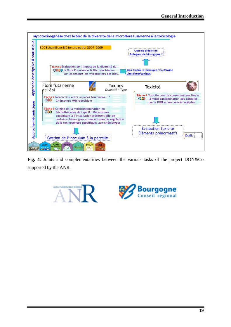
This projects includes four main tasks:

General Introduction
18
Diversity of Fusarium and Microdochium in relation with the types and levels of
mycotoxins in the grains.
An analysis of the mechanisms of production and regulation of toxins by different
chemotypes of F. graminearum and F. culmorum to understand the biological
significance of toxin production in planta.
An analysis of the impact of agronomic factors (waste management, rotation, tillage
system, fungicide) on the balance between species or between populations
(chemotypes) within the Fusarium and Microdochium complexes. A deeper focus
concerns the type of interactions between toxigenic Fusarium and crop residues.
Toxicity to the consumer related to multicontaminations of cereals by DON and its
acetylated derivatives.
A flow diagram illustrates the joints and complementarities between the various tasks of the
project (Fig. 4). My PhD work is part of task 3.
I should mention that the Conseil Regional de Bourgogne contributed to the financial support
of field experiments to promote the regional research for innovation in the frame of Le Plan
d'Actions Régional pour l'Innovation (PARI)-Agrale 7: Impacts des pratiques agricoles sur le
fonctionnement géochimique des sols et la qualité de l’environnement.
The present document will give first a general overview of what are mycotoxins and will
focus more on Fusarium mycotoxins. Following this literature review, we limited our
research using one strain of F. graminearum and one mycotoxin, the deoxynivalenol, as study
models. A chapter is dedicated to the materials and methods used to address the above
mentioned questions. Then we will explain how we dealt successively with a field approach
and a microcosm approach using both classical microbiology, molecular biology and
biochemistry tools. A general discussion will consider all the findings for each of the
approaches to provide the answers on the one hand the impact of deoxynivalenol on bacteria,
fungi, protozoa, nematodes and earthworms present in soil and residues and secondly to
define the putative competitive advantage the mycotoxinogenic fungus gets from this
molecule released into its surrounding environment.
General Introduction
19
300 Échantillons Blé tendre et dur 2007-2009
Appro
che d
esc
ripti
ve
&st
ati
stiq
ue
ToxicitéToxines Flore fusarienneQuantité ~ Type
Tâche 1 Évaluation de l’impact de la diversité de
la flore Fusarienne & Microdochienne
sur les teneurs en mycotoxines des blés.
Tâche 2 Interaction entre espèces fusariennes /
Chémotype /Microdochium
Tâche 3 Origine de la multicontamination en
trichothécènes de type B : Mécanismes
conduisant à l’installation préférentielle de
certains chémotypes et mécanismes de régulation
de la toxinogénèse spécifiques aux chémotypes.
Appro
che m
écanis
tique
Lien flore/toxines
Outil de prédiction Antagoniste biologique ?
Lien Itinéraire technique Flore/Toxine
Tâche 4 Toxicité pour le consommateur liée à
la multi-contamination des céréales
par le DON et ses dérivés acétylés
Évaluation toxicité
Éléments prénormatifs
Gestion de l’inoculum à la parcelle Outils
Mycotoxinogénèse chez le blé: de la diversité de la microflore fusarienne à la toxicologie
de l’épi
Fig. 4: Joints and complementarities between the various tasks of the project DON&Co
supported by the ANR.

Chapter 1
20
Chapter 1: Ecological role of mycotoxins

Chapter 1
21
Ecological role of mycotoxins
1- Introduction:
The chemical machinery of a fungus produces different kind of metabolites and enzymes,
which are necessary to perform the basic metabolic processes and the production of energy.
Apart from these basic needed compounds, some low molecular weight organic compounds
are also produced which are known as secondary metabolites. These chemical compounds are
not needed for the basic requirements of the producer (Fox and Howlett, 2008). The fungal
secondary metabolites may include the pharmaceutically beneficial compounds as penicillin
(Demain, 1999), plant growth regulators as gibberellins (Candau et al., 1992) and as well as
the harmful compounds (for human and animals) known as “Mycotoxins”. These are the
poisons produced by a moderate number of filamentous fungi also known as molds
(Bhatnagar et al., 2002; Yu and Keller, 2005). Mycotoxins are dangerous to the vertebrates
and reported to produce many severe diseases (known as Mycotoxicosis) in human and
animals (Pestka, 2010; Richard, 2007). These chemicals may be acute to chronic poisons
depending on their types, even in very low quantity to the high dose (Bryden, 2007; Döll and
Dänicke, 2011).
The significance of mycotoxins was realized in 1961, with the discovery of first mycotoxin
known as aflatoxin. In 1960, the brutal disease of young turkey birds known as Turkey X
disease killed 100,000 turkeys on the consumption of mold contaminated feed. This was the
first time that the proper cause of this disease by mold contaminated food was recognized
(Desjardins and Hohn, 1997). However, they have always been dangerous to human and
domestic animals in the past. The history of diseases caused by the consumption of these
mycotoxins producing molds contaminated food is very old. One of the famous examples is
Ergotism. Its epidemics called “St. Anthony’s Fire”, led to the death of thousands of humans
in Europe in the Middle Ages (Dotz, 1980). Ergotism is caused by utilization of cereal grains
(usually rye), infected with toxin producing fungi Claviceps purpurea, which produces toxins
known as Ergot alcaloids. Alimentary toxic aleukia (ATA) is another notorious example
which caused the death of more than 100,000 people in USSR from 1942 to 1948 (Pitt, 2000).
Later investigations showed that ATA was caused by eating Fusarium spp. infected grains
which were contaminated by T-2 mycotoxin.

Chapter 1
22
The discovery of mycotoxins opened a new era of importance in the health of Human and
animals and since then hundreds of mycotoxins have been discovered. These mycotoxigenic
(toxin producing) fungi are present all around the world and causing the great loss to the
world’s agriculture (Goswami and Kistler, 2004). The disease caused by these molds, reduces
the seed vigor, crop yield, grain quality, make them poisonous to human use, which leads to
the great economic loss for a farmer as well as to a country (Windels, 2000; Wu, 2007). These
mycotoxins are produced in the cereals, fruits as well as in vegetables, in the field, during
transportation, storage and processing (Drusch and Ragab, 2003; Goswami and Kistler, 2004;
Moss, 2008). The problem of mycotoxins fluctuates from year to year by the changes in the
environmental conditions favorable for the production of mycotoxins and the development of
the producing fungi (Logrieco et al., 2002). The world scientists, decided to limit the
mycotoxin in food stuff consuming for the human and animal, in order to avoid the adverse
effects. European Union (EU) has set the limits for most commonly found Agro-economical
important mycotoxins in different crops including cereals as wheat, maize, oats (The
commission of the European communities, 2007).The text (CE 1126/2007) published by these
EU commission is provided in Annexe at the end of thesis.
The cereal diseases caused by the toxin producing fungi got the world’s attention as the grains
contaminated with mycotoxin reduce the quality as well as the production (Placinta et al.,
1999). About 25% of the world’s food is contaminated by mycotoxin annually. This
phenomenon causes a great loss every year to the cereal producers and exporters. These
fungal toxins are constant threat to the economic losses and the regular danger to the health of
human beings and animals. Once they are produced in the food, it is difficult to eliminate
them during cooking and industrial processing as they are highly stable to heat (Bullerman
and Bianchini, 2007). The vertebrates including animals and human being are greatly affected
by the presence of mycotoxins in the food. The disease symptoms caused by mycotoxins is
known as “Mycotoxicosis”. The investigations show that mycotoxins may cause very serious
effects on the eukaryotic cells. They may inhibit the protein synthesis, DNA and RNA
synthesis, disturb the mitochondrial function, affect the normal cell division (Bin-Umer et al.,
2011; De Walle et al., 2010; Kouadio et al., 2007) and may lead to severe diseases in human
and animals. T-2 toxins are found to induce maternal and fetal toxicity in pregnant mice and
rats (Doi et al., 2008). Mycotoxins may cause acute and chronic poisoning to vertebrates. The
adverse effects of mycotoxins on human and animal health are highly important topic of
research since decades. They may produce disturbance in the immunity system, liver damage,

Chapter 1
23
diarrhea, vomiting, cancer and many other sever diseases (Fokunang et al., 2006; Peraica et
al., 1999; Placinta et al., 1999; Reddy et al., 2010; Ross et al., 1992; Shephard, 2008; Wild
and Gong, 2010). The mycotoxins are not only present in the food and feed stuff, they are
even reported in the indoor air and indoor environment damp including homes and buildings,
and can cause the health risk by inhalation (Hendry and Cole, 1993; Jarvis and Miller, 2005;
Robbins et al., 2000).
In this review, we will investigate the ecological role of mycotoxins in the environment
shared by micro to macro world and prokaryotes to eukaryotes on the basis of the research
conducted up to date.
2- Major Mycotoxins:
More than 300 fungal secondary metabolites are declared as mycotoxins due to their harmful
effects on vertebrates. Most of these mycotoxins are produced by the fungal species belonging
to three genera i.e. Fusarium, Aspergillus and Penicillium. All the species related to these
three genera are not toxigenic. Fusarium is known as the field fungi due to the production of
mycotoxins in the field while Aspergillus and Penicillium are called storage fungi as generally
these two genera are known for the production of toxins postharvest or during the food
storage. The list of some frequently found important mycotoxins and their major producers
are given in the Table 1.
No doubt all the mycotoxins have their importance but the mycotoxins produced on the cereal
are considered more important and extensive data is available on them. These mycotoxins are
produced during the growth of crop while standing in the field, so very difficult to avoid them
from food chain. Among the cereal mycotoxins, trichothecenes, zearalenone and fumonisins
are considered most important cereal mycotoxins from animals and human health point
because of both their toxicity and their frequency of presence in the cereals (González-Osnaya
and Farrés, 2011; Luo et al., 1990; Omurtag, 2008; Thuvander et al., 1999). European Union
has established the limits for some extensively found mycotoxins including deoxynivalenol
(DON), zearalenone (ZEA) and fumonisins (FUM) (The commission of the European
communities, 2007).
Trichothecenes are a large group of agriculturally important mycotoxins containing more than
150 members which are mainly produced by different Fusarium species. Trichothecenes are
accumulated in the kernels of Fusarium spp infected spikelets of many cereal crops including
wheat, maize, barley, rye and oat etc and rendering them inapt for the consumption of human

Chapter 1
24
and animals (Nielsen et al., 2009). Trichothecenes are simple to complex mycotoxins and
divided in four types (A, B, C and D) based on the substitution pattern of EPT (12,13-
epoxytrichothec-9-ene) (reviewed by McCormick et al., (2011)). Type A trichothecenes
(TCTA) and Type B trichothecenes (TCTB) are the most frequently found important
trichothecenes produced by different Fusarium spp (Table 1). Most of them are related to
Fusarium head blight (FHB) disease (Foroud and Eudes, 2009). Type A trichothecenes are
considered more toxic than Type B trichothecenes as T-2 is considered about 10 times more
toxic than DON (Thuvander et al., 1999). These mycotoxins are reported in all the regions of
the world due to the producing fungal adaptation to the different environments in USA, Asia
and Europe (Ward et al., 2008; Yli-Mattila, 2011; Yli-Mattila et al., 2011). Different countries
including USA, Canada and European Union made the legislations against certain frequently
founds mycotoxins of this group. The most frequently found Type A trichothecenes includes
T-2 toxin (T-2), HT-2 toxin (HT-2) and 4,15-diacetoxyscirpenol (4,15-DAS) (Mateo et al.,
2002). The most frequently found major Type B trichothecenes includes nivalenol (NIV),
deoxynivalenol (DON), 3-O-acetyl DON (3 ADON) and 15- O-acetyl DON (15 ADON) (Fig.
1).
DON
15 ADON3 ADON
NIV
Fig. 1: Molecular structures of Trichothecene-B produced by F. graminearum and F.
culmorum

Chapter 1
25
The most commonly reported mycotoxin in the cereal crop is DON and considered one of the
most important member of trichothecenes (Foroud and Eudes, 2009). DON is produced by
Fusarium graminearum and F. culmorum on several cereal crops under wide range of
environments (Doohan et al., 2003). These fungi are associated to the production of the
notorious FHB disease of cereal. FHB has appeared in many outbreaks and caused high losses
in different ages all around the world. DON interrupts the protein synthesis, reduces
nutritional efficiency, reduces immunity system, causes diarrhea and vomiting etc (Fokunang
et al., 2006; Placinta et al., 1999; Sobrova et al., 2010; Wild and Gong, 2010). In the
Fusarium-infected cereals DON is also followed by very important type B trichothecenes i.e.
NIV and 3ADON and 15 ADON but they occur at lower levels than DON. European Union
(EU) has made the legislations against the consumption of DON in our cereal food products.
The maximum limit for DON in unprocessed cereals other than durum wheat, oats and corn at
1250 µg / Kg (1250 ppb) and for maize at 1750 µg / Kg (1750 ppb) for human consumption.
Higher doses are accepted for animal feeding (The commission of the European communities,
2007).
In trichothecene type A T-2 and HT-2 toxins are also of the main mycotoxins. They are very
stable compounds and produced by F. poae, F. sporotrichoides, and F. acuminatum. T-2
toxin, inhibits both protein synthesis and mitochondrial translation (Bin-Umer et al., 2011)
and produces many disease symptoms in animals as alimentary toxic aleukia (ATA), weight
loss, decreases in leukocyte count and blood cell and can seriously damage the liver and
stomach (Weber et al., 2010). The combined temporary tolerable daily intake (TDI) for T-2
and HT-2 is established at 0.06 μg / Kg body weight (Scientific Committee on Food on
Fusarium-toxins, 2002).
Zearalenone (ZEA) is also one of the most commonly found mycotoxins produced by
Fusarium species (Table 1) and co-occurs with trichothecenes in cereals (González-Osnaya
and Farrés, 2011). It is mainly produced by F. graminearum, F. culmorum and F. langsethiae.
It is a non-steroidal estrogenic fungal toxin towards animals and is associated to the grains
infected by FHB in cereal crops. EU has made the legislations also for ZEA due to its
frequent presence in the food and threat to the animals. The limit for ZEA is 100 µg/Kg
(100ppb) in unprocessed cereals other than corn and is 200 µg / Kg in unprocessed maize to
(200 ppb) (The commission of the European communities, 2007).
Fumonisins is a group of mycotoxins mainly divided in four types (i.e. A, B, C and P) on the
basis of their chemical structure. Fumonisins are found all around the world (Marasas, 1996)

Chapter 1
26
and are produced by many Fusarium species including Fusarium verticillioides, F.
moniliforme and F. subglutinans in Europe (Marasas, 2001; Visconti and Doko, 1994).
Fumonisins produce the mycotoxicoses in animals through the interruption of sphingolipid
metabolism by hindering ceramide synthetase (Marasas et al., 2004).These mycotoxins are
reported on many cereals including wheat, maize, barley and sorghum (Visconti and Doko,
1994) but more often in maize. Its limit in Europe is fixed at 2000 ppb for unprocessed maize
and at 200 ppb for the processed maize-based foods and baby foods for infants and young
children (The commission of the European communities, 2007).

Chapter 1
27
Mycotoxin Major fungi Reference
Deoxynivalenol (DON) Fusarium graminearum, F. culmorum (Audenaert et al., 2009; Gang et al., 1998; Manka et
al., 1985; Mirocha et al., 1989; Mirocha et al., 1994)
3- O-acetyl deoxynivalenol (3ADON) F. graminearum, F. culmorum (Audenaert et al., 2009; Manka et al., 1985; Mirocha
et al., 1989; Mirocha et al., 1994)
15- O-acetyl deoxynivalenol (15ADON) F. graminearum, F. culmorum (Audenaert et al., 2009; Mirocha et al., 1989)
Nivalenol (NIV) F. graminearum, F. culmorum, F.poae (Gang et al., 1998; Mirocha et al., 1994; Thrane et
al., 2004)
Zearalenone (ZEA) F. graminearum, F. culmorum, F. sporotrichioides, (Greenhalgh et al., 1983; Manka et al., 1985)
Fumonisins ( FB1, FB2,F B3) F. proliferatum, F. verticillioides ( Marasas, 2001; Ross et al., 1992; Visconti and
Doko, 1994)
T-2 toxins (T-2) F. sporotrichioides, F. poae, F. acuminatum, F. langsethiae (Rabie et al., 1986; Thrane et al., 2004)
HT-2 toxins (HT-2) F. sporotrichioides, F. poae, F. langsethiae (Thrane et al., 2004)
Diacetoxyscirpenol (DAS) F. sporotrichioides, F. poae, F. langsethiae (Thrane et al., 2004)
Moniliformin (MON) F. subglutinans, F. acuminatum, F. concolor, F. equiseti,
F. semitectum, F. acuminatum, F. concolor, F. oxysporum
(Irzykowska et al., 2012; Lew et al., 1996; Marasas
et al., 1986; Rabie et al., 1982)
Enniatin (EN) F. langsethiae, F. tricinctum (Meca et al., 2010; Thrane et al., 2004)
Beauvericin (BEA) F. sporotrichioides, F. poae (Thrane et al., 2004)
Aflatoxin (B1, B2, G1, G2) Aspergillus. flavus, A. parasiticus, A. nomius (Elshafie et al., 2002; Olsen et al., 2008; Yu et al.,
1995)
Ochratoxin A A. ochraceus, A. carbonarius (Kapetanakou et al., 2009)
Patulin Penicillium expansum, P.patulum (Abrunhosa et al., 2001; Aziz and Moussa, 2002;
Northolt et al., 1978)
Table 1: Some commonly found mycotoxins and major producer fungi.

Chapter 1
28
3- Molecular basis of Trichothecenes:
The biosynthesis of mycotoxins is a complex process and mainly depends on the genetics of
fungi. It can be affected by various factors including the environmental conditions and the
nature of the substrate and the external (biotic or abiotic) stress (Paterson and Lima, 2011).
The amount of mycotoxin produced cannot be estimated by the growth of fungi in the kernels
as all the environmental conditions must be taken into account (Shim et al., 2003).
The fungal genetics have a fundamental importance in the mycotoxin type and the amount of
mycotoxin production on the same substrate (Vogelgsang et al., 2008). There can be different
chemotypes within the same species producing specific mycotoxins as well as the different
species can produce the same chemicals (Reynoso et al., 2011; Yli-Mattila and Gagkaeva,
2010). The geographical area determines the chemical phenotype of mycotoxin producing
fungi. Various trichothecene chemotypes are reported to be associated with FHB in the
different parts of the world: 3ADON chemotype of F. graminearum population is dominant in
the major wheat growing area in the North America and is rapidly replacing 15ADON
chemotype (Ward et al., 2008). In Europe 15ADON is reported as the more dominating
chemotype (Jennings et al., 2004; Prodi et al., 2009; Yli-Mattila, 2010). 3ADON and 15-
ADON are more commonly found together in Japan (Suga et al., 2008). The biosynthesis of
trichothecene in Fusarium is controlled by 15 Tri genes located at three different loci: a 12-
gene core Tri cluster locus, a two gene TRI1-TRI16 locus and a single gene TRI101 locus
(Alexander et al., 2009; Merhej et al., 2011). These genes play their roles at different steps of
the pathway of trichothecene biosynthesis which starts from farnesyl pyrophosphate (FPP)
and results in the formation of trichothecene through a series of reactions (Fig. 2).
Calonectrine protein is formed from FPP through a cyclization by the functioning of Tri5,
Tri4, Tri101, Tri11 and Tri3 (Alexander et al., 2011; Alexander et al., 2009; Brown et al.,
2002; Lee et al., 2002). These steps are common in the both T-2 toxin producing F.
sporotrichioides and Trichothecene B (TCTB) producing F. graminearum. The involevement
of Tri13 and Tri7 leads to the formation of 3,4,15-Triacetoxyscirpenol. FsTri1, FsTri16 and
Tri8 lead to the formation of T-2 toxin in F. sporotrichioides. In F. graminaerum FgTri1
genes makes the way to the formation of TCTB either from calonectrine or from 3,4,15-
triacetoxyscirpenol (Brown et al., 2001, 2002; Lee et al., 2002). Tri6 and Tri10 genes are
regarded as regulators genes for the other genes and their deletion in F. graminearum is
reported to reduce the pathogenicity in plants (Seong et al., 2009).The functioning of Tri7 and

Chapter 1
29
Tri13 genes determine the Chemotype NIV while these are not active in DON producing
strains (Brown et al., 2002; Lee et al., 2002). Tri8 gene is found to behave differently in 3-
ADON and 15ADON chemotypes in F. graminearum. In 3ADON chemotypes Tri8 gene
deacetyl 3,15-Acetyl DON at 15 carbon atom to form 3ADON and in 15ADON chemotype it
deacetyl 3,15-Acetyl DON at 3 carbon atom to convert it into 15ADON (Alexander et al.,
2011).

Chapter 1
30
DON
3 ADON 15 ADON
4,15-diANIV
TRI8 TRI7 TRI3 TRI4 TRI6 TRI5 TRI10 TRI9 TRI11 TRI12 TRI13 TRI14 TRI16 TRI11
24 kb
Farnesyl pyrophosphate
trichodiene
isotrichodermol
isotrichodiol
isotrichodermin
15-decalonectrin calonectrin 3,15-diacetoxyscirpenol
3,4,15-triacetoxyscirpenol
3-acetylnesolaniol
3-acetyl T -2 toxin
T-2 Toxin
Tri5
Tri4
Tri-6Tri-10
?
Tri101
Tri11 Tri3 Tri13
FsTri1
Tri7
FsTri16
Tri8
FgTri1
FgTri1
Trichothecene B
7,8-dihydroxycalonectrin
7,8-dihydroxy-TAS
(3,15-ADON) 3,15-diANIVTri13
Tri 7
3,4,15-triANIV
4-ANIV
NIV
FgTri8Trichothecene A
?
??
?
?
?
trichotriol?
Regulators
Fig. 2: A proposed biosynthetic pathway of trichothecene B and T-2 toxin in F. graminearum and F. sporotrichioides and the genes involved at
different steps (adapted from Alexander et al 2009 and Alexander 2011). The question mark (?) indicates unidentified genes.

Chapter 1
31
4-Factors affecting the production of Trichothecene B:
Generally, fungicides are considered to control the growth of pathogen in order to reduce the
disease, ultimately the mycotoxins but by the time they have increased the fungal adaptation
to them (Becher et al., 2010). In practical, fungicides are found in complex relationship with
the disease development and the mycotoxin production (Magan et al., 2002). The use of some
fungicides acts as a stress for fungus and enhance the production of mycotoxins (Mesterhazy
et al., 1999; Moss, 2008). Prothioconazole fungicide can induce H2O2 in the plant cells which
triggers the DON production (Audenaert et al., 2011). The biological components of the
environments affect the production of mycotoxins as the presence of other fungi in the
neighborhood can change the production of mycotoxins (Cooney et al., 2001; Xu et al., 2007).
Apart from the genetics of the fungi, the genetic variability of host plants has also key
importance in the disease production and the mycotoxin production (Mesterhazy et al., 1999) .
The environmental factors including temperature and the water activity have also a great
contribution in the production of mycotoxins by a fungal strain. In the literature, the studies
are based on the production of mycotoxins in the in-vitro conditions. The temperature,
humidity as well as the pH may increase or decrease the fungal growth and development as
well as the production of mycotoxins (Garcia et al., 2011; Kokkonen et al., 2010; Lasram et
al., 2010; Mateo et al., 2002; Medina and Magan, 2011; Mylona and Magan, 2011; Pose et al.,
2010; Price et al., 2005; Ramirez et al., 2006; Schmidt-Heydt et al., 2008; Spadaro et al.,
2010). The filamentous fungi may have different optimal temperature and humidity for the
toxin production than for the optimal conditions required for its mycelia growth. (Ramirez et
al., 2006) studied that F. graminearum showed highest growth at 25°C on water activity (aw)
0.995 while DON production was maximum at degrees 30°C on the same water activity. The
optimal water activity (aw) for the growth of fungus was from 0.900 to 0.995 but for
mycotoxin fabrication was from 0.950 to 0.995. The acidic pH usually favours the mycotoxin
production (Keller et al., 1997; Spadaro et al., 2010). The acidic pH in the de-germed maize
kernel favours the production FB1 mycotoxins than germed tissue in which the pH becomes
basic (Shim et al., 2003). In an in vitro study (Merhej et al., 2010) demonstrated that Tri genes
are stimulated as the pH goes from neutral to acidic and stimulates the production of
trichothecene B (TCTB).
The increase in the resistance in the plants may decrease the production of the disease which
ultimately can be helpful in the reduction of the accumulation of mycotoxins. The

Chapter 1
32
composition of the substrate is the major factor in the mycotoxin production (Bouras and
Strelkov, 2010; Kheiralla et al., 1992; Kokkonen et al., 2005; Mateo et al., 2002; Vogelgsang
et al., 2008).The phase of the kernel development can have an effect on the production of
mycotoxins. The embryonic stage of grains (presence of amylopectin) induces Fumonisin B1
(FB1) production by F. verticillioides and it is not produced in its absence (Bluhm and
Woloshuk, 2005). Some plant molecules are also reported to activate the production of
mycotoxins as polyamines trigger TRI genes expression and to increase the trichothecenes
biosynthesis in F. graminearum (Gardiner et al., 2009). Sugar (sucrose, 1-kestose and
nystose) activates Tri4 and Tri5 expressions and increases DON and 3ADON production in F.
graminearum (Jiao et al., 2008).
5- Ecological role of mycotoxins:
5.1- Mycotoxins in the multitrophic interaction in soil:
It is foremost important to know the place of the mycotoxins in the environment to understand
their possible role. Filamentous fungi are reported all around the world in the wide range of
environment and produce disease in cereals as well as in fruits and vegetables in the field on
the standing crops, after harvest and during storage (Olsen et al., 2008; Pietri et al., 2004;
Placinta et al., 1999; Reddy et al., 2010). The disease incidence and the mycotoxin production
varies from year to year and region to region.
Once the mycotoxins are produced they can get opportunity to flow in the environment. The
production of mycotoxins in the field (as cereals) is difficult to control due to the open
environmental conditions. Mycotoxins from the plants (i.e. grains, spike, spikelets, leaves and
stem) enter to the environment shared by plants, vertebrates, micro and macro fauna and flora
and even the producer themselves. One part of mycotoxins remains in the crop residue in the
fields and the other goes to storage, milling and processing. The flow in the environment
depends on the stability to adverse environmental conditions as temperature, pH, and
solubility. Most of the mycotoxins are usually found resistant to the high temperature and
adverse environmental conditions (Bullerman and Bianchini, 2007; Lauren and Smith, 2001)
and are more or less soluble in the water. These properties make them to flow easily in the
environment.

Chapter 1
33
The crop residues in the field contaminated with mycotoxins are potentially dangerous to the
environment. The rain water may solubilize the mycotoxins and take them to the field soil and
drainage water (Fig. 3). A very few research is done in this potential environmental issue.
Zearalenone (ZEA) is reported in the field soil and drainage water from the wheat
contaminated with F. graminearum (Hartmann et al., 2008). The water samples collected
animal feed processing mills and animal farms have also confirmed the presence of
mycotoxins in the water which may be a threat to the environment (Aragon et al., 2011;
Gajêcka et al., 2011; Gromadzka et al., 2009). The samples collected from various rivers of
Switzerland confirmed the presence of deoxynivalenol and zearalenone (Bucheli et al., 2008).
DON and ZEA were also recovered from the streams of United States (Wettstein et al., 2010).
This may be dangerous to the water life of the river when in high quantity but this is still a
question to be addressed.
The fungal toxins are found to be not only dangerous to the animals who consume them but
they could also be transferred to humans by animal milk (Signorini et al., 2012) which goes
back to the environment and make them risk for human health. Even a significant amount of
DON is reported in the eggs of hens which were fed on the DON contaminated cereal feed
(Sypecka et al., 2004) which is the indirect exposure of humans to the mycotoxins.
Mycotoxins are also recovered from animal feces and human urine (Warth et al., 2012).
During the last few decades, of most part of research on mycotoxin focused on their effects on
human and animals. Unfortunately, the role of mycotoxins in the life of fungi and their need
for the producer fungi is not fully elicited and is still a mystery. By definition, mycotoxins are
secondary metabolites and are not needed for the normal growth, development, energy
production, the metabolic activity and the reproduction of their producers (Fox and Howlett,
2008; Karlovsky, 2008). As they are not needed for the primary functions of a fungi, so their
role in the life of fungi becomes a big question.
Fungi may contest against other organisms in their ecology in diverse ways including growing
faster than competitors, sporulation under stress conditions, hindrance for competitors,
colonization of diverse hosts and substrates. We will try to summarize how these mycotoxins
can help the fungi in life under the light of some basic but important questions and research
taken place up to date.

Chapter 1
34
Processing
Soil
River and Stream water
underground water?
Dis
eas
e
Harvesting
Crop with mycotoxins and
Fungal myceliumHuman and animal
Intestinal microbes?
Fungal and plant genotype,
Temperature, humidity
and pH
myc
oto
xin
Drainage water
Mycotoxigenic fungi (Fusarium spp.)
Environmental or biological degradation?
Storage
Crop Residues left in field
Excretions
soil
Fungi, Bacteria, insects ?
Micro and macro organisms including
decomposers and antagonists?
Fig. 3: Possible flow of mycotoxins in the environment (black coloured circles), its
interaction with other organisms (red coloured circles) .The conditions which can favour
mycotoxin production (blue coloured circle). Single end arrow represents the flow of
mycotoxin or mycotoxin+producer fungi while two end arrow shows the competition between
mycotoxigenic fungus and the other organisms. The question marks and the dotted arrows
indicate the need for further research in these places.
Chapter 1
35
5.2- Do mycotoxins help in disease production or fungal development and
colonization on plants?
In general, most of the mycotoxin producing fungi are plant pathogens. So, the general
perception is that mycotoxin helps in the production of disease. The role of mycotoxin in the
primary infection is investigated extensively. All the reports shows that mycotoxins are not
involved in the primary fungal infection on the plants for the disease production. The
mycotoxin non producing mutants showed the same disease production as the mycotoxin
producing wild type fungi (Bai et al., 2002). On the other hand, mycotoxin producing
pathogens are found more virulent than their mutagens. In some studies NIV and DON are
reported to play a significant role in the virulence of pathogen fungus and help in colonization
in the plant tissue. In Fusarium spp. trichothecenes producing genes are activated and the
mycotoxin production is increased during the colonization of stem and grains in cereals. (Bai
et al., 2002; Desjardins et al., 1996; Hallen-Adams et al., 2011; Harris et al., 1999;
Mesterhazy et al., 1999; Mudge et al., 2006). Wang et al., (2006) described that disrupted
DON production in F. graminearum decreases the aggressiveness on wheat, barley and
triticale seedling root infection. Mycotoxin producing fungi are found more aggressive than
the mycotoxin non producing fungi. F. culmorum isolates producing DON were found more
aggressive than the isolates producing NIV in wheat crop (Muthomi et al., 2002). This
indicates that chemotype of a fungus also has a significant importance in the aggressiveness
of a pathogen during disease development process. Previously it was also reported that
hydrogen peroxide (H2O2) which may induce the programmed cells death is induced in the
plant cells in response to the release of DON by fungus (Desmond et al., 2008; Gechev and
Hille, 2005). The fungal toxins activate the cell death in the host cells which may help
colonization the fungus (Desmond et al., 2008).
This discussion concludes that the mycotoxins are not involved in the production of disease
but increase the aggressiveness of pathogen which helps the fungus rapid colonization. This
rapid colonization may give an advantage to mycotoxin producer fungus over the other
competitors during plant infection. Thus the fungus may occupy large part of plant material
before it becomes ecological habitats in the soil which possibly help the fungus during its
subsequent saprophytic survival.

Chapter 1
36
5.3 - Do mycotoxins help in fungal survival under environmental stress?
This is one of the most important question comes in our mind about the role of mycotoxins in
the fungal life which was completely ignored previously. The production of mycotoxins is
affected by many environmental and biological stresses as we described earlier. Various
environmental stresses increase the production of mycotoxins. This gives us a clue about the
role of mycotoxins in the fungal life under stress. This hypothesis can be supported by the
research argument which indicates that the sporulation and secondary metabolites are
genetically connected and controlled by the common factors. For example the asexual spore
production and the mycotoxins i.e. sterigmatocystin and aflatoxin production in Aspergillus
spp. are related to the same genetic basis (Hicks et al., 1997; Kale et al., 1994; Kale et al.,
2003). A direct relationship was observed in the conidia production of Claviceps purpurea
and toxic alkaloids on agar plates (Pažoutová et al., 1977). It has also been investigated that
the genes (FCC1) involved in the signal transduction are the same for regulating fumonisin
biosynthesis as well as fungal conidia formation in F. verticillioides (Shim and Woloshuk,
2001).
The correlation between mycotoxins production and sporulation points out hypothesis that
mycotoxins might be involved in the survival of fungus under adverse conditions. The
production of secondary metabolites including many Fusarium and Penicillium mycotoxins
and the sporulation are regulated by the same signaling G-proteins signalling pathways
(reviewed by Brodhagen and Keller, (2006)). Zearalenone takes part directly or indirectly in
formation of perithecia in Gibberella zeae (teleomorph of F. graminearum) (Wolf and
Mirocha, 1973, 1977). The stress may stimulate the production of zearalenone (Kim et al.,
2005) and can serve as a fungal tool for survival in the adverse conditions. On the other side,
this perithecia formation was found to be highly dependent on mycotoxin dose. A high dose
of zearalanone may inhibit the spore formation of the producer fungi (Mirocha and Swanson,
1983). This may help in the increasing fungal inoculum for infection and help in survival.
Further investigations are needed in this regards in order to suggest the better control of fungi
producing environmental threat.

Chapter 1
37
5.4- Do mycotoxins help in competition with other organisms?
The above studies give us an indication to establish hypotheses about the role of mycotoxins
in its ecology. It is usually believed that secondary metabolites are produced for the self
defense against the other organisms (Kempken and Rohlfs, 2010) but this hypothesis is also
still ignored in the case of mycotoxins. The mycotoxin producing fungi and their toxins
interact with other micro and macro organisms (including fungi, bacteria, nematodes,
protozoa, earthworms and arthropods) in their ecology as shown in Fig. 3. They come in
contact whether on the plant during disease development, during storage or during the
saprophytic survival in crop residues. The nature of interaction between them is still a
question.
A general prospective is that mycotoxin may be one of the tools which can be used by the
producer during the competition for food and survival against other fungi. Losada et al.,
(2009) reported that the competition with other fungi augmented the antifungal secondary
metabolites in Aspergillus spp. In another study, (Ramakrishna et al., 1996) found that during
a competition between T-2 toxin producing fungi F. sporotrichioides and two other fungi i.e.
A. flavus and Penicillium verrucosum the T-2 toxin significantly increased in barley grains.
The growth of F. sporotrichioides was negatively affected during this competition but it
stimulated the production of T-2 mycotoxin. What is not clear in this study is to know if the
two other competitors were negatively affected or not by the T-2 toxin and this toxin allowed
F. sporotrichioides to survive. Therefore the role of mycotoxin is not so clear. F.
graminearum mycotoxin production was 1500 % increased in the presence of F. culmorum
and F. poae (Xu et al., 2007). They also found that the mycotoxin production is changed
during the competition among two chemotypes. Cooney et al., (2001) found the production
of DON by F. graminearum was affected negatively or positively in the presence of other
Fusarium species. Recently, mycotoxin production and the growth of Alternaria tenuissima
isolates was studied in relation to F. graminearum and F. culmorum. The growth of
Alternaria and the Alternaria toxins (AOH, AME and ALT) were depressed and the DON and
ZEA production was increased (Müller et al., 2012).
The impact of these mycotoxins on the other fungi and the mechanism of action on other
fungi still need to be explored. Lutz et al., 2003 tested the impact of DON against a famous
fungal antagonist Trichoderma atroviride. They reported that chitinase gene expression was
repressed by the effect of DON. In another study, the impact of trichothecenes on yeast cells

Chapter 1
38
was observed by McLaughlin et al., (2009) and later by Bin-Umer et al., (2011). The results
illustrated that these mycotoxins inhibit the mitochondrial membrane potential and reactive
oxygen species, depending on the dose of mycotoxins. The higher dose of trichothecene can
inhibit the mitochondrial translation.
Bacteria are the most important competitors of fungi in the food web. They compete by using
the food resources or the production of chitinase enzymes. some mycotoxins including patulin
and penicillic acid, produced by Penicillium spp, are reported as quorum-sensing inhibitor
(QSI) compounds against Pseudomonas aeruginosa which may provide the fungus a
protection from the chitinase producing bacteria (Rasmussen et al., 2005). The growth of
rumen bacteria Ruminococcus albus and Methanobrevibacter ruminantium were reported to
be inhibited in the presence of Fusaric acid but not effected in the presence of DON or in the
combination of DON+Fusaric acid (May et al., 2000). More studies are needed in this context
about the relation between fungal mycotoxins and their impact on the bacterial growth on the
different food resources.
Nematodes are one of the active members of the soil web and are potential competitors of
mycotoxin producing fungi in soil and the plant debris or crop residues. They decompose the
organic matter including crop residues. A very little is deliberated on the role of mycotoxins
on the nematodes. In an in vitro experiment some mycotoxins including T2-toxin,
moniliforrnin, verrucarin A and cytochalasin B are reported to reduce the viability of
nematodes significantly. They were even died by the presence of mycotoxins but the rate of
mortality depends on the mycotoxin concentrations (Ciancio, 1995). Two trichothecene
compounds i.e. 4,15-diacetylnivalenol and diacetoxyscirpenol were described to reduce the
mobility in second-stage juveniles which were hatched in the presence of these mycotoxins
(Nitao et al., 2001). In a liquid medium DON reduced the egg hatching and the growth in the
Caenorhabditis elegans after their birth from eggs(Gowrinathan et al., 2011).
Insects are the primary decomposers of the crop residues in the soil. Mycotoxinogenic fungi
have to compete with insects on plants during food storage and as well as in the crop residues
decomposition. The mycotoxins are found to have the ability to control the insects by
infecting at different stages of insect life cycle. They have insecticidal effects depending on
the type and quantity of mycotoxins (Wright et al., 1982). Mycotoxins may give the fungi an
advantage against the fungivor insects. Indeed the latter are reported to prefer the non
mycotoxin producing fungi over the mycotoxin producing fungi (Rohlfs et al., 2007). Honey
bees are also found to be negatively affected by the presence of aflatoxin B1 (AB1) and

Chapter 1
39
ochratoxin A (OTA), which depends on the concentration and the time of exposure to the
mycotoxins (Niu et al., 2011). NIV, DON and FB1 are found to be cytotoxic to the SF-9
insect cells and can change the normal progression of the cell cycle and induce an apoptotic
process (Fornelli et al., 2004).
5.5-Are mycotoxins needed for the saprophytic survival?
When crop are harvested, whether they are contaminated or not with mycotoxins, the crop
residues were generally left in the field. They can be left at the surface or buried in the soil
depending on the tillage system used. All micro flora and micro meso fauna we discussed are
more or less involved in the colonization exploitation and decomposition of such trophic and
spatial resources. Among the colonizers the filamentous fungi including plant pathogenic ones
and mycotoxigenic ones play an important role in the decomposition process. They can
subsequently over winter and develop the primary inoculum for the next season. Therefore,
the place of mycotoxins and mycotoxin producers is worth being investigated.
Soil is a huge pool of different kind of microorganisms depending on the type of soil. One
gram of soil may contain 1 µg to about 200 µg of DNA containing virus, bacteria, fungi,
protozoa and nematodes etc in different ratios depending on the soil (Trevors, 2010). When
these residues become in contact with the soil they become the ecological habitats for the
mycotoxin producing fungus. These residues and the mycotoxin producer fungi are attacked
by other decomposers and antagonists. In that contest a review of literature was performed to
address the behavior of mycotoxigenic fungus Fusarium graminearum during its saprophytic
phase in the crop residues (Leplat et al., 2012). Despite of this competition for the food
filamentous fungi are reported to survive for many years in these crop residues and remain a
permanent threat.
However, the role of mycotoxins in the fungal defense, substrate colonization and
development, and competition with other soil biota still remains a question. May be these
mycotoxins are used as a chemical weapon for the fungus in the saprophytic survival in the
crop residues during off season.

Chapter 1
40
6-Detoxification of mycotoxins:
Mycotoxins are found highly resistant to the environment and not easily degraded even by
high temperature. A lot of researches are going on in regards to the detoxification of
mycotoxins on the plants by themselves and by the microorganisms.
Plants produce UDP-glucosyltransferase (UGT) enzyme which can detoxify DON and
15ADON but have no impact on nivalenol accumulation (Poppenberger et al., 2003). UGT
enzyme is induced in the presence of DON and converts it into DON-3-O-glucoside (D3G)
which leads to the resistance in the plants against DON (Poppenberger et al., 2003; Shin et al.,
2012).
Microorganism can be an effective source of detoxification of mycotoxins (Wu et al., 2009).
Usually it may depend on incubation time, temperature and pH and can be stimulated or
lessened by the presence of different minerals (Tinyiro et al., 2011). The bacteria are found to
be good candidates in this regards. Aflatoxin B1 is degraded by a gram positive bacteria
Rhodococcus erythropolis (Alberts et al., 2006) and by gram negative bacteria
Stenotrophomonas maltophilia (Guan et al., 2008). S. maltophilia activity increased with the
increase in temperature (up to 37° C) and the increase of pH (up to 8). The minerals also
played a significant role. Mg2+
and Cu2+
activated the degradation of Aflatoxin B1 and Zn2+
strongly inhibited it. This shows that the conditions are very important for the mycotoxin
degradation. So it might not be necessary for each mycotoxin degrading bacteria to degrade
the same mycotoxin at each place with varying conditions. The bacteria Bacillus subtili and B.
natto strains (B. subtilis 168 and B. natto CICC 24640) which are usually found in the food
stuff are important decomposers of zearalenone and therefore can be useful to reduce
mycotoxins from the food (Tinyiro et al., 2011). The soil has many kind of degrading bacteria
which are reported to degrade different mycotoxins. Sato et al., (2012) isolated 13 bacteria
aerobic (including gram-negative and gram-positive) bacteria from different environmental
samples (2 bacteria from wheat leaves and 11 from soil) having good ability to degrade DON
by converting it into 3-epi-DON. Soil-borne and organic matter resident bacteria Bacillus
licheniformis strain was found to reduced zearalenone up to 95.8% and 98% in Luria–Bertani
(LB) medium and in corn meal medium after 36 hours of incubation at 37° C (Yi et al., 2011).
Soil-borne bacteria Nocardioides sp strain WSN05-2 was found to use the DON as a sole
source of carbon and convert the DON into 3-epi-DON as an intermediate product in its
degradation (Ikunaga et al., 2011).

Chapter 1
41
There are not many reports about participation of fungi in the mycotoxin degradation. Only
some Rhizopus spp are found the good decomposer of ochratoxin A (OTA) (Varga et al.,
2005). Earthworms are considered the important decomposers, of soil and especially in the
agriculture point of view. Deoxynivalenol (DON) is reported to be degraded by earthworm
species as Lumbricus terrestris and Aporrectodea caliginosa (Oldenburg et al., 2008;
Schrader et al., 2009; Wolfarth et al., 2011).
So, although there are few examples of plants, microorganisms and earthworms which can
potentially degraded mycotoxins in given controlled conditions, the fate of DON in the crop
residues and in soil itself is not really known. Besides, the biological degradation illustrated in
previous example and the leaching underlined in the section 5, mycotoxins could potentially
accumulate through the adsorption mechanism on the organic matter in the soil but nothing is
known on that point too.
7- Conclusion and research prospects:
Apart from their impact on human and animal health a very few is known on the mycotoxins
in the environment. However they can be present in all the compartments of the environment
and many biotic agents may interact with positive and negative consequences on their fate
(Fig. 1). Owing to the complexity of the flow of mycotoxins along the trophic webs, one may
question about the ecological role of the mycotoxins in the environment. In the frame of
controlling the primary inoculum of F. graminearum responsible for the Fusarium head blight
on the wheat, we decided to focus more specifically on the role of DON towards micro flora
and micro meso fauna in relation to the fate of this pathogenic fungus in its habitat in the crop
residues. Therefore, in the first part of the thesis, I will describe the materials and methods we
used during the study. Then first study was conducted in the field which raised some
questions which needed to be addressed more deeply. So, the second part was conducted in
the microcosms in the controlled conditions to see in which way the mycotoxins affect biotic
inhabitants of the soil.

Chapter 2
42
Chapter 2: Materials and Methods

Chapter 2
43
Materials and Methods
1 - Fusarium graminearum strain MIAE00376:
F graminearum strain MIAE00376 was chosen for all the microcosm and field experiments.
This strain was taken from the fungal and bacterial collection “Microorganisms of Interest for
Agriculture and Environment” (MIAE), UMR-Agroécologie, INRA Dijon, France,
(http://www2.dijon.inra.fr/umrmse/spip.php?rubrique47).
2 - Production of conidial suspension:
Conidia were produced by the method adapted from (Hassan and Bullerman, 2009). Briefly,
the wheat bran liquid medium was prepared by adding 1% wheat bran in the distilled water
and was autoclaved at 120°C for 20 min. The small plugs of 7 days old cultures of F.
graminearum strain MIAE00376 grown on PDA were added and placed at 25°C on a rotary
shaker (150 rpm) for seven days. The cultures were filtered through sterile cheesecloth
(approximately 50–µm mesh size) to remove the mycelial mat and the remaining pieces of
bran and conidial concentration was assessed through Malassez counting chamber under the
microscope. The concentration (6.67 × 10² conidia/ml) was then adjusted by dilution with
sterile distilled water.
3 - Chemotyping of Fusarium graminearum strain MIAE00376:
Objective:
F. graminearum can potentially produce different mycotoxins including deoxynivalenol
(DON), 3–acetyl–4–deoxynivalenol (3-ADON) and 15–acetyl–4–deoxynivalenol (15
ADON), nivalenol (Niv) and zearalenone (ZEA) according to the various factors that are not
always known. The geographical distribution induces some populations to produce one set of
mycotoxins and the other populations the other mycotoxins. Each strain should therefore be
characterized for its chemotype pattern. Therefore, the objective of this assay was to
determine the chemotype of F. graminearum strain MIAE00376 to use in all the experiments.
Procedure:

Chapter 2
44
This assay was performed on the grains of three varieties of winter wheat (Triticum aestivum)
of different levels of susceptibility to Fusarium head blight (Maxwell > Charger >Apache
respectively, GEVES data) and wheat bran (unknown susceptibility). Their humidity was
adjusted up to 45% and then they were disinfected by autoclaving at 120°C for 20 min on
three successive days.
They were aseptically transferred into the small tubes of 50 ml in such a way that each tube
contained 15 g of wheat grains or 6 g of wheat bran. F. graminearum MIAE00376 conidia
were prepared with the method described above for the conidial suspension production. 2.5 ×
103 conidia/g of grains or wheat bran were distributed in each of the tube. The samples were
incubated at 25°C in an incubator. Three replicates were made for each modality for each
sampling time. Some tubes containing each of the substrate were left without inoculation as
controls.
Sampling was done at 0, 4, 10, 21 and 60 days of incubation and placed at -20°C until
mycotoxin extraction and further analysis. At each sampling time, the grains were ground and
homogenized to access the content of the grains. Humidity of each replicate was measured
with infrared balance (Mettler Toledo LJ16 Infrared Moisture Analyzer). Then mycotoxins
were extracted for each sample.
For the extraction of mycotoxins, 3.5 g of ground grains or wheat bran were taken in 50 ml
tubes and 10 ml of extraction solvent acetonitrile ((ACN)/H20mQ (84/16, v/v)) were added.
The tubes were mixed by shaking manually and incubated at 250 rpm at 25°C in the rotary
shaker for 3 h. Tubes were centrifuged for 3 min at 4500 rpm. Then 5 ml of the supernatant
from each tube were filtered by the purification/exclusion column P-5 (R-biopharm, France).
2 ml of that filtrate was transferred in 4 ml vials. The organic phase was evaporated to
dryness.The dry extract was dissolved in 200 µl of MeOH/H2O (1/1, v/v) by vortexing for 1
min. The samples were stored at -20°C until further analysis.
The analyses were performed by high performance liquid chromatography (HPLC, Beckman
System Gold) coupled with a diode array detector (DAD) and managed by the 32 Karat
software (v. 5.0). TCTB and ZEA were separated along a column HICHROM Ultrasphere
5octyl (150 mm × 4.6 mm, particle size 5µm) non-thermostated. A HICHROM Pre-Column
Ultrasphere Octyl Guard® (4.6 X 45 mm; particle size 5µm) was used as a guard-column.
Chromatographic system was ultra pure water pH 2.6 (eluent A) and ACN (eluent B). The
gradient started with 5% B. From 0 to 14 min, it linearly increased to 30% B. Over further 10
min, there was another linear increase to 90% B. This phase was kept up to 29 min. The

Chapter 2
45
gradient was then linearly decreased to 5% B during 2 min. Finally the column was
equilibrated for 14 min before next injection. The flow rate was set at 1ml/min. The injection
volume was 20 µl. The chromatograms were recorded for λ = 230, λ = 280 and λ = 320 nm. λ
= 230 nm was used for detection and quantification of DON. Retention times were 6.4, 8.7,
14.4, 14.8 and 23.6 for NIV, DON, 15ADON, 3ADON and ZEA respectively.
Results:
Only one mycotoxin i.e. DON was found to be produced during the experiment in all the
grains whether they were resistant or susceptible. The presence of other mycotoxins was not
detected. So, F. graminearum strain MIAE00376 was found as DON chemotype. This
experiment didn’t allow to determine it more precisely (15 or 3 ADON one). Owing to the
fact that this strain was isolated in France so it is quite likely that this strain should be 15
ADON which is more dominating in Europe (Jennings et al., 2004).
DON is detected after 10 days of incubation in the grains of 2 wheat varieties i.e. Maxwell &
Charger. Production of DON by MIAE00376 was detected on Apache, which is the less
susceptible of the three tested varieties, after 21 days of incubation. This production was
directly proportional to the susceptibility up to 21 days. After 60 days this trend remained for
two varieties Apache and Charger while in the case of Maxwell the DON concentration
remained almost the same as in the 21 days. DON was not detected in wheat bran till 60 days
of experimentation (Fig. 1). Anyway, this strain was not a NIV chemotype.
0
10
20
30
40
0 4 10 21 60
DO
N (µ
g/g
of
dry
su
bst
rat)
Time of incubation (days)
Charger
Apache
Maxwell
Wheat bran
Fig. 1: Quantity of DON produced by F. graminearum strain MIAE00376 in the grains of
three wheat varieties of different level of susceptibility to Fusarium Head Blight (Maxwell >

Chapter 2
46
Charger >Apache respectively) on different time intervals up to 60 days and
detected/quantified by HPLC-DAD.
4 - Soil
4.1- Field experiments
Field trials conducted during 2010-11 and 2011-12 campaigns were carried out on the field of
Epoisses, experimental unit of INRA Dijon, 21120 Bretenière (5°05''E, 47°14''N). The estate
is located in the plain, 15 km south of Dijon. Epoisses soil is a silty clay 54% silt, 38% clay,
8% sand, 2.3% organic matter, pH 8.
4.2- Microcosm experiments
The soil was collected from the meadow area near green house INRA, Dijon. The physico-
chemical characteristics of this soil are close to the ones of the soil of the experimental unit of
Epoisses where field experiments were conducted. This soil, instead of the one of Epoisses
was sampled for practical reasons. This soil is also a silty-clay with 39.2% clay, 45.6% silt
and 15.2% sand, 2.05% organic matter and pH 8. The soil C/N ratio was 9.85 and Cation
Exchange Capacity was 21.6 cmol(+) kg-1
soil. It was sieved at 4 mm to remove gravels and
the crop debris. A homogenous mixture of the soil was prepared by mixing manually. The
humidity of soil was measured and adjusted to 17 % w/w i.e. 80% of its water holding
capacity.
5 - Production of the field Inoculum:
5.1 - Maize inoculum:
Air dry maize stems were cut into 2-3 cm long pieces. Half of them were put in plastic boxes
(65X30X17cm, Fig. 2a) and inoculated with plugs of 10 days old F. graminearum strain
MIAE00376 grown on PDA culture. They were left for 3 weeks at room temperature. They
were mixed manually in order to homogenize the inoculum once in a week (Fig. 2b). Half of
the 2-3 cm pieces of maize stem were left non-inoculated.

Chapter 2
47
a
Fig. 2a: fragments of maize stems used for
inoculating field experimental plots
Fig. 2b: fragments of maize stems colonized
by the inoculated F. graminearum strain
MIAE00376 (characteristic pink color) 3
weeks post incubation.
Fig. 2c : nylon bags containing the fragments
of maize recovered after in situ incubation
5.2 - Nylon made bags:
Air dried maize stems were taken from the field area of Epoisses, and they were cut into small
pieces of about 2-3 cm. They were disinfected by autoclaving twice at 120°C for 20 min.
These stubbles were dried under laminar flow chamber in sterilized conditions for 2 days.

Chapter 2
48
Half of them were inoculated with F. graminearum strain MIAE00376 by dipping them in the
conidial suspension (106
conidia/ml) for two days and half were dipped in the sterile water to
give the same water treatment and were kept uncontaminated. Both were placed at 25°C in the
rotator shaker incubator at 125 rpm for 2 days in order to make the homogenous treatment for
all the maize stubbles. These two types of maize stubbles were dried under laminar flow
chamber in the sterile conditions separately. About 2.1 g (d.w.) (2 to 5 pieces) of these maize
stubbles were filled in the nylon gauze bags with dimensions 15 × 10 cm (length × height)
(Fig. 2c). Half of the bags were filled with inoculated stubbles and remaining half were filled
with non-inoculated maize stubbles. Half of the bags contaminated with F. graminearum and
half of the non-contaminated bags were contaminated with the solution of DON (Sigma
Aldrich, CAS No. 51481-10-8) by injecting the required amount in equal amount in all the
stubbles in a bag. Each DON contaminated bag contained 175µg DON / g (d.w.) of maize
stubbles. In this way, four kinds of bags were prepared depending on the treatment on the
maize stubbles. 1- Inoculated with F. graminearum, 2- Inoculated with F. graminearum and
contaminated with DON, 3- Contaminated with DON, 4- With no treatment.
6 - Earthworms (Lumbricus terrestris) collection:
Lumbricus terrestris adult specimens were caught from the meadow situated near green house
area of INRA Dijon, France. These earthworms were compelled to come on soil surface from
the deep soil by using water and chemical expellant “Formalin” (Bouché, 1972).
Several trees were selected and one square meter soil area was marked around each of them.
The grass in that marked area was cut up to the ground level. The marked areas were
humidified by spraying about 100 l of tap water per square meter. Two days later, 20 l of 0.5
% formalin solution was sprayed per square meter of soil, which forced earthworms to come
at the soil surface. These earthworms were caught and collected from the ground surface. This
step was repeated twice for each marked area to catch maximum number of earthworms. The
earthworms were washed immediately twice by tap water to remove the formalin from their
skin. They were put in the soil in different containers and were placed at 10°C for 3 weeks.
At the time of launching the experiment, earthworms were put in the experimental soil at
17°C for 4 days. Biomass of earthworms was noted for each microcosm before putting them
into the microcosms.

Chapter 2
49
7 - Extraction and quantification of nematodes:
The extraction and the counting of nematodes were carried out using the elutriator described
by Oostenbrink (1960). In order to extract the nematodes from each sample, 250 g moist soil
or soil-straw mixture were put into a bucket containing 1 l water. The contents in the bucket
were stirred to make the suspension and sieved at 1 mm sieve to remove the big fragments of
straw. Then the sieved soil suspension containing nematodes was put in the elutriator using an
upward flow of water (flow rate: 35 l/h) to accelerate the natural process of sedimentation of
the coarse particles. After 15 min, the soil fine particles along with nematodes were collected.
When the elutriator was completely filled, the contents were poured into a bucket and sieved
using four stacked sieves of 50 µm. The contents that didn’t pass through the sieves were
containing the nematodes, which were collected and transferred to a sieve containing a pre-
moistened tissue paper. The sieve was placed in a large Petri dish containing water in an
amount that could keep the permanent humidification of the sieve and migration of
nematodes. The nematodes migrated below the sieve into the Petri dish.
After 48 h, the contents of the Petri dish were poured in a graduated cylinder to the volume up
to 25 ml. Then 2 ml were spread on a plate grid specific for counting the individuals under a
dissecting microscope. This counting was performed 2 times for each sample. For each
sample, 2 ml of the extracted nematodes suspension was filled in the Eppendorf tubes and
placed at -20°C for DNA extraction.
8 - Direct counts of bacteria and fungi (Colony Forming Units):
At each sampling time, total cultivable densities of fungi and bacteria were estimated by
colony forming unit (CFU) on the appropriate media.
Briefly, 5 g of soil or soil-straw mixture were added to 45 ml of sterile water and shaken for
20 min in a three dimensional shaker at 700 oscillations/min. Tenfold dilutions were made for
each sample from the mother suspension. Fungi were quantified on MEA (malt extract 10 g/l,
agar 15 g/l) supplemented with citric acid (250 mg/l), and antibacterial antibiotics
(chlortetracycline 50 mg/l and streptomycin sulphate 100 mg/l). Bacteria were quantified on
YPGA (yeast 5 g/l, peptone 5 g/l, glucose 10 g/l, agar 15 g/l) supplied with cycloheximide (50
mg/l). Three repetitions for the bacteria and 5 repetitions for the fungi were performed.
Bacterial and fungal densities in all modalities on each sampling time were compared by
analysis of variance (ANOVA) using XLSTAT-Pro version 7.1 (Addinsoft).

Chapter 2
50
9 - Extraction of DNA:
Objective and principle:
The objective of this method was to extract total genomic DNA from 2 g of different
substrates (i.e. soil, straw, soil-straw mixture and maize stubbles) or 2 ml of nematode
suspension and to obtain non-degraded DNA. We needed DNA purified enough so that the
impurities do not interfere with enzymatic reactions (PCR amplifications).
In this method, stored and frozen samples were thawed in extraction buffer. Lysis was
mechanical (stirring in the presence of beads) and chemical denaturation of proteins and lipids
by the action of the detergent sodium dodecyl sulfate (SDS) at high temperature followed by
precipitation of complex protein/lipid-SDS denatured. The different components of the
extraction buffer had the following roles: the high concentration of
Ethylenediaminetetraacetic acid (EDTA) chelates the majority of divalent cations (Mg2+)
cofactor of many DNAses; the solution was buffered by Tris pH 8.0; the concentration of
NaCl limited the possible partial denaturation of DNA at 70°C and released many DNA-
protein interactions; SDS facilitated the lysis of membranes and protein denaturation. To
separate the DNA from denatured proteins, deproteinization was to precipitate the proteins in
the presence of a high salt concentration. This method had two advantages: the DNA-protein
interactions were destroyed and the products were not toxic (unlike organic solvents, phenol
and chloroform). The precipitates were separated by centrifugation at high speed. The clear
supernatant contained the DNA. DNA was precipitated with isopropanol and washed with
ethanol and two final purification steps were performed i.e. Polyvinylpyrrolidone (PVPP) and
Geneclean, which were used to eliminate possible polluting elements (proteins, sugars,
phenolic macromolecules, humic acids, etc). The PVPP polymer of high molecular weight
made a complex with phenolic compounds and alkaloids. DNA bound to silica columns
(Geneclean) in the presence of high concentrations of salts and was then eluted at low salt
concentrations.
Extraction method:
The DNA was extracted from different substrates (i.e. soil, straw, soil-straw mixture and
maize stubbles) by a procedure adapted from (Edel-Hermann et al., 2004). Two g of freeze
dried and ground soil or soil-straw mixture or 0.5 g of freeze dried and ground straw or maize
stubbleswere added to a 15 ml tube containing 4 g of 1.4 mm diameter silica beads, 5 g of 0.1
mm diameter ceramic beads and eight 2 mm diameter glass beads. Then 8 ml of lysis buffer

Chapter 2
51
containing 100 mM Tris HCl (pH 8.0), 100 mM EDTA (pH 8.0), 100 mM NaCl and 2% (w/v)
SDS were added to each sample. Samples were shaken for 90 s at 4 m/s in a Fast Prep-24
Instrument (MP Biomedicals, Illkirch, France) and incubated for 30 min at 70°C with mixing
after 15 and 30 min. Samples were centrifuged at 7000 × g for 5 min at 15°C. The
supernatants were recovered in 2 ml microcentrifuge tubes and incubated for 10 min on ice
with 0.1 volume of 5 M potassium acetate. After centrifugation at 14000 × g for 10 min at
4°C, the nucleic acids in the collected supernatants were precipitated with one volume of ice-
cold isopropanol for 15 min at -20°C. The precipitate was pelleted by centrifugation at 15000
× g for 20 min at 4°C, washed twice with 70% (v/v) ice-cold ethanol, air-dried and dissolved
in 200 µl of 10 mM Tris HCl (pH 8.0). Out of 200 µl, only 100 µl of extracts of soil nucleic
acids were used for the purification and 100 µl were freezed at -20°C for further use if
needed.
These extracts of soil nucleic acids were purified twice by using a PVPP spin column to
remove co-extracted humic acids. The two purification steps were miniaturized using Micro
Bio-Spin columns (Bio-Rad, Marnes La Coquette, France) adapted to microcentrifuge tubes.
For the preparation of PVPP columns, Micro Bio-Spin columns were filled with 93-95 mg of
PVPP (Sigma-Aldrich, Saint Quentin Fallavier, France). The spin columns were washed twice
by the addition of 400 µl of sterile water and centrifugation at 2000 × g for 4 min at 10°C.
The columns were plugged at the bottom and 400 µl of sterile water were added. Before use,
the PVPP spin columns were centrifuged at 2000 × g for 4 min at 10°C. 100 µl of crude DNA
extract were loaded slowly onto the top center of the PVPP spin column and the purified
extract was collected after 5 min of incubation of the column on ice, followed by 4 min of
centrifugation at 1000 × g at 10°C. Then, the DNA extract was purified using a Gene clean kit
(MP Biomedicals, Illkirch, France).
10 µl of the purified DNA extracts, were resolved by electrophoresis in a 0.8% agarose gel in
Tris-acetate-EDTA (TAE) buffer, together with dilutions of calf thymus DNA (Bio-Rad).
Gels were stained with ethidium bromide, photographed under a camera and the staining
intensities were measured with Bio-1D++
software (Vilber-Lourmat, Marne-la-Vallée,
France). The DNA concentrations in the DNA extracts were calculated using a standard curve
of 25-150 ng of calf thymus DNA versus intensity.
DNA extractions from the nematodes-water suspensions were carried by using the same
procedure as used for the soil with the following exceptions: 2 ml of nematodes-water
suspension were used instead of 2 g of soil and 2 ml of 2 times more concentrated lysis buffer

Chapter 2
52
were used for extraction of nucleic acids. The dry DNA was dissolved in 30 µl of 10 mM Tris
HCl pH 8.0. For the purification it was not passed though the PVPP spin column but only
purified by the Gene clean kit. The pure DNA obtained was quantified and stored at -20°C
until it was further used for T-RFLP analysis.
10 - Fungal, bacterial, protozoan and nematode community
structure analysis by terminal restriction fragment length
polymorphism (T-RFLP):
Principle:
The analysis of ribosomal DNA (rDNA) by T-RFLP (terminal restriction fragment length
polymophism) allows the characterization of communities by revealing length polymorphism
of terminal restriction fragments.
For this purpose, PCR amplification of the rDNA was performed on the DNA template
complex using fluorescently labeled primers specific for the community of fungi, bacteria,
protozoa and nematodes. The amplified products were digested with restriction enzymes, and
then labeled terminal restriction fragment were separated and analyzed by a sequencer.
Method:
The structural changes in the microbial and microfaunal communities during the straw
decomposition were determined by using the DNA based technique T-RFLP.
The fungal communities were amplified by targeting the internal transcribed spacer (ITS) of
the rDNA with the primers ITS1F (CTTGGTCATTTAGAGGAAGTAA) (Gardes and Bruns,
1993) and ITS4 (TCCTCCGCTTATTGATATGC) (White et al., 1990). Primer ITS1F was
5′end-labeled with the fluorescent dye D3 (Beckman Coulter, Fullerton, CA, USA). PCR
amplifications were performed in a final volume of 25 μl by mixing 20 ng of sample DNA
with 0.2 μM of each primer, 200 μM of each of dATP, dCTP, dGTP, and dTTP, 6 U of Taq
DNA polymerase (MP Biomedicals, Illkirch, France), 0.16 ng/ml bovine serum albumin
(BSA) and PCR reaction buffer containing 2 mM MgCl2. DNA amplifications were
performed in a Mastercycler (Eppendorf, Hamburg, Germany) with an initial denaturation of
5 min at 94°C followed by 35 cycles of denaturation (30 s at 94° C), primer annealing (30 s at
55°C), and extension (1 min at 72°C) and a final extension of 10 min at 72° C.
To assess the bacterial community structure, the bacterial specific region targeted was 16S
rDNA amplified by the primers 27F (AGAGTTTGATCCTGGCTCAG) (Edwards et al.,

Chapter 2
53
1989) and 1392R (ACGGGCGGTGTGTACA) (Braker et al., 2001). Primer 27F was 5′end-
labeled with the fluorescent dye D3 (Beckman Coulter). PCR amplifications were performed
in a final volume of 25 μl containing 20 ng of sample DNA with 0.2 μM of each primer, 200
μM of dNTP, 6 U of Taq DNA polymerase, and PCR reaction buffer containing 1.5 mM
MgCl2. DNA amplifications were performed in a Mastercycler (Eppendorf) with an initial
denaturation of 3 min at 94°C followed by 30 cycles of denaturation (1 min at 94°C), primer
annealing (1 min at 57°C), and extension (1 min at 72°C) and a final extension of 10 min at
72°C.
In the case of protozoan community structure, the amplification was done by the group-
specific PCR primers Kin24SF (TAGGAAGACCGATAGCGAACAAGTAG) with 5′end
labeled with the fluorescent dye D3 and Kin24SR (TTTCGGGTCCAAACAGGCACACT),
which target 24S rDNA of the flagellate kinetoplastids (Rasmussen et al., 2001). PCR
amplifications were performed in a final volume of 25 μl by mixing 20 ng of sample DNA
with 0.2 μM of each primer, 400 μM each of dATP, dCTP, dGTP, and dTTP, 6 U of Taq
DNA polymerase, 0.16 ng/ml of BSA, and PCR reaction buffer containing 3 mM MgCl2.
DNA amplifications were performed in a Mastercycler (Eppendorf) with an initial
denaturation of 3 min at 94°C followed by 35 cycles of denaturation (1 min at 94°C), primer
annealing (1 min at 60°C) and extension (1 min at 72°C), and a final extension of 5 min at
72°C.
Nematode community structure was characterized using the specific primers NEMF1
(CGCAAATTACCCACTCTC) 5′end-labeled with the fluorescent dye D3 and S3
(AGTCAAATTAAGCCGCAG), which target the 18S gene of rDNA (Waite et al., 2003).
PCR amplifications were performed in a final volume of 25 μl by mixing 20 ng of soil DNA
with 0.3 μM of each primer, 250 μM of each of dATP, dCTP, dGTP, and dTTP, 6U of Taq
DNA polymerase (MP Biomedicals), 16 ng/ml of BSA and PCR reaction buffer containing 2
mM MgCl2. DNA amplifications were performed in a Mastercycler with an initial
denaturation of 3min at 94°C followed by 30 cycles of denaturation (1 min at 94°C), primer
annealing (1 min at 53°C) and extension (1 min at 72°C), and a final extension of 10 min at
72°C.
PCR products were verified by electrophoresis in 2% agarose gels for fungi, protozoa and
nematodes and 1% agarose gels for bacteria and stained with ethidium bromide for
visualization under UV light. PCR products were purified by using a MinElute PCR
purification kit (Qiagen, Courtaboeuf, France) according to the instructions of the

Chapter 2
54
manufacturer, with two final elutions of the PCR products in 2 × 10 μl. The purified
amplicons were quantified by comparison with known quantities of the molecular weight
marker Smart Ladder (Eurogentec, Seraing, Belgium) in 2 % agarose gels for fungi, protozoa
and nematode while 1 % agarose gels for bacteria. Then 120 ng of pure amplicons were
digested with 5 U of restriction enzyme in a final volume of 100 μl. The restriction enzymes
giving the more diversity were used for each community as Hinf1 was used for fungi, HaeIII
for bacteria, AluI for protozoa and TaqI for nematode. The digestion reactions were incubated
for 3h at 37°C, except for TaqI at 65°C.
The digested products were precipitated with 2 μl of 2.5 mg/ml glycogen (Beckman Coulter),
10 μl of 3 M sodium acetate (pH5.2), and 250 μl of ice-cold ethanol and were centrifuged for
15 min at 12000 × g at 4°C. The digested DNA were rinsed twice with 200 μl of ice-cold 70%
ethanol and left at room temperature until the alcohol was evaporated. The DNA was re-
suspended in 63 µl of sample loading solution (SLS; Beckman Coulter) and mixed with 30 µl
of mixture containing 28.8 µl of SLS and 1.2 µl of size standard- 600 (Beckman Coulter). For
each sample 3 times 30 µl were deposited into three wells of a 96 well plate and loaded on to
a capillary electrophoresis sequencer CEQ™ 8000 (Beckman Coulter).
Analyses were run with the Frag 4-30 s method including a denaturation of 2 min at 90°C, an
injection at 2000 V during 30 s, and a separation at 4,800V during 70 min. After
electrophoresis, the length and the signal intensity of the fluorescently labeled terminal
restriction fragments (TRF) were automatically calculated by comparison with the size
standard using the CEQ 8000 Genetic Analysis System version 8.0.52. For each PCR product,
the T-RFLP analyses were performed in triplicate. Mean values for the intensity of peaks
found in at least two of the three analyses were considered for further statistical analyses of
microbial community structure. The fragments between 60 to 640 bp corresponding to the
size range of the standard were considered. The comparison of the TRF sizes between
samples was automated by assigning them to discrete categories using the program Lis with
an interval of 1.25 bp (Mougel et al., 2002). The communities characterized by the sizes of
the TRF and their intensity measured by the height of the peaks were compared by principal
component analysis (PCA) using the ADE-4 software (Thioulouse et al., 1997). This
ordination method summarizes multivariate data to a few variables or dimensions and
provides an arrangement of the communities in a two-dimensional diagram based on their
scores on the two first dimensions. The significance of the resulting structures was checked
using Monte-Carlo tests (p < 0.05) with 1000 random permutations of the data.

Chapter 2
55
11- Quantification of bacteria, fungi and F. graminearum by
real time polymerase chain reaction:
F. graminearum, overall fungal and bacterial densities were quantified in samples soil, straw,
soil-straw mixture and maize stubbles by targeting a specific region of DNA using the
molecular based technique real time polymerase chain reaction (real time Q-PCR). The
quantification was based on the fluorescence intensity of SYBR Green dye, which binds to the
double-stranded DNA. The real time Q-PCR reactions were carried out on an ABI PRISM
7900 HT Sequence Detection System (Applied Biosystems TM, USA).
F. graminearum was quantified by using the specific pair of primers Fg16NF (ACA GAT
GACAAGATTCAGGCACA)/Fg16NR (TTCTTTGACATCTGTTCAACCCA) (Nicholson et
al., 1998) which amplify a 284 bp fragment of DNA. For the PCR, 1 μl of DNA was mixed
with 0.25µM of each of the primers, 1 μg of T4 gene 32 protein (MP Biomedicals), 6.5 µl of
Q-PCR SYBR Green ROX MIX (2X) (Thermo Fisher scientific Inc., USA) and DNAse-
RNAse-free water to complete the final volume up to 13 µl. The real time Q-PCR conditions
consisted of an initial step of 10 min at 95°C for enzyme activation and followed by 45 cycles
of 15 s at 95°C (denaturation), 30 s at 64°C (annealing), 30 s at 72°C (elongation) and 30 s at
78°C (data acquisition). Then a melting curve analysis was performed as followed: 95°C for
30 s, 70°C for 30 s and then temperature was increased from 70 to 95°C at 2% (°C/min) ramp
rate.
The fungal quantification was carried out by targeting a fungal specific 348 bp fragment of
18S rDNA using the primer set FR1 (AICCATTCAATCGGTAIT)/FF390
(CGATAACGAACGAGACCT) (Prévost-Bouré et al., 2011; Vainio and Hantula, 2000). For
the PCR, 1 μl of DNA was mixed with 0.25 µM of each of the primers, 0.5 μg of T4 gene 32
protein, 6.5µl of QPCR SYBR Green ROX MIX (2X) and DNAse-RNAse-free water to
complete the final volume up to13 µl. The conditions for the real-time Q-PCR were consisted
of an initial step of 10 min at 95°C followed by 40 amplification cycles with 15 s at 95°C
(denaturation), 30 s at 50°C (annealing), 60 s at 72°C (elongation and data acquisition). Then
a melting curve analysis was followed with the cycling set as: 95°C for 15 s, 70°C for 15 s
and then temperature was increased from 70 to 95°C at 2% (°C/min) ramp rate.
The bacterial densities were measured by targeting a bacterial specific 194 bp fragment of
16S rDNA using the pair of primers 341F (CCTACGGGAGGCAGCAG) and 534R, also

Chapter 2
56
referred to as 515R, (ATTACCGCGGCTGCTGGCA) (López-Gutiérrez et al., 2004). For the
PCR, 1 μl of DNA was mixed with 0.25 µM of each of the primers, 0.5 μg of T4 gene 32
protein, 7.5 µl of QPCR SYBR Green ROX MIX (2X) and DNAse-RNAse-free water to
complete the final volume up to 13 µl. The real-time Q-PCR conditions consisted of an initial
step of 10 min 95°C for enzyme activation, a second step corresponding to the 35 cycles PCR
cycle with 15 s at 95°C (denaturation), 30 s at 60°C (annealing), 30 s at 72°C (elongation and
data acquisition). Then a melting curve analysis followed the cycling set: 30 s at 95°C, 30 s at
72°C and then temperature was increased from 72°C to 95°C at 2% (°C/min) ramp rate.
Three technical replicates were performed for each biological sample for all the communities.
For each community, a standard was prepared. For this purpose, the specific fragment of each
community was amplified by using their specific primers. These fragments were cloned and
then purified through Kit pGEM–T easy vector systems II (Promega, USA) by following the
manufacturer’s instructions. A standard curve was generated for each real time Q-PCR by
using ten-fold dilution series of plasmid DNA corresponding to 109 to 10
2 copies of target
DNA per PCR reaction. Two repeats of the standard curve were assessed during each
replication. The curve was used to quantify the amount of target DNA in the different DNA
samples. The threshold (CT) was significantly and linearly positively correlated to the
logarithm of the theoretical quantity of each cloned specific DNA region of gene copies on
the standard curve (r² > 0.9871for each target).
In microcosm experiment coefficient of correlation (r²) values for F. graminearum, fungi and
bacteria were 0.9981, 0.9871 and 0.9958 respectively and the efficiencies were 0.8373,
0.9629 and 0.9629 respectively. Final results were expressed as number of copies/g soil (dw).
In field experiment coefficient of correlation (r²) values for F. graminearum, fungi and
bacteria were 0.9945, 0.9882 and 0.9895 respectively and the efficiencies were 0.8027,
0.9073 and 0.9158 respectively. Final results were expressed as number of copies/g soil (dw).
12 - Preparation of deoxynivalenol (DON) solution:
Deoxynivalenol was obtained in powder form from Sigma Aldrich, France (CAS No. 51481-
10-8). It was slightly soluble in water and highly soluble in ethanol (10 mg/ml). So a highly
transparent solution was obtained by mixing 5 mg of DON powder in 3- 4 ml of pure ethanol.

Chapter 2
57
The required concentrations were prepared by dissolving the transparent solution of DON in
the sterile water.
13 - Extraction and quantification of DON from soil, straw,
soil-straw mixture and maize residues:
A measured amount (about 2 g) of each sample were weighted and transferred to 50 ml tubes
with 10 ml of sterilized ultrapure water in case of soil straw mixture while 20 ml in case of
wheat straw and maize. In order to allow transfer of DON from matrix to water, samples were
shaken at 230 rpm before centrifugation (10 min, 4 500 rpm). 10 ml (when soil-straw
mixture) or 3 ml (when straw or maize stubbles) of supernatant were deposited on Immuno
Affinity Column (IAC) DON-PREP® (P50 B, R-Biopharm AG, Darmstadt, Germany).
Supernatant passed by gravity and DON was retained by IAC anti-DON contained on the gel
of the column. Retained mixture was washed twice with 3 ml of sterilized ultrapure water
using gravity property. Elution was done with 1.5 ml of 100% pure methanol (MeOH).
Organic phase was evaporated to dryness and dissolved in 200 µl of MeOH/H2O ultrapure
(1/1) by vortexing vigorously for 1 min. They were maintained at -20°C until they were
analyzed by HPLC-DAD.
Detection and quantification of DON was carried out by a System Gold™ HPLC coupled
with a 168 detector module, a dual-pump 128 programmable solvent module, a 7725i manual
sample injector and 32 Karat™ software version 5.0, all purchased from Beckman-Coulter.
An Ultrasphere Octyl Analytical® reverse-phase column (4.6 X 150 mm; particle size 5µm)
was used as analytical column and a Pre-Column Ultrasphere Octyl Guard® (4.6 X 45 mm;
particle size 5µm) as a guard-column. Chromatographic system was ultrapure H2O pH 2.6
(eluent A) and ACN (eluent B). The gradient started with 5% B. From 0 to 14 min, it linearly
increased to 30% B. Over further 2 min, there was another linear increase to 90% B. This
phase was kept up to 18 min. The gradient was then linearly decreased to 5% B during 2 min.
Finally the column was equilibrated for 4 min before next injection. The flow rate was 1
ml/min. The injection volume was 20 µl. The detection wavelength was 220 nm and the
retention time was about 8.32 min.
Calibration curve was prepared by spiking matrix extract with standard solution of DON.
Detection limit was determined as a concentration with a signal to noise ratio of 3:1. The
quantification limit was at a signal to noise ratio of 10:1. This was done for each matrix type.

Chapter 2
58
DON standard was purchased from N’Tox (St Jean d’Illac, France; http://www.ntox.fr/) in
highest purity available. Organic solvents were of HPLC grade (VWR International).
14 - Minimum inhibitory concentration test:
Minimum inhibitory concentration (MIC) tests were performed to find the lowest
concentration of DON required restraining the growth of different fungi and bacteria. Two
types of methods were used in this regard i.e. radial growth measurement for the fungal
strains and optical density measurement for fungi and bacteria.
14.1- Radial growth measurement:
This test was performed on potato dextrose agar PDA (39 g/l ) Petri dishes having different
concentrations (low to high) of DON. The radial growth of the fungi was measured for certain
days according to the growth rate of fungi.
For this purpose, selected fungal strains were cultured on PDA Petri dishes at 25°C for 5-7
days. Specific amount of DON was mixed in the flasks containing liquid melting (60°C) PDA
to make the different concentrations of DON as 0 µg/ml, 0.1 µg/ml, 0.3 µg/ml, 0.9 µg/ml and
2.7 µg/ml. 16 ml of PDA containing specific concentrations of DON were poured in the
already marked Petri dishes having two perpendicular lines on their bottoms crossing each
other in the middle of Petri dishes. 5 mm diameter pieces of 5-7 days old PDA cultures of
selected fungi were placed in the center of the PDA Petri plates containing DON just at the
intersection of the lines. Five replications were performed for each concentration for each
fungal strain.
Radial growth of fungal strains were measured in mm on each of two lines regularly at 1, 2, 3,
4, 7, 8, 9 days or less when the fungal mycelium reached to the corresponding edge of the
Petri plate on control media (0 µg/ml).
14.2- Optical density measurement:
14.2.1- Preparation of liquid minimal medium:
The liquid minimal medium (LMM) was prepared by using a protocol adapted from (Correl et
al., 1987). Briefly, the media was prepared by adding to 1L of distilled water, 2 g of Na
(NO3), 1 g of KH2PO4, 2.5 g of MgSO4.7H2O, 0.5 g of KCL, 5 g of glucose and 2 ml of

Chapter 2
59
already prepared oligo elements solution (Citric acid 50 g/l, ZnSO4.7H2O 50 g/l, FeSO4.7H2O
47.5 g/l, Fe(NH4)2(SO4)2.6H2O 10 g/l, CuSO4.5H2O 2.5 g/l, MnSO4.H2O 0.5 g/l, H3BO3 0.5
g/l, NaMoO4.2H2O 0.5 g/l). The media was sterilized by autoclaving at 120°C for 20 min.
14.2.2- Fungal growth measurement:
The selected fungal strains were grown on the PDA medium on the Petri plates for 5-7 days.
For each strain a small square of PDA covered by mycelium was cut and put in 15 ml tubes
containing 10 ml of LMM. The tubes were incubated at 25°C on a rotary shaker (150 rpm).
After one week, the liquid cultures were filtered through sterile cheesecloth (approximately 50
µm mesh size) to remove the mycelium mats. The filtrates containing spores were centrifuged
at 8000 × g for 20 minutes at 15°C. The supernatants were discarded and the spores were re-
suspended in 1 ml of sterile water and counted by using a Malassez counting chamber.
Specific amounts of DON were mixed in LMM to make different final concentrations i.e. 0
µg/ml, 0.1 µg/ml, 0.3 µg/ml, 0.9 µg/ml 2.7 µg/ml, 8.1µg/ml and 10 µg/ml. Ninety-six wells
microtiter plates were marked for the specific concentrations of DON and the selected fungal
strains. The containing DON and the spores of selected fungal strains were filled in marked
specific wells of microtiter plates. Each well was filled with a total amount of 270 µl of LMM
containing DON and 106/ml spores of selected fungus. These microplates were incubated at
25°C and OD was measured at 590 nm wavelength at 0h, 22h, 27h, 43h, 51h, 67h, 70h, 75h,
91h and 137h.
14.2.3- Bacterial growth measurement:
In this assay, 12 ml hemolytic tubes were used instead of microtiter plates as these tubes
could contain large amount of inoculum and media and were easy to place for constant
shaking for bacteria homogenous growth. The different strains were cultured in LMM in 12
ml tubes for 3 days. The bacteria were counted by using a Malassez counting chamber. The
specific quantity of DON was mixed in LMM to make the same final concentrations as above
for fungi in 12 ml tubes. The bacterial suspensions were added to the media to make 103/ml
bacteria to each tube having different concentrations of DON. Three replicates were prepared
for each modality. Tubes were placed in an incubator rotary shaker at 25°C and 180 rpm. The
OD was measured at 590 nm for each bacterial strain with different concentrations of DON at
the same time as for fungi. At each sampling time 150 µl from each tube was put in a well of
a microtiter plate and the OD was measured by spectrophotometer.

Chapter 2
60
14.3- Data analysis:
From the data collected, the curves representing the change in OD or radial growth were
plotted versus time. The growth of fungus and bacteria (OD values or radial growth) were
calculated and compared to different concentrations of DON for all the strains. For this
purpose, the growth (OD values) curve of strains over time for each replication was calculated
by area under the growth progress curve (AUGPC) formula and then one way ANOVA was
performed for each strain.

Chapter 3
61
Chapter 3: Survival of Fusarium
graminearum in soil and mycotoxin
contaminated crop residues according to
the tillage system

Chapter 3
62
Survival of Fusarium graminearum in soil and
mycotoxin contaminated crop residues according to
the tillage system
1-Introduction:
Fusarium head blight (FHB) is one of the most dangerous disease of wheat, maize and other
small grain cereals (Parry et al., 2007; Sutton, 1982). FHB causes considerable reductions in
grain yield and quality, and its repeated epidemics result in massive losses worldwide
(McMullen et al., 1997; Nganje et al., 2004; Windels, 2000). In addition, the grains are filled
with a wide range of mycotoxins (Bottalico and Perrone, 2002; McMullen et al., 1997; Miller,
1995; Snijders, 1990) which make them hazardous to the humans and animals (Pestka, 2007,
2010). A complex of more than 17 Fusarium species is involved in causing this quite
deleterious disease of the cereals worldwide (Bottalico, 1998; Dorn et al., 2011; Tan et al.,
2012; Xu et al., 2008). Fusarium graminearum is the primary causal agent of FHB among the
others fungal species of the complex and leading the disease worldwide (Bottalico, 1998;
Bottalico and Perrone, 2002; Goswami and Kistler, 2004). FHB attacks on wheat have
consequences on both the yield quantity and on the grains quality by leaving the grains small,
light, degraded, wrinkled with degraded proteins, low baking quality and nutritive value
(Champeil et al., 2004). Moreover, most of the FHB fungal complex produces various types
of mycotoxins, some of which being quite deleterious for human and animal consumption
(Bottalico and Perrone, 2002; Foroud and Eudes, 2009).
Studies of plant pathogenic fungi generally focus on infection processes, disease development
and other concerns in plant–microorganism interactions, but the saprophytic period of these
pathogen’s life reveals the weakness that could be exploited to control the development of
these fungi (Leplat et al., 2012; Parry et al., 2007). The most serious stage in F.
graminearum's life cycle is its survival in the field after the harvest of the host crop (Fig. 1).
F. graminearum survives in the form of mycelium in non-decomposed crop residues and
develop the primary inoculum for the next season crop (Parry et al., 2007). At the time of
harvest, the mycotoxin contaminated crops are harvested and the residues are left in the field
in the form of grains, glumes and lemmas, leaves, stem and roots. F. graminearum develops

Chapter 3
63
fruiting body perithecia on the surface of infested residues and discharges the ascospores
during the suitable environmental conditions. These ascospores serve as the primary inoculum
during the development of head and initiate the disease on the plants (Parry et al., 2007; Trail,
2009; Yuen and Schoneweis, 2007).
Fig. 1: Disease cycle of Fusarium graminearum. Black, sigmoid-like arrows indicate habitats
provided by the crop and red arrows indicate infectious activity kept up by habitats
(photograph: courtesy of J. Leplat). Crop residues allow the production of F. graminearum
primary inoculum. The primary inoculum can provoke seedling blight as well as Fusarium
head blight by splash dispersal. F. graminearum-infested wheat ears can cause the production
of infected seeds which lead to seedling blight. (Leplat et al., 2012).
The management of these crop residues remains a big question. F. graminearum survival in
the crop residues is the potential threat to the next crops, to FHB epidemics and to economic
and health risk. Diverse strategies were generally proposed previously to eliminate the crop
residues such as burning of crop residues or post harvest nitrogen applications but their use

Chapter 3
64
produced questions for the soil quality, areal and ground-water pollution (Dormaar et al.,
1979; Khan et al., 2007; Wood, 1991). The incorporation of the crop residues by the soil
tillage was reported as an effective tool in the reduction of the F. graminearum inoculum and
the disease incidence (Dill-Macky and Jones, 2000; Fernandez et al., 2008; Steinkellner and
Langer, 2004). However, the world trend towards the tillage practices is changed and the
world is heading toward the reduced or zero tillage. In the frame of a more sustainable
agronomy, reduced tillage can reduce the soil erosion, conserve soil moisture, increase soil
fertility and expected yield as well as it preserves biodiversity and ecosystem services
(Cheatham et al., 2009; Coolman and Hoyt, 1993; Lestingi et al., 2010). These reduced tillage
practices however may provide safe haven to the pathogen for their growth and development
(Bockus and Shroyer, 1998). FHB is reported higher in the reduced tillage as compared to the
tillage (Dill-Macky and Jones, 2000).
Therefore, the problem of crop residue as place of pathogen overwintering and a foundation
of inoculum needs the solid approach that can address studies on the survival of the F.
graminearum. The presence of mycotoxins in the crop residues cannot be ignored as they are
produced on the crop during the development of disease and they are likely strongly involved
in the mechanisms of interactions between the pathogenic fungi and the host plant leading to
the accumulation of these deleterious compounds in the grains (Boutigny et al., 2008; Merhej
et al., 2011; Ponts et al., 2009). Beyond the end products dedicated to human and animal
consumption, the side products such as small and light diseased kernel, rachillas, glumes,
lemmas and paleas on one side, stem, leaves and to a lesser extent roots on the other side can
also be contaminated with the mycotoxins but these secondary products have received little
attention as they go back to the soil at harvest. Most of them are water soluble and could be
leached down to the ground water, they can also be adsorbed on the soil clay-humus complex
or they can integrate the food webs with unknown so far deleterious impacts on the microflora
and soil fauna providing thus a competitive advantage to the mycotoxigenic fungi. However,
the ecological role of interference competition through toxin production is not well
understood. In particular, it is unclear under what conditions the benefits of toxic killing
outweigh the metabolic costs involved. A killer advantage could rely on local competitive
interactions where the benefits of killing accrue to the toxin producer preferentially, but this
notion has little empirical support (Matarese et al., 2012). In addition, the abundance of
resources could modulate the benefits of toxin production. Indeed, this benefit should either
be highest when resources are abundant and metabolic costs are relatively low or when
resources are scarce and toxic killing is a 'last resort strategy' to obtain nutrients (Pintor et al.,

Chapter 3
65
2010). Crop residues are supposed to provide an important trophic resource to the microflora
and soil fauna, therefore the cost/benefit ratio mycotoxins production by F. graminearum
remains really questionable
The most commonly associated mycotoxins with F. graminearum are deoxynivalenol (DON)
and its acetylated forms 3-ADON and 15ADON (Foroud and Eudes, 2009). It is known that
DON causes many disease symptoms in the animals and humans (Pestka, 2007, 2010) but the
ecological role of this mycotoxin and its acetylated form towards the residue-borne microflora
and the soil fauna remains obscure.
The objective of our study was to identify a putative ecological role of DON all through the
saprophytic survival of F. graminearum during the decomposition of the crop residues in the
field. We evaluated the impact of DON on the other crop residue colonizing small organisms
including bacteria, fungi, protozoa and nematodes using fingerprinting methods. Because the
tillage system determines the fate of crop residues, the structures of the microbial and
microfauna communities were monitored both under mouldboard and shallow tillage systems.

Chapter 3
66
2-Materials and Methods:
Field trials conducted during 2010-11 and 2011-12 campaigns were carried out on the field of
Epoisses, experimental unit of INRA Dijon (Chapter 2, page 46)
Preliminary information:
For field experiments, two different but adjacent plots were used in 2010-11 and 2011-12
successively. For the first campaign (2010-11) the previous crop was wheat, for the second
campaign (2011-12), the previous crop was maize. For both years, the wheat variety Charger,
which is susceptible to Fusarium Head Blight (FHB), was cultivated in these plots according
to various treatments (tillage system, inoculation with F. graminearum strain MIAE00376)
and in both cases, the population dynamics of the strain in the crop residue was monitored by
a molecular method and the developpement of FHB on the wheat crop was monitored by
visual observations, regular subsamplings and image analyses, measurements of yield and
DON contamination.
Campaign 2010-2011 did not provide any exploitable results. Briefly, the experimental set up
was a classical split plot design including the following factors: (shallow vs mouldboard
tillage) × (inoculated vs non inoculated crop residues) × 3 blocs. Inoculation was performed
by dispersing quantified amounts of previously contaminated oats kernels at the surface of
corresponding subplots (9 × 3 m). Unfortunately (from a scientific point of view) the climatic
conditions were such that the Fusarium strain did not establish and that no FHB was recorded
this year in Burgundy. Therefore the data acquired during this campaign were too few, they
did not allow discriminating the treatments and so, they won't be neither presented nor
commented in this manuscript. However an important conclusion drawn from this
unsuccessful experimentation was that surface inoculation alone was risky and that it should
be completed by a localized inoculation allowing to monitor the fate of the inoculum in the
crop residues both left at the surface (shallow tillage) and buried (mouldboard tillage). For
further field experimentations it was decided to use nylon gauze bags whose mesh
(approximately 0.5 mm) allows exchanges between the content (inoculated or non inoculated
crop residues) and the soil surrounding. In the present study, the subsequent experimentation
was performed in 2011-12 in the plot previously cropped with maize. Maize was therefore the
crop residue that was inoculated with the Fusarium strain MIAE00376 both at the surface and

Chapter 3
67
using the nylon bags. Duplication over time of this field experiment could not be performed in
the frame of the thesis.
2.1- Preparation of field:
During campaign 2010-2011, the plot was cropped with maize. On the 16th August 2011,
stubbles were sampled and brought back to the lab to produce the inoculum. On the 16th
September 2011, maize was harvested, the residues were ground and left at the surface up to
the 25th
September. At that date, part of plot was plowed (mouldboard tillage) and
consequently the crop residues were buried. A cover-crop was used to break the surface of the
soil (shallow tillage) of the second half of the plot and consequently, the crop residues
remained at the surface or close to the surface of the soil. Field inoculation of F. graminearum
was performed by dispersing 500 g of previously contaminated or non contaminated pieces of
maize stubbles (Chapter 2, page 46) at the surface of corresponding subplots (9 × 3 m) on the
22nd
September 2012, i.e. 3 days before any tillage procedure to allow for a good installation
of the inoculum.
The experimental set up was a classical split plot design including the following factors:
(shallow vs mouldboard tillage) × (inoculated vs non inoculated crop residues) × 3 blocks
(Fig. 2). Wheat, variety Charger which is a susceptible variety towards FHB, was sown on the
24th
October 2011. The density of sowing was 400 seeds/m2. The subplots were separated by
variety Maxwell which is also susceptible to FHB and higher in size to avoid any cross
contamination between different treatments (Fig. 3).
In the following, mouldboard and shallow tillage will be considered as tillage and no tillage
systems respectively.
In this way four kinds of subplots were constructed within each block:
1- Tillage × inoculated with F. graminearum
2- Tillage × non-inoculated
3- No tillage × inoculated with F. graminearum
4- No tillage × non-inoculated

Chapter 3
68
Maxwell Maxwell
Maxwell Maxwell
Maxwell Maxwell
Maxwell Maxwell
Maxwell Maxwell
No tillage × Inoculated Tillage × non-inoculated
Tillage × InoculatedNo tillage × non-inoculated
Maxwell Maxwell
Tillage × InoculatedNo tillage × non-inoculated
No tillage × Inoculated Tillage × non-inoculated
No tillage × Inoculated Tillage × non-inoculated
No tillage × non-inoculated Tillage × Inoculated
Maxwell Maxwell
Fig. 2 : Experimental set up. For practical reason, the distribution of the subplots is not fully
random. The left part of the plot is dedicated to shallow tillage while the right part was
dedicated to mouldboard tillage.

Chapter 3
69
a b
c d
e f
Fig. 3 : Experimental plots set up to monitor the survival of F. graminearum strain MIAE in
crop residues in relation to the FHB development, according to the tillage system.
a : subplots conducted under shallow tillage at the end of winter (March 2012)
b : subplots conducted under mouldboard tillage at the end of winter (March 2012)
c : tracking bags buried in subplots "a" (March 2012)
d : tracking bags buried in subplots "b" (March 2012)
e : Experimental subplots planted with the susceptible variety Charger are separated from
each other by a cultivated subplot with a different variety (Maxwell) also susceptible to FHB
and higher in size to avoid cross contamination between treatments (18 June 2012).
f : Symptoms of Fusarium Head Blight can be observed all through a no tilled subplot.(18
June 2012).
(photo a-d : courtesy of E. Gautheron; photo e-f : courtesy of C. Steinberg)

Chapter 3
70
2.2- Preparation of nylon bags:
F. graminearum strain MIAE 0376 conidial suspension was produced (Chapter 2, page 43) in
order to inoculate the maize stubbles and deoxynivalenol solution was also prepared (Chapter
2, page 56) in order to contaminate the maize stubbles. Then four kinds of bags were prepared
depending on the treatment on the maize stubbles. 1- Inoculated with F. graminearum, 2-
Inoculated with F. graminearum and contaminated with DON, 3- Contaminated with DON, 4-
With no treatment.
2.3- Establishment of experiment:
A FHB susceptible variety of wheat (Charger) was sown in all the subplots on the 24 October
2011. The sowing density was 400 seeds / m2. Two weeks (7 November 2011) after wheat
sowing, all 4 kinds of bags were put in each of the 4 kinds of subplots in each block. They
were placed on 1 cm deep in the subplots with no tillage and 10 cm deep in the subplots with
tillage. For the last sampling time we used double number of bags to get enough material for
the extraction of DNA and the measurement of DON in case if the decomposition process got
too fast.
2.4 – Sampling, samples processing and analyses:
Small bags were sampled on 7 November, 13 December (week 5), 7 March (week 17) and 20
April (week 24). The maize stubbles were sorted out and filled in the separate vials. All the
samples were freeze dried and ground to make a homogenous mixture. They were placed at -
20°C.
From these samples DNA was extracted (chapter 2, page 50). From this DNA F.
graminearum, fungal and bacterial densities were determined by using real time PCR
(Chapter-2, page 55). The changes in the genetic structure in the fungal, bacterial, nematode
and protozoan communities were determined by DNA based technique terminal restriction
fragment length polymorphism (T-RFLP, Chapter 2, page 52).
From the homogenous mixture of ground maize stubbles, DON was extracted and quantified
by using high performance liquid chromatography (HPLC, Chapter 2, page 57).

Chapter 3
71
2.5-Disease development on the wheat crop:
Four blocks were used to monitor the wheat crop, among which were the 3 blocks used to
monitor F. graminearum and the microflora in the nylon bags. The development of the culture
was assessed by visual observations (counting seedlings or ears for example) and by objective
quantifications (e.g. green surface) performed by image analysis from photographs (a
template was prepared for that) or samples taken in the subplots and scanned before analysis.
The image analysis software used was Mesurim Pro (http://pedagogie.ac-
amiens.fr/svt/info/logiciels/Mesurim2/Index.htm;) thanks to the setting up performed by L.
Falchetto (unpublished results) in the frame of J. Leplat's thesis (Leplat 2012)
The measured parameters were:
a. number of emerging seedlings after sowing, at the end of winter and of plants in the course
of the season (counts)
b. cm2 of green surface /m
2 after sowing and at the end of winter (image analysis)
c. number of tillers, ear height and green surface in early spring (counts and image analyses)
d. number or ears, and relative number of FHB contaminated ears and spikelets (counts and
image analysis) (3 times in June)
e. yield and DON content.
ANOVA were performed on the sets of data to evaluate the role of the factors (inoculation
and tillage system) and their interaction on the development of the disease and the resulting
yield.

Chapter 3
72
3- Results:
3.1- Process of decomposition:
The dry weight was calculated for each sampling time and the loss in the weight (%) of maize
stubbles was noted in order to evaluate the state of decomposition of maize stubbles and
microbial activity and therefore adjust the sub-sampling strategy. The weight of maize
stubbles was reduced gradually and very fastly with the passage of time. We observed that
17% weight was lost after 5weeks, 51% weight loss after 17 weeks and 78% weight loss was
observed after 24 weeks of setting up the experiment. We observed no effect of tillage on the
loss of weight in the maize stubbles.
3.2- Structural changes in microbial and microfaunal communities
colonizing the maize stubbles:
The genetic structures of fungal, bacterial, protozoan and nematodes communities were
determined by using terminal restriction fragment length (T-RFLP) assay. The changes in the
community structures of all these communities were determined after 0, 5, 17 and 24 weeks of
the establishment of experiment. By this assay, the changes in the community structures were
followed up to 24 weeks to observe the impact of DON on the community structures
colonizing the maize stubbles in the field cultivated with wheat crop in the field with and
without soil tillage. We also observed the impact of field inoculation as well as the bagged
maize stubbles inoculation on the changes in the community structure to see how the presence
of F. graminearum can change the colonization of other fungi, bacteria, protozoa and
nematodes in the crop residues.
The TRFs (Terminal restriction fragments) occurring between 60 bp and 640 bp were taken
into account. The mean numbers of TRFs per DNA sample were 90 for bacteria, 81 for
protozoa, 37 for fungi and 83 for nematodes community. Principal component analyses (PCA)
were performed by integrating the number and the intensity of different TRFs for each of the
four communities. The result of the PCA was displayed as variations on a two dimensional
diagram for each community. The significance was determined at each sampling time by
Monte Carlo test with 95% level of significance to find the effect of different factors on the

Chapter 3
73
changes in the structure of all these four communities (Table 1-4). For sake of clarity only
some of the two dimensional diagrams are shown, results are summed up in tables to
elaborate in short but comprehensively.
3.2.1-Fungal Community structures:
The T-RFLP results for the fungal communities are determined in Table 1. The results
revealed that DON produced no impact on the structure of fungal community having
colonized the maize stubbles at any sampling time. The fungal community structure was
different in the maize stubbles inoculated with F. graminearum from the one in non-
inoculated stubbles at each sampling time till the end of experiment. The colonizing fungal
communities were significant affected by tillage system. This can be clearly observed in the
Fig. 3 which shows the variation along two dimensions. One dimension (PC1) shows 41.04 %
variation and the second dimension shows 15.62% variation. The fungal structure is more
spread along PC1 which shows that fungal community structure was highly different in the
stubbles contaminated with F. graminearum and the spread along PC2 shows that the tillage
system was also the main factor which changed the community structure colonizing the crop
residues.
Furthermore, the results showed the field inoculation produced no significant impact on
fungal colonization. In addition, the passage of time (seasonal effect) produced a significant
impact. The community profile changed from week 5 to 17 and also from week 17 to week
24.
3.2.2-Bacterial community structure:
The results showed that the bacterial community colonizing crop residues was not affected by
DON at any sampling time (Table 2). The community structure was significantly affected by
soil tillage and the maize stubbles inoculated by F. graminearum. The time also played a
significant role in the changes in the bacterial community structure in the maize stubbles.
3.2.3-Protozoan community structure:
According to the results from T-RFLP analysis, the protozoan community was significantly
affected by DON in the maize stubbles (Table 3). This impact of DON was observed
throughout the experiment at each sampling time. This can clearly be seen in the two
dimensional Fig. 4 of week 17 sampling time. The dimension 1 with 32.19% variation shows
the clear impact of DON on the community structure. Furthermore, the soil tillage produced

Chapter 3
74
also a significant impact on protozoan community colonization of maize stubbles. This impact
was observed on week 5 but then on week 24 it was disappeared and then again appeared on
week 24. The soil inoculation produced no impact on the protozoan communities as well. The
maize stubbles inoculation with F. graminearum produced no influence for the first two
sampling times but a significant impact was found after 24 weeks.
3.2.4- Nematodes community structure:
The nematodes community structure was not affected by the presence of DON in the maize
stubbles (Table 4). The nematodes community which attacked the crop residues was only
affected by the soil tillage and this was seen at each sampling time. The maize stubbles
inoculation produced also an impact on the nematodes colonizing the crop residues. The field
inoculated or not with F. graminearum produced no impact on the community colonizing the
maize crop residues. The time factor played a significant role also on the nematodes
communities. The community structure was significantly different between the time periods.

Chapter 3
75
1
2
3
4
5
6
7
8
9
1011
12
13
14
15
16
-39
PC1 41.04%
PC2 15.62%
23-30 39
Tillage
Non tillage
Fg inoculated
DON = 3,4,7,8,11,12,15, 16 Time 1 =after 5 weeks = 13 December
Fig. 3: Principal component analysis of T-RFLP data set in the two dimensional diagram from
all the treatments for the fungal community structure. Each number represents the mean of 3
biological replicates for one treatment at a specific time period. For each biological replicate, 3
technical replicates were performed and provided non significantly different results. The red
numbers represent the presence of DON.
1
2
3
4
5
67
8
9
10
11
12
13
14
1516
-20
18
-15 30
8
PC1 32.19%
PC2 11.80%Tillage
Non tillage
Fg inoculated
DON = 3,4,7,8,11,12,15, 16 Time 2 = week 17 = 7 March 2012
Fig. 4: Principal component analysis of T-RFLP data set in the two dimensional diagram from
all the treatments for the protozoan community structure. Each number represents the mean of
3 biological replicate for one modality at a specific time period. For each biological replicate, 3
technical replicates were performed and provided non significantly different results. The red
numbers represent the presence of DON.

Chapter 3
76
Table 1: The effect of different factors on fungal community structure colonizing maize stubbles in field on different time periods. The
significance was checked by Monte Carlo test with 95% level of confidence (S= Significant; NS= non-significant)
Table 2: The effect of different factors on bacterial community structure colonizing maize stubbles in field on different time periods. The
significance was checked by Monte Carlo test with 95% level of confidence (S= Significant; NS= non-significant)
Factors
Time – 1 (week 5) Time – 2 (week 17) Time - 3 (week 24) Overall effect
(7 November – 13 December) (13 December – 7 March) (7 March – 20 April) (7 November – 20 April)
Effect of Field inoculation NS NS NS NS
Effect of field tillage S S S S
Effect of DON NS NS NS NS
Effect of stubbles inoculation (in bags) S S S S
Effect of Blocks NS S S NS
Time Time 1 Vs Time 2= S Time 2 Vs Time 3= S S
Factors Time – 1 (week 5) Time – 2 (week 17) Time- 3 (week 24) Overall effect
(7 November – 13 December) (13 December – 7 March) (7 March – 20 April) (7 November – 20 April)
Effect of Field inoculation NS NS NS NS
Effect of field tillage S S S S
Effect of DON NS NS NS NS
Effect of stubbles inoculation (in bags) S NS S S
Effect of Blocks NS NS S NS
Time Time 1 Vs Time 2= S Time 2 Vs Time 3= S S

Chapter 3
77
Table 3: The effect of different factors on protozoa community structure colonizing maize stubbles in field on different time periods
The significance was checked by Monte Carlo test with 95% level of confidence (S= Significant; NS= non-significant)
Table 4: The effect of different factors on nematodes community structure colonizing maize stubbles in field on different time periods
The significance was checked by Monte Carlo test with 95% level of confidence (S= Significant; NS= non-significant).
Factors Time – 1 (week 5)
(7 November – 13 December) Time – 2 (week 17)
(13 December – 7 March) Time - 3 (week 24) (7 March – 20 April)
Overall effect (7 November – 20 April)
Effect of Field inoculation NS NS NS NS
Effect of field tillage S NS S S
Effect of DON S S S S
Effect of stubbles inoculation (in bags) NS NS S S
Effect of Blocks NS S S NS
Time
Time 1 Vs Time 2= S Time 2 Vs Time 3= S S
Factors Time – 1 (week 5)
(7 November – 13 December) Time – 2 (week 17)
(13 December – 7 March) Time - 3 (week 24) (7 March – 20 April)
Overall effect (7 November – 20 April)
Effect of Field inoculation NS NS NS NS
Effect of field tillage S S S S
Effect of DON NS NS NS NS
Effect of stubbles inoculation (in bags) NS NS S NS
Effect of Blocks NS NS S S
Time
Time 1 Vs Time 2= S Time 2 Vs Time 3= S S

Chapter 3
78
3.3- Fate of DON in maize crop residues in the field:
The DON was extracted and quantified in all treatments after 0, 5, 17 and 24 weeks of
launching the experiment (Table 5 and 6). The quantification on the week 0 determined that
DON present naturally in the maize stubbles was 51.83 (±28.79) µg/g (d.w.) (Table 5) and in
the artificially contaminated maize stubbles it was 122.09 (±3.6) µg/g (d.w.) (Table 6). The
quantity of DON in the maize stubbles was followed at each sampling time. We found that it
reduced considerably with the passage of time with the degradation of crop residues. In the
end of experiment (week 24) it remained 2.50 (± 1.3) µg/g (d.w.) in the naturally
contaminated maize residues and 5.16 (± 3.3) µg/g (d.w.) in the artificially contaminated
maize stubbles.
The results revealed that after passing 5 weeks of launching the experiment it was reduced
almost 2 times in all types of maize stubbles whether naturally contaminated or artificially
contaminated with DON. Furthermore, it was reduced about 5 times from week 5 to week 17
and more or less 2 times from week 17 to week 24. The overall analysis of results illustrate
that the quantity of DON was reduced gradually and the tillage system produced no impact on
the disappearance of mycotoxin. The rate of reduction of DON was about 2-5 µg/g (d.w.) of
maize per week. The presence of F. graminearum in maize stubbles produced no impact on
the quantity of DON. The results suggested that DON was not produced by F. graminearum
during its saprophytic survival in the maize stubbles.

Chapter 3
79
Table 5: The fate of DON in the artificially DON contaminated maize crop residues in the maize stubbles inoculated with F. graminearum (Fg) and
non-inoculated (0Fg) in tillage and no tillage field system along the season of saprophytic survival of F. graminearum.
Field treatments Maize stubbles
treatments
Quantity of DON (µg/g)
Week 0 (7 Nov.) Week 5 (13 Dec.) Week 17 (7 Mach) Week 24 (20 May)
Non tillage inoculated Fg + DON 122.09 ± 3.6 56.61 ± 15.0 07.01 ± 07.1 01.96 ± 00.1
0 Fg + DON 122.09 ± 3.6 49.72 ± 03.9 09.71 ± 00.6 03.99 ± 03.7
Non tillage non-inoculated Fg + DON 122.09 ± 3.6 67.03 ± 11.6 06.68 ± 02.6 05.51 ± 05.5
0 Fg + DON 122.09 ± 3.6 67.25 ± 13.7 12.72 ± 03.4 10.89 ± 02.7
Tillage inoculated Fg + DON 122.09 ± 3.6 49.45 ± 09.5 09.97 ± 02.5 02.40 ± 00.4
0 Fg + DON 122.09 ± 3.6 57.81 ± 13.2 10.43 ± 01.8 01.74 ± 01.4
Tillage non-inoculated Fg + DON 122.09 ± 3.6 63.48 ± 01.1 12.72 ± 00.2 08.74 ± 05.6
0 Fg+DON 122.09 ± 3.6 64.58 ± 01.7 23.66 ± 01.0 06.06 ± 05.1
Average concentration of DON 122.09 59.40 ± 07.2 11.60 ± 05.3 05.16 ± 03.3
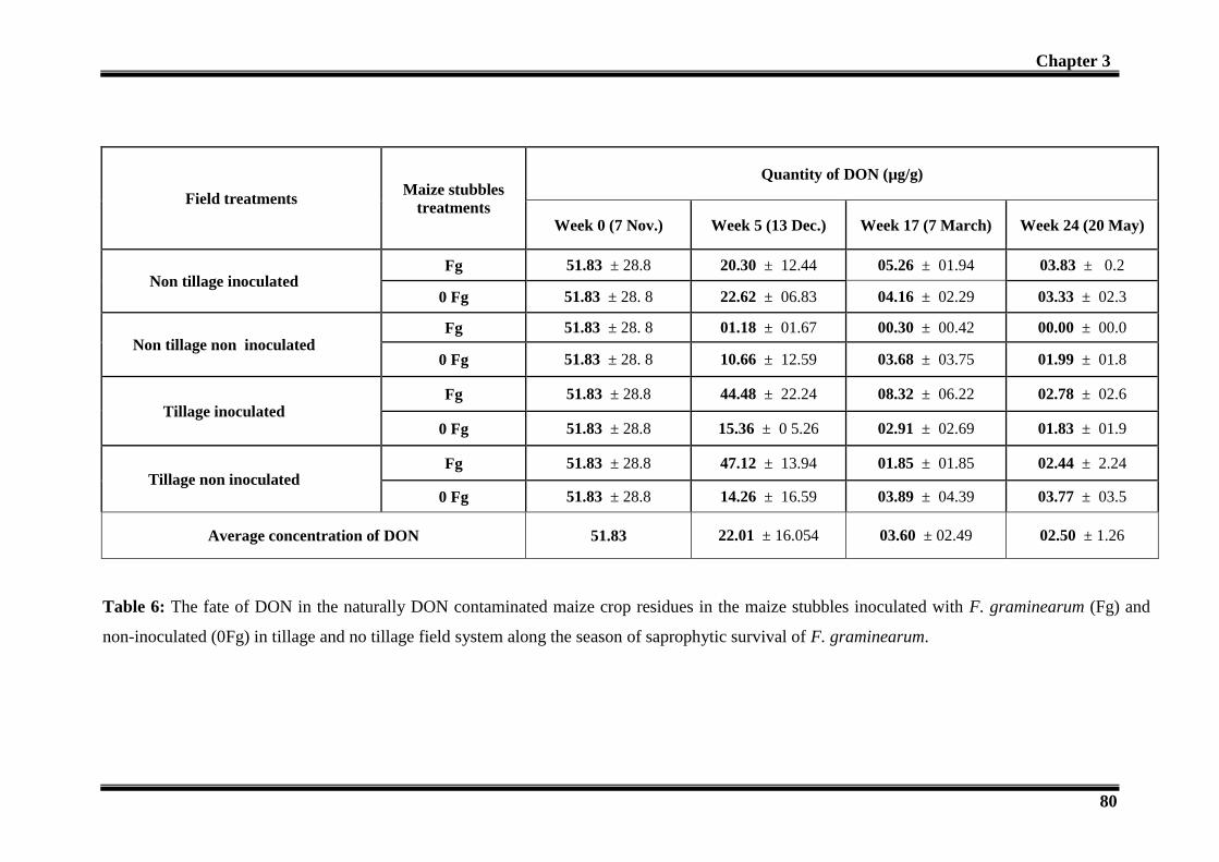
Chapter 3
80
Field treatments Maize stubbles
treatments
Quantity of DON (µg/g)
Week 0 (7 Nov.) Week 5 (13 Dec.) Week 17 (7 March) Week 24 (20 May)
Non tillage inoculated Fg 51.83 ± 28.8 20.30 ± 12.44 05.26 ± 01.94 03.83 ± 0.2
0 Fg 51.83 ± 28. 8 22.62 ± 06.83 04.16 ± 02.29 03.33 ± 02.3
Non tillage non inoculated Fg 51.83 ± 28. 8 01.18 ± 01.67 00.30 ± 00.42 00.00 ± 00.0
0 Fg 51.83 ± 28. 8 10.66 ± 12.59 03.68 ± 03.75 01.99 ± 01.8
Tillage inoculated Fg 51.83 ± 28.8 44.48 ± 22.24 08.32 ± 06.22 02.78 ± 02.6
0 Fg 51.83 ± 28.8 15.36 ± 0 5.26 02.91 ± 02.69 01.83 ± 01.9
Tillage non inoculated Fg 51.83 ± 28.8 47.12 ± 13.94 01.85 ± 01.85 02.44 ± 2.24
0 Fg 51.83 ± 28.8 14.26 ± 16.59 03.89 ± 04.39 03.77 ± 03.5
Average concentration of DON 51.83 22.01 ± 16.054 03.60 ± 02.49 02.50 ± 1.26
Table 6: The fate of DON in the naturally DON contaminated maize crop residues in the maize stubbles inoculated with F. graminearum (Fg) and
non-inoculated (0Fg) in tillage and no tillage field system along the season of saprophytic survival of F. graminearum.

Chapter 3 Chapter 3
81
3.4- Quantification of F. graminearum, fungi and bacteria:
The growth of F. graminearum and the total fungal and bacterial molecular biomass was
quantified from the maize stubbles in all the treatments from 7 Nov to 20 April 2012 using
real time polymerase chain reaction (Q-PCR).
The numbers of DNA copies for each week were calculated in terms of number of copies per
gram (d.w.) of maize. Analyses Of Variance (ANOVA) were applied at week 5, week 17 and
week 24 for F. graminearum, overall fungal and bacterial densities and the interaction was
checked in relation to the different treatments including impact of DON, tillage system, field
inoculation and maize stubbles inoculation.
Our results for the molecular biomass of F. graminearum (Fig 5, Table 7) indicate that the
population of F. graminearum was not affect by the presence or the quantity of DON in the
maize stubbles at any sampling time. The field tillage and the field inoculation also produced
no impact on the population of F. graminearum. The field tillage produced a significant
impact on the population of F. graminearum but it appeared only on week 17. Time produced
a highly significant effect on the population of F. graminearum (Table 10).
In case of fungi as a whole, the molecular biomass was not affected by the quantity of DON
on any sampling time (Table 8). The soil tillage and the field inoculation as well as the maize
inoculation with F. graminearum produced no impact on the fungal biomass in the maize
stubbles at any of the sampling times. The time produced a highly significant impact on the
fungal biomass (Table 10).
In case of bacteria the molecular biomass remained unaffected by the quantity of DON in the
maize stubbles at any sampling time (Table 9). The field tillage, field inoculation and the
maize stubbles with F. graminearum produced no impact on the bacterial biomass. The
biomass was only affected by time factor (Table 10).

Chapter 3
82
-1,00E+08
-5,00E+07
0,00E+00
5,00E+07
1,00E+08
1,50E+08
2,00E+08
2,50E+08
3,00E+08
3,50E+08
Fg Fg+DON Fg Fg+DON Fg Fg+DON Fg Fg+DON
Non tillage inoculated Non tillage non inoculated Tillage non inoculated Tillage inoculated
T1
T2
T3
3.50 × 10⁸
3.00 × 10⁸
2.50 × 10⁸
1.00 × 10⁸
5.00 × 10⁷
0.00
5.00 × 10⁻⁷
1.00 × 10⁻⁸
1.50 × 10⁸
2.00 × 10⁸
Co
pe
iso
f DN
A/g
of d
ry w
eig
ht
Fig. 5: The impact of DON on the biomass of F. graminearum in the maize stubbles put in the field tilled or not tilled and inoculated or not with
F. graminearum. (T1= week 5, T2= week 17 and T3=week 24)

Chapter 3
83
Table 7: Analysis of Variance (ANOVA) for Q-PCR (real time PCR) of F. graminearum for different treatments at three time periods.
df: degrees of freedom, MS : Mean Square, F : Fisher’s F.* p < 0.05
Table 8: Analysis of Variance (ANOVA) for Q-PCR (real time PCR) of fungi for different treatments at three time periods.
df: degrees of freedom, MS : Mean Square, F : Fisher’s F. * p < 0.05
Treatments (Source of variance)
Time 1 Time 2 Time 3
df MS F
MS F df MS F
Effect of tillage 1 1.98 ×10¹⁴
0.029 1 5.10 ×10¹⁵
13.410* 1 7.57×10¹¹
0.022
Effect of field inoculation 1 1.33×10¹⁴
0.019 1 6.49 ×10¹⁴
1.115 1 1.26 ×10¹³
0.364
Effect of DON 1 1.07×10¹⁶
1.677 1 4.46 ×10¹³
0.073 1 5.84 ×10¹¹
0.017
Treatments (Source of variance)
Time 1 Time 2 Time 3
df MS F df MS F df MS F
Effect of tillage 1 3.68 ×1026
1.071 1 1.24 ×1026
2.035 1 1.56 ×1024
0.192
Effect of field inoculation 1 7.48 ×1024
0.021 1 1.82 ×1025
0.287 1 2.32 ×1024
0.285
Effect of maize stubbles inoculation 1 4.56 ×1026
1.336 1 9.49 ×10²³
0.015 1 6.62 ×1024
0.824
Effect of DON 1 6.51 ×1026
1.931 1 1.23 ×1026
2.020 1 2.34 ×1024
0.288

Chapter 3
84
.
Treatments (Source of variance)
Time 1 Time 2 Time 3
df MS F df MS F df MS F
Effect of tillage 1 1.87 ×10²²
2.546 1 1.12 ×10²²
3.395 1 8.36 ×10²¹
1.706
Effect of field inoculation 1 8.37 ×10²¹
1.105 1 2.01 ×10²⁰
0.057 1 1.90 ×10²¹
0.377
Effect of maize stubbles inoculation 1 6.40 ×10²⁰
0.083 1 1.53 ×10²¹
0.439 1 7.28 ×10²¹
1.480
Effect of DON 1 9.28 ×10²¹
1.229 1 7.07 ×10²⁰
0.201 1 1.65 ×10²¹
0.328
Table 9: Analysis of Variance (ANOVA) for Q-PCR (real time PCR) of bacteria for different treatments at three time periods
df: degrees of freedom, MS : Mean Square, F : Fisher’s F. * p < 0.05
Treatments (Source of variance)
F. graminearum Fungi Bacteria
df MS F df MS F df MS F
Time 2 1.61 ×10¹⁶
6.735* 2 1.73 ×1027
12.515** 2 3.33×10²²
6.255*
Table 10: Analysis of Variance (ANOVA) for Q-PCR (real time PCR) of F. graminearum, fungi, bacteria and the impact of time.
df: degrees of freedom, MS : Mean Square, F : Fisher’s F. **p < 0.001, * p < 0.01

Chapter 3 Chapter 3
85
3.5- Disease development on the wheat crop:
Campaign 2011-2012 was completely different from the previous one. While in 2010-2011,
the dry climatic conditions prevented the wheat crop from FHB, in 2011-2012 wet weather
and mild temperatures during the flowering period of wheat favored the epidemic
development of Fusarium head blight. Therefore, the whole plot was affected what could
explain why none of the various parameters measured along this campaign were significantly
different between inoculated and non-inoculated subplots (Table 11).
However more significant differences were found when the comparison was performed
between no tilled and tilled plots. The same number (374 and 378) of emerging seedlings
were found out of the 400 seed initially sown/m2 what makes about 94% of success for both
sets of plots. However, the green surface was significantly higher in the no tilled plots than in
the tilled one in autumn (22 Nov 2011). This can be explained by the fact that seeds were
closer to the surface in no tilled plots than in tilled plots as a consequence of the tillage
system. Therefore, the former seedlings could grow more rapidly. Conversely, at the end of
winter (5 March) the trend was opposite because the seeds in the tilled plots were protected,
deep in the soil, to face the cold winter period and thus were able to re-start their growth faster
than the seedlings of the no tilled plot which suffered from the frost. Indeed, the percentage of
seedlings that survived the winter period was significantly higher (91.7%) in the tilled plots
than in the no tilled lots (79.9%). This handicap was more or less compensated as 1 month
later (2nd
April), the green surfaces were no more significantly different, thus indicating that
globally, the coverage of the soil was the same in both tillage systems. Actually, the situation
was different in the 2 systems as in the case of no tilled plots, the number of tillers was
significantly higher (4.27/plant) that in tilled plots (3.44/plant) but the height of the seedlings
at that stage (ears at 1 cm) was significantly higher in tilled plots (1,305) than in no tilled plots
(0,895). Therefore, the growth scenario of the wheat variety Charger depends on the tillage
system. At the time of flowering, the numbers of ears were similar (291/m2 as a mean). After
flowering, significantly different scenarii were also observed. Only the observations
performed on the 12 and 19 June 2012 were taken into account, those performed on the 28th
June were biased by the natural yellow coloration the ears getting matured. However, both the
numbers of spikelets exhibiting FHB symptoms and the intensity of the symptoms, as
assessed by image analyses, revealed that the wheat cultivated in the no tilled plots was
significantly more affected by FHB than the wheat in the tilled plots. The consequences are

Chapter 3 Chapter 3
86
revealed by the significant difference in yield. The wheat grown in the tilled system produced
8473 kg grains/ha while the same variety of wheat grown in no tilled system produced 7666kg
grains/ha i.e. 800 kg less. The significantly lower dry weight of 1000 grains in the no tilled
system revealed that the grain were smaller in these plots. At the present day, the DON
content of the different samples of grains issuing from the various subplots has not yet been
quantified.
The statistical analysis performed on the whole data set at each sampling occasion indicate
that the only block effect was observed at the end of winter and therefore it did not affect the
whole kinetics of development of both the crop and the disease. In the same way, there was no
significant interaction between the tillage system and the artificial inoculation of maize
residues, probably because the natural infestation by indigenous F. graminearum and other
fungi of the FHB complex obscured the artificial inoculation performed on half of the
subplots. However, it is noticeable that the natural infestation was more severe in the no tilled
subplots but it is difficult to draw general conclusion from this single experiment.

Chapter 3
87
no tillage tillage no inocul inocul I II III IV no tillage tillagerelative
std dev
400.7936 365.0793 357.1428 380.9523 non ino 365.0793 372.3545 9.6%
ino 382.9365 383.5978 11.6%
235.8 218.3 230.8 234.2 non ino 282.9 180.12 14.48%
ino 260.9 195.3 11.94%
655.9 690.7 695.6 608.875 non ino 517.9 769.775 2.55%
ino 633.125 730.275 12.18%
341.2698 330.0264 332.672 324.074 non ino 323.4127 344.5767 16.04%
ino 307.5397 352.5132 11.88%
1902.5 1891.75 1954.25 1858.00 non ino 1792,00 1937.5 1.65%
ino 1991.75 1885.25 4.76%
3.85 3.71 4.15 3.71 non ino 4.02 3.53 5.11%
ino 4.52 3.35 15.9%
1.139 1.098 1.127 1.035 non ino 0.866 1.174 17.41%
ino 0.924 1.435 13.98%
non ino
ino
290.8 276,00 287.5 313,00 non ino 281.7 298.9 2.08%
ino 293.2 293.4 9.2%
7.3 7.7 8.3 7.9 non ino 9.7 6.9 18,00%
ino 8.2 6.5 19,00%
1.08 1.08 1.45 1.38 non ino 1.31 1.13 13.7%
ino 1.72 0.84 25.2%
16.9 16.7 16.4 15.6 non ino 19.0 11.9 19.7%
ino 20.6 14.1 12.98%
7.5 7.6 7.6 7.3 non ino 8.7 4.9 28,00%
ino 10.1 6.2 24,00%
82.85 79.21 80.97 79.73 non ino 75.83 84.21 2.32%
ino 77.49 85.24 2.47%
71.02 71.3 71.67 72.34 non ino 70.55 72.27 1.21%
ino 71.25 72.3 1.18%
91.7
1938.5 S
296.18
8.3%
NS
NS7.4%
1.1795 NS
NSNS
88.7
nb ears 291.84
330.0264
287.5NS
green surface /cm² -
Ears at 1cm
% contaminated
surface
0.895 1.3045Height Ears at 1cm 2-avr. 1.09975 S
79.9% Survival (end of
Winter)
% contaminated
spickelets
NS
NS 290.4
NS
3.935
NS 1.02
293.3
NbTillers - Ears at
1cm2-avr. 3.855 HS 4.27 3.44 NS
HS 681.7
VariableDate of
notationmean
tillage system inoculation interactionBlock
12-juin 1.2% HS
3.775
12-juin 7.8% S 8.9% 6.7%
575.5125 750.025 NS 643.8375 S
NS1.5% 0.98% NS 1.22%
2-avr. 1901.625
662.765-marsgreen surface /cm²
nb seedlings - end of
winter2-avr. 332.01 NS
NS 1864.75
315.4762 348.5449 NS 333.9947
1891.875 1911.375 NS
13.0% NS 15.5%
S
NS
NS1.28%
NS NS
17.4% NS NS% contaminated
spickelets19-juin 16.4% S 19.8%
yield 23-juil. 80.69 HS
% contaminated
surface19-juin 7.5% S
84.73 NS
70.9
6.8% 8.2%5.6% NS9.4%
80.02
NS NS
81.37 NS NS
71.41 71.77
76.66
NSdry weight 23-juil. 71.59 S 72.29
229.8 S
NS
271.9 187.7 NS
374 378
NS
NS
NS
nb emerging
seedlings8-nov. 376 NS
231.525green surface /cm² 22-nov.
369 383 NS NS
NS
228.0875 NS NS
Table 11 : monitoring of the wheat crop in F. graminearum inoculated and non inoculated plots under tillage and no tillage systems.
S: Significant (p=0.05); HS: Highly Significant (p=0.01); NS: Non Significant.

Chapter 3
88
4-Discussion:
The soil has been too long considered an inert matrix in which farmers had to bring mineral
fertilizers for growing plants. In recent years, the idea of a living soil is increasingly present in
mind when managing crops. Even though the role of all biotic components of the soil is not
really known, taking into account multitrophic interactions in the management of preceding
crops, organic matter and tillage led to reflect upon the relationship between agricultural
practices and ecology of soil-borne plant pathogens. The crop residues are subjected to the
degradation by the soil microflora and fauna and the nutrients are liberated in the soil
(Lupwayi et al., 2004).
In this experiment, the process of decomposition was measured in terms of the weight of
maize stubbles in the nylon bags. We observed that once the maize residues were in the soil
and attacked by the microbial communities the process of degradation started. We saw the
soil communities worked very well and they degraded 51 % of the maize stubbles within 5
weeks post launching the experiment and then the process of degradation continued which
was slower than before but at the end of 24 weeks we got only 22 % dry matter back from the
soil. We found no clear difference between the maize stubbles placed on the soil surface (1
cm deep) and the one incorporated in the soil (10 cm deep) with soil tillage which may seem
surprising as it is well known that the decomposition process is much more faster when crop
residues are buried than when they remain at the surface of the soil. Anyway, the
decomposition process was not affected by the quantity of DON in the maize stubbles which
indicates the quantity of DON didn’t stop the decomposition process of the maize crop
residues.
During this decomposition process it was very interesting to see the community profile
associated with the process. We observed the community profile of fungi, bacteria, protozoa
and nematodes which colonized the crop residues and may be participated in the process of
decomposition. The first thing we observed that the quantity of DON in the maize stubbles
played no impact on fungi, bacteria and nematodes but significantly affected the protozoan
community structure whether the stubbles were on the soil surface or buried in the soil. This
showed that the protozoa were highly sensitive towards the DON during their attack on the
maize stubbles while the fungi, bacteria and the nematode populations were not much
sensitive towards the DON quantity in the crop residues. Unfortunately, the role of protozoa
in the decomposition process in arable crops is rather unknown therefore the impact of DON

Chapter 3
89
on their community cannot really be interpreted in relation to any functional consequence for
the fate of crop residues as for F. graminearum survival.
The impact of tillage practices on the community structures of these four soil-borne
communities (fungi, bacteria, protozoa and nematodes) was also observed. We got very
interesting information that almost all the communities were affected by the tillage practice. It
showed that the soil micro- flora and fauna colonizing the maize stubbles was completely
different when they are placed on the soil surface than the one which are present 10 cm deep
in the soil. It was surprising that both played equal role in the decomposition process of the
maize stubbles. This impact on the community structure of the different communities can be
due to the soil aeration, water moisture and the carbon nitrogen ratios which in turn can have
impact on the populations in the different soil communities (Doran, 1980; Spedding et al.,
2004).
In addition, we examined the impact of F. graminearum which was already provided to the
maize stubbles by inoculation, on the other communities attacking the crop residues. We
found the fungal structure was completely different in the presence of F. graminearum.
However, it was unclear whether the change was due to real changes in abundance ratios
between populations initially presented before the introduction or if they are simply the F.
graminearum TRF which are responsible for the observed changes. It was surprising to see
that the presence of F. graminearum in the stubbles greatly affected the bacterial populations
which were colonizing the crop residues which showed a relation between F. graminearum on
the bacterial communities or it might be the nutrients used by the F. graminearum which
discouraged the bacterial communities to colonize the crop residues. Moreover we saw that
nematodes and protozoa community structure was also clearly affected throughout by the
presence of F. graminearum. We may assume that F. graminearum was fighting or using
some kind of mechanisms to conserve its food resources without using DON. It might also be
proposed that F. graminearum rapidly colonized the maize stubbles to prevent populations of
some other communities to colonize the maize crop residues.
Furthermore, our results indicated that the field inoculation by F. graminearum brought no
change in the community structure of any of the four communities. Furthermore, we checked
the impact of plots on the community profile of all the four communities and we observed that
there was an effect of block on these communities. This effect was clear after 5 weeks on the
fungal and bacterial community structure and significantly appeared after 17 weeks on the
bacterial and nematodes community structure. This reveals the difficulty we faced in field
experiments. Despite everything was done to ensure the best homogeneity in preparing the

Chapter 3
90
plots and in distributing the nylon bags across the subplots, intrinsic heterogeneity was still
present and caused variability among the samples. For practical reason and for the sake of
feasibility, it was not possible to handle more than 3 biological replicates for which 3
technical replicates each were performed. Unfortunately, such variability created a limitation
in the biological interpretation of the data. This was also true for the Q-PCR measurements.
The molecular biomass of the F. graminearum was measured by Q-PCR to find out its
survival in the presence of DON and what kind of benefit it could obtain from the quantity of
DON present in the maize crop residues. Our results demonstrated that the quantity of DON
gave no advantage to the F. graminearum. The population of the F. graminearum was
reduced with the passage of the time or we can say with the decomposition of the maize
stubbles. Though statistically the application of ANOVA on the data showed that there was
no impact of DON but it is not so clear as we saw some higher densities of F. graminearum in
terms of mean value but a big standard deviation was found among the biological replicates
(plots). This standard deviation was observed throughout the experiment which restrains us to
give a solid conclusion about the impact of DON on the growth of F. graminearum and the
development of primary inoculum. Samples were carefully preserved at -20°C so we repeated
the Q-PCR measurements on the whole set of samples. We found the same results so we can
say it was the effect of block linked to the spatial heterogeneity of the plot which prevents to
draw any conclusion about the effect of DON and tillage on F. graminearum population. This
effect of the block can also be seen on the community profile of the overall fungal and
bacterial communities. The results about the disease development showed that the primary
inoculum was significantly higher in case of no tillage than soil tillage which shows that the
population the F. graminearum might be higher in zero tillage than in tillage system. We also
observed the impact of DON and the tillage system on the overall fungal and bacterial
densities and we found no impact of DON and the tillage system on them. Same was the case
with these two communities as with the population of F. graminearum. Even in the presence
of the high variability among the plots (biological replicates), we could see that the
decomposition of the crop residues produced a significant reduction in the fungal, bacterial
densities as well as the F. graminearum population.
The quantification of DON in the maize crop residue at zero sampling time was a bit
surprising as we found a certain big amount of DON in the maize stubbles (51 ± 28 µg/g of
dry matter) although they were autoclaved two times at 120°C to make them disinfected. By
the way we had 2.3 times more DON in the artificially contaminated maize stubbles to see the
impact on the F. graminearum and the other soil communities. We monitored the fate of

Chapter 3
91
DON by quantifying them in each sample at each sampling time. We also noticed that less
than 100% of DON was recovered a few hours after it was introduced. The quantification of
mycotoxins generally concerns plant material which is not contaminated with soil and
adequate procedures are proposed to evaluate the quantity of DON in cereal grains. These
procedure were not efficient to detect and quantify DON in soil-residues mixtures thus we had
to set up a method to extract DON from the mixture and to recover it from the extractant.
Ethanol was used as a solvent instead of acetonitrile and affinity column were used instead of
classical columns. Preliminary assays performed in controlled conditions predicted a 100%
recovering of DON introduced in complex mixture. Therefore, it is likely that part of the
DON introduced into the soil-maize mixture in natural conditions was either rapidly adsorbed
on this matrix or made unavailable by indigenous soil organisms. Anyway, the fate of DON
could be monitored as it was possible to depict quantitative variations over time relatively to
the initial quantification. We found that DON disappeared with the passage of time in all the
samples. It was correlated to the passage of time and the degradation of the crop residues. It
might be difficult to say that whether DON was degraded by the microbes or it was leached
down or liberated in the soil due to the decomposition of the organic matter and presence of
less material for its absorption. Our results also showed that DON was not increased in the
crop residues during the completion with other soil biota. In the end of the experiment we
found a very little amount of DON in the maize residues.
We monitored the development of the disease in the field to monitor the impact of the tillage
and the inoculum we provided artificially to the field. It was not possible to correlate what
was observed in the nylon bags with the development of the crop and the disease. Moreover,
the climatic conditions were quite favorable to the epidemic spray of FHB what obscured the
inoculated non–inoculated treatment of the natural maize crop residues. This experiment
revealed, but this is not really new, that the tilled plot was less affected by FHB than the no
tilled plot but it does not allow to ensure that tillage can prevent the crop from FHB disease. It
is probably wiser to evaluate the tillage system on a longer duration.
The field experiment suggests that DON has an impact on soil microflora and soil fauna
components. It is not known from the measurements we did if this impact should be
considered as positive or negative towards the biotic components but it seems that this impact
depends on the location of the crop residues and also, it seems that F. graminearum did not
get any competitive advantage of the presence of DON. However a huge variability of the
data acquired in this field experiments was a limitation to interpret and discuss the results and
to provide a solid answer. This variability was partly attributed to the field heterogeneity and
Chapter 3
92
to the climatic conditions so we suggest that such an approach should be conducted in
controlled conditions using microcosms as experimental unit.
6-Conclusion:
The main conclusions of this field experiment are briefly explained in the figure-6. We
conclude that most of the DON disappeared from the maize crop residues with the passage of
time and left in very low amount after six months. The disappearance of DON was not
dependent on the tillage system or stubbles inoculation. The fact that DON did not increase in
the presence of F. graminearum in the maize residues could be explained either by the
absence of production or by a concomitant production-disappearance (adsorption or
degradation) process.
The quantification of F. graminearum by real time PCR showed that there were high
variations among the blocks (biological replicates). Therefore, it was difficult to say anything
about the impact of DON or tillage system. So the question about the advantage of the
presence of DON to F. graminearum left unrevealed and need to be studied in controlled
environment to eliminate environmental factors. Similarly, it was difficult to conclude about
the impact of DON on the fungal and bacterial densities variability among the blocks. The
measurement of disease and yield in the field showed that the field tillage significantly
reduced disease development on the wheat crop and ultimately led to the high yield which
showed the better survival of F. graminearum in the no tillage system as compared to the
tillage system.
T-RFLP results showed that only protozoa community structure was affected by the presence
of DON independent of the tillage system. The fungal, bacterial and nematodes communities
remained unaffected by DON.

Chapter 3
93
Conclusions
Ecological role of mycotoxins?
Maize stubbles ± DON
Fate of DONImpact on soil micro flora and fauna
Advantage to F. graminearum
to develop
Densities Structure
Fungi Bacteria Fungi Bacteria Protozoa Nematodes
? ?
?
No No NoYes
Disappeared with time
Soil
No tillage impact
Fig. 6: Flow chart showing the main conclusion obtained during the different measurements
in the maize stubbles nylon bags in the field experiment. The violet colour rectangle shows
the disappearance of DON; yellow colour rectangles indicate that questions remained
unanswered; red colour rectangle shows the impact of DON while the grey colour shows no
impact of DON. Furthermore, Yes= DON had an impact; No= DON had no impact; ?= still
remained questions.

Chapter 4
94
Chapter 4: Fate of DON and its impact on
the soil microflora and soil fauna
communities

Chapter 4
95
Fate of DON and its impact on the soil microflora and
soil fauna communities
1. Introduction:
Mycotoxins are fungal secondary metabolites potentially harmful to humans and animals.
Trichothecenes are one of the most important mycotoxins produced in the field by the genus
Fusarium and are considered great threat to humans and animals (Rocha et al., 2005). They
are produced in the field during the pathogen invasion on the crop. Their incidence in the
cereal crops is a huge challenge for the agricultural industry (Bottalico and Perrone, 2002;
Tanaka et al., 1988). Deoxynivalenol (DON) is one of the most important trichothecene
metabolites and is found in cereals as wheat, maize, rye and barley. DON (also known as
vomitoxin) is a type B trichothecene and is mainly produced by F. graminearum. It is one of
the most commonly reported mycotoxins associated with the Fusarium head blight (FHB)
diseased cereals (Bottalico, 1998; Foroud and Eudes, 2009; Paul et al., 2005). DON causes
very destructive effects on mammals as immunity reduction, protein biosynthesis damage,
food refusal, diarrhea and vomiting and many sever disease symptoms (Fokunang et al., 2006;
Placinta et al., 1999; Wild and Gong, 2010). The European Union has set the threshold level
for DON in the winter wheat at 1.250 mg/kg (CE N°1116/2007). Beyer et al., (2007) reported
that 4.27% of Fusarium-damaged kernels can meet this limit of European Union.
FHB is the center of interest of the scientists worldwide and different approaches have been
investigated to overcome the disease such as forecasting of disease, cultivation of resistant
varieties, use of fungicides, cultural practices and biocontrol agents (Buerstmayr et al., 2009;
Champeil et al., 2004; De Wolf et al., 2003; Mesterhazy et al., 1999; Prandini et al., 2009;
Schisler et al., 2002). Despite all this experimental research, a durable solution to control FHB
has not been found and is still a challenge for the agriculture in the world. The management of
the disease is an ultimate tool for the reduction of the related mycotoxins, and the food losses,
to make the food secure for increasing world population. The fungal toxins including DON
are reported not obligatory for the production of disease on plants. The fungal mutants having
no mycotoxin production ability are demonstrated to produce the disease as the wild type of
F. graminearum fungus (Adams and Hart, 1989; Bai et al., 2002). However DON is reported
to have an active role in the aggressiveness of the fungus (Mesterházy, 2002; Mudge et al.,
2006) .

Chapter 4
96
Crop residues are important components of arable soil functioning thanks to the nutrients they
bring back to the soil. They are stimulating the high diversity of decomposer organisms in the
process of litter decomposition and fuelling the multitrophic interactions among the various
soil inhabitants with noticeable consequences for biogeochemical cycles. Potential
mechanisms include fungi-driven nutrient transfer among litter species, inhibition or
stimulation of microorganisms by specific litter compounds, and positive feedback of soil
fauna due to greater habitat and food diversity (Hättenschwiler et al., 2005; Kreuzer et al.,
2004; Rantalainen et al., 2004). How the species richness of decomposer fungi, bacteria and
soil fauna including protozoa, or their relative frequencies of occurrence (i.e. community
structure) influence the decomposition of organic matter in arable soil is poorly known
compared to the one of forest litter (Buee et al., 2009). Using different sets of fungi isolated
from plant litters, Deacon et al., (2006) showed that there was apparently a high degree of
functional redundancy in assemblages of cultivable decomposer fungi what could buffer the
impact of external event on the decomposition process. However, the authors pointed out that
some less abundant taxa might have a key role in the degradation of complex or recalcitrant
substrates. Moreover, community structures are not invariant during the process of
decomposition over time. The number of trophic levels among soil fauna and soil microflora,
species identity as well as keystone species may replace one another as their dynamic
communities alter in space and time, each functional species adapting for occupation of the
successive habitats resulting from the decomposition process (Frankland, 1998). In turn, the
decomposition process may be depending on the multitrophic interactions. For instance,
microbial succession with shifting enzymatic capabilities enhances decomposition, whereas
antagonistic interactions among organisms that compete for similar resources or/and produce
deleterious secondary metabolites slow litter decay (Deacon et al., 2006). Moreover, external
factors such as the agricultural practices and more precisely the tillage system could affect
both the consortium of decomposers and the decomposition process (Cookson et al., 1998).
Therefore, one may question about how the phytopathogenic fungi during their saprotrophic
phase can be included in this food web, and can survive the dynamics of successions. One
example of a very strong and effective interaction is provided by the common earthworms
(Lumbricus terrestris L) which remove sources of phytopathogenic fungi (Venturia
inaequalis) in orchards thanks to their efficient grazing on leaf litter (Holb et al., 2006).
The diseased crop residues (grains, straw and stubbles), are colonized by the fungus and are
the source of saprophytic survival during off seasons (Pereyra and Dill-Macky, 2008). The

Chapter 4
97
modern age agriculture is moving towards reducing the soil tillage practices, in order to
conserve the field soil (Bai and Shaner, 2004). However, this reduced tillage supports the
survival of the fungus more than deep tillage (Pereyra and Dill-Macky, 2008). F.
graminearum survives in these crop residues saprotrophically and serves as primary inoculum
to the next crop (Parry et al., 2007; Trail, 2009). Under appropriate climatic conditions, the
fungus receives the warm moist climatic condition which leads to the conidia formation. The
ascospores are produced in the perithecia and make the fungal spread to the seasonal cereal
crops as wheat (Trail, 2009).
Therefore we can wonder about the strategy used by F. graminearum to survive in the crop
residues which appeared as a very coveted resource while this plant pathogenic fungus is not
such a good saprophytic competitor (Leplat et al., 2012) unless the mycotoxins it produced in
planta provide it with a significant competitive advantage towards the microbial and soil
fauna communities. The presence of mycotoxins in the soil and their impact on the soil biota
are still an enigma. There is no research performed on the fate of mycotoxin in the crop
residues and the soil environment. It is still a question to understand the role of DON in the
ecological habitat of microorganisms.
Owing this background the main question of this study was to understand the ecological role
of mycotoxin during the survival of F. graminearum in the crop residues by monitoring the
survival of other soil biota including fungi, bacteria, protozoa, nematodes and earthworms.
The research was conducted in microcosms in non-disinfected soil containing straw as crop
residue and DON as mycotoxin. All the conditions closed to the natural field environment
were established in these microcosms.
Chapter 4
98
2. Materials and methods:
A-DON chemotype Fusarium graminearum strain MIAE00376 (Chapter 2, page 44) was
used in this experiment. The inoculum was produced according to the procedure described
Chapter 2.
Lumbricus terrestris adult specimens were captured (Chapter 2, page 49) and were put in the
soil in different containers and were placed at 10°C for 3 weeks. At the time of launching the
experiment, earthworms were put in the experimental soil at 17°C for 4 days.
2.1- Soil and straw preparation:
The soil was collected from the meadow area near green house (described in chapter 2, page
46). The air dry straw was taken from the winter wheat (Triticum aestivum) originated from a
field of Epoisses (Bretenières, France). The straw was cut into approximately 2-3 cm small
pieces.
The water holding capacity of straw was found as 1 g of straw (d.w.) could hold 3.5 ml of
water. The straw was made infected with F. graminearum by spraying and mixing with 1.5 ml
containing conidial suspension (6.67 × 10² conidia/ml) per gram of straw (d.w.). Then the
straw was divided into two halves, one half was made contaminated with DON by spraying
and mixing with 1.5 ml DON solution (66.7 µg/ ml, DON solution preparation is described in
Chapter 2, page 56) while the other half was moistened by spraying and mixing with distilled
water to give the homogenous humidity to all straw. Finally, the straw contained 103
conidia/g
of straw (d.w.) and the DON contaminated straw was containing DON at the rate of 100 µg/g
of straw (d.w.).
2.2- Preparation of microcosms:
Small plastic pots of 11 cm × 15.5 cm × 6.5 cm in size (width × length × height) were used as
experimental units (microcosms). In the lid of each pot, a hole was made for the breathing of
earthworms and elimination of gases produced as a result of decomposition of straw. This
hole was covered with plastic gaze to avoid the escape of earthworms.
Each microcosm was filled with 1kg of soil (dry weight, soil is described in chapter 2, page
48) and 10 g of F. graminearum infected straw (dry weight) either contaminated with DON or
not. The straw was either incorporated in the soil or placed on the soil surface in the form of

Chapter 4
99
layer. Four previously collected earthworms (chapter 2, page 49) of total biomass 13.67 (±
0.76) g were added to each required microcosm. In this way six types of microcosms were
prepared representing six treatments: microcosms with or without DON, straw left at the
surface or incorporated in the soil and the microcosms with or without earthworms. Three
replicates (microcosms) were prepared for each treatment for each sampling time. These
microcosms were placed in an incubator at 17°C (Fig. 1).
a
b
c
Fig. 1: Microcosms were incubating at 15°C in a climatic chamber (a). Straw was mixed with
the soil in one half microcosms (b) while it was left at the surface of soil in the other half (c).
2.3- Sampling, samples processing and analyses:
Sampling was done on 0, 1, 2, 8 and 24 weeks after the establishment of experiment. At each
sampling time, the earthworms were removed from the microcosms. The whole remaining
contents (i.e. straw and soil) were mixed and were filled in the vials in a way that each vial
had 30 g of soil-straw mixture. These vials were conserved at -20°C. Then all the samples
were freeze-dried and ground to powder to make a homogenous mixture of straw and soil and
conserved at -20°C. These samples were ready for the extraction of DNA. The humidity in the

Chapter 4
100
microcosms was measured from time to time by weighing the microcosms and was adjusted
by spraying water if needed.
At each sampling time all the earthworms were sorted out of the microcosms. They were
washed twice by water to remove their outer soil and then dried on the tissue paper. They
were counted and the total weights of all the earthworms were measured for each microcosm
and were termed as their biomass.
Nematodes were extracted and quantified (Chapter 2, page page 49),
DON was extracted and quantified by HPLC (chapter 2, page 57).
DNA was extracted from soil and soil-straw mixture and nematodes suspension (Chapter 2,
page 50). From the extracted DNA Fungal, bacterial, protozoan and nematode communities
structure analysis by terminal restriction fragment length polymorphism (T-RFLP) (Chapter
2, page 52). The quantification of bacteria, fungi and F. graminearum was also determined by
real time PCR (Chapter 2, page 55).
At each sampling time, total cultivable densities of fungi and bacteria were estimated by
colony forming unit (CFU) on the appropriate media (Chapter 2, page 49)

Chapter 4
101
3- Results:
3.1- Visual observation of decomposition process:
The process of straw disappearance was observed in the presence or absence of earthworms and
in the presence or absence of DON up to six months. Visual observations of straw
disappearance process showed that Lumbricus terrestris played an important role in the
incorporation of straw in the soil (Fig. 2). Surprisingly, the straw contaminated with DON was
more attractive for the earthworms.
without DON with DON
Week 8
Week 24
without DON with DON
Without Earthworms With Earthworms
Week 0 (Start)
Fig. 2: The process of incorporation of straw placed on the soil surface in the presence or
absence of earthworms and DON.
3.2- Fate of DON in wheat straw in soil:
The fate of DON during the incorporation of straw in the soil was followed up to six months
(24 weeks) after the establishment of experiment. DON was extracted and quantified in each
treatment in the presence of earthworms when the straw was left on the soil surface
incorporated in the soil on 0, 1, 2, 8 and 24 weeks and in the absence of earthworms when the

Chapter 4
102
soil was left at the surface on 0, 8 and 24 weeks whether the straw was contaminated or not
with DON (Table -1).
Ten g straw inoculated with 10 µg DON/ g (d.w) were introduced into 1kg soil (d.w.) in order
to provide each microcosm 1µg DON/g soil-straw mixture, which was close to the threshold
limit recommended by European union for unprocessed wheat grains (1.250 µg of DON / g).
The sample of representative homogenous DON contaminated soil-straw mixture at the same
day of establishment of experiment (week 0) was considered as start point. The quantity of
DON indicated that 0.831 µg of DON / g of soil-straw mix was present in the system at day
zero or at start for all treatments.
Treatments Quantity of DON (DON µg / g soil-straw mixture)
Week 0 Week 1 Week 2 Week 8 Week 24
SME ND Traces 0 0 Traces
SMDE 0.831 0.349 ± 0.03 0.033 ± 0.02 Traces Traces
SSE ND 0.105 ± 0.02 0.006 ± 0.01 0 0
SSDE 0.831 0.889 ± 0.02 0.885 ± 0.17 0.012 ± 0.00 0
SS ND ND ND 0 0
SSD 0.831 ND ND 0.400 ± 0.09 0
Table 1: Quantity of DON in the different treatments (SS=Straw at the Surface, SSD=
SS+DON, SSE= SS+Earthworms, SSDE= SSD+Earthworms, SME= Straw Mixed with the
soil+Earthworms, SMDE=SME+DON) at the different time intervals during the incorporation
of straw in the soil. ND = not determined; Traces = below the limit of quantification (LOQ =
0.0018 µg/g soil-straw mixture); 0 = below the limit of detection (LOD = 0.00034 µg/g soil
straw mixture).
The percentage of recovery of the DON inoculated into the system was of 83% at day 0,
suggesting that either part of the DON was actively adsorbed by the mineral and/or organic
matrix made by the soil-straw mixture or that the extraction procedure was not 100%
efficient. Anyway, relative to the initial value at day 0, DON disappeared very rapidly when
the straw was incorporated in the soil as compared to left at the surface. Soon after the setting
up of experiment (week 1), DON started disappearing in the treatments where the DON
contaminated straw was mixed in the soil (SMDE) and remained about half (0.349 ± 0.034

Chapter 4
103
µg/g) as compared to the starting point (0.831 µg/g). On the opposite, no disappearance of
DON was found the first two weeks when the DON contaminated straw was placed at the
surface of the soil in the presence of the earthworms. The disappearance of DON continued
very rapid when DON contaminated straw was incorporated in the soil and the quantity of
DON became significantly very low (0.033 ± 0.02 µg/g) after 2 weeks and soon went below
the limit of quantification after 2 weeks. On the other hand, the disappearance of DON was
very slow when the DON contaminated straw was left on the soil surface. DON started
disappearing after 2 weeks and reached to a very low amount after 8 weeks (0.012 ± 0.00
µg/g) and completely disappeared up to week 24.
Surprisingly, a low quantity of DON was also found during the first week in the microcosms
where the DON was not introduced and the straw was placed on the soil surface (0.105 ± 0.02
µg/g). It also disappeared and went below the limit of detection after first two weeks. Traces
where found in the when the DON non-contaminated straw was mixed in the soil.
The comparison of quantity of DON on week 8 and 24 when the DON contaminated straw
was placed on the soil surface in the presence or absence of earthworms showed that there is a
link between the presence of earthworms and the reduction of DON from the system. On
week 8, we found a significantly high amount of DON (0.400 ± 0.09 µg/g) as compared to the
presence of DON (0.012 ± 0.00) in presence of earthworms in the microcosms. In all the
treatments DON completely disappeared or went below the limit of quantification (LOQ =
0.0018 µg/g soil-straw mixture) or limit of detection (LOD = 0.00034 µg/g soil straw
mixture) after 24 weeks.
3.3- Structural changes in the microbial and microfaunal communities:
The T-RFLP assay was performed for the four soil communities i. e. bacteria, fungi, protozoa
and nematodes in all the treatments after 1, 2, 8 and 24 weeks of establishment of experiment.
By this assay, the changes in these community structures was followed in the presence or
absence of DON when the straw was incorporated in the soil in the presence of earthworms or
left at the surface in the presence or absence of earthworms. The mean number of TRFs
(Terminal restriction fragments) per soil sample was 115 for bacteria, 85 for protozoa, 69 for
fungi and 98 for nematodes communities. PCA was performed by integrating the number and
the intensity of different TRFs for each of the four communities. The result of the PCA was
displayed as variations on a two dimensional diagram for each community.

Chapter 4
104
The results showed that the presence of DON in the system had an impact on the microbial
communities of the soil. The impact of DON was depending on the type of community and
the location of the DON contaminated wheat straw i.e. whether it was incorporated or placed
at the surface of the soil.
3.3.1- Bacterial community structure:
PCA results for the bacteria are explained in the two dimensional diagram (Fig. 3). One
dimension (PC1) is showing 24.86 % variability and the second dimension (PC2) is showing
11.45 % variability. The bacterial community structure moved along the time frame in all the
six treatments, which is more explained along the first dimension. The presence of DON
contaminated straw produced a significant effect on the bacterial community structure
immediately after the establishment of the experiment. The effect of DON contaminated straw
was observed very clearly during the first two weeks whether the straw was placed at the
surface or mixed in the soil. After the first two weeks the differences in the bacterial
community structures started disappearing and at the eighth week, the differences disappeared
between both the treatments contaminated or not with DON. This corresponds with the
quantity of DON as it was disappeared or degraded up to the eight weeks in the presence of
earthworms (Table -1).
The location of straw i.e. left at the surface or incorporated in the soil played a significant role
on the community structure only during the first two weeks. The comparison among the
treatments with and without DON when the straw was left at the surface of the soil on week 8
and week 24 showed that the presence of the earthworms played a significant role on the
bacterial community structures changes. The effect of DON when the straw was left at the
surface of the soil in the absence of earthworm was not clear throughout the experiment. On
the opposite, the community structures after eight weeks was different in the presence or
absence of earthworms when the straw was contaminated with DON while this effect was not
found after 24 weeks.
3.3.2- Fungal community structure:
The two dimensional diagram for the fungal communities is explained in the one dimension
(PC1) with 53.23 % and the second dimension (PC2) with 16.56 % variability (Fig. 4).
The genetic structure of the fungal community changed over time from week 1 to week 8
mainly when the straw has been left at the surface while it remained almost constant overtime

Chapter 4
105
where the straw was incorporated in the soil. The same interaction of location-time can
explain the variability revealed by PC 2.
The main point anyway was the fact that the fungal community structure as a whole was not
directly affected by the presence of DON whether the straw was left at the surface or
incorporated in the soil. On the same way it is difficult to illustrate the direct role of
earthworms on the fungal community structure but an interaction DON-earthworms seem to
affect the fungal community along PC1 when the straw was left at the surface.
3.3.3- Protozoa community structure:
In case of protozoa, the two dimensional diagram (Fig. 5) based on the PCA result is showing
31.57 % variability along one dimension (PC1) and 19.02 % on the second dimension (PC2).
The community structure of the protozoa in the treatments in which the straw is placed at the
surface showed more variability in the biological replicates. The presence of DON showed a
clear impact on the community structure of protozoa throughout the experiment when the
straw was left on the soil surface in the presence as well as in the absence of earthworms.
DON did not impact the protozoan community structure when the straw was incorporated in
the soil. The presence of earthworms produced no significant impact on the protozoan
community structure when the community structures were compared between the treatments
where the straw was left at the surface of the soil in presence or absence of earthworms on
week 8 and week 24.
3.3.4- Nematode community structure:
The community structure of the nematode is explained in a two dimensional diagram, one
dimension is explaining 27.74% variability and second 21.38% variability (Fig. 6). DON did
not impact the nematode community. The changes in the nematode community structures
were more explained by the time. The nematodes communities remained unchanged in the
first two weeks but then they moved along the time factor and this movement with time
continued till the end of six months. The placement of straw in the soil (incorporated or
placed at the surface) and the earthworms did not affect the nematode community structure.

Chapter 4
106
1
2
3
4
5
6
7
8
9
10
11
12
13
14
15
16
17
18
19
20
PC1 24.86 %
PC2 11.45 %
Fig. 3: Principal component analysis of T-RFLP data set in the two dimensional diagram
from all the treatments for the bacterial community structure. Each number represents the
mean of 3 biological replicate for one treatment at a specific time period. For each biological
replicate, 3 technical replicates were performed and provided non significantly different results
The red colour numbers represents the presence of DON. SS=Straw at the Surface, SSD=
SS+DON, SSE= SS+Earthworms, SSDE= SSD+Earthworms, SME= Straw Mixed with the
soil+Earthworms, SMDE=SME+DON.
Treatments Week 1 Week 2 Week 8 Week 24
SS 9 15
SSD 11 17
SSE 1 5 10 16
SSDE 2 6 12 18
SME 3 7 13 19
SMDE 4 8 14 20

Chapter 4
107
50
1
2
34
5
6 7
89
10
11
12
1314
1516
17
18
19
20
-63
63
-63 63
PC1 53.23 %
PC2 16.56 %
Fig. 4: Principal component analysis of T-RFLP data set in the two dimensional diagram from
all the treatments for the fungal community structure. Each number represents the mean of 3
biological replicate for one treatment at a specific time period. For each biological replicate, 3
technical replicates were performed and provided non significantly different results The red
colour numbers represents the presence of DON. SS=Straw at the Surface, SSD= SS+DON,
SSE= SS+Earthworms, SSDE= SSD+Earthworms, SME= Straw Mixed with the
soil+Earthworms, SMDE=SME+DON.
Treatments Week 1 Week 2 Week 8 Week 24
SS 9 15
SSD 11 17
SSE 1 5 10 16
SSDE 2 6 12 18
SME 3 7 13 19
SMDE 4 8 14 20

Chapter 4
108
12 3
4
5
6
7
8
910
11
12
13
14
15
16
17
18
19
20
PC1 31.57 %
PC2 19.02 %
Fig. 5: Principal component analysis of T-RFLP data set in the two dimensional diagram
from all the treatments for the protozoa community structure. Each number represents the
mean of 3 biological replicate for one treatment at a specific time period. For each biological
replicate, 3 technical replicates were performed and provided non significantly different results
The red colour numbers represents the presence of DON. SS=Straw at the Surface, SSD=
SS+DON, SSE= SS+Earthworms, SSDE= SSD+Earthworms, SME= Straw Mixed with the
soil+Earthworms, SMDE=SME+DON.
Treatments Week 1 Week 2 Week 8 Week 24
SS 9 15
SSD 11 17
SSE 1 5 10 16
SSDE 2 6 12 18
SME 3 7 13 19
SMDE 4 8 14 20

Chapter 4
109
12
3
4
5 67
8
910
11
12
13
14
15
16
17
18
19
20
PC1 27.74 %
PC2 21.38 %
Fig. 6: Principal component analysis of T-RFLP data set in the two dimensional diagram from
all the treatments for the nematodes community structure. Each number represents the mean
of 3 biological replicate for one treatment at a specific time period. For each biological
replicate, 3 technical replicates were performed and provided non significantly different results
The red colour numbers represents the presence of DON. SS=Straw at the Surface, SSD=
SS+DON, SSE= SS+Earthworms, SSDE= SSD+Earthworms, SME= Straw Mixed with the
soil+Earthworms, SMDE=SME+DON.
Treatments Week 1 Week 2 Week 8 Week 24
SS 9 15
SSD 11 17
SSE 1 5 10 16
SSDE 2 6 12 18
SME 3 7 13 19
SMDE 4 8 14 20

Chapter 4
110
3.4- Impact of DON on the microbial and faunal densities
3.4.1-Effect of DON on earthworms:
Each microcosm was provided with 4 adult earthworms of biomass 13.67 (±0.76) g at the time
of setting up the experiment. This value of biomass was considered as starting point for all the
treatments. At each sampling time, the total biomass of earthworms was measured (i.e. week 1,
2, 8 and 24). The average value of each treatment comprised three independent biological
replicates (microcosms). The results illustrate that the presence of DON had no impact on the
biomass of earthworms. The cumulative biomass of earthworms decreased remarkably after
first two weeks and it continued along with the disappearance of straw until week 24 ( Fig. 7).
This fall in biomass was not related to the presence or absence of DON but seems to be more
dependent on the exhaustion of the food. At the end of 24 weeks we found some new-borne
earthworms in all the treatments, which indicates that even the reproduction of earthworms or
the cocoon hatching was not affected by the presence of mycotoxin. These new-borne
earthworms were counted and the biomass was measured. A significantly high number of new-
borne earthworms were found when DON contaminated straw was left at the surface as
compared to non-contaminated straw. On average 4 earthworms per microcosm (0.12 ±
0.04g) were found in the DON contaminated straw left at the surface, while only 1 earthworm
(0.045 g) from the three biological replicates was found in the absence of DON contaminated
straw. On the opposite no significant impact was observed when the straw was incorporated in
the soil. On average 5 new-borne earthworms (0.174 ± 0.053g) per microcosm were observed
in the treatment with DON contaminated straw and 4 new-borne earthworms (0.503 ± 0.52g)
in the treatment without DON where the straw was incorporated in the soil.
We found that no earthworm was died till the 8 weeks while a very few mortality was
observed on week 24, which was a bit higher in the presence of DON. The mortality of
earthworms in the treatment when the straw left on the soil surface in the presence of DON
was 41%, it was 33% without DON, it was 12% in the treatment when the straw was
incorporated in the soil in the presence of DON and 0% in the absence of DON.

Chapter 4
111
3.4.2- Effect of DON on nematodes:
The numbers of nematodes were counted and were compared in the presence or absence of
DON when the straw was left at the surface or incorporated in the soil in the presence of
earthworms after 1, 2, 8 and 24 weeks and in the absence of earthworms after 8 and 24 weeks.
The number of nematodes (2.4 ×10³
± 3.9×10²) in the soil counted at the establishment of
experiment was considered as starting point for all the treatments. The nematodes in the system
increased soon after the establishment of the experiment (Fig. 8). The presence of DON had no
influence on the nematode density during the first two weeks. Overall a negative trend was
observed on the community of nematodes in the presence of DON in the week 8. This trend
became significant on the week 24 where the straw was left at the surface of the soil with or
without the presence of earthworms. On the other hand, earthworms were also found to
suppress nematodes on the 24 week.
3.4.3 -Effect of DON on fungal and bacterial biomass:
The total fungal and bacterial molecular biomass was measured and expressed in the form of
numbers of DNA copies/g of the soil-straw mixture at each sampling time (i.e. after 1, 2, 8
and 24 weeks). The soil-straw mixture just at the establishment of the experiment was taken
as starting point for all the treatments.
The fungal and bacterial biomass increased after the establishment of microcosms and then
started decreasing till the end of experiment (Figures-9 & 10). On the week-1 fungal densities
were significantly higher where straw was incorporated in the soil. Further, the fungal as well
as bacterial densities when the straw contaminated with DON was incorporated in the soil
were significantly higher over the other treatments. On the week-2, their biomass was
suddenly significantly suppressed where the DON contaminated straw was incorporated in the
soil. This negative impact was observed even after 8 weeks. The fungal as well as bacterial
density again increased on week 24 where DON contaminated straw was incorporated in the
soil. On the other hand, DON contaminated straw left on the soil surface produced no impact
on the fungal biomass in the presence of earthworms throughout the experiment. Fungal
biomass was found higher in the absence of earthworms on week 24 while no impact was
found on week-8. The bacterial biomass was not affected by the DON contaminated straw left
at the surface in the presence or absence of earthworms.

Chapter 4
112
3.4.4 - Fusarium graminearum biomass in relation with DON:
The densities of F. graminearum were observed in all the treatments in the presence or
absence of DON. It was measured in the presence of earthworms (on week 0, 1, 2, 8 and 24)
and in the absence of earthworms (on week 0, 8 and 24). The soil-straw mixture just at the
establishment of the experiment was taken as starting point for all the treatments as for the
fungi and bacteria (Fig. 11). The overall molecular biomass of F. graminearum increased just
after the start of experiment especially in the treatments where the DON contaminated straw
was incorporated in the soil. Later, no impact was observed till the week 8. DON was found
to support F. graminearum again on week 24 when DON contaminated straw was
incorporated in the soil as compared to the other treatments.
The DON contaminated straw produced no positive or negative impact on the F.
graminearum density when the straw was left on the soil surface in the presence or absence of
earthworms throughout the experiment.
3.4.5 - Colony Forming Unit (CFU):
The cultivable fungal and bacterial densities were also measured for each sampling date by
colony forming unit (CFU) per gram of soil. There was no significant effect of mycotoxins on
the fungal and bacterial cultivable communities whether the DON contaminated straw was
incorporated or left at the surface of the soil.
The bacterial communities increased in the first week and then decreased along the time factor
(Fig. 12), which is more or less same to the total molecular densities (DNA). The cultivable
fungal communities on contrary to the total molecular densities increased after two weeks (Fig.
13).

Chapter 4
113
0
2
4
6
8
10
12
14
16
18
Week 0 Week 1 Week 2 Week 8 Week 24
bio
mas
so
f e
arth
wo
rms
(g)
Time period
Start
SSE
SSDE
SME
SMDE
week 0
a aa
a
aa
a
a
aa a a
a aa a
A A A
B
C
0
2
4
6
8
10
12
14
16
18
Week 0 Week 1 Week 2 Week 8 Week 24
bio
mas
so
f e
arth
wo
rms
(g)
Time period
Start
SSE
SSDE
SME
SMDE
week 0
a aa
a
aa
a
a
aa a a
a aa a
A A A
B
C
Fig 7: The impact of DON on biomass of earthworms with the passage of time in four
treatments (SSE = Straw placed on Surface of soil + Earthworms, SSDE = SSE + DON,
SME= Straw Mixed with the soil + Earthworms, SMDE = SME + DON). A comparison was
done among different treatments on all the sampling times by analysis of variance (ANOVA)
and Fisher LSD tests (p=0.05). Different small letters indicate significant difference between
the treatments on the different sampling times. Different capital letters indicate the significant
difference between the sampling times.

Chapter 4
114
1,00E+02
1,00E+03
1,00E+04
1,00E+05
1,00E+06
week 0 Week 1 Week 2 Week 8 Week 24
Nu
mb
er o
f ne
mat
od
es/
Kg
soil
Time period
SSE
SSDE
SME
SMDE
SS
SSD
soil
ba
bb
aa
aa bc
ca
aba
ab
bc
c ca
b
week 0 (Start)
1 × 10²
1 × 10⁴
1 × 10⁵
1 × 10⁶
1 × 10³
1,00E+02
1,00E+03
1,00E+04
1,00E+05
1,00E+06
week 0 Week 1 Week 2 Week 8 Week 24
Nu
mb
er o
f ne
mat
od
es/
Kg
soil
Time period
SSE
SSDE
SME
SMDE
SS
SSD
soil
ba
bb
aa
aa bc
ca
aba
ab
bc
c ca
b
week 0 (Start)
1 × 10²
1 × 10⁴
1 × 10⁵
1 × 10⁶
1 × 10³
1,00E+02
1,00E+03
1,00E+04
1,00E+05
1,00E+06
week 0 Week 1 Week 2 Week 8 Week 24
Nu
mb
er o
f ne
mat
od
es/
Kg
soil
Time period
SSE
SSDE
SME
SMDE
SS
SSD
soil
ba
bb
aa
aa bc
ca
aba
ab
bc
c ca
b
week 0 (Start)
1 × 10²
1 × 10⁴
1 × 10⁵
1 × 10⁶
1 × 10³
1,00E+02
1,00E+03
1,00E+04
1,00E+05
1,00E+06
week 0 Week 1 Week 2 Week 8 Week 24
Nu
mb
er o
f ne
mat
od
es/
Kg
soil
Time period
SSE
SSDE
SME
SMDE
SS
SSD
soil
ba
bb
aa
aa bc
ca
aba
ab
bc
c ca
b
week 0 (Start)
1 × 10²
1 × 10⁴
1 × 10⁵
1 × 10⁶
1 × 10³
0
2
4
6
8
10
12
14
16
18
Week 0 Week 1 Week 2 Week 8 Week 24
bio
mas
so
f e
arth
wo
rms
(g)
Time period
Start
SSE
SSDE
SME
SMDE
week 0 (Start)
a aa
a
aa
a
a
aa a a
a aa a
0
2
4
6
8
10
12
14
16
18
Week 0 Week 1 Week 2 Week 8 Week 24
bio
mas
so
f e
arth
wo
rms
(g)
Time period
Start
SSE
SSDE
SME
SMDE
week 0 (Start)a a
aa
aa
a
a
aa a a
a aa a
C
B B B A
1,00E+02
1,00E+03
1,00E+04
1,00E+05
1,00E+06
1,00E+07
Day 0 week 1 week 2 week 8 week 24
Co
pie
s o
f DN
A/g
dry
we
igh
t
Time period
SSE
SSDE
SME
SMDE
SS
SSD
S+S
b b ba
a a aa
a a a a aa
bb
ba
bb
week 0
1 × 10²
1 × 10³
1 × 10⁴
1 × 10⁵
1 × 10⁶
1 × 10⁷
Fig. 8: The impact of DON on nematodes densities in microcosms, straw being incorporated
or placed on the soil surface in the presence or absence of earthworms. (SS=Straw at the
Surface, SSD= SS+DON, SSE= SS+Earthworms, SSDE= SSD+Earthworms, SME= Straw
Mixed with the soil+Earthworms, SMDE=SME+DON). A comparison was done among
different treatments on all the sampling times by analysis of variance (ANOVA) and Fisher
LSD tests (p=0.05). Different small letters indicate significant difference between the
treatments on the different sampling times. Different capital letters indicate the significant
difference of cumulative total between the sampling times.
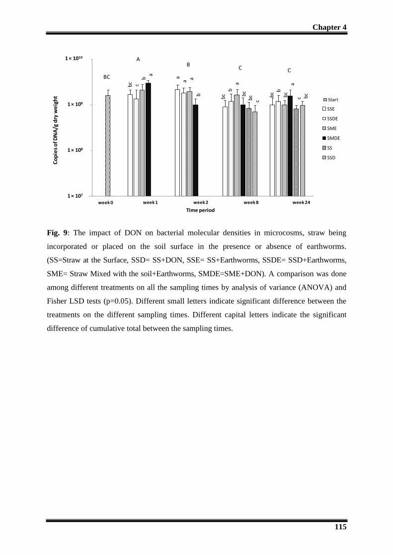
Chapter 4
115
1,00E+07
1,00E+08
1,00E+09
1,00E+10
Day 0 week 1 week 2 week 8 week 24
Co
pie
s o
f DN
A/g
dry
we
igh
t
Time period
SSE
SSDE
SME
SMDE
SS
SSD
S+S
bc c
ba
aa a
b
bcb
abc
bc c b
c bb
ca
c bc
week 0 (Start)
1 × 10⁷
1 × 10⁸
1 × 10⁹
1 × 10¹⁰
1,00E+07
1,00E+08
1,00E+09
1,00E+10
Day 0 week 1 week 2 week 8 week 24
Co
pie
s o
f DN
A/g
dry
we
igh
t
Time period
SSE
SSDE
SME
SMDE
SS
SSD
S+S
bc c
ba
aa a
b
bc
ba
bc
bc c b
c bb
ca
c bc
week 0 (Start)
1 × 10⁷
1 × 10⁸
1 × 10⁹
1 × 10¹⁰
1,00E+07
1,00E+08
1,00E+09
1,00E+10
Day 0 week 1 week 2 week 8 week 24
Co
pie
s o
f DN
A/g
dry
we
igh
t
Time period
SSE
SSDE
SME
SMDE
SS
SSD
S+S
bc cb
a aa a
b
bcb
abc
bc c bc
bbc
ac bc
week 0 (Start)
1 × 10⁷
1 × 10⁸
1 × 10⁹
1 × 10¹⁰
1,00E+07
1,00E+08
1,00E+09
1,00E+10
Day 0 week 1 week 2 week 8 week 24
Co
pie
s o
f DN
A/g
dry
we
igh
t
Time period
SSE
SSDE
SME
SMDE
SS
SSD
S+S
bc cb
a aa a
b
bcb
abc
bc c bc
bbc
ac bc
week 0 (Start)
1 × 10⁷
1 × 10⁸
1 × 10⁹
1 × 10¹⁰
1,00E+07
1,00E+08
1,00E+09
1,00E+10
Day 0 week 1 week 2 week 8 week 24
Co
pie
s o
f DN
A/g
dry
we
igh
t
Time period
SSE
SSDE
SME
SMDE
SS
SSD
S+S
bc c
ba
aa a
b
bc
ba
bc
bc c b
c bb
ca
c bc
week 0 (Start)
1 × 10⁷
1 × 10⁸
1 × 10⁹
1 × 10¹⁰
1,00E+07
1,00E+08
1,00E+09
1,00E+10
Day 0 week 1 week 2 week 8 week 24
Co
pie
s o
f DN
A/g
dry
we
igh
t
Time period
SSE
SSDE
SME
SMDE
SS
SSD
S+S
bc cb
a aa a
b
bcb
abc
bc c bc
bbc
ac bc
week 0 (Start)
1 × 10⁷
1 × 10⁸
1 × 10⁹
1 × 10¹⁰
1,00E+07
1,00E+08
1,00E+09
1,00E+10
Day 0 week 1 week 2 week 8 week 24
Co
pie
s o
f DN
A/g
dry
we
igh
t
Time period
SSE
SSDE
SME
SMDE
SS
SSD
S+Sb
c cb
a
aa a
b
bc
ba
bc
bc
c b
c bb
ca
c bc
week 0 (Start)
1 × 10⁷
1 × 10⁸
1 × 10⁹
1 × 10¹⁰
1,00E+07
1,00E+08
1,00E+09
1,00E+10
Day 0 week 1 week 2 week 8 week 24
Co
pie
s o
f DN
A/g
dry
we
igh
t
Time period
SSE
SSDE
SME
SMDE
SS
SSD
S+S
1,00E+02
1,00E+03
1,00E+04
1,00E+05
1,00E+06
1,00E+07
Day 0 week 1 week 2 week 8 week 24
Co
pie
s o
f DN
A/g
dry
we
igh
t
Time period
SSE
SSDE
SME
SMDE
SS
SSD
S+S
b b ba
a a aa
a a a a aa
bb
ba
bb
week 0
1 × 10²
1 × 10³
1 × 10⁴
1 × 10⁵
1 × 10⁶
1 × 10⁷
BC
AB
C C
0
2
4
6
8
10
12
14
16
18
Week 0 Week 1 Week 2 Week 8 Week 24
bio
mas
so
f e
arth
wo
rms
(g)
Time period
Start
SSE
SSDE
SME
SMDE
week 0 (Start)
a aa
a
aa
a
a
aa a a
a aa a
0
2
4
6
8
10
12
14
16
18
Week 0 Week 1 Week 2 Week 8 Week 24
bio
mas
so
f e
arth
wo
rms
(g)
Time period
Start
SSE
SSDE
SME
SMDE
week 0 (Start)
a aa
a
aa
a
a
aa a a
a aa a
Fig. 9: The impact of DON on bacterial molecular densities in microcosms, straw being
incorporated or placed on the soil surface in the presence or absence of earthworms.
(SS=Straw at the Surface, SSD= SS+DON, SSE= SS+Earthworms, SSDE= SSD+Earthworms,
SME= Straw Mixed with the soil+Earthworms, SMDE=SME+DON). A comparison was done
among different treatments on all the sampling times by analysis of variance (ANOVA) and
Fisher LSD tests (p=0.05). Different small letters indicate significant difference between the
treatments on the different sampling times. Different capital letters indicate the significant
difference of cumulative total between the sampling times.

Chapter 4
116
1,00E+08
1,00E+09
1,00E+10
1,00E+11
1,00E+12
Day 0 week 1 week 2 week 8 week 24
DN
A c
op
ies/
g d
ry w
eig
ht
Time period
SSE
SSDE
SME
SMDE
SS
SSD
S+S
week 0 (Start)
cc
ba
ab ab
c
bc bc
ab b
cc
cd bc
db b
a
1 × 10⁸
1 × 10⁹
1 × 10¹⁰
1 × 10¹¹
1 × 10¹²
1,00E+08
1,00E+09
1,00E+10
1,00E+11
1,00E+12
Day 0 week 1 week 2 week 8 week 24
DN
A c
op
ies/
g d
ry w
eig
ht
Time period
SSE
SSDE
SME
SMDE
SS
SSD
S+S
week 0 (Start)
cc
ba
ab ab
c
bc bc
ab b
cc
cd bc
db b
a
1 × 10⁸
1 × 10⁹
1 × 10¹⁰
1 × 10¹¹
1 × 10¹²
1,00E+08
1,00E+09
1,00E+10
1,00E+11
1,00E+12
Day 0 week 1 week 2 week 8 week 24
DN
A c
op
ies/
g d
ry w
eig
ht
Time period
SSE
SSDE
SME
SMDE
SS
SSD
S+S
week 0 (Start)
cc
ba
ab ab
c
bc bca
b bc c
cd bcd
b b a
1 × 10⁸
1 × 10⁹
1 × 10¹⁰
1 × 10¹¹
1 × 10¹²
1,00E+08
1,00E+09
1,00E+10
1,00E+11
1,00E+12
Day 0 week 1 week 2 week 8 week 24
DN
A c
op
ies/
g d
ry w
eig
ht
Time period
SSE
SSDE
SME
SMDE
SS
SSD
S+S
week 0 (Start)
cc
ba
ab
ab
c
bc bc
ab b
cc
cd bc
db b
a
1 × 10⁸
1 × 10⁹
1 × 10¹⁰
1 × 10¹¹
1 × 10¹²
1,00E+07
1,00E+08
1,00E+09
1,00E+10
Day 0 week 1 week 2 week 8 week 24
Co
pie
s o
f DN
A/g
dry
we
igh
t
Time period
SSE
SSDE
SME
SMDE
SS
SSD
S+S
bc cb
a aa a
b
bcb
abc
bc c bc
bbc
ac bc
week 0 (Start)
1 × 10⁷
1 × 10⁸
1 × 10⁹
1 × 10¹⁰
1,00E+07
1,00E+08
1,00E+09
1,00E+10
Day 0 week 1 week 2 week 8 week 24
Co
pie
s o
f DN
A/g
dry
we
igh
t
Time period
SSE
SSDE
SME
SMDE
SS
SSD
S+S
bc c
ba
aa a
b
bc
ba
bc
bc
c b
c bb
ca
c bc
week 0 (Start)
1 × 10⁷
1 × 10⁸
1 × 10⁹
1 × 10¹⁰
1,00E+02
1,00E+03
1,00E+04
1,00E+05
1,00E+06
1,00E+07
Day 0 week 1 week 2 week 8 week 24
Co
pie
s o
f DN
A/g
dry
we
igh
t
Time period
SSE
SSDE
SME
SMDE
SS
SSD
S+S
b b ba
a a aa
a a a a aa
bb
ba
bb
week 0
1 × 10²
1 × 10³
1 × 10⁴
1 × 10⁵
1 × 10⁶
1 × 10⁷
D
A
B
C
D
0
2
4
6
8
10
12
14
16
18
Week 0 Week 1 Week 2 Week 8 Week 24
bio
mas
so
f e
arth
wo
rms
(g)
Time period
Start
SSE
SSDE
SME
SMDE
week 0 (Start)
a aa
a
aa
a
a
aa a a
a aa a
0
2
4
6
8
10
12
14
16
18
Week 0 Week 1 Week 2 Week 8 Week 24
bio
mas
so
f e
arth
wo
rms
(g)
Time period
Start
SSE
SSDE
SME
SMDE
week 0 (Start)
a aa
a
aa
a
a
aa a a
a aa a
Fig. 10: The impact of DON on fungal molecular densities in microcosms, straw being
incorporated or placed on the soil surface in the presence or absence of earthworms.
(SS=Straw at the Surface, SSD= SS+DON, SSE= SS+Earthworms, SSDE= SSD+Earthworms,
SME= Straw Mixed with the soil+Earthworms, SMDE=SME+DON). A comparison was done
among different treatments on all the sampling times by analysis of variance (ANOVA) and
Fisher LSD tests (p=0.05). Different small letters indicate significant difference between the
treatments on the different sampling times. Different capital letters indicate the significant
difference of cumulative total between the sampling times.

Chapter 4
117
1,00E+02
1,00E+03
1,00E+04
1,00E+05
1,00E+06
1,00E+07
Day 0 week 1 week 2 week 8 week 24
Co
pie
s o
f DN
A/g
dry
we
igh
t
Time period
SSE
SSDE
SME
SMDE
SS
SSD
S+S
b b ba
a a aa
a a a a aa
bb
ba
bb
week 0 (Start)
1 × 10²
1 × 10³
1 × 10⁴
1 × 10⁵
1 × 10⁶
1 × 10⁷
1,00E+02
1,00E+03
1,00E+04
1,00E+05
1,00E+06
1,00E+07
Day 0 week 1 week 2 week 8 week 24
Co
pie
s o
f DN
A/g
dry
we
igh
t
Time period
SSE
SSDE
SME
SMDE
SS
SSD
S+S
b b ba
a a aa
a a a a aa
bb
ba
bb
week 0 (Start)
1 × 10²
1 × 10³
1 × 10⁴
1 × 10⁵
1 × 10⁶
1 × 10⁷
1,00E+02
1,00E+03
1,00E+04
1,00E+05
1,00E+06
1,00E+07
Day 0 week 1 week 2 week 8 week 24
Co
pie
s o
f DN
A/g
dry
we
igh
t
Time period
SSE
SSDE
SME
SMDE
SS
SSD
S+Sb b b
a
a a aa
a a a a aa
bb
ba
bb
week 0 (Start)
1 × 10²
1 × 10³
1 × 10⁴
1 × 10⁵
1 × 10⁶
1 × 10⁷
1,00E+02
1,00E+03
1,00E+04
1,00E+05
1,00E+06
1,00E+07
Day 0 week 1 week 2 week 8 week 24
Co
pie
s o
f DN
A/g
dry
we
igh
t
Time period
SSE
SSDE
SME
SMDE
SS
SSD
S+S
b b ba
a a aa
a aa a a
a
bb
ba
bb
week 0 (Start)
1 × 10²
1 × 10³
1 × 10⁴
1 × 10⁵
1 × 10⁶
1 × 10⁷
1,00E+02
1,00E+03
1,00E+04
1,00E+05
1,00E+06
1,00E+07
Day 0 week 1 week 2 week 8 week 24
Co
pie
s o
f DN
A/g
dry
we
igh
t
Time period
SSE
SSDE
SME
SMDE
SS
SSD
S+S
b b ba
a a aa
a aa a a
a
bb
ba
bb
week 0
1 × 10²
1 × 10³
1 × 10⁴
1 × 10⁵
1 × 10⁶
1 × 10⁷
0
2
4
6
8
10
12
14
16
18
Week 0 Week 1 Week 2 Week 8 Week 24
bio
mas
so
f e
arth
wo
rms
(g)
Time period
Start
SSE
SSDE
SME
SMDE
week 0 (Start)
a aa
a
aa
a
a
aa a a
a aa a
0
2
4
6
8
10
12
14
16
18
Week 0 Week 1 Week 2 Week 8 Week 24
bio
mas
so
f e
arth
wo
rms
(g)
Time period
Start
SSE
SSDE
SME
SMDE
week 0 (Start)
a aa
a
aa
a
a
aa a a
a aa a
AB
CC
BC
Fig. 11: Population dynamics of F. graminearum in wheat straw in the presence or absence of
DON and earthworms, straw being placed on the soil or incorporated in the soil. (SS=Straw at
the Surface, SSD= SS+DON, SSE= SS+Earthworms, SSDE= SSD+Earthworms, SME= Straw
Mixed with the soil+Earthworms, SMDE=SME+DON). A comparison was done among
different treatments on all the sampling times by analysis of variance (ANOVA) and Fisher
LSD tests (p=0.05). Different small letters indicate significant difference between the
treatments on the different sampling times. Different capital letters indicate the significant
difference of cumulative total between the sampling times.

Chapter 4
118
1,00E+05
1,00E+06
1,00E+07
1,00E+08
1,00E+09
1,00E+10
1,00E+11
Week 0 Week 1 Week 2 Week 8 Week 24
CFU
/g d
ry w
eig
ht
Time period
SSE
SSDE
SME
SMDE
SS
SSD
S+S
aa a
a
aa a
a
a aa a a
a
ab
ab ab abb
week 0 (Start)
1 × 10⁵
1 × 10⁶
1 × 10⁷
1 × 10⁸
1 × 10¹⁰
1 × 10¹¹
1 × 10⁹
1,00E+05
1,00E+06
1,00E+07
1,00E+08
1,00E+09
1,00E+10
1,00E+11
Week 0 Week 1 Week 2 Week 8 Week 24
CFU
/g d
ry w
eig
ht
Time period
SSE
SSDE
SME
SMDE
SS
SSD
S+S
aa a
a
aa a a
a aa a a
a
ab
ab ab abb
week 0 (Start)
1 × 10⁵
1 × 10⁶
1 × 10⁷
1 × 10⁸
1 × 10¹⁰
1 × 10¹¹
1 × 10⁹
1,00E+05
1,00E+06
1,00E+07
1,00E+08
1,00E+09
1,00E+10
1,00E+11
Week 0 Week 1 Week 2 Week 8 Week 24
CFU
/g d
ry w
eig
ht
Time period
SSE
SSDE
SME
SMDE
SS
SSD
S+S
aa
aa
aa a
a
a aa a a
a
ab
ab
ab
ab
b
week 0 (Start)
1 × 10⁵
1 × 10⁶
1 × 10⁷
1 × 10⁸
1 × 10¹⁰
1 × 10¹¹
1 × 10⁹
1,00E+02
1,00E+03
1,00E+04
1,00E+05
1,00E+06
1,00E+07
Day 0 week 1 week 2 week 8 week 24
Co
pie
s o
f DN
A/g
dry
we
igh
t
Time period
SSE
SSDE
SME
SMDE
SS
SSD
S+S
b b ba
a a aa
a a a a aa
bb
ba
bb
week 0
1 × 10²
1 × 10³
1 × 10⁴
1 × 10⁵
1 × 10⁶
1 × 10⁷
0
2
4
6
8
10
12
14
16
18
Week 0 Week 1 Week 2 Week 8 Week 24
bio
mas
so
f e
arth
wo
rms
(g)
Time period
Start
SSE
SSDE
SME
SMDE
week 0 (Start)
a aa
a
aa
a
a
aa a a
a aa a
0
2
4
6
8
10
12
14
16
18
Week 0 Week 1 Week 2 Week 8 Week 24
bio
mas
so
f e
arth
wo
rms
(g)
Time period
Start
SSE
SSDE
SME
SMDE
week 0 (Start)a a
aa
aa
a
a
aa a a
a aa a
A
BB B
Fig. 12: The impact of DON cultivable bacterial colonies in microcosms, straw being
incorporated or placed on the soil surface in the presence or absence of earthworms.
(SS=Straw at the Surface, SSD= SS+DON, SSE= SS+Earthworms, SSDE= SSD+Earthworms,
SME= Straw Mixed with the soil+Earthworms, SMDE=SME+DON). A comparison was done
among different treatments on all the sampling times by analysis of variance (ANOVA) and
Fisher LSD tests (p=0.05). Different small letters indicate significant difference between the
treatments on the different sampling times. Different capital letters indicate the significant
difference of cumulative total between the sampling times.

Chapter 4
119
1000
10000
100000
1000000
Week 0 Week 1 Week 2 Week 8 Week 24
CFU
/g d
ry w
eig
ht
Time priod
SSE
SSDE
SME
SMDE
SS
SSD
S+S
week 0 (Start)
1 × 10³
1 × 10⁴
1 × 10⁵
1 × 10⁶
aa
aa
aa a a
ab
b ab
ba ab
acd
bc
cd d
1000
10000
100000
1000000
Week 0 Week 1 Week 2 Week 8 Week 24
CFU
/g d
ry w
eig
ht
Time priod
SSE
SSDE
SME
SMDE
SS
SSD
S+S
week 0 (Start)
1 × 10³
1 × 10⁴
1 × 10⁵
1 × 10⁶
aa
aa
aa a a
ab
b ab
ba a
b
acd
bc
cd d
1000
10000
100000
1000000
Week 0 Week 1 Week 2 Week 8 Week 24
CFU
/g d
ry w
eig
ht
Time priod
SSE
SSDE
SME
SMDE
SS
SSD
S+S
week 0 (Start)
1 × 10³
1 × 10⁴
1 × 10⁵
1 × 10⁶
aa
aa
aa a a
ab b abb
a ab
acd
bc
cd d
1,00E+02
1,00E+03
1,00E+04
1,00E+05
1,00E+06
1,00E+07
Day 0 week 1 week 2 week 8 week 24
Co
pie
s o
f DN
A/g
dry
we
igh
t
Time period
SSE
SSDE
SME
SMDE
SS
SSD
S+S
b b ba
a a aa
a a a a aa
bb
ba
bb
week 0
1 × 10²
1 × 10³
1 × 10⁴
1 × 10⁵
1 × 10⁶
1 × 10⁷
0
2
4
6
8
10
12
14
16
18
Week 0 Week 1 Week 2 Week 8 Week 24
bio
mas
so
f e
arth
wo
rms
(g)
Time period
Start
SSE
SSDE
SME
SMDE
week 0 (Start)
a aa
a
aa
a
a
aa a a
a aa a
0
2
4
6
8
10
12
14
16
18
Week 0 Week 1 Week 2 Week 8 Week 24
bio
mas
so
f e
arth
wo
rms
(g)
Time period
Start
SSE
SSDE
SME
SMDE
week 0 (Start)
a aa
a
aa
a
a
aa a a
a aa a
B B
A AB
Fig. 13: The impact of DON cultivable fungal colonies in microcosms, straw being
incorporated or placed on the soil surface in the presence or absence of earthworms.
(SS=Straw at the Surface, SSD= SS+DON, SSE= SS+Earthworms, SSDE= SSD+Earthworms,
SME= Straw Mixed with the soil+Earthworms, SMDE=SME+DON). A comparison was done
among different treatments on all the sampling times by analysis of variance (ANOVA) and
Fisher LSD tests (p=0.05). Different small letters indicate significant difference between the
treatments on the different sampling times. Different capital letters indicate the significant
difference of cumulative total between the sampling times.

Chapter 4
120
4- Discussions:
4.1-Fate of DON:
In this study, we followed the fate of DON in the winter wheat straw during the saprophytic
survival of F. graminearum up to six months in the microcosms where the straw was left at
the surface or incorporated in the soil in the presence of the whole soil biota.
It was observed that with the passage of time DON disappeared from all the microcosms after
six months passage, whether the straw was left at the surface or incorporated in the soil. The
rate of the disappearance of DON was considerably higher when the straw was incorporated
in the soil as compared to when it was left at the surface thanks to greater exchange surface
between the substrate and microorganisms. This may be because of the faster contact of the
soil communities with DON when the straw was incorporated in the soil. The incorporation of
crop residues is a great reservoir of microbial activity, they are decomposed faster as
compared to the above surface (Lupwayi et al., 2004). In a recent in vitro study, Sato et al.,
(2012) reported some soil-borne bacteria can degrade DON efficiently. So the presence of
similar functional bacteria and of the other soil communities could have made the DON
unavailable in the system.
Furthermore, we also found that earthworms played a significant role in the degradation of
DON. Our results revealed that even after passing 8 weeks, the concentration of DON in the
absence of the earthworms was 40 times higher as compared to the presence of earthworms
when the straw was placed at the surface of the soil. The earthworms increase the microbial
activity by breaking down the big pieces of straw into small pieces and burrowing them,
increasing even more the area of exchange between the contaminated straw and the soil
microorganisms. This resulted in the faster disappearance of DON. This comparative
assessment goes with the recent demonstration that earthworms have a specific preference for
DON contaminated straw compared to non-contaminated straw (Oldenburg et al., 2008).
Although this preference has not yet been explained, it is likely that the choice is related to the
earthworm gut microflora, the later being able to degrade the DON and therefore to provide
the earthworm with specific trophic resources (Schrader et al., 2009).

Chapter 4
121
4.2-Impact of DON on the microbial and micro-faunal community
structures:
We chased the changes in the community structures along six months to determine the impact
of DON on the soil microbial and microfaunal communities when it is present in the crop
residues. We observed that the community structure of bacteria and protozoa were
significantly affected in the presence of DON while fungal and nematodes community
showed respectively conditional and no reaction towards DON. From these results we cannot
assume whether some populations of bacteria and protozoa were definitively favored or
suppressed by the presence of the DON but at least it appeared that the ratio between
populations within a community changed, what reveals some microorganisms are more
susceptible to the toxin.
The bacterial community structures were significantly different in the presence of DON
whether the straw was left at the surface or incorporated in the soil. This effect lasted only for
the first two weeks. This corresponds to the presence of the high quantity of DON in all the
treatments in the start of the experiment. After the passage of two weeks the quantity of DON
was highly reduced in all the treatments and on the eighth week, there was no significant
difference in the community structure of bacteria in DON contaminated and no contaminated
treatments. In the mean while, our comprehensive analysis revealed that the bacterial
community structure in the presence of DON was clearly different in the presence and
absence of earthworms, which also goes to the difference in the quantity of DON in the both
treatments. Surprisingly, the impact of DON was not clear in the absence of earthworms on
the eighth week even we found a high amount of DON. The change in the bacterial structure
can be positive in the sense that certain bacteria from the soil accumulated in order to degrade
DON. Recently, some soil-borne bacteria were reported to be involved in the degradation of
DON (Ikunaga et al., 2011; Sato et al., 2012). This impact of DON could also be negative and
suppress certain populations of bacteria. The negative impact of DON on bacteria has never
been studied or reported before.
In the case of fungi, overall the fungal genetic structure was different when the straw was left
at the surface than when it was incorporated in the soil. The genetic structure was affected by
the presence of DON only in the absence of earthworms. The presence of earthworms
significantly affected the community structure of fungi all over the experiment in all the
modalities. The presence of earthworms might have masked the effect of DON on the fungal

Chapter 4
122
communities. Indeed, on the one hand certain fungal species are reported to be preferred over
the other fungi as food by earthworms (Bonkowski et al., 2000). On the other hand, it might
be because of the rapid degradation of DON in the presence of earthworms which suppressed
the effect on the fungal communities.
On the protozoa the effect of DON was related to the location of the straw. The impact of
DON is very clear throughout the experiment when the straw was placed on the soil in the
presence as well as in the absence of the earthworms from the start till the end of six months.
As DON was no more detectable at this stage so we can assume that the changes DON caused
previously on the protozoan community were long lasting if not definitive. It would be worth
to check for the possible erosion of the protozoan diversity. When the straw was incorporated
in the soil the presence of DON showed less impact on the protozoa.
The nematodes communities showed no reaction against the presence of DON in all the
treatments during the whole period of the experiment. The community structure was only
changed with the passage of time which appeared after the passage of two weeks. The
presence of earthworms had no impact on the nematodes communities.
4.3- Impact of DON on the soil microflora and faunal densities:
The impact of DON was observed on the biomass of earthworms and on the densities of
nematodes, bacteria and fungi during the saprophytic survival of F. graminearum on the
wheat straw during its decomposition in the soil.
Earthworms are well known bio-indicators of the soil health and have great importance in
agriculture due to their ability to incorporate and degrade wheat crop residues and organic
matter (Friberg et al., 2008; Friberg et al., 2005; Kreuzer et al., 2004; McLean et al., 2006).
Our results are in agreement with these previous findings and they more specifically
emphasize the role of L. terrestris in the degradation and incorporation of the straw in the soil.
Despite the biomass of the earthworms reduced over time, probably because the food contents
were exhausting, the positive and original point was that this total biomass was not affected
by the DON contaminated straw. At the contrary, the DON contaminated straw was found
more attractive for the earthworms which coincides with finding of 11 week study of
(Oldenburg et al., 2008).
Furthermore, the presence of the new-borne earthworms in all the treatments containing DON
contaminated straw showed that DON even had no negative effect on the reproduction of
earthworms or the cocoon hatching. The presence of higher number of new-borne earthworms

Chapter 4
123
when the DON contaminated straw was left at the surface of the soil showed that DON
stimulated reproduction in the earthworms or increased the hatching of cocoons. We also
counted the number of earthworms at each sampling time and compared to the initial number
of earthworms put in the microcosms. We observed that the mortality of earthworms was
appeared only in the end of six months. Overall more earthworms were died when the straw
was left at the surface. We also found that mortality of earthworms was a bit higher in the
presence of DON especially when the straw was incorporated in the soil. These mortalities
produced no significant impact on the overall reduction in the biomass of earthworms which
was not significantly different in all the treatments in the presence or absence of DON.
Nematodes are also a very important component of arable soil functioning and could too be
considered indicators for evaluating the soil quality (Schloter et al., 2003). However, more
than their role in decomposing organic matter and in controlling soil-borne fungal pathogens
(Hasna et al., 2007) this is the effect of crop residues and the nature of the organic matter on
the structure of nematodes communities which are taken into account (Berry et al., 2007).
Indeed, the nematodes include both pathogenic and non-pathogenic taxa. Management
practices including previous crops or organic amendments aim at influencing the structure of
nematodes communities to increase the control of the pathogenic ones either through a direct
effect of the organic matter or through a microbes mediated effect (Berry et al., 2007; Mateille
et al., 2009). Unfortunately, little is known concerning the direct or microbial mediated
interaction between nematodes and wheat straw. Our results showed that the number of
nematodes increased with the passage of time during the decomposition of straw. This was
contrary to the decomposition of straw and the biomass decrease of the earthworms. The
impact of DON appeared very late in the experiment on the nematodes density. DON had no
sudden poisoning effect on nematodes but the effect appeared later which may indicate that
DON showed rather an acute progressive poisoning. There was no impact of DON on the first
two weeks but after eight weeks a negative trend of DON was developed and it was found
significant after 24 weeks. In a recent report deoxynivalenol is reported to reduce the
developmental process and inhibit the nematodes (Caenorhabditis elegans) egg hatching in
the liquid medium (Gowrinathan et al., 2011). The nematodes densities increased in the
system with the decrease in the biomass of earthworms and on week 24 it is significantly clear
that earthworms were higher in the absence of earthworms. It seems that earthworms and
DON were playing also a role in the suppression of nematodes densities. Dominguez et al.,
(2003) previously reported that the nematode densities are in negative relation with biomass
of earthworms during the decomposition of organic substrate. The effect of DON appeared on

Chapter 4
124
the nematodes as well as on the earthworms late in the experiment. In case of nematodes it is
significant while in case of earthworms it was not so high. This indicates that DON was not
involved in the sudden death but a slow poisoning.
We also measured the molecular mass of bacteria and fungi to determine the impact of DON
contaminated straw on them. We found that molecular biomass of bacteria and fungi were
increased soon after the establishment of experiment and then started decreasing with the
decomposition of the wheat straw. This same trend was observed on the plates in the case of
bacteria and not confirmed in the case of fungi. The availability of the nutrients and the
aeration in the start of the experiment seems to be involved in boosting their densities. The
bacterial and fungal densities showed the reaction against the presence of DON only when the
straw was incorporated in the soil and DON produced no impact when the straw was left at
the surface. This might be because of the rapid contact of DON contaminated straw with
bacterial and fungal communities. They showed positive response first just after the
experimental setup and then DON significantly reduced their densities which were detectable
up to 8 weeks till the detection of DON. The reaction of DON was same towards the bacterial
and fungal densities. They showed the positive reaction in the start of the experiment but
suddenly decreased significantly. This may show that the presence of DON served as an
external stress and in response the fungi first produced more spores but later DON showed a
negative reaction on growth of mycelium and spore production. The reaction of DON was
limited only in the treatments where straw was incorporated in the soil. This might be again
because of the rapid exposure of DON to the soil bacterial and fungal communities.
The growth and the development of F. graminearum were observed in the presence or the
absence of DON. F. graminearum showed the same kind of behavior as the other fungal and
bacterial communities. F. graminearum also showed the reaction towards the presence of
DON only in the case where the straw was mixed in the soil. F. graminearum was found to
get benefit of the presence of DON and the biomass was increased like overall fungal and
bacterial communities. F. graminearum density was also decreased with the passage of time
like the bacterial and fungal communities. Contrary to other fungal populations within the
fungal communities, the presence of DON had no negative effect on the density F.
graminearum as we compare its population with control in the experiment. The comparison
among the treatments after 8 or 24 weeks showed also that the presence of earthworms had no
significant impact in the reduction of F. graminearum.

Chapter 4
125
The multiple interactions occurring in the system studied could have obscured a stronger
impact of DON than the one revealed by the community structure fingerprintings or the
community molecular biomass. At the contrary, the classic microbiological methods revealed
some variability among the microorganisms in their response to the presence of DON but they
are imprecise and cannot ensure that the observed differences must be attributed to DON, to
biotic interactions or to technical limitations. Therefore, although molecular and classic
microbiological assessments of the fate of bacteria and fungi in the crop residues in presence
or absence of DON gave occasionally different results, they are complementary and they both
revealed similar trends, the main ones being that the microbial communities, including the
protozoa are affected at various extents by the presence of DON but that F. graminearum is
not affected by the presence of DON it its surrounding.
5-Conclusions:
Finally, this study is original. It provides for the first time a wide overview and a
comprehensive knowledge on the fate of DON in the crop residues in the soil and its impact
on the soil microflora and fauna.
We conclude that DON didn’t stay for a long time in the straw and disappeared completely in
all the treatments during the six months. The location of straw played a significant role in the
disappearance of DON. It disappeared more rapidly when the straw was incorporated in the
soil as compared to when left at the surface of the soil. The presence of earthworms played a
key role in the disappearance of DON which was highly slow in their absence. DON was not
produced in situ or produced in very small quantity, which disappeared very rapidly during
the saprophytic survival of F. graminearum.
DON significantly changed the community structure of bacteria which was dependent on the
quantity of DON present in the microcosms. In case of protozoa the location of straw was
more important and the affect of DON remained throughout the experiment. The effect on the
fungal community structure was seen only in the absence of earthworms. The nematodes
community structure remained unaffected by presence of DON.
The biomass of earthworms was not affected by their exposure to DON. The reproduction or
cocoon hatching of earthworms was stimulated in the presence of DON when the straw was
placed on the soil surface. Nematodes densities were negatively affected and their number
were reduced but the impact appeared late in the experiment. The bacterial and the fungal
densities were negatively affected only when the straw was incorporated in the soil. F.

Chapter 4
126
graminearum got no advantage of the presence of mycotoxin in the straw whether it was left
at the soil surface or incorporated in the soil but conversely, it was not negatively impacted by
the presence of this mycotoxins in its surrounding.
Conclusion
Fate of DONImpact on micro flora and fauna
Advantage to F. graminearum to develop
Densities Structure
Fungi Bacteria Fungi Bacteria Protozoa NematodesEarthworms Nematodes
Rate of disappearance:Incorporation > SurfaceEarthworms > without earthworms
Ecological role of mycotoxins?
wheat straw ± DON
Soil
+Ve -Ve -Ve Yes-Ve Yes Yes No
straw location had impact
No
straw location had impact
Fig. 14: Flow chart showing the main conclusion obtained during the different analyses in the
microcosm experiment. The violet colour rectangle shows the disappearance of DON; the
green colour rectangle shows the positive impact of DON; red colour rectangles show the
impact of DON while the grey colour shows no impact of DON. Furthermore, Yes= DON had
an impact; No= DON had no impact; +Ve = positive impact; -Ve = negative impact.

Chapter 5
127
Chapter-5: Population dynamics of
Fusarium graminearum in the presence of
decomposers and deoxynivalenol

Chapter 5
128
Population dynamics of Fusarium graminearum in
the presence of decomposers and deoxynivalenol
1-Introduction:
Reduced tillage tends to be more practiced to preserve soil structure and reduce erosion, to
maintain soil moisture, save energy and working time. Therefore, crop residues are going to
be more and more frequently left on the soil surface rather than being incorporated into the
soil. Consequently, many soil-borne plant pathogens survive in the previous year's crop
residue, making diseases more problematic under reduced-tillage conditions (chapter 3).
Indeed, reduced tillage can favor pathogens by protecting the pathogen's refuge in the residue
from microbial degradation, by lowering soil temperature, or by increasing soil moisture
(Bailey and Lazarovits, 2003; Bateman et al., 2007). Therefore additional controls are needed
for pathogens. Besides breeding host plants for resistance towards bio-aggressors, chemical,
biological and cultural controls can be used alone or in combination to limit damage from
diseases (Bockus and Shroyer, 1998). In the case of soil- or residue- borne microorganisms,
chemical control proved to be not really efficient to protect the plant from the pathogens
(Bolton et al., 2010; Nel et al., 2007) while crop rotation schemes including intermediate
crops such as Brassicaceae appeared more promising (Buhre et al., 2009; Paulitz et al., 2010).
The use of biocontrol agents is a bit tricky because many parameters must be met to ensure an
efficient bioprotection. Among these parameters, the proper timing of antagonist applications
is probably the more difficult to adjust, unless the inoculum can establish in and be retained
by the residues so that this inoculum can antagonize continuously the pathogen (Matarese et
al., 2012). The problem is even more crucial with mycotoxigenic fungi such as F.
graminearum and other Fusarium sp belonging to the Fusarium Head Blight (FHB) complex
and whose mycotoxins may affect other residue-borne microorganisms or inoculated
antagonists. Any competitive advantage conferred by mycotoxins would complicate efforts to
control F. graminearum during its saprophytic growth on crop residues. For instance,
deoxynivalenol (DON) produced by F. graminearum can repress chitinase gene expression in
a biocontrol agent Trichoderma atroviride (Lutz et al., 2003). Conversely, the use of a
nontoxigenic mutant of F. graminearum in controlled conditions revealed that DON did not
provide any further advantage to the wild strain towards T. atroviride (Naef et al., 2006).

Chapter 5
129
Most of the studies so far focused on the pathogenic phase of F. graminearum lifecycle and
on the way to biocontrol the mycotoxins production but more rarely on the saprophytic phase
of its lifecycle while this phase could be the weakness of the pathogenic fungus.
Indeed, on the one hand, the mycotoxigenic Fusarium sp. generally overwinter in crop
residues and they remain potential threat to the coming susceptible crops, what is stated by
most of the studies (Leplat et al., 2012; Parry et al., 2007). On the other hand most soil fungi
are saprotrophs that feed on decaying organic materials and contribute at different extents to
the decomposition process of plant–derived substrates. Therefore, they could limit the
development of the future primary inoculum. Therefore, the direct role of mycotoxins towards
decomposers and direct antagonists need to be assessed to understand the strategy used by F.
graminearum, to survive for years in crop residues.
The main objective of this study was to find how strain MIE 00376 of F. graminearum could
get profit of the presence of the DON it produces towards i) other fungal decomposers and ii)
other DON non producing fungi of the FHB fungal complex. Assuming that nitrogen could be
a limiting factor for the decomposition processes tested in subsequent experiments, some free
fixing nitrogen bacteria were also included in the test. For this purpose the response to
different doses of DON of different soil-borne fungi previously isolated from decaying wheat
straw as well as the susceptibility of known strains of Fusarium sp. and Microdochium nivale
issuing from the FHB fungal complex and of a set of rhizospheric bacteria was evaluated
towards to choose DON resistant microorganisms. Then the population dynamics of F.
graminearum was observed in presence of DON resistant microorganisms, alone or in
combinations, in the presence or absence of DON in the microcosms.

Chapter 5
130
2-Materials and Methods:
A set of 17 fungal strains and 4 bacterial strains was used in this study (Table 1). Four fungal
strains are part of the FHB fungal complex and exhibit different abilities to produce or not
various mycotoxins, 8 fungi were isolated from decaying straw at various stages of
decomposition and are therefore associated to the decomposition process although their
potential enzymatic activities were not tested for each of them, 2 strains are pathogenic fungi
which could cause damping off and root rot on wheat but they were selected because of their
saprophytic abilities, 3 strains are saprophytic fungi isolated from soil and previously
characterized for their antagonistic activities towards some plant pathogenic fungi but not
towards F. graminearum. Three bacterial strains were isolated from rhizospheric soil of
cereals and were chosen for their ability to fix nitrogen freely while the 4th
one was chosen as
a common rhizospheric bacterium.
Among the 8 fungi isolated from decaying straw 4 were previously identified by sequencing
the ITS and EF regions of rDNA. The sequences were then compared using blast and NCBI
data library and were preserved to the MIAE collection. The other 4 fungi isolated from
decaying straw in microcosm experiment (Chapter 4) were just identified phenotypically. All
other fungal and bacterial strains have been previously identified and were preserved as so in
the MIAE collection.
2.1- Minimal Inhibition Concentration test for screening:
Two types of methods were used to evaluate the minimal inhibition concentration needed to
limit, or not, the microbial development. Radial growth of fungal colonies was measured on
PDA medium in Petri dishes and optical density of fungal and bacterial growth was measured
in liquid medium in microtiter plates as described in detail in the Chapter-2, , page 58.

Chapter 5
131
Table 1: Strains and isolates of fungi and bacteria used and their respective Minimal
Inhibitory Concentration (MIC) determined in this study
Fungal strains Accession
number1
Origin -
Traits of interest MIC
2
Population
dynamics3
Fusarium graminearum MIAE00376 Maize stubbles - FHB complex-
DON producer > 10 µg/ml X
F. culmorum MIAE00288 Diseased plant - FHB complex-
DON producer < 2.7 µg/ml X
F. poae MIAE00300
Diseased plant - FHB complex-
does not produce DON (but
produces TCTA4 and other TCTB
4)
< 0.3 µg/ml X
Microdochium nivale MIAE00318 Diseased plant - FHB complex-
does not produce mycotoxin < 0.9 µg/ml X
T. koningiopsis MIAE00725 Isolated from decaying straw > 10 µg/ml X
F. oxysporum MIAE00726 - > 10 µg/ml X
F. oxysporum MIAE00800 - > 10 µg/ml
Epicoccum nigrum MIAE00727 - > 10 µg/ml
Gliocladium spp. PC5 - < 0.1 µg/ml
Aspergillus spp. PC - > 10 µg/ml
A. niger PC - > 10 µg/ml
Penicillium spp. PC - > 10 µg/ml
Rhizoctonia solani MIAE00078 Soil- responsible for root rot; good
saprophytic abilities < 2.7 µg/ml
R. solani MIAE00066 - < 2.7 µg/ml
F. oxysporum MIAE00047
Fusarium wilt suppressive soil of
Chateaurenard (France) –
Antagonistic abilities toward
pathogenic F. oxysporum
> 10 µg/ml
Trichoderma gamssi MIAE00029 Soil- antagonistic abilities towards
pathogenic fungi < 2.7 µg/ml
T. velutinum MIAE00044 - < 0.9 µg/ml
Bacterial strains
Azospirillum brasilense MIAE00334 Rhizospheric soil - free nitrogen
fixing bacteria > 10 µg/ml
A. lipoferum MIAE00337 - > 10 µg/ml
A. lipoferum MIAE00338 - > 10 µg/ml X
Pseudomonas fluorescens MIAE00587 Rhizospheric bacteria < 2.7 µg/ml
1 accession number in collection MIAE (Micro-organisms on Agro-Environmental Interest - UMR-
MSE, Dijon, France (http://www2.dijon.inra.fr/umrmse/spip.php?rubrique47). 2 MIC : Minimal Inhibitory Concentration (µg DON/ml medium)
3 strains which were selected for the population dynamic study are indicated with an X
4 TCTA: trichothecenes A; TCTB: trichothecenes B,
5 PC : personal collection. Strains were phenotypically identified. The molecular identification is being
performed before the strains as a prerequisite to get an accession number from MIAE collection

Chapter 5
132
2.2- Preparation of microcosms and analyses:
2.2.1-Inoculum production:
Three kinds of media were prepared:
1- Wheat bran liquid medium (WB): 2 % wheat bran in the distilled water
2- Luria betari (LB) media: 10 g/l Bacto trypton, 5 g /l yeast extract and 10 g/l NaCl, pH =7.0
3- Malt liquid media (ML): 10 g/l malt.
All of these media were autoclaved at 120°C for 20 min. Meanwhile, all the required fungal
strains were cultured on the PDA and were placed at 25° C for seven days. Small plugs of 7
days old PDA cultures of different fungi were added to their appropriate media. F.
graminearum, F. poae and F. culmorum were added WB medium, M. nivale and T.
koningiopsis were added to ML medium and F. oxysporum and A. lipoferum were added to
LB medium. They were placed at 25°C in the rotary shaker (150 rpm) for ten days for fungi
and 2 days for bacteria. The fungal cultures were filtered through sterile cheesecloth
(approximately 50 µm mesh size) to remove the mycelial mat. The number of bacteria and the
conidial concentrations were assessed through Malassez chamber under the microscope. The
concentrations were then adjusted by dilution with sterile distilled water. M. nivale didn’t
produce the spores, so the propagules were counted by colony forming unit (CFU) just before
putting in the microcosms.
2.2.2-Straw treatments and microcosms preparation:
The air dry straw was taken from the winter wheat (Triticum aestivum) originated from a field
of Epoisses (Bretennières, France). The straw was cut into approximately 2-3 cm small pieces
and was sterilized by gamma radiations.
Small glass jars of dimensions 10 cm × 7 cm × 7cm (height × length × width) were used as
microcosms. Two gram dry sterilized straw was filled in each of these microcosms. The equal
amount of each fungi was added to the required microcosms in the form of spore suspensions
(103 spores/g of straw (d.w.)) or in the form of propagules in case of Microdochium nivale
(103 CFU/g of straw (d.w.)) or number of bacteria in case of bacteria (10
5 bacteria/g of straw
(d.w.)). They were spread on the straw with micropipettes. DON solution (Chapter 2, page
56) at the rate of 10 µg/g straw (d.w) was added to each of the required microcosms. The final
humidity in each microcosm was maintained as 80% which was equal to its water holding
capacity. In the end half microcosms of each treatment were contaminated with DON solution

Chapter 5
133
at the rate of 10 µg of DON/g of straw. All the microcosms were closed and were vigorously
shaken by hand to mix all the contents of the microcosms. Half of the microcosms containing
DON contaminated straw were prepared in extra to measure the quantity of DON in the start
and the end of experiment. In this way 8 types of microcosm were prepared containing
different combinations of different fungi which were further divided as with and without
DON (table 2).
F. graminearum
MIAE00376
FHB complex:
F. poae
MIAE00300 F. culmorum
MIAE00288 M. nivale
MIAE00318
Decomposers:
F. oxysporum
MIAE00726 T. koningiopsis
MIAE00725
Decomposers + free
N2 fixing bacteria: F. oxysporum
MIAE00726 T. koningiopsis
MIAE00725 A. lipoferum
MIAE00338
DON
X X X
X X
X X X
X X
X X X
X X
X X
X
Table 2: Experimental design to monitor the population dynamics of F. graminearum in
presence of different combinations of different fungi with and without DON.
Three independent microcosms were prepared for each treatment. Some extra microcosms
were prepared to measure the loss of humidity. All the microcosms were placed in an
incubator at 25°C.
2.3 -Sampling and samples processing and analyses:
The sampling was done at 0, 14, 28 and 49 days. Three independent microcosms for each
treatment were picked. The entire contents were shifted in the small vials and were freeze
dried. They were ground to fine particles by sterilized pestles and mortars. These fine
particles of straw were placed again at -20°C and were used for the extraction of DNA and

Chapter 5
134
DON. The humidity was measured at each sampling time and was found 78-80% till the end
of experiment.
The DNA from the straw was extracted and purified (chapter 2, page 50).
From the extracted DNA F. graminearum was quantified by using real time PCR (chapter 2,
page 51).
DON was extracted from the straw in the start and at the end of experiment and it was
quantified by using high performance liquid chromatography (HPLC, Chapter 2, page 57).

Chapter 5
135
3-Results:
3.1-Minimal inhibition concentrations:
In the experiment we tested different soil-borne fungal and bacterial strains against different
doses of DON from 0 µg/ml to 10 µg/ml on the Petri dish and OD measurement. The results
showed that all the fungal species were not susceptible or resistant to DON (Table 1). The
MIC values of DON for different fungal species showed that the susceptibility was dependent
on the dose of DON. Some fungal strains were highly resistant even to the maximum dose we
tested.
F. graminearum strain MIAE00376 chemotype DON was resistant to highest dose of DON
we tested (10 µg/ml). This indicated that the producer of DON was resistant to the mycotoxin
it produces. Among other fungal species all the FHB causing fungal species were susceptible
to DON. The growth of F. culmorum was restricted at 2.7 µg/ml, F. poae was restricted at 0.3
µg/ml and M. nivale was inhibited at 0.9 µg/ml.
DON produced no impact on the growth of some antagonistic and decomposer fungal species
including F. oxysporum strain MIAE00726, F. oxysporum strain MIAE00800, T. koningiopsis
strain MIAE00725 and E. nigrum strain MIAE00727 were resistant to DON even to a very
high amount 10 µg/ml. Some other universal antagonistic fungi of Trichoderma spp. i.e. T.
gamssi strain MIAE00029 and T. velutinum strain MIAE00044 showed susceptibility towards
DON. T. gamssi strain MIAE00029 was found susceptible at 2.7 µg/ml and T. velutinum
susceptibility to DON was even lower as it was susceptible at 0.9 µg/ml. The 2 strains of R.
solani were found to be both susceptible to 2.7 µg/ml of DON. Some fungi were isolated
during decomposition of straw as putative active decomposer in the microcosm experiment
(Chapter-4). Among them Gliocladium spp. was found highly sensitive towards DON that
even the low amount 0.1 µg/ ml restricted the growth of fungus. On the other hand, other
fungal species Aspergillus niger and Penicillium spp. were highly resistant to even the highest
dose of DON we tested (10 µg/ml).
We also tested the susceptibility of some nitrogen fixing bacterial species (Table 1). Most of
the bacterial species we tested were resistant towards DON even to the highest dose of DON
used in the experiment (10 µg/ml). The fourth bacterial strain tested, P. fluorescens strain
MIAE00587 was found sensitive to DON < 2.7 µg/ ml.

Chapter 5
136
3.2-Population dynamics of Fusarium graminearum:
The population dynamics of F. graminearum in the presence of FHB-complex or the fungal
decomposers with or without added nitrogen fixing bacteria and in the presence or absence of
DON was monitored using qPCR in destructive independent microcosms including 2 g dw of
straw inoculated with the various combinations of microorganisms (= treatments) according to
the experimental set up.
The F. graminearum strain has developed similarly in all microcosms (Fig. 1). The number of
DNA copies of strain MIAE00376 was 6.5 × 10⁵ copies/g dw straw as an average just after
the inoculation process. It increased within 2 weeks up to 1.2 × 10⁹ copies/g of dw straw
(average value) and then up to 1.43 × 10⁹, (average value) 4 weeks post inoculation to reach
1.73 × 10⁹ copies of DNA/g dw straw as an average value 7 weeks post inoculation. Data
acquired on day 0 and those acquired on the other sampling dates were analyzed separately.
Indeed, the inoculum value was supposed to be the same for all the microcosms and was not
affected by any of the tested factors on day 0. However the densities of DNA copies of strain
MIAEO376 appeared dissimilar among treatments just after the inoculation process at day 0
with a high relative standard deviation of 0.55. An ANOVA performed on the dataset at day 0
revealed that the dispersion of the densities of DNA was not related to the treatments what
means that this dispersion was random and should not determine the subsequent putative
effect of the tested factors on the population dynamics of the strain MIAE00376.
An ANOVA performed on the whole set of data acquired on week 2 , 4 and 7 post inoculation
indicated that the density of DNA copies of the Fusarium strain significantly increased from
week 2 to week 7 although this increase was not significant between week 2 and 4 and
between week 4 and 7.
Important points were that in no case, neither the presence of DON nor the presence of fungi
of the FHB complex had an effect of the population dynamics of strain MIAE00376. For
cons, the presence of decomposers and the presence of decomposers with the nitrogen fixing
bacteria significantly reduced the development of strain MIAE.

Chapter 5
137
1,00E+05
1,00E+06
1,00E+07
1,00E+08
1,00E+09
1,00E+10
0 10 20 30 40 50 60
Fg + FHBC + DON Fg+decomposers+DON Fg+decomposers+Azo+DON Fg alone+DONFg+ FHBC Fg+Decomposers Fg+ decomposers+ Azo Fg alone
1010
109
108
107
106
105
Number of days
DN
A c
op
ies/
g o
f dry
we
igh
t
Fig. 1: Population dynamics of F. graminearum MIAE00376 (Fg) in straw with and without
DON. The Fusarium strain was inoculated either alone or in combination with i) FHBC=
fungi of the FHB complex (F. poae MIAE00300, F. culmorum MIAE00288 and M. nivale
MIAE00318), ii) decomposers fungi isolated from decomposing straw (F. oxysporum
MIAE00726 and T. koningiopsis MIAE00725), iii) decomposers + Azo = decomposers + a
free N2 fixing bacteria (A. lipoferum MIAE00338). The population dynamics of F.
graminearum was monitored using Q-PCR.
3.3- Concentration of DON:
DON was extracted and quantified in the microcosms at the initial stage (day of
contamination) and at the end of the experiment (week 7) to evaluate the fate of the
mycotoxins in the various treatments (Fig. 2). Theoretically, the concentration of DON
introduced in the straw was 10 µg DON/g dw straw but average quantities found just after the
contamination at day 0 varied from 3.8 to 4.4 µg/g in the 4 sets of microcosms in which F.
graminearum strain was inoculated alone or in combination with other microorganisms. The
relative standard deviations were really important, ranging from 0.36 to 0.78 and the
concentrations were not significantly different among the 4 treatments. However, almost 60%
of the inoculated DON was no more accessible for quantification soon after inoculation either

Chapter 5
138
because the mycotoxin is adsorbed in the organic matrix, or because a technical error
occurred. At week 7, the relative standard deviations were much more lower than at time 0,
ranging from 0.1 to 0.26 but surprisingly, the concentration of DON remained significantly
stable in the set of microcosms used to monitor the strain of Fusarium when it was in
interactions with other inoculated microorganisms. At the contrary, the concentration was
significantly lower in the set of microcosms where the strain MIAE00376 was alone, what is
difficult to explain from a biological point of view. However, probably because of the high
level of variability among the data acquired at week 0 and at a lesser extent at week 7, there
was no significant difference between initial and final stage for the 4 treatments. In
conclusion, only 40% of the mycotoxins introduced in the straw were quantifiable at day 0 but
this proportion remained constant for the 7 weeks of incubation, i.e. the DON was not either
degraded or used by the microorganisms present in the microcosms. Conversely, no DON was
produced by F. graminearum both when the fungus was alone in the microcosms and when it
was in interaction with the other fungal and bacterial species.
0,00
1,00
2,00
3,00
4,00
5,00
6,00
7,00
8,00
Fg + FHB complex Fg + Decomposers Fg + Decomposers + Azo Fg alone
Week 0 Week 7
0.00
1.00
2.00
3.00
4.00
5.00
6.00
7.00
8.00
DO
N/g
of d
ry w
eigh
t
Fig. 2: Quantity of DON in the straw contaminated with DON inoculated with Fusarium
strain MIAE00376 (Fg) was inoculated either alone or in combination with i) FHBC= fungi of
the FHB complex (F. poae MIAE00300, F. culmorum MIAE00288 and M. nivale
MIAE00318), ii) decomposers fungi isolated from decomposing straw (F. oxysporum
MIAE00726 and T. koningiopsis MIAE00725), iii) decomposers + Azo = decomposers + a
free N2 fixing bacteria (A. lipoferum MIAE00338).

Chapter 5
139
4-Discussion:
Our results from MIC revealed that DON was not deleterious for its producer and the same
results were confirmed from our results of the population dynamics of F. graminearum in
microcosms. On the other hands it was not beneficial for its producer in the sense of
increasing its population. Another mycotoxins, zearalenone (ZEA), produced by this fungus is
reported to increase its sporulation what should promote greater dispersion and better
development of the fungus, although this has not been tested directly in terms of population
dynamics in the study cited (Wolf and Mirocha, 1973). In our study, at the contrary, we didn’t
directly test fungal sporulation nor physiologic or metabolisms changes but we can draw that
DON is not used by the fungus to increase the population size during the saprophytic survival
in the crop residues. These results are consistent with our field and microcosm experiment.
In addition, we observed that DON could be deleterious at various doses to other different
fungal species. We tested the MIC of a limited number of strains from the FHB causing fungal
species which we call FHB-complex but they were found susceptible at various extents
according to the strains. These results lead to the hypothesis that DON produced by F.
graminearum could eliminate members of the FHB-complex during the competition in the
crop residues during saprophytic survival or on the plants during disease development and
colonization. The competition among the FHB-complex and F. graminearum and the
dominance of F. graminearum associated to an increase in the DON concentration during the
fungal interaction was previously observed (Xu et al., 2007). Our results from the population
dynamics and the quantification of DON negated this hypothesis. In our study, during any
competition there was no significant increase in DON (Fig. 2) and the presence of artificially
given dose of DON didn’t give any advantage to F. graminearum. In order to get DON
resistant decomposers for fast decomposition of crop residues to control the saprophytic
survival of F. graminearum, strain isolated from decaying straw as well as others saprotrophic
fungi and nitrogen fixing bacteria were also tested for their growth in presence of various
doses of DON. MIC results revealed that among the other decomposers and antagonists we
tested some of them were found susceptible to the different doses of DON. This result is
consistent with the field and the microcosm studies previously conducted (chapter 3 and 4)
which revealed that weak but significant modifications in the genetic structure of bacterial and
fungal communities were detected when DON was present in the crop residues, according to
the location of the latter. MIC results indicate that overall 47% of fungal species used in our
test were susceptible towards DON depending on the concentration of DON. Deeper in the

Chapter 5
140
details, the F. oxysporum strains which were also isolated from wheat straw or from soil were
surprisingly highly resistant towards DON. Although F. oxysporum and F. graminearum are
grouped in the same genus, they are quite different from a phytopathology point of view, with
different specific host plants and different mode of actions towards these host plants and
therefore, one may wonder why F. oxysporum could have built up mechanisms to resist to
DON unless DON is not a mycotoxins produced to antagonize other fungi. However, the two
species have a common phase in their life cycles, the saprophytic phase during which they can
interact. F. oxysporum are generally reported to colonize the crop residues very fast and are
more efficient as saprotrophs as compared to the FHB causing fungal pathogens (Pereyra et
al., 2004).
Three different Trichoderma species which were previously isolated from wheat straw during
decomposition and are known as crop residues decomposers and for their antagonistic ability
as T. gamsii were tested (Anees et al., 2010; Matarese et al., 2012). Only one strain i.e.
Trichoderma koningiopsis was found resistant to DON while the other two out of three
Trichoderma spp were found sensitive towards the different doses of DON. Contrary to what
was observed with F. oxysporum which competes with F. graminearum for the exploitation of
common trophic resources, DON could be used by F. graminearum for defense against fungal
species having antagonistic abilities. Indeed, DON can down regulate T. atroviride strain P1
chitinase genes (Lutz et al., 2003). In contrast to that a recently published study Matarese et
al., (2012) suggested that Trichoderma spp. could be used as biocontrol agent and can even
down regulate the production of DON and the growth of F. graminearum but it was substrate
dependent.
Having in mind that nitrogen could be a limiting factor to decompose crop residues which are
mainly carbon based, it was assume that this element could be input in the decomposition
process via the activity of free nitrogen fixing bacteria, although this bacteria are more
frequently found in the rhizosphere than in litters but they are generally associated to the
rhizosphere of cereals, hence the idea of testing their susceptibility to DON (Sanguin et al.,
2009; Venieraki et al., 2011). Only one out of four species were affected by DON. Overall the
results from MIC values revealed that DON seems to be very selective in its targets and very
selective towards different soil communities.
The MIC was an in vitro test which anyway allowed sorting out some DON resistant
decomposers and nitrogen fixing bacteria which could help to destroy the crop residues which
are considered the main cause of fungal primary inoculum. We used the decomposers which
included T. koningiopsis strain MIAE00725 and Fusarium oxysporum strain MIAE00726.

Chapter 5
141
Trichoderma spp. are also well known antagonists and as well as soil-borne fungi and are
aggressive colonizer of maize and wheat crop residues and take an active part in the
decomposition process (Broder and Wagner, 1988).
The test was scaled up small microcosms to include both i) crop residues, i.e. wheat straw, ii)
interacting microorganisms i.e. decomposers in one case and fungi of the FHB complex in the
other case, and iii) the monitoring over time of the impact of these microbial interactions on
the F. graminearum strain on a specific substrate in presence or absence of DON.
Interesting results were obtained with this experimental set up and in a way, they confirmed
the hypothesis we had about the possible control of F. graminearum by fastening the
decomposition of their habitat i.e. the wheat crop residues but we need first to be cautious
with the discussion of the results we got for various reasons. The first one is that only 40 % of
the DON was accessible for quantification immediately after its inoculation in the crop
residues. This could be due to an experimental mistake we did in preparing the solution. In
previous experiments (chapter 3 and 4) less quantities of DON than the quantities added to
maize stubbles or to wheat straw-soil mixtures were recovered but in that case, besides
technical aspects, both biotic and abiotic factors were incriminated. In the present study, as
experiments were conducted in sterile conditions, we can exclude that indigenous
microorganisms degraded the mycotoxins. Therefore, the remaining alternative explanations
could be linked to the difficulties faced to extract the DON from the straw, or to recover it
from the solvent with the affinity columns used for that purposed despite previous tests were
performed to specifically choose these affinity columns. At last, it is also quite possible that
DON is rapidly physically adsorbed by the straw itself and could be released later, once the
straw is decomposed.
The second reason we have to consider is that the MIC were evaluated with known increasing
doses of DON from 0 up to 10µg/L incorporated in agar medium but we didn't check for the
availability of the DON, therefore the same phenomena could have occurred as well. We have
been anyway able to rank the strains for susceptibility to DON in the agar medium and to
select the more resistant ones, what was, in some cases, confirmed by test performed in liquid
medium. Therefore we are confident with the ranking and with the selection we did.
However, it is now difficult to compare the doses used for the selection of resistant strain and
the active doses of DON available in the microcosms. The active dose of DON in the
microcosms was less than the one used for the MIC tests and this could explain why, no effect
of DON was observed on the population dynamics of the F. graminearum strain MIAE 00376
when the strain was either alone or interacting with other microorganisms.

Chapter 5
142
At last, the microcosms where a miniature system and the incubation lasted for only 7 weeks,
therefore we noticed that the growth i.e. increase in the number of DNA copies of the F.
graminearum strain was reduced in presence of a few strains representing decomposers but no
significant decrease was observed.
Anyway, having in mind the above statements, one interesting point issuing from this
experiment is that neither the presence of DON nor the presence of fungi of the FHB complex
had an effect of the population dynamics of F. graminearum strain MIAE00376. So the
mycotoxins produced by a member of this fungal complex do not provide any saprotrophic
competitive advantage to the producer. This does not preclude the fact that the advantage can
be found by the mycotoxins producer during the infectious phase of its lifecycle but our
results do not help solving the question about the reasons why mycotoxins are produced.
The main and promising point that this experiment provided is that the presence of
decomposers and the presence of decomposers with the nitrogen fixing bacteria significantly
reduced the development of the F. graminearum strain MIAE. The decomposers can't be
restricted to the only 2 or 3 fungal strains we used. The soil fauna and many other
microorganisms may contribute to the process but we hereby underlined a weakness in the
lifecycle of F. graminearum which should be exploited to improve the control of the
pathogenic fungus during its saprophytic phase. This could be achieved either by stimulating
indigenous decomposers, but this is not always easy to do unless a global and systemic way of
managing the agricultural plots takes into account this parameter, or it can be achieved by
means of inoculating a consortium of decomposers including enzymes-well equipped
decomposers fungi and nitrogen fixing bacteria, onto the straw getting out of the combine
harvester for cereals. Once again, the second alternative should be included in the frame of an
overall reasoning of rotation, including intermediate crops and cropping practices.
Of course, this proposal cannot yet be applied and further experiments should be conducted to
validate it but it shows that the management of the biotic diversity and biotic potential of the
soil may help to suggest ecological ways to control pathogens.
5-Conclusion:
The main conclusion from this study is shown in the form of flow chart diagram (Fig. 3) From
MIC tests we conclude that DON produced negative impact on different strains of fungi as
well as bacteria and was dependent on the concentration of DON. From the microcosm study
of population dynamics of F. graminearum, we conclude that DON gave no advantage to F.

Chapter 5
143
graminearum when alone in the sterile straw or in the interaction with decomposers or
decomposer+ N2 fixing bacteria or FHB complex. F. graminearum growth was reduced by
decomposers and decomposers + N2 fixing bacteria. The quantification of DON revealed that
DON was not increased or decreased in the sterile straw when F. graminearum was alone or
in competition with other fungal and bacterial strains.
Conclusions
Ecological role of mycotoxins?
Fungi
FHB complex
Direct impact (MIC) on soil-borne fungi and bacteria
Bacteria
Impact on the growth of F. graminearum
Impact of DONDecomposers
+ N2 fixing bacteriaDecomposers
-Ve-Ve
No No
-Ve -Ve
strain dependent
Fig 3: Flow chart showing the main conclusion obtained during MIC and microcosm
experiment. The red colour rectangles show the impact of DON in the MIC assay. The grey
colour rectangles show no impact of DON and FHB complex on the F. graminearum growth.
The blue circles show the negative impact of decomposers on the F. graminearum growth.
Furthermore, No= no impact; -Ve = negative impact.

General Discussion
144
General Discussion

General Discussion
145
General Discussion
Fusarium graminearum (teleomorph Gibberella zeae) is a major plant pathogenic fungus and
is main responsible for Fusarium head blight (FHB) of cereals including wheat and maize
causing enormous losses to the world (Nganje et al., 2004). F. graminearum is very
cosmopolitan, occurring on all continents, being often associated with the cultivation of
cereals but still in very different soil and climatic conditions. It is thus possible for this fungus
to achieve the pathogenic and saprophytic phases of its life cycle, ensuring its sexual
reproduction in developping in crop residues (Leplat et al., 2012; Parry et al., 2007).
Moreover, F. graminearum, as other Fusarium sp. involved in the FHB complex, produces
mycotoxins whose character deleterious to human and animal consumers has been shown and
discussed in the literature review presented in Chapter 1. F. graminearum can produce
various mycotoxins although deoxynivalenol (DON) appeared as the main one. (Fokunang et
al., 2006; Pestka, 2010; Placinta et al., 1999; Sobrova et al., 2010; Wild and Gong, 2010).
However, the literature review revealed that chemotypes could be different from one place to
another at a very broad scale. By the way, most of the studies focused on the role of these
mycotoxins in the fungus-host plant interactions but less is known about their role in the
saprophyte phase of the F. graminearum life cycle, neither in the saprophytic phase of all the
mycotoxigenic Fusarium sp. involved in the FHB complex.
It was therefore tempting to assess whether the mycotoxins produced by mycotoxigenic fungi
could be used by these fungi as a strategic tool to enable them to adapt to different biotic
conditions encountered on different continents and major regions where different soil,
climatic and anthropogenic conditions determine a culture of cereals which the life-cycle of
the fungus may be associated. Our goal was not to focus on the co-evolution between cereals
and mycotoxigenic fungus, although this should be quite interesting. It was rather to identify a
putative ecological interest for the fungus to produce mycotoxins whose energetic cost for the
producer is important and can only be compensated by the acquisition of spatial or trophic
resources scarce but allowing it to ensure its cycle.
It is in this context that our study is taking part using F. graminearum, DON producer and
wheat pathogen as a biological model. One of the possible applied aim of this project was to
be able to offer, taking into account the presence of mycotoxins, a way to control the
development of the pathogen during its saprophytic stage which recently published literature
review showed that it had weakness points (Leplat et al 2012).

General Discussion
146
In the first part of the thesis (Chapter 1), the review of literature showed the thirst of
literature and research about the role of mycotoxins towards human health and subsequently
towards economic cost for cereal growers. The frequency and the development of mycotoxins
is great environmental challenge. The physico-chemical properties of DON shows that it is a
highly stable molecule and even at high temperature (Kushiro, 2008; Trigo-Stockli, 2002).
The aim of the production of mycotoxin production in the fungal life is not clear and needs
further investigation as they are not required in the production of the disease (Mesterházy,
2002). Most of the studies showed that the most critical stage of the fungal life is its
saprophytic survival in the crop residues where it can survive for several years (Leplat et al.,
2012; Parry et al., 2007). The mechanism of survival of F. graminearum in these wheat
residues is not fully understood. The current PhD study was conducted mainly to find the
impact of mycotoxins on soil biotic components, the fate of mycotoxins in the crop residues
and the importance of mycotoxins in the life of F. graminearum to gain a competitive
advantage during its saprophytic phase.
In the Chapter 3 we focused on the survival of F. graminearum in maize crop residues with
or without DON in the field in the tillage and no tillage system and the role of deoxynivalenol
(DON) in the saprophytic survival and development of primary inoculum of F. graminearum
in the maize crop residues in the inoculated and non inoculated field in the tillage and no
tillage system. The negative experience we got from previous experiments led us to combine
surface and localized inoculations procedures. We used inoculated maize residues as a natural
agronomical situation and we used some nylon gaze bags filled with maize residues
inoculated or not with F. graminearum and contaminated or not with DON to observe the
survival and the development of the F. graminearum. More exploitable data were acquired
with the nylon bags where the biotic interactions were probably concentrated while they were
probably diluted in dispersed crop residues. We observed the more disease in case of no
tillage as compared to soil tillage where the crop residues were buried in the soil. We also
measured the concentration of DON in the field experiment and found that with the passage
of time DON started disappearing in the maize residues and remained in very low amount at
the end of 24 weeks. The molecular quantification of F. graminearum by Q-PCR indicated
that this fungus got no advantage of the DON in the maize stubbles during the development of
primary inoculum and the inoculum decreased with the decomposition of the maize stubbles.
The molecular biomass of the F. graminearum was not significantly higher in the no tillage as
compared to the tillage system and this is in line with the previous studies (Dill-Macky and
Jones, 2000). The molecular quantification represented that DON produced no impact on

General Discussion
147
overall fungal and bacterial densities in the maize crop residues. The community structure of
fungi, bacteria, protozoa and nematodes were determined. DON produced significant impact
on the protozoan community structure while the fungal, bacterial and nematodes community
structure remained unaffected. Unfortunately too few data are available in the literature about
the soil-borne protozoa so it was not really possible to propose an ecological interpretation of
the changes we observed. This taxon appears to be forgotten by most soil microbial
ecologists. It is a bit frustrating because they obviously play a role in the functioning of arable
soil as they do in rhizospheric soils (Bonkowski, 2004; Bonkowski et al., 2009) and forest
litter (Lenoir et al., 2007). The results demonstrated that the community structure of fungi,
bacteria and nematodes were significantly different in tillage and no tillage systems. The soil
inoculum produced no impact on any of the communities in terms of their molecular biomass
as well as on the structure of the communities and the production of the disease. The fungal as
well as the bacterial community structure was clearly changed by the F. graminearum
presence in the crop residues. In the case of the fungal structure, it was unclear whether the
change was due to real changes in abundance ratios between populations initially present
before the introduction or if they are simply the F. graminearum TRF which are responsible
for the observed changes. By cons, in the case of bacteria, there is indeed a change in the
structure of their community which is induced by the presence of the fungus in inoculated
residues. This shows some interaction between the F. graminearum and the microbial
communities. This was also found in case of nematodes and protozoa but it appeared later in
the experiment.
We moved from field experiment where external factors and investigated mechanisms were
diluted by the scale of approach to microcosm experiment where controlled conditions were
expected to emphasize the phenomena we suspected in field but were unable to clearly
demonstrate. In the Chapter 4, we studied the fate of DON in the wheat crop residues in the
soil in the presence of the whole soil biota including, fungi, bacteria, protozoa, nematodes and
earthworms in the controlled conditions in the microcosms. The crop residues are the natural
residence of pathogens as well as the residence of soil-borne eukaryotes and prokaryotes
which are important members of the soil food web and take a great part in the liberation of
nutrients by the decomposition of the organic matter (Clarholm, 1985; Saetre, 1998; Schaefer
et al., 2009). Our results about the investigation of the fate of DON illustrated that DON
disappeared from the wheat straw with the passage of time. These results were in the line with
our field experiment conducted on the maize crop residues (Chapter 3). In addition, we found
the disappearance of DON was considerably accelerated when straw was incorporated in the

General Discussion
148
soil rather placed at the surface the soil. Earthworms played also a significant role in the
disappearance of DON. Our results from the quantification of DON in the crop residues in the
field as well as in the microcosms illustrated that F. graminearum doesn’t produce DON in
the crop residues during the saprophytic survival in crop residues as they were never
increased during its presence in these residues. We investigated the impact of DON on the
community profile and the molecular biomass measurements of the fungi, bacteria, protozoa
and nematodes illustrate that the presence of DON in the crop residues in the soil produces a
significant impact on them depending on the community and the location of the straw. DON
significantly changed the structure of fungal communities affecting the growth of some fungal
species but not all. It may also reduce the quantity of overall fungal densities. The bacterial
communities as well as densities were also affected by the presence of DON. Protozoan
community structure again was affected by DON. The influence of DON was not clear on the
nematodes as the structure of nematodes was not affected but DON seems to have an impact
on the nematodes densities. DON doesn’t cause acute poisoning but may be slow poisoning.
Unexpectedly, the straw contaminated with DON was more attractive to the earthworms.
Actually DON has a rather positive effect on earthworms. It was not clearly demonstrated in
our experimental set but it is a global interpretation we are able to provide based on the
combination of observations we did, the main ones being the fast incorporation of DON
contaminated straw and the birth of new juveniles in the microcosms. A MIC looking like
test, close to the one we used for fungi and bacteria should be set up and used to evaluate if
indeed, DON is of real interest for earthworms. We evoked the fact that earthworms could
host it in their gut a specific microflora which degradation activity of mycotoxins could
provide by-products of interest for earthworms metabolism. This hypothesis needs to be
tested.
The results from the molecular biomass measurement revealed that the presence of DON gave
no advantage to F. graminearum for the growth and development of primary inoculum.
Moreover, DON was found to produce no negative impact on F. graminearum growth. In all
the results of Chapter 3 and Chapter 4 showed that F. graminearum biomass was
significantly reduced with the decomposition of the crop residues. In the last Chapter 5 we
tested different doses of DON on the fungal and bacterial strains and the MIC test provided
promising results that DON was deleterious towards the different fungal species including
especially the FHB causing fungal strains and also antagonistic fungi. Overall, 46 % fungal
species were found susceptible to DON. One out of four bacterial strains was also found
susceptible to DON. The impact on the fungi and bacteria was dependent on the dose of

General Discussion
149
DON. These results were consistent with results of Chapter 4 that revealed that DON can
have differential impacts on fungal species. However, MIC showed the potential
susceptibility of microbial strains to mycotoxins but the effect of DON fades when the tests
are carried out in soil or crop residues. Indeed, the population dynamics of F. graminearum in
the presence of decomposers and FHB producing fungi was determined up to 7 weeks. We
observed the dynamics of F. graminearum in the straw in the presence of decomposers and a
nitrogen fixing bacteria to control the fungus by increasing the decomposition their ecological
habitat. Our results suggested that decomposers significantly reduced the inoculums of F.
graminearum even in the presence of DON and the presence of DON gave no advantage to
the F. graminearum population to increase the primary inoculum. The amount of DON was
never increased or decreased during any competition with the decomposers or the FHB
complex as well as alone in the microcosms during the seven weeks experiment. This gives
evidence that DON was not used by F. graminearum against other organism.
Hence throughout the thesis we observed that F. graminearum gets no advantage of the
presence of DON in the crop residues and in the soil environment. The presence of DON in
the crop residues in the soil produces a significant impact on the soil inhibiting communities
that may be positive or negative depending on the communities. The right selection of
decomposers and rapid destruction of crop residues may be the right solution for the reduction
of the disease.

Conclusions and Perspectives
150
Conclusions and Perspectives

Conclusions and Perspectives
151
General conclusions:
Our results illustrated that the presence of DON in the crop residues gives no benefit to F.
graminearum to grow. We observed the development of F. graminearum in two kinds of crop
residues including maize stubbles and wheat straw in the field as well as in microcosms in the
soil. They were confirmed in straw alone without soil in competition with decomposers and
FHB producing fungal complex as well as alone but all results revealed that the presence of
DON gives no advantage in the development of F. graminearum primary inoculum.
We followed the fate of DON in the both kinds of crop residues including maize stubbles and
wheat straw in the field and in the microcosms and our results revealed that DON disappears
from the crop residues with the decomposition of the crop residues with the passage of time.
The rate of disappearance of DON was dependent on the location of the crop residues and the
soil communities. Earthworms were clearly associated with the disappearance of DON from
the crop residues and the soil but the mechanisms and the possible attractive role of
mycotoxins towards earthworm need to be further investigated. We found no evidence of the
increase of DON in the crop residues during the saprophytic survival of F. graminearum in
the crop residues.
Our results from community profiling and the molecular biomass measurement of the fungi,
bacteria, protozoa and nematodes illustrate that the presence of DON in the crop residues in
the soil produces the significant impact on them but different communities behaved
differently against DON. DON significantly changed the structure of fungal communities in
the absence of earthworms in the microcosm. The affect on the fungal communities was
deleterious which was confirmed by the molecular density and minimal inhibitory
concentration test. The location of the crop residues in the soil and the quantity of DON
played a significant role on the growth of other fungal species. The bacterial communities as
well as densities were also affected by the presence of DON. The minimal inhibitory test and
the molecular density measurement in the microcosm experiment and the location showed a
significant role of the DON impact on bacteria. Protozoan community structure was strongly
affected by the DON presence in the crop residues. The influence of DON was not clear on
the nematodes as the structure of their community was not affected but DON seems to have
an impact on the nematodes densities which appeared later in the experiment in the
microcosm. Our results about impact of DON on earthworms and their role in crop residues
degradation demonstrate that 1) DON produced no negative impact on the earthworm biomass
in the soil and 2) did not alter their important and already known role in the incorporation of

Conclusions and Perspectives
152
crop residues. In addition, the straw contaminated with DON was more attractive to the
earthworms. The presence of DON also stimulated the reproduction or cocoon hatching in the
earthworms depending on the location of the crop residues.
In the field experiment our results revealed that no tillage caused higher Fusarium head blight
disease (FHB) and lower production than the deep tillage even in the season of high disease
incidence. However, these results should not lead to the conclusion that tillage is the only
solution to control F. graminearum and FHB. Less disease in tilled plot does not mean no
disease at all. Mouldboard tillage provided an immediate positive result which is what the
growers expect. However, this can also be seen as a way to postpone the resolution of the
problem. In our experimentation all the results confirmed that F. graminearum biomass was
significantly reduced with the decomposition of the crop residues. In the microcosm
experiment based on wheat crop residues, we saw that some decomposers and a nitrogen
fixing bacteria can reduce the growth the F. graminearum population during its saprophytic
survival in these crop residues. These decomposers were selected on a very weak basis. For
sure, this was not a clear and strong demonstration but this experiment underlined the
possibility to use indigenous or/and inoculated selected consortia of microorganisms to
control the pathogenic fungus during its saprophytic phase.
This was one of the applied objectives of this work and we can now provide a new track of
investigation leading to a sustainable solution. Indeed, the tillage system is part of Integrated
Pest Management (IPM) and its use should be thought in combination with rotation schemes,
use of intermediate crops and management of the biodiversity within and around the plots.
However, the primary question of this work was about the ecological role of DON, used as a
study model for mycotoxins, toward the soil microflora and soil fauna. Actually three issues
are hidden behind this question (Fig-1).
The first one is "Does DON affect the biotic component of soil and crop residues?
and the answer is Yes!
The second one is Does DON provide a competitive advantage to the DON producer?
and the answer is NO!
F. graminearum gets no advantage of the presence of DON either towards bacteria and fungi,
nor towards nematodes and protozoa, and or even towards fungi of the FHB complex. At the
contrary, it seems that the fungus gets a handicap towards earthworms and the later should be
considered in the IPM strategy.

Conclusions and Perspectives
153
The third one is "What are the conditions to make the benefits of mycotoxic secondary
product outweigh the metabolic costs involved?
and the answer is STILL PENDING!
What means that this work opened track for more investigations in Ecology, both on the
applied aspect and on the more conceptual aspect.
General Conclusion
Does DON affect the biotic component of soil and cropresidues ?
YES!Does DON provide a competitive advantage to theDON producer?
NO!What benefits of mycotoxin outweigh the metaboliccosts involved?
STILL PENDING!
Conclusions
Ecological role of mycotoxins?
Maize stubbles ± DON
Fate of DONImpact on soil micro flora and fauna
Advantage to F. graminearum
to develop
Densities Structure
Fungi Bacteria Fungi Bacteria Protozoa Nematodes
? ?
?
No No NoYes
Disappeared with time
Soil
No tillage impact
Conclusion
Fate of DONImpact on micro flora and fauna
Advantage to F. graminearum to develop
Densities Structure
Fungi Bacteria Fungi Bacteria Protozoa NematodesEarthworms Nematodes
Rate of disappearance:Incorporation > SurfaceEarthworms > without earthworms
Ecological role of mycotoxins?
wheat straw ± DON
Soil
+Ve -Ve -Ve Yes-Ve Yes Yes No
straw location had impact
No
straw location had impact
Conclusions
Ecological role of mycotoxins?
Fungi
FHB complex
Direct impact (MIC) on soil-borne fungi and bacteria
Bacteria
Impact on the growth of F. graminearum
Impact of DONDecomposers
+ N2 fixing bacteriaDecomposers
-Ve-Ve
No No
-Ve -Ve
strain dependent
Fig-1: The flow chart showing the combination conclusions of three chapters (Chapter 3, 4, 5)
to drive the main conclusion to respond the three main questions.

Conclusions and Perspectives
154
Future prospects:
This thesis work responded to the different questions and in the mean while opened new
windows in terms of research perspectives. They can be ranked on short term and long term
perspectives.
1 -Applied research could be the frame to propose a strategy to control F. graminearum using
IPM strategies. In addition to the descriptive approach we used in the present work, the
selection of efficient decomposers based on their enzymatic machinery and enzymatic
potential could be used as a mean to investigate the mechanisms driving the assemblages of
successive functional microbial groups (including bacteria, fungi and protozoa) during the
decomposition process of wheat straw. The study could be later on extended to the FHB
complex on one hand to explain the relationship between the various species composing this
complex, in which way the external factors may determine its composition and the issue of
competing strains during the saprophyte phase as well as at the time of interaction with a
given host plant. On the other hand, the study could be also extended to various crop residues.
This proposal may include different topics which were raised during the present work:
-It might be interesting to find the competition among the different chemotypes in the
presence or absence of different mycotoxins produced by them and the advantage for the one
chemotype.
- DON was selective toward the different fungal strains. It can be important to see
what kind of mechanism exists between the fungal strains and the mycotoxin produced by the
fungus.
- The field experiment was performed for one year on a given plot (2010-11 with
wheat as preceding crop, 2011-2012 with maize as preceding crop but the 2 plots were
adjacent). Therefore the tillage system was shortly evaluated what could lead to biaised
conclusion such as the shallow tillage promotes FHB disease. Actually, soil microflora and
soil fauna established on the crop residues. Studying the succession of these organisms for a
several years duration could be a way to evaluate the establishment of a specific functional
community which might give rise to either a suppressive or a conducive system towards soil-
and residues-borne plant pathogenic fungi.
- DON was attractive for the earthworms which were in turn quite efficient in
incorporating the wheat straw, hence their role in an IPM strategy. Therefore, it might be
interesting to check if DON has any analogy in their chemical structure with earthworm
hormones or volatile organic compounds as already observed in the use of flower-insect

Conclusions and Perspectives
155
interactions. This investigation could be enlarged to other earthworms genera as well as to
various mycotoxins. Another explanation for this DON attractively toward earthworms could
be favoured in the gut of the earthworm. Indeed, we can assume that the gut microflora
provides by-product of metabolic interest for the earthworm when the endophytic bacteria (if
any) degrade DON in the earthworm gut. One point could be the role of this putative by-
product on the reproduction of earthworms. Indeed, DON seemed to stimulate the
reproduction or cocoon hatching of the earthworms so it might be interesting to see the long
term effect of DON and other mycotoxins on the earthworms.
2 – Academic research. We still don't know the ins and outs of mycotoxin production by F.
graminearum and other mycotoxigenic fungi. It seems that DON at least does not give
advantage to the fungus during its sasprophytic phase. Many studies are presently dealing
with the DON produced during the fungus-plant interaction but it is still unclear whether the
mycotoxins are produced on purpose by the fungus to inhibit putative competitors, to weaken
the host plant or as a fungal defense reaction towards the plant defense reactions.
From an ecological and theroretical point of view, a poisoning advantage relies on local
competitive interactions where the benefits of poisoning accrue to the toxin producer
preferentially. Therefore, it would be exciting to check if such theory could be applied to the
mycotoxins producer F. graminearum.

References
156
References

References
157
A
Abramson, D., 1991, Development of molds, mycotoxins and odors in moist cereals during
storage. Developments in food science 26, 119-147.
Abrunhosa, L., Paterson, R., Kozakiewicz, Z., Lima, N., Venâncio, A., 2001, Mycotoxin
production from fungi isolated from grapes. Letters in applied microbiology 32, 240-
242.
Adams, G.C., Hart, L.P., 1989, The role of deoxynivalenol and 15-acetyldeoxynivalenol in
pathogenesis by Gibberella zeae, as elucidated through protoplast fusions between
toxigenic and nontoxigenic strains. Phytopathology 79, 404-408.
Alberts, J., Engelbrecht, Y., Steyn, P., Holzapfel, W., van Zyl, W., 2006, Biological
degradation of aflatoxin B1 by Rhodococcus erythropolis cultures. International
Journal of Food Microbiology 109, 121-126.
Alexander, N.J., McCormick, S.P., Waalwijk, C., Van Der Lee, T., Proctor, R.H., 2011, The
genetic basis for 3-ADON and 15-ADON trichothecene chemotypes in Fusarium.
Fungal Genetics and Biology 48, 485-495.
Alexander, N.J., Proctor, R.H., McCormick, S.P., 2009, Genes, gene clusters, and
biosynthesis of trichothecenes and fumonisins in Fusarium. Toxin Reviews 28, 198-
215.
Anees, M., Tronsmo, A., Edel-Hermann, V., Hjeljord, L.G., Héraud, C., Steinberg, C., 2010,
Characterization of field isolates of Trichoderma antagonistic against Rhizoctonia
solani. Fungal biology 114, 691-701.
Aragon, Y.A., Rodrigues, I., Hofstetter, U., Binder, E., 2011, Mycotoxins in Silages:
Occurrence and Prevention. Iranian Journal of Applied Animal Science 1, 1-10.
Audenaert, K., Callewaert, E., Höfte, M., De Saeger, S., Haesaert, G., 2011, Hydrogen
peroxide induced by the fungicide prothioconazole triggers deoxynivalenol (DON)
production by Fusarium graminearum. Plant Breeding and Seed Science 63, 3-21.
Audenaert, K., Van Broeck, R., Bekaert, B., De Witte, F., Heremans, B., Messens, K., Höfte,
M., Haesaert, G., 2009, Fusarium head blight (FHB) in Flanders: population diversity,
inter-species associations and DON contamination in commercial winter wheat
varieties. European Journal of Plant Pathology 125, 445-458.
Aziz, N.H., Moussa, L.A.A., 2002, Influence of gamma-radiation on mycotoxin producing
moulds and mycotoxins in fruits. Food Control 13, 281-288.

References
158
B
Bai, G., Shaner, G., 2004, Management and resistance in wheat and barley to Fusarium head
blight. Annual Reveview of Phytopathology 42, 135-161.
Bai, G.H., Desjardins, A., Plattner, R., 2002, Deoxynivalenol-nonproducing Fusarium
graminearum causes initial infection, but does not cause diseasespread in wheat
spikes. Mycopathologia 153, 91-98.
Bailey, K., Lazarovits, G., 2003, Suppressing soil-borne diseases with residue management
and organic amendments. Soil and Tillage Research 72, 169-180.
Bateman, G., Gutteridge, R., Gherbawy, Y., Thomsett, M., Nicholson, P., 2007, Infection of
stem bases and grains of winter wheat by Fusarium culmorum and F. graminearum
and effects of tillage method and maize‐stalk residues. Plant pathology 56, 604-615.
Becher, R., Hettwer, U., Karlovsky, P., Deising, H.B., Wirsel, S.G.R., 2010, Adaptation of
Fusarium graminearum to tebuconazole yielded descendants diverging for levels of
fitness, fungicide resistance, virulence, and mycotoxin production. Phytopathology
100, 444-453.
Berry, S., Spaull, V., Cadet, P., 2007, Impact of harvesting practices on nematode
communities and yield of sugarcane. Crop Protection 26, 1239-1250.
Beyer, M., Klix, M.B., Verreet, J.A., 2007, Estimating mycotoxin contents of Fusarium-
damaged winter wheat kernels. International Journal of Food Microbiology 119, 153-
158.
Bhatnagar, D., Yu, J., Ehrlich, K.C., 2002, Toxins of filamentous fungi. Chemical
Immunology 81, 167-206.
Bin-Umer, M.A., McLaughlin, J.E., Basu, D., McCormick, S., Tumer, N.E., 2011,
Trichothecene Mycotoxins Inhibit Mitochondrial Translation—Implication for the
Mechanism of Toxicity. Toxins 3, 1484-1501.
Blandino, M., Pilati, A., Reyneri, A., Scudellari, D., 2010, Effect of maize crop residue
density on Fusarium head blight and on deoxynivalenol contamination of common
wheat grains. Cereal Research Communication 38, 550-559.
Bluhm, B., Woloshuk, C., 2005, Amylopectin induces fumonisin B1 production by Fusarium
verticillioides during colonization of maize kernels. Molecular plant-microbe
interactions 18, 1333-1339.

References
159
Bockus, W., Shroyer, J., 1998, The impact of reduced tillage on soilborne plant pathogens.
Annual review of phytopathology 36, 485-500.
Bolton, M.D., Panella, L., Campbell, L., Khan, M.F.R., 2010, Temperature, moisture, and
fungicide effects in managing Rhizoctonia root and crown rot of sugar beet.
Phytopathology 100, 689-697.
Bonkowski, M., 2004, Protozoa and plant growth: the microbial loop in soil revisited. New
Phytologist 162, 617-631.
Bonkowski, M., Griffiths, B.S., Ritz, K., 2000, Food preferences of earthworms for soil fungi.
Pedobiologia 44, 666-676.
Bonkowski, M., Villenave, C., Griffiths, B., 2009, Rhizosphere fauna: the functional and
structural diversity of intimate interactions of soil fauna with plant roots. Plant and soil
321, 213-233.
Bottalico, A., 1998, Fusarium diseases of cereals: species complex and related mycotoxin
profiles, in Europe. Journal of Plant Pathology 80, 85-103.
Bottalico, A., Perrone, G., 2002, Toxigenic Fusarium species and mycotoxins associated with
head blight in small-grain cereals in Europe. Eur. J. Plant Pathol. 108, 611-624.
Bouché, M.B., 1972, Lombriciens de France: écologie et systématique. INRA, Annales de
zoologie - Ecologie animale. Institut National de la Recherche Agronomique.
Bouras, N., Strelkov, S.E., 2010, Influence of carbon source on growth and mycotoxin
production by isolates of Pyrenophora tritici-repentis from wheat. Canadian Journal
of Microbiology 56, 874-884.
Boutigny, A.L., Barreau, C., Atanasova-Penichon, V., Verdal-Bonnin, M.N., Pinson-Gadais,
L., Richard-Forget, F., 2009, Ferulic acid, an efficient inhibitor of type B
trichothecene biosynthesis and Tri gene expression in Fusarium liquid cultures.
Mycological research 113, 746-753.
Boutigny, A.L., Richard-Forget, F., Barreau, C., 2008, Natural mechanisms for cereal
resistance to the accumulation of Fusarium trichothecenes. European Journal of Plant
Pathology 121, 411-423.
- - , H.L., Devol, A.H., Fesefeldt, A., Tiedje, J.M., 2001, Community
structure of denitrifiers, Bacteria, and Archaea along redox gradients in Pacific
Northwest marine sediments by terminal restriction fragment length polymorphism
analysis of amplified nitrite reductase (nirS) and 16S rRNA genes. Applied and
environmental microbiology 67, 1893-1901.

References
160
Broder, M., Wagner, G., 1988, Microbial colonization and decomposition of corn, wheat, and
soybean residue. Soil Science Society of America Journal 52, 112-117.
Brodhagen, M., Keller, N.P., 2006, Signalling pathways connecting mycotoxin production
and sporulation. Molecular Plant Pathology 7, 285-301.
Brown, D.W., McCormick, S.P., Alexander, N.J., Proctor, R.H., Desjardins, A.E., 2001, A
Genetic and Biochemical Approach to Study Trichothecene Diversity in Fusarium
sporotrichioides and Fusarium graminearum. Fungal Genetics and Biology 32, 121-
133.
Brown, D.W., McCormick, S.P., Alexander, N.J., Proctor, R.H., Desjardins, A.E., 2002,
Inactivation of a cytochrome P-450 is a determinant of trichothecene diversity in
Fusarium species. Fungal Genetics and Biology 36, 224-233.
Bryden, W.L., 2007, Mycotoxins in the food chain: human health implications. Asia Pac J
Clin Nutr 16, 95-101.
Bucheli, T.D., Wettstein, F.E., Hartmann, N., Erbs, M., Vogelgsang, S., Forrer, H.R.,
Schwarzenbach, R.P., 2008, Fusarium mycotoxins: overlooked aquatic
micropollutants? Journal of agricultural and food chemistry 56, 1029-1034.
Buee, M., Reich, M., Murat, C., Morin, E., Nilsson, R.H., Uroz, S., Martin, F., 2009, 454
Pyrosequencing analyses of forest soils reveal an unexpectedly high fungal diversity.
New Phytologist 184, 449-456.
Buerstmayr, H., Ban, T., Anderson, J.A., 2009, QTL mapping and marker-assisted selection
for Fusarium head blight resistance in wheat: a review. Plant breeding 128, 1-26.
Buhre, C., Kluth, C., Bürcky, K., Märländer, B., Varrelmann, M., 2009, Integrated control of
root and crown rot in sugar beet: combined effects of cultivar, crop rotation, and soil
tillage. Plant disease 93, 155-161.
Bullerman, L.B., Bianchini, A., 2007, Stability of mycotoxins during food processing.
International Journal of Food Microbiology 119, 140-146.
C
Candau, R., Avalos, J., Cerdá-Olmedo, E., 1992, Regulation of gibberellin biosynthesis in
Gibberella fujikuroi. Plant physiology 100, 1184.

References
161
Champeil, A., Dore, T., Fourbet, J., 2004, Fusarium head blight: epidemiological origin of the
effects of cultural practices on head blight attacks and the production of mycotoxins
by Fusarium in wheat grains. Plant Science 166, 1389-1415.
Cheatham, M., Rouse, M., Esker, P., Ignacio, S., Pradel, W., Raymundo, R., Sparks, A.,
Forbes, G., Gordon, T., Garrett, K., 2009, Beyond yield: Plant disease in the context of
ecosystem services. Phytopathology 99, 1228-1236.
Ciancio, A., 1995, Observations on the nematicidal properties of some mycotoxins.
Fundamental and applied Nematology 18, 451-454.
Clarholm, M., 1985, Interactions of bacteria, protozoa and plants leading to mineralization of
soil nitrogen. Soil Biology and Biochemistry 17, 181-187.
Cookson, W., Beare, M., Wilson, P., 1998, Effects of prior crop residue management on
microbial properties and crop residue decomposition. Applied Soil Ecology 7, 179-
188.
Coolman, R., Hoyt, G., 1993, The effects of reduced tillage on the soil environment.
HortTechnology 3, 143-145.
Cooney, J.M., Lauren, D.R., di Menna, M.E., 2001, Impact of competitive fungi on
trichothecene production by Fusarium graminearum. Journal of agricultural and food
chemistry 49, 522-526.
Correl, J., Klittich, C., Leslie, J., 1987, Nitrate nonutilizing mutants of Fusarium oxysporum
and their use in vegetative compatibility tests. Phytopathology 77, 1640-1646.
D
De Walle, J.V., Sergent, T., Piront, N., Toussaint, O., Schneider, Y.J., Larondelle, Y., 2010,
Deoxynivalenol affects in vitro intestinal epithelial cell barrier integrity through
inhibition of protein synthesis. Toxicology and applied pharmacology 245, 291-298.
De Wolf, E., Madden, L., Lipps, P., 2003, Risk assessment models for wheat Fusarium head
blight epidemics based on within-season weather data. Phytopathology 93, 428-435.
Deacon, L.J., Janie Pryce-Miller, E., Frankland, J.C., Bainbridge, B.W., Moore, P.D.,
Robinson, C.H., 2006, Diversity and function of decomposer fungi from a grassland
soil. Soil Biology and Biochemistry 38, 7-20.
Demain, A., 1999, Pharmaceutically active secondary metabolites of microorganisms.
Applied microbiology and biotechnology 52, 455-463.

References
162
Desjardins, A., Proctor, R., 2007, Molecular biology of Fusarium mycotoxins. International
Journal of Food Microbiology 119, 47-50.
Desjardins, A.E., Hohn, T.M., 1997, Mycotoxins in plant pathogenesis. Molecular plant-
microbe interactions 10, 147-152.
Desjardins, A.E., Proctor, R.H., Bai, G., McCormick, S.P., Shaner, G., Buechley, G., Hohn,
T.M., 1996, Reduced virulence of trichothecene-nonproducing mutants of Gibberella
zeae in wheat field tests. MPMI-Molecular Plant Microbe Interactions 9, 775-781.
Desmond, O.J., Manners, J.M., Stephens, A.E., MacLean, D.J., Schenk, P.M., Gardiner,
D.M., Munn, A.L., Kazan, K., 2008, The Fusarium mycotoxin deoxynivalenol elicits
hydrogen peroxide production, programmed cell death and defence responses in
wheat. Molecular plant pathology 9, 435-445.
Dill-Macky, R., Jones, R., 2000, The effect of previous crop residues and tillage on Fusarium
head blight of wheat. Plant disease 84, 71-76.
Doi, K., Ishigami, N., Sehata, S., 2008, T-2 toxin-induced toxicity in pregnant mice and rats.
International Journal of Molecular Sciences 9, 2146-2158.
Döll, S., Dänicke, S., 2011, The Fusarium toxins deoxynivalenol (DON) and zearalenone
(ZON) in animal feeding. Preventive Veterinary Medicine 102, 132-145.
Dominguez, J., Parmelee, R., Edwards, C., 2003, Interactions between Eisenia andrei
(Oligochaeta) and nematode populations during vermicomposting. Pedobiologia 47,
53-60.
Doohan, F., Brennan, J., Cooke, B., 2003, Influence of climatic factors on Fusarium species
pathogenic to cereals. European Journal of Plant Pathology 109, 755-768.
Dormaar, J., Pittman, U., Spratt, E., 1979, Burning crop residues: effect on selected soil
characteristics and long-term wheat yields. Canadian journal of soil science 59, 79-86.
Dorn, B., Forrer, H., Jenny, E., Wettstein, F., Bucheli, T., Vogelgsang, S., 2011, Fusarium
species complex and mycotoxins in grain maize from maize hybrid trials and from
grower’s fields. Journal of applied microbiology 111, 693-706.
Doran, J.W., 1980, Soil microbial and biochemical changes associated with reduced tillage.
Soil Science Society of America Journal 44, 765-771.
Doran, J.W., 1980, Soil microbial and biochemical changes associated with reduced tillage.
Soil Science Society of America Journal 44, 765-771.
Dotz, W., 1980, St. Anthony's Fire. The American Journal of Dermatopathology 2, 249.
Drusch, S., Ragab, W., 2003, Mycotoxins in fruits, fruit juices, and dried fruits. Journal of
Food Protection 66, 1514-1527.

References
163
E
Edel-Hermann, V., Dreumont, C., Pérez‐Piqueres, A., Steinberg, C., 2004, Terminal
restriction fragment length polymorphism analysis of ribosomal RNA genes to assess
changes in fungal community structure in soils. FEMS microbiology ecology 47, 397-
404.
Edwards, U., Rogall, T., Blöcker, H., Emde, M., Böttger, E.C., 1989, Isolation and direct
complete nucleotide determination of entire genes. Characterization of a gene coding
for 16S ribosomal RNA. Nucleic Acids Research 17, 7843-7853.
Elshafie, A.E., Al-Rashdi, T.A., Al-Bahry, S.N., Bakheit, C.S., 2002, Fungi and aflatoxins
associated with spices in the Sultanate of Oman. Mycopathologia 155, 155-160.
F
Fernandez, M., Huber, D., Basnyat, P., Zentner, R., 2008, Impact of agronomic practices on
populations of Fusarium and other fungi in cereal and noncereal crop residues on the
Canadian Prairies. Soil and Tillage Research 100, 60-71.
Finlay, R.D., 2007, The Fungi in soil, In: Elsas, J.D., Jansson, J., Trevors, J.T., (Ed.) Modern
Soil Microbiology. CRC Press, New York, pp. 107-146.
Fokunang, C., Tembe-Fokunang, E., Tomkins, P., Barkwan, S., 2006, Global impact of
mycotoxins on human and animal health management. Outlook on Agriculture 35,
247-253.
Fornelli, F., Minervini, F., Mule, G., 2004, Cytotoxicity induced by nivalenol,
deoxynivalenol, and fumonisin B 1 in the SF-9 insect cell line. In Vitro Cellular &
Developmental Biology-Animal 40, 166-171.
Foroud, N.A., Eudes, F., 2009, Trichothecenes in cereal grains. International Journal of
Molecular Sciences 10, 147-173.
Fox, E.M., Howlett, B.J., 2008, Secondary metabolism: regulation and role in fungal biology.
Current opinion in microbiology 11, 481-487.
Frankland, J.C., 1998, Fungal succession—unravelling the unpredictable. Mycological
Research 102, 1-15.
Friberg, H., Lagerlöf, J., Hedlund, K., Rämert, B., 2008, Effect of earthworms and
incorporation of grass on Plasmodiophora brassicae. Pedobiologia 52, 29-39.

References
164
Friberg, H., Lagerlöf, J., Rämert, B., 2005, Influence of soil fauna on fungal plant pathogens
in agricultural and horticultural systems. Biocontrol Science and technology 15, 641-
658.
G
Gajêcka, M., Zielonka, Ł., Dabrowski, M., Gajêcki, M., 2011, Threats resulting from the
presence of zearalenone in water. Medycyna Weterynaryjna 67, 643-646.
Gang, G., Miedaner, T., Schuhmacher, U., Schollenberger, M., Geiger, H., 1998,
Deoxynivalenol and nivalenol production by Fusarium culmorum isolates differing in
aggressiveness toward winter rye. Phytopathology 88, 879-884.
Garcia, D., Ramos, A.J., Sanchis, V., Marín, S., 2011, Is intraspecific variability of growth
and mycotoxin production dependent on environmental conditions? A study with
Aspergillus carbonarius isolates. International Journal of Food Microbiology 144,
432-439.
Gardes, M., Bruns, T., 1993, ITS primers with enhanced specificity for basidiomycetes
application to the identification of mycorrhizae and rusts. Molecular ecology 2, 113-
118.
Gardiner, D.M., Kazan, K., Manners, J.M., 2009, Nutrient profiling reveals potent inducers of
trichothecene biosynthesis in Fusarium graminearum. Fungal Genetics and Biology
46, 604-613.
Gechev, T.S., Hille, J., 2005, Hydrogen peroxide as a signal controlling plant programmed
cell death. The Journal of cell biology 168, 17-20.
González-Osnaya, L., Farrés, A., 2011, Deoxynivalenol and zearalenone in Fusarium-
contaminated wheat in Mexico City. Food Additives and Contaminants 4, 71-78.
Goswami, R.S., Kistler, H.C., 2004, Heading for disaster: Fusarium graminearum on cereal
crops. Molecular Plant Pathology 5, 515-525.
Gowrinathan, Y., Pacan, J., Hawke, A., Zhou, T., Sabour, P., 2011a, Toxicity assay for
deoxynivalenol using Caenorhabditis elegans. Food additives & contaminants. Part A,
Chemistry, analysis, control, exposure & risk assessment 28, 1235.
Gowrinathan, Y., Pacan, J., Hawke, A., Zhou, T., Sabour, P., 2011b, Toxicity assay for
deoxynivalenol using Caenorhabditis elegans. Food Additives & Contaminants: Part
A 28, 1235-1241.

References
165
Greenhalgh, R., Neish, G., Miller, J., 1983, Deoxynivalenol, acetyl deoxynivalenol, and
zearalenone formation by Canadian isolates of Fusarium graminearum on solid
substrates. Applied and environmental microbiology 46, 625-629.
Gromadzka, K., Waskiewicz, A., Golinski, P., Swietlik, J., 2009, Occurrence of estrogenic
mycotoxin-Zearalenone in aqueous environmental samples with various NOM
content. Water research 43, 1051-1059.
Guan, S., Ji, C., Zhou, T., Li, J., Ma, Q., Niu, T., 2008, Aflatoxin B1 degradation by
Stenotrophomonas maltophilia and other microbes selected using coumarin medium.
International Journal of Molecular Sciences 9, 1489-1503.
H
Hallen-Adams, H.E., Wenner, N., Kuldau, G.A., Trail, F., 2011, Deoxynivalenol
biosynthesis-related gene expression during wheat kernel colonization by Fusarium
graminearum. Phytopathology 101, 1091-1096.
Harris, L., Desjardins, A.E., Plattner, R., Nicholson, P., Butler, G., Young, J., Weston, G.,
Proctor, R., Hohn, T., 1999, Possible role of trichothecene mycotoxins in virulence of
Fusarium graminearum on maize. Plant Disease 83, 954-960.
Hartmann, N., Erbs, M., Forrer, H.R., Vogelgsang, S., Wettstein, F.E., Schwarzenbach, R.P.,
Bucheli, T.D., 2008, Occurrence of zearalenone on Fusarium graminearum infected
wheat and maize fields in crop organs, soil, and drainage water. Environmental
science & technology 42, 5455-5460.
Hasna, M., Insunza, V., Lagerlöf, J., Rämert, B., 2007, Food attraction and population growth
of fungivorous nematodes with different fungi. Annals of Applied Biology 151, 175-
182.
Hassan, Y.I., Bullerman, L.B., 2009, Wheat bran as an alternative substrate for macroconidia
formation by some Fusarium species. Journal of Microbiological Methods 77, 134-
136.
Hättenschwiler, S., Tiunov, A.V., Scheu, S., 2005, Biodiversity and litter decomposition in
terrestrial ecosystems. Annual Review of Ecology, Evolution, and Systematics, 191-
218.
Hendry, K.M., Cole, E.C., 1993, A review of mycotoxins in indoor air. Journal of Toxicology
and Environmental Health, Part A 38, 183-198.

References
166
Hicks, J.K., Yu, J.H., Keller, N.P., Adams, T.H., 1997, Aspergillus sporulation and
mycotoxin production both require inactivation of the FadA Gα protein-dependent
signaling pathway. The EMBO journal 16, 4916-4923.
Holb, I.J., Heijne, B., Jeger, M.J., 2006, Effects of integrated control measures on
earthworms, leaf litter and Venturia inaequalis infection in two European apple
orchards. Agriculture, ecosystems & environment 114, 287-295.
I
Ikunaga, Y., Sato, I., Grond, S., Numaziri, N., Yoshida, S., Yamaya, H., Hiradate, S.,
Hasegawa, M., Toshima, H., Koitabashi, M., 2011, Nocardioides sp. strain WSN05-2,
isolated from a wheat field, degrades deoxynivalenol, producing the novel
intermediate 3-epi-deoxynivalenol. Applied microbiology and biotechnology 89, 419-
427.
J
Jarvis, B.B., Miller, J.D., 2005, Mycotoxins as harmful indoor air contaminants. Applied
microbiology and biotechnology 66, 367-372.
JEFCA, J.F.W.E.C.o.F.A., 2001, Safety evaluation of certain mycotoxins in food. FAO Food
and Nutrition Paper, pp. 701.
Jennings, P., Coates, M., Walsh, K., Turner, J., Nicholson, P., 2004, Determination of
deoxynivalenol- and nivalenol- producing chemotypes of Fusarium graminearum
isolated from wheat crops in England and Wales. Plant pathology 53, 643-652.
Jiao, F., Kawakami, A., Nakajima, T., 2008, Effects of different carbon sources on
trichothecene production and Tri gene expression by Fusarium graminearum in liquid
culture. FEMS microbiology letters 285, 212-219.
K
Kale, S.P., Bhatnagar, D., Bennett, J., 1994, Isolation and characterization of morphological
variants of Aspergillus parasiticus deficient in secondary metabolite production.
Mycological Research 98, 645-652.

References
167
Kale, S.P., Cary, J.W., Baker, C., Walker, D., Bhatnagar, D., Bennett, J.W., 2003, Genetic
analysis of morphological variants of Aspergillus parasiticus deficient in secondary
metabolite production. Mycological Research 107, 831-840.
Kapetanakou, A.E., Panagou, E.Z., Gialitaki, M., Drosinos, E.H., Skandamis, P.N., 2009,
Evaluating the combined effect of water activity, pH and temperature on ochratoxin A
production by Aspergillus ochraceus and Aspergillus carbonarius on culture medium
and Corinth raisins. Food Control 20, 725-732.
Karlovsky, P., 2008, Secondary metabolites in soil ecology. Secondary Metabolites in Soil
Ecology, 1-19.
Keller, N.P., Nesbitt, C., Sarr, B., Phillips, T.D., Burow, G.B., 1997, pH regulation of
sterigmatocystin and aflatoxin biosynthesis in Aspergillus spp. Phytopathology 87,
643-648.
Kempken, F., Rohlfs, M., 2010, Fungal secondary metabolite biosynthesis-a chemical defence
strategy against antagonistic animals? Fungal Ecology 3, 107-114.
Khan, S., Mulvaney, R., Ellsworth, T., Boast, C., 2007, The myth of nitrogen fertilization for
soil carbon sequestration. Journal of environmental quality 36, 1821-1832.
Kheiralla, Z.H., Hassanin, N.I., Amra, H., 1992, Effect of incubation time, temperature and
substrate on growth and aflatoxin production. International biodeterioration &
biodegradation 30, 17-27.
Kim, Y.T., Lee, Y.R., Jin, J., Han, K.H., Kim, H., Kim, J.C., Lee, T., Yun, S.H., Lee, Y.W.,
2005, Two different polyketide synthase genes are required for synthesis of
zearalenone in Gibberella zeae. Molecular microbiology 58, 1102-1113.
Kokkonen, M., Jestoi, M., Rizzo, A., 2005, The effect of substrate on mycotoxin production
of selected Penicillium strains. International Journal of Food Microbiology 99, 207-
214.
Kokkonen, M., Ojala, L., Parikka, P., Jestoi, M., 2010, Mycotoxin production of selected
Fusarium species at different culture conditions. International Journal of Food
Microbiology 143, 17-25.
Kouadio, J.H., Dano, S.D., Moukha, S., Mobio, T.A., Creppy, E.E., 2007, Effects of
combinations of Fusarium mycotoxins on the inhibition of macromolecular synthesis,
malondialdehyde levels, DNA methylation and fragmentation, and viability in Caco-2
cells. Toxicon 49, 306-317.
Kreuzer, K., Bonkowski, M., Langel, R., Scheu, S., 2004, Decomposer animals (Lumbricidae,
Collembola) and organic matter distribution affect the performance of Lolium perenne

References
168
(Poaceae) and Trifolium repens (Fabaceae). Soil Biology and Biochemistry 36, 2005-
2011.
Kushiro, M., 2008, Effects of milling and cooking processes on the deoxynivalenol content in
wheat. International journal of molecular sciences 9, 2127-2145.
L
Lasram, S., Oueslati, S., Valero, A., Marin, S., Ghorbel, A., Sanchis, V., 2010, Water activity
and temperature effects on fungal growth and Ochratoxin A production by
Ochratoxigenic Aspergillus carbonarius isolated from Tunisian Grapes. Journal of
food science 75, 89-97.
Lauren, D., Smith, W., 2001, Stability of the Fusarium mycotoxins nivalenol, deoxynivalenol
and zearalenone in ground maize under typical cooking environments. Food Additives
& Contaminants 18, 1011-1016.
Lee, T., Han, Y.K., Kim, K.H., Yun, S.H., Lee, Y.W., 2002, Tri13 and Tri7 determine
deoxynivalenol-and nivalenol-producing chemotypes of Gibberella zeae. Applied and
environmental microbiology 68, 2148-2154.
Lenoir, L., Persson, T., Bengtsson, J., Wallander, H., Wiren, A., 2007, Bottom–up or top–
down control in forest soil microcosms? Effects of soil fauna on fungal biomass and
C/N mineralisation. Biology and Fertility of Soils 43, 281-294.
Leplat, J. 2012. Développement saprotrophe de Fusarium graminearum : rôle respectif de
différents habitats naturels du champignon dans le processus d'infection du blé en
Bourgogne; recherche d'indicateurs prédictifs du risque de fusariose. PhD thesis,
University of Burgundy. pp. 450.
Leplat, J., Friberg, H., Abid, M., Steinberg, C., 2012, Survival of Fusarium graminearum, the
causal agent of Fusarium head blight. A review. Agronomy for Sustainable
Development, In press.
Lestingi, A., Bovera, F., De Giorgio, D., Ventrella, D., Tateo, A., 2010, Effects of tillage and
nitrogen fertilisation on triticale grain yield, chemical composition and nutritive value.
Journal of the Science of Food and Agriculture 90, 2440-2446.

References
169
Lew, H., Chelkowski, J., Pronczuk, P., Edinger, W., 1996, Occurrence of the mycotoxin
moniliformin in maize (Zea mays L.) ears infected by Fusarium subglutinans
(Wollenw. & Reinking) Nelson et al. Food Additives & Contaminants 13, 321-324.
Logrieco, A., Mule, G., Moretti, A., Bottalico, A., 2002, Toxigenic Fusarium species and
mycotoxins associated with maize ear rot in Europe. European Journal of Plant
Pathology 108, 597-609.
López-Gutiérrez, J.C., Henry, S., Hallet, S., Martin-Laurent, F., Catroux, G., Philippot, L.,
2004, Quantification of a novel group of nitrate-reducing bacteria in the environment
by real-time PCR. Journal of microbiological methods 57, 399-407.
Losada, L., Ajayi, O., Frisvad, J.C., Yu, J., Nierman, W.C., 2009, Effect of competition on the
production and activity of secondary metabolites in Aspergillus species. Medical
Mycology 47, 88-96.
Luo, Y., Yoshizawa, T., Katayama, T., 1990, Comparative study on the natural occurrence of
Fusarium mycotoxins (trichothecenes and zearalenone) in corn and wheat from high-
and low-risk areas for human esophageal cancer in China. Applied and environmental
microbiology 56, 3723-3726.
Lupwayi, N., Clayton, G., O'Donovan, J., Harker, K., Turkington, T., Rice, W., 2004,
Decomposition of crop residues under conventional and zero tillage. Canadian journal
of soil science 84, 403-410.
Lutz, M.P., Feichtinger, G., Défago, G., Duffy, B., 2003, Mycotoxigenic Fusarium and
deoxynivalenol production repress chitinase gene expression in the biocontrol agent
Trichoderma atroviride P1. Applied and environmental microbiology 69, 3077-3084.
M
Magan, N., Hope, R., Colleate, A., Baxter, E., 2002, Relationship between growth and
mycotoxin production by Fusarium species, biocides and environment. European
Journal of Plant Pathology 108, 685-690.
Manka, M., Visconti, A., Chełkowski, J., Baottalico, A., 1985, Pathogenicity of Fusarium
isolates from wheat, rye and triticale towards seedlings and their ability to produce
trichothecenes and zearalenone. Journal of Phytopathology 113, 24-29.
Marasas, W., 1996, Fumonisins: history, world-wide occurrence and impact. Advances in
experimental medicine and biology 392, 1-17.

References
170
Marasas, W., 2001, Discovery and occurrence of the fumonisins: a historical perspective.
Environmental Health Perspectives 109, 239-243.
Marasas, W., Thiel, P., Rabie, C., Nelson, P.E., Toussoun, T., 1986, Moniliformin production
in Fusarium section Liseola. Mycologia 78, 242-247.
Marasas, W.F.O., Riley, R.T., Hendricks, K.A., Stevens, V.L., Sadler, T.W., Gelineau-van
Waes, J., Missmer, S.A., Cabrera, J., Torres, O., Gelderblom, W.C.A., 2004,
Fumonisins disrupt sphingolipid metabolism, folate transport, and neural tube
development in embryo culture and in vivo: a potential risk factor for human neural
tube defects among populations consuming fumonisin-contaminated maize. The
Journal of nutrition 134, 711-716.
Matarese, F., Sarrocco, S., Gruber, S., Seidl-Seiboth, V., Vannacci, G., 2012, Biocontrol of
Fusarium head blight: interactions between Trichoderma and mycotoxigenic
Fusarium. Microbiology 158, 98-106.
Mateille, T., Fould, S., Dabiré, K., Diop, M., Ndiaye, S., 2009, Spatial distribution of the
nematode biocontrol agent Pasteuria penetrans as influenced by its soil habitat. Soil
Biology and Biochemistry 41, 303-308.
Mateo, J., Mateo, R., Jimenez, M., 2002, Accumulation of type A trichothecenes in maize,
wheat and rice by Fusarium sporotrichioides isolates under diverse culture conditions.
International Journal of Food Microbiology 72, 115-123.
May, H.D., Wu, Q., Blake, C.K., 2000, Effects of the Fusarium spp. mycotoxins fusaric acid
and deoxynivalenol on the growth of Ruminococcus albus and Methanobrevibacter
ruminantium. Canadian Journal of Microbiology 46, 692-699.
McCormick, S.P., Stanley, A.M., Stover, N.A., Alexander, N.J., 2011, Trichothecenes: From
simple to complex mycotoxins. Toxins 3, 802-814.
McLaughlin, J.E., Bin-Umer, M.A., Tortora, A., Mendez, N., McCormick, S., Tumer, N.E.,
2009, A genome-wide screen in Saccharomyces cerevisiae reveals a critical role for
the mitochondria in the toxicity of a trichothecene mycotoxin. Proceedings of the
National Academy of Sciences 106, 21883-21888.
McLean, M., Migge-Kleian, S., Parkinson, D., 2006, Earthworm invasions of ecosystems
devoid of earthworms: effects on soil microbes. Biological invasions 8, 1257-1273.
McMullen, M., Jones, R., Gallenberg, D., 1997, Scab of wheat and barley: a re-emerging
disease of devastating impact. Plant disease 81, 1340-1348.

References
171
Meca, G., Ruiz, M., Soriano, J., Ritieni, A., Moretti, A., Font, G., Mañes, J., 2010, IIsolation
and purification of enniatins A, A1, B, B1, produced by Fusarium tricinctum in solid
culture, and cytotoxicity effects on Caco-2cells. Toxicon 56, 418-424.
Medina, A., Magan, N., 2011, Temperature and water activity effects on production of T-2
and HT-2 by Fusarium langsethiae strains from north European countries. Food
microbiology 28, 392-398.
Merhej, J., Boutigny, A.L., Pinson-Gadais, L., Richard-Forget, F., Barreau, C., 2010, Acidic
pH as a determinant of TRI gene expression and trichothecene B biosynthesis in
Fusarium graminearum. Food Additives and Contaminants 27, 710-717.
Merhej, J., Richard-Forget, F., Barreau, C., 2011, Regulation of trichothecene biosynthesis in
Fusarium: recent advances and new insights. Applied microbiology and biotechnology
91, 519-528.
Mesterházy, Á., 2002, Role of deoxynivalenol in aggressiveness of Fusarium graminearum
and F. culmorum and in resistance to Fusarium head blight. European Journal of Plant
Pathology 108, 675-684.
Mesterhazy, A., Bartok, T., Mirocha, C., Komoroczy, R., 1999, Nature of wheat resistance to
Fusarium head blight and the role of deoxynivalenol for breeding. Plant Breeding 118,
97-110.
Miller, J.D., 1995, Fungi and mycotoxins in grain: implications for stored product research.
Journal of Stored Products Research 31, 1-16.
Mirocha, C., Abbas, H., Windels, C., Xie, W., 1989, Variation in deoxynivalenol, 15-
acetyldeoxynivalenol, 3-acetyldeoxynivalenol, and zearalenone production by
Fusarium graminearum isolates. Applied and environmental microbiology 55, 1315-
1316.
Mirocha, C., Swanson, S., 1983, Regulation of perithecia production in Fusarium roseum by
zearalenone. Journal of Food Safety 5, 41-53.
Mirocha, C., Xie, W., Xu, Y., Wilcoxson, R., Woodward, R., Etebarian, R., Behele, G., 1994,
Production of trichothecene mycotoxins by Fusarium graminearum and Fusarium
culmorum on barley and wheat. Mycopathologia 128, 19-23.
Moss, M., 2008, Fungi, quality and safety issues in fresh fruits and vegetables. Journal of
applied microbiology 104, 1239-1243.
Mougel, C., Thioulouse, J., Perri, G., Nesme, X., 2002, A mathematical method for
determining genome divergence and species delineation using AFLP. International
journal of systematic and evolutionary microbiology 52, 573-586.

References
172
Mudge, A.M., Dill-Macky, R., Dong, Y., Gardiner, D.M., White, R.G., Manners, J.M., 2006,
Arole for the mycotoxin deoxynivalenol in stem colonisation during crown rot disease
of wheat caused by Fusarium graminearum and Fusarium pseudograminearum.
Physiological and Molecular Plant Pathology 69, 73-85.
Müller, M., Steier, I., Köppen, R., Siegel, D., Proske, M., Korn, U., Koch, M., 2012,
Cocultivation of phytopathogenic Fusarium and Alternaria strains affects fungal
growth and mycotoxin production. Journal of applied microbiology.
Muthomi, J., OERKE, E.C., DEHNE, H.W., Mutitu, E., 2002, Susceptibility of Kenyan wheat
varieties to head blight, fungal invasion and deoxynivalenol accumulation inoculated
with Fusarium graminearum. Journal of Phytopathology 150, 30-36.
Mylona, K., Magan, N., 2011, Fusarium langsethiae: Storage environment influences dry
matter losses and T2 and HT-2 toxin contamination of oats. Journal of Stored Products
Research 47, 321-327.
N
Naef, A., Senatore, M., Defago, G., 2006, A microsatellite based method for quantification of
fungi in decomposing plant material elucidates the role of Fusarium graminearum
DON production in the saprophytic competition with Trichoderma atroviride in maize
tissue microcosms. FEMS microbiology ecology 55, 211-220.
Nel, B., Steinberg, C., Labuschagne, N., Viljoen, A., 2007, Evaluation of fungicides and
sterilants for potential application in the management of Fusarium wilt of banana.
Crop Protection 26, 697-705.
Nganje, I.B., Bangsund, D.A., Leistritz, F.L., Wilson, W.W., Tiapo, N.M., 2002. Estimating
the economic impact of crop disease: the case of Fusarium head blight in U.S. wheat
and barley. In: 2002 National Fusariul Head Blight Forum, East Lansing: Michigan
State University, pp. 275-281.
Nganje, W.E., Bangsund, D.A., Leistritz, F.L., Wilson, W.W., Tiapo, N.M., 2004, Regional
economic impacts of Fusarium head blight in wheat and barley. Applied Economic
Perspectives and Policy 26, 332-347.
Nicholson, P., Simpson, D.R., Weston, G., Rezanoor, H.N., Lees, A.K., Parry, D.W., Joyce,
D., 1998, Detection and quantification of Fusarium culmorum and Fusarium
graminearum in cereals using PCR assays. Physiol. Mol. Plant Pathol. 53, 17-37.

References
173
Nielsen, C., Casteel, M., Didier, A., Dietrich, R., Märtlbauer, E., 2009, Trichothecene-
induced cytotoxicity on human cell lines. Mycotoxin Research 25, 77-84.
Nitao, J.K., Meyer, S.L.F., Schmidt, W.F., Fettinger, J.C., Chitwood, D.J., 2001, Nematode-
antagonistic trichothecenes from Fusarium equiseti. Journal of chemical ecology 27,
859-869.
Niu, G., Johnson, R.M., Berenbaum, M.R., 2011, Toxicity of mycotoxins to honeybees and its
amelioration by propolis. Apidologie 42, 79-87.
Northolt, M., Van Egmond, H., Paulsch, W., 1978, Patulin production by some fungal species
in relation to water activity and temperature. Journal of Food Protection 41, 885-890.
O
Oldenburg, E., Kramer, S., Schrader, S., Weinert, J., 2008, Impact of the earthworm
Lumbricus terrestris on the degradation of Fusarium-infected and deoxynivalenol-
contaminated wheat straw. Soil Biology and Biochemistry 40, 3049-3053.
Olsen, M., Johnsson, P., Möller, T., Paladino, R., Lindblad, M., 2008, Aspergillus nomius, an
important aflatoxin producer in Brazil nuts? World Mycotoxin Journal 1, 123-126.
Omurtag, G.Z., 2008, Fumonisins, trichothecenes and zearalenone in cereals. International
Journal of Molecular Sciences 9, 2062-2090.
Oostenbrink, M. (1960) – Estimating nematode populations by some selected methods. In :
“Nematology” (Sasser, J.N., Jenkins, W.R. , éds) University of North Carolina Press,
Chapel Hill , pp. 85 – 102
P
Parry, D., Jenkinson, P., McLeod, L., 2007, Fusarium ear blight (scab) in small grain
cereals—a review. Plant pathology 44, 207-238.
Paterson, R., Lima, N., 2011, Further mycotoxin effects from climate change. Food Research
International 44, 2555-2566.
Paul, P., Lipps, P., Madden, L., 2005, Relationship between visual estimates of Fusarium
head blight intensity and deoxynivalenol accumulation in harvested wheat grain: a
meta-analysis. Phytopathology 95, 1225-1236.
References
174
Paulitz, T., Schroeder, K., Schillinger, W., 2010, Soilborne pathogens of cereals in an
irrigated cropping system: effects of tillage, residue management, and crop rotation.
Plant disease 94, 61-68.
Pažoutová, S., Pokorný, V., eháček, Z., 1977, The relationship between conidiation and
alkaloid production in saprophytic strains of Claviceps purpurea. Canadian Journal of
Microbiology 23, 1182-1187.
Peraica, M., Radic, B., Lucic, A., Pavlovic, M., 1999, Toxic effects of mycotoxins in humans.
Bulletin of the World Health Organization 77, 754-766.
Pereyra, S., Dill-Macky, R., 2008, Colonization of the residues of diverse plant species by
Gibberella zeae and their contribution to Fusarium head blight inoculum. Plant disease
92, 800-807.
Pereyra, S., Dill-Macky, R., Sims, A., 2004, Survival and inoculum production of Gibberella
zeae in wheat residue. Plant disease 88, 724-730.
Pestka, J.J., 2007, Deoxynivalenol: toxicity, mechanisms and animal health risks. Animal
Feed Science and Technology 137, 283-298.
Pestka, J.J., 2010, Deoxynivalenol: mechanisms of action, human exposure, and toxicological
relevance. Archives of toxicology 84, 663-679.
Pietri, A., Bertuzzi, T., Pallaroni, L., Piva, G., 2004, Occurrence of mycotoxins and ergosterol
in maize harvested over 5 years in Northern Italy. Food Additives and Contaminants
21, 479-487.
Pintor, A.F.V., Krockenberger, A.K., Seymour, J.E., 2010, Costs of venom production in the
common death adder (Acanthophis antarcticus). Toxicon 56, 1035-1042.
Pitt, J., 2000, Toxigenic fungi and mycotoxins. British medical bulletin 56, 184-192.
Placinta, C., d'Mello, J., Macdonald, A., 1999, A review of worldwide contamination of
cereal grains and animal feed with Fusarium mycotoxins. Animal Feed Science and
Technology 78, 21-37.
Ponts, N., Couedelo, L., Pinson‐Gadais, L., Verdal‐Bonnin, M.N., Barreau, C.,
Richard‐Forget, F., 2009, Fusarium response to oxidative stress by H2O2 is
trichothecene chemotype‐dependent. FEMS microbiology letters 293, 255-262.
Poppenberger, B., Berthiller, F., Lucyshyn, D., Sieberer, T., Schuhmacher, R., Krska, R.,
Kuchler, K., Glössl, J., Luschnig, C., Adam, G., 2003, Detoxification of the Fusarium
mycotoxin deoxynivalenol by a UDP-glucosyltransferase from Arabidopsis thaliana.
Journal of Biological Chemistry 278, 47905-47914.

References
175
Pose, G., Patriarca, A., Kyanko, V., Pardo, A., Fernández Pinto, V., 2010, Water activity and
temperature effects on mycotoxin production by Alternaria alternata on a synthetic
tomato medium. International Journal of Food Microbiology 142, 348-353.
Prandini, A., Sigolo, S., Filippi, L., Battilani, P., Piva, G., 2009, Review of predictive models
for Fusarium head blight and related mycotoxin contamination in wheat. Food and
chemical toxicology 47, 927-931.
Prévost-Bouré, N.C., Christen, R., Dequiedt, S., Mougel, C., Lelièvre, M., Jolivet, C.,
Shahbazkia, H.R., Guillou, L., Arrouays, D., Ranjard, L., 2011, Validation and
application of a PCR primer set to quantify fungal communities in the soil
environment by real-time quantitative PCR. PloS one 6, 1-16.
Price, M.S., Conners, S.B., Tachdjian, S., Kelly, R.M., Payne, G.A., 2005, Aflatoxin
conducive and non-conducive growth conditions reveal new gene associations with
aflatoxin production. Fungal Genetics and Biology 42, 506-518.
Prodi, A., Tonti, S., Nipoti, P., Pancaldi, D., Pisi, A., 2009, Identification of deoxynivalenol
and nivalenol producing chemotypes of Fusarium graminearum isolates from durum
wheat in a restricted area of northern Italy. Journal of Plant Pathology 91, 727-731.
R
Rabie, C., Marasas, W., Thiel, P., Lübben, A., Vleggaar, R., 1982, Moniliformin production
and toxicity of different Fusarium species from Southern Africa. Applied and
environmental microbiology 43, 517-521.
Rabie, C.J., Sydenham, E., Thiel, P., Lübben, A., Marasas, W., 1986, T-2 toxin production by
Fusarium acuminatum isolated from oats and barley. Applied and environmental
microbiology 52, 594-596.
Ramakrishna, N., Lacey, J., Smith, J., 1996, The effects of fungal competition on colonization
of barley grain by Fusarium sporotrichioides on T-2 toxin formation. Food Additives
& Contaminants 13, 939-948.
Ramirez, M.L., Chulze, S., Magan, N., 2006, Temperature and water activity effects on
growth and temporal deoxynivalenol production by two Argentinean strains of
Fusarium graminearum on irradiated wheat grain. International Journal of Food
Microbiology 106, 291-296.

References
176
Rantalainen, M.L., Kontiola, L., Haimi, J., Fritze, H., Setälä, H., 2004, Influence of resource
quality on the composition of soil decomposer community in fragmented and
continuous habitat. Soil Biology and Biochemistry 36, 1983-1996.
Rasmussen, L.D., Ekelund, F., Hansen, L.H., Sørensen, S.J., Johnsen, K., 2001, Group-
specific PCR primers to amplify 24S a-subunit rRNA genes from Kinetoplastida
(Protozoa) used in denaturing gradient gel electrophoresis. Microbial ecology 42, 109-
115.
Rasmussen, T.B., Skindersoe, M.E., Bjarnsholt, T., Phipps, R.K., Christensen, K.B., Jensen,
P.O., Andersen, J.B., Koch, B., Larsen, T.O., Hentzer, M., 2005, Identity and effects
of quorum-sensing inhibitors produced by Penicillium species. Microbiology 151,
1325-1340.
Reddy, K., Salleh, B., Saad, B., Abbas, H., Abel, C., Shier, W., 2010, An overview of
mycotoxin contamination in foods and its implications for human health. Toxin
Reviews 29, 3-26.
Reynoso, M.M., Ramirez, M.L., Torres, A.M., Chulze, S.N., 2011, Trichothecene genotypes
and chemotypes in Fusarium graminearum strains isolated from wheat in Argentina.
International Journal of Food Microbiology 145, 444 - 448.
Richard, J.L., 2007, Some major mycotoxins and their mycotoxicoses--an overview.
International Journal of Food Microbiology 119, 3-10.
Robbins, C.A., Swenson, L.J., Nealley, M.L., Kelman, B.J., Gots, R.E., 2000, Health effects
of mycotoxins in indoor air: a critical review. Applied Occupational and
Environmental Hygiene 15, 773-784.
Rocha, O., Ansari, K., Doohan, F., 2005, Effects of trichothecene mycotoxins on eukaryotic
cells: A review. Food Additives and Contaminants 22, 369-378.
Rohlfs, M., Albert, M., Keller, N.P., Kempken, F., 2007, Secondary chemicals protect mould
from fungivory. Biology letters 3, 523-525.
Ross, P.F., Rice, L.G., Osweiler, G.D., Nelson, P.E., Richard, J.L., Wilson, T.M., 1992, A
review and update of animal toxicoses associated with fumonisin-contaminated feeds
and production of fumonisins by Fusarium isolates. Mycopathologia 117, 109-114.

References
177
S
Saetre, P., 1998, Decomposition, microbial community structure, and earthworm effects along
a birch-spruce soil gradient. Ecology 79, 834-846.
Sanguin, H., Sarniguet, A., Gazengel, K., Moënne-Loccoz, Y., Grundmann, G., 2009,
Rhizosphere bacterial communities associated with disease suppressiveness stages of
take-all decline in wheat monoculture. New Phytologist 184, 694-707.
Sato, I., Ito, M., Ishizaka, M., Ikunaga, Y., Sato, Y., Yoshida, S., Koitabashi, M., Tsushima,
S., 2012a, Thirteen novel deoxynivalenol-degrading bacteria are classified within two
genera with distinct degradation mechanisms. FEMS microbiology letters 327, 110-
117.
Schaefer, M., Migge-Kleian, S., Scheu, S., 2009, The role of soil fauna for decomposition of
plant residues. Functioning and Management of European Beech Ecosystems 208,
207-230.
Schisler, D., Khan, N., Boehm, M., Slininger, P., 2002, Greenhouse and field evaluation of
biological control of Fusarium head blight on durum wheat. Plant Disease 86, 1350-
1356.
Schloter, M., Dilly, O., Munch, J., 2003, Indicators for evaluating soil quality. Agriculture,
ecosystems & environment 98, 255-262.
Schmidt-Heydt, M., Magan, N., Geisen, R., 2008, Stress induction of mycotoxin biosynthesis
genes by abiotic factors. FEMS microbiology letters 284, 142-149.
Schrader, S., Kramer, S., Oldenburg, E., Weinert, J., 2009, Uptake of deoxynivalenol by
earthworms from Fusarium-infected wheat straw. Mycotoxin Research 25, 53-58.
Scientific Committee on Food on Fusarium-toxins, (SCF/CS/CNTM/MYC/27), 2002.
http://ec.europa.eu/food/fs/sc/scf/out123_en.pdf
Seong, K.Y., Pasquali, M., Zhou, X., Song, J., Hilburn, K., McCormick, S., Dong, Y., Xu,
J.R., Kistler, H.C., 2009, Global gene regulation by Fusarium transcription factors
Tri6 and Tri10 reveals adaptations for toxin biosynthesis. Molecular microbiology 72,
354-367.
Shephard, G., 2008, Impact of mycotoxins on human health in developing countries. Food
Additives and Contaminants 25, 146-151.

References
178
Shim, W.B., Flaherty, J.E., Woloshuk, C.P., 2003, Comparison of fumonisin B1 biosynthesis
in maize germ and degermed kernels by Fusarium verticillioides. Journal of Food
Protection 66, 2116-2122.
Shim, W.B., Woloshuk, C.P., 2001, Regulation of fumonisin B1 biosynthesis and conidiation
in Fusarium verticillioides by a cyclin-like (C-type) gene, FCC1. Applied and
environmental microbiology 67, 1607-1612.
Shin, S., Torres-Acosta, J.A., Heinen, S.J., McCormick, S., Lemmens, M., Paris, M.P.K.,
Berthiller, F., Adam, G., Muehlbauer, G.J., 2012, Transgenic Arabidopsis thaliana
expressing a barley UDP-glucosyltransferase exhibit resistance to the mycotoxin
deoxynivalenol. Journal of experimental botany 63, 4731-4740.
Signorini, M., Gaggiotti, M., Molineri, A., Chiericatti, C., Zapata, B.M.L., Basílico, J., Pisani,
M., 2012, Exposure assessment of mycotoxins in cow's milk in Argentina. Food and
chemical toxicology. Food and Chemical Toxicology, 50, 250-257.
Sinsabaugh, R.L., 2005, Fungal enzymes at the community scale, In: Dighton, J., White, J.F.,
Oudemans, P. (Ed.) The Fungal Community: its organisation and role in the
ecosystem. Taylor & Francis, New York, pp. 349-360.
Snijders, C., 1990, Fusarium head blight and mycotoxin contamination of wheat, a review.
European Journal of Plant Pathology 96, 187-198.
Sobrova, P., Adam, V., Vasatkova, A., Beklova, M., Zeman, L., Kizek, R., 2010,
Deoxynivalenol and its toxicity. Interdisciplinary toxicology 3, 94-99.
Spadaro, D., Patharajan, S., Lorè, A., Gullino, M.L., Garibaldi, A., 2010, Effect of pH, water
activity and temperature on the growth and accumulation of ochratoxin A produced by
three strains of Aspergillus carbonarius isolated from Italian vineyards.
Phytopathologia Mediterranea 49, 65-73.
Spedding, T., Hamel, C., Mehuys, G., Madramootoo, C., 2004, Soil microbial dynamics in
maize-growing soil under different tillage and residue management systems. Soil
Biology and Biochemistry 36, 499-512.
Steinkellner, S., Langer, I., 2004, Impact of tillage on the incidence of Fusarium spp. in soil.
Plant and soil 267, 13-22.
Stromberg, M.E., 2005, Fungal communities of Agroecosystems, In: Dighton, J., White, J.F.,
Oudemans, P. (Ed.) The Fungal Community: its organisation and role in the
ecosystem. Taylor & Francis, New York, pp. 813-822.

References
179
Suga, H., Karugia, G., Ward, T., Gale, L., Tomimura, K., Nakajima, T., Miyasaka, A.,
Koizumi, S., Kageyama, K., Hyakumachi, M., 2008, Molecular characterization of the
Fusarium graminearum species complex in Japan. Phytopathology 98, 159-166.
Sutton, J.C., 1982b, Epidemiology of wheat head blight and maize ear rot caused by
Fusarium graminearum. Canadian journal of plant pathologythol. 4, 195-209.
Sypecka, Z., Kelly, M., Brereton, P., 2004, Deoxynivalenol and zearalenone residues in eggs
of laying hens fed with a naturally contaminated diet: Effects on egg production and
estimation of transmission rates from feed to eggs. Journal of agricultural and food
chemistry 52, 5463-5471.
T
Tan, D.C., Flematti, G.R., Ghisalberti, E.L., Sivasithamparam, K., Chakraborty, S., Obanor,
F., Jayasena, K., Barbetti, M.J., 2012, Mycotoxins produced by Fusarium spp.
associated with Fusarium head blight of wheat in Western Australia. Mycotoxin
Research 28, 1-8.
Tanaka, T., Hasegawa, A., Yamamoto, S., Lee, U.S., Sugiura, Y., Ueno, Y., 1988, Worldwide
contamination of cereals by the Fusarium mycotoxins nivalenol, deoxynivalenol, and
zearalenone. 1. Survey of 19 countries. Journal of agricultural and food chemistry 36,
979-983.
The commission of the European communities, (CE N° 1126/2007), 2007. http://eur-
lex.europa.eu/LexUriServ/LexUriServ.do?uri=OJ:L:2007:255:0014:0017:FR:PDF
Thioulouse, J., Chessel, D., Dole´ dec, S., Olivier, J.M., 1997, ADE-4: a multivariate analysis
and graphical display software. Statistics and computing 7, 75-83.
Thrane, U., Adler, A., Clasen, P.E., Galvano, F., Langseth, W., Lew, H., Logrieco, A.,
Nielsen, K.F., Ritieni, A., 2004, Diversity in metabolite production by Fusarium
langsethiae, Fusarium poae, and Fusarium sporotrichioides. International Journal of
Food Microbiology 95, 257-266.
Thuvander, A., Wikman, C., Gadhasson, I., 1999, In vitro exposure of human lymphocytes to
trichothecenes: individual variation in sensitivity and effects of combined exposure on
lymphocyte function. Food and chemical toxicology 37, 639-648.
Tinyiro, S.E., Wokadala, C., Xu, D., Yao, W., 2011, Adsorption and degradation of
zearalenone by bacillus strains. Folia microbiologica 56, 321-327.
References
180
Trail, F., 2009, For blighted waves of grain: Fusarium graminearum in the postgenomics era.
Plant physiology 149, 103-110.
Trail, F., Xu, J.R., San Miguel, P., Halgren, R.G., Kistler, H.C., 2003, Analysis of expressed
sequence tags from Gibberella zeae (anamorph Fusarium graminearum). Fungal
Genet. Biol. 38, 187-197.
Trevors, J., 2010, One gram of soil: a microbial biochemical gene library. Antonie van
Leeuwenhoek 97, 99-106.
Trigo-Stockli, D.M., 2002, Effect of processing on deoxynivalenol and other trichothecenes.
Mycotoxins and Food Safety 504, 181-188.
V
Vainio, E.J., Hantula, J., 2000, Direct analysis of wood-inhabiting fungi using denaturing
gradient gel electrophoresis of amplified ribosomal DNA. Mycological Research 104,
927-936.
Varga, J., Péteri, Z., Tábori, K., Téren, J., Vágvölgyi, C., 2005, Degradation of ochratoxin A
and other mycotoxins by Rhizopus isolates. International Journal of Food
Microbiology 99, 321-328.
Venieraki, A., Dimou, M., Pergalis, P., Kefalogianni, I., Chatzipavlidis, I., Katinakis, P.,
2011, The genetic diversity of culturable nitrogen-fixing bacteria in the rhizosphere of
wheat. Microbial ecology 61, 277-285.
Visconti, A., Doko, M.B., 1994, Survey of fumonisin production by Fusarium isolated from
cereals in Europe. Journal of AOAC International 77, 546-550.
Vogelgsang, S., Sulyok, M., Bänziger, I., Krska, R., Schuhmacher, R., Forrer, H.R., 2008,
Effect of fungal strain and cereal substrate on in vitro mycotoxin production by
Fusarium poae and Fusarium avenaceum. Food Additives and Contaminants 25, 745-
757.
W
Waite, I.S., O'Donnell, A.G., Harrison, A., Davies, J.T., Colvan, S.R., Ekschmitt, K., Dogan,
H., Wolters, V., Bongers, T., Bongers, M., 2003, Design and evaluation of nematode

References
181
18S rDNA primers for PCR and denaturing gradient gel electrophoresis (DGGE) of
soil community DNA. Soil Biology and Biochemistry 35, 1165-1173.
Wang, H., Hwang, S., Eudes, F., Chang, K., Howard, R., Turnbull, G., 2006, Trichothecenes
and aggressiveness of Fusarium graminearum causing seedling blight and root rot in
cereals. Plant pathology 55, 224-230.
Ward, T.J., Clear, R.M., Rooney, A.P., O’Donnell, K., Gaba, D., Patrick, S., Starkey, D.E.,
Gilbert, J., Geiser, D.M., Nowicki, T.W., 2008, An adaptive evolutionary shift in
Fusarium head blight pathogen populations is driving the rapid spread of more
toxigenic Fusarium graminearum in North America. Fungal Genetics and Biology 45,
473-484.
Warth, B., Sulyok, M., Fruhmann, P., Berthiller, F., Schuhmacher, R., Hametner, C., Adam,
G., Fröhlich, J., Krska, R., 2012, Assessment of human deoxynivalenol exposure using
an LC―MS/MS based biomarker method. Toxicology letters 211, 85-90.
Weber, M., Balogh, K., Fodor, J., Erdélyi, M., Ancsin, Z., Mézes, M., 2010, Effect of T-2 and
HT-2 toxin during the growing period on body weight, lipid peroxide and glutathione
redox status of broiler chickens. Acta Veterinaria Brno 79, 27-31.
Wettstein, F.E., Hoerger, C.C., Bucheli, T.D., Meyer, M.T., Kolpin, D.W., Hubbard, L.E.,
2010, Phytoestrogens and mycotoxins in Iowa streams: an examination of
underinvestigated compounds in agricultural basins. Journal of environmental quality
39, 2089-2099.
White, T.J., Bruns, T., Lee, S., Taylor, J., 1990, Amplification and direct sequencing of fungal
ribosomal RNA genes for phylogenetics. PCR protocols A guide to methods and
applications, 315-322.
Wild, C.P., Gong, Y.Y., 2010, Mycotoxins and human disease: a largely ignored global health
issue. Carcinogenesis 31, 71-82.
Windels, C.E., 2000, Economic and social impacts of Fusarium head blight: changing farms
and rural communities in the Northern Great Plains. Phytopathology 90, 17-21.
Wolf, J., Mirocha, C., 1973, Regulation of sexual reproduction in Gibberella zeae (Fusarium
roseum 'Graminearum') by F-2 (zearalenone). Canadian Journal of Microbiology 19,
725-734.
Wolf, J., Mirocha, C., 1977, Control of sexual reproduction in Gibberella zeae (Fusarium
roseum “Graminearum”). Applied and environmental microbiology 33, 546-550.

References
182
Wolfarth, F., Schrader, S., Oldenburg, E., Weinert, J., 2011, Contribution of the endogeic
earthworm species Aporrectodea caliginosa to the degradation of deoxynivalenol and
Fusarium biomass in wheat straw. Mycotoxin Research, 1-6.
Wood, A.W., 1991, Management of crop residues following green harvesting of sugarcane in
north Queensland. Soil and Tillage Research 20, 69-85.
Wright, V., Vesonder, R., Ciegler, A., 1982, Mycotoxins and other fungal metabolites as
insecticides.In: Microbial and viral pesticides. E. Kurstak (Ed.), 1982. Marcel Dekker
AG Publishers, Basel. 720 pp
Wu, F., 2007, Measuring the economic impacts of Fusarium toxins in animal feeds. Animal
Feed Science and Technology 137, 363-374.
Wu, Q., Jezkova, A., Yuan, Z., Pavlikova, L., Dohnal, V., Kuca, K., 2009, Biological
degradation of aflatoxins. Drug Metabolism Reviews 41, 1-7.
X
Xu, X., Nicholson, P., Ritieni, A., 2007, Effects of fungal interactions among Fusarium head
blight pathogens on disease development and mycotoxin accumulation. International
Journal of Food Microbiology 119, 67-71.
Xu, X.M., Nicholson, P., Thomsett, M., Simpson, D., Cooke, B., Doohan, F., Brennan, J.,
Monaghan, S., Moretti, A., Mule, G., 2008, Relationship between the fungal complex
causing Fusarium head blight of wheat and environmental conditions. Phytopathology
98, 69-78.
Y
Yi, P.J., Pai, C.K., Liu, J.R., 2011, Isolation and characterization of a Bacillus licheniformis
strain capable of degrading zearalenone. World Journal of Microbiology and
Biotechnology 27, 1035-1043.
Yli-Mattila, T., 2010, Ecology and evolution of toxigenic Fusarium species in cereals in
northern Europe and Asia. Journal of Plant Pathology 92, 7-18.
Yli-Mattila, T., 2011. Detection of Trichothecene-producing Fusarium Species in Cereals in
Northern Europe and Asia. In, pp. 521-526.

References
183
Yli-Mattila, T., Gagkaeva, T., 2010, Molecular chemotyping of Fusarium graminearum, F.
culmorum, and F. cerealis Isolates from Finland and Russia. Molecular Identification
of Fungi, 159-177.
Yli-Mattila, T., Ward, T.J., O'Donnell, K., Proctor, R.H., Burkin, A.A., Kononenko, G.P.,
Gavrilova, O.P., Aoki, T., McCormick, S.P., 2011, Fusarium sibiricum sp. nov, a
novel type A trichothecene-producing Fusarium from northern Asia closely related to
F. sporotrichioides and F. langsethiae. International Journal of Food Microbiology
147, 58-68.
Yu, J., Chang, P.K., Cary, J.W., Wright, M., Bhatnagar, D., Cleveland, T.E., Payne, G.A.,
Linz, J.E., 1995, Comparative mapping of aflatoxin pathway gene clusters in
Aspergillus parasiticus and Aspergillus flavus. Applied and environmental
microbiology 61, 2365-2371.
Yu, J.H., Keller, N., 2005, Regulation of secondary metabolism in filamentous fungi. Annual
Review of Phytopathology 43, 437-458.
Yuen, G.Y., Schoneweis, S.D., 2007, Strategies for managing Fusarium head blight and
deoxynivalenol accumulation in wheat. International Journal of Food Microbiology
119, 126-130.

Annexes
184
Annexes

REVIEWARTICLE
Survival of Fusarium graminearum, the causal agentof Fusarium head blight. A review
Johann Leplat & Hanna Friberg & Muhammad Abid &
Christian Steinberg
Accepted: 10 May 2012# INRA and Springer-Verlag, France 2012
Abstract Wheat is one of the most cultivated crops world-wide. In 2010, 20 % of wheat and durum wheat werecultivated in Europe, 17 % in China and 9 % in Russiaand in North America. Wheat yield can be highly decreasedby several factors. In particular Fusarium graminearumSchwabe is a worldwide fungal pest impacting wheat pro-duction. F. graminearum is the causal agent of Fusariumhead blight, root and stem-base rot of cereals. Losses causedby Fusarium head blight in Northern and Central Americafrom 1998 to 2002 reached $2.7 billion. Moreover, F. gra-minearum produces mycotoxins which affect human andanimal health. The threshold of these mycotoxins in food-stuffs is regulated in Europe since 2007. F. graminearumsurvives for several years saprotrophically in the soil, on
dead organic matter, particularly on crop residues. F. gra-minearum adapts to a wide range of environmental varia-tions, and produces extracellular enzymes allowing feedingon different crop residues. However, F. graminearum com-petes with other decomposers such as other Fusarium spp.belonging to the same complex of species. Actually, it is notknown whether F. graminearum mycotoxins give F. grami-nearum a competitive advantage during the saprotrophicperiod. Anthropogenic factors including preceding crops,tillage system and weed management can alter the develop-ment of the soil biota, which in turn can change the sapro-trophic development of F. graminearum and disease risk.We review the ecological requirements of F. graminearumsaprotrophic persistence. The major conclusions are: (1)temperature, water, light and O2 are key conditions for F.graminearum growth and the development of its sexualreproduction structures on crop residues, although the fun-gus can resist for a long time under extreme conditions. (2)F. graminearum survival is enhanced by high quantities ofavailable crop residues and by rich residues, while sexualreproduction structures occur on poor residues. (3) F. gra-minearum is a poor competitor over time for residues de-composition. F. graminearum survival can be controlled bythe enhancement of the decomposition processes by otherorganisms. In addition, the development of F. graminearumon crop residues can be limited by antagonistic fungi andsoil animals growing at the expense of F. graminearum-infested residues. (4) Agricultural practices are key fac-tors for the control of F. graminearum survival. Asuitable crop rotation and an inversive tillage can limitthe risk of Fusarium head blight development.
Keywords Crop residues . Ecological requirements .
Habitat . Mycotoxins . Preceding crop . Saprotrophicdevelopment . Soil microbial ecology . Tillage . Wheatdiseases
J. Leplat :M. Abid :C. Steinberg (*)INRA, UMR1347 Agroécologie,Interactions Plante–Microorganismes (IPM),21065 Dijon, Francee-mail: [email protected]
J. Leplate-mail: [email protected]
M. Abide-mail: [email protected]
J. Leplat :M. Abid :C. SteinbergUniversité de Bourgogne, UMR1347 Agroécologie,Interactions Plante–Microorganismes (IPM),21065 Dijon, France
J. Leplat :M. Abid :C. SteinbergAgrosup, UMR1347 Agroécologie,Interactions Plante–Microorganismes (IPM),21065 Dijon, France
H. FribergSwedish University of Agricultural Sciences (SLU),Department of Forest Mycology and Pathology,Box 7026, 750 07 Uppsala, Swedene-mail: [email protected]
Agron. Sustain. Dev.DOI 10.1007/s13593-012-0098-5

Contents
1. Introduction . . . . . . . . . . . . . . . . . . . . . . . . . . . . . . . 22. Fusarium graminearum . . . . . . . . . . . . . . . . . . . . . . 3
2.1 Fusarium diseases on wheat . . . . . . . . . . . . . . . 32.2 Saprotrophic growth . . . . . . . . . . . . . . . . . . . . 42.3 Environmental factors controlling saprotrophic
survival . . . . . . . . . . . . . . . . . . . . . . . . . . . . . . 53. Effect of crop residues as F. graminearum growth
substrates in the soil . . . . . . . . . . . . . . . . . . . . . . . . . 6
3.1 Effect of crop residues quantities . . . . . . . . . . . . 63.2 Effect of plant species . . . . . . . . . . . . . . . . . . . . 6
4. Competition and antagonism . . . . . . . . . . . . . . . . . . . 7
4.1 Organismssuccessionduringresiduedecomposition . .74.2 Interactions with soil microorganisms . . . . . . . . . 74.3 Fusarium species displacement on residues . . . . . 84.4 Interactions with the soil fauna . . . . . . . . . . . . . 8
5. Importance of agricultural practices for the diseasedevelopment . . . . . . . . . . . . . . . . . . . . . . . . . . . . . . . 9
5.1 Preceding crop . . . . . . . . . . . . . . . . . . . . . . . . . 95.2 Soil tillage . . . . . . . . . . . . . . . . . . . . . . . . . . . . . . . . 95.3 Fertilization and pesticides . . . . . . . . . . . . . . . 10
6. Conclusion . . . . . . . . . . . . . . . . . . . . . . . . . . . . . . 11
1 Introduction
Wheat (Triticum aestivum L. ssp. aestivum) is the secondmost cultivated crop in the world after maize (Zea mays L.).In 2010, 653 million tons of wheat and durum wheat wereproduced in the world, of which 140.7 million tons wereproduced in Europe (FAO 2011). Moreover, the wheat isone of the most traded crops worldwide, with 125.9 milliontons traded in 2010 (AGPB 2012). The cultural practicestrends due to economical and environmental reasons,i.e., reduction of soil tillage and pesticides use, raisethe issue of re-emerging wheat diseases, such as fungaldiseases (McMullen et al. 1997; Millennium EcosystemAssessment 2005).
Studies of plant pathogenic fungi generally focus on infec-tion processes, disease development and other concerns inplant–microorganism interactions, but the saprotrophic periodof these pathogens’ life cycle is not well known. Most soilfungi are decomposers or saprotrophs that feed on decayingorganic material. In fact, they play a key role in the decom-position of organic polymers that takes place in the soil. Fungiare considered primary decomposers in forests, where littercontains high concentrations of complex polymers. Fungihave a unique role in the degradation of plant-derived woodysubstrates containing lignocellulose, i.e., cellulose complexedwith lignin (Finlay 2007; Sinsabaugh 2005). They also play
an important role in arable soils by breaking down and recy-cling plant residues, primarily cellulose and hemicellulose(Stromberg 2005). Among them, some plant pathogenic fungitake place and their role should be considered. Indeed, plantpathogenic fungi are categorised as either biotrophs or necrotr-ophs, and as either obligate pathogens or facultative sapro-trophs. For example, the disease cycle of the deleteriousfungus Fusarium graminearum (Fig. 1), the anamorph stageof Gibberella zeae (Schwein.) Petch is well studied (Trail2009). In a previous review, Goswami and Kistler (2004)provided an update on the pathogenesis, genetics, evolutionand genomics of F. graminearum but the ecological require-ments of its saprotrophic stage are less well understood.
Fusarium head blight, root rot and foot rot (crown rot) arediseases that cause significant yield loss in several cropsworldwide such as wheat (Fig. 2), maize, oat (Avena sativaL.), barley (Hordeum vulgare L.) and rice (Oryza sativa L.)(Parry et al. 1995; Pereyra and Dill-Macky 2008; Trail et al.2003). Yield losses caused by Fusarium head blight inNorthern and Central America from 1998 to 2002 wereevaluated to reach $2.7 billion (Nganje et al. 2002). Severalspecies are involved in the fungal complex that causes thesediseases. Many of them also produce mycotoxins, such asdeoxynivalenol (commonly known as DON) and its acety-lated forms 3-acetyl-4-deoxynivalenol (3-ADON) and 15-acetyl-4-deoxynivalenol (15-ADON), nivalenol (NIV) andzearalenone (ZEA) (Desjardins and Proctor 2007). Thesemycotoxins are of major concern because of their effect onhuman and animal health and because they persist duringstorage and are heat-resistant (JEFCA 2001). The thresholdof these mycotoxins in foodstuffs is regulated in Europe since2007 (CE N°1881/2006). Among the species involved in thecomplex causing Fusarium disease on wheat, F. graminearumpredominates in many parts of the world (Bottalico 1998;Bottalico and Perrone 2002; Parry et al. 1995).
Like other Fusarium species in the complex, F. grami-nearum survives saprotrophically on crop residues in theabsence of its hosts (Sutton 1982). Fusarium head blightseverity and deoxynivalenol contamination significantlyincrease with the density of residues left from the precedingcrop (Blandino et al. 2010). Moreover, surface residuesprovide a substrate for active growth of F. graminearumfor a longer period of time than buried residues (Pereyra etal. 2004). Burying F. graminearum-infested crop residuesdeeper in the soil can efficiently reduce F. graminearumpopulations; however, the pathogen may survive for severalyears. During the decomposition process, the chemical com-position and the availability of the plant material changes assome resources are used up while others are made availablefor saprotrophic growth. To survive over time, F. graminea-rum has to be able to use available resources and to competewith the different organisms that are invading the material,each of them being specific for each of the decomposition
J. Leplat et al.

stages. To develop control strategies of F. graminearumprimary inoculum, a better understanding of the complexinteractions that determine its ability to grow and competefor crop residues is needed.
This review focuses on the saprotrophic phase of the lifecycle of F. graminearum. Discussion includes severaltopics: (1) how environmental factors affect its saprotrophicsurvival; (2) in which ways crop residues provide a habitatfor it and impact on its survival depending on their quantityand on plant species they come from; (3) whether cropresidue colonization by it is a matter of competition andantagonism; and (4) what is the incidence of agriculturalpractices on its survival.
2 Fusarium graminearum
2.1 Fusarium diseases on wheat
On wheat, Fusarium fungi cause several distinct diseases(Colbach et al. 1996; Kohl et al. 2007). First, seedlingdiseases, which cause damping-off, seedling blight, and footrot. In Europe and North America, these symptoms aremainly due to Microdochium nivale (Fr.) Samuels & I.C.Hallet, but F. culmorum (Wm.G. Sm.) Sacc., F. graminea-rum and F. pseudograminearum O’Donnell & T. Aoki arealso frequently associated depending on the geographicalconditions and climatic conditions (Bateman 1993; Smileyet al. 2005). Second, Fusarium head blight, which is themature plant disease caused by a complex of species. TheFusarium species predominantly found in Europe are F.graminearum, F. avenaceum (Fr.) Sacc. and F. culmorum(Bottalico 1998; Bottalico and Perrone 2002; Nielsen et al.2011a, b). A survey conducted in France between 2000 and2002 showed that, in addition to F. graminearum, F. avena-ceum and F. poae (Peck) Wollenw. were also found regular-ly, whereas M. nivale and F. culmorum were less frequentthan previously recorded. Other species, such as F. tricinc-tum (Corda) Sacc., F. sambucinum Fuckel, F. equiseti(Corda) Sacc., F. acuminatum Ellis & Everh. and F. sporo-trichioides Sherb., were found in lower quantities (Ioos et al.2004). The composition, the development and the structureof the Fusarium community depend on a combination offactors, among which climate plays a major role (Muller etal. 2010). F. graminearum, together with several otherencountered species, can produce toxigenic compounds(Bottalico 1998). Like the other Fusarium species associatedwith Fusarium head blight, F. graminearum overwinters insoil and on infested crop residues (Fernandez et al. 2008;Pereyra and Dill-Macky 2008; Sutton 1982). The myceliumon crop residues allows the production of both macroconidia
Fig. 1 Macroscopic and microscopic pictures of Fusarium graminea-rum, the causal agent of Fusarium head blight (photograph: courtesy ofJ. Leplat). Macroscopic pictures were taken after growth on potatodextrose agar. The undersurface shows the typical carmine red color ofF. graminearum species. The microscopic picture shows macroconidia
with the typical spindle shape which gives its name to the Fusariumgenus. The cylindrical shape of the macroconidia, i.e., dorsal andventral surfaces parallel, and the foot shape of the basal cell are typicalof F. graminearum species
Fig. 2 Wheat ear infested by Fusarium graminearum (photograph:courtesy of J. Leplat). The ear shriveling suggests a low grains fillinginferring yield losses. The orange spikelet is due to F. graminearumgrowth suggesting mycotoxins production
Survival of Fusarium graminearum

(asexual spores) and ascospores (sexual spores produced inperithecia), which constitute the primary inoculum that causesprimary infection of wheat heads (Parry et al. 1995; Shaner2003; Yuen and Schoneweis 2007; Fig. 3).
F. graminearum is one of the predominant species in-volved in Fusarium diseases. F. graminearum overwinterson crop residues which provide the primary inoculum forFusarium head blight development. Therefore, a betterknowledge of F. graminearum survival on crop residues isimportant to control this disease development.
2.2 Saprotrophic growth
Cell-wall degrading enzymes produced by plant pathogenicfungi are considered important during the pathogenic part ofthe life cycle and may also be relevant during the saprotro-phic part (Belien et al. 2006; Van den Brink and de Vries2011). In the case of F. graminearum, scanning electronmicrographs and immuno-labelling showed that the fungus
penetrates and invades its hosts by secreting cell-wall-degrading enzymes (Kikot et al. 2009). The plant cell-wallcomponents cellulose, xylan, and pectin are damaged whenthey are in direct contact with the pathogen growing inter-and intracellularly in the tissues of wheat spikelets (Wanjiruet al. 2002). Kikot et al. (2010) examined F. graminearumisolates for their production of different extracellularenzymes with activities of potential biotechnological interest:pectinases (polygalacturonase and polymethylgalacturonase),cellulase (carboxymethylcellulase) and hemicellulase (xyla-nase). Although enzymatic activities varied among the differ-ent isolates, polygalacturonase activity was evidenced early(after 2 days’ incubation in the presence of oat bran) and wasthe highest for all isolates. Only some of the isolates showed ahigh level of polymethylgalacturonase activity; carboxyme-thylcellulase and endoxylanase activities were particularlyhigh at late stages, i.e., after 4 and 7 days’ incubation, respec-tively, and their maximum values were lower than pectinasevalues (Kikot et al. 2010). The production of these enzymes
Fusarium head blight
Macroconidia, ascospores, mycelium
Survival of the pathogen on crop residues (wheat straw, maize stalks)
Infected seeds
Blight seedlings
Primary inoculum
Fig. 3 Disease cycle of Fusarium graminearum. Black, sigmoid-likearrows indicate habitats provided by the crop and red arrows indicateinfectious activity kept up by habitats (photograph: courtesy of J.Leplat). Crop residues allow the production of F. graminearum primary
inoculum. The primary inoculum can provoke seedling blight as wellas Fusarium head blight by splash dispersal. F. graminearum-infestedwheat ears can cause the production of infected seeds which lead toseedling blight
J. Leplat et al.

requires inducers that are likely to be present in the substrateand regulated by various mitogen-activated proteins (MAP)kinases, some of which have already been identified(Jenczmionka and Schafer 2005). Besides being factors in-volved during infection, these polysaccharide-degradingenzymes are also important for the colonisation of crop residues.
The chemical composition of crop biomass differs fromone plant species to another and from one plant part toanother. This may influence the decomposition of the cropresidues by microbial colonisers, and thereby the saprotro-phic survival of pathogens such as F. graminearum (Khongaand Sutton 1988; Nicolardot et al. 2007). There are alsoconsequences on soil organic C inputs, which have beendiscussed elsewhere (Johnson et al. 2007). A comparativescreening of F. graminearum exoproteome on culture mediacontaining glucose or hop (Humulus lupulus L.) showed thatthe number of enzymes secreted by the fungus was higher inthe presence of plant material (Phalip et al. 2005). Eighty-four proteins were identified on medium containing hops,whereas only 23 were identified on medium containingglucose. Among them, 11 degraded cellulose, 19 degradedpectin and 25 degraded hemicellulose. Two amylases andtwo chitinases were also identified. Obviously, F. graminea-rum has the enzymatic ability to degrade compounds of theprimary cell wall. Moreover, 30 xylanase-related genes weretranscribed in the presence of different carbon sources, i.e.,hop cell wall, xylan, xylose or carboxymethylcellulose, withdifferent expression patterns for a specific enzyme, whichsuggests that F. graminearum can also adapt to a range ofvariations in its environment (Hatsch et al. 2006).
Briefly, F. graminearum can overwinter on crop residuesthanks to its enzymatic ability to degrade and use theseresidues as nutrients.
2.3 Environmental factors controlling saprotrophic survival
Temperature, water activity and other physico-chemical fac-tors can influence the different aspects of residues colonisa-tion by F. graminearum. Its growth and the germination ofconidia and ascospores are favoured by warm, humid con-ditions. Ramirez et al. (2006) found that the mycelial growthof two strains of F. graminearum reached an optimum at25°C at water activities ranging between 0.950 and 0.995,and that no growth was observed below 5°C. Both strainswere able to grow in drier conditions, at a minimum wateractivity of 0.900. By contrast, when the water activity wasmaintained at high levels, overall microbial activity wasstimulated, resulting in a rapid decrease in the quantity ofF. graminearum on wheat and maize residues buried in thesoil (Burgess and Griffin 1968). Soil characteristics such assoil compaction affect water availability. M. nivale causedmore foot rot in a non-compacted soil than in a compactedsoil, probably due to reduced water availability leading to
poor fungal development and mobility (Colbach et al. 1996;Toyota et al. 1996).
Inch and Gilbert (2003a) studied the maturation of F.graminearum sexual structures on damaged kernels of wheatat three different temperatures. Even if the fungus survivesat −10°C, perithecia are only formed between 2°C and 20°Cand ascospores only appear at 20°C. The optimal temperaturerange for the maturation of perithecia is between 15.0°C and28.5°C, whereas the optimum for the production of asco-spores is between 25°C and 28°C at high water activity(Dufault et al. 2006; Sutton 1982; Tschanz et al. 1976).
The optimal temperature for the production of compoundssuch as mycotoxins is different. For two strains of F. grami-nearum, mycotoxin production was highest at a temperatureof 30°C at a water activity of 0.995 (Ramirez et al. 2006).Deoxynivalenol was only produced at water activities be-tween 0.950 and 0.995 even though growth was possiblebetween 0.900 and 0.995, which indicates that mycotoxinproduction requires more specific conditions than growth.
Although F. graminearum can survive on residues buried20 to 25 cm deep for more than 4 years, it can only developon plant debris in the upper centimetres of the soil(Champeil et al. 2004). In addition to favourable tempera-ture and water availability, its development depends on soilaeration (Cassini 1970). Furthermore, some stages of its lifecycle require light. For example, perithecia initiation andascospore production are light-dependent (Gilbert andTekauz 2000; Sutton 1982; Tschanz et al. 1976). A F.graminearum survival test on damaged kernels left on thesoil surface or buried in the first layer of the soil at 5- and10-cm depths for 24 months showed that perithecia wereproduced at all depths but ascospores were only formed atthe soil surface (Inch and Gilbert 2003a).
Soil characteristics such as pH could also have an effecton F. graminearum survival. A negative correlation betweensoil pH values ranging between 4.4 and 6.4, and the amountof crown rot on wheat has been found, but it was not clearwhether this is because F. graminearum has a better sapro-trophic capacity in acidic soils or because it is more aggres-sive under these conditions (Smiley et al. 1996). Althoughmycelial growth and conidial germination were limited un-der acidic and alkaline conditions, F. graminearum couldgrow on media whose pH values range between 4 and 10(Thompson et al. 1993). Macroconidia germination on solidmedia reached almost 100 % after 18 h at pH values rangingbetween 3 and 7 (Beyer et al. 2004). The time needed forfreshly discharged ascospores to germinate and the rate ofascospore germination were both affected by the pH. Thetime required for 50 % of the viable ascospores to germinatewas shortest at pH 3.5, and ascospore germination washighest at pH 3.7. Changing the pH from 3.7 to 2.5 andfrom 3.7 to 6.5 decreased the germination of ascospores by66 % and 56 %, respectively (Beyer and Verreet 2005). This
Survival of Fusarium graminearum

might explain the better saprotrophic capacity of the fungusin soil at pH 4.4 than in soil at pH 6.4.
Although F. graminearum can survive when exposed tounfavourable environmental conditions, its growth and thedevelopment of its sexual reproduction structures requiresufficient temperature, water, light and O2 availabilities.
3 Effect of crop residues as F. graminearum growthsubstrates in the soil
3.1 Effect of crop residues quantities
There is a general relationship between the yield of a givencrop and the amount of residues left on the ground afterharvesting. In most cases, the residues/yield ratio is between1 and 2 (Kumar et al. 2003; Scarlat et al. 2011). The amountof residue may vary from 2 to 9 tons ha-1, depending on thetype of crop: in rape (Brassica napus L.), barley and wheatleaves, values were 2.0, 2.5 and 3.5 tons residues ha−1,respectively, whereas in alfalfa (Medicago sativa L.) andmaize leaves they were 8.5 and 9.0 tons residues ha−1,respectively (Morel 1996; Vilain 1989). Part of the residuesis exported for further transformation (e.g., animal bedding,animal feed and biofuel production; Berndes et al. 2003);however, a large part of them is left in and on the soil (Malhiet al. 2011). The residues are either ploughed down (inver-sion tillage) or left at the surface when conservation tillage isbeing practiced, as in zero-tillage or other types of non-inversion tillage. Non-inversion tillage may increase wheatgrain infection by F. graminearum as compared to inversiontillage whereby residues are buried in the soil. The effectsvary to a great extent with climatic conditions and precedingcrop type (Blandino et al. 2010; Fernandez et al. 2008). Inconservation tillage, more than 30 % of the soil surface iscovered by crop residues (Bockus and Shroyer 1998).Steinkellner and Langer (2004) found up to 9.103 colony–forming units of F. graminearum and of F. culmorum g−1 ofsoil when non-inversion tillage was used, whereas ten timesas few colony-forming units were found after 20-cm deepinversion tillage. F. graminearum and F. culmorum survivalis favoured by high quantities of available residues(Bateman et al. 1998). For example, maize productionresults in large amounts of residues which promote theproduction of inoculum (Champeil et al. 2004). Comparingfour different densities of maize residues left on the soilsurface showed that disease severity and deoxynivalenoloccurrence in wheat grains both increased with residuequantity (Blandino et al. 2010; Maiorano et al. 2008).
To sum up, F. graminearum survival is enhanced byimportant quantities of available crop residues, whichdepends on the production capacity of the preceding cropand on the crop residues management.
3.2 Effect of plant species
Not only can climatic conditions and residue quantitiesinfluence F. graminearum development, but the nature ofcrop residues can also affect its biology. For example, wheatand durum wheat produce similar amounts of residues, butwheat infection is more severe after durum wheat than afterwheat (Champeil et al. 2004). The production of reproduc-tive structures also varies with the plant species. Pereyra andDill-Macky (2008) found the induction of a higher asco-spore production on wheat and barley than on maize or onsome selected weed species (i.e., Digitaria sanguinalis (L.)Scop., Setaria spp., Lolium multiflorum Lam. and Cynodondactylon (L.) Pers.), while no ascospore production wasfound on sunflower (Helianthus annuus L.) residues. Simi-larly, inoculum production varies over time, and depends onthe plant part. F. graminearum survival on maize stems andears on the one hand and on wheat stems, spikelets andgrains on the other hand, was compared over 3 years. Thelength of macroconidia and perithecia production variedaccording to the type of residue. For example, peritheciawere produced on all types of residues during the first year,while only wheat spiklets and grains allowed the peritheciaproduction during the third year. (Khonga and Sutton 1988).Similarly, the amount of Fusarium was found to decreasefaster on wheat internodes than on stem bases, and faster onnodes than on internodes (Kohl et al. 2007; Pereyra et al.2004). Finally, F. graminearum ascospore production washigher on kernels than on nodes and floral bracts (Pereyraand Dill-Macky 2005). These observations can be partlyexplained by the chemical composition of the residues andparticularly by their C/N ratio: the C/N ratio varies over timedepending on the decomposition stage of the residues, andinfluences growth and the production of macroconidia andsexual structures. For example, the C/N value of wheatstraw is high (134), whereas the C/N value of wheatleaves is ten times as low (13.4; Nicolardot et al.2001). Macroconidia are produced on residues that arein the early stage of decomposition whereas peritheciaproduction occurs later, when decomposition is muchmore advanced and growth conditions are less favour-able. Rich residues with a low C/N ratio (such as maizestems, maize kernels, wheat spikelets and lowly infestedwheat grains) provide a favourable habitat, allowing lon-ger saprotrophic development before perithecia produc-tion, as compared to poor residues with a high C/N ratio,such as wheat stems and severely infested wheat grains(Khonga and Sutton 1988).
Briefly, F. graminearum survival depends on the C/Nratio of the residues, and consequently on the plantspecies, on the plant part and on the degradation rateof the residues. F. graminearum survival is enhanced byrich residues, with a low C/N ratio. F. graminearum
J. Leplat et al.

sexual reproductive structures appear on residues withhigh C/N, when growth conditions are less favourableto fungal development.
4 Competition and antagonism
4.1 Organisms’ succession during residue decomposition
Organisms of different species and from different trophicgroups are involved at different stages of the decompositionprocess (Frankland 1998; Thirup et al. 2001). The ability ofa specific species to grow or survive on the material dependson its ability to use the nutrients available at a particulardecomposition stage and on its ability to compete for themwith other organisms that are colonising the material con-comitantly. Among these groups, microarthropods, nemat-odes, protozoa, bacteria and fungi have important predator–prey interactions that may determine community assemb-lages during the early decomposition of crop residuesdepending on their nature (Ponge 2005). The fungal com-munity seems to be the least affected by such interactions(Georgieva et al. 2005a), and the increase in fungal biomassthat occurs later during succession is more correlated withthe decomposition of different crop residues (Georgieva etal. 2005b). Therefore controlling the decomposition processcould represent a way of controlling primary inoculumquantities of soil- and residue-borne plant pathogenic fungisuch as F. graminearum. The succession of fungal popula-tions and the decomposition process are also affected byexternal factors such as climatic conditions and agronomicpractices, which may determine the competitive exclusionamong the complex of Fusarium species (Doohan et al.2003; Fernandez et al. 2008). Crop residues present in thesoil or at the soil surface are degraded by a wide range oforganisms that use the material for their growth. Larger soilorganisms such as microarthropods and earthworms frag-ment the plant material, thereby making it more available formicrobial degradation. In general, weak and aggressivepathogens dominate during the initial stages of degradation.Later, the material is more and more colonised by fungi thatare specialised for saprotrophic growth (Frankland 1998;Kjöller and Struwe 2002). Fresh residue-colonising micro-organisms are copiotrophs, which can be considered as r-strategists using easily available carbon sources andmaximising their intrinsic growth rate when resources areabundant (Pianka 1970). The microorganisms that coloniseresidues at later stages of decomposition are oligotrophs,which can be considered as K-strategists. Compared withr-strategists, K-strategists have a slower growth rate, abetter ability to degrade recalcitrant organic substancesand better survival rates when resources are limited(Bastian et al. 2009).
To sum up, the decomposition of the crop residues is acomplex process involving the whole soil biota. F. grami-nearum survival can be controlled by driving the balance ofthis process.
4.2 Interactions with soil microorganisms
By applying the r- and K-strategy concept to the comparisonof the different microorganisms involved in crop residuesdecomposition, we could consider F. graminearum as an r-strategist during its saprotrophic phase because it can growrapidly when fresh matter is available. This can be linked tothe enzymes it excretes in the presence of plant material.The many cellulases, hemicellulases and pectinases that F.graminearum produces are important early in the decompo-sition process (Phalip et al. 2005). The relative abundance ofF. graminearum can be decreased rapidly by keeping thehumidity of wheat residues at a high level, which favoursthe activity of other microbes and can hence hamper F.graminearum growth (Burgess and Griffin 1968). Indeed,F. graminearum seems to be a poor competitor over time,particularly compared with other Fusarium species. Forinstance, the amount of F. graminearum found on wheatresidues rapidly decreases whereas those of F. solani (Mart.)Sacc., F. oxysporum Schlecht., F. poae and F. sporotri-chioides increase. All these species are known to have bettersaprotrophic capacity in crop residues or in soil than F.graminearum (Pereyra and Dill-Macky 2008). Nevertheless,F. graminearum can survive on residues for more than24 months (Pereyra et al. 2004). Fusarium poae and F. spor-otrichioides, also involved in Fusarium head blight, are lessaggressive than F. graminearum on plants, but they have abetter saprotrophic capacity and can outcompete other organ-isms on residues (Fernandez et al. 2008). Few studies havebeen carried out about the interactions that occur among thedifferent pathogens on residues in the field, but experiments incontrolled conditions emphasise how specific these interac-tions are (Simpson et al. 2004; Velluti et al. 2000). F.culmorum growth andM. nivale var. majus growth were bothlimited when the strains were grown together in the sameliquid medium, compared with their respective growth rateswhen they were inoculated alone (Simpson et al. 2004). Onwheat seedlings, F. culmorum thoroughly inhibited M. nivalevar. nivale and var. majus growth; however, when M. nivalevar. majus was established before F. culmorum, it co-suppressed F. culmorum growth. Similarly, the presence ofF. graminearum reduced F. moniliforme and F. proliferatum(T. Matsushima) Nirenberg growth on sterile maize grains,whereas its own growth was not affected by the other twospecies (Velluti et al. 2000).
Moreover, intraspecific diversity of phenotypic traitswithin F. graminearum species leads to interactions betweenF. graminearum populations for crop residues colonisation.
Survival of Fusarium graminearum
This can be revealed through different levels of aggressive-ness or different chemotypes among any given set of F.graminearum strains interacting with host plants. This canalso be revealed through the intrinsic competitive skills usedby different F. graminearum populations to rapidly coloniseand efficiently exploit crop residues such as maize stalks(Miedaner et al. 2004; Naef and Defago 2006).
The role of deoxynivalenol — and of mycotoxins ingeneral — in the multitrophic interactions F. graminearumfaces during its saprotrophic growth, is poorly understood.While the deleterious impact on animals, including humans,is rather well studied (Nielsen et al. 2011a, b; Pestka 2010;Sampietro et al. 2010), the impact it might have on soilorganisms has less been considered (Abid et al. 2011). Thedifferent studies results are conflicting. In some cases,mycotoxins have been shown to have antimicrobial activi-ties against Trichoderma atroviride P. Karst. (Lutz et al.2003), and a possible regulatory role in bacterial–fungalinteractions has been proposed (Duffy and Defago 1997),but in other case, the comparison of toxigenic and non-toxigenic F. graminearum strains showed that there was noevidence that deoxynivalenol played a role in the defenceagainst T. atroviride (Naef et al. 2006). Moreover, the po-tential impact of mycotoxins on the microflora can be re-duced by bacteria able to degrade these mycotoxins (Awadet al. 2010; Fuchs et al. 2002). Therefore the putative com-petitive advantage mycotoxins might confer to F. graminea-rum versus other antagonistic colonisers remains to bedemonstrated.
F. graminearum seems to be a poor competitor over timefor crop residues colonisation, even among Fusariumspecies. The enhancement of the residues decompositionprocesses by other organisms could be efficient to limit F.graminearum survival.
4.3 Fusarium species displacement on residues
It is possible to limit F. graminearum survival and growthon residues by adding microorganisms that can outcompeteit. Already known biocontrol agents like T. atroviride and T.harzianum Rifai (Naef et al. 2006), or Clonostachys rosea(Link) Schroers, Samuels, Seifert and W. Gams (Gromadzkaet al. 2009) are among the possible candidates. It is alsopossible to screen species isolated from residues (Luongo etal. 2005; Singh et al. 2009). The latter approach increasesthe likelihood that the microorganisms are adapted to theenvironment of the studied residues. Magan and Lynch(1986) tested ten fungal isolates for their capacity to colo-nise straw residues at different temperatures and at differentwater potentials. Among them, only Penicillium spp. and F.culmorum were able to grow at a low water potential, con-firming the role played by water availability in competitiveinteractions. T. harzianum, F. equiseti and F. nygamai L.W.
Burgess & Trimboli, which were all isolated from wheatstubble, have been shown to reduce F. pseudograminearumgrowth on culture medium (Singh et al. 2009). The displace-ment of F. pseudograminearum by these fungal isolates onbarley straw was tested at different temperatures (5–35°C)and at different water potentials (−0.3 to −5.0 MPa). T.harzianum was the most efficient antagonist but displacedF. pseudograminearum very poorly at low temperatures andlow water potentials. F. equiseti and F. nygamai gave mod-erate displacement. F. equiseti was the most efficient at lowtemperatures and low water potentials, showing that fieldconditions need to be taken into account when looking foran efficient antagonistic agent. C. rosea isolates were foundto suppress sporulation of F. graminearum and F. culmorumon wheat straw and to suppress sporulation of F. graminea-rum, F. culmorum, F. proliferatum and F. verticillioides(Sacc.) Nirenberg on maize stalks under controlled condi-tions. When tested under field conditions, however, theirefficiency was limited (Luongo et al. 2005).
With regard to sexual reproduction, a Microsphaeropsissp. isolate significantly reduced ascospore production by F.graminearum on wheat and maize residues under controlledconditions (Bujold et al. 2001). Microsphaeropsis sp. sup-pressed ascospore production when inoculated on wheatresidues 2 weeks before the pathogen was inoculated, con-comitantly, or 4 weeks later. Furthermore, it still suppressedascospore production when inoculated on maize residues6 weeks after the pathogen. However, Microsphaeropsissp. significantly reduced the production of perithecia oncrop residues under field conditions at only a few samplingdates. This highlights once again that although the antago-nist can be found efficient under laboratory conditions, fieldapplication may not be so easy.
Along with their effect on pathogen growth and sporula-tion, competitors for residues can also decrease mycotoxinproduction. When T. atroviride and F. graminearum wereinoculated together on autoclaved maize leaves, deoxyniva-lenol production by F. graminearum was 36 % lower perbiomass unit of the pathogenic fungus than when F. grami-nearum was inoculated alone (Naef et al. 2006).
To sum up, the use of specific outcompeting microorgan-isms could be an efficient option to limit F. graminearumsurvival on crop residues. Several fungal species with aninteresting effect under laboratory conditions were identi-fied. However, the field efficiency of these fungal strains isstill limited, highlighting the difficulties to transpose labo-ratory experiments to field application.
4.4 Interactions with the soil fauna
The soil animals influence pathogenic fungi and other soilmicrobes directly by feeding on them and by dispersingthem. They also have an indirect influence: they alter the
J. Leplat et al.

physical environment by fragmenting organic matter, bur-rowing through the soil, mixing it and by depositing faeces(Brown 1995; Coleman and Crossley 1996; Swift et al.1979). The soil fauna is often divided up into groups, whichis useful for understanding how animals move within thesoil and influence soil structure and other abiotic and bioticconditions (Coleman and Crossley 1996; Lavelle and Spain2001; Swift et al. 1979). The smaller soil animals grazing onmycelia with selective feeding habits have an importantinfluence on saprotrophically growing fungi. The larger soilanimals, which are less selective in their feeding habits, areimportant consumers of plant residues and soil. Their maininfluence can probably be seen through their effects on thefragmentation of organic material and on soil mixing(Friberg et al. 2005).
Anecic earthworms, such as Lumbricus terrestris L. andAporrectodea longa Ude, which take fresh litter from thesoil surface and pull it down into the soil through theirburrows, can reduce the quantities of fungal pathogens suchas Fusarium spp. (Moody et al. 1996). The fact that infestedwheat straw with high levels of deoxynivalenol was incor-porated faster than straw with low levels of deoxynivalenolshows that deoxynivalenol is not repellent for L. terrestrisand possibly attractive (Oldenburg et al. 2008). Earthwormsalso grew better on infested straw than on non-infestedstraw, either because the decomposition by F. graminearummade compounds in the straw more easily available orbecause the fungal biomass itself was a source of nutrientsfor them. Since, fungal biomass and deoxynivalenol degra-dation increased in the presence of earthworms, it is possiblethat earthworms take part in deoxynivalenol degradation,maybe through the activity of their associated gut micro-organisms (Schrader et al. 2009).
It seems that soil animals able to feed on F. graminearum-infested crop residues could participate in F. graminearumsurvival control.
5 Importance of agricultural practices for the diseasedevelopment
5.1 Preceding crop
The preceding crop is an important factor determining therisk for Fusarium diseases on wheat (Blandino et al. 2010;Klem et al. 2007; Muller et al. 2010). If the preceding cropis a good host of the pathogen, the disease risk increases dueto the inoculum initially present on the crop. The severity ofFusarium head blight caused by F. graminearum on wheatand the amount of mycotoxins produced have been foundhigher after maize than after soya, and continuous wheatcropping implied a higher risk of crown rot developmentcaused by F. graminearum than a wheat–pea or wheat–
fallow rotation (Dill-Macky and Jones 2000; Smiley et al.1996; Teich and Hamilton 1985). Variations also existamong host plants: the disease is more severe after maizeor durum wheat than after wheat or barley due to thequantities of residues produced and to their chemicalcomposition. Schaafsma et al. (2005) found the numberof viable propagules trapped at anthesis in wheat fieldsplanted on maize or wheat stubble higher than thenumber of viable propagules in wheat fields followingnon-host crops, and the highest number of propaguleswas found after maize.
The composition of the fungal community causing Fusa-rium head blight changes depending on the preceding crop.In a survey performed in New Zealand on the communitycomposition of Fusarium fungi isolated from harvestedwheat grains in relation to the preceding crop, F. graminea-rum was recovered, along with F. avenaceum, F. poae and F.culmorum (Cromey et al. 2002). In each case, F. graminea-rum was the most common species. F. graminearum quan-tities were highest after maize, whereas F. avenaceum and F.poae quantities were highest after other crops. Where crownrot and Fusarium head blight were caused by F. graminea-rum and F. culmorum, the stem-bases were more frequentlycolonised by F. culmorum than by F. graminearum, but thesituation was reversed on ears. And in this case, after maize,the stem-base disease was generally reduced whereas headblight increased (Bateman et al. 2007).
Most studies about the effects of different precedingcrops on the development of Fusarium fungi were carriedout on crops that are classically included in major rotationsystems, such as wheat, maize or rape (Dill-Macky andJones 2000; Klem et al. 2007; Muller et al. 2010). It couldbe worth assessing the role of specific, less frequently stud-ied intermediate crops such as Indian mustard (Brassicajuncea (L.) Czern & Coss), which has been shown tosuppress F. graminearum growth (Kirkegaard et al. 1996;Sarwar et al. 1998).
Briefly, preceding crop is an important factor in thedevelopment of Fusarium head blight on wheat. Using ahost of the disease as preceding crop carries a risk.Indeed, F. graminearum could settle on crop residues,in particular in case of maize which produces high quan-tities of residues. More care must be paid to the croprotation scheme to limit the risk of Fusarium head blightdevelopment.
5.2 Soil tillage
Inversion tillage generally decreases the risk of Fusariumhead blight compared with non-inversion tillage (Dill-Macky and Jones 2000; Fernandez et al. 2008; Steinkellnerand Langer 2004). Several factors can explain why tillagecan affect plant disease. Infection caused by splash dispersal
Survival of Fusarium graminearum

during rainfalls or overhead irrigation is increased in thepresence of large amounts of infested crop residues at thesoil surface (Bateman 2005; Osborne and Stein 2007; Sutton1982). Bateman (2005) found that the presence of plantmaterial infested by F. culmorum on the soil surface 3–4 weeks before anthesis was necessary to cause infectionon ears. As for the preceding crop, tillage can have differenteffects on the different diseases caused by the same patho-gens spectrum. In a study comparing disease on stem-basesand on ears, infection on stem-bases, mainly caused by F.culmorum, was decreased by non-inversion tillage, whereasinfection on ears, mainly caused by F. graminearum, wasincreased (Bateman et al. 2007). However, a test usingplating method to determine whether the quantity of F.culmorum in a soil sampled to a 10-cm depth was affectedby soil tillage showed that the number of propagules pergram of soil was higher with non-inversion tillage than withinversion tillage (Bateman et al. 1998). As already mentioned,the fungus can survive in soil for a extend periods(more than 2 years) despite low O2 availability andunfavourable abiotic conditions, but it cannot grow orproduce ascospores (Champeil et al. 2004; Khonga andSutton 1988).
Tillage has an effect on the decomposition rate of resi-dues. Decomposition of buried residues is faster and morecomplete than decomposition of surface residues. Pereyra etal. (2004) found that for wheat residues that were buried for24 months, only 2 % of dry matter remained, whereas 25 %remained when the residues were left at the soil surface. In acomparative study between barley straw and red cloverfoliage (Trifolium pratense L.), the limited contact betweenthe soil matrix and the residues affected decompositiondynamics, particularly in the case of straw residues, whichare rich in cellulose and hemicelluloses (Henriksen andBreland 2002). This slow decomposition may be due toinsufficient colonisation and growth of holocellulose-degrading microorganisms. The influence of soil tillage onthe composition of microbial communities was confirmedby the analysis of bacterial and fungal communities duringthe decomposition process. The genetic structure of com-munities on residues is specific to residue location withinthe soil (soil surface or incorporated in soil; Nicolardot et al.2007). Comparison of buried and non-buried residuesshowed that the quantity of F. graminearum inoculum wastwice as high on non-buried residues as on buried residues.Indeed, a slower decomposition at the soil surface allows alonger-lasting availability of nutrients that are essential forF. graminearum development (Pereyra et al. 2004). Apositive effect of reduced tillage is that the soil fauna isbetter preserved and can play a more active role in thedecomposition and mineralization of crop residues, thuslimiting habitats for saprotrophically surviving plant patho-genic fungi.
Different tillage practices affect the presence of weeds.Weeds can be a source of inoculum: in particular, F. grami-nearum inoculum has been found on Festuca, D. sanguina-lis, Setaria spp., L. multiflorum and C. dactylon. When F.graminearum was the most common isolated species, otherfungi belonging to the complex such as F. avenaceum, F.sambucinum and F. poae were also found (Pereyra and Dill-Macky 2008). Over the years, the composition of fungicausing Fusarium head blight founds on weeds changes. Ina study by Inch and Gilbert (2003b), F. graminearum wasfound on 11 out of 34 grass species investigated. F. sporo-trichioides, F. equiseti, F. avenaceum, F. culmorum and F.poae were isolated too, but only F. graminearum and F.sporotrichioides were isolated in June. In July, F. equisetiand F. culmorum were isolated in addition to F. graminea-rum and F. sporotrichioides, while in August, all specieswere isolated. Although weed removal has been an impor-tant part in the control of different fungal plant diseases formany decades (Mantle et al. 1977; Miedaner et al. 2011), itis clear that the reduction soil tillage and sustainable pesti-cide use are clearly going to bring about with them a newfocus on the role of weeds in Fusarium head blight media-tion (Landschoot et al. 2011; Postic et al. 2012).
Shortly, soil tillage is an important factor in the develop-ment of Fusarium head blight on wheat. Inversion tillagereduces the risk of disease development in comparison tonon-inversion tillage. Inversion tillage (1) hides the primaryinoculum infecting wheat ears and prevents splash dispersal,(2) enhances the residues decomposition process and thuslimits F. graminearum survival, and (3) allows controllingweed species which could be a source of F. graminearuminoculum.
5.3 Fertilization and pesticides
The fungi in the complex causing Fusarium head blight areable to use all forms of nitrogen; however, the effects offertilizers are mainly indirect and mediated via increasedplant vigour (Huber and Watson 1974). Different forms ofnitrogen have different effects on the survival of pathogenicFusarium spp. On one hand, in some cases, the number ofpropagules of F. culmorum in the soil and of F. graminea-rum on residues increases with inorganic nitrogen fertiliza-tion and with the application of calcium ammonium nitrate,respectively (Bateman and Coskun 1995; Lemmens et al.2004; Yi et al. 2002). On the other hand, urea can hinder thereproduction of fungi, inhibit the formation of chlamydo-spores and also prevent ascospore maturation (Teich 1989).
The herbicides used can influence inoculum levels. Asweeds can be inoculum source, applying herbicides couldreduce the risks of disease break-outs; however, little informa-tion is available about the direct effect of herbicides on F.graminearum, or about the way its saprotrophic ability could
J. Leplat et al.

be affected by their presence. Some glyphosate-based herbi-cides can stimulate F. avenaceum and F. graminearum growth,under in vitro conditions as well as on crop residues in the field,which leads to an increase in wheat stem-base and ear infec-tions (Fernandez et al. 2009; Hanson and Fernandez 2003).Weed control with glyphosate seems to provide a source ofenergy for Fusarium leading to a proliferation of populations.The numbers of other fungal species decrease when glyphosateis used, suggesting important changes in the structure of fungalcommunities that should in turn affect their functioning.
Data from European surveys have recently shown thatorganically produced cereal grains have an equal or a lowerlevel of Fusarium-generated mycotoxins than conventionallyproduced cereals (Bernhoft et al. 2010; Birzele et al. 2002;Edwards 2009; Meister 2009). There are currently no clearexplanations for that fact, but one factor may be differences inthe microflora associated with the different cropping systems.
To summarise, little is known about the effect of fertilizersand pesticides on F. graminearum saprotrophic survival.Some glyphosate-based herbicides may stimulate F. grami-nearum survival by changing the structure of fungal commu-nities and providing a source of energy.
6 Conclusion
An important part of the life cycle of F. graminearum, themain causal agent of Fusarium head blight, takes place
outside the plant. The fungus produces an array of enzymeswhich allow it to use crop residues as a trophic and spatialresource for its saprotrophic development.
Figure 4 shows the proposed model exposing the role ofthe various factors that affect F. graminearum survival oncrop residues. Depending on environmental factors, it isable to survive on crop residues, grow, and produce conidiaand sexual structures which provide the primary inoculumcausing disease on wheat heads. The development of F.graminearum is favoured by the presence of large amountsof residues and by nutrient rich residues, with a low C/Nratio. Since F. graminearum appears to be a poor competitorover time compared to other organisms that colonise cropresidues, strategies based on competition for the growthsubstrate could be an efficient way to control the productionof primary inoculum. Some fungal species have been foundto suppress sporulation and ascospore production by F.graminearum under controlled conditions in that way, buttheir efficiency still remains to be confirmed under fieldconditions. In addition, strategies that favour residue decom-position, via the activity of the soil’s microflora and fauna,may reduce F. graminearum survival.
Since F. graminearum overwinters on crop residues,agricultural practices including crop rotation and residuemanagement play a large part in Fusarium head blightmanagement. Primary inoculum production can be lim-ited by using a non-host plant as a preceding crop.Inversion tillage buries the primary inoculum and thusprevents inoculum being splashed up to wheat heads. Inaddition, inversion tillage favours the decomposition ofcrop residues in comparison with non-inversion tillage.Finally, inversion tillage makes it possible to control thesource of F. graminearum inoculum provided by weedspecies.
Acknowledgements This review is part of a PhD work funded by theVitagora-FUI programme Farine+2007-11. We thank the SwedishFarmers’ Foundation for Agricultural Research (SLF) and LaFondation Franco-Suédoise for financing H. Friberg. We are gratefulto P. Mangin and L. Falchetto (INRA, UE Epoisses-France) for fruitfuldiscussions. We thank A. Buchwalter and C. Woods, proofreaders, forcorrecting the English language.
References
Abid M, Leplat J, Fayolle L, Gautheron E, Heraud C, Gautheron N,Edel-Hermann V, Cordier C, Steinberg C (2011) Ecological roleof mycotoxins in wheat crop residues: consequences on the multi-trophic interactions and the development of Fusarium graminea-rum. In: Multitrophic interactions in soil. IOBC Bull 71:1–5
Awad WA, Ghareeb K, Bohm J, Zentek J (2010) Decontamination anddetoxification strategies for the Fusarium mycotoxin deoxyniva-lenol in animal feed and the effectiveness of microbial biodegra-dation. Food Addit Contam Part A Chem 27(4):510–520.doi:10.1080/19440040903571747
Fig. 4 Saprotrophic survival of Fusarium graminearum. Crop resi-dues are the main habitat of F. graminearum. On the one hand, theyprovide spatial and trophic resources the fungus has to exploit ininteraction with the rest of the microflora and the soil fauna. On theother hand, they buffer the impact of environmental factors, includingagricultural practices
Survival of Fusarium graminearum

Bastian F, Bouziri L, Nicolardot B, Ranjard L (2009) Impact of wheatstraw decomposition on successional patterns of soil microbialcommunity structure. Soil Biol Biochem 41(2):262–275.doi:10.1016/j.soilbio.2008.10.024
Bateman GL (1993) Development of disease symptoms and fungalpatogens on shoot bases in continuous winter–wheat, and effectsof fungicides. Plant Pathol 42(4):595–608. doi:10.1111/j.1365-3059.1993.tb01540.x
Bateman GL (2005) The contribution of ground-level inoculum ofFusarium culmorum to ear blight of winter wheat. Plant Pathol54(3):299–307. doi:10.1111/j.1365-3059.2005.01181.x
Bateman GL, Coskun H (1995) Populations of Fusarium spp. in soilgrowing continuous winter wheat, and effects of long-term appli-cation of fertilizers and of straw incorporation. Mycol Res99:1391–1394. doi:10.1016/S0953-7562(09),81227-6
Bateman GL, Murray G, Gutteridge RJ, Coskun H (1998) Effects ofmethod of straw disposal and depth of cultivation on populationsof Fusarium spp. in soil and on brown foot rot in continuouswinter wheat. Ann Appl Biol 132(1):35–47. doi:10.1111/j.1744–7348.1998.tb05183.x
Bateman GL, Gutteridge RJ, Gherbawy Y, Thomsett MA, Nicholson P(2007) Infection of stem bases and grains of winter wheat byFusarium culmorum and F. graminearum and effects of tillagemethod and maize-stalk residues. Plant Pathol 56(4):604–615.doi:10.1111/j.1365–3059.2007.01577.x
Belien T, Van Campenhout S, Robben J, Volckaert G (2006) Microbialendoxylanases: Effective weapons to breach the plant cell-wallbarrier or, rather, triggers of plant defense systems? Mol PlantMicrobe Interact 19(10):1072–1081. doi:10.1094/mpmi-19-1072
Berndes G, Hoogwijk M, van den Broek R (2003) The contribution ofbiomass in the future global energy supply: a review of 17 studies.Biomass Bioenerg 25(1):1–28. doi:10.1016/s0961-9534(02)00185-x
Bernhoft A, Clasen PE, Kristoffersen AB, Torp M (2010) Less Fusa-rium infestation and mycotoxin contamination in organic than inconventional cereals. Food Addit Contam Part A Chem 27(6):842–852. doi:10.1080/19440041003645761
Beyer M, Verreet JA (2005) Germination of Gibberella zeae asco-spores as affected by age of spores after discharge and environ-mental factors. Eur J Plant Pathol 111(4):381–389. doi:10.1007/s10658-004-6470-9
Beyer M, Roding S, Ludewig A, Verreet JA (2004) Germination andsurvival of Fusarium graminearum macroconidia as affected byenvironmental factors. J Phytopathol 152(2):92–97. doi:10.1111/j.1439-0434.2003.00807.x
Birzele B, Meier A, Hindorf H, Kramer J, Dehne HW (2002) Epide-miology of Fusarium infection and deoxynivalenol content inwinter wheat in the Rhineland, Germany. Eur J Plant Pathol 108(7):667–673. doi:10.1023/a:1020632816441
Blandino M, Pilati A, Reyneri A, Scudellari D (2010) Effect of maizecrop residue density on Fusarium head blight and on deoxyniva-lenol contamination of common wheat grains. Cereal ResCommun 38(4):550–559. doi:10.1556/crc.38.2010.4.12
Bockus WW, Shroyer JP (1998) The impact of reduced tillage onsoilborne plant pathogens. Annu Rev Phytopathol 36:485–500.doi:10.1146/annurev.phyto.36.1.485
Bottalico A (1998) Fusarium diseases of cereals: species complex andrelated mycotoxin profiles, in Europe. J Plant Pathol 80(2):85–103. doi:10.4454/jpp.v80i2.807
Bottalico A, Perrone G (2002) Toxigenic Fusarium species and myco-toxins associated with head blight in small-grain cereals inEurope. Eur J Plant Pathol 108(7):611–624. doi:10.1023/A:1020635214971
Brown GG (1995) How do earthworms affect microfloral and faunalcommunity diversity. Plant Soil 170(1):209–231. doi:10.1007/BF02183068
Bujold I, Paulitz TC, Carisse O (2001) Effect of Microsphaeropsis sp.on the production of perithecia and ascospores of Gibberella zeae.Plant Dis 85(9):977–984. doi:10.1094/PDIS.2001.85.9.977
Burgess LW, Griffin DM (1968) The recovery of Gibberella zeae fromwheat straws. Aust J Exp Agric Anim Husb 8(32):364–370
Cassini R (1970)Facteurs favorables ou défavorables au développe-ment des fusarioses et septorioses du blé. In: Meeting of SectionsCereals and Physiology, Dijon, 1970. Eucarpia, pp 271–279
Champeil A, Dore T, Fourbet JF (2004) Fusarium head blight:epidemiological origin of the effects of cultural practices on headblight attacks and the production of mycotoxins by Fusarium inwheat grains. Plant Sci 166(6):1389–1415. doi:10.1016/j.plantsci.2004.02.004
Colbach N, Maurin N, Huet P (1996) Influence of cropping system onfoot rot of winter wheat in France. Crop Prot 15(3):295–305.doi:10.1016/0261-2194(95),00150-6
Coleman DC, Crossley DA Jr (1996) Fundamentals of soil ecology.Academic Press, London
Cromey MG, Shorter SC, Lauren DR, Sinclair KI (2002) Cultivarand crop management influences on Fusarium head blightand mycotoxins in spring wheat (Triticum aestivum) in NewZealand. N Z J Crop Hortic Sci 30(4):235–247. doi:10.1080/01140671.2002.9514220
Association Générale des Producteurs de Blé (2012) Récoltes. http://www.agpb.fr/documentation-et-publications/recoltes/
Desjardins AE, Proctor RH (2007) Molecular biology of Fusariummycotoxins. Int J Food Microbiol 119:47–50. doi:10.1016/j.ijfoodmicro.2007.07.024
Dill-Macky R, Jones RK (2000) The effect of previous crop residuesand tillage on Fusarium head blight of wheat. Plant Dis 84(1):71–76. doi:10.1094/PDIS.2000.84.1.71
Doohan FM, Brennan J, Cooke BM (2003) Influence of climaticfactors on Fusarium species pathogenic to cereals. Eur J PlantPathol 109(7):755–768. doi:10.1023/a:1026090626994
Dufault NS, De Wolf ED, Lipps PE, Madden LV (2006) Role oftemperature and moisture in the production and maturation ofGibberella zeae perithecia. Plant Dis 90(5):637–644. doi:10.1094/pd-90-0637
Duffy BK, Defago G (1997) Zinc improves biocontrol of Fusariumcrown and root rot of tomato by Pseudomonas fluorescens andrepresses the production of pathogen metabolites inhibitory tobacterial antibiotic biosynthesis. Phytopathology 87(12):1250–1257. doi:10.1094/phyto.1997.87.12.1250
Edwards SG (2009) Fusarium mycotoxin content of UK organic andconventional wheat. Food Addit Contam Part A Chem 26(4):496–506. doi:10.1080/02652030802530679
FAO (2011) Food Outlook Report. November 2011. p. 186. FAO Tradeand Markets Division. http://www.fao.org/giews/, Rome, Italy
Fernandez MR, Huber D, Basnyat P, Zentner RP (2008) Impact ofagronomic practices on populations of Fusarium and other fungiin cereal and noncereal crop residues on the Canadian Prairies.Soil Tillage Res 100(1–2):60–71. doi:10.1016/j.still.2008.04.008
Fernandez MR, Zentner RP, Basnyat P, Gehl D, Selles F, Huber D(2009) Glyphosate associations with cereal diseases caused byFusarium spp. in the Canadian Prairies. Eur J Agron 31(3):133–143. doi:10.1016/j.eja.2009.07.003
Finlay RD (2007) The Fungi in soil. In: Elsas JD, Jansson J, Trevors JT(eds)Modern soil microbiology. CRC Press, NewYork, pp 107–146
Frankland JC (1998) Fungal succession – unravelling the unpredict-able. Mycol Res 102:1–15. doi:10.1017/S0953756297005364
Friberg H, Lagerlöf J, Ramert B (2005) Influence of soil faunaon fungal plant pathogens in agricultural and horticulturalsystems. Biocontrol Sci Technol 15(7):641–658. doi:10.1080/09583150500086979
Fuchs E, Binder EM, Heidler D, Krska R (2002) Structural character-ization of metabolites after the microbial degradation of type A
J. Leplat et al.

trichothecenes by the bacterial strain BBSH 797. Food AdditContam 19(4):379–386. doi:10.1080/02652030110091154
Georgieva S, Christensen S, Petersen H, Gjelstrup P, Thorup-Kristensen K (2005a) Early decomposer assemblages of soilorganisms in litterbags with vetch and rye roots. Soil BiolBiochem 37(6):1145–1155. doi:10.1016/j.soilbio.2004.11.012
Georgieva S, Christensen S, Stevnbak K (2005b) Nematode successionand microfauna–microorganism interactions during root residuedecomposition. Soil Biol Biochem 37(10):1763–1774. doi:10.1016/j.soilbio.2005.02.010
Gilbert J, Tekauz A (2000) Review: recent developments inresearch on fusarium head blight of wheat in Canada. CanJ Plant Pathol Rev Can Phytopathol 22(1):1–8. doi:10.1080/07060660009501155
Goswami RS, Kistler HC (2004) Heading for disaster: Fusariumgraminearum on cereal crops. Mol Plant Pathol 5:515–525.doi:10.1111/J.1364-3703.2004.00252
Gromadzka K, Chelkowski J, Popiel D, Kachlicki P, Kostecki M,Golinski P (2009) Solid substrate bioassay to evaluate the effectof Trichoderma and Clonostachys on the production of zearale-none by Fusarium species. World Mycotoxin J 2(1):45–52.doi:10.3920/WMJ2008.x046
Hanson KG, Fernandez MR (2003) Effect of glyphosate herbicides onPyrenophora tritici-repentis and other cereal pathogens. In:Proceedings of Fourth International Wheat Tan Spot and SpotBlotch Workshop, Bemidji, MN, USA, 21–24 July, 2002, 2003.Agricultural Experiment Station, North Dakota State University,pp 128–131
Hatsch D, Phalip V, Petkovski E, Jeltsch JM (2006) Fusarium grami-nearum on plant cell wall: No fewer than 30 xylanase genestranscribed. Biochem Biophys Res Commun 345(3):959–966.doi:10.1016/bbrc.2006.04.171
Henriksen TM, Breland TA (2002) Carbon mineralization, fungal andbacterial growth, and enzyme activities as affected by contactbetween crop residues and soil. Biol Fertil Soils 35(1):41–48.doi:10.1007/s00374-001-0438-0
Huber DM, Watson RD (1974) Nitrogen form and plant disease. AnnuRev Phytopathol 12:139–165. doi:10.1146/annurev.py.12.090174.001035
Inch SA, Gilbert J (2003a) Survival of Gibberella zeae in Fusarium-damaged wheat kernels. Plant Dis 87(3):282–287. doi:10.1094/PDIS.2003.87.3.282
Inch S, Gilbert J (2003b) The incidence of Fusarium species recoveredfrom inflorescences of wild grasses in southern Manitoba. Can JPlant Pathol Rev Can Phytopathol 25(4):379–383. doi:10.1080/07060660309507093
Ioos R, Belhadj A, Menez M (2004) Occurrence and distribution ofMicrodochium nivale and Fusarium species isolated from barley,durum and soft wheat grains in France from 2000 to 2002.Mycopathologia 158(3):351–362. doi:10.1007/s11046-004-2228-3
Jenczmionka NJ, Schafer W (2005) The Gpmk1 MAP kinase ofFusarium graminearum regulates the induction of specificsecreted enzymes. Curr Genet 47(1):29–36. doi:10.1007/s00294-004-0547-z
Johnson JMF, Barbour NW, Weyers SL (2007) Chemical compositionof crop biomass impacts its decomposition. Soil Sci Soc Am J 71(1):155–162. doi:10.2136/sssaj2005.0419
Joint FAO/WHO Expert Committee on Food Additives (2001) Safetyevaluation of certain mycotoxins in food. FAO Food andNutrition Paper (74)
Khonga EB, Sutton JC (1988) Inoculum production and survival ofGibberella zeae in maize and wheat residues. Plant Pathol10:232–239. doi:10.1080/07060668809501730
Kikot GE, Hours RA, Alconada TM (2009) Contribution of cellwall degrading enzymes to pathogenesis of Fusarium
graminearum: a review. J Basic Microbiol 49(3):231–241.doi:10.1002/jobm.200800231
Kikot GE, Hours RA, Alconada TM (2010) Extracellular enzymes ofFusarium graminearum isolates. Braz Arch Biol Technol 53(4):779–783. doi:10.1590/s1516-89132010000400005
Kirkegaard JA, Wong PTW, Desmarchelier JM (1996) In vitro suppres-sion of fungal root pathogens of cereals by Brassica tissues. PlantPathol 45(3):593–603. doi:10.1046/j.1365-3059.1996.d01-143.x
Kjöller AH, Struwe S (2002) Fungal communities, succession,enzymes, and decomposition. In: Burns RG, Dick RP (eds)Enzymes in the environment: activity, ecology, and applications.Marcel Dekker, New York, pp 267–284
Klem K, Vanova M, Hajslova J, Lancova K, Sehnalova M (2007) Aneural network model for prediction of deoxynivalenol content inwheat grain based on weather data and preceding crop. Plant SoilEnviron 53(10):421–429
Kohl J, de Haas BH, Kastelein P, Burgers S, Waalwijk C (2007)Population dynamics of Fusarium spp. and Microdochium nivalein crops and crop residues of winter wheat. Phytopathology 97(8):971–978. doi:10.1094/phyto-97-8-0971
Kumar A, Cameron JB, Flynn PC (2003) Biomass power cost andoptimum plant size in western Canada. Biomass Bioenerg 24(6):445–464. doi:10.1016/s0961-9534(02)00149-6
Landschoot S, Audenaert K, Waegeman W, Pycke B, Bekaert B, DeBaets B, Haesaert G (2011) Connection between primaryFusarium inoculum on gramineous weeds, crop residues andsoil samples and the final population on wheat ears inFlanders, Belgium. Crop Prot 30(10):1297–1305. doi:10.1016/j.cropro.2011.05.018
Lavelle P, Spain AV (2001) Soil ecology. Kluwer Academic Publishers,Dordrecht
Lemmens M, Haim K, Lew H, Ruckenbauer P (2004) The effect ofnitrogen fertilization on Fusarium head blight development anddeoxynivalenol contamination in wheat. J Phytopathol 152(1):1–8. doi:10.1046/j.1439-0434.2003.00791.x
Luongo L, Galli M, Corazza L, Meekes E, De Haas L, Van der PlasCL, Kohl J (2005) Potential of fungal antagonists for biocontrol ofFusarium spp. in wheat and maize through competition in cropdebris. Biocontrol. Sci Technol 15(3):229–242. doi:10.1080/09583150400016852
Lutz MP, Feichtinger G, Defago G, Duffy B (2003) MycotoxigenicFusarium and deoxynivalenol production repress chitinase geneexpression in the biocontrol agent Trichoderma atroviride P1.Appl Environ Microbiol 69(6):3077–3084. doi:10.1128/aem.69.6.3077-3084.2003
Magan N, Lynch JM (1986) Water potentiel, growth and cellulolysis offungi involved in decomposition of cereal residues. J GenMicrobiol 132:1181–1187. doi:10.1099/00221287-132-5-1181
Maiorano A, Blandino M, Reyneri A, Vanara F (2008) Effects of maizeresidues on the Fusarium spp. infection and deoxynivalenol(DON) contamination of wheat grain. Crop Prot 27(2):182–188.doi:10.1016/j.cropro.2007.05.004
Malhi SSMSS, Nyborg M, Goddard T, Puurveen D (2011) Long-termtillage, straw and N rate effects on quantity and quality of organicC and N in a Gray Luvisol soil. Nutr Cycl Agroecosyst 90(1):1–20. doi:10.1007/s10705-010-9399-8
Mantle PG, Shaw S, Doling DA (1977) Role of weed grasses inetiology of ergot disease in wheat. Ann Appl Biol 86(3):339–351. doi:10.1111/j.1744-7348.1977.tb01848.x
McMullen M, Jones R, Gallenberg D (1997) Scab of wheat and barley:A re-emerging disease of devastating impact. Plant Dis 81(12):1340–1348. doi:10.1094/PDIS.1997.81.12.1340
Meister U (2009) Fusarium toxins in cereals of integrated and organiccultivation from the Federal State of Brandenburg (Germany)harvested in the years 2000–2007. Mycotoxin Res 25(3):133–139. doi:10.1007/s12550-009-0017-z
Survival of Fusarium graminearum

Miedaner T, Schilling AG, Geiger HH (2004) Competition effectsamong isolates of Fusarium culmorum differing in aggressivenessand mycotoxin production on heads of winter rye. Eur J PlantPathol 110(1):63–70. doi:10.1023/B:EJPP.0000010136.38523.a9
Miedaner T, Klocke B, Flath K, Geiger HH, Weber WE (2011) Diversity,spatial variation, and temporal dynamics of virulences in theGerman leaf rust (Puccinia recondita f. sp secalis) population inwinter rye. Eur J Plant Pathol 132(1):23–35. doi:10.1007/s10658-011-9845-8
Millennium Ecosystem Assessment (2005) Ecosystems and humanwell-being: synthesis. Island Press, Washington
Moody SA, Piearce TG, Dighton J (1996) Fate of some fungal sporesassociated with wheat straw decomposition on passage through theguts of Lumbricus terrestris and Aporrectodea longa. SoilBiol Biochem 28(4–5):533–537. doi:10.1016/0038-0717(95)00172-7
Morel R (1996) Cultivated soils; Les sols cultivés – Technique etdocumentation, 2nd edn. Lavoisier, Paris
Muller MEH, Brenning A, Verch G, Koszinski S, Sommer M (2010)Multifactorial spatial analysis of mycotoxin contamination ofwinter wheat at the field and landscape scale. Agric EcosystEnviron 139(1–2):245–254. doi:10.1016/j.agee.2010.08.010
Naef A, Defago G (2006) Population structure of plant-pathogenicFusarium species in overwintered stalk residues from Bt-transformed and non-transformed maize crops. Eur J Plant Pathol116(2):129–143. doi:10.1007/s10658-006-9048-x
Naef A, Senatore M, Defago G (2006) A microsatellite based methodfor quantification of fungi in decomposing plant material eluci-dates the role of Fusarium graminearum DON production in thesaprophytic competition with Trichoderma atroviride in maizetissue microcosms. FEMS Microbiol Ecol 55(2):211–220.doi:10.1111/j.1574-6941.2005.00023.x
Nganje IB, Bangsund DA, Leistritz FL, Wilson WW, Tiapo NM (2002)Estimating the economic impact of crop disease: the case ofFusarium head blight in U.S. wheat and barley. In: 2002 NationalFusariul Head Blight Forum. Michigan State University, EastLansing, pp 275–281
Nicolardot B, Recous S, Mary B (2001) Simulation of C and Nmineralisation during crop residue decomposition: a simpledynamic model based on the C:N ratio of the residues. Plant Soil228(1):83–103. doi:10.1023/a:1004813801728
Nicolardot B, Bouziri L, Bastian F, Ranjard L (2007) A microcosmexperiment to evaluate the influence of location and quality ofplant residues on residue decomposition and genetic structure ofsoil microbial communities. Soil Biol Biochem 39(7):1631–1644.doi:10.1016/j.soilbio.2007.01.012
Nielsen JKS, Vikstroem AC, Turner P, Knudsen LE (2011a) Deoxy-nivalenol transport across the human placental barrier. FoodChem Toxicol 49:2046–2052. doi:10.1016/j.fct.2011.05.016
Nielsen LK, Jensen JD, Nielsen GC, Jensen JE, Spliid NH, ThomsenIK, Justesen AF, Collinge DB, Jorgensen LN (2011b) Fusariumhead blight of cereals in Denmark: species complex and relatedmycotoxins. Phytopathology 101(8):960–969. doi:10.1094/phyto-07-10-0188
Oldenburg E, Kramer S, Schrader S, Weinert J (2008) Impact of theearthworm Lumbricus terrestris on the degradation of Fusarium-infected and deoxynivalenol-contaminated wheat straw. Soil BiolBiochem 40(12):3049–3053. doi:10.1016/j.soilbio.2008.09.004
Osborne LE, Stein JM (2007) Epidemiology of Fusarium head blighton small-grain cereals. Int J Food Microbiol 119(1–2):103–108.doi:10.1016/j.ijfoodmicro.2007.07.032
Parry DW, Jenkinson P, McLeod L (1995) Fusarium ear blight (scab) insmall-grain cereals — a review. Plant Pathol 44(2):207–238.doi:10.1111/j.1365-3059.1995.tb02773.x
Pereyra SA, Dill-Macky R (2005) Colonization and inoculumproduction of Gibberella zeae in components of wheat
residue. Cereal Res Commun 33(4):755–762. doi:10.1556/CRC.33.2005.2-3.145
Pereyra SA, Dill-Macky R (2008) Colonization of the residues ofdiverse plant species by Gibberella zeae and their contributionto Fusarium head blight inoculum. Plant Dis 92(5):800–807.doi:10.1094/pdis-92-5-0800
Pereyra SA, Dill-Macky R, Sims AL (2004) Survival and inoculumproduction of Gibberella zeae in wheat residue. Plant Dis 88(7):724–730. doi:10.1094/PDIS.2004.88.7.724
Pestka JJ (2010) Deoxynivalenol: mechanisms of action, human expo-sure, and toxicological relevance. Arch Toxicol 84(9):663–679.doi:10.1007/s00204-010-0579-8
Phalip V, Delalande F, Carapito C, Goubet F, Hatsch D, Leize-WagnerE, Dupree P, Van Dorsselaer A, Jeltsch JM (2005) Diversity of theexoproteome of Fusarium graminearum grown on plant cell wall.Curr Genet 48(6):366–379. doi:10.1007/s00294-005-0040-3
Pianka E (1970) On r- and K-selection. Am Nat 104:592–597Ponge JF (2005) Fungal communities: relation to resource succession.
In: Dighton J, White JF, Oudemans P (eds) The fungal commu-nity: its organisation and role in the ecosystem. Taylor & Francis,New York, pp 169–180
Postic J, Cosic J, Vrandecic K, Jurkovic D, Saleh AA, Leslie JF (2012)Diversity of Fusarium species isolated from weeds and plantdebris in Croatia. J Phytopathol 160(2):76–81. doi:10.1111/j.1439-0434.2011.01863.x
Ramirez ML, Chulze S, Magan N (2006) Temperature and wateractivity effects on growth and temporal deoxynivalenol produc-tion by two Argentinean strains of Fusarium graminearum onirradiated wheat grain. Int J Food Microbiol 106(3):291–296.doi:10.1016/j.ijfoodmicro.2005.09.004
Sampietro DA, Marin P, Iglesias J, Presello DA, Vattuone MA, CatalanCAN, Gonzalez Jaen MT (2010) A molecular based strategy forrapid diagnosis of toxigenic Fusarium species associated to cerealgrains from Argentina. Fungal Biol 114:74–81. doi:10.1016/j.mycres.2009.10.008
Sarwar M, Kirkegaard JA, Wong PTW, Desmarchelier JM (1998)Biofumigation potential of brassicas – III. In vitro toxicity ofisothiocyanates to soil-borne fungal pathogens. Plant Soil 201(1):103–112. doi:10.1023/a:1004381129991
Scarlat N, Blujdea V, Dallemand JF (2011) Assessment of the avail-ability of agricultural and forest residues for bioenergy productionin Romania. Biomass Bioenerg 35(5):1995–2005. doi:10.1016/j.biombioe.2011.01.057
Schaafsma AW, Tamburic-Ilincic L, Hooker DC (2005) Effect ofprevious crop, tillage, field size, adjacent crop, and samplingdirection on airborne propagules of Gibberella zeae/Fusarium graminearum, Fusarium head blight severity, anddeoxynivalenol accumulation in winter wheat. Can J PlantPathol Rev Can Phytopathol 27(2):217–224. doi:10.1080/07060660509507219
Schrader S, Kramer S, Oldenburg E, Weinert J (2009) Uptake ofdeoxynivalenol by earthworms from Fusarium-infected wheatstraw. Mycotoxin Res 25(1):53–58. doi:10.1007/s12550-009-0007-1
Shaner G (2003) Epidemiology of Fusarium head blight of small graincereals in North America. In: Leonard KJ, Bushnell WR (eds)Fusarium head blight of wheat and barley. American Phytopath-ological Society Press, St. Paul, MN, pp 84–119
Simpson DR, Thomsett MA, Nicholson P (2004) Competitive inter-actions between Microdochium nivale var. majus, M. nivale var.nivale and Fusarium culmorum in planta and in vitro. EnvironMicrobiol 6(1):79–87. doi:10.1046/j.1462-2920.2003.00540.x
Singh DP, Backhouse D, Kristiansen P (2009) Interactions of temper-ature and water potential in displacement of Fusarium pseudog-raminearum from cereal residues by fungal antagonists. BiolControl 48(2):188–195. doi:10.1016/j.biocontrol.2008.10.015
J. Leplat et al.

Sinsabaugh RL (2005) Fungal enzymes at the community scale. In: DightonJ,White JF, Oudemans P (eds) The fungal community: its organisationand role in the ecosystem. Taylor & Francis, New York, pp 349–360
Smiley RW, Collins HP, Rasmussen PE (1996) Diseases of wheat inlong-term agronomic experiments at Pendleton, Oregon. Plant Dis80(7):813–820. doi:10.1094/PD-80-0813
Smiley RW, Gourlie JA, Easley SA, Patterson LM (2005) Pathogenicityof fungi associated with the wheat crown rot complex in Oregon andWashington. Plant Dis 89(9):949–957. doi:10.1094/pd-89-0949
Steinkellner S, Langer I (2004) Impact of tillage on the incidence ofFusarium spp. in soil. Plant Soil 267(1–2):13–22. doi:10.1007/s11104-005-2574-z
Stromberg ME (2005) Fungal communities of agroecosystems. In:Dighton J, White JF, Oudemans P (eds) The fungal community:its organisation and role in the ecosystem. Taylor & Francis, NewYork, pp 813–822
Sutton JC (1982) Epidemiology of wheat head blight and maize ear rotcaused by Fusarium graminearum. Can J Plant Pathol Rev CanPhytopathol 4:195–209. doi:10.1080/07060668209501326
Swift MJ, Heal OW, Anderson JM (1979) Decomposition in terrestrialecosystems. Blackwell Scientific Publications, Oxford
Teich AH (1989) Epidemiology of wheat (Triticum aestivum L.) scabcaused by Fusarium spp. In: Chelkovski J (ed) Fusarium: mycotox-ins, taxonomy and pathogenicity. Elsevier Science, Amsterdam, pp269–282
Teich AH, Hamilton JR (1985) Effect of cultural practices, soilphosphorus, potassium, and pH on the incidence of Fusariumhead blight and deoxynivalenol levels in wheat. Appl EnvironMicrobiol 49(6):1429–1431
Thirup L, Johnsen K, Torsvik V, Spliid NH, Jacobsen CS (2001)Effects of fenpropimorph on bacteria and fungi during decompo-sition of barley roots. Soil Biol Biochem 33(11):1517–1524.doi:10.1016/s0038-0717(01)00067-0
Thompson DP, Metevia L, Vessel T (1993) Influence of pH alone andin combination with phenolic antioxydants on growth and germi-nation of mycotoxigenic species of Fusarium and Penicillium. JFood Prot 56(2):134–138
Toyota K, Young IM, Ritz K (1996) Effects of soil matric potential andbulk density on the growth of Fusarium oxysporum f. sp. raphani.Soil Biol Biochem 28(9):1139–1145. doi:10.1016/0038-0717(96)00134-4
Trail F (2009) For blighted waves of grain: Fusarium graminearum inthe postgenomics era. Plant Physiol 149(1):103–110. doi:10.1104/pp.108.129684
Trail F, Xu JR, San Miguel P, Halgren RG, Kistler HC (2003) Analysisof expressed sequence tags from Gibberella zeae (anamorphFusarium graminearum). Fungal Genet Biol 38(2):187–197.doi:10.1016/s1087-1845(02)00529-7
Tschanz AT, Horst RK, Nelson PE (1976) Effect of environment onsexual reproduction of Gibberrella zeae. Mycologia 68(2):327–340. doi:10.2307/3759003
Van den Brink J, de Vries RP (2011) Fungal enzyme sets for plantpolysaccharide degradation. Appl Microbiol Biotechnol 91(6):1477–1492. doi:10.1007/s00253-011-3473-2
Velluti A, Marin S, Bettucci L, Ramos AJ, Sanchis V (2000) The effectof fungal competition on colonization of maize grain by Fusariummoniliforme, F. proliferatum and F. graminearum and on fumo-nisin B-1 and zearalenone formation. Int J Food Microbiol 59(1–2):59–66. doi:10.1016/S0168-1605(00)00289-0
Vilain M (1989) Plant production volume 2. La production vegetale.Volume 2 – La maîtrise technique de la production. Lavoisier,Paris
Wanjiru WM, Kang ZS, Buchenauer H (2002) Importance of cell walldegrading enzymes produced by Fusarium graminearum duringinfection of wheat heads. Eur J Plant Pathol 108(8):803–810.doi:10.1023/A:1020847216155
Yi CL, Kaul HP, Kubler E, Aufhammer W (2002) Populations ofFusarium graminearum on crop residues as affected by incorpo-ration depth, nitrogen and fungicide application. Z PflanzenkPflanzens J Plant Dis Prot 109(3):252–263
Yuen GY, Schoneweis SD (2007) Strategies for managingFusarium head blight and deoxynivalenol accumulation inwheat. Int J Food Microbiol 119(1–2):126–130. doi:10.1016/j.ijfoodmicro.2007.07.033
Survival of Fusarium graminearum

RÈGLEMENT (CE) No 1126/2007 DE LA COMMISSION
du 28 septembre 2007
modifiant le règlement (CE) no 1881/2006 portant fixation de teneurs maximales pour certainscontaminants dans les denrées alimentaires en ce qui concerne les toxines du Fusarium dans le
maïs et les produits à base de maïs
(Texte présentant de l'intérêt pour l'EEE)
LA COMMISSION DES COMMUNAUTÉS EUROPÉENNES,
vu le traité instituant la Communauté européenne,
vu le règlement (CEE) no 315/93 du Conseil du 8 février 1993portant établissement des procédures communautaires relativesaux contaminants dans les denrées alimentaires (1), et notam-ment son article 2, paragraphe 3,
considérant ce qui suit:
(1) Le règlement (CE) no 1881/2006 de la Commission du19 décembre 2006 portant fixation de teneurs maxi-males pour certains contaminants dans les denréesalimentaires (2) fixe les teneurs maximales applicablesaux toxines de Fusarium dans certaines denrées alimen-taires.
(2) Les teneurs maximales doivent être fixées de façon stricteà un niveau pouvant raisonnablement être atteint grâceau respect des bonnes pratiques agricoles et de fabrica-tion, compte tenu du risque lié à la consommation desaliments.
(3) Les conditions climatiques durant la croissance, en parti-culier à la floraison, influent considérablement sur lateneur en toxines du Fusarium. Toutefois, de bonnespratiques agricoles réduisant les facteurs de risque auminimum peuvent, dans une certaine mesure, empêcherla contamination par les champignons Fusarium. Larecommandation 2006/583/CE de la Commission du17 août 2006 sur la prévention et la réduction destoxines du Fusarium dans les céréales et produits céréa-liers (3), y compris le maïs et les produits à base de maïs,énonce les principes généraux de prévention et de réduc-tion de la contamination des céréales par les toxines duFusarium (zéaralénone, fumonisines and trichothécènes),dont l’application doit être assurée par des codes d’usagesnationaux.
(4) Des teneurs maximales ont été fixées en 2005 pour lestoxines du Fusarium dans les céréales et les produitscéréaliers, y compris le maïs et les produits à base de
maïs. En ce qui concerne le maïs, tous les facteurs contri-buant à la formation des toxines du Fusarium, en parti-culier de la zéaralénone et des fumonisines B1 et B2,n’étaient pas encore connus avec précision. Par consé-quent il était prévu que les teneurs maximales concernantle maïs et les produits à base de maïs s’appliqueraientseulement à partir du 1er juillet 2007 dans le cas dudéoxynivalénol et de la zéaralénone et à partir du 1eroctobre 2007 dans le cas des fumonisines B1 et B2, àcondition que d’autres teneurs maximales fondées sur denouvelles informations sur leur présence et leur forma-tion n’aient pas été établies entre-temps. Ce délai apermis aux exploitants du secteur alimentaire actifsdans la filière céréalière d’effectuer des études sur lessources de formation de ces mycotoxines et sur le typede mesures de gestion à prendre pour prévenir leurprésence autant qu’il est raisonnablement possible de lefaire.
(5) Il est nécessaire, à la lumière des nouvelles informationsrecueillies depuis 2005, de modifier les teneurs maxi-males concernant le maïs et les produits à base demaïs ainsi que la date d’application de ces teneurs.
(6) Des informations obtenues récemment attestent que lesteneurs relevées dans le maïs de la récolte 2005 et 2006sont supérieures à celles relevées dans celui de la récolte2003 et 2004 pour ce qui concerne principalement lazéaralénone et les fumonisines et, dans une moindremesure, le déoxynivalénol. Cette évolution est liée auxconditions météorologiques. Les teneurs en zéaralénoneet en fumonisines prévues pour le maïs ne peuvent dèslors pas être respectées dans certaines conditions météo-rologiques, même si l’on applique des mesures de préven-tion de manière optimale. Il est par conséquent néces-saire, pour éviter une désorganisation du marché, demodifier les teneurs maximales tout en maintenant unniveau élevé de protection de la santé publique en veil-lant à ce que l’exposition de la population reste nette-ment inférieure à la valeur maximale recommandéeétablie aux fins de la protection de la santé.
(7) Il convient également, pour garantir une applicationcorrecte et aisée de ces teneurs maximales, que celles-cis’appliquent à tout le maïs récolté au cours d’une saisonet à tous les produits fabriqués à partir de ce maïs, c’estpourquoi la date d’application doit correspondre au débutde la campagne de commercialisation de la prochainerécolte. Étant donné que la récolte du maïs en Europedébute d’ordinaire à la mi-septembre et s’achève à la finoctobre, il convient de rendre ces teneurs applicables àpartir du 1er octobre 2007.
FRL 255/14 Journal officiel de l’Union européenne 29.9.2007
(1) JO L 37 du 13.2.1993, p. 1. Règlement modifié par le règlement(CE) no 1882/2003 du Parlement européen et du Conseil (JO L 284du 31.10.2003, p. 1).
(2) JO L 364 du 20.12.2006, p. 5.(3) JO L 234 du 29.8.2006, p. 35.

(8) Eu égard à ce qui précède, le présent règlement doits’appliquer à partir du 1er juillet 2007.
(9) Il convient en outre d’apporter un certain nombre demodifications techniques mineures.
(10) Il convient de prévoir que la teneur maximale nes’applique pas au maïs brut destiné à être transformépar mouture humide (production d’amidon), car il estscientifiquement démontré que les toxines du Fusariumne sont pas détectées ou ne sont détectées qu’en trèsfaible quantité dans l’amidon de maïs quelles que soientles teneurs en toxines du Fusarium du maïs brut. Néan-moins, la protection de la santé publique et animalecommande que les exploitants du secteur alimentaireactifs dans le domaine de la mouture humide contrôlentde manière intensive les sous-produits issus du processusde mouture humide destinés à l’alimentation des animauxafin de vérifier qu’ils respectent les teneurs maximalesrecommandées citées dans la recommandation2006/576/CE de la Commission du 17 août 2006concernant la présence de déoxynivalénol, de zéaralé-none, d’ochratoxine A, des toxines T-2 et HT-2 et defumonisines dans les produits destinés à l’alimentationanimale (1).
(11) À partir d’un même lot de maïs brut, le processus demouture à sec débouche sur des fractions de mouturedont la taille des particules est différente. Il est scientifi-quement démontré que les fractions de mouture à parti-cules plus fines présentent une teneur en toxines duFusarium plus élevée. Les fractions de mouture de maïssont classées selon différentes positions de la nomencla-ture combinée en fonction de la taille des particules.Cette classification repose sur le taux de passage dansun tamis d’une ouverture de mailles de 500 microns. Ilconvient de fixer des teneurs maximales différentes pourles fractions de mouture inférieures et supérieures à 500microns afin de tenir compte du niveau de contamina-tion des différentes fractions.
(12) Les mesures prévues par le présent règlement sontconformes à l’avis du comité permanent de la chaînealimentaire et de la santé animale,
A ARRÊTÉ LE PRÉSENT RÈGLEMENT:
Article premier
Le règlement (CE) no 1881/2006 est modifié comme suit:
1) À l’article 11, le point b) est remplacé par le texte suivant:
«b) 1er octobre 2007, pour ce qui est des teneurs maximalesen déoxynivalénol et zéaralénone fixées aux points 2.4.3,2.4.8, 2.4.9, 2.5.2, 2.5.4, 2.5.6, 2.5.8, 2.5.9 et 2.5.10 del’annexe;»
2) La section 2 de l’annexe est modifiée comme suit:
a) Les dispositions relatives au déoxynivalénol (2.4), à lazéaralénone (2.5) et aux fumonisines (2.6) sont rempla-cées par celles de l’annexe du présent règlement.
b) Le texte de la note 20 de bas de page est remplacé par letexte suivant: «La teneur maximale est applicable à partirdu 1er octobre 2007.»
c) La note 21 de bas de page est supprimée.
Article 2
Le présent règlement entre en vigueur le jour suivant celui de sapublication au Journal officiel de l’Union européenne.
Il s’applique à partir du 1er juillet 2007.
Le présent règlement est obligatoire dans tous ses éléments et directement applicable dans toutÉtat membre.
Fait à Bruxelles, le 28 septembre 2007.
Par la CommissionMarkos KYPRIANOU
Membre de la Commission
FR29.9.2007 Journal officiel de l’Union européenne L 255/15
(1) JO L 229 du 23.8.2006, p. 7.

ANNEXE
«2.4 Déoxynivalénol (17)
2.4.1 Céréales brutes (18) (19) autres que le blé dur, l’avoine et le maïs 1 250
2.4.2 Blé dur et avoine bruts (18) (19) 1 750
2.4.3 Maïs brut (18) à l’exception du maïs brut destiné à être transformé par mouturehumide (*)
1 750 (20)
2.4.4 Céréales destinées à la consommation humaine directe, farine de céréales, son etgerme en tant que produit fini commercialisé pour la consommation humainedirecte, à l’exception des denrées alimentaires figurant aux points 2.4.7, 2.4.8 et2.4.9
750
2.4.5 Pâtes (sèches) (22) 750
2.4.6 Pain (y compris les petits produits de boulangerie), pâtisseries, biscuits, collations auxcéréales et céréales pour petit-déjeuner
500
2.4.7 Préparations à base de céréales et aliments pour bébés destinés aux nourrissons etenfants en bas âge (3) (7)
200
2.4.8 Fractions de mouture de maïs dont la taille des particules est > 500 micronsauxquelles s’applique le code NC 1103 13 ou 1103 20 40 et autres produits demouture de maïs dont la taille des particules est > 500 microns non destinés à laconsommation humaine directe auxquels s’applique le code NC 1904 10 10
750 (20)
2.4.9 Fractions de mouture de maïs dont la taille des particules est ≤ 500 micronsauxquelles s’applique le code NC 1102 20 et autres produits de mouture de maïsdont la taille des particules est ≤ 500 microns non destinés à la consommationhumaine directe auxquels s’applique le code NC 1904 10 10
1 250 (20)
2.5 Zéaralénone (17)
2.5.1 Céréales brutes (18) (19) autres que le maïs 100
2.5.2 Maïs brut (18) à l’exception du maïs brut destiné à être transformé par mouturehumide (*)
350 (20)
2.5.3 Céréales destinées à la consommation humaine directe, farine de céréales, son etgerme en tant que produit fini commercialisé pour la consommation humainedirecte, à l’exception des denrées alimentaires figurant aux points 2.5.6, 2.5.7,2.5.8, 2.5.9 et 2.5.10
75
2.5.4 Huile de maïs raffinée 400 (20)
2.5.5 Pain (y compris les petits produits de boulangerie), pâtisseries, biscuits, collations auxcéréales et céréales pour petit-déjeuner, à l’exclusion des collations au maïs et descéréales pour petit-déjeuner à base de maïs
50
2.5.6 Maïs destiné à la consommation humaine directe, collations à base de maïs etcéréales pour petit-déjeuner à base de maïs
100 (20)
2.5.7 Préparations à base de céréales (à l’exception des préparations à base de maïs) etaliments pour bébés destinés aux nourrissons et enfants en bas âge (3) (7)
20
2.5.8 Préparations à base de maïs destinées aux nourrissons et enfants en bas âge (3) (7) 20 (20)
FRL 255/16 Journal officiel de l’Union européenne 29.9.2007

2.5.9 Fractions de mouture de maïs dont la taille des particules est > 500 micronsauxquelles s’applique le code NC 1103 13 ou 1103 20 40 et autres produits demouture de maïs dont la taille des particules est > 500 microns non destinés à laconsommation humaine directe auxquels s’applique le code NC 1904 10 10
200 (20)
2.5.10 Fractions de mouture de maïs dont la taille des particules est ≤ 500 micronsauxquelles s’applique le code NC 1102 20 et autres produits de mouture de maïsdont la taille des particules est ≤ 500 microns non destinés à la consommationhumaine directe auxquels s’applique le code NC 1904 10 10
300 (20)
2.6 Fumonisines Somme B1 + B2
2.6.1 Maïs brut (18) à l’exception du maïs brut destiné à être transformé par mouturehumide (*)
4 000 (23)
2.6.2 Maïs destiné à la consommation humaine directe, aliments à base de maïs destinés àla consommation humaine directe, à l’exception des aliments figurant aux points2.6.3 et 2.6.4
1 000 (23)
2.6.3 Céréales pour petit-déjeuner à base de maïs et collations à base de maïs 800 (23)
2.6.4 Préparations à base de maïs et aliments pour bébés destinés aux nourrissons etenfants en bas âge (3) (7)
200 (23)
2.6.5 Fractions de mouture de maïs dont la taille des particules est > 500 micronsauxquelles s’applique le code NC 1103 13 ou 1103 20 40 et autres produits demouture de maïs dont la taille des particules est > 500 microns non destinés à laconsommation humaine directe auxquels s’applique le code NC 1904 10 10
1 400 (23)
2.6.6 Fractions de mouture de maïs dont la taille des particules est ≤ 500 micronsauxquelles s’applique le code NC 1102 20 et autres produits de mouture de maïsdont la taille des particules est ≤ 500 microns non destinés à la consommationhumaine directe auxquels s’applique le code NC 1904 10 10
2 000 (23)
(*) L’exception s’applique uniquement au maïs dont l’étiquetage ou la destination, par exemple, font clairement apparaître qu’il est destiné àêtre utilisé dans un processus de mouture humide (production d’amidon).»
FR29.9.2007 Journal officiel de l’Union européenne L 255/17
Copyright © 2022 FDOKUMEN