Early dysfunction of functional connectivity in healthy elderly with subjective memory complaints
10
Early dysfunction of functional connectivity in healthy elderly with subjective memory complaints Ricardo Bajo & Nazareth P. Castellanos & Maria Eugenia López & José María Ruiz & Pedro Montejo & Mercedes Montenegro & Marcos Llanero & Pedro Gil & Raquel Yubero & Evgenia Baykova & Nuria Paul & Sara Aurtenetxe & Francisco Del Pozo & Fernando Maestu Received: 5 November 2010 / Accepted: 18 March 2011 /Published online: 6 April 2011 # American Aging Association 2011 Abstract It is still an open question whether subjec- tive memory complaints (SMC) can actually be considered to be clinically relevant predictors for the development of an objective memory impairment and even dementia. There is growing evidence that suggests that SMC are associated with an increased risk of dementia and with the presence of biological correlates of early Alzheimer's disease. In this paper, in order to shed some light on this issue, we try to discern whether subjects with SMC showed a differ- ent profile of functional connectivity compared with subjects with mild cognitive impairment (MCI) and healthy elderly subjects. In the present study, we compare the degree of synchronization of brain signals recorded with magnetoencephalography be- tween three groups of subjects (56 in total): 19 with MCI, 12 with SMC and 25 healthy controls during a memory task. Synchronization likelihood, an index based on the theory of nonlinear dynamical systems, was used to measure functional connectivity. Briefly, results show that subjects with SMC have a very similar pattern of connectivity to control group, but on average, they present a lower synchronization value. These results could indicate that SMC are representing an initial stage with a hypo-synchronization (in comparison with the control group) where the brain system is still not compensating for the failing memory networks, but behaving as controls when compared with the MCI subjects. Keywords Subjective memory complaints . Mild cognitive impairment . Magnetoencephalography . Alzheimer's disease . Functional connectivity . Synchronization Likelihood AGE (2012) 34:497–506 DOI 10.1007/s11357-011-9241-5 R. Bajo (*) : N. P. Castellanos : M. E. López : E. Baykova : S. Aurtenetxe : F. Del Pozo : F. Maestu Laboratory of Cognitive and Computational Neuroscience, Centre for Biomedical Technology (Complutense University of Madrid and Technical University of Madrid), Madrid, Spain e-mail: [email protected] M. E. López : J. M. Ruiz : S. Aurtenetxe : F. Maestu Departamento de Psicología Básica II (Procesos Cognitivos), Universidad Complutense de Madrid, Madrid, Spain J. M. Ruiz : P. Montejo : M. Montenegro : M. Llanero Centro de Prevención del Deterioro Cognitivo del Ayuntamiento de Madrid, Madrid, Spain M. Montenegro : N. Paul Departamento de Psicología Básica I, Universidad Complutense de Madrid, Madrid, Spain P. Gil : R. Yubero Hospital Universitario San Carlos, Servicio de Geriatría Unidad de Memoria, Madrid, Spain
Transcript of Early dysfunction of functional connectivity in healthy elderly with subjective memory complaints

Early dysfunction of functional connectivity in healthyelderly with subjective memory complaints
Ricardo Bajo & Nazareth P. Castellanos & Maria Eugenia López &
José María Ruiz & Pedro Montejo & Mercedes Montenegro & Marcos Llanero &
Pedro Gil & Raquel Yubero & Evgenia Baykova & Nuria Paul & Sara Aurtenetxe &
Francisco Del Pozo & Fernando Maestu
Received: 5 November 2010 /Accepted: 18 March 2011 /Published online: 6 April 2011# American Aging Association 2011
Abstract It is still an open question whether subjec-tive memory complaints (SMC) can actually beconsidered to be clinically relevant predictors for thedevelopment of an objective memory impairment andeven dementia. There is growing evidence that
suggests that SMC are associated with an increasedrisk of dementia and with the presence of biologicalcorrelates of early Alzheimer's disease. In this paper,in order to shed some light on this issue, we try todiscern whether subjects with SMC showed a differ-ent profile of functional connectivity compared withsubjects with mild cognitive impairment (MCI) andhealthy elderly subjects. In the present study, wecompare the degree of synchronization of brainsignals recorded with magnetoencephalography be-tween three groups of subjects (56 in total): 19 withMCI, 12 with SMC and 25 healthy controls during amemory task. Synchronization likelihood, an indexbased on the theory of nonlinear dynamical systems,was used to measure functional connectivity. Briefly,results show that subjects with SMC have a very similarpattern of connectivity to control group, but on average,they present a lower synchronization value. These resultscould indicate that SMC are representing an initial stagewith a hypo-synchronization (in comparison with thecontrol group) where the brain system is still notcompensating for the failing memory networks, butbehaving as controls when compared with the MCIsubjects.
Keywords Subjective memory complaints . Mildcognitive impairment .Magnetoencephalography .
Alzheimer's disease . Functional connectivity .
Synchronization Likelihood
AGE (2012) 34:497–506DOI 10.1007/s11357-011-9241-5
R. Bajo (*) :N. P. Castellanos :M. E. López :E. Baykova : S. Aurtenetxe : F. Del Pozo : F. MaestuLaboratory of Cognitive and Computational Neuroscience,Centre for Biomedical Technology (ComplutenseUniversity of Madrid and Technical University of Madrid),Madrid, Spaine-mail: [email protected]
M. E. López : J. M. Ruiz : S. Aurtenetxe : F. MaestuDepartamento de Psicología Básica II (ProcesosCognitivos), Universidad Complutense de Madrid,Madrid, Spain
J. M. Ruiz : P. Montejo :M. Montenegro :M. LlaneroCentro de Prevención del Deterioro Cognitivo delAyuntamiento de Madrid,Madrid, Spain
M. Montenegro :N. PaulDepartamento de Psicología Básica I,Universidad Complutense de Madrid,Madrid, Spain
P. Gil :R. YuberoHospital Universitario San Carlos,Servicio de Geriatría Unidad de Memoria,Madrid, Spain
Introduction
Subjective memory complaints (SMC) represent asubjective noticeable decline from previous level ofmemory functioning (Vestberg et al. 2009) and arecommon in older people both with and withoutobjective evidence of memory impairment (Mitchell,2008a). It is estimated that between 17% and 57% ofelderly people report them (Ganguli et al. 2004;Jessen et al. 2007; Mitchell 2008b), with theiroccurrence being more frequent in females (Gagnonet al. 1994). This percentage increases with age, risingto 43% for those aged 65–74, 51% for those aged 75–84, and 88% for those aged 85 and older (Bassett andFolstein 1993). A meta-analysis made by Mitchell(2008b) indicates that SMCs are present in 42.8% ofpatients with dementia, in 38.2% of those with mildcognitive impairment (MCI) and in 17.4% of healthyelderly controls.
Over recent years, many studies involving differentpopulations have reported that memory complaintsappear to be associated with depression, mainly insamples based on volunteers and self-referrals (Jonkeret al. 2000). However, in community-based samples,a significant relationship was observed between self-reported memory problems and poor memory perfor-mance independently of depressive symptomatology(Jonker et al. 1996). Apart from the depression, theSMC have also been related to anxiety and personal-ity traits (Jorm et al. 2004). However, there is alsogrowing evidence to suggest that SMC are associatedwith an increased risk of dementia and with thepresence of biological correlates of early Alzheimer'sdisease (Rodda et al. 2009).
Some cross-sectional studies have found associa-tions between SMC and objective memory perfor-mance (Jonker et al. 1996; Gagnon et al. 1994),although others did not (Bolla et al. 1991; Minett etal. 2007). Nevertheless, longitudinal studies seem toshow this relationship more clearly (Tobiansky et al.1995; Schmand et al. 1996)(see Jonker et al. 2000 fora review). Although subjects with objective memoryimpairment normally report SMC, there are otherelderly subjects that perform as well as healthycontrols on memory tests but still report complaintsabout their memory. It is still an open questionwhether these SMCs can actually be considered tobe clinically relevant predictors for the development ofan objective memory impairment and even dementia
(Roberts et al. 2009; Riedel-Heller and Matschinger1999; Glodzik-Sobanska et al. 2007; Treves et al.2005; Wang et al. 2004).
Thus, although differences are not found inneuropsychological test scores in comparison with a‘healthy’ group, some neuroimaging studies in peoplewith SMC showed a smaller entorhinal cortex (Jessenet al. 2006), a reduced hippocampal volume (Van derFlier et al. 2004), reduced metabolism (Mosconi et al.2008) and subcortical parieto-occipital white matterlesions (Stenset et al. 2008). Studies of brain activityduring cognitive tasks note that subjects with SMCpresent an increase in activity in comparison with thecontrol group (Rodda et al. 2009; Rodda et al. 2010;Maestu et al. 2010; Jessen et al. 2010; Elfgren et al.2010; Gallassi et al. 2010; Benito-León et al. 2010;Luck et al. 2010). All these studies have evaluated thespatial or the spatiotemporal profiles of activity, butnone of them assess whether the functional connec-tivity between brain regions was spared or not insubjects with SMC. Functional connectivity measuresthe statistical interdependencies between two brainsignals and seems to be related to the brain ability tocommunicate information between brain regions(Varela et al. 2001). Functional connectivity is,probably, an essential tool for the study of brainfunctioning, with its deviation from healthy referencebeing an indication of lesion (Schnitzler and Gross2005, Guggisberg et al. 2008). The evaluation offunctional connectivity in subjects with SMC is arelevant issue due to the ideas developed by Braakand Braak (Braak and Braak 1991), which indicatepathophysiological changes up to decades before thediagnosis of dementia. Furthermore, the idea thatviews Alzheimer's disease (AD) as a disconnectionsyndrome (Morris and Becker 1994) has not yet beentested in subjects with SMC.
In the present study, we try to overcome some ofthe limitations of the previous studies by (1) removingvariables that could confound the origins of SMCsuch as psychiatric conditions; (2) using a structuredquestionnaire with more than 20 questions as opposedto a list of 2 or 3 questions used in most previousstudies; (3) recording the brain magnetic activity withmagnetoencephalography (MEG) during a memorytask in three different groups of elderly people:healthy volunteers with neither subjective nor objec-tive memory impairment, subjects with SMC withoutobjective memory impairment and MCI subjects with
498 AGE (2012) 34:497–506

both subjective and objective memory impairmentswith the aim to evaluate differences in brain activitybetween these three groups; (4) evaluating thefunctional interaction between brain regions in differ-ent frequency bands through the use of MEG signals.We predict that subjects with SMC will show adifferential profile of functional connectivity com-pared with both MCI and healthy elderly subjectsindicating some preliminary signs of neurophysiolog-ical impairment.
Methods
Participants
Fifty-six, right handed, elderly participants recruitedfrom the Geriatric Unit of the ‘Hospital UniversitarioSan Carlos Madrid’ and the ‘Centro de Prevención delDeterioro Cognitivo, Ayuntamiento de Madrid’, par-ticipated in the study. Participants were divided intothree groups based on their clinical profiles: 19participants were considered as multidomain MCIpatients, 25 as elderly control participants and 12 asSMC participants.
The SMC group was composed of 12 (9 women,average 72.5 years old) elderly participants whocame, on their own initiative, to the ‘Centre for thePrevention of Cognitive Decline’ and reported expe-riencing memory deficits. This is a public healthcentre in Madrid (Spain), which runs memory trainingprogrammes for both healthy elders and MCI patients.Participants for the SMC group were selected follow-ing the criteria proposed by Abdularab and Heun(2008): (1) Patient stating that their memory functionhas deteriorated compared to earlier stages in life; (2)time of onset being in adulthood; (3) providing a validexample; (4) memory deterioration confirmed by aninformant (close relative or friend); (5) normalobjective memory performance. The assessment wasbased on structured interview and a neuropsycholog-ical assessment. To ensure that memory complaintswere not caused by a psychiatric condition, allpatients were interviewed by an experienced psychi-atrist (PM) and had to score below 9 in the geriatricdepression scale (Yesavage and Brooks 1991). Addi-tionally, to confirm the memory complaints, partic-ipants from this group had to score higher than 13(mean 27.6) in the memory failures of everyday test
(Sunderland et al. 1983). Given that the associationbetween subjective ratings and future cognitivedecline is stronger when complaints have beenconfirmed by an informant (Farias et al. 2005), werequired confirmation from relatives or close friends.None of these patients met the criteria for MCI andhad no history of psychiatric or neurological disor-ders. Most SMC patients were following educationalcourses at local social centres.
MCI diagnosis was established according to thecriteria proposed by Petersen et al. (Grundman et al.2004; Petersen 2004). Thus, MCI patients fulfilled thefollowing criteria: (1) cognitive complaint corroboratedby an informant (a person who stays with the patient atleast for half a day at least 4 days a week); (2) objectivecognitive impairment, documented by delayed recall inthe logical memory II subtest of the revised WechslerMemory Scale (score ≤16/50 for patients with morethan 15 years of education; score ≤8/50 for patientswith 8–15 years of education); (3) normal generalcognitive function, as assessed by a clinician during astructured interview with the patient and an informantand, additionally, a mini mental state examination(MMSE) score greater than 24; (4) relatively preserveddaily living activities as measured by the Lawton scale;(5) not sufficiently impaired, cognitively and function-ally to meet criteria for dementia. Age and years ofeducation were matched to the SMC group. Accordingto their clinical and neuropsychological profile, allpatients in this group were considered multi-domainMCI patients (see (Petersen 2004). As for the geriatricdepression scale, none of the MCI showed depression(score lower than 9) (Yesavage and Brooks 1991).
Twenty-five age-matched healthy elderly partici-pants were included as a control group. Age and yearsof education were matched to the SMC group(see Table 1). To confirm the absence of memorycomplaints, a score of 0 was required in a four-question questionnaire (see (Mitchell 2008a)). Noneof the participants had a history of neurological orpsychiatric condition. To summarize, MCI patientsshowed both subjective and objective memory im-pairment, SMC participants presented only withmemory complaints with a normal score on thememory test and healthy elders showed neithersubjective nor objective memory impairment.
MCI patients, SMC subjects and healthy partic-ipants underwent a neuropsychological assessment, inorder to establish their cognitive status with respect to
AGE (2012) 34:497–506 499

multiple cognitive functions. Specifically, memoryimpairment was assessed by the logical memory test(immediate and delayed) from the Wechsler MemoryScale-III-R. Two scales of cognitive and functionalstatus were applied as well: the Spanish version of theMMSE (Lobo et al. 1979), and the Global Deteriora-tion Scale/Functional Assessment Staging GDS/FAST. Participants were selected so that the numberof years of education was as similar as possible forthe three groups (MCI patients 8.5, SMC patients 8.3and control participants 8.9 on average). Before theMEG recording, all participants or their legal repre-sentatives gave written informed consent to partici-pate in the study, which was approved by the localethics committee.
Stimuli and task
MEG scans were obtained in the context of amodified version of the Sternberg's letter-probe task(deToledo-Morrell et al. 1991; Maestu et al. 2001) inwhich a set of five letters was presented andparticipants were asked to keep the letters in mind.After the presentation of the five-letter set, a series ofsingle letters (500 ms in duration with a random ISIbetween 2 and 3 s) was introduced one at a time, andparticipants were asked to press a button with theirright hand when a member of the previous set wasdetected. The list consisted of 250 letters in whichhalf were targets (previously presented letters) and halfdistracters (not previously presented letters). Partici-pants undertook a training series before the actual test,which did not start until the participant demonstratedthat he/she remembered the five-letter set. Letters wereprojected through a LCD video-projector (SONY VPL-X600E), situated outside the shielded-room onto a series
of in-room mirrors, the last of which was suspendedapproximately 1* metre above the participant's face.Letters subtended 1.8° and 3° of horizontal and verticalvisual angle, respectively.
MEG data collection
The MEG signal was recorded with a 254-Hzsampling frequency and a band pass of 0.5–50 Hz,using a 148-channel whole-head magnetometer(MAGNES® 2500 WH, 4-D Neuroimaging) confinedin a magnetically shielded room. An environmentalnoise reduction algorithm using reference channels ata distance from the MEG sensors was applied to thedata. Thereafter, single-trial epochs were visuallyinspected by an experienced investigator, and epochscontaining visible blinks, eye movements or muscularartefacts were excluded from further analysis.Artefact-free epochs from each channel were thenclassified into four different categories according tothe subject's performance in the experiment: hits, falsealarms, correct rejections and omissions. Only hitswere considered for further analysis because we wereinterested in evaluating the functional connectivitypatterns which support recognition success. Thirty-five epochs (1 s each one) were used to calculatesynchronization likelihood (SL) values. This lowerbound was determined by the participant with leastepochs. To have an equal number of epochs acrossparticipants, 35 epochs were randomly chosen fromeach of the other participants.
In-house Fortran code was used to implement theSL algorithm as described by Stam and van Dijk(2002). The SL algorithm was applied to the 35extracted artefact-free 1-s epochs for each subject. Foreach frequency band, optimal SL parameter valueswere chosen according to Montez et al. (2006) foreach frequency band and 1-s length:
Lag : L ¼ fs= 3 » HFð Þ;Embedding dimension : M¼ 3 » HF=LF;Theiler window : W1¼ 2»L» M � 1ð Þ;Pref below 0:05;Window length : W2 > 10=Pref þW1� 1:
Where fs sampling rate, and HF and LF are the high-and low-frequency bands, respectively.
The following frequency bands were considered:alpha1 (α1, 8–11 Hz), alpha2 (α2, 11–14 Hz), beta1
Table 1 Distribution of age and cognitive test scores in eachgroup
Age MMSE GDS LM2 Hits
Control 72±8 29.5±0.7 1 27±7 100±25
SMC 72±6 29±1 1 24±10 109±22
MCI 75±3 28±1 3 14±6 102±31
MMSE mini mental state examination, GDS global deteriorationscale, LM2 logical memory delayed free recall, Hits mean ofcorrect responses to the target stimuli, SMC subjective memorycomplaints group, MCI mild cognitive impairment group
500 AGE (2012) 34:497–506
(β1, 14–25 Hz), beta2 (β2, 25–35 Hz), gamma (γ,35–45 Hz). The SL index was not computed for bandsunder 8 Hz as the epoch length and sampling rate donot allow an accurate enough estimation (Montez etal. 2006).
All epochs were digitally filtered off-line at theabove frequency bands. Subsequently, the SL wascalculated for each of the 35 1-s epochs with148*147/2 channel pairs for each frequency band,unfiltered epochs and each subject (25 controls, 19MCIs and 12 SMCs).
Statistical analysis
To compare the level of SL between the three groups,SL values were first averaged across epochs for eachparticipant and channel pair. Then, false discoveryrate Benjamini and Yekutieli 2001; Genovese et al.2002) was applied to find channel pairs withsignificant differences between each couple of groups(MCI vs. control, MCI vs. SMC and control vs.SMC). For each channel pair, a between-groupsKruskal–Wallis (non-parametric) test was calculated.From the resulting p values, a significance thresholdwas calculated with a corresponding q=0.2 using thetype I false discovery rate implementation.
Results
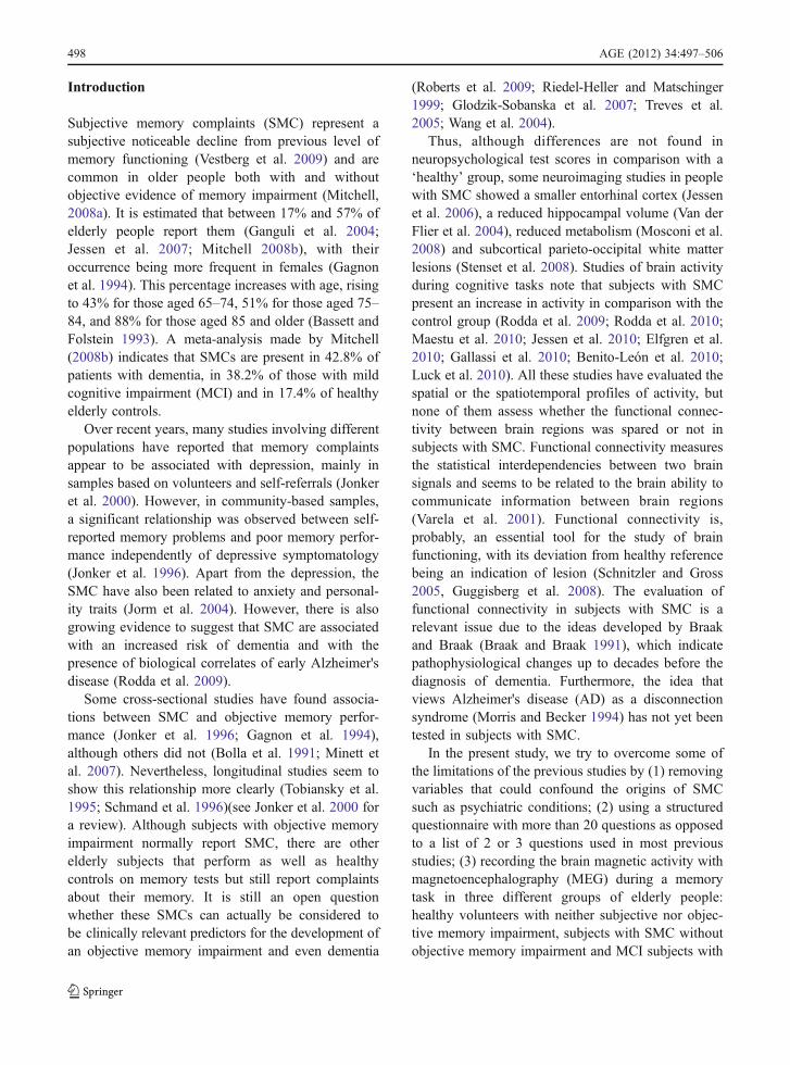
MCI vs. control participants
Comparing these two groups (see Fig. 1), MCIpatients showed a clear cluster of higher synchroni-zation values over the anterior and central regions inall frequency bands. Additionally, MCI patientsshowed higher inter-hemispheric SL values than thecontrol group between left and right temporo-frontalsensors in all frequency bands (except in band α2).
Two non-functionally related clusters of localinteractions showed higher SL values in the controlgroup: one among left temporal sensors and anotherone in central-posterior channels. Both of them werefound in all frequency bands. Additionally, mainly inγ band, there is a higher posterior synchronization incontrols compared to MCI patients.
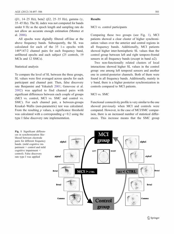
MCI vs. SMC
Functional connectivity profile is very similar to the oneshowed previously when MCI and controls werecompared. However, in the case of MCI/SMC compar-ison, there is an increased number of statistical differ-ences. This increase means that the SMC group
Fig. 1 Significant differen-ces in synchronization like-lihood between electrodepairs for different frequencybands. (mild cognitive im-pairment > control and mildcognitive impairment <control). False discoveryrate type I was applied
AGE (2012) 34:497–506 501
presents, on average, lower synchronization than controlgroup when compared with MCI patients (see Fig. 2).
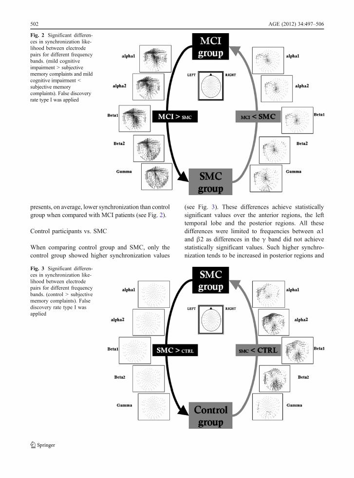
Control participants vs. SMC
When comparing control group and SMC, only thecontrol group showed higher synchronization values
(see Fig. 3). These differences achieve statisticallysignificant values over the anterior regions, the lefttemporal lobe and the posterior regions. All thesedifferences were limited to frequencies between α1and β2 as differences in the γ band did not achievestatistically significant values. Such higher synchro-nization tends to be increased in posterior regions and
Fig. 2 Significant differen-ces in synchronization like-lihood between electrodepairs for different frequencybands. (mild cognitiveimpairment > subjectivememory complaints and mildcognitive impairment <subjective memorycomplaints). False discoveryrate type I was applied
Fig. 3 Significant differen-ces in synchronization like-lihood between electrodepairs for different frequencybands. (control > subjectivememory complaints). Falsediscovery rate type I wasapplied
502 AGE (2012) 34:497–506

in the frontal region, mainly in the α2 frequencyband.
Discussion
In this paper, we try to show whether subjects withSMC but not objective memory impairment,showed a different profile of functional connectiv-ity compared with subjects with MCI and healthyelderly subjects. In different frequency bands (α toγ), SMC subjects showed lower synchronizationvalues than MCI and healthy elderly subjects,although with a different topology. Conversely,SMC subjects showed only higher synchronizationvalues in comparison with the MCI subjects. Infact, their profile, in comparison with the MCIsubjects, was very similar to the one showed whenMCI and controls were compared. Regardless ofwhether or not we understand SMC as the initialstage of a cognitive continuum decline, all theseresults could indicate that SMCs are representingan initial stage with a hypo-synchronization (incomparison with the control group) where thebrain system is not still compensating for thefailure of the memory networks, but behaving ascontrols when compared with the MCI subjects(see Fig. 4).
When functional connectivity profiles from sub-jects with SMC were compared with those of the MCIgroup, SMC showed, in all frequency bands, both ahypo and hyper-synchronization, but with a differentnetwork topology. Thus, MCI subjects showed highersynchronization over the anterior and central regionsas well as in the left and right temporal regions. In theβ2 and γ, this profile of higher synchronization iseven more widely distributed to include the posteriorregions bilaterally. This could indicate that at the timewhen the memory impairment is severe enough to bedetected by memory test, MCI subjects tend tointegrate information between the left and rightfrontal lobes to develop better memory strategies(Cabeza et al. 2002) and thus achieve a similar levelof performance to the rest of the groups. Conversely,subjects with SMCs showed higher synchronizationthan MCIs in the central and left temporal sensors. Itis interesting to point out that these profiles offunctional connectivity are mirroring those foundwhen comparing MCI and controls. Thus, subjects
with SMC behave this time as controls demonstratingthat they have less severe neurophysiological impair-ment than that showed by the MCI subjects.
When functional connectivity profiles were com-pared between SMC and controls, the control groupshowed higher synchronization values. These differ-ences achieve statistically significant values over theanterior regions, the left temporal lobe and theposterior regions. All these differences were limitedto frequencies between α1 and β2 since differences inthe γ band did not achieve statistically significantvalues. There are some more differences in the profileof synchronization between controls and SMC. Forexample, in the β band, while the comparisonbetween controls and MCI showed higher synchroni-
Fig. 4 Cognitive decline continuum: from pathology tocompensatory phenomena. Although SMC subjects show adifferent profile of functional connectivity pattern than MCIand healthy controls, synchronization changes are focused onthe anterior, central, posterior and left temporal regions. MCIsubject showed a hyper-synchronization (compensatory effect),whereas SMC a hypo-synchronization (the brain is not stillcompensating for the fails of the memory networks, being apathological or dementia effect). AD patients show animpairment of their functional connectivity (Stam et al. 2009)
AGE (2012) 34:497–506 503

zation in favour of the MCIs over anterior and centralregions, these patterns are widely distributed whencomparing MCIs and SMC, which accounts for theirdiminution in the synchronization values along thewhole sensor area. This hypo-synchronization couldunderlie the memory difficulties in the day-to-dayactivities reported by SMC subjects. Thus, thedifficulties in achieving a correct coupling betweenbrain regions diminish the probability of an efficienttransmission of information (Stam et al. 2009) leadingto memory failures. A compensation mechanismshowed by MCI subjects, seems to be unnecessaryat this point since memory difficulties are not yetsevere enough.
As far as we know, there are only three previouspapers published with functional neuroimaging inpatients with SMC (Rodda et al. 2010; Rodda et al.2009; Maestu et al. 2010). In all three of them, SMCsubjects showed higher activation than healthy con-trols. This result seems to be contradictory with theprofile of lower synchronization found in this study.These three previous papers used measurements ofthe magnitude of the signal, such as the increased ordecreased blood flow or power of the magnetic fieldin particular regions of the brain. However, none ofthem calculated functional connectivity between brainregions. The fact that one particular brain regionincreases in blood flow or magnetic field does notnecessarily imply that it is having better communica-tion with other brain regions. Furthermore, functionalconnectivity is evaluating how brain regions arecommunicating with each other rather than justassessing the magnitude of the signal in a particularbrain region. So here, we try to test the integrationrather than the segregation of the functional activity.Thus, it seems like while subjects with SMC areincreasing their local activity, they fail in their abilityof communicating information between brain regions.In any case, we think that fMRI and MEG findingsare complementary because they may suggest thepresence of early functional changes in SMC.
In conclusion, the profiles of functional connectiv-ity described in this work, point toward a model ofhow the brain mechanisms behave in different stagesof a possible cognitive decline continuum. Taking thehealthy elderly subjects' profile of functional connec-tivity as a standard at the SMC stage, subjects show ahypo-synchronization of their memory-related net-works and at the MCI stage, memory networks
increase their coupling as a compensatory mechanism(Bajo et al. 2010) and finally AD patients show animpairment of their functional connectivity (Stam etal. 2009). Thus, at the stage of SMC, compensatorymechanisms are still not necessary because thememory deficit is not severe enough to be detectedby memory tests and does not dramatically disturbdaily living activities. However, when the memoryimpairment is severe enough to be detected by theneuropsychological assessment, then the brain com-pensatory mechanisms are required to enhance infor-mation communication by means of synchronization.Finally, due to the severe damage of the biochemicaland anatomical structure of the brain at the AD stage,all these compensatory mechanisms are no longerpossible. Future studies should consider a follow-upof the SMC subjects to determine which of thesesubjects develop an objective memory impairment,with the aim of describing profiles of prediction fromSMC to MCI.
Acknowledgements This study was supported by two grantsfor the Spanish Ministry of Science and Innovation (SEJ-2006-07560; PSI2009-14415-C03-01).
References
Abdulrab K, Heun R (2008) Subjective memory impairment. Areview of its definitions indicates the need for a compre-hensive set of standardized and validated criteria. EurPsychiatr 23:321–330
Bajo R, Maestú F, Nevado A, Sancho M et al (2010) Functionalconnectivity in mild cognitive impairment during amemory task: implications for the disconnection hypoth-esis. J Alzheimers Dis 22:183–193
Bassett SS, Folstein (1993) MF: memory complaint, memoryperformance performance, and psychiatric diagnosis: acommunity study. J Geriatr Psychiatr Neurol 6:105–111
Benito-León J, Mitchell AJ, Vega S, Bermejo-Pareja F (2010)A population-based study of cognitive function in olderpeople with subjective memory complaints. J AlzheimersDis 22(1):159–170
Benjamini Y, Yekutieli D (2001) The control of the falsediscovery rate in multiple testing under dependency. AnnStat 29(4):1165–1188
Bolla KI, Lindgren KN, Bonaccorsy C, Bleecker ML (1991)Memory complaints in older adults. Arch Neurol 48:61–64
Braak H, Braak E (1991) Neuropathological stageing ofAlzheimer-related changes. Acta Neuropathol 82:239–259
deToledo-Morrell L, Evers S, Hoeppner TJ, Morrell F, GarronDC, Fox JH (1991) A ‘stress’ test for memory dysfunction.Electrophysiologic manifestations of early Alzheimer'sdisease. Arch Neurol 48:605–609
504 AGE (2012) 34:497–506

Cabeza R, Dolcos F, Graham R, Nyberg L (2002) Similaritiesand differences in the neural correlates of episodic memoryretrieval and working memory. Neuroimage 16:317–330
Elfgren C, Gustafson L, Vestberg S, Passant U (2010)Subjective memory complaints, neuropsychological per-formance and psychiatric variables in memory clinicattendees: a 3-year follow-up study. Arch Gerontol Geriatr51(3):e110–e114
Farias ST, Mungas D, Jagust W (2005) Degree of discrepancybetween self and other- reported everyday functioning bycognitive status: dementia, mild cognitive impairment andhealthy elders. Int J Geriatr Psychiatry 20:827–834
Gagnon M, Dartigues JF, Mazaux JM, Dequae L, Letenneur L,Giroire JM, Barberger-Gateau P (1994) Self-reportedmemory complaints and memory performance in elderlyFrench community residents: results of the PAQUIDresearch program. Neuroepidemiology 13(4):145–154
Gallassi R, Oppi F, Poda R, Scortichini S, Stanzani Maserati M,Marano G, Sambati L (2010) Are subjective cognitivecomplaints a risk factor for dementia? Neurol Sci 31(3):327–336
Ganguli M, Dodge HH, Shen C, DeKosky ST (2004) Mildcognitive impairment, amnestic type: an epidemiologicstudy. Neurology 63:115–121
Genovese R, Lazar NA, Nichols T (2002) Thresholding ofstatistical maps in functional neuroimaging using the falsediscovery rate. Neuroimage 15:870–878
Glodzik-Sobanska L, Reisberg B, De Santi S, Babb JS,Pirraglia E, Rich KE, Brys M, de Leon MJ (2007)Subjective memory complaints: presence, severity andfuture outcome in normal older subjects. Dement GeriatrCogn Disord 24:177–184.
Grundman M, Petersen RC, Ferris SH, Thomas RG, Aisen PS,Bennett DA et al (2004) Mild cognitive impairment can bedistinguished from Alzheimer disease and normal agingfor clinical trials. Arch Neurol 61:59–66
Guggisberg AG, Honma SM, Findlay A, Dalal SS, Kirsch HE,Berger MS et al (2008) Mapping functional connectivityin patients with brain lesions. Ann Neurol 63:193–203
Jessen F, Feyen L, Freymann K, Tepest R, Maier W, Heun R et al(2006) Volume reduction of the entorhinal cortex in subjec-tive memory impairment. Neurobiol Aging 27:1751–1756
Jessen F, Wiese B, Cvetanovska G, Fuchs A, Kaduszkiewicz H,Kolsch H et al (2007) Patterns of subjective memoryimpairment in the elderly: association with memoryperformance. Psychol Med 37:1753–1762
Jessen F, Wiese B, Bachmann C, Eifflaender-Gorfer S, Haller F,Kölsch H, Luck T, Mösch E, Van den Bussche H, WagnerM, Wollny A, Zimmermann T, Pentzek M, Riedel-HellerSG, Romberg HP, Weyerer S, Kaduszkiewicz H, Maier W,Bickel H (2010) Prediction of dementia by subjectivememory impairment: effects of severity and temporalassociation with cognitive impairment. Arch Gen Psy-chiatr 67(4):414–422
Jonker C, Launer LJ, Hooijer C, Lindeboom J (1996) Memorycomplaints and memory impairment in older individuals. JAm Geriatr Soc 44(1):44–49
Jonker C, Geerlings MI, Schmand B (2000) Are memorycomplaints predictive for dementia? A review of clinicaland population- based studies. Int J Geriatr Psychiatry 15(11):983–991
Jorm AF, Butterworth P, Anstey KJ et al (2004) Memorycomplaints in a community sample aged. 60–64 years:associations with cognitive functioning, psychiatric symp-toms, medical conditions, APOE genotype, hippocampusand amygdala volumes, and white-matter hyperintensities.Psychol Med 34(8):1495–1506
Lobo A, Ezquerra J, Gomez BF, Sala JM, Seva DA (1979)Cognocitive mini-test (a simple practical test to detectintellectual changes in medical patients). Actas Luso EspNeurol Psiquiatr Cienc Afines 7:189–202
Luck T, Riedel-Heller SG, Luppa M, Wiese B, Wollny A,Wagner M, Bickel H, Weyerer S, Pentzek M, Haller F,Moesch E, Werle J, Eisele M, Maier W, Van Den BusscheH, Kaduszkiewicz H (2010) Risk factors for incident mildcognitive impairment—results from the German study onageing, cognition and dementia in primary care patients(AgeCoDe). Acta Psychiatr Scand 121(4):260–272
Maestu F, Fernandez A, Simos PG, Gil-Gregorio P, Amo C,Rodriguez R, Arrazola J, Ortiz T (2001) Spatio-temporalpatterns of brain magnetic activity during a memory taskin Alzheimer's disease. Neuroreport 12:3917–3922
Maestu F, Baykova E, Ruiz JM, Montejo P, Montenegro M,Llanero M, Solesio E, Gil P, Yubero R, Paul N, Pozo F,Nevado A (2011) Increased biomagnetic activity inhealthy elderly with subjective memory complaints. ClinNeurophysiol 122:499–505, Accepted
Minett TSC, Da Silva RV, Ortiz KZ, Bertolucci PHF (2007)Subjective memory complaints in an elderly sample: across- sectional study. Int J Geriatr Psychiatry 23:49–54
Mitchell AJ (2008a) Is it time to separate subjective cognitivecomplaints from the diagnosis of mild cognitive impair-ment? Age Ageing 37:497–499
Mitchell AJ (2008b) The clinical significance of subjectivememory complaints in the diagnosis of mild cognitiveimpairment and dementia: a meta-analysis. Int J GeriatrPsychiatry 23:1191–1202
Montez T, Linkenkaer-Hansen K, Van Dijk BW, Stam CJ(2006) Synchronization likelihood with explicit time-frequency priors. Neuroimage 33:1117–1125
Morris R, Becker J (1994) Cognitive Neuropsychology ofAlzheimer Disease. Oxford University Press
Mosconi L, De Santi S, Brys M, Tsui WH, Pirraglia E,Glodzik-Sobanska L et al (2008) Hypometabolism andaltered cerebrospinal fluid markers in normal apolipopro-tein E4 carriers with subjective memory complaints. BiolPsychiatr 63:609–618
Petersen RC (2004) Mild cognitive impairment as a diagnosticentity. J Intern Med 256:183–194
Riedel-Heller SG,Matschinger H (1999) Domemory complaintsindicate the presence of cognitive impairment? Results of afield study. Eur Arch Psychiatr Neurol Sci 249:197–204
Roberts JL, Clare L, Woods RT (2009) Subjective memorycomplaints and awareness of memory functioning in mildcognitive impairment: a systematic review. Dement GeriatrCognit Disord 28:95–109
Rodda JE, Dannhauser TM, Cutinha DJ, Shergill SS, Walker Z(2009) Subjective cognitive impairment: increased pre-frontal cortex activation to control during an encodingtask. Int J Geriatr Psychiatry 24:865–874
Rodda J, Dannhauser T, Cutinha DJ, Shergill SS, Walker Z(2010) Subjective cognitive impairment: functional MRI
AGE (2012) 34:497–506 505
during a divided attention task. Eur Psychiatr. doi:10.1016/j.eurpsy.2010.07.003, ISSN 0924–9338
Schmand B, Jonker C, Hooijer C, Lindeboom J (1996)Subjective memory complaints may announce dementia.Neurology 46(1):121–125
Schnitzler A, Gross J (2005) Normal and pathologicaloscillatory communication in the brain. Nat Rev Neurosci6:285–296
Stam CJ, Van Dijk BW (2002) Synchronization likelihood: anunbiased measure of generalized synchronization inmultivariate data sets. Physica D 163:236–241
Stam CJ, De Haan W, Daffertshofer A, Jones BF, ManshandenI, Van Cappellen van Walsum AM et al (2009) Graphtheoretical analysis of magnetoencephalographic func-tional connectivity in Alzheimer's disease. Brain132:213–224
Stenset V, Hofoss D, Berstad AE, Negaard A, Gjerstad L,Fladby T (2008) White matter lesion subtypes andcognitive deficits in patients with memory impairment.Dement Geriatr Cognit Disord 26:424–431
Sunderland A, Harris JE, Baddeley AD (1983) Do laboratorytests predict everyday memory? A neuropsychologicalstudy. Journal of Verbal Learning and Verbal Behavior 22(3):341–357. doi:10.1016/S0022-5371(83)90229-3, ISSN0022–5371
Tobiansky R, Blizard R, Livingston G, Mann A (1995) Thegospel oak study stage IV: the clinical relevance ofsubjective memory impairment in older people. PsycholMed 25:779–786
Treves TA, Verchovsky R, Klimovitzky S, Korczyn AD (2005)Incidence of dementia in patients with subjective memorycomplaints. Int Psychogeriatr 17:265–273.
Van der Flier WM, Van Buchem MA, Weverling-RijnsburgerAW, Mutsaers ER, Bollen EL, Admiraal-Behloul F et al(2004) Memory complaints in patients with normalcognition are associated with smaller hippocampal vol-umes. J Neurol 251(6):671–675
Varela F, Lachaux JP, Rodriguez E, Martinerie J (2001) Thebrainweb: phase synchronization and large-scale integra-tion. Nat Rev Neurosci 2:229–239
Vestberg S, Passant U, Elfgren C (2010) Stability in the clinicalcharacteristics of patients with memory complaints. ArchGerontol Geriatr 50:e26–e30
Wang L, Van Belle G, Crane PK, Kukull WA, Bowen JD,McCormick WC, Larson EB (2004) Subjective memorydeterioration and future dementia in people aged 65 andolder. J Am Geriatr Soc 52:2045–2051.
Yesavage JA, Brooks JO (1991) On the importance oflongitudinal research in Alzheimer's disease. J Am GeriatrSoc 39(9):942–944
506 AGE (2012) 34:497–506