Dynamics of guest molecules in PHTP inclusion compounds as probed by solid-state NMR and...
14
Dynamics of guest molecules in PHTP inclusion compounds as probed by solid-state NMR and fluorescence spectroscopy G. Srinivasan, a J. A. Villanueva-Garibay, a K. Mu¨ller,* ab D. Oelkrug, c B. Milian Medina, de D. Beljonne, d J. Cornil, d M. Wykes, d L. Viani, d J. Gierschner,* cdf R. Martinez-Alvarez, g M. Jazdzyk, g M. Hanack g and H.-J. Egelhaafw* ch Received 18th November 2008, Accepted 12th March 2009 First published as an Advance Article on the web 1st April 2009 DOI: 10.1039/b820604j Partially deuterated 1,4-distyrylbenzene (2PV) is included into the pseudohexagonal nanochannels of perhydrotriphenylene (PHTP). The overall and intramolecular mobility of 2PV is investigated over a wide temperature range by 13 C, 2 H NMR as well as fluorescence spectroscopy. Simulations of the 2 H NMR spectral shapes reveal an overall wobble motion of 2PV in the channels with an amplitude of about 41 at T = 220 K and 101 at T = 410 K. Above T = 320 K the wobble motion is superimposed by localized 1801 flips of the terminal phenyl rings with a frequency of 10 6 Hz at T = 340 K. The activation energies of both types of motions are around 40 kJ mol 1 which imply a strong sterical hindrance by the surrounding PHTP channels. The experimental vibrational structure of the fluorescence excitation spectra of 2PV is analyzed in terms of small amplitude ring torsional motions, which provide information about the spatial constraints on 2PV by the surrounding PHTP host matrix. Combining the results from NMR and fluorescence spectroscopy as well as of time-dependent density functional calculations yields the complete potential surfaces of the phenyl ring torsions. These results, which suggest that intramolecular mobility of 2PV is only reduced but not completely suppressed by the matrix, are corroborated by MD simulations. Unrealistically high potential barriers for phenyl ring flips are obtained from MD simulations using rigid PHTP matrices which demonstrate the importance of large amplitude motions of the PHTP host lattice for the mobility of the guest molecules. 1. Introduction Polyconjugated chromophores such as poly(phenylenevinylene)s and polythiophenes are widely used in organic optoelectronic devices. However, applications are limited by the reduction of luminescence efficiency due to aggregation effects 1,2 and by low photostability. 3–5 The inclusion of oligomeric chromo- phores into channel-forming host materials such as perhydrotriphenylene (PHTP, see Fig. 1) provides a solution to these problems. 6–16 In the presence of rod-shaped oligomers, such as 1,4-bis[(E)-2-phenylvinyl]benzene (2PV, see Fig. 1), PHTP forms a pseudo-hexagonal host-lattice with parallel 2PV filled channels. 7,8,17 In the crystal structure of pure PHTP, no channels are formed. This implies that in the host–guest compound the guest must be in close contact to the host. Thus, it is expected that the mobility of 2PV in the channels is significantly restricted as compared to solution or gas phase. The mobility of the included p-oligomer is of significance in high-precision calculations of excitation energy transfer in light harvesting devices, 18,19 and in the design of functional architectures which require a certain extent of molecular mobility, such as molecular switches. The possible degrees of freedom of guest molecules in the PHTP matrix comprise both intramolecular and overall motions. Intra- molecular motions of 2PV are mainly the torsional vibrations and hindered rotations of the phenyl rings around the formal single bonds. The torsional vibrations of 2PV depend sensitively on the rigidity of the environment as has been shown by fluorescence studies in liquid and solid solutions. 20 Thus, 2PV represents the ideal molecule for probing the effect of the matrix on the intramolecular mobility of guest molecules in inclusion compounds. However, fluorescence spectroscopy is restricted to monitoring those molecular motions which are coupled to electronic transitions. In contrast, NMR spectroscopy offers the opportunity to obtain full 3D characterizations of a Institute for Physical Chemistry, University of Stuttgart, Pfaffenwaldring 55, 70569, Stuttgart, Germany b Dipartimento di Ingegneria dei Materiali e Tecnologie Industriali, Universita ´ degli Studi di Trento, via Mesiano 77, I-38100, Italy and INSTM, Trento, UdR Trento, Italy. E-mail: [email protected], [email protected] c Institute for Physical Chemistry, University of Tu ¨bingen, Auf der Morgenstelle 8, 72076, Tu ¨bingen, Germany d Laboratory for Chemistry of Novel Materials, University of Mons-Hainaut, Place du Parc 20, 7000, Mons, Belgium e Institute for Molecular Science, University of Valencia, P.O. Box 22085, 46071, Valencia, Spain f IMDEA Nanoscience, Madrid, Spain. E-mail: [email protected] g Institute for Organic Chemistry, University of Tu ¨bingen, Auf der Morgenstelle 18, 72076, Tu ¨bingen, Germany h Christian-Doppler Laboratory for Surface-Optical Methods, Johannes-Kepler-University, Altenbergerstr. 69, 4040, Linz, Austria. E-mail: [email protected] w Present address: Konarka Technologies GmbH, Landgrabenstr. 94, D-90443 Nu¨rnberg 4996 | Phys. Chem. Chem. Phys., 2009, 11, 4996–5009 This journal is c the Owner Societies 2009 PAPER www.rsc.org/pccp | Physical Chemistry Chemical Physics
Transcript of Dynamics of guest molecules in PHTP inclusion compounds as probed by solid-state NMR and...

Dynamics of guest molecules in PHTP inclusion compounds as probed
by solid-state NMR and fluorescence spectroscopy
G. Srinivasan,a J. A. Villanueva-Garibay,a K. Muller,*ab D. Oelkrug,c
B. Milian Medina,de
D. Beljonne,dJ. Cornil,
dM. Wykes,
dL. Viani,
dJ. Gierschner,*
cdf
R. Martinez-Alvarez,gM. Jazdzyk,
gM. Hanack
gand H.-J. Egelhaafw*ch
Received 18th November 2008, Accepted 12th March 2009
First published as an Advance Article on the web 1st April 2009
DOI: 10.1039/b820604j
Partially deuterated 1,4-distyrylbenzene (2PV) is included into the pseudohexagonal nanochannels
of perhydrotriphenylene (PHTP). The overall and intramolecular mobility of 2PV is investigated
over a wide temperature range by 13C, 2H NMR as well as fluorescence spectroscopy. Simulations
of the 2H NMR spectral shapes reveal an overall wobble motion of 2PV in the channels with an
amplitude of about 41 at T = 220 K and 101 at T = 410 K. Above T = 320 K the wobble
motion is superimposed by localized 1801 flips of the terminal phenyl rings with a frequency of
106 Hz at T = 340 K. The activation energies of both types of motions are around 40 kJ mol�1
which imply a strong sterical hindrance by the surrounding PHTP channels. The experimental
vibrational structure of the fluorescence excitation spectra of 2PV is analyzed in terms of small
amplitude ring torsional motions, which provide information about the spatial constraints on
2PV by the surrounding PHTP host matrix. Combining the results from NMR and fluorescence
spectroscopy as well as of time-dependent density functional calculations yields the complete
potential surfaces of the phenyl ring torsions. These results, which suggest that intramolecular
mobility of 2PV is only reduced but not completely suppressed by the matrix, are corroborated
by MD simulations. Unrealistically high potential barriers for phenyl ring flips are obtained from
MD simulations using rigid PHTP matrices which demonstrate the importance of large amplitude
motions of the PHTP host lattice for the mobility of the guest molecules.
1. Introduction
Polyconjugated chromophores such as poly(phenylenevinylene)s
and polythiophenes are widely used in organic optoelectronic
devices. However, applications are limited by the reduction of
luminescence efficiency due to aggregation effects1,2 and by
low photostability.3–5 The inclusion of oligomeric chromo-
phores into channel-forming host materials such as
perhydrotriphenylene (PHTP, see Fig. 1) provides a solution
to these problems.6–16 In the presence of rod-shaped oligomers,
such as 1,4-bis[(E)-2-phenylvinyl]benzene (2PV, see Fig. 1),
PHTP forms a pseudo-hexagonal host-lattice with parallel
2PV filled channels.7,8,17 In the crystal structure of pure
PHTP, no channels are formed. This implies that in the
host–guest compound the guest must be in close contact to
the host. Thus, it is expected that the mobility of 2PV in the
channels is significantly restricted as compared to solution or
gas phase. The mobility of the included p-oligomer is of
significance in high-precision calculations of excitation energy
transfer in light harvesting devices,18,19 and in the design of
functional architectures which require a certain extent of
molecular mobility, such as molecular switches. The possible
degrees of freedom of guest molecules in the PHTP matrix
comprise both intramolecular and overall motions. Intra-
molecular motions of 2PV are mainly the torsional vibrations
and hindered rotations of the phenyl rings around the formal
single bonds. The torsional vibrations of 2PV depend
sensitively on the rigidity of the environment as has been
shown by fluorescence studies in liquid and solid solutions.20
Thus, 2PV represents the ideal molecule for probing the effect
of the matrix on the intramolecular mobility of guest
molecules in inclusion compounds.
However, fluorescence spectroscopy is restricted to
monitoring those molecular motions which are coupled to
electronic transitions. In contrast, NMR spectroscopy
offers the opportunity to obtain full 3D characterizations of
a Institute for Physical Chemistry, University of Stuttgart,Pfaffenwaldring 55, 70569, Stuttgart, Germany
bDipartimento di Ingegneria dei Materiali e Tecnologie Industriali,Universita degli Studi di Trento, via Mesiano 77, I-38100,Italy and INSTM, Trento, UdR Trento, Italy.E-mail: [email protected], [email protected]
c Institute for Physical Chemistry, University of Tubingen,Auf der Morgenstelle 8, 72076, Tubingen, Germany
dLaboratory for Chemistry of Novel Materials,University of Mons-Hainaut, Place du Parc 20, 7000, Mons,Belgium
e Institute for Molecular Science, University of Valencia,P.O. Box 22085, 46071, Valencia, Spain
f IMDEA Nanoscience, Madrid, Spain.E-mail: [email protected]
g Institute for Organic Chemistry, University of Tubingen,Auf der Morgenstelle 18, 72076, Tubingen, Germany
hChristian-Doppler Laboratory for Surface-Optical Methods,Johannes-Kepler-University, Altenbergerstr. 69, 4040, Linz, Austria.E-mail: [email protected]
w Present address: Konarka Technologies GmbH, Landgrabenstr. 94,D-90443 Nurnberg
4996 | Phys. Chem. Chem. Phys., 2009, 11, 4996–5009 This journal is �c the Owner Societies 2009
PAPER www.rsc.org/pccp | Physical Chemistry Chemical Physics

molecular mobilities in restricted volumes.21,22 It has been
applied to a variety of inclusion compounds of small molecules
and polymers in organic and inorganic hosts.21,23–43 From 2H
and 13C NMR studies it has been found for small molecules
such as cyclohexane,36,41 cyclohexane derivatives33,41 or
trioxane21,41 that intramolecular motions such as ring
inversions are basically not affected by the host. Even for
overall rotations, as for instance for benzene guests,35,38 rather
small activation energies have been reported. 13C and 2H
NMR studies on PHTP inclusion compounds with alkane
guests have shown that also hydrocarbon chains possess an
appreciable mobility.26,28,29 In this case, librations at a number
of consecutive bonds introduce large amplitude reorientations
of the C–2H-bonds by introducing local twisting of
the polymer chain. Recent investigations of a,o-alkyl-oligo-thiophenes in tris(o-phenylenedioxy)spirocyclotriphosphazene
(TPP) have shown that the terminal alkane chains act as
hinges around which the central bithiophene moiety rotates
in a quasi-free way.34
Molecular dynamics (MD) simulations and quantum
chemical calculations44–49 provided a detailed insight into
the mechanisms of molecular motions and showed that the
roles of the organic ‘‘zeolites’’, such as PHTP and TPP, are
completely different from that of real zeolites. Whereas zeolites
form fairly rigid channels, the shape and size of the PHTP
channels are variable to a certain degree. The interior of the
channels and the cross-section depend on the shape of the
included molecule and in turn determines the guest mobility.
Thus, guest molecules which have a cylindrical shape induce
cylindrical PHTP channels and show much higher mobility
than found for guests which induce distortions of the
hexagonal symmetry of the channel.44,46 The importance of
the dynamic nature of the nanochannel walls for the mobility
of the guest molecules becomes obvious from the fact
that the results from MD simulations are only in accordance
with the NMR results if the artificial constraint of rigidity
of the PHTP matrix is removed.44 In summary, NMR
experiments and MD simulations of host–guest compounds
revealed an amazing degree of mobility for the included
molecules. However, it also becomes clear that the actual
degree of freedom depends sensitively on the structure of the
guest species.
In this contribution we report on the intramolecular and
overall mobility of 2PV, a representative of the class of
phenylenevinylene oligomers and polymers, in PHTP. We
present variable temperature fluorescence and 2H NMR
investigations on the PHTP inclusion compound with two
selectively deuterated 2PVs. Both spectroscopic methods yield
independent information about molecular dynamics of the
guest species and thus allow us to obtain a detailed picture
of the mobility of a medium sized guest molecule in the
nanochannels of PHTP. In particular, 2H NMR is able to
distinguish between intramolecular motions, such as phenyl
ring flips and overall (hindered) reorientations of the molecule
around its principal axis. The combination of fluorescence
spectroscopy and quantum chemical calculations allows to
model the barrier height for torsional motion. The work is
completed by MD simulations which should give independent
evidence for the experimentally detected motional contribu-
tions of the guest species.
2. Experimental
Synthesis and sample preparation
1,4-Bis[(E)-2-phenylvinyl]benzene-d10 (2PV-d10) was prepared
following a two-step synthetic procedure (see Scheme 1).
Thus, (chloromethyl)benzene-d5 (Aldrich) reacts with
triphenylphosphine (Fluka) in xylene to form benzyl(triphenyl)-
phosphonium-d5 chloride 1. The classical Wittig reaction50 of
the phosphonium salt 1 with terephthaldehyde (Fluka) affords
the deuterated compound 2PV-d10.
1,4-Bis[(E)-2-phenylvinyl]benzene-d2 (2PV-d2) was prepared
in an analogous procedure (see Scheme 1) from (chloromethyl)-
benzene (Aldrich) and terephthaldehyde-d2 (Aldrich).
Perhydrotriphenylene was kindly provided by Dr. Borchers
(BASF, Ludwigshafen, Germany). Binary inclusion
compounds were crystallized from solutions of PHTP and
2PV (weight ratio 2 : 1) in methylethylketone.51 Ternary
inclusion compounds, PHTP/1PV/2PV, were crystallized from
Fig. 1 (a) Schematic structure of the 2PV-d10/PHTP inclusion compound. Left and lower right: view perpendicular to the crystal c-axis; upper
right: view parallel to the c crystal axis (i—wobble motion of 2PV; ii—torsional vibrations around the formal single bonds of 2PV); (b) chemical
structures of guest and host components.
This journal is �c the Owner Societies 2009 Phys. Chem. Chem. Phys., 2009, 11, 4996–5009 | 4997

solutions of PHTP, stilbene (1PV), and 2PV (weight ratio
2 : 1 : 10�3) in methylethylketone. The obtained crystal needles
were isolated, washed with cold methylethylketone and dried
in vacuum.
The guest/host molar ratios were determined by fluoro-
metry, UV/VIS absorption, and 1H NMR spectroscopy. The
UV/VIS and fluorescence spectra of a given amount of inclu-
sion compound were recorded after dissolution in dioxane.
The results were compared with the fluorescence intensity and
the absorbance of the pure guest in the same solvent. 1H NMR
intensities of methylethylketone were between 0 and 1%; thus
solvent co-inclusion in PHTP can be neglected.
Fluorescence experiments
Steady-state fluorescence and fluorescence excitation spectra
were recorded on a SPEX 222 fluorometer equipped with
Glan-Thompson polarizers (Instruments S A., Longjumeau,
France). The light source was a 450WXe arc lamp, the detector
was a R928 photomultiplier tube. Fluorescence excitation
spectra are corrected for excitation intensity with the help of
a rhodamine B quantum counter. Emission spectra are
corrected for the sensitivity of the detection system with a
correction file. 10�7 M solutions of 2PV in EPA (a mixture of
diethylether, i-pentane, and ethanol) were prepared using
Merck UVASOL solvents. Unpolarized fluorescence spectra
of inclusion compounds were obtained on powders of randomly
oriented crystallites which were pressed onto fused silica or
aluminium substrates. For low-temperature measurements the
samples were cooled down by a closed-cycle He cryostat.
13C NMR experiments
13C MAS NMR spectra were recorded at 75.47 MHz on
a CXP 300 Bruker (Bruker/Rheinstetten) spectrometer
(magnetic field strength: 7.04 T) equipped with a Tecmag
control unit and using a standard 4 mm Bruker MAS probe.
All magic angle spinning experiments were done at the sample
spinning rate of 5 kHz applying either single pulse (SP) or
cross polarization (CP) excitation schemes (contact time: 1 ms).
The p/2 pulse widths for carbon and protons were 4 ms.Recycle delays were chosen to 20 and 60 s for CP and SP13C NMR experiments, respectively. 13C chemical shifts were
determined relative to the external standard adamantane. All13C chemical shifts are expressed relative to tetramethylsilane
(TMS) with d = 0 ppm.
2H NMR experiments
All 2H NMRmeasurements were carried out on a Bruker CXP
300 spectrometer (Bruker/Rheinstetten) operating at a
frequency of 46.07 MHz for deuterium, by using a standard
(liquid nitrogen flow) 5 mm double-resonance probe (p/2 pulselength = 2 ms). The variable temperature 2H NMR spectra
were recorded by applying the quadrupole echo sequence
[(p/2)x–te–(p/2)y–te-acquisition] with pulse intervals te as givenin the text. The number of scans varied between 1024 and 2560
for 2PV-d10 or between 8000 and 10 000 for 2PV-d2 depending
on the S/N ratio, and the recycle delay between successive
scans was 20 s. The sample temperature was controlled by a
Bruker BVT 1000 unit. The accuracy of the temperature was
within �1 K.
NMR data processing
The data processing of the experimental and simulated NMR
signals was performed on a SUN Sparc 10 workstation using
the NMRi and Sybyl software packages (Tripos, St. Louis).
2H NMR simulations
NMR line shape simulations were done with appropriate
FORTRAN programs, developed in our group for the
behaviour of an isolated I = 1 spin system for different types
of dynamic NMR experiments.21,38,43 The simulation
programs are very general and take into account various types
of molecular motions of the system under investigation. In all
simulations, the final best fit was chosen by direct comparison
between experimental and theoretical spectra, taking into
account the overall appearance of the line shapes as well as
their relative amplitudes.
Quantum chemical calculations
Quantum-chemical calculations of the vibrational frequencies
in the electronic ground and the first excited state were
performed at the (time-dependent) density functional theory
level of theory, (TD)-DFT, using Becke’s three-parameter
B3LYP exchange–correlation functional52 with the 6-311G*
basis set53 within the TURBOMOLE V5-7 package.54
Torsional potentials for the outer phenyl rings in the ground
and first excited state were obtained from systematic variation
of the torsional angle around the C1–C7 bond, keeping C2
symmetry, and subsequent calculation of the single point
ground and excited-state energies. The absorption spectrum
was calculated using an in-house programme by mirroring the
emission spectrum at the electronic origin and convoluting the
Scheme 1 Preparation of deuterated 2PV-d10 (top) and 2PV-d2 (bottom).
4998 | Phys. Chem. Chem. Phys., 2009, 11, 4996–5009 This journal is �c the Owner Societies 2009

spectrum with the line spectrum, G(~n), of the torsional modes
as obtained for transitions between the vibrational quanta v in
the S0 state and the vibrational quanta w in the S1 state within
the model of distorted, undisplaced oscillators.20
Molecular dynamics simulations
The intramolecular mobility of 2PV in vacuo and in a periodic
PHTP system was investigated by molecular dynamics
(MD) simulations using the molecular modeling package
TINKER 4.2,55 and by molecular mechanics (MM) using
the force field MM3. To obtain an accurate description of
the potential for the rotation of the phenyl rings around the
outer single bonds, the MM3 torsional parameters were
modified in order to reproduce the DFT potential described
above. The periodic PHTP cell with empty channels was built
from the X-ray structure of a similar compound6 and the guest
was subsequently inserted in the channels. One repetition cell
contained four 2PV molecules and 40 PHTP molecules,
yielding a dye concentration of C2PV = 0.33 M, somewhat
smaller than the value found experimentally (C2PV = 0.48 M).
However, it is not expected that this affects the torsional
barrier. MD simulations of the periodic structure were then
performed in the NPT ensemble using the Andersen thermostat
to maintain a temperature of 300 K and the Berendsen
barostat to maintain a pressure of 1 atmosphere. Non-bonded
forces were calculated with atom-based cutoffs of 10.0 A. MD
simulations in vacuo used the same thermostat and cutoff.
Torsional potentials were mapped out by recording the change
of the angle C2C1C7C8 (see Fig. 1) during the MD run, thus
giving a probability histogram. The corresponding potential
was then obtained via the Boltzmann equation. A drawback of
this method is the poor sampling in the regions where the
potential barrier reaches its maximum. Thus, the error in the
calculated potential barrier in these regions is large. To avoid
this source of error and to improve the quality of the results,
an umbrella potential was applied to increase sampling of
energetically unfavorable angles. The weighted histogram
analysis method (WHAM)56 was then applied to the resulting
distributions to remove the effect of the umbrella potential and
provide a well defined torsional free energy profile over the full
range of the torsion.
3. Results and discussion
3.1 NMR spectroscopy
3.1.1 13C NMR studies. Representative 13C MAS NMR
spectra of the 2PV/PHTP, 2PV-d2/PHTP inclusion
compounds and pure 2PV-d2 are given in Fig. 2. The assign-
ment of the signals and the corresponding 13C chemical shift
values are indicated in the Figure and are summarized in
Table 1. It can be seen that the 13C resonances of the 2PV
guests in PHTP are almost identical to those reported for pure
2PV or poly(p-phenylene vinylene), indicating negligible
influence by the incorporation into the PHTP matrix.57–59
Likewise, the 13C shifts of PHTP are in agreement with former
published values.27,60 The experimental 13C NMR spectra at
220 K are somewhat better resolved than those at T = 300 K,
which can be traced back to the presence of guest motions
interfering with the rf decoupler field at elevated temperatures.61,62
The presence of guest motions is also evident from the
enhanced signal intensity of the 2PV signals in the single pulse
(SP) experiments (Fig. 2e and f) as compared to the
corresponding 13C CP/MAS experiment (Fig. 2c and d). That
is, polarization transfer during contact time is slower for
mobile species, here 2PV, as compared to that of the rigid
structural components, i.e. the PHTP host molecules, resulting
in reduced signal intensities of the former ones during the CP
experiment. In principle, it should be possible to extract
further information about the dynamic properties of the guest
species from such 13C NMR experiments, for instance by
analyzing the spinning side bands or via relaxation studies,
supposed the size and orientation of the 13C chemical shift
tensors and of the 13C–1H dipolar couplings are known. The
former information is only accessible through the analysis of
single crystal experiments which were not available in the
present case.
3.1.2 2H NMR studies. 2H NMR experiments on selec-
tively deuterated compounds are easier to handle, as the
spectra and relaxation are only determined by the well-defined
nuclear quadrupolar interaction. In the following, 2H NMR
spectroscopy is used to examine the overall and internal
(intramolecular) mobility of 2PV in PHTP inclusion
compounds. Hence, 2PV was deuterated at the outer
phenylene rings (2PV-d10) and at the CH groups next to the
inner phenylene ring (2PV-d2), as depicted in Fig. 1. Variable
temperature 2H NMR experiments were then performed on
the respective PHTP inclusion compounds, 2PV-d10/PHTP
and 2PV-d2/PHTP.
In Fig. 3, representative 2H NMR spectra are shown for
2PV-d10/PHTP which were obtained with the quadrupole echo
experiment at pulse spacings of te = 30, 60 and 100 ms. All
three series of 2H NMR spectra exhibit characteristic
line shape changes depending on the actual sample tempera-
ture and the pulse spacing te. These findings can be
traced back to the presence of distinct guest motion
on a timescale which matches the sensitive range of the
quadrupole echo experiment, with correlation times tein the order of 10�4 to 10�8 s. Below 220 K the 2H NMR
experiments yield typical ‘‘rigid limit’’ NMR line shapes,
where the motional processes are slow and outside the
sensitive range.
Heating above 220 K is accompanied by some spectral
narrowing along with the appearance of non-axially sym-
metric 2H NMR line shapes. At around 300 K the 2H NMR
spectra with te = 30 ms (left column) are further distorted, as
reflected by a loss in signal intensity for the central part of the
powder pattern as well as for the inner part of the shoulders.
This signal loss becomes even more pronounced at longer
pulse delays, te = 60 and 100 ms (central and right columns).
It is well known that such spectral alterations are very sensitive
to the type and timescale of the underlying molecular
processes. Basically, they reflect a distinct anisotropy of the
spin–spin relaxation time T2, arising from motional effects
modulating the underlying dominant quadrupolar interaction.
Above 340 K additional spectral changes are registered due to
a second motional contribution which enters the sensitive
time-scale for the NMR experiment.
This journal is �c the Owner Societies 2009 Phys. Chem. Chem. Phys., 2009, 11, 4996–5009 | 4999

In order to reproduce the experimental 2H NMR line shapes
of 2PV-d10/PHTP, a series of model calculations was
performed, taking into account the particular structural
composition of the present sample as well as the data and
results from previous work on related systems. It turned out
that the present experimental 2H NMR data can be at best
reproduced by considering a dominant quadrupolar inter-
action with typical interaction parameters for aromatic
deuterons (coupling constant e2qQ/h = 185 kHz, asymmetry
parameter Z = 0.05) and by assuming two superimposed
motional processes (see Fig. 1), namely: (i) overall reorienta-
tional (wobbling) of the whole guest molecule; and (ii) local
1801-ring flips of the deuterated phenyl rings).
To describe the wobble process we have taken a motional
model which was proposed earlier by Vold et al.63 during a
study of the biphenyl/a-cyclodextrin inclusion compound.
Following this approach, the overall molecular wobbling is
described by small angle jumps. Hence, 19 jump sites were
chosen in such a way that a network is constructed with one
unique site at the pole, 6 sites forming a cone with an opening
angle y, and 12 sites of a second cone with an opening angle 2y.
It is further assumed that exchange only takes place between
neighbouring sites.
It should be noted that for the final simulations of the
experimental 2PV-d10/PHTP data, two types of non-
equivalent deuterons had to be taken into account, namely
two deuterons along the para-axis and the remaining eight
deuterons, whose C–2H bond directions are inclined at an
angle of 601 with respect to the p-axis. Thus, the simulated
spectra represent a superposition of two sub-spectra with an
intensity ratio of 4 : 1. The deuterons with the C–2H bond
direction parallel to the para-axis are not affected by the ring
flip motion at all. For the overall wobbling the situation is
different, as it affects all deuterons. However, the effect is
different for the aforementioned two groups of deuterons,
which is again a consequence of the different orientations with
respect to the wobble symmetry axis.
Representative results from model simulations using the
19-site motional process are given in Fig. 4. It is seen that
the motional correlation times, the pulse spacing te, and the
actual opening angle strongly affect the 2H NMR line shapes.
Moreover, the theoretical spectra display the same spectral
Fig. 2 Experimental 13C MAS NMR spectra at 220 K (a, b) and 300 K (c–g). 13C CP/MAS NMR spectra for 2PV/PHTP (protonated guests) are
given in (a) and (c), while (b) and (d) refer to the corresponding spectra for 2PV-d2/PHTP. 13C SP/MASNMR spectra at 300 K for 2PV/PHTP and
2PV-d2/PHTP are shown in (e) and (f), respectively. The 13C CP MAS NMR spectrum of pure 2PV-d2 at 300 K is given in (g). Asterisks indicate
spinning side bands. The 13C NMR assignments are given in Table 1.
Table 1 13C chemical shift values in ppm of 2PV-d2, PHTP and related compounds
Guest d (C10) d (C1) d (C2,3) d (C4) d (C5,6) d (C7,8)
2PV-d2a 138.3 135.7 128.6b 125.0 130.1 128.6b
DSB57 137.1 136.2 128.9 124.4 130.7 127.8PPV57 136.6 — 124.9 — 132.0 128.3PPV59 136.0 — 124.0 — 131.0 128.0Host d (C1h) d (C2h) d (C3h)PHTPa 47.8 32.2 28.4PHTP32 48.4 32.7 28.8
a This work. b Not resolved.
5000 | Phys. Chem. Chem. Phys., 2009, 11, 4996–5009 This journal is �c the Owner Societies 2009

features that are registered in the experimental 2H NMR
spectra below 340 K. On the basis of these model simulations
it was thus possible to reproduce the experimental 2H NMR
line shapes of the 2PV-d10/PHTP inclusion compound in the
temperature range below 340 K.
For temperatures above 320 K the aforementioned internal
1801-ring flip process had to be superimposed. Representative
model simulations demonstrating the influence of this
motional contribution on the 2H NMR spectra are given in
Fig. 5. Again, both the actual correlation time tc and the
chosen pulse separation time te have a strong influence on the
overall appearance of the corresponding 2H NMR line shapes.
The final results from the best fit simulations of the experi-
mental 2H NMR spectra, based on the combined ring
flip/overall wobble model, are given in Fig. 3. In general, a
very good reproduction of the experimental spectra could be
achieved. It is obvious that a finite jump model for the overall
guest motion can only be a crude approach for a diffusive
process. It would be therefore natural to increase the number
of jump sites, thus approaching the scenario of a diffusive
process. However, in view of the present very good agreement
between experiment and theory, there was no need to follow
up in this direction.
The simulation parameters used for the present 2H line
shape calculations are summarized in Table 2. The derived
motional correlation times are given in an Arrhenius
representation in Fig. 6 which yield activation energies of
EA,w = 43 � 2 kJ mol�1 for the overall wobble process, and
EA,rf = 40 � 3 kJ mol�1 for the ring flip motion. In addition,
the opening angle 2y was found to vary between 81 at low
temperature and 201 at the highest temperature examined
here. The pre-exponential factors for the ring-flip and
wobble motions were found to Arf = A = 1.4 � 1012 s�1
Fig. 3 Experimental and theoretical 2H NMR spectra of 2PV-d10 in
PHTP obtained from the quadrupole echo experiment with different
echo delays te = 30, 60 and 100 ms, respectively. The experimental and
simulated spectra are superimposed. Simulation parameters are given
in Table 2.
Fig. 4 Theoretical 2H NMR spectra (quadrupolar echo experiment)
obtained from the 19-site jump model (overall fluctuations).
(a) Influence of correlation times on the general appearance of the
line shapes (te = 30 ms) at three different opening angles. (b) Influence
of pulse spacing te on the partially relaxed spectra (y = 51) at three
different correlation times.
Fig. 5 Theoretical 2H NMR spectra (quadrupolar echo experiment)
obtained from the superposition of a 19-site jump model (overall
fluctuations, with te = 1 � 10�7 s, y = 51) and 1801-ring flip motions.
(a) Influence of 1801-ring flip dynamics on the general appearance of
the line shapes (te = 30 ms). (b) Influence of pulse spacing te (19-sitejump model) on the partially relaxed spectra at three different
correlation times for the 1801-ring flip.
This journal is �c the Owner Societies 2009 Phys. Chem. Chem. Phys., 2009, 11, 4996–5009 | 5001

and Aw = A= 5.1 � 1013 s�1, respectively. It should be noted
that we did not go beyond 410 K, since at such elevated
temperatures the inclusion compound starts to decompose.
Fig. 7 shows for comparison some representative 2H NMR
spectra for 2PV-d2/PHTP and 2PV-d10/PHTP. Both the 2H
NMR spectra of 2PV-d2/PHTP and 2PV-d10/PHTP exhibit
the same trends, although—due to the low amount of
deuterons—the S/N ratio of the former sample is rather poor.
Again, for sample 2PV-d2/PHTP at 300 K the central part of
the NMR line shape breaks down, and a general reduction of
the spectral splitting is observed upon increasing temperature.
Both observations are again a clear indication for the presence
of overall reorientational motions.
However, at 373 K the quadrupolar splitting for
2PV-d2/PHTP is found to be smaller than for 2PV-d10/PHTP,
as reflected by the reduction factors of 0.796 and 0.968 (ratios
between the quadrupolar splittings at 373 and 220 K). We
attribute these discrepancies to additional internal motional
contributions (e.g. librational motions) in the inner molecular
segment which bears the deuteron label. Due to the lack of
further information, it is presently not possible to discuss
this issue in a quantitative way. In summary, the experimental2H NMR spectra for 2PV-d2/PHTP thus confirm the
applied model assumptions during the data analysis of
2PV-d10/PHTP. A flip motion of the molecule at T 4 320 K
as a whole can be thus excluded.
A comparison of the derived kinetic parameters of the
present system with the data from investigations on related
PHTP inclusion compounds is difficult. So far only a few
solid-state 13C and 2H NMR studies for PHTP inclusion
compounds exist, where polymers were used as guest
components28–30,32,33 and which behave quite differently.
In the present case, the local ring-flip motions are found to
be slower than the wobble process. The derived correlation
times and the activation energy for the ring flip motion fall
within the typical range which is discussed for solids with
phenyl rings that undergo rotational motion around a C–C
single bond (polymers,22,64,65 proteins, molecular crystals,
etc.).66–73 For instance, 13C NMR studies were performed on
Table 2 Simulation parameters for the 2H NMR line shape analysis of 2PV-d10/PHTP
T/K
Wobble motion Ring flip
Correlation time (tc)/s Opening angle y/deg Correlation time (tc)/s
220 2.5 � 10�4 4240 5.0 � 10�5 4260 1.5 � 10�5 4280 2.0 � 10�6 4300 3.5 � 10�7 6320 1.0 � 10�7 6340 7.0 � 10�8 7 1.0 � 10�6
373 3.0 � 10�8 8 3.0 � 10�7
398 1.0 � 10�8 9 1.5 � 10�7
410 10 8.0 � 10�8
Quadrupolar coupling constant e2qQ/h = 185 kHz; asymmetry parameter Z = 0.05.
Fig. 6 Arrhenius representation of the correlation times for overall
wobble (19-site jump model; circles) and 1801-ring flip motion
(triangles) of 2PV-d10 in PHTP.
Fig. 7 Experimental 2H NMR spectra for the inclusion compound of
PHTP with 2PV-d2 and 2PV-d10 along with the quadrupolar splitting
between the perpendicular singularities.
5002 | Phys. Chem. Chem. Phys., 2009, 11, 4996–5009 This journal is �c the Owner Societies 2009
poly(p-phenylene vinylene) (PPV) films, which have shown
that the phenyl rings in both the amorphous and crystalline
regions undergo 1801-ring flips with activation energies
ranging from 20 to 60 kJ mol�1.59,73 It is interesting to note
that in inclusion compounds also other intramolecular
processes, like methyl group rotation, ring inversion or
conformational changes, have been examined which again
were found to remain almost unaffected by the surrounding
host lattice.21,38,41,43
3.2 Fluorescence spectroscopy
Earlier polarized fluorescence spectroscopy studies revealed
the immobilizing effect of the PHTP matrix on the overall
mobility of 2PV.8,74,75 In the present work, fluorescence
spectroscopy is used to investigate the effect of the PHTP
matrix on the intramolecular mobility of 2PV. For this
purpose, ternary inclusion compounds of the composition
PHTP/1PV/2PV were prepared, with relative molar concen-
trations of approximately 9/1/10�3. 1PV (= trans-stilbene)
has one styrene unit less than 2PV and thus absorbs at shorter
wavelengths. 1PV thus acts as a spectroscopically inactive
diluent which is not excited, neither directly by the excitation
light nor indirectly by energy transfer, but reduces the
concentration of 2PV to 5 � 10�4 M, so that the absorbances
of all samples are well below A = 0.1.
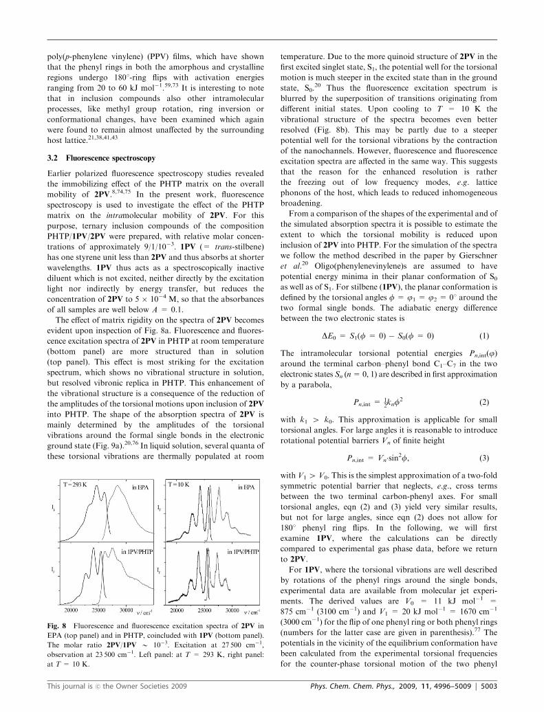
The effect of matrix rigidity on the spectra of 2PV becomes
evident upon inspection of Fig. 8a. Fluorescence and fluores-
cence excitation spectra of 2PV in PHTP at room temperature
(bottom panel) are more structured than in solution
(top panel). This effect is most striking for the excitation
spectrum, which shows no vibrational structure in solution,
but resolved vibronic replica in PHTP. This enhancement of
the vibrational structure is a consequence of the reduction of
the amplitudes of the torsional motions upon inclusion of 2PV
into PHTP. The shape of the absorption spectra of 2PV is
mainly determined by the amplitudes of the torsional
vibrations around the formal single bonds in the electronic
ground state (Fig. 9a).20,76 In liquid solution, several quanta of
these torsional vibrations are thermally populated at room
temperature. Due to the more quinoid structure of 2PV in the
first excited singlet state, S1, the potential well for the torsional
motion is much steeper in the excited state than in the ground
state, S0.20 Thus the fluorescence excitation spectrum is
blurred by the superposition of transitions originating from
different initial states. Upon cooling to T = 10 K the
vibrational structure of the spectra becomes even better
resolved (Fig. 8b). This may be partly due to a steeper
potential well for the torsional vibrations by the contraction
of the nanochannels. However, fluorescence and fluorescence
excitation spectra are affected in the same way. This suggests
that the reason for the enhanced resolution is rather
the freezing out of low frequency modes, e.g. lattice
phonons of the host, which leads to reduced inhomogeneous
broadening.
From a comparison of the shapes of the experimental and of
the simulated absorption spectra it is possible to estimate the
extent to which the torsional mobility is reduced upon
inclusion of 2PV into PHTP. For the simulation of the spectra
we follow the method described in the paper by Gierschner
et al.20 Oligo(phenylenevinylene)s are assumed to have
potential energy minima in their planar conformation of S0as well as of S1. For stilbene (1PV), the planar conformation is
defined by the torsional angles f = j1 = j2 = 01 around the
two formal single bonds. The adiabatic energy difference
between the two electronic states is
DE0 = S1(f = 0) � S0(f = 0) (1)
The intramolecular torsional potential energies Pn,int(j)around the terminal carbon–phenyl bond C1–C7 in the two
electronic states Sn (n=0, 1) are described in first approximation
by a parabola,
Pn,int =12knf
2 (2)
with k1 4 k0. This approximation is applicable for small
torsional angles. For large angles it is reasonable to introduce
rotational potential barriers Vn of finite height
Pn,int = Vn�sin2f, (3)
with V1 4 V0. This is the simplest approximation of a two-fold
symmetric potential barrier that neglects, e.g., cross terms
between the two terminal carbon-phenyl axes. For small
torsional angles, eqn (2) and (3) yield very similar results,
but not for large angles, since eqn (2) does not allow for
1801 phenyl ring flips. In the following, we will first
examine 1PV, where the calculations can be directly
compared to experimental gas phase data, before we return
to 2PV.
For 1PV, where the torsional vibrations are well described
by rotations of the phenyl rings around the single bonds,
experimental data are available from molecular jet experi-
ments. The derived values are V0 = 11 kJ mol�1 =
875 cm�1 (3100 cm�1) and V1 = 20 kJ mol�1 = 1670 cm�1
(3000 cm�1) for the flip of one phenyl ring or both phenyl rings
(numbers for the latter case are given in parenthesis).77 The
potentials in the vicinity of the equilibrium conformation have
been calculated from the experimental torsional frequencies
for the counter-phase torsional motion of the two phenyl
Fig. 8 Fluorescence and fluorescence excitation spectra of 2PV in
EPA (top panel) and in PHTP, coincluded with 1PV (bottom panel).
The molar ratio 2PV/1PV B 10�3. Excitation at 27 500 cm�1,
observation at 23 500 cm�1. Left panel: at T = 293 K, right panel:
at T = 10 K.
This journal is �c the Owner Societies 2009 Phys. Chem. Chem. Phys., 2009, 11, 4996–5009 | 5003
rings, n0 = 9 cm�1 , n1 = 47 cm�1 and their overtones.76,77
The ground state potential has been found to be very
anharmonic,76 while the potential in S1 is well approximated
by a harmonic function.77 We used these experimental data to
assess the reliability of our (TD-)DFT (B3LYP/6-311G*)
calculations which are presented in Table 3.
Although the frequencies of the vibrations in both, S0 and
S1, are overestimated by the (TD-)DFT method, the calcula-
tions describe the ground state vibration sufficiently well.
Assuming approximately harmonic potentials for the torsional
vibration in S0 and S1, the force constants k01 and k00 are
obtained from the calculated frequencies, ni0, and the torsional
angles in the vibrational ground state, ji0,
k0i ¼hni0j2i0
ð4Þ
In the harmonic regime (i.e. f o 201 for S0, f o 651 for S1)
both harmonic potential curves agree perfectly with the
(TD-)DFT single point calculations performed at different
torsional angles. Since at room temperature kT corresponds
to a torsional angle of about 251, the harmonic approximation
should be a good estimate for the thermal population of the
torsional modes. The (TD-)DFT single point calculations of
the torsional potentials yielded for the barriers for the simul-
taneous torsion of two rings in the gas phase V0 = 3400 cm�1
and V1 = 14 900 cm�1. The value for the ground state barrier
agrees well with the results of other quantum chemical
calculations78,79 and with the experimental value in the gas
phase.77
For 2PV, no experimental data are available for the two
low-frequency torsional vibrations, ta and tb, which couple
effectively to the electronic transition S0 - S1. Therefore, the
vibrational frequencies and the corresponding potential curves
were taken from the results of the density functional
((TD-)DFT) calculations (Table 3). The lowest frequency
(~na = 7.5 cm�1) torsional vibration, ta, in the ground state
of 2PV is described quite satisfactorily by considering only
Fig. 9 Top panel: Torsional vibrations ta and tb of 2PV with one and two nodes, respectively. The wavenumbers ~ni0 and ~ni1 are the results of
(TD-)DFT B3LYP/6-311G* calculations for the molecule in vacuum and refer to the electronic ground and excited states, S0 and S1, respectively.
Bottom panel: solid curves are the harmonic potentials for the vibration ta in S0 (GS) and S1 (ES), calculated from the data in Table 3 using eqn (4).
Points are the energies obtained from single point (TD-)DFT calculations for the simultaneous torsion of both terminal phenyl rings around the
formal single bonds. Dotted curves through these points are harmonic fits of the points at small torsion angles (f o 301). The inset shows the
angular dependence of the D-factor as calculated from the single point (TD-)DFT potential curves.
Table 3 Results of (TD-)DFT calculations for the lowest frequencytorsional vibration of 1PV and 2PV
1PV 2PV
S0 S1 S0 S1
~n/cm�1 12 111 7.5 108m/g mol�1 3.586 3.812 3.953 3.790ki/N m�1 0.031 2.791 0.013 2.608fN,i/1 4.230 4.558 2.0481 1.673k0i/cm
�1/(1)2 0.682 5.362 1.788 38.586Vi/cm
�1 3400 14 900 3800 10 600
~n—vibrational wavenumber, ki—force constant in Si (in linear
coordinates), m—reduced mass, fN,i –normalized angle (v = 1) in
Si, k0 i—force constant in Si in angular coordinates, Vi—torsional
barrier for the simultaneous flip of the two terminal rings in state Si.
5004 | Phys. Chem. Chem. Phys., 2009, 11, 4996–5009 This journal is �c the Owner Societies 2009

torsions of the outer phenyl rings around the two terminal
bonds, as obvious from the comparison of the harmonic
potential curve for the vibration ta with the potential of the
torsion of the terminal phenyl rings around the adjacent single
bonds obtained from single point (TD-)DFT calculations
(Fig. 9). The barrier for the flip of one ring is calculated to
V0,int = 1900 cm�1. However, in the excited electronic state S1,
the potential curve for the torsional vibration (~na E 108 cm�1)
deviates significantly from that of the ring torsions. Due to the
quinoid character of S1, the vibrations ta and tb rather
describe torsional motions of the whole molecule around the
long molecular axis with one and two nodes, respectively,
perpendicular to this axis. In the excited state, the barrier for
the flip of one ring amounts to 5300 cm�1.
To take into account the effect of thermal population of the
excited torsional levels in S0 on the shape of absorption and
fluorescence spectra, the absorption spectrum I(n) of the rigidmolecule (e.g., in solid matrices at low temperatures) is
convoluted with the spectral distribution function Gabs(n),which is described by an exponential
Gabs(n) = exp[�D(n)/((D � 1)kT)] (5)
where D(n) = P1 � P0 is the energy difference, and D = P1/P0
is the energy ratio of the two vibrational potentials in S1 and
S0. Inserting the approximations of eqn (2) or (3) into eqn (5),
the difference D(n) depends on j, while the ratio D does not.
The spectral distribution function Gfluo(n) of the fluorescence
bands due to thermal population of excited torsional levels in
S1 is described by
Gfluo(n) = exp[�D(n) D/((D � 1)kT)]. (6)
Since D 4 1, the fluorescence bands are less broadened by
torsional motions than the S0 - S1 absorption bands. Within
this model we are able to simulate the absorption and
fluorescence spectra of nPVs in low-viscous solvents like
tetrahydrofuran (THF) quite satisfactorily (Fig. 10, top
panel), after inserting values of D = 3.8 for 1PV. For the
longer homologues 2PV and 3PV, D = 5.5 and D = 7.0 are
obtained, respectively.20 In the following, the same formalism
is applied to the spatially constrained situation of the PHTP
channels.
The potential barriers, Vn, and the form of the torsional
potential, Pn, i.e. its dependence on f, are not available for
both the dissolved state and the PHTP inclusion compound.
For the band shape calculations, to the intramolecular
potential, Pn,int, of the free molecules as obtained from the
(TD-)DFT calculations an external potential, Pext, was there-
fore added which accounts for impact of the solvent or for the
PHTP lattice.
Pn = Pn,int + Pext (7)
The external potential is assumed to be the same in both
electronic states. Thus, the spectral distribution function is
modified mainly by the D-parameter, whereas D remains
approximately constant. Upon inclusion of 2PV into the
PHTP host at room temperature, the D-parameter was
reduced to D = 1.5, as demonstrated by comparison of the
experimental absorption spectrum of 2PV with the simulated
spectra (Fig. 10, bottom panel). This reduction in D, from
D = 5.5 in THF to D = 1.5 in PHTP, supports the intuitive
notion that with increasing ‘‘viscosity’’ of the environment, the
torsional motions are more and more determined by the
external potential imposed on the molecule by the surrounding
‘‘solvent’’.
The value of the potential barrier Vext can be estimated by
combining the results of the (TD-)DFT calculations and of the
NMR investigations. From the (TD-)DFT single point
calculation of the potential curves for the torsions of the
terminal rings of 2PV in the ground and excited electronic
state of DSB in vacuo, a D-factor of D E 4 was deduced
(Fig. 9). This value is obviously too small, mainly because the
steepness of the torsional potential in the excited state is
underestimated. Nevertheless it provides a reasonable
starting point for the consideration of the effect of the PHTP
matrix. According to the NMR measurements of the
dynamics of 2PV in PHTP, the activation energy for the ring
Fig. 10 Experimental (solid lines) and calculated absorption spectra
of 2PV in THF (1) and of 2PV/1PV/PHTP (3). The calculated spectra
are obtained by applying eqn (5) (for details see text), inserting the
following D-parameters: D = 7 (2), D = 1.5 (4), D = 5.5 (5).
The vibration sub-bands in the calculated spectra are additionally
broadened with a Gaussian width of 800 cm�1. For fitting
the spectrum in PHTP, only the range between 24 000 cm�1 and
28 500 cm�1 is considered, because above 28 500 cm�1 the absorption
of 1PV cannot be neglected.
This journal is �c the Owner Societies 2009 Phys. Chem. Chem. Phys., 2009, 11, 4996–5009 | 5005
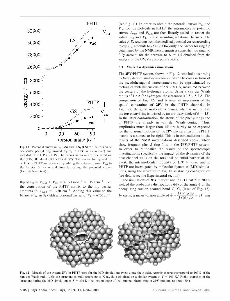
flip of V0 = V0,int + Vext = 40 kJ mol�1 = 3350 cm�1 , i.e.,
the contribution of the PHTP matrix to the flip barrier
amounts to V0,ext = 1450 cm�1. Adding this value to the
barrier V1,int in S1 yields a torsional barrier of V1 = 6750 cm�1
(see Fig. 11). In order to obtain the potential curves Pint and
Pext for the molecule in PHTP, the intramolecular potential
curves, P0,int and P1,int are then linearly scaled to render the
values, V0 and V1, of the according rotational barriers. The
value ofD, resulting from the modified potential curves according
to eqn (6), amounts to DE 2. Obviously, the barrier for ring flip
determined by the NMRmeasurements is somewhat too small to
fully account for the decrease to D = 1.5 obtained from the
analysis of the UV/Vis absorption spectra.
3.3 Molecular dynamics simulations
The 2PV/PHTP system, shown in Fig. 12, was built according
to X-ray data of analogous compounds.6 The cross sections of
the pseudohexagonal nanochannels can be approximated by
rectangles with dimensions of 5.9 � 8.1 A, measured between
the centers of the hydrogen atoms. Using a van der Waals
radius of 1.2 A for hydrogen, the clearance is 3.5 � 5.7 A. The
comparison of Fig. 12a and b gives an impression of the
spatial constraints of 2PV in the PHTP channels. In
Fig. 12a, the guest molecule is planar, whereas in Fig. 12b
the top phenyl ring is twisted by an arbitrary angle of f= 151.
In the latter conformation, the atoms of the phenyl rings and
of PHTP are already in van der Waals contact. Thus,
amplitudes much larger than 151 are hardly to be expected
for the torsional motions of the 2PV phenyl rings if the PHTP
matrix is assumed to be rigid. This is in contradiction to the
results of the NMR investigations described above, which
show frequent phenyl ring flips in the 2PV/PHTP system.
In order to rationalize the results of the spectroscopic
investigations, specifically the impact of the dynamics of the
host channel walls on the torsional potential barrier of the
guest, the intramolecular mobility of 2PV in vacuo and in
PHTP are investigated by molecular dynamics (MD) simula-
tions, using the structure in Fig. 12 as starting configuration
(for details see the Experimental section).
The simulations of 2PV in vacuo and in PHTP at T= 300 K
yielded the probability distributions f(f) of the angle f of the
phenyl ring torsion around bond C1–C7 (inset of Fig. 13).
In vacuo, a mean torsion angle of �f ¼Rf ðfÞf dfRf ðfÞ df � 23� was
Fig. 11 Potential curves in S0 (GS) and in S1 (ES) for the torsion of
one outer phenyl ring around C1–C7 in 2PV in vacuo (vac) and
included in PHTP (PHTP). The curves in vacuo are calculated on
the (TD-)DFT-level (B3LYP/6-311G*). The curves for S0 and S1of 2PV in PHTP are obtained by adding the external barrier Vext to
the barrier in vacuo and linearly scaling the potential curves
(for details see text).
Fig. 12 Models of the system 2PV in PHTP used for the MD simulations (view along the c-axis). Atomic spheres correspond to 100% of the
van der Waals radii. Left: the structure as built according to X-ray data obtained on a similar system at T = 100 K.6 Right: snapshot of the
structure during the MD simulation at T = 300 K (the torsion angle of the terminal phenyl ring in 2PV amounts to about 301).
5006 | Phys. Chem. Chem. Phys., 2009, 11, 4996–5009 This journal is �c the Owner Societies 2009
obtained. This value seems reasonable, considering the mean
torsional amplitude of �f = 301 obtained for 1PV in the
gas phase at 473 K,80 from which at 293 K, assuming a
harmonic potential, a value of �f E 231 was estimated. Upon
inclusion of 2PV into the channels of the PHTP matrix, the
mean torsion angle as obtained by the MD simulations is
reduced to �f E 181.
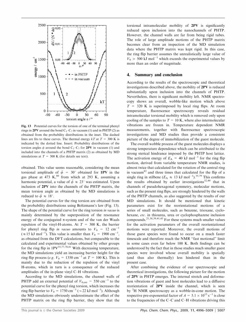
The potential curves for the ring torsion are obtained from
the probability distributions using Boltzmann’s law (Fig. 13).
The shape of the potential curve for the ring torsion in vacuo is
mainly determined by the superposition of the resonance
energy of the conjugated p-system and of the van der Waals
repulsion of the vinyl-H-atoms. At T = 300 K, the barrier
for phenyl ring flip in vacuo amounts to V0 = 12 cm�1
(E15 kJ mol�1). This value is smaller than V0 = 1900 cm�1,
as obtained from the DFT calculations, but comparable to the
calculated and experimental values obtained by other groups
for the ring flip in 1PV35,77,78,81 With decreasing temperature,
the MD simulations yield an increasing barrier height for the
ring flip process (e.g. V0 = 1350 cm�1 at T = 100 K). This is
mainly due to the reduction of the repulsion of the vinyl
H-atoms, which in turn is a consequence of the reduced
amplitudes of the in-plane vinyl C–H vibrations.
According to the MD simulations, the channel walls of
PHTP add an external potential of V0,ext = 550 cm�1 to the
potential curve for the phenyl ring torsion, which increases the
ring flip barrier to V0 = 1750 cm�1 (B22 kJ mol�1). Although
the MD simulations obviously underestimate the effect of the
PHTP matrix on the ring flip barrier, they show that the
torsional intramolecular mobility of 2PV is significantly
reduced upon inclusion into the nanochannels of PHTP.
However, the channel walls are far from being rigid tubes.
The role of large amplitude motions of the PHTP matrix
becomes clear from an inspection of the MD simulation
data where the PHTP matrix was kept rigid. In this case,
the ring flip barrier assumes the unrealistically large value of
V0 4 500 kJ mol�1 which exceeds the experimental values by
more than an order of magnitude.
4. Summary and conclusion
According to the results of the spectroscopic and theoretical
investigations described above, the mobility of 2PV is reduced
substantially upon inclusion into the channels of PHTP.
Nevertheless, there is significant mobility left. NMR spectro-
copy shows an overall, wobble-like motion which above
T = 320 K is superimposed by local ring flips. At room
temperature, fluorescence spectroscopy reveals residual
intramolecular torsional mobility which is removed only upon
cooling of the samples to T= 10 K, where also intermolecular
vibrations are frozen in. Temperature dependent NMR
measurements, together with fluorescence spectroscopic
investigations and MD studies thus provide a consistent
picture of the degree of immobilization of the guest molecule.
The overall wobble process of the guest molecules displays a
strong temperature dependence which can be attributed to the
strong sterical hindrance imposed by the PHTP host lattice.
The activation energy of EA = 40 kJ mol�1 for the ring flip
motion, derived from variable temperature NMR studies, is
almost twice that calculated for the rotation of the central ring
in vacuum82 and three times that calculated for the flip of a
single ring in stilbene (EA E 13 kJ mol�1).35,79 This confirms
the results obtained by earlier investigations32,44 that in
channels of pseudohexagonal symmetry, molecular motions,
such as the present ring flips, are strongly hindered by the walls
of the PHTP channels, as also suggested by the results of our
MD simulations. It should be mentioned that kinetic
parameters exist for the reorientational motions of a
series of small molecules, such as benzene, trioxane, cyclo-
hexane, etc. in thiourea, urea or cyclophosphazene inclusion
compounds.21,38,39,41,43 For these systems much smaller values
for the activation parameters of the overall reorientational
motions were reported. Moreover, the overall motions of
those guest species were found to occur on a much faster
timescale and therefore reach the NMR ‘‘fast motional’’ limit
in some cases even far below 100 K. Both findings can be
understood by the fact that in those studies much smaller guest
species were involved whose overall mobility is spatially
(and thus also thermally) less hindered than in the
present case.
After combining the evidence of our spectroscopic and
theoretical investigations, the following picture for the motion
of 2PV in PHTP emerges. The internal stretch and deforma-
tion vibrations of guest and host molecules lead to a diffusive
reorientation of 2PV inside the channel, which is seen
by 2H NMR spectroscopy as a wobble-in-cone motion. The
respective pre-exponential factor of A= 5.1 � 1013 s�1 is close
to the frequencies of the C–C and C–H vibrations driving this
Fig. 13 Potential curves for the torsion of one of the terminal phenyl
rings in 2PV around the bond C1–C7 in vacuum (1) and in PHTP (2) as
obtained from the probability distributions in the inset. The dashed
lines are fits to these curves. The thermal energy kT at T = 300 K is
indicated by the dotted line. Insert: Probability distributions of the
torsion angles f around the bond C1–C7 for 2PV in vacuum (1) and
included into the channels of a PHTP matrix (2) as obtained by MD
simulations at T = 300 K (for details see text).
This journal is �c the Owner Societies 2009 Phys. Chem. Chem. Phys., 2009, 11, 4996–5009 | 5007

process. At the same time, the intramolecular torsional
motions of 2PV lead to a blurring of the fluorescence
excitation spectra. At T 4 320 K these torsional vibrations
lead to a measurable rate of ring flips, similar to what has been
previously observed in stretched sheets of deuterated PPV.22
Again, for this intramolecular type of motion, the
pre-exponential factor of A= 1.4 � 1012 s�1 shows reasonable
agreement with the calculated frequencies of the torsional
vibrations (n0 E 9 � 1011 s�1). The observed activation
energies of around 40 kJ mol�1 for both processes are larger
than, but still comparable to the results of MD simulations of
2PV included into the channels of a non-rigid PHTP matrix.
When the PHTP matrix is kept rigid during the simulation
runs, unrealistically high activation barriers of4500 kJ mol�1
for the ring flips are obtained. This confirms the results of
earlier investigations on PHTP44,45 and on zeolite83 systems
which clearly demonstrate that the dynamics of the matrix
plays an important role for the mobility of the included guest
molecules.
Acknowledgements
This work was supported by the European Commission
through the Human Potential Program (RTN ‘‘Nanochannel’’
Contract No. HPRN-CT-2002-00323 and RTN ‘‘Nanomatch
Contract No. MRTN-CT-2006-035884) and by the Deutsche
Forschungsgemeinschaft (Graduiertenkolleg ‘‘Chemie in
Interphasen’’, Grant No. 441/2, and ‘‘Moderne Methoden
der magnetischen Resonanz in der Materialforschung’’, Grant
No. 448). R.M.-A. thanks the Alexander von Humboldt
Foundation (Germany) for a grant. B.M.M. thanks the
Ministerio de Ciencia e innovacion (MICINN) of Spain for
a postdoctoral grant and a ‘Juan de la Cierva’ contract. J.G. is
‘Ramon y Cajal’ research fellow, financed by the MICINN. At
present he is a visiting researcher at the ICMol, Valencia,
through the Consolider Ingenio 2010 ‘‘Molecular
Nanoscience’’ activity of the MICINN. The work in Mons is
partly supported by the EU through the Marie Curie Research
Training Network THREADMILL (MRTN-CT-2006-
036040) and by the Belgian National Science Foundation
(FNRS/FRFC). J. C. is a senior research associate and
D.B. research director of FNRS.
References
1 D. Oelkrug, H.-J. Egelhaaf, D. Worrall and F. Wilkinson,J. Fluoresc., 1995, 5, 165.
2 D. Oelkrug, A. Tompert, J. Gierschner, H.-J. Egelhaaf,M. Hanack, M. Hohloch and E. Steinhuber, J. Phys. Chem. B,1998, 102, 1902.
3 R. Pacios, A. Chatten, K. J. Kawano, J. R. Durrant, D. D. C.Bradley and J. Nelson, Adv. Funct. Mater., 2006, 16, 2117.
4 L. Luer, H.-J. Egelhaaf, D. Oelkrug, G. Cerullo, G. Lanzani,B.-H. Huisman and D. deLeeuw, Org. Electron., 2004, 5, 83.
5 K. K. Lin, S.-J. Chua and W. Wang, Thin Solid Films, 2002, 417,36.
6 O. Konig, H.-B. Burgi, T. Armbruster, J. Hulliger and T. Weber,J. Am. Chem. Soc., 1997, 119, 10632.
7 C. Botta, R. Ferro, G. Di Silvestro and R. Tubino, SupramolecularPhotosensitive and Electroactive Materials, ed. H. S. Nalwa,Academic Press, San Diego/CA, USA, 1st edn, 2001, pp. 439–524.
8 J. Gierschner, L. Luer, D. Oelkrug, E. Musluoglu, B. Behnisch andM. Hanack, Adv. Mater., 2000, 12, 757.
9 J. Gierschner, L. Luer, D. Oelkrug, E. Musluoglu, B. Behnisch andM. Hanack, Synth. Met., 2001, 121, 1695.
10 J. Gierschner, H.-J. Egelhaaf, H.-G. Mack, D. Oelkrug,R. M. Alvarez and M. Hanack, Synth. Met., 2003, 137, 1449.
11 G. Bongiovanni, C. Botta, J. E. Communal, F. Cordella,L. Magistrelli, A. Mura, G. Patrinoiu, P. Picouet andG. DiSilvestro, Mater. Sci. Eng., C, 2003, 23, 909.
12 C. Botta, S. Destri, M. Pasini, P. Picouet, G. Bongiovanni,A. Mura, M. Uslenghi, G. Di Silvestro and R. Tubino, Synth.Met., 2003, 139, 791.
13 C. Botta, G. Bongiovanni, A. Mura, G. Di Silvestro andR. Tubino, Synth. Met., 2001, 116, 175.
14 M. A. Loi, A. Mura, G. Bongiovanni, C. Botta, G. Di Silvestroand R. Tubino, Synth. Met., 2001, 121, 1299.
15 G. Bongiovanni, C. Botta, G. Di Silvestro, M. A. Loi, A. Muraand R. Tubino, Chem. Phys. Lett., 2001, 345, 386.
16 C. Botta, R. Treviganti, G. Bongiovanni, A. Mura and R. Tubino,Synth. Met., 1999, 101, 565.
17 M. Farina, G. Di Silvestro and P. Sozzani, Comp. Supramol.Chem., 1996, 6, 371.
18 L. Poulsen, M. Jazdzyk, J.-E. Communal, J. C. Sancho-Garcıa,A. Mura, G. Bongiovanni, D. Beljonne, J. Cornil, M. Hanack,H.-J. Egelhaaf and J. Gierschner, J. Am. Chem. Soc., 2007, 129,8585.
19 J. C. Sancho-Garcıa, L. Poulsen, J. Gierschner, R. Martınez-Alvarez, E. Hennebicq, M. Hanack, H.-J. Egelhaaf, D. Oelkrug,D. Beljonne, J. L. Bredas and J. Cornil, Adv. Mater., 2004, 16,1193.
20 J. Gierschner, H.-G. Mack, L. Luer and D. Oelkrug, J. Chem.Phys., 2002, 116, 8596.
21 A. Liebelt, A. Detken and K. Muller, J. Phys. Chem. B, 2002, 106,7781.
22 J. H. Simpson, D. M. Rice and F. E. Karasz, Macromolecules,1992, 25, 2099.
23 P. Sozzani, A. Comotti, S. Bracco and R. Simonutti, Chem.Commun., 2004, 768.
24 P. Sozzani, R. Simonutti, S. Bracco and A. Comotti, Polym.Prepr., 2003, 44, 297.
25 R. Simonutti, M. Mauri, S. Bracco, A. Comotti and P. Sozzani,Polym. Prepr., 2003, 44, 361.
26 F. C. Schilling, K. R. Amundson and P. Sozzani, Macromolecules,1994, 27, 6498.
27 P. Sozzani, F. A. Bovey and F. C. Schilling,Macromolecules, 1991,24, 6764.
28 F. C. Schilling, P. Sozzani and F. A. Bovey,Macromolecules, 1991,24, 4369.
29 F. C. Schilling, F. A. Bovey and P. Sozzani, Polym. Prepr., 1990,31, 113.
30 G. Di Silvestro, P. Sozzani and M. Farina, Mol. Cryst. Liq. Cryst.,1990, 187, 383.
31 P. Sozzani, R. W. Behling, F. C. Schilling, S. Bruckner, E. Helfand,F. A. Bovey and L. W. Jelinski, Macromolecules, 1989, 22, 3318.
32 P. Sozzani, F. A. Bovey and F. C. Schilling,Macromolecules, 1989,22, 4225.
33 S. Bruckner, P. Sozzani, C. Boeffel, S. Destri and G. Di Silvestro,Macromolecules, 1989, 22, 607.
34 M. Brustolon, A. Barbon, M. Bortolus, A. L. Maniero, P. Sozzani,A. Comotti and R. Simonutti, J. Am. Chem. Soc., 2004, 126, 15512.
35 E. Meirovitch, I. Belsky and S. Vega, J. Phys. Chem., 1984, 88,1522.
36 A. E. Aliev and K. D. M. Harris, J. Phys. Chem. A, 1997, 101,4541.
37 J. A. Villanueva-Garibay and K. Muller, Lect. Notes Phys., 2006,684, 65.
38 J. A. Villanueva-Garibay and K. Muller, J. Phys. Chem. B, 2004,108, 15057.
39 R. Poupko, E. Furman, K. Muller and Z. Luz, J. Phys. Chem.,1991, 95, 405.
40 E. Gelerinter, Z. Luz, R. Poupko and H. Zimmermann, J. Phys.Chem., 1990, 94, 5391.
41 K. Muller, J. Phys. Chem., 1992, 96, 5733.42 K. Muller, Magn. Reson. Chem., 1995, 33, 113.43 J. Schmider and K. Muller, J. Phys. Chem. A, 1998, 102, 1181.44 R. Dodge and W. L. Mattice, Macromolecules, 1991, 24, 2709.45 T. Haliloglu and W. L. Mattice, Macromol. Symp., 1996, 101, 435.
5008 | Phys. Chem. Chem. Phys., 2009, 11, 4996–5009 This journal is �c the Owner Societies 2009
46 Y. Zhan and W. L. Mattice, Macromolecules, 1992, 25, 3439.47 Y. Zhan and W. L. Mattice, Macromolecules, 1992, 25, 4078.48 R. Tubino, E. Fois, A. Gamba, G. Macchi, F. Meinardi and
A. Minoia, in Studies in Surface Science and Catalysis,ed. A. Gamba, C. Collela and S. Collucia, Elsevier, Amsterdam,The Netherlands, 2005, vol. 155, p. 501.
49 S. O. Vasquez, Comput. Mater. Sci., 2006, 37, 572.50 Z. Yang, F. E. Karasz and J. Geise,Macromolecules, 1993, 26, 6570.51 J. Hulliger, O. Konig and R. Hoss, Adv. Mater., 1995, 7, 719.52 A. D. Becke, J. Chem. Phys., 1993, 98, 1372.53 For the TURBOMOLE calculations basis sets were obtained from the
Extensible Computational Chemistry Environment Basis SetDatabase, Version 02/25/04, as developed and distributed by theMolecular Science Computing Facility, Environmental andMolecular Sciences Laboratory which is part of the PacificNorthwest Laboratory, P.O. Box 999, Richland, Washington99352, USA, and funded by the U.S. Department of Energy. ThePacific Northwest Laboratory is a multi-program laboratoryoperated by Battelle Memorial Institute for the U.S. Departmentof Energy under contract DE-AC06-76RLO 1830. Contact KarenSchuchardt for further information.
54 R. Ahlrichs, et al., TURBOMOLE, version 5.71, UniversitatKarlsruhe, Karlsruhe, 2003.
55 J. W. Ponder, TINKER: Software Tools for Molecular Design, 4.2,Washington University School of Medicine, Saint Louis, MO,2004.
56 (a) S. Kumar, D. Bouzida, R. H. Swendsen, P. A. Kollman andJ. M. Rosenberg, J. Comput. Chem., 1992, 13, 1011; (b) WHAMcode: http://membrane.urmc.rochester.edu/.
57 S. Lefrant, J. P. Buisson, M. Baitoul and I. Orion, Pure Appl. Opt.,1996, 5, 613.
58 J. H. Simpson, N. Egger, M. A. Masse, D. M. Rice andF. E. Karasz, J. Polym. Sci., Part B: Polym. Phys., 1990, 28, 1859.
59 E. R. deAzebedo, R. W. A. Franco, A. Marletta, R. M. Faria andT. Bonagamba, J. Chem. Phys., 2003, 119, 2923.
60 P. Sozzani, R. Simonutti and A. Comotti, Mol. Cryst. Liq. Cryst.,1996, 227, 299.
61 M. M. Maricq and J. S. Waugh, J. Chem. Phys., 1979, 70, 3300.
62 D. Suwelack, W. P. Rothwell and J. S. Waugh, J. Chem. Phys.,1980, 73, 2559.
63 A. D. Ronemus, R. R. Vold and R. L. Vold, J. Chem. Soc.,Faraday Trans. 1, 1988, 84, 3761.
64 O. Yano and Y. Wada, J. Polym. Sci., Part A2, 1971, 9, 669.65 A. V. Lyulin, N. K. Balabaev and M. A. J. Michels,
Macromolecules, 2002, 35, 9595.66 T. Brauniger, R. Poupko and Z. Luz, J. Chem. Phys., 2000, 112,
10858.67 P. Speier, A. Muller, C. Meinel and U. Haeberlen, Mol. Phys.,
1998, 95, 859.68 G. A. Facey, T. J. Connolly, C. Bensimon and T. Durst, Can. J.
Chem., 1996, 74, 1844.69 T. Hiraoki, A. Kogame, N. Nishi and A. Tsutsumi, J. Mol. Struct.
(THEOCHEM), 1998, 441, 243.70 M. A. Keniry, T. M. Rothgeb, R. L. Smith, H. S. Gutowsky and
E. Oldfield, Biochemistry, 1983, 22, 1917.71 M. A. Kennedy, R. R. Vold and R. L. Vold, J. Magn. Reson., 1991,
91, 301.72 M. H. Wann and G. S. Harbison, J. Chem. Phys., 1994, 101, 231.73 J. H. Simpson, D. M. Rice and F. M. Karasz, J. Polym. Sci.,
Part B: Polym. Phys., 1992, 30, 11.74 H.-J. Egelhaaf, L. Luer, A. Tompert, P. Bauerle, K. Mullen and
D. Oelkrug, Synth. Met., 2000, 115, 63.75 F.-C. Hsu, M. Hayashi, H.-W. Wang, S. H. Lin and J.-K. Wang,
J. Phys. Chem. A, 2007, 111, 759.76 T. Suzuki, N. Mikami and M. Ito, J. Phys. Chem., 1986, 90, 6431.77 J. Laane, J. Phys. Chem. A, 2000, 104, 7715.78 S. P. Kwasniewski, L. Claes and J. P. Francois, J. Chem. Phys.,
2003, 118, 7823.79 O. Lhost and J. L. Bredas, J. Chem. Phys., 1992, 96, 5279.80 M. Trætteberg, E. B. Frantsen, F. C. Mijehoff and A. Hoeckstra,
J. Mol. Struct. (THEOCHEM), 1975, 26, 57.81 S. P. Kwasniewski, J. P. Francois and M. S. Deleuze, J. Phys.
Chem. A, 2003, 107, 5168.82 G. C. Claudio and E. R. Bittner, Chem. Phys., 2002, 276, 81.83 P. C. M. M. Magusin and R. A. van Santen, Chem. Phys. Lett.,
2003, 373, 630.
This journal is �c the Owner Societies 2009 Phys. Chem. Chem. Phys., 2009, 11, 4996–5009 | 5009