Dragon's Paradise Lost: Palaeobiogeography, Evolution and Extinction of the Largest-Ever Terrestrial...
15
Dragon’s Paradise Lost: Palaeobiogeography, Evolution and Extinction of the Largest-Ever Terrestrial Lizards (Varanidae) Scott A. Hocknull 1 *, Philip J. Piper 2 , Gert D. van den Bergh 3 , Rokus Awe Due 4 , Michael J. Morwood 3 , Iwan Kurniawan 5 1 Geosciences, Queensland Museum, Brisbane, Queensland, Australia, 2 Archaeological Studies Program, University of the Philippines, Quezon City, Philippines, 3 School of Earth and Environmental Sciences, University of Wollongong, Wollongong, New South Wales, Australia, 4 Indonesian Centre for Archaeology, Jakarta, Indonesia, 5 Geological Survey of Indonesia, Bandung, Indonesia Abstract Background: The largest living lizard species, Varanus komodoensis Ouwens 1912, is vulnerable to extinction, being restricted to a few isolated islands in eastern Indonesia, between Java and Australia, where it is the dominant terrestrial carnivore. Understanding how large-bodied varanids responded to past environmental change underpins long-term management of V. komodoensis populations. Methodology/Principal Findings: We reconstruct the palaeobiogeography of Neogene giant varanids and identify a new (unnamed) species from the island of Timor. Our data reject the long-held perception that V. komodoensis became a giant because of insular evolution or as a specialist hunter of pygmy Stegodon. Phyletic giantism, coupled with a westward dispersal from mainland Australia, provides the most parsimonious explanation for the palaeodistribution of V. komodoensis and the newly identified species of giant varanid from Timor. Pliocene giant varanid fossils from Australia are morphologically referable to V. komodoensis suggesting an ultimate origin for V. komodoensis on mainland Australia (.3.8 million years ago). Varanus komodoensis body size has remained stable over the last 900,000 years (ka) on Flores, a time marked by major faunal turnovers, extinction of the island’s megafauna, the arrival of early hominids by 880 ka, co-existence with Homo floresiensis, and the arrival of modern humans by 10 ka. Within the last 2000 years their populations have contracted severely. Conclusions/Significance: Giant varanids were once a ubiquitous part of Subcontinental Eurasian and Australasian faunas during the Neogene. Extinction played a pivotal role in the reduction of their ranges and diversity throughout the late Quaternary, leaving only V. komodoensis as an isolated long-term survivor. The events over the last two millennia now threaten its future survival. Citation: Hocknull SA, Piper PJ, van den Bergh GD, Due RA, Morwood MJ, et al. (2009) Dragon’s Paradise Lost: Palaeobiogeography, Evolution and Extinction of the Largest-Ever Terrestrial Lizards (Varanidae). PLoS ONE 4(9): e7241. doi:10.1371/journal.pone.0007241 Editor: Samuel T. Turvey, Zoological Society of London, United Kingdom Received February 18, 2009; Accepted August 24, 2009; Published September 30, 2009 Copyright: ß 2009 Hocknull et al. This is an open-access article distributed under the terms of the Creative Commons Attribution License, which permits unrestricted use, distribution, and reproduction in any medium, provided the original author and source are credited. Funding: Australian Research Council LP0453664 and LP0883991. The funders had no role in study design, data collection and analysis, decision to publish, or preparation of the manuscript. Competing Interests: The authors have declared that no competing interests exist. * E-mail: [email protected] Introduction Fossils of giant varanids ($3 m Total Body Length) were first reported in the 1850s with the description of Megalania prisca from the Pleistocene of Australia [1,2]. Since that time, and with the discovery of living Komodo Dragons (V. komodoensis) on the east Indonesian islands of Flores, Rinca and Komodo [3] considerable attention was paid in trying to understand the evolution of body size in monitor lizards [4–6]. Though several processes are proposed to explain the evolution of giantism in varanids, two competing hypotheses dominate the literature: autapomorphic giantism (i.e. Island Rule) and phyletic giantism (i.e. Cope’s Rule) [7]. Both processes were previously invoked for the evolution of V. komodoensis [4,6,7]. It is commonly thought that V. komodoensis is a classic example of autapomorphic giantism having evolved large body size sometime in the past from a small-bodied ancestor that arrived on isolated Indonesian islands, which were devoid of predatory competition [3,8]. Some proposals suggest that V. komodoensis attained large body size on Flores as a specialist hunter of pygmy Stegodon [3,9], the only large-bodied prey inhabiting Flores throughout the middle and late Pleistocene to as recently as 12,000 years ago [10,11]. The alternative, phyletic giantism, is supported by independent phylogenetic studies of morphology [12–14] and genetics [15,16], which nest V. komodoensis within an Australopa- puan clade of varanids containing the two large-sized living species, V. salvadorii and V. varius, and the largest of all known lizards Megalania prisca ( = Varanus prisca) [14]. Thus, large body size is a synapomorphy of the clade and is not an autapomorphic trait of V. komodoensis. The implications of the phyletic model are that: 1. The extant populations of V. komodoensis are relictual, having had a PLoS ONE | www.plosone.org 1 September 2009 | Volume 4 | Issue 9 | e7241
-
Upload
independent -
Category
Documents
-
view
3 -
download
0
Transcript of Dragon's Paradise Lost: Palaeobiogeography, Evolution and Extinction of the Largest-Ever Terrestrial...

Dragon’s Paradise Lost: Palaeobiogeography, Evolutionand Extinction of the Largest-Ever Terrestrial Lizards(Varanidae)Scott A. Hocknull1*, Philip J. Piper2, Gert D. van den Bergh3, Rokus Awe Due4, Michael J. Morwood3, Iwan
Kurniawan5
1 Geosciences, Queensland Museum, Brisbane, Queensland, Australia, 2 Archaeological Studies Program, University of the Philippines, Quezon City, Philippines, 3 School
of Earth and Environmental Sciences, University of Wollongong, Wollongong, New South Wales, Australia, 4 Indonesian Centre for Archaeology, Jakarta, Indonesia,
5 Geological Survey of Indonesia, Bandung, Indonesia
Abstract
Background: The largest living lizard species, Varanus komodoensis Ouwens 1912, is vulnerable to extinction, beingrestricted to a few isolated islands in eastern Indonesia, between Java and Australia, where it is the dominant terrestrialcarnivore. Understanding how large-bodied varanids responded to past environmental change underpins long-termmanagement of V. komodoensis populations.
Methodology/Principal Findings: We reconstruct the palaeobiogeography of Neogene giant varanids and identify a new(unnamed) species from the island of Timor. Our data reject the long-held perception that V. komodoensis became a giantbecause of insular evolution or as a specialist hunter of pygmy Stegodon. Phyletic giantism, coupled with a westwarddispersal from mainland Australia, provides the most parsimonious explanation for the palaeodistribution of V. komodoensisand the newly identified species of giant varanid from Timor. Pliocene giant varanid fossils from Australia aremorphologically referable to V. komodoensis suggesting an ultimate origin for V. komodoensis on mainland Australia (.3.8million years ago). Varanus komodoensis body size has remained stable over the last 900,000 years (ka) on Flores, a timemarked by major faunal turnovers, extinction of the island’s megafauna, the arrival of early hominids by 880 ka, co-existencewith Homo floresiensis, and the arrival of modern humans by 10 ka. Within the last 2000 years their populations havecontracted severely.
Conclusions/Significance: Giant varanids were once a ubiquitous part of Subcontinental Eurasian and Australasian faunasduring the Neogene. Extinction played a pivotal role in the reduction of their ranges and diversity throughout the lateQuaternary, leaving only V. komodoensis as an isolated long-term survivor. The events over the last two millennia nowthreaten its future survival.
Citation: Hocknull SA, Piper PJ, van den Bergh GD, Due RA, Morwood MJ, et al. (2009) Dragon’s Paradise Lost: Palaeobiogeography, Evolution and Extinction ofthe Largest-Ever Terrestrial Lizards (Varanidae). PLoS ONE 4(9): e7241. doi:10.1371/journal.pone.0007241
Editor: Samuel T. Turvey, Zoological Society of London, United Kingdom
Received February 18, 2009; Accepted August 24, 2009; Published September 30, 2009
Copyright: � 2009 Hocknull et al. This is an open-access article distributed under the terms of the Creative Commons Attribution License, which permitsunrestricted use, distribution, and reproduction in any medium, provided the original author and source are credited.
Funding: Australian Research Council LP0453664 and LP0883991. The funders had no role in study design, data collection and analysis, decision to publish, orpreparation of the manuscript.
Competing Interests: The authors have declared that no competing interests exist.
* E-mail: [email protected]
Introduction
Fossils of giant varanids ($3 m Total Body Length) were first
reported in the 1850s with the description of Megalania prisca from the
Pleistocene of Australia [1,2]. Since that time, and with the discovery
of living Komodo Dragons (V. komodoensis) on the east Indonesian
islands of Flores, Rinca and Komodo [3] considerable attention was
paid in trying to understand the evolution of body size in monitor
lizards [4–6]. Though several processes are proposed to explain the
evolution of giantism in varanids, two competing hypotheses
dominate the literature: autapomorphic giantism (i.e. Island Rule)
and phyletic giantism (i.e. Cope’s Rule) [7]. Both processes were
previously invoked for the evolution of V. komodoensis [4,6,7].
It is commonly thought that V. komodoensis is a classic example of
autapomorphic giantism having evolved large body size sometime
in the past from a small-bodied ancestor that arrived on isolated
Indonesian islands, which were devoid of predatory competition
[3,8]. Some proposals suggest that V. komodoensis attained large
body size on Flores as a specialist hunter of pygmy Stegodon [3,9],
the only large-bodied prey inhabiting Flores throughout the
middle and late Pleistocene to as recently as 12,000 years ago
[10,11]. The alternative, phyletic giantism, is supported by
independent phylogenetic studies of morphology [12–14] and
genetics [15,16], which nest V. komodoensis within an Australopa-
puan clade of varanids containing the two large-sized living
species, V. salvadorii and V. varius, and the largest of all known
lizards Megalania prisca ( = Varanus prisca) [14]. Thus, large body size
is a synapomorphy of the clade and is not an autapomorphic trait
of V. komodoensis. The implications of the phyletic model are that: 1.
The extant populations of V. komodoensis are relictual, having had a
PLoS ONE | www.plosone.org 1 September 2009 | Volume 4 | Issue 9 | e7241

much wider geographic distribution in the past [17,18]. 2. Varanus
komodoensis arrived on Flores already large and did not evolve
giantism there through the processes of insular evolution [7].
We aim to reconstruct the palaeobiogeography and geochronol-
ogy of Neogene large-bodied varanids by using the fossil remains
available from deposits in India, Java, Flores, Timor and Australia.
Methods
MorphometricsFive measurements were taken of fossil and modern Varanus
cervical, dorsal, sacral and anterior caudal vertebrae (Figure 1).
Measurements were undertaken using dial or digital callipers to
0.5 mm resolution. See Table S1 for specimen list and data.
Measurements are in millimetres (mm) and include:
1. Prezygapophysis to postzygapophysis length (Pre-Post), mea-
sured from the anterior margin of the prezygapophyses to the
posterior margin of the postzygapophyses (Figure 1, A).
2. Centrum length (CL), measured from the posterior margin of
the cotyle to the posterior margin of the condyle (Figure 1, B).
3. Cotylar width (CW), measured from the left lateral margin of
the cotyle to the right lateral margin of the cotyle (Figure 1, C).
4. Postzygapophysis to postzygapophysis width (Post-Post), mea-
sured from the lateral margin of the left postzygapophysis to
the lateral margin of the right postzygapophysis (Figure 1, D).
5. Prezygapophysis to prezygapophysis width (Pre-Pre), measured
from the lateral margin of the left prezygapophysis to the
lateral margin of the right prezygapophysis (Figure 1, E).
Two measurements were taken of fossil and modern Varanus
teeth (Figure 1).
1. Crown height, measured from the base of the tooth plicidentine
to the crown tip if preserved (Figure 1, F).
2. Basal width, measured from the anterior margin to the
posterior margin of the base of the tooth (Figure 1, G).
Two measurements were taken from the humerus of fossil and
modern Varanus (Figure 1) [19].
1. Maximum diaphysis width of humeri (Figure 1, H).
2. Maximum width of the distal end (Figure 1, I).
Capturing Maximal Size VariationOur modern sample of V. salvator may not represent the
maximal size limit seen in the largest V. salvator individuals. Our
comparative sample of V. salvator was close to the maximal snout-
vent lengths (SVL) recorded in a large sample of this taxon;,
however, the largest specimen from our sample had a total length
approximately 15–20% shorter than the total length of the longest
recorded V. salvator (321 cm) [20]. Therefore, we took the
Figure 1. Morphometric measurements. A. Pre-postzygapophysis length. B. Centrum length. C. Cotylar width. D. Post-postzygapophysis width.E. Pre-prezygapophysis width. F. Tooth crown height. G. Tooth base length. H. Diaphysis width (humerus). I. Distal condyle width (humerus).doi:10.1371/journal.pone.0007241.g001
Fossil Giant Varanids
PLoS ONE | www.plosone.org 2 September 2009 | Volume 4 | Issue 9 | e7241

measurements of our largest V. salvator vertebral specimens and
increased them by 20%, adding these additional measurements to
the main dataset. This provided a more accurate estimate of V.
salvator maximal size limits.
Descriptive StatisticsBivariate plots of morphometric data were plotted to determine
the position of fossil specimens in relation to the modern samples
taken of Varanus. Convex hulls were drawn to delineate the area of
maximal variation observed in the samples. Due to the different
preservation states of many of the specimens, only a single
measurement might be available (univariate data). For these data
frequency distribution histograms or box-plots showing the
median value, 25–75% quartiles and the minimal and maximal
values, were produced to determine where in the range of
variation the fossil specimens plotted out. Principle components
analysis (PCA) was applied to analyse multivariate data; however,
most multivariate analyses were uninformative due to the large
amount of missing data from the fossil specimens.
Where possible statistical tests were carried out where fossil
sample sizes were sufficient to return an informative result.
Descriptive statistics and tests were conducted using PAST
software version 1.82b [21].
Results
Varanus komodoensis Ouwens 1912Australia (Early Pliocene – middle Pleistocene). Fossil
specimens from Pliocene and Pleistocene-aged sites in Australia
(Table 1) were identified as belonging to Varanus komodoensis on the
basis of the following combination of unique cranial and post-
cranial characteristics. Overall similar size and proportions of all
preserved skeletal elements. Maxilla contributes to the labial
margin of the premaxillary-maxillary aperture (pmp). Maxillary
margin of the pmp shallow. Premaxillary-maxillary suture faces
antero-lingually. Angulate maxillary crest. Labio-lingually
compressed, closely-set recurved and serrated dentition both on
maxillae and dentaries. At least 12 tooth loci in dentary. Parietal
with distinct supratemporal crests, with fronto-parietal suture
interlocking. Humerus stockier than all other members of Varanus,
except V. prisca.
Maxillae (Figure 2, B–D and Figure 3, H–I). Three
maxillae; a near complete right maxilla (QMF 874), the anterior
section of a right maxilla (QMF 42105) and the posterior portion
of a left maxilla (QMF 54605) share closest morphology and size
with Varanus komodoensis. QMF42105 represents a marginally larger
individual than specimens QMF874 and QMF 54605. All three
share with V. komodoensis closely-spaced, labio-lingually
compressed, recurved dentition with finely grooved plicidentine,
and serrated mesial and distal margins. The teeth are
morphometrically similar to the modern V. komodoensis sample
(Figure S1).
QMF 874 and QMF 42105 also share with V. komodoensis a
distinct interlocking premaxillary-maxillary suture with an open
premaxillary-maxillary aperture (pmp) and an angulate narial
crest. The circumference of the premaxillary-maxillary aperture is
made up by the premaxilla and maxilla to varying degrees in
different Varanus groups (Figure S2). In the fossil maxillae only the
posterior and labial margins of the pmp are enclosed by the
maxilla, the remainder is enclosed by the premaxilla. This key
feature allies the fossils to taxa where the premaxilla contributes to
the anterior and lingual margin of the pmp (V. indicus, V. varius
group and V. gouldii group).
The fossils differ morphologically from V. indicus by possessing
an interlocking and anterolingually oriented premaxillary-maxil-
lary suture articulation, more labially angulate maxillary crest and
more recurved dentition. Morphometrically, the fossils are also
much larger.
Table 1. Pliocene – Pleistocene fossils from Queensland representing Varanus komodoensis.
Specimen Registration Location (Fauna) Age Age Reference
Cervical vertebra QMF 23684 Bluff Downs Local Fauna, north-eastern Queensland Early Pliocene [32]
Dorsal vertebra QMF 23686 Bluff Downs Local Fauna, north-eastern Queensland Early Pliocene [32]
Right maxilla QMF 874 Chinchilla Sands Local Fauna, south-east Queensland Middle Pliocene [33]
Right maxilla (partial) QMF 42105 Chinchilla Sands Local Fauna, south-east Queensland Middle Pliocene [33]
Left dentary (partial) QMF 870+QMF 871 Chinchilla Sands Local Fauna, south-east Queensland Middle Pliocene [33]
Quadrate QMF 42156 Chinchilla Sands Local Fauna, south-east Queensland Middle Pliocene [33]
Supraorbital QMF 25392 Chinchilla Sands Local Fauna, south-east Queensland Middle Pliocene [33]
Parietal QMF 53956 Chinchilla Sands Local Fauna, south-east Queensland Middle Pliocene [33]
Scapulocoracoid QMF 866 Chinchilla Sands Local Fauna, south-east Queensland Middle Pliocene [33]
Left humerus (partial) QMF 53954 Chinchilla Sands Local Fauna, south-east Queensland Middle Pliocene [33]
Right humerus (partial) QMF 53955 Chinchilla Sands Local Fauna, south-east Queensland Middle Pliocene [33]
Vertebrae QM Colln (numerous) Chinchilla Sands Local Fauna, south-east Queensland Middle Pliocene [33]
Left maxilla (partial) QMF 54605 Mt Etna Local Fauna, central eastern Queensland Middle Pleistocene [28]
Supraoccipital QMF 54607 Mt Etna Local Fauna, central eastern Queensland Middle Pleistocene [28]
Quadrate (right) QMF 54606 Mt Etna Local Fauna, central eastern Queensland Middle Pleistocene [28]
Tibia QMF54608 Mt Etna Local Fauna, central eastern Queensland Middle Pleistocene [28]
Ulna diaphysis QMF 54604 Mt Etna Local Fauna, central eastern Queensland Middle Pleistocene [28]
Dorsal vertebra QMF 54120 Mt Etna Local Fauna, central eastern Queensland Middle Pleistocene [28]
Caudal vertebra QMF 1418 Marmor Quarry, eastern Queensland Middle Pleistocene [34]
doi:10.1371/journal.pone.0007241.t001
Fossil Giant Varanids
PLoS ONE | www.plosone.org 3 September 2009 | Volume 4 | Issue 9 | e7241

Figure 2. Varanus komodoensis (Pliocene, Australia). A. Modern V. komodoensis skull in dorsal view (NNM 17504). B. QMF 874, right maxilla inlateral view. C. QMF 42105, partial right maxilla in dorsal view. D. QMF 42105, right maxilla in dorsal view. E. QMF 25392, complete left supraorbital indorsal view. F. QMF 53956, partial parietal in dorsal view. G–H. QMF 42156, right quadrate in anterior and lateral views. I–J. QMF 870+871, partial leftdentary in lingual view, J illustrating the tooth loci. Abbreviations: mcrst, dorsal maxillary crest; pmp, premaxillary-maxillary aperture; pms, premaxilla-maxilla suture; sym, dentary symphysis; mg, Meckalian groove. Dashed line represents broken edge of maxilla. Scale bar = 5 cm.doi:10.1371/journal.pone.0007241.g002
Fossil Giant Varanids
PLoS ONE | www.plosone.org 4 September 2009 | Volume 4 | Issue 9 | e7241

Figure 3. Varanus komodoensis (Neogene, Australia). A–B, E–G. Pliocene V. komodoensis (Australia)A–B. QMF 53955, partial left humerus indorsal view showing position of insertion for the latissimus dorsi (lat dors). C–D. Left and right humerus of a modern V. komodoensis (NNM 17504). E–F. QMF 53954, partial right humerus in ventral and dorsal views, showing the position of the ectepicondyle (ect). G. QMF 866, partial scapulocoracoid.H–P. Pleistocene V. komodoensis (Australia). H–I. QMF 54605, partial left maxilla in lingual and labial views. J. QMF 54606, partial right quadrate inanterior view. K. QMF 54607, supraoccipital bone in posterior view. L. QMF 54608, proximal left tibia. M. QMF 54604, ulna diaphysis. N–P. QMF 1418,proximal mid-caudal in cranial, oblique lateral and dorsal views. Scale bar = 1 cm.doi:10.1371/journal.pone.0007241.g003
Fossil Giant Varanids
PLoS ONE | www.plosone.org 5 September 2009 | Volume 4 | Issue 9 | e7241

Morphologically the fossil specimens are most similar to
members of the V. varius group and to some members of the V.
gouldii group, in particular V. varius, V. komodoensis, V. panoptes and V.
mertensi (Figure S2). The maxillary margin of the pmp in the V.
varius group is shallower than those from the V. gouldii group. The
pmp is similarly shallow in both fossil maxillae, allying them closer
to the V. varius group. The only taxon present in either the gouldii or
varius groups that reaches the large size of the fossils and possesses
the closely-spaced, recurved dentition, is V. komodoensis.
Supraorbital (Figure 2, E). QMF 25392 is a complete left
supraorbital bone, which matches closely V. komodoensis, including
the possession of a thick postorbital bar which projects postero-
laterally, is shallowly curved and is suboval in cross-section.
Proximally, the supraorbital flares in an antero-posterior direction,
producing a ‘Y’ shaped bone. The dorsal surface is smooth, whilst
the ventral surface preserves a rugose margin.
Parietal (Figure 2, F). QMF 53956 possesses distinct
dorsally expressed supratemporal crests which ally this specimen
to large-sized members of the V. varius group (V. salvadorii, V.
komodoensis and V. prisca). QMF 53956 is smaller than V. prisca with
less defined crests and a broader central roof. Based on overall
size, QMF 53956 is most similar to V. komodoensis and larger than
V. salvadorii. It also possesses an interlocking frontal-parietal suture
articulation, which is only seen in V. komodoensis and V. prisca.
Quadrates (Figure 2, G–H. Figure 3, J). Two right
quadrates are known, including a complete specimen (QMF
42156) and the proximal half of another (QMF 54606). Both are
similar in overall morphology and size to one another and to V.
komodoensis. In both specimens and in V. komodoensis, the proximal
condyle is antero-posteriorly expanded into two articular facets,
both rounded basins that are relatively smooth. One or two thin
laminae run ventrally to the distal condyle which is medio-laterally
expanded into two similar-sized condyles. A distinct medial crest
originates from the midline of the proximal articular end and runs
medio-distally along the medial side of the quadrate, terminating
at the antero-medial corner of the disto-medial condyle. A broad,
rounded and straight ridge originates at the antero-lateral corner
of the proximal articular face and runs distally to the antero-lateral
corner of the disto-lateral condyle.
Supraoccipital (Figure 3, K). QMF 54607 is an isolated but
complete supraoccipital bone from the skull of a large species of
Varanus. In dorsal view, it is trapezoidal in shape with a ventral
width wider than the dorsal width. A ridge occurs in the midline of
the bone oriented dorso-ventrally and constricted toward the
middle. A cup-like recess is present on the dorsal face of the bone,
which would have received the processus ascendens, which seems to
have been unossified or at least not attached to the supraoccipital
(as it is in V. priscus). The angle of the supraoccipital in relation to
the parietal, and to the paraoccipitals would be more acute than
seen in V. priscus, similar to that of V. komodoensis and less so than
most other species of Varanus.
Dentary (Figure 2, I–J). QMF 870 and QMF 871 represent
either a single left dentary, which is badly broken at its midline, or
two separate fragments of two left dentaries. Although not noted as
the same specimen, preservation and size indicates that these two
specimens come from a very similar, if not the same, individual.
QMF 871 is an anterior-most portion of a left dentary preserving
the dentary symphysis and the first six tooth loci. The first tooth
occurs just postero-dorsal of the dentary symphysis, which is
rounded and bisected by the proximal origin of the Meckelian
groove. The second tooth is complete and the best preserved of
both specimens. The tooth is compressed labio-lingually, has a
rounded distal margin and a constricted and serrate mesial cutting
edge. QMF 870 is a portion of the posterior section of a left
dentary, preserving five tooth loci. The dentary is missing below
the dorsal margin of the Meckelian groove. The dorsal half of the
posterior mental foramen can be seen in labial profile. Considered
together, these two specimens indicate that the dentary preserved
12–13 tooth loci, where the teeth are closely-spaced, labio-
lingually compressed, distinctly recurved and serrated. When
compared to a range of Varanus species, it is clear that adult V.
komodoensis possesses 12 or 13 tooth loci for each dentary; whereas
other species of Varanus possess 11 or fewer tooth loci. V. salvator
(10–11); V. albigularis (9–10); V. indicus (9–10); V. varius (9–10); V.
salvadorii (10–11); V. panoptes (10–11); V. tristis (10–11). The only
complete dentary of V. priscus possesses 11 tooth loci.
Humeri (Figure 3, A–B, E–F). A right (QMF 53955) and a
left (QMF 53954) humerus, both missing the proximal and distal-
most epiphyses are of similar size and morphology to V.
komodoensis. The humeri of V. priscus and V. komodoensis are stocky
and robust when compared to humeri found in all other members
of Varanus. Both fossil humeri indicate a stout humerus with broad
proximal and distal epiphyses. When comparing the maximum
diaphyseal width of the two specimens with species of extant and
fossil Varanus, both specimens fall within the size range of V.
komodoensis and outside that of small and large Varanus prisca (Figure
S3).
Vertebrae (Figure 3, M–P. Figure 4.). Thirty eight dorsal
vertebrae were measured from two Pliocene localities, Chinchilla
(n = 37) and Bluff Downs (n = 1). All of these vertebrae fell within
the size range of modern V. komodoensis (p.0.8) (Figure S4). In all
features, the fossil sample reflects directly similar features seen in
V. komodoensis.
A partial dorsal vertebra (QMF 54120) and a caudal vertebra
collected from middle Pleistocene-aged sites at Mt. Etna and
Marmor Quarry respectively represent a large-bodied varanid
which is much smaller than V. prisca, but larger than any living
varanid on mainland Australia and New Guinea (e.g. Varanus
giganteus, V. varius, V. salvadorii) (Figure 4). QMF 54120 is a
fragmentary dorsal vertebra, preserving the left lateral half of the
cotyle and the left lateral portion of the postzygapophysis. On
direct comparison with V. komodoensis it shares similar size and
morphology. QMF1418 is a near complete proximal mid-caudal
vertebra and falls within the size range of V. komodoensis (Figure S9).
Other postcranial elements (Figure 3, L–M). In addition
to the above diagnostic skeletal elements, several other postcranial
remains recovered from these Pliocene and Pleistocene sites match
V. komodoensis in over size and general morphology. These
specimens include the proximal end of a left tibia (QMF 54608),
the proximal end of a dorsal rib (QMF 54603), the diaphysis of an
ulna (QMF 54604) and a partial scapulocoracoid (QMF866).
Flores (early Pleistocene - Holocene)Fossil specimens of V. komodoensis were recovered from the early-
middle Pleistocene Ola Bula Formation in central Flores (Tangi
Talo, Dhozo Dhalu) and from a late Pleistocene-Holocene cave
deposit in central-western Flores (Liang Bua) [10,22]. Fossil
specimens of V. komodoensis include many cranial and postcranial
elements (Table S1).
Teeth (Figure 5, E). Twelve teeth were studied from the
Pleistocene of Flores, including six isolated teeth from early
Pleistocene Tangi Talo and six teeth from late Pleistocene-
Holocene Liang Bua. Morphometrically these teeth fall within the
size range of V. komodoensis (Figure S1). Morphologically, the teeth
bear the unique features of being greatly recurved and compressed
labio-lingually. This feature is only present in modern V.
komodoensis and fossil specimens referable to V. komodoensis.
Fossil Giant Varanids
PLoS ONE | www.plosone.org 6 September 2009 | Volume 4 | Issue 9 | e7241

Figure 4. Varanus komodoensis (Pliocene, Australia). A–F. QMF 42104, posterior dorsal vertebra compared with modern V. komodoensis (white),in anterior (A–B), posterior (C–D) and left lateral (E–F) views. G–L. QMF 42096, mid-dorsal vertebra compared with modern V. komodoensis (white), inanterior (G–H), posterior (I–J) and right lateral (K–L) views. M–R. QMF 42102, mid-dorsal vertebra compared with modern V. komodoensis (white), inanterior (M–N), posterior (O–P) and left lateral (Q–R) views. S–V. QMF 23684, cervical vertebra compared with modern V. komodoensis (white), in leftlateral (S–T) and anterior (U–V) views. W–X. QMF 23686, anterior dorsal vertebra compared with modern V. komodoensis (white) in anterior view. Scalebar = 1 cm.doi:10.1371/journal.pone.0007241.g004
Fossil Giant Varanids
PLoS ONE | www.plosone.org 7 September 2009 | Volume 4 | Issue 9 | e7241

Cervical vertebrae (Figure 5, D). Four well preserved
cervical specimens were used in this study, including one from
early Pleistocene Tangi Talo and three from late Pleistocene-
Holocene Liang Bua. The Tangi Talo specimen is only slightly
larger than the modern V. komodoensis sample (Figure S5, A). Two
Liang Bua specimens are only slightly larger and one is within the
Figure 5. Varanus komodoensis (Pleistocene, Flores). A–B. Sacral vertebrae from modern (A) and fossil (LB558a) V. komodoensis in anterior view.C. Articulated dorsal vertebrae (LB19/20-9-04) in dorsal view. D. Cervical vertebra (LB517b) in dorsal view. E. Four isolated teeth (LB04 unreg) in lingualview. F–H. Ulna diaphysis (LB-447a/16.8.04) in medial (F), cranial (G) and lateral (H) views. I. Radius diaphysis (LB-28.7.03) in medial view. Scalebar = 1 cm.doi:10.1371/journal.pone.0007241.g005
Fossil Giant Varanids
PLoS ONE | www.plosone.org 8 September 2009 | Volume 4 | Issue 9 | e7241
morphometric range of our modern V. komodoensis sample (Figure
S5, B). No statistical test was conducted due to the very small
sample sizes for each site (1 and 3 respectively).
Dorsal vertebrae (Figure 5, C). Fifteen dorsal vertebrae
were used in this study, all coming from Liang Bua. Due to the
differing preservation states of each vertebra the only available
measurement for all dorsal vertebrae was the cotylar width. When
the Liang Bua fossil sample is compared to our sample of modern
V. komodoensis the mean value for cotylar width is significantly
different (p,0.001). When comparing the maximal size limits of
our two samples the Liang Bua fossil sample is most-likely biased
toward large individuals (Figure S6). Therefore, we consider the
significant difference in mean cotylar width to be related to a
taphonomic bias toward large individuals being preserved at the
Liang Bua site, not an overall larger size. These large individuals
are still within the maximal size limits of our modern sample of V.
komodoensis. In addition, three well-preserved specimens were used
in a bivariate plot of prezygapophysis – postzygapophysis (Pre-
post) length over prezygapophysis – prezygapophysis (Pre-pre)
width. The fossils fall within the morphometric range of modern V.
komodoensis, with the exception of a single specimen that possesses a
slightly broader pre-pre width (Figure S7). Morphologically, the
vertebrae are identical to modern V. komodoensis.
Sacral vertebrae (Figure 5, B). A single sacral vertebra
from Liang Bua is directly comparable in size to V. komodoensis and
places morphometrically within the sample of modern V.
komodoensis (Figure S8). No statistical tests were able to be carried
out due to the small sample size (n = 1).
Anterior caudal vertebrae. Six anterior caudal vertebrae
were studied from Liang Bua, all of which fall within the
morphometric and morphological variation of modern V.
komodoensis; their mean sizes not significantly different (p.0.67)
(Figure S9).
Humerus. A single humeral diaphysis represents V. komodoensis
from Tangi Talo both in size and morphology. When compared to
modern V. komodoensis and humeri from Chinchilla, the Tangi Talo
specimen has a slightly broader diaphysis (Figure S3). This may
simply reflect the biased preservation of larger individuals within
this deposit, as is seen in the Liang Bua collection.
Other postcranial elements (Figure 5, H–I). In addition to
the above diagnostic specimens several other remains recovered
from Liang Bua are considered to represent V. komodoensis,
including fragments of ilia, metapodials, a phalanx, partial right
mandible, and the diaphyses of an ulna and a radius (Table S1).
These remains will form part of a future review of V. komodoensis
fossils from Liang Bua.
Varanus sp. cf. V. komodoensisJava (Middle Pleistocene). A single anterior dorsal vertebra
(CD6392) of a large-bodied varanid is recorded from the middle
Pleistocene Kedung Brubus deposit (Figure 6, A–F).
Morphometrically this specimen falls within the middle range of
modern and fossil V. komodoensis and is well outside the largest V.
salvator (+20%) sample (Figure S7). CD6392 was considered to be
V. komodoensis [17]. It is remarkably similar to V. komodoensis in both
size and morphology, possessing steep zygapophyses, dorsally
oriented condyles, distinct precondylar constriction and an open
neural canal. Although the specimen is close in morphology,
assignment to V. komodoensis is tentative and should await more
specimens for verification.
Varanus sivalensis Falconer 1868India, (Pliocene - early Pleistocene). Three specimens
were previously described to represent Varanus sivalensis [23–25], a
distal humerus and two dorsal vertebrae (anterior and mid-dorsal
vertebrae). Whether these three specimens represent a single taxon
(V. sivalensis) will depend on the discovery of more fossil specimens
referable to this taxon. The humerus is morphologically distinct
from Varanus komodoensis to warrant its unique taxonomic status;
however, the two referred dorsal vertebrae fall within the variation
of modern and fossil V. salvator. Therefore, it is unlikely that these
three specimens represent the same taxon.
Humerus (Figure 7, C–D). Morphologically the humerus
differs from V. komodoensis by features already described [12].
NHMR40816 plots in the middle range of V. komodoensis and
outside V. salvator.
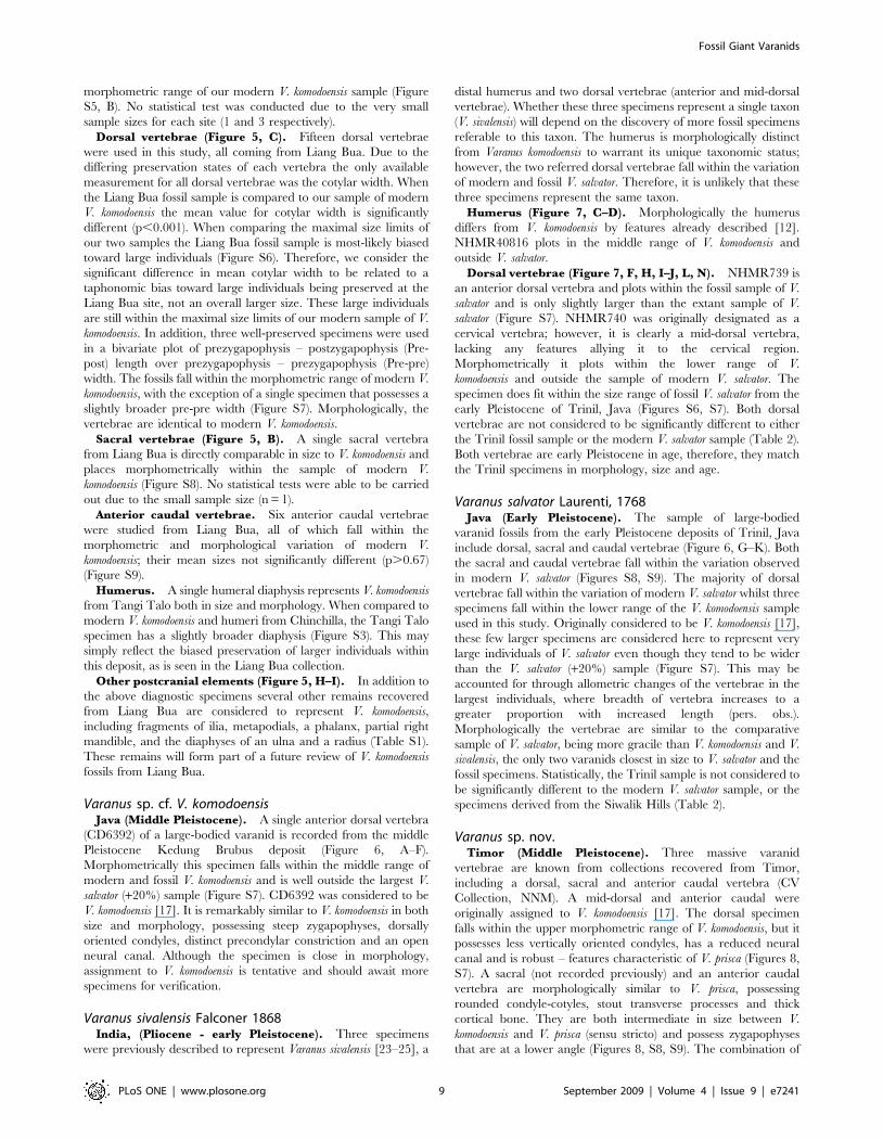
Dorsal vertebrae (Figure 7, F, H, I–J, L, N). NHMR739 is
an anterior dorsal vertebra and plots within the fossil sample of V.
salvator and is only slightly larger than the extant sample of V.
salvator (Figure S7). NHMR740 was originally designated as a
cervical vertebra; however, it is clearly a mid-dorsal vertebra,
lacking any features allying it to the cervical region.
Morphometrically it plots within the lower range of V.
komodoensis and outside the sample of modern V. salvator. The
specimen does fit within the size range of fossil V. salvator from the
early Pleistocene of Trinil, Java (Figures S6, S7). Both dorsal
vertebrae are not considered to be significantly different to either
the Trinil fossil sample or the modern V. salvator sample (Table 2).
Both vertebrae are early Pleistocene in age, therefore, they match
the Trinil specimens in morphology, size and age.
Varanus salvator Laurenti, 1768Java (Early Pleistocene). The sample of large-bodied
varanid fossils from the early Pleistocene deposits of Trinil, Java
include dorsal, sacral and caudal vertebrae (Figure 6, G–K). Both
the sacral and caudal vertebrae fall within the variation observed
in modern V. salvator (Figures S8, S9). The majority of dorsal
vertebrae fall within the variation of modern V. salvator whilst three
specimens fall within the lower range of the V. komodoensis sample
used in this study. Originally considered to be V. komodoensis [17],
these few larger specimens are considered here to represent very
large individuals of V. salvator even though they tend to be wider
than the V. salvator (+20%) sample (Figure S7). This may be
accounted for through allometric changes of the vertebrae in the
largest individuals, where breadth of vertebra increases to a
greater proportion with increased length (pers. obs.).
Morphologically the vertebrae are similar to the comparative
sample of V. salvator, being more gracile than V. komodoensis and V.
sivalensis, the only two varanids closest in size to V. salvator and the
fossil specimens. Statistically, the Trinil sample is not considered to
be significantly different to the modern V. salvator sample, or the
specimens derived from the Siwalik Hills (Table 2).
Varanus sp. nov.Timor (Middle Pleistocene). Three massive varanid
vertebrae are known from collections recovered from Timor,
including a dorsal, sacral and anterior caudal vertebra (CV
Collection, NNM). A mid-dorsal and anterior caudal were
originally assigned to V. komodoensis [17]. The dorsal specimen
falls within the upper morphometric range of V. komodoensis, but it
possesses less vertically oriented condyles, has a reduced neural
canal and is robust – features characteristic of V. prisca (Figures 8,
S7). A sacral (not recorded previously) and an anterior caudal
vertebra are morphologically similar to V. prisca, possessing
rounded condyle-cotyles, stout transverse processes and thick
cortical bone. They are both intermediate in size between V.
komodoensis and V. prisca (sensu stricto) and possess zygapophyses
that are at a lower angle (Figures 8, S8, S9). The combination of
Fossil Giant Varanids
PLoS ONE | www.plosone.org 9 September 2009 | Volume 4 | Issue 9 | e7241

intermediate size and unique morphology indicate that these
specimens most likely represent a new unnamed taxon of large-
bodied varanid.
Central Australia (Middle-Late Pleistocene)Vertebrae. A large sample of dorsal vertebrae were
measured from collections of Pleistocene varanids from central
Australia. The morphometric variation encompassed by these
specimens indicates the presence of a varanid intermediate in size
between V. komodoensis and V. prisca (sensu stricto) (Figure S10,
S11). The three samples of vertebrae were significantly different
to one another with the central Australian sample showing
significantly smaller size when compared to the eastern
Australian V. prisca (sensu stricto) sample (p,0.0002) and
significantly larger size when compared to modern and fossil V.
komodoensis (p,0.004). Whether these specimens represent a
diachronous sample across the middle to late Pleistocene, or a
morphocline of Pleistocene giant varanids from smaller central
Australian forms toward larger eastern Australian forms is yet to
be determined. Regardless, these specimens indicate a giant
Figure 6. Varanus sp. cf. V. komodoensis and V. salvator (Pleistocene, Java). A–F. V. sp. cf. V. komodoensis. Anterior dorsal vertebra (CD 6392)compared with modern V. komodoensis in anterior (A–B), dorsal (C–D) and left lateral view (E–F). G–K. V. salvator. G–I. CD 8873, mid-dorsal vertebra,compared with modern V. komodoensis in dorsal (G–H) and anterior (H–I) views. J–K. CD 216, sacral vertebra, compared with modern V. komodoensisin anterior view. Scale bar = 1 cm.doi:10.1371/journal.pone.0007241.g006
Fossil Giant Varanids
PLoS ONE | www.plosone.org 10 September 2009 | Volume 4 | Issue 9 | e7241

varanid present in central Australia during the Pleistocene that
resembles, at least in size, the taxon present on Timor during the
middle Pleistocene.
Discussion
Archaeological and paleontological excavations at sites in
central and western Flores produced teeth and post-cranial
elements of V. komodoensis dating from the early Pleistocene to
the late Holocene (,900–2 ka) [10,22]. This fossil record provides
an opportunity to evaluate long-term morphological and morpho-
metric changes in V. komodoensis on Flores over ca. 900,000 years.
Comparisons between fossil and extant V. komodoensis show that
there are few morphometric or morphological differences between
the fossil specimens and those of modern V. komodoensis. Therefore,
maximal body size of this species remained stable for at least
900,000 years despite the fact that the biostratigraphic sequence
on Flores records at least three faunal turnovers, marked by the
extinction of the giant tortoise Colossochelys [26], two species of
Stegodon and Homo floresiensis, as well as the arrival of hominins by
880 ka and modern humans by 10 ka [22]. Even in the absence of
any moderately-sized prey between the extinction of Stegodon
florensis insularis (,12. ka) [10,11] and the introduction of the pig
Figure 7. Varanus sivalensis (Pliocene, India). A–B. NNM 17504, modern Varanus komodoensis humerus. C–D. NHMR 40819, distal humerus indorsal (C) and ventral (D) views. E–I. NHMR 740, posterior dorsal vertebra compared with modern V. komodoensis (white) in anterior (E–F), left lateral(G–H) and dorsal (I) views. J–N. NHMR 739, anterior dorsal compared with modern V. komodoensis (white) in left lateral (J–K), anterior (L–M) andposterior (N) views. Scale bar = 1 cm.doi:10.1371/journal.pone.0007241.g007
Table 2. Tukey’s pairwise comparisons (ANOVA) table offossil and modern Varanus dorsal vertebrae, pre-post lengthmeasurements.
Liang Bua SiwaliksV.komodoensis
V. salvator(n = 29)
Trinil (n = 11) p,0.0004* p.0.9 p,0.002* p.0.9
Liang Bua (n = 3) p,0.006* p.0.9 p,0.0002*
Siwaliks (n = 2) p,0.03* p.0.7
V. komodoensis(n = 74)
p,0.0007*
*indicates a significant difference between samples.doi:10.1371/journal.pone.0007241.t002
Fossil Giant Varanids
PLoS ONE | www.plosone.org 11 September 2009 | Volume 4 | Issue 9 | e7241

from Sulawesi (,7 ka) [10] V. komodoensis was able to persist on
Flores. The stability of V. komodoensis body size over a long
temporal sequence and during periods of major ecological change
implies that insular evolutionary processes had limited effect, and
more importantly illustrate the adaptive flexibility and resilience of
a generalist carnivore, rather than a specialist predator of the
island’s pygmy Stegodon.
So, if V. komodoensis did not evolve on an isolated island in
Wallacea, from where did it disperse? India and Australia are the
only regions that preserve a giant varanid fossil record older than
900 ka, and are the only identifiable sources for large-bodied
Varanus [2]. The oldest recorded large-bodied Varanus from both
regions occur in the Pliocene, suggesting a relatively synchronous
yet independent evolution of lizard giantism. In India large-bodied
Figure 8. Varanus sp. nov. (Pleistocene, Timor). A–F. Mid-dorsal vertebra (CV Raebia 1) compared with modern V. komodoensis in anterior (A–B),left lateral (C–D), dorsal (E–F) views. G–L. Anterior caudal vertebra (CV Raebia 2) compared with modern V. komodoensis in anterior (G–H), dorsal (I–J)and oblique posterior (K–L) views. M–R. Sacral vertebra (CV Raebia 3) compared with modern V. komodoensis in anterior (M–N), dorsal (O–P) andventral (Q–R) views. S. QMF 8968, sacral vertebra of Varanus prisca in anterior view.doi:10.1371/journal.pone.0007241.g008
Fossil Giant Varanids
PLoS ONE | www.plosone.org 12 September 2009 | Volume 4 | Issue 9 | e7241

varanid fossils are rare, being represented by two vertebrae and a
partial humerus, each assigned to the extinct V. sivalensis [24]. Both
vertebrae probably represent Varanus salvator. The humerus is of
similar size to V. komodoensis but differs morphologically [12]. The
absence of V. sivalensis from younger deposits at the same locales
suggests that large-bodied varanids were either very rare or more
likely extinct on the Indian sub-continent by the end of the early
Pleistocene. Therefore, based on both morphology and chronol-
ogy, V. sivalensis is an unlikely source for V. komodoensis on Flores.
Varanus sivalensis is associated with a Late Pliocene Siwalik fauna
that includes diverse mammalian megafauna, including the
placental carnivores Crocuta, Hyaena and Panthera [27]. This record
alone demonstrates that varanids can evolve giantism on
continental landmasses with competition from large placental
carnivores.
Varanids appear in the Australian fossil record by the Miocene
and possess a more or less continuous record of large-bodied forms
from the early Pliocene (,3.8 mya) through to the late Pleistocene
[2]. Varanids most likely dispersed eastward from Asia to
Australia, then radiated to produce a clade containing V.
komodoensis [15,16,18]. Although the taxonomy of the Australian
Miocene-Pleistocene varanids remains largely unresolved [2], it is
most likely that they are contained within this monophyletic clade.
There are at least three giant varanid taxa present in Australia
during the Neogene, including one species from the Pliocene, one
from the Pleistocene of central Australia and Varanus prisca (sensu
stricto) from the middle-late Pleistocene. On the basis of both size
and a unique combination of morphological features shared only
with V. komodoensis the Pliocene taxon is here considered to be
conspecific with V. komodoensis. Newly recovered large-bodied
varanid fossils from middle Pleistocene [28] deposits in north-
eastern Australia are also referable to V. komodoensis, demonstrating
the longevity of Varanus komodoensis on mainland Australia and
the coexistence of two giant varanids, V. prisca and V. komodoensis.
In combination, the evidence from the fossil record as well as the
morphological and molecular phylogenetic studies clearly supports
Australia as the ancestral source for V. komodoensis.
Large-bodied varanid fossils were previously recovered from
two middle Pleistocene sites along the Solo River in Java, west of
the Wallace Line - the Trinil (,900 ka) and Kedung Brubus (800–
700 ka) Faunas [22]. Although large, the Trinil vertebrae fall
closest to the variation of modern V. salvator, with a few specimens
comparable in size with the smallest modern V. komodoensis. These
few larger specimens, considered previously to be V. komodoensis
[17], more likely represent very large individuals of V. salvator. A
single vertebra from the younger Kedung Brubus site is much
bigger, comparable closely in both size and morphology with large
V. komodoensis. We therefore conclude that it is likely that V.
komodoensis, having reached Flores by the early Pleistocene,
dispersed westward, across Wallace’s Line to arrive in Java
sometime during the middle Pleistocene.
Differential timing for the initial appearance of Komodo dragon
in Australia, Flores and Java, therefore indicates that V. komodoensis
dispersed from east to west, perhaps reaching Java during a period
of lowered sea-level. At the time of Kedung Brubus, Java was part
of the Asian mainland, and the fauna included large placental
carnivores such as Panthera and Hyaena [22], further illustrating the
ability of giant varanids to exist as part of a continental, placental-
dominated fauna. There is currently no evidence that giant
varanids survived on Java beyond the middle Pleistocene.
Further support for the westward dispersal of giant varanids
comes from Timor, an island between Flores and Australia. Three
vertebral specimens from Raebia in the Atambua Basin, central
Timor, represent a new unnamed species of giant varanid
intermediate in size between V. komodoensis and V. prisca (sensu
stricto). The Timor specimens were derived from the uppermost
part of the folded, regressive Noele Formation, of which the
marine part correlates with planktonic foraminifera zones N18-
N22 [29,30]. This suggests that the specimens are at least middle
Pleistocene in age. Pleistocene varanid fossils from central
Australia, usually identified as V. prisca, are also intermediate in
size between V. komodoensis and V. prisca (sensu stricto), and may
represent the same intermediate taxon present in Timor. Formal
description of the new Timor-Australian varanid waits until more
diagnostic specimens are available.
ConclusionThe fossil record suggests that giant varanids evolved indepen-
dently on mainland Asia and the island-continent of Australia
during the Pliocene, alongside large-bodied mammalian carni-
vores. Only the Indonesian-Australian giant varanids appear to
have survived beyond the early Pleistocene.
We conclude that V. komodoensis evolved in Australia by the early
Pliocene and dispersed west as far as Flores by 900 ka and Java by
8002700 ka. It is likely that the Timor varanid represents another
large-bodied varanid lineage, attaining a larger body size than that
of V. komodoensis, having evolved on mainland Australia and
dispersed west to Timor. Continuing along this same evolutionary
trajectory, Varanus prisca, reached gigantic proportions by the late-
Middle Pleistocene, but was extinct in Australia by the end of the
Pleistocene (Figure 9).
We conclude that V. komodoensis is the last of a clade of giant
varanids that was once a ubiquitous part of Australasia, distributed
from Australia across Wallacea, as far as continental Asia (Java).
There is now only a relict population on Flores and a few small
adjacent islands. Komodo dragon distribution has also retracted
significantly on Flores itself; being present at Liang Bua in the
uplands of West Flores until ,2 ka, but now only occurring in
isolated habitats along the northern and western coastal lowlands
[3,31]. The retraction is likely due to habitat loss and persecution
by modern humans over the last few millennia and emphasizes the
continuing threat of extinction to this, the last of the giant
varanids.
Supporting Information
Figure S1 Histogram of tooth base length measurements for
modern (A–B), Pleistocene (A) and Pliocene (B) V. komodoensis.
Tangi Talo (n = 4), Liang Bua (n = 5), Chinchilla (n = 5) and V.
komodoensis (n = 68). Measurements in mm.
Found at: doi:10.1371/journal.pone.0007241.s001 (0.23 MB
DOC)
Figure S2 Morphological comparisons between Indo-Asian and
Indo-Australian varanid maxillae based on the phylogenetic
reconstruction of Ast (2001). Varanus varius group with fossil
specimens for comparison (to scale with V. komodoensis).
Found at: doi:10.1371/journal.pone.0007241.s002 (4.69 MB
DOC)
Figure S3 Histogram of humerus maximum diaphysis width
with normal curve fitted to Varanus sample. Varanus spp. (n = 71),
Varanus komodoensis (n = 18) (see Hutchinson & Reed (2005) for taxa
used). Measurements in mm.
Found at: doi:10.1371/journal.pone.0007241.s003 (0.14 MB
DOC)
Figure S4 Histogram of dorsal vertebrae pre-post measurements
with normal curve fitted. Varanus komodoensis modern (n = 100),
Fossil Giant Varanids
PLoS ONE | www.plosone.org 13 September 2009 | Volume 4 | Issue 9 | e7241
Pliocene (Chinchilla & Bluff Downs) (n = 38). Measurements in
mm.
Found at: doi:10.1371/journal.pone.0007241.s004 (0.19 MB
DOC)
Figure S5 Measurements of varanid cervical vertebrae. A.
Bivariate Plot of pre-pre length vs pre-post length. B. Bivariate
Plot of cotylar width vs centrum length. Convex hulls applied to
show limits of sample variation. Measurements in mm.
Found at: doi:10.1371/journal.pone.0007241.s005 (0.11 MB
DOC)
Figure S6 Box-plot of dorsal vertebrae cotylar width measure-
ments. Varanus salvator (n = 24), Trinil (n = 15), Varanus sivalensis
(n = 2), modern Varanus komodoensis (n = 112). Liang Bua (n = 16).
Measurements in mm.
Found at: doi:10.1371/journal.pone.0007241.s006 (0.05 MB
DOC)
Figure S7 Measurements of varanid dorsal vertebrae. Bivariate
Plot of pre-pre length vs pre-post length. Convex hulls applied to
show limits of sample variation. Measurements in mm.
Found at: doi:10.1371/journal.pone.0007241.s007 (0.09 MB
DOC)
Figure S8 Measurements of varanid sacral vertebrae. A. Bivariate
plot of pre-pre length vs pre-post length. Convex hulls applied to
show limits of sample variation. B. Box-plot of sacral vertebrae
cotylar width measurements. Varanus salvator (n = 10), Trinil (n = 2),
Varanus komodoensis (n = 9), V. prisca (n = 4). Measurements in mm.
Found at: doi:10.1371/journal.pone.0007241.s008 (0.10 MB
DOC)
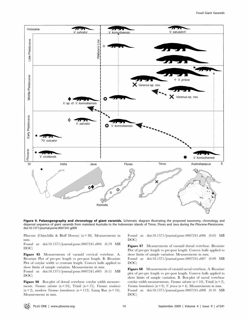
Figure 9. Palaeogeography and chronology of giant varanids. Schematic diagram illustrating the proposed taxonomy, chronology anddispersal sequence of giant varanids from mainland Australia to the Indonesian islands of Timor, Flores and Java during the Pliocene-Pleistocene.doi:10.1371/journal.pone.0007241.g009
Fossil Giant Varanids
PLoS ONE | www.plosone.org 14 September 2009 | Volume 4 | Issue 9 | e7241

Figure S9 Box-plot of caudal vertebrae prezygapophysis-post-
zygapophysis length measurements. Varanus salvator (n = 9), Varanus
komodoensis (n = 24), Liang Bua (n = 4), V. prisca (n = 8). Measure-
ments in mm.
Found at: doi:10.1371/journal.pone.0007241.s009 (0.06 MB
DOC)
Figure S10 Box plot of dorsal vertebra pre-postzygapophysis
length for V. prisca (n = 53), Varanus sp. nov. (n = 11) and modern V.
komodoensis (n = 32). Measurements in mm.
Found at: doi:10.1371/journal.pone.0007241.s010 (0.04 MB
DOC)
Figure S11 Measurements of varanid dorsal vertebrae. Bivariate
plot of pre-pre length vs pre-post length. Convex hulls applied to
show limits of sample variation. Measurements in mm.
Found at: doi:10.1371/journal.pone.0007241.s011 (0.07 MB
DOC)
Table S1 Specimens used in this study.
Found at: doi:10.1371/journal.pone.0007241.s012 (0.28 MB
DOC)
Acknowledgments
We thank Addison Wynn, Chris Thacker, Gasso Miracle, George Zug,
Jeffrey Seigel, Jennie McGuire, Jim Mead, Liz Reed, Ryan Rabett and
Traci Hartsell for assistance in attaining varanid morphometric data. John
de Vos, Bob Jones, Tom Rich, Chris Smeenk, John McCarthy, Pim
Arntzen, Michael Lee, Mark Hutchinson, Patrick Couper and Andrew
Amey are thanked for access to fossil and comparative varanids held at
their various institutions. Fachroel Aziz, Suyono, Ruli Setiawan, Slamat
Sudiarwadi assisted with fieldwork in Timor.
Author Contributions
Conceived and designed the experiments: SAH. Performed the experi-
ments: SAH PP GvdB MM. Analyzed the data: SAH. Wrote the paper:
SAH PP GvdB MM. Integral to the excavation and recovery of material
for this study: RAD IK.
References
1. Owen R (1859) Description of some remains of a gigantic land-lizard (Megalania
prisca, Owen) from Australia. Philosophical Transactions of the Royal Society of
London 149: 43–48.
2. Molnar RE (2004) Dragons in the Dust: The palaeobiology of the Giant Monitor
Lizard Megalania. Indiana: Indiana University Press. 210 p.
3. Auffenberg W (1981) The Behavioural Ecology of the Komodo Monitor.
Gainesville FL: University Press of Florida. 406 p.
4. Pianka ER (1995) Evolution of body-size: Varanid lizards as a model system.
American Naturalist 146(3): 398–414.
5. Christian A, Garland Jr (1996) Scaling of Limb Proportions in Monitor Lizards
(Squamata: Varanidae). Journal of Herpetology 30(2): 219–230.
6. Pianka ER (2004) Evolution of Body Size and Reproductive Tactics. In:
Pianka ER, King DR, eds. Varanoid Lizards of the World. Bloomington,
Indiana: Indiana University Press. pp 549–555.
7. Gould GC, MacFadden BJ (2005) Gigantism, Dwarfism and Cope’s Rule:
‘‘Nothing in Evolution Makes Sense without a Phylogeny’’. Bulletin of the
American Museum of Natural History 285(1): 219–237.
8. Burness GP, Diamond J, Flannery T (2001) Dinosaurs, dragons, and dwarfs:
The evolution of maximal body size. PNAS 98(25): 14518–14523.
9. Diamond JM (1987) Did Komodo dragons evolve to eat pygmy elephants?
Nature 326: 832.
10. van den Bergh GD, et al. (2008) The youngest stegodon remains in Southeast Asia
from the Late Pleistocene archaeological site Liang Bua, Flores, Indonesia.
Quaternary International 182: 16–48.
11. Morwood MJ, et al. (2004) Archaeology and age of Homo floresiensis, a new
hominin from Flores in eastern Indonesia. Nature 431: 1087–1091.
12. Dunn, ER (1927) Results of the Douglas Burden Expeditions to the Island of
Komodo, I. Notes on Varanus komodoensis. American Museum Novitates 286:
1–10.
13. Conrad J (2008) Phylogeny and systematics of Squamata (Reptilia) based on
morphology. Bulletin of the American Museum of Natural History 310: 1–182.
14. Head JJ, Barrett PLS, Rayfield EJ (2009) Neurocranial osteology and systematic
relationships of Varanus (Megalania) prisca Owen, 1859 (Squamata: Varanidae).
Zoological Journal of the Linnean Society 155: 445–457.
15. Ast J (2001) Mitochondrial DNA evidence and evolution in Varanoidea
(Squamata). Cladistics 17: 211–226.
16. Fitch AJ, Goodman AE, Donnellan SC (2006) A molecular phylogeny of the
Australian monitor lizards (Squamata: Varanidae) inferred from mitochondrial
DNA sequences. Australian Journal of Zoology 54: 253–269.
17. Hooijer D (1972) Varanus (Reptilia, Sauria) from the Pleistocene of Timor.
Zoologische Mededelingen 47: 445–447.
18. Fuller S, Braverstock P, King D (1998) Biogeographic origins of goannas
(Varanidae): A molecular perspective. Molecular Phylogenetics and Evolution
9(2): 294–307.
19. Reed EH, Hutchinson, MN (2005) First record of a giant varanid (Megalania,
Squamata) from the Pleistocene of Naracoorte, South Australia. Memoirs of the
Queensland Museum 51(1): 203–214.20. Gaulke M, Horn HG (2004) Varanus salvator (nominate form). In: Pianka ER,
King DR, eds. Varanoid Lizards of the World. Bloomington, Indiana: IndianaUniversity Press. pp 244–255.
21. Hammer O, Harper DAT, Ryan PD (2001) PAST: Palaeontological Statisticssoftware package for education and data analysis. Palaeontologica Electronica
4(1): 1–9.
22. van den Bergh GD, de Vos J, Sondaar PY (2001) The Late Quaternarypalaeogeography of mammal evolution in the Indonesian Archipelago.
Palaeogeography, Palaeoclimatology, Palaeoecology 171: 385–408.23. Falconer HP (1868) Paleontological Memoirs and Notes of the Late Hugh
Falconer, A. M., M. D. Robert Hardwicke Publishers: Picadilly. 465 p.
24. Lydekker R (1886) Fauna of the Karnul caves. Palaeontologica Indica 10(4):1–58.
25. Lydekker R (1888) Catalogue of the fossil Reptilia and Amphibia in the BritishMuseum (Natural History), Cromwell Road, S.W. Pt. 1. The Orders
Ornithosauria, Crocodilia, Dinosauria, Squamata, Rhynchocephalia, andPreterosauria. London: The Trustees. 245 p.
26. Setiyabudi E (2006) Paleontological study on fossil giant tortoises from the
Indonesian islands. (unpublished Thesis, Kagoshima University, Japan) 325 p.27. Dennell RR, Coard D, Turner A (2006) The biostratigraphy and magnetic
polarity zonation of the Pabbi Hills, northern Pakistan: An Upper Siwalik (PinjorStage) Upper Pliocene–Lower Pleistocene fluvial sequence. Palaeogeography,
Palaeoclimatology, Palaeoecology 234: 168–185.
28. Hocknull SA, Zhao J-x, Feng Y-x, Webb GE (2007) Responses of Quaternaryrainforest vertebrates to climate change in Australia, Earth and Planetary
Science Letters 264: 317–331.29. Suwitodirdjo K, Tjokrosapoetro S (1975) Geologic Quadrangle Map, Timor,
(GRDC, Bandung (1974/75).30. de Smet MEM (1990) Detection of collision related vertical movements in the
outer Banda Arc (Timor, Indonesia) using micropaleontological data. Journal of
South East Asian Earth Sciences 4(4): 337–356.31. Murphy JB, et al. (2002) Komodo Dragons: Biology and Conservation.
Smithsonian Institution Press, Washington. 324 p.32. Mackness BS, Whitehead PW, McNamara GC (2000) New Potassium-Argon
basalt date in relation to the Pliocene Bluff Downs Local Fauna, northern
Australia. Australian Journal of Earth Sciences 47: 807–811.33. Dawson L, Muirhead J, Wroe S (1999) The Big Sink Fauna: a lower Pliocene
mammalian fauna from the Wellington Caves complex, Wellington, New SouthWales. Records of the Western Australian Museum Supplement No. 57:
265–290.
34. Price GJ, Zhao J-x, Feng Y-x, Hocknull SA (2009) New Records of Plio-Pleistocene Koalas from Australia: Palaeoecological and Taxonomic Implica-
tions. Records of the Australian Museum 61: 39–48.
Fossil Giant Varanids
PLoS ONE | www.plosone.org 15 September 2009 | Volume 4 | Issue 9 | e7241
All in-text references underlined in blue are linked to publications on ResearchGate, letting you access and read them immediately.