Distinct Roles of the First Introns on the Expression of Arabidopsis Profilin Gene Family Members
14
Distinct Roles of the First Introns on the Expression of Arabidopsis Profilin Gene Family Members 1 Young-Min Jeong, Jeong-Hwan Mun 2 , Ilha Lee, Je Chang Woo, Choo Bong Hong, and Sang-Gu Kim* Department of Biological Sciences, Seoul National University, Seoul 151–742, Republic of Korea (Y.-M.J., J.-H.M., I.L., C.B.H., S.-G.K.); and Department of Biology, Mokpo National University, Jeonnam 534–729, Republic of Korea (J.C.W.) Profilin is a small actin-binding protein that regulates cellular dynamics of the actin cytoskeleton. In Arabidopsis (Arabidopsis thaliana), five profilins were identified. The vegetative class profilins, PRF1, PRF2, and PRF3, are expressed in vegetative organs. The reproductive class profilins, PRF4 and PRF5, are mainly expressed in pollen. In this study, we examined the role of the first intron in the expression of the Arabidopsis profilin gene family using transgenic plants and a transient expression system. In transgenic plants, we examined PRF2 and PRF5, which represent vegetative and reproductive profilins. The expression of the PRF2 promoter fused with the b-glucuronidase (GUS) gene was observed in the vascular bundles, but transgenic plants carrying the PRF2 promoter-GUS with its first intron showed constitutive expression throughout the vegetative tissues. However, the first intron of PRF5 had little effect on the reporter gene expression pattern. Transgenic plants containing PRF5 promoter-GUS fusion with or without its first intron showed reproductive tissue-specific expression. To further investigate the different roles of the first two introns on gene expression, the first introns were exchanged between PRF2 and PRF5. The first intron of PRF5 had no apparent effect on the expression pattern of the PRF2 promoter. But, unlike the intron of PRF5, the first intron of PRF2 greatly affected the reproductive tissue-specific expression of the PRF5 promoter, confirming a different role for these introns. The results of a transient expression assay indicated that the first intron of PRF1 and PRF2 enhances gene expression, whereas PRF4 and PRF5 do not. These results suggest that the first introns of profilin genes are functionally distinctive and the first introns are required for the strong and constitutive gene expression of PRF1 and PRF2 in vegetative tissues. Diverse actin-binding proteins regulate cellular dy- namics of the actin cytoskeleton to perform various biological processes, such as locomotion, elongation, shape change, cytoplasmic streaming, division, and development of the cell (Bamburg et al., 1999; Kost et al., 1999; Meagher et al., 1999; Balu˘ ska et al., 2001). Among these proteins, the small actin-binding protein, profilin (12–15 kD), plays an important role in actin dynamics (Gibbon and Staiger, 2000; Kovar et al., 2000; McKinney et al., 2001). In plants, profilin was origi- nally identified in birch pollen as a potent allergen (Valenta et al., 1991), and it has been isolated and characterized in various plants, such as maize (Zea mays), tobacco (Nicotiana tabacum), bean (Phaseolus vulgaris), Arabidopsis (Arabidopsis thaliana), tomato (Lycopersicon esculentum), and castor bean (Ricinus communis; Staiger et al., 1993; Mittermann et al., 1995; Vidali et al., 1995; Christensen et al., 1996; Huang et al., 1996; Yu et al., 1998; Guillen et al., 1999; Schobert et al., 2000). Plant profilins have been shown to bind G-actin in vitro, and they are biochemically similar to nonplant profilins (Valenta et al., 1993; Guillen et al., 1999; Kovar et al., 2000). Arabidopsis profilin complemented both a budding yeast (Saccharomyces cerevisiae) profilin de- letion mutant and a fission yeast (Schizosaccharomyces pombe) cdc3-124/profilin mutant, indicating that plant profilin is functionally equivalent with yeast profilin in vivo (Christensen et al., 1996). Like many other plant cytoskeletal genes, profilins exist as a multigene family and fall into either vegetative or reproductive classes, as shown in maize and Arabidopsis (Staiger et al., 1993; Christensen et al., 1996; Huang et al., 1996). In Arabidopsis, three vegetative and two reproductive profilins have been isolated and characterized. Pro- filins expressed in vegetative tissues are encoded by PRF1, PRF2, and PRF3. Both PRF4 and PRF5 encode reproductive class profilins that are preferentially ex- pressed in pollen (Christensen et al., 1996; Huang et al., 1996). Analysis of transgenic plants harboring PRF1 and PRF2 promoter-b-glucuronidase (GUS) fusion constructs showed that these two profilins are mainly expressed in the vascular bundles of vegetative tissues (Christensen et al., 1996; Ramachandran et al., 2000). Recently, Kandasamy et al. (2002) investigated the spatial and developmental expression of profilins in Arabidopsis using vegetative and reproductive pro- filin-specific antibodies. They examined the root tip, sepal, and pistil using a monoclonal antibody that 1 This work was supported by the Ministry of Education and Human Resources Development (BK21 Research Fellowship to Y.-M.J.). 2 Present address: Department of Plant Pathology, University of California, Davis, CA 95616. * Corresponding author; e-mail [email protected]; fax 82–2– 878–7256. The author responsible for distribution of materials integral to the findings presented in this article in accordance with the policy described in the Instructions for Authors (www.plantphysiol.org) is: Sang-Gu Kim ([email protected]). Article, publication date, and citation information can be found at www.plantphysiol.org/cgi/doi/10.1104/pp.105.071316. 196 Plant Physiology, January 2006, Vol. 140, pp. 196–209, www.plantphysiol.org Ó 2005 American Society of Plant Biologists www.plant.org on March 5, 2014 - Published by www.plantphysiol.org Downloaded from Copyright © 2006 American Society of Plant Biologists. All rights reserved.
Transcript of Distinct Roles of the First Introns on the Expression of Arabidopsis Profilin Gene Family Members

Distinct Roles of the First Introns on the Expression ofArabidopsis Profilin Gene Family Members1
Young-Min Jeong, Jeong-Hwan Mun2, Ilha Lee, Je Chang Woo, Choo Bong Hong, and Sang-Gu Kim*
Department of Biological Sciences, Seoul National University, Seoul 151–742, Republic of Korea(Y.-M.J., J.-H.M., I.L., C.B.H., S.-G.K.); and Department of Biology, Mokpo National University,Jeonnam 534–729, Republic of Korea (J.C.W.)
Profilin is a small actin-binding protein that regulates cellular dynamics of the actin cytoskeleton. In Arabidopsis (Arabidopsisthaliana), five profilins were identified. The vegetative class profilins, PRF1, PRF2, and PRF3, are expressed in vegetative organs.The reproductive class profilins, PRF4 and PRF5, are mainly expressed in pollen. In this study, we examined the role of the firstintron in the expression of the Arabidopsis profilin gene family using transgenic plants and a transient expression system. Intransgenic plants, we examined PRF2 and PRF5, which represent vegetative and reproductive profilins. The expression of thePRF2 promoter fused with the b-glucuronidase (GUS) gene was observed in the vascular bundles, but transgenic plants carryingthe PRF2 promoter-GUS with its first intron showed constitutive expression throughout the vegetative tissues. However, the firstintron of PRF5 had little effect on the reporter gene expression pattern. Transgenic plants containing PRF5 promoter-GUS fusionwith or without its first intron showed reproductive tissue-specific expression. To further investigate the different roles of the firsttwo introns on gene expression, the first introns were exchanged between PRF2 and PRF5. The first intron of PRF5 had noapparent effect on the expression pattern of the PRF2 promoter. But, unlike the intron of PRF5, the first intron of PRF2 greatlyaffected the reproductive tissue-specific expression of the PRF5 promoter, confirming a different role for these introns. The resultsof a transient expression assay indicated that the first intron of PRF1 and PRF2 enhances gene expression, whereas PRF4 andPRF5 do not. These results suggest that the first introns of profilin genes are functionally distinctive and the first introns arerequired for the strong and constitutive gene expression of PRF1 and PRF2 in vegetative tissues.
Diverse actin-binding proteins regulate cellular dy-namics of the actin cytoskeleton to perform variousbiological processes, such as locomotion, elongation,shape change, cytoplasmic streaming, division, anddevelopment of the cell (Bamburg et al., 1999; Kostet al., 1999; Meagher et al., 1999; Baluska et al., 2001).Among these proteins, the small actin-binding protein,profilin (12–15 kD), plays an important role in actindynamics (Gibbon and Staiger, 2000; Kovar et al., 2000;McKinney et al., 2001). In plants, profilin was origi-nally identified in birch pollen as a potent allergen(Valenta et al., 1991), and it has been isolated andcharacterized in various plants, such as maize (Zeamays), tobacco (Nicotiana tabacum), bean (Phaseolusvulgaris), Arabidopsis (Arabidopsis thaliana), tomato(Lycopersicon esculentum), and castor bean (Ricinuscommunis; Staiger et al., 1993; Mittermann et al., 1995;
Vidali et al., 1995; Christensen et al., 1996; Huang et al.,1996; Yu et al., 1998; Guillen et al., 1999; Schobert et al.,2000). Plant profilins have been shown to bind G-actinin vitro, and they are biochemically similar to nonplantprofilins (Valenta et al., 1993; Guillen et al., 1999; Kovaret al., 2000). Arabidopsis profilin complemented botha budding yeast (Saccharomyces cerevisiae) profilin de-letion mutant and a fission yeast (Schizosaccharomycespombe) cdc3-124/profilin mutant, indicating that plantprofilin is functionally equivalent with yeast profilin invivo (Christensen et al., 1996). Like many other plantcytoskeletal genes, profilins exist as a multigene familyand fall into either vegetative or reproductive classes,as shown in maize and Arabidopsis (Staiger et al.,1993; Christensen et al., 1996; Huang et al., 1996). InArabidopsis, three vegetative and two reproductiveprofilins have been isolated and characterized. Pro-filins expressed in vegetative tissues are encoded byPRF1, PRF2, and PRF3. Both PRF4 and PRF5 encodereproductive class profilins that are preferentially ex-pressed in pollen (Christensen et al., 1996; Huang et al.,1996). Analysis of transgenic plants harboring PRF1and PRF2 promoter-b-glucuronidase (GUS) fusionconstructs showed that these two profilins are mainlyexpressed in the vascular bundles of vegetative tissues(Christensen et al., 1996; Ramachandran et al., 2000).Recently, Kandasamy et al. (2002) investigated thespatial and developmental expression of profilins inArabidopsis using vegetative and reproductive pro-filin-specific antibodies. They examined the root tip,sepal, and pistil using a monoclonal antibody that
1 This work was supported by the Ministry of Education andHuman Resources Development (BK21 Research Fellowship toY.-M.J.).
2 Present address: Department of Plant Pathology, University ofCalifornia, Davis, CA 95616.
* Corresponding author; e-mail [email protected]; fax 82–2–878–7256.
The author responsible for distribution of materials integral to thefindings presented in this article in accordance with the policydescribed in the Instructions for Authors (www.plantphysiol.org) is:Sang-Gu Kim ([email protected]).
Article, publication date, and citation information can be found atwww.plantphysiol.org/cgi/doi/10.1104/pp.105.071316.
196 Plant Physiology, January 2006, Vol. 140, pp. 196–209, www.plantphysiol.org � 2005 American Society of Plant Biologists www.plant.org on March 5, 2014 - Published by www.plantphysiol.orgDownloaded from
Copyright © 2006 American Society of Plant Biologists. All rights reserved.

reacts strongly with PRF1 and PRF2, and weakly withPRF3. Based on the results of biochemical and struc-tural localization analysis, the authors concluded thatvegetative profilins are expressed in all vegetativeorgans and tissues. The discrepancy with previouswork on profilin gene expression may reflect the pres-ence of regulatory sequences in profilin genes. How-ever, such regulatory elements have not yet beenidentified in profilin genes. Recently, it has been shownthat some introns are involved in the regulation of spa-tial or temporal expression in potato (Solanum tuber-osum) Sus3 (Fu et al., 1995), bean PsaD (Bolle et al.,1996), tobacco Ubi.U4 (Plesse et al., 2001), petunia(Petunia hybrida) PhADF1 (Mun et al., 2002), and Arab-idopsis PAT1 (Rose and Last, 1997), AGAMOUS(Deyholos and Sieburth, 2000), and FLC (Sheldonet al., 2002). Particularly, in the case of genes that areknown to express constitutively, such as PAT1,PhADF1, and Ubi.U4, introns were required for theirstrong and constitutive expression (Rose and Last,1997; Plesse et al., 2001; Mun et al., 2002).
In this study, to identify the regulatory elementscontrolling profilin gene expression, we examined theeffect of an intron of the profilin genes. Although genesencoding profilins are interrupted by two introns, weonly tested the first intron, because introns that regu-late gene expression are generally located near the 5#region of the gene (Clancy and Hannah, 2002; Rose,2002), and the first intron was about 4 times longer inlength than the second intron. Our results show that thefirst intron of vegetative profilins (PRF1 and PRF2)plays an important role in strong and constitutiveexpression in vegetative tissues both in the transgenicand/or transient assay and enhances expression bothat the mRNA and at the protein level. We also tested thefirst intron of reproductive profilins (PRF4 and PRF5)to determine whether a regulatory role of introns isconserved in reproductive profilin. In contrast withPRF1 and PRF2, the first intron of PRF4 and PRF5 didnot alter expression patterns significantly. Further-more, the first intron of PRF2 was able to alter spatialexpression patterns of both PRF2 and PRF5 promoters.
RESULTS
The First Intron of PRF2 Is Required for Strong and
Constitutive PRF2 Expression in Vegetative Tissues
Although introns are removed during the mRNAmaturation process, some introns are known to en-hance or regulate gene expression in various ways. Tocharacterize the role of introns in vegetative profilingene expression, we have cloned the PRF2 gene andgenerated two PRF2 promoter-GUS fusion constructs.The first construct, pPRF2-5#, includes the 1.6-kb pro-moter, 5#-untranslated region (UTR), and the startcodon fused with the GUS gene in the pBI101 vector.The second construct, pPRF2e-p2i, contains the pro-moter, the entire first exon including 5#-UTR, the first
intron, and 24 bp of the second exon (Fig. 1A). Theshort second exon was included for proper intronsplicing. Three independent transgenic plants carryingsingle copies of the pPRF2-5# or pPRF2e-p2i constructswere examined to analyze the quantitative effect of thefirst intron of PRF2 on gene expression.
We analyzed the expression of the PRF2 promoter-GUS fusion construct in various developmental stagesof transgenic Arabidopsis. In 3-d-old seedlings con-taining pPRF2-5#, GUS staining was mainly observedin the vascular bundles of cotyledons. Some faintstaining was detected near the vascular bundles, andthere was no expression in the shoot apical meristemregion (Fig. 1B). Most parts of the root showed GUSexpression, except the root tip (Fig. 1C), and relativelystrong staining was observed in the vascular bundlesof the root (data not shown in detail). This preferentialexpression in vascular bundles was also maintainedin 15-d-old plants. In this case, GUS expression wasdetected in the vascular bundles of rosette leaves andpetioles (Fig. 1, D and E). Most of the roots werestained, but no GUS expression was observed in theroot tips. The older expanded leaves showed strongerexpression than smaller ones (Fig. 1D). In flowers, thefilaments of stamens and the veins of sepals werestained. No GUS staining was observed in anthers,pollen, carpels, or petals (Fig. 1F). In young siliques,staining was detected in the receptacle (Fig. 1G). Theseexpression patterns were largely in agreement withprevious reports (Christensen et al., 1996). However,expression of the PRF2 promoter-GUS was completelychanged by the inclusion of the first intron within theconstruct. In 3-d-old seedlings containing pPRF2e-p2i,strong GUS staining was detected throughout theplant. Most parts of the seedlings, including the roothairs, were deeply stained. Shoot apical meristems androot tips also showed GUS expression (Fig. 1, H and I).These strong and constitutive GUS expressions wereobserved also in 15-d-old plants. The deep blue stain-ing was detected in the rosette leaves, petioles, roots,root hairs, and trichomes (Fig. 1, J and K). Almostevery part of the floral organs, such as the petals,sepals, anthers, filaments, and stigmas, were stained(Fig. 1L). In young developing siliques, the stigmaregion and receptacle were stained (Fig. 1M). Al-though these results suggest a significant role of theintron in PRF2 gene expression, they cannot rule outthe effect of coding regions on gene expression be-cause pPRF2e-p2i has a full length of the first exonencoding 41 amino acids. Therefore, to examine theeffect of the coding region, a pPRF2e construct thatharbors the promoter region and the first exon fusedwith the GUS gene was generated, and the expressionpatterns of this construct were analyzed in 23 lines ofT1 transgenic plants. Because of copy number varia-tions of the transgene or positional effects, there weresome variations, but most of the plants showed similarGUS expression patterns with pPRF2-5# (data notshown), suggesting that the first exon of PRF2 has nosignificant effect on the gene expression pattern.
Effect of the First Intron on Profilin Gene Expression
Plant Physiol. Vol. 140, 2006 197 www.plant.org on March 5, 2014 - Published by www.plantphysiol.orgDownloaded from
Copyright © 2006 American Society of Plant Biologists. All rights reserved.
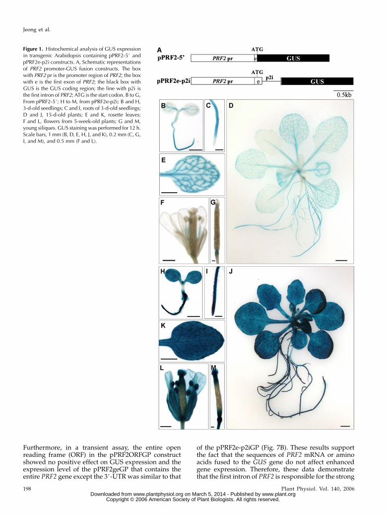
Furthermore, in a transient assay, the entire openreading frame (ORF) in the pPRF2ORFGP constructshowed no positive effect on GUS expression and theexpression level of the pPRF2geGP that contains theentire PRF2 gene except the 3#-UTR was similar to that
of the pPRF2e-p2iGP (Fig. 7B). These results supportthe fact that the sequences of PRF2 mRNA or aminoacids fused to the GUS gene do not affect enhancedgene expression. Therefore, these data demonstratethat the first intron of PRF2 is responsible for the strong
Figure 1. Histochemical analysis of GUS expressionin transgenic Arabidopsis containing pPRF2-5# andpPRF2e-p2i constructs. A, Schematic representationsof PRF2 promoter-GUS fusion constructs. The boxwith PRF2 pr is the promoter region of PRF2; the boxwith e is the first exon of PRF2; the black box withGUS is the GUS coding region; the line with p2i isthe first intron of PRF2; ATG is the start codon. B to G,From pPRF2-5#; H to M, from pPRF2e-p2i; B and H,3-d-old seedlings; C and I, roots of 3-d-old seedlings;D and J, 15-d-old plants; E and K, rosette leaves;F and L, flowers from 5-week-old plants; G and M,young siliques. GUS staining was performed for 12 h.Scale bars, 1 mm (B, D, E, H, J, and K), 0.2 mm (C, G,I, and M), and 0.5 mm (F and L).
Jeong et al.
198 Plant Physiol. Vol. 140, 2006 www.plant.org on March 5, 2014 - Published by www.plantphysiol.orgDownloaded from
Copyright © 2006 American Society of Plant Biologists. All rights reserved.

and constitutive expression of PRF2 throughout planttissues.
The First Intron of PRF2 Increases BothmRNA and Protein Levels
To investigate the basis for different expression ofpPRF2-5# and pPRF2e-p2i constructs, levels of mRNAand protein were analyzed. The accumulation of GUStranscripts was analyzed by reverse transcription (RT)-PCR analyses. One microgram of total RNA from roots,leaves, stems, and flowers was used for RT, and PCRwas performed for 25 cycles to prevent saturation ofPCR products. Sequencing of RT-PCR products ofpPRF2e-p2i indicated that splicing occurred precisely(data not shown). In all tissues, the transcript level oftransgenic plants carrying pPRF2e-p2i was increasedwhen compared to pPRF2-5# (Fig. 2), suggesting thatdifferent expression occurred at the transcription level.The increased mRNA levels were particularly apparentin the leaf and stem. To examine whether the increase intranscript level is fully reflected in the protein level,protein accumulation in the leaves was analyzed bywestern-blot analysis and GUS assay (Fig. 3). Twentymicrograms of total soluble proteins from the leaves of4-week-old plants were used for immunoblot analy-sis with the GUS antibody. Similar to the transcript ac-cumulation pattern, the levels of protein were muchhigher in pPRF2e-p2i than pPRF2-5# plants. Results offluorometric GUS assay indicated that pPRF2e-p2igene expression was enhanced by about 15-fold rela-tive to that of pPRF2-5# (Fig. 3). Thus, the increase intranscript accumulation induced by the first intron wasreflected in the GUS protein level. Therefore, strongexpression of pPRF2e-p2i is correlated with increasedaccumulation of the GUS transcript. These resultssuggest that the role of the first intron of PRF2 is closelyrelated to increasing steady-state mRNA levels.
The First Intron of PRF5 Has Little Effect onPRF5 Gene Expression
We demonstrated the important role of the first in-tron in gene expression of PRF2, the vegetative profi-lin. To determine whether the role of first introns is
conserved in a reproductive profilin, we examined thefirst intron of PRF5. About 1.5 kb of the promoterregion of PRF5 and the first exon were translationallyfused with the GUS reporter gene in a pPRF5e con-struct that tests promoter activity. To analyze theintron’s role, a pPRF5e-p5i construct, which includesall parts of pPRF5e as well as the first intron of PRF5with 24 bp of the second exon, was generated. Theschematic structures of pPRF5e and pPRF5e-p5i aredepicted in Figure 4A. Because we mainly focused onthe effect of the intron on the PRF5 spatial expressionpattern, we did not isolate the single-copy lines, andmore than 10 lines of T1 plants harboring pPRF5e andpPRF5e-p5i constructs were examined for the analysis.GUS histochemical analysis of transgenic plants har-boring pPRF5e showed strong expression in pollen.Staining was also observed in the stigmas and anthers.Very weak staining was detected in the upper part offilaments and petals (Table I; Fig. 4D). GUS expressionwas not detected in 3-d-old seedlings and 15-d-oldplants in most lines that were examined (Fig. 4, B andC). Although it is not clear whether GUS stainingin other floral organs is an actual expression or justdiffusion from pollen, GUS expression of pPRF5e waspredominant in pollen. A pollen-specific expressionpattern was in agreement with a previous report(Christensen et al., 1996). pPRF5e-p5i, which includesits own first intron within the construct, showedalmost identical expression with pPRF5e. GUS stain-ing was not observed in the vegetative tissues of nearlyall transgenic plants (Fig. 4, E and F). Like pPRF5e,GUS staining in flowers was mainly detected in thepollen and anthers (Table I; Fig. 4G). These resultssuggest that the first intron of PRF5 has no significanteffect on the expression pattern of the PRF5 gene.
The Role of Introns under the Control ofHeterogeneous Promoters
The analysis of transgenic plants carrying PRF2 andPRF5 promoter-GUS fusion constructs revealed thatthe first intron of PRF2 plays an important role in geneexpression, while the first intron of PRF5 has littleeffect. However, it is unclear whether only the intronitself is responsible for these differences. Other factors,
Figure 2. RT-PCR analysis of transcript expression of transgenic Arabidopsis containing pPRF2-5# and pPRF2e-p2i constructsin various tissues. One microgram of total RNA from each organ was used for RT. TUBULIN2 (TUB2) amplified with TUB-F(5#-CTCAAGAGGTTCTCAGCAGTA-3#) and TUB-R (5#-CTCAAGAGGTTCTCAGCAGTT-3#) primers was used as a control. Afterelectrophoresis of PCR products, it was transferred to the nylon membrane and hybridized with 32P-labeled GUS or TUB2probes.
Effect of the First Intron on Profilin Gene Expression
Plant Physiol. Vol. 140, 2006 199 www.plant.org on March 5, 2014 - Published by www.plantphysiol.orgDownloaded from
Copyright © 2006 American Society of Plant Biologists. All rights reserved.

such as interaction between intron and promoter,could be involved in profilin gene expression. Thus,to investigate the roles of introns in detail, introns wereexchanged between PRF2 and PRF5. For the construc-tion of pPRF2e-p5i, the first intron of PRF5 was in-serted at the 3# end of the first exon in the pPRF2econstruct. In the same way, the first intron of PRF2 wasintroduced into the pPRF5e construct, thus givingpPRF5e-p2i (Fig. 5A). For efficient splicing, both in-trons were accompanied with short flanking exons.The resulting constructs were transformed into Arabi-dopsis, and the expression pattern was analyzed inmore than 20 independent T1 plants. In 3-d-old seed-lings of pPRF2-p5i, expression was mainly observed incotyledons and roots (Fig. 5B). This vascular bundle-specific expression was also observed in leaves, pet-ioles, and roots of 15-d-old plants (Fig. 5C). In flowers,expression was observed in the filaments and sepals.No GUS staining was detected in pollen or in anthers(Table I; Fig. 5D). The overall expression patternsof pPRF2-p5i plants were very similar to those ofpPRF2-5# (Fig. 1, B–G). These data indicate that thefirst intron of PRF5 has no significant effect on theexpression of the PRF2 promoter. However, unlikethe first intron of PRF5, the first intron of PRF2 hadsignificant influence on the expression of the PRF5promoter. GUS histochemical analysis of pPRF5e-p2ishowed strong expression throughout the plant. Deepblue staining was observed in nearly every part of theplant, including root hairs and tips of 3-d-old seedlings(Fig. 5E). The expression pattern was totally different
from that of pPRF5e or pPRF5e-p5i (Table I; Fig. 4, B–G).This strong expression was also observed in 15-d-oldplants (Fig. 5F). Most of the floral organs, includingstigmas, anthers, pollen, filaments, petals, and sepals,also showed GUS expression (Table I; Fig. 5G). Thisconstitutive expression pattern was quite similar tothat of pPRF2e-p2i (Fig. 1, H–M). Thus, the propertiesof the two introns were maintained under heteroge-neous promoters, and this confirmed the functionaldistinctiveness of the two introns. Particularly, the firstintron of PRF2 had a significant effect on the expres-sion pattern of both the PRF2 and PRF5 promoters, andthis demonstrates that the first intron of PRF2 is re-sponsible for the constitutive expression of pPRF2e-p2iand pPRF5e-p2i.
The First Intron of PRF2 Alters the Spatial
Expression Pattern of PRF Genes
The expression of pPRF2-5# was mainly observed inthe vascular bundles of vegetative tissues. But faintGUS staining was detected in nonvascular tissues.In the pPRF2e-p2i plants, GUS staining is slightlystronger in the vascular tissues. In addition, increasedexpression induced by the intron was observed in theRT-PCR and western-blot analysis. Therefore, it ispossible that strong expression of pPRF2e-p2i couldbe a result of a quantitative increase in the express-ion pattern of pPRF2-5#. However, the result withpPRF5-p2i suggested that the intron alters the spatialexpression pattern of the PRF5 promoter. To determinethe role of the intron in detail, GUS staining wasperformed in 3-d-old seedlings at various incubationtimes (Fig. 6). In pPRF2-5#, GUS staining was observedto occur weakly in the vascular bundles of cotyledonsafter 1-h staining. This vascular expression patternwas maintained and staining intensity was increasedaccording to the staining time. After 12-h staining,weak staining was observed in the vascular bundlesand nearby tissues. In pPRF2e-p2i plants, no vascularpatterning was observed in the cotyledons after 1-hincubation. The overall parts of the cotyledons wereevenly stained. This pattern was maintained afterstaining for 4, 8, and 12 h. In the roots, a differentpattern was observed and maintained in both samplesfrom the beginning. If the intron just increased ex-pression, the early stage of the staining pattern ofpPRF2e-p2i would be similar to that of pPRF2-5#.Therefore, these results suggest that the intron doesnot just increase or enhance gene expression, but in-stead is also involved in regulating spatial gene expres-sion patterns.
The First Intron of PRF2 Affects Gene Expressionin a Position-Dependent Manner
The results above demonstrate that the first intron ofPRF2 enhances gene expression and can induce ec-topic expression under the control of a tissue-specific
Figure 3. Western-blot analysis and determination of GUS activity intransgenic Arabidopsis containing pPRF2-5# and pPRF2e-p2i. A, Totalsoluble protein (20 mg) from mature leaves was loaded per lane andanalyzed using monoclonal GUS antibody. B, GUS activity is ex-pressed as pmol 4-methyl-umbelliferone min21 mg21 protein. Valuesare means 6 SD (n 5 3).
Jeong et al.
200 Plant Physiol. Vol. 140, 2006 www.plant.org on March 5, 2014 - Published by www.plantphysiol.orgDownloaded from
Copyright © 2006 American Society of Plant Biologists. All rights reserved.

promoter. To determine whether controlling elementsexist within the intron, three intron deletion constructswere generated (Fig. 7A). The resulting constructswere transiently expressed in Arabidopsis leaf meso-phyll protoplasts, and normalized GUS activity withcotransfected luciferase activity was taken as a mea-sure of promoter activity. Among the three intron de-letions, the intron deletion d1 showed decreased GUSexpression when compared to the full-length intron(Fig. 7B). The intron deletion analysis indicates that theenhancing elements would exist within the intron.
To examine the possibility that the first intron ofPRF2 acts as an enhancer, the first intron of PRF2 wascloned upstream of the PRF2 promoter-GUS fusionconstruct in a forward or reverse direction (Fig. 7A).The resulting constructs were transiently expressed inArabidopsis leaf mesophyll protoplasts. Althoughboth pp2iF-PRF2eGP and pp2iR-PRF2eGP constructshave the intron upstream of the promoter region,their relative GUS activity was similar to that of thepPRF2eGP construct (Fig. 7B). This suggested that thefirst intron of PRF2 is not a classical enhancer. To fur-ther confirm this result, the intron was cloned upstreamof the cauliflower mosaic virus 35S minimal promoter-GUS fusion construct (Fig. 7A). There was no detect-
able enhancer activity under the control of the minimalpromoter (Fig. 7B). Therefore, these results demon-strate that the intron is not a classical transcriptionalenhancer and it enhances gene expression in a position-dependent manner.
Figure 4. Histochemical analysis of GUS expressionin transgenic Arabidopsis containing pPRF5e andpPRF5e-p5i constructs. A, Schematic representationsof PRF5 promoter-GUS fusion constructs. The graybox with PRF5 pr is the promoter region of PRF5; thegray box with e is the first exon of PRF5; the blackbox with GUS is theGUS coding region; the line withp5i is the first intron of PRF5; ATG is the start codon.B to D, From pPRF5e; E to G, from pPRF5e-p5i; B andE, 3-d-old seedlings; C and F, 15-d-old plants; D andG, flowers. Scale bars, 0.5 mm (B, D, E, and G), 1 mm(C and F), and 0.1 mm (anthers in small box).
Table I. Summary of expression patterns of PRF constructs inreproductive organs
GUS staining intensity in various tissues was indicated by a minus(2), negative or weakly detectable; plus (1), light blue; 11, blue;111, dark blue. The flower stalks were cut and incubated in GUSstaining solution for 12 h. The ratio of the number of transgenic plantsthat show GUS staining to the total number of independent transgenicplants is indicated below the staining intensity.
Construct Stigma Anther Filament Petal Sepal Cut Edge
pPRF2e 2 2 11 2 11 1
(0/23) (0/23) (21/23) (0/23) (16/23) (16/23)pPRF2e-p5i 2 2 11 2 1 2
(0/23) (0/23) (14/23) (0/23) (7/23) (2/23)pPRF5e 11 111 2 2 2 2
(12/12) (12/12) (2/12) (1/12) (0/12) (0/12)pPRF5e-p5i 11 111 2 2 2 2
(10/13) (13/13) (1/13) (1/13) (0/13) (0/13)pPRF5e-p2i 11 111 11 1 11 111
(15/24) (21/24) (19/24) (8/24) (15/24) (23/24)
Effect of the First Intron on Profilin Gene Expression
Plant Physiol. Vol. 140, 2006 201 www.plant.org on March 5, 2014 - Published by www.plantphysiol.orgDownloaded from
Copyright © 2006 American Society of Plant Biologists. All rights reserved.

The Role of the First Intron in the Profilin Gene Family
To examine whether the role of the first intronobserved in PRF2 and PRF5 gene expression is alsoapplicable to the entire Arabidopsis profilin genefamily members, the promoter-GUS fusion constructswith or without their first introns were generated andtheir expression levels were compared in the Arabi-dopsis protoplast transient expression system (Fig. 8).The promoter-GUS fusion constructs of two vegetativeprofilins, PRF1 and PRF2, showed comparable changesin the GUS expression level depending on the presence
of their own first intron. This suggests that the firstintrons of PRF1 and PRF2 have similar effects on geneexpression. On the contrary, the promoter-GUS fusionconstructs of two reproductive profilins, PRF4 andPRF5, showed a basal level GUS activity in leaf meso-phyll protoplasts regardless of the presence or absenceof their first intron. Because they are not expressedproperly in vegetative tissues, it is uncertain whethertheir introns enhance gene expression or not. Thus, therole of the first introns of PRF4 and PRF5 is not clear inthis system. Nonetheless, considering that the first
Figure 6. GUS staining patterns of transgenic Arabi-dopsis containing pPRF2-5# and pPRF2e-p2i con-structs at various incubation time points. Seedlingsgrown on Murashige and Skoog medium for 3 d wereincubated for 1, 4, 8, and 12 h, respectively, and thereaction was stopped and the tissues cleared byincubation with 100% ethanol.
Figure 5. Histochemical analysis of GUS expressionin transgenic Arabidopsis containing pPRF2e-p5i andpPRF5e-p2i. A, Schematic representation of pro-moter-GUS fusion constructs. The white box withPRF2 pr is the promoter region of PRF2; the white boxwith e is the first exon of PRF2; the line with p2i is thefirst intron of PRF2; the gray box with PRF5 pr is thepromoter region of PRF5; the gray box with e isthe first exon of PRF5; the line with p5i is the firstintron of PRF5; the black box with GUS is the GUScoding region; ATG is the start codon. B to D, FrompPRF2e-p5i; E to G, from pPRF5e-p2i; B and E,3-d-old seedlings; C and F, 15-d-old plants; D and G,flowers. Scale bars, 0.5 mm (B, D, E, and G), 1 mm(C and F), and 0.1 mm (anthers in small box).
Jeong et al.
202 Plant Physiol. Vol. 140, 2006 www.plant.org on March 5, 2014 - Published by www.plantphysiol.orgDownloaded from
Copyright © 2006 American Society of Plant Biologists. All rights reserved.

Figure 7. Transient expression analysis of various PRF2 and PRF5 promoter-GUS fusion constructs. A, Schematic representationof construction for transient expression. GPat the end of the construct name indicates a vector for transient expression. The whitebox with m is a 35S minimal promoter; the black box with GUS is the GUS ORF; the thin line with p2i is the first intron of PRF2;the white box with PRF2 pr is the PRF2 promoter region; the white box with e is the first exon of PRF2; ATG is the start codon;ORF is the ORF of PRF2 cDNA; the white boxes with e2 and e3 are the second and third exons of PRF2; the line with p2i-2 is the
Effect of the First Intron on Profilin Gene Expression
Plant Physiol. Vol. 140, 2006 203 www.plant.org on March 5, 2014 - Published by www.plantphysiol.orgDownloaded from
Copyright © 2006 American Society of Plant Biologists. All rights reserved.

intron of the PRF2 altered the expression pattern of thePRF5 promoter (Fig. 5, E–G), our results indicate thatthe first introns of reproductive profilin genes arefunctionally different from those of PRF1 and PRF2.Interestingly, although PRF3 encodes a vegetative-typeprofilin, the PRF3 promoter-GUS fusion construct wasexpressed at a basal level and a positive role of the firstintron was not detected (Fig. 8B).
DISCUSSION
Profilin is a small actin-binding protein that regu-lates actin cytoskeleton dynamics and plays a rolein cell elongation, cell shape, and flowering(Ramachandran et al., 2000). In Arabidopsis, thereare five genes encoding three vegetative (PRF1, PRF2,and PRF3) and two reproductive (PRF4 and PRF5)profilins. All five members are very similar in aminoacid sequences and genomic structures. All of theseprofilin genes are interrupted by two introns. The firstintron of vegetative profilins is located between aminoacids 41 and 42 (Gln and Leu) and that of reproductiveprofilins is located between amino acids 44 and 45 (Glnand Phe). Because PRF4 and PRF5 have three ad-ditional amino acids in the first exon, virtually allArabidopsis profilin genes contain first introns atidentical positions. In spite of conserved intron posi-tion, our results indicate that the roles of introns ingene expression are quite different between vegetative(PRF1 and PRF2) and reproductive (PRF4 and PRF5)profilin genes. GUS histochemical analyses of promoter-GUS fusions of PRF1 and PRF2 that encode majorvegetative profilins indicate that their expressionsare vascular bundle specific (Christensen et al., 1996;Ramachandran et al., 2000). These expression patternswere unexpected because vegetative profilin was as-sumed to show constitutive expression like vegetativeactins. In our research, the expression of PRF2 pro-moter-GUS fusions was nearly identical with previousresults. However, when the first intron was includedin the construct, the expression pattern was greatlychanged (Fig. 1). Strong and constitutive GUS stainingwas observed in most tissues and at diverse develop-mental stages. In the seedlings, expression was detectedin the cotyledons, shoot apical meristems, roots, roottips, and root hairs. In 15-d-old plants, rosette leaves,roots, root hairs, and trichomes were stained. In flow-ers, stigmas, anthers, filaments, petals, and sepalsshowed GUS expression. Different expression patternsof promoter-GUS fusion constructs, with or without
introns, were also evident in mRNA and proteinaccumulation. The presence of the intron resulted ina 15-fold increase in GUS activity in leaves (Figs. 2 and3). This constitutive expression pattern of pPRF2-p2i ismostly in good agreement, except in ovules with in situimmunolocalization of vegetative class profilins (PRF1,PRF2, and PRF3) in Arabidopsis (Kandasamy et al.,2002). Therefore, it is evident that expression of PRF2 isnot vascular bundle specific, and the first intron playsan important role in the strong and constitutive expres-sion of PRF2.
To further investigate the role of profilin introns, weexamined the first intron of the reproductive profilinPRF5. GUS staining with the pPRF5e construct wasmainly detected in pollen and anthers. But, unlike theresults obtained with the intron of PRF2, GUS expres-sion patterns of the pPRF5e and pPRF5e-p5i constructswere nearly identical (Fig. 4; Table I), indicating thatthe first intron of PRF5 has little effect on the geneexpression pattern. Although the spliceable intronwas placed downstream of the first exon of PRF2(pPRF2e-p5i), there was no significant alteration ofexpression pattern, confirming that the PRF5 intronhad little effect on the gene expression pattern.
The result of intron exchange between PRF2 andPRF5 suggests that the first intron of PRF2 does notjust enhance or increase the level of gene expression(Fig. 5). Both pPRF5e and pPRF5e-p5i showed repro-ductive tissue-specific expression. But, when the firstintron was replaced with the intron of PRF2, strongGUS expression was observed throughout the plantbody. This expression pattern of pPRF5e-p2i is quitesurprising and demonstrates that the PRF2 introncompletely changes tissue-specific expression of thePRF5 promoter. To determine whether this alsooccurred in the expression of PRF2, GUS stainingpatterns of plants harboring pPRF2-5# and pPRF2e-p2iconstructs were analyzed at various incubation times(Fig. 6). If the PRF2 intron just increased the expressionlevel, the expression pattern of pPRF2e-p2i in the earlystage of staining would be similar to that of pPRF2-5#.Plants of pPRF2e-p2i incubated for 1 h were stainedevenly in the cotyledons and roots, and the stainingwas obviously different from pPRF2-5# (Fig. 6). There-fore, these results suggest that the first intron of PRF2is involved in regulating spatial gene expression.
Because the two first introns examined in this studymaintained their own properties under the control ofboth the PRF2 and PRF5 promoters (Fig. 5), the dif-ferent roles of these introns seem to be the properties
Figure 7. (Continued.)second intron of PRF2; the gray box with PRF5 pr is the PRF5 promoter region; the gray boxes with e2 and e3 are the second andthird exons of PRF5; the lines with p5i and p5i-2 are the first and second introns of PRF5. Arrows indicate the orientation ofintron. The numbers in intron deletion constructs indicate the deletion points from the 5# end of the intron. B, Relativenormalized GUS activity in percentage. The full name of each construct is the addition of each insert name into the brackets ofthe vector name. For example, p[p2iF-]35SmGP is the pp2iF-35SmGP construct. Each transfection was performed at least threetimes. GUS activity was normalized with cotransfected luciferase activity and relative activity of pPRF2eGP was set to 100%.Bars are means 6 SD.
Jeong et al.
204 Plant Physiol. Vol. 140, 2006 www.plant.org on March 5, 2014 - Published by www.plantphysiol.orgDownloaded from
Copyright © 2006 American Society of Plant Biologists. All rights reserved.

Figure 8. Effect of the first intron on the expression of the Arabidopsis profilin gene family. A, Schematic representation ofconstruction for transient expression. GP at the end of the construct name indicates a vector for transient expression. The whitebox is vegetative genes; the gray box is reproductive genes; the white or gray box with e is the first exon of each gene; the blackbox with GUS is GUSORF; the thin lines with p1i, p2i, p3i, p4i, and p5i are the first introns of PRF1, PRF2, PRF3, PRF4 and PRF5,respectively; the white or gray box with gene name pr is the promoter region of each gene; ATG is the start codon. B, Relativenormalized GUS activity in percentage. A plus (1) indicates the presence of an intron within the construct; a minus (2) indicatesno intron. Each transfection was performed at least three times. GUS activity was normalized with cotransfected luciferaseactivity and relative activity of pPRF2eGP was set to 100%. Bars are means 6 SD.
Effect of the First Intron on Profilin Gene Expression
Plant Physiol. Vol. 140, 2006 205 www.plant.org on March 5, 2014 - Published by www.plantphysiol.orgDownloaded from
Copyright © 2006 American Society of Plant Biologists. All rights reserved.
of the introns themselves. Although sequences of in-trons are greatly variable in higher plants, AU richnessis common in plant introns and is required for efficientsplicing (Goodall and Filipowicz, 1989). Therefore, weanalyzed the sequences of our two introns to deter-mine whether there are significant differences in AUcontent. The AU content of the first intron was slightlylower in PRF2 (64.4%) than in PRF5 (67.7%), but bothwere similar to the average (67.5%) of Arabidopsisintrons (Table II). Because AU content of both intronsis above 60%, which is required for efficient splicing indicot plants (Goodall and Filipowicz, 1989), it is likelythat there is no significant difference in AU content.The most remarkable difference between two intronswas in the length of them. The first introns of PRF2 andPRF5 are 478 and 260 bp in length, and they were2.9 and 1.6 times longer than the average length ofArabidopsis introns (165.4 bp), respectively (Table II).Although the biological significance of long intronsis not certain at present, long introns may serve asbinding sites for various regulatory elements. Theresults of our intron deletion analysis suggest thatthe first intron of PRF2 may have the binding motifs orregulatory elements and these are to be specified infurther research.
Although the precise mechanism is not clear, the firstintron of PRF2 increased the corresponding mRNAlevel, and this was reflected in the protein level as well,as shown in Figures 2 and 3. In intron-mediated en-hancement, generally the enhancement is related toincreased steady-state mRNA accumulation (Calliset al., 1987; Rethmeier et al., 1997; Rose, 2002). Further-more, the first intron of PRF2 enhanced the gene ex-pression in a position-dependent manner and could notinduce the expression of the minimal promoter in atransient expression assay (Fig. 7). Therefore, it is sup-posed that the first intron of PRF2 increases gene ex-pression by the cotranscriptional or posttranscriptionalmechanisms as discussed in the earlier reports on intron-mediated enhancement (Jeon et al., 2000; Bourdon et al.,2001; Clancy and Hannah, 2002; Rose, 2002, 2004).
Among the five profilins of Arabidopsis, we mainlyexamined the role of the first introns on the expressionof PRF2 and PRF5 in transgenic plants. Genes encoding
vegetative (PRF1 and PRF2) and reproductive (PRF4and PRF5) profilin are highly similar to each other inmany aspects, such as DNA and amino acid sequences,expression patterns, and, especially, the length of thefirst intron (Table II). These high degrees of similarityseem to be closely related to the evolutionary history ofthe profilin gene family. In the Arabidopsis genome,genes encoding PRF1 and PRF5 are located in chromo-some 2, and PRF2 and PRF4 are located in chromosome4 where they are arranged in tandem arrays. Interest-ingly, both of the PRF1 and PRF2 genes encodingvegetative profilins are flanked with a gene encodinga reproductive profilin. Considering that about 60% ofthe Arabidopsis genome was duplicated (ArabidopsisGenome Initiative, 2000), this suggests that one set oftandem arrayed profilin genes was duplicated to givetwo sets. Therefore, we expected that the role of the firstintron on gene expression would also be similar. Asexpected, only the first introns of PRF1 and PRF2increased reporter activity, while the first introns ofPRF4 and PRF5 did not. These results indicate thatdifferent regulatory mechanisms by introns controlgene expression in vegetative (except PRF3) and re-productive profilins. Considering that PRF1 and PRF2are major vegetative profilins, our data demonstratethat their first introns are required for their strong andconstitutive expression in vegetative tissues. In thisrespect, the PRF3 gene is unique among profilin genesas shown in Table II. The PRF3 gene is not coupled withother profilin genes in the Arabidopsis genome, andthe length of the first intron was smaller than othervegetative profilins, PRF1 or PRF2. The total number ofPRF3 expressed sequence tags (ESTs) found in theNational Center for Biotechnology Information (NCBI)and The Institute for Genomic Research (TIGR) data-bases was only 25% of PRF1 or PRF2, indicating thatPRF3 is not expressed as much as PRF1 or PRF2. Ourresults indicating that PRF3 promoter-GUS fusionconstructs showed basal levels of expression in thetransient expression system also support the aboveexplanation (Fig. 8). At present, we do not knowwhether weak expression of PRF3 is due to loss ofintron-enhancing activity or significantly reduced pro-moter activity. Therefore, further examination of PRF3
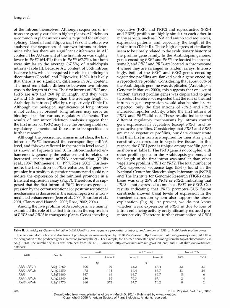
Table II. Arabidopsis Genome Initiative (AGI) identification, sequence properties of introns, and number of ESTs of Arabidopsis profilin genes
The genomic distribution and structures of profilin genes were analyzed by NCBI Map Viewer (http://www.ncbi.nlm.nih.gov/mapview/). AGI ID isthe designation of the predicted genes that were given by the AGI. For example, the 1,976th annotated gene counting from the top of chromosome 2 isAt2g19760. The number of ESTs was obtained from the NCBI Unigene (http://www.ncbi.nlm.nih.gov/UniGene) and TIGR (http://www.tigr.org)database.
Gene AGI IDLength AU Content No. of ESTs
Intron I Intron II Intron I Intron II NCBI TIGR
bp %
PRF1 (PFN1) At2g19760 506 92 63.2 67.4 22 23PRF2 (PFN2) At4g29350 478 111 64.4 66.7 23 24PRF3 At5g56600 367 66 68.7 69.7 5 6PRF4 (PFN3) At4g29340 256 85 70.3 67.1 2 4PRF5 (PFN4) At2g19770 260 575 67.7 70.2 0 2
Jeong et al.
206 Plant Physiol. Vol. 140, 2006 www.plant.org on March 5, 2014 - Published by www.plantphysiol.orgDownloaded from
Copyright © 2006 American Society of Plant Biologists. All rights reserved.

gene expression would provide additional clues forunderstanding the importance of introns on expressionof the profilin gene family.
Recently, the effect on gene expression of the leaderintron in the Arabidopsis ACT1 gene encoding re-productive actin was reported. ACT1 is expressedmost strongly in mature pollen, but it is also expressedin vegetative tissues, such as young vascular tissuesand root tips. The leader intron was found to berequired for the high-level expression of ACT1 in re-productive tissues. In addition, substituting its leaderintron with that of ACT2 into an ACT1 promoter-GUSfusion resulted in failure of expression in pollen (Vitaleet al., 2003). Although only two introns under thecontrol of a reproductive actin promoter were tested,at least these results suggest that introns play a role inactin gene expression. Therefore, it is possible that arole of introns in gene expression may have coevolvedin the cytoskeletal genes.
In this study, we have demonstrated that the firstintrons of PRF1 and PRF2 play an important role inconstitutive expression in most vegetative tissues andare functionally different from those of PRF4 andPRF5. And we have discussed the possible importanceof introns in the expression of profilin and actin genes.In future research, we will determine how the intron ofthe vegetative profilin genes affects gene expressionand also examine the role of introns in the expressionof various cytoskeletal genes.
MATERIALS AND METHODS
Plant Materials and Construction of Promoter-GUSFusions for Transgenic Plants
Arabidopsis (Arabidopsis thaliana) ecotype Columbia was grown in a growth
chamber (23�C, 16 h day/8 h night, 300 mE m22). For construction of PRF2
promoter fusions, the 1.6-kb promoter region, including up to a 5#-UTR and the
start codon (pPRF2-5#), the entire exon 1 (pPRF2e), and the first intron (pPRF2e-
p2i), was amplified using P2-F (5#-GTCGACAATGTTCCACCACCTAC-3#),P2-R (5#-GGATCCCATCTTTCTTCTTCTCC-3#), P2e-R (5#-AGGAAGAAAA-
ACGGATCCCTGAGGGAAAG-3#), and P2i-R (5#-GGATCCTGCTATCTCTGC-
AGG-3#) primers, respectively. The PCR products were cloned into the
pGEM-T easy vector (Promega) and confirmed by sequencing. The cloned
fragments were digested with SalI/BamHI and ligated into the pBI101 vector
(CLONTECH). Similar methods were used for construction of PRF5 promoter
fusions, pPRF5e and pPRF5e-p5i, using PRF5-specific primers: P5-F
(5#-AAGCTTCTGGTCCTGTATTTGCCTAACCAAG-3#), P5e-R (5#-GGATC-
CCTGAGGAAAATTAGCGCTCTGAG-3#), and P5i-R (5#-GGATCCTGTGAT-
CTCTTGAGGTTTGAAC-3#). For intron exchange experiments, introns were
amplified with P2int-F (5#-GGATCCGCTTTCCCTCAGGTTTTTCTTC-3#) and
P2i-R for the first intron of PRF2 and with P5int-F (5#-GGATCCAATT-
TTCCTCAGGTATAATTAC-3#) and P5i-R for the intron of PRF5. Amplified
introns were inserted into the BamHI site of pPRF2e or pPRF5e. The resulting
constructs were inserted into Agrobacterium tumefaciens strain C58C1Rif1.
Plant Transformation and GUS Histochemical Analysis
Transgenic Arabidopsis was generated by the floral-dip method (Clough
and Bent, 1998). Transformed plants were selected on 0.53 Murashige and
Skoog 0.8% (w/v) agar plates containing 50 mg L21 kanamycin and 100 mg L21
cefotaxim. For analysis of pPRF2-5# and pPRF2e-p2i lines, T1 transgenic plants
were self-pollinated, and single-copy lines were selected by Southern-blot
analysis and tested for the segregation ratio of T2 seeds on agar plates
containing kanamycin. The selected single home lines were used for further
analysis. Histochemical and fluorometric analysis of GUS activity was per-
formed as described previously (Mun et al., 2002). Images were obtained using
either Nikon (Tokyo) Coolpix 4500 or Stemi SV11 (Zeiss) with photomatrix
Coolsnap cf digital camera (Photomatrix).
RT-PCR Analysis
To investigate the expression patterns of the GUS transcripts in transgenic
Arabidopsis, RT-PCR analyses were performed. Total RNA was purified from
leaves of mature plants by Tri-Reagent (Molecular Research Center). One
microgram of total RNA was used for RT in a volume of 25 mL and incubated
at 42�C for 1 h with avian myeloblastosis virus reverse transcriptase and
oligo(dT) primer (Promega), and inactivated by incubating for 5 min at 95�C.
After completion of RT, 2 mL of RT products were amplified using P2RT-F
(5#-AAACAGTCTCATCTCGCCGGAGAAG-3#) and GUSRT-R (5#-AAAGA-
CTTCGCGCTGATACCAGAC-3#) primers. PCR was performed using Bio-
therm DNA polymerase (Genecraft) in a GeneAmp PCR system 9700
(Perkin-Elmer). The PCR condition was 5 min at 95�C, followed by 25 cycles
of 30 s at 94�C, 30 s at 55�C, and 30 s at 72�C, followed by 7 min at 72�C. The
PCR products were run on agarose gel, transferred to Hybond XL membranes
(Amersham), and hybridized. Probes were generated from GUS ORF from
pBI101 and radiolabeled with [a-32P]dCTP using the Rediprime II random
prime system (Amersham). After high stringent washing, blots were exposed
to the film. To confirm that correct splicing had occurred, RT-PCR products
were cloned into the pGEM-T easy vector and sequenced using the ABI 3100
sequence analyzer (Applied Biosystems).
Western-Blot Analysis
Monoclonal GUS antibody (Molecular Probes) was used at a titer of 1:5,000
for western-blot analysis. Twenty micrograms of total soluble proteins from
leaves were loaded per lane. Western-blot analysis was performed as de-
scribed previously (Mun et al., 2000).
Sequence Analysis of Arabidopsis Profilin Genes
The genomic sequences of five profilin genes in Arabidopsis were obtained
from Plant Genome Central at the NCBI (http://www.ncbi.nlm.nih.gov/
genomes/PLANTS/PlantList.html). The total numbers of ESTs cloned were
obtained from Unigene (http://www.ncbi.nlm.nih.gov/UniGene/) and TIGR
(http://www.tigr.org). The average length and AU content of the Arabidopsis
intron was calculated based on whole intron sequences obtained from The
Arabidopsis Information Resource (TAIR; ftp://ftp.arabidopsis.org/home/
tair/Sequences/blast_datasets/At_intron_20040301).
Transient Expression Analysis in Arabidopsis LeafMesophyll Protoplasts
For transient gene expression, the HindIII and EcoRI fragment from pBI101,
which includes the GUS gene, was inserted into pUC19 and named as a pGP.
The minimal promoter region of pBI121 was amplified with 35sm-F
(5#-CTGCAGTCGACGCAAGACCCTTCCTCTATA-3#) and GUSRT-R primers
and the PstI/BamHI fragment was ligated into pGP to generate p35smGP. For
construction of pp2iF-35smGP and pp2iR-35smGP, the first intron of PRF2,
which was amplified with P2int-sal-F (5#-GTCGACGCTTTCCCTCAGGTTTT-
TCTTC-3#) and P2int-sal-R (5#-GTCGACTGCTATCTCTGCAGGCTTCAA-3#)primers, was inserted into the SalI site of p35smGP. The SalI/BamHI fragments
of PRF2 and PRF5 promoter-GUS fusion constructs in pBI101 were cloned into
pGP (Fig. 8A). For construction of pp2iF-PRF2eGP and pp2iR-PRF2eGP, the
first intron of PRF2 was inserted into the SalI site of pPRF2eGP. To make
pP2ORFGP, the SalI/BamHI fragment of the pPRF2-5# construct was ligated
into pGP, thus generating pPRF2-5#GP, and the ORF of the PRF2 gene was
amplified with P2orf-F (5#-GGATCCATGTCGTGGCAATCATACGTC-3#) and
P2orf-R (5#-GGATCCGAGACCAGACTCGATAAGGTAATC-3#) primers us-
ing cDNA made from total RNA and inserted into the BamHI site of pPRF2-
5#GP. The genomic region was amplified with P2-F and P2orf-R for pPRF2geGP
and P5-F and P5orf-R (5#-GGATCCAAGACCCTGTTCGATCAAGTAATC-3#)for pPRF5geGP and ligated into pGP vector. For construction of intron dele-
tion constructs, P2id1-R (5#-TTATTTGAGACAGAGGATCGGAAGAAAC-3#),
Effect of the First Intron on Profilin Gene Expression
Plant Physiol. Vol. 140, 2006 207 www.plant.org on March 5, 2014 - Published by www.plantphysiol.orgDownloaded from
Copyright © 2006 American Society of Plant Biologists. All rights reserved.
P2id1-F (5#-AGTCGGTTGAAGCTAATTGCCTACTTTG-3#), p2id2-R (5#-ACA-
CCTATAGACAAGATTTGACTACTG-3#), and p2id3-F (5#-TTTGCTTGG-
CACAGAATCTTATCTCC-3#) were used. Schematic representation of these
transient vectors is depicted in Figure 7A. For construction of PRF1, PRF4, and
PRF3 promoter-GUS fusions, genomic fragments were amplified with the
following gene-specific primers: for PRF1, P1-F (5#-AAGCTTCATATACATAC-
CAAAGCCATCATGGA-3#), P1e-R (5#-GGATCCCTGAGGAAATTTGGCGC-
TCTGAGC-3#), and P1i-R (5#-GGATCCATCGATTTCTTGAGGCTTCAAC-3#);for PRF3, P3-F (5#-AAGCTTCCCATAGAAAGCAATGTATATGCTC-3#), P3e-R
(5#-GGATCCCTGAGGAAAATTGTTGCTCTGAGCC-3#), and P3i-R (5#-GGA-
TCCCTGAATTTCCTCAGGCTTCACCTA-3#); and for PRF4, P4-F (5#-AAG-
CTTTTATCAGTCATACCATTTTTCTGGAC-3#), P4e-R (5#-GGATCCCTGAG-
GGAAGTTGGCGCTCTGAGCC-3#), and P4i-R (5#-GGATCCACTGAACTCC-
TGTCCCTTGAACTATATG-3#). They were inserted into the HindIII/BamHI
sites of pGP (Fig. 8A). For construction of p35SLucP, the internal control for
transient expression analysis, the 35S promoter region from pBI121 and the gene
encoding a firefly luciferase amplified with Luc-F (5#-GGATCCATGGAA-
GACGCCAAAAACATAAAG-3#) and Luc-R (5#-GAGCTCTTACACGGC-
GATCTTTCCGCCCT-3#) primers using pGL3-promoter vector (Promega)
were introduced into the HindIII/SacI sites of the pGP vector.
Arabidopsis mesophyll protoplasts from 3-week-old plants were trans-
fected by the polyethylene glycol method (Sheen, 2002). Ten micrograms of
plasmid DNA containing promoter-GUS fusion constructs were cotransfected
with 10 mg of p35SLucP as an internal control. After transfection, protoplasts
were incubated for 24 h at 24�C in the dark. Proteins were extracted by 13 cell
culture lysis reagent buffer (Promega) and used for the GUS fluorometric and
luciferase assay. Luciferase assay was performed according to the manufac-
turer’s instructions (Promega) using the Victor2 multilabel microplate reader
(Perkin-Elmer).
ACKNOWLEDGMENTS
The authors wish to express sincere thanks to Prof. Donald J. Armstrong
(Oregon State University) for helpful discussions and critical review. We
would also like to thank Hyung-Seok Choi for help with analyzing sequences
of the Arabidopsis whole intron.
Received September 21, 2005; revised November 9, 2005; accepted November
10, 2005; published December 16, 2005.
LITERATURE CITED
Arabidopsis Genome Initiative (2000) Analysis of the genome sequence
of the flowering plant Arabidopsis thaliana. Nature 408: 796–815
Baluska F, Jasik J, Edelmann HG, Salajova T, Volkmann D (2001)
Latrunculin B induced plant dwarfism: plant cell elongation is F-actin
dependent. Dev Biol 231: 113–124
Bamburg JR, McGough A, Ono S (1999) Putting a new twist on actin:
ADF/cofilins modulate actin dynamics. Trends Cell Biol 9: 364–370
Bolle C, Herrmann RG, Oelmuller R (1996) Intron sequences are involved
in the plastid- and light-dependent expression of the spinach PsaD gene.
Plant J 10: 919–924
Bourdon V, Harvey A, Lonsdale DM (2001) Introns and their positions
affect the translational activity of mRNA in plant cells. EMBO Rep 2:
394–398
Callis J, Fromm M, Walbot D (1987) Introns increase gene expression in
cultured maize cells. Genes Dev 1: 1183–1200
Christensen HEM, Ramachandran S, Tan CT, Surana U, Dong CH, Chua
NH (1996) Arabidopsis profilins are functionally similar to yeast
profilins: identification of a vascular bundle-specific profilin and a
pollen-specific profilin. Plant J 10: 269–279
Clancy M, Hannah LC (2002) Splicing of the maize Sh1 first intron is
essential for enhancement of gene expression, and a T-rich motif
increases expression without affecting splicing. Plant Physiol 130:
918–929
Clough SJ, Bent AF (1998) Floral dip: a simplified method for Agro-
bacterium-mediated transformation of Arabidopsis thaliana. Plant J 16:
735–743
Deyholos MK, Sieburth LE (2000) Separable whorl-specific expression and
negative regulation by enhancer element within the AGAMOUS second
intron. Plant Cell 12: 1799–1810
Fu H, Kim SY, Park WD (1995) A potato Sus3 sucrose synthase
gene contains a context-dependent 3# element and a leader intron
with both positive and negative tissue-specific effects. Plant Cell 7:
1395–1403
Gibbon BC, Staiger CJ (2000) Profilin. In CJ Staiger, F Baluska, D
Volkmann, P Barlow, eds, Actin: A Dynamic Framework for Multiple
Plant Cell Functions. Kluwer Academic Publishers, Dordrecht, The
Netherlands, pp 45–65
Goodall GJ, Filipowicz W (1989) The AU-rich sequences present in the
introns of plant nuclear pre-mRNAs are required for splicing. Cell 58:
473–483
Guillen G, Valdes-Lpoez V, Noguez R, Oliveares J, Rodriguez-Zapata LC,
Perez H, Viali L, Villanueva MA, Sanchez F (1999) Profilin in Phaseolus
vulgaris is encoded by two genes (only one expressed in root nodules)
but multiple isoforms are generated in vivo by phosphorylation on
tyrosine residues. Plant J 19: 497–508
Huang S, McDowell JM, Weise MJ, Meagher RB (1996) The Arabidopsis
profilin gene family: evidence for an ancient split between constitutive
and pollen-specific profilin genes. Plant Physiol 111: 115–126
Jeon JS, Lee S, Jung KH, Jun SH, Kim C, An G (2000) Tissue-preferential
expression of a rice a-tubulin gene, OsTubA1, mediated by the first
intron. Plant Physiol 123: 1005–1014
Kandasamy MK, McKinney EC, Meagher RB (2002) Plant profilin isovar-
iants are distinctly regulated in vegetative and reproductive tissues. Cell
Motil Cytoskeleton 52: 22–32
Kost B, Mathur J, Chua NH (1999) Cytoskeleton in plant development.
Curr Opin Plant Biol 2: 462–467
Kovar DR, Drøbak BK, Staiger CJ (2000) Maize profilin isoforms are
functionally distinct. Plant Cell 12: 583–598
McKinney EC, Kandasamy MK, Meagher RB (2001) Small changes in the
regulation of one Arabidopsis profilin isovariant, PRF1, alter seedling
development. Plant Cell 13: 1179–1191
Meagher RB, McKinney EC, Vitale AV (1999) The evolution of new
structures: clues from plant cytoskeletal genes. Trends Genet 15:
278–284
Mittermann I, Swoboda I, Pierson E, Eller N, Kraft D, Valenta R, Heberle-
Bors E (1995) Molecular cloning and characterization of profilin from
tobacco (Nicotiana tabacum): increased profilin expression during pollen
maturation. Plant Mol Biol 27: 137–146
Mun JH, Lee SY, Yu HJ, Jeong YM, Shin MY, Kim H, Lee I, Kim SG (2002)
Petunia actin depolymerizing factor is mainly accumulated in vascular
tissue and its gene expression is enhanced by the first intron. Gene 292:
233–243
Mun JH, Yu HJ, Lee HS, Kwon YM, Lee JS, Lee I, Kim SG (2000)
Two closely related cDNAs encoding actin-depolymerizing factors of
petunia are mainly expressed in vegetative tissues. Gene 257:
167–176
Plesse B, Criqui MC, Durr A, Parmentier Y, Fleck J, Genschik P (2001)
Effects of the polyubiquitin gene Ubi. U4 leader intron and first
ubiquitin monomer on reporter gene expression in Nicotiana tabacum.
Plant Mol Biol 45: 655–667
Ramachandran S, Christensen HE, Ishimaru Y, Dong CH, Chao-Ming W,
Cleary AL, Chua NH (2000) Profilin plays a role in cell elongation, cell
shape maintenance, and flowering in Arabidopsis. Plant Physiol 124:
1637–1647
Rethmeier N, Kramer E, Van Montagu M, Cornelissen M (1997) Intron-
mediated enhancement of transgene expression in maize is a nuclear,
gene-dependent process. Plant J 12: 895–899
Rose AB (2002) Requirements for intron-mediated enhancement of gene
expression in Arabidopsis. RNA 8: 1444–1453
Rose AB (2004) The effect of intron location on intron-mediated enhance-
ment of gene expression in Arabidopsis. Plant J 40: 744–751
Rose AB, Last RL (1997) Introns act post-transcriptionally to increase
expression of the Arabidopsis thaliana tryptophan pathway gene PAT1.
Plant J 11: 455–464
Schobert C, Gottschalk M, Kovar DR, Staiger CJ, Yoo BC, Lucas WJ (2000)
Characterization of Ricinus communis phloem profilin, RcPRO1. Plant
Mol Biol 42: 719–730
Sheen J (2002) A transient expression assay using Arabidopsis mesophyll
protoplasts. http://genetics.mgh.harvard.edu/sheenweb/ (September
15, 2004)
Sheldon CC, Conn AB, Dennis ES, Peacock WJ (2002) Different regu-
latory regions are required for the vernalization-induced repression of
Jeong et al.
208 Plant Physiol. Vol. 140, 2006 www.plant.org on March 5, 2014 - Published by www.plantphysiol.orgDownloaded from
Copyright © 2006 American Society of Plant Biologists. All rights reserved.

FLOWERING LOCUS C and for the epigenetic maintenance of repres-
sion. Plant Cell 14: 2527–2537
Staiger CJ, Goodbody KC, Hussey PJ, Valenta R, Drøbak BK, Lloyd CW
(1993) The profilin multigene family of maize: differential expression of
three isoforms. Plant J 4: 631–641
Valenta R, DucheneM, Pettenburger K, Sillaber C, Valent P, Bettelheim P,
Breitenbach M, Rumpold H, Kraft D, Scheiner O (1991) Identification
of profilin as a novel pollen allergen; IgE autoreactivity in sensitized
individuals. Science 253: 557–560
Valenta R, Ferreira F, Grote M, Swoboda I, Vrtala S, Duchene M, Deviller
P, Meagher RB, McKinney E, Heberle-Bors E, et al (1993) Identification
of profilin as an actin-binding protein in higher plants. J Biol Chem 268:
22777–22781
Vidali L, Perez HE, Lopez VV, Noguez R, Zamudio F, Sanchez F (1995)
Purification, characterization, and cDNA cloning of profilin from
Phaseolus vulgaris. Plant Physiol 108: 115–123
Vitale A, Wu RJ, Cheng Z, Meagher RB (2003) Multiple conserved 5#elements are required for high-level pollen expression of the Arabidop-
sis reproductive actin ACT1. Plant Mol Biol 52: 1135–1151
Yu LX, Nasrallah J, Valenta R, Parthasarathy MV (1998) Molecular cloning
and mRNA localization of tomato pollen profilin. Plant Mol Biol 36:
699–707
Effect of the First Intron on Profilin Gene Expression
Plant Physiol. Vol. 140, 2006 209 www.plant.org on March 5, 2014 - Published by www.plantphysiol.orgDownloaded from
Copyright © 2006 American Society of Plant Biologists. All rights reserved.