Static, Yet Fluctuating: The Evolution of Batman and His ...
Upload
independentCategory
view
0download
0
Dissection of the sequence-specific DNA binding andexonuclease activities reveals a superactive yet apoptoticallyimpaired mutant p53 protein
Jinwoo Ahn1, Masha V. Poyurovsky5, Nicole Baptiste5, Rachel Beckerman5, ChristineCain5, Melissa Mattia2, Kristine McKinney3, Jianmin Zhou5, Andrew Zupnick5, VanesaGottifredi4, and Carol Prives5,*1Department of Structural Biology; University of Pittsburgh School of Medicine; Pittsburgh, PA USA2Department of Oncological Sciences; Mount Sinai School of Medicine; New York, NY USA3Dana-Farber Cancer Institute; Harvard Medical School; Boston, MA USA4Cell Cycle and Genomic Stability Laboratory; Fundación Instituto Leloir-CONICET; Universidad deBuenos Aires; Buenos Aires, Argentina5Department of Biological Sciences; Columbia University; New York, NY USA
AbstractBoth sequence-specific DNA binding and exonuclease activities have been mapped to the centralconserved core domain of p53. To gain more information about these two activities a series of mutantswere generated that changed core domain histidine residues. Of these mutants, only one, H115N p53,showed markedly reduced exonuclease activity (ca. 15% of wild-type). Surprisingly, purified H115Np53 protein was found to be significantly more potent than wild-type p53 in binding to DNA byseveral criteria including gel mobility shift assay, filter binding and DNase I footprinting.Interestingly as well, non-specific DNA binding by the core domain of H115N p53 is superior to thatof wild-type p53. To study H115N p53 in vivo, clones of H1299 cells expressing tetracyclineregulated wild-type or H115N p53 were generated. H115N was both more potent than wild-type p53in inducing p53 target genes such as p21 and PIG3 and was also more effective in arresting cells inG1. Unexpectedly, in contrast to wild-type p53, H115N p53 was markedly impaired in causingapoptosis when cells were subjected to DNA damage. Our results indicate that the exonucleaseactivity and transcriptional activation functions of p53 can be separated. They also extend previousfindings showing that cell cycle arrest and apoptosis are separable functions of p53. Finally, theseexperiments confirm that DNA binding and exonuclease activities are distinct features of the p53core domain.
Keywordsp53; apoptosis; exonuclease activity; DNA binding; transcription; cell cycle
©2009 Landes Bioscience*Correspondence to: Carol Prives; Department of Biological Sciences; Columbia University; New York, NY 10027 USA; Tel.:212.854.2557; Fax: 212.865.8246; [email protected].
NIH Public AccessAuthor ManuscriptCell Cycle. Author manuscript; available in PMC 2010 July 7.
Published in final edited form as:Cell Cycle. 2009 May 15; 8(10): 1603–1615.
NIH
-PA Author Manuscript
NIH
-PA Author Manuscript
NIH
-PA Author Manuscript

IntroductionThe p53 tumor suppressor protein is central to the DNA damage signal response pathway thatresults in cell cycle arrest or apoptosis.1–4 Upon DNA damage to cells, p53 proteinaccumulates and becomes activated by post-transcriptional modifications followed byinduction of its down-stream target genes involved in eliciting those two responses.5–7 To datehundreds of p53 target genes have been identified either individually or as the result of differentgene expression screens. More than 50% of human cancer cells contain missense mutant formsof p53 protein underlying the key roles of this protein in tumor suppression. The great majority(97%) of mutations in such tumor derived p53 variants are located within its central coredomain, a region that is highly conserved in p53 proteins among different species and also inrelated p53 family members, p63 and p73 (The database is available athttp://www.iarc.fr/P53). The core domain binds specifically to DNA containing 2 closelyspaced copies of the p53 consensus sequence 5'-RRRCATGYYY-3' and tumor-derivedmutation in this region usually leads to defective DNA binding reviewed in 8 and 9. p53 is thusa sequence-specific transcriptional regulator of genes that facilitate the stress response circuitryin cells.
In addition to the regulation of genes involved in cell cycle and apoptosis, p53 has also beenimplicated in DNA repair. First, p53 can affect DNA repair processes through its ability totransactivate genes involved in these processes such as p48,10 GADD45,11 and R2.12 Second,a number of studies indicate that p53 can directly participate in DNA repair processes such asnucleotide excision repair (NER)1 or base excision repair (BER) as well as regulaterecombination.13–15 Several features of the p53 protein support its possible roles in DNArepair. p53 can directly interact with DNA repair related cellular factors including thetranscription factor IIH (TFIIH) protein complex,16 single-stranded DNA binding proteinRPA,17 DNA polymerase beta,18 AP endonuclease (APE),19 Rad 51,20,21 and mammalianhomologs of the RecQ helicase family, BLN22,23 and Wrn proteins.23,24 Moreover, the p53C-terminal domain can interact with various forms of damaged DNA in a sequence independentmanner.25–29 Finally, the central core domain contains an intrinsic 3'→5' exonuclease activity.30–35
It is intriguing that the central core domain contains two independent biochemical activitiesi.e., sequence-specific DNA binding and non-sequence-specific 3'→5' exonuclease activity.This poses the question as to whether sequence specific DNA binding and exonucleaseactivities reflect similar properties of the core domain (e.g., the ability to bind tightly to DNA)or whether they are separable. Some tumor derived hot-spot mutants are defective in bothactivities30 although it was later reported that these two activities are differentially regulatedeither by interaction with an antibody recognizing the C-terminus of p53 or post-translationalmodification of the p53 C-terminus.31 We initiated this project to determine whether the DNAbinding surface of the core domain and the exonuclease active site may be separable. SinceHis residues are frequently located within the active site of exonucleases (see Discussion formore details), we generated seven His/Asn point mutants within the core domain and comparedtheir sequence-specific DNA binding and 3'→5' exonuclease activity with wild-type p53. Ourresults showed that one of these mutants H115N was unique in being severely impaired inexonuclease activity. Remarkably, however, H115N p53 was more active than wild-type p53in binding to DNA in vitro. Further characterization of H115N revealed that, when expressedat physiological levels in vivo, this mutant produces different effects in cells than does wild-type p53. Although H115N p53 can induce cell cycle arrest more efficiently than wild-typep53, its ability to produce apoptosis in DNA damaged cells is markedly impaired. We discussthe significance of these observations.
Ahn et al. Page 2
Cell Cycle. Author manuscript; available in PMC 2010 July 7.
NIH
-PA Author Manuscript
NIH
-PA Author Manuscript
NIH
-PA Author Manuscript
ResultsComparison of exonuclease and sequence-specific DNA binding activity of wild type andseven His/Asn mutants
To confirm and extend previous observations that p53 possesses an intrinsic exonucleaseactivity, we compared the activity of bacterially expressed p53 that has been purified throughthree chromatographic columns with that of commercially obtained bacterial exonuclease III.The substrate was a heteroduplex consisting of a 5' end-labeled 25-mer annealed to a 5' end-labeled 45-mer. As shown in Figure 1A, the exonuclease activity of p53 protein (0.5 to 1.0pmol) was comparable to that of 1 × 10⁻⁴ to 1 ×10⁻⁵ pmol of exonuclease III. Therefore, underthese conditions, p53 displayed far weaker exonuclease activity than exonuclease III. In eachcase the 25 base oligonucleotide was significantly more susceptible to digestion than the 45base olignucleotide suggesting that both exonucleases prefer recessive 3' ends as substrates.
Since His residues are located within the active sites of other known exonucleases (seeDiscussion section) we generated seven His/Asn point mutants (relative positions shown inFig. 1B below diagram). Histidine mutants were restricted to those within the p53 core domain(residues 100–300) since the exonuclease activity of p53 was previously localized to thisdomain.30 The two remaining core histidine residues (His178 and His296) were not mutatedto Asn since each was adjacent to another histidine residue (His179 or His297) that was chosento be mutated. Wild-type and seven His mutant p53 proteins were purified from E. coli usingNi-NTA agarose column chromatography and then analyzed by SDS-PAGE (Fig. 1C). Theexonuclease activities of these proteins were then measured by an immuno-exonuclease assayas described in Experimental Procedures. The enzymatic activities of mutant p53 proteins werecompared to that of wild-type p53 measured by this assay (Table 1). Exonuclease activities ofH168N, H179N and H297N p53 proteins were approximately comparable to that of wild-typep53, while H193N, H214N and H233N mutant p53 proteins actually showed significantlyincreased exonuclease activity. Only one mutant, H115N p53, showed just 15% of wild-typeexonuclease activity in this assay. The reduced exonuclease activity of H115N p53 relative towild-type p53 was confirmed when these proteins were extensively purified through threedifferent columns as described in Experimental Procedures (Fig. 1D) and then tested as solubleproteins (Fig. 1E).
We then examined the sequence-specific DNA binding activity of the seven His/Asn mutants(H115N, H168N, H179N, H214N, H233N and H297N) as well as wild-type p53 by purifyingeach protein and performing gel shift assays using a [32P] labeled oligo-nucleotide duplexcontaining the GADD45 p53 binding site. The results of this experiment are also summarizedin Table 1. While mutation of p53 to H168N, H233N and H297N resulted in little change inDNA binding activity when compared to wild type p53, the H179N, H193N and H214Nmutants each had significantly reduced DNA binding activity. Interestingly, however, H115Nshowed markedly enhanced DNA binding activity when compared to wild-type p53 protein.Therefore the reduced exonuclease enzymatic activity of H115N is not the result of global orpartial unfolding as is the case with some of the hot-spot conformational p53 mutants such asR273H p53.42 Our results with these p53 His substitution mutants thus indicate that the DNAbinding and exonuclease activities of the p53 core domain are genetically separable.
H115N is more active than wild type in sequence-specific DNA binding as measured byseveral different assays
Since many tumor derived mutations located within the core domain of p53 disable sequencespecific DNA binding it was of interest that H115N p53 showed superior binding than wild-type p53. Nevertheless, results with one type of assay for p53 DNA binding are not alwaysconfirmed when a different assay is used.43 For example, the p53 monoclonal antibody PAb421
Ahn et al. Page 3
Cell Cycle. Author manuscript; available in PMC 2010 July 7.
NIH
-PA Author Manuscript
NIH
-PA Author Manuscript
NIH
-PA Author Manuscript
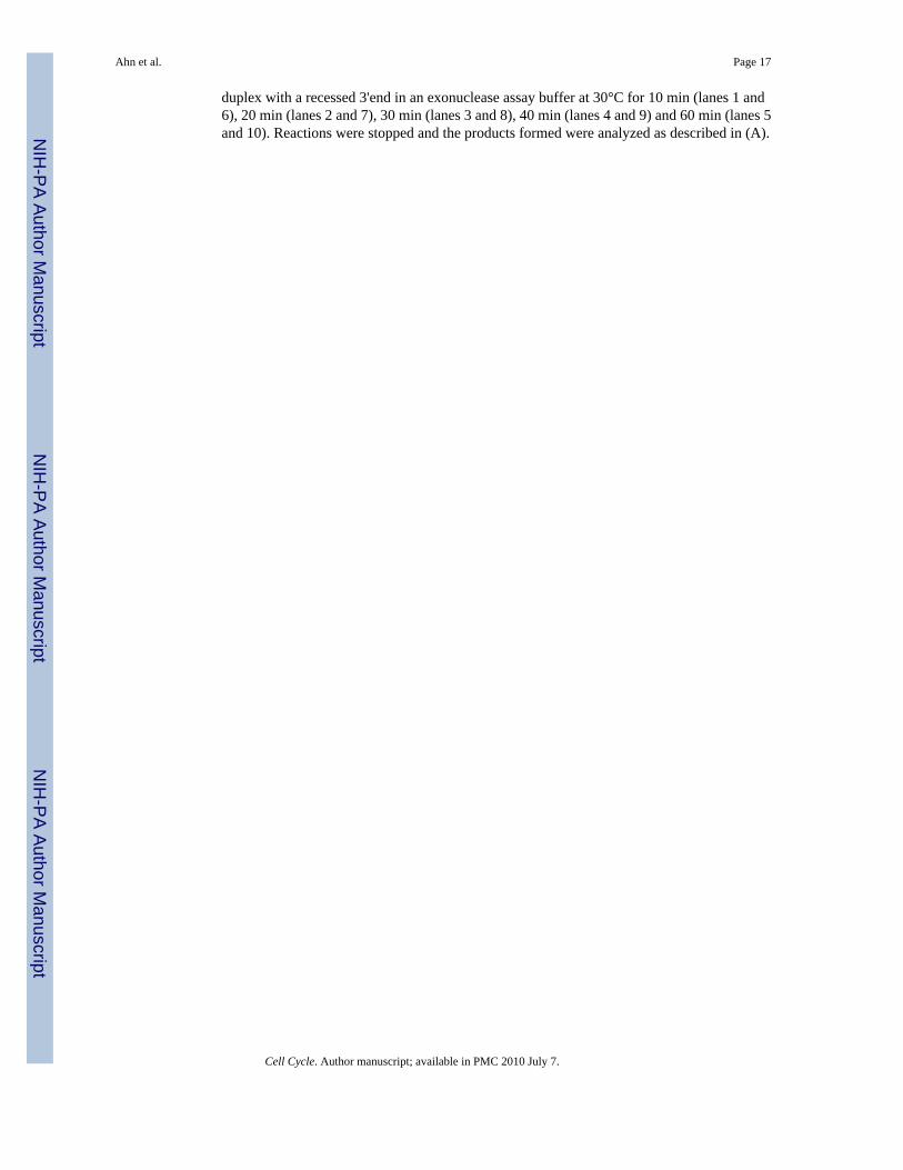
stimulates p53 binding to a DNA fragment when assessed by gel mobility shift assays usingshort oligonucleotides44–46 but not by DNase I footprinting40 and acetylation of p53 C-terminalresidues was reported to stimulate p53 binding to short but not long DNA.47 Accordingly, wewished to determine whether or not the superior DNA binding by H115N p53 could beconfirmed by different assays measuring protein-DNA interactions (Fig. 2). To ensure thatcomparable amounts of proteins were used in the different assays, the amounts of wild typeand H115N mutant proteins used in the assays were assessed by immunoblotting purifiedproteins using a mixture of three antibodies DO-1, 1801 and 421 (Fig. 2A). Consistent withthe results summarized in Table 1 as measured by EMSA, the purified H115N mutant p53 wasmore potent than wild-type p53 in binding to a labeled 32 base pair oligonucleotide containingthe p53 binding within the p21 promoter over a range of protein concentrations (0.2–0.6 ng/µL; Fig. 2B). Note that this assay was performed using p53 bound to the C-terminal antibodyPAb 421 that greatly stimulates p53 DNA binding under these conditions. This suggests thatincreased binding by H115N does not involve relief of repression by the C-terminus.Additionally, as measured by a filter binding assay, H115N p53 bound the same [32P] labeledp21 site containing 32-mer oligonucleotide 2–3 fold better than wild-type p53 in the absenceof PAb 421 (Fig. 2C). Furthermore, after performing a DNase I footprinting assay with a 405base pair DNA fragment from the p21 promoter containing the 5' p53 binding site, it was clearthat the H115N mutant protein was more effective that wild type p53 in protecting its site withinthis longer fragment (Fig. 2D). Thus, using 3 different assays employing DNAs of differentlength, DNA binding by H115N p53 is superior to that of wild-type p53. Combining data inTable 1 and Figure 2 H115N is better than wild-type p53 in binding to two different sites (p21and GADD45). On average H115N p53 binds two- to four-fold better than wild-type p53,although on occasion it has bound as much as 8-fold more efficiently than wild-type p53 (datanot shown).
H115N p53 non-specific DNA binding is superior to that of wild-type p53We previously showed that the p53 C-terminus positively affects the ability of the core DNAbinding domain to interact with its specific binding site within a mini-circular but not a linearDNA molecule.39 Our results demonstrated that the p53 C-terminus can provide a positivestructure-specific component of DNA binding by p53. Although our results in Figure 2 indicatethat H115N does not involve relief of negative regulation of core DNA binding, it was ofinterest to determine whether differences in the abilities of wild-type and H115N p53 proteinsin binding to circular DNA would be observed. Gel shift assays were performed using bothlinear and circular 66 BP DNAs containing wild-type and mutated forms of the p53 bindingsite within the GADD45 promoter (Fig. 3A). In fact, as seen with other assays the p53 H115Nmutant interacted with circular DNA with a wild-type GADD45 site about two- to four-foldmore efficiently than wild-type p53. This indicates that in this assay the functional relationshipbetween the C-terminus and core DNA binding domains is preserved in the H115N mutant.Surprisingly, however, H115N p53 also bound to the mutant GADD45 circular probe abouttwo- to four-fold more efficiently than wild-type p53, even though we had previously shownthat binding this probe requires only the intact p53 C-terminus.39 Since the H115N mutationis in the core DNA binding domain this result suggests that the H115N mutation affects theability of the core of the protein to interact with DNA in a sequence-nonspecific fashion. Toconfirm that H115N p53’s superior binding involves both a sequence-nonspecific andstructure-nonspecific interaction with DNA, we examined the relative abilities of wild-typeand H115N p53 proteins to bind to a linear DNA fragment containing a mutant GADD45 site(Fig. 3B). Here the amount of p53 used in the EMSA was increased in order to be able to detectbinding to the mutant linear DNA. Again we observed the same two- to four-fold increase inDNA binding ability to mutant linear probe. Based on these results we conclude that theunderlying mechanism of increased DNA binding by p53 H115N is an increase in its coredomain non-sequence specific interaction with DNA.
Ahn et al. Page 4
Cell Cycle. Author manuscript; available in PMC 2010 July 7.
NIH
-PA Author Manuscript
NIH
-PA Author Manuscript
NIH
-PA Author Manuscript
H115N is more active than wild type p53 in inducing p53 target genes in vivo and inducingcell cycle arrest
To extend our in vitro results, we went on to examine the response to wild-type and H115Nmutant forms of p53 in H1299 cells that were engineered to express tetracycline-dependentversions of these genes. In these cells, p53 proteins were tagged with the HA epitope at the N-terminus. By varying the concentrations of tetracycline in medium of cell cultures (0–2,500ng/mL) the levels of HA-p53 proteins in cells expressed ranged from high to low (Fig. 4). Theprotein levels of two well validated down-stream p53 target genes, p21 and PIG3, wereanalyzed before and after p53 protein was induced (Fig. 4A). At lower levels of expressed p53protein induction of both genes was more pronounced with H115N than with wild-type p53.To exclude the possibility of clonal variation two separate H1299 clones expressing eitherwild-type (WT-1 and WT-2) or H115N p53 (H-1 and H-2) proteins were also analyzed andagain at limiting concentrations H115N was more effective in inducing PIG3 (Fig. 4B, upper)and p21 (not shown). To confirm that the increased ability of H115N p53 to induce p53 targetswas due to increased target gene transcription RT-PCR analysis of p21 mRNA was performedon the same samples as shown in Figure 4B. Here again, there were discernibly greaterquantities of p21 mRNA induced by both H115N expressing clones when compared to thewild-type p53 expressing clones of H1299 cells and such differences were more pronouncedat lower levels of p53 proteins (Fig. 4B, lower). Transcriptional activities of WT and H115Np53 were also quantified and compared by q-PCR. Specific concentration of tetracycline werechosen for both cell lines, in which similar levels of p53 expressions were detected (Fig. 4C).Transcriptional level of p21 gene was higher in H115N expressing cells than WT by a factorof two- to three-fold fold (Fig. 4D). These data, taken together, indicate that the increased DNAbinding by H115N p53 correlates with increased transcriptional activity when this mutant isexpressed in cells. These data were not entirely consistent with the results of experimentsexamining the DNA binding properties of H115N p53 in vitro which shows that H115N p53binds to DNA better than wild-type p53 at all concentrations tested. By contrast, results inFigure 4 show that the differential induction of two p53 target genes by mutant and wild-typep53 were most apparent at lower levels of expressed p53 protein. Thus we cannot exclude thatH115N p53’s superior ability to activate transcription is the result of properties other than orin addition to its increased affinity for DNA. Nevertheless, the following experiment providesthe possibility that there may be one or more target genes that are better induced by H115Np53 even at higher levels of protein.
In H1299 cells engineered to express tetracycline regulated (“tet-off ”) p53, removal oftetracycline from the medium leads to cell cycle arrest in both G1 and G2.41,48–51 We examinedthe cell cycle profiles of H1299 cells after induction of the two forms of p53 at various levelsas described in Figure 4, by FACS analysis (Fig. 5). To be able to present all the relevant datawe show only values for S phase cells. Without induction of p53 proteins, both H1299derivative cell lines showed similar cell cycle profiles (Suppl. Figs. 1 and 2) and similarproportion (30–35%) of S phase cells (Fig. 5). However, upon induction of p53 protein, at eachcomparable level of p53 protein expressed, cells expressing H115N p53 displayed asignificantly greater reduction in S-phase than did wild-type p53 expressing cells. These resultswere quite dramatic. The two clones of wild-type p53 showed a reduction in S phase extendingfrom 35% to 15% as a function of amount of p53 expressed, while the two clones of H115Nunderwent a greater S phase reduction at each level of p53 protein such that there wereapproximately 3-fold greater % of cells in S phase in the wild-type p53 expressing H1299 cellswhen p53 was fully induced. Since the levels of p53 protein in the different cells are verysimilar (Fig. 4A), this was not due to overexpression of mutant vs. wild-type p53 protein. Thus
NoteSupplementary materials can be found at: www.landesbioscience.com/supplement/AhnCC8-10-Sup.pdf
Ahn et al. Page 5
Cell Cycle. Author manuscript; available in PMC 2010 July 7.
NIH
-PA Author Manuscript
NIH
-PA Author Manuscript
NIH
-PA Author Manuscript
the increased DNA binding by H115N p53 seen in vitro is correlated with increased cell cyclearrest in vivo. Our results also show that marked reduction in exonuclease activity does notnegatively affect the ability of p53 to cause cell cycle arrest.
Wild-type p53 is superior to H115N p53 in eliciting DNA damage facilitated apoptosisH1299 cells engineered to express inducible p53 undergo only a modest extent of apoptosisupon induction of p53.51 Treatment of these cells with DNA damaging agents, however,markedly augments the apoptotic response to p53 without altering the protein levels of p53 incells.41,51 To determine whether differences in such DNA damage-facilitated apoptosisbetween wild-type and H115N p53 could be observed, H1229 cells were either not induced(−) or induced to express either wild-type or H115N p53 protein at maximal (++) or lower (+)levels prior to treatment with NCS for the times indicated (Fig. 6A). The extent of apoptosiswas measured by determining the % of cells with sub-G1 content after FACS analysis ofpropidium mainiodide stained cells as previously described.41,51 Surprisingly, despite itsability to produce a more profound cell cycle arrest, H115N p53 was markedly impaired inbeing able to elicit apoptosis. After 24 hours of NCS treatment, 11% of cells maximallyexpressing wild-type p53 (++) underwent apoptosis while only 2% of cells maximallyexpressing H115N (++) did so. Further, when treatment with NCS was extended to 48 hours,22% of wild-type p53-expressing cells became apoptotic, and yet again, less than 3% of cellsexpressing H115N had subG1 DNA content. The increase in apoptotic subG1 cells after NCStreatment was due to p53 expression since cells with lower levels of p53 expression (+) had asmaller percentage of apoptotic cells and only 1–2% of cells with no p53 (−) underwentapoptosis. When the second clone of H1299 cells expressing H115N p53 was examined for itsability to undergo DNA damage facilitated apoptosis, it too was defective in this respect (datanot shown). Therefore, while cells expressing H115N p53 can undergo a more profound cellcycle arrest, they are impaired in producing the apoptotic response in H1299 cells.
It was also important both to measure apoptosis by another assay as well as to confirm that thesecond clone of H115N p53 was also defective in producing apoptosis. Here we measuredcaspase-3 activity (Fig. 6B) in cell expressing either wild-type p53 (W-1) or the two clones ofH1299 cells expressing H115N p53 (H-1 and H-2). In these experiments the background ofthe assay was quite high. Nevertheless we were able to show as before that H115N in the H-1clone is severely defective in eliciting caspase-3 activity. Moreover there was also a significantreduction in caspase-3 activity with in H-2 cells (the second H115N expressing clone) afterNCS treatment of cells although the decrease was not as drastic. Taken together H115N p53is both better at causing cell cycle arrest and less effective in producing apoptosis in H1299cells when compared to wild-type p53.
DiscussionOur initial interest was to generate mutant p53 that is devoid of exonuclease activity while theDNA binding activity was maintained intact. While the significance of p53 mutations renderingthe transactivation function of the protein inactive has been well studied, the impact ofdeficiency of exonuclease activity in p53 is not known. This might be partly due to theobservation that tumor derived hot-spot mutants not only fail to function as transcriptionalactivators but also were reported to be deficient in exonuclease activity.30 p53 hot spot mutantswere categorized into two classes; structural and functional mutants.52 Since representativemembers of both classes were defective in exonuclease activity it is likely that both structuralintegrity of the protein and DNA binding activity are essential for each of these two biochemicalactivities.
We first compared the exonuclease activity of bacterial exonuclease III and p53. The enzymaticactivity of p53 was dramatically lower than that of Exo III with a turnover rate of 0.046 s−1.
Ahn et al. Page 6
Cell Cycle. Author manuscript; available in PMC 2010 July 7.
NIH
-PA Author Manuscript
NIH
-PA Author Manuscript
NIH
-PA Author Manuscript
We have not determined the turnover rate of p53 exonuclease isolated from eukaryotic cellsand it remains possible that it would be higher than bacterially expressed p53. After confirmingthat bacterial p53 is active as an exonuclease (albeit with very low activity), we generated sevencore domain His/Asn point mutants (H115N, H168N, H179N, H193N, H214N, H233N andH297N). The rationale behind this was that other nucleases such as DNase I,53 Exo III54 andapurinic/apyrimidinic endonuclesae (HAP1)55 use a His-Asp catalytic dyad at the active site.Consistent with this, mutation of His or Asp within the catalytic site of HAP1 results insignificant loss of enzymatic activity.56,57 We chose Asn for substitution of His residuesbecause it is considered to be the most conservative mutation. Asn is less bulky than His, butpartly maintains hydrogen bonding donor and acceptor properties. Of the His mutantsgenerated only H115N, showed reduced enzymatic activity. Although this suggests that H115may be a part of the catalytic dyad, the reduction of its activity, while pronounced (i.e., by afactor of 5–6) is less defective than other exonucleases, which show a virtual ablation (i.e., bya factor of 100) of their enzymatic activity upon mutation of catalytic residues.56,57 Thereforewe cannot conclusively state that H115 is the active site residue for the p53 exonuclease. Theless severe reduction in enzymatic activity of H115N could result from a conformationalchange close to the active site. Indeed, it is possible that p53 does not utilize a His/Asp catalyticdyad for its exonuclease activity. It is important to stress that the reduced exonuclease activityof H115N was not a result of general unfolding that is a characteristic of the conformationallyaltered tumor derived hot-spot mutants mentioned above since H115N p53 is superior in DNAbinding activity in vitro and cell cycle arrest in vivo when compared to wild-type p53.Furthermore, our data indicate that markedly reduced exonuclease activity does not negativelyaffect the ability of p53 to cause cell cycle arrest.
It was not only unexpected that H115N displays supra-wild-type DNA binding, but thesuggested mechanism was also unanticipated. The fact that the protein shows superior corenon-specific binding is unprecedented. H115N is located at the boundary of the L1 loop definedfrom the original structure.52 The structure-functional role of the L1 loop containing H115 hasnot been well studied but it is known to be a “cold-spot” for mutation in human tumors.58
Interestingly as well, yeast based screens have revealed mutants in this region that displayeither altered transactivation specificity of target promoter reporters or altered DNA bindingactivity.59–65 Based on the crystal structure of the core domain of p53 complexed with DNAthe H115 residue does not directly contact DNA.52 NMR analyses of the p53 core domain withand without DNA suggest that the L1 loop forms a dimerization interface upon DNA binding.66 Thus, it is possible that H115N mutant is more effective in cooperative DNA binding as atetramer. The crystal structure of the mouse core domain without bound DNA revealed thatthis loop may undergo a rearranged position upon DNA binding, and further, that its positionwithout DNA would actually disfavor contact with DNA.67 Perhaps mutation of H115 allowsfor greater ease of assuming the favorable conformation for DNA binding. Our data comparingbinding of p53 proteins to linear and circular DNA with mutated p53 binding site show as wellthat the core possesses measurable non-specific DNA binding ability and such non-specificbinding is increased by mutation of H115. Thus the function of H115 and perhaps other L1loop residues may be to reduce non-specific interactions of the core with DNA.
We found that H115N is more potent than wild-type p53 in inducing expression of p21, PIG3and HDM2 (not shown) in H1299 cells which suggests that its enhanced DNA binding activitycan be translated to concomitantly increased induction of target genes. However, it should bementioned that we failed to observe enhanced sequence-specific DNA binding activity of ortransactivation by H115N over wild-type p53 when the proteins were overexpressed inmammalian cells by transient transfection (data not shown). This is most likely due to the factthat such transiently expressed proteins are often vastly overproduced when compared to theamounts normally present in cells even after induction by DNA damage. Indeed, even whenwild-type and H115N p53 proteins were fully expressed in H1299 cells by complete removal
Ahn et al. Page 7
Cell Cycle. Author manuscript; available in PMC 2010 July 7.
NIH
-PA Author Manuscript
NIH
-PA Author Manuscript
NIH
-PA Author Manuscript
of tetracycline from the culture medium as shown in Figure 5, there was little difference in theinduction of the targets that were measured including p21 and PIG3 (Fig. 4) or Mdm2 (notshown). It is therefore intriguing that at the highest levels of the two forms of p53, while p21levels were similar, H115N expressing cells produced a more severe cell cycle arrest than wild-type p53 cells. This suggests the interesting possibility that there may be additional targets thatcan also cause cell cycle arrest whose expression may require either higher levels of wild-typep53 or more active forms of p53. Surprisingly, H115N p53 was significantly less effective inDNA damage facilitated apoptosis. p21 has been shown in numerous studies to protect cellsfrom apoptosis.68–70 While it is possible that the superior ability of H115N p53 to induce p21at lower levels of increased protein is responsible for its defect in initiating apoptosis, weconsider this to be quite unlikely. Most relevantly, under conditions such that H115N inducedequivalent levels of p21 as WT p53, there was still a selective impairment in apoptosis inducedby the mutant p53. Another possible explanation is that the superior transactivation activity ofH115N allows for the induction of an anti-apoptotic factor(s) that is normally not induced bywild-type protein. If so this would imply that it is not advantageous for p53 to have very highDNA binding.
H115N p53 is not the first example of a p53 core domain variant that is selectively defectivein arrest or apoptosis. For example human p53 mutants such as R143A71 and R175P72–74 areable to induce cell cycle arrest and a number of candidate targets such as p21 but are defectivein causing apoptosis and in activating some pro-apoptotic p53 targets. Interestingly, anotherhuman p53 L1 loop mutant S121F produces essentially the opposite phenotype in displayingsuperior ability to induce apoptosis and yet is impaired in causing cell cycle arrest whencompared to wild-type p53.61 Thus future analysis of L1 loop mutants may eventually provideimportant insight into the individual roles of p53-mediated apoptosis or arrest in tumorsuppression.
Experimental ProceduresSite-specific mutagenesis, protein expression and purification
Single mutations (His→Asn) at H115, H168, H179, H193, H214, H233 and H297 wereintroduced to p53 cDNA using a previously published method.36 Briefly, E. coli, strain CJ236(ung−1dut−1) was transformed with pRSET-p53,37 and infected with T4 helper phage(Promega, Madison, WI) to produce single-stranded DNA containing uracil. Double-strandedDNA was synthesized with synthetic mutagenic primers (Invitrogen, Carlsbad, CA), T4 DNApolymerase and DNA ligase (New England BioLabs, Beverly, MA). Mutations were screenedby transforming E. coli. DH5α with double-stranded DNA and sequencing the entire codingregion of purified plasmids from selected clones. Proteins were expressed in BL21(DE3) at22°C for 2.5 h. The N-terminally His-tagged p53 proteins were first purified on a Ni-NTAagarose column (Qiagen, Valencica, CA). For further purification, the proteins were subjectedto heparin, and then Superose 6 liquid column chromatography.18
Exonuclease assaysDNA oligonucleotides to be used as substrates were purchased from Invitrogen (Carlsbad,CA). The sequences of the oligomers are as follows: 5'-GCC TCG CAG CCG TCC AAC CAACTC A-3', and 5'-GGA CGG CAT TGG ATC GAG ATT GAG TTG GTT GGA CGG CTGCGA GGC-3'. These sequences do not share any homology to the cognate p53 bindingsequence. The DNA oligomers were further purified by electrophoresis in 16% (w/v)polyacrylamide/8 M urea gels. Duplex substrates were prepared by purifying annealed DNAfrom 10% (w/v) polyacrylamide gels and quantitated as described in Ahn et al.38 DNAsubstrates were labeled at the 5'-end with 32P by incubating with T4 polynucleotide kinase and[γ-32P] ATP (4,500 Ci/mmol). The 5'-radiolabelled DNA was separated from unreacted
Ahn et al. Page 8
Cell Cycle. Author manuscript; available in PMC 2010 July 7.
NIH
-PA Author Manuscript
NIH
-PA Author Manuscript
NIH
-PA Author Manuscript
[γ-32P] ATP on a G-25 microspin column (0.8 mL). Typically, 2–4 times molar excess oftetrameric p53 was incubated with DNA substrates in an exonuclease activity assay buffercontaining 50 mM TrisHCl (pH 8.0), and 10 mM MgCl2 at 30°C for 30 min. The reaction wasquenched with gel loading buffer, denatured at 85°C for 5 min and run on an 18% (w/v)polyacrylamide/8 M urea gel. Product formation was analyzed by PhosphoImager (MolecularDynamics). For exonuclease III (New England BioLabs, Beverly, MA), 1–1 ×10⁻⁷ pmol ofprotein was incubated with 0.25 pmol of substrate as described above. Forimmunoprecipitation-exonuclease assays, each p53 protein (200 ng) purified from Ni-NTAcolumn was immunoprecipitated with 20 µL of mAb1801 conjugated to Protein A-Agarosebeads (Pharmacia LKB, Piscataway, NJ) in an exonuclease buffer. After washing beads withthe exonuclease buffer 5 times, the exonuclease activities of p53 proteins bound to mAb1801were measured as described above.
Electromobility shift assays (EMSA)p53 proteins were incubated with or without PAb 421 in 20 µL EMSA buffer containing 20mM HEPES (pH 7.9), 25 mM KCl, 0.5 mM EDTA (pH 8.0), 10% glycerol, 2 mM MgCl2,0.025% NP-40, 50 ng dI/dC DNA and 2.5 ng of 32P-labeled oligonucleotide containing theGADD45 site or p21 site. The sequences of the oligomers are as follows: 5'-TAG AGC GAACAT GTC TAA GCA TGC TGG CGT CG-3' and 5'-CGA CGC CAG CAT GCT TAG ACATGT TCG CTC TA-3' are the GADD45 site.
5'-TAG AGC GAA CAT GTC CCA ACA TGT TGG CGT CG-3' and 5'-CGA CGC CAACAT GTT GGG ACA TGT TCG CTC TA-3' are the p21 site. The duplex forms of DNA wereprepared as described above.
For EMSA using 66 bp linear and circular DNA, 20-µL reaction mixtures contained 20 mMHEPES (pH 7.9), 25 mM KCl, 0.5 mM EDTA (pH 8.0), 10% glycerol, 2 mM MgCl2, 2 mMspermidine, 0.025% NP-40, 60 nmol of unlabeled 66 bp blunt-ended mutant GADD45, and0.15 nmol (or 3 ng) of the indicated 32P-labeled oligonucleotide. Sixty-six-base-pairoligonucleotides containing the p53 binding site from the GADD45 promoter had the followingsequences: wild-type, 5'-AGC TGA TAT CGA ATT CTC GAG CAG AAC ATG TCT AAGCAT GCT GGG CTC GAG AAT TCC TGC AGC GCT-3' and mutant, 5'-AGC TGA TATCGA ATT CTC GAG CAG AAA ATT TCT AAG AAT TCT GGG CTC GAG AAT TCCTGC AGC GCT-3'. 66 bp circular probes containing wild-type and mutant GADD45 bindingsites of identical sequence were generated as described previously.39 Probes were labeled with[γ-32P] ATP and T4 polynucleotide kinase. After 30 min of incubation at room temperature,the reaction mixtures were subjected to electrophoresis on a 4% native polyacrylamide gel(30:1 acrylamide/bisacrylamide) containing 0.5x Tris-borate-EDTA buffer at 165 V for 1.5 hat room temperature. DNA-protein complexes were quantified by phosphorimaging withImageQuant software (Molecular Dynamics).
Filter binding assaysp53 protein samples (0–60 ng) were incubated in EMSA buffer with 2.5 ng of 32P-labeledoligonucleotide containing the p21 site and 125 ng of unlabeled competitor DNA (mutantGADD45 site) for 20 min at RT. The sequences of oligomers (mutant GADD45 site) are asfollows: 5'-TAG AGC GAA AAT TTC TAA GAA TTC TGG CGT CG-3' and 5'-CGA CGCCAG AAT TCT TAG AAA TTT TCG CTC TA-3'. Reaction mixtures were layered onto andfiltered through 25 mm nitrocellulose filters (Protran, Schleicher & Schuell, Keene, NH) undervacuum. Filters were washed 3 times with 5 mL of 25 mM Hepes buffer pH 7.9, dried andcounted by liquid scintillation.
Ahn et al. Page 9
Cell Cycle. Author manuscript; available in PMC 2010 July 7.
NIH
-PA Author Manuscript
NIH
-PA Author Manuscript
NIH
-PA Author Manuscript

DNase I footprinting assayThe assay was performed essentially as described in Cain et al.40 Briefly, a 405-bp HindIII-ScaI fragment containing the 5' p53 response element from the p21 promoter (5'-GAA CATGTC CCA ACA TGT TG-3') was labeled with 32P via a fill-in reaction using the large fragmentKlenow of E. coli DNA polymerase I (New England BioLabs, Beverly, MA). Each reactionmixture contained DNA probe (100,000 cpm), 32.5 mM HEPES, pH 7.9, 6.25 mM MgCl2, 0.5mM dithiothreitol, 50 mM KCl, 100 µg/mL bovine serum albumin, 0.05 mM EDTA, 5%glycerol, 0.025% Nonidet P-40, 0.5 mM spermidine, 5 ng/mL poly(dG·dC). Reaction mixtures(50 µL) containing indicating amounts of proteins were incubated for 40 min at roomtemperature, following which 50 µL of ice-cold DNase I digestion buffer (5 mM CaCl2/10mM MgCl2) with 45 ng Dnase I (Worthington, Lakewood, NJ) was added to each mixture onice. Reactions were terminated by the addition of 90 µL of Stop Solution (1% SDS, 20 mMEDTA, 200 mM KCl and 250 µg/mL yeast tRNA) after 2.5 min. DNA samples were thendeproteinized by phenol/chloroform extraction, and loaded onto an 8% (w/v) polyacrylamide/8 M urea gel. Electrophoresis was performed at room temperature for 1 h at 25 mA after whichthe gels were dried and autoradiographed.
Cell culture condition and generation of HA-p53 inducible cell linesH1299 cells (ATCC) were grown in RPMI medium supplemented with 10% fetal bovine serumin a 37°C incubator with 5% CO2. Cell lines expressing inducible HA-p53 wild type or HA-H115N mutant p53 were generated as described in Chen et al.41 Cells were selected andmaintained with 5 µg/mL of puromycin (Invitrogen, Carlsbad, CA), 400 µg/mL of G418(Invitrogen), and 2.5 µg/mL of tetracycline (Sigma, St. Louis, MO). Individual clones werescreened for inducible expression of the p53 protein after removal of tetracycline byimmunoblot analysis using anti-HA antibody. Clones, p6 and p15 were found to express wild-type p53 (W-1 and W-2, respectively for ease of reference) in a tetracycline dependent manner.Clones, H-6 and H-14 (H-1 and H-2, respectively for ease of reference) express H115N mutantp53 in a tetracycline dependent manner.
Cell cycle analysisFor cell cycle analysis, approximately 2 × 10⁵ cells were seeded per 60-mm culture dish withoutor with tetracycline as indicated after washing the plates 3 times with medium lackingtetracycline. Twelve hours later medium was removed from cultures, and replaced with freshmedium with the appropriate amount of tetracycline (0–2.5 µg/mL). Twenty four hours laterthe cells were treated with a radiomimetic compound neocarzinostatin (NCS; 100 ng/mL,Kayaku Co., Tokyo, Japan). After an additional twenty four hours, the cells were trypsinizedand then cells and media were pooled and centrifuged at 1,500 r.p.m for 5 min. The pellet wasresuspended in 0.3 mL phosphate-buffered saline (PBS: 8 mM Na2HPO4, 1.5 mM KH2PO4,140 mM NaCl, 3 mM KCl, pH 7.5) and then fixed with 5 mL of ice-cold methanol for at least1 h at −20°C. The fixed cells were then centrifuged and resuspended in 2 mL of cold PBS.After incubation at 4°C for at least 30 min, cells were centrifuged and resuspended in 0.5 mLof PBS solution containing RNase (50 µg/mL, Sigma) and propidium iodide (PI) (60 µg/mL,Sigma). After incubation at 20°C for 30 min, the stained cells were analyzed in a fluorescence-activated cell sorter (FACSCalibur, Becton Dickinson, Franklin Lakes, NJ). The percentagesof cells in various phases of the cell cycle were determined using the ModFit LT program(Becton Dickinson, Franklin Lakes, NJ).
ImmunoblottingCells were collected from 60 mm plates by trypsinization followed by centrifugation at 1,000rpm. The cells were lysed with a buffer containing 50 mM HEPES, pH 7.8, 150 mM KCl, 10mM NaCl, 0.1 mM EDTA, 1.5 mM MgSO4, 1 mM DTT, 1% NP-40, 0.25 mM PMSF, 60 nM
Ahn et al. Page 10
Cell Cycle. Author manuscript; available in PMC 2010 July 7.
NIH
-PA Author Manuscript
NIH
-PA Author Manuscript
NIH
-PA Author Manuscript
okadaic acid, 240 pM Cypermethrin, 1 mM NaF, 100 µM NaVO4 and 20% glycerol. Extractswere cleared by centrifugation at 13,000 rpm for 30 min at 4°C. Extracts were separated using8 or 12% SDS-PAGE gels and then transferred to nitrocellulose. Anti-HA antibody (Covance,Princeton, NJ) was used to detect p53 and anti-actin antibodies (Sigma) were used for loadingcontrol. The p21 monoclonal antibody, WAF1 (Ab-1), was obtained from Calbiochem (SanDiego, CA). The PIG3 antibody was a generous gift from D Hill (Oncogene Research Products,Cambridge, MA). In the indicated experiments anti-mouse or anti-rabbit secondary antibodiesconjugated to IRDye 800 or 680 (Licor) were incubated with membranes and detected by theOdyssey fluorescence system (Licor).
RT-PCR detection of p21 mRNA expression in H1299 cellsTotal mRNA was isolated from H1299 cells expressing tetracycline regulated p53 afterwithdrawal of tetracycline using a RNeasy protection mini kit according to the protocolprovided by the manufacturer (Qiagen, Valencia, CA). Shuichi Ohkubo kindly provided uswith all primers for RT-PCR. To amplify cDNAs we used as forward primer, 5'-CTC TAAGGT TGG GCA GGG TG-3' and as reverse primer, 3'-GAA GAA GGG TAG CTG GGGCTC-5' that anneal to the p21 coding region. In a parallel experiment, for an internal controlforward primer, 5'-CGA GAT CCC TCC AAA ATC AAG-3', and reverse primer 5'-ATC CACACT CTT CTG GGT GG-3' were used to amplify GAPDH mRNA. The PCR reactions wereperformed at 94°C for 30 sec, 55°C for 30 sec, and 72°C for 1 min for 25 cycle for GAPDHand 30 cycles for p21 using 1 µg of total cDNA. The PCR products were run on a 1.7% agarosegel and stained with ethidium bromide for visualization.
For qRT-PCR analysis RNA was harvested using a QIAGEN RNeasy Mini kit and first-strandcDNA synthesis was performed with the Superscript III Supermix for qRT-PCR kit(Invitrogen) according to the manufacturers’ specifications. A “No RT” reaction, in whichRNA was subjected to the conditions of cDNA synthesis without reverse transcriptase enzyme,was included as a negative control in all RT-PCR experiments to confirm the purity of RNAsamples. Samples were analyzed by quantitative Real-Time PCR on an ABI 7300 Real-timePCR instrument. PCR reaction mixtures contained 2 ng of cDNA, 1X SYBR Green Mix(Applied Biosystems), and 100 nM primers. Values were normalized to those of the hprt1housekeeping gene. All primer sequences are available upon request.
Caspase-3 activity assaysCells were collected from 100 mm plates and lysates were prepared as described above.Activity of caspase-3 in lysates was measured by using a ApoAlert™ Caspase colorimetricassay kit (BD Biosciences Clontech, San Jose, CA) according to the manufacturer’s protocol.Briefly, cell lysates (100 µg) were incubated with a caspase-3 substrate (DEVD-pNA; 50 µM)at 37°C for 1 h. Samples were read at 405 nm in a microplate reader (Bio-Rad, Hercules, CA).
Abbreviations
NER nucleotide excision repair
BER base excision repair
HA-p53 N-terminally HA-tagged p53 protein
EMSA electromobility shift assays
PBS phosphate-buffered saline
SDS-PAGE sodium dodacyl sulfate polyacrylamide gel electrophoresis
NCS neocarzinostatin
Ahn et al. Page 11
Cell Cycle. Author manuscript; available in PMC 2010 July 7.
NIH
-PA Author Manuscript
NIH
-PA Author Manuscript
NIH
-PA Author Manuscript
FACS fluorescent activated cell sorter
AcknowledgmentsWe are grateful to Ella Freulich for expert technical assistance. This work was supported by NIH grant CA77742.
References1. Ko LJ, Prives C. p53: puzzle and paradigm. Genes Dev 1996;10:1054–1072. [PubMed: 8654922]2. Gottlieb TM, Oren M. p53 and apoptosis. Semin Cancer Biol 1998;8:359–368. [PubMed: 10101801]3. Sionov RV, Haupt Y. The cellular response to p53: the decision between life and death. Oncogene
1999;18:6145–6157. [PubMed: 10557106]4. Vousden KH, Lu X. Live or let die: the cell’s response to p53. Nat Rev Cancer 2002;2:594–604.
[PubMed: 12154352]5. Jimenez GS, Khan SH, Stommel JM, Wahl GM. p53 regulation by post-translational modification and
nuclear retention in response to diverse stresses. Oncogene 1999;18:7656–7665. [PubMed: 10618705]6. Prives C, Hall PA. The p53 pathway. J Pathol 1999;187:112–126. [PubMed: 10341712]7. Appella E, Anderson CW. Post-translational modifications and activation of p53 by genotoxic stresses.
Eur J Biochem 2001;268:2764–2772. [PubMed: 11358490]8. Vogelstein B, Kinzler KW. p53 function and dysfunction. Cell 1992;70:523–526. [PubMed: 1505019]9. Prives C. How loops, beta sheets and alpha helices help us to understand p53. Cell 1994;78:543–546.
[PubMed: 8069906]10. Hwang BJ, Ford JM, Hanawalt PC, Chu G. Expression of the p48 xeroderma pigmentosum gene is
p53-dependent and is involved in global genomic repair. Proc Natl Acad Sci USA 1999;96:424–428.[PubMed: 9892649]
11. Kastan MB, Zhan Q, el-Deiry WS, Carrier F, Jacks T, Walsh WV, et al. A mammalian cell cyclecheckpoint pathway utilizing p53 and GADD45 is defective in ataxia-telangiectasia. Cell1992;71:587–597. [PubMed: 1423616]
12. Tanaka H, Arakawa H, Yamaguchi T, Shiraishi K, Fukuda S, Matsui K, et al. A ribonucleotidereductase gene involved in a p53-dependent cell cycle checkpoint for DNA damage. Nature2000;404:42–49. [PubMed: 10716435]
13. Albrechtsen N, Dornreiter I, Grosse F, Kim E, Wiesmuller L, Deppert W. Maintenance of genomicintegrity by p53: complementary roles for activated and non-activated p53. Oncogene 1999;18:7706–7717. [PubMed: 10618711]
14. Smith ML, Seo YR. p53 regulation of DNA excision repair pathways. Mutagenesis 2002;17:149–156. [PubMed: 11880544]
15. Robles AI, Harris CC. p53-mediated apoptosis and genomic instability diseases. Acta Oncol2001;40:696–701. [PubMed: 11765063]
16. Wang XW, Yeh H, Schaeffer L, Roy R, Moncollin V, Egly JM, et al. p53 modulation of TFIIH-associated nucleotide excision repair activity. Nat Genet 1995;10:188–195. [PubMed: 7663514]
17. Abramova NA, Russell J, Botchan M, Li R. Interaction between replication protein A and p53 isdisrupted after UV damage in a DNA repair-dependent manner. Proc Natl Acad Sci USA1997;94:7186–7191. [PubMed: 9207066]
18. Zhou J, Ahn J, Wilson SH, Prives C. A role for p53 in base excision repair. EMBO J 2001;20:914–923. [PubMed: 11179235]
19. Gaiddon C, Moorthy NC, Prives C. Ref-1 regulates the transactivation and pro-apoptotic functionsof p53 in vivo. EMBO J 1999;18:5609–5621. [PubMed: 10523305]
20. Buchhop S, Gibson MK, Wang XW, Wagner P, Sturzbecher HW, Harris CC. Interaction of p53 withthe human Rad51 protein. Nucleic Acids Res 1997;25:3868–3874. [PubMed: 9380510]
21. Linke SP, Sengupta S, Khabie N, Jeffries BA, Buchhop S, Miska S, et al. p53 interacts with hRAD51and hRAD54, and directly modulates homologous recombination. Cancer Res 2003;63:2596–2605.[PubMed: 12750285]
Ahn et al. Page 12
Cell Cycle. Author manuscript; available in PMC 2010 July 7.
NIH
-PA Author Manuscript
NIH
-PA Author Manuscript
NIH
-PA Author Manuscript
22. Wang XW, Tseng A, Ellis NA, Spillare EA, Linke SP, Robles AI, et al. Functional interaction of p53and BLM DNA helicase in apoptosis. J Biol Chem 2001;276:32948–32955. [PubMed: 11399766]
23. Yang Q, Zhang R, Wang XW, Spillare EA, Linke SP, Subramanian D, et al. The processing ofHolliday junctions by BLM and WRN helicases is regulated by p53. J Biol Chem 2002;277:31980–31987. [PubMed: 12080066]
24. Blander G, Kipnis J, Leal JF, Yu CE, Schellenberg GD, Oren M. Physical and functional interactionbetween p53 and the Werner’s syndrome protein. J Biol Chem 1999;274:29463–29469. [PubMed:10506209]
25. Bakalkin G, Selivanova G, Yakovleva T, Kiseleva E, Kashuba E, Magnusson KP, et al. p53 bindssingle-stranded DNA ends through the C-terminal domain and internal DNA segments via the middledomain. Nucleic Acids Res 1995;23:362–369. [PubMed: 7885831]
26. Lee S, Elenbaas B, Levine A, Griffith J. p53 and its 14 kDa C-terminal domain recognize primaryDNA damage in the form of insertion/deletion mismatches. Cell 1995;81:1013–1020. [PubMed:7600570]
27. Reed M, Woelker B, Wang P, Wang Y, Anderson ME, Tegtmeyer P. The C-terminal domain of p53recognizes DNA damaged by ionizing radiation. Proc Natl Acad Sci USA 1995;92:9455–9459.[PubMed: 7568153]
28. Lee S, Cavallo L, Griffith J. Human p53 binds Holliday junctions strongly and facilitates theircleavage. J Biol Chem 1997;272:7532–7539. [PubMed: 9054458]
29. Zotchev SB, Protopopova M, Selivanova G. p53 C-terminal interaction with DNA ends and gaps hasopposing effect on specific DNA binding by the core. Nucleic Acids Res 2000;28:4005–4012.[PubMed: 11024181]
30. Mummenbrauer T, Janus F, Muller B, Wiesmuller L, Deppert W, Grosse F. p53 Protein exhibits 3'-to-5' exonuclease activity. Cell 1996;85:1089–1099. [PubMed: 8674115]
31. Janus F, Albrechtsen N, Knippschild U, Wiesmuller L, Grosse F, Deppert W. Different regulation ofthe p53 core domain activities 3'-to-5' exonuclease d sequence-specific DNA binding. Mol Cell Biol1999;19:2155–2168. [PubMed: 10022902]
32. Skalski V, Lin ZY, Choi BY, Brown KR. Substrate specificity of the p53-associated 3'-5' exonuclease.Oncogene 2000;19:3321–3329. [PubMed: 10918588]
33. Bakhanashvili M. Exonucleolytic proofreading by p53 protein. Eur J Biochem 2001;268:2047–2054.[PubMed: 11277927]
34. Shakked Z, Yavnilovitch M, Kalb Gilboa AJ, Kessler N, Wolkowicz R, Rotter V, Haran TE. DNAbinding and 3'–5' exonuclease activity in the murine alternatively-spliced p53 protein. Oncogene2002;21:5117–5126. [PubMed: 12140762]
35. Lilling G, Elena N, Sidi Y, Bakhanashvili M. p53-associated 3'→5' exonuclease activity in nuclearand cytoplasmic compartments of cells. Oncogene 2003;22:233–245. [PubMed: 12527892]
36. Kunkel TA, Roberts JD, Zakour RA. Rapid and efficient site-specific mutagenesis without phenotypicselection. Methods Enzymol 1987;154:367–382. [PubMed: 3323813]
37. Shieh SY, Ahn J, Tamai K, Taya Y, Prives C. The human homologs of checkpoint kinases Chk1 andCds1 (Chk2) phosphorylate p53 at multiple DNA damage-inducible sites. Genes Dev 2000;14:289–300. [PubMed: 10673501]
38. Ahn J, Werneburg BG, Tsai MD. DNA polymerase beta: structure-fidelity relationship from Pre-steady-state kinetic analyses of all possible correct and incorrect base pairs for wild type and R283Amutant. Biochemistry 1997;36:1100–1107. [PubMed: 9033400]
39. McKinney K, Prives C. Efficient specific DNA binding by p53 requires both its central and C-terminaldomains as revealed by studies with high-mobility group 1 protein. Mol Cell Biol 2002;22:6797–6808. [PubMed: 12215537]
40. Cain C, Miller S, Ahn J, Prives C. The N terminus of p53 regulates its dissociation from DNA. J BiolChem 2000;275:39944–39953. [PubMed: 10993878]
41. Chen X, Ko LJ, Jayaraman L, Prives C. p53 levels, functional domains, and DNA damage determinethe extent of the apoptotic response of tumor cells. Genes Dev 1996;10:2438–2451. [PubMed:8843196]
Ahn et al. Page 13
Cell Cycle. Author manuscript; available in PMC 2010 July 7.
NIH
-PA Author Manuscript
NIH
-PA Author Manuscript
NIH
-PA Author Manuscript
42. Bullock AN, Henckel J, DeDecker BS, Johnson CM, Nikolova PV, Proctor MR, et al. Thermodynamicstability of wild-type and mutant p53 core domain. Proc Natl Acad Sci USA 1997;94:14338–14342.[PubMed: 9405613]
43. Ahn J, Prives C. The C-terminus of p53: the more you learn the less you know. Nat Struct Biol2001;8:730–732. [PubMed: 11524665]
44. Halazonetis TD, Davis LJ, Kandil AN. Wild-type p53 adopts a ‘mutant’-like conformation whenbound to DNA. EMBO J 1993;12:1021–1028. [PubMed: 8458320]
45. Hupp TR, Meek DW, Midgley CA, Lane DP. Regulation of the specific DNA binding function ofp53. Cell 1992;71:875–886. [PubMed: 1423635]
46. Anderson ME, Woelker B, Reed M, Wang P, Tegtmeyer P. Reciprocal interference between thesequence-specific core and nonspecific C-terminal DNA binding domains of p53: implications forregulation. Mol Cell Biol 1997;17:6255–6264. [PubMed: 9343386]
47. Espinosa JM, Emerson BM. Transcriptional regulation by p53 through intrinsic DNA/chromatinbinding and site-directed cofactor recruitment. Mol Cell 2001;8:57–69. [PubMed: 11511360]
48. Zhu J, Zhou W, Jiang J, Chen X. Identification of a novel p53 functional domain that is necessaryfor mediating apoptosis. J Biol Chem 1998;273:13030–13036. [PubMed: 9582339]
49. Zhu J, Jiang J, Zhou W, Zhu K, Chen X. Differential regulation of cellular target genes by p53 devoidof the PXXP motifs with impaired apoptotic activity. Oncogene 1999;18:2149–2155. [PubMed:10321740]
50. Zhu J, Zhang S, Jiang J, Chen X. Definition of the p53 functional domains necessary for inducingapoptosis. J Biol Chem 2000;275:39927–39934. [PubMed: 10982799]
51. Baptiste N, Friedlander P, Chen X, Prives C. The proline-rich domain of p53 is required forcooperation with anti-neoplastic agents to promote apoptosis of tumor cells. Oncogene 2002;21:9–21. [PubMed: 11791172]
52. Cho Y, Gorina S, Jeffrey PD, Pavletich NP. Crystal structure of a p53 tumor suppressor- DNAcomplex: understanding tumorigenic mutations. Science 1994;265:346–355. [PubMed: 8023157]
53. Suck D, Oefner C. Structure of DNase I at 2.0 A resolution suggests a mechanism for binding to andcutting DNA. Nature 1986;321:620–625. [PubMed: 3713845]
54. Mol CD, Kuo CF, Thayer MM, Cunningham RP, Tainer JA. Structure and function of themultifunctional DNA-repair enzyme exonuclease III. Nature 1995;374:381–386. [PubMed:7885481]
55. Gorman MA, Morera S, Rothwell DG, de La Fortelle E, Mol CD, Tainer JA, et al. The crystal structureof the human DNA repair endonuclease HAP1 suggests the recognition of extra-helical deoxyriboseat DNA abasic sites. EMBO J 1997;16:6548–6558. [PubMed: 9351835]
56. Barzilay G, Mol CD, Robson CN, Walker LJ, Cunningham RP, Tainer JA, Hickson ID. Identificationof critical active-site residues in the multifunctional human DNA repair enzyme HAP1. Nat StructBiol 1995;2:561–568. [PubMed: 7664124]
57. Barzilay G, Walker LJ, Robson CN, Hickson ID. Site-directed mutagenesis of the human DNA repairenzyme HAP1: identification of residues important for AP endonuclease and RNase H activity.Nucleic Acids Res 1995;23:1544–1550. [PubMed: 7784208]
58. Hainaut P, Hollstein M. p53 and human cancer: the first ten thousand mutations. Adv Cancer Res2000;77:81–137. [PubMed: 10549356]
59. Freeman J, Schmidt S, Scharer E, Iggo R. Mutation of conserved domain II alters the sequencespecificity of DNA binding by the p53 protein. EMBO J 1994;13:5393–5400. [PubMed: 7957105]
60. Brachmann RK, Yu K, Eby Y, Pavletich NP, Boeke JD. Genetic selection of intragenic suppressormutations that reverse the effect of common p53 cancer mutations. EMBO J 1998;17:1847–1859.[PubMed: 9524109]
61. Saller E, Tom E, Brunori M, Otter M, Estreicher A. Mack DHRI. Increased apoptosis induction by121F mutant p53. EMBO J 1999;18:4424–4437. [PubMed: 10449408]
62. Flaman JM, Robert V, Lenglet S, Moreau V, Iggo R, Frebourg T. Identification of human p53mutations with differential effects on the bax and p21 promoters using functional assays in yeast.Oncogene 1998;16:1369–1372. [PubMed: 9546439]
Ahn et al. Page 14
Cell Cycle. Author manuscript; available in PMC 2010 July 7.
NIH
-PA Author Manuscript
NIH
-PA Author Manuscript
NIH
-PA Author Manuscript

63. Inga A, Monti P, Fronza G, Darden T, Resnick MA. p53 mutants exhibiting enhanced transcriptionalactivation and altered promoter selectivity are revealed using a sensitive, yeast-based functionalassay. Oncogene 2001;20:501–513. [PubMed: 11313981]
64. Inga A, Resnick MA. Novel human p53 mutations that are toxic to yeast can enhance transactivationof specific promoters and reactivate tumor p53 mutants. Oncogene 2001;20:3409–3419. [PubMed:11423991]
65. Campomenosi P, Monti P, Aprile A, Abbondandolo A, Frebourg T, Gold B, et al. p53 mutants canoften transactivate promoters containing a p21 but not Bax or PIG3 responsive elements. Oncogene2001;20:3573–3579. [PubMed: 11429705]
66. Klein C, Planker E, Diercks T, Kessler H, Kunkele KP, Lang K, et al. NMR spectroscopy reveals thesolution dimerization interface of p53 core domains bound to their consensus DNA. J Biol Chem2001;276:49020–49027. [PubMed: 11606582]
67. Zhao K, Chai X, Johnston K, Clements A, Marmorstein R. Crystal structure of the mouse p53 coreDNA-binding domain at 2.7 A resolution. J Biol Chem 2001;276:12120–12127. [PubMed:11152481]
68. Polyak K, Waldman T, He TC, Kinzler KW, Vogelstein B. Genetic determinants of p53-inducedapoptosis and growth arrest. Genes Dev 1996;10:1945–1952. [PubMed: 8756351]
69. Fotedar R, Brickner H, Saadatmandi N, Rousselle T, Diederich L, Munshi A, et al. Effect of p21waf1/cip1 transgene on radiation induced apoptosis in T cells. Oncogene 1999;18:3652–3658. [PubMed:10380888]
70. Seoane J, Le HV, Massague J. Myc suppression of the p21(Cip1) Cdk inhibitor influences the outcomeof the p53 response to DNA damage. Nature 2002;419:729–734. [PubMed: 12384701]
71. Friedlander P, Haupt Y, Prives C, Oren M. A mutant p53 that discriminates between p53-responsivegenes cannot induce apoptosis. Mol Cell Biol 1996;16:4961–4971. [PubMed: 8756655]
72. Ludwig RL, Bates S, Vousden KH. Differential activation of target cellular promoters by p53 mutantswith impaired apoptotic function. Mol Cell Biol 1996;16:4952–4960. [PubMed: 8756654]
73. Rowan S, Ludwig RL, Haupt Y, Bates S, Lu X, Oren M, Vousden KH. Specific loss of apoptotic butnot cell cycle arrest function in a human tumor derived p53 mutant. EMBO J 1996;15:827–838.[PubMed: 8631304]
74. Ryan KM, Vousden KH. Characterization of structural p53 mutants which show selective defects inapoptosis but not cell cycle arrest. Mol Cell Biol 1998;18:3692–3698. [PubMed: 9632751]
Ahn et al. Page 15
Cell Cycle. Author manuscript; available in PMC 2010 July 7.
NIH
-PA Author Manuscript
NIH
-PA Author Manuscript
NIH
-PA Author Manuscript
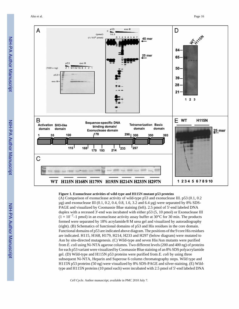
Figure 1. Exonuclease activities of wild-type and H115N mutant p53 proteins(A) Comparison of exonuclease activity of wild-type p53 and exonuclease III. p53 (0.1, 0.2µg) and exonuclease III (0.1, 0.2, 0.4, 0.8, 1.6, 3.2 and 6.4 µg) were separated by 8% SDS-PAGE and visualized by Coomassie Blue staining (left). 2.5 pmol of 5'-end labeled DNAduplex with a recessed 3'-end was incubated with either p53 (5, 10 pmol) or Exonuclease III(1 × 10⁻⁷–1 pmol) in an exonuclease activity assay buffer at 30°C for 30 min. The productsformed were separated by 18% acrylamide/8 M urea gel and visualized by autoradiography(right). (B) Schematics of functional domains of p53 and His residues in the core domain.Functional domains of p53 are indicated above diagram. The positions of the 9 core His residuesare indicated. H115, H168, H179, H214, H233 and H297 (below diagram) were mutated toAsn by site-directed mutagenesis. (C) Wild-type and seven His/Asn mutants were purifiedfrom E. coli using Ni-NTA agarose columns. Two different levels (200 and 400 ng) of proteinsfor each p53 variant were visualized by Coomassie Blue staining of an 8% SDS polyacrylamidegel. (D) Wild-type and H115N p53 proteins were purified from E. coli by using threesubsequent Ni-NTA, Heparin and Superose 6 column chromatography steps. Wild type andH115N p53 proteins (50 ng) were visualized by 8% SDS-PAGE and silver-staining. (E) Wild-type and H115N proteins (10 pmol each) were incubated with 2.5 pmol of 5'-end labeled DNA
Ahn et al. Page 16
Cell Cycle. Author manuscript; available in PMC 2010 July 7.
NIH
-PA Author Manuscript
NIH
-PA Author Manuscript
NIH
-PA Author Manuscript
duplex with a recessed 3'end in an exonuclease assay buffer at 30°C for 10 min (lanes 1 and6), 20 min (lanes 2 and 7), 30 min (lanes 3 and 8), 40 min (lanes 4 and 9) and 60 min (lanes 5and 10). Reactions were stopped and the products formed were analyzed as described in (A).
Ahn et al. Page 17
Cell Cycle. Author manuscript; available in PMC 2010 July 7.
NIH
-PA Author Manuscript
NIH
-PA Author Manuscript
NIH
-PA Author Manuscript
Figure 2. DNA binding by H115N p53 is greater than that of wild-type p53 as measured by EMSA,filter binding assay, and DNase I footprinting(A) Wild-type p53 and H115N proteins were purified from bacteria by NI-NTA, Heparin andgel-filtration column chromatography as described in Experimental Procedures. The relativeamounts of p53 proteins (4, 8, 12 and 16 ng) used in the following assays were detected byimmunoblot analysis using mixture of p53 monoclonal antibodies PAb DO-1, 1801 and 421.(B) Wild-type (lanes 2–7) and H115N mutant (lanes 8–13) proteins (4, 8 and 12 ng each) wereincubated with a 32 BP DNA duplex containing the 5' p21 promoter p53 binding site (2.5 ng)with PAb 421 (50 ng). DNA was resolved on a 4% polyacrylamide gel, dried and visualizedby autoradiography. (C) Either wild-type (solid circles) or H115N (solid squares) p53 protein
Ahn et al. Page 18
Cell Cycle. Author manuscript; available in PMC 2010 July 7.
NIH
-PA Author Manuscript
NIH
-PA Author Manuscript
NIH
-PA Author Manuscript
(each at 0, 10, 20, 30, 40, 50 and 60 ng) was incubated with [32P] labeled DNA duplex (2.5ng) containing the p21 site and 125 ng of competitor DNA oligonucleotide containing a mutantGADD45 site and then filtered through nitrocellulose membrane. Labeled DNA bound to p53protein was quantitated by liquid scintilation. (D) Wild-type p53 (100, 300, 500, 700 ng; lanes3–6) or the same quantities of H115N p53 (lanes 7–10) were incubated with a 5'-end labeled405 bp HindIII-ScaI fragment (approximately 25 ng) from the p21 promoter containing the 5'p53 binding site and subjected to DNAse I footprinting. Control digestion with DNase I wasalso performed (lanes 1 and 2, 11 and 12). The p53 binding site is indicated.
Ahn et al. Page 19
Cell Cycle. Author manuscript; available in PMC 2010 July 7.
NIH
-PA Author Manuscript
NIH
-PA Author Manuscript
NIH
-PA Author Manuscript
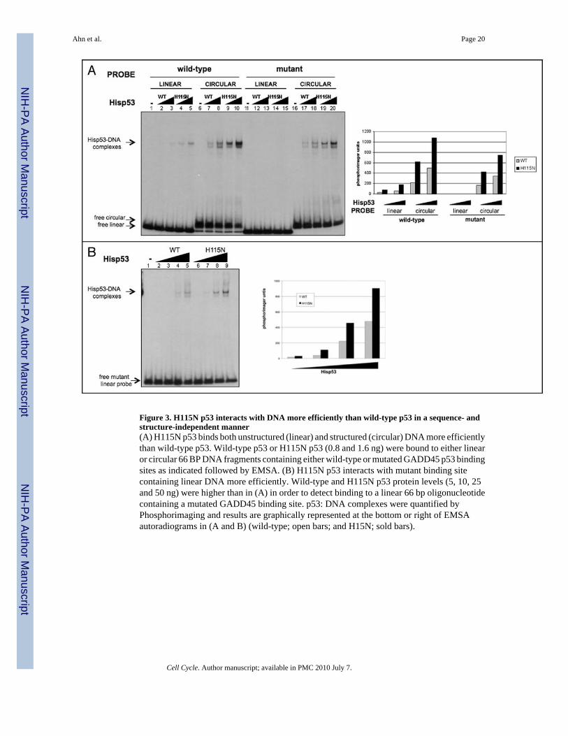
Figure 3. H115N p53 interacts with DNA more efficiently than wild-type p53 in a sequence- andstructure-independent manner(A) H115N p53 binds both unstructured (linear) and structured (circular) DNA more efficientlythan wild-type p53. Wild-type p53 or H115N p53 (0.8 and 1.6 ng) were bound to either linearor circular 66 BP DNA fragments containing either wild-type or mutated GADD45 p53 bindingsites as indicated followed by EMSA. (B) H115N p53 interacts with mutant binding sitecontaining linear DNA more efficiently. Wild-type and H115N p53 protein levels (5, 10, 25and 50 ng) were higher than in (A) in order to detect binding to a linear 66 bp oligonucleotidecontaining a mutated GADD45 binding site. p53: DNA complexes were quantified byPhosphorimaging and results are graphically represented at the bottom or right of EMSAautoradiograms in (A and B) (wild-type; open bars; and H15N; sold bars).
Ahn et al. Page 20
Cell Cycle. Author manuscript; available in PMC 2010 July 7.
NIH
-PA Author Manuscript
NIH
-PA Author Manuscript
NIH
-PA Author Manuscript

Figure 4. Induction of down-stream target genes and cell cycle arrest by wild-type p53 and H115Nin H1299 cells(A) Cultures maintained in medium containing 2,500 ng/mL tetracycline were switched tomedium containing various tetracycline concentration as indicated above panel for 24 hoursprior to cell lysis. Cell extracts (25 µg total protein) were resolved by SDS PAGE and subjectedto immunoblotting for p53 (α-HA), PIG3 (α-Pig), p21 (α-p21) and as control actin (α-Actin).(B) Top (IB). Two separate clones of H1299 cells expressing wild-type p53 (W-1; used in A,and W-2; the second clone) or H115N p53 (H1; shown in A, and H2; the other clone) weremaintained in medium containing tetracycline (2,500 ng/mL) prior to switching to mediumcontaining 0 (lanes 1, 5, 9 and 13), 4 (lanes 2, 6, 10 and 14), 6 (lanes 3, 7, 11 and 15) or 8 (lanes4, 8, 12 and 16) ng/mL tetracycline for 24 hours prior to lysis and immunoblotting for PIG3and actin as in (A) but using 100 µg of total cell extracts. Botton (RT-PCR). Parallel cultureswere lysed, and RNA extracted and levels of p21 and GAPDH mRNA were determined byRT-PCR as described in Experimental Procedures. (C) Tetracycline levels were regulated asindicated to express equivalent amounts of wild-type or mutant p53 protein as assayed byimmunoblotting. Actin levels were visualized as a loading control. p53 levels were quantifiedusing the Odyssey system (Licor). (D) Increase in p53-H115N transcriptional activity asquantified by q-PCR. RNA and cDNA were made from samples in (C) expressing roughly thesame p53 levels (WT p53: 5 ng/uL tet (C, lane 2); H115N no tet (C, lane 6)) and subjected toq-PCR for the p21 gene. Samples in which no p53 was expressed (“+ tet”) were used as acontrol. Values from the PCR were normalized to the p53 levels quantified by the Odysseysystem in (C).
Ahn et al. Page 21
Cell Cycle. Author manuscript; available in PMC 2010 July 7.
NIH
-PA Author Manuscript
NIH
-PA Author Manuscript
NIH
-PA Author Manuscript
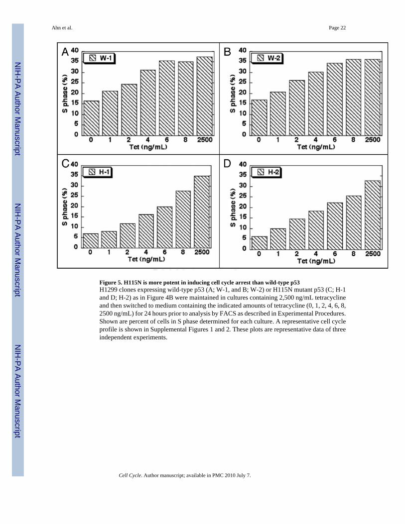
Figure 5. H115N is more potent in inducing cell cycle arrest than wild-type p53H1299 clones expressing wild-type p53 (A; W-1, and B; W-2) or H115N mutant p53 (C; H-1and D; H-2) as in Figure 4B were maintained in cultures containing 2,500 ng/mL tetracyclineand then switched to medium containing the indicated amounts of tetracycline (0, 1, 2, 4, 6, 8,2500 ng/mL) for 24 hours prior to analysis by FACS as described in Experimental Procedures.Shown are percent of cells in S phase determined for each culture. A representative cell cycleprofile is shown in Supplemental Figures 1 and 2. These plots are representative data of threeindependent experiments.
Ahn et al. Page 22
Cell Cycle. Author manuscript; available in PMC 2010 July 7.
NIH
-PA Author Manuscript
NIH
-PA Author Manuscript
NIH
-PA Author Manuscript
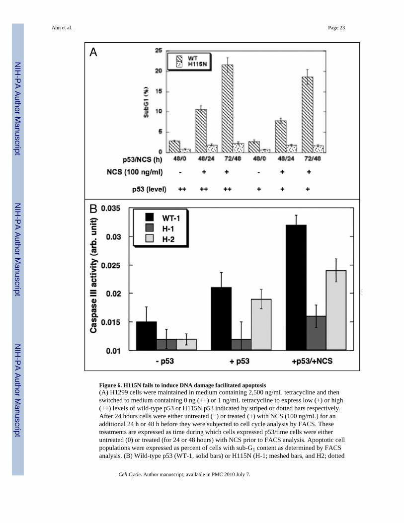
Figure 6. H115N fails to induce DNA damage facilitated apoptosis(A) H1299 cells were maintained in medium containing 2,500 ng/mL tetracycline and thenswitched to medium containing 0 ng (++) or 1 ng/mL tetracycline to express low (+) or high(++) levels of wild-type p53 or H115N p53 indicated by striped or dotted bars respectively.After 24 hours cells were either untreated (−) or treated (+) with NCS (100 ng/mL) for anadditional 24 h or 48 h before they were subjected to cell cycle analysis by FACS. Thesetreatments are expressed as time during which cells expressed p53/time cells were eitheruntreated (0) or treated (for 24 or 48 hours) with NCS prior to FACS analysis. Apoptotic cellpopulations were expressed as percent of cells with sub-G1 content as determined by FACSanalysis. (B) Wild-type p53 (WT-1, solid bars) or H115N (H-1; meshed bars, and H2; dotted
Ahn et al. Page 23
Cell Cycle. Author manuscript; available in PMC 2010 July 7.
NIH
-PA Author Manuscript
NIH
-PA Author Manuscript
NIH
-PA Author Manuscript
bars) was expressed in H1299 cells for 24 h by removing tetracycline from the medium. Cellsexpressing p53 proteins were treated (+p53/+NCS) with NCS (100 ng/mL) or untreated (+p53)for additional 24 h before they were collected for Caspase III assays. As a negative control,cells without p53 (−p53) were also collected and the activity of Caspase III in the lysate wasmeasured.
Ahn et al. Page 24
Cell Cycle. Author manuscript; available in PMC 2010 July 7.
NIH
-PA Author Manuscript
NIH
-PA Author Manuscript
NIH
-PA Author Manuscript
NIH
-PA Author Manuscript
NIH
-PA Author Manuscript
NIH
-PA Author Manuscript
Ahn et al. Page 25
Table 1
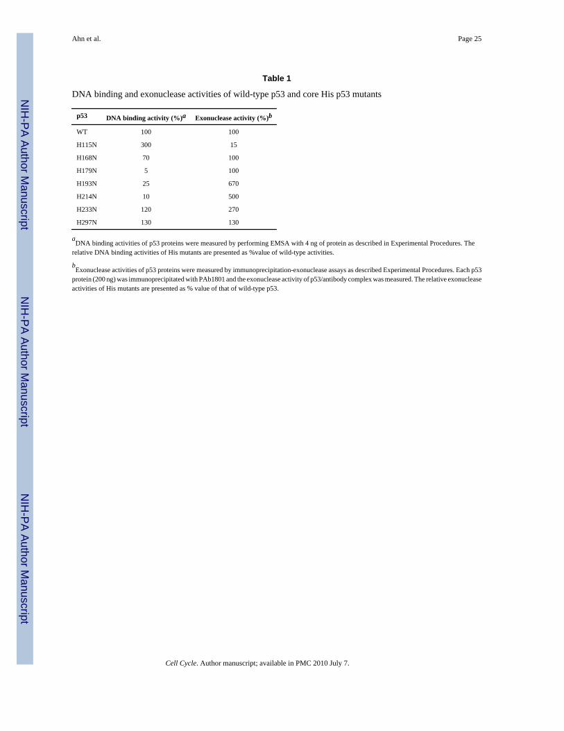
DNA binding and exonuclease activities of wild-type p53 and core His p53 mutants
p53 DNA binding activity (%)a Exonuclease activity (%)b
WT 100 100
H115N 300 15
H168N 70 100
H179N 5 100
H193N 25 670
H214N 10 500
H233N 120 270
H297N 130 130
aDNA binding activities of p53 proteins were measured by performing EMSA with 4 ng of protein as described in Experimental Procedures. The
relative DNA binding activities of His mutants are presented as %value of wild-type activities.
bExonuclease activities of p53 proteins were measured by immunoprecipitation-exonuclease assays as described Experimental Procedures. Each p53
protein (200 ng) was immunoprecipitated with PAb1801 and the exonuclease activity of p53/antibody complex was measured. The relative exonucleaseactivities of His mutants are presented as % value of that of wild-type p53.
Cell Cycle. Author manuscript; available in PMC 2010 July 7.
Copyright © 2022 FDOKUMEN