Development of Skrjabinocerca prima (Nematoda: Acuarioidea) in Hyalella azteca (Amphipoda) and...
10
Development of Skrjabinocerca prima (Nematoda: Acuarioidea) in Hyalella azteca (Amphipoda) and Recurvirostra americana (Aves: Charadriiformes), with comments on its precocity CHERYL M. BARTLETT,~ R. C. ANDERSON, AND P. L. WONG Department of Zoology, College of Biological Sciences, University of Guelph, Guelph, Ont., Canada NlG 2 Wl Received February 17, 1989 BARTLETT, C. M., ANDERSON, R. C., and WONG, P. L. 1989. Development of Skrjabinocerca prima (Nematoda: Acuarioidea) in Hyalella azteca (Amphipoda) and Recurvirostra americana (Aves: Charadriiformes), with comments on its precocity. Can. J. Zool. 67: 2883 -2892. Skrjabinocerca prima Shikhobalova, 1930 developed to the infective third stage in 40 days in Hyalella azteca (Saussure) maintained at 21 OC. Infective larvae occur free in the haemocoel and may impede the amphipod's motility because they lie semirigid and parallel to the longitudinal axis in the posterior abdominal segments. The genital primordium in infective-stage males was 200 - 520 pm long (averaging 19 % of body length) and in females 860 - 1175 pm (averaging 42 % of body length). This is unusually large, even compared with other acuarioids in which precocial development of the third-stage genital primor- dium is known. Additional evidence is provided for the hypothesis that precocity in nematodes is related to rapid maturity in the final host; in experimentally infected American Avocets (Recurvirostra americana Gmelin), moulting third-stage S. prima were found 2 days postinfection and moulting fourth stage at 4 days. Female nematodes contained embryonated eggs at 7 days; eggs in females became larvated between 10 and 18 days postinfection. In birds, the heads of larval and adult nematodes were attached to the mucosa of the buccal cavity or the upper third of the oesophagus, and adults provoked chronic inflammation in the form of yellow plaques. Worms are able to detach and reattach. Third-stage S. prima given to Marbled Godwits (Limosa fedoa (L.)) and Western Willets (Catoptrophorus semipalmatus inornatus (Brewster)) developed to maturity; the parasite has not been reported from the former species in the wild. BARTLETT, C. M., ANDERSON, R. C., et WONG, P. L. 1989. Development of Skrjabinocerca prima (Nematoda: Acuarioidea) in Hyalella azteca (Amphipoda) and Recurvirostra americana (Aves: Charadriiformes), with comments on its precocity. Can. J. Zool. 67 : 2883 -2892. Des Skrjabinocerca prima Shikhobalova, 1930 ont atteint le troisikme stade infectieux en 40 jours chez des Hyalella azteca (Saussure) gardCes h 21 OC. Les larves infectieuses sont libres dans 1'hCmockle et peuvent entraver la mobilitC de l'amphipode, puisqu'elles sont semi-rigides et qu'elles sont disposCes parallklement h l'axe longitudinal dans les segments abdominaux postkrieurs. Le primordium gCnital mesure 200 - 520 pm de longueur (19 % de la longueur totale en moyenne) chez les miles infectieux et 860- 1175 pm de longueur (42% de la longueur totale en moyenne) chez les femelles infectieuses. I1 s'agit 1h de valeurs particulikrement ClevCes, mCme en comparaison de celles qui prkvalent chez d'autres acuario'ides qui subissent le dCve- loppement prCcoce de leur primordium gCnital au troisikme stade infectieux. D'autres indices permettent de croire h l'hypothkse selon laquelle la prCcocitC des nCmatodes est reliCe h leur maturation rapide chez l'h8te terminal; chez des Avocet- tes d'AmCrique (Recurvirostra americana Gmelin) infectkes expCrimentalement, il y a dCjB des larves de troisikme stade de S. prima en train de muer 2 jours aprks l'infection et des larves de quatrikme stade en train de muer 4 jours aprks l'infection. Les nCmatodes femelles contiennent des oeufs h embryons aprks 7 jours; les larves apparaissent dans les oeufs entre les jours 10 et 18 aprks l'infection. Chez les oiseaux, la t6te des parasites, larves et adultes, est attachCe h la muqueuse de la cavitC buc- cale ou au tiers supCrieur de l'oesophage et la prCsence des adultes entraine une inflammation chronique sous forme de plaques jaunes. Les vers peuvent se dCtacher et se rattacher. Des larves de troisikme stade de S. prima administrkes h des Barges marbrkes (Limosa fedoa (L.)) et h des Chevaliers semipalmCs (Catoptrophorus semipalmatus inomatus (Brewster)) se sont dCveloppCes jusqu'h maturitC; le parasite n'a jamais CtC trouvC en nature chez la Barge marbrke. [Traduit par la revue] Introduction dium in the third-stage larva of S. prima to be unusually Skrjabinocerca prima Shikhobalova, 1930 is a nematode parasite of the oesophagus of birds which was redescribed by Wong et al. (1987) on the basis of material from American Avocets (Recurvirostra americana Gmelin) collected on their breeding grounds in the prairies of western Canada. The para- site also occurs in the Palaearctic, and in the Bering Sea region, Tsimbalyuk and Kulikov (1966) found larvae, includ- ing the third stage, in wild-caught beach fleas (Orchestia ochotensis) which are marine semiterrestrial amphipods . The present paper describes development of S. prima in experimen- tally infected Hyalella azteca, a freshwater amphipod which occurs commonly throughout North America. We found the state of development of the genital primor- 'Present address: Department of Mathematics and Natural Sciences, University College of Cape Breton, P.O. Box 5300, Sydney, N.S., Canada B 1P 6L2. advanced. Anderson (i988) proposed ihe use of the term pre- cocity for growth of nematodes beyond the expected stage in intermediate and paratenic hosts and predicted it would accelerate gamete production in the final host. In the present study, development of S. prima in experimentally infected avocets is described and additional evidence provided for this hypothesis. The gross pathology associated with infections in avocets, as well as successful attempts to transmit the parasite to two additional species of scolopacid charadriiforms, are also reported herein. Methods and materials Female Skrjabinocerca prima containing larvated eggs were obtained from three sources: (i) wild adult American Avocets (Recur- virostra americana Gmelin) shot near Gem, Alberta, Canada, in June 1988; (ii) one experimentally infected Western Willet (Catop- trophorus semipalmatus inomatus (Brewster)) killed 18 d post- Printed in Canada I 1mprirnC au Canada Can. J. Zool. Downloaded from www.nrcresearchpress.com by "Institute of Vertebrate Paleontology and Paleoanthropology,CAS" on 05/28/13 For personal use only.
Transcript of Development of Skrjabinocerca prima (Nematoda: Acuarioidea) in Hyalella azteca (Amphipoda) and...

Development of Skrjabinocerca prima (Nematoda: Acuarioidea) in Hyalella azteca (Amphipoda) and Recurvirostra americana (Aves: Charadriiformes),
with comments on its precocity
CHERYL M. BARTLETT,~ R. C. ANDERSON, AND P. L. WONG Department of Zoology, College of Biological Sciences, University of Guelph, Guelph, Ont., Canada NlG 2 Wl
Received February 17, 1989
BARTLETT, C. M., ANDERSON, R. C., and WONG, P. L. 1989. Development of Skrjabinocerca prima (Nematoda: Acuarioidea) in Hyalella azteca (Amphipoda) and Recurvirostra americana (Aves: Charadriiformes), with comments on its precocity. Can. J. Zool. 67: 2883 -2892.
Skrjabinocerca prima Shikhobalova, 1930 developed to the infective third stage in 40 days in Hyalella azteca (Saussure) maintained at 21 OC. Infective larvae occur free in the haemocoel and may impede the amphipod's motility because they lie semirigid and parallel to the longitudinal axis in the posterior abdominal segments. The genital primordium in infective-stage males was 200 - 520 pm long (averaging 19 % of body length) and in females 860 - 1 175 pm (averaging 42 % of body length). This is unusually large, even compared with other acuarioids in which precocial development of the third-stage genital primor- dium is known. Additional evidence is provided for the hypothesis that precocity in nematodes is related to rapid maturity in the final host; in experimentally infected American Avocets (Recurvirostra americana Gmelin), moulting third-stage S. prima were found 2 days postinfection and moulting fourth stage at 4 days. Female nematodes contained embryonated eggs at 7 days; eggs in females became larvated between 10 and 18 days postinfection. In birds, the heads of larval and adult nematodes were attached to the mucosa of the buccal cavity or the upper third of the oesophagus, and adults provoked chronic inflammation in the form of yellow plaques. Worms are able to detach and reattach. Third-stage S. prima given to Marbled Godwits (Limosa fedoa (L.)) and Western Willets (Catoptrophorus semipalmatus inornatus (Brewster)) developed to maturity; the parasite has not been reported from the former species in the wild.
BARTLETT, C. M., ANDERSON, R. C., et WONG, P. L. 1989. Development of Skrjabinocerca prima (Nematoda: Acuarioidea) in Hyalella azteca (Amphipoda) and Recurvirostra americana (Aves: Charadriiformes), with comments on its precocity. Can. J. Zool. 67 : 2883 -2892.
Des Skrjabinocerca prima Shikhobalova, 1930 ont atteint le troisikme stade infectieux en 40 jours chez des Hyalella azteca (Saussure) gardCes h 21 OC. Les larves infectieuses sont libres dans 1'hCmockle et peuvent entraver la mobilitC de l'amphipode, puisqu'elles sont semi-rigides et qu'elles sont disposCes parallklement h l'axe longitudinal dans les segments abdominaux postkrieurs. Le primordium gCnital mesure 200 - 520 pm de longueur (19 % de la longueur totale en moyenne) chez les miles infectieux et 860- 1175 pm de longueur (42% de la longueur totale en moyenne) chez les femelles infectieuses. I1 s'agit 1h de valeurs particulikrement ClevCes, mCme en comparaison de celles qui prkvalent chez d'autres acuario'ides qui subissent le dCve- loppement prCcoce de leur primordium gCnital au troisikme stade infectieux. D'autres indices permettent de croire h l'hypothkse selon laquelle la prCcocitC des nCmatodes est reliCe h leur maturation rapide chez l'h8te terminal; chez des Avocet- tes d'AmCrique (Recurvirostra americana Gmelin) infectkes expCrimentalement, il y a dCjB des larves de troisikme stade de S. prima en train de muer 2 jours aprks l'infection et des larves de quatrikme stade en train de muer 4 jours aprks l'infection. Les nCmatodes femelles contiennent des oeufs h embryons aprks 7 jours; les larves apparaissent dans les oeufs entre les jours 10 et 18 aprks l'infection. Chez les oiseaux, la t6te des parasites, larves et adultes, est attachCe h la muqueuse de la cavitC buc- cale ou au tiers supCrieur de l'oesophage et la prCsence des adultes entraine une inflammation chronique sous forme de plaques jaunes. Les vers peuvent se dCtacher et se rattacher. Des larves de troisikme stade de S. prima administrkes h des Barges marbrkes (Limosa fedoa (L.)) et h des Chevaliers semipalmCs (Catoptrophorus semipalmatus inomatus (Brewster)) se sont dCveloppCes jusqu'h maturitC; le parasite n'a jamais CtC trouvC en nature chez la Barge marbrke.
[Traduit par la revue]
Introduction dium in the third-stage larva of S. prima to be unusually
Skrjabinocerca prima Shikhobalova, 1930 is a nematode parasite of the oesophagus of birds which was redescribed by Wong et al. (1987) on the basis of material from American Avocets (Recurvirostra americana Gmelin) collected on their breeding grounds in the prairies of western Canada. The para- site also occurs in the Palaearctic, and in the Bering Sea region, Tsimbalyuk and Kulikov (1966) found larvae, includ- ing the third stage, in wild-caught beach fleas (Orchestia ochotensis) which are marine semiterrestrial amphipods . The present paper describes development of S. prima in experimen- tally infected Hyalella azteca, a freshwater amphipod which occurs commonly throughout North America.
We found the state of development of the genital primor-
'Present address: Department of Mathematics and Natural Sciences, University College of Cape Breton, P.O. Box 5300, Sydney, N.S., Canada B 1P 6L2.
advanced. Anderson (i988) proposed ihe use of the term pre- cocity for growth of nematodes beyond the expected stage in intermediate and paratenic hosts and predicted it would accelerate gamete production in the final host. In the present study, development of S. prima in experimentally infected avocets is described and additional evidence provided for this hypothesis. The gross pathology associated with infections in avocets, as well as successful attempts to transmit the parasite to two additional species of scolopacid charadriiforms, are also reported herein.
Methods and materials Female Skrjabinocerca prima containing larvated eggs were
obtained from three sources: (i) wild adult American Avocets (Recur- virostra americana Gmelin) shot near Gem, Alberta, Canada, in June 1988; (ii) one experimentally infected Western Willet (Catop- trophorus semipalmatus inomatus (Brewster)) killed 18 d post-
Printed in Canada I 1mprirnC au Canada
Can
. J. Z
ool.
Dow
nloa
ded
from
ww
w.n
rcre
sear
chpr
ess.
com
by
"Ins
titut
e of
Ver
tebr
ate
Pale
onto
logy
and
Pal
eoan
thro
polo
gy,C
AS"
on
05/2
8/13
For
pers
onal
use
onl
y.

2884 CAN. J. ZOOL. VOL. 67, 1989
infection (see below); and (iii) one experimentally infected American Avocet killed 23 d postinfection (see below). Female nematodes were dissected in avian saline and eggs removed were given to amphipods (see below). Nematode eggs from wild avocets were stored (for 2 weeks) in avian saline at 4OC before they were used.
Amphipods (Hyalella azteca (Saussure) and Gammarus fasciatus Say) collected in a pond near Guelph, Ontario, Canada, were placed individually in small glass dishes containing well water and numerous eggs of S. prima. Twenty-four hours later, amphipods were removed. They were maintained together in aerated well water at room tempera- ture (21 OC) in large glass aquaria and fed fish food pellets (no. 3 GR for trout and salmon, Martin Feed Mills Limited, Elmira, Ontario, Canada N3B 3A2).
Amphipods were dissected at various times up to 50 d postexposure. First- and second-stage nematode larvae found were fixed in 4-5 drops of formal -saline (10% buffered formalin mixed with an equal volume of avian saline) on a slide. The preparation was then covered with a vaseline-ringed cover glass and larvae were studied. Some third-stage larvae were fixed in hot glycerin-alcohol and studied in glycerin. Other third-stage larvae were used as outlined below.
Variable numbers of live third-stage larvae were orally adminis- tered with a Pasteur pipette to 10 hand-reared juvenile American Avocets, one hand-reared juvenile Western Willet, and one hand- reared juvenile Marbled Godwit (Limosa fedoa (L.)) (see rearing and maintenance techniques in Wong et al. 1989). Third-stage larvae given to two avocets (those killed at 10 and 23 d) and to the willet ori- ginated from amphipods infected with eggs from the first of the three sources previously identified. Third-stage larvae given to two other avocets (those killed at 26 d) originated from the second source, and third-stage larvae given to all other birds originated from the third source. Birds were killed (with an overdose of sodium pentobarbital administered intraperitoneally) at various times postinfection and examined for nematodes. Nematodes found in experimentally infected birds were fixed in hot glycerin-alcohol and studied in glycerin.
En face and transverse sections of nematodes were cut freehand with a mounted razor blade. The former were studied in glycerin jelly and the latter in glycerin.
Specimens of S. prima have been deposited in the Invertebrate Col- lection (Parasites) of the National Museum of Natural Sciences in Ottawa, Canada (Nos. NMCP1989-0059 to 0065).
Twenty-five Hyalella azteca and 25 Gammarus fasciatus collected from the same pond as those mentioned above were dissected for tax- onomic purposes. Nematodes were not present in these amphipods.
Results Examination of amphipods
Skrjabinocerca prima developed to the infective third stage in Hyalella azteca (Table 1). Three groups of this amphipod were examined: (1) 10 of 11 exposed to eggs from nematodes in wild avocets contained larvae when examined 40 d after exposure, and intensity ranged from 2 to 29 (mean 9.5); (2) all of 19 exposed to eggs from nematodes from an experimentally infected willet contained larvae when examined 1 -3 1 and 50 d after exposure, and intensity ranged from 1 to 25 (mean 7.8); (3) all of 65 exposed to eggs from nematodes from an experi- mentally infected avocet contained larvae when examined 45 and 48 d after exposure, and intensity ranged from 1 to 12 (mean 3.6). Thirty-four of 35 Gammarus fasciatus exposed to eggs from nematodes in wild avocets did not contain larval S. prima when examined 40 d after exposure; the 35th indi- vidual contained one moribund first-stage larva.
Moulting first-stage larvae were first observed 21 d post- infection, moulting second-stage larvae at 31 d, and infective third-stage larvae at 40 d; development was highly asyn- chronous (Table 1). First- and second-stage larvae were found free in saline during dissection of the amphipod. Infective third-stage larvae were free in the haemocoel in the posterior-
TABLE 1. Numbers and stage of development of Skrjabinocerca prima in infected Hyalella azteca examined at various times after
ingesting eggs of S. prima
Moulting Moulting Time First first Second second Third (d) N stage stage stage stage stage
NOTE: N, number of amphipods examined. aCollective total of first-stage, moulting first-stage, second-stage, and moulting second-
stage larvae.
most segments of the abdomen. They tended to lie semirigid and parallel to the longitudinal axis of the amphipod. When more than one third-stage larva was present, larvae were generally adjacent to one other.
Descriptions of nematodes from amphipods First-stage larva (Figs. 1 -3) GENERAL: Cuticle thin, with delicate transverse striations.
Cephalic extremity apparently lacking hook or tooth. Nerve ring not observed. Excretory pore visible, excretory cell large (Figs. 1 - 3). Buccal cavity delicate and narrow, cuticle not striated. Oesophagus in early-stage larva at 2 d represented by narrow primordium without visible nuclei (Fig. 1). Oesopha- gus in midstage larva at 6 d (Fig. 2) and late-stage larva at 12 d (Fig. 3) represented by broad multinucleate primordium; posterior extremity of primordium wider and with larger nuclei than other portions. Oesophageal -intestinal junction clearly defined. Intestine in early-stage larva represented by granular primordium, in later-stage larvae by thin-walled sacculate structure with large lumen. R1 cell present in early-stage larva (Fig. 1); daughter cells of R1 cell present in later-stage larvae (Figs. 2, 3). R2 -Rq cells prominent, forming walls of rectum (Figs. 1 - 3). Small anal plug present. Tail attenuated, extrem- ity pointed and ending in tiny, nipple-like protuberance. Geni- tal primordium not observed.
MEASUREMENTS: N = 3, consisting, in order, of one each of early-stage larva at 2 d, midstage larva at 6 d, and late-stage larva at 12 d. Length 180, 180, 210 pm. Maximum width 10, 16, 21 pm. Excretory pore 50, 50, 45 pm from anterior extremity. Length of buccal cavity 27, 33, 27 pm; of oesopha- geal primordium 50, 45, 47 pm. Anus 35, 45, 50 pm from posterior extremity.
Moulting first-stage larva (Fig. 4) GENERAL: New and old cuticle present at anterior and
posterior extremities. Narrow posterior portion of second-stage buccal cavity surrounded by multinucleate mass. Genital pri- mordium represented by oval multinucleate mass in posterior third of body.
MEASUREMENTS: N = 2. Length 386, 410 pm. Maximum width 45, 50 pm. Excretory pore 60, 70 pm from anterior extremity. Length of buccal cavity 25, 25 pm; of oesophageal
Can
. J. Z
ool.
Dow
nloa
ded
from
ww
w.n
rcre
sear
chpr
ess.
com
by
"Ins
titut
e of
Ver
tebr
ate
Pale
onto
logy
and
Pal
eoan
thro
polo
gy,C
AS"
on
05/2
8/13
For
pers
onal
use
onl
y.

BARTLETT ET AL. 2885
primordium 100, 1 10 pm. Genital primordium 120, 123 pm and anus 45, 55 pm from posterior extremity.
Second-stage larva (Figs. 5 - 7) GENERAL: Cuticle .thin, with delicate transverse striations.
Cephalic extremity with small conical cuticular projection (Figs. 5, 6). Nerve ring present. Excretory pore visible, excre- tory cell large (Figs. 5, 6). Deirids of third-stage larva visible below cuticular surface of late-stage larva (not illustrated). Buccal cavity consisting of V-shaped anterior portion and nar- row tubular posterior portion; posterior portion in early-stage larva at 25 d (Fig. 5) surrounded by multinucleate mass, in late-stage larva at 25 d (Fig. 6) by vacuolate hyaline structure. Oesophagus divided; division in early-stage larva poorly defined (Fig. 5), in late-stage larva clearly defined (Fig. 6); anterior portion with few large nuclei, and in late-stage larva, with readily apparent muscular fibres; posterior portion slightly swollen immediately posterior to junction with anterior portion and containing numerous small nuclei throughout. Intestine well developed. R2 -& cells readily apparent, forming rectum (Figs. 5, 7). Small anal plug present. Hypodermal protuber- ance present immediately posterior to anus, larger in late-stage than in early-stage larvae (Figs. 5, 7). Tail attenuated, extrem- ity rounded and cuticle detached.
MALE: Larvae suitable for study were not available. FEMALE: N = 2, one each of early- and late-stage larvae.
Length 0.88, 2.22 mm. Maximum width 60, 80 pm. Nerve ring 70, 75 pm and excretory pore 110, 135 pm from anterior extremity. Length of V-shaped portion of buccal cavity 12, 12, pm; of tubular portion 12, 12 pm; of muscular portion of oesophagus 60, 115 pm; of glandular portion of oesophagus 190, 340 pm. Midregion of genital primordium attached by few large cells (representing vaginal primordium) to hypoder- mis 220, 340 pm from posterior extremity of body. Remainder of genital primordium bifurcate, with anteriorly and posteriorly directed branches; branches stumpy, shorter in early-stage (Fig. 5) than in late-stage (Fig. 7) larvae; extremity of each branch in late-stage larvae formed by large cap cell with ter- minal nucleus. Anus 80, 95 pm from posterior extremity.
Infective third-stage larva (Figs. 8 - 21 ; Tables 2, 3) GENERAL: Cuticle thick, multilayered, with delicate trans-
verse striations. Cephalic extremity with pseudolabia, two amphids, and four large papillae (Figs. 10 - 12) ; four tiny non- protuberant sensilla (not illustrated) perhaps present on shoulders of pseudolabia. Delicate cordons present, origi- nating on inner side of pseudolabia near apices, extending to dorsoventral sides of oral opening and around lateral sides of cephalic papillae (Figs. 10 - 12), continuing posteriorly as straight or slightly undulant parallel cords and terminating between nerve ring and deirids (Figs. 8, 9); lengths slightly unequal (note: rarely, the distal end of one cordon curves 90" medially or laterally, but anastomoses were absent). Lateral alae present, with smooth outer edge (Fig. 9) but marked within by transverse striations corresponding to those of body cuticle (Fig. 10); originating at level of cephalic papillae and terminating in last quarter of body; portion anterior to deirid shaped like an equilateral triangle (see Fig. 22, moulting third stage), portion behind deirid wing-like (Fig. 21) and diminish- ing in size posteriorly, with height near midintestine similar to that of longitudinal ridges. Longitudinal cuticular ridges (Fig. 21) originating near posterior end of cordons and extend- ing to anal region. Deirids conical, in line of lateral alae slightly anterior to excretory pore, adjacent or almost adjacent to each
other (Fig. 9). Postdeirids present, bluntly diamond-shaped; slightly dorsal to lateral alae in region of anterior third of intes- tine (Figs. 13, 17, 19), one 20 -200 bm ahead of other. Cuti- cle of apices of pseudolabia confluent with that of buccal cavity. Buccal cavity elongate, cuticle not striated (Fig. 9). Oesophagus divided, junction between muscular and glandular portions clearly defined (Figs. 8, 9). Oesophageal - intestinal junction clearly defined (Fig. 13). R2 - R4 cells large, forming anteriormost portion of rectum, with remainder formed by smaller cells (Figs. 14, 15). Single pedunculate papilla present immediately posterior to anus (Figs. 14, 15). Tail attenuated, tip variable as follows: (1) with slight constriction and large pointed protuberance (Figs. 15, 16a); (2) rounded, with or without small protuberances (Figs. 14, 16b); (3) with subter- minal neck-like constriction and terminal triangular appendage (Fig. 16c). Cuticle of posterior portion of tail with pronounced multilayered appearance (Figs. 16a - 16c) and containing ter- minal hyaline inclusion (Figs. 16a, 16b) when terminal tri- angular appendage absent. Phasmids subterminal, small, papilliform.
MALE: N = 10. Genital primordium 200 -520 (mean 403) pm long ( = 10 -24 % of body length, mean 19 %) , free within pseudocoelom at level of middle third of intestine (Fig. 17); anterior portion (Fig. 18a) narrow, extremity formed by elongate cap cell with prominent nucleus; posterior portion (Fig. 18b) broad, extremity representing germinal zone of testis.
FEMALE: N = 10. Genital primordium 860 - 1 175 (1009) pm long (= 36 -49 % of body length, mean 42 %) . Portion repre- senting vaginal primordium attached to hypodermis in posterior quarter of body (Fig. 19) by one or more large cells (Fig. 20b). Remainder of primordium represented by one anterior and one posterior branch (Fig. 19); branches narrow and tubular, not recognizably differentiated into regions, not convoluted; anterior branch 375 -550 (450) pm long, ending near oesophageal -intestinal junction; posterior branch 350 - 515 (448) pm long, ending near anal region; walls of proximal portion of each branch consisting of inner and outer layer of cells (Fig. 20b); distal extremity of each branch formed by elongate cap cell with prominent nucleus (Figs. 20a, 20c).
Examination of experimentally infected avocets Skrjabinocerca prima was found in all experimentally
infected avocets and 5 -65% of .the numbers of nematodes given were recovered (Table 4). Moulting third-stage larvae were found 2 d postinfection and moulting fourth-stage larvae at 4 d. Intact, shed fourth-stage cuticles and adult nematodes were found at 7 d. Thick-shelled embryonated egges were pre- sent within females at 7 and 10 d postinfection but none was larvated. Thick-shelled larvated eggs were present within females at 18 d and all later times postinfection, with the exception of an avocet killed 26 d in which only female nema- todes were present; the latter females contained ova lacking shells.
Most nematodes (regardless of time postinfection) were found in the upper third of the oesophagus; a few were in the buccal cavity. The anterior end of each nematode was embed- ded in the mucosa and they could not easily be detached. Shed cuticles were also attached to the mucosa but were easily removed.
At 10 d postinfection, the oesophageal mucosa around the heads of some nematodes was abnormally red; such lesions were about 2 mm in diameter. Similar red spots without nema- todes but with a round central pit were also present. At 18, 26,
Can
. J. Z
ool.
Dow
nloa
ded
from
ww
w.n
rcre
sear
chpr
ess.
com
by
"Ins
titut
e of
Ver
tebr
ate
Pale
onto
logy
and
Pal
eoan
thro
polo
gy,C
AS"
on
05/2
8/13
For
pers
onal
use
onl
y.

2886 CAN. J . ZOOL. VOL. 67, 1989
FIGS. 1 - 7. First- and second-stage larvae of Skrjabinocerca prima from experimentally infected Hyalella azteca, lateral views. Fig. 1. Early first stage at 2 d. Fig. 2. Mid first stage at 6 d. Fig. 3. Late first stage at 12 d. Fig. 4. Moulting first stage at 2 1 d. Fig. 5. Early second stage female at 25 d. Fig. 6. Anterior end, late second stage female at 25 d. Fig. 7 . Posterior end, late second stage female at 25 d. GP, genital primordium.
Can
. J. Z
ool.
Dow
nloa
ded
from
ww
w.n
rcre
sear
chpr
ess.
com
by
"Ins
titut
e of
Ver
tebr
ate
Pale
onto
logy
and
Pal
eoan
thro
polo
gy,C
AS"
on
05/2
8/13
For
pers
onal
use
onl
y.

BARTLETT ET AL. 2887
TABLE 2. Measurements (in micrometres unless otherwise specified) of male Skrjabinocerca prima; infective third-stage larvae were obtained from experimentally infected amphipods (Hyalella azteca) and moulting third-stage larvae, moulting fourth-stage larvae, and adult S. prima from
experimentally infected avocets (Recurvirostra americana)
Infective Moulting Moulting third stage third stagea fourth stageb Adult Adult Adult (N = 10) (N = 5) (N = 5) (N = 5) (N = 2) (N = 2)
Time (d)" Length (mm) Maximum width Nerve ringd Deiridsd Excretory pored Cordonse Buccal cavitye Muscular oesophaguse Glandular oesophaguse Total oesophaguse Right spiculee Left spiculee nu sf
NOTE: Measurements are given as the range, followed by the mean in parentheses, when N r 5; measurements are of individual nematodes when N < 5. "Maximum width includes old and new cuticle; distances from anterior or posterior extremity are from those of the developing fourth stage; deirids and cordons are those of the third
stage. b~aximum width includes old and new cuticle; distances from anterior or posterior extremity are from those of the developing adult; deirids and cordons are those of the fourth stage. 'Time postexposure of amphipods to larvated eggs for infective third stage; time postinfection of avocets for all other stages. d~istance from anterior extremity. 'Length. /Distance from posterior extremity.
TABLE 3. Measurements (in micrometres unless otherwise specified) of female Skrjabinocerca prima; infective third-stage larvae were obtained from experimentally infected amphipods (Hyalella azteca) and moulting third-stage larvae, moulting fourth-stage larvae, and adult S. prima from
experimentally infected avocets (Recurvirostra americana)
Infective Moulting Moulting third stage third stageu fourth stageb Adult Adult Adult (N = 10) (N = 5) (N = 5) (N = 3) (N = 5) (N = 2)
Time (d)' Length (mm) Maximum width Nerve ringd Deiridsd Excretory pored Cordonse Buccal cavity' Muscular oesophaguse Glandular oesophaguse Total oesophaguse Vulvafx Anusf
NOTE: Measurements are given as the range, followed by the mean in parentheses, when N r 5; measurements are of individual nematodes when N < 5. 'Maximum width includes old and new cuticle; distances from anterior or posterior extremity are from those of the developing fourth stage; deirids and cordons are those of the third
stage. b~ax imum width includes old and new cuticle; distances from anterior or posterior extremity are from those of the developing adult; deirids and cordons are those of the fourth stage. 'Time postexposure of amphipods to larvated eggs for infective third stage; time postinfection of avocets for all other stages. d~istance from anterior extremity. 'Length. /Distance from posterior extremity. g"Vulva" is the vaginal primordium in third and fourth stages.
and 60 d similar lesions were observed; more commonly, Examination of other experimentally infected birds however, the oesophageal mucosa around the heads of nema- Five adult male and five adult female S. prima were found in todes was yellow and slightly elevated, forming a plaque a godwit given 25 third-stage larvae and three adult males and 2-3 mm in diameter. Plaques without nematodes but with a two adult females in a willet given 20 third-stage larvae; both round central pit, some of which contained a plug of yellow birds were killed 18 d postinfection. Lesions similar to those in caseous material, were also present. an avocet at 18 d were observed in the godwit. No attempt was
Can
. J. Z
ool.
Dow
nloa
ded
from
ww
w.n
rcre
sear
chpr
ess.
com
by
"Ins
titut
e of
Ver
tebr
ate
Pale
onto
logy
and
Pal
eoan
thro
polo
gy,C
AS"
on
05/2
8/13
For
pers
onal
use
onl
y.

2888 CAN. J . ZOOL. VOL. 67, 1989
Can
. J. Z
ool.
Dow
nloa
ded
from
ww
w.n
rcre
sear
chpr
ess.
com
by
"Ins
titut
e of
Ver
tebr
ate
Pale
onto
logy
and
Pal
eoan
thro
polo
gy,C
AS"
on
05/2
8/13
For
pers
onal
use
onl
y.

BARTLETT ET AL. 2889
TABLE 4. Numbers, sex, and stage of development of Skrjabi- nocerca prima in experimentally infected American Avocets (Recurvirostra americana) killed at various times postinfection
Moulting Moulting No- of larvae third stage fourth stage Adult
Time given / % (d) recovereda 0 Q 0 Q 0 Q
NOTE: TWO avocets were killed at 23 d and two at 26 d. 'Number of nematodes recovered is expressed as a percentage of the number of
infective third-stage larvae orally administered. (Note: Third-stage larvae were from Hyalella azteca dissected 40- 50 d after having been exposed in the labora- tory to eggs of S. prima.)
made to observe the presence or absence of lesions in the willet. Female nematodes in both birds contained larvated eggs and those in the willet were infective to amphipods; those in the godwit were not exposed to amphipods.
Descriptions of nematodes from avocets Moulting third-stage larva (Figs. 22 -25; Tables 2, 3) GENERAL: Old and new cuticle present, in tight apposition
except at anterior and posterior extremities, both transversely striated except at extremities; both multilayered. Cuticle of buccal cavity striated, partially extruded. Information pertain- ing to the third stage: cordons as in infective stage except more strongly developed; lateral alae, longitudinal ridges, deirids, and postdeirids as in infective stage. Information pertaining to the fourth stage: cordons readily visible in transverse section at level of mid muscular oesophagus (Fig. 22) but only faint out- line of developing posterior portions visible in surface view (as for those of adult shown in Fig. 27, moulting fourth stage); lateral alae visible only in transverse section (Fig. 23); longi- tudinal cuticular ridges absent; oesophagus divided; pedun- culate cuticle present immediately posterior to anus, with delicate central nerve stalk generally apparent only in female (Fig. 25); tip of tail with 3 - 8 finger-like cuticular projections in both sexes (Figs. 24, 25); phasmids small, papilliform.
MALE: Reproductive tract with recognizable testis and vas deferens. Testis broad, growth zone filled with spermatocytes, distal portion with 180' flexure and with or without additional convolutions. Transition between testis and vas deferens not clearly differentiated. Vas deferens long and opening into ven- tral side of rectum (Fig. 24). Spicular primordia oblong, mul- tinucleate (Fig. 24).
FEMALE: Reproductive tract with recognizable vulvar and vaginal regions, vulva not patent ,(Fig. 25). Anterior and posterior branches of tract not recognizably differentiated into regions and with or without convolutions, nucleus of cap cell at distal extremity of each branch indiscernible (Fig. 25).
Moulting fourth-stage larva (Figs. 26-29; Tables 2, 3) GENERAL: Old and new cuticle present, in tight apposition
except at anterior and posterior extremities; both transversely striated except at extremities; both multilayered; both lacking longitudinal ridges. Cuticle of buccal cavity striated, partially extruded (Fig. 27). Information pertaining to the fourth stage: cordons similar to those in infective third stage except longer, more strongly developed, and extending around medial sides of cephalic papillae (Fig. 27); lateral alae originating slightly posterior to deirids, diminishing in size posteriorly and ter- minating in male in last sixth of body and in female slightly anterior to vulvar region, with smooth outer edge but marked within by transverse striations corresponding to those of body cuticle; deirids crescent-shaped or crescent-shaped with small central protuberance (Fig. 27); postdeirids linguiform, slightly dorsal to lateral alae in region of anterior third of intestine, one 100-200 pm ahead of other. Information pertaining to the adult: faint outline of posterior portions of developing cordons visible (Fig. 27).
MALE: Reproductive tract with clearly defined testis, seminal vesicle, and vas deferens; testis convoluted, growth zone filled with spermatocytes. Spicules incompletely differentiated, multinucleate mass present at head of each (Fig. 26). Caudal papillae of adult readily apparent under fourth-stage cuticle (Fig. 26). Tip of adult tail rounded (Fig. 26), with or without small protuberance.
FEMALE: Reproductive tract with clearly defined vulva, vagina Vera, vagina uterina, uteri, oviducts, and ovaries. Vagina flexed posteriorly before bifurcation in remainder of tract (Fig. 28). Oocytes present in oviduct and growth zone of ovaries, cap cell at distal extremity of each ovary indiscern- ible. Tip of adult tail abruptly flattened or with several spike- like hypodermal projections (Fig. 29).
Adult (Figs. 30-33; Tables 2, 3) Morphology as described by Wong et al. (1987), with the
following additions. Cuticle multilayered. Cephalic inflations not equally developed in all specimens. Cordons originating on inner side of pseudolabia near apices, continuing posteriorly as straight or slightly undulant parallel cords. Cuticle anterior to deirids slightly thickened over lateral fields in both sexes, forming small triangular elevation in male. Lateral alae origi- nating slightly posterior to deirids, formed initially by elevated cuticle and prominent central hyaline structure but more pos- teriorly only by small central hyaline structure (Fig. 31), with smooth outer edge but marked within by transverse striations corresponding to those of body cuticle, diminishing in size posteriorly and terminating in male in ventrally curved caudal
FIGS. 8 - 2 1 . Third-stage larva of Skrjabinocerca prima from experimentally infected Hyalella azteca at 40 - 50 d. Fig. 8. Anterior end of female, lateral view. Fig. 9. Anterior end of male, ventral view. Figs. 10- 12. Cephalic extremity; ventral, lateral, and en face views, respec- tively (note striations within lateral alae in Fig. 10). Fig. 13. Oesophageal-intestinal junction, postdeirid, and right ala of female, lateral view. Fig. 14. Tail of male, ventral view. Fig. 15. Tail of female, lateral view (posterior portion of genital primordium not illustrated, see Fig. 20c). Figs. 16a - 16c. Tip of tail, showing variation. Fig. 17. Outline of male, showing extent of oesophagus and genital primordium, lateral view. Figs. 18a, 18b. Male genital primordium; anterior and posterior portions, respectively, lateral view. Fig. 19. Outline of posterior half of female, showing extent of genital primordium and position of vaginal primordium, lateral view. Figs. 20a -20c. Female genital primordium; anterior, vaginal, and posterior portions, respectively, lateral view. Fig. 21. Transverse section of male at level of mid glandular oesophagus. A, lateral ala; D, deirid; GP, genital primordium; P, phasmid; PD, post deirid; VP, vaginal primordium.
Can
. J. Z
ool.
Dow
nloa
ded
from
ww
w.n
rcre
sear
chpr
ess.
com
by
"Ins
titut
e of
Ver
tebr
ate
Pale
onto
logy
and
Pal
eoan
thro
polo
gy,C
AS"
on
05/2
8/13
For
pers
onal
use
onl
y.

Can
. J. Z
ool.
Dow
nloa
ded
from
ww
w.n
rcre
sear
chpr
ess.
com
by
"Ins
titut
e of
Ver
tebr
ate
Pale
onto
logy
and
Pal
eoan
thro
polo
gy,C
AS"
on
05/2
8/13
For
pers
onal
use
onl
y.

BARTLETT ET AL. 289 1
region (Fig. 30) and in female slightly anterior to vulva. Postdeirids linguiform, slightly dorsal to lateral alae in region of anterior half of intestine, one 90 - 350 pm ahead of other. Male with ventral preanal cuticular crest (= "crista media ventralis" of Shikhobalova 1930) in curved caudal region (Figs. 30, 32, 33); alternately formed by triangular cuticle with central hyaline structure (Fig. 32a) or by triangular cuticle only (Fig. 32b). Female postanal papilla strongly developed, weakly developed, or absent.
Discussion Natural infections of Skrjabinocerca p r i m have been
reported in 22 species of birds representing 11 families in five orders (Wong et al. 1987). In addition, P. L. Wong and R. C. Anderson (unpublished data) have found S. p r i m in four spe- cies of Charadriiformes not previously reported as hosts, namely Western Willets, Wilson's Phalaropes (Phalaropus tri- color (Vieillot) , Black-bellied Plovers (Pluvialis squatarola (L.)), and Killdeer (Charadrius vociferus L.) collected near Brooks, Alberta, in May and June, 1987 and 1988. The present study shows that S. p r i m will also mature in Marbled God- wits, which belong to the Charadriiformes. The diverse host distribution of S. p r i m may reflect the variety of birds that naturally encounter third-stage larvae by consuming infected amphipods, i.e., the freshwater species Hyalella azteca (pre- sent study) or the marine semiterrestrial species Orchestia ochotensis (see Tsimbalyuk and Kulikov 1966). A fish para- tenic host might be involved in transmission of the parasite to piscivorous hosts such as those in the family Alcidae.
The high prevalence of infection in experimentally infected H. azteca (99% of 95) in the present study is similar to that reported by Tsimbalyuk and Kulikov (1966) in naturally infected 0. ochotensis (100% of 500). In contrast, the fresh- water amphipod Gammrus fasciatus is not a suitable inter- mediate host, as only 1 of 35 individuals exposed to eggs of S. prima became infected and this individual harboured a mori- bund larva. Apparently, therefore, S. p r i m develops in amphi- pods which are more closely related phylogenetically than ecologically; H. azteca and 0 . ochotensis belong to the super- family Talitroidea and G. fasciatus to the Gammaroidea (classification according to Bous field 1983).
Longitudinal cuticular ridges are unusual among third-stage larvae of acuarioids. Those on S. p r i m may hinder flexibility and contribute to the rather rigid posture of infective third- stage larvae. They might also help larvae in amphipods to lie adjacent and parallel to one another. The presence of one or more such larvae in the longitudinal axis of the posterior abdominal segments might impede the amphipod's motility. Hyalella azteca swims rapidly using its well-developed pleo- pods (Bousfield 1958) but, in leaving the substratum, amphi- pods gain initial thrust by quick extension of the abdomen
(Barnes 1980). Thus, infected amphipods may be more suscep- tible to predation than uninfected amphipods.
The intermediate hosts of a number of acuarioid nematodes have now been determined and include crustaceans (Branchi- opoda, Ostracoda, Decapoda, Isopoda, and Amphipoda) and insects (Coleoptera and Orthoptera) (see references in Ander- son and Wong 1982; Wong and Anderson 1982, 1987, 1988; Wong et al. 1989). Unfortunately, most transmission studies do not include a detailed description of the genital primordium in the infective third stage and fewer still describe development in the final host. Nevertheless, based on detailed information from four species, Wong et al. (1989) identified a pattern in which the parasite's maturation time in the final host appears related to the state of development of the genital primordium in the infective stage. Skrjabinocerca p r i m , like Paracuaria adunca (Creplin, 1846) (see Anderson and Wong 1982) and Skrjabinoclava inornatae Wong and Anderson, 1988 (see Wong and Anderson 1989), exhibits precocious development of the genital primordium, and all three species mature rapidly (within 10- 18, 16, and 15 d, respectively). However, even among these three, third-stage S. p r i m have an unusually large genital primordium. It is 200-520 pm long in male S. p r i m versus 138-198 pm in S. inornatae, and 860- 1 175 pm in female S. p r i m versus 326 - 392 pm in S. inorna- tae . In male and female P. adunca its range is 190 - 600 pm .
Anderson (1988) suggested that precocity may often be asso- ciated with final host behaviour that restricts transmission to reduced limits of time and space, and he cited two excellent examples, namely Eustrongylides tubifex (Nitzsch in Rudolphi, 1819) and a species of Rhabdochona. With respect to S. p r i m , additional epizootiologic information is required to understand the relationship of precocity to transmission. Transmission probably occurs on the prairies of Alberta, where H. azteca is common in marshes and sloughs and where avocets nest and rear their young. Avocets leave the nesting areas as soon as the young are fledged. As the worms mature rapidly in the birds, the water bodies in the nesting area can be contaminated with eggs before the birds leave. It is also not known, however, if infections found in adult birds in the spring are acquired the previous summer or soon after the birds arrive in Alberta, from nematodes that overwintered in amphipods, although the latter seems more likely. Of Anderson's two examples, E. strongy- lides overwinters in a paratenic host (Measures 1988) and Rhabdochona sp. in an intermediate host (Byrne 1989).
Cordons in third through adult stages of S. p r i m are similar and in the final host all stages attach to the mucosa in the buc- cal cavity or upper third of the oesophagus. Adults of S. inor- natae and Cosmocephalus obvelatus (Creplin, 1825) have elaborate cordons and also attach to the oesophagus, but infec- tive third-stage larvae lack cordons and invade the proventricu- lus of the final host. In C. obvelatus, cordons appear during the late third stage and are well developed on fourth-stage larvae,
FIGS. 22 -33. Skrjabinocerca prima from experimentally infected Recurvirostra americana. Figs. 22 -25. Moulting third-stage larva at 2 d. Figs. 26 - 29. Moulting fourth-stage larva at 4 d. Figs. 30 - 33. Adult. Fig. 22. Transverse section of male at level of mid muscular oesophagus. Fig. 23. Transverse section of male at level of mid glandular oesophagus. Fig. 24. Posterior end of male, lateral view. Fig. 25. Posterior end of female, lateral view. Fig. 26. Posterior end of male, lateral view. Fig. 27. Anterior end of male, note posterior portions of developing adult cor- dons, lateral view. Fig. 28. Vulva, vagina, and proximal portions of uteri of female, lateral view. Fig. 29. Posterior end of female, lateral view. Fig. 30. Posterior end of male, note ventral preanal cuticular crest, ventral view. Fig. 3 1. Partial transverse section of male at level of end of first third of intestine, showing lateral ala. Figs. 32a, 32b. Whole and partial transverse sections of male at level of middle region of ventral preanal cuticular crest; Fig. 32a shows crest as formed by triangular cuticle and central hyaline structure, and Fig. 32b shows crest as formed by triangu- lar cuticle only. Fig. 33. Transverse section of male at level of second anteriormost pair of caudal papillae; note ventral preanal cuticular crest. A, ala; C, ventral preanal cuticular crest; D, deirid; DC, developing cordon of adult; L,, cuticle of third-stage larva; L,, cuticle of fourth-stage larva; LS, developing left spicule.
Can
. J. Z
ool.
Dow
nloa
ded
from
ww
w.n
rcre
sear
chpr
ess.
com
by
"Ins
titut
e of
Ver
tebr
ate
Pale
onto
logy
and
Pal
eoan
thro
polo
gy,C
AS"
on
05/2
8/13
For
pers
onal
use
onl
y.
2892 CAN. J. ZOOL. VOL. 67, 1989
the latter being the earliest stage that attaches to the oesopha- gus (Wong and Anderson 1982). In S. inornatae, cordons appear during the fourth stage but only adults attach to the oesophagus (Wong et al. 1989).
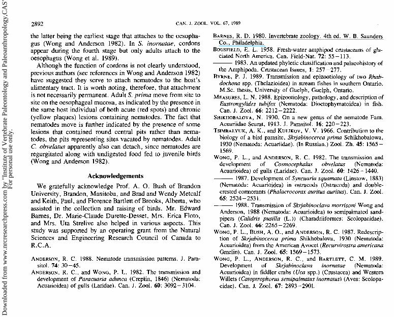
Although the function of cordons is not clearly understood, previous authors (see references in Wong and Anderson 1982) have suggested they serve to attach nematodes to the host's alimentary tract. It is worth noting, therefore, that attachment is not necessarily permanent. Adult S. prima move from site to site on the oesophageal mucosa, as indicated by the presence in the same host individual of both acute (red spots) and chronic (yellow plaques) lesions containing nematodes. The fact that nematodes move is further indicated by the presence of some lesions that contained round central pits rather than nema- todes, the pits representing sites vacated by nematodes. Adult C. obvelatus apparently also can detach, since nematodes are regurgitated along with undigested food fed to juvenile birds (Wong and Anderson 1982).
Acknowledgements We gratefully acknowledge Prof. A. 0. Bush of Brandon
University, Brandon, Manitoba, and Brad and Wendy Metcalf and Keith, Paul, and Florence Bartlett of Brooks, Alberta, who assisted in the collection and raising of birds. Mr. Edward Barnes, Dr. Marie-Claude Durette-Desset , Mrs. Erica Floto, and Mrs. Uta Strelive also helped in various aspects. This study was supported by an operating grant from the Natural Sciences and Engineering Research Council of Canada to R.C.A.
ANDERSON, R. C. 1988. Nematode transmission patterns. J. Para- sitol. 74: 30-45.
ANDERSON, R. C., and WONG, P. L. 1982. The transmission and development of Paracuaria adunca (Creplin , 1 846) (Nematoda : Acuarioidea) of gulls (Laridae) . Can. J. Zool. 60: 3092 - 3 104.
BARNES, R. D. 1980. Invertebrate zoology. 4th ed. W. B. Saunders Co., Philadelphia.
BOUSFIELD, E. L. 1958. Fresh-water amphipod crustaceans of gla- ciated North America. Can. Field-Nat. 72: 55 - 1 13.
1983. An updated phyletic classification and palaeohistory of the Amphipoda. Crustacean Issues, 1 : 257 - 277.
BYRNE, P. J. 1989. Transmission and epizootiology of two Rhab- dochona spp. (Thelazioidea) in stream fishes in southern Ontario. M .Sc. thesis, University of Guelph, Guelph, Ontario.
MEASURES, L. N. 1988. Epizootiology, pathology, and description of Eustrongylides tubifex (Nematoda: Dioctophymatoidea) in fish. Can. J. Zool. 66: 2212 -2222.
SHIKHOBALOVA, N. 1930. On a new genus of the nematode Fam. Acuariidae Seurat, 19 13. J. Parasitol. 16: 220 - 223.
TSIMBALYUK, A. K . , and KULIKOV, V. V. 1966. Contribution to the biology of a bird parasite, Skrjabinocerca prima Schikhobalowa, 1930 (Nematoda: Acuariidae). (In Russian.) Zool. Zh. 45: 1565 - 1569.
WONG, P. L., and ANDERSON, R. C. 1982. The transmission and development of Cosmocephalus obvelatus (Nematoda: Acuarioidea) of gulls (Laridae). Can. J. Zool. 60: 1426 - 1440.
1987. Development of Syncuaria squamata (Linstow , 1883) (Nematoda: Acuarioidea) in ostracods (Ostracoda) and double- crested cormorants (Phalacrocorax auritus auritus) . Can. J. Zool. 65: 2524 - 253 1 .
1988. Transmission of Skrjabinoclava morrisoni Wong and Anderson, 1988 (Nematoda: Acuarioidea) to semipalmated sand- pipers (Calidris pusilla (L.)) (Charadriiformes: Scolopacidae) . Can. J. Zool. 66: 2265 -2269.
WONG, P. L., BUSH, A. O., and ANDERSON, R. C. 1987. Redescrip- tion of Skrjabinocerca prima Shikhobalova, 1930 (Nematoda: Acuarioidea) from the American Avocet (Recurvirostra americana Gmelin). Can. J. Zool. 65: 1569- 1573.
WONG, P. L., ANDERSON, R. C., and BARTLETT, C. M. 1989. Development of Skrjabinoclava inomatae (Nematoda: Acuarioidea) in fiddler crabs (Uca spp.) (Crustacea) and Western Willets (Catoptrophorus semipalmatus inomatus) (Aves: Scolopa- cidae). Can. J. Zool. 67: 2893-2901.
Can
. J. Z
ool.
Dow
nloa
ded
from
ww
w.n
rcre
sear
chpr
ess.
com
by
"Ins
titut
e of
Ver
tebr
ate
Pale
onto
logy
and
Pal
eoan
thro
polo
gy,C
AS"
on
05/2
8/13
For
pers
onal
use
onl
y.