Detection and characterization of a sialoglycosylated bacterial ABC-type phosphate transporter...
15
Detection and characterization of a sialoglycosylated bacterial ABC-type phosphate transporter protein from patients with visceral leishmaniasis Angana Ghoshal & Sumi Mukhopadhyay & Rodion Demine & Michael Forgber & Saulius Jarmalavicius & Bibhuti Saha & Shyam Sundar & Peter Walden & Chhabinath Mandal & Chitra Mandal Received: 5 April 2008 / Revised: 23 September 2008 / Accepted: 15 December 2008 / Published online: 29 January 2009 # Springer Science + Business Media, LLC 2009 Abstract We report the discovery and characterization of a glycosylated bacterial ABC-type phosphate transporter iso- lated from the peripheral blood mononuclear cell (PBMC) fraction of patients with visceral leishmaniasis (VL). Three disease-associated 9-O-acetylated sialoglycoproteins (9-O- AcSGPs) of 19, 56 and 65 kDa, respectively, had been identified and their purity, apparent mass and pI established by SDS-PAGE and isoelectric focusing. Western blot analyses showed that the 9-O-acetylated sialic acid is linked via α2→6 linkage to a subterminal N-acetylgalactosamine. For the 56 kDa protein, N- as well as O-glycosylations were demonstrated by specific glycosidase treatment and found to account for more than 9 kDa of the protein mass. The presence of sialic acids was further confirmed through thin layer chromatography, fluorimetric HPLC and electro- spray ionization-mass spectrometry. The protein was identified by mass spectrometry and de novo sequencing of five tryptic fragments as a periplasmic ABC-type phosphate transporter of Pseudomonas aeruginosa. The amino acid sequences of the assigned peptides had 83– 100% identity with the NCBI entry for a Pseudomonas transporter protein. Based on the recently reported X-ray structure of a human phosphate-binding protein, we predicted a 3D structural model for the 56 kDa protein using homology and threading methods. The most probable N- and O-glycosylation sites were identified by combinations of sequence motif-searching bioinformatics tools, solvent accessibility calculations, structural environment analyses and mass spectrometric data. This is the first reported glycosyla- tion as well as sialylation of the periplasmic component of an ABC-type phosphate transporter protein and of one of few identified bacterial glycoproteins. Glycoconj J (2009) 26:675–689 DOI 10.1007/s10719-008-9223-8 Electronic supplementary material The online version of this article (doi:10.1007/s10719-008-9223-8) contains supplementary material, which is available to authorized users. A. Ghoshal : S. Mukhopadhyay : C. Mandal Department of Infectious Disease and Immunology, Indian Institute of Chemical Biology, 4 Raja S.C. Mullick Road, Kolkata 700032, India C. Mandal Structural Biology and Bioinformatics Division, Indian Institute of Chemical Biology, 4 Raja S.C. Mullick Road, Kolkata 700032, India R. Demine : M. Forgber : S. Jarmalavicius : P. Walden Department of Dermatology, Charité-Universitätsmedizin Berlin, Humboldt University, 10098 Berlin, Germany B. Saha Department of Tropical Medicine, School of Tropical Medicine, Kolkata, India S. Sundar Department of Medicine, Institute of Medical Sciences, Banaras Hindu University, Varanasi, India C. Mandal (*) Infectious Disease and Immunology Division, Indian Institute of Chemical Biology, 4, Raja S. C. Mullick Road, Kolkata 700032, India e-mail: [email protected]
-
Upload
independent -
Category
Documents
-
view
1 -
download
0
Transcript of Detection and characterization of a sialoglycosylated bacterial ABC-type phosphate transporter...

Detection and characterization of a sialoglycosylated bacterialABC-type phosphate transporter protein from patientswith visceral leishmaniasis
Angana Ghoshal & Sumi Mukhopadhyay &
Rodion Demine & Michael Forgber &
Saulius Jarmalavicius & Bibhuti Saha & Shyam Sundar &
Peter Walden & Chhabinath Mandal & Chitra Mandal
Received: 5 April 2008 /Revised: 23 September 2008 /Accepted: 15 December 2008 / Published online: 29 January 2009# Springer Science + Business Media, LLC 2009
Abstract We report the discovery and characterization of aglycosylated bacterial ABC-type phosphate transporter iso-lated from the peripheral blood mononuclear cell (PBMC)fraction of patients with visceral leishmaniasis (VL). Threedisease-associated 9-O-acetylated sialoglycoproteins (9-O-AcSGPs) of 19, 56 and 65 kDa, respectively, had beenidentified and their purity, apparent mass and pI establishedby SDS-PAGE and isoelectric focusing. Western blotanalyses showed that the 9-O-acetylated sialic acid is linkedvia α2→6 linkage to a subterminal N-acetylgalactosamine.For the 56 kDa protein, N- as well as O-glycosylationswere demonstrated by specific glycosidase treatment andfound to account for more than 9 kDa of the protein mass.The presence of sialic acids was further confirmed throughthin layer chromatography, fluorimetric HPLC and electro-
spray ionization-mass spectrometry. The protein wasidentified by mass spectrometry and de novo sequencingof five tryptic fragments as a periplasmic ABC-typephosphate transporter of Pseudomonas aeruginosa. Theamino acid sequences of the assigned peptides had 83–100% identity with the NCBI entry for a Pseudomonastransporter protein. Based on the recently reported X-raystructure of a human phosphate-binding protein, wepredicted a 3D structural model for the 56 kDa proteinusing homology and threading methods. The most probableN- and O-glycosylation sites were identified by combinationsof sequence motif-searching bioinformatics tools, solventaccessibility calculations, structural environment analyses andmass spectrometric data. This is the first reported glycosyla-tion as well as sialylation of the periplasmic component of anABC-type phosphate transporter protein and of one of fewidentified bacterial glycoproteins.
Glycoconj J (2009) 26:675–689DOI 10.1007/s10719-008-9223-8
Electronic supplementary material The online version of this article(doi:10.1007/s10719-008-9223-8) contains supplementary material,which is available to authorized users.
A. Ghoshal : S. Mukhopadhyay : C. MandalDepartment of Infectious Disease and Immunology,Indian Institute of Chemical Biology,4 Raja S.C. Mullick Road,Kolkata 700032, India
C. MandalStructural Biology and Bioinformatics Division,Indian Institute of Chemical Biology,4 Raja S.C. Mullick Road,Kolkata 700032, India
R. Demine :M. Forgber : S. Jarmalavicius : P. WaldenDepartment of Dermatology, Charité-Universitätsmedizin Berlin,Humboldt University,10098 Berlin, Germany
B. SahaDepartment of Tropical Medicine, School of Tropical Medicine,Kolkata, India
S. SundarDepartment of Medicine, Institute of Medical Sciences,Banaras Hindu University,Varanasi, India
C. Mandal (*)Infectious Disease and Immunology Division,Indian Institute of Chemical Biology,4, Raja S. C. Mullick Road,Kolkata 700032, Indiae-mail: [email protected]

Keywords Pseudomonas aeruginosa . SialylatedABC-typephosphate transporter protein .De novo peptide sequencing .
Molecular modeling . Visceral leishmaniasis
Abbreviations9-O-AcSA 9-O-acetylated sialic acid9-O-AcSGP 9-O-acetylated sialoglycoproteinABC ATP-binding cassetteBSA bovine serum albuminBSM bovine submandibular mucinDa daltonDIG digoxigeninDSA Datura stramonium agglutininDMB 1,2-diamino-4,5-methylenedioxybenzeneddH2O double distilled waterEDTA ethylenediaminetetraacetic acidELISA enzyme-linked immunosorbent assaysESI-MS electrospray ionization mass spectrometryFITC fluorescein isothiocyanateGalNAc N-acetylgalactosamineGNA Galanthus nivalis agglutininGP-56 56 kDa O-acetylated sialoglycoproteinGT glycosyltransferaseHPLC high-performance liquid chromatographyHRP horseradish peroxidaseHPBP human phosphate-binding proteinIEF isoelectric focusingkDa kilodaltonLD Leishmania donovaniMAA Maackia amurensis agglutininMALDI-TOFMS
matrix-assisted laser desorption/ionizationtime-of-flight mass spectrometry
Mab monoclonal antibodyMS mass spectrometryGlcNAc N-acetylglucosamineNeu5Ac N-acetylneuraminic acidOAcSGP O-acetylated sialoglycoproteinsPA Pseudomonas aeruginosaPB potassium phosphate bufferPBMC peripheral blood mononuclear cellsPBS phosphate-buffered salinePDB protein data basePE phycoerythrinPMF peptide mass fingerprintPMSF phenylmethylsulfonyl fluoridePNA peanut agglutininPSD post-source decayRIA radioimmunoassayRMS root mean squareRMSD root mean square deviationSA sialic acidSDS-PAGE sodium dodecyl sulfate polyacrylamide gel
electrophoresis
SNA Sambucus nigra agglutininTBS Tris-buffered salineTLC thin layer chromatographyTM transmembraneVL visceral leishmaniasis
Introduction
Sialic acids (SA) constitute a family of N- and O-substituted 9-carbon-carboxylated monosaccharides, themajor member being N-acetylneuraminic acid (Neu5Ac)[1]. Most frequent are O-acetylations at positions C-7/8/9that generate a family of O-acetylated sialoglycoconjugates(O-AcSGs) [2]. Molecular modifications of sialic acids byO-acetylation and alterations of their patterns have beenfound to be of great importance in different pathologicalconditions and hence have become the focus of intenseresearch in our and other laboratories around the world [3,4]. There is growing evidence that a number of physiologicaland pathological processes involving cell–cell adhesion,signaling, differentiation and metastasis may be associatedwith O-acetylated sialoglycotopes [3]. The levels of SAmay signal acuteness of disease and, thereby, indicate theneed to combat the causative agents or processes [2, 3]. Weused the specificity of the snail lectin Achatinin-H forglycoproteins with terminal 9-O-acetylated sialic acid (9-O-AcSA) derivatives linked in α2-6 linkage to subterminalN-acetylgalactosamine (GalNAc) to identify and analyzethese specific sialoglycotopes [5]. In previous studies, wehave detected 9-O-AcSGPs on erythrocytes of patients withvisceral leishmaniasis (VL), not on normal erythrocytes andthese sialylations correlate directly with the degree ofcomplement-mediated hemolysis [6]. Likewise, VL-associated9-O-AcSGPs corresponding to molecular masses of 19, 56and 65 kDa were found on peripheral blood mononuclearcells (PBMC) of patients and not on PBMC of normalindividuals [7].
VL is caused by the intracellular parasitic kinetoplastidprotozoa Leishmania donovani (LD) [8]. An estimated12 million humans are infected, with an incidence of0.5 million cases of VL per year [9]. Approximately 50% ofthe world’s VL cases occur on the Indian subcontinent,90% of them in Bihar alone [9]. Because of the associationof 9-O-AcSGPs with VL pathology, we investigated theover-all density of these sialoglycotopes on PBMC andreport the purification, identification and molecular charac-terization of the PBMC-associated 56 kDa glycoprotein,here named GP-56. Both N- and O-linked glycosylations ofthis protein were demonstrated. Tryptic fragments of theprotein were sequenced by matrix-assisted laser desorption/ionization time-of-flight (MALDI-TOF) with post source
676 Glycoconj J (2009) 26:675–689

decay (PSD) mass spectrometry that led to its identificationas the periplasmic component of an ABC-type phosphatetransporter of Pseudomonas aeruginosa (PA), which hashomology with a recently discovered human plasmaphosphate-binding protein (HPBP) [10]. We used thepublished structure of HPBP to develop a 3-D structurefor GP-56 by molecular modeling and the resulting modelto predict the glycosylation sites.
Materials and methods
Clinical samples
The study involved clinically confirmed VL patients (20males, 10 females, median age, 30 years, range, 10–50 years)of the Kala-Azar Medical Research Center, Muzaffarpur andthe School of Tropical Medicine, Kolkata. Diagnosis of VLwas based on microscopic demonstration of Leishmaniaamastigotes in splenic aspirates according to WHO recom-mendations [11]. The median white blood cell count of theVL patients was 1.2×10⁶/mL (range 1.0–1.4×10⁶/mL).PBMC of patients with high parasitemia and spleen sizes of5 to 8 cm were prepared from venous blood (5–6 mL) bydensity centrifugation (Ficoll-Hypaque, Amersham Pharma-cia, Uppsala, Sweden), washed thoroughly at 4°C andprocessed within 2–12 h of collection. The InstitutionalHuman Ethical Committee had approved the study andsamples were taken with the consent of the donors, patients,or their parents or guardians.
Detection reagents and flow cytometry
All chemicals were purchased from Sigma (St. Louis, USA)unless stated otherwise. The lectin, Achatinin-H, wasaffinity-purified from the hemolymph of African giant landsnail Achatina fulica using bovine submandibular mucin(BSM), known to contain a high percentage of 9-O-AcSAs,as affinity matrix [5]. It was conjugated with fluoresceinisothiocyanate (FITC) for flow cytometry and coupled toSepharose 4B [12] for purification of 9-O-AcSGPs. Themonoclonal antibodies (Mabs) including anti CD3, CD13,CD16 and CD19 were purchased from Pharmingen (SanDiego, CA). For flow cytometry, PBMC (1×10⁶/100 μL)were suspended in RPMI-1640 supplemented with 2 mMglutamine, gentamicin and 10% heat-inactivated human ABserum and stained on ice for 1 h with FITC-Achatinin-Hand phycoerythrin (PE)-anti-CD Mabs along with appro-priate isotype controls. The cells were then washed, fixed inparaformaldehyde (1%) and analyzed with a FACS Caliburflow cytometer (Becton Dickinson, Mountain View, CA).The data were analyzed with the Cell Quest software (BectonDickinson). The presence of 9-O-AcSGPs on the PBMC was
confirmed by flow cytometry with FITC-Achatinin-H after3 h at 37°C pretreatment or no pretreatment with 9-O-acetylhemagglutinin esterase of influenza C virus known tospecifically cleave off O-AcSAs [13].
Purification of 9-O-AcSGPs from PBMC
PBMC (1×10⁷) of clinically confirmed VL patients (n=30)with 80–90% 9-O-AcSGP-positive cells were extensivelywashed with ice-cold phosphate buffered saline (PBS),suspended in cold homogenization buffer Tris–HCl (50mM, pH 7.2; TB) containing 1 mM ethylenediaminetetra-acetic acid (EDTA) with a protease inhibitor cocktailconsisting of 1 mM phenylmethylsulfonyl fluoride (PMSF),10 μg/mL leupeptin and 10 μg/mL aprotinin, and homog-enized on ice with a Teflon pestle in glass tube. Cell debriswas removed by centrifugation at 1,000×g for 5 min at 4°Cand the supernatant centrifuged at 21,000×g for 30 min at4°C. The membrane pellet was resuspended in solubilizationbuffer, i.e. TB with 150 mM NaCl (TBS), glycerol (10%),10 mM EDTA, 1.5 mM MgCl2 and protease inhibitorcocktail as above by sonication for 15 s intermittently on ice[12, 14]. The suspension was adjusted to 0.1% SDS andcentrifuged. The supernatant containing 0.8 mg protein wasdialyzed against TBS containing 0.03 M CaCl2 and 0.02%sodium azide (TBS–Ca2+) at 4°C and then passed through anAchatinin-H Sepharose 4B affinity column (1×2 cm; 1.0 mgAchatinin-H/mL gel) equilibrated with TBS–Ca2+ at 4°C.After washing off unbound material with the same buffer,bound 9-O-AcSGPs were eluted at 25°C with TBS contain-ing 0.04 M sodium citrate, dialyzed at 4°C against TBS andstored at −70°C.
Electrophoresis
Affinity-purified 9-O-AcSGPs (30 μg/lane) were analyzedby SDS-PAGE with 10% acryl amide in a Minigelapparatus (BioRad, Hercules, CA) and stained withCoomassie brilliant blue R-250 [12]. For Western blotanalysis, the proteins were electroblotted onto nitrocelluloseat 100 V for 2 h. After blocking the nitrocellulose with 10%BSA in TBS, the membrane was probed with Achatinin-H(150 μg/mL) in presence of 0.03 M Ca2+. After washing,the blots were incubated with rabbit anti-Achatinin-Hantibodies (1:400) at 4°C and washed. The antigen–antibody complexes were then detected with horseradishperoxidase (HRP)-conjugated goat anti-rabbit IgG(1:10,000; Cappel, St. Louis, MO). To obtain pure protein,Coomassie-stained bands were excised from the electro-phoresis gels and the proteins eluted using an Electro-EluterModel 422 (Bio Rad) according to the manufacturer’sinstruction. IEF was carried out with 9-O-AcSGPs andpurified GP-56 (1.5 μg) in capillary tubes with ampholine
Glycoconj J (2009) 26:675–689 677

polyacrylamide gels (4%), pH range 3.5–10.0 using Mini-PROTEAN II tube cell apparatus (Bio-Rad). The sampleswere focused at a constant voltage of 400 V for 6 h. The gelswere then fixed, washed and silver-stained [12]. The isoelec-tric points (pI) of the individual proteins were determined as afunction of their migration from the cathode using standard pImarkers for a pH range of 3.5 to 10 (Bio-Rad).
Analysis of carbohydrates
The presence of terminal sugars on 9-O-AcSGPs wasanalyzed by a Digoxigenin (DIG) enzyme assay using acommercially available DIG-Glycan differentiation kit(Roche Applied Science, Mannheim, Germany) with theplant lectins Galanthus nivalis agglutinin (GNA), Sambucusnigra agglutinin (SNA), Maackia amurensis agglutinin(MAA), peanut agglutinin (PNA) and Datura stramoniumagglutinin (DSA) according to the manufacturer’s protocol.Total SA contents were measured fluorimetrically using pureSA as standard. The SA of the glycoproteins was mildlyoxidized with sodium metaperiodate and the resultingformaldehyde determined by reaction with acetylacetone andammonium acetate, which produces a fluorogen, detected at510 nm upon excitation at 410 nm with an F-4010spectrofluorimeter (Hitachi, Tokyo, Japan). 8- or 9-O-acetylated sialic acids were not oxidized under theseconditions and are quantified by measuring the amount offluorogen before and after de-O-acetylation with 0.1 MNaOH [15]. To differentiate N- or O-linked glycosylation,glycoproteins were deglycosylated with specific glycosidasesusing the deglycosylation kit as per manufacturer’s instruc-tions (Roche Applied Science). Briefly, the GP-56 wasincubated overnight with Arthrobacter ureafaciens neur-aminidase (specific activity, 0.5 mU) in denaturation buffer(100 mM sodium phosphate, 10 mM EDTA 0.5% (w/v)Triton X-100, 0.05% (w/v) SDS, 1% (v/v) 2-mercaptoethanol,pH 8.6) at 37°C. The reaction mixture was heated at 100°C for3 min and centrifuged. Then, reaction buffer at pH 7.2containing CHAPS and N-glycosidase F was added and thesample incubated overnight at 37°C. For O-glycosidase (1–2mU) or combination of N- and O-glycosidase treatment,neuraminidase treated samples were incubated overnightwith the enzymes in potassium phosphate buffer (20 mM)containing EDTA (10 mM), Triton X-100 (0.5%) and SDS(0.05%) at pH 7.3 (PB) and 37°C. For IEF, the samples weredesialylated overnight with neuraminidase (0.5–1 mU) in PBat 37°C.
Detection of sialic acids of GP-56 through TLC
Gel-eluted GP-56 (25 μg) was hydrolyzed with 4 M propionicacid for 4 h at 80°C to release sialic acid. It was subsequentlypassed onto Dowex 50WX8 (100–200 mesh) cation exchange
and Dowex 2X8 (200–400 mesh) anion exchange columns.The eluted free sialic acids were collected, lyophilized andresuspended in double distilled water (ddH2O, 40 μL), spotted(5 μg/10 μL) onto TLC plates (Merck KGaA, Germany) andrun in 1-propanol/H2O (7:3 v/v). Sialic acids released fromBSM along with Neu5Ac (Sigma) were used as internalstandards. Plates were developed by spraying with orcinol/HCl/FeCl3 spray reagent and heated at 180°C for 20 min [16].
Fluorimetric HPLC for estimation of sialic acids of GP-56
Gel-eluted GP-56 was lyophilized and subjected to mild acidhydrolysis with 200 μL of 4 M propionic acid for 4 h at 80°Cto release glycosidically bound Neu5Ac. Reaction mixturewas cooled on ice for 15 min. An aliquot of this sample wasderivatized with 1,2-diamino-4,5-methylenedioxybenzene(DMB) for fluorimetric reverse-phase HPLC analysis [12,13, 16]. Analysis of the derivatized sialic acids was done onan RP-18 column (LichroCART 125-4 HPLC-cartridge,5 μm; Merck, Germany) using isocratic elution with water/acetonitrile/methanol (84:9:7, v/v/v) at a flow rate 1 mL/minand compared with authentic sialic acid purified from BSM.Fluorescence was detected using an excitation and emissionwavelength of 373 and 448 nm, respectively.
ESI-Q-TOF-MS of DMB derivatized sialic acids obtainedfrom GP-56
The mass spectrometric data was acquired on a Q-Tof micro-mass spectrometer (Waters Corporation, Massachusetts, USA).The fraction corresponding to DMB derivatized Neu5Ac fromGP-56 was collected after fluorimetric HPLC, dried, dissolved(water/acetonitrile/methanol; 84:9:7, v/v/v) and analyzed di-rectly via ESI-MS at a flow rate of 10 μL/min. The ion sprayvoltage was operated at 5 kV. The tuning and calibrationsolution consisted of an equimolecular mixture of polypro-pylene glycol 425, 1,000, and 2,000 in 50/50/0.1 H2O/methanol/formic acid (v/v/v), NH4OAc (1.0 mM). Spectrawere acquired by scanning from m/z=80 to m/z=800 [17].
Binding assays
To analyze the kinetics of sialoglycoproteins binding byAchatinin-H, the lectin was iodinated with 125I (AmershamPharmacia Biotech, Sweden) by the chloramine T method[18] yielding a specific activity of 1.5×10⁶ cpm/μg. Formeasuring the binding to cells, PBMC (2×10⁶/150 μL pertube) from five patients were suspended in 10 μL RPMI-1640 medium containing CaCl2 (0.3 M) and bovine serumalbumin (BSA, 0.2%), pH 7.2 (reaction buffer). The cellswere incubated with increasing doses of 125I-Achatinin-Hfor 90 min at 4°C and washed. The bound radioactivity inthe cell pellets was measured with a gamma counter
678 Glycoconj J (2009) 26:675–689

(Electronic Corporation, India). For controlling the specificityof binding, a 100-fold excess of unlabeled Achatinin-H wasadded. The data were processed by Scatchard analyses and thenumber of binding sites for Achatinin-H per cell wascalculated from the intersection of the curve with the X-axisand the dissociation constant, Kd, obtained by dividing thenumber of receptors per cell by the bound/free ratio derivedfrom the Y-axis intercept of the Scatchard plot [18].
Protein/peptide mass spectrometry
The measurements to identify the glycoprotein were donewith a Bruker-Daltonik reflector MALDI-TOF mass spec-trometer Reflex IV with PSD capabilities (Bruker Daltonik,Bremen, Germany). The samples were prepared by dried-droplet procedure using 2,5-dihydroxybenzoic acid asmatrix. Calibration was done externally with a mixture ofAngiotensin I, Angiotensin II, Substance P, Bombesine,ACTH clip 1–17 and ACTH clip 18–39. The accuracy ofpeptide mass measurement was 0.1 Da and that of peptidefragment mass measurement 0.5 Da. 10–100 pmol ofpeptide samples were used for analysis. Tryptic fragmentswere generated by digestion of GP-56 from a SDS-PAGEgel band overnight in ammonium hydrocarbonate (5 mM)with 10 ng trypsin (Promega, Mannheim, Germany) persample. For sequence analysis, selected tryptic fragments ofthe protein were subjected to PSD and fragment massdetection. MALDI-PSD data was processed using theSequit! 4.0 de novo sequencing software with subsequentsearches in the NCBI database version July 25, 2006 usingBLAST version 2.2.13 [19]. MS/MS ion searches andpeptide mass fingerprint (PMF) analyses were done withMASCOT using the same settings as for Sequit!.
Database searches, prediction of glycosylation sites
For searches in sequence databases, standard BLASTwith theNCBI database version as cited was used. Sequence align-ments were done with CLUSTALW [20]. For prediction ofN-glycosylation sites we employed PROSITE software [21].In addition, NetNglyc 1.0 (http://www.cbs.dtu.dk/services/NetNGlyc) was used for the prediction of N-glycosylationsites. Predictions of O-linked glycosylation sites were donewith the neural network-based NetOGlyc 3.1.
3-D structural modeling and refinement
3-D structural modeling was done with homology modelingsoftware SWISS-MODEL [22] and with the threadingmethod-based LOOPP software package [23]. LOOPP usessequence alignment, sequence profile, threading, secondarystructure and exposed surface area predictions, and mergesthem into a single score, generating atomic coordinates
based on alignment of few homologous template structures.Both approaches were based on structures available in theprotein data bank (PDB). 3-D structure alignments weredone with ABGEN [24] to select core structures as startingscaffolds of target proteins for subsequent refinement. Thestarting structures were refined using the software suiteInsight II (2005) (Accelrys, San Diego, CA) equipped withDISCOVER for energy minimization and molecular dynam-ics simulation. Structural optimization involved moleculardynamics simulation followed by energy minimization (100steps each of steepest descent and conjugate gradientmethods) of the lowest energy conformation using cff91 forcefield. A typical dynamics run for structure regularizationconsisted of 10,000 steps of 1 fs after 1,000 steps ofequilibration with a conformation sampling of one in 10 stepsat 300 K. At the end of the molecular dynamics simulation,the conformation with the lowest potential energy was pickedfor energy minimization with the ANALYSIS module ofInsight II followed by a new cycle of refinement. The energy-minimized structure was subjected to various validation teststo check the quality of the refined segments. The conforma-tions of each identified segment were regularized separatelyby this combination of molecular dynamics and energyminimization while keeping the rest of the molecule fixedby applying positional constraints. This was continued untilsatisfactory conformational parameters were obtained. Struc-tures, thus obtained, were energy-minimized as beforekeeping all the atoms of the protein free. The resulting modelwas taken as the initial crude model and used for side chainregeneration and backbone conformation refinement. Sidechain regeneration was done with SCWRL 3.0 [25]. This isthe most recent version of the SCWRL using graph theory tosolve combinatorial problems encountered in side-chainpredictions and allows sophisticated energy functions forincorporation of side-chain flexibility around rotamericpositions. PROCHECK [26] was used for checking theRamachandran’s plots of regularized individual loops as wellas the overall protein models for their stereochemical quality.Amino acids whose Φ and Ψ dihedral angle pair fell outsidethe core and allowed zones of the Ramachandran’s plot wereidentified and grouped into segments. Backbone conforma-tions of these segments were refined to drive the amino acidsto adopt proper Φ–Ψ values by molecular dynamics andenergy minimization while applying constraints on thedihedral angles of the amino acids with Φ–Ψ combinationsmapping to the core and allowed zones of the plot. Formolecular dynamics and energy minimization, the CHARMmodule of Insight II was used with CharmM27 force field.Conformations with correct Φ–Ψ combinations were pickedfrom stored conformations of the molecular dynamics runs.This procedure of backbone conformation refinement wasdone for each identified segment separately until all segmentswere covered and the Ramachandran’s plot checked with
Glycoconj J (2009) 26:675–689 679

PROCHECK. Overall structural validation of the refinedstructures was done with MOLPROBITY [27] which measuresall-atom contacts in terms of clash scores (number of atomshaving atom pair overlaps ≥0.4 Å per 1000 atoms) andcalculates rotamer outliers of the side chains. Our in-housesoftware MODELYN [28] was used for calculation of dihedralangles of protein backbones as well as standard deviations ofbond lengths and bond angles.
Solvent accessibility and stereo-chemical environmentof potential glycosylation sites
Differential solvent accessibilities of all atoms grouped intoamino acids of the refined models were calculated with theACCESS software [29] and the most probable N- and O-linked glycosylation sites were identified by the degree ofsurface exposure. X-ray crystallographic structures with knownN-linked glycosylation taken from PDB were superimposedwith the homology model of the target protein. Then the dis-tances between the nitrogen (ND2) of Asn and oxygen (OG1)of the Ser/Thr residue at the third position of the consensussequence Asn-Xaa-Ser/Thr were measured for assessing thestereo-chemical environment. Likewise, for O-linked glyco-sylation the X-ray structure of an O-glycosylated protein wassuperimposed and distances between two oxygen atoms(OG1) of Thr measured and compared with the model. Thecapacity of accessible sites of the modeled protein to be glyco-sylated was checked by fitting in a covalently linked glycotope.
Computational platforms
ABGEN, PROCHECK and SCWRL 3.0 were run on aFUEL workstation of Silicon Graphics, Inc. in the IRIX 6.5environment. Insight II equipped with the bio-sciencemodules BIOPLOYMER, DISCOBER, CHARMm,ANALYSIS, AFFINITY and others was run on a FUELworkstation as well as, or on an ALTRIX high-end server ofSilicon Graphics, Inc. in the IRIX 6.5 environment. MODE-LYN and ANALYN were run on IBM-compatible PCs inWindows environment. SWISS-MODEL, PROSITE, LOOPP,CLUSTALW, MOLPROBITY, NetNGlyc and NetOGlyc wereused at the web-servers at the internet sites http://swissmodel.expasy.org//SWISS-MODEL.html, http://www.expasy.org/prosite/, http://cbsuapps.tc.cornell.edu/loopp.aspx, http://www.ebi.ac.uk/clustalw/ and http://molprobity.biochem.duke.edu/,www.cbs.dtu.dk/services/netnGlyc, http://www.cbs.dtu.dk/services/NetOGlyc.
Statistical analysis
Results are expressed as mean±SD for individual sets ofdata. Each experiment was performed two to three timesand the results shown are representatives of each set of
experiments. One or two-tailed t tests for significance wereperformed where applicable. P<0.05 was consideredsignificant.
Results
9-O-acetyl-sialoglycoproteins in VL patients
The PBMCs of VL patients were tested by flow cytometryfor 9-O-AcSGPs using FITC-Achatinin-H. Consistently,80–90% of the PBMC were positive for 9-O-AcSGPs.Two-color flow cytometry with FITC-Achatinin-H and PE-labeled leukocyte subpopulation-specific antibodies showedthat 9-O-AcSGPs were present at the surfaces of T cells(CD3, 60.43±5.78%), NK cells (CD16, 11.76±3.53%),monocytes (CD13, 47.82±0.86%) and B cells (CD19,19.54±0.26%). Increased 9-O-AcSGPs were also detectedon erythrocytes of these patients as determined by flowcytometry, enzyme-linked immunosorbent assay (ELISA)and hemagglutination assay [15]. The sera of the patientshad high-levels of anti-9-O-AcSGPs antibodies as detectedby ELISA using BSM as coating antigen [30]. Moreover,the sera also had high levels of parasite-specific antibodiesas determined by parasite-specific ELISA with crudeparasite lysate as coating antigen [30]. The average numbersof 9-O-AcSGP molecules determined by binding measure-ments with 125I-Achatinin-H and Scatchard analysis donewith the PBMC of five VL patients were found to be 2.8×10⁷/cell (Fig. 1a). For confirmation of the binding specificity,an excess of up to 100-fold unlabelled Achatinin-H wasadded and the apparent dissociation constant (Kd) calculatedfrom the inhibition of binding of the radio labeled probe as3.7±0.02 pM.
Purification and characterization of 9-O-AcSGPs
PBMC of clinically confirmed VL patients (n=25) withhigh parasitemia were selected for purification of 9-O-AcSGPs from the membrane fractions by affinity chroma-tography on Achatinin-H-Sepharose 4B. The yield ofpurified 9-O-AcSGP from 1.2×10⁷cells was 0.79±0.12mg, which corresponds to 42.7±2.15% of the total mem-brane proteins (1.85±0.25 mg). SDS-PAGE of the purified9-O-AcSGPs (Fig. 1b, lane 2) revealed three VL-associatedsialoglycoproteins with apparent masses of 19, 56 and 65kDa, respectively. These three glycoproteins are not foundon the PBMC of healthy individuals unlike the two othersialoglycoproteins of 36 and 144 kDa, as has beendemonstrated by Western blot analysis earlier [7]. Thespecificity of these preparations was confirmed by Westernblot analysis with Achatinin-H as reagent for 9-O-AcSGPs(Fig. 1b, lane 3). The first lane of Fig. 1b shows protein
680 Glycoconj J (2009) 26:675–689

staining of the crude membrane extract. For furthercharacterization, GP-56 was eluted from the gel, quantifiedand re-analyzed by SDS-PAGE (Fig. 1b, lane 4). Theamount of GP-56 was 66.36±1.22 μg which correspondedto 8.40±1.15% of the total affinity-purified 9-O-AcSGPs.Treatment of the 9-O-AcSGPs with alkali prior to SDS-PAGE completely abolished the binding by Achatinin-Hfurther supporting the specificity of the detection of O-acetylated glycoproteins (Fig. 1b, lane 5). To furthercharacterize the glycosylation of GP-56, the purifiedmolecule (Fig. 1c, lane 1) was treated with neuraminidasefrom Arthrobacter ureafaciens followed by glycosidases.Treatment with neuraminidase resulted in a shift 1.03 kDa(Fig. 1c, lane 2) corresponding to a band of 54.97 kDa.Subsequent treatment with N-glycosidase F resulted in a shiftof the band to an apparent molecular mass of 49.3 kDa,
corresponding to a reduction by 6.7 kDa (Fig. 1c, lane 3).After O-glycosidase treatment, a shift by 2.13 kDa wasdetected resulting in a molecular mass of 53.87 kDa (Fig. 1c,lane 4). Desialylation and deglycosylation with N- and O-glycosidase yielded a protein band with an apparent mass of47 kDa (Fig. 1c, lane 5) indicating that the glycosylationsaccount for about 9 kDa of the total mass of GP-56. The 9-O-AcSGPs associated with PBMC of VL patients were furtherinvestigated by IEF (Fig. 1d) which produced five distinctbands within a pI range of 3.7 and 6.3, indicating five differentsialoglycoproteins with a similar type of glycotopes (lane B).The distinct band confirms their homogeneity. The purifiedGP-56 showed a single band with an acidic pI of 3.7 (lane C).Neuraminidase treatment of this protein resulted in a singleband with a pI value of about 8.9 (lane D) proving thepresence of SA. Lane A shows the pI markers.
Fig. 1 Characterization of the 9-O-AcSGPs present on PBMC of VLpatient. a Binding of 125I-Achatinin-H. A fixed amount of PBMC (2×10⁶/150 μL) was incubated with increasing amounts of 125I-Achatinin-H.For evaluating the specific nature of binding, a 100-fold excess ofunlabeled Achatinin-H was added. Bound and unbound Achatinin-H wasseparated at 4°C and the bound radioactivity determined. Specificbinding (filled triangles) was calculated as difference between totalbinding (open diamonds) and binding in presence of a 100-fold excess ofunlabeled Achatinin-H (filled squares). Inset Scatchard plot of thebinding data. The results are expressed as mean±SD of data fromtriplicate experiments. b SDS PAGE (10%) and Western blot of 9-O-AcSGP purified from PBMC of VL patients. Crude membranepreparation of PBMC (10 μg, lane 1) from VL patients and 30 μgpurified 9-O-AcSGPs (lane 2) were electrophoresed on SDS-PAGE. Thegel was fixed and stained with Coomassie brilliant blue R-250. Affinitypurified 9-O-AcSGPs (30 μg) from PBMC of VL patients after SDS-
PAGE was subjected to Western blot analysis with Achatinin-H (lane 3).GP-56 was electro eluted from SDS-PAGE and a sample (5 μg)reanalyzed by SDS-PAGE (lane 4). Binding of Achatinin-H wascompletely abolished by de-O-acetylation of the blot by alkali treatment(lane 5). c SDS-PAGE of GP-56 before and after treatment withneuraminidase, N- and O-glycosidase. Affinity purified GP-56 fromPBMC of VL patients was electro eluted from SDS-PAGE (lane 1) andwas treated with neuraminidase from Arthrobacter ureafaciens (lane 2)followed by N-glycosidase F (lane 3), O-glycosidase (lane 4) or acombination of N- and O-glycosidase (lane 5) and further analyzed bySDS-PAGE. d Isoelectric focusing of 9-O-AcSGP and purified GP-56from PBMC of VL patients. 9-O-AcSGPs (1.5 μg, lane B), GP-56electro eluted from SDS-PAGE (lane C) and GP-56 after treatment withArthrobacter ureafaciens neuraminidase (lane D) was analyzed by IEFwith a pH gradient of 3.5 to 10. Lane A shows pI markers
Glycoconj J (2009) 26:675–689 681

To establish the linkage-specific arrangement of theterminal sugars present on the purified 9-O-AcSGPs, weused a DIG glycan differentiation kit with various lectins ofknown specificities for different terminal sugar moieties(Fig. 2a). For the purified 9-O-AcSGPs the binding was 4.0fold higher with SNA (specific for α2→6 linked Neu5Ac)than with MAA (specific for α2→3 linked Neu5Ac). Thecorresponding densitometric scores were 62,313±1,749versus 15,222±1,041 (P<0.0001). Concordantly, the den-sitometric scores for GP-56 binding to SNA were 8.74 foldhigher than to MAA (55,169±1,102 versus 6,308±137,P<0.0001). In contrast, both the 9-O-AcSGPs and thepurified GP-56 showed low binding to GNA, which isspecific for terminal Man (α1→3), (α1→6) and (α1→2)Man, binding being 20,211±1,055 and 10,212±1,023,
respectively. Similarly low were the bindings to DSA,which is specific for Gal (β1→4) GlcNAc and PNA, whichis specific for Gal (β1→3) GalNAc. Fluorimetric analysisof the purified 9-O-AcSGPs demonstrated 316±4.5 μg SA,which corresponds to 40.0% of the 9-O-AcSGPs purifiedby Achatinin-H affinity chromatography from 1.2×10⁷ cells. 81±11.16% of this total SA was 9-O-acetylated.For GP-56, 29.53±1.82 μg SA was detected whichcorresponds to 44.0% of total GP-56, of which 84.0±9.2% was 9-O-acetylated (Fig. 2b).
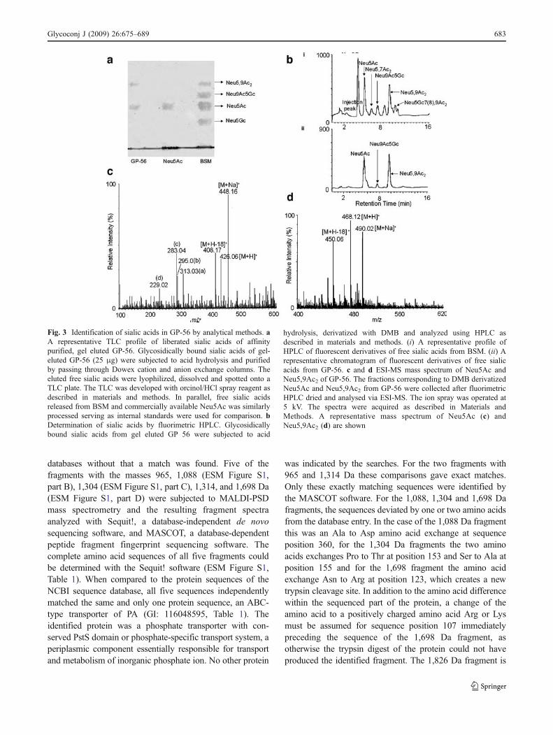
The presence of sialic acid and O-acetylated sialic acid onGP-56 was further substantiated by several analyticaltechniques. When the glycosidically bound sialic acids ofGP-56 were separated by acid hydrolysis and spotted ontothe TLC plate, both sialic acid and O-acetylated sialic acidwere detected by development with orcinol reagent (Fig. 3a).The RF values corresponded to standard Neu5Ac and freesialic acids purified similarly from BSM.
To confirm the presence of SA in GP-56, freed SA wasderivatized with DMB and analyzed by fluorimetric HPLC.Typically, the fluorimetric HPLC chromatogram of acidhydrolysates of GP-56 exhibited a well-resolved intensepeak, which coincided with that of Neu5Ac (Fig. 3b ii).Also a peak comigrating with Neu5,9Ac2 was obtained(Fig. 3b ii) comparable with the profile of sialic acids fromBSM used as internal standard (Fig. 3b i).
For further confirmation of the presence of Neu5Ac(Fig. 3c) and Neu5,9Ac2 (Fig. 3d) in GP-56, ESI-MSstudies of DMB derivatized sialic acid was performed. Thefraction corresponding to Neu5Ac was collected afterfluorimetric HPLC, dried and analysed via ESI-MS. TheESI-MS spectra of Neu5Ac demonstrated the characteristictwo groups of ions for Neu5Ac. The first group corre-sponds to the molecular ion (m/z 426, [M+H]+), and itssodium adduct (448, [M+Na]+). The fragmentation alsoproduced the characteristic second group of ions at m/z 408,[M+H-18]+, corresponding to a first dehydration reaction,and ions (a), (b), (c), and (d) at m/z 313, 295, 283, and 229in Fig. 3c. Similarly, the ESI-MS spectra of Neu5,9Ac2 wasidentified by the characteristic ions at m/z at 450, 468, 490corresponding to [M+H-18]+ , [M+H]+, [M+Na]+ respec-tively (Fig. 3d).
Identification of GP-56
For identification, the 56 kDa band was excised from aCoomassie-stained SDS-PAGE gel with Achatinin-H affinity-purified glycoproteins, destained and treated with trypsin. Thefragments were extracted from the gels and analyzed byMALDI-TOF mass spectrometry given as a ESM Figure S1(part A). Using the MASCOT software, the resulting PMFspectrum was compared to profiles of tryptic fragmentsgenerated in silico from all proteins in the NCBI sequence
Fig. 2 Characterization of the terminal sugars on 9-O-AcSGPs andpurified GP-56. a Differentiation of the terminal glycans by dot blotanalyses. For determination of the terminal linkage-specific carbohy-drates of 9-O-AcSGPs and GP-56 using different lectins in a DIG-linked enzyme assay, densitometric scoring of the scanned blots wasperformed. The intensities of the spots of 9-O-AcSGPs (open squares)and GP-56 electro eluted from SDS-PAGE (filled squares) werequantified densitometrically and represented in arbitrary units usingthe Master Totallab Software, version 1.11 (Amersham PharmaciaBiotech, Sweden). The results are shown as mean±SD. b Fluorimetricquantitation of 9-O-acetylated sialic acid on purified 9-O-AcSGPs andGP-56 from PBMC of VL patients. The O-acetyl groups of 9-O-AcSGPs (open squares) and GP-56 (filled squares) were oxidized withsodium metaperiodate before and after saponification and the resultingformaldehyde quantified by reaction with acetyl acetone and ammoniumacetate by fluorimetric determination of the fluorescent product. Thefractions (in percent) of 9-O-acetylated sialic acids (mean±SD) werecalculated by subtracting fluorescence signal measured for the SA fromthe signals obtained after de-O-acetylation. Pure SA was used as astandard
682 Glycoconj J (2009) 26:675–689
databases without that a match was found. Five of thefragments with the masses 965, 1,088 (ESM Figure S1,part B), 1,304 (ESM Figure S1, part C), 1,314, and 1,698 Da(ESM Figure S1, part D) were subjected to MALDI-PSDmass spectrometry and the resulting fragment spectraanalyzed with Sequit!, a database-independent de novosequencing software, and MASCOT, a database-dependentpeptide fragment fingerprint sequencing software. Thecomplete amino acid sequences of all five fragments couldbe determined with the Sequit! software (ESM Figure S1,Table 1). When compared to the protein sequences of theNCBI sequence database, all five sequences independentlymatched the same and only one protein sequence, an ABC-type transporter of PA (GI: 116048595, Table 1). Theidentified protein was a phosphate transporter with con-served PstS domain or phosphate-specific transport system, aperiplasmic component essentially responsible for transportand metabolism of inorganic phosphate ion. No other protein
was indicated by the searches. For the two fragments with965 and 1,314 Da these comparisons gave exact matches.Only these exactly matching sequences were identified bythe MASCOT software. For the 1,088, 1,304 and 1,698 Dafragments, the sequences deviated by one or two amino acidsfrom the database entry. In the case of the 1,088 Da fragmentthis was an Ala to Asp amino acid exchange at sequenceposition 360, for the 1,304 Da fragments the two aminoacids exchanges Pro to Thr at position 153 and Ser to Ala atposition 155 and for the 1,698 fragment the amino acidexchange Asn to Arg at position 123, which creates a newtrypsin cleavage site. In addition to the amino acid differencewithin the sequenced part of the protein, a change of theamino acid to a positively charged amino acid Arg or Lysmust be assumed for sequence position 107 immediatelypreceding the sequence of the 1,698 Da fragment, asotherwise the trypsin digest of the protein could not haveproduced the identified fragment. The 1,826 Da fragment is
Fig. 3 Identification of sialic acids in GP-56 by analytical methods. aA representative TLC profile of liberated sialic acids of affinitypurified, gel eluted GP-56. Glycosidically bound sialic acids of gel-eluted GP-56 (25 μg) were subjected to acid hydrolysis and purifiedby passing through Dowex cation and anion exchange columns. Theeluted free sialic acids were lyophilized, dissolved and spotted onto aTLC plate. The TLC was developed with orcinol/HCl spray reagent asdescribed in materials and methods. In parallel, free sialic acidsreleased from BSM and commercially available Neu5Ac was similarlyprocessed serving as internal standards were used for comparison. bDetermination of sialic acids by fluorimetric HPLC. Glycosidicallybound sialic acids from gel eluted GP 56 were subjected to acid
hydrolysis, derivatized with DMB and analyzed using HPLC asdescribed in materials and methods. (i) A representative profile ofHPLC of fluorescent derivatives of free sialic acids from BSM. (ii) Arepresentative chromatogram of fluorescent derivatives of free sialicacids from GP-56. c and d ESI-MS mass spectrum of Neu5Ac andNeu5,9Ac2 of GP-56. The fractions corresponding to DMB derivatizedNeu5Ac and Neu5,9Ac2 from GP-56 were collected after fluorimetricHPLC dried and analysed via ESI-MS. The ion spray was operated at5 kV. The spectra were acquired as described in Materials andMethods. A representative mass spectrum of Neu5Ac (c) andNeu5,9Ac2 (d) are shown
Glycoconj J (2009) 26:675–689 683

an extension of the 1,698 Da fragment by a C-terminal Lys.This fragment could now be identified by incorporating thenew sequence of the 1,698 Da fragment into the databasesequence for the protein. Once GP-56 was identified as theABC-type phosphate transporter of PA recorded in the NCBIdatabase, two more fragments could be assigned to the sameprotein by their masses of 1,100 Da and 1,975 Da. Thereby,eight masses of the tryptic digest of GP-56 could be assignedto the bacterial phosphate transporter. The sequence of thePA periplasmic ABC-like phosphate transporter plus thesequences of the identified fragments are shown in Table 1with the variant amino acids in boldface letters. Thesequence coverage of the five sequenced fragments plusthe three additional fragments identified by their fragment
masses was 27% of the full length of the protein sequence.However, the five completely sequenced fragments could bematched unequivocally by sequence comparison thus iden-tifying GP-56 as a variant of the PA periplasmic ABC-likephosphate transporter listed in the NCBI database.
BLAST searches with the amino acid sequence of the PAABC-like phosphate transporter as query sequence yieldeda number of proteins with conserved PstS (ABC-typephosphate transport system, periplasmic component) do-main. The sequence of the protein, we identified, showed a92% identity with the hypothetical periplasmic bindingprotein PaerP_01000347 of PA7 and 73% identity with aphosphate ABC transporter, periplasmic phosphate bindingprotein of Pseudomonas fluorescens. The periplasmic
Table 1 Identification of GP-56 by mass spectrometric de novo sequencing and peptide mass analyses
No. Predictedmassa [Da]
Observedmassb [Da]
Sequencec Sequencepositiond
GI: 116048595 sequencee Method of identificationf
De novoSequit!
PFFMASCOT
Peptidemass
1 965.5414 965.5700 SGAITVVYR 158–166 SGAITVVYR ✓ ✓ ✓
2 1,044.5877 1,088.4400 FVPLPDSWK 355–363 FVPLPASWK ✓
3 1,100.5118 1,100.5300 NVHWAGSDSK 78–87 NVHWAGSDSK ✓
4 1,314.6172 1,314.6200 SESSGTTELFTR 167–178 SESSGTTELFTR ✓ ✓ ✓
5 1,316.6594 1,303.9310 LTDWSQITGAGR 146–157 LTDWSQIPGSGR ✓
6 g 1,698.0840 LIQVPSVATSVALPFR 108–123 LIQVPSVATSVALPFN ✓
7 g 1,826.1640 LIQVPSVATSVALPFRK 108–124 LIQVPSVATSVALPFNK ✓
8 1,974.9919 1,975.0300 AAFLNNDYTKFVAGTTNK 60–77 AAFLNNDYTKFVAGTTNK ✓
S. Sequence of the Pseudomona aeruginosa ABC-type phosphate transporter, periplasmic domain, GI: 116048595 with the identified trypticfragments of GP-56. Sites where amino acid exchanges have created new trypsin cleavage sites: exchange of P to either K or R in position 107, Nto R exchange in position 123 identified by sequencing the 1698 Da fragment are denoted by an asterisk. All identified deviations from the GI:116048595 sequence are indicated with bold face letter. The predicted N-glycosylation site NETN (92–95) and the two predicted O-glycosylationsites T273 and 306 are indicated with bold face and underlined letter
1
MYKRSLIAASLSVAALVSAQAMADINGGGATLPQQLYQEPGVLTAGFAAYIGVGSGNGKAAFLNNDYTKFVAGTTNKNVHWAGSDSKLSKTNETNPYLSA
AAFLNNDYTKFVAGTTNK
NVHWAGSDSK
101 * *
HGSAWGPLIQVPSVATSVALPFNKSGSNAVNFADVNTLCGVFSGRLTDWSQIPGSGRSGAITVVYRSESSGTTELFTRFLNASCSSTLEGGTFAITTSFG
LIQVPSVATSVALPFR LTDWSQITGAGR SESSGTTELFTR
LIQVPSVATSVALPFRK SGAITVVYR
201
SSFSGGLPAGAVSAQGSQAVMNALNAAQGRITYMSPDFAAPTLAGLDDATKVAQVRGVSPAPANVSAAIGAVTPPTTAQRSDPNNWVPVFAATANPNDPS
301
VRPYPTSGYPILGFTNLIFSQCYANATQTQQVRDFFTRHYGATANNDTAITNHRFVPLPASWKLAVRQSFLTSTNNLYIGHSNVCNGIGRPL
FVPLPDSWK
aMasses of tryptic fragments predicted from the sequence of the Pseudomonas aeruginosa ABC-type phosphate transporter, periplasmic domain,GI: 116048595 (MASCOT)bMass of the tryptic fragments determined by MALDI-TOF mass spectrometryc Sequence of the tryptic fragment of GP-56d Sequence position of the tryptic fragment of GP-56 according to the GI: 116048595 sequencee Sequence of the fragments of the Pseudomona aeruginosa ABC-type phosphate transporter corresponding to the identified tryptic fragment ofGP-56f Identification of the tryptic fragment of GP-56 by complete database-independent de novo sequencing using the Sequit! Software, peptidefragment fingerprint using MASCOT, peptide mass including the mutations identified with this workg The indicated fragments could not be predicted from the database entry GI: 116048595 because of mutation framing or in this fragment that havecreated new trypsin cleavage sites
684 Glycoconj J (2009) 26:675–689

component of another ABC-type phosphate transportsystem of PA showed only 45% sequence identity. A closesimilarity of GP-56 was also found with the HPBP, whichhas 65% sequence identity. The other eukaryotic proteinwith PstS domains and similarity to GP-56 was p27SJ ofHypericum perforatum with 73% sequence identity. Usingthe sequences of GP-56 and HPBP, we searched the wholehuman and mouse genome databases for other homologuesand found no other sequences with significant similaritybesides the above which excluded the possibility that GP-56 is a human protein and by all likelihood, GP-56 is aprotein of Pseudomonas aeruginosa.
3-D structural modeling of the GP-56
We used two approaches to predict 3-D structural modelsfor GP-56. First, homology modeling based on the recentlyreleased (January 2007) X-ray structure of the humanphosphate-binding apolipoprotein (HPBP), PDB code2CAP. A BLAST search with the sequence of GP-56showed 65% amino acid identity with HPBP, 74% positivescore, 6% gaps and an expect value of 2×10−110 implying ahigh confidence in sequence match.
Second, we employed a threading method-based ap-proach using LOOPP software, which compares secondaryconformations of proteins of known structures for buildingan initial model for the target sequence. With the input ofthe GP-56 sequence, five models were predicted from thestructures of the PDB entries 1QUJ, 1A55, 1QUK, 1IXHand 1A40. These five proteins are all of the class ofperiplasmic phosphate-binding and transport proteins of E.coli and contain a defined phosphate-binding domain.Multiple sequence alignment of the sequences of the fiveproteins and GP-56 with CLUSTALW showed that all fivewere of the same size and cover the same segments as GP-56.The model based on 1QUJ had the best score. The other fourmodels were superimposed onto this model to identifystructural segments common to all five. The RMS deviationsfor the identified core structures ranged between 0.47 and0.66 Å and included 55.5% of the Cα atoms indicating closesimilarity of the core structures of the models but deviationsin a number of loops. The loops were identified for the1QUJ-based model and, in order to obtain the best loopconformation, subjected to regularization by moleculardynamic simulation and energy minimizations. The resultinginitial threading-based model was used for subsequentanalysis and refinement of the backbone and side chainconformations. Using the PROCHECK software [26],additional refinement, showed 73.7% of the backboneresidues in the most favored core regions, 26.3% in allowedregions and none in unfavorable regions of Ramachandran’splot. Finally, the side chain conformations were regeneratedusing SCWRL3.0. The same analyses and refinements were
done for the backbone conformations of the 2CAP-(HPBP)-based homology model. Initially, 87.9% and 11.4% of theΦ–Ψ pairs were in the favorable core and allowed regions,respectively, and only three amino acids (0.7%) in unfavor-able regions of the Ramachandran’s plot. After backbonerefinement, 87.6% and 12.4% of the Φ–Ψ pairs were in thecore and allowed regions, respectively. The side chainconformations were then regenerated on the refined back-bone structure as before.
Clash scores and rotamer outliers of the refined threadedand homology models were computed using MOLPROBITYand compared with those of the X-ray structure of HPBP(2CAP). The clash scores of the 3D structures of the threadingand homology models, with respective values of 3.92 and3.83, were comparable to that of the X-ray structure 2CAP(7.10%) suggesting good structural compatibility with all-atom contacts. With 4.78% and 4.90% rotamer outliers for thethreading and homology models, respectively, the side chainconformations were in good agreement with the 1.08% of theX-ray structure of HPBP. The RMS deviations from therespective standard values of all bond lengths and bond angleswere 0.0185Å and 3.3° for the threadingmodel, 0.0161Å and2.6° for the homology model, and 0.0268 Å and 2.6° for theX-ray structure 2CAP indicating good general structuralproperties of the models.
The secondary structures and their arrangements in thehomology model of GP-56 are displayed in Fig. 4a andcompared to those of the X-ray structure of HPBP (notshown). Superimposition of the modeled structure onto theX-ray structure of HPBP with respect to the Cα atoms gavean RMSD of 0.101 Å for 342 (87%) of the 392 Cα atoms.As the homology model was derived from the X-raystructure of HPBP that has 65% amino acid identity withGP-56 such close resemblance of the two structures isexpected. The overall folding pattern of the two structuresis also very similar. Both have elongated structures withtwo similar globular domains connected by a hinge-likeregion. The two-disulphide bonds present in HPBP areconserved in the modeled structure of GP-56. Majordifferences are in loops, where the number of amino acidsvaries between the two structures. Of the 4 prominent loopsof HPBP, loop 2 [10] is completely missing from thestructure of GP-56 (arrow in Fig. 4a). Computation of theelectrostatic potentials at the surface of the homology-modeled GP-56 with the MOLMOL software revealed apredominantly neutral stretch at the front face of themolecule and an intensely positively charged region at thereverse side (not shown).
Prediction of N- and O-linked glycosylation sites of GP-56
As we had established that GP-56 is N- and O-glycosylated,we opted for the identification of the glycosylation sites.
Glycoconj J (2009) 26:675–689 685

Using the programs PROSITE and NetNglyc 1.0 weindependently identified six potential N-glycosylation sitesthat all match Asn-containing consensus sequences requiredfor such modifications (Table 2). With the NetOglyc 3.1software, also six O-linked glycosylation sites werepredicted, all containing Thr, with scores above thethreshold value of the program of 0.5. As both N- and O-linked glycosylations are mediated by glycosyl transferases,the glycosylated residues need to be accessible at thesurface of the proteins. We, therefore, calculated the solventaccessibilities of the predicted glycosylation sites using thesoftware ACCESS. Only one of the predicted N-linkedglycosylation sites, Asn-Glu-Thr-Asn (92–95), was highlyaccessible. The accessible surface of the Asn 92 was calculated
to be 114.3 Å2. For all other predicted N-glycosylation sites theaccessible surfaces are less than half of this value (Table 2)identifying Asn-Glu-Thr-Asn (92–95) as the most probable N-glycosylation site. Of the six predicted O-linked glycosylationsites only two, Thr 273 and Thr 306 are sufficiently accessibleat the surface of the protein. Their solvent-accessible surfaceareas are 141.1 and 122.0 Å2, respectively (Table 2). For thepredicted glycosylation sites Asn-Glu-Thr-Asn (92–95), Thr273 and Thr 306 no corresponding tryptic fragments wereidentified (Table 1), which is consistent with the proposedmodifications of these fragments.
The hydroxyl group Ser or Thr in the third position ofthe N-glycosylation consensus sequence Asn-Xaa-Ser/Thris believed to mediate the glycosyl transfer by polarizing
Table 2 Solvent accessibility of N- and O-glycosylation sites in the homology model of GP-56
N-glycosylation sitesa Accessibile surface area [ Å2]b O-glycosylation sitesb Accessibile surface area [ Å2]b
Asn-Glu-Thr-Asn [92–95] 114.3 Thr 250 34.3Asn-Lys-Ser-Gly [123–126] 56.9 Thr 273 141.1Asn-Ala-Ser-Cys [181–184] 70.4 Thr 276 82.8Asn-Val-Ser-Ala [264–267] 38.3 Thr 277 107.9Asn-Ala-Thr-Gln [325–328] 49.3 Thr 293 105.0Asn-Asp-Thr-Ala [346–349] 1.9 Thr 306 122.0
a The predictions of potential glycosylations sites were based on the NCBI database sequence for the periplasmic component of the ABC-typephosphate transporter of PA, GI: 116048595b In case of N-glycosylation the Asn residues and in case of O-glycosylation the Thr residues with more than 110 square Å solvent-accessiblesurface areas were taken as potential glycosylation sites and are shown in italicized letters
Fig. 4 3D structural modeling and suitability of the stereo-chemicalenvironment of the predicted, accessible glycosylation sites. a 3Dstructural of the homology model of GP-56. The molecular model ofGP-56 is shown as ribbon representation with the phosphate ion asspace filling model colored by atom (red for oxygen, pink forphosphor atom). The secondary structures are shown with differentcolor codes: bronze for β-sheets, blue for α-helices and green forloops. The arrow indicates a missing elongated loop that was observedin X-ray structure. b N-glycosylation site in the homology model ofGP-56. Superposition of the relevant atoms at the N-glycosylation siteAsn 92 of the homology model of GP-56 with the correspondingatoms of the X-ray structure 1NC4 with known N-glycosylation atAsn-88. The Asn residues (optimally superimposed) and the equiva-
lent accessory Thr residues of both the structures are shown in stickmodel with the atoms colored red for oxygen, blue for nitrogen andgreen for carbon. The neighboring protein backbone is shown inribbon representations for the homology model of GP-56 (yellow) andX-ray structures (pink). c Proper fit of a sugar moiety to the mostprobable predicted site for N-glycosylationin the homology model ofGP-56. Space-filling representations of GlcNAc attached to Asn 92residue, the most probable N-glycosylation site of GP-56 and theequivalent Asn 88 site in the X-ray structure of 1NC4. The atoms ofthe sugar moiety are colored red for oxygen, blue for nitrogen andgreen for carbon. The rest of the protein structures are shown asConnolly surface colored in yellow for the homology model of GP-56
686 Glycoconj J (2009) 26:675–689

the glycosyl acceptor residue in the activation complexthrough hydrogen bonding. This function requires properpositioning of the hydroxyl group of the accessory Ser orThr side chains with respect to the nitrogen of the Asn sidechain (ND2). To establish whether these conditions arefulfilled in the predicted N-glycosylation site Asn-Glu-Thr-Asn (92–95), we calculated the distance between the sidechain nitrogen atom of Asn in the first and oxygen atom of Thrin the third position of the motif. This distance was comparedwith the distances of the equivalent atoms in the X-raystructures 1NC4 and 1AYO for which N-glycosylations areestablished. The distance between ND2 and Thr-OH in thehomology model of GP-56 was 9.40 Å compared to 9.37and 9.44 Å in the X-ray structures 1NC4 and 1AYO,respectively. In order to confirm the suitability of the stereo-chemical environment of the glycosylation site, we super-imposed the relevant atoms of 1NC4 and the homologymodel of GP-56 and found the environments to be verysimilar in both structures (Fig. 4b). Finally, to assess the fitof a sugar moiety to the predicted N-glycosylation site Asn-Glu-Thr-Asn (92–95), an N-acetylglucosamine (GlcNAc)was modeled into the structure of GP-56 using the structureof the GlcNAc residue at an N-glycosylation site of 1NC4.This GlcNAc structure was transferred to the N-linkedglycosylation site of the GP-56 model and the structuralparameters were optimized by molecular dynamics simula-tion and energy minimization. As shown in Fig. 4c, the sugarmoiety fits well into the predicted site GP-56.
For O-glycosylation sites, no dependence on the neigh-boring amino acids in the sequence is known. To assess thesuitability of the structural environment of the predicted O-glycosylation sites Thr 273 and Thr 306 of GP-56, wesuperimposed the local structures of these sites with thelocal structures of the two known O-glycosylation sites inthe X-ray structure 1LSL, Thr 432 and Thr 489, andcalculated the distances between the two sets of equivalentoxygen atoms. By applying torsion around the CB-OGsingle bond of the homology model, the distances betweenthe corresponding oxygen atoms at the O-glycosylationsites could be brought to as close as 0.02 and 0.03 Å,respectively, without causing any conflicting contacts withother atoms (not shown).
Discussion
We report here the identification of a glycosylated GP-56 asa periplasmic component of an ABC-type phosphatetransporter of PA from the PBMC of VL patients (Fig. 1b,Table 1, ESM Figure S1). Firstly, sequencing of five trypticfragments of GP-56 independent of database information[19] evidenced a match with the same protein (ESM FigureS1). In addition, three more tryptic fragments were assigned
to the same protein by their fragment masses. Although thesequence coverage by the eight assigned fragments is, with27%, borderline, with five complete sequences of trypticfragments all fitting the same protein the identification isunambiguous. The sequence of the isolated protein is,however, not exactly identical to the corresponding NCBIdatabase entry GI: 116048595. Amino acid variations andside chain modifications account, in parts, for the lowsequence coverage of the PMF, which is the typical methodfor protein identification by proteomics. Hence, the identi-fication was only possible by de novo sequencing of trypticfragments, a much more definite approach than PMF. Thesequence differences in a few amino acids could indicatethat GP-56 is a closely related protein or just that it is avariant of the database entry protein. Since the databaseentry is from complete genome sequencing, the second isthe more likely option and it may be envisaged that GP-56is an allele (maybe homologue) of the ABC phosphatetransporter of PA listed in the genome database.
Secondly, the identity, homogeneity and glycosylation ofGP-56 with regard to sialylation and 9-O-acetylation wasreconfirmed by several biochemical and analytical techni-ques (Figs. 1, 2, and 3). It is a new protein that had not beenpurified, characterized or cloned for biochemical studiesbefore with no prior report on its glycosylation status. It is,to the best of our knowledge, the first report of the presenceof O-AcSAs on a bacterial protein and it is one of the fewbacterial glycoproteins reported so far. In PA, another O-linked glycan was found in pilin [31]. However, PA alsopossesses a sialic acid-like sugar called pseudaminic acidthat has an O-acetyl group at C-4 position [32].
The identification followed by subsequent confirmationof a sialylated glycoprotein of PA, leaves us with an open-ended question on the presence of sialylation machineryand status of SA in the bacteria. In fact, the genes for keyenzymes for glycosylation are found in the genome of PA,like a cluster of pel genes encoding putative glycosyltrans-ferases [33]. The possible involvement of sialidase, incolonization of PA in the respiratory epithelium has beenreported [34]. Considering, the present state of the ever-expanding field of sialobiology, it is probable that PA can eithersynthesize sialic acids or may scavenge from the environment.However, till date there are no reports on the presence ofsialylation machinery and is a matter of future research.
Using the canonical sequence motif for N-glycosylation,Asn-X-Thr/Ser-X, and, an ANN-based algorithm for O-glycosylation, six potential glycosylation sites of each classwere identified (Table 2). One of the predicted N-glycosylation sites, i.e. Asn-Lys-Ser-Gly (123–126) couldbe excluded because, as found byMS, Asn (123) is altered toArg in GP-56. In the absence of an X-ray structure, tosubstantiate or exclude the predicted N- and O-glycosylationsites, we modeled its structure by homology modeling
Glycoconj J (2009) 26:675–689 687

based on the X-ray structure of HPBP [10]. Secondly, athreading approach on the basis of known X-ray structuresof six E. coli proteins was used. After iterative refinements,we obtained two comparable models of similar quality. Wedo, therefore, feel that the models are valid representationsof the structure of GP-56 and suited for testing thepredicted glycosylation sites. The homology model (ESMFigure S1, part A) was slightly superior and, therefore, usedfor the subsequent analyses.
Calculations of the solvent accessible surfaces of theamino acids as a measure of accessibility for glycosyl-transferases narrowed down the predicted sites to three,Asn-Glu-Thr-Asn (92–95) for N-, and Thr 273 and Thr 306for O-glycosylation. For N-glycosylations, the hydroxylgroups of Thr or Ser in the third position of the sequencemotif need to be in defined proximity to the nitrogen of theAsn side chain. This condition was fulfilled for thepredicted Thr 94 (ESM Figure S1, part B). Whereas, forO-glycosylation sites, we superimposed the atom arrange-ments around known O-glycosylations onto the predictedsites for the GP-56 structural model and compared thedistances of functional groups in the vicinities of thehydroxyl groups of the threonines. For both predicted sitesthe structural arrangements were very similar to those in theX-ray structures used for comparison. Modeled sugarmoieties into the three sites confirmed that they couldaccommodate glycans without steric conflicts (ESM FigureS1, part C). We feel that the consistency of all structuralarrangements of glycosylation sites based on models andexperimental data validates our analysis and suggest theirprobability of being glycosylated.
PA, the epitome of opportunistic bacteria is a major causeof co-infection in immune-compromised clinically con-firmed VL patients [35, 36]. Hence, one of the intriguingquestions remaining to be addressed is the presence of aprotein of PA origin in the VL PBMC. Except for a fewreports of host recognition via SA in Campylobacter jejuni,Nesseria sp., Streptococcus sp. [37–39], our study possiblyopens a door for proposing a SA-mediated recognition ofPA in the immunosuppressed host. It is conceivable thatGP-56 or GP-56 like proteins are shed by PA and becomeassociated with the VL-PBMC. It appears to have verydistinct positively charged patches at its molecular surfacewhich may mediate or assist in its binding to PBMCmembranes. Alternatively, it could be envisaged that GP-56may exhibit lectin-like activities [40]. Investigations in thisdirection are ongoing.
Acknowledgements The authors do not have a commercial or otherassociation that might pose a conflict of interest. Ms. Angana Ghoshaland Dr. Sumi Mukhopadhyay are Senior Research Fellows of Councilof Scientific and Industrial Research (CSIR), Govt. of India. Thiswork received financial support from the CSIR, Indian Institute ofChemical Biology, Indian Council of Medical Research, New Delhi,
Govt. of India, the Volkswagen Foundation, Germany, GermanResearch Council (DFG) and the ProFit programme, EU/Berlin. Weare grateful to Mr. Kalyan K. Sarkar for the ESI-MS study andMr. Asish Mallick and Mr. Arthur O’Connor for their technical help.
References
1. Varki, A.: Multiple changes in sialic acid biology during humanevolution. Glycoconj. J. (2008) (in press)
2. Schauer, R.: Achievements and challenges in sialic acid research.Glycoconj. J. 17, 485–499 (2000). doi:10.1023/A:1011062223612
3. Ghosh, S., Bandyopadhyay, S., Mukherjee, K., Mallick, A., Pal, S.,Mandal, C., Bhattacharya, D.K., Mandal, C.: O-acetylation of sialicacids is required for the survival of lymphoblasts in childhood acutelymphoblastic leukemia (ALL). Glycoconj. J. 24, 17–24 (2007).doi:10.1007/s10719-006-9007-y
4. Mukhopadhyay, S., Mandal, C.: Targeting glycoproteins andglycolipids and their metabolic pathways for antiparasite therapy.In: Mazumdar, H. K. (ed.) Drug targets in kinetoplastid parasites,vol 625, pp. 87–99. Landes Biosciences, Austin (2008)
5. Sen, G., Mandal, C.: The specificity of the binding site of Achatinin-H,a sialic-acid binding lectin from Achatina fulica. Carbohydr.Res. 268, 115–125 (1995). doi:10.1016/0008-6215(94)00311-3
6. Chava, A.K., Chatterjee, M., Sharma, V., Sundar, S., Mandal, C.:Variable degree of alternative complement pathway-mediatedhemolysis in Indian visceral leishmaniasis induced by differentialexpression of 9-O-acetylated sialoglycans. J. Infect. Dis. 189,1257–1264 (2004). doi:10.1086/382752
7. Bandyopadhyay, S., Chatterjee, M., Sundar, S., Mandal, C.:Identification of 9-O-acetylated sialoglycans on peripheral bloodmononuclear cells in Indian visceral leishmaniasis. Glycoconj. J.20, 531–536 (2004). doi:10.1023/B:GLYC.0000043289.86611.44
8. Mukhopadhyay, S., Mandal, C.: Glycobiology of Leishmaniadonovani. Indian J. Med. Res. 123, 203–220 (2006)
9. Croft, S.L., Sundar, S., Fairlamb, A.H.: Drug resistance inleishmaniasis. Clin. Microbiol. Rev. 19, 111–126 (2006).doi:10.1128/CMR.19.1.111-126.2006
10. Morales, R., Berna, A., Carpentier, P., Contreras-Martel, C.,Renault, F., Nicodeme, M., Chesne-Seck, M.L., Bernier, F.,Dupuy, J., Schaeffer, C., Diemer, H., Dorsselaer, A.V., Fontecilla-Camps, J.C., Masson, P., Rochu, D., Chabriere, E.: Serendipitousdiscovery and X-ray structure of a human phosphate bindingapolipoprotein. Structure 14, 601–609 (2006). doi:10.1016/j.str.2005.12.012
11. WHO: The leishmaniases. W. H. O. Tech. Rep. Ser. 793, 154(1990), (Geneva)
12. Pal, S., Ghosh, S., Mandal, C., Kohla, G., Brossmer, R., Isecke, R.,Merling, A., Schauer, R., Schwartz-Albiez, R., Bhattacharya, D.K.,Mandal, C.: Purification and characterization of 9-O-acetylatedsialoglycoproteins from leukemic cells and their potential asimmunological tool for monitoring childhood acute lymphoblasticleukemia. Glycobiology 14, 859–870 (2004). doi:10.1093/glycob/cwh111
13. Chatterjee, M., Chava, A.K., Kohla, G., Pal, S., Merling, A.,Hinderlich, S., Unger, U., Strasser, P., Gerwig, G.J., Kamerling, J.P.,Vlasak, R., Crocker, P.R., Schauer, R., Schwartz-Albiez, R.,Mandal, C.:Identification and characterization of adsorbed serum sialoglycans onLeishmania donovani promastigotes. Glycobiology 13, 351–361(2003). doi:10.1093/glycob/cwg027
14. Desai, R., Peretz, A., Idelson, H., Lazarovici, P., Attali, B.: Molecularcloning, biochemical and functional characterization. J. Biol. Chem.275, 39954–39963 (2000). doi:10.1074/jbc.M001562200
688 Glycoconj J (2009) 26:675–689

15. Sharma, V., Chatterjee, M., Mandal, C., Sen, S., Basu, D.: Rapiddiagnosis of Indian visceral leishmaniasis using AchatininH, a 9-O-acetylated sialic acid binding lectin. Am. J. Trop. Med. Hyg.58, 115–125 (1998)
16. Reuter, G., Schauer, R.: Determination of sialic acids. MethodsEnzymol. 230, 168–199 (1994). doi:10.1016/0076-6879(94)30012-7
17. Klein, A., Diaz, S., Ferreira, I., Lamblin, G., Roussel, P.,Manzi, A.E.: New sialic acids from biological sources identifiedby a comprehensive and sensitive approach: liquid chromatogra-phy-electrospray ionization-mass spectrometry (LC-ESI-MS) ofSIA quinoxalinones. Glycobiology 7, 421–432 (1997).doi:10.1093/glycob/7.3.421
18. Bandyopadhyay, S., Chatterjee, M., Das, T., Bandyopadhyay, S.,Sundar, S., Mandal, C.: Antibodies directed against O-acetylatedsialoglycoconjugates accelerate complement activation in Leishmaniadonovani promastigotes. J. Infect. Dis. 190, 2010–2019 (2004).doi:10.1086/425519
19. Demine, R., Walden, P.: Sequit: software for de novo peptidesequencing by matrix-assisted laser desorption/ionization post-source decay mass spectrometry. Rapid Commun. Mass Spectrom.18, 907–913 (2004). doi:10.1002/rcm.1420
20. Higgins, D., Thompson, J., Gibson, T., Thompson, J.D.,Higgins, D.G., Gibson, T.J.: CLUSTAL W: improving thesensitivity of progressive multiple sequence alignment throughsequence weighting, position-specific gap penalties and weightmatrix choice. Nucleic Acids Res. 22, 4673–4680 (1994).doi:10.1093/nar/22.22.4673
21. de Castro, E., Sigrist, C.J., Gattiker, A., Bulliard, V., Langendijk-Genevaux, P.S., Gasteiger, E., Bairoch, A., Hulo, N.: ScanProsite:detection of PROSITE signature matches and ProRule-associatedfunctional and structural residues in proteins. Nucleic Acids Res.34, W362–W365 (2006). doi:10.1093/nar/gkl124
22. Schwede, T., Kopp, J., Guex, N., Peitsch, M.C.: SWISS-MODEL:an automated protein homology-modeling server. Nucleic AcidsRes. 31, 3381–3385 (2003). doi:10.1093/nar/gkg520
23. Levefelt, C., Lundh, D.: A fold-recognition approach to loopmodeling. J. Mol. Model. 12, 125–139 (2006). doi:10.1007/s00894-005-0003-0
24. Mandal, C., Kingery, B.D., Anchin, J.M., Subramaniam, S.,Linthicum, D.S.: ABGEN: a knowledge-based automated ap-proach for antibody structure modeling. Nat. Biotechnol. 14, 323–328 (1996). doi:10.1038/nbt0396-323
25. Canutescu, A.A., Shelenkov, A.A., Dunbrack Jr., R.L.: A graph-theory algorithm for rapid protein side-chain prediction. ProteinSci. 12, 2001–2014 (2003). doi:10.1110/ps.03154503
26. Laskowski, R.A., Rullmannn, J.A., MacArthur, M.W., Kaptein, R.,Thornton, J.M.: AQUA and PROCHECK-NMR: programs forchecking the quality of protein structures solved by NMR. J.Biomol. NMR 8, 477–486 (1996). doi:10.1007/BF00228148
27. Arendall 3rd., W.B., Tempel, W., Richardson, J.S., Zhou, W.,Wang, S., Davis, I.W., Liu, Z.J., Rose, J.P., Carson, W.M., Luo, M.,Richardson, D.C., Wang, B.C.: A test of enhancing model accuracyin high-throughput crystallography. J. Struct. Funct. Genomics 6,1–11 (2005). doi:10.1007/s10969-005-3138-4
28. Mandal, C.: MODELYN—a molecular modelling program ver-sion PC-1.0, Indian Copyright No. 9/98 (1998)
29. Ahmad, S., Gromiha, M., Fawareh, H., Sarai, A.: ASAView: databaseand tool for solvent accessibility representation in proteins. BMCBioinformatics 5, 1471–2105 (2004). doi:10.1186/1471-2105-5-51
30. Chatterjee, M., Sharma, V., Mandal, C., Sundar, S., Sen, S.:Identification of antibodies directed against O-acetylated sialic acidsin visceral leishmaniasis: its diagnostic and prognostic role. Glyco-conj. J. 15, 1141–1147 (1998). doi:10.1023/A:1006 963806318
31. DiGiandomenico, A., Matewish, M.J., Bisaillon, A., Stehle, J.R.,Lam, J.S., Castric, P.: Glycosylation of Pseudomonas aeruginosa1244 pilin:glycan substrate specificity. Mol. Microbiol. 2, 519–530 (2002). doi:10.1046/j.1365-2958.2002.03171.x
32. Knirel, Y.A., Kocharova, N.A., Shashkov, A.S., Dmitriev, B.A.,Kochetkov, N.K., Stanislavsky, E.S., Mashilova, G.M.: Somaticantigens of Pseudomonas aeruginosa. The structure of O-specificpolysaccharide chains of the lipopolysaccharides from P. aeruginosaO5 (Lányi) and immunotype 6 (Fisher). Eur. J. Biochem. 163, 639–652 (1987). doi:10.1111/j.1432-1033.1987.tb10913.x
33. Lee, D.G., Urbach, J.M., Wu, G., Liberati, N.T., Feinbaum, R.L.,Miyata, S., Diggins, L.T., He, J., Saucier, M., Deziel, E.,Friedman, L., Li, L., Grills, G., Montgomery, K., Kucherlapati, R.,Rahme, L.G., Ausubel, F.M.: Genomic analysis reveals thatPseudomonas aeruginosa virulence is combinatorial. Genome Biol.7, R90 (2006). doi:10.1186/gb-2006-7-10-r90
34. Cacalano, G., Kays, M., Saiman, L., Prince, A.: Production of thePseudomonas aeruginosa neuraminidase is increased underhyperosmolar conditions and is regulated by genes involved inalginate expression. J. Clin. Invest. 89, 1866–1874 (1992).doi:10.1172/JCI115791
35. Xia, B., Sachdev, G.P., Cummings, R.D.: Pseudomonas aeruginosamucoid strain 8830 binds glycans containing the sialyl-Lewis x epitope.Glycoconj. J. 24, 87–95 (2007). doi:10.1007/s10719-006-9015-y
36. Sipahi, T., Tavil, B., Oksal, A.: Visceral leishmaniasis andPseudomonas septicemia associated with hemophagocytic syn-drome and myelodysplasia in a Turkish child. Turk. J. Pediatr. 47,191–194 (2005)
37. Avril, T., Wagner, E.R., Willison, H.J., Crocker, P.R.: Sialic acid-binding immunoglobulin-like lectin 7 mediates selective recognitionof sialylated glycans expressed on Campylobacter jejuni lipooligo-saccharides. Infect. Immun. 74, 4133–4141 (2006). doi:10.1128/IAI.02094-05
38. Jones, C., Virji, M., Crocker, P.R.: Recognition of sialylatedmeningococcal lipopolysaccharide by Siglecs expressed on mye-loid cells leads to enhanced bacterial uptake. Mol. Microbiol. 49,1213–1225 (2003). doi:10.1046/j.1365-2958.2003.03634.x
39. Lewis, A.L., Hensler, M.E., Varki, A., Nizet, V.: The group Bstreptococcal sialic acid O-acetyl transferase is encoded by neuD,a conserved component of bacterial sialic acid biosynthetic geneclusters. J. Biol. Chem. 281, 11186–11192 (2006). doi:10.1074/jbc.M513772200
40. Sonawane, A., Jyot, J., Ramphal, R.: Pseudomonas aeruginosaLecB is involved in pilus biogenesis and protease IV activity butnot in adhesion to respiratory mucins. Infect. Immun. 74, 7035–7039 (2006). doi:10.1128/IAI.00551-06
Glycoconj J (2009) 26:675–689 689