Dalton Transactions PAPER
11
Dalton Transactions PAPER Cite this: Dalton Trans., 2014, 43, 6880 Received 10th December 2013, Accepted 21st February 2014 DOI: 10.1039/c3dt53469c www.rsc.org/dalton Synthesis, antiradical activity and in vitro cytotoxicity of novel organotin complexes based on 2,6-di-tert-butyl-4-mercaptophenol† D. B. Shpakovsky, a C. N. Banti, b E. M. Mukhatova, c Yu. A. Gracheva, a V. P. Osipova, d N. T. Berberova, c D. V. Albov, a T. A. Antonenko, a L. А. Aslanov, a E. R. Milaeva* a and S. K. Hadjikakou b A series of organotin complexes with Sn–S bonds of formulae Me 2 Sn(SR) 2 (1); Et 2 Sn(SR) 2 (2); (n-Bu) 2 Sn- (SR) 2 (3); Ph 2 Sn(SR) 2 (4); R 2 Sn(SR) 2 (5); Me 3 SnSR (6); Ph 3 SnSR (7) (R = 3,5-di-tert-butyl-4-hydroxyphenyl) were synthesized and characterized by elemental analysis, 1 H, 13 C NMR, and IR. The crystal structures of compounds 1, 4, 5, and 7 were determined by X-ray diffraction analysis. The tetrahedral geometry around the Sn center in the monocrystals of 1, 4, 5, and 7 was confirmed by X-ray crystallography. The high radical scavenging activity of the complexes was confirmed spectrophotometrically in a DPPH-test. The binding affinity of 1–7 and the starting R 2 SnCl 2 (8) towards tubulin through their interaction with SH groups of proteins was studied. It was found that the hindered organotin complexes could interact with the colchicine site of tubulin, which makes them promising antimitotic drugs. Compounds 1–8 were tested for their in vitro cytotoxicity against human breast (MCF-7) and human cervix (HeLa) adeno- carcinoma cells. Complexes 1–8 were also tested against normal human fetal lung fibroblast cells (MRC-5). Complexes 2–4 and 8 exhibit significantly lower cytostatic activity against the normal MRC-5 cell line compared to the tumor cell lines MCF-7 and HeLa used. A high activity against both cell lines 250 nM (MCF-7) and 160 nM (HeLa) was determined for the triphenyltin complex 7 while the introduction of hindered phenol groups decreases the cytotoxicity of the complexes against normal cells. Introduction Organotin compounds show a wide spectrum of biological activities despite their high toxicity and nonspecific mode of action. It is well known that the Sn atom interacts with free sulfhydryl groups in proteins, which leads to distortion of the protein structure. Organotins can promote lipid peroxidation in cellular membranes and cause oxidative stress in living organisms. 1,2 One of the biomolecular modes of organotin action is suggested to be metal-induced apoptosis. The thymotoxic di-n-butyltin dichloride and tri-n-butyltin chloride influence macromolecular DNA synthesis in rat thymocytes in vitro. 3 Organotin complexes with heterocyclic thioamides demonstrate high anticancer and cytotoxic activity which was correlated with their lipoxygenase inhibitory activity. 4–6 The complexes of tri-n-butyltin(IV) and triphenyltin(IV) with 2-thio- barbituric acid were found to exhibit higher cytotoxic activity than that of cisplatin against cancer cells, in the case of human breast adenocarcinoma cells (MCF-7, ER positive), and their IC 50 values were 272- and 179-fold lower than that of cis- platin, respectively. 7,8 It is suggested that the antiproliferative activity of organotin complexes correlates with their inter- action with protein –SH groups. 9 The toxicity of organotin compounds is related with the binding of Sn atoms with protein SH-groups as well as the induced oxidative stress in living organisms. In order to lower their toxic effect the use of antioxidants is suggested. The derivatives of hindered 2,6-di- alkylphenols are used as antioxidants and models of vitamin E in industry and medicine. The use of polyfunctional ligands combining both the antioxidant 2,6-di-tert-butylphenol and chelating groups for complexation of organotin compounds was proposed as a way to lower their nonspecific toxicity † Electronic supplementary information (ESI) available: Dose–response survival curves for 1–7 against MCF-7 and HeLa cells are presented in Fig. S9 and S10. CCDC 967798–967801. For ESI and crystallographic data in CIF or other elec- tronic format see DOI: 10.1039/c3dt53469c a Moscow State Lomonosov University, Department of Chemistry, Lenin Hill 1-3, Moscow, 119991, Russian Federation. E-mail: [email protected]; Fax: +7 495 9328846; Tel: +7 495 9393864 b Section of Inorganic and Analytical Chemistry, Department of Chemistry, University of Ioannina, Ioannina, Greece c Astrakhan’ State Technical University, Astrakhan’, Russian Federation d South Research Center, Russian Academy of Sciences, Rostov on Don, Russian Federation 6880 | Dalton Trans. , 2014, 43, 6880–6890 This journal is © The Royal Society of Chemistry 2014
Transcript of Dalton Transactions PAPER

DaltonTransactions
PAPER
Cite this: Dalton Trans., 2014, 43,6880
Received 10th December 2013,Accepted 21st February 2014
DOI: 10.1039/c3dt53469c
www.rsc.org/dalton
Synthesis, antiradical activity and in vitrocytotoxicity of novel organotin complexes basedon 2,6-di-tert-butyl-4-mercaptophenol†
D. B. Shpakovsky,a C. N. Banti,b E. M. Mukhatova,c Yu. A. Gracheva,a V. P. Osipova,d
N. T. Berberova,c D. V. Albov,a T. A. Antonenko,a L. А. Aslanov,a E. R. Milaeva*a andS. K. Hadjikakoub
A series of organotin complexes with Sn–S bonds of formulae Me2Sn(SR)2 (1); Et2Sn(SR)2 (2); (n-Bu)2Sn-
(SR)2 (3); Ph2Sn(SR)2 (4); R2Sn(SR)2 (5); Me3SnSR (6); Ph3SnSR (7) (R = 3,5-di-tert-butyl-4-hydroxyphenyl)
were synthesized and characterized by elemental analysis, 1H, 13C NMR, and IR. The crystal structures of
compounds 1, 4, 5, and 7 were determined by X-ray diffraction analysis. The tetrahedral geometry around
the Sn center in the monocrystals of 1, 4, 5, and 7 was confirmed by X-ray crystallography. The high
radical scavenging activity of the complexes was confirmed spectrophotometrically in a DPPH-test. The
binding affinity of 1–7 and the starting R2SnCl2 (8) towards tubulin through their interaction with SH
groups of proteins was studied. It was found that the hindered organotin complexes could interact with
the colchicine site of tubulin, which makes them promising antimitotic drugs. Compounds 1–8
were tested for their in vitro cytotoxicity against human breast (MCF-7) and human cervix (HeLa) adeno-
carcinoma cells. Complexes 1–8 were also tested against normal human fetal lung fibroblast cells
(MRC-5). Complexes 2–4 and 8 exhibit significantly lower cytostatic activity against the normal MRC-5
cell line compared to the tumor cell lines MCF-7 and HeLa used. A high activity against both cell lines 250
nM (MCF-7) and 160 nM (HeLa) was determined for the triphenyltin complex 7 while the introduction of
hindered phenol groups decreases the cytotoxicity of the complexes against normal cells.
Introduction
Organotin compounds show a wide spectrum of biologicalactivities despite their high toxicity and nonspecific mode ofaction. It is well known that the Sn atom interacts with freesulfhydryl groups in proteins, which leads to distortion of theprotein structure. Organotins can promote lipid peroxidationin cellular membranes and cause oxidative stress in livingorganisms.1,2 One of the biomolecular modes of organotinaction is suggested to be metal-induced apoptosis. The
thymotoxic di-n-butyltin dichloride and tri-n-butyltin chlorideinfluence macromolecular DNA synthesis in rat thymocytesin vitro.3 Organotin complexes with heterocyclic thioamidesdemonstrate high anticancer and cytotoxic activity which wascorrelated with their lipoxygenase inhibitory activity.4–6 Thecomplexes of tri-n-butyltin(IV) and triphenyltin(IV) with 2-thio-barbituric acid were found to exhibit higher cytotoxic activitythan that of cisplatin against cancer cells, in the case ofhuman breast adenocarcinoma cells (MCF-7, ER positive), andtheir IC50 values were 272- and 179-fold lower than that of cis-platin, respectively.7,8 It is suggested that the antiproliferativeactivity of organotin complexes correlates with their inter-action with protein –SH groups.9 The toxicity of organotincompounds is related with the binding of Sn atoms withprotein SH-groups as well as the induced oxidative stress inliving organisms. In order to lower their toxic effect the use ofantioxidants is suggested. The derivatives of hindered 2,6-di-alkylphenols are used as antioxidants and models of vitamin Ein industry and medicine. The use of polyfunctional ligandscombining both the antioxidant 2,6-di-tert-butylphenol andchelating groups for complexation of organotin compoundswas proposed as a way to lower their nonspecific toxicity
†Electronic supplementary information (ESI) available: Dose–response survivalcurves for 1–7 against MCF-7 and HeLa cells are presented in Fig. S9 and S10.CCDC 967798–967801. For ESI and crystallographic data in CIF or other elec-tronic format see DOI: 10.1039/c3dt53469c
aMoscow State Lomonosov University, Department of Chemistry, Lenin Hill 1-3,
Moscow, 119991, Russian Federation. E-mail: [email protected];
Fax: +7 495 9328846; Tel: +7 495 9393864bSection of Inorganic and Analytical Chemistry, Department of Chemistry, University
of Ioannina, Ioannina, GreececAstrakhan’ State Technical University, Astrakhan’, Russian FederationdSouth Research Center, Russian Academy of Sciences, Rostov on Don, Russian
Federation
6880 | Dalton Trans., 2014, 43, 6880–6890 This journal is © The Royal Society of Chemistry 2014

against normal cells.10,11 The cytotoxicity of a series of bis-(3,5-di-tert-butyl-4-hydroxyphenyl)tin complexes with heterocyclicthioamides against the MCF-7 cell line exceeds that of cisplatin(IC50 values was 32-fold lower than that of cisplatin in thecase of R2Sn(MPMT)2; R = 3,5-di-tert-butyl-4-hydroxyphenyl;MPMTH = 2-mercapto-4-methylpyrimidine).12
We proposed a novel approach to the design of polyfunc-tional agents which combines the Sn atom and antioxidantsbased on 2,6-di-tert-butyl-4-mercaptophenol. It is well knownthat hindered phenols 2,6-di-tert-butylphenols are not involvedin complexation via the OH group with large metal ions.The use of 2,6-di-tert-butyl-4-mercaptophenol as a ligand hasaimed at the simplest way of introducing an antioxidant frag-ment in an effective S-donor ligand which can form stablecomplexes with Sn and hence could lower the toxicity of poten-tial cytotoxic agents against normal cells. In the present paper,we report on the synthesis and characterization of variousorganotin complexes based on 2,6-di-tert-butyl-4-mercapto-phenol (Scheme 1).
The X-ray crystal structures of the complexes are describedherein. Antiradical properties of the complexes were studied inorder to clarify the mechanism of biological activity. Thein vitro anti-tumor activity of the complexes against humanbreast (MCF-7) and human cervix (HeLa) adenocarcinomacells was also studied. The complexes were also tested againstnormal cells (MRC-5).
Results and discussionSyntheses
Compounds 1 and 6 were synthesized as was described pre-viously.13 New organotin(IV) complexes 2–5 and 7 have beensynthesized by interaction of organotin chlorides RnSnCl4−n(n = 2, 3) with 2,6-di-tert-butyl-4-mercaptophenol (RSH) inmethanol solution in the presence of an equivalent amount ofKOH as shown in Scheme 1. The diorganotin dichloride 8was obtained by the remetallation reaction of RHgCl and Sn ino-xylene under reflux.12 Compounds 1–7 are stable in air andin solution. Compounds 2–5 and 7 were characterized by IR,1H and 13C NMR spectroscopy and elemental analysis.
Crystal structures
Re-crystallization of the complexes from CH3CN solutions gavecolourless crystals which were used for crystallographic analy-sis. The crystal and molecular structures of complexes 1, 4, 5,and 7 determined by X-ray diffraction are shown in Fig. 1–4,while selected bond distances and angles are given in Table 1.The symmetry operator is (i): 1 − x, y, 1.5 − z. Thermal displa-cement ellipsoids are given at the 50% probability level for 1,4, 5, and 7. All compounds were found to be covalent mono-mers in the solid state with a distorted tetrahedral geometryaround the Sn center, while the 2,6-di-tert-butyl-4-mercapto-phenol is coordinated to the tin(IV) ion via a S atom. The Sn–Sbond distances in 1–7 range between 2.4033(15) and 2.4228(15)Å. These are slightly shorter than the corresponding onesfound in the tin(IV) analogues:12 {R2Sn(PMT)2} (Sn1–S2 =2.4616(14) Å and Sn2–S3 = 2.4631(14) Å, PMTH = 2-mercapto-pyrimidine), {R2Sn(MPMT)2} (Sn1–S1 = 2.4664(8), Sn1–S2 =2.4265(8) Å, MPMTH = 2-mercapto-4-methyl-pyrimidine),{R2SnCl(PYT)} (Sn1–S1 = 2.4504(8) Å and Sn2–S2 = 2.4543(8) Å,PYTH = 2-mercapto-pyridine) and {R2SnCl(MBZT)} (Sn1–S1 =2.4796(10) Å, MBZTH = 2-mercapto-benzothiazole).
In the case of 1 the two methyl groups are coordinated withthe tin atom as well as two 2,6-di-tert-butyl-4-mercaptophenol
Scheme 1 Organotin complexes based on 2,6-di-tert-butyl-4-mercaptophenol.
Fig. 1 Ellipsoid plot of 1 with the atom numbering scheme. Hydrogenatoms are omitted for clarity. Thermal displacement ellipsoids are drawnat the 50% probability level.
Fig. 2 Ellipsoid plot of 4 with the atom numbering scheme. Hydrogenatoms are omitted for clarity. Symmetry operator i: 1 − x, y, 1.5 − z.Thermal displacement ellipsoids are drawn at the 50% probability level.
Dalton Transactions Paper
This journal is © The Royal Society of Chemistry 2014 Dalton Trans., 2014, 43, 6880–6890 | 6881

ligands (Fig. 1). The bond angles around Sn(IV) vary between101.2(3) and 117.8(4)°. The whole molecule is twisted in a waythat allows the Sn(IV) atoms to be exposed in the free spacedue to the higher distortion of the C–Sn–C bond angle fromthe ideal tetrahedral value. In the case of 4 the planes of 2,6-di-tert-butyl-4-mercaptophenol ligands and two phenyl groupsare angled at 81 and 41 degrees relative to each other, respecti-vely (Fig. 2). The bond angles around tin(IV) vary between106.24(13) and 112.1(3)° for C15–Sn–S1i and C15–Sn–C15i,respectively.
Analysing the crystal structure of 5 with four hinderedphenol groups we have found that the coordination poly-hedron is much more distorted than the starting diorganotincompound R2SnCl2.
12 The S1–Sn–S2 angle in compound 5 isthe smallest one in the series of 1, 4, 5, and 7 due to the sterichindrance of phenol groups.
A hydrogen bond O4–H4⋯S1ii (symmetry operator ii: x + 1,y, z) with distance H4⋯S1ii = 2.936 Å was found in the struc-ture. The bond angles around Sn(IV) vary between 98.0(7) and114.85(16)° showing a distorted tetrahedral arrangement. Inthe case of 7 the bond angles around Sn vary between 102.0(2)and 114.2(5)°.
Thus, the low steric effects from the methyl and phenylgroups make Sn(IV) atoms in 1 and 4 more accessible forcoordination with active groups in biological systems. In thecase of 5 with four hindered phenol groups, hydrogen bondformation with proteins and biosubstrates is maximally feasi-ble owing to the diphilic character of phenol groups.
Spectroscopy
(a) Vibrational spectroscopy. The solid state IR spectrumof ligand RSH shows strong vibrational bands at 1425,
Fig. 3 Ellipsoid plot of 5 with the atom numbering scheme. Hydrogenatoms are omitted for clarity. Thermal displacement ellipsoids are drawnat the 50% probability level.
Fig. 4 Ellipsoid plot of 7 with the atom numbering scheme. Hydrogenatoms are omitted for clarity. Thermal displacement ellipsoids are drawnat the 50% probability level.
Table 1 Selected bond lengths (Å) and angles (°) for complexes 1, 4, 5, and 7. Symmetry operator i: 1 − x, y, 1.5 − z
Complex, bond lengths (Å)
1 4 5 7
Sn–C30 2.119(7) Sn–C15 2.126(5) Sn–C61 2.081(7) Sn–C21 2.116(9)Sn–C29 2.137(8) Sn–S1 2.4139(14) Sn–C41 2.130(7) Sn–C15 2.145(10)Sn–S2 2.404(2) S1–C1 1.794(5) Sn–S1 2.4228(15) Sn–C27 2.159(12)Sn–S1 2.4119(18) O1–C4 1.375(6) Sn–S2 2.4033(13) Sn–S1 2.413(3)S1–C1 1.801(7) S1–C1 1.773(5) S1–C1 1.782(10)O1–C4 1.381(8) O1–C4 1.387(6) O1–C4 1.374(11)S2–C15 1.783(7) S2–C21 1.765(7)O2–C18 1.382(8) O2–C24 1.375(8)
O3–C44 1.352(9)
Angles (°)
C30–Sn–C29 117.8(4) C15–Sn–C15i 112.1(3) C61–Sn–C41 107.3(3) C21–Sn–C15 114.2(5)C30–Sn–S1 111.3(2) C15–Sn–S1 111.15(13) C61–Sn–S1 114.25(18) C21–Sn–C27 114.0(5)C29–Sn–S1 101.2(3) C15–Sn–S1i 106.24(13) C41–Sn–S1 114.85(16) C15–Sn–C27 109.0(3)C30–Sn–S2 103.4(4) S1–Sn–S1i 109.97(7) C61–Sn–S2 111.97(14) C21–Sn–S1 102.0(2)C29–Sn–S2 114.3(3) C41–Sn–S2 110.23(15) C15–Sn–S1 109.5(4)S1–Sn–S2 108.93(10) S1–Sn–S2 98.07(5) C27–Sn–S1 107.8(4)
Paper Dalton Transactions
6882 | Dalton Trans., 2014, 43, 6880–6890 This journal is © The Royal Society of Chemistry 2014

1234 cm−1 that are assigned to δ vibrations of the aromaticsystem, 2871–3000 cm−1 ν(CH), 2573 cm−1 ν(SH) and3618 cm−1 ν(OH). The IR spectra of the complexes in the2000–400 cm−1 region have mainly the function of finger-prints, the vibration bands being too numerous for a reaso-nably correct assignment. The vibration band is observed in theIR spectrum of RSH; the thiol ligand in the 3618 cm−1 regioncorresponds to the stretching vibrations of the O–H bond ofthe hindered phenol non-associated OH group. The ν(SH)vibration band is not observed due to the Sn–S bond formationin the spectra of 1–7. Upon coordination of the ligand with atin atom in 1–7, the characteristic ν(OH) absorption bandof the non-associated phenol group appeared in the3620–3640 cm−1 region. The shifts of characteristic ν(OH)vibrational bands for complexes 1–7 at 3620–3639 cm−1 to aregion of higher frequencies when compared with non-coordinated ligands confirm S-coordination of the ligand andthe redistribution of electron density in tin complexes. It iswell known that in non-coordinating hindered phenols withdonor substituents in the para-position and in non-polar sol-vents, the phenol group is not involved in hydrogen bond for-mation. This causes the OH stretching to be registered in theregion of 3600–3650 cm−1 in the IR spectra of the complexes.14
Thus, in the IR spectrum of 5, the non-associated phenolgroup appeared in the 3623–3639 cm−1 region, as expected,since hydrogen bonds were found according to structural dataof 5 (see above).
(b) NMR spectroscopy. 1H NMR data for complexes 1–7are given in Table 2. No signals for SH protons were observedin the spectra of thiolates 1–7 in CDCl3 solution. The charac-teristic signals of tert-butyl protons and hindered phenolgroups are shifted to strong field in the spectra of thiol com-plexes in comparison with those of the RSH one, confirmingthe ligand–metal coordination. Furthermore, the aromaticC–H protons of 5 show a coupling constant with paramagneticisotopes 117,119Sn (S = 1/2). The value of 3J (SnH) in the caseof starting bis-aryltin dichloride 8 was found to be equal to84 Hz,12 which is different from the one of complex 5 (66 Hz).
(c) ESR spectroscopy. It is known that the antioxidantactivity of 2,6-di-tert-butylphenols is influenced by the stabilityof the corresponding phenoxyl radicals.15 For this reason the
chemical oxidation of phenol derivatives 1–7 was carried outin toluene using PbO2 yielding phenoxyl radicals 1′–7′(Scheme 2). The presence of either one or up to four phenolgroups in organotin complexes 1–7 might create several radicalcenters. The intensity of signal of radical 8′ derived from 8 waslower than that of radical 1′ that may be due to the influenceof electron-acceptor chloride atoms on the distribution of theunpaired electron in the phenoxyl radical. In general, the orga-nometallic derivatives with the phenoxyl group can decomposeby an intramolecular mechanism as was shown for the radicalformed from RPt(PPh3)2SnCl3.
16
The X-band ESR spectra measured at 293 K show the spindensity distribution in the organic ligands. The isotropicg-values for the radicals 1′–7′ are in the range 2.0041–2.0060with hyperfine coupling constants derived from protons(Table 3).
The EPR spectra of radicals 1′–7′ (Fig. 5) exhibit multipletsignal corresponding to the coupling of the unpaired electron
Scheme 2 Chemical oxidation of 1–7 to phenoxyl radicals.
Table 3 Parameters of ESR spectra of phenoxyl radicals (toluene, PbO2,293 K)
Radical g-Factor a(2H) (G)Number of linesin spectrum
RSH′ 2.0047 1.51 31′ 2.0050 1.60 32′ 2.0045 1.47 33′ 2.0051 1.50 34′ 2.0043 1.51 35′ 2.0046 a 16′ 2.0047 1.46 37′ 2.0046 1.53 38′ 2.0041 1.50 11
a A broad singlet with low intensity was detected.
Table 2 Characteristic signals in 1H, 13C NMR spectra of organotin derivatives in CDCl3
Compound
1H-NMR δ (ppm) 13C-NMR δ (ppm)
C(CH3)3 OH C2H C1 C2 C3 C4
RSH 1.44 5.17 7.19 118.27 128.34 137.00 152.891a 1.42 5.13 7.36 120.88 131.49 136.58 153.202 1.44 5.16 7.35 120.95 131.62 136.59 153.003 1.44 5.15 7.36 121.26 131.64 136.54 153.024 1.29 5.08 7.19 119.29 132.13 138.23 153.195 1.27; 1.36 5.00; 5.28 7.20; 7.23 121.35; 128.42 130.94; 132.61 136.17; 136.66 152.52; 155.506a 1.43 5.10 7.21 122.74 131.15 136.40 152.547 1.22 5.04 7.11 120.12 136.75 137.97 152.93
a Ref. 13.
Dalton Transactions Paper
This journal is © The Royal Society of Chemistry 2014 Dalton Trans., 2014, 43, 6880–6890 | 6883

with equivalent meta-protons of the phenoxyl ring (1H). Thesimilarity of spectra and absence of hyperfine coupling con-stants with 117/119Sn confirm the impossibility of spin densitydelocalization from radicals via S atoms toward Sn. Theradicals are stable at room temperature under an inert atmos-phere for several hours.
The intensity of signals of radicals derived from diorgano-tin complexes with mercaptanes was lower than that of radical1′ that may be explained by the influence of the electron donortin center in the distribution of unpaired electrons in phenoxylradicals. Previously, the radicals generated in chemical oxi-dation of organotins with 2,6-di-tert-butylphenol pendantswere studied. The values of a(2H) were 1.7 G (meta-protonsof the phenoxyl ring) and those of a(117/119Sn) were 57.2 and59.4 G for radicals from bis-methyl-bis(3,5-di-tert-butyl-4-hydroxyphenyl)tin, respectively.17 The stability and thevalues of hyperfine splitting constants of these radicalswere influenced by the electron-donating character of thepara-substituent in the phenyl ring.
DPPH radical scavenging activity
There is some proof that antioxidants such as ascorbic acid(vitamin C), α-tocopherol (vitamin E) and vitamin K can retardcarcinogenesis and development of various types ofcancers.18–20 It is well known that the 2,6-di-tert-butylphenolsare efficient antioxidants due to the ability to form stablephenoxyl radicals. The presence in the organotin complexes ofsuch fragments allows us to suggest that these compoundsmight possess antiradical properties and, thus, might decreasethe undesirable toxicity against normal cells. The radicalscavenging activity of compounds has been studied in theprocess of hydrogen atom transfer from the phenol moiety tothe stable free radical 2,2-diphenyl-1-picrylhydrazyl (DPPH) togive diphenylpicrylhydrazine and phenoxyl radicals which canundergo further reactions such as coupling, fragmentationand addition. These factors affect the reaction rates and alter
the stoichiometry of antioxidant reaction with DPPH. The reac-tion involves a color change from violet to yellow, which can bemonitored spectrophotometrically by measuring the decreasein absorbance at 517 nm.21
Antiradical activity was evaluated as the amount of antioxi-dant necessary to decrease the initial concentration of DPPHby 50% (Efficient Concentration = EC50). The EC50 indicatesthe reactivity of a compound toward DPPH, giving restrictedinformation on the mechanistic outcome of the reaction.
The EC50 values for 1–7 vary between 8 and 24 μM whereasthe activity of RSH was lower (55 ± 10 μM). The results of aDPPH test at 20 °C show that the activity depends strongly onthe presence of tin atoms and the number of phenol groups inmolecules of 1–7; the antiradical activity was found to be thehighest for complex 5. The reaction was completed afterseveral seconds in equimolar ratio between DPPH and 5(Fig. 6). The reaction followed second-order kinetics in theinitial period; the rate constants k (for each concentration)were obtained from a plot of 1/[DPPH] vs. time. Linearregression (r2 > 0.99) gave the parameter k as a slope of thecurve. For RSH the rate constant was found to be lower thanthat of complexes (Table 4). The reaction rate increased withthe concentration of compounds. By multiplying EC50 by twothe approximate stoichiometry (s) can be estimated:
s ¼ 2EC50=C0
where s is the number of moles of the antioxidant whichare required to reduce 1 mol of DPPH. C0 is the initial con-centration of DPPH (0.1 mM).
When the reaction involves only hydrogen abstraction, thestoichiometry equals the number of hydroxyl hydrogens.Otherwise the mechanism is more complex. The value s−1
points out the number of moles of DPPH which interact with1 mol of the antioxidant. The parameters s and s−1 let us evalu-ate the stoichiometry of the reaction between DPPH and thetest compound (Table 4). While 1 mol of RSH can react with1 mol of DPPH, complexes 1–7 possess higher reducing pro-perties than RSH. The s−1 values for complexes 1–7 vary from2.1 up to 6.2 (Table 4), which means that each molecule of 1–7
Fig. 5 ESR spectra of radicals 1’ (a) and 8’ (b) (toluene, PbO2, 293 K).
Fig. 6 The decrease of absorbance during the reaction of DPPH(0.1 mM) with different concentrations of 5 monitored at 517 nm(control – without additive, MeOH, 20 °C).
Paper Dalton Transactions
6884 | Dalton Trans., 2014, 43, 6880–6890 This journal is © The Royal Society of Chemistry 2014

reacts with approximately 2–6 molecules of DPPH. Beingdivided by the number of phenol groups in the molecules thestoichiometry per group can be calculated. For instance, eachphenol group in 5 can react with approximately 1.5 mol ofDPPH.
Thus, the high antiradical activity of complexes 1–7 with2,6-di-tert-butylphenol pendants is related to phenoxyl radicalformation and possible homolytic cleavage of C–Sn bonds fol-lowed by the secondary reactions of the radicals formed. Thisfact was demonstrated previously using ESR.22
Binding with sulfhydryl groups of tubulin
Effective cancer treatment can be achieved using drugs thattarget certain proteins which participate in cell cycle pro-gression. Among anticancer drugs, the chemical compoundsinhibiting the function of the mitotic spindle are the ones withmost prospects. Tubulin of microtubules is one of the vali-dated targets for cancer chemotherapeutic drugs. Microtubulesare dynamic cytoskeletal proteins which form the mitoticspindle and they are responsible for the maintenance of cellshape and polarity and intracellular transport of vesicles andorganelles. The mechanism of anti-cancer activity mainly liesin their inhibitory effects on spindle microtubule dynamics.The microtubule-targeted antimitotic drugs can bind in“vinca” or the “colchicine” domains. The vinca site binds thevinca alkaloids (vinblastine, vincristine, etc.). The colchicine-site binds its colchicine analogues (podophyllotoxin, etc.).The “microtubule-stabilizing” agents enhance microtubulepolymerization at high drug concentrations, e.g. paclitaxel(Taxol™), docetaxel (Taxotere™) and certain steroids.23,24
The trialkyltin compounds demonstrate colchicine-bindingactivity and prevent the assembly of tubulin into neuro-tubules.25 It is well known that organotin compounds bindbiological sulfhydryl groups.26 Methyl-, phenyl- and tributyltinchlorides and dichlorides inhibit tubulin polymerization.27
Tributyltin chloride interaction with SH-groups results indepolymerisation of F-actin.28 Since tubulin is a sulfhydryl-rich protein with 20 cysteine residues distributed across bothsubunits it can react with sulfhydryl-directed reagents. Thereagent binding can be detected and measured through
tubulin reactivity with the sulfhydryl reagent 5′,5′-dithiobis-(2-nitrobenzoate) (DTNB or Ellman’s reagent). DTNB reactswith free thiols to produce mixed protein disulfide and 2-nitro-5-thiobenzoate dianion (TNB). The latter has an absorbancemaximum at 412 nm (ε 14 150).29,30
A comparative study of organotins 1–8 sulfhydryl bindingactivity has been performed using bovine tubulin. The absor-bance (A) during the reaction of tubulin with DTNB was moni-tored spectrophotometrically at 412 nm. The time range wasfound to provide significant differences in slope between thecontrol reaction (without the test compound) and the one withthe high compound concentration (50 μM). This time neededfor a fast reaction of cysteines in tubulin is usually enoughwithin the first 5–10 min of the DTNB reaction according tothe method described.30 Almost all complexes decreased theamount of free SH groups. The most effective binding agentswere complexes 5, 7, and 8 which probably interact effectivelywith tubulin active sites due to structural peculiarities (Fig. 7).The kinetic curves of TNB formation in the presence ofdifferent concentrations of 5 are shown in Fig. 8. The values ofbinding activity I (%) were calculated to estimate the influenceof complexes 1–8:
Ið%Þ ¼ 100� ½SHo�=½SHi� ¼ 100� ðAo � AiÞ=Aowhere [SHo] is the concentration of free tubulin SH groups incontrol experiments, [SHi] is the concentration of free tubulin
Table 4 The values of EC50 and rate constants k in the DPPH-test for 1–7 and RSH (MeOH, 20 °C)
Compound ЕC50 (μM)
k (L mol−1 s−1)
s s−10.01 mM 0.02 mM 0.04 mM 0.06 mM
1 12 ± 2 1.8 ± 0.1 8.6 ± 0.5 29.1 ± 1.5 a 0.24 4.12 16 ± 2 2.5 ± 0.2 3.3 ± 0.3 51 ± 3 59 ± 8 0.33 3.03 14 ± 2 1.7 ± 0.1 8.7 ± 0.6 54 ± 4 a 0.27 3.64 12 ± 2 3.2 ± 0.2 17.7 ± 1.1 a a 0.24 4.15 8 ± 1 15.1 ± 0.5 33 ± 1 134 ± 3 255 ± 22 0.16 6.26 24 ± 3 1.3 ± 0.1 3.2 ± 0.2 13 ± 1 37 ± 2 0.47 2.17 15 ± 4 1.3 ± 0.1 2.5 ± 0.1 15 ± 1 54 ± 3 0.30 3.3RSH 55 ± 10 0.5 ± 0.1 1.6 ± 0.2 5.1 ± 0.4 a 1.1 0.9
a The rate of the reaction was too high to be determined distinctly.
Fig. 7 Kinetic curves of TNB formation in the presence of organotincomplexes or colchicine (50 μM). Control – without additive.
Dalton Transactions Paper
This journal is © The Royal Society of Chemistry 2014 Dalton Trans., 2014, 43, 6880–6890 | 6885

SH groups in the presence of the test compound, Ao isthe absorbance at 412 nm in control experiments after 10 minand Ai is the absorbance in experiments with the drug after10 min.
The binding activities of tested organotins were found toresemble those of the well-known inhibitor of tubulin polymeri-zation – colchicine (Table 5). The value ЕC50 = 8.6 μM forpodophyllotoxin which also binds with the colchicine site oftubulin was determined previously.30 The values of EC50
(Efficient Concentration of tested compounds that decreasesthe amount of free SH groups by 50% of the control) for theseries of active compounds (Table 5) were calculated graphi-cally by the use of A values in experiments in the presence ofdifferent concentrations of the test compound. The similarityof I values for complexes 5, 7, 8 and colchicine points out thatthe binding site for organotins could be the colchicine one.
This may be a result of the structural complementaritybetween the complex and the tubulin active site. In the case of8, binding with tubulin could be related with complex for-mation with free tubulin thiol groups during Cl atoms substi-tution. Thus, such compounds can be considered as potentialantimitotic agents.
Cell viability studies
The cytotoxicity of complexes 1–7 and their precursor 8 againsthuman breast (MCF-7) and human cervix (HeLa)
adenocarcinoma cells has been evaluated upon their incu-bation for 48 h with the complexes by means of the Trypanblue method.
The cytotoxicity of 1–8 was also evaluated against the non-tumour cell line MRC-5 (normal human fetal lung fibroblastcells). Table 6 summarizes the IC50 values of 1–8 againstMCF-7, HeLa and MRC-5 cells and the corresponding ones ofother related organotin compounds. Among compounds 1–8,the lipophilic triphenyltin complex 7 shows higher activityagainst both cell lines 250 nM (MCF-7) and 160 nM (HeLa)(Fig. S9 and S10†). In a series of triorganotin derivatives 6–7,compound 7 exhibits higher activity because of its higher lipo-philicity. This general trend is also observed among diorgano-tin complexes 1–3 where dimethyltin, diethyltin and di-n-butyltin are coordinated with the same thiol ligand and astronger activity is observed for 3 which also adopts higherlipophilicity. In general, lipophilic triorganotin compoundsare more active than diorganotin ones (Table 6). Therefore weconcluded that the higher the lipophilicity a compound pos-sesses the better the cytostatic activity it exhibits. Moreover,the organotin compounds 2, 3, 4, 6 and 7 show higher activitythan the known anti-cancer agent cisplatin, while the morehydrophilic compounds 1 and 5 show lower activity in com-parison with cisplatin. A stronger activity of 2–7 against HeLathan MCF-7 cells is also observed, which is probably due tothe different types of tissue that the cells originate from. Com-plexes 2–4 and 8 exhibit lower cytotoxic activity against normalMRC-5 cell line compared to the tumor cell lines MCF-7 andHeLa used. Thus, the IC50 of 4, against MRC-5, is two-foldhigher than that towards tumor cell lines. In contrast, 1 and
Fig. 8 Kinetic curves of TNB formation in the presence of differentconcentrations of R2Sn(SR)2 (5) or colchicine (50 μM). Control – withoutadditive.
Table 5 The values of binding activity (I) of organotin compoundstowards free tubulin SH-groups
Compound I a (%) EC50 (µM)
1 Me2Sn(SR)2 7 >1002 Et2Sn(SR)2 5 >1003 (n-Bu)2Sn(SR)2 5 >1004 Ph2Sn(SR)2 15 >1005 R2Sn(SR)2 28 2.9 ± 0.46 Me3SnSR 10 >1007 Ph3Sn(SR) 31 5.5 ± 0.58 R2SnCl2 31 2.1 ± 0.3RSH n.a. n.a.Colchicine 30 6.5 ± 0.6
a The experiments were performed at 50 µM concentration of each testcompound, n.a. – not active.
Table 6 IC50 values for cell viability found for compounds 1–8 andother organotin(IV)–thioamide complexes against MCF-7, HeLa andMRC-5 cell lines
Compound
IC50 (μM)
Ref.MCF-7 HeLa MRC-5
1 19.20 ± 1.70 23.90 ± 1.30 19.50 ± 1.40 a
2 6.20 ± 0.80 4.90 ± 0.70 7.30 ± 0.60 a
3 0.40 ± 0.06 0.40 ± 0.07 0.61 ± 0.07 a
4 6.20 ± 0.80 5.90 ± 0.70 12.40 ± 1.40 a
5 >30 >30 >30 a
6 4.90 ± 0.50 2.90 ± 0.30 3.36 ± 0.13 a
7 0.25 ± 0.03 0.16 ± 0.01 0.22 ± 0.01 a
R2SnCl2 (8) 3.12 ± 0.38 20.64 ± 0.94a >20a 12R2Sn(PMT)2 7.86 ± 0.87 — 12R2Sn(MPMT)2 0.58 ± 0.1 — 12R2SnCl(PYT) >30 — 12R2SnCl(MBZT) >30 — 12{[Ph3Sn(O-HTBA)·0.7 H2O]}n 0.10 0.105 7[(n-Bu)3Sn(O-HTBA)·H2O] 0.07 0.065 8[Ph3Sn]2(MNA)·Me2CO 0.03 — 22[(n-Bu)2Sn(L)2] 0.12 — 31[Ph2Sn(L)2] 0.56 — 31[(Ph-CH2)2Sn(L)2] 0.54 — 31Cisplatin 18.5 10.5 19.6 7,32
a This work; R = 3,5-di-tert-butyl-4-hydroxyphenyl; PMTH = 2-mercapto-pyrimidine; MPMTH = 2-mercapto-4-methyl-pyrimidine; PYTH = 2-mercapto-pyridine; MBZTH = 2-mercapto-benzothiazole; H2TBA = 2-thiobarbituric acid;H2MNA = 2-mercapto-nicotinic acid, HL = 2-pyridinethiol-N-oxide.
Paper Dalton Transactions
6886 | Dalton Trans., 2014, 43, 6880–6890 This journal is © The Royal Society of Chemistry 2014
6–7 show similar cytotoxicity against both normal and tumorcell lines, respectively.
Conclusions
The aim of the present study was to search for novel polyfunc-tional antitumor agents, by combining the antioxidant 2,6-di-tert-butylphenol moiety and cytotoxic organotin derivatives.The synthesis, structures, and antiradical properties of a seriesof new Sn(IV) complexes based on 2,6-di-tert-butyl-4-mercapto-phenol are presented. Four compounds (1, 4, 5, and 7) werestructurally characterized by the X-ray diffraction method. TheSn centre is vested by aryl and thiol ligands in a distorted tetra-hedral geometry arrangement. The high radical scavengingactivity of complexes 1–8 with 2,6-di-tert-butylphenol pendantsis related to phenoxyl radical formation that was demonstratedby ESR and DPPH methods. The complexes decrease thecontent of SH groups in tubulin, and their capability to bindtubulin allows one to consider these compounds as potentialantimitotic agents. Compounds 5, 7, and 8 exhibit the highestbinding activity towards tubulin. The cytotoxicity of 1–7against MCF-7 and HeLa cell lines has been also evaluated.The highest activity against both cell lines 250 nM (MCF-7)and 160 nM (HeLa) was determined for the triphenyltincomplex 7. Among triorganotin derivatives R′3SnSR, complex 7is more active because of its high lipophilicity. The high cyto-toxic activity of complex 7 might also be attributed to the inter-action with tubulin active sites due to structural peculiaritiesor to the blocking capacity of estrogen receptors for MCF-7cells. It should be pointed out that the introduction ofhindered phenol groups decreases the cytotoxicity of com-plexes against normal cells. Complexes 2–4 and 8 exhibit sig-nificantly lower cytotoxic activity against normal MRC-5 cellline compared to the tumor cell lines MCF-7 and HeLa used.The IC50 against normal cell line MRC-5 is two-fold higherthan that toward tumor cell lines. This result opens up thepossibility of designing novel anticancer drugs that mightpossess lower undesirable toxicity against normal cells.
ExperimentalMaterials and instruments
All solvents used were of reagent grade, and starting organotincompounds Me2SnCl2, Me3SnCl, Bu2SnCl2, Ph2SnCl2, Ph3SnCl,Et2SnCl2 (Sigma-Aldrich, Merck) were used with no furtherpurification. 2,6-Di-tert-butyl-4-mercaptophenol,33 complexes 1and 6,13 812 were prepared as described previously. Infraredspectra in the region of 4000–370 cm−1 were obtained with anIR200 Thermo Nicolet spectrometer in KBr pellets. Electronicabsorption spectra were measured on an Evolution 300,Thermo Scientific spectrophotometer. The 1H, 13C-NMRspectra were recorded on a Bruker Avance-400 spectrometeroperating at 400.1 (1H) and 100.6 MHz (13C) in CDCl3. Chemi-cal shifts are given in ppm using 1H-TMS as an internal
reference. Elemental analyses were performed at the MoscowState Lomonosov University (Moscow, Russia).
Synthesis of complexes
Complexes 2–5 and 7 were prepared as follows: A solutionwhich contains 0.5 mmol of the organotin dichloride or chlor-ide (124 mg Et2SnCl2 (2); 152 mg Bu2SnCl2 (3); 172 mgPh2SnCl2 (4); 300 mg R2SnCl2 (5) and 193 mg Ph3SnCl (7)) in4 ml MeOH was added to a distilled water solution (6 ml) of1.0 mmol (2–5) or 0.5 mmol (7) RSH (238 mg (2–5) or 119 mg(7)) which was previously treated with an equimolar amount ofKOH 1 M (1.0 ml (1.0 mmol) (2–5) or 0.5 ml (0.5 mmol) (7))under stirring. A white precipitate was immediately formed,while the mixture was stirred for 30 min. The precipitate wasfiltered off, washed with 5 ml of distilled water, petroleumether and dried in air overnight.
2; Et2Sn(SR)2: The reaction mixture was left for 12 h. Theprecipitate recrystallized from CHCl3. Mol. Wt.: 651.59; Yield92%; m.p. 106–108 °C. C32H52SnO2S2: Anal. calcd C, 58.99; H,8.04; S, 9.84. Found: C, 59.19; H, 7.99; S, 9.40%. IR (cm−1):3639.0 (OH), 2956.3–2871.5 (C–H), 1427.1, 1234.2, 1120.4,875.2, 713.5.
1H-NMR (δ (ppm), CDCl3): 1.08 (q, 4 H, CH3CH2Sn,3JHH =
8 Hz, 2JSnH = 109 Hz); 1.15 (t, 3 H, CH3CH2,3JHH = 8 Hz); 1.44
(s, 36 H, 4 C(CH3)3); 5.16 (s, 2 H, 2 OH); 7.35 (s, 4 H, 4 C2H).13C (δ (ppm), CDCl3): 10.13 (2 CH3CH2); 10.82 (2 CH3CH2);
30.27 (2 C(CH3)3), 34.38 C(CH3)3); 120.95 (C1); 131.62 (C2);136.59 (C3); 153.00 (C4).
3; Bu2Sn(SR)2: The reaction was carried out in EtOH. Mol.Wt.: 707.70; Yield 76%; m.p. 98 °C. (m.p. 90–91 °C34).C36H60O2S2Sn: Anal. calcd C, 61.10; H, 8.55; S, 9.06. Found: C,61.08; H, 8.41, S, 8.90%. IR (cm−1): 3629.4 (OH), 3004.5–2844.5(C–H), 1421.3, 1230.4, 1149.4, 869.7, 713.5.
1H-NMR (δ (ppm), CDCl3): 0.79 (t, 6 H, 2 CH3CH2CH2CH2,3JHH = 8 Hz); 1.12–1.22 (m, 8 H, 2 CH3CH2CH2CH2); 1.30–1.40(m, 4 H, 2 CH3CH2CH2CH2); 1.44 (s, 36 H, 4 C(CH3)3), 5.15(s, 2 H, 2 OH); 7.36 (s, 4 H, 4 C2H).
13C (δ (ppm), CDCl3): 13.51 (CH3CH2CH2CH2Sn); 18.92(CH3CH2CH2CH2Sn); 26.79 (CH3CH2CH2CH2Sn); 27.97(CH3CH2CH2CH2Sn); 30.26 (2 C(CH3)3); 34.35 (2 C(CH3)3);121.26 (C1); 131.64 (C2); 136.54 (C3); 153.02 (C4).
4; Ph2Sn(SR)2: The reaction was carried out in MeOH. Mol.Wt.: 747.67; Yield 75%; m.p. 118–120 °C. C40H52O2S2Sn: Anal.calcd C, 64.26; H, 7.01; S, 8.58. Found: C, 64.18; H, 6.84; S,8.40%. IR (cm−1): 3633.2 (OH), 2994–2869 (C–H), 1425.1,1232.3, 1153.2, 877.4, 728.9, 698.1, 447.4.
1H-NMR (δ (ppm), CDCl3): 1.29 (s, 36 H, 4 C(CH3)3); 5.08(s, 2 H, 2 OH); 7.19 (s, 4 H, 4 C2H); 7.25–7.38 (m, 10 H, 2 C6H5).
13C (δ (ppm), CDCl3): 30.02 (C(CH3)3); 34.19 (C(CH3)3);119.29 (C1); 128.68 (2JSnC = 66 Hz); 129.82; 132.13 (C2); 136.10(3JSnC = 46 Hz); 136.52; 138.23 (C3); 153.19 (C4) (Ar).
Monocrystals of 4 were obtained by slow evaporation of aCH3CN solution.
5; R2Sn(SR)2: The reaction mixture in MeOH became clearafter addition of KOH and it was left to crystallize for 1 h. Crys-tals of 5 were suitable for X-ray analysis. Mol. Wt.: 1004.11;
Dalton Transactions Paper
This journal is © The Royal Society of Chemistry 2014 Dalton Trans., 2014, 43, 6880–6890 | 6887

Yield 61%; m.p. 191–193 °C. C56H84O4S2Sn: Anal. calcd C,66.99; H, 8.43; S, 6.39. Found: C, 66.90; H, 8.39; S, 6.30%. IR(cm−1): 3639.0–3623.6 (OH), 3000.7–2871.5 (C–H), 1427.1,1234.2, 1153.2, 1120.4, 875.5, 713.5, 572.7.
1H-NMR (δ (ppm), CDCl3): 1.27 (s, 36 H, 4 C(CH3)3), 1.36 (s,36H, 4 C(CH3)3); 5.00 (s, 2 H, 2 OH); 5.28 (s, 2 H, 2 OH); 7.20(s, 4 H, 4 C2H); 7.23 (t, 4 H, 4 C2′H, 3JSnH = 66 Hz).
13C (δ (ppm), CDCl3): 30.19 (2 C(CH3)3); 30.27 (2 C(CH3)3);34.24 (C(CH3)3); 34.38 (C(CH3)3); 121.35 (C1); 128.42 (C1′);130.94 (C2); 132.61 (C2′,
2JSnC = 56 Hz); 136.17 (C3′); 136.66(C3′); 152.52 (C4); 155.50 (C4′).
Monocrystals of 5 were obtained by slow evaporation of aCH3CN solution.
7; Ph3SnSR: The reaction was carried out in MeOH. Mol.Wt.: 587.40; Yield 74%; m.p. 158–160 °C. C32H36OSSn: Anal.calcd C, 65.43; H, 6.18; S, 5.46. Found: C, 65.53; H, 6.14; S,5.26%. IR (cm−1): 3619.7 (OH), 2998.8–2871.5 (C–H), 1427.1,1232.3, 727.0, 696.2, 449.3.
1H-NMR (δ (ppm), CDCl3): 1.22 (s, 18 H, 4 C(CH3)3); 5.04(s, 1 H, 1 OH); 7.11 (s, 2 H, 2 C2H); 7.34–7.54 (m, 15 H, 3 C6H5).
13C (δ (ppm), CDCl3): 29.95 (C(CH3)3); 34.09 (C(CH3)3);120.12 (C1); 128.73 (2JSnC = 55 Hz); 129.58 (C2); 132.07; 136.33;136.75 (C2,
2JSnC = 42 Hz); 137.97 (C3); 152.93 (C4).Monocrystals of 7 were obtained by recrystallization from a
CH3CN solution.
ESR spectroscopy
ESR spectra were recorded with a Bruker EMX spectrometer atХ-band frequency (9.8 GHz). Compounds (0.1 mM) were dis-solved in anhydrous toluene (1 ml) and were placed in thetubes, and then the tenfold excess of PbO2 was added.After tubes with the sample solutions were pre-vacuumed(10−2 Torr) three times at the temperature of liquid nitrogen,the temperature was increased up to 293 K and the ESRspectra of the corresponding radicals were registered.Diphenylpicrylhydrazyl was used as a standard in the determi-nation of g-factor (g = 2.0037).
DPPH radical scavenging activity
All experiments were performed with a 96-cell “Zenyth 200RT,Anthos” microplate spectrophotometer.
The free radical scavenging activity was evaluated using thestable radical DPPH, according to the method described byBrand-Williams35 with a slight modification. For each testcompound different concentrations in MeOH were used (0.02,0.04, 0.08, 0.12, 0.16, 0.2 mM). The stock DPPH solution con-tained 0.2 mM of radical in MeOH. 0.1 ml of the test com-pound solution was added to 0.1 ml of DPPH solution(0.2 mM) in each cell so that the initial DPPH concentration incells was 0.1 mM. The microplate was placed in a spectropho-tometer and the decrease in the absorbance values of DPPHsolution for 30 min at 20 °C was measured at λmax 517 nm.The results were expressed as scavenging activity, calculated
as follows:
Scavenging activity;% ¼ ½ðA0 � A1Þ=A0� � 100
The concentration of the compound needed to decrease50% of the initial DPPH concentration (EC50) was determinedto evaluate the antioxidant effect. The EC50 values were calcu-lated graphically by plotting scavenging activity against com-pounds concentration.
Determination of the total amount of tubulin thiol groups
Pipes, DTNB, guanidine hydrochloride (GuHCl), colchicine,and vinblastine were purchased from Sigma-Aldrich. Tubulinwas from Cytoskeleton (USA).
The effect of compounds on the tubulin sulphydryls wasestimated according to a known procedure using the DTNBassay.30 The absorbance of a yellow product (TNB) wasmeasured at λmax 412 nm on the Zenyth 200RT “Anthos”microplate spectrophotometer.
The reaction mixture contained 140 μl of PM buffer (0.1 MPipes buffer supplemented with 0.5 mM MgCl2, pH 7.0), 3 μlof stock tubulin solution (1 mg per 100 μl 0.1 Pipes buffer),and 6 μl of the test compound in DMSO. The solutions weremultiplied by the number of cells to be examined. The mix-tures were left for 5 min at room temperature before addingDTNB. Then 15 μl 3 mM DTNB in PM buffer were added toeach reaction cell. The absorbance at 412 nm was registeredevery 30 s for 30 min. Then 75 μl of 5 M GuHCl to each reac-tion cell were added. After 2 min the final absorbance at412 nm should be registered to ensure that the total thiol con-tents across all reactions were equal. A blank solution con-tained 0.3 mM DTNB, DMSO (4% v/v) in PM buffer. Thetubulin stock solution was kept on ice. All experiments wereperformed in triplicate.
Crystallographic data collection and structure determination
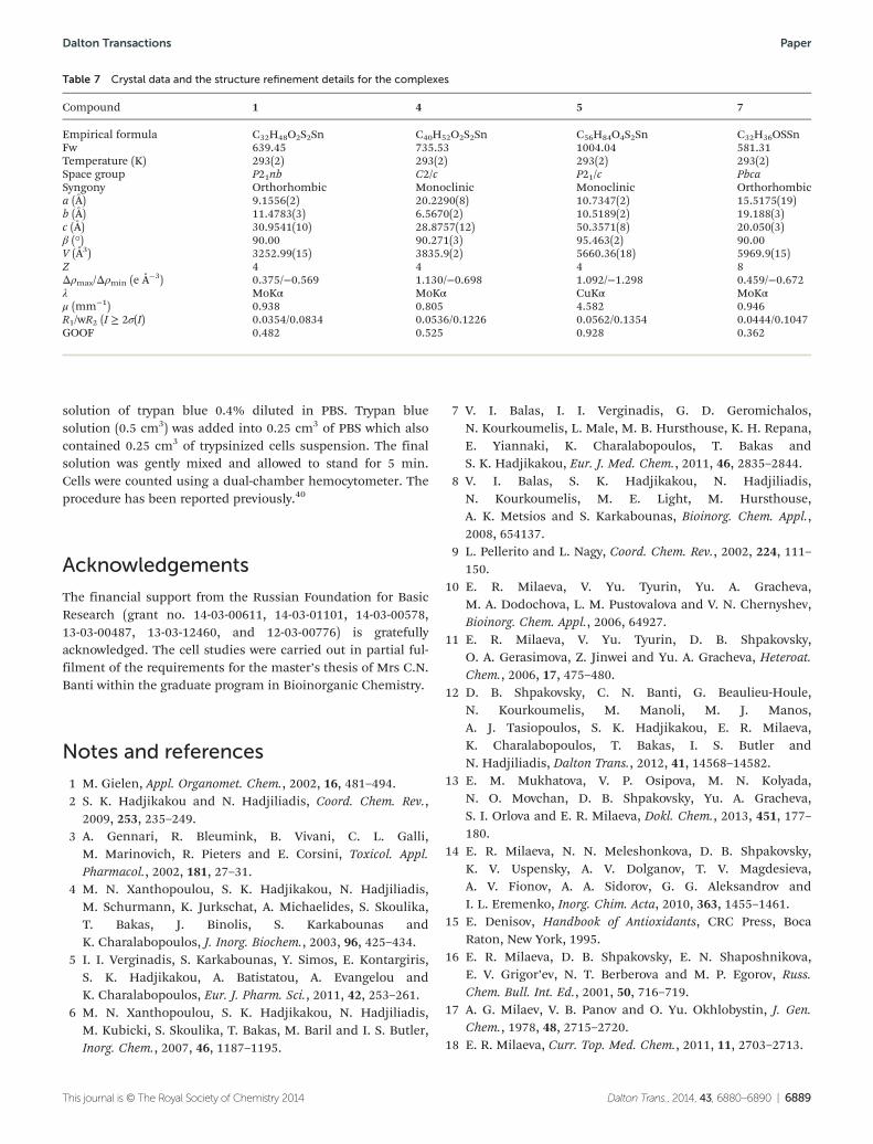
All diffraction data were collected on a STOE StadiVari Pilatus100 K diffractometer [λ(CuKα) = 1.5418 Å, λ(MoKα) =0.71073 Å, ω-scans] at 293 K.36 The primary processing of theexperimental data array was performed using the WinGXprogram package.37 The structures were solved by directmethods and refined by full-matrix least-squares procedureson F2 using SHELXL97.38 All non-hydrogen atoms were refinedanisotropically, and hydrogen atoms were located at calculatedpositions and refined via the “riding model”. Crystal data andstructure refinement parameters are listed in Table 7. CCDC967799 (1), 967798 (4), 967801 (5) and 967800 (7) containthe supplementary crystallographic data for this paper. Thestructures of complexes were drawn using the MERCURY CSD3.1 program.39
Biological tests
Trypan blue assay: MCF-7, HeLa and MRC-5 cells were seededonto twenty four-well plates at a density of 3 × 10⁴ cellsper well, respectively, and incubated for 24 h before the experi-ment. Cell viability was measured after 48 h by adding a
Paper Dalton Transactions
6888 | Dalton Trans., 2014, 43, 6880–6890 This journal is © The Royal Society of Chemistry 2014
solution of trypan blue 0.4% diluted in PBS. Trypan bluesolution (0.5 cm3) was added into 0.25 cm3 of PBS which alsocontained 0.25 cm3 of trypsinized cells suspension. The finalsolution was gently mixed and allowed to stand for 5 min.Cells were counted using a dual-chamber hemocytometer. Theprocedure has been reported previously.40
Acknowledgements
The financial support from the Russian Foundation for BasicResearch (grant no. 14-03-00611, 14-03-01101, 14-03-00578,13-03-00487, 13-03-12460, and 12-03-00776) is gratefullyacknowledged. The cell studies were carried out in partial ful-filment of the requirements for the master’s thesis of Mrs C.N.Banti within the graduate program in Bioinorganic Chemistry.
Notes and references
1 M. Gielen, Appl. Organomet. Chem., 2002, 16, 481–494.2 S. K. Hadjikakou and N. Hadjiliadis, Coord. Chem. Rev.,
2009, 253, 235–249.3 A. Gennari, R. Bleumink, B. Vivani, C. L. Galli,
M. Marinovich, R. Pieters and E. Corsini, Toxicol. Appl.Pharmacol., 2002, 181, 27–31.
4 M. N. Xanthopoulou, S. K. Hadjikakou, N. Hadjiliadis,M. Schurmann, K. Jurkschat, A. Michaelides, S. Skoulika,T. Bakas, J. Binolis, S. Karkabounas andK. Charalabopoulos, J. Inorg. Biochem., 2003, 96, 425–434.
5 I. I. Verginadis, S. Karkabounas, Y. Simos, E. Kontargiris,S. K. Hadjikakou, A. Batistatou, A. Evangelou andK. Charalabopoulos, Eur. J. Pharm. Sci., 2011, 42, 253–261.
6 M. N. Xanthopoulou, S. K. Hadjikakou, N. Hadjiliadis,M. Kubicki, S. Skoulika, T. Bakas, M. Baril and I. S. Butler,Inorg. Chem., 2007, 46, 1187–1195.
7 V. I. Balas, I. I. Verginadis, G. D. Geromichalos,N. Kourkoumelis, L. Male, M. B. Hursthouse, K. H. Repana,E. Yiannaki, K. Charalabopoulos, T. Bakas andS. K. Hadjikakou, Eur. J. Med. Chem., 2011, 46, 2835–2844.
8 V. I. Balas, S. K. Hadjikakou, N. Hadjiliadis,N. Kourkoumelis, M. E. Light, M. Hursthouse,A. K. Metsios and S. Karkabounas, Bioinorg. Chem. Appl.,2008, 654137.
9 L. Pellerito and L. Nagy, Coord. Chem. Rev., 2002, 224, 111–150.
10 E. R. Milaeva, V. Yu. Tyurin, Yu. A. Gracheva,M. A. Dodochova, L. M. Pustovalova and V. N. Chernyshev,Bioinorg. Chem. Appl., 2006, 64927.
11 E. R. Milaeva, V. Yu. Tyurin, D. B. Shpakovsky,O. A. Gerasimova, Z. Jinwei and Yu. A. Gracheva, Heteroat.Chem., 2006, 17, 475–480.
12 D. B. Shpakovsky, C. N. Banti, G. Beaulieu-Houle,N. Kourkoumelis, M. Manoli, M. J. Manos,A. J. Tasiopoulos, S. K. Hadjikakou, E. R. Milaeva,K. Charalabopoulos, T. Bakas, I. S. Butler andN. Hadjiliadis, Dalton Trans., 2012, 41, 14568–14582.
13 E. M. Mukhatova, V. P. Osipova, M. N. Kolyada,N. O. Movchan, D. B. Shpakovsky, Yu. A. Gracheva,S. I. Orlova and E. R. Milaeva, Dokl. Chem., 2013, 451, 177–180.
14 E. R. Milaeva, N. N. Meleshonkova, D. B. Shpakovsky,K. V. Uspensky, A. V. Dolganov, T. V. Magdesieva,A. V. Fionov, A. A. Sidorov, G. G. Aleksandrov andI. L. Eremenko, Inorg. Chim. Acta, 2010, 363, 1455–1461.
15 E. Denisov, Handbook of Antioxidants, CRC Press, BocaRaton, New York, 1995.
16 E. R. Milaeva, D. B. Shpakovsky, E. N. Shaposhnikova,E. V. Grigor’ev, N. T. Berberova and M. P. Egorov, Russ.Chem. Bull. Int. Ed., 2001, 50, 716–719.
17 A. G. Milaev, V. B. Panov and O. Yu. Okhlobystin, J. Gen.Chem., 1978, 48, 2715–2720.
18 E. R. Milaeva, Curr. Top. Med. Chem., 2011, 11, 2703–2713.
Table 7 Crystal data and the structure refinement details for the complexes
Compound 1 4 5 7
Empirical formula C32H48O2S2Sn C40H52O2S2Sn C56H84O4S2Sn C32H36OSSnFw 639.45 735.53 1004.04 581.31Temperature (K) 293(2) 293(2) 293(2) 293(2)Space group P21nb C2/c P21/c PbcaSyngony Orthorhombic Monoclinic Monoclinic Orthorhombica (Å) 9.1556(2) 20.2290(8) 10.7347(2) 15.5175(19)b (Å) 11.4783(3) 6.5670(2) 10.5189(2) 19.188(3)c (Å) 30.9541(10) 28.8757(12) 50.3571(8) 20.050(3)β (°) 90.00 90.271(3) 95.463(2) 90.00V (Å3) 3252.99(15) 3835.9(2) 5660.36(18) 5969.9(15)Z 4 4 4 8Δρmax/Δρmin (e Å−3) 0.375/−0.569 1.130/−0.698 1.092/−1.298 0.459/−0.672λ MoKα MoKα CuKα MoKαμ (mm−1) 0.938 0.805 4.582 0.946R1/wR2 (I ≥ 2σ(I) 0.0354/0.0834 0.0536/0.1226 0.0562/0.1354 0.0444/0.1047GOOF 0.482 0.525 0.928 0.362
Dalton Transactions Paper
This journal is © The Royal Society of Chemistry 2014 Dalton Trans., 2014, 43, 6880–6890 | 6889

19 E. White, J. S. Shannon and R. E. Patterson, Cancer Epide-miol. Biomarkers Prev., 1997, 6, 769–774.
20 P. Kovacic and J. D. Jacintho, Curr. Med. Chem., 2001, 8,773–796.
21 P. Molyneux, Songklanakarin J. Sci. Technol., 2004, 26, 211–219.22 M. N. Xanthopoulou, S. K. Hadjikakou, N. Hadjiliadis,
E. R. Milaeva, J. A. Gracheva, V. Yu. Tyurin,N. Kourkoumelis, K. C. Christoforidis, A. K. Metsios,S. Karkabounas and K. Charalabopoulos, Eur. J. Med.Chem., 2008, 43, 327–335.
23 J. Zhou and P. Giannakakou, Curr. Med. Chem.: Anti-CancerAgents, 2005, 5, 65–71.
24 M. A. Jordan and L. Wilson, Nat. Rev. Cancer, 2004, 4, 253–265.
25 L. P. Tan, M. L. Ng and V. G. Kumar Das, J. Neurochem.,1978, 31, 1035–1041.
26 R Huang, A. Wallqvist and D. G. Covell, Biochem. Pharma-col., 2005, 69, 1009–1039.
27 A. Desai and T. J. Mitchison, Ann. Rev. Cell Dev. Biol., 1997,13, 83–117.
28 S. C. Chow and S. Orrenius, Toxicol. Appl. Pharmacol., 1994,127, 19–26.
29 H. F. Gilbert, Adv. Enzymol., 1990, 63, 69–172.
30 A. Begaye and D. L. Sackett, Methods Cell Biol., 2010, 95,391–401.
31 C. Ma and J. Zhang, Appl. Organomet. Chem., 2003, 17, 788–794.
32 C. N. Banti and S. K. Hadjikakou, Metallomics, 2013, 5,569–596.
33 E. Muller, H. B. Stegman and K. Scheffler, Liebigs Ann.Chem., 1961, 645, 79–91.
34 L. V. Glushkova, L. A. Skripko, L. I. Iofis, A. I. Medvedev,T. S. Romanchenko, A. A. Efimov and G. V. Kutimova,Patent U.S.S.R, SU 514842 A1 19760525, 1976.
35 W. Brand-Williams, M. E. Cuvelier and C. Berset, Food Sci.Technol-Leb., 1995, 28, 25–30.
36 Stoe & Cie, X-AREA, X-RED32, Stoe & Cie, Darmstadt,Germany, 2012.
37 L. J. Farrugia, J. Appl. Crystallogr., 2012, 45, 849–854.38 G. M. Sheldrick, Acta Crystallogr., Sect. A: Fundam. Crystal-
logr., 2008, 64, 112–122.39 C. F. Macrae, P. R. Edington, P. McCabe, E. Pidcock,
G. P. Shields, R. Taylor, M. Towler and J. van de Streek,J. Appl. Crystallogr., 2006, 39, 453–457.
40 J. Kumi-Diaka, V. Nguyen and A. Butler, Biol. Cell, 1999, 91,515–523.
Paper Dalton Transactions
6890 | Dalton Trans., 2014, 43, 6880–6890 This journal is © The Royal Society of Chemistry 2014