Cytogenetic and molecular study of the PRDX4 gene in a t(X;18)(p22;q23): a cautionary tale
10
Cytogenetic and molecular study of the PRDX4 gene in a translocation (X;18)(p22;q23) – a cautionary tale Heidrun D. Gerr 1,‡ , Michele L. Nassin 2,‡ , Elizabeth M. Davis 2 , Nimanthi Jayathilaka 2 , Mary E. Neilly 2 , Brigitte Schlegelberger 1 , Yanming Zhang 2 , and Janet D. Rowley 2 1 Institute for Cell and Molecular Pathology, Medizinische Hochschule Hannover, Hannover Germany 2 Department of Medicine, Section Hematology/Oncology, University of Chicago, Chicago, Illinois, USA Abstract The PRDX4 gene located at Xp22 codes for a member of the peroxiredoxin gene family. Genes within this family exhibit thioredoxin-dependent peroxidase activity and have been implicated in cellular functioning, including proliferation and differentiation. Recently, PRDX4 has been identified as a partner gene in an X;21 translocation in a patient with acute myeloid leukemia (AML). To determine whether PRDX4 was involved in other translocations, leukemia cells from fifteen patients with Xp22 abnormalities were screened for the gene’s involvement using fluorescence in situ hybridization (FISH). One sample from a 41 year old female with acute lymphoblastic leukemia (ALL) showed three signals when hybridized with the PRDX4 probe. Cytogenetic analysis of the sample had identified a t(X;18)(p22,q23). Assuming that the three signals indicated a break within the PRDX4 gene, we performed FISH experiments and successfully narrowed the breakpoint on chromosome 18 to a 50 kB region. Subsequent analysis using spectral karyotyping showed that the leukemic cells had undergone multiple rearrangements and that a third X chromosome, albeit rearranged, was present. Additional FISH experiments revealed that the third PRDX4 signal was the result of a third copy of the gene. Analysis of the other rearrangements has helped to characterize the multiple abnormalities within the leukemic cells. Our findings are significant because they underline the importance of using multiple techniques when analyzing complex chromosomal rearrangements in malignant cells. 1) Introduction Chromosome translocations are known to be an important factor in causing a wide range of cancers, especially leukemia [1]. Although most common translocations have been identified and the breakpoints have been cloned, many rare translocations remain to be detected. Many of the genes cloned from translocation breakpoints have been shown to be critical in malignant transformation; thus identifying and cloning these new breakpoints is important to increase our understanding of genetic changes in leukemic cells. We recently cloned a novel breakpoint in a t(X;21)(p22;q22) translocation, which involved the AML1 gene on 21 and the PRDX4 gene on chromosome Xp22 that had not previously been identified in translocations in leukemia [2]. Peroxiredoxins play many functional roles in the cell including proliferation, differentiation, and cell signaling [3]. In the t(X;21)(p22;q22) the 5′ portion of AML1 is fused Corresponding Author: Janet D. Rowley, M.D, Section of Hematology/Oncology, Department of Medicine, The University of Chicago, 5841 S. Maryland Avenue, Chicago, IL 60637, E-mail: [email protected], Phone (773) 702-6117/Fax: (773) 702-3002. ‡ The first two authors contributed equally to the paper. Publisher's Disclaimer: This is a PDF file of an unedited manuscript that has been accepted for publication. As a service to our customers we are providing this early version of the manuscript. The manuscript will undergo copyediting, typesetting, and review of the resulting proof before it is published in its final citable form. Please note that during the production process errors may be discovered which could affect the content, and all legal disclaimers that apply to the journal pertain. NIH Public Access Author Manuscript Cancer Genet Cytogenet. Author manuscript; available in PMC 2008 July 15. Published in final edited form as: Cancer Genet Cytogenet. 2007 July 15; 176(2): 131–136. NIH-PA Author Manuscript NIH-PA Author Manuscript NIH-PA Author Manuscript
Transcript of Cytogenetic and molecular study of the PRDX4 gene in a t(X;18)(p22;q23): a cautionary tale
Cytogenetic and molecular study of the PRDX4 gene in atranslocation (X;18)(p22;q23) – a cautionary tale
Heidrun D. Gerr1,‡, Michele L. Nassin2,‡, Elizabeth M. Davis2, Nimanthi Jayathilaka2, MaryE. Neilly2, Brigitte Schlegelberger1, Yanming Zhang2, and Janet D. Rowley2
1 Institute for Cell and Molecular Pathology, Medizinische Hochschule Hannover, Hannover Germany
2 Department of Medicine, Section Hematology/Oncology, University of Chicago, Chicago, Illinois, USA
AbstractThe PRDX4 gene located at Xp22 codes for a member of the peroxiredoxin gene family. Genes withinthis family exhibit thioredoxin-dependent peroxidase activity and have been implicated in cellularfunctioning, including proliferation and differentiation. Recently, PRDX4 has been identified as apartner gene in an X;21 translocation in a patient with acute myeloid leukemia (AML). To determinewhether PRDX4 was involved in other translocations, leukemia cells from fifteen patients with Xp22abnormalities were screened for the gene’s involvement using fluorescence in situ hybridization(FISH). One sample from a 41 year old female with acute lymphoblastic leukemia (ALL) showedthree signals when hybridized with the PRDX4 probe. Cytogenetic analysis of the sample hadidentified a t(X;18)(p22,q23). Assuming that the three signals indicated a break within the PRDX4gene, we performed FISH experiments and successfully narrowed the breakpoint on chromosome18 to a 50 kB region. Subsequent analysis using spectral karyotyping showed that the leukemic cellshad undergone multiple rearrangements and that a third X chromosome, albeit rearranged, waspresent. Additional FISH experiments revealed that the third PRDX4 signal was the result of a thirdcopy of the gene. Analysis of the other rearrangements has helped to characterize the multipleabnormalities within the leukemic cells. Our findings are significant because they underline theimportance of using multiple techniques when analyzing complex chromosomal rearrangements inmalignant cells.
1) IntroductionChromosome translocations are known to be an important factor in causing a wide range ofcancers, especially leukemia [1]. Although most common translocations have been identifiedand the breakpoints have been cloned, many rare translocations remain to be detected. Manyof the genes cloned from translocation breakpoints have been shown to be critical in malignanttransformation; thus identifying and cloning these new breakpoints is important to increase ourunderstanding of genetic changes in leukemic cells. We recently cloned a novel breakpoint ina t(X;21)(p22;q22) translocation, which involved the AML1 gene on 21 and the PRDX4 geneon chromosome Xp22 that had not previously been identified in translocations in leukemia[2]. Peroxiredoxins play many functional roles in the cell including proliferation,differentiation, and cell signaling [3]. In the t(X;21)(p22;q22) the 5′ portion of AML1 is fused
Corresponding Author: Janet D. Rowley, M.D, Section of Hematology/Oncology, Department of Medicine, The University of Chicago,5841 S. Maryland Avenue, Chicago, IL 60637, E-mail: [email protected], Phone (773) 702-6117/Fax: (773)702-3002.‡The first two authors contributed equally to the paper.Publisher's Disclaimer: This is a PDF file of an unedited manuscript that has been accepted for publication. As a service to our customerswe are providing this early version of the manuscript. The manuscript will undergo copyediting, typesetting, and review of the resultingproof before it is published in its final citable form. Please note that during the production process errors may be discovered which couldaffect the content, and all legal disclaimers that apply to the journal pertain.
NIH Public AccessAuthor ManuscriptCancer Genet Cytogenet. Author manuscript; available in PMC 2008 July 15.
Published in final edited form as:Cancer Genet Cytogenet. 2007 July 15; 176(2): 131–136.
NIH
-PA Author Manuscript
NIH
-PA Author Manuscript
NIH
-PA Author Manuscript

to the 3′ portion of PRDX4. It is very possible that this fusion has a dominant negative effecton the wild type AML1 and may also lead to loss of PRDX4 function. This was the first exampleof antioxidant enzyme involvement in a chromosome translocation in leukemia. Our presentstudy was performed to determine if PRDX4 was also involved in other translocations inpatients with leukemia.
2) Case ReportFifteen patients with leukemia and Xp22 translocations on cytogenetic analysis were screenedusing fluorescence in situ hybridization (FISH) with a 173 kb BAC probe covering thePRDX4 gene (19 kb) (RP1140P7 location 23, 464, 482 to 23, 637, 849). 14 patients showednormal results for the hybridization. A third FISH signal was observed in one patientimplicating PRDX4 involvement.
The patient was a 39 year old Caucasian female who was diagnosed with Pro-B acutelymphoblastic leukemia (ALL) in 2001. She initially presented with a six-week history offatigue and petechiae and was found to be pancytopenic (WBC 5.4× 10⁹/l, hemoglobin 9.6 g/dL and platelets 11× 109/l). A bone marrow biopsy was performed and showed 95% blasts,positive for CD19, CD79 and TdT. The ISCN-karyotype on presentation was 46,XX,t(4;11)(q21;q23) [73%]. The patient was started on chemotherapy per protocol CALGB 19802 [4].She underwent induction therapy with daunorubicin, cyclophosphamide, vincristine, PEG-asparaginase and prednisone and obtained complete remission (CR). After 5 months ofmaintenance therapy with Vincristine, MTX, prednisone, 6-mercaptopurin she relapsed in2002. Her bone marrow had 48% blasts and in a repeat cytogenetic analysis showed the t(4;11)(q21;q23). After off-protocol treatment with ifosfamide, MTX, vincristine, cytarabine,etoposide and dexamethasone failed to induce a remission, a matched unrelated donor bonemarrow transplant was done. After a short CR she relapsed again, this time showing karyotypicevolution, 47,X,t(X;18)(p22;q23), add(1)(q44), t(4;11)(q21;q23), t(6;14)(q23;q21), der(7)t(7;7)(p15,q11),+13, −14, +17, +20 in 60 % of metaphase cells (Fig 1). A second bone marrowtransplantation was performed, yielding only a three month period of CR. Cytogenetic analysisof the 3rd relapse in 2003 showed the t(X;18)(p22;q23) in 50 % of metaphase cells. The patientdied shortly after that in a hospice.
FISH with BACRP11-40P7 was performed on cells from the bone marrow aspirate on 2nd
relapse and showed three signals on hybridization (Fig 2). Assuming that PRDX4 was involvedin this translocation, we tried to identify the location of the breakpoint on 18q22 and the partnergene of the translocation on chromosome18. A 22 Mb region on chromosome 18 wasinvestigated with 44 BAC clones. Eighteen BAC clones mapping from 54.6 Mb to 67.1 Mbon chromosome 18 (RP11-108P20, RP11- 299P2, RP11-727B4, RP11-454J3, RP11-74B10,RP11-563H6, RP11-255L11, RP11-755G16, RP11-704G7, RP11-484N16, CTD-2311I12,CTD-2523K8, RP11-41O4, RP11-433A23, RP11- 792O7, CTD-2593F24, RP1-47G4,RP11-94I6) remained on chromosome 18 indicating that they were proximal to the breakpoint.Twenty-three probes from 67.2 Mb to 76.1 Mb (RP11-510D19, RP11-752P2, RP11-723G8,RP11-702M18, RP11-1061K3, RP11-131K21, RP11-676J15, RP11-396D4, RP11-504I13,RP11-162A12, RP11-751H17, RP11-130J1, RP11-849I19, RP11-345O15, RP11-042B2,RP11-298P14, CTD-2286N8, RP11-1136J12, RP11-800A18, RP11-196B3, RP11-154H12,RP11-680N10, RP11-245M2) translocated to the X chromosome, thus showing that they weretelomeric of the breakpoint. The large number of BAC clones used for the location of thebreakpoint can be explained in part by incorrect information provided from an online genedatabase. One of the BAC clones we used in the beginning (RP11-755G16) was initiallymapped by the database 1.64 Mb further telomeric on chromosome 18 than its actual location.Thus we initially obtained contradictory results and the identification of the breakpoint wasdelayed. We notified the database and the erroneous map location was corrected.
Gerr et al. Page 2
Cancer Genet Cytogenet. Author manuscript; available in PMC 2008 July 15.
NIH
-PA Author Manuscript
NIH
-PA Author Manuscript
NIH
-PA Author Manuscript
Finally the potential breakpoint could be narrowed to a 50 kilobase region between the genesSOCS6 and CBLN2 on 18q23. Two BAC clones (RP11-139B13, location 67128221 to67283891 on 18q22 and RP11-432G11, 67033407 to 67239179) were found that showed asplit signal on both the derivative X and 18 chromosome and thus appeared to span thetranslocation breakpoint. Six uncharacterized expressed sequence tags (ESTs) but no knowngenes were located in the breakpoint area on chromosome 18.
However, our mapping data lead to unexpected results. When the split chromosome 18 probeswere hybridized with the original PRDX4 probe (RP11-40P7), we did not see a fusion signalon chromosome 18 as expected. Two PRDX4 signals appeared on the derivative X involvedin the translocation with chromosome 18 and on what was thought to be a normal chromosomeX. The third PRDX4 signal appeared on a small unidentified chromosome (Fig 3). To clarifythis unexpected result, spectral karyotyping (SKY) was used to identify all chromosomes. SKYanalysis showed that the unidentified chromosome with the third PRDX4 signal was actuallythe short arm of a third X that appeared in all of the abnormal cells (Fig 4). This X had initiallybeen identified as a third chromosome 17 in the banding karyotype (Fig 1). It appears that thelong arm of a second X chromosome was involved in a translocation with chromosome 1 andthe residual short arm was involved with chromosome 18.
Now the unresolved question was whether the break on the X chromosome really involved thePRDX4 gene or a region on the X chromosome further telomeric to the gene. Using two probesRP13 314C10 (Accession number AC131011.5), from 23.64 Mb to 23.81 Mb, and RP11911N20, from 23,37 Mb to 23,55 Mb on Xp22, that flank PRDX4, we determined thatPRDX4 was not involved. Both signals colocalized on all three X chromosomes, proving thatthe three PRDX4 signals were a result of a third copy of the gene and not a split within the gene(Fig 5).
3) DiscussionPRDX4 is a member of the peroxiredoxin gene family that exhibits thioredoxin dependentperoxidase activity. All peroxiredoxin genes are highly conserved in eukaryotes andprokaryotes and are ubiquitously expressed [5,6]. PRDX4 was shown to play a role in tumorsuppression and is significantly down regulated in specific leukemia subtypes [7,8]. Recently,altered expression of PRDX4 was also shown in stomach and colon cancer metastases [9,10].After we reported on the involvement of PRDX4 in a translocation of an AML patient [2], wewanted to determine if this gene might also be involved in other leukemia translocations. Thesplit FISH signal in an ALL patient with a t (X;18)(p22;q23) using a probe spanning PRDX4implied that PRDX4 was involved in the translocation. However, this assumption wasdisproved with further FISH experiments. Using BAC probes that span the breakpoint onchromosome 18 and the PRDX4 probe, no fusion signal was seen on the derivative chromosome18. SKY analysis helped to clarify the chromosomal abnormalities in the leukemic cells. Theseabnormalities include the presence of an additional X chromosome in the abnormal cells,explaining the three signals for the PRDX4 probe, and further that this third X chromosomehad undergone additional rearrangement with the long arm of chromosome 1. The initialcytogenetic analysis of the karyotype did not identify these chromosomal changes. Althoughkaryotyping with chromosomal banding is the standard method for identifying chromosomalaberrations, it appears that highly rearranged karyotypes are often difficult to interpret withconventional cytogenetics alone. Studies have demonstrated that SKY analysis couldsubstantially improve the accurate assessment of genetic abnormalities in malignant cells andeven correct the cytogenetic interpretation [11–13]. Our additional investigation of thekaryotypic changes were crucial to obtain the correct analysis and they stress that the use ofmultiple techniques in malignant cells is required to confirm complex rearrangements. Without
Gerr et al. Page 3
Cancer Genet Cytogenet. Author manuscript; available in PMC 2008 July 15.
NIH
-PA Author Manuscript
NIH
-PA Author Manuscript
NIH
-PA Author Manuscript
these additional studies the complex rearrangements that are likely responsible for themalignant behavior of this leukemia would not have been properly identified.
The (X;18) translocation in our patient appeared as a secondary chromosomal aberration inaddition to the known t(4;11) after relapse following bone marrow transplantation. Additionalaberrations do not have an impact on survival or prognosis in patients with 11q23abnormalities; however the characterization of these changes is important to determine theirpotential role in the evolution of MLL-driven leukemogenesis [14–16].
ConclusionsWe conclude that combining different techniques, i.e. conventional chromosomal bandinganalysis, FISH and SKY provides the most comprehensive cytogenetic approach. SKY wasshown again to help to clarify the chromosomal rearrangements in complex karyotypes.
The two BAC probes flanking PRDX4 finally confirmed that this gene was not involved in thetranslocation. Our initial assumption that the original split of BACRP11-40P7 meantinvolvement of PRDX4 can be explained by the larger size of this clone (173 kb) compared toPRDX4 (19 kb).
Considering the incorrect database mapping of one of our BAC clones, our study can also beseen as an alert to other groups that one should not always rely on the information from a singleonline database.
Acknowledgements
This study has been supported by National Institutes of Health Grant CA84405 and the Spastic Paralysis Foundationof the Illinois, Eastern Iowa Branch of Kiwanis International (JDR), an Howard Hughes Institute undergraduateEducation initiative grant at the University of Chicago (MLN) and German Academic Research Program (HDG).
References1. Rowley JD. The critical role of chromosome translocations in human leukemias. Annu Rev Genet
1998;32:495–519. [PubMed: 9928489]2. Zhang Y, Emmanuel N, Kamboj G, Chen J, Shurafa M, Van Dyke DL, Wiktor A, Rowley JD. PRDX4,
a member of the peroxiredoxin family, is fused to AML1 (RUNX1) in an acute myeloid leukemiapatient with a t(X;21)(p22;q22). Genes Chromosomes Cancer 2004;40:365–70. [PubMed: 15188461]
3. Immenschuh S, Baumgart-Vogt E. Peroxiredoxins, oxidative stress, and cell proliferation. AntioxidRedox Signal 2005;7:768–77. [PubMed: 15890023]
4. Stock W, Johnson J, Yun D, Bennett D, Sher D, Stone R, Kolitz J, Powell B, Wetzler M, Vardiman J,Bloomfield C, Larson R. Daunorubicin Dose Intensification during Treatment of Adult AcuteLymphoblastic Leukemia (ALL): Final Results from Cancer and Leukemia Group B Study 19802.Blood ASH Annual Meeting Abstracts 2005;106:1833.
5. Wood ZA, Schroder E, Robin Harris J, Poole LB. Structure, mechanism and regulation ofperoxiredoxins. Trends Biochem Sci 2003;28:32–40. [PubMed: 12517450]
6. Wong CM, Chun AC, Kok KH, Zhou Y, Fung PC, Kung HF, Jeang KT, Jin DY. Characterization ofhuman and mouse peroxiredoxin IV: evidence for inhibition by Prx-IV of epidermal growth factor-and p53-induced reactive oxygen species. Antioxid Redox Signal 2000;2:507–18. [PubMed:11229364]
7. Jin DY, Chae HZ, Rhee SG, Jeang KT. Regulatory role for a novel human thioredoxin peroxidase inNF-kappaB activation. J Biol Chem 1997;5(272):30952–61. [PubMed: 9388242]
8. Kohlmann A, Schoch C, Schnittger S, Dugas M, Hiddemann W, Kern W, Haferlach T. Molecularcharacterization of acute leukemias by use of microarray technology. Genes Chromosomes Cancer2003;37:396–405. [PubMed: 12800151]
Gerr et al. Page 4
Cancer Genet Cytogenet. Author manuscript; available in PMC 2008 July 15.
NIH
-PA Author Manuscript
NIH
-PA Author Manuscript
NIH
-PA Author Manuscript
9. Li M, Lin YM, Hasegawa S, Shimokawa T, Murata K, Kameyama M, Ishikawa O, Katagiri T, TsunodaT, Nakamura Y, Furukawa Y. Genes associated with liver metastasis of colon cancer, identified bygenome-wide cDNA microarray. Int J Oncol 2004;24:305–12. [PubMed: 14719106]
10. Jang JS, Cho HY, Lee YJ, Ha WS, Kim HW. The differential proteome profile of stomach cancer:identification of the biomarker candidates. Oncol Res 2004;14:491–9. [PubMed: 15559763]
11. Schrock E, Veldman T, Padilla-Nash H, Ning Y, Spurbeck J, Jalal S, Shaffer LG, Papenhausen P,Kozma C, Phelan MC, Kjeldsen E, Schonberg SA, O’Brien P, Biesecker L, du Manoir S, Ried T.Spectral karyotyping refines cytogenetic diagnostics of constitutional chromosomal abnormalities.Hum Genet 1997;101:255–62. [PubMed: 9439652]
12. Rowley JD, Reshmi S, Carlson K, Roulston D. Spectral karyotype analysis of T-cell acute leukemia.Blood 1999;93:2038–42. [PubMed: 10068677]
13. Mohr B, Bornhauser M, Thiede C, Schakel U, Schaich M, Illmer T, Pascheberg U, Ehninger G.Comparison of spectral karyotyping and conventional cytogenetics in 39 patients with acute myeloidleukemia and myelodysplastic syndrome. Leukemia 2000;14:1031–8. [PubMed: 10865969]
14. Schoch C, Rieder H, Freund M, Hoelzer D, Riehm H, Fonatsch C. Twenty-three cases of acutelymphoblastic leukemia with translocation t(4;11)(q21;q23): the implication of additionalchromosomal aberrations. Ann Hematol 1995;70:195–201. [PubMed: 7748964]
15. Moorman AV, Raimondi SC, Pui CH, Baruchel A, Biondi A, Carroll AJ, Forestier E, Gaynon PS,Harbott J, Harms DO, Heerema N, Pieters R, Schrappe M, Silverman LB, Vilmer E, Harrison CJ.Ponte di Legno Working Group. No prognostic effect of additional chromosomal abnormalities inchildren with acute lymphoblastic leukemia and 11q23 abnormalities. Leukemia 2005;19:557–63.[PubMed: 15744345]
16. Johansson B, Moorman AV, Haas OA, Watmore AE, Cheung KL, Swanton S, Secker-Walker LM.Hematologic malignancies with t(4;11)(q21;q23)--a cytogenetic, morphologic, immunophenotypicand clinical study of 183 cases. European 11q23 Workshop participants. Leukemia 1998;12:779–87.[PubMed: 9593281]
Gerr et al. Page 5
Cancer Genet Cytogenet. Author manuscript; available in PMC 2008 July 15.
NIH
-PA Author Manuscript
NIH
-PA Author Manuscript
NIH
-PA Author Manuscript
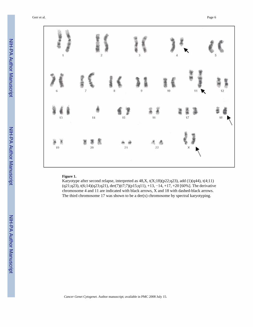
Figure 1.Karyotype after second relapse, interpreted as 48,X, t(X;18)(p22;q23), add (1)(q44), t(4;11)(q21;q23), t(6;14)(q23;q21), der(7)(t7;7)(p15;q11), +13, −14, +17, +20 [60%]. The derivativechromosome 4 and 11 are indicated with black arrows, X and 18 with dashed-black arrows.The third chromosome 17 was shown to be a der(x) chromosome by spectral karyotyping.
Gerr et al. Page 6
Cancer Genet Cytogenet. Author manuscript; available in PMC 2008 July 15.
NIH
-PA Author Manuscript
NIH
-PA Author Manuscript
NIH
-PA Author Manuscript
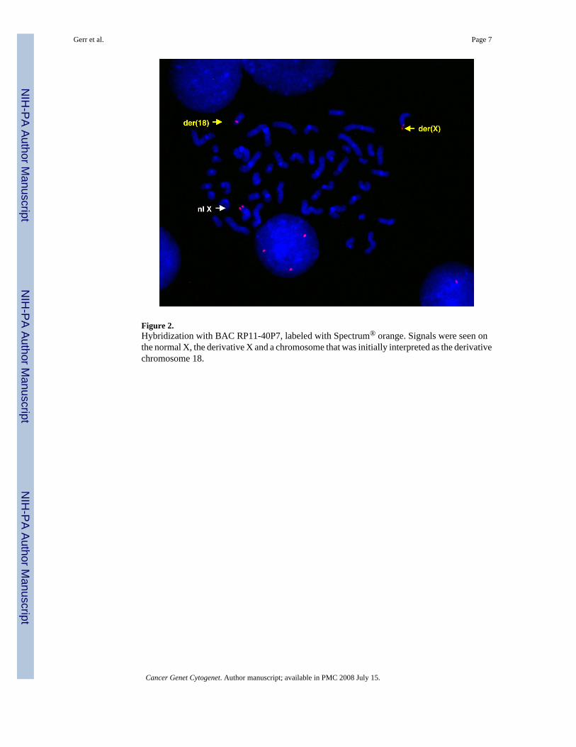
Figure 2.Hybridization with BAC RP11-40P7, labeled with Spectrum® orange. Signals were seen onthe normal X, the derivative X and a chromosome that was initially interpreted as the derivativechromosome 18.
Gerr et al. Page 7
Cancer Genet Cytogenet. Author manuscript; available in PMC 2008 July 15.
NIH
-PA Author Manuscript
NIH
-PA Author Manuscript
NIH
-PA Author Manuscript
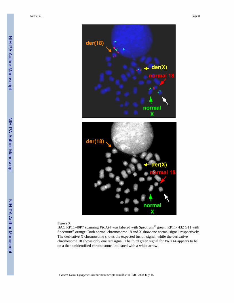
Figure 3.BAC RP11-40P7 spanning PRDX4 was labeled with Spectrum® green, RP11- 432 G11 withSpectrum® orange. Both normal chromosome 18 and X show one normal signal, respectively.The derivative X chromosome shows the expected fusion signal, while the derivativechromosome 18 shows only one red signal. The third green signal for PRDX4 appears to beon a then unidentified chromosome, indicated with a white arrow.
Gerr et al. Page 8
Cancer Genet Cytogenet. Author manuscript; available in PMC 2008 July 15.
NIH
-PA Author Manuscript
NIH
-PA Author Manuscript
NIH
-PA Author Manuscript

Figure 4.SKY Image and partial karyotype. A: DAPI stained metaphase cell, B: Spectral image, C:Classified image, D: Partial karyotype for chromosomes 1, 4, 11, 18 and X.
Gerr et al. Page 9
Cancer Genet Cytogenet. Author manuscript; available in PMC 2008 July 15.
NIH
-PA Author Manuscript
NIH
-PA Author Manuscript
NIH
-PA Author Manuscript

Figure 5.FISH using two probes flanking the PRDX4 gene. The centromeric probe RP13 314C10 islabeled with Spectrum® green, the telomeric probe (RP11-911N20) with Spectrum® orange.Both probes colocalize on all three X chromosomes indicating that PRDX4 is not split by thetranslocation.
Gerr et al. Page 10
Cancer Genet Cytogenet. Author manuscript; available in PMC 2008 July 15.
NIH
-PA Author Manuscript
NIH
-PA Author Manuscript
NIH
-PA Author Manuscript