Curcuma longa L leaf aqueous extract on α - Research Square
26
Page 1/26 Application of a novel lung protective drug formulated by silver nanoparticles containing Curcuma longa L leaf aqueous extract on α - Guttiferin-induced DNA fragmentation and apoptosis in lung HEL 299, MRC-5, IMR-90, CCD- 19Lu, WI-38, and BEAS-2B cell lines Yi Han Capital Madical University Yuan Gao Capital Medical University Xiaoqing Cao Capital Medical University Akram Zangeneh Razi University Mohammad Mahdi Zangeneh ( [email protected] ) Razi University Shuku Liu Capital Medical University Jie Li Capital Medical University Research Keywords: Lung protective drug, α-Guttiferin, DNA fragmentation, Apoptosis, Silver nanoparticles, Curcuma longa L leaf. Posted Date: June 17th, 2020 DOI: https://doi.org/10.21203/rs.3.rs-34153/v1 License: This work is licensed under a Creative Commons Attribution 4.0 International License. Read Full License
-
Upload
khangminh22 -
Category
Documents
-
view
0 -
download
0
Transcript of Curcuma longa L leaf aqueous extract on α - Research Square

Page 1/26
Application of a novel lung protective drugformulated by silver nanoparticles containingCurcuma longa L leaf aqueous extract on α-Guttiferin-induced DNA fragmentation andapoptosis in lung HEL 299, MRC-5, IMR-90, CCD-19Lu, WI-38, and BEAS-2B cell linesYi Han
Capital Madical UniversityYuan Gao
Capital Medical UniversityXiaoqing Cao
Capital Medical UniversityAkram Zangeneh
Razi UniversityMohammad Mahdi Zangeneh ( [email protected] )
Razi UniversityShuku Liu
Capital Medical UniversityJie Li
Capital Medical University
Research
Keywords: Lung protective drug, α-Guttiferin, DNA fragmentation, Apoptosis, Silver nanoparticles,Curcuma longa L leaf.
Posted Date: June 17th, 2020
DOI: https://doi.org/10.21203/rs.3.rs-34153/v1
License: This work is licensed under a Creative Commons Attribution 4.0 International License. Read Full License
Page 2/26
AbstractRecently, scientists have understood that metallic nanoparticles green-synthesized by medicinal plantshave signi�cant anti-cancer effects in the in vitro, in vivo, and clinical trial conditions. Also, the anti-lungcancer properties of metallic nanoparticles containing natural compounds have been indicated in manystudies. In the recent research, we tried to investigate the application of a novel lung protective drugformulated by silver nanoparticles containing Curcuma longa L leaf aqueous extract on α-Guttiferin-induced DNA fragmentation and apoptosis in HEL 299, MRC-5, IMR-90, CCD-19Lu, WI-38, and BEAS-2Bcell lines. Also, we assessed the concentrations of in�ammatory cytokines, activity of caspase-3, andpotential of mitochondrial membrane in the in vitro condition. Silver nanoparticles were characterized andanalyzed by common physicochemical techniques including Transmission Electron Microscopy,Ultraviolet–Visible Spectroscopy, Fourier-Transform Infrared Spectroscopy, and Field Emission-ScanningElectron Microscopy. In the biological part of the present research, the cell viability of HEL 299, MRC-5,IMR-90, CCD-19Lu, WI-38, and BEAS-2B cell lines was measured by trypan blue assay. Caspase-3 activitywas assessed by the caspase activity colorimetric assay kit and mitochondrial membrane potential wasstudied by Rhodamine123 �uorescence dye. Terminal deoxynucleotidyl transferase dUTP nick endlabeling (TUNEL) test was used to show DNA fragmentation and apoptosis. Also, the Rat in�ammatorycytokine assay kit was used to measure the concentrations of in�ammatory cytokines. Silvernanoparticles-treated cell cutlers signi�cantly (p ≤ 0.01) reduced the DNA fragmentation, caspase-3activity, and in�ammatory cytokines concentrations, and raised the mitochondrial membrane potentialand cell viability in the high concentration of α-Guttiferin-treated HEL 299, MRC-5, IMR-90, CCD-19Lu, WI-38, and BEAS-2B cells. The best result of lung protective and antioxidant properties of silver nanoparticlescontaining Curcuma longa L leaf aqueous extract was seen in the high dose of silver nanoparticles i.e.,4 µg. Assessment of the antioxidant properties of silver nanoparticles was done with the common freeradical scavenging test i.e., DPPH in the presence of butylated hydroxytoluene as the positive control. Thenanoparticles inhibited half of the DPPH molecules in the concentration of 149 µg/mL. According to theabove results, silver nanoparticles containing Curcuma longa L leaf aqueous extract can beadministrated as a lung protective drug for the treatment of lung diseases after approving in the clinicaltrial studies in humans.
1. IntroductionMetallic nanoparticles are one of main products of nanobiotechnology science. The recent evidenceshave indicated that metallic nanoparticles have signi�cant therapeutic properties in the in vitro, in vivo,and clinical trial conditions. Different techniques and approaches were developed to synthesize metallicnanoparticles to make them one of the most applicable and widely used materials in science [1,2]. Forinstance, silver nanoparticles are used as antioxidants and biocides, and they are utilized either barely orintegrated into other structures/substances. Additionally, silver nanoparticles have pharmaceutical andbiomedical applications, such as cancer treatment and medical imaging, and not to mention other uses inengineering, cosmetics, and agriculture [2,3]. One crucial branch in nanotechnology is nano-medicine,

Page 3/26
which has evolved as an extension for the application of nanomaterials in treating several disorders [3].Among all nanomaterials, the role of silver nanoparticles green synthesized by medicinal plants asprotective and chemotherapeutic supplements in treating various diseases is unique [2].
Several studies have indicated signi�cant antifungal effects of AgNPs green-synthesized by plants in thecure of candida diseases and their antibacterial properties in the treatment of Streptococcus,Staphylococcus, Pseudomonas, Salmonella, and Bacillus infectious. Also, silver nanoparticlessynthesized by plants have been formulated due to the antiviral, antibacterial, antioxidant, anti-parasitic,anti-in�ammatory, antifungal, wound healing, and anti-cancer properties [2,3]. In detail, it was reportedthat silver nanoparticles as useful metallic nanoparticles in medicine, can be used to diagnose severaltypes of lung cancers [3a]. In this regard, Ahmeda et al. introduced a chemotherapeutic drug formulatedby silver nanoparticles containing Melissa o�cinalis for the treatment of several types of blood cancer[3e]. Another study was showed that silver nanoparticles green-synthesized by Spinacia oleracea L. hadexcellent anti-acute myeloid leukemia. In the previous research, silver nanoparticles signi�cantly removedthe leukemia cell lines and regulated the histopathological, immunological, biochemical, andhematological parameters in the animals [3f]. About lung protective properties of silver nanoparticles,Que et al. presented when silver nanoparticles are added to the lung cancer cell lines, ability of lungadenocarcinoma cells (A549) for migrating and metastasis reduce, the levels of reactive oxygen species(RPS) increase, and NF-κB lead to cellular apoptosis. In the previous study was reported the silvernanoparticles with size of more than 100 nm don’t have lung protective effects against human lungcancer cell lines. There is a limit studies about the lung protective effects of medicinal plants green-synthesized silver nanoparticles [4]. He et al. reported the lung protective effects of green-synthesizedsilver nanoparticles on human lung cancer cell (H1299) in the in vitro condition. Green-synthesized silvernanoparticles inhibit NF-κB activity, reduce bcl-2 expression, raise survivin and caspase-3 expression andapply remarkable lung protective effects [5].
There is a list of medicinal plants with lung protective effects in traditional medicine includingAcanthopanax senticosus, Aconitum tanguticum, Alisma orientale Juzepzuk, Angelica decursiva,Antrodia camphorate, Alstonia scholaris, Azadirachta indica, Callicarpa japonica Thunb, Chrysanthemumindicum, Cnidium monnieri, Eleusine indica, Galla chinensis, Euterpe oleracea Mart., Galla chinensis,Ginkgo biloba, Gleditsia sinensis, Glycyrrhiza uralensis, Houttuynia cordata, Lonicera japonica �os, Morusalba, Nigella sativa, Paeonia suffruticosa, Phellodendri corte, and Punica granatum [6].
In Iranian traditional medicine, many sub-traditional medicines are there that the most well-known isKurdish traditional medicine in the west of Iran. Every year many medicinal supplements and drugs areformulated and produced from the plants of Kurdish traditional medicine [1–3]. One of these plants isCurcuma longa L from Tracheophytes, Angiosperms, Monocots, and Commelinids clades, Zingiberalesorder, Zingiberaceae family, and Curcuma genus [7]. The plant has chemical antioxidant componentssuch as curcumin; curcumadiol; β-phellandrene; terpinolene; β-turmerone; 2,5-dihydroxybisabola-3,10-diene and Procurcumadiol α-turmerone; germacrone-13-al; epiprocurcumenol; procurcumenol;zedoaronediol; isoprocurcumenol; curcumenol; bisacurone; bisacumol; arturmerone; dehydrocurdione; and

Page 4/26
curcumenone. In traditional medicine, Curcuma longa L is used for its gastroprotective, splenoprotective,hematoprotective, immunoprotective, nephroprotective, hepatoprotective, anti-anemic, antiepileptic, lipid-lowering, anticancer, chemopreventive, diuretic, anti-abscesses, carminative, cutaneous wound healing,pain-killing, antifungal, antipyretic, anti-cataract, antiviral, anti-parasitic, anti-allergic, antiemetic, anti-in�ammatory, antidepressant, antifertility, antibacterial, antioxidant, and especially lung protectiveproperties [7]. Maybe, remedial capacities of Curcuma longa L are related to the above antioxidantcompounds [7].
In the recent research, we decided to investigate the application of a novel lung protective drugformulated by silver nanoparticles containing Curcuma longa L on α-Guttiferin-induced cell death in HEL299, MRC-5, IMR-90, CCD-19Lu, WI-38, and BEAS-2B cell lines.
2. Experimental
2.1. MaterialDimethyl sulfoxide (DMSO), Antimycotic antibiotic solution, hydrolysate, decamplmaneh fetal bovineserum, α-Guttiferin, Ehrlich solution, 4-(Dimethylamino)benzaldehyde, 2,2-diphenyl-1-picrylhydrazyl(DPPH), carbazole reagent, borax-sulphuric acid mixture, Dulbecco's Modi�ed Eagle Medium (DMED), andphosphate buffer solution (PBS) all were achieved from Sigma-Aldrich company of USA.
2.2. Preparation of Curcuma longa L leaf aqueous extractFully matured dark green leave of Curcuma longa L was collected in Kermanshah province, Iran inJanuary 2020 (Fig. 1). After veri�cation of the colored graphics, identi�cation and characterizationprocesses were performed.
Plant shells were washed with water for a while to remove them from their natural powders. After thisprocedure, the shells were dried in a sterile environment (botanical laboratory) for 20 days to dry in thenatural environment. The next process was to grind these shells into powder.
The obtained powder samples were brought to the appropriate grain size for use and were used in theextraction. Then, 20 g of powder Curcuma longa L leaf was taken and added to 500 mL of boiling waterand boiled for 5 minutes to obtain the extract. The resulting mixture was centrifuged for 15 minutes at5000 rpm, then �ltered and stored at 4 oC in the bottle for further experiments [2].
2.3. Synthesis of AgNPsThe AgNPs green synthesis was started with a reaction mixture of 100 mL of AgNO3 × H2O in the
concentration of 1 × 10− 3 M and 200 mL of Curcuma longa L leaf extract (20 µg/mL) in the proportion1:10 in a conical �ask. The reaction mixture was kept under magnetic stirring for 12 h at 25 oC. The blackcolored colloidal solution of Ag was formed at the reaction time end.

Page 5/26
After centrifuging the mixture at 12000 rpm for 20 min, the supernatant was discarded and the solidsample was washed with abundant pure water. After washing, it was centrifuged again at 1000 rpm for15 minutes [3].
The characterizations of AgNPs were conducted using advanced spectroscopic techniques like FTIR andUV-Vis spectroscopy, TEM, and FE-SEM.
The presence of AgNPs was primarily con�rmed by UV-Vis spectroscopy at 400–700 nm (Jasco V670Spectrophotometer). The biomolecules that play an effective role in the reduction of AgNPs wereinvestigated by the FT-IR spectrophotometer (Shimadzu IR a�nity.1). Morphological properties of silvernanoparticles have been checked concerning structure, form, and thickness by applying FE-SEM and TEMmicroscopic techniques.
2.4. Lung protective analyses of silver nanoparticles
2.4.1 Cell cultureIn this experiment, lung HEL 299, MRC-5, IMR-90, CCD-19Lu, WI-38, and BEAS-2B cell lines were culturedaccording to protocol rules in the gibco RPMI1640 cell culture environment. In �rst, 100 IU/mL penicillin(Sigma), 100 µg/mL streptomycin (Sigma) and 10%fetal bovine serum (FBS, Gibco) supported in cellcultures and standard case in T-25cm2 tissue culture bottles (5% incubated at 37ºC in CO2). It led tochanges in the morphology of α-Guttiferin cells, so apoptotic cells led to the cell being treated with dilutedα-Guttiferin with 100 mM water.
For AgNO3 × H2O, Curcuma longa L leaf aqueous extract, and silver nanoparticles solutions in theRPMI1640 water cell cultural environment were �rst dissolved in DMSO and added to the culturalenvironment with a �nal volume of 0.1%. Then, 12 h after the plating and washing, they were classi�edinto several groups including [8]:
1. I) α-Guttiferin: Cell culture contains 100 µM α-Guttiferin.2. Control: Cell culture medium without α-Guttiferin, AgNO3 × H2O, Curcuma longa L leaf aqueous
extract, and silver nanoparticles.3. T1: Cell culture contains 100 µM α-Guttiferin and 2 µg of AgNO3 × H2O.4. T2: Cell culture contains 100 µM α-Guttiferin and 4 µg of AgNO3 × H2O.5. T3: Cell culture contains 100 µM α-Guttiferin and 2 µg of Curcuma longa L leaf aqueous extract.�. T4: Cell culture contains 100 µM α-Guttiferin and 4 µg of Curcuma longa L leaf aqueous extract.7. T5: Cell culture contains 100 µM α-Guttiferin and 2 µg of silver nanoparticles.�. T6: Cell culture contains 100 µM α-Guttiferin and 4 µg of silver nanoparticles.
2.4.2. Cell death index

Page 6/26
For determining of cell death index in the various treatments of I-VII, TUNEL staining was used Eightrandom wells were selected to count TUNEL positive cells with an Olympus AX-70 �uorescencemicroscope. The ratio of cell death index to apoptotic cells to total cells is equal [9].
2.4.3. Cell viabilityTrypan blue was used for assessing cell viability. Vital Dye, whose membranes penetrate brokendamaged or Dead Cells, is seen by penetrating dead cells in the Blue lobes in the Neubauer Lamela. For12 hours, 96 wells with a 5 × 10⁴ cell/mL density were coated on the cultural plate, then, they werecultured with various I-VII treatments and incubated at 37ºC at 5% CO2 for 48 h. After trypinization of thecells, 200µL of the cell suspension was suspended by Neubauer Lamela 2–3 minutes after mixing with0.4% 40µL of Trippan blue. The samples cell viability was measured by following the formula below [8].
Cell viability: Non-colored cells number/Total cells number
2.4.4. Caspase-3 activityFor plating the above cells, the well cell culture plate containing the PRMI1640 medium was used. After12 h, the plate was washed by PBS. Then, the different treatments of I-VII were added to the cells. Trypsinwas used for separating the cells from the �ask. For removing the supernatant, all samples werecentrifuged for 10 minutes. Then the centrifuging was done by adding lysate buffer and �nally, they weretransferred to the well cell culture plate. Later, 5µL N-acetyl-Asp-Glu-Val-Asp-p-nitroanilineDEVD-pNA wasadded to the well cell culture plate and incubated in 37ºC for 2 h. Then, releasing of pNA as a result ofcaspase-3 effect was recorded by Biotek (USA) Spectrophotometer [10].
2.4.5. Mitochondrial membrane potential (MMP)For half an hour different treatments were exposed to 10 mg / mL rhodamine-123. The cell was thenwashed using PBS. Subsequently, 900µL triton X-100 was added to each well and held at 4 °C for 2hours. The solutions were taken into microtubes to be centrifuged at 16000 rpm for 20 minutes.Fluorescent absorbing in cells was performed using a �uorescent microplate reader (488 nm excitationand 520 nm emissions) [10].
2.4.6. Secretion of in�ammatory cytokinesThe pro-in�ammatory cytokines concentrations including TNFα, IL-6, and IL-1β were measured using RatV-Plex Kit.
2.5 Determination of the antioxidant property of silvernanoparticlesAt the beginning of the study, 100 mL of methanol (50%) was added to the 39.4 g of DPPH. Also, severalconcentrations of AgNO3 × H2O, Curcuma longa L leaf aqueous extract, and silver nanoparticles i.e., 0-1000 µg/mL, were considered. The DPPH was added to the various concentrations of AgNO3 × H2O,Curcuma longa L, and AgNPs, and all samples were transferred to an incubator at the temperature of
Page 7/26
37 °C. After 30 min incubating, the absorbances were determined at 517 nm. In this study, methanol(50%) and butylated hydroxytoluene (BHT) were negative and positive controls, respectively. Acceding tothe following formula, the antioxidant properties of AgNO3 × H2O, Curcuma longa L leaf aqueous extract,and silver nanoparticles were determined in detail [11]:
2.6. Statistical analysisThe data obtained as a result of experimental studies were evaluated by using the SPSS-22 program andin coordination with Tukeys post hoc process with ANOVA (p ≤ 0.01).
3. Results And Discussion
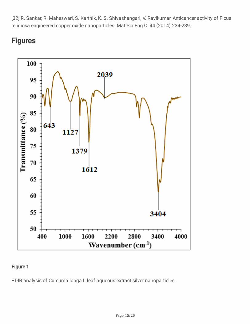
3.1. FT-IR analysisFourier-transform infrared spectroscopy (FTIR) is the technique used to acquire a solid, liquid or gasinfrared spectrum of absorption or emission. Around the same time, an FTIR spectrometer gathers high-speci�c data across a wide variety of the spectrum. This offers a signi�cant advantage over a dispersionspectrometer, which measures the intensity at a time over a restricted range of wavelengths. Metal-oxygen vibration of the FT-IR spectrum for metal nanoparticular biosynthesis induces peaks that arelocated at 400 and 700 cm− 1 [2,3]. The silver nanoparticles formation in the present study con�rms bythe existence of a peak at 643 cm− 1 belongs to the Ag-O bending vibration. In the �eld of naturalproducts, the IR spectroscopic approach is also an appropriate way to identify the bioactive components.Then the technique is a useful tool for identifying the presence of secondary metabolite over silvernanoparticles in the plants. The analysis has shown that in Curcuma longa L there are different IR bandsrelated to the presence of different functional groups at 2039 cm− 1 band related to aliphatic "C-H"stretching; peaks of 3404 cm− 1 related to "O-H" stretching (For alcohols, carboxylic acids and phenols);peaks of 1127 cm− 1 could be compared to "-C‐O" stretching and peaks of 1379 and 1612 cm− 1
correspond to "C = C" and "C = O" stretching found in phenolic and �avonoid compounds, respectively(Fig. 2). The explanation for these peaks can be thought to be different compounds from previousresearch, such as the recorded �avonoid, phenolic and carboxylic compounds [2,3].
3.2. UV-Vis analysis

Page 8/26
Ahmeda et al. studied Melissa o�cinalis leaf aqueous extract mediated synthesis of silver nanoparticles.Absorption of the continuum was detected at 462 nm [3e]. Hamelian et al. reported Thymus kotschyanusaqueous extract synthesized silver nanoparticles with a peak at 440 nm in the UV-Vis spectrum [3a].Hemmati et al. recorded 430 nm of the absorption coe�cient for silver nanoparticles utilizing the polyolprocess [3b]. Mohammadi et al. observed the peak of silver nanoparticles containing Phoenix dactyliferaseed ethanolic extract at the wavelength of 438 nm [3d]. Zangeneh et al. revealed the absorbance at462 nm for silver nanoparticles synthesized by Spinacia oleracea L. [3f]. Hamelian et al. recorded Pistaciaatlantica leaf extract induced silver nanoparticles as well as absorption maximum was detected at440 nm [3c].
3.3. FE-SEM analysisAn FE-SEM is an electron microscope (particles that have negative charge), rather than light, which works.Those electrons are emitted through a source of �eld emissions. The object is scanned by electronsaccording to a zig-zag pattern and recorded the surface morphology and the size of materials [2,3]. TheFE-SEM image of Curcuma longa L leaf aqueous extract mediated silver nanoparticles is shown in Fig. 4.The silver nanoparticles appeared as an agglomerated structure. The hydroxyl groups present in Curcumalonga L leaf aqueous extract could be responsible for agglomeration [2,3].
In the silver nanoparticles, the range size of 14–27 nm was observed. Many similar observations arenoted by Ahmeda et al. [3e], Zangneh et al. [3f], Hamelian et al. [3b], and Mohammadi et al. [3d].
3.4. TEM analysisTEM is the standard tool for calculating the size of nanoparticles, grain size, size distribution, andmorphology directly [2,3]. Therefore, TEM analyses of silver nanoparticles were conducted to evaluateparticle distributions and mean particle size (Fig. 5). The range size of the nanoparticles calculatedthrough TEM images. Besides, the histogram plot in the TEM image uniformly revealed the particle sizedistribution of the biosynthesized silver nanoparticles between 14 and 27 nm.
In the previous studies, the size of silver nanoparticles formulated by aqueous extract of medicinal plantshad been calculated in the ranges of 10–50 nm with the shape of spherical [2,3]. These literature studiesand analyzes support the results of our study and experiment.
3.5. Lung protective potentials of green-synthesized silvernanoparticlesα-Guttiferin is prescribed as a chemical material and may be of concern because of its side effects[12,13]. Abuse of this chemical material may affect the upper respiratory system (including nasal cavity,pharynx, and larynx) and lower respiratory tract (Including trachea, primary bronchi, secondary bronchi,and lung); its metabolites may be harmful to the digestive and excretion system [14,15]. The pathway forthe breakdown of α-Guttiferin passes through the liver and kidneys, and consequently, the potential of

Page 9/26
side effects is high in these organs [16,17]. In a study, the effects of α-Guttiferin on the lung and brain(cerebral cortex and hippocampus) are revealed. They emphasized the role of oxidative stress, neuronal,and pulmonary damage in the disruptive effects of α-Guttiferin abuse [16–18].
α-Guttiferin has direct effects on the upper and lower respiratory systems and increases the reactiveoxygen species (ROS) by reducing the level of antioxidant activity in the plasma. Therefore, it seemsnecessary to evaluate the toxic effect of α-Guttiferin in upper and lower respiratory systems cells andstudy on compounds that can decline these toxic effects [16–18].
Different studies have indicated that α-Guttiferin decrease the anti-in�ammatory cytokines such as IL13,IL10, IL5, IL4, and IL3 and increases the pro-in�ammatory cytokines such as IL1α, IL1β, IL6, and TNFα.The cytotoxicity properties of α-Guttiferin increase cell death signi�cantly and reduce cell proliferation inupper and lower respiratory system cells [15–18]. Also, a study showed that α-Guttiferin throughapoptosis induction, caspase-3 and caspase-9 activation, and cell proliferation inhibition induced celldeath in cells [17]. The �ndings of our experiment also agreed with these results and revealed that α-Guttiferin at high concentrations (100 µM) reduced signi�cantly (p ≤ 0.01) cell viability and increasedin�ammatory cytokines concentrations and caspase-3 activity. Treatment of these cells with both dosesof silver nanopartivles synthesized using Curcuma longa L leaf aqueous extract increased the cellproliferation and cell viability potentials due to the cell cytotoxicity reduction (Fig. 6–9).
The previous researches presented that α-Guttiferin produced many free radicals, especially ROS in thebody [16–18]. Free radicals with the degradation of DNA molecules cause cellular degradation andapoptosis in the cells [19]. ROS directly damages the DNA and increases the apoptosis in upper and lowerrespiratory systems cells through the production of free radicals [19–21]. In our study, the experiment ofapoptosis by the TUNEL test indicated that α-Guttiferin caused DNA fragmentation and inducedapoptosis in lung HEL 299, MRC-5, IMR-90, CCD-19Lu, WI-38, and BEAS-2B cells. Further experimentsrevealed that α-Guttiferin causes apoptosis in these cells by reducing the mitochondrial membranepotential. Our �ndings also indicated that silver nanoparticles synthesized using Curcuma longa L leafaqueous extract signi�cantly (p ≤ 0.01) increased the mitochondrial membrane potential and reduced therate of DNA fragmentation in lung HEL 299, MRC-5, IMR-90, CCD-19Lu, WI-38, and BEAS-2B cells treatedwith α-Guttiferin (Fig. 10,11).
3.6. Antioxidant potentials of green-synthesized silvernanoparticlesTraditional medicinal plants are well-known and essential natural antioxidant sources. Medicinal plant-derived natural antioxidants are very e�cient in blocking the process of oxidation by neutralizing freeradicals. It is also commonly accepted that medicines taken from plant products are safer than theirsynthetic counterparts; however, the toxicity pro�le of most medicinal plants have not beencomprehensively assessed [22–24]. Combining them with metallic salts is a good option for increasingthe antioxidant competencies of therapeutic plants.

Page 10/26
In previous studies, a signi�cant increase in antioxidant properties has been reported when produced bygreen synthesis with salts such as titanium, manganese, cobalt, palladium, gold, zinc, copper andiron[24–26]. So far, signi�cant antioxidant potentials of silver nanoparticles synthesized by many plantssuch as Melissa o�cinalis leaf, Thymus kotschyanus leaf, Phoenix dactylifera seed, Spinacia oleracea Lleaf, and Pistacia atlantica leaf were proved [2].
In the study, concentration-dependent DPPH radical scavenging effect of Curcuma longa L leaf extractand silver nanoparticles such as BHT was observed. The interaction between Curcuma longa L leafaqueous extract and AuNPs and DPPH may have occurred by transferring electrons and hydrogen ions tothe "2,2-diphenyl-1-picrylhydrazyl" radical to form a stable "2,2-diphenhydrazine". ”Molecule (DPPH) [27–32].
The IC50 values of BHT, Curcuma longa L leaf aqueous extract, and silver nanoparticles were 141, 272,and 125 µg/mL, respectively (Fig. 12).
The antioxidant property created by Curcuma longa L, which is caused by the presence of variousantioxidant compounds including curcumin; curcumadiol; β-phellandrene; terpinolene; β-turmerone; 2,5-dihydroxybisabola-3,10-diene and Procurcumadiol α-turmerone; germacrone-13-al; epiprocurcumenol;procurcumenol; zedoaronediol; isoprocurcumenol; curcumenol; bisacurone; bisacumol; arturmerone;dehydrocurdione; and curcumenone, may be thought to be neutralizer for reactive nitrogen species (RNS)and reactive oxygen species (ROS) [7].
It seems that silver nanoparticles green-synthesized by Curcuma longa L leaf, due to its antioxidantpotential, signi�cantly (p ≤ 0.01) raised cell viability and mitochondrial membrane potential, and reducedin�ammatory cytokines concentrations, caspase-3 activity, and DNA fragmentation in the highconcentration of α-Guttiferin-treated lung HEL 299, MRC-5, IMR-90, CCD-19Lu, WI-38, and BEAS-2B cells.Today, antioxidants are introduced as a reducer of cell cytotoxicity because they prevent ROS productionand oxidative stresses in the cells [2,3].
4. ConclusionCurcuma longa L leaf harvested from the Kermanshah city, Iran, was used for synthesizing silvernanoparticles as a suitable and safe material. The synthesized silver nanoparticles were characterizedand analyzed by TEM, FE-SEM, and FT-IR and UV-Vis spectroscopy. In the UV-Vis, a clear peak with awavelength of 439 nm indicated the formation of silver nanoparticles. The presence of many antioxidantcompounds with related bonds caused the excellent condition to reduce silver in the silver nanoparticles.FE-SEM and TEM analyses revealed a range size of 14–27 nm of the silver nanoparticles with a sphericalshape.
In the biological experiments, we concluded that a high dose of α-Guttiferin causes cell death in lung HEL299, MRC-5, IMR-90, CCD-19Lu, WI-38, and BEAS-2B cell lines through the induction of cell in�ammationand apoptosis. Silver nanoparticles enhanced cell viability in a high dose of α-Guttiferin-treated cells.

Page 11/26
They repressed in�ammatory cytokines (TNF-α, IL-6, IL1α, and IL-1β) production, mitochondrialmembrane disruption, and caspase 3 activity. These events reveal that silver nanoparticles suppressed α-Guttiferin-induced cell death in a dose-dependent manner in lung HEL 299, MRC-5, IMR-90, CCD-19Lu, WI-38, and BEAS-2B cell lines. In the future, silver nanoparticles green-synthesized by Curcuma longa L leafcan be consumed for increasing the physiological activity of the respiratory system.
Declarations
Con�ict of InterestThe authors declare that there is no con�ict of interest.
AcknowledgmentsThe recent project (Project No: KJ2020CX009) was supported by Tongzhou District Science andTechnology Committee (Establishment of a high-throughput screening platform for lead compoundsbased on organoid lung cancer model supported by Tongzhou District Science and TechnologyCommittee).
References[1] (a) L. Hagh-Nazari, N. Goodarzi, M.M. Zangeneh, A. Zangeneh, R. Tahvilian, R. Moradi, Stereologicalstudy of kidney in streptozotocin-induced diabetic mice treated with ethanolic extract of Steviarebaudiana (bitter fraction), Comp. Clin. Pathol. 26(2) (2017) 455-463. (b) A. Ghashghaii, M. Hashemnia,Z. Nikousefat, M. M. Zangeneh, A. Zangeneh. Wound healing potential of methanolic extract ofScrophularia striata in rats, Pharm. Sci. 23(4) (2017) 256–263. (c) R. Moradi, M. Hajialiani, S. Salmani, M.Almasi, A. Zangeneh, M. M. Zangeneh. Effect of aqueous extract of Allium saralicum R.M. Fritsch on fattyliver induced by high-fat diet in Wistar rats, Com. Cli. Path. (2018) https://doi.org/10.1007/s00580-018-2834-y. (d) T. Sayyedrostami, P. Pournaghi, S. Ebrahimi Vosta-Kalaeea, M.M. Zangeneh, Evaluation of thewound healing activity of Chenopodium botrys leaves essential oil in rats (A short-term study), J. Essent.Oil. Bear. Pl. 21(1) (2018) 164-174. (e) M. Zhaleh, N. Sohrabi, M.M. Zangeneh, A. Zangeneh, R. Moradi, H.Zhaleh, Chemical composition and antibacterial effects of essential oil of Rhus coriaria fruits in the westof Iran (Kermanshah), J. Essent. Oil. Bear. Pl. 21(2) (2018) 493-501. (f) H. Sherkatolabbasieh, L. Hagh-Nazari, S. Sha�ezadeh, N. Goodarzi, M.M. Zangeneh, A. Zangeneh, Ameliorative effects of the ethanolicextract of Allium saralicum R.M. Fritsch on CCl4-induced nephrotoxicity in mice: A stereologicalexamination, Arch. Biol. Sci. 69(3) (2017) 535-543.
[2] (a) A. Ahmeda, A. Zangeneh, M.M. Zangeneh, Green synthesis and chemical characterization of goldnanoparticle synthesized using Camellia sinensis leaf aqueous extract for the treatment of acute myeloidleukemia in comparison to daunorubicin in a leukemic mouse model. Appl. Organometal. Chem. 33

Page 12/26
(2019) e5290. DOI:10.1002/aoc.5290. (b) S. Hemmati, Z. Joshani, A. Zangeneh, M.M. Zangeneh, Greensynthesis and chemical characterization of Thymus vulgaris leaf aqueous extract conjugated goldnanoparticles for the treatment of acute myeloid leukemia in comparison to doxorubicin in a leukemicmouse model. Appl. Organometal. Chem. 34 (2020) e5267. DOI:10.1002/aoc.5267. (c) M. Zhaleh, A.Zangeneh, S. Goorani, N. Seydi, M.M. Zangeneh, R. Tahvilian, E. Pirabbasi, In vitro and in vivo evaluationof cytotoxicity, antioxidant, antibacterial, antifungal, and cutaneous wound healing properties of goldnanoparticles produced via a green chemistry synthesis using Gundelia tournefortii L. as a capping andreducing agent. Appl. Organometal. Chem. 33 (2019) e5015. (d) M. Shahriari, S. Hemmati, A. Zangeneh,M.M. Zangeneh, Biosynthesis of gold nanoparticles using Allium noeanum Reut. ex Regel leaves aqueousextract; characterization and analysis of their cytotoxicity, antioxidant, and antibacterial properties. Appl.Organometal. Chem. 33 (2019) e5189. DOI: 10.1002/aoc.5189. (e) M.M. Zangeneh, S. Saneei, A.Zangeneh, R. Toushmalani, A. Haddadi, M. Almasi, A. Amiri‐Paryan, Preparation, characterization, andevaluation of cytotoxicity, antioxidant, cutaneous wound healing, antibacterial, and antifungal effects ofgold nanoparticles using the aqueous extract of Falcaria vulgaris leaves. Appl. Organometal. Chem. 33(2019) e5216. DOI: 10.1002/aoc.5216.
[3] (a) M. Hamelian, M.M. Zangeneh, A. Amisama, K. Varmira, H. Veisi, Green synthesis of silvernanoparticles using Thymus kotschyanus extract and evaluation of their antioxidant, antibacterial andcytotoxic effects. Appl. Organometal. Chem. 32 (2018) e4458. (b) S. Hemmati, A. Rashtiani, M.M.Zangeneh, P. Mohammadi, A. Zangeneh, H. Veisi, Green synthesis and characterization of silvernanoparticles using Fritillaria �ower extract and their antibacterial activity against some humanpathogens. Polyhedron. 158 (2019) 8-14. (c) M. Hamelian, M.M. Zangeneh, A. Shahmohammadi, K.Varmira, H. Veisi, Pistacia atlantica leaf extract mediated synthesis of silver nanoparticles and theirantioxidant, cytotoxicity, and antibacterial effects under in vitro condition. Appl. Organometal. Chem. 33(2019) e5278. DOI:10.1002/aoc.5278. (d) G. Mohammadi, M.M. Zangeneh, A. Zangeneh, Z.M. SiavoshHaghighi, Chemical characterization and anti-breast cancer effects of silver nanoparticles using Phoenixdactylifera seed ethanolic extract on 7,12-Dimethylbenz[a] anthracene-induced mammary glandcarcinogenesis in Sprague Dawley male rats. Appl. Organometal. Chem. 34 (2020) e5136.DOI:10.1002/aoc.5136. (e) A. Ahmeda, A. Zangeneh, M.M. Zangeneh, Preparation, formulation, andchemical characterization of silver nanoparticles using Melissa o�cinalis leaf aqueous extract for thetreatment of acute myeloid leukemia in vitro and in vivo conditions. Appl. Organometal. Chem. 34 (2020)e5378. DOI:10.1002/aoc.5378. (f) M.M. Zangeneh, Green synthesis and formulation a modernchemotherapeutic drug of Spinacia oleracea L. leaf aqueous extract conjugated silver nanoparticles;Chemical characterization and analysis of their cytotoxicity, antioxidant, and anti-acute myeloid leukemiaproperties in comparison to doxorubicin in a leukemic mouse model. Appl. Organometal. Chem. 34(2020) e5295. DOI:10.1002/aoc.5295.
[4] Y. M. Que, X. Q. Fan, X. J. Lin, X. L. Jiang, P. P. Hu, X. Y. Tonga, Q. Y. Tan, Size dependent anti-invasiveness of silver nanoparticles in lung cancer cells. RSC Adv. 9 (2019) 21134-21138.
Page 13/26
[5] Y. He, Z. Du, S. Ma, Y. Liu, D. Li, H. Huang, S. Jiang, S. Cheng, W. Wu, K. Zhang, X. Zheng, Effects ofgreen-synthesized silver nanoparticles on lung cancer cells in vitro and grown as xenograft tumors invivo. Int J Nanomedicine. 11 (2016) 1879–1887.
[6] H. P. Kim, H. Lim, Y. S. Kwon, Therapeutic Potential of Medicinal Plants and Their Constituents on LungIn�ammatory Disorders. Biomol Ther (Seoul). 25 (2017) 91–104.
[7] A. Niranjan, D. Prakash, Chemical constituents and biological activities of turmeric (Curcuma longa L.)- A review. J. Food. Sci. Technol, 45 (2008) 109–116
[8] W. Strober, Curr. Protoc. Immunol. 111 (2015) 1-3.
[9] A. Byrne, J. Southgate, Brison D, H. Leese, J. Reprod. Infertil. 117 (1999) 97-105.
[10] A. Baracca, G. Sgarbi, G. Solaini, G. Lenaz, Biochim. Biophys. Acta. Bioenerg. 1606 (2003) 137-146.
[11] S.J. Hosseinimehr, A. Mahmoudzadeh, A. Ahmadi, S.A. Ashra�, N. Shafaghati, N. Hedayati, Theradioprotective effect of Zataria multi�ora against genotoxicity induced by γ irradiation in human bloodlymphocytes, Cancer. Biother. Radiopharm. 26(3) (2011) 325-329.
[12] Angiosperm Phylogeny Group. An update of the Angiosperm Phylogeny Group classi�cation for theorders and families of �owering plants: APG III". Botanical Journal of the Linnean Society. 161 (2009)105–121.
[13] Christenhusz, M. J. M. & Byng, J. W. The number of known plants species in the world and its annualincrease". Phytotaxa. 261 (2016) 201–217.
[14] Gustafsson, Mats H. G. Phylogeny of Clusiaceae Based on rbcL sequences", International Journal ofPlant Sciences, 163 (2002) 1045–1054.
[15] Stevens, P. F. A revision of the Old World species of Calophyllum (Guttiferae). J. Arnold Arboretum 61(1980) 117–699.
[16] Ruhfel, B. R., V. Bittrich, C. P. Bove, M. H. G. Gustafsson, C. T. Philbrick, R. Rutishauser, Z. Xi, and C. C.Davis. Phylogeny of the Clusioid Clade (Malpighiales): Evidence from the Plastid and MitochondrialGenomes. American Journal of Botany 98 (2011) 306–25.
[17] K.V. Rao, P.L. Rao. Antibiotic principles of Garcinia morella: IX. Antimicrobial activity of alpha- & beta-guttiferins & their derivatives. Indian J Exp Biol. 5 (1967) 101-105.
[18] K. Santhanam, P.L. Narasimha Rao. Antibiotic principles of Garcinia morella: 13--Antimicrobialactivity and toxicity of alpha 1 and Y-guttiferins and their derivatives. Indian J Exp Biol. 7 (1969) 34-36.
[19] J. Naja�an, A. Nasri, M. Etemadifar, F. Salehzadeh, Late Cardiotoxicity in MS Patients Treated withMitoxantrone. Int. J. Prev. Med. 10 (2019) 211.

Page 14/26
[20] R.G. Ghalie, G. Edan, M. Laurent, E. Mauch, S. Eisenman, H.P. Hartung, R.E. Gonsette, M.D. Butine, D.E.Goodkin, Cardiac Adverse Effects Associated With Mitoxantrone (Novantrone) Therapy in Patients WithMS. Neurol, 59 (2002) 909-913.
[21] E. Kingwell, M. Koch, B. Leung, S. Isserow, J. Geddes, P. Rieckmann, H. Tremlett. Cardiotoxicity andother adverse events associated with mitoxantrone treatment for MS. Neurol. 74 (2010) 1822-1826.
[22] M. Harshiny, C.N. Iswarya, M. Matheswaran, Biogenic synthesis of iron nanoparticles usingAmaranthus dubius leaf extract as a reducing agent. Powder. Technol. 286 (2015) 744-749.
[23] L. Katata-Seru, T. Moremedi, O.S. Aremu, I. Bahadur, Green synthesis of iron nanoparticles usingMoringa oleifera extracts and their applications: Removal of nitrate from water and antibacterial activityagainst Escherichia coli. J. Mol. Liq. 256 (2018) 296-304.
[24] S. Taghavi Fardood, A. Ramazani, Green Synthesis and Characterization of Copper OxideNanoparticlesUsing CoffeePowder Extract. J. Nanos. 6 (2016) 167-171.
[25] K. Rajesh, B. Ajitha, Y.A.K. Reddy, Y. Suneetha, P.S. Reddy, Assisted green synthesis of coppernanoparticles using Syzygium aromaticum bud extract: Physical, optical and antimicrobial properties.Optik. 154 (2018) 593-600.
[26] P. Kaur, R. Thakur, A. Chaudhury, Biogenesis of copper nanoparticles using peel extract of Punicagranatum and their antimicrobial activity against opportunistic pathogens. Green. Chem. Lett. Rev. 9(2016) 33-38.
[27] S. Reuter, S. C. Gupta, M. M. Chaturvedi, B. B. Aggarwal, Oxidative stress, in�ammation, and cancer:how are they linked? Biol Med. 11 (2010) 1603-1616.
[28] D. D. Gultekin, A. A. Gungor, H. Onem, A. Babagil, H. Nadaroglu, Synthesis of copper nanoparticlesusing a different method: Determination of their antioxidant and antimicrobial activity. J Turk Chem SocA: Chem. 3 (2016) 623-636.
[29] D. Rehana, D. Mahendiran, R. S. Kumar, A. K. Rahiman, Evaluation of antioxidant and anticanceractivity of copper oxide nanoparticles synthesized using medicinally important plant extracts. BiomedPharmacother. 89 (2017) 1067-1077.
[30] M. Del Mar Delgado-Povedano, V. S. De Medina, J. Bautista, F. Priego-Capote, M. D. De Castro MD,Tentative identi�cation of the composition of Agaricus bisporus aqueous enzymatic extracts withantiviral activity against HCV: A study by liquid chromatography–tandem mass spectrometry in highresolution mode. J Function Foods. 24 (2016) 403-419.
[31] S. C. Jeong, S. R. Koyyalamudi, Y. T. Jeong, C. H. Song, G. Pang, Macrophage immunomodulatingand antitumor activities of polysaccharides isolated from Agaricus bisporus white button mushrooms. JMed Food. 1 (2012) 58-65.
Page 15/26
[32] R. Sankar, R. Maheswari, S. Karthik, K. S. Shivashangari, V. Ravikumar, Anticancer activity of Ficusreligiosa engineered copper oxide nanoparticles. Mat Sci Eng C. 44 (2014) 234-239.
Figures
Figure 1
FT-IR analysis of Curcuma longa L leaf aqueous extract silver nanoparticles.

Page 16/26
Figure 2
UV-Vis spectroscopy of Curcuma longa L leaf aqueous extract silver nanoparticles.

Page 17/26
Figure 3
FE-SEM image of Curcuma longa L leaf aqueous extract-silver nanoparticles.

Page 18/26
Figure 4
TEM image of Curcuma longa L leaf aqueous extract-silver nanoparticles.

Page 19/26
Figure 5
The cell viability of different treatments after 48h. I: α-Guttiferin, II: Control, III: α-Guttiferin and 2 μgAgNO3, IV: α-Guttiferin and 4 μg AgNO3, V: α-Guttiferin and 2 μg Curcuma longa L leaf aqueous extract,VI: α-Guttiferin and 4 μg Curcuma longa L leaf aqueous extract, VII: α-Guttiferin and 2 μg silvernanoparticles, VIII: α-Guttiferin and 4 μg of silver nanoparticles. A: HEL 299, B: MRC-5, C: IMR-90, D: CCD-19Lu, E: WI-38, F: BEAS-2B. *Present remarkable change between α-Guttiferin group and other groups.

Page 20/26
Figure 6
The cytokine concentration (IL6 and TNFα) of different treatments after 48h. I: α-Guttiferin, II: Control, III:α-Guttiferin and 2 μg AgNO3, IV: α-Guttiferin and 4 μg AgNO3, V: α-Guttiferin and 2 μg Curcuma longa Lleaf aqueous extract, VI: α-Guttiferin and 4 μg Curcuma longa L leaf aqueous extract, VII: α-Guttiferin and2 μg silver nanoparticles, VIII: α-Guttiferin and 4 μg of silver nanoparticles. A: HEL 299, B: MRC-5, C: IMR-
Page 21/26
90, D: CCD-19Lu, E: WI-38, F: BEAS-2B. *Present remarkable change between α-Guttiferin group and othergroups.
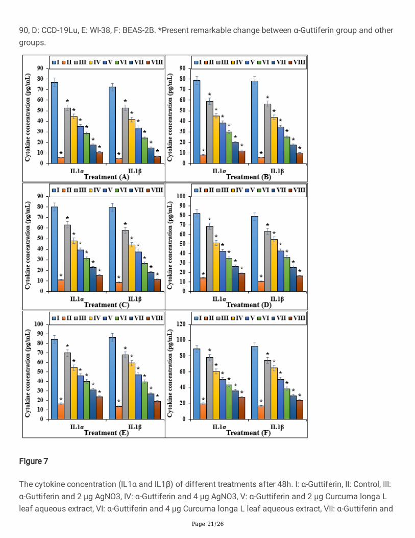
Figure 7
The cytokine concentration (IL1α and IL1β) of different treatments after 48h. I: α-Guttiferin, II: Control, III:α-Guttiferin and 2 μg AgNO3, IV: α-Guttiferin and 4 μg AgNO3, V: α-Guttiferin and 2 μg Curcuma longa Lleaf aqueous extract, VI: α-Guttiferin and 4 μg Curcuma longa L leaf aqueous extract, VII: α-Guttiferin and
Page 22/26
2 μg silver nanoparticles, VIII: α-Guttiferin and 4 μg of silver nanoparticles. A: HEL 299, B: MRC-5, C: IMR-90, D: CCD-19Lu, E: WI-38, F: BEAS-2B. *Present remarkable change between α-Guttiferin group and othergroups.
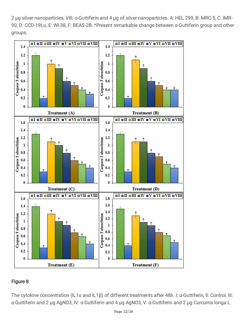
Figure 8
The cytokine concentration (IL1α and IL1β) of different treatments after 48h. I: α-Guttiferin, II: Control, III:α-Guttiferin and 2 μg AgNO3, IV: α-Guttiferin and 4 μg AgNO3, V: α-Guttiferin and 2 μg Curcuma longa L

Page 23/26
leaf aqueous extract, VI: α-Guttiferin and 4 μg Curcuma longa L leaf aqueous extract, VII: α-Guttiferin and2 μg silver nanoparticles, VIII: α-Guttiferin and 4 μg of silver nanoparticles. A: HEL 299, B: MRC-5, C: IMR-90, D: CCD-19Lu, E: WI-38, F: BEAS-2B. *Present remarkable change between α-Guttiferin group and othergroups.
Figure 9

Page 24/26
The apoptosis index of different treatments after 48h. I: α-Guttiferin, II: Control, III: α-Guttiferin and 2 μgAgNO3, IV: α-Guttiferin and 4 μg AgNO3, V: α-Guttiferin and 2 μg Curcuma longa L leaf aqueous extract,VI: α-Guttiferin and 4 μg Curcuma longa L leaf aqueous extract, VII: α-Guttiferin and 2 μg silvernanoparticles, VIII: α-Guttiferin and 4 μg of silver nanoparticles. A: HEL 299, B: MRC-5, C: IMR-90, D: CCD-19Lu, E: WI-38, F: BEAS-2B. *Present remarkable change between α-Guttiferin group and other groups.
Figure 10

Page 25/26
The Rh123 absorption of different treatments after 48h. I: α-Guttiferin, II: Control, III: α-Guttiferin and 2 μgAgNO3, IV: α-Guttiferin and 4 μg AgNO3, V: α-Guttiferin and 2 μg Curcuma longa L leaf aqueous extract,VI: α-Guttiferin and 4 μg Curcuma longa L leaf aqueous extract, VII: α-Guttiferin and 2 μg silvernanoparticles, VIII: α-Guttiferin and 4 μg of silver nanoparticles. A: HEL 299, B: MRC-5, C: IMR-90, D: CCD-19Lu, E: WI-38, F: BEAS-2B. *Present remarkable change between α-Guttiferin group and other groups.
Figure 11
The antioxidant properties of AgNO3, Curcuma longa L leaf aqueous extract, silver nanoparticles, andBHT against DPPH.

Page 26/26
Figure 12
The image of Curcuma longa L leaf.