Corynosoma australe Johnston, 1937 and C. cetaceum Johnston & Best, 1942 (Acanthocephala:...
14
Corynosoma australe Johnston, 1937 and C. cetaceum Johnston & Best, 1942 (Acanthocephala: Polymorphidae) from marine mammals and fishes in Argentinian waters: allozyme markers and taxonomic status Norma H. Sardella 1, *, Simonetta Mattiucci 2 , Juan T. Timi 1,3 , Ricardo O. Bastida 3,4 , Diego H. Rodrı´guez 3,4 & Giuseppe Nascetti 5 1 Laboratorio de Parasitologı´a, Departamento de Biologı´a, Facultad de Ciencias Exactas y Naturales, Universidad Nacional de Mar del Plata, Funes 3350, 7600, Mar del Plata, Argentina 2 Department of Public Health Sciences, (Section of Parasitology), University of Rome ‘‘La Sapienza’’, P. le Aldo Moro, 5, 00185, Rome, Italy 3 Consejo Nacional de Investigaciones Cientı´ficas y Te´cnicas (CONICET), Funes 3350, 7600, Mar del Plata, Argentina 4 Laboratorio de Mamı´feros Marinos, Departamento de Ciencias Marinas, Facultad de Ciencias Exactas y Naturales, Universidad Nacional de Mar del Plata, Funes 3350, 7600, Mar del Plata, Argentina 5 Department of Ecology and Sustainable Economic Development (DECOS), Tuscia University, Via S. Giovanni Decollato, 01100, Viterbo, Italy Accepted for publication 25th October, 2004 Abstract Genetic and morphological studies were carried out on acanthocephalans belonging to Corynosoma Lu¨he, 1904 and referable to the species C. cetaceum Johnston & Best, 1942 and C. australe Johnston, 1937, which were recovered from both definitive and intermediate hosts in Argentinian waters. The aims were to estimate the level of genetic differentiation between the two taxa at any stage of their life-cycle, to provide genetic (allozyme) markers for their recognition and to analyse the systematic status of both taxa. Acanthocephalans were collected from the stomach and intestine of Arctocephalus australis (Zimmerman), the intestine of Mirounga leonina (Linnaeus) and the stomach of Pontoporia blainvillei Gervais & D’Orbigny (definitive hosts) in Argentinian waters. Alternative alleles at all the 13 enzymatic loci studied were observed for C. australe and C. cetaceum. The specimens from the stomach of both P. blainvillei and A. australis were identified, on the basis of the great number of diagnostic loci found, as C. cetaceum; those from intestine of both A. australis and M. leonina as C. australe. A high level of genetic differentiation (D Nei =¥: I Nei =0.00) between the two taxa was found, suggesting a generic distinction between the two species. Cystacanths of the two species from the body-cavity of the fish Cynoscion guatucupa (Cuvier) collected from the same geo- graphical area were identified genetically. Morphological patterns, such as the number of hooks and hook rows on the proboscis, the distribution of somatic and genital armature, and other morphometric and meristic differences, in addition to ecological data, enabled the identification of these two species at cyst- acanth, juvenile and adult stages. However, a number of morphological and morphometric features of the Argentinian material were different to those of C. australe and C. cetaceum described from other regions of the world. Introduction Corynosoma Lu¨he, 1904 (Acanthocephala: Poly- morphidae) presently comprises numerous species which utilise pinnipeds, cetaceans and fish-eating *Author for correspondence (E-mail: [email protected]) Systematic Parasitology (2005) 61:143–156 ȑ Springer 2005 DOI 10.1007/s11230-005-3131-0
Transcript of Corynosoma australe Johnston, 1937 and C. cetaceum Johnston & Best, 1942 (Acanthocephala:...

Corynosoma australe Johnston, 1937 and C. cetaceum Johnston & Best,1942 (Acanthocephala: Polymorphidae) from marine mammals and fishesin Argentinian waters: allozyme markers and taxonomic status
Norma H. Sardella1,*, Simonetta Mattiucci2, Juan T. Timi1,3, Ricardo O. Bastida3,4,Diego H. Rodrıguez3,4 & Giuseppe Nascetti51Laboratorio de Parasitologıa, Departamento de Biologıa, Facultad de Ciencias Exactas y Naturales,Universidad Nacional de Mar del Plata, Funes 3350, 7600, Mar del Plata, Argentina2Department of Public Health Sciences, (Section of Parasitology), University of Rome ‘‘La Sapienza’’, P. leAldo Moro, 5, 00185, Rome, Italy3Consejo Nacional de Investigaciones Cientıficas y Tecnicas (CONICET), Funes 3350, 7600, Mar del Plata,Argentina4Laboratorio de Mamıferos Marinos, Departamento de Ciencias Marinas, Facultad de Ciencias Exactas yNaturales, Universidad Nacional de Mar del Plata, Funes 3350, 7600, Mar del Plata, Argentina5Department of Ecology and Sustainable Economic Development (DECOS), Tuscia University, ViaS. Giovanni Decollato, 01100, Viterbo, Italy
Accepted for publication 25th October, 2004
Abstract
Genetic and morphological studies were carried out on acanthocephalans belonging to Corynosoma Luhe,1904 and referable to the species C. cetaceum Johnston & Best, 1942 and C. australe Johnston, 1937, whichwere recovered from both definitive and intermediate hosts in Argentinian waters. The aims were to estimatethe level of genetic differentiation between the two taxa at any stage of their life-cycle, to provide genetic(allozyme) markers for their recognition and to analyse the systematic status of both taxa. Acanthocephalanswere collected from the stomach and intestine of Arctocephalus australis (Zimmerman), the intestine ofMirounga leonina (Linnaeus) and the stomach of Pontoporia blainvillei Gervais & D’Orbigny (definitivehosts) in Argentinian waters. Alternative alleles at all the 13 enzymatic loci studied were observed forC. australe and C. cetaceum. The specimens from the stomach of both P. blainvillei and A. australis wereidentified, on the basis of the great number of diagnostic loci found, as C. cetaceum; those from intestine ofboth A. australis and M. leonina as C. australe. A high level of genetic differentiation (DNei=¥: INei=0.00)between the two taxa was found, suggesting a generic distinction between the two species. Cystacanths of thetwo species from the body-cavity of the fish Cynoscion guatucupa (Cuvier) collected from the same geo-graphical area were identified genetically. Morphological patterns, such as the number of hooks and hookrows on the proboscis, the distribution of somatic and genital armature, and other morphometric andmeristic differences, in addition to ecological data, enabled the identification of these two species at cyst-acanth, juvenile and adult stages. However, a number of morphological and morphometric features of theArgentinian material were different to those of C. australe and C. cetaceum described from other regions ofthe world.
Introduction
Corynosoma Luhe, 1904 (Acanthocephala: Poly-morphidae) presently comprises numerous specieswhich utilise pinnipeds, cetaceans and fish-eating*Author for correspondence (E-mail: [email protected])
Systematic Parasitology (2005) 61:143–156 � Springer 2005DOI 10.1007/s11230-005-3131-0
birds as definitive hosts, and crustaceans and fishesas intermediate hosts (Delyamure, 1968). Despitetheir wide distribution among vertebrates, thesystematic status of several species is still notclear. Currently, there is a long list of synony-mies and misidentifications (see e.g. Amin,1985), including the type-species, C. strumosum(Rudolphi, 1802) Luhe, 1904, whose descriptionhas recently been amended (Nickol et al., 2002a).This is probably due to two main reasons: (1) thesystematics of this genus is presently based on afew variable morphological features (such as thenumber of hook rows and of hooks per row on theproboscis, and the distribution patterns of somaticand genital spines) and general morphometry; and(2) several sources of intraspecific variabilitywithin populations of Corynosoma spp. (such asthe effect of parasite age and size, host-inducedeffects and geographical influence) which migthaffect morphological features (George-Nascimento& Marin, 1992; Aznar et al., 1999a). Similarly,confusion has been reported between genera of theFamily Polymorphidae Meyer, 1931 (Schmidt,1973, 1975; Amin, 1992; Aznar et al., 1999a;Nickol et al., 1999), which arose for the samereasons as problems at the specific level.
Records of adult Corynosoma in marine mam-mals from the South-West Atlantic Ocean presenta similar state of taxonomic uncertanty. Indeed,along the Argentinian coast, Morini & Boero(1960) recorded C. otariae Morini & Boero, 1960,based upon six specimens from the South Amer-ican sea lion Otaria flavescens (Shaw), which wasdifferentiated from the original description ofC. australe Johnston, 1937 from the Australiansea lion Neophoca cinerea (Peron) in Australianwaters (Johnston, 1937) by the similar size of thesexes, the number of proboscis hook rows (20instead of 18), a larger body and embriophore size,and the distribution of the somatic armature inboth sexes. However, Zdzitowiecki (1989), com-paring the description of Morini & Boero (1960)with his redescription of C. australe (Zdzitowiecki,1984a), synonymised C. otariae with C. australewithout any mention of the differences reported byMorini & Boero (1960). Later, George-Nascimen-to & Marin (1992) were unable to identify at thespecific level specimens from O. flavescens and theSouth American fur seal Arctocephalus australisZimmerman on the Uruguayan coast; laterspecimens from A. australis off Uruguay were
identified as C. australe by Aznar et al. (2004).C. australe was also recently recorded from acetacean, the dusky dolphin Lagenorhynchusobscurus (Gray), also from Argentinian waters(Dans et al., 1999).
C. cetaceum has commonly been reported as aparasite of the franciscana Pontoporia blainvilleiGervais & D’Orbigny off the Uruguayan (Schmidt& Dailey, 1971; Kagei et al., 1976; Aznar et al.,1994a) and Argentinian coasts (Aznar et al.,1994a, b), and from the short beaked commondolphin Delphinus delphis Linnaeus off Argentina(Aznar et al., 2002a). Unidentified species of thisgenus have also been reported from P. blainvillei(see Dailey & Brownell, 1972). A geographicalcomparison of samples of C. cetaceum showedseveral differences (such as the number of hooksper row on the proboscis and the distributionpatterns of the somatic spines) between SouthAmerican and South Australian specimens (Aznaret al., 1999a). The generic status of C. cetaceumhas also been a matter of controversy (see Schmidt& Dailey; 1971; Smales, 1986; Aznar et al., 1999a).This taxon was transferred to Polymorphus Luhe,1911, due to the absence of genital spines in bothsexes, but was reinstated as C. cetaceum by Aznaret al. (1999a); however its generic status remainsunresolved (Garcıa-Varela et al., 2005).
Records of cystacanths belonging to Corynoso-ma from fishes off Argentina are: C. australe (seeZdzitowiecki, 1989; Sardella et al., 1995; Tanzolaet al., 1997; Cremonte & Sardella, 1997; Tanzola &Guagliardo, 2000; Timi, 2003); and C. hammani(Linstow, 1892) (see Tanzola et al., 1997; Tanzola& Guagliardo, 2000). Unidentified cystacanths ofCorynosoma have also been reported (Szidat, 1949,1969; Suriano, 1966; Ivanov, 1996; Sardella &Timi, 1996; Sardella et al., 1998). From the numer-ous reports listed above, it is clear that thesystematic status of material of Corynosoma atthe cystacanth stage in the South-West AtlanticOcean also remains uncertain.
On the other hand, genetic markers obtainedfrommultilocus allozyme electrophoresis have beendemonstrated to be a useful tool for answeringquestions related to the systematics of severalparasites and for detecting various cryptic or siblingspecies, as well as establishing genetic relationshipsbetween congeneric taxa of endoparasites (Andrews& Chilton, 1999), including those from marinemammals (Nascetti et al., 1986, 1993; Mattiucci
144

et al., 1997, 2001, 2003). Allozyme markers havealso previously been applied to the systematics ofother acanthocephalan species (De Buron et al.,1986; Aho et al., 1992; Vainola et al., 1994).Molecular-genetic studies on acanthocephalans ofthe genus Corynosoma have recently been carriedout by Garcıa-Varela et al. (2005).
In the present work, morphological and geneticstudies were carried out on acanthocephalansreferable to the morphospecies C. australe (anuncontroversial taxon) and C. cetaceum (a prob-lematical species) recovered from definitive andintermediate hosts in Argentinian waters. The aimsof the study were: to estimate the level of geneticdifferentiation between the two taxa at any stageof their life-cycle, to provide genetic (allozyme)markers for their recognition and to analyse thesystematic status of both taxa.
Materials and methods
Acanthocephalans were obtained from the follow-ing definitive hosts: the South American fur sealArctocephalus australis (Zimmerman) found deadalong Claromeco beach and San Clemente delTuyu, Buenos Aires Province; the southern ele-phant seal Mirounga leonina (Linnaeus) founddead at San Clemente del Tuyu; and the francis-
cana Pontoporia blainvillei, an accidental by-catch,at Mar del Plata (details of the collection data aregiven in Table 1).
In order to avoid the use of the term ‘juvenile’for any non-adult specimen, acanthocephalansrecovered from fish hosts are referred to as‘cystacanths’, those worms from the definitive hostwhich have not reached sexual maturity aredenoted as ‘juveniles’ and mature specimens as‘adults’.
Cystacanths were collected from eight speci-mens of striped weakfish Cynoscion guatucupa(Cuvier) caught by fishermen using trawl nets andlanded at the Mar del Plata harbour (Table 1).
The gastrointestinal tracts of the definitivehosts were frozen prior to dissection and parasi-tological examination. Acanthocephalans werecollected from the stomach and/or intestine oftheir definitive hosts, washed in saline solution anda subsample frozen in distilled water in Eppen-dorf-like tubes, then stored at )70 �C for geneticanalysis. The same procedure was applied to livingcystacanths collected from body-cavity of fish. Formorphological analysis, thawed and living speci-mens were extended in distilled water for severalhours prior to fixation, then fixed in 4% formal-dehyde, stored in 70% alcohol, cleared in lactoph-enol and examined under a light microscope. Thenumber of proboscis hook rows were countedfrom transverse sections of the distal end of the
Table 1. Collection data for the samples of Corynosoma spp. studied from the Argentinian coast.
Hosts Collecting site Date of
collection
Nh C. australe C. cetaceum nMAE
Stomach Intes-
tine
Body-
cavity
Stomach Intes-
tine
Body-
cavity
C.
australe
C.
cetaceum
A. australis Claromeco
(38�22¢S, 60�16¢W)
September,
1999
1 – 5,231 – 47 63 – – –
A. australis San Clemente del
Tuyu
(36�30¢S, 56�20¢W)
August,
2000
1 – 150* – 6 – – 37 5
M. leonina San Clemente del
Tuyu
(36�30¢S, 56�20¢W)
August,
2000
1 – 51 – – – – 22 –
P. blainvillei Mar del
Plata
(38�08¢S, 57�32¢W)
August,
1999
1 – – – 1,961 – – – 28
C. guatucupa Mar del Plata
(38�08¢S, 57�32¢W)
May,
2000
8 – – 87 – – 74 17 17
Nh, number of hosts examined. *, not all parasites counted. nMAE, specimens studied by multilocus allozyme electrophoresis.
145

proboscis mounted in apical view. General mea-surements were taken on specimens mounted inlateral position, as shown in Figure 1. Somaticspines were measured on the dorsal posteriorborder of the trunk armature. Drawings weremade using a drawing tube. Measurements ofembryophores are based on fully-developedembryophores (with an identifiable acanthor in-side). Measurements are given in millimetres,unless otherwise indicated, as the mean followedby range in parentheses. Only those morphologicalfeatures showing differences with previous descrip-tions of both species are provided.
Voucher specimens are deposited in the Cole-ccion Zoologıa Invertebrados del Museo de LaPlata (Helmintos). Corynosoma australe: 5 adultmales (Coll. No. 5405) and 5 adult females (Coll.No. 5405) from the intestine of Arctocephalusaustralis; 3 adult males (Coll. No. 5406) and 3 adult
females (Coll. No. 5406) from the intestine ofMirounga leonina; 5 cystacanth males (Coll. No.5407) and 5 cystacanth females (Coll. No. 5407)from the body-cavity of Cynoscion guatucupa.Corynosoma cetaceum: 5 adult males (Coll. No.5408) and 5 adult females (Coll. No. 5408) fromstomach of Pontoporia blainvillei; 5 juvenile males(Coll. No. 5409) and 5 juvenile females fromstomach and intestine of A. australis; 5 cystacanthmales (Coll. No. 5410) and 5 cystacanth females (Coll.No.5410) fromthebody-cavityofCynoscionguatucupa.
Genetic analysis was performed on 92 acantho-cephalans (see Table 1) as follows: 42 fromA. australis, 22 from M. leonina and 28 fromP. blainvillei. Thirty-four cystacanths from C. gua-tucupa were also analysed. Acanthocephalans werekept frozen at )70 �C, then transported in dry iceto Rome for genetic analysis. Standard horizontalstarch gel electrophoresis was performed at 5 �Cand 7–9 cm V/cm for 4–5 hr, according to Mat-tiucci et al. (1997). Single specimens were crushedin distilled water. The following enzymes (listed bytheir code number) were tested: malate dehydro-genase (Mdh-1) E.C. 1.1.1.37; malic enzyme (Me)E.C. 1.1.1.40; 6-phosphogluconate dehydrogenase(6Pgdh) E.C. 1.1.1.44; glyceraldehyde-3-phosphatedehydrogenase (Gapdh) E.C. 1.2.1.12; superoxidedismutase (Sod-1) E.C. 1.15.1.1; aspartate aminotransferase (Aat-2) E.C. 2.6.1.1; adenylate kinase(Adk-1) E.C. 2.7.4.3; acid phosphatase (Acph)E.C.3.1.3.2; leucine aminopeptidase (Lap-1, Lap-2) E.C. 3.4.11; peptidase (Leu-Leu) (Pep B) E.C.3.4.11; peptidase Leu-Ala (Pep C-1) E.C. 3.4.11;and glucose phosphate isomerase (Gpi) E.C.5.3.1.9. The buffer systems and staining proceduresused were those detailed in Mattiucci et al. (1997).Isozymes were numbered in order of decreasingmobility from the most anodal one. Allozymeswere namedwith numbers indicating their mobility (inmm, standardised conditions) relative to the mostcommon allele, designated as 100, found in a referencepopulation (i.e. a population of Corynosoma australecollected from A. australis in Argentinean waters).
The statistical significance of departures fromthe Hardy-Weinberg (H-W) equilibrium was esti-mated using chi-square test (v2). The genetic diver-gence was estimated using the following indices:standard genetic Distance and Identity (DNei andINei, Nei, 1972). Population genetic analysis wasperformed using BIOSYS software (Swofford &Selander, 1989).
Fig. 1. Scheme of measurements for specimens of Corynoso-ma: a + b + c, total length; b + c, trunk length; d, ventralfore-trunk length; e, dorsal fore-trunk length; f, ventral hind-trunk length; g, dorsal hind-trunk length.
146
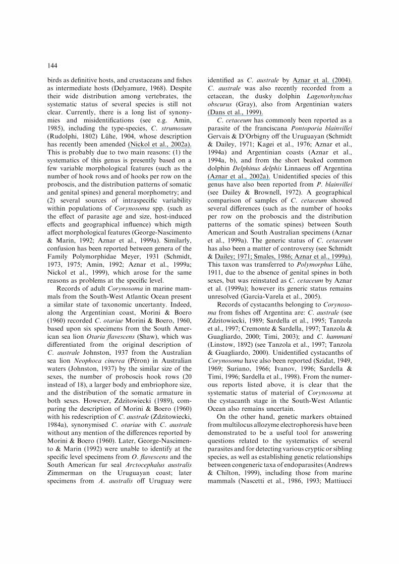
Corynosoma australe Johnston, 1937
Description (Figures 2–8)
With characteristics of previous descriptions ofthis species. Hooks arranged in 18–20 rows (usu-ally 18). Each row comprises 12–14 hooks, 9–11
anterior hooks (usually 10) with well-developed,posteriorly directed roots and 2–4 (usually 3)small basal hooks with small, anteriorly directedroots. Following combinations of anterior/basalhooks were observed: 9/3, 9/4, 10/2, 10/3, 10/4, 11/2 and 11/3, usually 10/3. Up to 4 differentcombinations were recorded in single individual.
Figures 2–8. Corynosoma australe Johnston, 1937, adults from the stomach of Arctocephalus australis. 2. Detail of the proboscis,lateral view. 3. Detail of the proboscis showing 19 rows of hooks, apical view. 4. Embryophore. 5. Posterior end of female showinggenital spines, lateral view. 6. Posterior end of female showing genital spines, apical view. 7. Posterior end of male showing genitalspines, lateral view. 8. Posterior end of male showing genital spines, apical view.
147

Cystacanths from fish hosts with characteristics ofadults, but smaller in size, although proboscis isalmost of same size as adults; with developinggenitalia. Measurements are given in Table 2.
Male (based on 12 adults and 6 cystacanths).Ventrally somatic armature covers 80.2 (77.7–88.3)% of trunk length in adults and 80.8 (78–83.5)% in cystacanths. Genital opening surroundedby 3 irregular rows of 18–34 triangular genitalspines, larger than somatic spines.
Female (based on 10 adults and 6 cystacanths).Ventrally somatic armature covers 89.2 (84.6–100)% of trunk length in adults and 87.4 (85–89)% in cystacanths. Genital opening surrounded
by irregular rows of 18–35 triangular genital spinesshifted to ventral side, shorter and wider thansomatic spines and smaller than those of males.In some specimens genital and somatic spinesare contiguous in ventral region but clearlydistinguishable.
New definitive host: Mirounga leonina Linnaeus.New intermediate host: Cynoscion guatucupa(Cuvier).New localities: Mar del Plata (38�08¢S, 57�32¢W)and San Clemente del Tuyu (36�30¢S, 56�20¢W),Argentina.
Corynosoma cetaceum Johnston & Best, 1942
Syn. Polymorphus arctocephali Smales, 1986
Table 2. Measurements (mm) of Corynosoma australis from Arctocephalus australis and Cynoscion guatucupa.
Host A. australis C. guatucupa
male (n = 12) female (n = 10) male (n = 6) female (n = 6)
Total length 4.60 (4.20–5.40) 4.91(4.22–5.50) 2.82 (2.56–3.18) 3.20 (3.02–3.42)
Maximum width 1.43 (1.30–1.62) 1.59 (1.34–1.90) 0.85 (0.76–0.96) 0.94 (0.80–1.14)
Proboscis length 0.63 (0.58–0.72) 0.68 (0.60–0.74) 0.63 (0.58–0.68) 0.67 (0.60–0.74)
Proboscis width 0.22 (0.21–0.24) 0.23 (0.22–0.25) 0.21 (0.18–0.23) 0.22 (0.15–0.28)
Neck length 0.19 (0.16–0.24) 0.20 (0.15–0.26) 0.22 (0.19–0.24) 0.23 (0.20–0.27)
Neck maximum width 0.40 (0.36–0.46) 0.44 (0.38–0.50) 0.34 (0.26–0.42) 0.31 (0.26–0.42)
Trunk length 3.69 (3.42–4.30) 3.94 (3.30–4.66) 1.87 (1.64–2.18) 2.30 (2.18–2.42)
Fore-trunk ventral length 1.62 (1.40–1.90) 1.77 (1.38–2.30) 1.06 (0.96–1.14) 1.18 (0.90–1.48)
Fore-trunk dorsal length 2.21 (2.00–2.60) 2.41 (1.94–3.00) 1.29 (1.14–1.40) 1.40 (1.10–1.60)
Hind-trunk ventral length 1.90 (1.67–2.16) 1.93 (1.72–2.26) 0.88 (0.82–0.98) 1.11 (0.86–1.44)
Hind-trunk dorsal length 1.51 (1.30–1.78) 1.47(1.20–1.66) 0.74 (0.60–0.82) 0.89 (0.74–0.96)
Hind-trunk width at mid-length 0.55 (0.50–0.62) 0.72 (0.58–0.86) 0.37 (0.30–0.42) 0.43 (0.34–0.54)
Somatic spine length* 40 (30–50) 40 (36–46) 38 (31–42) 43 (40–46)
Somatic spine width* 8 (6–10) 9 (6–10) 7 (6–7) 8
Genital spine length* 45 (42–48) 31 (25–38) 46 (44–48) 36 (31–40)
Genital spine width* 22 (17–29) 11 (8–15) 25 (21–27) 9 (8–10)
Proboscis receptacle length 0.82 (0.74–0.92) 0.94 (0.84–1.04) 0.93 (0.86–1.02) 1.20 (1.10–1.30)
Proboscis receptacle width 0.16 (0.14–0.20) 0.18 (0.16–0.20) 0.21 (0.16–0.26) 0.20 (0.14–0.28)
Lemnisc length 0.67 (0.56–0.74) 0.69 (0.56–0.80) 0.47 (0.38–0.56) 0.47 (0.40–0.56)
Lemnisc width 0.47 (0.42–0.52) 0.49 (0.36–0.60) 0.27 (0.24–0.30) 0.27 (0.20–0.32)
Right testis length 0.54 (0.44–0.70) – 0.12 (0.10–0.14) –
Right testis width 0.34 (0.26–0.42) – 0.09 (0.08–0.12) –
Left testis length 0.54 (0.40–0.66) – 0.11 (0.10–0.14) –
Left testis width 0.34 (0.28–0.42) – 0.09 (0.08–0.12) –
Everted bursa length 0.57 (0.42–0.72) – – –
Everted bursa width 0.56 (0.52–0.62) – – –
Embryophore length* – 103 (92–115) – –
Embryophore width* – 33 (27–42) – –
*In micrometres.
148

Description (Figures 9–15)
With characteristics of previous descriptions ofthis species. Hooks arranged in 20–21 rows (usu-ally 20). Each row comprises 13–15 hooks, 11–13anterior ones (usually 12) with well-developed,anteriorly directed roots and 2–3 (usually 2) smallbasal hooks with small, posteriorly directed roots.Following combinations of anterior/basal hookswere observed: 11/2, 11/3, 12/2, 12/3 and 13/2(usually 12/2). Up to 3 different combinations ofanterior/basal hooks were recorded in singlespecimen.
Genital armature absent in both sexes. Allspecimens from A. australis smaller than thosefrom P. blainvillei and immature, especially obvi-ous in females, whose embryophores were notobserved. Cystacanths from fish hosts with char-acteristics of adults, but smaller in size, althoughproboscis is almost of same size, with developinggenitalia. Measurements are given in Table 3.
Male (based on 6 adults from P. blainvillei, 6juveniles from A. australis and 6 cystacanths fromC. guatucupa). Somatic armature covers 71.5(65.8–75.2)% of trunk length ventrally in adults,69.1 (63.6–74.6)% in juveniles and 71 (57.5–77.1)%. Genital opening devoid of spines.
Female (based on 6 adults from P. blainvillei, 6juveniles from A. australis and 6 cystacanths fromC. guatucupa). Somatic armature covers 95.2(93.9–96.5)% of trunk length ventrally in adults,96.8 (96–97.4)% in juveniles and 94.9 (94.3–95.8)% in cystacanths. Ventrally trunk exhibits 2transverse folds delimiting blunt lobe betweenfore- and hind-trunks. In most specimens, at levelof both transverse folds, ventral somatic armatureis interrupted by 2 fields devoid of spines, or lessfrequently having smaller spines, situated at 43.5(34.8–48.8)% and 61.6 (52.2–68.3)% of trunklength, respectively, in adults; in juveniles thesepercentages were 49.1 (47.1–52.6)% and 65 (63.6–
Figures 9–10. Corynosoma cetaceum Johnston & Best, 1942, cystacanths from the body-cavity of Cynoscion guatucupa. 9. Male,lateral view. 10. Female, lateral view.
149

66.5)%; in cystacanths were 48.4 (43.2–52)% and64.4 (55.5–70.9)%. In some specimens theseunarmed fields are only ventrolateral and narrowcontinuous ventral field of spines is observed alongentire trunk. Genital opening devoid of spines.
New definitive host: Arctocephalus australis(Zimmerman).New intermediate host: Cynoscion guatucupa(Cuvier).New localities: Mar del Plata (38�08¢S, 57�32¢W)and San Clemente del Tuyu (36�30¢S, 56�20¢W),Argentina.
Genetic differentiation between Corynosomaaustrale and C. cetaceum and allozyme markers
for their identification
The alleles found and their frequencies observed inthe populations of C. australe and C. cetaceumanalysed are reported in Table 4. Most of the
enzymatic loci examined were found to bemonomorphic in both species. Some enzyme loci(i.e. Lap-1, Pep B, Pep C-1) were found to bepolymorphic in C. cetaceum with no statisticallysignificant departure from the H-W equilibrium.Genetic homogeneity was also found withinC. australe, despite its occurrence in two differentdefinitive hosts, A. australis and M. leonina. InC. australe, the enzyme loci 6-Pgdh and Lap-1were observed to be polymorphic (Table 4) with-out statistically significant departures from theH-W equilibrium. Alternative alleles at all the 13enzymatic loci studied were observed betweenC. australe, from the intestine of A. australis, andC. cetaceum, from the stomach of P. blainvillei.
In addition, electrophoretic migration at thelocus Acph was found to be anodal in C. cetaceumbut cathodal in C. australe. Nei’s standard geneticDistance and Identity values between the twospecies were found to be DNei=¥ (INei=0.00),respectively. Indeed, no alleles were shared byC. australe and C. cetaceum at any of the studied
Figures 11–15. Corynosoma cetaceum Johnston & Best, 1942. 11. Detail of the proboscis, lateral view.12. Detail of the proboscisshowing 20 rows of hooks, apical view. 13. Embryophore. 14. ventral trunk of female showing somatic spines, lateral view. 15.Posterior end of female showing somatic spines near the genital opening, similar in shape and size to the anterior spines, lateralview.
150

Table
3.Measurements
(mm)ofCorynosomacetaceum
adultsfrom
Pontoporiablainvillei,juveniles
from
Arctocephalusaustralisandcystacanthsfrom
Cynoscionguatucupa.
Host
P.blainvillei
A.australis
C.guatucupa
male
(n=
6)
female
(n=
6)
male
(n=
6)
female
(n=
6)
male
(n=
6)
female
(n=
6)
Totallength
7.47(7.10–8.00)
5.09(4.50–5.40)
5.92(5.08–6.58)
4.75(4.42–4.98)
5.26(5.04–5.64)
4.61(4.30–4.80)
Maxim
um
width
2.46(2.28–2.60)
2.33(1.86–2.70)
1.77(1.64–2.04)
1.79(1.60–1.92)
1.53(1.40–1.64)
1.62(1.44–1.76)
Proboscislength
1.14(1.00–1.20)
1.11(1.06–1.16)
1.03(0.94–1.10)
1.04(0.94–1.10)
0.95(0.90–1.04)
1.10(1.04–1.20)
Probosciswidth
0.33(0.32–0.36)
0.32(0.28–0.34)
0.33(0.29–0.34)
0.34(0.32–0.37)
0.30(0.26–0.34)
0.33(0.32–0.36)
Necklength
0.28(0.26–0.32)
0.27(0.24–0.29)
0.28(0.22–0.32)
0.28(0.25–0.32)
0.32(0.22–0.38)
0.34(0.30–0.40)
Neckmaxim
um
width
0.63(0.58–0.66)
0.66(0.56–0.74)
0.59(0.48–0.70)
0.54(0.48–0.58)
0.53(0.44–0.60)
0.54(0.40–0.60)
Trunklength
6.14(5.80–6.54)
3.66(3.14–4.10)
4.67(4.08–5.20)
3.38(3.04–3.58)
4.11(3.86–4.36)
3.09(2.82–3.30)
Fore-trunkventrallength
2.48(2.00–3.16)
2.25(2.00–2.80)
2.13(1.70–2.30)
2.20(2.02–2.30)
2.00(1.80–2.20)
1.99(1.72–2.24)
Fore-trunkdorsallength
3.22(2.90–3.60)
2.95(2.36–3.80)
2.50(2.20–2.96)
2.20(1.40–2.58)
2.38(2.20–2.56)
2.30(2.20–2.40)
Hind-trunkventrallength
3.54(3.20–3.90)
1.19(1.00–1.30)
2.42(2.00–2.86)
1.13(1.10–1.20)
1.96(1.84–2.10)
0.91(0.84–1.04)
Hind-trunkdorsallength
3.24(3.00–3.60)
1.29(1.06–1.40)
2.15(1.86–2.60)
1.13(0.96–1.20)
1.65(1.44–1.84)
1.02(0.92–1.12)
Hind-trunkwidth
atmidlength
0.97(0.84–1.10)
1.16(0.96–1.40)
0.69(0.62–0.76)
0.79(0.68–0.88)
0.52(0.40–0.70)
0.73(0.66–00.84)
Somaticspinelength*
55(52–59)
60(54–53)
58(52–65)
49(44–52)
52(46–57)
47(42–52)
Somaticspinewidth*
22(21–27)
20(19–21)
15(15–17)
23(21–25)
16(15–19)
24(23–25)
Proboscisreceptaclelength
1.51(1.44–1.56)
1.50(1.40–1.60)
1.22(1.24–1.26)
1.33(1.26–1.44)
1.36(1.20–1.46)
1.43(1.30–1.50)
Proboscisreceptaclewidth
0.40(0.36–0.44)
0.38(0.30–0.44)
0.24(0.20–0.26)
0.22(0.20–0.24)
0.31(0.26–0.36)
0.37(0.32–0.40)
Lem
nisclength
0.73(0.60–0.90)
0.74(0.60–0.84)
0.74(0.60–0.88)
0.72(0.60–0.84)
0.73(0.54–0.83)
0.62(0.50–0.70)
Lem
niscwidth
0.58(0.50–0.68)
0.55(0.36–0.70)
0.42(0.46–0.76)
0.48(0.42–0.56)
0.41(0.36–0.46)
0.41(0.36–0.46)
Righttestislength
0.68(0.44–0.80)
–0.39(0.24–0.50)
–0.18(0.12–0.24)
–
Righttestiswidth
0.44(0.30–0.52)
–0.27(0.14–0.44)
–0.15(0.08–0.26)
–
Lefttestislength
0.65(0.40–0.78)
–0.41(0.26–052)
–0.18(0.14–0.26)
–
Lefttestiswidth
0.44(0.34–0.56)
–0.26(0.16–0.34)
–0.15(0.10–0.24)
–
Evertedbursalength
1.17(1.04–1.30)
––
––
–
Evertedbursawidth
0.96(0.90–1.06)
––
––
–
Embryophore
length*
–158(147–178)
––
––
Embryophore
width*
–50(44–58)
––
––
*In
micrometres.
151

loci. On the other hand, a low value of geneticdistance was found between populations ofC. aust-rale from the two definitive hosts (DNei=0.01).The corresponding interpopulational geneticdiversity in this taxon was Fst=0.03.
A similar level of genetic variability, based on the13 enzyme loci, for the parameter He was found inboth species (Table 4). The percentage of polymor-phic loci, according to the 0.99 criterion, resultedP99=0.15 inC. australe andP99=0.23 inC. cetaceum.
All of the specimens collected from the intestineof both A. australis and M. leonina were found tocorrespond to C. australe. While, the 28 acantho-cephalan specimens recovered from the stomach ofP. blainvillei were identified genetically asC. cetaceum. Moreover, the few individualscollected from the stomach of one specimen ofA. australis were also found to correspond toC. cetaceum. Furthermore, cystacanths recoveredfrom the fish C. guatucupa were identified genet-ically; of the 34 cystacanths tested, 17 corre-sponded to C. australe and 17 to C. cetaceum.
Discussion
The specific status of Corynosoma australe andC. cetaceum in the Southwestern Atlantic Ocean
The original description of C. australe by Johnston(1937) was brief and a number of external and
internal features were not included. AccordingSmales (1986), the type-specimens were incom-pletely extended and the internal features black-ened. Therefore, for comparative purposes,measurements for the Australian material weretaken from redescription given by Smales (1986).Comparisons of the specimens studied herein withprevious descriptions of adult specimens ofC. australe from Australia (Smales, 1986), theAntarctic (Zdzitowiecki, 1984a) and Uruguay(Morini & Boero, 1960), as well as of cystacanthsfrom Brazil (Pereira & Neves, 1993; Knoff et al.,2001), showed that most morphometric character-istics are relatively uniform; nevertheless, Argen-tinian and Uruguayan adult specimens have alarger embryophore and body size, and 18 to 20hook rows on the proboscis. Embryophores fromthe present study were markedly larger (almostoutside the reported ranges) than those given inother papers, including those parasites from thesame host species given by Morini & Boero (1960).
George-Nascimento & Marın (1992) foundadult specimens of Corynosoma sp. in Otariabyronia (=O. flavescens) and in A. australis fromUruguay. These authors observed morphometricdifferences between parasites from both hostspecies, although they considered that they belongto the same species and that the size variability is aconsequence of host-induced effects, suggestingthat A. australis is the most suitable host species.
Table 4. Characteristic alleles, with their frequencies (in parenthesis), at 13 enzymatic loci studied in Corynosoma australe in rela-tion to C. cetaceum.
Locus C. australe C. cetaceum
Mdh-1 100 (1.00) 90 (1.00)
Me 100 (1.00) 110 (1.00)
6Pgdh 100 (0.82), 93 (0.13), 105 (0.05) 80 (1.00)
Gapdh 100 (1.00) 115 (1.00)
Sod-1 100 (1.00) 80 (1.00)
Aat-2 100 (1.00) 107 (1.00)
Adk-1 100 (1.00) 110 (1.00)
Lap-1 100 (0.96), 93 (0.04) 85 (0.97), 80 (0.03)
Lap-2 100 (1.00) 95 (1.00)
Acph 100 (1.00) 115 (1.00)
Pep B 100 (1.00) 90 (0.90), 85 (0.10)
Pep C-1 100 (1.00) 95 (0.96), 85 (0.03), 105 (0.01)
Gpi 100 (1.00) 105 (1.00)
He 0.029 (±0.08) 0.024 (±0.05)
The sign ‘‘)’’ indicates cathodal migration.He, Expected mean heterozygosity per locus.
152

They also postulated that parasites partly agreedwith either C. australe or C. pseudohammaniZdzitowiecki, 1984. However, some of the mor-phometric (i.e. lemnisci larger than the proboscisreceptacle, proboscis width) and meristic (numberof hooks per row on the proboscis) characteristicsgiven by George-Nascimento & Marın (1992)do not coincide with either C. australe orC. pseudohammani (see Zdzitowiecki, 1984b; pres-ent study). Taking into account the fact thatC. australe is found in the same host species and inthe proximity of the study areas (Argentina andUruguay) (Aznar et al., 2004; present study),George-Nascimento & Marın’s parasites probablybelonged to C. australe.
Specimens of C. cetaceum examined in thepresent paper agree in general terms with thecharacteristics that define the species; however,some differences in the number of proboscis hookrows (20–21 versus 18–9) and in the number ofhooks per row (13–15 versus 12–13 or 14–16) wereobserved. The ventrally transverse folds delimitinga blunt lobe between the fore- and hind-trunks inthe female, described by Aznar et al. (1999b,2002b), were also observed in cystacanths in thepresent study.
Comparisons of samples of C. cetaceum on alarge geographical scale showed a high level ofvariability between Southwestern Atlantic andSouth Australian populations, which weregrouped into two well-defined clusters by PrincipalComponent Analysis (Aznar et al., 1999a). Inparticular, these authors observed a smaller num-ber of hooks per row, a different pattern ofsomatic spines in females and a tendency for thegenital pore to be subterminal in some SouthAmerican specimens.
C. cetaceum was also found in A. australis, butall specimens were smaller than those fromP. blainvillei and all were juveniles without eggs.This species typically inhabits the stomach of itsdefinitive cetacean hosts (Aznar et al., 2001), but inA. australis it was also found parasitising theintestine. A. australis appears to be an unsuitableor accidental host for C. cetaceum in the studyarea. Similarly, this parasite has been found injuvenile form in A. pusillus doriferus fromAustralia (Smales, 1986; Aznar et al., 1999a).
Regarding the life-cycle of C. cetaceum, and aspointed out by Aznar et al. (1999a), fish are alikely necessary component of their life-cycle,
given the feeding habits of their mammalian hosts.In the present paper, the presence of cystacanths ofthis species in fish is confirmed by both morpho-logical and allozyme evidence, and their firstdescription is provided.
Genetic comparisons with further populationsfrom different geographical areas will enable anassessment of whether the genetic homogeneityobserved within samples of C. australe andC. cetaceum from Argentina is constant over alarge geographical range.
The generic status of Corynosoma cetaceum
Corynosoma cetaceum was first described from thecommon dolphin Delphinus delphis and from thebottlenose dolphin Tursiops truncatus in Austra-lian waters (Johnston & Best, 1942). These authorsincluded it in Polymorphus due to the absence ofgenital spines in both sexes, which is the onlycriterion separating Corynosoma Luhe, 1904 fromPolymorphus Luhe, 1911 (see Schmidt & Dailey,1971). However, the presence of six cement-glands(instead of four) is an additional criterion forseparating C. cetaceum from typical species ofPolymorphus. This feature was discussed bySmales (1986), who found that, due to the numberof cement-glands, C. cetaceum (as P. cetaceum)falls within Hexaglandula Petrochenko, 1950,rather than in Polymorphus (according to the keyproposed by Schmidt, 1973). Hexaglandula waslater synonymised with Polymorphus by Amin(1992), but this has not been accepted by otherauthors (see Nickol et al., 2002b).
As stated by Aznar et al. (1999a), the concept ofgenital spines is problematical, due to its ambiguousdefinition with respect to somatic spines. Therefore,using a less restricted definition of a genital spine,P. cetaceum was retained as C. cetaceum by Aznaret al. (1999a) and sustained by Aznar et al. (2002a).This problem appears to arise from the definition ofa genital spine based only on its position relative tothe genital opening (see Aznar et al., 1999a) andwithout consideration of genital and somatic spinemorphology. Females of C. cetaceum bear spines inthe vicinity of the genital pore, which are identicalto the somatic spines, whereas in C. australe genitalspines are stout, triangular and clearly discernablein both sexes. Therefore, C. cetaceum lacks genitalspines (sensu stricto). In a recent paper, Aznar et al.
153

(2002a) found one of 80 males of C. cetaceum fromtwo specimens of D. delphis off Patagonia as havingtwo small spines adjacent to the genital pore andisolated from the somatic spines; they stated thatthis finding confirmed the reassignment of C.cetaceum to Corynosoma (see Aznar et al., 1999a).However, the presence of only one male havinggenital spines in over 3,000 males examined fromdifferent host in several regions (Aznar et al., 2002a)suggests that it cannot be taken as a reliablediagnostic feature at the generic level. This, how-ever, does not preclude C. cetaceum from Coryno-soma, because some species from marine mammals,which lack genital spines in both sexes, are includedwithin Corynosoma, e.g. C. falcatum Van Cleave,1953, C. sudsuche Belopolskaja, 1959 and C.septentrionalis Treshtchev, 1966 (see Aznar et al.,1999a).
Despite the evidence relating to both the genitalspine definition and the number of cement-glands,the discrimination between Corynosoma and Poly-morphus remains controversial and further revi-sion is required, specifically for C. cetaceum. Infact, Smales (1986) stated that the combination ofcharacters found in C. cetaceum does not agreewith any of the generic diagnoses proposed bySchmidt (1973, 1975). Furthermore, and despite ofthe morphological similarity that this speciesshares with most species of Corynosoma that usemarine mammals and fish as definitive andparatenic hosts, respectively, most species of Co-rynosoma mature in pinnipeds, whereas C. cetace-um parasitises cetaceans. On the other hand,typical species of Polymorphus exhibit a freshwatercycle with crustaceans as intermediate hosts andwaterfowl as the normal definitive hosts, but donot use fish as paratenic hosts (Aznar et al.,1999a).
Nickol et al. (1999) concluded that, for poly-morphid genera, the occurrence in different groupsof crustacean intermediate hosts implies substan-tial life-history differences and justifies differenti-ation at the generic level. Perhaps this criterion canalso be applied to the use of definitive hosts(pinnipeds for most Corynosoma spp. maturing inmammals and cetaceans for C. cetaceum). In thissense, Hexaglandula, which has six cement-glandsas C. cetaceum, parasitises decapod crustaceans,fish and birds as intermediate, paratenic anddefinitive hosts, respectively, and, accordingNickol et al. (2002b), must be retained as a valid
genus; therefore, C. cetaceum is excluded fromHexaglandula.
Allozyme studies have indicated a high geneticdivergence between C. australe and C. cetaceum,suggesting a generic distinction between them.Similarly, Garcıa-Varela et al. (2005) inferred thephylogenetic relationships between 10 nominalspecies of Corynosoma (including C. australe andC. cetaceum) based on the analysis of the internaltranscribed spacer and 5.8S ribosomal RNAsequences. These authors found that C. cetaceumappeared to be the sister taxon of all other speciesof Corynosoma. However, they could not resolvedthe generic status of this species because of therelatively limited taxa sampled. They also sug-gested that the genital spines could be a homo-plastic character (Aznar, pers. comm.). The highgenetic differentiation here inferred from othernuclear markers (allozymes), of C. cetaceum withrespect to a species from pinnipeds, C. australe,appears to confirm this finding, and that thepresence of genital spines could only be anadaptative character of limited phylogenetic value.
Indeed, the high genetic heterogeneity foundwithin Corynosoma and the high geneticdifferentiation observed between C. australe andC. cetaceum in the present study are at the samelevel as that found, using the same genetic mark-ers, between two congeneric acanthocephalans, themarine Echinorhynchus gadi Zoega in Muller, 1776and the brackish-water E. salmonis Muller, 1784,(I=0.00) (Vainola et al., 1994). Similarly, a veryhigh genetic divergence was also reported betweenspecies of anisakid nematodes of the genusContracaecum Railliet & Henry, 1913, whosedefinitive hosts are pinnipeds, with respect tospecies of the same genus that use fish-eating birdsas definitive hosts (Nascetti et al., 1990) and whichexhibit the same morphological characters at thegeneric level.
Further extensive studies, including other spe-cies of Corynosoma from other hosts and geo-graphical areas are needed to determine whetherC. cetaceum should be retained as a member ofCorynosoma.
Acknowledgements
We are grateful to Dr D.I. Gibson (The NaturalHistory Museum, London) for his comments to a
154

draft of the manuscript and to Dr F.J. Aznar(Universitat de Valencia, Spain) for his commentsand discussion on the systematics of Corynosoma.We are also grateful to two anonymous reviewers,whose observations and suggestions were veryuseful in improving the manuscript. We alsothank Lic. Damian M. Romero (Museo Munici-pal de Ciencias Naturales, Lorenzo Scaglia. Mardel Plata) for providing some of the samples. Thepresent study was funded by grants from the Uni-versidad Nacional de Mar del Plata (No. E117/97) and CONICET (P.I.P. 0516/98).
References
Aho J.M., Mulvei M., Jacobson K.C. & Esch G.W. (1992)Genetic differentiation among congeneric acanthocephalansin the yellow-bellied slider turtle. Journal of Parasitology, 78,974–981.
Amin O.M. (1985) Classification. In: Crompton, D.W.T. &Nickol, B.B. (Eds) Biology of the Acanthocephala. Cam-bridge: Cambridge University Press, pp. 27–72.
Amin O.M. (1992) Review of the genus Polymorphus Luhe,1911 (Acanthocephala: Polymorphidae), with the synony-mization of Hexaglandula Petrochenko, 1950, and Subco-rynosoma Hoklova, 1967, and a key to the species. QatarUniversity Science Journal, 12, 115–123.
Andrews R.H. & Chilton N.B. (1999) Multilocus allozymeelectrophoresis: a valuable technique for providing answersto problems in parasite systematics. International Journal forParasitology, 29, 213–253.
Aznar F.J., Raga J.A., Corcuera J. & Monzon F. (1994a)Helminths as biological tags for franciscana (Pontoporiablainvillei) (Cetacea, Pontoporidae) in Argentinian andUruguayan waters. Mammalia, 59, 427–435.
Aznar F.J., Balbuena J.A. & Raga J.A. (1994b) Helminthcommunities of Pontoporia blainvillei (Cetacea, Pontopori-dae) in Argentinian waters. Canadian Journal of Zoology, 72,702–706.
Aznar F.J., Bush A.O. & Raga J.A. (1999a) Polymorphus arc-tocephali Smales, 1986, a synonym of Corynosoma cetaceumJohnston, Best, 1942 (Acanthocephala: Polymorphidae).Systematic Parasitology, 44, 61–72.
Aznar F.J., Bush A.O., Fernandez M. & Raga J.A. (1999b)Constructional morphology and mode of attachment of thetrunk of Corynosoma cetaceum (Acanthocephala: Polymor-phidae). Journal of Morphology, 241, 237–249.
Aznar F.J., Bush A.O, Balbuena J.A. & Raga J.A. (2001) Co-rynosoma cetaceum in the stomach of franciscanas, Ponto-poria blainvillei (Cetacea): an exceptional case of habitatselection by an acanthocephalan. Journal of Parasitology, 87,536–541.
Aznar F.J., Beron-Vera B., Crespo E. & Raga J.A. (2002a)Presence of genital spines in a male Corynosoma cetaceumJohnston, Best, 1942 (Acanthocephala). Journal of Parasi-tology, 88, 403–404.
Aznar F.J., Bush A.O. & Raga J.A. (2002b) Reductionand variability of trunk spines in the acanthocephalan
Corynosoma cetaceum: the role of physical constraints onattachment. Invertebrate Biology, 121, 104–114.
Aznar F.J., Cappozzo H.L., Taddeo D., Montero F.E. & RagaJ.A. (2004) Recruitment, populations structure, and habitatselection of Corynosoma australe (Acanthocephala) in SouthAmerican fur seals, Arctocephalus australis, from Uruguay.Canadian Journal of Zoology, 82, 741–748.
Cremonte F. & Sardella N.H. (1997) The parasitofauna ofScomber japonicus Houttuyn, 1782 (Pisces: Scombridae) intwo zones of the Argentine Sea. Fisheries Research, 31, 1–9.
Dailey M.D. & Brownell R.L. Jr (1972) A checklist of marinemammal parasites. In: Ridgway, S.H. (Ed), Mammals of thesea. Biology and medicine. Springfield, Illinois: Charles C.Thomas, pp. 528–589.
Dans S.L., Reyes L.M., Pedraza S.M, Raga J.A. & Crespo E.A.(1999) Gastrointestinal helminths of the dusky dolphin,Lagenorhynchus obscurus (Gray, 1828), off Patagonia, in theSouthwest Atlantic. Marine Mammal Science, 15, 649–660.
De Buron I., Renaud F. & Euzet L. (1986) Speciation andspecificity of acanthocephalans. Genetic and morphologicalstudies of Acanthocephaloides geneticus sp.nov. parasitizingArnoglossus laterna (Bothidae) from the Mediterranean lit-toral (Sete – France). Parasitology, 92, 165–171.
Delyamure, S.L. (1968) Helminthofauna of marine mammals(ecology and phylogeny). Jerusalem: Israel Program forScientific Translation, 522 pp. (English translation of Rus-sian original (1955) [The helminthofauna of marine mammalsof the world their ecology and phylogeny.] Moscow: Nauka,517 pp.).
Garcıa-Varela, M., Aznar, F.J., Perez-Ponce de Leon, G., Pinero,D.&Laclette, J.P. (2005)Molecular phylogeny ofCorynosomaLuhe, 1904 (Acanthocephala) based on 5.8S and internaltranscribed spacer sequences. Journal of Parasitology, in press.
George-Nascimento M.F. & Marın S.L. (1992) Efecto de dosespecies hospedadoras, el lobo fino austral Arctocephalusaustralis (Zimmerman) y el lobo marino comun Otariabyronia (Blainville) (Carnivora; Otariidae), sobre la mor-fologıa y la fecundidad de Corynosoma sp. (Acanthocephala;Polymorphidae) en Uruguay. Revista Chilena de HistoriaNatural, 65, 183–193.
Ivanov V.A. (1996) Ecologıa de los helmintos parasitos de pecesmarinos. Tesis Doctoral. Universidad Nacional de La Plata,Facultad de Ciencias Naturales y Museo, 176 pp.
Johnston T.H. (1937) Entozoa from the Australian hair seal.Proceedings of the Linnean Society of New South Wales, 62,9–16.
Johnston T.H. & Best E.W. (1942) Australian Acanthocephala.No. 3. Transactions of the Royal Society of South Australia,66, 250–254.
Kagei N., Tobayama T. & Nagasaki Y. (1976) On the hel-minthum of franciscana, Pontoporia blainvillei. ScientificReport of the Whales Research Institute, 28, 161–166.
Knoff M., Sao Clemente S.C., Pinto R.M. & Gomes D.C.(2001) Digenea and acanthocephala of elasmobranch fishedfrom the Southern coast of Brazil. Memorias do InstitutoOswaldo Cruz, 96, 1095–1101.
Mattiucci S., Nascetti G., Cianchi R., Paggi L., Arduino P.,Margolis L., Brattey J., Webb S., D’Amelio S., Orecchia P.& Bullini L. (1997) Genetic and ecological data on the An-isakis simplex complex, with evidence for a new species(Nematoda, Ascaridoidea, Anisakidae). Journal of Parasi-tology, 83, 401–416.
Mattiucci S., Paggi L., Nascetti G., Abollo E., Webb S.,Pascual S., Cianchi R. & Bullini L. (2001) Genetic
155

divergence and reproductive isolation between Anisakisbrevispiculata and Anisakis physeteris (Nematoda: Anisaki-dae). International Journal for Parasitology, 31, 9–14.
Mattiucci S., Cianchi R., Nascetti G., Paggi L., Sardella N.,Timi J., Webb S., Bastida R., Rodriguez D. & Bullini L.(2003) Genetic evidence for two sibling species within Con-tracaecum ogmorhini Johnston et Mawson, 1941 (Nematoda:Anisakidae) from otariid seals of boreal and austral regions.Systematic Parasitology, 54, 13–23.
Morini, E.G., & Boero, J.J. (1960) Corynosoma otariae n. sp.(Acanthocephala; Polymorphidae) parasito de un lobomarino (Otaria flavescens). Actas y Trabajos 1� CongresoSudamericano de Zoologıa, La Plata, 1959, pp. 229–234.
Nascetti G., Paggi L., Orecchia P., Smith J., Mattiucci S. &Bullini L. (1986) Electrophoretic studies on the Anisakissimplex complex (Ascaridida: Anisakidae) from Mediterra-nean and North-East Atlantic. International Journal forParasitology, 16, 633–640.
Nascetti G., Bullini L., Cianchi R., Paggi L., Orecchia P.,Mattiucci S. & Berland B. (1990) Genetic relationshipsamong anisakid species belonging to the genera Contracae-cum and Phocascaris. Bulletin de la Societe Francaise deParasitologie, 8, 261.
Nascetti G., Cianchi R., Matiucci S., D’Amelio S., Orecchia P.,Paggi L., Brattey J., Berland B., Smith J.W. & Bullini L.(1993) Three sibling species within Contracaecum osculatum(Nematoda, Ascaridida, Ascaridoidea) from the AtlanticArctic-Boreal region: reproductive isolation and host pref-erences. International Journal for Parasitology, 23, 105–120.
Nei M. (1972) Genetic distance between populations. TheAmerican Naturalist, 106, 283–292.
Nickol B.B., Crompton D.W.T. & Searle D.W. (1999) Rein-troduction of Profilicollis Meyer, 1931, as a genus in Acan-thocephala: significance of the intermediate host. Journal ofParasitology, 85, 716–718.
Nickol B.B., Helle E. & Valtonen E.T. (2002a) Corynosomamagdaleni in gray seals from the Gulf of Bothnia, with emen-ded descriptions of Corynosoma strumosum and Corynosomamagdaleni. Journal of Parasitology, 88, 1222–1229.
Nickol B.B., Heard R.W. & Smith N.F. (2002b) Acanthoceph-alans from crabs in the southeastern U.S., with the firstintermediate hosts known for Arhythmorhynchus frassoni andHexaglandula corynosoma. Journal of Parasitology, 88, 79–83.
Pereira J. Jr & Neves L.F.M. (1993) Corynosoma australeJohnston, 1937 (Acanthocephala, Polymorphidae) em Micr-opogonias furnieri (Desmarest, 1823) (Perciformes, Sciaeni-dae) de litoral doRioGrande do Sul.Comunicacoes doMuseude Ciencias e Tecnologia da PUCRS, Serie Zoologia, 6, 51–61.
Sardella N.H. & Timi J.T. (1996) Parasite communities ofMerluccius hubbsi from the Argentinean-Uruguayan Com-mon Fishing Zone. Fisheries Research, 27, 81–88.
Sardella N.H., Etchegoin J.A. & Martorelli S.R. (1995) Lascomunidades parasitarias de Micropogonias furnieri(corvina) en Argentina. Boletın del Instituto Oceanografico(Univ. de Oriente - Venezuela), 34, 41–47.
Sardella N.H., Avendano M.F. & Timi J.T. (1998) Parasitescommunities of Genypterus blacodes and G. brasiliensis
(Pisces: Ophidiidae) from Argentina. Helminthologia, 35,209–218.
Schmidt G.D. (1973) Resurrection of Southwellina Witenberg,1932, with a description of Southwellina dimorpha sp. n., anda key to genera in Polymorphidae (Acanthocephala). Journalof Parasitology, 59, 299–305.
Schmidt G.D. (1975) Andracantha, a new genus of Acantho-cephala (Polymorphidae) from fish-eating birds, withdescriptions of three species. Journal of Parasitology, 61,615–620.
Schmidt G.D. & Dailey M.D. (1971) Zoogeography and thegeneric status of Polymorphus (Polymorphus) cetaceum(Johnston, Best, 1942) comb. n. (Acanthocephala). Proceed-ings of the Helminthological Society of Washington, 38, 137.
Smales L.R. (1986) Polymorphidae (Acanthocephala) fromAustralian mammals with description of two new species.Systematic Parasitology, 8, 91–100.
Suriano D.M. (1966) Estudio de la fauna de ‘‘Micropogon oper-cularis’’ en relacion con problemas zoogeograficos del Atlan-tico Sur. Comunicaciones del Museo Argentino de CienciasNaturales ‘‘Bernardino Rivadavia’’, Parasitologıa, 1, 31–47.
Swofford D.L., Selander R.B. (1989) BYOSYS-1, a computerprogram for the analysis of allelic variation in populationgenetics and biochemical systematics, version 1.7. Cham-paign: Illinois, Natural History Survey, 31 pp.
Szidat L. (1949) Los parasitos del robalo (Eleginops maclovinusCuv. & Val.). 1� Congreso Nacional de Pesquerıas Marıtimase Industrias Derivadas, Mar del Plata, pp. 235–269.
Szidat L. (1969) Los parasitos de la ‘‘palometa’’ Parona signata(Jenyns, 1842) Berg, 1895, y su aplicacion a problemaszoogeograficos del Atlantico Sur. Neotropica, 15, 125–131.
Tanzola R.D. & Guagliardo S.E. (2000) Helminth fauna of theArgentine conger, Conger orbignyanus (Pisces: Anguillifor-mes). Helminthologia, 37, 229–232.
Tanzola R.D., Guagliardo S.E., Brizzola S.M. & Arias M.B.(1997) Helminth fauna of Porichthys porosissimus (Pisces:Batrachoidiformes) in the estuary of Bahıa Blanca Argen-tina. Helminthologia, 34, 221–227.
Timi J.T. (2003) Parasites of Argentine anchovy, Engraulisanchoita, in the Southwest Atlantic, latitudinal patterns andtheir use for discrimination of host populations. Journal ofFish Biology, 63, 90–107.
Vainola R., Valtonen E.T. & Gibson D.I. (1994) Molecularsystematics in the acanthocephalan genus Echinorhynchus(sensu lato) in northern Europe. Parasitology, 108, 105–114.
Zdzitowiecki K. (1984a) Some antarctic acanthocephalans ofthe genus Corynosoma parasitizing Pinnipedia, withdescriptions of three new species. Acta Parasitologica Polo-nica, 29, 359–377.
Zdzitowiecki K. (1984b) Redescription of Corynosoma hamanni(Linstow, 1892) and description of C. pseudohamanni sp. n.(Acanthocephala) from the environs of the South Shetlands(Antarctic). Acta Parasitologica Polonica, 29, 379–393.
Zdzitowiecki K. (1989) New data on the morphology and dis-tribution of two acanthocephalans, Andracantha baylisi(Zdzitowiecki, 1986) comb. n. and Corynosoma australeJohnston, 1937. Acta Parasitologica Polonica, 34, 167–172.
156