Copyright by Devinder Kaur Ubhi 2013 - Front Matter Template
151
Copyright by Devinder Kaur Ubhi 2013
-
Upload
khangminh22 -
Category
Documents
-
view
0 -
download
0
Transcript of Copyright by Devinder Kaur Ubhi 2013 - Front Matter Template

Copyright
by
Devinder Kaur Ubhi
2013
The Dissertation Committee for Devinder Kaur Ubhi Certifies that this is the
approved version of the following dissertation:
Structural Analysis and Discovery of Lead Compounds for the Fungal
Methionine Synthase Enzyme
Committee:
Jon D. Robertus, Supervisor
Eric V. Anslyn
Dean R. Appling
Walter L. Fast
Adrian T. Keatinge-Clay

Structural Analysis and Discovery of Lead Compounds for the Fungal
Methionine Synthase Enzyme
by
Devinder Kaur Ubhi, B.A.&S.
Dissertation
Presented to the Faculty of the Graduate School of
The University of Texas at Austin
in Partial Fulfillment
of the Requirements
for the Degree of
Doctor of Philosophy
The University of Texas at Austin
December 2013

Dedication
To my wonderful family and friends who have supported and encouraged me.

v
Acknowledgements
I would like to thank Dr. Robertus for taking me into his lab and giving me the
opportunity to experience the highs and lows of crystallography and drug discovery. I
appreciate his patience, kind support and guidance. I would also like to thank Dr.
Kathryn Kavanagh who provided me with excellent training, guidance and support. I
would also like to thank the members of my supervisory committee, Dr. Anslyn, Dr.
Appling, Dr. Fast, and Dr. Keatinge-Clay, for their guidance and insight.
I greatly appreciate the help and advice of Dr. Art Monzingo, Dr. Josh Beckham
and former members of the Robertus lab: Grace Kago, Lawrence Manzano and Beth
Eisenhut. I would also like to thank Natalie Potts for seamlessly taking care of financial
and bureaucratic matters.
Finally, it has been a privilege to attend graduate school and to have had the
support and encouragement of my family and friends. I would like to especially thank
Steven R. Ritchie for his unwavering support, encouragement and exchange of ideas.

vi
Structural Analysis and Discovery of Lead Compounds for the Fungal
Methionine Synthase Enzyme
Devinder Kaur Ubhi, Ph.D.
The University of Texas at Austin, 2013
Supervisor: Jon D. Robertus
Methionine synthases catalyze methyl transfer from 5-methyl-tetrahydrofolate (5-
methyl-THF) to L-homocysteine (Hcy) in order to generate methionine (Met).
Mammals, including humans, use a cobalamin dependent form, while fungi use a
cobalamin independent protein called Met6p. The large structural differences between
them make Met6p a potential anti-fungal drug target.
Met6p is a 90 kDa protein with the active site located between two (βα)8 barrels.
The active site has a catalytic Zn2+
and binding sites for the two substrates, Hcy and
folate. I present the crystal structures of three engineered variants of the Met6p enzyme
from Candida albicans. I also solved Met6p in complex with several substrate and
product analogs, including Hcy, Met, Gln, 5-methyl-THF-Glu3 and Methotrexate-Glu3
(MTX-Glu3), and the bi-dentate ligand S-adenosyl homocysteine. Also described is a
new fluorescence-based activity assay monitoring Hcy. Lastly, a high-throughput
Differential Scanning Fluorimetry (DSF) assay was used to screen thousands of
compounds in order to identify ligands which bind Met6p.
My work details the mode of interaction of Hcy and folate with the Met6p
protein. Several residues important to activity were discovered, like Asn 126 and Tyr
660, and proven to be important by site directed mutagenesis. Structural analysis

vii
revealed an important aspect of the mechanism. When Hcy binds to its pocket it makes
strong ion pairs with the enzyme. In particular, 614 moves toward the substrate amine
and triggers a rearrangement of active site loops; this draws the catalytic Zn2+
toward the
Hcy thiol where a new ligand bond is formed, activating the thiol for methyl transfer.
The work presented here lays the groundwork for structure based drug design and
makes the development of Met6p specific bi-dentate ligands feasible. The fluorescence
based activity assay I developed was successfully used to test the folate analog MTX-
Glu3, which inhibits with an IC50 of ~4 mM. I also discovered our first bi-dentate ligand
in the form of S-adenosyl homocysteine.
viii
Table of Contents
List of Tables ......................................................................................................... xi
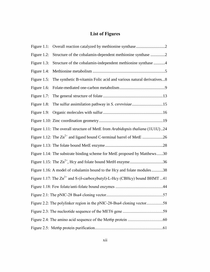
List of Figures ....................................................................................................... xii
Chapter 1 Introduction and Background ..................................................................1
Introduction .....................................................................................................1
Background .....................................................................................................7
Folate-Mediated One-Carbon Metabolism ............................................7
The chemical and physical properties of folates ..................................11
The sulfur containing compounds: Homocysteine and Methionine ....14
The role of Zinc in polypeptides ..........................................................18
Cobalamin-independent Methionine Synthases (EC 2.1.1.14) ............22
Structure of cobalamin-independent Methionine Synthase ........23
Proposed mechanism ..................................................................29
Cobalamin-dependent Methionine Synthases (EC 2.1.1.13) ...............33
Structure of cobalamin-dependent Methionine Synthase ...........33
Proposed mechanism ..................................................................37
Betaine Homocysteine Methyltransferase (BHMT) ............................40
S-Methylmethionine Hcy S-Methyltransferase (HMT) .......................42
Folate and anti-folate bound enzymes deposited in the PDB ..............42
Cobalamin-independent Methionine Synthase as a drug target ...........46
Structural perspective..................................................................46
Biological perspective .................................................................47
Candidiasis and candidemia .................................................................51
Summary and Project Goals.................................................................53
Aim 1: Crystallization of the Met6p enzyme ..............................54
Aim 2: Characterization of the Met6p active site ......................54
Aim 3: Development of a fluorescence-based assay .................54
Aim 4: Optimization of a high-throughput DSF assay ..............55
Aim 5: Discovering lead compounds by rational drug design ...55

ix
Chapter 2 Materials and Methods ..........................................................................56
Cloning and expression of Met6p in E. coli .................................................56
Protein purification .......................................................................................59
Surface Entropy Reduction (SER) ................................................................61
Site-directed mutagenesis .............................................................................62
Crystallization ...............................................................................................64
Ligand-replete crystals ..................................................................................66
Data collection and processing .....................................................................67
5-Methyl-THF-Glu3 synthesis ......................................................................67
Hcy synthesis ................................................................................................69
Absorbance based Met6p activity assay .......................................................69
Fluorescence based Met6p activity assay .....................................................70
Differential Scanning Fluorimetry assay (DSF) ...........................................73
Atomic coordinates .......................................................................................74
Chapter 3 Results and Discussion ..........................................................................76
The structures of three Met6p variants crystallized using SER ....................76
Crystal packing properties of Met6pA, Met6pT, and Met6pY .....................82
The overall structure of the fungal Met6p enzyme .......................................82
The zinc coordination site in Met6pA and Met6pY .....................................83
Ligand bound Met6pA structures .................................................................85
Hcy/Met specificity pocket in the C-terminal barrel ....................................86
Zinc coordination in the ligand bound C-terminal barrel .............................90
A flexible C-terminal barrel with a key role for Tyr660 ..............................93
The folate-bound binary and ternary complexes ..........................................95
Methotrexate-Glutamate3 and Hcy ternary complex ....................................98
S-adenosyl homocysteine (SAH) bound Met6pA .......................................100
Development of a fluorescence-based Met6p activity assay ......................105
The development of a high-throughput DSF assay.....................................112

x
Conclusion ...........................................................................................................120
Appendix A Abbreviations .................................................................................123
Appendix B Materials .........................................................................................124
References ............................................................................................................126
Vita ....................................................................................................................138

xi
List of Tables
Table 1.1: Structures of cobalamin-independent methionine synthase ..............22
Table 1.2: Rate and equilibrium constants for substrate binding in MetE .........30
Table 1.3: Structures of cobalamin-dependent methionine synthase .................34
Table 1.4: Folate/anti-folate bound proteins available in the PDB ....................45
Table 2.1: Deposited structures of the fungal Met6p enzyme ...........................75
Table 3.1: The activity of wild-type Met6p and engineered SER mutants ........78
Table 3.2: X-ray data collection and refinement of SER mutants .....................78
Table 3.3: X-ray data collection and refinement of ligand-bound Met6pA .......86
Table 3.4: Enzyme activity of Met6pA mutants ................................................95
Table 3.5: X-ray data collection and refinement of SAH bound Met6pA .......102
Table 3.6: Background signals in Absorbance Based Assay ...........................106
xii
List of Figures
Figure 1.1: Overall reaction catalyzed by methionine synthase ............................2
Figure 1.2: Structure of the cobalamin-dependent methionine synthase ..............2
Figure 1.3: Structure of the cobalamin-independent methionine synthase ...........4
Figure 1.4: Methionine metabolism ......................................................................5
Figure 1.5: The synthetic B-vitamin Folic acid and various natural derivatives ...8
Figure 1.6: Folate-mediated one-carbon metabolism ............................................9
Figure 1.7: The general structure of folate ..........................................................13
Figure 1.8: The sulfur assimilation pathway in S. cerevisiae ..............................15
Figure 1.9: Organic molecules with sulfur ..........................................................16
Figure 1.10: Zinc coordination geometry ..............................................................19
Figure 1.11: The overall structure of MetE from Arabidopsis thaliana (1U1U) ..24
Figure 1.12: The Zn2+
and ligand bound C-terminal barrel of MetE ....................26
Figure 1.13: The folate bound MetE enzyme ........................................................28
Figure 1.14: The substrate binding scheme for MetE proposed by Matthews ......30
Figure 1.15: The Zn2+
, Hcy and folate bound MetH enzyme ................................36
Figure 1.16: A model of cobalamin bound to the Hcy and folate modules ...........38
Figure 1.17: The Zn2+
and S-(δ-carboxybutyl)-L-Hcy (CBHcy) bound BHMT ...41
Figure 1.18: Few folate/anti-folate bound enzymes ..............................................44
Figure 2.1: The pNIC-28 Bsa4 cloning vector.......................................................57
Figure 2.2: The polylinker region in the pNIC-28-Bsa4 cloning vector................58
Figure 2.3: The nucleotide sequence of the MET6 gene .......................................59
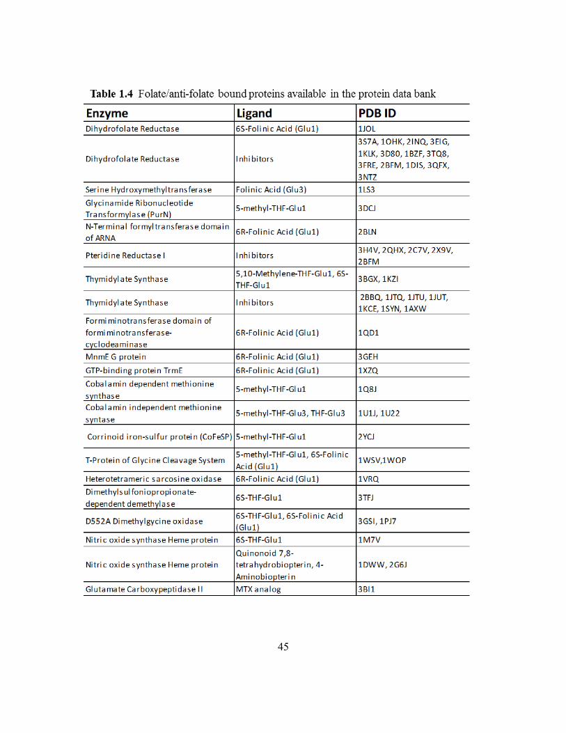
Figure 2.4: The amino acid sequence of the Met6p protein ..................................60
Figure 2.5: Met6p protein purification..................................................................61

xiii
Figure 2.6: PCR primers and reaction conditions ..................................................63
Figure 2.7: Protein crystals of the SER mutants ....................................................65
Figure 3.1: Sequence alignment of Met6p homologs ............................................77
Figure 3.2: Crystal packing interactions observed for each SER variant ..............79
Figure 3.3: Overlay of Met6pA with structural homologs shown in stereo ..........83
Figure 3.4: Zn2+
coordination site in the SER variants ..........................................84
Figure 3.5: The ternary Met6p complex ................................................................87
Figure 3.6: The Met6pA substrate binding site .....................................................88
Figure 3.7: Overlay of a substrate-free and Hcy-bound Met6pA ..........................90
Figure 3.8: Overlay of the ligand-bound and free Met6pA zinc binding site ........92
Figure 3.9: Overlay of the apo and ligand-bound C-terminal barrel .....................94
Figure 3.10: The folate binding pocket bound with 5-methyl-THF-Glu3 ..............96
Figure 3.11: The folate pocket bound with MTX-Glu3 .........................................99
Figure 3.12: Inhibitor dose-response curve .........................................................100
Figure 3.13: SAH bound Met6pA ........................................................................103
Figure 3.14: Overlay of the Zn2+
coordination site ..............................................104
Figure 3.15: Development of the fluorescence based activity assay ...................107
Figure 3.16: Development of the fluorescence based activity assay ...................109
Figure 3.17: Steady state kinetics ........................................................................110
Figure 3.18: Fluorescence based assay over a 20 minute time course.................111
Figure 3.19: Melting curves with the DSF assay .................................................114
Figure 3.20: Dose-response curves for compounds from the NCC. ....................115
Figure 3.21: Dose-response curves for compounds from the Fragment Lib.. .....116
Figure 3.22: Dose-response curves for compounds from the KINAset Lib.. ......118

1
CHAPTER 1
INTRODUCTION AND BACKGROUND
INTRODUCTION
In mammals the amino acid methionine (Met) cannot be synthesized de novo and
must be acquired through diet, either as Met or as Homocysteine (Hcy). Plants, bacteria
and yeast are equipped to biosynthesize Met through the sulfur assimilation pathway.
However, all organisms metabolize Met and recycle the end-product, Hcy, either through
the methionine synthase, betaine homocysteine methyltransferase (BHMT) or S-
methylmethionine Hcy S-methyltransferase (HMT) enzymes. The methionine synthases
can be further divided into two groups, one that uses the cobalamin cofactor and another
which is cobalamin-independent. Organisms such as mammals and some bacteria
contain the cobalamin-dependent methionine synthase referred to as MetH. Organisms
such as fungi, plants, archae and insects are deficient in vitamin B12 and thus contain the
cobalamin-independent methionine synthase known as MetE. Certain bacteria have both
enzymes.
MetE and MetH are zinc dependent enzymes that generate methionine by
catalyzing the transfer of a methyl from 5-methyl-tetrahydrofolate-glutamate (5-methyl-
THF-Glu) to Hcy (Figure 1.1). Although both carry out the same overall reaction, these
proteins are dissimilar in sequence and structure. MetH from E.coli has been extensively
characterized by biochemical/biophysical methods and X-ray crystallography. It is a
large 140 kDa enzyme which consists of 4 modules that bind Hcy and Zn2+
, 5-methyl-
THF-Glun, vitamin B-12, and S-adenosyl methionine (AdoMet), respectively (Figure
1.2). The B12-module mediates the methyl transfer reaction between the folate and Hcy
through a methylcobalamin intermediate. MetE has also been well characterized from E.

2

3
coli through biochemical/biophysical methods with X-ray structures solved from a
variety of organisms. MetE is about half the size of MetH (90 kDa) with just two
domains that are connected by a flexible linker (Figure 1.3). The active site is located in
a cleft between the two domains and binds a Zn2+
, Hcy, and 5-methyl-THF-Glu3. MetE
catalyzes a direct methyl transfer reaction between the Hcy thiolate and the N5 methyl
group of 5-methyl-THF-Glu3.
The Robertus and Appling groups have long been interested in the cobalamin-
independent methionine synthase gene from Candida albicans (MET6) as a potential
anti-fungal drug design target. Fungal pathogens are a health concern, particularly to
immuno-compromised individuals, and the Candida species are associated with the
highest mortality rate. In particular C. albicans leads in the number of patients diagnosed
with fungal nosocomial bloodstream infections (Edmond, Wallace et al. 1999). The
MET6 gene encodes the Met6p enzyme which shares ~40-50% sequence identity with
the E. coli MetE. In fungi, Met6p carries out the last step in methionine biosynthesis and
is a point in the metabolic map where the one-carbon folate metabolism cycle converges
with the methyl cycle (Figure 1.4). Methionine is used to generate S-adenosyl
methionine (AdoMet) which is the major source for one-carbon units vital for
biosynthesis. A defective Met6p is expected to have lethal consequences as a result of
flawed DNA/RNA synthesis, AdoMet mediated methylation reactions, and protein
synthesis. In theory, the structural differences between the MetH of the mammalian host
and the fungal Met6p enzyme make it possible to selectively target and inhibit the
pathogenic enzyme.
The Robertus and Appling groups have investigated the importance of the C.
albicans MET6 gene for fungal growth and have characterized the Met6 protein by
steady state kinetics. Suliman cloned, expressed, purified and characterized the

4
methionine synthase from C. albicans and S. cerevisiae (Suliman, Sawyer et al. 2005).
The MET6 gene from either yeast was able to complement the other and had similar
substrate specificities. Both were able to utilize the 5-methyl-THF-Glun with at least a
di-glutamate tail at KM values of around 100 µM and kcat of around 25/min. The KM for
Hcy was ~14 µM. Suliman also used homologous recombination techniques to test the
growth of the diploid Candida albicans strains missing one or both MET6 genes; she also
created another strain containing one functional MET6 gene under a regulatable GAL
promoter (Suliman, Appling et al. 2007). A single MET6 gene knockout had little effect
on fungal growth, however a double gene knockout was found to be lethal, even if the
growth media was supplemented with methionine. As expected, the conditional mutant

5
grew as well as the wild-type under inducing conditions, zero growth was observed under
repressing conditions, and colonies were observed under repressing conditions
supplemented with exogenous methionine. This growth was due to a leaky GAL
promoter. This work showed that the MET6 gene from the pathogen C. albicans is
essential for fungal growth and that some amount of Met6p must be present in order to
rescue fungal growth.
Although attempts made to crystallize the wild-type Met6p failed, Prasannan was
able to create a Met6p model using the existing MetE structures and used site directed
mutagenesis to identify a number of key catalytic and substrate binding residues
(Prasannan, Suliman et al. 2009). She found that the ionic interaction observed between
the side-chain of Asp614 and the Hcy ammonium group in MetE homologs is essential

6
for enzyme activity and C. albicans growth. Asp504 is another conserved residue that
uses its side chain to bind and orient the folate ring. Mutants of Asp504 were also
kinetically inactive and each strain failed to grow. Lastly, the aromatic ring of Trp576 is
believed to make non-specific hydrophobic interactions with the folate ring and
according to this mutagenesis study, could be replaced by phenylalanine but not alanine.
In continuing this research project, I used X-ray crystallography, a novel
fluorescence-based activity assay and the biophysical method known as differential
scanning fluorimetry (DSF) to study the Met6p protein structure and mechanism, and to
identify novel fungistatic compounds. Since the native Met6p enzyme could not be
crystallized using traditional high-throughput screens, the surface entropy reduction
technique (Cooper, Boczek et al. 2007) was used to create three fully active variants that
formed useful crystals which diffracted to at least 2.7 Å (Ubhi, Kavanagh et al. 2011). In
these variants, Lys103, Lys104, and Glu107 were all converted to Tyr (Met6pY), Thr
(Met6pT), and Ala (Met6pA).
Met6pA was used to obtain enzyme-ligand complexes due to its high diffraction
resolution and possession of just one molecule in the asymmetric unit. Several X-ray
structures were solved with Met6pA in a binary or ternary complex with: Hcy, Met,
Glutamine (Gln), 5-methyl-THF-Glu3, Methotrexate-Glutamate3 (MTX-Glu3), and S-
adenosyl homocysteine (SAH). These structures provided a wealth of information with
respect to substrate binding pockets, structural dynamics and potential drug design
strategies.
A novel fluorescence based assay was developed to monitor Met6p activity, to
characterize various mutants, and to test potential inhibitors, such as MTX-Glu3. The
commercially available compound Measure-iTTM
was used in the fluorescence-based
activity assay to selectively measure the concentration of unused Hcy in a Met6p

7
reaction. Lastly, the DSF method was used to screen for ligands which bound and raised
the melting temperature (Tm) of the wild-type Met6p enzyme. The method was
optimized for high-throughput 96-well format with excellent repeatability, low standard
deviation, and a Z’ factor of 0.5-1 per plate. Compounds (cmnds) were screened from
three diverse libraries: NIH Clinical Collection (731 cmnds), ChemBridge Fragment
(4000 cmnds) and ChemBridge KINASet libraries (4000 cmnds). The observed hits were
further evaluated using the absorbance based kinetic assay and X-ray crystallography.
BACKGROUND
Folate-Mediated One-Carbon Metabolism
Many processed foods are fortified with the synthetic B-vitamin, folic acid, while
green leafy vegetables contain a number of natural folate derivatives (Figure 1.5). These
derivatives include: dihydrofolate (DHF), tetrahydrofolate (THF), 5-methyl-THF, 10-
formyl-THF, and 5,10-methylene-THF. These dietary sources provide us with an
essential vitamin which can best be described as a carrier of one carbon units in folate-
mediated one-carbon metabolism. Folate-dependent one-carbon metabolism is
compartmentalized in the cytoplasm, mitochondria, nucleus and chloroplast and involves
numerous enzymes and impacts the overall metabolic balance at the cellular level (Figure
1.6) (Appling 1991, Tibbetts and Appling 2010, Stover and Field 2011).
The overall picture of folate metabolism includes the one-carbon generating
enzymes, the folate-interconverting enzymes, the folate-dependent biosynthetic enzymes
and folate binding proteins (Appling 1991, Scott 1999, Litwach 2008). There is
competition between these enzymes for free folate which appears to be tightly regulated
by cells (Herbig, Chiang et al. 2002). Many of these enzymes have functionally
independent domains, specialized for the generation of one-carbon units and for

8

9
subsequent inter-conversion of the C1-folates. In order to enter the one-carbon
metabolism cycle, the aromatic ring of folic acid must first be reduced in two steps by the
enzyme dihydrofolate reductase (DHFR) to tetrahydrofolate (THF) (Schnell, Dyson et al.
2004). THF is then able to accept one carbon units in a range of oxidation states for use
in a variety of biosynthetic reactions.
The one-carbon generating enzymes include serine hydroxymethyltransferase
(SHMT), 10-formyltetrahydrofolate synthetase (FTHFS), and the bifunctional enzyme

10
complex of glutamate formiminotransferase and formimidoyltetrahydrofolate
cyclodeaminase (Litwach 2008). In a reversible reaction, SHMT catabolizes the amino
acid serine, making 5,10-methylene-THF and glycine (Rao, Talwar et al. 2000). The
limited pool of methylene-THF can support thymidylate biosynthesis, methionine
synthesis or purine biosynthesis. Formate is used as a source of carbon by FTHFS in
order to synthesize 10-FormylTHF (McGuire and Rabinowitz 1978). The degradation of
histidine generates formiminoglutamic acid which is processed by glutamate
formiminotransferase to make 5-formiminoTHF (Kohls, Sulea et al. 2000). This
intermediate is converted to 5,10-methenylTHF through the formimidoyltetrahydrofolate
cyclodeaminase to be used for one-carbon transfer reactions.
The folate-interconverting enzymes include the 5,10-methenylTHF
cyclohydrolase/dehydrogenase (MTHFC)/(MTHFD), 10-FormylTHF dehydrogenase
(FDH), 5,10-MethenylTHF synthetase (MTHFS), and 5,10-MethyleneTHF reductase
(MTHFR) (Litwach 2008). MTHFC carries out the reversible inter-conversion of 10-
formylTHF and 5,10-methenylTHF. MTHFD carries out the reversible inter-conversion
of 5,10-methenylTHF and 5,10-methyleneTHF (Eadsforth, Cameron et al. 2012). FDH
catalyzes the irreversible hydrolysis of 10-formylTHF to THF and formate (Krupenko
2009). MTHFS carries out the irreversible conversion of 5-formylTHF to 5,10-
methenylTHF (Holmes and Appling 2002). It is the only known enzyme to use folinic
acid. MTHFR catalyzes the irreversible reduction of 5,10-methyleneTHF to 5-
methylTHF and commits the C1-THF for methionine synthesis (Cathou and Buchanan
1963).
The folate dependent biosynthetic enzymes include thymidylate synthase (TS),
methionine synthase (MS), the glycinamide ribonucleotide formyltransferase (GARFT)
and 5-aminoimidazole-4-carboxamide ribonucleotide formyltransferase (AICARFT)
11
enzymes. TS uses 5,10-methyleneTHF to convert dUMP to dTMP, which is essential to
DNA synthesis/replication and repair (Finer-Moore, Santi et al. 2003).
MS uses 5-methylTHF to methylate Hcy and produce Met (Gonzalez, Banerjee et
al. 1992). MS thereby supports various methyl transfer reactions since it provides the
precursor for the potent methyl donor, AdoMet. MS also removes Hcy, a toxic
biomarker, from the cell and regenerates the THF to support folate-mediated one-carbon
metabolism. GARFT and AICARFT use 10-formylTHF to add formate into the C8 and
C2 positions of purine (Daubner, Schrimsher et al. 1985).
An intricate network of interdependent biosynthetic pathways drives folate
metabolism. Folate mediated one-carbon metabolism is directly responsible for the
synthesis of methionine, purines and thymidylate, and indirectly involved with amino
acid and formate metabolism and DNA/protein methylation.
Folate deficiency results in seriously pathological phenotypes in mammals,
including megaloblastic anemia, spina bifida, cardiovascular disease, neuropsychiatric
disorders and increased sensitivity to oxidative stress (Litwach 2008). Mutations in the
folate-mediated one-carbon pathway can lead to inborn errors in folate metabolism.
Some of the most serious phenotypes are associated with mutations in the gene encoding
methionine synthase. Enzymes currently targeted by anti-folates for the treatment of
cancer, parasitic infection, malarial infection, and rheumatoid arthritis include DHFR,
TS, SHMT, MTHFR, and GARFT.
The chemical and physical properties of folates
A number of different folates exist in nature and share the same overall structure
consisting of a covalently linked pterin ring, p-aminobenzoic acid (PABA) and a
glutamate residue (Figure 1.7). Folates vary in the level of oxidation of the pterin ring,
12
the nature of the carbon moiety at the N5 and/or N10 position, and in the number of
glutamates in the polyanionic tail. All biologically relevant derivatives have a reduced
pyrazine ring and exist as THF scaffolds (Maden 2000). THF can undergo methylation at
position N5 and formylation at positions N5 or N10 (Scott 1999). The length of the poly-
γ-glutamate tail is modified by the enzyme folylpolyglutamate synthetase and varies by
cell type as well as compartment. Tail length effects affinity for certain folate-dependent
enzymes, mediates cellular retention of folates, functions in substrate channeling and
regulation of one carbon metabolism (Chabner, Allegra et al. 1985, Appling 1991).
The chemical and physical properties of folates and pteridines/pterins have been
under investigation since 1925 with the discovery of folate in spinach and of
pteridine/pterin as the pigment in butterfly wings (Blakley 1969). The properties of
folates are largely determined by the pteridine constituent. Pteridine provides the
platform for numerous substituted pterins, the most important of which is pterin, a 2-
amino-4-oxo pteridine. Such compounds are found not only in wings, but also the skin of
reptiles, amphibians, and fish.
Pteridine consists of a bicyclic nitrogenous ring system (fused pyrimidine and
pyrazine rings) which is soluble in water and organic solvents (Figure 1.7). Substitution
of a keto group at position 4 and amino group at position 2 results in a pterin. Pterins are
poorly soluble in water (but can be dissolved in 0.05 M NaOH) and are very stable and
resistant to oxidation. The UV spectra of pterins exhibit a maximum at wavelengths
greater than 300 nm. Reduction takes place in the pyrazine ring to form 7,8-
dihydropterins, 5,6,7,8-tetrahydropterins and 6-substituted tetrahydropterins but these
derivatives are unstable and readily oxidized (Basu and Burgmayer 2011).
Folic acid can also be described as a 6-alkylpterin but is completely oxidized and
therefore relatively stable at physiological pH under anaerobic conditions. However,

13
alkaline hydrolysis under aerobic conditions or photodecomposition by sunlight will yield
p-aminobenzoylglutamic acid and a pteridine. Folic acid has a UV absorbance maxima at
neutral pH (ε282 = 27.6 mM-1
). Reduction of folic acid results in the biologically relevant
7,8-dihydrofolate (DHF) and 5,6,7,8-tetrahydrofolates (THF). These compounds are
labile and must be protected from light and air and if possible stored with reducing agents
and ascorbate. Naturally occurring THFs have an asymmetric center at the C-6 position
corresponding to the S configuration (Fontecilla-Camps 1979). The THF dissociation
constants have been reported: N1, pKa 1.24; N5, pKa 4.82; N10, pKa -1.25; amide, pKa
10.5; gamma-carboxyl, pKa 4.8; alpha-carboxyl, pKa 3.5 (Kallen and Jencks 1966).

14
The sulfur containing compounds: Homocysteine and Methionine
The element sulfur is present in a number of organic compounds including the
amino acids cysteine (Cys) and methionine (Met), the vitamins biotin and thiamin, the
cofactors S-adenosyl methionine (AdoMet), coenzyme A and lipoic acid, and the anti-
oxidants glutathione and taurine (Takahashi, Kopriva et al. 2011). These molecules are
synthesized through various pathways which can all be traced back to a sulfur
assimilation event. The vast amounts of inorganic sulfur present in the atmosphere and
soil is assimilated by plants, fungi, and some bacteria through their respective sulfur
assimilation pathways (Figure 1.8). Plants and bacteria reduce the readily available
sulfate (SO4-2
) into cysteine (Cys) (Hatzios and Bertozzi 2011, Takahashi, Kopriva et al.
2011). Yeast or fungi assimilate the inorganic sulfate into the organic molecule Hcy
(Thomas and Surdin-Kerjan 1997). Cys and Hcy enter the transsulfuration or the Hcy
methylation cycles to make other necessary organic molecules. In organisms that
assimilate sulfate, the sulfur containing amino acids like Met or Cys are synthesized de
novo. Most microorganisms also have transporters for Met. Mammals on the other hand
depend on their diet to acquire the sulfur-containing amino acids, although they can
interconvert them once acquired.
Hcy is similar in structure and reactivity to the amino acid Cys and in S.
cerevisiae both are interconverted through the transsulfuration pathway (Figure 1.8).
Both have an amino acid backbone and sulfur containing hydrocarbon side chains of
different lengths (Figure 1.9). Under physiological conditions, the sulfur exists as a
thiol/sulfhydryl group. The amino acid Cys can form disulfide bonds that stabilize
protein structure. The amino acid can also mediate enzyme catalysis as a general acid or
by serving as a mechanistic nucleophile. The thiolates also serve as metal ligands in

15
many proteins. The reactivity of the sulfhydryl group can be modified by the pH and the
reducing or oxidizing environment.

16
Hcy is a non-protein amino acid with similar properties as Cys. It is a metabolite,
which either gets recycled through the folate-mediated one-carbon metabolism (or
BHMT, HMT) to form methionine, or enters the transsulfuration pathway to make Cys.
A high concentration of Hcy is a biomarker for disease in fungi, bacteria and mammals
(Selhub 1999, Roe, O'Byrne et al. 2002, Pascon, Ganous et al. 2004). Accumulation of

17
Hcy is indicative of inborn errors in metabolism and leads to oxidative stress, toxicity and
eventual death (McCully 2009).
The amino acid Met is heavily utilized by all organisms due to its role as a
building block of proteins and as an intermediate in numerous transmethylation reactions.
Unlike the reactive sulfhydryl group in Hcy and Cys, the sulfur in Met is a relatively inert
thioether (Figure 1.9). In eukaryotes, Met successfully initiates translation by acting as a
hydrophobic bridge between the initiator tRNA-Met and eIF-2 (Brosnan and Brosnan
2006). In proteins, Met is frequently found in the hydrophobic core or the membrane
spanning domains, and to a lesser extent on the protein surface or at the active site. In
solvent exposed regions, it can act as an endogenous antioxidant and form methionine
sulfoxide (Figure 1.9). In its free form, Met is metabolized by the enzyme methionine
adenosyl-transferase (MAT) to its active form S-adenosyl methionine (AdoMet).
AdoMet is a powerful methylating agent and the second most widely used enzyme
substrate after ATP (Figure 1.9) (Fontecave, Atta et al. 2004). AdoMet impacts a variety
of cellular events such as gene expression and is implicated in fetal development and
brain function. Numerous AdoMet dependent methyltransferases (MTases) use AdoMet
as a methyl donor to methylate DNA, protein, hormones, neurotransmitters and
phospholipids. This reaction produces the byproduct S-adenosyl homocysteine (SAH)
which is reversibly cleaved by the enzyme AdoHcy hydrolase to form Hcy and adenosine
(Figure 1.9). The remethylation of Hcy to Met completes the methylation metabolism
cycle. The Hcy and Met metabolism pathways are linked through the commonly shared
intermediate Hcy (Figure 1.8).

18
The role of Zinc in polypeptides
In living cells, the metal ion composition consists primarily of calcium (Ca2+
),
magnesium (Mg2+
), sodium (Na1+
) and potassium (K1+
) while transition metals like iron
(Fe2+
), zinc (Zn2+
), nickel (Ni2+
), cobalt (Co2+
) and copper (Cu2+
) are quite rare (Gillespie
1998). Of these, the rare transition metals are most likely to participate in enzymatic
reactions. Enzymes interact with the metal based on its size, its ability to coordinate with
available residues, and its coordination chemistry. A metal can have a catalytic and/or
structure role and its behavior depends on whether it is a soft, hard or borderline metal.
A hard metal is characterized by a small atomic radius, high nuclear charge and low
polarizability. A soft metal is associated with a large atomic radius, low
electronegativity, and high polarizability (Ho, Ho et al. 1978). According to the hard and
soft acids and bases (HSAB) principle, hard acids react preferentially with hard bases and
soft acids with soft bases while borderline acids are more flexible.
Zinc is a borderline metal and has unique properties that are ideal for a dynamic
catalytic site or a rigid structural role in proteins. Zn2+
is the only member of the first
row of transition metals with filled d-shell orbitals. Unlike the other reactive members
which are often involved in redox reactions, Zn2+
can act as a redox-stable Lewis acid
(McCall, Huang et al. 2000). Zn2+
is a stable ion which can also easily alternate between
multiple coordination geometries with no energy penalty. Over 3,000 Zn2+
proteins are
encoded in the human genome and belong to six different classes of enzymes (Andreini,
Bertini et al. 2011). Over 200 Zn2+
-bound protein structures are available. The metal
coordinates with 3, 4, 5 or 6 ligands through geometry resembling distorted tetrahedral,
trigonal-bipyramidal or octahedral (Figure 1.10) (Auld 2001). Structural sites favor a
stable tetrahedral geometry while catalytic sites benefit from a more dynamic metal
coordination geometry. Zinc can coordinate with both hard and soft ligands such as

19
oxygen (hard), nitrogen or sulfur (soft). In proteins Zn2+
is often coordinated by a
combination of protein side chains including the nitrogen of histidine, oxygen of
aspartate or glutamate, and the sulfur of cysteine. Less frequently observed ligands
include the hydroxyl of tyrosine, the carbonyl of protein backbone, and the side chain
carbonyls of asparagine or glutamine (McCall, Huang et al. 2000). The reactivity or
function of Zn2+
depends on the first coordination sphere which dictates the oxidation
state of Zn2+
and the second coordination sphere which orients and stabilizes the Zn2+
ligands and establishes the dielectric medium (Lee and Lim 2008, Maret and Li 2009,
Andreini, Bertini et al. 2011). In other words, the chemistry of Zn2+
can be modulated by

20
different classes of enzymes in order to have desired stability constants, reactivities and
functions (Vallee and Auld 1990).
The catalytic role of Zn2+
depends on its function as a Lewis acid, its ability to
access different coordination geometries and fast ligand exchange. A catalytic Zn2+
binding site is generally “pre-organized” in the absence of metals. The metal is typically
coordinated by three protein ligands, an exchangeable water, and/or a ligand (Maret and
Li 2009). Histidine is the most commonly found protein ligand to coordinate the Zn2+
followed by glutamate, aspartate and cysteine. Four histidine ligands would retain an
oxidation state of Zn2+
, making the metal a powerful Lewis acid. A water is almost
always a ligand in the catalytic sites. Some enzymes like carbonic anhydrase use the
metal to ionize the water while others like carboxypeptidase A use it to polarize the water
(Rees, Lewis et al. 1981, Lindskog 1997). In either case, the metal helps to generate a
hydroxyl nucleophile (Alberts, Nadassy et al. 1998). In the case of alcohol
dehydrogenase, water is an exchangeable member of Zn2+
’s coordination sphere
(Kleifeld, Frenkel et al. 2003). In coordination sites containing a cysteine or a non-
protein sulfur ligand, as in the different protein prenyltransferases, or the Ada protein, the
metal enhances the nucleophilicity of the free thiol by converting it to the active thiolate
at neutral pH (McCall, Huang et al. 2000, Maurer-Stroh, Washietl et al. 2003, Maret and
Li 2009). The Zn2+
/Thiol reactivity can be modified by adjusting the pH or the
oxidizing/reducing environment. The sulfur reactivity is employed by the “Zn2+
switches” found in voltage gated K+ channels (Wang, Strang et al. 2007).
A cocatalytic site has more than one Zn2+
and while one Zn2+
is often involved in
making or breaking bonds, the others (bridged through water or protein residues) enhance
its reactivity (Auld 2001). The metal can also stabilize negative charge.

21
A purely structural role of zinc is observed in a number of enzymes with functions
ranging from Zn2+
sensing to transcription regulation. Structural Zn2+
binding sites can
usually be distinguished from catalytic ones based on the type of metal ligands, lack of
water, coordination geometry and general hydrophobicity (Andreini, Bertini et al. 2011).
Coordination geometry is predominately tetrahedral and consists of CYS4, CYS3HIS1 or
CYS2HIS2 while Asp or Glu are rarely found in the coordination sphere. Cysteine
residues dominate because Zn2+
-S complexes have high stability constants similar to
those observed in disulfides (Vallee and Auld 1990). This allows Zn2+
to organize small
domains, such as the zinc-finger containing metallothioneins or larger proteins like 3-
methyladenosine DNA glycosylase I (Coyle, Philcox et al. 2002, Kwon, Cao et al. 2003,
Maret and Li 2009). Metallothioneins are enriched with the classic CYS2HIS2 zinc-finger
motif also found in a number of DNA and RNA binding proteins as well as between
protein-protein binding domains (Laity, Lee et al. 2001).
The metal-ligand bond lengths and angles were recently evaluated from the zinc-
replete structures available in the PDB (Alberts, Nadassy et al. 1998, Lee and Lim 2008).
The zinc coordination numbers of 4 (T4), 5 (T5), and 6 (T6) were observed in catalytic
sites with frequencies of 48, 44 and 6%, respectively, and in structural sites with
frequencies of 79, 6 and 12%, respectively. All T4 sites have tetrahedral coordination
while the majority of T5 sites have trigonal bipyramidal geometries. In catalytic T4 sites,
the average metal to ligand distance for Zn2+
-NHistidine is ~2.07 Å, Zn2+
-Owater is ~2.12 Å,
Zn2+
-OGlu/Asp/Asn/Gln is ~2.04 Å, Zn2+
-SCys is ~2.21 Å. In catalytic T5 sites, the metal to
ligand distance for Zn2+
-NHistidine is ~2.11 Å, Zn2+
-Owater is ~2.09 Å, Zn2+
-OGlu/Asp/Asn/Gln is
~2.16 Å, Zn2+
-SCys is ~2.30 Å. In structural T4 sites, the metal to ligand distance for
Zn2+
-NHistidine is ~2.09 Å, Zn2+
-Owater is ~2.15 Å, Zn2+
-OGlu/Asp/Asn/Gln is ~1.95 Å, Zn2+
-SCys
is 2.35 Å. In crystal structures, the angles observed between the zinc and the four ligands

22
in T4 or T5 sites often deviate from ideality. The ideal angles for a metal with tetrahedral
geometry are 109.5° and with trigonal bipyrimidal geometry are 90° (Eq-Zinc-Ax), 120°
(Eq-Zinc-Eq) or 180° (Ax-Zinc-Ax).
Cobalamin-independent Methionine Synthases (EC 2.1.1.14)
The cobalamin-independent methionine synthase enzyme (MetE) binds Hcy and
5-methyl-THF-Glu3 and catalyzes a Zn2+
-mediated alkylation reaction to generate
methionine. This is the last step in the de novo methionine biosynthesis pathway in
plants and microorganisms, and a means to recycle Hcy in all organisms. In the last 10
years, crystal structures of the Zn2+
-bound apo and substrate-replete MetE homologs from
Arabidopsis thaliana, Streptococcus mutans, and Thermatoga maritima have been solved
by various groups (Table 1.1) (Ferrer, Ravanel et al. 2004, Pejchal and Ludwig 2005,

23
Koutmos, Pejchal et al. 2008, Fu, Almqvist et al. 2011). Extensive kinetic
characterization of MetE has also been done by the Matthews group using the E. coli
homolog (Matthews, Smith et al. 2003, Taurog, Jakubowski et al. 2006, Taurog and
Matthews 2006). MetE gene expression and enzyme properties were reported from the
higher plant Catharantus roseus by the Schroder group (Eichel, Gonzalez et al. 1995,
Eckermann, Eichel et al. 2000). Gene deletion studies and steady state kinetics were
done using the fungal homolog from Candida albicans and Saccharomyces cerevisiae by
the Robertus group (Suliman, Sawyer et al. 2005, Suliman, Appling et al. 2007,
Prasannan, Suliman et al. 2009). The role of MetE in establishing virulence by
Streptococcus pneumoniae and Cryptococcus neoformans was tested by McCusker and
Brown, respectively (Pascon, Ganous et al. 2004, Basavanna, Chimalapati et al. 2013).
Structure of cobalamin-independent Methionine Synthase
The MetE protein from the plant A. thaliana was first crystallized and solved by
Ferrer in 2004 (Ferrer, Ravanel et al. 2004). One year later, the bacterial structure from
T. maritima was published by Pejchal (Pejchal and Ludwig 2005). The structure of MetE
from the pathogenic bacterium Streptococcus mutans was also recently solved (Fu,
Almqvist et al. 2011). In general, the MetE from these organisms is ~90 kDa. The
amino acid sequence is only 40-50% similar but all three structures align with an RMSD
< 2 Å.
The MetE enzyme consists of two (βα)8 TIM barrels that are connected by a
linker and arranged face to face such that the extended surface loops form an inter-
domain interface near the linker (Figure 1.11). The active site Zn2+
is coordinated by
residues located on the top of the C-terminal barrel and facing the inter-domain interface.
Inductively coupled plasma-atomic emission spectrometry was initially used by the

24
Matthews group to calculate one equivalent of Zn2+
per monomer and circular dichroism
was used to confirm its non-structural role (Gonzalez, Peariso et al. 1996). EDTA
treatment was also found to inhibit MetE activity. Extended X-ray absorption fine
structure (EXFAS) and site-directed mutagenesis were used to establish the three
essential metal ligands His641, Cys643 and Cys726 (in E. coli), and a fourth
exchangeable water or glutamate (Zhou, Peariso et al. 1999).
The Hcy and Met bound structures of MetE from A. thaliana and T. maritima
identified a small substrate/product binding pocket adjacent to the active site metal
(Ferrer, Ravanel et al. 2004, Pejchal and Ludwig 2005). In both homologs, the backbone
amino group is secured by a Glu462, Asp577, and carbonyl of Ile409 (Figure 1.12) (T.

25
maritima). The carboxylate group forms hydrogen bonds with the backbone amide and
side chain OH of Ser411 (T. maritima). Prasannan used site directed mutagenesis to
show that the Asp577 equivalent in C. albicans is essential for enzyme activity
(Prasannan, Suliman et al. 2009). The entry of Hcy or L-selenohomocysteine into the
active site has also been evaluated by the Matthews group using EXFAS (Peariso, Zhou
et al. 2001). They found that the active site Zn2+
retains tetrahedral geometry and
coordinates with four ligands in both the apo and Hcy/selenohomocysteine bound
enzymes. However, the coordination environment around the Zn2+
changes from 2S + 2
(N/O) to 2S + 1N + 1Se/S. These results showed that Zn2+
easily switches between
coordination geometries and ligands, and suggests that it acts as a Lewis acid to
deprotonate and activate the Hcy. However, the Zn2+
-SHcy distances reported from the A.
thaliana and T. maritima structures are too long at 4.1 Å and 3.2 Å, respectively.
In the substrate-free and substrate-bound plant structures, the metal does not
coordinate with the Hcy thiol and instead remains tetrahedrally coordinated to Cys649,
Cys733, His647 and a water molecule (Figure 1.12). In T. maritima the metal is
coordinated by Cys620, Cys704, His618 and Glu642 in the apo structure but ends up
moving 0.75 Å towards the substrate in the Hcy-bound structure (Figure 1.12). This
results in two elongated bonds between Zn2+
-O642 and Zn2+
-SHcy and metal geometry
somewhere between trigonal planar and trigonal bipyramidal. In a more recent paper
published by Matthews and Ludwig, the T. maritima MetE was solved with and without
Hcy at pH 8.5 (Koutmos, Pejchal et al. 2008). Under these conditions the results were
consistent with the EXFAS data; the ligands around Zn2+
changed from S620, S704, H618,
O642 in the apo structure to S620, S704, H618, SHcy in the substrate bound structure with the
complete inversion of tetrahedral geometry (Figure 1.12).

26

27
So far both the natural substrate 5-methyl-THF-Glu5 and substrate analog pteroyl-
Glu5 have been used to characterize the folate binding site of MetE. Unlike the well-
defined Hcy/Met pocket in the C-terminal barrel, the folate binds at the inter-domain
interface near the linker. Few folate bound MetE structures have been solved and the
available structures are incomplete and somewhat inconsistent.
In the A. thaliana protein bound with Met and 5-methyl-THF-Glu5 or Hcy and
pteroyl-Glu5, the reduced (THF) or aromatic (pteroyl) pterins form non-specific stacking
interactions with a conserved Trp567, and the first glutamyl of the polyglu tail forms
ionic or hydrogen bonds with Arg521 and Lys18, or Cys522, respectively (Figure 1.13)
(33). In the superimposed structures, the polyglu tails and PABA moieties align
relatively well but the pterins appear to slide around on the Trp567 ring. Each pterin is
loosely secured through hydrogen bonds with the side-chain residues in its surroundings.
These structures were the first to show that the folate binds at the inter-domain interface
by folding into an ‘L’ configuration, where the base of the letter corresponds to the
pterin.
In the A. thaliana structures, the N5 methyl group is ~7 Å away the Hcy thiolate.
In the T. maritima homolog, 5-methyl-THF-Glu3 binds in the same location primarily
through stacking interactions between the pterin and Trp539 and hydrogen/ionic bonds
between side chains of Lys18, Cys494, and Arg493 and the first glutamyl residue of the
polyglu tail (Figure 1.13) (34). However, unlike the A. thaliana structures in which the
folate folds onto itself and resembles the letter ‘L’, in T. maritima it binds in an extended
configuration. Compared to the ‘L’, the pterin in the extended form rotates ~180°,
forming an inverted L, and drops another ~45°. As a result, the N5 methyl group
undergoes a rotation of ~180° and translation of ~3 Å, placing it 11 Å away from the Hcy
thiolate. The authors believe that this is an intermediate conformation that later changes

28
to resemble the A. thaliana complex. Site-directed mutagenesis done by Prasannan
identified the two essential folate binding residues Asp467 and Trp539 in T. maritima,
and the Asp495 and Trp567 in the A. thaliana homolog (Prasannan, Suliman et al. 2009).

29
The substrate-free and substrate-bound MetE structures solved so far crystallize in
an ‘open’ enzyme configuration. Since the methyl donor and acceptor are too far apart
for a productive transfer reaction, a transient ‘closed’ configuration is expected to exist.
Proposed mechanism
MetE was initially purified from E. coli and evaluated for folate binding and
catalysis by Whitfield and Weissbach (Whitfield, Steers et al. 1970, Whitfield and
Weissbach 1970). They identified the requirement for polyglutamated 5-methyl-THF-
Glu, Hcy, phosphate and Mg2+
for optimal MetE catalysis. The MetE homolog from the
plant Catharanthus roseus was purified in 1995 and found to be similar in size to the
bacterial homolog and shared a sequence identity of 49.5% (Eichel, Gonzalez et al.
1995). The plant enzyme only required phosphate, Hcy and 5-methyl-THF-Glu3 for
maximal activity (Eckermann, Eichel et al. 2000). The homologs from Candida albicans
(CaMet6p) and Saccharomyces cerevisiae (ScMet6p) are about 48% identical to the E.
coli MetE and were characterized in 2005 by Suliman (Suliman, Sawyer et al. 2005).
Both were tested with phosphate and Mg2+
and found to be fully active with 5-methyl-
THF-Glu2.
The Matthews group has had a longstanding interest in the chemistry of methyl
transfer; the chemically difficult direct transfer from 5-methyl-THF to Hcy has been
particularly intriguing. They determined the kinetic constants and equilibrium
dissociation constants for Hcy and 5-methyl-THF-Glu3 (Taurog, Jakubowski et al. 2006).
A substrate binding scheme is shown in Figure 1.14 and the rate constants and
equilibrium binding constants are shown in Table 1.2. Since MetE carries out a bi-
substrate reaction, they first tested the order in which each substrate enters the active site.
They found that each binary complex (MetE*5-methyl-THF-Glu3 and MetE*L-Hcy) is
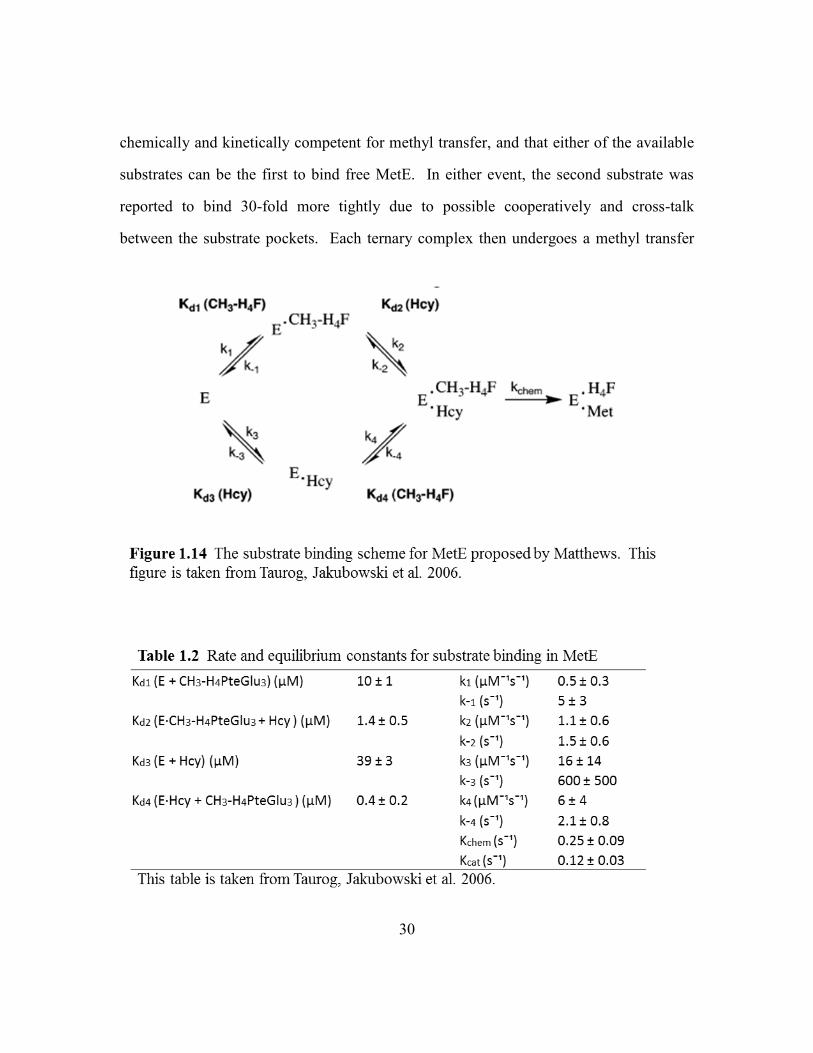
30
chemically and kinetically competent for methyl transfer, and that either of the available
substrates can be the first to bind free MetE. In either event, the second substrate was
reported to bind 30-fold more tightly due to possible cooperatively and cross-talk
between the substrate pockets. Each ternary complex then undergoes a methyl transfer

31
reaction with a reported kcat of 0.12 ± 0.03 s-1
and Kchem of 0.25 ± 0.09 s-1
(at 25 °C). A
separate paper by the same group demonstrated the existence of a “mixed” ternary
complex MetE*THF-Glu3*Hcy, and suggested that the release of byproduct THF-Glu3
from either the binary or ternary complex is partially rate limiting (Taurog and Matthews
2006). This was supported by the observed kcat and KM, which show that something other
than chemistry is rate limiting.
Steady state enzyme kinetic experiments have also been done by various groups.
The MetE from E.coli, C. roseus, S. cerevisae and C. albicans is reported to have a 5-
methyl-THF-Glu3 KM of 4.7 µM, 80 µM, 84 ± 8 µM, and 129 ± 25 µM, respectively
(Whitfield, Steers et al. 1970, Eckermann, Eichel et al. 2000, Suliman, Sawyer et al.
2005). Hcy is reported to have a KM of 14 ± 2.3 µM and 13 ± 2.6 µM in the C. albicans
and S. cerevisiae homologs, respectively, with an overall turnover or kcat of 25/min
(Suliman, Sawyer et al. 2005).
The cobalamin-independent methionine synthase catalyzes an otherwise
unfavorable direct methyl transfer reaction by first activating the Hcy thiol and 5-methyl-
THF-Glu3. The enzyme uses Zn2+
to deprotonate the Hcy thiol, converting it into a
thiolate anion; the thiolate becomes the fourth metal ligand (Matthews, Smith et al.
2003). In MetE, this zinc-thiolate complex [Zinc(Scys)3(NHis)1] has a net charge of -1.
According to model studies done by Wilker and Lippart, this set of ligands and overall
charge are essential in enhancing the thiolate reactivity towards alkylation by facilitating
the dissociation from the zinc complex followed by alkylation (Matthews and Goulding
1997, Wilker and Lippard 1997). The question of whether alkylation takes place while
the thiolate is Zn2+
bound or free is still under investigation. Model studies designed
specifically to replicate the MetE catalyzed alkylation reaction suggest that a Zn2+
bound
thiolate attacks the methyl group followed by product (Met) dissociation (Brand,

32
Rombach et al. 2001). Other zinc-containing enzymes that also catalyze thiol alkylation
contain cysteine sulfur ligands and a net negative charge (Zhou, Peariso et al. 1999). The
nucleophilicity of a zinc-thiolate is critical for alkylation reactions, and appears to be
modified by the zinc ligands, the hydrogen bonds or environment, and net charge (Picot,
Ohanessian et al. 2008).
A successful thiolate mediated alkylation reaction is possible only if the 5-methyl-
THF-Glu3 is also activated by protonation (Smith and Matthews 2000). Although the
Zn2+
activated thiolate is an excellent nucleophile, the resulting THF-Glu3 anion is a poor
leaving group and must be activated prior to the removal of the N5 methyl group. In
solution, protonation is associated with a pKa of 5.05 and can occur on the nitrogen (N5)
or on a conjugated carbon (C8a or C2). The Matthews group monitored folate
absorbance under various conditions and found that 5-methyl-THF-Glu3 binds MetE in
the unprotonated form (Taurog and Matthews 2006). The formation of this complex does
not involve a proton uptake or release and the UV-visible absorbance spectrum is
associated with the folate entering a hydrophobic environment. They found that
protonation only occurs in a ternary complex prior to the methyl transfer reaction (Smith
and Matthews 2000, Taurog and Matthews 2006). It is not rate limiting and occurs at the
N5 position. The proton donor remains unidentified; however, the active site of MetE in
the ternary complex is expected to stabilize the protonated folate. It is expected to be in a
hydrophobic (> 80% acetonitrile) environment and bind ~7 Å away from the Hcy thiolate
anion. A non-specific stacking interaction is also expected to exist between the charged
pterin and a conserved tryptophan. This cation-pi interaction is theorized to raise the pKa
of the protonated 5-methyl-THF-Glu3 so that in the ternary complex, the folate pKa is
well above 7 (Matthews, Smith et al. 2003).

33
Lastly, Whitfield and Weissbach tested the ability of the E. coli MetE to bind the
individual components of the 5-methyl-THF-Glu3 substrate. They found that neither the
pteroic acid nor γ-L-glutamylglutamic acid can independently bind MetE or inhibit
catalysis. In order to bind and inhibit MetE, the folate required an oxidized or reduced
pterin connected by a PABA moiety to at least two glutamates (Whitfield, Steers et al.
1970, Whitfield and Weissbach 1970).
Cobalamin-Dependent Methionine Synthases (EC 2.1.1.13)
The methionine synthase found in mammals and some bacteria catalyzes the Zn2+
mediated transfer of a methyl group from 5-methyl-THF-Glu1 to Hcy in a cobalamin-
dependent reaction, producing Met and THF-Glu1. In prokaryotes, this is the last step in
methionine biosynthesis and Hcy regeneration but in mammals it is the only means of
regenerating methionine from Hcy. The cobalamin-dependent methionine synthase
enzyme (MetH) is encoded by the metH gene. The enzyme is best characterized from E.
coli and T. maritima, which share a sequence identity of ~55% (Gruber and Kratky
2001). Eubacteria like E. coli can express both MetE and MetH but the two are used
under different growth conditions and have different enzyme requirements (Banerjee and
Matthews 1990). MetH requires vitamin B12, AdoMet, and a reducing system; it can use
a mono or polyglutamated folate. In contrast, the bacterial MetE requires magnesium (or
manganese), phosphate ions and a polyglutamated folate. In addition, the MetE and
MetH amino acid sequences are unrelated, meaning the proteins have arisen by
convergent evolution (Gonzalez, Banerjee et al. 1992).
Structure of cobalamin-dependent methionine synthase
The metH gene encodes a large 136 kDa modular enzyme (1227 residues) that
contains a 38 kDa Hcy-binding domain, 33 kDa 5-methyl-THF-binding domain, 27 kDa

34
cobalamin-binding domain, and a 38 kDa activation domain (Gruber and Kratky 2001).
The four modules are connected in this order by three polypeptide linkers. The enzyme
has been successfully crystallized by dividing it in parts: 1-566 (Hcy & Folate), 744-1227
(Cobalamin & activation domain), 651-896 (Cobalamin), and 901-1227 (Activation
domain) (Table 1.3). A model of the overall MetH structure has been created by
combining together the existing crystal structures. The MetH activity can be evaluated
by using four segregated modules or the entire holoenzyme (Drennan, Matthews et al.
1994).

35
The N-terminal half of MetH consists of the Hcy and 5-methyl-THF-Glu1 binding
domains. Each domain is a (βα)8 TIM barrel, like the barrels found in the cobalamin-
independent methionine synthase, MetE. Unlike MetE, in which the barrels are arranged
face-to-face, in MetH the barrels are arranged side-to-side with their axes perpendicular
to one another (Evans, Huddler et al. 2004). The Hcy and folate binding sites are located
on the face of each barrel and set ~50 Å apart from each other. The two barrels are
tethered through a linker and no direct communication exists between the two active
sites. Each domain is believed to be an independent entity, containing within it the
binding and activation determinants for each respective substrate.
The Hcy binds in a pocket located on the top of the C-terminal barrel and adjacent
to the active site Zn2+
(residues 1-353) (Evans, Huddler et al. 2004). The backbone
carboxylate of Hcy forms ionic interactions with side chain residues Glu146 and Asp105
and the backbone amino group forms hydrogen bond with the mainchain amides of Tyr22
and Gly23 (Figure 1.15). The Hcy sulfur coordinates with the active site Zn2+
and this
side-chain is sequestered from the solvent by surrounding residues Phe66 and Thr147. In
the Hcy-free structure (3BOF), Zn2+
is tetrahedrally coordinated by four conserved
ligands, Cys207, Cys272, Cys273, and Asn234 with Zn2+
-S207, Zn2+
-S272, Zn2+
-S273, Zn2+
-
O234 bonds of 2.23 Å, 2.37 Å, 2.29 Å, and 2.12 Å, respectively (Figure 1.15). The entry
of Hcy (3BOL) results in the displacement of Zn2+
by 1.97 Å towards the Hcy thiol. The
Zn2+
-O bond is broken while a Zn2+
-SHcy bond of 2.34 Å is made with complete inversion
of tetrahedral geometry (Peariso, Zhou et al. 2001).
The folate binding pocket is also located on the face of the C-terminal barrel
consisting of residues 345-649. In MetH, the 5-methyl-THF-Glu1 binds in an extended
mode, unlike the L- or V-configurations adopted by folates in DHFR, TS and MetE
(Evans, Huddler et al. 2004). The methylated N5 pterin is secured through an extensive

36
network of hydrogen bonds between its proton donors/acceptors and the side chains of
surrounding residues Asp390, Asn411, Asp473 and Asn508 (Figure 1.15). The PABA
moiety stacks against Glu320 side chain and its backbone carbonyl donates a hydrogen
bond to Arg516. These interactions help to point the N5 methyl in the direction of the
incoming cobalamin.
The cobalamin prosthetic group is embedded in the third module, in between a
helical bundle and a α/β domain (Drennan, Huang et al. 1994, Drennan, Matthews et al.
1994). This module is expected to cycle between the folate and Hcy pockets which are
separated by ~70 Å (Evans, Huddler et al. 2004). The cobalt (Co) in the cobalamin
corrin ring has six possible coordination sites out of which two are exchangeable. One of
these sites carries the methyl group (from folate) and the other can make or break an
37
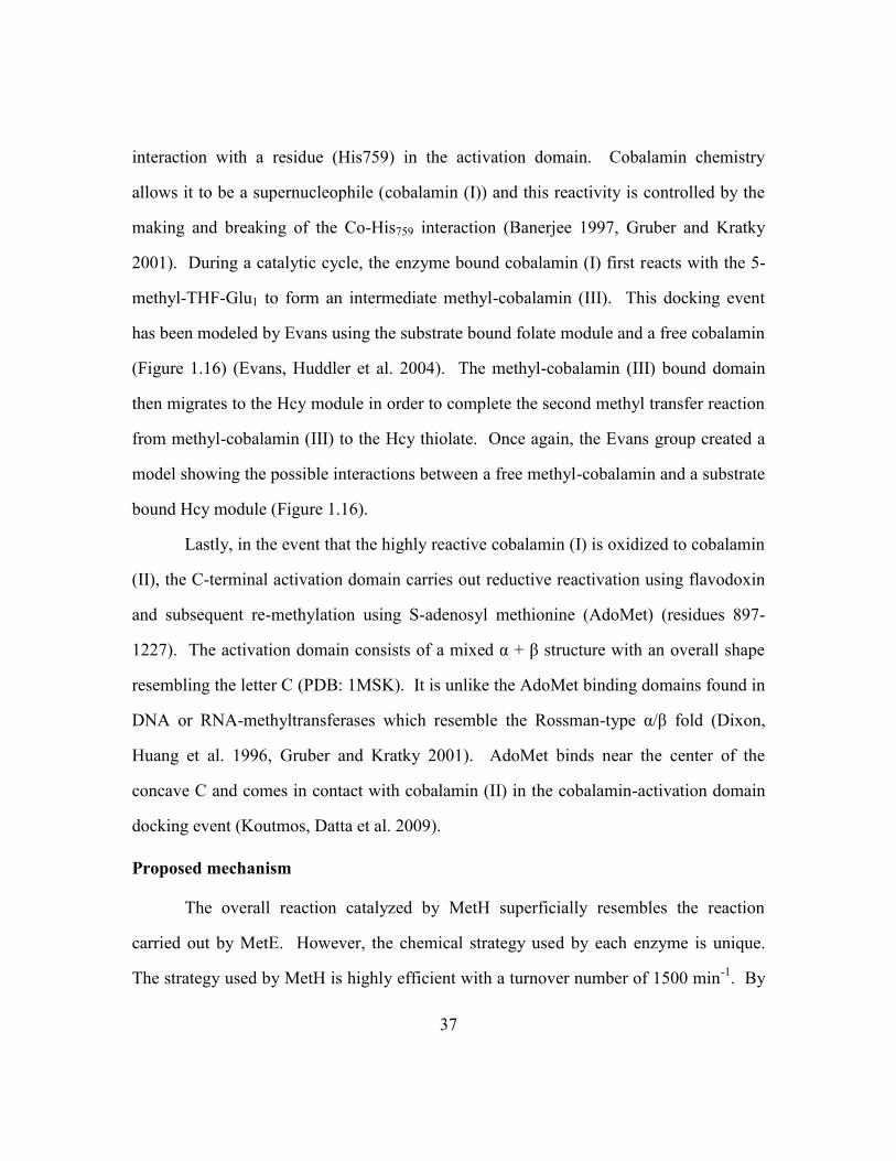
interaction with a residue (His759) in the activation domain. Cobalamin chemistry
allows it to be a supernucleophile (cobalamin (I)) and this reactivity is controlled by the
making and breaking of the Co-His759 interaction (Banerjee 1997, Gruber and Kratky
2001). During a catalytic cycle, the enzyme bound cobalamin (I) first reacts with the 5-
methyl-THF-Glu1 to form an intermediate methyl-cobalamin (III). This docking event
has been modeled by Evans using the substrate bound folate module and a free cobalamin
(Figure 1.16) (Evans, Huddler et al. 2004). The methyl-cobalamin (III) bound domain
then migrates to the Hcy module in order to complete the second methyl transfer reaction
from methyl-cobalamin (III) to the Hcy thiolate. Once again, the Evans group created a
model showing the possible interactions between a free methyl-cobalamin and a substrate
bound Hcy module (Figure 1.16).
Lastly, in the event that the highly reactive cobalamin (I) is oxidized to cobalamin
(II), the C-terminal activation domain carries out reductive reactivation using flavodoxin
and subsequent re-methylation using S-adenosyl methionine (AdoMet) (residues 897-
1227). The activation domain consists of a mixed α + β structure with an overall shape
resembling the letter C (PDB: 1MSK). It is unlike the AdoMet binding domains found in
DNA or RNA-methyltransferases which resemble the Rossman-type α/β fold (Dixon,
Huang et al. 1996, Gruber and Kratky 2001). AdoMet binds near the center of the
concave C and comes in contact with cobalamin (II) in the cobalamin-activation domain
docking event (Koutmos, Datta et al. 2009).
Proposed mechanism
The overall reaction catalyzed by MetH superficially resembles the reaction
carried out by MetE. However, the chemical strategy used by each enzyme is unique.
The strategy used by MetH is highly efficient with a turnover number of 1500 min-1
. By
38

39
comparison, MetE is sluggish with a turnover number of just 12.3 min-1
(Gonzalez,
Banerjee et al. 1992). Despite being a large modular enzyme, MetH has a significant
advantage over the smaller MetE because it uses the supernucleophile cobalamin. The
domain rearrangements are expected to be the slow steps in substrate turnover (Evans,
Huddler et al. 2004). As in MetE, the Hcy is activated by the active site zinc to create a
thiolate nucleophile and the folate is thought to be activated by protonation at the N5
amine carrying the methyl. The protonation event is only observed in the ternary
complex containing MetH, 5-methyl-THF-Glu1, and cobalamin (I) through an unknown
mechanism (Smith and Matthews 2000, Matthews, Smith et al. 2003).
The MetH catalyzed methyl transfer reaction can be divided into two half
reactions. In the first reaction, the cobalamin (I) nucleophile is expected to attack the
folate methyl group located on a quaternary N5 amine, generating methylcobalamin (III)
and the byproduct THF-Glu1. This event has an observed rate constant of 250 s-1
and the
folate KM of ~27.8 uM (Banerjee, Frasca et al. 1990). In the second half reaction, the
B12-module carrying the methylcobalamin (III) first docks against the Hcy-module
followed by a nucleophilic attack by the Hcy thiolate on the CH3-cobalamin (III). This
reaction is essentially irreversible and generates the final product methionine and
cobalamin (I) with an observed rate constant of 140 s-1
and an estimated Hcy KM of ~1
uM. The Matthews group also evaluated catalysis using free cobalamin (I) and the
substrate bound folate and hcy modules, or a free methylcobalamin (III) in the presence
of the substrate bound Hcy module (Goulding, Postigo et al. 1997). Both conditions were
found to be catalytically competent, meaning the hcy and folate binding modules are
independently active.
Once every 1000-2000 turnovers the cobalamin (I) is oxidized to the inactive
cobalamin (II) (Liptak, Datta et al. 2008). Reductive remethylation takes place in a
40
complex formed by the enzyme flavodoxin and the B12-binding and activation domains
(AdoMet) of MetH (Hall, Jordan-Starck et al. 2000). In this complex, flavodoxin
provides the electrons used to reduce the cobalamin metal and AdoMet donates a methyl
to create the catalytically competent methylcobalamin (III) intermediate.
Betaine Homocysteine Methyltransferase (BHMT)
The enzyme BHMT (EC 2.1.1.5), is a Zn2+
dependent thiolmethyltransferase
which generates methionine by catalyzing the transfer of a methyl group from betaine to
homocysteine (Figure 1.17) (Pajares and Perez-Sala 2006). In mammals and some
bacteria, BHMT is one of the two major enzymes (MetH is the other) which convert
homocysteine to methionine. In solution and in crystal structures, BHMT exists as a
tetramer of 45 kDa subunits (407 residues). Each monomer has a (βα)8 barrel (residues
1-318) followed by an extended structure called the dimerization arm. Surprisingly,
BHMT is 40% similar in sequence to the N-terminal region of MetH (Evans, Huddler et
al. 2002). Although the MetH and BHMT do not share any sequence identity with MetE,
all three of these enzymes have structurally similar (βα)8 barrels which bind Zn2+
and
Hcy. In fact, the Hcy/Met binding pockets in all three enzymes (MetH: 3BOF, MetE: ,
BHMT: 1LT8) use at least one negatively charged residue to anchor the amino group, use
backbone nitrogens to stabilize the carboxylate group, and have a hydrophobic pocket to
sequester the thiol containing side-chain. All three also exploit the nucleophilicity of the
adjacent Zn2+
ion to deprotonate the Hcy thiol (pKa =10) under physiological conditions,
converting it into a thiolate anion (Millian and Garrow 1998). In rat BHMT, the Zn2+
is
coordinated by four conserved residues, Cys217, Cys299, Cys300 and Tyr160 (Gonzalez,
Pajares et al. 2004). In the presence of Hcy, the zinc is expected to have a net charge of -
2, which is identical to the net charge in MetH.

41
Unlike MetE and MetH, which dedicate an entire domain to bind and activate 5-
methyl-THF, BHMT is much smaller and its compact structure accommodates the much
smaller methyl donor betaine. Betaine bound BHMT structures are currently unavailable
but mutational studies along with a BHMT bound transition state bi-substrate analog S-
(δ-carboxybutyl)-L-homocysteine (CBHcy), have identified a ring of aromatic residues
which participate in betaine binding (Pajares and Perez-Sala 2006). The substrates enter
42
the active site in order of Hcy followed by betaine (Castro, Gratson et al. 2004).
Substrate binding induces conformational changes that are expected to bring the two
substrates in proximity. A direct methyl transfer reaction is expected to proceed between
a thiolate nucleophile and a methyl group attached to the quaternary amine of betaine.
S-Methylmethionine Hcy S-Methyltransferase (HMT)
The enzyme HMT (EC 2.1.1.12) is a zinc metalloenzyme which generates
methionine by catalyzing the transfer of a methyl group from L-S-methylmethionine
(SMM) to homocysteine (Ranocha, Bourgis et al. 2000). SMM is a major sulfur
containing metabolite in plants, created and depleted through the SMM cycle. Organisms
such as bacteria, yeast and mammals can obtain SMM through diet and use it to make
methionine via the HMT enzyme.
Enzymes with HMT activity have been isolated from Homo sapiens (BHMT-2)
and E. coli (YagD) (Thanbichler, Neuhierl et al. 1999, Szegedi, Castro et al. 2008). The
bacterial enzyme, YagD shares some sequence similarity with the amino-terminal domain
of MetH and to BHMT. It synthesizes methionine using SMM or S-adenosyl methionine
and Hcy. The mammalian enzyme, BHMT-2 encodes a 40 kDa protein which is 73%
identical to the BHMT protein. BHMT-2 uses SMM and to a lesser degree AdoMet as a
methyl donor. A model of BHMT-2 was built by Garrow and colleagues using BHMT as
a scaffold (Swiss model software). Like the amino-domain of MetH, BHMT, and the C-
terminal domain of MetE, BHMT-2 consists of a (βα)8 barrel with conserved cysteine
residues which coordinate with a zinc.
Folate and anti-folate bound enzymes deposited in the PDB
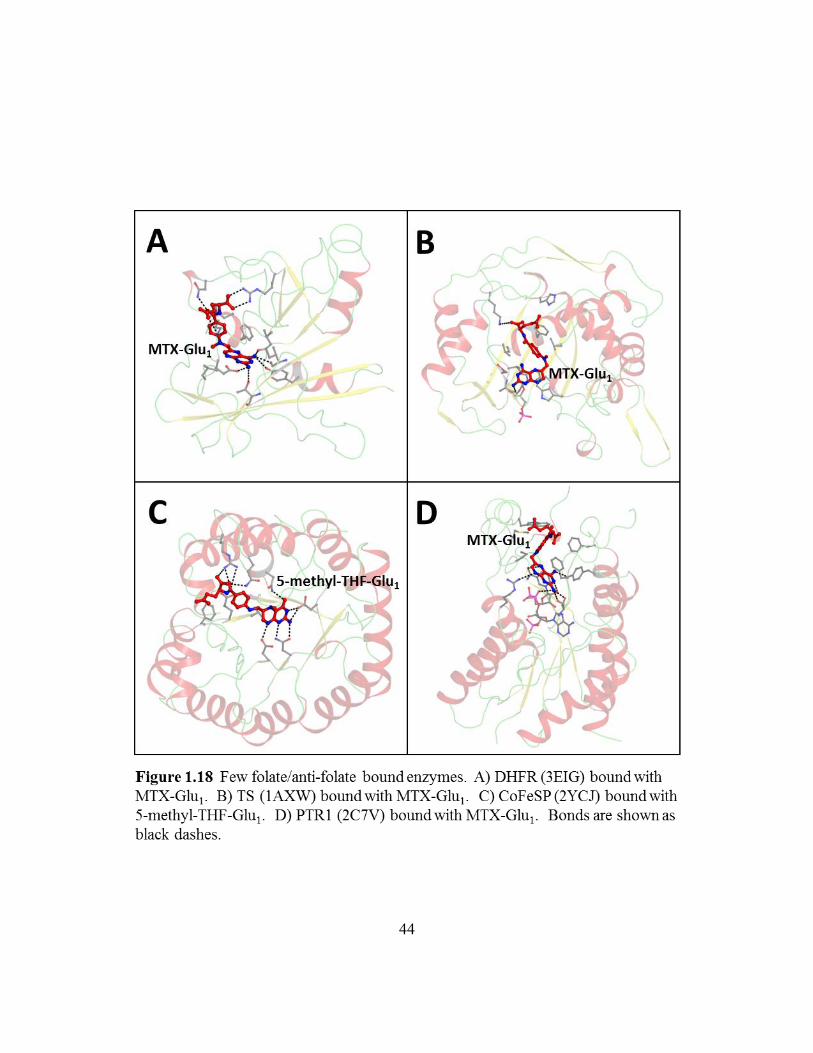
The majority of folate and anti-folate bound structures available in the PDB are of
Dihydrofolate reductase (DHFR) and Thymidylate synthase (TS). DHFR has long been a
43
target of anticancer and antibiotic drugs due to its role as the sole provider of THF. THF
is an essential intermediate for purine and thymidylate synthesis and indirectly effects
cell growth and proliferation (Schnell, Dyson et al. 2004). Thymidylate synthase is also a
target for anticancer chemotherapy due to its role as the sole provider of de novo
thymidylate, an essential precursor required for DNA replication and repair (Garg,
Henrich et al. 2010). Although the two enzymes are structurally unique, a number of
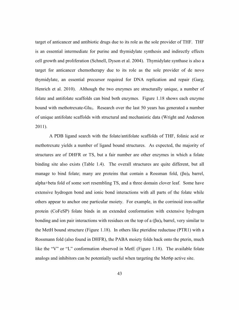
folate and antifolate scaffolds can bind both enzymes. Figure 1.18 shows each enzyme
bound with methotrexate-Glu1. Research over the last 50 years has generated a number
of unique antifolate scaffolds with structural and mechanistic data (Wright and Anderson
2011).
A PDB ligand search with the folate/antifolate scaffolds of THF, folinic acid or
methotrexate yields a number of ligand bound structures. As expected, the majority of
structures are of DHFR or TS, but a fair number are other enzymes in which a folate
binding site also exists (Table 1.4). The overall structures are quite different, but all
manage to bind folate; many are proteins that contain a Rossman fold, (βα)8 barrel,
alpha+beta fold of some sort resembling TS, and a three domain clover leaf. Some have
extensive hydrogen bond and ionic bond interactions with all parts of the folate while
others appear to anchor one particular moiety. For example, in the corrinoid iron-sulfur
protein (CoFeSP) folate binds in an extended conformation with extensive hydrogen
bonding and ion pair interactions with residues on the top of a (βα)8 barrel, very similar to
the MetH bound structure (Figure 1.18). In others like pteridine reductase (PTR1) with a
Rossmann fold (also found in DHFR), the PABA moiety folds back onto the pterin, much
like the “V” or “L” conformation observed in MetE (Figure 1.18). The available folate
analogs and inhibitors can be potentially useful when targeting the Met6p active site.
44
45

46
Cobalamin-independent Methionine Synthase as a drug target
Structural perspective
The cobalamin-independent methionine synthase enzyme is a promising new anti-
fungal drug target because structurally and mechanistically it is unlike the methionine
synthase found in mammals (MetH). Although both enzymes bind the same substrates,
prior to the alkylation reaction, these substrates are set 50 Å apart in MetH and only 7 Å
apart in MetE. Both enzymes rely on large structural rearrangements to complete the
methyl transfer reaction. In MetH, the B12 module cycles between the tops of the
immobile (βα)8 barrels and the reaction takes place between a methylcobalamin
intermediate and the Hcy. In contrast, MetE mediates a direct methyl transfer reaction by
merging the two individual substrate pockets into one as it structurally transitions from
the ‘open’ configuration to a ‘closed’ configuration.
A drug design strategy aimed at selectively binding and inhibiting the cobalamin-
independent enzyme is feasible for both the open and closed MetE configurations.
Clearly, both enzymes have evolved to bind an amino acid (Hcy) and a folate. Each site
represents a potential target for inhibitor design, however a single bi-dentate substrate
analog that spans both substrate binding pockets has great drug potential. From a
practical standpoint, the binding affinity of such a ligand could be strong since the free
energy of each component adds to the binding of the complete ligand. Also, the
specificity is enhanced since no other protein binds both moieties in close proximity. The
therapeutic window for such a compound would be large since the human enzyme is so
different from the fungal one. That is, a bi-substrate inhibitor designed for MetE will be
a poor ligand for the methylcobalamin-Hcy transition state pocket in MetH. A model of
the methylcobalamin bound to the Hcy module was created by Evans in which a flat B12
corrin ring stacks against the top of the (βα)8 barrel (Figure 1.16) (Evans, Huddler et al.

47
2004). Hydrophobic interactions and specific hydrogen bonds help to create a unique
Hcy-methylcobalamin binding site.
High quality substrate-bound structures of the fungal Met6p enzyme are very
desirable in order to design decent bi-substrate inhibitors. So far the bacterial and plant
MetE homologs have only been crystallized in the ‘open’ configuration and the available
substrate-replete structures are of poor quality. Nevertheless, a MetE specific bi-
substrate analog can be designed to bind and immobilize this ‘open’ configuration. Also,
if the Met6p enzyme is successfully crystallized in the ‘closed’ form it may lead to more
potent inhibitors of MetE that immobilize it in a closed configuration.
Biological perspective
The gene encoding the Met6p enzyme is absolutely required for C. albicans
growth (Suliman, Appling et al. 2007). The Robertus and Appling labs have already
shown that a diploid C. albicans strain lacking both copies of the MET6 gene is unable to
grow in media supplemented with methionine. Since C. albicans can acquire methionine
from the media via a number of amino acid transporters, the lethal phenotype cannot be
explained by a simple methionine auxotrophy (Kaur and Mishra 1991, Sophianopoulou
and Diallinas 1995, Wipf, Ludewig et al. 2002). When one MET6 locus was deleted and
the second placed under a regulatable, but slightly leaky promoter, the mutant grew under
inducing conditions. However, it would not grow under repressing conditions, unless
supplemented with exogenous methionine. The conditional mutant is different from the
double knockout strain because its growth was rescued with exogenous methionine while
the double knockout is lethal. This is attributed to the low amount of MET6 expressed
under repressing conditions by the leaky conditional promoter, and further supports the
requirement of the Met6p protein for fungal growth.

48
Similar experiments have also been done in Cryptococcus neoformans, Fusarium
graminearum, Aspergillus nidulans, Saccharomyces cerevisiae, and
Schizosaccharomyces pombe (Kacprzak, Lewandowska et al. 2003, Pascon, Ganous et al.
2004, Seong, Hou et al. 2005, Suliman, Sawyer et al. 2005, Fujita, Ukena et al. 2006).
Although the complete depletion of the MET6 gene is not lethal to the pathogenic fungus
C. neoformans and the wheat scub fungus F. graminearum, it does result in a significant
growth defect. In both fungi, the MET6 mutants also grew poorly in media supplemented
with methionine. The doubling time for the mutant C. neoformans was ~3.5 hours
compared to the ~1.6 hours observed for the wild-type strain. In Aspergillus nidulans,
and Saccharomyces cerevisiae the MET6 gene is not essential and growth of a MET6
mutant can be fully rescued with exogenous methionine (Kacprzak, Lewandowska et al.
2003, Suliman, Sawyer et al. 2005). Interestingly, the MET6 mutant in
Schizosaccharomyces pombe was rescued when both methionine and adenine were added
to the growth media (Fujita, Ukena et al. 2006).
The slow growing MET6 mutant strains of C. neoformans and F. graminearum
have also been evaluated for virulence in mice and plants, respectively. Pascon and
coworkers discovered that when mice were infected with the wild type C. neoformans
they died between 21-25 days post infection; in contrast, those infected with the MET6
mutant strain remained healthy until they were euthanized 70 days post-infection
(Pascon, Ganous et al. 2004). When grown in cell culture, the MET6 mutant strain
showed signs of compromised virulence including reduced capsule formation and
lowered thermotolerance. The MET6 mutant was also more susceptible to the antifungal
drugs fluconazole, FK506 and CsA. Likewise, plants infected with the MET6 mutant
strain (msy1) of F. graminearum appeared healthy and viable, meaning the mutant failed

49
to establish wheat head infection. In cell culture, this mutant also produced very little
aerial hyphae; an important virulence factor (Seong, Hou et al. 2005).
The complete disruption of the methionine synthase gene is lethal for C. albicans,
C. neoformans and F. graminearum, even when supplemented with Met. It may be that
high concentrations of the metabolite Hcy are toxic to the cells. A high level of Hcy is
usually indicative of defects in enzymes responsible for remethylation (methionine
synthase), trans-sulfuration (cystathionine β-synthase) or folate metabolism (Zimny,
Sikora et al. 2006). However, in the case of methionine synthase, even if the mutation is
compensated by adding the product methionine, the MET6 mutant is not fully rescued
possibly because of the accumulating Hcy. A high concentration of Hcy is linked to
toxicity in animals, yeast, and bacteria. Although the exact mechanism of toxicity is
unknown, it might be linked to the conversion of Hcy to Hcy-thiolactone. If Hcy is not
metabolized to Hcy-thiolactone, it can be incorporated into proteins to form toxic Hcy-
proteins. On the other hand, the synthesis of Hcy-thiolactone consumes cellular energy
in the form of ATP, and the resulting product is a reactive intermediate which damages
proteins through protein homocysteinylation (Jakubowski 1999). Hcy-thiolactone is
either excreted or hydrolyzed back to Hcy by a Hcy-thiolactonase. The accumulation of
Hcy is also predicted to inhibit purine biosynthesis, and high levels of Hcy have been
found to inhibit ergosterol synthesis in yeast (Hatanaka, Ariga et al. 1974, Parks and
Casey 1995, Fujita, Ukena et al. 2006).
The concentration of Hcy was measured by HPLC in a wild-type and MET6
mutant strain of Schizosaccharomyces pombe. As expected, the total Hcy content in the
methionine synthase mutant was 18 times higher than the wild-type strain (Fujita, Ukena
et al. 2006). In E. coli both the Hcy and Hcy-thiolactone metabolites are known to

50
upregulate the mRNA and enzyme levels of methionine synthase (Kacprzak,
Lewandowska et al. 2003).
The gene deletion studies have established that MetE is important for growth and
virulence. This is in line with gene expression studies which show that MET6 is strongly
expressed in plants, yeast and bacteria during growth, stress or infection. Methionine is
an important metabolite since cells invest significant energy for its synthesis through
methionine synthase or acquisition through the amino acid transporters. During growth
and in the absence of cobalamin and methionine, the MetE enzyme is extremely
abundant, constituting 3% of the total soluble protein in E.coli cells (Gonzalez, Banerjee
et al. 1992). And during plant infection, the gene expression of MetE in the bacterium
Ralstonia solanacearum was found to be specifically and strongly induced (10-fold more
than MetH) (Plener, Boistard et al. 2012). During normal growth, fungi (Cladosporium
fulvum, Saccharomyces cerevisiae, Aspergilus nidulans), plants (Catharantus roseus) and
bacteria (E. coli) modulate the expression of MET6/MetE based on methionine
availability (Weissbach and Brot 1991, Eichel, Gonzalez et al. 1995, Solomon, Nielsen et
al. 2000). Starvation and high stress conditions experienced during host infection by the
pathogen C. fulvum also appeared to increase MET6 gene expression (Solomon, Nielsen
et al. 2000). When the plant cells of C. roseus or leaves of Solanum tuberosum were
deprived of nutrients by using sucrose, a significant increase in the expression of MetE
was observed (Eichel, Gonzalez et al. 1995, Zeh, Leggewie et al. 2002). Since
methionine is a precursor of S-adenosyl methionine (AdoMet), the major methyl-group
donor required for protein synthesis and gene expression, the synthesis or acquisition of
methionine is understandably up-regulated for growth and survival.

51
Candidiasis and candidemia
Various microorganisms, including bacteria, fungi, yeast, archae and viruses,
colonize the skin, oral cavity, respiratory pathways, stomach, the gastrointestinal tract,
and the urinary and genital tracts of the human body (Tannock 1995). These microbes
form complex communities and establish an ecosystem that can benefit the host (Rajilic-
Stojanovic 2013). The highest microbe density is reported in the intestinal tract where
the microbiota’s collective genome contains 100 fold more genes than the host. The
microbial activity is beneficial since it improves digestion, provides vitamins, inhibits
pathogenic growth and stimulates the immune system.
However, an imbalance in the indigenous microbial composition leads to
problems in metabolism and causes infections (Kim, Jeon et al. 2013). A few bacterial
strains make up 99.9% of the ecosystem; the less abundant, but more diverse 0.1% ‘rare
biosphere’ includes the fungal microbiome (Huffnagle and Noverr 2013). Aside from the
known benefits of Saccharomyces cerevisiae, there is no strong evidence in favor of a
mutual fungal-host relationship. Many fungal species such as those belonging to the
genus Candida are members of the normal microbiome in the various mucosal surfaces
and skin. However, in response to a compromised host immune system or alterations in
the normal microbiota, the opportunistic Candida species becomes pathogenic and cause
infection. Candida infections, or candidiasis, can be superficial or severe, as in systemic
candidiasis. In fact, Candida infections are ranked as the 3rd
or 4th
most common cause
of hospital-acquired bloodstream infections, called candidemia (Moyes and Naglik 2011).
Over the last two decades, the incidence of candidemia has increased significantly
and can be attributed to the ever growing number of immuno-compromised individuals
who undergo transplantations, chemotherapy or suffer from HIV infections (Pfaller,
Jones et al. 1998, Falagas, Roussos et al. 2010, Das, Nightingale et al. 2011). The mode

52
of virulence is perhaps best characterized for C. albicans since it is the most prevalent
and usually associated with the highest mortality (Pfaller, Jones et al. 1998). Normally,
C. albicans grows as budding yeast and this growth is kept under control by the diverse
microbe community and an effective immune system. The host-fungus interaction is
quite complex and still under investigation. However, a few studies have shown that
Candida is not a passive participant in the process of pathogenesis (Rizzetto and
Cavalieri 2011, Sardi, Scorzoni et al. 2013). Candida albicans, in particular, is equipped
with fitness attributes and virulence factors which are used to actively establish
candidiasis and evade the host immune system. These include the ability to attach to host
cells (using adhesins), growth as filamentous hyphae (yeast-hyphae transition),
penetration of the host cell (using invasins and hydrolytic enzymes), formation of
impregnable biofilms in host cells and catheters, adaptation to fluctuations in
environmental pH, acquisition of nutrients, and survival under stress (heat, oxidation)
(Mayer, Wilson et al. 2013). The ability to transition between yeast and hyphae and to
form a biofilm is critical for establishing virulence, characteristics also possessed by the
newly emerging pathogens C. parapsilosis, C. tropicalis and C. glabrata.
The current arsenal of anti-fungal drugs is limited due to the similarities between
mammalian and fungal pathways. The first class consists of polyene derivatives which
associate with the ergosterol in the cell membrane and promote cell leakage. The azole
derivatives also target the cell membrane by inhibiting the fungal cytochrome P450
dependent conversion of lanosterol to ergosterol. Finally, the echinochandins weaken the
cell wall by inhibiting 1,3-ß -glucan synthase (Lai, Tan et al. 2008).
Unfortunately, substantial adverse effects and limited anti-fungal activity are
associated with the use of polyene and azole derivatives. Echinochandins target a
uniquely fungal pathway and therefore have the lowest toxicity and the widest spectrum

53
of use. However, in order to treat the often drug-resistant biofilms, drugs from the
different classes must be combined or the infected medical device must be removed
(Sardi, Scorzoni et al. 2013). A major problem is that drug resistant Candida strains have
already been isolated and include the mostly fluconazole resistant C. glabrata isolates
(Krcmery and Barnes 2002).
Summary and Project Goals
The cobalamin-independent (Met6p) and dependent (MetH) methionine synthases
are two very different enzymes which catalyze the same overall reaction in the
pathogenic fungi and the human host, respectively. The methionine synthase enzyme in
each organism is metabolically essential and sits at a point of convergence where folate-
mediated one-carbon metabolism meets methionine synthesis. Defects in methionine
synthase impact folate metabolism, AdoMet mediated methyl transfer reactions and Hcy
metabolism. In fact, in the fungal species, Candida albicans, the MET6 gene which
encodes the Met6p enzyme is absolutely required for fungal growth. This fungal species
is one of the most prevalent causes of candedimia in the immunocompromised population
around the world, and the primary fungal target in the Robertus lab.
We believe that the structural and biological attributes of the Met6p enzyme make
it an ideal antifungal drug target and potent Met6p specific inhibitors should be able to
control a fungal infection with minimal toxicity against the human host. The overall goal
of this project is to discover a variety of drug-like scaffolds for the Met6p active site and
to develop structural and kinetic methods to characterize the drug-Met6p interaction. The
resulting lead compounds could then be derivatized into highly specific and potent Met6p
inhibitors in collaboration with an organic chemist, and ultimately tested in mice infected

54
with Candida albicans. I successfully tested a part of this hypothesis by completing the
following aims:
Aim 1: Crystallization of the Met6p enzyme using SER
The wild type Met6p protein could not be crystallized by traditional screening
methods so Dr. Kate Kavanagh suggested we use a protein engineering strategy known as
Surface Entropy Reduction (SER). Under her guidance I created three variants of Met6p
in which three surface residues were replaced by alanines (Met6pA), threonines
(Met6pT), or tyrosines (Met6pY). The X-ray structures and crystal packing arrangement
of each crystal were reported in “Structure of Candida albicans methionine synthase
determined by employing surface residue mutagenesis” (Ubhi, Kavanagh et al. 2011).
Aim 2: Characterization of the Met6p active site in the ‘open’ configuration
Met6p catalyzes a direct methyl transfer reaction by cycling between the ‘open’
configuration we observed in the Met6pA, Met6pT and Met6pY variants, to the yet
unobserved ‘closed’ configuration. The ‘open’ configuration has two substrate binding
pockets separated by ~7 Å and a metal coordination site. I solved a number of substrate
or product bound binary and ternary complexes which were analyzed and compared to
each other and the apo structure. Any interesting residues were mutated by site-directed
mutagenesis and their function was evaluated using an activity assay.
Aim 3: Development of a fluorescence-based Met6p activity assay
The established absorbance based kinetic assay cannot be used to evaluate
inhibitors which also absorb at 350 nm, so an alternative fluorescence based kinetic assay
was created, optimized and used to test potential inhibitors. This assay uses the Measure-
iTTM
thiol quantitation reagent to selectively measure the concentration of unused Hcy in
a Met6p reaction.

55
Aim 4: Optimization of a high-throughput DSF assay used to screen diverse
compound libraries
The high-throughput Differential Scanning Fluorimetry (DSF) method is
frequently used in drug discovery programs to identify compounds which bind and
increase the melting temperature (Tm) of a target protein. I optimized a 96-well DSF
method for Met6p and used it to screen 731 compounds from the NIH clinical collection,
4000 compounds from the Fragment library (Chembridge) and 4000 compounds from the
KINAset library (Chembridge). The hits were further evaluated by an activity assay and
the IC50s were determined using dose-response experiments.
Aim 5: Discovering lead compounds by rational drug design
The last strategy used to find potential Met6p inhibitors was based on principles
of ligand-based and structure-based drug design. I searched for commercially available
ligands which could occupy the folate, Hcy or both binding pockets simultaneously.
These ligands include anti-folates like MTX-Glu3, amino acids like Gln, or a folate-Hcy
bi-substrate analog like S-adenosyl homocysteine. These ligands were tested for binding
by X-ray crystallography and for inhibition by the fluorescence based activity assay.

56
CHAPTER 2: MATERIALS AND METHODS
Cloning and expression of Met6p in E. coli
The MET6 gene encoding the Candida albicans Met6p protein was amplified
from Yep24-CaMET6 vector and cloned into the kanamycin resistant E. coli expression
vector pNIC28-Bsa4 purchased from Addgene (Figure 2.1) (Suliman, Sawyer et al.
2005). We amplified the MET6 gene by using PCR primers with the extensions
TACTTCCAATCCatg on the 5’ end of the upstream primer and
TATCCACCTTTACTGtca on the 5’ end of the downstream primer. These extensions
overlap the vector sequences that flank the cloning site in pNIC28-Bsa4 (Figure 2.1). A
detailed cloning, annealing, and transformation protocol described by Savitsky was used
to create the pNIC28-CaMET6 vector (Savitsky, Bray et al. 2010). In this vector the
MET6 gene is fused with an N-terminal tag of 23 residues
(MHHHHHHSSGVDLGTENLYFQSM) that contains a hexahistidine (His6) site for Ni-
NTA purification followed by a TEV protease cleavage site (ENLYFQS) (Fig 2.2). The
fusion gene and protein sequences are shown in Figures 2.3 and 2.4, respectively.
The pNIC28-CaMET6 expression vector was transformed into One Shot
BL21(DE3) E. coli cells and Met6p protein was expressed in 2-4 L LB media
supplemented with 500 µM ZnSO4 and 50 mg/L kanamycin. Cells were grown at 37 °C
until the OD600 reached 0.8. The cultures were shifted to 25 °C and expression was
induced with 500 µM IPTG and allowed to continue for 4 hours. Cells were harvested by
centrifugation and the pellets were stored at -80 °C.

57

58

59
Protein purification
Cell pellets were thawed and resuspended in lysis buffer (1X PBS, 20 mM
Imidazole, 10 mM β-mercaptoethanol, pH 8.0) with protease inhibitors (Roche Complete
EDTA-free, 1 tablet/50 mL). The cells were homogenized using a French pressure cell (3
passes, 1000 psi) and the cell debris removed by centrifugation for 1 hour at 25,000g and
4°C. The cell lysate was added to 2 mL of Ni-NTA resin and both were gently mixed for
60
30 min at 4 °C. This mixture was applied to a gravity flow column, washed with 20 mL
lysis buffer and eluted with 6 mL of lysis buffer containing 500 mM imidazole. The
eluted protein was first buffer exchanged into 20 mM Tris-HCL, 150 mM NaCl, 10 mM
β-mercaptoethanol, pH 7.4 using a PD10 column and then further purified using a size
exclusion column (S200 16/60) run at 1 ml/min on an Akta purification system. The
fractions were analyzed by SDS-PAGE (8% gel) and those containing Met6p protein
were pooled and concentrated using a Centricon filtration unit with 30 kDa MW cut-off.
Protein purity was estimated to be >95% by SDS-PAGE gel (Figure 2.5). Protein
concentration was measured using the absorbance at 280 nm and an extinction coefficient
calculated from the protein sequence ( = 90,760 M-1
cm-1
).

61
Surface Entropy Reduction (SER)
The SERp server (http://nihserver.mbi.ucla.edu/SER/) was used to identify
potential residues for surface entropy reduction mutations (Goldschmidt, Cooper et al.

62
2007). The top scoring cluster, 103
LysLysAlaThrGlu107
, with the SERp score of 7.01 was
then compared to known bacterial and plant structures and found to be on a loop that is
~10 residues longer in the fungal protein. This loop region was chosen for mutation.
Based upon a previous study that looked at the role of different amino acids in promoting
crystal contacts, the two lysine and one glutamate residues were mutated either all to Ala,
all to Thr, or all to Tyr in variants labeled Met6pA, Met6pT, and Met6pY, respectively
(Cooper, Boczek et al. 2007).
Site-directed mutagenesis
The desired mutations were introduced using PCR-based site-directed
mutagenesis by following the instructions in the QuickChangeTM
Site-Directed
Mutagenesis Kit (Stratagene, La Jolla, CA, USA) (Kim and Maas 2000). We used the
online QuikChange Primer Design program
(http://www.genomics.agilent.com/primerDesignProgram) to make each set of
complementary PCR primers. The PCR primers used to create the SER mutants Met6pA,
Met6pT and Met6pY, were designed to introduce multiple mutations in the wild-type
MET6 gene (Figure 2.6). The PCR primers used to generate each point mutant
Tyr660Ala, Tyr660Gln, Tyr660Phe, and Asn126Ala were shorter and introduced a single
mutation in the gene encoding the Met6pA variant (Figure 2.6). The general PCR
reaction conditions are listed in Figure 2.6. We used KOD Hot Start DNA polymerase
(Novagen) for plasmid amplification and destroyed each parent vector with the enzyme
DpnI (Biolabs). The gene mutations were verified by DNA sequencing. All mutant
plasmids were transformed into BL21 cells followed by protein expression and
purification using the methods described above.

63

64
Crystallization
The SER variant proteins were concentrated to ~20 mg/ml in 20 mM Tris, 150
mM NaCl, 10 mM β-mercaptoethanol, pH 7.4. Crystallization trials were set up with
both apo protein solutions and protein solutions containing 10 mM Hcy using the JCSG+
suite (Qiagen) and PEG/Ion HT (Hampton Research) 96-well screening kits. The plates
were set up using a Phoenix crystallization robot (Art Robbins Instruments) and were
incubated at 20 °C. Several conditions producing initial crystals were identified, and two
were chosen for optimization: JCSG+ G9 (30% [w/v] PEG-MME 2000, 0.1 M KSCN)
and PEG/Ion A10 (20% [w/v] PEG3350, 0.2 M NaI). Crystal optimization experiments
were set up with all three mutants in 96-well and 24-well formats.
Optimized crystals of the Met6pA mutant were rod-shaped with a maximum
length of 800 µm (Figure 2.7). They were grown in hanging drops at 20 °C by mixing 1
µL of 20 mg/ml apo protein and 2 µL of a well solution consisting of 27 % (w/v) PEG
3350, 50 mM NaI and equilibrated against 500 µL of the well solution. Prior to data
collection, a single crystal was cryo-protected in 30% (w/v) PEG 3350, 50 mM NaI and
flash-cooled in liquid nitrogen.
Optimized crystals of the Met6pT mutant were large plates with dimensions 800 x
300 x 50 µm (Figure 2.7). They were grown in hanging drops at 20 °C by mixing 1 µL
of 20 mg/ml apo protein with 2 µL of a well solution composed of 25% (w/v) PEG-MME
2000, 75 mM KSCN and equilibrated against 500 µL of well solution. Prior to data
collection, a single crystal was cryo-protected in 30% (w/v) PEG-MME 2000, 75 mM
KSCN, and plunged into liquid nitrogen.
Optimized crystals of the Met6pY mutant were rod-shaped with a maximum
length of 200 µm (Figure 2.7). They were grown in hanging drops at 20 °C by mixing
0.7 µL of 20 mg/ml apo protein and 1.3 µL of a well solution consisting of 20% (w/v)
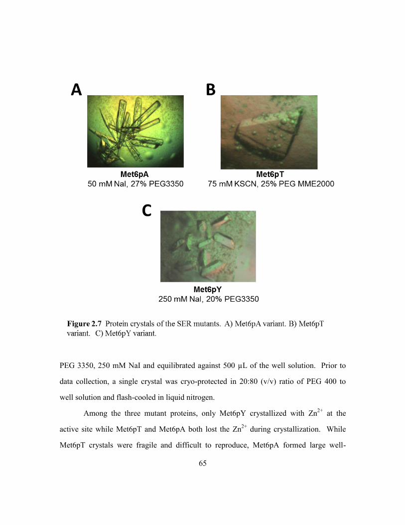
65
PEG 3350, 250 mM NaI and equilibrated against 500 µL of the well solution. Prior to
data collection, a single crystal was cryo-protected in 20:80 (v/v) ratio of PEG 400 to
well solution and flash-cooled in liquid nitrogen.
Among the three mutant proteins, only Met6pY crystallized with Zn2+
at the
active site while Met6pT and Met6pA both lost the Zn2+
during crystallization. While
Met6pT crystals were fragile and difficult to reproduce, Met6pA formed large well-

66
diffracting crystals and were soaked in a Zn2+
containing solution in order to obtain a
zinc-replete crystal. Met6pA crystals were first soaked in reservoir solution
supplemented with 250 µM zinc sulfate and 500 µM DTT for 3 hours followed by a 8-12
hour soak in reservoir solution including 500 µM DTT. Met6pA crystals plus zinc were
then cryo-protected in 30% (w/v) PEG 3350, 50 mM NaI and flash-cooled in liquid
nitrogen.
Ligand-replete crystals
The SER variant Met6pA was used for all ligand bound experiments because
Met6pA diffracts to high resolution and has a single molecule in the asymmetric unit.
Large well-diffracting crystals were first soaked in a zinc containing solution in order to
obtain zinc-replete crystals (described above). The zinc-replete crystals were then soaked
for 4 hours in the crystallization condition with the ligand of choice.
The soaking experiments done to map out the substrate and product binding
pockets included Met6pA in a binary complex with 5 mM Hcy, 10 mM Met, >70mM
Gln, 5 mM 5-methyl-THF-Glu3, and 10 mM SAH. Crystals were soaked with both 5 mM
5-methyl-THF-Glu3 and 10 mM Met for the ternary substrate complex. A ternary
inhibitor complex was prepared with 10 mM MTX-Glu3 and 5 mM Hcy.
The soaking experiments done with compounds from the NIH clinical collection,
Fragment library and KINAset library were limited to 5 mM. These compounds are
normally dissolved in 100% DMSO and Met6pA crystals can only tolerate up to 5-7%
DMSO for extended periods of time. Prior to data collection, a single crystal from each
condition was cryo-protected in each respective soak condition containing 30% (w/v)
PEG 3350 and flash-cooled in liquid nitrogen.

67
Data collection and processing
Diffraction data from the three SER variant crystals and most of the Met6pA-
ligand complexes was collected at 100 K at the Advanced Light Source at the Lawrence
Berkeley national Laboratory. Data for the SAH bound Met6pA crystal was collected at
100 K at the Macromolecular Crystallography Facility at UT Austin using the Rigaku
MicroMax-007HF generator and the R-AXIS IV++ plate detector (Rigaku, The
Woodlands, TX). The Met6pY and Met6pT data were integrated and scaled with
Mosfilm and Scala (Leslie 1992). HKL2000 was used to process each Met6pA data set
(Otwinowski 1997). A molecular replacement solution for the Met6pY crystal was
determined with Phaser using the structure of A. thaliana methionine synthase (PDB ID:
1U1H) as the search model (Ferrer, Ravanel et al. 2004, McCoy 2007). A molecular
replacement solution for Met6pT was found with Phaser. A solution for ligand-free
Met6pA was found with MOLREP using the Met6pY structure as the search model,
while Phaser was used for the ligand-bound Met6pA structures. The models were
improved through iterative rounds of model building with COOT and refinement using
Phenix and Refmac (Collaborative Computational Project 1994, Murshudov, Vagin et al.
1997, Emsley and Cowtan 2004, Adams, Afonine et al. 2010). Each structure was
reviewed using MOLPROBITY (Lovell, Davis et al. 2003).
5-Methyl-THF-Glu3 synthesis
(6-R,S) 5-CH3-THF-Glu3 was synthesized from Pteroyl-Glu3 by using a modified
procedure (http://appling.cm.utexas.edu/methylTHF.pdf) originally developed by Yeo
and Wagner (Yeo and Wagner 1992). The starting material Pteroyl-Glu3, intermediate
THF-Glu3 and final product 5-CH3-THF-Glu3 absorb at wavelengths 282 nm (23.4 mM-
1), 298 nm (30 mM
-1), and 292 nm (31.7 mM
-1).

68
40 µmol Pteroyl-Glu3 was first combined with 200 µL of 0.265 mM Pb(NO3)2
and dissolved by vortexing and slowly adding 15-35 µL of 5N NaOH until the final pH
was ~7.5. Pteroyl-Glu3 was reduced to THF-Glu3 by adding 132 µmol NaBH4 over 20
minutes. This was done at 4°C and pH was maintained at 8.5-8.8 using 20% (w/v) citric
acid. The solution was flushed with N2 and mixed for 2 hours in the dark at ~23°C. The
vial was cooled to 4°C and the excess NaBH4 was destroyed by reducing the pH to 5.0
using 5M acetic acid. The pH was adjusted back to 7.8 using 5 N NaOH. The
intermediate product, THF-Glu3, was then condensed with HCHO by adding 80 µmol of
a 37% formaldehyde solution and mixing at 45°C for 15 minutes in the dark. This
produced the intermediate CH2-THF-Glu3, which was subsequently reduced to 5-CH3-
THF-Glu3 by adding 264 µmol NaBH4. As before, the NaBH4 was added slowly over 20
minutes and pH of 8.5-8.8 was maintained with 20% (w/v) citric acid. The solution was
immediately flushed with N2 and mixed for 1 hour in the dark at 45°C. The solution was
cooled and 50 mM of the reducing agent BME was added. As before, excess NaBH4 was
destroyed by acetic acid and pH was adjusted to 7.5 using NaOH. The solution was
flushed with N2 and stored in the dark (with desiccant) at -80°C in 25 µL aliquots.
The concentration of 5-methyl-THF-Glu3 was determined by measuring the
absorbance at 298 nm. The yield was determined by diluting 2 µL of the product 1:4000
in 10 mM Tris pH 7.5. 400 µL of the dilution was combined with 100 µL of 5N
HCL/60% Formate and heated for 10 minutes at 80°C. The reaction was cooled and the
absorbance was measured over the 200-500 nm range. A peak at 350 nm is diagnostic of
THF and lack of a peak is indicative of 5-methyl-THF-Glu3.

69
Hcy synthesis
Hcy was synthesized from L-homocysteine thiolactone using a modified method
originally described by Uerre & Miller (Uerre and Miller 1966). Prior to use, all
solutions including the ddH2O, 5 M acetic acid, and 5 N NaOH were flushed with N2 for
2-5 minutes. 0.25 mmol L-Hcy thiolactone was dissolved in 750 µL 5 N NaOH and
incubated at 37 °C for 5 minutes. The solution was neutralized with 750 µL of 5 M
acetic acid. The Hcy was further diluted to ~100 mM by adding 1 mL ddH2O, flushed
with N2, and divided up in 100 µL aliquots. All aliquots were flushed with N2 and stored
at -80 °C. The concentration of Hcy was determined by using the thiol quantitation
reagent DTNB (Ellman 1959). DTNB reacts with a free thiol and generates the p-
nitrothiophenol which absorbs at 412 nm and has an extinction coefficient of 13.6 mM-1
.
A 2 mL reaction was prepared with 500 uM DTNB and 1 µL of Hcy in 100 mM NaPO4
pH 8.0 buffer and incubated for 5 minutes at room temperature. The absorbance at 412
nm was converted to mM Hcy using the Beer’s Law.
Absorbance based Met6p activity assay
The enzyme activity of wild-type or mutant Met6p was evaluated using a
modified methionine synthase assay described by Drummond et al. (Drummond, Jarrett
et al. 1995). The (6R,S)-5-methyl-tetrahydrofolate-Glu3 substrate (5-methyl-THF-Glu3)
was synthesized from pterin with a three glutamate tail, PteGlu3, using a modified
procedure of Yeo and Wagner, and the Hcy was made by alkaline hydrolysis of L-
homocysteine thiolactone (Yeo and Wagner 1992, Drummond, Jarrett et al. 1995,
Suliman, Appling et al. 2007). When available, 5-methyl-THF-Glu3 was purchased from
Schircks Laboratories (catalog # 16.243).
Initially a 380 µL reaction containing 50 mM potassium phosphate (pH 7.0), 50
mM Tris-HCl (pH 7.4), 100 µM MgSO4, 10 mM DTT, 140 µM 5-methyl-THF-Glu3, and

70
400 µM Hcy was prepared and preheated to 37 °C. The reaction was initiated by adding
9 µg of enzyme (in a 20 µL volume) and allowed to proceed for 20 min at 37 °C. The
reaction was terminated by adding 100 µL of 5 N HCl/60% formic acid and incubating
for 10 min at 80 °C. During this high temperature step unmethylated THF-Glu3 is
converted to methenyl-THF-Glu3 by the formic acid. The reaction mixture was incubated
on ice for 3 min and centrifuged to remove any precipitate. Methenyl-THF-Glu3 was
quantified spectroscopically by measuring absorbance at 350 nm ( = 26,500 M-1 cm-1).
The KM and kcat for the 5-methyl-THF-Glu3 purchased from Schircks Laboratories
(Jona, Switzerland) was determined by varying the concentration of 5-methyl-THF-Glu3
in the presence of effectively saturating levels of Hcy (400 µM) over a 13 minute time
course. All reactions were done in duplicates and SigmaPlot (Systat Software Inc, San
Jose CA) was used to plot and fit experimental data to the Michaelis-Menten equation.
The IC50 of any potential inhibitor was determined by measuring the enzyme
activity in the presence of varying inhibitor concentrations (0-1 mM). A routine activity
assay was done with an enzyme plus + inhibitor pre-incubation step of 15 minutes at
room temperature. Both substrates were added in last at concentrations equal to 1x the
KM. The µM Methenyl-THF-Glu3 produced by the enzyme was converted to fractional
activity and plotted against the concentration of inhibitor. When appropriate, a three-
parameter logistic equation was used to calculate the IC50 (Copeland 2005).
Fluorescence based Met6p activity assay
I created a novel fluorescence based assay that can be used to measure enzyme
activity; this can be used to characterize the enzymes catalytic parameters and to screen
for potential inhibitors. The existing absorbance assay described by Drummond et al.
(Drummond, Jarrett et al. 1995), was modified to selectively measure the concentration

71
of Hcy using the Measure-iTTM
thiol quantitation reagent from Invitrogen (M30550);
steady state kinetics could be followed by monitoring the decrease in [Hcy] over time.
For these assays, 5-methyl-THF-Glu3 was purchased from the Schircks Laboratories
(Jona, Switzerland) and re-suspended in 10 mM Tris pH 7.4. Hcy was made by alkaline
hydrolysis of L-homocysteine thiolactone (Yeo and Wagner 1992, Drummond, Jarrett et
al. 1995, Suliman, Appling et al. 2007). Both substrates were flushed with N2 gas,
protected from light and stored at -80 °C. Prior to use, reducing agent was removed from
the Met6p protein aliquot using the ZebaTM
Micro Spin Desalting Column, 7K MWCO
from Thermo Scientific (89878). All reagents and buffers were flushed with N2 gas and
if necessary kept on ice and protected from light.
In order to use this detection system to evaluate Met6p activity, we first tested for
any interactions between the fluorescent probe and the individual components of the
Met6p assay. Concentration gradients of the protein, Hcy, Met, 5-methyl-THF-Glu3 and
potential inhibitor Methotrexate-Glu3 were tested for a fluorescence signal at
excitation/emission wavelengths of 464/510 nm. A significant and linear increase in
fluorescence was only observed for Hcy over a range of 0 to 200 µM.
Preliminary assay development suggested steady state kinetics could be reliably
measured using about 100 nM wildtype enzyme and substrate concentrations of 100-200
μM Hcy and 500 μM 5-methyl-THF-Glu3. Under these conditions the reaction is linear
for up to 20 minutes. KM and kcat values for both substrates were measured by varying
the concentration of each in the presence of effectively saturating levels of the other; the
time course was adjusted to be sure the time course was linear for low substrate
concentrations. All reactions were done in duplicates. SigmaPlot (Systat Software Inc,
San Jose CA) was used to plot and fit experimental data to the Michaelis-Menten
equation.

72
A standard 10 μL reaction, used to assess wild-type, mutant, and inhibitor
activity, contains 50 mM potassium phosphate (pH 7.0), 50 mM Tris–HCl (pH 7.4), 500
uM 5-methyl-THF-Glu3, and 200 uM Hcy. The reaction is initiated by adding 0.1 μg of
enzyme (113 nM) to the test and buffer to the control and allowed to proceed for 10 min
at 37 °C. The reaction is stopped by incubation on ice for 5 minutes and transferred to a
96 well polypropylene plate from Roche (Part # 4729692001) containing 100 µL of the
1:100 Measure-iTTM
thiol quantitation reagent per well. The plate is immediately sealed
and centrifuged at 1000 RPM for 1 minute. The fluorescence is measured using the
Roche LightCycler® 480 RT-PCR instrument (Roche, USA) with excitation/emission
filters of 464/510 nm. The fluorescence reading is converted to concentration of Hcy
using a standard curve. The concentration of Hcy consumed during the reaction is
calculated as the difference between the control and test sample.
The IC50 for MTX-Glu3 was determined by measuring the enzyme activity in the
presence of 0-20 mM inhibitor. A routine activity assay was done with an enzyme plus +
inhibitor pre-incubation step of 15 minutes at room temperature. Both substrates were
added in last at concentrations equal to two times the KM. The Hcy consumed by the
enzyme was converted to fractional activity and plotted against substrate concentration in
order to determine inhibitor parameters. IC50 and the Hill coefficient were computed
using a three-parameter logistic equation appropriate for the inhibition curve (Copeland
2005).
The Met6pA mutant enzymes (Tyr660Ala, Tyr660Gln, Tyr660Phe, and
Asn126Ala) were tested for activity using the standard assay described above but with
570 nM protein. Mutant activity was estimated as a simple fraction of wild type activity,
normalized for concentration differences.

73
Differential scanning fluorimetry assay (DSF)
The DSF method was optimized to measure the melting temperature (Tm) of the
fungal Met6p protein (Niesen, Berglund et al. 2007). Concentration gradients of the
protein, Sypro® Orange dye (Life TechnologiesTM
, USA) and dimethyl sulfoxide
(DMSO) were tested in a 25 µL reaction buffered with 100 mM hepes, 150 mM NaCl,
pH 7.4. The condition that generated the best fluorescence signal and excellent
repeatability (across a single 96 well plate) consisted of 750 nM protein, 5X Sypro®
Orange dye and ≤ 2% DMSO. The observed protein Tm was 71 ≤ 0.07 °C. The addition
of 500 uM Hcy raised the Tm to 72.5 ≤ 0.07 °C. This method was optimized for 96-well
format in order to screen the thousands of commercially available compounds and
potentially identify some that bind and shift the Tm of Met6p.
Thermal melting experiments were done in high throughput 96-well format using
the Roche LightCycler® 480 RT-PCR instrument (Roche, USA). The TI3D facility at
UT Austin provided the NIH Clinical Collection (Evotec, Germany), ChemBridge
Fragment and the KINASet libraries (ChemBridge, USA) and prepared the compound
dilution plates. Compounds were stored in 100% DMSO at ~10 mM. TI3D prepared an
intermediate dilution plate with 0.8 µL of the library and 69 µL of the hepes reaction
buffer (115 µM compound in 1.15% DMSO). The Janus dispensing robot (PerkinElmer,
USA) was used to transfer 21 µL of the intermediate mix to the polypropylene reaction
plate (Roche, part 4729692001). Compounds were dispensed in all but the first and last
columns; column 1 was used for the negative control and column 12 was used for the
positive control. A Phoenix crystallization robot (Art Robbins Instruments) was used to
add 750 nM of protein per well. 500 µM of Hcy was added to the last column. The plate
was briefly centrifuged at 1000 rpm and incubated for 25 minutes at room temperature to
allow for equilibration. Sypro® Orange dye (2 µL of 62.5X) was added using a multi-

74
channel pipette, bringing the final reaction volume to 25 µL. The plate was sealed,
briefly centrifuged and secured in the 96 well block inside the instrument. The thermal
melt program first equilibrated the plate for 15 seconds at 25 °C followed by an increase
in temperature at a ramp rate of 0.02 °C/s until it reached 95 °C. The excitation and
emission filters used to measure the fluorescence produced by the Sypro® Orange dye
were 465 and 580 nm, respectively. The program generated a plot of fluorescence vs.
temperature and the protein melting software was used to calculate the exact Tm.
The positive and negative controls from each plate were used to calculate the z-
factor. Inhibitors that shifted the Tm ≥ 0.3 °C were further tested only if the plate had a
Z’ factor of 0.5-1 (Zhang, Chung et al. 1999).
Atomic coordinates
The coordinates and structure factors of the refined models of Met6pA with and
without zinc in the active site, Met6pT (-Zn2+
), and Met6pY (+Zn2+
) have been deposited
in the PDB with entry codes 3PPG, 3PPF, 3PPH, and 3PPC, respectively. The
coordinates and structure factors of the refined models of Met6pA in complex with Hcy,
Met, Gln, and 5-methyl-THF-Glu3 have been deposited in the PDB with entry codes
4L5Z, 4L61, 4L6O, and 4L64, respectively. The models of Met6pA in a ternary complex
with 5-methyl-THF-Glu3 and Met or MTX-Glu3 and Hcy have entry codes, 4L65 and
4L6H, respectively. A list of solved and deposited structures can be found in Table 2.1.

75
76
CHAPTER 3: RESULTS AND DISCUSSION
The Structures of three Met6p variants crystallized using SER
We undertook the SER engineering of C. albicans Met6p because we were unable
to crystallize the wild-type protein. Our engineering focused on an elongated surface
loop, initially identified by the SERp server. Residues 103-112 in the fungal enzyme
lengthen a surface loop common to all members of the family (Figure 3.1). Three
variants were constructed by site-directed mutagenesis: Met6pA, Met6pT and Met6pY.
Protein was purified for all three and tested for activity using the absorbance based
kinetic assay. The catalytic activity of the mutant proteins was indistinguishable from the
wild-type protein, showing that the surface mutations had not affected enzyme function
(data are summarized in Table 3.1). All three proteins formed well-diffracting crystals
which allowed us to collect data at 2.0 - 2.8 Å resolution. Crystallographic statistics are
given in Table 3.2.
The Met6pA protein crystallized in spacegroup P212121 with one monomer in the
asymmetric unit. The N-terminally His-tagged Met6pA consists of 789 amino acids, of
which 740 were modeled into the electron density. The histidine tag, residues within the
engineered loop (106-110; see Figure 3.1) and four loops connecting α-helices and β-
strands in the C-terminal domain were disordered and left un-modeled. The final model
includes Met6p residues 1-105, 111-659, 663-679, 689-702, 710-740, 744-767, one Zn2+
ion and 501 water molecules.
Analysis of crystal packing supports an important role of the mutated residues in
forming crystal contacts. The engineered loop is arranged as an extended β-hairpin with
the main-chain atoms of residues at the base of the loop (101-103 and 112-114) forming
two short antiparallel strands. Although the tip of the loop is disordered, it projects
towards the interface of the two symmetry-related molecules and directly facilitates

77

78
crystal packing (Figures 3.2). Ala103 and Ala104 have van der Waals contacts with
Glu511, Gln512, Leu513 and Asn514 of a symmetry-related molecule. Analysis by
PISA shows that this interaction buries 88 Ų of surface area for Ala103-Ala104 and 75
Ų for amino acids 511-514 (Krissinel and Henrick 2007). This contact would not be

79

80
possible for the wild-type loop since the elongated and highly mobile Lys103 and Lys104
would sterically interfere with this packing. Although residues 106-110 are disordered,
there is plenty of space at the interface to accommodate different conformations of the
loop.
The zinc-depleted Met6pA crystal structure closely resembles its zinc-containing
counterpart with an RMSD value of 0.45 Å over 739 equivalent Cα (calculated with
SSM) (Krissinel and Henrick 2004). The loss of Zn2+
allows a disulfide bridge to form
between two of the Zn2+
-ligating residues, Cys659 and Cys739. Both Met6pA structures,
with and without Zn2+
, have the same unit cell dimensions as well as identical crystal
packing.
The Met6pT mutant protein crystallized in spacegroup C2 with two molecules in
the asymmetric unit. As observed in Met6pA, the protein also lost Zn2+
during
crystallization and a disulfide bond is formed between Cys659 and Cys739. As can be
seen from figure 3.2 (A & C), the crystal packing of Met6pT is similar to the packing for
the Met6pA variant. When molecule A of Met6pT is superimposed onto Met6pA, the
orientation of the Met6pT B molecule is related to a Met6pA symmetry molecule
(denoted Mol A’) by a ~7° rotation and ~11 Å translation. This slight rearrangement is
enough to shift the spacegroup from P212121 to C2. Disordered residues in molecules A
and B include the N-terminal His tag and loops connecting βα motifs in the C-terminal
domain. In particular, residues 661-662, 705-712 and 767 could not be modeled in
molecule A and molecule B is missing electron density for residues 103-113, 661-662
and 705-711. An important difference between the monomers is that the SER loop for
molecule A is well ordered with all residues defined by electron density, while residues
103-113 are mobile and disordered in molecule B. The final model for Met6pT includes
1502 protein residues and 50 water molecules.
81
The introduced Thr residues participate in crystal packing in only one of the
monomers in the asymmetric unit. The SER loop from molecule A is again in an
extended β-hairpin conformation and interacts with a cavity on molecule B (Figure 3.2).
Thr103A and Thr104A are not directly involved in crystal packing although Thr103A
does stabilize the antiparallel β structure at the base of the loop. Engineered Thr107A
donates a hydrogen bond to the carbonyl of Phe516B and accepts one from the amide
nitrogen of Gly515B. This Gly515-Phe516 region is remarkably close to the loop-
interaction area for the Met6pA mutant (residues 511-514).
The Met6pY variant crystallized in spacegroup P212121 with two monomers in the
asymmetric unit. Like Met6pA and Met6pT, the disordered regions include the N-
terminal His tag, portions of the SER loop and loops between the βα motifs in the C-
terminal domain. In molecule A residues 107-114, 413, 452-454 and 682-685 are
disordered while molecule B lacks density for residues 105-114, 453-468 and 682-688.
The final model includes 1485 protein residues, two Zn2+
ions, eight chloride ions
(located by their anomalous signal), and 432 waters.
The Met6pY crystal lattice is distinct from the other two lattices in that the
engineered SER loop forms a more compact structure and participates in crystal contacts
which bury more surface area. As shown in Figure 3.2, the engineered Tyr103 and
Tyr104 at the base of the loop form a helical structure. The introduced tyrosines interact
with other residues within the monomer to create a surface for intermolecular contacts.
Some of these interactions are shown in Figure 3.2 whereby Tyr103 joins an aromatic
stack with Arg99 and Trp26. The side chain of Tyr104 sits roughly perpendicular to that
stack, making non-polar contacts, while the hydroxyl of Tyr104 accepts a hydrogen bond
from Arg99. This patch within each molecule is then utilized to interact with an adjacent

82
molecule. Formation of this intermolecular dimer buries ~1300 Å2, and packing of the
engineered loops contributes 415 Å2 of buried surface area.
Crystal packing properties of Met6pA, Met6pT, and Met6pY
All three SER variants crystallized using the loop to create new packing
interfaces. Of the three variant proteins, Met6pY crystals have the lowest solvent content
with a Matthews coefficient (Vm) of 1.94 and 37% solvent content. Crystal contacts
bury 4792 Å2 of the surface area for the two molecules in the asymmetric unit. The
Met6pA and Met6pT crystals are not as densely packed. The Met6pA crystal form has
Vm = 2.17 and 43% solvent content with 1108 Å2 buried surface area. Met6pT has the
highest solvent content with Vm = 2.77 and 56% solvent content and only 2082 Å2 of
buried surface area for the two molecules in the asymmetric unit.
The overall Structure of the fungal Met6p enzyme
C. albicans Met6p has a distinct two-domain structure that is similar to other
cobalamin-independent methionine synthases for which structures have been determined.
Each domain is a (βα)8 barrel with extended βα loops; the barrels are arranged face to
face and the extended βα loops form the interface. The active site lies between the two
barrels, and has binding sites for both the folate methyl doner and the Hcy. In C.
albicans Met6p the N-terminal domain contains residues 1-398, and a linker of 5 residues
separates it from the C-terminal domain comprised of residues 405-767. Using the
program SSM, the Cα positions for 648 aligned residues between Met6pA and A.
thaliana MetE superimpose with an RMSD of 1.75 Å (Krissinel and Henrick 2004). The
resemblance can be clearly seen in Figure 3.3 which shows the superposition of the
backbone of Met6pA (3PPG) with that of methionine synthase from A. thaliana (1U1H),
S. mutans (3L7S) and T. maritima (3BQ5).

83
Methionine synthase transfers a methyl group from a folate cofactor to Hcy
through a Zn2+
mediated alkylation reaction. The residues which secure Hcy, folate, and
zinc in structures of methionine synthase from A. thaliana and T. maritima align well
with their counterparts in our substrate-free Met6pA structure. Figure 3.1 shows that
despite an overall sequence homology of <50%, residues that bind the 5-methyl-THF-
Glu3, Hcy, and Zn2+
(Trp576, Asp504, Asp614, Glu499, Ser448, His657, Cys659,
Glu679, and Cys739) are highly conserved.
The zinc coordination site in Met6pA and Met6pY
To activate Hcy for catalysis Met6p uses a Zn2+
ion that is bound in the C-
terminal domain and is ligated by three conserved residues; His657, Cys659, and Cys739.

84
Zinc typically has tetrahedral coordination and analysis of our apo protein structures
shows that Glu679 is the fourth ligand for Met6pA while Met6pY has trigonal planar
geometry with a distant axial water (Figure 3.4). Variations in Zn2+
coordination are also
seen in other methionine synthases: in addition to the His and two Cys ligands, Ferrer et
al. observed water to be the fourth ligand in the A. thaliana enzyme, while in the S.
mutans structure (2L7S, unpublished) and in the substrate free T. maritima enzyme
glutamate is the fourth ligand (Ferrer, Ravanel et al. 2004, Pejchal and Ludwig 2005).
According to EXAFS experiments and a few ligand bound homologs, the entry of Hcy is
expected to invert the tetrahedral geometry but the Hcy-replete structure from T.

85
maritima exhibit trigonal bipyrimidal geometry with relatively long distances for the
axial ligands (Peariso 1998, Pejchal and Ludwig 2005). Although the implications of
these zinc coordination differences for methionine synthase activity are unclear at the
moment, structural reorganization of ligands surrounding a metal ion are also observed in
zinc-dependent alcohol dehydrogenases and are proposed to be a necessary component of
catalysis (Baker, Britton et al. 2009).
Ligand bound Met6pA structures
We soaked pre-formed crystals of the fungal Met6pA (PDB ID: 3PPG) with
substrate and products or their analogs, as well as an inhibitor, at pH 7.4. Binary
complexes include Hcy, Met and Gln (Glutamine) to map the methyl-receptor binding
site, and 5-methyl-THF-Glu3 to map the folate site. Ternary complexes include 5-
methyl-THF-Glu3 and Met, or Methotrexate-Glutamate3 (MTX-Glu3) and Hcy. Soaking
experiments with both substrates, 5-methyl-THF-Glu3 and Hcy, cracked the crystals and
could not be observed. Crystallographic statistics are given in Table 3.3. The overall
structure of all the complexes is similar and also closely resembles the open configuration
previously observed in other homologs. Figure 3.5 shows a model of the complex with
5-methyl-THF-Glu3 and Met; it shows the overall folding and domain pattern as well as
the two substrate binding areas and the position of the active site Zn2+
ion.
Complexes with substrates and analogs have been seen previously for the enzyme
from A. thaliana, and Thermotoga maritima under pH conditions ranging from 4.6 to 8.5
(33,34,39) (Ferrer, Ravanel et al. 2004, Pejchal and Ludwig 2005, Koutmos, Pejchal et al.
2008). Enzymes from each homolog are found in an open configuration, where the
distance between substrates is too large for methyl transfer, and it is assumed that the
enzymes must have an as yet unobserved closed form that brings these compounds closer

86
together. Hcy binds deep in the C terminal domain near the active site Zn2+
ion, while
the folate lies atop the N terminal domain but has interactions with both domains (Figure
3.5). The available structures also suggest a flexible metal binding site with the Zn2+
readily changing ligands and coordination geometries (Koutmos, Pejchal et al. 2008).
Hcy/Met specificity pocket in the C-terminal barrel
The available apo and ligand bound structures of methionine synthases from A.
thaliana and T. maritima crystallize in an open configuration with a flexible metal
binding site and a fairly rigid Hcy/Met binding pocket. In these structures, the essential
residues in the Hcy/Met binding site are ideally positioned for bonding in the Zn2+
-replete
structures and the entry of either ligand appears to have no significant effect on the
overall protein structure.

87
We collected datasets of the fungal Met6pA bound with Hcy, Met, or Gln at 2.18
Å, 2.13 Å, and 1.88 Å, respectively. In each ligand bound structure, strong Fo-Fc density
was observed on the surface of the C-terminal barrel adjacent to the Zn2+
and in place of
loops otherwise disordered in the Met6pA search model.
Both the Hcy substrate and Met product are anchored into the same pocket by
their amino and carboxyl groups (Figure 3.6). The amino group ion pairs with the side
chains of Asp614 and Glu499, and donates a hydrogen bond to the backbone carbonyl of
Ile446; we previously used site directed mutagenesis to show that Asp614 is essential for

88
enzyme activity and that conversion to Ala or even to Asn leaves no detectable activity
(Prasannan, Suliman et al. 2009). The importance of Glu499 has not been tested by
mutagenesis. The Hcy or Met carboxylate forms hydrogen bonds with the side chain and
backbone amide of Ser448. A Ser448Ala mutant was shown to retain 67% enzyme
activity with growth on minimal media. The side chains of Hcy or Met are sequestered
from the solvent by the surrounding hydrophobic residues Met505 and Pro616. The free
thiol in Hcy coordinates with the active site Zn2+
, however the sulfur of Met is too far
away at 3.65 Å to form a bond. Structural analysis by the program PDBePISA shows
89
that these interactions bury 100% of the solvent accessible surface area of Hcy and 95%
of Met (Krissinel and Henrick 2007).
We further investigated the Hcy/Met pocket for its ability to bind an alternative
ligand containing an amino acid backbone. We soaked Met6pA crystals with the polar
amino acid Gln. Gln is similar in size to the natural ligands and exhibited a binding mode
which was essentially indistinguishable from the natural substrate/product (Figure 3.6).
The amine group forms the same ion pairs, and the carboxylate makes the same hydrogen
bonds. The side-chain is sandwiched between the adjacent zinc coordination site and the
side chains of Met505 and Pro616. The Gln side chain accepts a hydrogen bond from a
neighboring water molecule. According to PDBePISA, these interactions bury 88.96 %
of the solvent accessible surface area of Gln (Krissinel and Henrick 2007).
We superimposed the ligand-bound (Hcy) and apo Met6pA structures using the
program SSM in order to identify any ligand induced changes in the substrate/product
pocket (Krissinel and Henrick 2004). The Cα positions for 736 aligned residues
superimposed with an RMSD of < 1 Å, which suggests two nearly identical structures. A
closer look at the substrate pocket, shown in Figure 3.7, shows a well aligned polypeptide
backbone with mostly fixed and a few mobile active site residues. Of the latter, the two
most noticeable residues are His657 and Asp614. In the apo structure, the backbone
amide of His657 donates a 2.9 Å hydrogen bond to the side-chain carboxylate of Asp614.
This interaction is also secured by a hydrogen bond between the Asp614 Oδ2 atom and
Gln612. Upon ligand (Hcy, Gln, or Met) entry, the Asp614 side chain rotates away from
His657 and makes a 2.7 Å ionic bond with the Hcy backbone amine. In the process,
Asp614 breaks the hydrogen bond between Oδ2 and Gln612 and remakes another between
its Oδ1 and Gln612. As a result, His657 protrudes into the space vacated by Asp614. The
active site Zn2+
, coordinated by the side chain of His657, also travels in the direction of

90
Hcy. In both structures, residues Glu499, Ser448, Met505, and Pro616 are almost
perfectly aligned and form nearly equivalent interactions with the substrate. The
hydrogen bond between the backbone carbonyl of Ile446 and the Hcy amine is only
observed in the substrate bound structure.
Zinc coordination in the ligand bound C-terminal barrel
The cobalamin-independent and dependent enzymes require a Zn2+
to orient and
activate the Hcy thiol. Ligand bound Met6pA homologs from A. thaliana and T.

91
maritima were crystallized at pH 4.6 or 6.5, respectively; the distance between Zn2+
and
the Hcy sulfur is too long in each case to form a reasonable bond. At a higher pH,
however, the equilibrium favors the thiolate form of Hcy and in T. maritima structures
solved at pH 8.5, the Zn2+
-S bond is 2.36 Å. We evaluated the metal site as well as the
overall configuration of the C-terminal barrel in Met6pA crystals soaked with Hcy, Met
or Gln at a physiologically relevant pH of 7.4.
In the ligand-free Met6pA structure, zinc is tetrahedrally coordinated by four
conserved residues His657, Cys659, Cys739, and Glu679 with the N657- Zn2+
, S659- Zn2+
,
S739- Zn2+
, and O679- Zn2+
distances of 2.3, 2.7, 2.4, and 2.3 Å, respectively. In a Hcy-
complex structure, the zinc migrates 3.69 Å away from Glu679 and towards the Hcy side
chain thiol, clearly inverting the tetrahedral geometry observed in the apo enzyme (Figure
3.8). In this configuration, the bond lengths between N657- Zn2+
, S659- Zn2+
, S739- Zn2+
and SHcy- Zn2+
are 2.16 Å, 2.64 Å, 2.32 Å, and 2.27 Å, respectively.
When a methyl transfer reaction converts Hcy to Met, an electron-rich ligand like
sulfur is converted to a relatively inert thioether. Nevertheless, when Met is soaked into
the crystals, the O679- Zn2+
interaction is broken, zinc moves 2.17 Å towards the product,
resting in between the apo and Hcy metal positions, the bond to Glu679 is broken, and a
water molecule replaces it as a ligand (Figure 3.8). Zinc coordinates with His657,
Cys659, Cys739 and a water with bond lengths of 2.22 Å, 2.44 Å, 1.93 Å, and 2.18 Å,
respectively, and retains the tetrahedral geometry observed in the apo structure. The
amino acid Gln binds in essentially the same manner as Met (Figure 3.8). The zinc in the
Gln complex is less than 0.5 Å from the Met induced position, and both are about 1.7 Å
from the position in the Hcy complex. The bond lengths between N657- Zn2+
, S659- Zn2+
,
S739- Zn2+
and Owater- Zn2+
are 2.32 Å, 2.52 Å, 2.28 Å, and 1.89 Å, respectively.

92

93
In the apo structure the catalytic Zn2+
is coordinated by four strong ligands but the
entry of a weak (Gln/Met) or strong (Hcy) ligand impacts Zn2+
coordination. The
thioether causes at least a 1.93 Å displacement of Zn2+
and disrupts the O679- Zn2+
interaction while a strong thiolate ligand pulls the Zn2+
another 1.8 Å. The binding of a
ligand like Gln, which lacks a sulfur in the side chain, is very informative. It suggests
that the Hcy pocket has evolved to recognize and strongly interact with an amino acid
backbone. The binding of the amino acid amine and carboxylates orients the side chain
toward the active site Zn2+
of the enzyme. This must trigger a conformational change and
initiate the inversion of the Zn2+
coordination which goes to completion only in the
presence of Hcy.
The four Zn2+
ligands, His657, Cys659, Cys739, and Glu679 are located near the
ends of three β-strands in the C-terminal barrel. In the apo structure the loops that
connect these strands to their following α-helices are disordered while in the Hcy, Met
and Gln bound structures, the loops in question: residues 660-662, 680-688, 703-709 and
741-743 are almost completely ordered. Residues at positions 660, 681, 705-709, and
741-743 are highly conserved. The resolved loops, shown in red in Figure 3.9, surround
the Zn2+
coordination site located at the top of the C-terminal barrel.
A flexible C-terminal barrel with a key role for Tyr660
The entry of Hcy, Met or Gln into the C-terminal barrel triggers conformational
changes in the βα loops surrounding the Zn2+
coordination site. However one highly
conserved residue, Tyr660, remains in a disordered state in this open enzyme structure.
We evaluated the importance of Tyr660 by creating three mutants in which the tyrosine is
replaced by Ala (Tyr660Ala), Gln (Tyr660Gln), or Phe (Tyr660Phe) and tested each
variant using the fluorescence activity assay. The first two mutants have no detectable

94
activity but the Tyr660Phe mutant appears to be fully active (Table 3.4). The Gln side
chain is long with a flat polar amide that could mimic many of the Tyr contacts, but
clearly this did not happen. On the other hand, Phe, which cannot form hydrogen bonds,
is fully active indicating that Tyr660 has a hydrophobic role. We hypothesize that
Tyr660 is an important part of a system that detects Hcy binding and triggers a
conformation change that facilitates catalysis. We predict that Tyr660 does this by
making important hydrophobic interactions that link the C and N terminal barrels and
stabilizes the as yet unseen closed conformation. By doing so, it would contribute to the
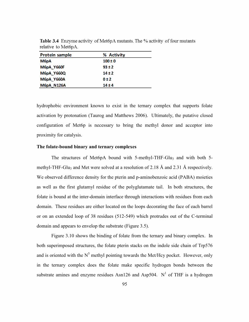
95
hydrophobic environment known to exist in the ternary complex that supports folate
activation by protonation (Taurog and Matthews 2006). Ultimately, the putative closed
configuration of Met6p is necessary to bring the methyl donor and acceptor into
proximity for catalysis.
The folate-bound binary and ternary complexes
The structures of Met6pA bound with 5-methyl-THF-Glu3 and with both 5-
methyl-THF-Glu3 and Met were solved at a resolution of 2.18 Å and 2.31 Å respectively.
We observed difference density for the pterin and p-aminobenzoic acid (PABA) moieties
as well as the first glutamyl residue of the polyglutamate tail. In both structures, the
folate is bound at the inter-domain interface through interactions with residues from each
domain. These residues are either located on the loops decorating the face of each barrel
or on an extended loop of 38 residues (512-549) which protrudes out of the C-terminal
domain and appears to envelop the substrate (Figure 3.5).
Figure 3.10 shows the binding of folate from the ternary and binary complex. In
both superimposed structures, the folate pterin stacks on the indole side chain of Trp576
and is oriented with the N5 methyl pointing towards the Met/Hcy pocket. However, only
in the ternary complex does the folate make specific hydrogen bonds between the
substrate amines and enzyme residues Asn126 and Asp504. N1 of THF is a hydrogen

96
bond acceptor; it receives a bond from the Nδ2 of Asn126. N8 of THF is a hydrogen bond
donor and it donates to Oδ1 of Asn126. The acceptor/donor pair of Asn126 matches that
of the reduced substrate. Asp504 accepts a hydrogen bond from both the N3 and the
exocyclic amine of THF (Oefner, D'Arcy et al. 1988, Davies, Delcamp et al. 1990). In
the binary structure, the pterin ring stacks on the side-chain of Trp576, but is rotated
slightly and the pattern of hydrogen bonds is disrupted. No bonds are formed directly to
Asn126, although a water molecule bridges between it and N8 of THF. The N
2

97
(exocyclic) amine of THF donates one hydrogen bond to the backbone carbonyl of
Trp576. The side chain of Asp504 is oriented toward the substrate amines but remains
too far to form a hydrogen bond.
We believe the folate binding described above for the ternary complex reflects the
interactions of a productive substrate complex. In particular, residues involved in making
specific hydrogen bonding interactions were shown to be important by mutagenesis. We
mutated Asn126 to Ala and found that the mutant protein had negligible enzyme activity
(Table 3.4). Site directed mutagenesis shows Asp504 is essential for activity and
converting it to Ala reduced catalysis by 25-fold (Prasannan, Suliman et al. 2009). The
differences between the binary and ternary complex suggest that the hydrophobic
stacking interaction is the primary binding force for folate binding, but it is non-
directional. The specific hydrogen bonding interactions are not particularly strong and
can be cancelled by other subtle forces that are difficult to quantify.
In both binary and ternary complexes, the PABA moiety of THF appears to make
an edge to face hydrophobic interactions with the side-chain of Tyr527; the Tyr side
chain swings into this position upon substrate binding, but the orientation suggests it is a
rather weak interaction compared to an ideal aromatic edge to face bonding (Burley and
Petsko 1985). A key interaction is the formation of a short ion pair between the α-
carboxylate of the first glutamate and the side chains of Lys19; interaction with the side
chain of Arg530 is observed in some, but not all folate complexes. In addition the α-
amino nitrogen donates a hydrogen bonds to the carbonyl of Tyr531. These interactions
are specific to the first glutamate residue and are invariant, independent of the number of
glutamates in the tail. The side chain carboxylate of the first glutamate does not make
any specific interactions with this open form of the protein structure. This is the side to

98
which additional glutamates would be condensed in a poly-glutamate tail, but we have
not observed any density for these longer tails in the open Met6p conformation.
Methotrexate-Glutamate3 and Hcy ternary complex
Methotrexate is a well-known anti-folate, frequently used in the treatment of
cancer. It is thought to function by binding to the folate site of dihydrofolate reductase
and interfering with nucleotide synthesis. Structurally, it closely resembles 5-methyl-
THF-Glu3; however the pterin ring is aromatic and flat, and the drug has two exocyclic
amines at positions 2 and 4. We carried out kinetic measurements that show MTX-Glu3
inhibits Met6p activity, and soaked both MTX-Glu3 and Hcy into Met6pA crystals to
form a ternary complex which diffracts to 1.75 Å.
MTX-Glu3 binds to the folate site of Met6pA and makes generally similar
contacts observed with 5-methyl-THF-Glu3. We solved several complexes with MTX-
Glu3, and each showed electron density that was consistent with a mixture of states
(Figure 3.11). In the predominant mode, the fully aromatic pterin stacks against the
Trp576 side chain. Asn126 does not hydrogen bond to the oxidized pteridine ring since
the donor/acceptor pattern is disrupted. However, Oδ2 of Asp504 receives a hydrogen
bond from the exocyclic N4 and the backbone carbonyl of Trp576 receives a hydrogen
bond from the exocyclic N2. These interactions are possible because the oxidized
pteridine is positioned closer to Trp576 and Asp504. Only the natural, reduced, substrate
induces Asp126 to rotate and Asp504 to move into position to make their specific
interactions. In the second MTX-Glu3 configuration, the pteridine ring is flipped. It still
stacks over Trp576, but is rotated so that N2 forms a hydrogen bond with the backbone
carbonyl of Trp576 and N4 donates a hydrogen bond to a neighboring water molecule.
Asp504 is too far away to make a useful hydrogen bond. In both orientations, the

99
benzene ring of the PABA moiety makes a face to edge interaction with Tyr527, and the
first glutamate retains the hydrogen bond and ion pair with Tyr531 and Lys19,
respectively.
Enzyme inhibition by MTX-Glu3 was tested using the fluorescence activity assay.
The dose-response curve in Figure 3.12 shows that MTX-Glu3 inhibits with an IC50 of 4 ±
1.4 mM and has a Hill coefficient of 1.4 ± 0.1. The kinetic data is consistent with the
observed 1:1 binding stoichiometry to the enzyme. The IC50 is modest, but consistent
with a freely rotating and weakly bound pteridine moiety. MTX-Glu3 is one of the many

100
available anti-folates, which can now be screened by crystallography and an activity
assay. The only other anti-folates that bind and inhibit catalysis were first identified in
the bacterial homolog MetE, and at minimum required an oxidized or reduced pterin
connected by a PABA moiety to two glutamates (Whitfield and Weissbach 1970).
S-adenosyl homocysteine (SAH) bound Met6pA
So far, the various substrate, product, and inhibitor-bound or free Met6pA
structures have been crystallized in an ‘open’ configuration. In this form, Met6p has two
distinct substrate binding pockets which are separated by ~7 Å. The two pockets and the

101
region in between could potentially be spanned by a single, Met6p specific bi-dentate
analog. Prior to entering the rigorous process of design and synthesis, I searched the
Sigma-Aldrich website (among other vendors) for a bi-substrate analog with an amino
acid backbone, a linker, and a heterocyclic ring. The commercially available S-adenosyl
homocysteine (SAH) was a potential candidate with a free amino acid backbone of Hcy
linked via a ribose sugar to an adenine base (Figure 1.9). The overall length of
SAH was ideal and could place the amino acid and adenine in the appropriate substrate
pockets, but the bulky sugar linker did raise some steric concerns.
The rationale behind using SAH is based on the ability of the C-terminal barrel to
recognize and strongly interact with an amino acid backbone, and the ability of the folate
pocket to accommodate simple aromatic or reduced heterocyclic rings. In a Met6pA
crystal soaked with SAH, we observed excellent Fo-Fc density that spanned both
substrate pockets (Figure 3.13). The crystallographic statistics are shown in Table 3.5.
An entire molecule of SAH was successfully modeled in this ‘open’ Met6p structure
(Figure 3.13). As expected, the amino acid backbone is the only component of SAH to
make specific hydrogen and ionic interactions. The amino group is secured through ionic
interactions with Glu499 and Asp614, and hydrogen bonds with the backbone carbonyl
of Ile446. The carboxylate forms hydrogen bonds with the backbone amine and side
chain hydroxyl of Ser448.
The rest of SAH, including the ribose sugar and adenine ring form non-specific
hydrophobic or stacking interactions. The space between the Hcy/Met pocket and the
folate site is mostly solvent exposed except for a few hydrophobic residues which line the
top of the C-terminal barrel. This hydrophobic patch of residues includes Met505,
Pro616 and Trp576. The side-chains of Met505 and Pro616 form hydrophobic
interactions with the side chain of Hcy and the ribose sugar. The face of the sugar ring

102
packs against the hydrophobic patch while the 2’ and 3’ hydroxyls are rotated away in the
direction of solvent. The adenine base forms non-specific stacking interactions with the
side chain of Trp576, reminiscent of the interactions made by folates. The exocyclic and
endocyclic amines at position C6 and N
1, respectively, form hydrogen bonds with a
neighboring water molecule.
SAH makes specific or non-specific interactions with residues located exclusively
in the C-terminal barrel and this triggers conformational changes in the barrel in the same
way as Hcy, Gln and Met. An overlay of the apo and the four ligand bound structures is
shown in figure 3.14; the four ligands align in the same pocket and make the same
contacts via the amino acid backbones. The amino acid side chain of SAH contains a
thioether, like Met, instead of the free thiol found in the natural substrate (Hcy). As a
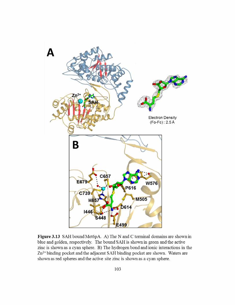
103

104
result, the zinc coordination geometry in the SAH-replete structure closely resembles the
geometry observed in the Met and Gln bound structures. That is, the Zn2+
ion moves
toward the ligand, breaking the interaction with Glu679 and forming a new bond with
water. Zinc coordinates with His657, Cys659, Cys739, and the water with bond lengths
of 2.15 Å, 2.04 Å, 2.39 Å, and 2.09 Å, respectively. SAH binding pulls the Zn2+
2.8 Å
from the apo position, compared to 2.1 Å for the thioether in the Met complex. The SAH
induced displacement places the Zn2+
in between the metal positions for the Met and Hcy

105
complexes. The βα loops which surround the Zn2+
coordination site are also mostly
ordered in the SAH bound structure.
SAH is the first bi-substrate analog to successfully bind both substrate pockets
and will be used as a template to design and synthesize highly specific and potent Met6p
inhibitors. One of the most important features of SAH is the amino acid backbone; it
anchors the SAH into the Hcy site and allows the adenine to form a stable but non-
specific stacking interaction with Trp576. In the solvent exposed folate pocket, the side
chain of Trp576 behaves as a hydrophobic anchor, and is used to attract and non-
specifically bind a variety of pterins. Residues Asp504 and Asn126 correctly orient the
natural substrate 5-methyl-THF-Glu3 and could also be recruited by the right substituted
heterocyclic ring. The folate pocket is lined with a few more hydrophobic (Tyr527) and
basic (Arg530, Lys19) residues which help bind the PABA moiety and polyglutamate
tail, and could also be exploited by the right Met6p inhibitors.
Development of a fluorescence-based Met6p activity assay
The enzyme activity of the wild-type or any mutant Met6p can be evaluated by
using the existing absorbance assay; however this detection method cannot be used to test
inhibitors that resemble anti-folates like methotrexate. The absorbance based method
was introduced by Drummond and Matthews in 1995 as an alternative to the existing
radioactive assay (Drummond, Jarrett et al. 1995). This group introduced a chemical and
heat treatment step to stop the enzymatic reaction and convert the product THF-Glu3 into
methenyl-THF-Glu3, which absorbs at a wavelength of 350 nm. Many available anti-
folates also absorb near 350 nm. The natural substrate, 5-methyl-THF-Glu3 does not get
derivatized or absorb at this wavelength and generally produces a signal in the range of
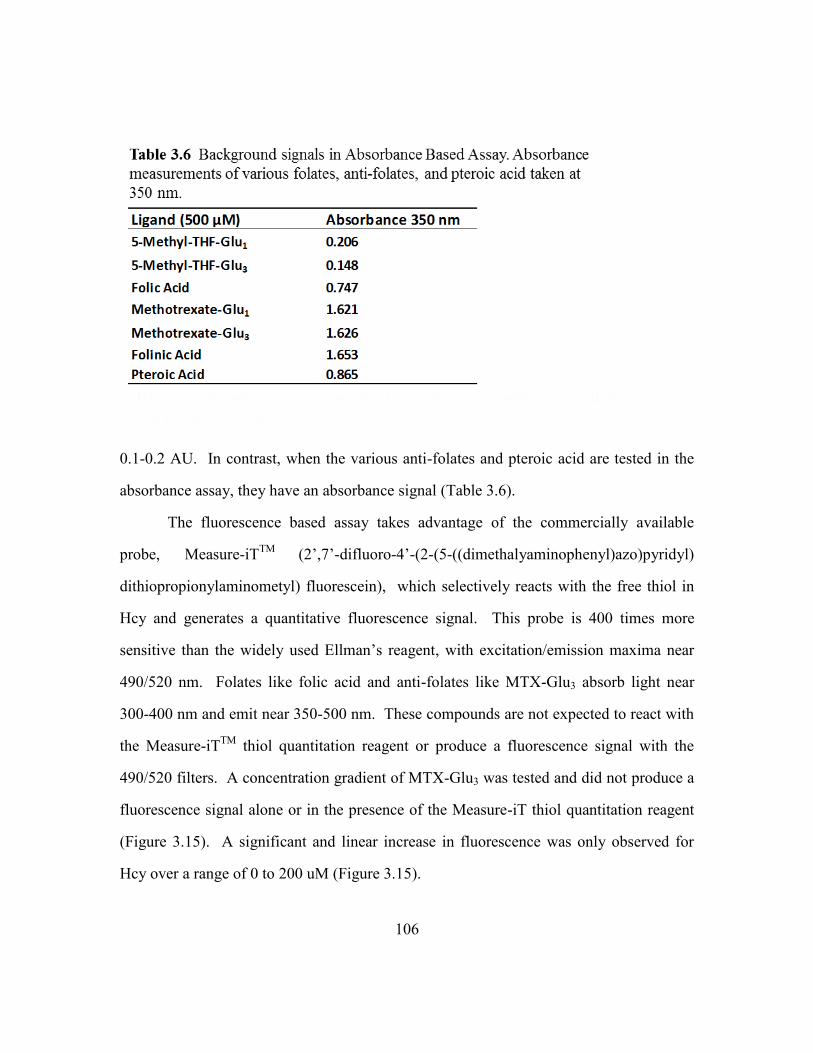
106
0.1-0.2 AU. In contrast, when the various anti-folates and pteroic acid are tested in the
absorbance assay, they have an absorbance signal (Table 3.6).
The fluorescence based assay takes advantage of the commercially available
probe, Measure-iTTM
(2’,7’-difluoro-4’-(2-(5-((dimethalyaminophenyl)azo)pyridyl)
dithiopropionylaminometyl) fluorescein), which selectively reacts with the free thiol in
Hcy and generates a quantitative fluorescence signal. This probe is 400 times more
sensitive than the widely used Ellman’s reagent, with excitation/emission maxima near
490/520 nm. Folates like folic acid and anti-folates like MTX-Glu3 absorb light near
300-400 nm and emit near 350-500 nm. These compounds are not expected to react with
the Measure-iTTM
thiol quantitation reagent or produce a fluorescence signal with the
490/520 filters. A concentration gradient of MTX-Glu3 was tested and did not produce a
fluorescence signal alone or in the presence of the Measure-iT thiol quantitation reagent
(Figure 3.15). A significant and linear increase in fluorescence was only observed for
Hcy over a range of 0 to 200 uM (Figure 3.15).

107

108
The fluorescence assay was developed by slightly modifying the reaction
conditions used in the absorbance assay and completely replacing the heat and chemical
treatment steps previously required for methenyl-THF-Glu3 synthesis. Overall, the
enzyme reaction conditions remain the same, but all reducing agents were omitted
because the thiol quantitation reagent reacts with DTT, BME and TCEP. Instead, all
reagents and samples are flushed with N2 gas and when necessary protected from light
and/or air. The final reaction volume is also scaled down from the 380 µL used in the
absorbance assay to 10 µL. The reaction is stopped by an incubation step in ice and the
substrate Hcy is quantified by adding saturating concentrations of the Measure-iTTM
reagent. The reaction between the free thiol and the reagent is proprietary. This reaction
goes to completion in 2-3 minutes at room temperature. Fluorescence measurements are
taken using the Roche LightCycler® 480 RT-PCR instrument using the available
excitation/emission filters of 464/510 nm.
Once the enzyme reaction and detection steps were successfully combined, steady
state kinetics experiments were done to determine the substrate KMs and the kcat. The
steady state concentration of the enzyme (110 nm) was determined from an activity vs.
protein concentration plot shown in Figure 3.16. I carried out steady state kinetics and
measured the KM of 25 ± 16 μM and kcat of 25 ± 5/min for Hcy, and the KM of 87 ± 31
uM and kcat of 29 ± 2/min for 5-methyl-THF-Glu3. The kcat and KM parameters for each
substrate closely resemble the values previously reported by the absorbance assay (Figure
3.17).
The steady state parameters were used to establish a standard, single time point
activity assay which can be used to efficiently test the wild-type enzyme, and to evaluate
any Met6p mutants. We tested a few interesting mutants located in the folate pocket
(Asn126Ala) and at the inter-domain interface (Tyr660Ala, Tyr660Asn, Tyr660Phe). A

109
few of these mutants, including Asn126Ala, Tyr660Ala and Tyr660Asn had no
detectable activity in the standard assay. The standard assay contains saturating substrate
and steady state protein concentrations. Although the initial results clearly support an
essential role of Tyr660 and Asn126, we also tested and reported the enzyme activities at
saturating enzyme concentrations (Table 3.4). The standard assay was also used to test
the activity of the wild-type Met6p in the presence of the methyl source 5-methyl-THF-
Glu1, or with a protein pre-incubation step of 5 minutes with 500 µM EDTA. Zero
enzyme activity was observed with either test condition (results not shown).
An inhibition study with the anti-folate MTX-Glu3 was also done using a slightly
modified standard assay. We reduced the substrate concentrations to 2x the KM values in

110
order to give competitive inhibitors a chance to bind. Data from a 20 minute time course
experiment with S = 2x[KM] and steady state protein concentration is shown in figure
3.18. The raw fluorescence readings for each sample (Hcy, reaction background, and
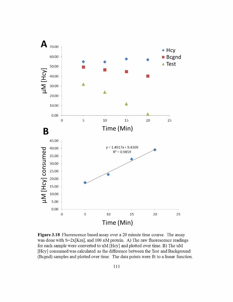
111

112
test) were converted to µM Hcy and plotted against time. The µM Hcy consumed at each
time point was determined by calculating the difference between the background, or
“blank rate” and the test sample. The data points from the entire time course were fit to a
linear function with an R2 of 1. This experiment was repeated at a single time point of 10
minutes in the presence of 0-20 mM MTX-Glu3 and the resulting dose-response curve is
shown in Figure 3.12.
The Development of a high-throughput DSF assay
Differential scanning fluorimetry (DSF) is a popular biophysical method which
can be used to screen ligands that bind and stabilize protein (Niesen, Berglund et al.
2007). This method is inexpensive, high-throughput, and requires low quantities of
protein, a special fluorescent dye, and an RT-PCR instrument. The protein is heated in
the presence of a fluorescent dye, and as the protein unfolds, the dye binds to the exposed
hydrophobic regions and fluoresces. The instrument is used to control the temperature
gradient (usually 25 °C to 100 °C), and as the protein melts, the instrument records the
fluorescence vs. temperature protein melting curve. The inflection point in this curve is
the Tm, or the temperature at which half the protein population is denatured. In the
presence of a stabilizing ligand the Tm is expected to increase, generating a positive Tm
shift.
A high-throughput DSF method was successfully developed (described in
materials and methods) for the fungal Met6p enzyme. The final 25 µL reaction includes
750 nM protein, 5X Sypro Orange dye, and ≤ 2% DMSO buffered by 20 mM Hepes, 150
mM NaCl, pH 7.5. The Tm for Met6p alone (negative control) was 71 ± 0.07 °C. In the
presence of Hcy (positive control), a Tm shift of 1.5 ± 0.07 °C was observed. The
product Met and substrate 5-methyl-THF-Glu3 shift the Tm by 0.7 °C ± 0.07 °C and 0.8 ±

113
0.05 °C, respectively. The inhibitor MTX-Glu3 shifts the Tm by 0.7 °C ± 0.02 °C. The
melting curves for the negative and positive controls are shown in Figure 3.19. The
standard deviation for the negative and positive control was consistently ≤ 0.07 °C
(across 96 wells) and any compound which could bind and shift the Tm by at least 0.3 °C
(3 times the standard deviation) was considered a hit.
Three distinct libraries were screened using DSF in order to identify compounds
(tested at 100 µM final concentration), which bound and increased the Tm of Met6p. The
entire NIH clinical collection of 731 compounds consists of FDA approved drug-like
molecules which are well characterized, soluble and commercially available. The second
library from Chembridge consists of 7,000 small fragments (MW < 300) which are
highly soluble, pure and have excellent physicochemical properties. The third set of
11,000 molecules from Chembridge is a kinase inhibitor-biased library called KINASet.
This library is enriched with ligands containing heterocyclic rings.
Out of the 731 compounds in the NIH clinical collection (NCC), 26 were found to
increase the protein Tm by ≥ 0.3 °C. False positives were removed by retesting freshly
purchased compounds and reduced the number of hits to just 13. These hits were further
validated using the absorbance based enzyme activity assay at a single inhibitor
concentration of 500 µM. Only 3 molecules inhibited the enzyme and reduced the
activity from the 100% observed in wild-type to 40-60%, while two potential candidates,
Benidipine and Pterostillbene, could not be further evaluated due to their high
background signals at 350 nm. Triclabendazole could not be purchased and remains to
be tested, however, dose-response experiments were done using Triclosan, PD81723, and
Phenothiazine over the range of 0-1 mM (Figure 3.20). When possible, the data points
were fit to a 3 parameter logistic equation, or evaluated by eye in order to determine the
IC50, defined as the concentration of inhibitor required for 50% inhibition. Triclosan and
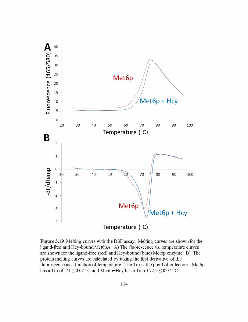
114
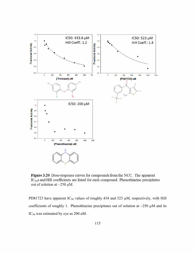
115
PD81723 have apparent IC50 values of roughly 434 and 523 µM, respectively, with Hill
coefficients of roughly 1. Phenothiazine precipitates out of solution at ~250 µM and its
IC50 was estimated by eye as 200 uM.

116
Out of the first 4,000 compounds in the Fragment library, 13 increased the protein
Tm by ≥ 0.3 °C. Ten out of the thirteen were successfully retested and shifted the Tm ≥
0.3 °C. These hits were further validated by the absorbance based activity assay in which
four compounds, 5214970, 5376760, 5303077 and 5279826, reduced the enzyme activity
to 0-30% (relative to wild-type) when tested at 500 µM. Dose-response experiments
were done for all four compounds, but the IC50 values could only be measured for two
because at higher concentrations compounds 5214970 and 5376760 absorbed at 350 nm.
The apparent IC50 values for 5303077 and 5279826 were 191 µM and 84 µM,
respectively, and Hill coefficients were roughly equal to 1 (Figure 3.21).
Out of the first 4,000 compounds in the KINAset library, 20 shifted the Tm by ≥
0.3 °C. Sixteen out of the twenty were successfully retested. These hits were further

117
validated using the absorbance based activity assay in which 12 compounds reduced the
enzyme activity to 6-63% (relative to wild-type) when tested at 500 µM. Only 10 out of
the 12 were tested further because compounds 5848343 and 6385797 could not be
purchased. Dose-response experiments were successfully done with 7 compounds which
had apparent IC50s ranging from 57-328 µM, and Hill coefficients close to 1 (Figure
3.22). The IC50 values for compounds 6215005, 7014609, and 7031899, could not be
determined due to the high background signals at 350 nm.
The DSF and the absorbance based kinetic assays were successfully used to
identify 3 potential Met6p inhibitors from the NIH clinical collection, 2 from the
fragment library, and 7 from the KINAset library. These compounds are enriched with
aromatic and/or reduced rings. Our experience with MTX-Glu3, 5-methyl-THF-Glu3,
folinic acid, folic acid, and SAH has shown that an otherwise solvent exposed folate
pocket has a tendency to form non-specific stacking or hydrophobic interactions with
heterocyclic rings. In order to understand the mode of binding, Met6pA crystals were
soaked with saturating concentrations of each compound (5 mM). With some of these
compounds, we observed patches of density near the solvent exposed side of Trp576
(folate pocket). Unfortunately, the density is consistently weak and incomplete, making
it difficult to dock and/or rationalize the mode of binding. Co-crystallization experiments
have also been done, but were unsuccessful. These compounds should be retested using
the fluorescence based activity assay and co-crystallized and/or soaked using the Met6pT
or Met6pY proteins. However, based on the drug screening, kinetic, and structural work
done so far, this strategy has failed.

118

119

120
Conclusion
Candidemia is a serious health concern which afflicts millions and results in
considerable mortality. Fungal infections are often serious medical problems and
difficult to treat due to the similarity of most human and fungal enzymes. It would be
useful to find a novel fungal target which is present in a broad range of fungi; methionine
synthase is such a target. The Candida albicans species is the most prevalent cause of
candidemia and has been the focus of previous studies which identified an essential C.
albicans gene, MET6. This gene encodes the zinc-dependent fungal enzyme cobalamin-
independent methionine synthase (Met6p) which synthesizes methionine by directly
transferring a methyl group from 5-methyl-tetrahydrofolate (5-methyl-THF) to L-
homocysteine (Hcy). This 88 kDa protein is structurally and mechanistically different
from the modular 140 kDa cobalamin-dependent methionine synthase (MetH) used by
mammals.
The overall goal of this project was to characterize the structure and action of
Met6p and to discover a variety of drug-like scaffolds. I used the SER strategy to
initially crystallize three variants of the wild-type enzyme, Met6pA, Met6pT and
Met6pY, all of which crystallized in the ‘open’ configuration and retained full enzyme
activity.
Crystals of the Met6pA variant were soaked with natural substrate, products, anti-
folates (MTX-Glu3) and substrate analogs (Gln) in order to characterize the Zn2+
, Hcy,
and 5-methyl-THF-Glu3 binding pockets. Structural studies were accompanied with site-
directed mutagenesis and mutants were tested by a novel fluorescence based activity
assay. We uncovered key roles for two active site residues, Tyr660 and Asn126, not
previously implicated in Met6p activity.

121
Other groups have previously characterized the Hcy pocket by using the natural
substrate and product, however, we are the first to capture a Gln bound Met6p and to
show that it was the binding of the amino acid backbone that triggered an ordering of
active site loops and the movement of the catalytic Zn2+
towards the substrate. Hcy, Met
and Gln all bind at the top of the C-terminal barrel through an identical amino acid
backbone and all three ligands stabilize the ‘open’ configuration by ordering the flexible
loops in the C-terminal barrel.
Other groups have previously characterized the folate pocket by using the natural
substrate and product; however, we are the first to capture a MTX-Glu3 bound Met6p and
to observe a productive ternary complex. The folate pocket was able to form non-
specific stacking interactions between the conserved side chain of Trp576 and an
oxidized or reduced pterin. However, the aromatic 2,4-diaminopteridine of MTX-Glu3
was found to easily rotate and performed as a modest inhibitor with an IC50 of ~4 mM.
On the other hand, THF with its three distinguishable endocyclic nitrogens, a carbonyl
group and an exocyclic amine made numerous specific contacts. Asn126 was especially
interesting; the acceptor/donor pair on its side-chain aligned with the donor/acceptor pair
at position N8 and N
1 in 5-methyl-THF-Glu3. The two available hydrogen donors on the
otherwise oxidized pyrimidine ring donated hydrogen bonds to the side chain of Asp504.
The extensive structural characterization has made rational drug design a feasible
strategy. It was successfully implemented in identifying the bi-substrate analog SAH.
Although SAH is not an ideal folate-Hcy analog, it contains the basic components we
initially found to be important for binding in each substrate pocket: the amino acid
backbone and a heterocyclic ring. This compound successfully bound the ‘open’
configuration of Met6p by using the amino acid backbone to anchor into the Hcy/Met
site, the Hcy side-chain and ribose sugar for hydrophobic interactions with M505 and

122
Pro616, and the aromatic adenine ring to form non-specific stacking interactions with the
conserved residue Trp576.
Thousands of compounds were screened using an optimized high-throughput DSF
assay, and a few were successfully tested by the absorbance based activity assay with
reported IC50 values in the micromolar range. However, these inhibitors have failed to
bind the Met6pA crystals by soaking or co-crystallization experiments. Since similar
methods were successfully used for substrate, MTX-Glu3, and SAH, it is possible that
compounds in the NIH clinical collection, Fragment library and KINAset library do not
bind the ‘open’ configuration. A stable ‘closed’ methionine synthase structure remains to
be seen, however, for now; the ‘open’ configuration can be successfully targeted using
rational drug design.

123
Appendix A
Abbreviations
B12 Cobalamin (Vitamin)
BHMT Betaine homocysteine methyltransferase
BME β-Mercaptoethanol
Cys CysteineDHF Dihydrofolate (Glutamate1-3)DHFR Dihdrofolate reductaseDSF Differential Scanning FluorimetryDTNB Ellman's reagent
DTT Dithiothreitol
EXFAS Extended X-ray absorption fine structureGARFT Glycinamide ribonucleotide formyltransferaseGln GlutamineHcy L-Homocysteine
HMT S-methylmethionine Hcy S-methyltransferase
Met Methionine
MetE Cobalamin-independent methionine synthase (bacteria and plants)
MetH Cobalamin-dependent methionine synthase
Met6p Cobalamin-independent methionine synthase (yeast)
Met6pA 103AlaAlaAlaThrAla
107
Met6pY 103TyrTyrAlaThrTyr
107
Met6pT 103ThrThrAlaThrThr
107
MS Methionine synthase
MTHFR 5,10-Methylenetetrahydrofolate reductase
MTX-Glu3 Methotrexate-Glutamate3
PABA Para-aminobenzoic acid
PDB Protein Data Bank
Pteroyl-Glu5 Pteroylglutamaic5 acid (Folic Acid with polyanionic tail)RMSD Root mean square deviation
SAH S-Adenosyl-L-homocysteine
SAM S-Adenosyl methionine
SER Surface Entropy Reduction
SHMT Serine hydroxymethyltransferase
SMM L-S-methylmethionine
THF Tetrahydrofolate (Glutamate1-3)Tm Protein melting temperature
TS Thymidylate synthase
Zn Zinc
5-methyl-THF-Glun 5-methyltetrahydrofolate-glutamate1-3

124
Appendix B
Materials
Material Manufacturer
Acetic Acid Fisher Scientific, Fair Lawn, NJAgarose Life technologies, Carlsbad, CAβ-Mercaptoethanol Sigma Aldrich, St. Louis, MOCentricon Filter
(concentrator 30
kDa)
Millipore, Billerica, MA
Citric Acid Sigma Aldrich, St. Louis, MODithiothreitol Sigma Aldrich, St. Louis, MODMSO Fisher Scientific, Fair Lawn, NJDpnI New England Biolabs, Ipswich, MADTNB Thermo Scientific, Rockford, ILFormaldehyde Fisher Scientific, Fair Lawn, NJFormate Fisher Scientific, Fair Lawn, NJFragment library ChemBridge, San Diego, CAGlutamine Sigma Aldrich, St. Louis, MOHCl Fisher Scientific, Fair Lawn, New JerseyHcy Thiolactone Sigma Aldrich, St. Louis, MOHepes Sigma Aldrich, St. Louis, MOImidazole Acros organics, Morris, New Jersey
IPTG Gold Biotechnology, St. Louis, MO
JCSG+ Suite Qiagen, Bethesda, MarylandKanamycin Sigma Aldrich, St. Louis, MO
KCl Fisher Scientific, Fair Lawn, New JerseyKH2PO4 Sigma Aldrich, St. Louis, MOK2HPO4 Fisher Scientific, Fair Lawn, New JerseyKINAset library ChemBridge, San Diego, CA
KOD Hot Start DNA
polymeraseNovagen, San Diego, CA
KSCN Hampton Research, Aliso Viejo, CA
Measure-iTTM thiol
quantitation reagentInvitrogen, Oregon, CA
Methionine Sigma Aldrich, St. Louis, MOMgSO4 Fisher Scientific, Fair Lawn, NJ
MTX-Glu3 Schircks Laboratories, Jona, Switzerland
Na2HPO4 EM Science, Cherry Hill, Jew JerseyNaH2PO4 EM Science, Cherry Hill, Jew Jersey
NaOH EM Science, Cherry Hill, Jew Jersey
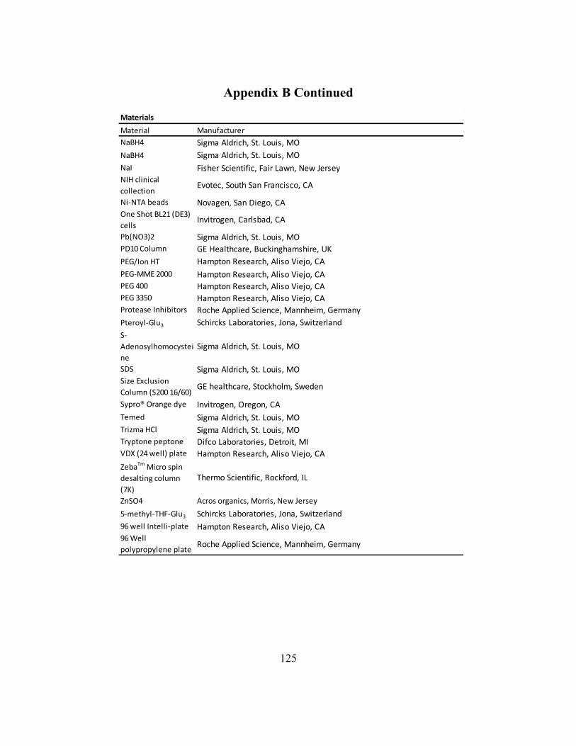
125
Appendix B Continued
Materials
Material Manufacturer
NaBH4 Sigma Aldrich, St. Louis, MO
NaBH4 Sigma Aldrich, St. Louis, MO
NaI Fisher Scientific, Fair Lawn, New JerseyNIH clinical
collectionEvotec, South San Francisco, CA
Ni-NTA beads Novagen, San Diego, CAOne Shot BL21 (DE3)
cellsInvitrogen, Carlsbad, CA
Pb(NO3)2 Sigma Aldrich, St. Louis, MOPD10 Column GE Healthcare, Buckinghamshire, UK
PEG/Ion HT Hampton Research, Aliso Viejo, CA
PEG-MME 2000 Hampton Research, Aliso Viejo, CAPEG 400 Hampton Research, Aliso Viejo, CAPEG 3350 Hampton Research, Aliso Viejo, CAProtease Inhibitors Roche Applied Science, Mannheim, Germany
Pteroyl-Glu3 Schircks Laboratories, Jona, Switzerland
S-
Adenosylhomocystei
ne
Sigma Aldrich, St. Louis, MO
SDS Sigma Aldrich, St. Louis, MOSize Exclusion
Column (S200 16/60)GE healthcare, Stockholm, Sweden
Sypro® Orange dye Invitrogen, Oregon, CA
Temed Sigma Aldrich, St. Louis, MO
Trizma HCl Sigma Aldrich, St. Louis, MOTryptone peptone Difco Laboratories, Detroit, MIVDX (24 well) plate Hampton Research, Aliso Viejo, CA
ZebaTm Micro spin
desalting column
(7K)
Thermo Scientific, Rockford, IL
ZnSO4 Acros organics, Morris, New Jersey
5-methyl-THF-Glu3 Schircks Laboratories, Jona, Switzerland
96 well Intelli-plate Hampton Research, Aliso Viejo, CA96 Well
polypropylene plateRoche Applied Science, Mannheim, Germany

126
References
Adams, P. D., P. V. Afonine, G. Bunkoczi, V. B. Chen, I. W. Davis, N. Echols, J. J.
Headd, L. W. Hung, G. J. Kapral, R. W. Grosse-Kunstleve, A. J. McCoy, N. W.
Moriarty, R. Oeffner, R. J. Read, D. C. Richardson, J. S. Richardson, T. C.
Terwilliger and P. H. Zwart (2010). "PHENIX: a comprehensive Python-based
system for macromolecular structure solution." Acta Crystallogr D Biol
Crystallogr 66(Pt 2): 213-221.
Alberts, I. L., K. Nadassy and S. J. Wodak (1998). "Analysis of zinc binding sites in
protein crystal structures." Protein Sci 7(8): 1700-1716.
Andreini, C., I. Bertini and G. Cavallaro (2011). "Minimal functional sites allow a
classification of zinc sites in proteins." PLoS One 6(10): e26325.
Appling, D. R. (1991). "Compartmentation of folate-mediated one-carbon metabolism in
eukaryotes." FASEB J 5(12): 2645-2651.
Auld, D. S. (2001). "Zinc coordination sphere in biochemical zinc sites." Biometals 14(3-
4): 271-313.
Baker, P. J., K. L. Britton, M. Fisher, J. Esclapez, C. Pire, M. J. Bonete, J. Ferrer and D.
W. Rice (2009). "Active site dynamics in the zinc-dependent medium chain
alcohol dehydrogenase superfamily." Proc Natl Acad Sci U S A 106(3): 779-784.
Banerjee, R. (1997). "The Yin-Yang of cobalamin biochemistry." Chem Biol 4(3): 175-
186.
Banerjee, R. V., V. Frasca, D. P. Ballou and R. G. Matthews (1990). "Participation of
cob(I) alamin in the reaction catalyzed by methionine synthase from Escherichia
coli: a steady-state and rapid reaction kinetic analysis." Biochemistry 29(50):
11101-11109.
Banerjee, R. V. and R. G. Matthews (1990). "Cobalamin-dependent methionine
synthase." FASEB J 4(5): 1450-1459.
Basavanna, S., S. Chimalapati, A. Maqbool, B. Rubbo, J. Yuste, R. J. Wilson, A. Hosie,
A. D. Ogunniyi, J. C. Paton, G. Thomas and J. S. Brown (2013). "The effects of
methionine acquisition and synthesis on Streptococcus pneumoniae growth and
virulence." PLoS One 8(1): e49638.
Basu, P. and S. J. Burgmayer (2011). "Pterin chemistry and its relationship to the
molybdenum cofactor." Coord Chem Rev 255(9-10): 1016-1038.
Blakley, R. L. (1969). The Biochemistry of Folic Acid and Related Pteridines, North-
Holland

127
Brand, U., M. Rombach, J. Seebacher and H. Vahrenkamp (2001). "Functional modeling
of cobalamine-independent methionine synthase with pyrazolylborate-zinc-
thiolate complexes." Inorg Chem 40(24): 6151-6157.
Brosnan, J. T. and M. E. Brosnan (2006). "The sulfur-containing amino acids: an
overview." J Nutr 136(6 Suppl): 1636S-1640S.
Burley, S. K. and G. A. Petsko (1985). "Aromatic-aromatic interaction: a mechanism of
protein structure stabilization." Science 229(4708): 23-28.
Castro, C., A. A. Gratson, J. C. Evans, J. Jiracek, M. Collinsova, M. L. Ludwig and T. A.
Garrow (2004). "Dissecting the catalytic mechanism of betaine-homocysteine S-
methyltransferase by use of intrinsic tryptophan fluorescence and site-directed
mutagenesis." Biochemistry 43(18): 5341-5351.
Cathou, R. E. and J. M. Buchanan (1963). "Enzymatic synthesis of the methyl group of
methionine. V. Studies with 5, 10-methylenetetrahydrofolate reductase from
Escherichia coli." J Biol Chem 238: 1746-1751.
Chabner, B. A., C. J. Allegra, G. A. Curt, N. J. Clendeninn, J. Baram, S. Koizumi, J. C.
Drake and J. Jolivet (1985). "Polyglutamation of methotrexate. Is methotrexate a
prodrug?" J Clin Invest 76(3): 907-912.
Collaborative Computational Project, N. (1994). "The CCP4 suite: programs for protein
crystallography." Acta Crystallogr D Biol Crystallogr 50(Pt 5): 760-763.
Cooper, D. R., T. Boczek, K. Grelewska, M. Pinkowska, M. Sikorska, M. Zawadzki and
Z. Derewenda (2007). "Protein crystallization by surface entropy reduction:
optimization of the SER strategy." Acta Crystallogr D Biol Crystallogr 63(Pt 5):
636-645.
Copeland, R. A. (2005). Evaluation of Enzyme Inhibitors in Drug Discovery, John Wiley
& Sons.
Coyle, P., J. C. Philcox, L. C. Carey and A. M. Rofe (2002). "Metallothionein: the
multipurpose protein." Cell Mol Life Sci 59(4): 627-647.
Das, I., P. Nightingale, M. Patel and P. Jumaa (2011). "Epidemiology, clinical
characteristics, and outcome of candidemia: experience in a tertiary referral center
in the UK." Int J Infect Dis 15(11): e759-763.
Daubner, S. C., J. L. Schrimsher, F. J. Schendel, M. Young, S. Henikoff, D. Patterson, J.
Stubbe and S. J. Benkovic (1985). "A multifunctional protein possessing
glycinamide ribonucleotide synthetase, glycinamide ribonucleotide
transformylase, and aminoimidazole ribonucleotide synthetase activities in de
novo purine biosynthesis." Biochemistry 24(25): 7059-7062.
Davies, J. F., 2nd, T. J. Delcamp, N. J. Prendergast, V. A. Ashford, J. H. Freisheim and J.
Kraut (1990). "Crystal structures of recombinant human dihydrofolate reductase
complexed with folate and 5-deazafolate." Biochemistry 29(40): 9467-9479.

128
Dixon, M. M., S. Huang, R. G. Matthews and M. Ludwig (1996). "The structure of the C-
terminal domain of methionine synthase: presenting S-adenosylmethionine for
reductive methylation of B12." Structure 4(11): 1263-1275.
Drennan, C. L., S. Huang, J. T. Drummond, R. G. Matthews and M. L. Lidwig (1994).
"How a protein binds B12: A 3.0 A X-ray structure of B12-binding domains of
methionine synthase." Science 266(5191): 1669-1674.
Drennan, C. L., R. G. Matthews and M. L. Ludwig (1994). "Cobalamin-dependent
methionine synthase: the structure of a methylcobalamin-binding fragment and
implications for other B12-dependent enzymes." Curr Opin Struct Biol 4(6): 919-
929.
Drummond, J. T., J. Jarrett, J. C. Gonzalez, S. Huang and R. G. Matthews (1995).
"Characterization of nonradioactive assays for cobalamin-dependent and
cobalamin-independent methionine synthase enzymes." Anal Biochem 228(2):
323-329.
Eadsforth, T. C., S. Cameron and W. N. Hunter (2012). "The crystal structure of
Leishmania major N(5),N(10)-methylenetetrahydrofolate
dehydrogenase/cyclohydrolase and assessment of a potential drug target." Mol
Biochem Parasitol 181(2): 178-185.
Eckermann, C., J. Eichel and J. Schroder (2000). "Plant methionine synthase: new
insights into properties and expression." Biol Chem 381(8): 695-703.
Edmond, M. B., S. E. Wallace, D. K. McClish, M. A. Pfaller, R. N. Jones and R. P.
Wenzel (1999). "Nosocomial bloodstream infections in United States hospitals: a
three-year analysis." Clin Infect Dis 29(2): 239-244.
Eichel, J., J. C. Gonzalez, M. Hotze, R. G. Matthews and J. Schroder (1995). "Vitamin-
B12-independent methionine synthase from a higher plant (Catharanthus roseus).
Molecular characterization, regulation, heterologous expression, and enzyme
properties." Eur J Biochem 230(3): 1053-1058.
Ellman, G. L. (1959). "Tissue sulfhydryl groups." Arch Biochem Biophys 82(1): 70-77.
Emsley, P. and K. Cowtan (2004). "Coot: model-building tools for molecular graphics."
Acta Crystallogr D Biol Crystallogr 60(Pt 12 Pt 1): 2126-2132.
Evans, J. C., D. P. Huddler, M. T. Hilgers, G. Romanchuk, R. G. Matthews and M. L.
Ludwig (2004). "Structures of the N-terminal modules imply large domain
motions during catalysis by methionine synthase." Proc Natl Acad Sci U S A
101(11): 3729-3736.
Evans, J. C., D. P. Huddler, J. Jiracek, C. Castro, N. S. Millian, T. A. Garrow and M. L.
Ludwig (2002). "Betaine-homocysteine methyltransferase: zinc in a distorted
barrel." Structure 10(9): 1159-1171.

129
Falagas, M. E., N. Roussos and K. Z. Vardakas (2010). "Relative frequency of albicans
and the various non-albicans Candida spp among candidemia isolates from
inpatients in various parts of the world: a systematic review." Int J Infect Dis
14(11): e954-966.
Ferrer, J. L., S. Ravanel, M. Robert and R. Dumas (2004). "Crystal structures of
cobalamin-independent methionine synthase complexed with zinc, homocysteine,
and methyltetrahydrofolate." J Biol Chem 279(43): 44235-44238.
Finer-Moore, J. S., D. V. Santi and R. M. Stroud (2003). "Lessons and conclusions from
dissecting the mechanism of a bisubstrate enzyme: thymidylate synthase
mutagenesis, function, and structure." Biochemistry 42(2): 248-256.
Fontecave, M., M. Atta and E. Mulliez (2004). "S-adenosylmethionine: nothing goes to
waste." Trends Biochem Sci 29(5): 243-249.
Fontecilla-Camps, J. C., Bugg, C.E., Temple, C., Rose, J.D., Montgomery, J.A., Kisliuk,
R.L. (1979). "Absolute configuration of biological tetrahydrofolates. A
crystallographic determination." Journal of the American Chemical Society
101(20): 6114-6115.
Fu, T. M., J. Almqvist, Y. H. Liang, L. Li, Y. Huang and X. D. Su (2011). "Crystal
structures of cobalamin-independent methionine synthase (MetE) from
Streptococcus mutans: a dynamic zinc-inversion model." J Mol Biol 412(4): 688-
697.
Fujita, Y., E. Ukena, H. Iefuji, Y. Giga-Hama and K. Takegawa (2006). "Homocysteine
accumulation causes a defect in purine biosynthesis: further characterization of
Schizosaccharomyces pombe methionine auxotrophs." Microbiology 152(Pt 2):
397-404.
Garg, D., S. Henrich, O. M. Salo-Ahen, H. Myllykallio, M. P. Costi and R. C. Wade
(2010). "Novel approaches for targeting thymidylate synthase to overcome the
resistance and toxicity of anticancer drugs." J Med Chem 53(18): 6539-6549.
Gillespie, R. J., Robinson, E.A. (1998). Advances in Molecular Structure Research, JAI
Press LTD.
Goldschmidt, L., D. R. Cooper, Z. S. Derewenda and D. Eisenberg (2007). "Toward
rational protein crystallization: A Web server for the design of crystallizable
protein variants." Protein Sci 16(8): 1569-1576.
Gonzalez, B., M. A. Pajares, M. Martinez-Ripoll, T. L. Blundell and J. Sanz-Aparicio
(2004). "Crystal structure of rat liver betaine homocysteine s-methyltransferase
reveals new oligomerization features and conformational changes upon substrate
binding." J Mol Biol 338(4): 771-782.
Gonzalez, J. C., R. V. Banerjee, S. Huang, J. S. Sumner and R. G. Matthews (1992).
"Comparison of cobalamin-independent and cobalamin-dependent methionine

130
synthases from Escherichia coli: two solutions to the same chemical problem."
Biochemistry 31(26): 6045-6056.
Gonzalez, J. C., K. Peariso, J. E. Penner-Hahn and R. G. Matthews (1996). "Cobalamin-
independent methionine synthase from Escherichia coli: a zinc metalloenzyme."
Biochemistry 35(38): 12228-12234.
Goulding, C. W., D. Postigo and R. G. Matthews (1997). "Cobalamin-dependent
methionine synthase is a modular protein with distinct regions for binding
homocysteine, methyltetrahydrofolate, cobalamin, and adenosylmethionine."
Biochemistry 36(26): 8082-8091.
Gruber, K. and C. Kratky (2001). Cobalamin-Dependent Methionine Synthase.
Handbook of Metalloproteins., John Wiley & Sons, Ltd.
Hall, D. A., T. C. Jordan-Starck, R. O. Loo, M. L. Ludwig and R. G. Matthews (2000).
"Interaction of flavodoxin with cobalamin-dependent methionine synthase."
Biochemistry 39(35): 10711-10719.
Hatanaka, H., N. Ariga, J. Nagai and H. Katsuki (1974). "Accumulation of a sterol
intermediate during reaction in the presence of homocysteine with cell-free
extract of yeast." Biochem Biophys Res Commun 60(2): 787-793.
Hatzios, S. K. and C. R. Bertozzi (2011). "The regulation of sulfur metabolism in
Mycobacterium tuberculosis." PLoS Pathog 7(7): e1002036.
Herbig, K., E. P. Chiang, L. R. Lee, J. Hills, B. Shane and P. J. Stover (2002).
"Cytoplasmic serine hydroxymethyltransferase mediates competition between
folate-dependent deoxyribonucleotide and S-adenosylmethionine biosyntheses." J
Biol Chem 277(41): 38381-38389.
Ho, T. L., H. C. Ho and L. D. Hamilton (1978). "Biochemical significance of the hard
and soft acids and bases principle." Chem Biol Interact 23(1): 65-84.
Holmes, W. B. and D. R. Appling (2002). "Cloning and characterization of
methenyltetrahydrofolate synthetase from Saccharomyces cerevisiae." J Biol
Chem 277(23): 20205-20213.
Huffnagle, G. B. and M. C. Noverr (2013). "The emerging world of the fungal
microbiome." Trends Microbiol 21(7): 334-341.
Jakubowski, H. (1999). "Protein homocysteinylation: possible mechanism underlying
pathological consequences of elevated homocysteine levels." FASEB J 13(15):
2277-2283.
Kacprzak, M. M., I. Lewandowska, R. G. Matthews and A. Paszewski (2003).
"Transcriptional regulation of methionine synthase by homocysteine and choline
in Aspergillus nidulans." Biochem J 376(Pt 2): 517-524.

131
Kallen, R. G. and W. P. Jencks (1966). "The dissociation constants of tetrahydrofolic
acid." J Biol Chem 241(24): 5845-5850.
Kaur, S. and P. Mishra (1991). "Amino acid uptake as a function of differentiation in
Candida albicans: studies of a non-germinative variant." FEMS Microbiol Lett
66(3): 341-344.
Kim, B. S., Y. S. Jeon and J. Chun (2013). "Current Status and Future Promise of the
Human Microbiome." Pediatr Gastroenterol Hepatol Nutr 16(2): 71-79.
Kim, Y. G. and S. Maas (2000). "Multiple site mutagenesis with high targeting efficiency
in one cloning step." Biotechniques 28(2): 196-198.
Kleifeld, O., A. Frenkel, J. M. Martin and I. Sagi (2003). "Active site electronic structure
and dynamics during metalloenzyme catalysis." Nat Struct Biol 10(2): 98-103.
Kohls, D., T. Sulea, E. O. Purisima, R. E. MacKenzie and A. Vrielink (2000). "The
crystal structure of the formiminotransferase domain of formiminotransferase-
cyclodeaminase: implications for substrate channeling in a bifunctional enzyme."
Structure 8(1): 35-46.
Koutmos, M., S. Datta, K. A. Pattridge, J. L. Smith and R. G. Matthews (2009). "Insights
into the reactivation of cobalamin-dependent methionine synthase." Proc Natl
Acad Sci U S A 106(44): 18527-18532.
Koutmos, M., R. Pejchal, T. M. Bomer, R. G. Matthews, J. L. Smith and M. L. Ludwig
(2008). "Metal active site elasticity linked to activation of homocysteine in
methionine synthases." Proc Natl Acad Sci U S A 105(9): 3286-3291.
Krcmery, V. and A. J. Barnes (2002). "Non-albicans Candida spp. causing fungaemia:
pathogenicity and antifungal resistance." J Hosp Infect 50(4): 243-260.
Krissinel, E. and K. Henrick (2004). "Secondary-structure matching (SSM), a new tool
for fast protein structure alignment in three dimensions." Acta Crystallogr D Biol
Crystallogr 60(Pt 12 Pt 1): 2256-2268.
Krissinel, E. and K. Henrick (2007). "Inference of macromolecular assemblies from
crystalline state." J Mol Biol 372(3): 774-797.
Krupenko, S. A. (2009). "FDH: an aldehyde dehydrogenase fusion enzyme in folate
metabolism." Chem Biol Interact 178(1-3): 84-93.
Kwon, K., C. Cao and J. T. Stivers (2003). "A novel zinc snap motif conveys structural
stability to 3-methyladenine DNA glycosylase I." J Biol Chem 278(21): 19442-
19446.
Lai, C. C., C. K. Tan, Y. T. Huang, P. L. Shao and P. R. Hsueh (2008). "Current
challenges in the management of invasive fungal infections." J Infect Chemother
14(2): 77-85.

132
Laity, J. H., B. M. Lee and P. E. Wright (2001). "Zinc finger proteins: new insights into
structural and functional diversity." Curr Opin Struct Biol 11(1): 39-46.
Lee, Y. M. and C. Lim (2008). "Physical basis of structural and catalytic Zn-binding sites
in proteins." J Mol Biol 379(3): 545-553.
Leslie, A. G. W. (1992). Joint CCP4 and ESF-EACMB Newsletter on Protein
Crystallography(27): 7.
Lindskog, S. (1997). "Structure and mechanism of carbonic anhydrase." Pharmacol Ther
74(1): 1-20.
Liptak, M. D., S. Datta, R. G. Matthews and T. C. Brunold (2008). "Spectroscopic study
of the cobalamin-dependent methionine synthase in the activation conformation:
effects of the Y1139 residue and S-adenosylmethionine on the B12 cofactor." J
Am Chem Soc 130(48): 16374-16381.
Litwach, G. (2008). Folic acid and Folates, Elsevier Inc.
Lovell, S. C., I. W. Davis, W. B. Arendall, 3rd, P. I. de Bakker, J. M. Word, M. G.
Prisant, J. S. Richardson and D. C. Richardson (2003). "Structure validation by
Calpha geometry: phi,psi and Cbeta deviation." Proteins 50(3): 437-450.
Maden, B. E. (2000). "Tetrahydrofolate and tetrahydromethanopterin compared:
functionally distinct carriers in C1 metabolism." Biochem J 350 Pt 3: 609-629.
Maret, W. and Y. Li (2009). "Coordination dynamics of zinc in proteins." Chem Rev
109(10): 4682-4707.
Matthews, R. G. and C. W. Goulding (1997). "Enzyme-catalyzed methyl transfers to
thiols: the role of zinc." Curr Opin Chem Biol 1(3): 332-339.
Matthews, R. G., A. E. Smith, Z. S. Zhou, R. E. Taurog, V. Bandarian, J. C. Evans and
M. Ludwig (2003). "Cobalamin-Dependent and Cobalamin-Independent
Methionine Synthases: Are there two solutions to the same chemical problem?"
Helvetica Chimica Acta 86(12): 3939-3954.
Maurer-Stroh, S., S. Washietl and F. Eisenhaber (2003). "Protein prenyltransferases."
Genome Biol 4(4): 212.
Mayer, F. L., D. Wilson and B. Hube (2013). "Candida albicans pathogenicity
mechanisms." Virulence 4(2): 119-128.
McCall, K. A., C. Huang and C. A. Fierke (2000). "Function and mechanism of zinc
metalloenzymes." J Nutr 130(5S Suppl): 1437S-1446S.
McCoy, A. J., Grosse-Kunstleve, R.W., Adams, P.D., Winn, M.D., Storoni, L.C., Read,
R.J. (2007). "Phaser crystallographic software." Journal of Applied
Crystallography 40: 16.

133
McCully, K. S. (2009). "Chemical pathology of homocysteine. IV. Excitotoxicity,
oxidative stress, endothelial dysfunction, and inflammation." Ann Clin Lab Sci
39(3): 219-232.
McGuire, J. J. and J. C. Rabinowitz (1978). "Studies on the mechanism of
formyltetrahydrofolate synthetase. The Peptococcus aerogenes enzyme." J Biol
Chem 253(4): 1079-1085.
Millian, N. S. and T. A. Garrow (1998). "Human betaine-homocysteine methyltransferase
is a zinc metalloenzyme." Arch Biochem Biophys 356(1): 93-98.
Moyes, D. L. and J. R. Naglik (2011). "Mucosal immunity and Candida albicans
infection." Clin Dev Immunol 2011: 346307.
Murshudov, G. N., A. A. Vagin and E. J. Dodson (1997). "Refinement of
macromolecular structures by the maximum-likelihood method." Acta Crystallogr
D Biol Crystallogr 53(Pt 3): 240-255.
Niesen, F. H., H. Berglund and M. Vedadi (2007). "The use of differential scanning
fluorimetry to detect ligand interactions that promote protein stability." Nat Protoc
2(9): 2212-2221.
Oefner, C., A. D'Arcy and F. K. Winkler (1988). "Crystal structure of human
dihydrofolate reductase complexed with folate." Eur J Biochem 174(2): 377-385.
Otwinowski, Z. (1997). "Processing of X-ray Diffraction Data Collected in Oscillation
Mode." Methods in Enzymology 276: 20.
Pajares, M. A. and D. Perez-Sala (2006). "Betaine homocysteine S-methyltransferase:
just a regulator of homocysteine metabolism?" Cell Mol Life Sci 63(23): 2792-
2803.
Parks, L. W. and W. M. Casey (1995). "Physiological implications of sterol biosynthesis
in yeast." Annu Rev Microbiol 49: 95-116.
Pascon, R. C., T. M. Ganous, J. M. Kingsbury, G. M. Cox and J. H. McCusker (2004).
"Cryptococcus neoformans methionine synthase: expression analysis and
requirement for virulence." Microbiology 150(Pt 9): 3013-3023.
Peariso, K., Goulding, C.W., Huang, S., Matthews, R.G., Penner-Hahn, J.E. (1998).
"Characterization of the Zinc Binding Site in Methionine Synthase Enzymes of
Escherichia coli: The Role of Zinc in the Methylation of Homocysteine." Journal
of The American Chemical Society 120(33): 8410-8416.
Peariso, K., Z. S. Zhou, A. E. Smith, R. G. Matthews and J. E. Penner-Hahn (2001).
"Characterization of the zinc sites in cobalamin-independent and cobalamin-
dependent methionine synthase using zinc and selenium X-ray absorption
spectroscopy." Biochemistry 40(4): 987-993.

134
Pejchal, R. and M. L. Ludwig (2005). "Cobalamin-independent methionine synthase
(MetE): a face-to-face double barrel that evolved by gene duplication." PLoS Biol
3(2): e31.
Pfaller, M. A., R. N. Jones, G. V. Doern, H. S. Sader, R. J. Hollis and S. A. Messer
(1998). "International surveillance of bloodstream infections due to Candida
species: frequency of occurrence and antifungal susceptibilities of isolates
collected in 1997 in the United States, Canada, and South America for the
SENTRY Program. The SENTRY Participant Group." J Clin Microbiol 36(7):
1886-1889.
Picot, D., G. Ohanessian and G. Frison (2008). "The alkylation mechanism of zinc-bound
thiolates depends upon the zinc ligands." Inorg Chem 47(18): 8167-8178.
Plener, L., P. Boistard, A. Gonzalez, C. Boucher and S. Genin (2012). "Metabolic
adaptation of Ralstonia solanacearum during plant infection: a methionine
biosynthesis case study." PLoS One 7(5): e36877.
Prasannan, P., H. S. Suliman and J. D. Robertus (2009). "Kinetic analysis of site-directed
mutants of methionine synthase from Candida albicans." Biochem Biophys Res
Commun 382(4): 730-734.
Rajilic-Stojanovic, M. (2013). "Function of the microbiota." Best Pract Res Clin
Gastroenterol 27(1): 5-16.
Ranocha, P., F. Bourgis, M. J. Ziemak, D. Rhodes, D. A. Gage and A. D. Hanson (2000).
"Characterization and functional expression of cDNAs encoding methionine-
sensitive and -insensitive homocysteine S-methyltransferases from Arabidopsis."
J Biol Chem 275(21): 15962-15968.
Rao, N. A., R. Talwar and H. S. Savithri (2000). "Molecular organization, catalytic
mechanism and function of serine hydroxymethyltransferase--a potential target
for cancer chemotherapy." Int J Biochem Cell Biol 32(4): 405-416.
Rees, D. C., M. Lewis, R. B. Honzatko, W. N. Lipscomb and K. D. Hardman (1981).
"Zinc environment and cis peptide bonds in carboxypeptidase A at 1.75-A
resolution." Proc Natl Acad Sci U S A 78(6): 3408-3412.
Rizzetto, L. and D. Cavalieri (2011). "Friend or foe: using systems biology to elucidate
interactions between fungi and their hosts." Trends Microbiol 19(10): 509-515.
Roe, A. J., C. O'Byrne, D. McLaggan and I. R. Booth (2002). "Inhibition of Escherichia
coli growth by acetic acid: a problem with methionine biosynthesis and
homocysteine toxicity." Microbiology 148(Pt 7): 2215-2222.
Sardi, J. C., L. Scorzoni, T. Bernardi, A. M. Fusco-Almeida and M. J. Mendes Giannini
(2013). "Candida species: current epidemiology, pathogenicity, biofilm formation,
natural antifungal products and new therapeutic options." J Med Microbiol 62(Pt
1): 10-24.

135
Savitsky, P., J. Bray, C. D. Cooper, B. D. Marsden, P. Mahajan, N. A. Burgess-Brown
and O. Gileadi (2010). "High-throughput production of human proteins for
crystallization: the SGC experience." J Struct Biol 172(1): 3-13.
Schnell, J. R., H. J. Dyson and P. E. Wright (2004). "Structure, dynamics, and catalytic
function of dihydrofolate reductase." Annu Rev Biophys Biomol Struct 33: 119-
140.
Scott, J. M. (1999). "Folate and vitamin B12." Proc Nutr Soc 58(2): 441-448.
Selhub, J. (1999). "Homocysteine metabolism." Annu Rev Nutr 19: 217-246.
Seong, K., Z. Hou, M. Tracy, H. C. Kistler and J. R. Xu (2005). "Random Insertional
Mutagenesis Identifies Genes Associated with Virulence in the Wheat Scab
Fungus Fusarium graminearum." Phytopathology 95(7): 744-750.
Smith, A. E. and R. G. Matthews (2000). "Protonation state of methyltetrahydrofolate in
a binary complex with cobalamin-dependent methionine synthase." Biochemistry
39(45): 13880-13890.
Solomon, P. S., P. S. Nielsen, A. J. Clark and R. P. Oliver (2000). "Methionine synthase,
a gene required for methionine synthesis, is expressed in planta by Cladosporium
fulvum." Mol Plant Pathol 1(5): 315-323.
Sophianopoulou, V. and G. Diallinas (1995). "Amino acid transporters of lower
eukaryotes: regulation, structure and topogenesis." FEMS Microbiol Rev 16(1):
53-75.
Stover, P. J. and M. S. Field (2011). "Trafficking of intracellular folates." Adv Nutr 2(4):
325-331.
Suliman, H. S., D. R. Appling and J. D. Robertus (2007). "The gene for cobalamin-
independent methionine synthase is essential in Candida albicans: a potential
antifungal target." Arch Biochem Biophys 467(2): 218-226.
Suliman, H. S., G. M. Sawyer, D. R. Appling and J. D. Robertus (2005). "Purification
and properties of cobalamin-independent methionine synthase from Candida
albicans and Saccharomyces cerevisiae." Arch Biochem Biophys 441(1): 56-63.
Szegedi, S. S., C. C. Castro, M. Koutmos and T. A. Garrow (2008). "Betaine-
homocysteine S-methyltransferase-2 is an S-methylmethionine-homocysteine
methyltransferase." J Biol Chem 283(14): 8939-8945.
Takahashi, H., S. Kopriva, M. Giordano, K. Saito and R. Hell (2011). "Sulfur
assimilation in photosynthetic organisms: molecular functions and regulations of
transporters and assimilatory enzymes." Annu Rev Plant Biol 62: 157-184.
Tannock, G. W. (1995). Normal Microflora: An introduction to Microbes inhabiting the
Human Body, Chapman & Hall.

136
Taurog, R. E., H. Jakubowski and R. G. Matthews (2006). "Synergistic, random
sequential binding of substrates in cobalamin-independent methionine synthase."
Biochemistry 45(16): 5083-5091.
Taurog, R. E. and R. G. Matthews (2006). "Activation of methyltetrahydrofolate by
cobalamin-independent methionine synthase." Biochemistry 45(16): 5092-5102.
Thanbichler, M., B. Neuhierl and A. Bock (1999). "S-methylmethionine metabolism in
Escherichia coli." J Bacteriol 181(2): 662-665.
Thomas, D. and Y. Surdin-Kerjan (1997). "Metabolism of sulfur amino acids in
Saccharomyces cerevisiae." Microbiol Mol Biol Rev 61(4): 503-532.
Tibbetts, A. S. and D. R. Appling (2010). "Compartmentalization of Mammalian folate-
mediated one-carbon metabolism." Annu Rev Nutr 30: 57-81.
Ubhi, D., K. L. Kavanagh, A. F. Monzingo and J. D. Robertus (2011). "Structure of
Candida albicans methionine synthase determined by employing surface residue
mutagenesis." Arch Biochem Biophys 513(1): 19-26.
Uerre, J. A. and C. H. Miller (1966). "Preparation of L-homocysteine from L-
homocysteine thiolactone." Anal Biochem 17(2): 310-315.
Vallee, B. L. and D. S. Auld (1990). "Zinc coordination, function, and structure of zinc
enzymes and other proteins." Biochemistry 29(24): 5647-5659.
Wang, G., C. Strang, P. J. Pfaffinger and M. Covarrubias (2007). "Zn2+-dependent redox
switch in the intracellular T1-T1 interface of a Kv channel." J Biol Chem 282(18):
13637-13647.
Weissbach, H. and N. Brot (1991). "Regulation of methionine synthesis in Escherichia
coli." Mol Microbiol 5(7): 1593-1597.
Whitfield, C. D., E. J. Steers, Jr. and H. Weissbach (1970). "Purification and properties of
5-methyltetrahydropteroyltriglutamate-homocysteine transmethylase." J Biol
Chem 245(2): 390-401.
Whitfield, C. D. and H. Weissbach (1970). "Binding of the folate substrate to 5-
methyltetrahydropteroyltriglutamate-homocysteine transmethylase." J Biol Chem
245(2): 402-409.
Wilker, J. J. and S. J. Lippard (1997). "Alkyl Transfer to Metal Thiolates: Kinetics,
Active Species Identification, and Relevance to the DNA Methyl Phosphotriester
Repair Center of Escherichia coli Ada." Inorg Chem 36(6): 969-978.
Wipf, D., U. Ludewig, M. Tegeder, D. Rentsch, W. Koch and W. B. Frommer (2002).
"Conservation of amino acid transporters in fungi, plants and animals." Trends
Biochem Sci 27(3): 139-147.
Wright, D. L. and A. C. Anderson (2011). "Antifolate agents: a patent review (2006 -
2010)." Expert Opin Ther Pat 21(9): 1293-1308.

137
Yeo, E. J. and C. Wagner (1992). "Purification and properties of pancreatic glycine N-
methyltransferase." J Biol Chem 267(34): 24669-24674.
Zeh, M., G. Leggewie, R. Hoefgen and H. Hesse (2002). "Cloning and characterization of
a cDNA encoding a cobalamin-independent methionine synthase from potato
(Solanum tuberosum L.)." Plant Mol Biol 48(3): 255-265.
Zhang, J. H., T. D. Chung and K. R. Oldenburg (1999). "A Simple Statistical Parameter
for Use in Evaluation and Validation of High Throughput Screening Assays." J
Biomol Screen 4(2): 67-73.
Zhou, Z. S., K. Peariso, J. E. Penner-Hahn and R. G. Matthews (1999). "Identification of
the zinc ligands in cobalamin-independent methionine synthase (MetE) from
Escherichia coli." Biochemistry 38(48): 15915-15926.
Zimny, J., M. Sikora, A. Guranowski and H. Jakubowski (2006). "Protective mechanisms
against homocysteine toxicity: the role of bleomycin hydrolase." J Biol Chem
281(32): 22485-22492.

138
Vita
Devinder Kaur Ubhi was born in 1984 in Jalandhar, Punjab, India. She emigrated
to the United States of America in 1992 with her family. In 2002 she entered the
University of California at Davis and completed her Bachelor of Arts in German with
honors and Bachelor of Science in Genetics with honors. As an undergraduate she
completed a summer lab internship at Chiron Corporation in 2005 and at Novozymes in
2006. Upon graduating in 2006, she was hired as an Associate I in the QC Analytical
Technology Department at Novartis Incorporated. In Fall of 2008 she entered graduate
school at the University of Texas at Austin. Research has resulted in one publication and
one in preparation:
Ubhi, D., K. L. Kavanagh, A. F. Monzingo and J. D. Robertus (2011). "Structure of
Candida albicans methionine synthase determined by employing surface residue
mutagenesis." Arch Biochem Biophys 513(1): 19-26.
Ubhi, D., G. Kago, A. F. Monzingo and J. D. Robertus (2013). "Structural analysis of the
fungal methionine synthase with substrates and inhibitors." (in preparation).
Permanent email: [email protected]
This dissertation was typed by the author.