Contribution of Classic and Alternative Effector Pathways in Peanut-Induced Anaphylactic Responses
12
Contribution of Classic and Alternative Effector Pathways in Peanut-Induced Anaphylactic Responses Joost J. Smit 1,2 *, Karina Willemsen 1 , Ine Hassing 1 , Danielle Fiechter 1 , Gert Storm 3 , Louis van Bloois 3 , Jeanette H. W. Leusen 4 , Maarten Pennings 2,4 , Dietmar Zaiss 5 , Raymond H. H. Pieters 1 1 Immunotoxicology, Institute for Risk Assessment Sciences, Utrecht University, Utrecht, The Netherlands, 2 Utrecht Centre for Food Allergy, Utrecht, The Netherlands, 3 Department of Pharmaceutics, Utrecht Institute for Pharmaceutical Sciences, Utrecht University, Utrecht, The Netherlands, 4 Utrecht University Medical Center, Utrecht, The Netherlands, 5 Faculty of Veterinary Sciences, Utrecht University, Utrecht, The Netherlands Abstract Food allergy affects approximately 5% of children and is the leading cause of hospitalization for anaphylactic reactions in westernized countries. However, the pathways of anaphylaxis in food allergy are still relatively unknown. We investigated the effector pathways of allergic and anaphylactic responses of different strains of mice in a clinical relevant model of peanut allergy. C3H/HeOuJ, C57BL/6 and BALB/c mice were sensitized by intragastric peanut extract and challenged by intragastric or intraperitoneal injection of peanut. Peanut-specific T cell responses, IgE, IgG1 and IgG2a and mucosal mast cell degranulation were induced to different extent in C3H/HeOuJ, C57BL/6 and BALB/c mice. Interestingly, anaphylactic symptoms after systemic challenge were highest in C3H/HeOuJ followed by C57BL/6 but were absent in BALB/c mice. Mechanistic studies showed that the food allergic systemic anaphylaxis was dependent on platelets, FcRc and mast cells, and partially dependent on platelet activating factor and monocytes/macrophages, depending on mouse strain. These data demonstrate that in three mouse strains, components of the classic and alternative anaphylactic cascade are differently expressed, leading to differential outcomes in parameters of allergic disease and food induced systemic anaphylaxis. Citation: Smit JJ, Willemsen K, Hassing I, Fiechter D, Storm G, et al. (2011) Contribution of Classic and Alternative Effector Pathways in Peanut-Induced Anaphylactic Responses. PLoS ONE 6(12): e28917. doi:10.1371/journal.pone.0028917 Editor: Jacques Zimmer, Centre de Recherche Public de la Sante ´ (CRP-Sante ´), Luxembourg Received August 24, 2011; Accepted November 17, 2011; Published December 14, 2011 Copyright: ß 2011 Smit et al. This is an open-access article distributed under the terms of the Creative Commons Attribution License, which permits unrestricted use, distribution, and reproduction in any medium, provided the original author and source are credited. Funding: The authors have no support or funding to report. Competing Interests: The authors have declared that no competing interests exist. * E-mail: [email protected] Introduction Food allergy is generally defined as an adverse health effect arising from a specific immune response that occurs reproducibly on exposure to a given food. Although other food allergies such as milk allergy are generally outgrown in children, peanut allergy is persistent to adulthood [1,2]. Even more concerning is that peanut and tree nut allergy account for more than half of all hospitalizations for anaphylaxis [3]. Despite that diagnosis of food allergy relies strongly on the detection of specific IgE and mast cell-mediated skin-prick testing, it is argued that allergic reactions may occur independently of antigen-specific IgE and mast cells [4].Already in the 70’s it was demonstrated that human anaphylaxis could be mediated by IgG antibodies [5]. More recently, two pathways of systemic anaphylaxis have been demonstrated in mice: a classical pathway involving IgE, FceRI, mast cells and histamine versus an alternative pathway mediated by IgG, FccRIII, neutrophils, macrophages, basophils and platelet activating factor (PAF) [6,7]. Interestingly, PAF was associated with the severity of anaphylaxis in humans [8], which was confirmed in a mouse study [9]. Nonetheless, most mouse studies looking at the role of the alternative pathway of anaphylaxis did not use relevant food allergens, or used mice preconditioned for responsiveness by using vasoactive mediators [10,11,12]. Therefore, until now no evidence exists for a role of alternative pathway in food allergy and food-induced anaphy- lactic responses. Without doubt, animal models have contributed to the insight in the mechanisms of oral sensitization to food proteins. Generally, C3H/HeJ mice or C3H/HeOuJ mice are used [13,14,15,16,17], but other mouse strains, including BALB/c or C57BL/6 have been used in food allergy models as well [18,19]. To assess the relevance of both pathways of anaphylaxis, we compared food allergic responses in C3H/HeOuJ, C57BL/6 and BALB/c mice. Marked differences were already observed in the level of oral sensitization to peanut, but most pronounced differences were observed with regard to anaphylactic responses in these mouse strains. These responses involved a variable, genetically determined combination of components of the classical and the alternative pathway of systemic anaphylaxis. This finding is of relevance to the human situation where inter-individual differences may be the cause of sometimes inconclusive diagnosis of and limited success of therapeutic approaches for food allergy. Results Specific antibody and T cell responses and mucosal mast cell degranulation differ considerably in different mouse strains First, we compared antibody responses in C3H/HeOuJ, C57BL/6 and BALB/c mice intragastrically exposed to peanut extract (PE) with cholera toxin (CT). Oral PE oral sensitization led to the induction of PE-specific IgA, IgG1, IgG2a and IgE in all mouse strains (figure 1). However, the levels of these antibodies PLoS ONE | www.plosone.org 1 December 2011 | Volume 6 | Issue 12 | e28917
-
Upload
umcutrecht -
Category
Documents
-
view
0 -
download
0
Transcript of Contribution of Classic and Alternative Effector Pathways in Peanut-Induced Anaphylactic Responses
Contribution of Classic and Alternative Effector Pathwaysin Peanut-Induced Anaphylactic ResponsesJoost J. Smit1,2*, Karina Willemsen1, Ine Hassing1, Danielle Fiechter1, Gert Storm3, Louis van Bloois3,
Jeanette H. W. Leusen4, Maarten Pennings2,4, Dietmar Zaiss5, Raymond H. H. Pieters1
1 Immunotoxicology, Institute for Risk Assessment Sciences, Utrecht University, Utrecht, The Netherlands, 2 Utrecht Centre for Food Allergy, Utrecht, The Netherlands,
3 Department of Pharmaceutics, Utrecht Institute for Pharmaceutical Sciences, Utrecht University, Utrecht, The Netherlands, 4 Utrecht University Medical Center, Utrecht,
The Netherlands, 5 Faculty of Veterinary Sciences, Utrecht University, Utrecht, The Netherlands
Abstract
Food allergy affects approximately 5% of children and is the leading cause of hospitalization for anaphylactic reactions inwesternized countries. However, the pathways of anaphylaxis in food allergy are still relatively unknown. We investigatedthe effector pathways of allergic and anaphylactic responses of different strains of mice in a clinical relevant model ofpeanut allergy. C3H/HeOuJ, C57BL/6 and BALB/c mice were sensitized by intragastric peanut extract and challenged byintragastric or intraperitoneal injection of peanut. Peanut-specific T cell responses, IgE, IgG1 and IgG2a and mucosal mastcell degranulation were induced to different extent in C3H/HeOuJ, C57BL/6 and BALB/c mice. Interestingly, anaphylacticsymptoms after systemic challenge were highest in C3H/HeOuJ followed by C57BL/6 but were absent in BALB/c mice.Mechanistic studies showed that the food allergic systemic anaphylaxis was dependent on platelets, FcRc and mast cells,and partially dependent on platelet activating factor and monocytes/macrophages, depending on mouse strain. These datademonstrate that in three mouse strains, components of the classic and alternative anaphylactic cascade are differentlyexpressed, leading to differential outcomes in parameters of allergic disease and food induced systemic anaphylaxis.
Citation: Smit JJ, Willemsen K, Hassing I, Fiechter D, Storm G, et al. (2011) Contribution of Classic and Alternative Effector Pathways in Peanut-InducedAnaphylactic Responses. PLoS ONE 6(12): e28917. doi:10.1371/journal.pone.0028917
Editor: Jacques Zimmer, Centre de Recherche Public de la Sante (CRP-Sante), Luxembourg
Received August 24, 2011; Accepted November 17, 2011; Published December 14, 2011
Copyright: � 2011 Smit et al. This is an open-access article distributed under the terms of the Creative Commons Attribution License, which permits unrestricteduse, distribution, and reproduction in any medium, provided the original author and source are credited.
Funding: The authors have no support or funding to report.
Competing Interests: The authors have declared that no competing interests exist.
* E-mail: [email protected]
Introduction
Food allergy is generally defined as an adverse health effect
arising from a specific immune response that occurs reproducibly
on exposure to a given food. Although other food allergies such as
milk allergy are generally outgrown in children, peanut allergy is
persistent to adulthood [1,2]. Even more concerning is that
peanut and tree nut allergy account for more than half of all
hospitalizations for anaphylaxis [3]. Despite that diagnosis of food
allergy relies strongly on the detection of specific IgE and mast
cell-mediated skin-prick testing, it is argued that allergic reactions
may occur independently of antigen-specific IgE and mast cells
[4].Already in the 70’s it was demonstrated that human
anaphylaxis could be mediated by IgG antibodies [5]. More
recently, two pathways of systemic anaphylaxis have been
demonstrated in mice: a classical pathway involving IgE, FceRI,
mast cells and histamine versus an alternative pathway mediated
by IgG, FccRIII, neutrophils, macrophages, basophils and
platelet activating factor (PAF) [6,7]. Interestingly, PAF was
associated with the severity of anaphylaxis in humans [8], which
was confirmed in a mouse study [9]. Nonetheless, most mouse
studies looking at the role of the alternative pathway of
anaphylaxis did not use relevant food allergens, or used mice
preconditioned for responsiveness by using vasoactive mediators
[10,11,12]. Therefore, until now no evidence exists for a role of
alternative pathway in food allergy and food-induced anaphy-
lactic responses.
Without doubt, animal models have contributed to the insight
in the mechanisms of oral sensitization to food proteins. Generally,
C3H/HeJ mice or C3H/HeOuJ mice are used [13,14,15,16,17],
but other mouse strains, including BALB/c or C57BL/6 have
been used in food allergy models as well [18,19].
To assess the relevance of both pathways of anaphylaxis, we
compared food allergic responses in C3H/HeOuJ, C57BL/6 and
BALB/c mice. Marked differences were already observed in the
level of oral sensitization to peanut, but most pronounced
differences were observed with regard to anaphylactic responses
in these mouse strains. These responses involved a variable,
genetically determined combination of components of the classical
and the alternative pathway of systemic anaphylaxis. This finding
is of relevance to the human situation where inter-individual
differences may be the cause of sometimes inconclusive diagnosis
of and limited success of therapeutic approaches for food allergy.
Results
Specific antibody and T cell responses and mucosal mastcell degranulation differ considerably in different mousestrains
First, we compared antibody responses in C3H/HeOuJ,
C57BL/6 and BALB/c mice intragastrically exposed to peanut
extract (PE) with cholera toxin (CT). Oral PE oral sensitization led
to the induction of PE-specific IgA, IgG1, IgG2a and IgE in all
mouse strains (figure 1). However, the levels of these antibodies
PLoS ONE | www.plosone.org 1 December 2011 | Volume 6 | Issue 12 | e28917

differed significantly between these mouse strains. Levels of IgA
and IgG2a were highest in C3H/HeOuJ mice while the levels of
IgG1 and IgE were significantly higher in BALB/c mice. The
small increase in IgG2a in C57BL/6 mice is likely due to cross
reactivity of the used antibodies, since these mice do not express
IgG2a but IgG2c instead (figure 1). To investigate the underlying
T cell responses in the used mouse strains after oral sensitization,
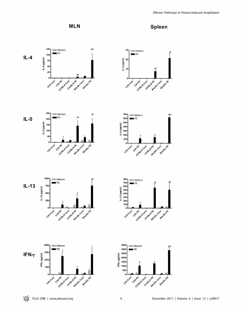
spleen and MLN cells were re-stimulated with PE (figure 2). This
led to an induction of the Th2 cytokines IL-4, IL-5 and IL-13 in
both spleen and MLN cells of C57BL/6 and BALB/c mice, but
much less in C3H/HeOuJ mice. This induction was highest in
BALB/c mice and significantly less pronounced in C3H/HeOuJ
mice. IFN-c was induced equally in all mouse strains in MLN
cultures but was induced significantly higher in spleen cultures of
BALB/c mice. This level of Th1 (IFNc) cytokine induction after
restimulation reflects a mixed ex vivo Th1/Th2 response in all
strains in our model. Overall, oral sensitization and challenge with
PE led to increased levels of PE-specific antibody and T cell
responses, including IgE and Th2 cytokine production. Nonethe-
less, the level of these allergic parameters differed significantly
among the used mouse strains.
The degree of systemic anaphylaxis and ear swelling ismouse strain dependent
Next, we assessed whether the differences in antibody and T cell
responses in different mouse strains would lead to differences in
clinical manifestations of food allergy as well. Therefore, we
compared mast cell degranulation and systemic anaphylaxis after
intra-gastric and systemic challenge with PE and observed that
both challenges in PE-sensitized mice led to a significant release of,
mucosal mast cell-derived, Mouse Mast Cell Protease-1 (MMCP-I)
in the serum of C3H/HeOuJ and BALB/c, but not C57BL/6
mice (figure 3a). However, MMCP-I is not produced by
connective tissue mast cells and to investigate the in vivo response
of this type of mast cells, we measured mast cell-mediated ear
swelling after intradermal injection of PE in the ears of sensitized
mice. This showed that C3H/HeOuJ, C57BL/6 and BALB/c
mice developed significant ear swelling upon challenge with PE
(figure 3b). C3H/HeOuJ mice showed a significantly higher ear
swelling compared to C57BL/6 and BALB/c mice.
Hereafter, we studied the development of systemic anaphylaxis
after PE oral sensitization and systemic (i.p.) challenge. We
measured an unambiguous anaphylactic response, reflected by a
strong drop in temperature and increased clinical score, in C3H/
HeOuJ and to lesser extent in C57BL/6 mice (figure 3c).
Surprisingly, BALB/c mice only showed a very minimal
anaphylactic response upon challenge. In addition to many
previous attempts, oral challenge with PE of sensitized mice did
not lead to any anaphylactic responses in all three mouse strains
(data not shown).
Together, these data shows a clear discrepancy between
mucosal and connective tissue mast cell degranulation and
development of systemic anaphylaxis to PE in different mouse
strains. C3H/HeOuJ mice displayed mucosal mast cell degranu-
lation and the strongest level of systemic anaphylaxis. C57BL/6
mice did not show mucosal mast cell degranulation and exhibited
less pronounced mast cell-mediated ear swelling and systemic
anaphylaxis when compared to C3H/HeOuJ.
PE-induced systemic anaphylaxis is FcRc- mediatedSubsequently, to assess whether the observed PE-induced
systemic anaphylaxis in C57BL/6 mice was IgG and IgE
mediated, we used FcRc2/2 knockout C57BL/6 mice in our
model. The Fc associated c chain is a key component of FccRI, III
and IV and FceRI but not of FccRIIb and FceRII (CD23).
Therefore, in FcRc2/2 mice both IgG and IgE cannot bind with
high affinity and/or activate responsive receptors. Remarkably,
FcRc2/2 mice showed normal levels of IgE, IgG1 and IgG2a
antibodies after oral sensitization in comparison to WT positive
control mice (figure 4a), but did not develop systemic anaphylaxis
upon PE challenge (figure 4b). This shows that PE-induced
systemic anaphylaxis but not oral PE sensitization is FcRc-
mediated and dependent on activation via IgE or IgG.
The role of basophils, neutrophils, monocytes/macrophages and mast cells in PE-induced systemicanaphylaxis
The occurrence of antibody mediated anaphylactic responses to
PE despite no obvious mucosal mast cell response and a lower
connective tissue mast cell response in C57BL/6 mice led us to
explore alternative pathways of anaphylaxis in these mice
compared to C3H/HeOuJ mice. It has been suggested that
basophils, neutrophils or macrophages can contribute to or cause
anaphylactic responses as well. Importantly, a comparison of the
number of platelets, basophils, monocytes/macrophages and
neutrophils showed no differences in the number of these cell
types in spleen or peritoneal cavity of C3H/HeOuJ and C57BL/6
mice (figure S1). Therefore, we first investigated whether basophils
influenced the development of systemic anaphylaxis in C57BL/6
mice and C3H/HeOuJ mice. Basophil depletion with the
antibody BA103 did not influence PE oral sensitization as
measured by levels of PE-specific IgG1, IgG2a and IgE
(figure 5a) or systemic anaphylactic responses of C3H/HeOuJ
and C57BL/6 mice (figure 5b). As a control, MMCP-I levels after
PE challenge were equal among control and basophil-depleted
mice (figure 5c), showing that mast cell responses were unaffected
by BA103 treatment. In addition, neutrophil depletion using Gr-1
did not influence systemic anaphylactic responses of C3H/HeOuJ
and C57BL/6 mice (figure S2). Hereafter, we investigated whether
monocytes/macrophages play a role in PE-induced systemic
anaphylaxis. Remarkably, selective depletion of monocytes/
macrophages using clodronate liposomes did not affect the drop
in temperature in C3H/HeOuJ mice, but it significantly lowered
the anaphylactic response in C57BL/6 mice (figure 5d).
The induction of systemic anaphylaxis in the absence of
mucosal mast cell degranulation was further examined in mast
cell-deficient C57BL/6W-sh/W-sh mice. Importantly, mast cell
deficiency did not lead to differences in IgE, IgG1 or IgG2a
levels compared to littermates (figure 6a). Interestingly, in contrast
to C57BL/6 littermates C57BL/6W-sh/W-sh mice did not develop
any anaphylactic manifestations (figure 6b). Thus, basophils do not
play a role in PE-induced oral sensitization and systemic
anaphylaxis. In contrast, monocytes/macrophages do play a
significant role in PE-induced systemic anaphylaxis in C57BL/6
mice, while playing no role in C3H/HeOuJ mice. This would
suggest that parts of the alternative pathway of anaphylaxis,
dependent on connective tissue mast cells, are mediating peanut-
induced systemic anaphylaxis in C57BL/6 mice.
PAF and platelets mediate PE-induced systemicanaphylaxis, depending on mouse strain
PAF is considered an important mediator in the alternative
pathway of anaphylaxis. For that reason, we looked whether
inhibition of the PAF pathway would affect PE-induced systemic
anaphylaxis. However, treatment before challenge with the PAFR
inhibitor WEB2086 did not significantly inhibit systemic anaphy-
laxis responses in C3H/HeOuJ mice although the observed
Effector Pathways in Peanut-Induced Anaphylaxis
PLoS ONE | www.plosone.org 2 December 2011 | Volume 6 | Issue 12 | e28917

Figure 1. Antibody responses after PE oral sensitization in different mouse strains. C3H/HeOuJ (C3H), C57BL/6 and BALB/c mice wereexposed to PBS+CT (cont) or PE+CT (PE) during a 4 week period, as described in material and methods. Graphs depict serum levels of PE-specific IgA,IgG1, IgG2a, IgE and IgG2c at day 36. Data are represented as mean AU6 SEM of 8 mice. *: p,0.05 between indicated PE sensitized groups.doi:10.1371/journal.pone.0028917.g001
Effector Pathways in Peanut-Induced Anaphylaxis
PLoS ONE | www.plosone.org 3 December 2011 | Volume 6 | Issue 12 | e28917
Effector Pathways in Peanut-Induced Anaphylaxis
PLoS ONE | www.plosone.org 4 December 2011 | Volume 6 | Issue 12 | e28917

temperature drop did seem less severe at later time points after
PAF inhibition (figure 7a). In contrast, PAF inhibition significantly
lowered anaphylactic responses in C57BL/6 mice. Using another
PAF R inhibitor, CV3988 gave the same results (data not shown).
Antibody-mediated depletion of platelets did significantly inhibit
systemic anaphylaxis in both C3H/HeOuJ and C57BL/6 mice
(figure 7b). Therefore, although PE induced systemic anaphylaxis
involves platelets in both C3H/HeOuJ and C57BL/6 it only
significantly involves PAF in C57BL/6 mice. Again suggesting that
parts of the alternative pathway of anaphylaxis are mediating
peanut-induced systemic anaphylaxis in C57BL/6 mice
Discussion
Manifestations of food allergy and food induced anaphylaxis
may vary widely among individuals, even among patients reacting
to the same responsible allergen. In addition, the underlying
immunological mechanisms of these differences may be the reason
why a classical diagnosis of food allergy and/or anaphylaxis is not
always conclusive. In an attempt to reveal possible mechanisms of
this pleiomorphy, we set out to investigate and compare
parameters and manifestations of food allergy in different well-
known mouse strains, i.e. C3H/HeOuJ, C57BL/6 and BALB/c
mice. We observed that oral sensitization to PE, when comparing
IgE and Th2 cytokine levels, was most effective in BALB/c mice,
followed by C57BL/6 mice, and least effective in C3H/HeOuJ
mice. Remarkably, the degree of systemic anaphylaxis was strain-
dependent but, in contrast to oral sensitization, consistently most
severe in C3H/HeOuJ, less pronounced in C57BL/6 and very
low or minimal in BALB/c mice. This observed differences
obviously point to variable genetic susceptibility to food sensitiza-
tion in mice, as in human subjects.
The parameters of food sensitization in BALB/c mice in our
model were different from some observations in literature,
reporting lower levels of IgE and Th2 responses in these mice
[14,20]. Notably, anaphylaxis in BALB/c mice is clearly allergen-
dependent as evidenced by the findings that less complex allergens
such as ovalbumin or ß-lactoglobuline [11,18] induce anaphylactic
responses in these mice. Furthermore, the challenge dose and
route of exposure is very important in this model. We as well as
others [19] were not able to observe anaphylactic responses after
various intragastric doses of PE in any mouse strain. But orally
induced anaphylaxis could be demonstrated to very high amounts
of protein (200 mg protein/mouse), comparable to 700 grams or
more than 1000 peanuts in a human situation [17]. The
occurrence of anaphylaxis after either systemic exposure, or after
extreme oral doses or to single purified and relatively small
allergens is in line with a recent study showing that ingested
allergens must be absorbed systemically to induce anaphylaxis
[21]. Notwithstanding, the anaphylactic responses measured in
our studies after oral sensitization and systemic challenge are
strong and consistent, resulting in a robust model and a notable
experimental advantage. In this model, BALB/c mice did not
show anaphylaxis despite a relative high level of expression of Th2
cytokines and IgE after oral sensitization. In addition, despite high
levels of IgE and the occurrence of systemic anaphylaxis, C57BL/
6 mice did not show increased mucosal mast cell degranulation
upon intra-gastric or systemic PE challenge. This clearly points to
multiple mechanisms involved in susceptibility to peanut-induced
anaphylaxis.
Classically, the mast cell has always been the central mediator of
allergy-mediated anaphylaxis. Two types of mast cells have been
described in the mouse: mucosal mast cells expressing MMCP-I and
connective tissue mast cells [22,23,24]. To investigate the responses
of connective tissue mast cells, we measured acute ear swelling,
which occurred in C57BL/6 mice and in BALB/c mice but clearly
less than C3H/HeOuJ mice. This suggests that the observed
differences in systemic anaphylactic symptoms between the mouse
strains may be due to differential involvement of either mucosal or
connective tissue mast cell subsets. In addition, the apparent lack of
involvement of mucosal mast cells in C57BL/6 led us to investigate
the role of the classical versus alternative pathways of anaphylaxis in
these mice compared to C3H/HeOuJ mice.
The role of the alternative pathway of anaphylaxis involving
IgG, macrophages, basophils and PAF in food allergy and food-
induced anaphylaxis has already been suggested [6]. Recently,
studies by Jonsson suggested a novel alternative pathway in which
neutrophils, activated via IgG contributed to systemic anaphylactic
shock in mice as well [7].In our experiments, peanut-induced
systemic anaphylaxis in C57BL/6 mice was shown to be entirely
dependent on FcRc and mast cells and partially dependent on
PAF, platelets and monocytes/macrophages. Systemic anaphylaxis
in C3H/HeOuJ was also dependent in platelets, but not on PAF
or monocytes/macrophages. Despite earlier reports on the role of
basophils in other allergic diseases [25], basophils did not play a
role in oral sensitization and systemic anaphylaxis in any of the
strains. Moreover, neutrophil depletion using Gr-1 did not
influence systemic anaphylactic responses of C3H/HeOuJ and
C57BL/6 mice in our studies. This suggests that the involvement
of components of the alternative anaphylaxis pathway, i.e.
macrophages and PAF, in food allergy is strain-dependent but
still dependent on mast cell activation.
The earlier mentioned studies investigating alternative pathways
were performed in mouse models using passive cutaneous or
systemic anaphylaxis, penicillin-, or TNP-OVA-mediated anaphy-
laxis [6,7,11,12]. Moreover, these experiments were done with
substantial immunization protocols (such as Freund’s adjuvant)
followed by injection of large amounts of antigen, which may not
recapitulate the development of hypersensitivity in food allergy.
More relevant to food allergy, it has been shown that in mice
preconditioned for responsiveness to vasoactive mediators, peanuts
could directly induce anaphylaxis by an IgE-independent but
alternative pathway-dependent mechanism [10]. In addition, in a
classical mouse model of food allergy using C57BL/6, Arias et al
showed that a PAF-antagonist, but not a histamine antagonist
inhibited systemic PE anaphylaxis [9].Recently in the same model,
very high amounts (3.75 mg) of peanut injected intravenously
induced anaphylaxis in mast cell deficient mice, which was
dependent on macrophages and to lesser extent on basophils [26].
Again, this shows that probably the dose and route of exposure of
allergen is crucial in which pathway of anaphylaxis will be
involved. A very high dose or systemic exposure of allergen may
involve the alternative pathway of anaphylaxis, as suggested by
others [11].
Figure 2. T cell responses after PE oral sensitization in different mouse strains. C3H/HeOuJ (C3H), C57BL/6 and BALB/c mice were exposedto PBS+CT (cont) or PE+CT (PE) during a 4 week period, as described in material and methods. Spleens and MLN were isolated and single cellsuspensions of these tissues were incubated in the absence (medium) or presence of sterilized peanut extract (PE) in complete RPMI1640. Graphsdepict levels of IL-4, IL-5, IL-13 and IFN-c 96 h after stimulation. Data are represented as mean 6 SEM of 6 mice. *: p,0.05 compared to control. #:p,0.05 compared to C3H/HeOuJ PE group. ‘: p,0.05 compared to C57BL/6 PE group.doi:10.1371/journal.pone.0028917.g002
Effector Pathways in Peanut-Induced Anaphylaxis
PLoS ONE | www.plosone.org 5 December 2011 | Volume 6 | Issue 12 | e28917

Effector Pathways in Peanut-Induced Anaphylaxis
PLoS ONE | www.plosone.org 6 December 2011 | Volume 6 | Issue 12 | e28917

It has been demonstrated that PAF was an important mediator
of systemic anaphylaxis in the earlier mentioned models [9,27].
We now append these studies showing that the role of PAF, and
additionally of monocytes/macrophages is strain-dependent. PAF
is produced by monocytes/macrophages, mast cells and many
other immune cells, activates platelets but also has effects on
vascular and endothelial cells, explaining its possible role in
systemic anaphylaxis [28]. A human study showed increased levels
of PAF during anaphylaxis and more severe anaphylaxis in
individuals who slowly catabolize PAF [8]. Moreover, it was
suggested that PAF secretion by monocytes/macrophages, via
platelets, contributes to pathophysiology of allergic asthma [29].
We therefore can envision a situation in food-induced anaphylaxis
where initial activation of mast cells and secretion of histamine and
PAF is followed by activation of monocytes/macrophages leading
to secretion of additional PAF and activation of platelets.
However, the present findings suggest that this may be determined
by predisposing genetically factors in man as it is in the mouse. We
consider it very well possible that the relatively high level of mast
cell degranulation in C3H/HeOuJ mice and humans displaying
the classical pathway obscures a possible activation and involve-
ment of components of the alternative pathway.
Previous mechanistic studies indicate that the present mouse
model of peanut allergy resembles the situation in human patients
to a great extent. Classically, in humans, food allergy and food-
induced anaphylaxis is suggested to be mostly IgE mediated [6].
However, the current study showing strain-dependent differences
in the role of classical versus alternative pathways may be
translational to the human situation. Some patients may also
display alternative pathway-driven clinical effects (e.g. PAF
expression) and thus resemble a genetically predisposed mouse
strain as the C57BL/6 mouse. Our studies are in line with the
suggestion by Finkelman that the differences between mouse and
man in this respect may be caused by the used mouse models more
than by a difference in immune system between mouse and human
[6].
Figure 3. PE-induced mucosal mast cell degranulation, ear swelling and systemic anaphylaxis in different mouse strains. C3H/HeOuJ(C3H), C57BL/6 and BALB/c mice were exposed to PBS+CT (cont) or PE+CT (PE) during a 4 week period, as described in material and methods. A, Micewere challenged intragastrically (i.g.) or intraperitonially (i.p.) and blood was taken to measure MMCP-I. B, Mice were challenged intradermally with PEin the ear and ear swelling was measured after 3 hours. Ear swelling was calculated by correcting the thickness of the right ear with the left controlear thickness minus the basal ear thickness before challenge. C, Mice were challenged i.p. and rectal temperature was measured at indicated timepoints after challenge. In addition, peak anaphylactic scores were taken. Data are represented as mean 6 SEM of 6–8 mice. *: p,0.05 compared tocontrol. #: p,0.05 compared to BALB/c PE group. ‘: p,0.05 compared to BALB/c PE and C57BL/6 PE group.doi:10.1371/journal.pone.0028917.g003
Figure 4. Humoral immunity and PE-induced systemic anaphylaxis. C57BL/6 and FcRc 2/2 mice were exposed to PBS+CT (cont) or PE+CT(PE) during a 4 week period, as described in material and methods. A, Serum levels of PE-specific IgE, IgG1 and IgG2a at day 36. B, Temperature afteri.p. challenge. Data are represented as mean 6 SEM of 4–6 mice. *: p,0.05 compared to control.doi:10.1371/journal.pone.0028917.g004
Effector Pathways in Peanut-Induced Anaphylaxis
PLoS ONE | www.plosone.org 7 December 2011 | Volume 6 | Issue 12 | e28917

Effector Pathways in Peanut-Induced Anaphylaxis
PLoS ONE | www.plosone.org 8 December 2011 | Volume 6 | Issue 12 | e28917

In summary, in this study we describe marked differences in
parameters and manifestations of food allergy in different mouse
strains. In addition, we show a role of components of the
alternative pathway of anaphylaxis in a food allergy model.
However, clear differences exist between mouse strains in
participation of this pathway. In conclusion, clinical manifestations
of food allergy and anaphylaxis are not simply and directly linked
to the classical components of the allergic cascade. This might
explain the pleiomorphic manifestations of the clinical phenomena
in the human situation. In addition, these findings may have
important implications for diagnosis of and finding new targets for
treatment of food allergy and food-induced anaphylaxis.
Methods
MiceFive-week-old specific pathogen-free female C3H/HeOuJ,
C57BL/6J, and BALB/cByJ mice were purchased from Charles
River (France) and housed under specific pathogen-free conditions
within the animal care facility at the Utrecht University. Mast cell-
deficient C57BL/6W-sh/W-sh or FcRc2/2 mice, both on a
C57BL/6 background, were bred and maintained at the animal
care facility at the Utrecht University. Experiments in this study
were approved by the Animal Experiments Committee of the
Utrecht University (ID #2011.III.03.031).
Peanut oral sensitization and challengePeanuts were kindly provided by Intersnack BV (the Nether-
lands) and peanut extract (PE) was prepared as previously
described [15]. Cholera toxin (CT) was obtained from List
Biological Laboratories, Inc. (CA, USA) To elicit oral sensitization
to PE, mice were intragastrically (i.g.) dosed with 6 mg PE plus
15 mg CT per mouse for three consecutive days, and this was
repeated every week for four weeks (exposure on days 0, 1, 2, 7,
14, 21 and 28). Control groups received PBS plus 15 mg CT.
Thereafter, mice received an i.g. challenge of 15 mg PE on day 35
and were sacrificed one day later.
Measurement of ear swellingAcute ear swelling in PE-sensitized mice was determined after
intradermal challenge with 10 mg PE in the right ear pinnae. As a
Figure 5. Basophils and monocytes/macrophages in PE-induced systemic anaphylaxis. C3H/HeOuJ (C3H) and C57BL/6 mice were exposedto PBS+CT (cont) or PE+CT (PE) during a 4 week period, as described in material and methods. Prior to oral sensitization (sens) or i.p. challenge (chall)indicated groups received BA103, a basophil depleting antibody. A, Serum levels of PE-specific IgE, IgG1 and IgG2a at day 36. B, Temperature afteri.p. challenge. C, Serum MMCP-I levels after challenge. Prior to challenge, in indicated groups, monocytes/macrophages were depleted usingclodronate liposomes. D, Temperature after i.p. challenge. Data are represented as mean 6 SEM of 6–8 mice. *: p,0.05 compared to control. #p,0.05 compared to C3H PE group or to C57BL/6 PE group.doi:10.1371/journal.pone.0028917.g005
Figure 6. Mast cells in PE-induced systemic anaphylaxis. C57BL/6 WT and mast cell deficient C57BL/6W-sh/W-sh mice were exposed to PBS+CT(cont) or PE+CT (PE) during a 4 week period, as described in material and methods. A, Serum levels of PE-specific IgE, IgG1 and IgG2a at day 36. B,Temperature after i.p. challenge. Data are represented as mean 6 SEM of 6 mice. *: p,0.05 compared to control.doi:10.1371/journal.pone.0028917.g006
Effector Pathways in Peanut-Induced Anaphylaxis
PLoS ONE | www.plosone.org 9 December 2011 | Volume 6 | Issue 12 | e28917

negative control, mice were challenged with PBS in the left ear.
Before challenge and after 3 hours, ear thickness of both ears was
measured in duplicate using a digital micrometer (Mitutoyo, the
Netherlands). Ear swelling was calculated by correcting the
thickness of the right ear with the left control ear minus the basal
ear thickness before challenge.
Measurement of systemic anaphylaxisIn separate experiments, mice received a challenge of 1 mg PE
intraperitoneally (i.p.) on day 35. As an objective parameter of
anaphylactic shock, body temperature was measured by means of
rectal thermometry every 10–20 minutes for 90 minutes after
challenge. In addition, clinical symptoms were scored using a
scoring system, as used before [19].
Cell and cell mediator antagonistsPAF function was inhibited using the PAF-receptor antagonists
WEB 2086 or CV 3988 (Biomol, USA), which were injected i.p.
one hour before challenge at 5 mg/kg BW. Mouse blood platelets
were depleted using a rabbit anti mouse platelet serum (Accurate
Chemical & Scientific corp, USA) at 50 ml/mouse i.p. three hours
before challenge [30]. Depletion of platelets was confirmed by
whole blood analysis (Abbott Cell-Dyn CD-1800 Hematology
Analyzer, USA) (figure S1). Basophils were depleted with one i.p.
injection of 50 mg BA103, one day before oral sensitization or
challenge as described previously [31] (figure S1). Monocytes/
macrophages were depleted 3 days before challenge using
liposomes containing dicloromethylene-biphosphonate (clodro-
nate), as described previously [32] at 5 mg/mouse i.p. Macro-
phage depletion was confirmed by differential cell counts and
flowcytometric analysis of F4/80 + cells in peritoneal fluid (figure
S1). In addition, clodronate liposome treatment did not affect
FceRI + cells (mast cells/basophils) in peritoneal fluid. Neutrophils
were depleted one day before challenge by i.p. injection of
500 mg/mouse Gr-1 antibody [33].
Measurement of PE-specific antibodies and MMCP-I inserum
PE-specific IgG1, IgG2a, IgG2c and IgE in serum of day 36
were measured by ELISA as described previously [34]. This
Figure 7. Platelet Activating Factor (PAF) and platelets in PE-induced systemic anaphylaxis. C3H/HeOuJ (C3H) and C57BL/6 mice wereexposed to PBS+CT (cont) or PE+CT (PE) during a 4 week period, as described in material and methods. A, Temperature after i.p. challenge. Prior tochallenge, indicated groups received the PAF R inhibitor WEB2086. B, Temperature after i.p. challenge. Prior to challenge, in indicated groupsplatelets were depleted using a rabbit anti-platelet serum. *: p,0.05 compared to control. # p,0.05 compared to PE groups.doi:10.1371/journal.pone.0028917.g007
Effector Pathways in Peanut-Induced Anaphylaxis
PLoS ONE | www.plosone.org 10 December 2011 | Volume 6 | Issue 12 | e28917

protocol was amended: a positive pool serum derived from PE/
alum sensitized mice was used as reference value to calculate
Arbitrary Units (AU). Levels of IgG2c were depicted as OD at
405 nm. In addition, serum was collected within 45 minutes after
oral challenge and 3 hours after i.p. challenge on day 35, and
MMCP-I was determined using a specific ELISA kit according to
instructions of the manufacturer (Moredun Scientific, Scotland).
Cell culture and cytokine measurementSpleen and MLN single cell suspensions (2.56106/ml) were
incubated in the presence or absence of PE (200 mg/ml) for 96 h
at 37uC in complete RPMI1640 with 10% FCS, after which
culture supernatants were harvested and stored at 220uC until
analysis, as described before [14]. Levels of IFN-c, IL-4, IL-5 and
IL-13 in culture supernatants were determined by sandwich
ELISA according to the instructions of the manufacturers
(eBioscience, Austria).
Statistical analysisData are presented as means 6 standard error of the mean
(SEM) and analyzed using GraphPad Prism software. Antibody,
MMCP-I and cytokine levels and data on ear swelling were
logarithmic transformed followed by a one-way ANOVA and
Bonferroni as a post-hoc test. Temperature curves were statistically
analyzed using a repeated measures ANOVA and clinical scores
were statistically analyzed by the Kruskall-Wallis test.
Supporting Information
Figure S1 Number of platelets, basophils, macrophagesand neutrophils in control and depleted mice. (A) Number
of platelets in blood of control and mice treated with a rabbit anti-
mouse platelet serum. The number of platelets was performed by
whole blood analysis on a hematology analyzer. Data are
represented as mean 6 SEM of 4 mice. # p,0.001 compared
to untreated C3H or C57BL/6 mice. (B) Number of basophils in
spleens of control and mice treated with BA103, a basophil
depetion antibody. Cells were analyzed using flow cytometry,
gated based on FSC-SSC pattern and FceRa+ and CD49b+
staining for basophils. Pictures show representative dot plots with
indicated average number of gated cells. (C) Number of
macrophages in peritoneal fluid of control and mice treated with
monocyte/macrophage depleting clodronate liposomes. Cells were
analyzed using flow cytometry, gated based on FSC-SSC pattern
and GR-1-CD11b+F4/80+ staining for macrophages. Pictures
show representative dot plots with indicated average number of
gated cells. (D) Number of macrophages in peritoneal fluid of
control and mice treated with monocyte/macrophage depleting
clodronate liposomes. Cytospins were made of peritoneal washings
and stained with DiffQuick (H&E). Pictures show representative
micrographs at 206magnification. (E) Number of neutrophils in
spleen of control and mice treated with anti-Gr-1. Cells were
analyzed using flow cytometry, gated based on FSC-SSC pattern
and GR-1+CD11b+F4/802 staining for neutrophils. Pictures
show representative dot plots with indicated average number of
gated cells.
(TIF)
Figure S2 Neutrophils in PE-induced systemic anaphy-laxis. C3H/HeOuJ (C3H) and C57BL/6 mice were exposed to
PBS+CT (cont) or PE+CT (PE) during a 4 week period, as
described in material and methods. Prior to i.p. challenge
indicated groups received Gr-1, a neutrophil depleting antibody.
Depicted is the temperature after i.p. challenge. Data are
represented as mean 6 SEM of 6–8 mice. *: p,0.05 compared
to control.
(TIF)
Acknowledgments
The authors would like to thank Hajime Karasuyama, Department of
Immune Regulation at Tokyo Medical and Dental University Graduate
School, Tokyo, Japan for kindly providing the BA103 antibody.
Author Contributions
Conceived and designed the experiments: JJS RHHP MP. Performed the
experiments: KW IH DF. Analyzed the data: JJS. Contributed reagents/
materials/analysis tools: JHWL GS LvB DZ MP. Wrote the paper: JJS
RHHP.
References
1. Sicherer SH (2011) Epidemiology of food allergy. J Allergy Clin Immunol 127:
594–602.
2. Burks AW (2008) Peanut allergy. Lancet 371: 1538–1546.
3. Ross MP, Ferguson M, Street D, Klontz K, Schroeder T, et al. (2008) Analysis of
food-allergic and anaphylactic events in the National Electronic Injury
Surveillance System. J Allergy Clin Immunol 121: 166–171.
4. Ring J, Darsow U (2002) Idiopathic anaphylaxis. Curr Allergy Asthma Rep 2:
40–45.
5. Parish WE (1970) Short-term anaphylactic IgG antibodies in human sera.
Lancet 2: 591–592.
6. Finkelman FD (2007) Anaphylaxis: lessons from mouse models. J Allergy Clin
Immunol 120: 506–515; quiz 516–507.
7. Jonsson F, Mancardi DA, Kita Y, Karasuyama H, Iannascoli B, et al. (2011)
Mouse and human neutrophils induce anaphylaxis. J Clin Invest 121:
1484–1496.
8. Vadas P, Gold M, Perelman B, Liss GM, Lack G, et al. (2008) Platelet-activating
factor, PAF acetylhydrolase, and severe anaphylaxis. N Engl J Med 358: 28–35.
9. Arias K, Baig M, Colangelo M, Chu D, Walker T, et al. (2009) Concurrent
blockade of platelet-activating factor and histamine prevents life-threatening
peanut-induced anaphylactic reactions. J Allergy Clin Immunol 124: 307–314,
314 e301–302.
10. Khodoun M, Strait R, Orekov T, Hogan S, Karasuyama H, et al. (2009)
Peanuts can contribute to anaphylactic shock by activating complement.
J Allergy Clin Immunol 123: 342–351.
11. Strait RT, Morris SC, Finkelman FD (2006) IgG-blocking antibodies inhibit
IgE-mediated anaphylaxis in vivo through both antigen interception and Fc
gamma RIIb cross-linking. J Clin Invest 116: 833–841.
12. Strait RT, Morris SC, Yang M, Qu XW, Finkelman FD (2002) Pathways of
anaphylaxis in the mouse. J Allergy Clin Immunol 109: 658–668.
13. Berin MC, Mayer L (2009) Immunophysiology of experimental food allergy.
Mucosal Immunol 2: 24–32.
14. Berin MC, Zheng Y, Domaradzki M, Li XM, Sampson HA (2006) Role of
TLR4 in allergic sensitization to food proteins in mice. Allergy 61: 64–71.
15. Bol-Schoenmakers M, Marcondes Rezende M, Bleumink R, Boon L, Man S,
et al. (2011) Regulation by intestinal gammadelta T cells during establishment of
food allergic sensitization in mice. Allergy 66: 331–340.
16. Schouten B, van Esch BC, Hofman GA, van den Elsen LW, Willemsen LE, et al.
(2008) Acute allergic skin reactions and intestinal contractility changes in mice
orally sensitized against casein or whey. Int Arch Allergy Immunol 147:
125–134.
17. Srivastava KD, Qu C, Zhang T, Goldfarb J, Sampson HA, et al. (2009) Food
Allergy Herbal Formula-2 silences peanut-induced anaphylaxis for a prolonged
posttreatment period via IFN-gamma-producing CD8+ T cells. J Allergy Clin
Immunol 123: 443–451.
18. Eigenmann PA, Asigbetse KE, Frossard CP (2008) Avirulant Salmonella
typhimurium strains prevent food allergy in mice. Clin Exp Immunol 151:
546–553.
19. Sun J, Arias K, Alvarez D, Fattouh R, Walker T, et al. (2007) Impact of CD40
ligand, B cells, and mast cells in peanut-induced anaphylactic responses.
J Immunol 179: 6696–6703.
20. Morafo V, Srivastava K, Huang CK, Kleiner G, Lee SY, et al. (2003) Genetic
susceptibility to food allergy is linked to differential TH2-TH1 responses in
C3H/HeJ and BALB/c mice. J Allergy Clin Immunol 111: 1122–1128.
21. Strait RT, Mahler A, Hogan S, Khodoun M, Shibuya A, et al. (2011) Ingested
allergens must be absorbed systemically to induce systemic anaphylaxis. J Allergy
Clin Immunol.
22. Kumar V, Sharma A (2010) Mast cells: emerging sentinel innate immune cells
with diverse role in immunity. Mol Immunol 48: 14–25.
Effector Pathways in Peanut-Induced Anaphylaxis
PLoS ONE | www.plosone.org 11 December 2011 | Volume 6 | Issue 12 | e28917

23. Knoops L, Louahed J, Van Snick J, Renauld JC (2005) IL-9 promotes but is not
necessary for systemic anaphylaxis. J Immunol 175: 335–341.24. Miller HR, Huntley JF, Newlands GF, Mackellar A, Lammas DA, et al. (1988)
Granule proteinases define mast cell heterogeneity in the serosa and the
gastrointestinal mucosa of the mouse. Immunology 65: 559–566.25. Karasuyama H, Mukai K, Obata K, Tsujimura Y, Wada T (2011)
Nonredundant Roles of Basophils in Immunity. Annu Rev Immunol.26. Arias K, Chu DK, Flader K, Botelho F, Walker T, et al. (2011) Distinct immune
effector pathways contribute to the full expression of peanut-induced
anaphylactic reactions in mice. J Allergy Clin Immunol 127: 1552–1561 e1551.27. Brandt EB, Strait RT, Hershko D, Wang Q, Muntel EE, et al. (2003) Mast cells
are required for experimental oral allergen-induced diarrhea. J Clin Invest 112:1666–1677.
28. McManus LM, Pinckard RN (2000) PAF, a putative mediator of oralinflammation. Crit Rev Oral Biol Med 11: 240–258.
29. Kasperska-Zajac A, Brzoza Z, Rogala B (2008) Platelet activating factor as a
mediator and therapeutic approach in bronchial asthma. Inflammation 31:112–120.
30. Fujimi S, MacConmara MP, Maung AA, Zang Y, Mannick JA, et al. (2006)
Platelet depletion in mice increases mortality after thermal injury. Blood 107:
4399–4406.
31. Obata K, Mukai K, Tsujimura Y, Ishiwata K, Kawano Y, et al. (2007) Basophils
are essential initiators of a novel type of chronic allergic inflammation. Blood
110: 913–920.
32. Rozemuller H, Knaan-Shanzer S, Hagenbeek A, van Bloois L, Storm G, et al.
(2004) Enhanced engraftment of human cells in RAG2/gammac double-
knockout mice after treatment with CL2MDP liposomes. Exp Hematol 32:
1118–1125.
33. Daley JM, Thomay AA, Connolly MD, Reichner JS, Albina JE (2008) Use of
Ly6G-specific monoclonal antibody to deplete neutrophils in mice. J Leukoc Biol
83: 64–70.
34. van Wijk F, Hoeks S, Nierkens S, Koppelman SJ, van Kooten P, et al. (2005)
CTLA-4 signaling regulates the intensity of hypersensitivity responses to food
antigens, but is not decisive in the induction of sensitization. J Immunol 174:
174–179.
Effector Pathways in Peanut-Induced Anaphylaxis
PLoS ONE | www.plosone.org 12 December 2011 | Volume 6 | Issue 12 | e28917