Bone is functionally impaired in dystrophic mice but less so than skeletal muscle
TH
EJ
OU
RN
AL
OF
CE
LL
BIO
LO
GY
JCB: ARTICLE
© The Rockefeller University Press $8.00The Journal of Cell Biology, Vol. 174, No. 2, July 17, 2006 231–243http://www.jcb.org/cgi/doi/10.1083/jcb.200512085
JCB 231
IntroductionCells can reach specifi c target tissues through the general circu-
lation by different mechanisms. Homing has been studied exten-
sively both in vitro and in vivo with different cell types, such as
leukocytes or hematopoietic stem cells (Butcher, 1991; Fu and
Liesveld, 2000); it is believed to rely on adhesion molecules and
cytokines receptors by a multistep cascade, consisting of a roll-
ing process followed by fi rm adhesion and transmigration into
the surrounding tissue (Peled et al., 1999; Grabovsky et al.,
2000). The repertoire and the level of cytokine expression by the
target tissue, as well as expression of the relative receptors on
endothelium, infl uence the effi ciency of homing. For example,
stromal-derived factor (SDF) 1 favors the arrest of progenitors
on vascular endothelium, whereas interleukin (IL) 8 promotes
stem cell mobilization from the marrow (Laterveer et al., 1996;
Peled et al., 1999). Although these mechanisms have been eluci-
dated to a large extent for leukocytes and hematopoietic stem cells,
far less is known for other types of stem cells. Mesoangioblasts
were recently characterized as a population of vessel-associated
stem cells that differentiate into several mesoderm cell types,
including skeletal muscle (Minasi et al., 2002). They have been
shown to restore to a signifi cant extent muscle structure and
function in a mouse model of muscular dystrophy (Sampaolesi
et al., 2003). One main reason for the partial effect of mesoan-
gioblasts in this model is likely to be ascribed to the limited
ability of these cells to reach and colonize the muscle, depending
in turn on incomplete adhesion and extravasation.
Mesoangioblast extravasation must be directed by selec-
tive mesoangioblast–endothelial cell recognition. Microarray
analysis revealed that mesoangioblasts express E-selectin, β7
integrin, AlCAM, several cytokines receptors, and CD44 but
lack many of the leukocyte molecules implicated in transmigra-
tion (Tagliafi co et al., 2004). To increase the effi ciency of mus-
cle repair by mesoangioblasts, it would be essential to increase
their migration to skeletal muscle, with the additional benefi t of
reducing unspecifi c trapping in the capillary fi lters of the body,
such as liver and lung. Here, we report that expression of α4
integrin and exposure of cells to SDF-1 or TNF-α improve up to
fi vefold migration of wild-type (WT) mesoangioblasts to the
dystrophic muscles and consequent production of new fi bers
that express the normal copy of the mutated gene. These results
Complete repair of dystrophic skeletal muscle by mesoangioblasts with enhanced migration ability
Beatriz G. Galvez,1 Maurilio Sampaolesi,1,2 Silvia Brunelli,1,3 Diego Covarello,1 Manuela Gavina,4 Barbara Rossi,6
Gabriela Costantin,6 Yvan Torrente,4 and Giulio Cossu1,5
1Stem Cell Research Institute, San Raffaele Hospital, 20132 Milan, Italy2Department of Experimental Medicine, Human Anatomy Institute, University of Pavia, 27100 Pavia, Italy3Department of Experimental, Environmental Medicine, and Medical Biotechnology, University of Milano-Bicocca, 20126 Milan, Italy4Stem Cell Laboratory, Department of Neurological Science, and 5Department of Biology, University of Milan, 20122 Milan, Italy6Department of Pathology, Division of General Pathology, University of Verona, 37129 Verona, Italy
Effi cient delivery of cells to target tissues is a major
problem in cell therapy. We report that enhancing
delivery of mesoangioblasts leads to a complete
reconstitution of downstream skeletal muscles in a mouse
model of severe muscular dystrophy (α-sarcoglycan ko).
Mesoangioblasts, vessel-associated stem cells, were
exposed to several cytokines, among which stromal-
derived factor (SDF) 1 or tumor necrosis factor (TNF) α
were the most potent in enhancing transmigration in vitro
and migration into dystrophic muscle in vivo. Transient
expression of α4 integrins or L-selectin also increased sev-
eral fold migration both in vitro and in vivo. Therefore,
combined pretreatment with SDF-1 or TNF-α and expres-
sion of α4 integrin leads to massive colonization (>50%)
followed by reconstitution of >80% of α-sarcoglycan–
expressing fi bers, with a fi vefold increase in effi ciency
in comparison with control cells. This study defi nes the
requirements for effi cient engraftment of mesoangioblasts
and offers a new potent tool to optimize future cell therapy
protocols for muscular dystrophies.
Correspondence to Giulio Cossu: [email protected]
Abbreviations used in this paper: ctx, cardiotoxin; HMGB, high mobility group box; ICAM, intercellular adhesion molecule; IL, interleukin; mdx, X chromosome–linked muscular dystrophy; MMP, metalloproteinase; PECAM, platelet endothelial cell adhesion molecule; SDF, stromal-derived factor; SG, sarcoglycan; VCAM, vascular cell adhesion molecule; WT, wild-type.
JCB • VOLUME 174 • NUMBER 2 • 2006 232
elucidate the migration mechanism of mesoangioblasts and
open a therapeutic opportunity for improving effi cacy of cell
therapy in muscular dystrophy.
ResultsDifferentiated myotubes and SDF-1 or TNF-𝛂 cytokines induce mesoangioblast transmigration in vitroMesoangioblasts (clone D16; Sampaolesi et al., 2003) were
starved for 12 h in the absence of serum and then subjected to
transmigration assays using transwell chambers, as described
previously (Mohle et al., 1997). In a fi rst series of experiments,
we created an artifi cial environment where mesoangioblasts
would face an activated endothelial layer separating them from
differentiating muscle cells (much as it happens in regenerating
muscle) or specifi c cytokines with known chemoattractive
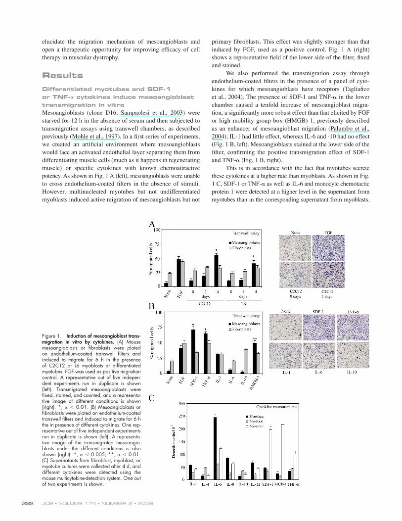
potency. As shown in Fig. 1 A (left), mesoangioblasts were unable
to cross endothelium-coated fi lters in the absence of stimuli.
However, multinucleated myotubes but not undifferentiated
myoblasts induced active migration of mesoangioblasts but not
primary fi broblasts. This effect was slightly stronger than that
induced by FGF, used as a positive control. Fig. 1 A (right)
shows a representative fi eld of the lower side of the fi lter, fi xed
and stained.
We also performed the transmigration assay through
endothelium-coated fi lters in the presence of a panel of cyto-
kines for which mesoangioblasts have receptors (Tagliafi co
et al., 2004). The presence of SDF-1 and TNF-α in the lower
chamber caused a tenfold increase of mesoangioblast migra-
tion, a signifi cantly more robust effect than that elicited by FGF
or high mobility group box (HMGB) 1, previously described
as an enhancer of mesoangioblast migration (Palumbo et al.,
2004); IL-1 had little effect, whereas IL-6 and -10 had no effect
(Fig. 1 B, left). Mesoangioblasts stained at the lower side of the
fi lter, confi rming the positive transmigration effect of SDF-1
and TNF-α (Fig. 1 B, right).
This is in accordance with the fact that myotubes secrete
these cytokines at a higher rate than myoblasts. As shown in Fig.
1 C, SDF-1 or TNF-α as well as IL-6 and monocyte chemotactic
protein 1 were detected at a higher level in the supernatant from
myotubes than in the corresponding supernatant from myoblasts.
Figure 1. Induction of mesoangioblast trans-migration in vitro by cytokines. (A) Mouse mesoangioblasts or fi broblasts were plated on endothelium-coated transwell fi lters and induced to migrate for 6 h in the presence of C2C12 or L6 myoblasts or differentiated myotubes. FGF was used as positive migration control. A representative out of fi ve indepen-dent experiments run in duplicate is shown (left). Transmigrated mesoangioblasts were fi xed, stained, and counted, and a representa-tive image of different conditions is shown (right). *, α < 0.01. (B) Mesoangioblasts or fi broblasts were plated on endothelium-coated transwell fi lters and induced to migrate for 6 h the in presence of different cytokines. One rep-resentative out of fi ve independent experiments run in duplicate is shown (left). A representa-tive image of the transmigrated mesoangio-blasts under the different conditions is also shown (right). *, α < 0.005; **, α < 0.01. (C) Supernatants from fi broblast, myoblast, or myotube cultures were collected after 4 d, and different cytokines were detected using the mouse multicytokine-detection system. One out of two experiments is shown.
MESOANGIOBLAST MIGRATION • GALVEZ ET AL. 233
These data suggest that differentiated myotubes may secrete
growth factors or cytokines such as SDF-1 or TNF-α that favor
mesoangioblast transmigration by either a chemotactic effect
and/or by modifying the endothelium barrier.
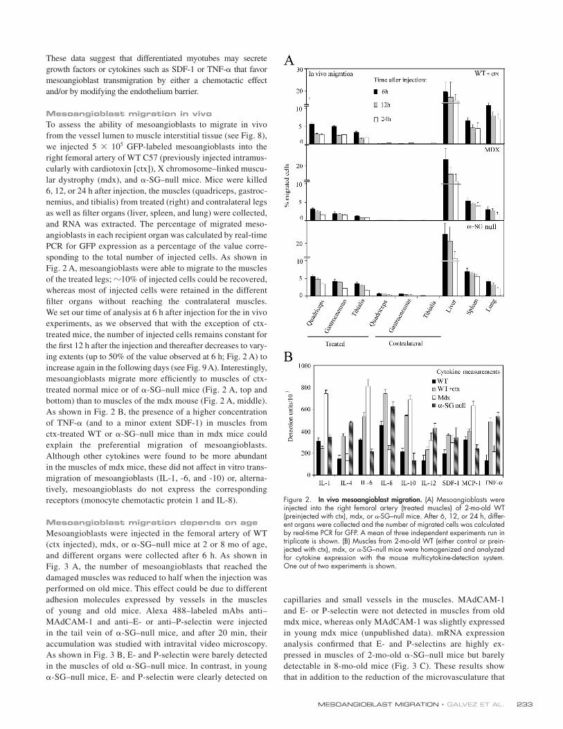
Mesoangioblast migration in vivoTo assess the ability of mesoangioblasts to migrate in vivo
from the vessel lumen to muscle interstitial tissue (see Fig. 8),
we injected 5 × 10⁵ GFP-labeled mesoangioblasts into the
right femoral artery of WT C57 (previously injected intramus-
cularly with cardiotoxin [ctx]), X chromosome–linked muscu-
lar dystrophy (mdx), and α-SG–null mice. Mice were killed
6, 12, or 24 h after injection, the muscles (quadriceps, gastroc-
nemius, and tibialis) from treated (right) and contralateral legs
as well as fi lter organs (liver, spleen, and lung) were collected,
and RNA was extracted. The percentage of migrated meso-
angioblasts in each recipient organ was calculated by real-time
PCR for GFP expression as a percentage of the value corre-
sponding to the total number of injected cells. As shown in
Fig. 2 A, mesoangioblasts were able to migrate to the muscles
of the treated legs; �10% of injected cells could be recovered,
whereas most of injected cells were retained in the different
fi lter organs without reaching the contralateral muscles.
We set our time of analysis at 6 h after injection for the in vivo
experiments, as we observed that with the exception of ctx-
treated mice, the number of injected cells remains constant for
the fi rst 12 h after the injection and thereafter decreases to vary-
ing extents (up to 50% of the value observed at 6 h; Fig. 2 A) to
increase again in the following days (see Fig. 9 A). Interestingly,
mesoangioblasts migrate more effi ciently to muscles of ctx-
treated normal mice or of α-SG–null mice (Fig. 2 A, top and
bottom) than to muscles of the mdx mouse (Fig. 2 A, middle).
As shown in Fig. 2 B, the presence of a higher concentration
of TNF-α (and to a minor extent SDF-1) in muscles from
ctx-treated WT or α-SG–null mice than in mdx mice could
explain the preferential migration of mesoangioblasts.
Although other cytokines were found to be more abundant
in the muscles of mdx mice, these did not affect in vitro trans-
migration of mesoangioblasts (IL-1, -6, and -10) or, alterna-
tively, mesoangioblasts do not express the corresponding
receptors (monocyte chemotactic protein 1 and IL-8).
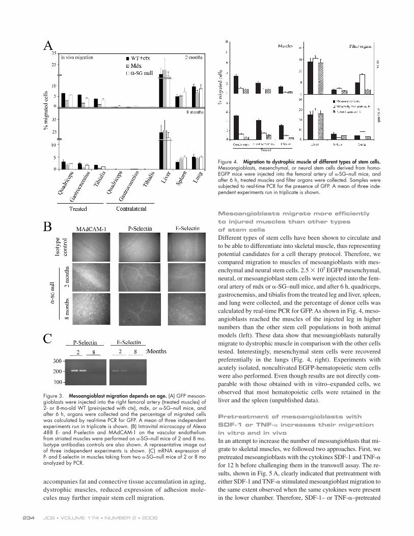
Mesoangioblast migration depends on ageMesoangioblasts were injected in the femoral artery of WT
(ctx injected), mdx, or α-SG–null mice at 2 or 8 mo of age,
and different organs were collected after 6 h. As shown in
Fig. 3 A, the number of mesoangioblasts that reached the
damaged muscles was reduced to half when the injection was
performed on old mice. This effect could be due to different
adhesion molecules expressed by vessels in the muscles
of young and old mice. Alexa 488–labeled mAbs anti–
MAdCAM-1 and anti–E- or anti–P-selectin were injected
in the tail vein of α-SG–null mice, and after 20 min, their
accumulation was studied with intravital video microscopy.
As shown in Fig. 3 B, E- and P-selectin were barely detected
in the muscles of old α-SG–null mice. In contrast, in young
α-SG–null mice, E- and P- selectin were clearly detected on
capillaries and small vessels in the muscles. MAdCAM-1
and E- or P-selectin were not detected in muscles from old
mdx mice, whereas only MAdCAM-1 was slightly expressed
in young mdx mice (unpublished data). mRNA expression
analysis confi rmed that E- and P-selectins are highly ex-
pressed in muscles of 2-mo-old α-SG–null mice but barely
detectable in 8-mo-old mice (Fig. 3 C). These results show
that in addition to the reduction of the microvasculature that
Figure 2. In vivo mesoangioblast migration. (A) Mesoangioblasts were injected into the right femoral artery (treated muscles) of 2-mo-old WT (preinjected with ctx), mdx, or α-SG–null mice. After 6, 12, or 24 h, differ-ent organs were collected and the number of migrated cells was calculated by real-time PCR for GFP. A mean of three independent experiments run in triplicate is shown. (B) Muscles from 2-mo-old WT (either control or prein-jected with ctx), mdx, or α-SG–null mice were homogenized and analyzed for cytokine expression with the mouse multicytokine-detection system.One out of two experiments is shown.
JCB • VOLUME 174 • NUMBER 2 • 2006 234
accompanies fat and connective tissue accumu lation in aging,
dystrophic muscles, reduced expression of adhesion mole-
cules may further impair stem cell migration.
Mesoangioblasts migrate more effi ciently to injured muscles than other types of stem cellsDifferent types of stem cells have been shown to circulate and
to be able to differentiate into skeletal muscle, thus representing
potential candidates for a cell therapy protocol. Therefore, we
compared migration to muscles of mesoangioblasts with mes-
enchymal and neural stem cells. 2.5 × 10⁵ EGFP mesenchymal,
neural, or mesoangioblast stem cells were injected into the fem-
oral artery of mdx or α-SG–null mice, and after 6 h, quadriceps,
gastrocnemius, and tibialis from the treated leg and liver, spleen,
and lung were collected, and the percentage of donor cells was
calculated by real-time PCR for GFP. As shown in Fig. 4, meso-
angioblasts reached the muscles of the injected leg in higher
numbers than the other stem cell populations in both animal
models (left). These data show that mesoangioblasts naturally
migrate to dystrophic muscle in comparison with the other cells
tested. Interestingly, mesenchymal stem cells were recovered
preferentially in the lungs (Fig. 4, right). Experiments with
acutely isolated, noncultivated EGFP-hematopoietic stem cells
were also performed. Even though results are not directly com-
parable with those obtained with in vitro–expanded cells, we
observed that most hematopoietic cells were retained in the
liver and the spleen (unpublished data).
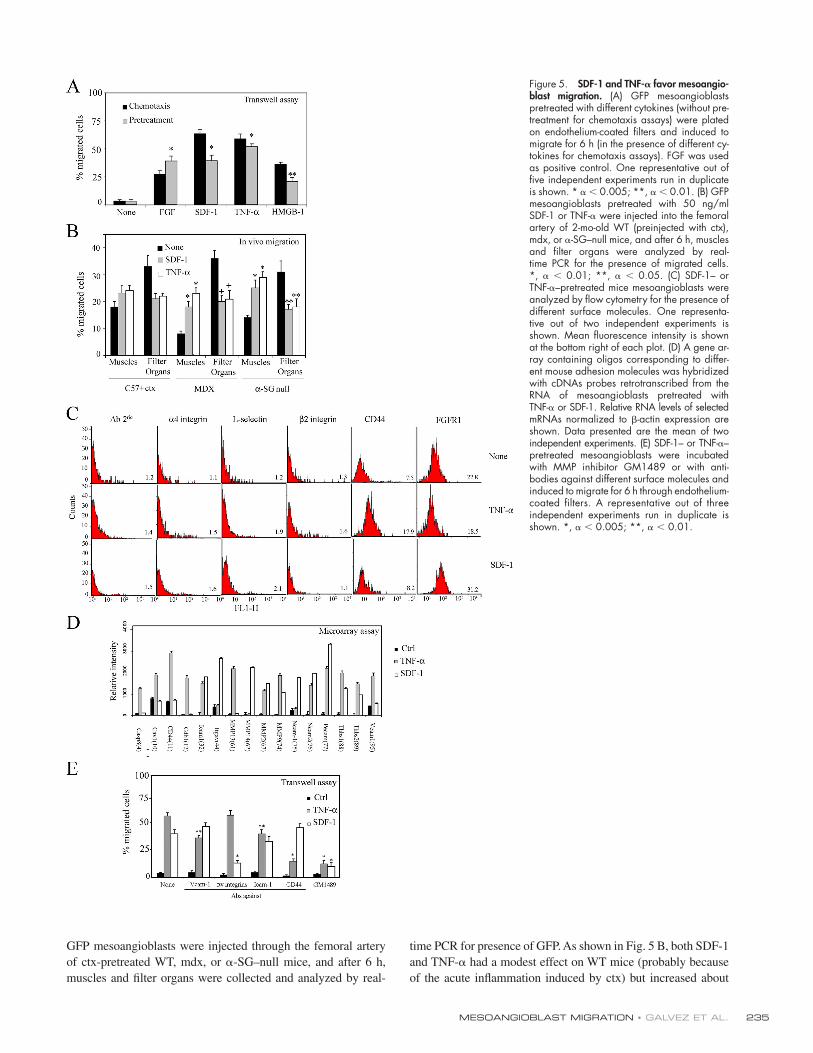
Pretreatment of mesoangioblasts with SDF-1 or TNF-𝛂 increases their migration in vitro and in vivoIn an attempt to increase the number of mesoangioblasts that mi-
grate to skeletal muscles, we followed two approaches. First, we
pretreated mesoangioblasts with the cytokines SDF-1 and TNF-α
for 12 h before challenging them in the transwell assay. The re-
sults, shown in Fig. 5 A, clearly indicated that pretreatment with
either SDF-1 and TNF-α stimulated mesoangioblast migration to
the same extent observed when the same cytokines were present
in the lower chamber. Therefore, SDF-1– or TNF-α–pretreated
Figure 3. Mesoangioblast migration depends on age. (A) GFP mesoan-gioblasts were injected into the right femoral artery (treated muscles) of 2- or 8-mo-old WT (preinjected with ctx), mdx, or α-SG–null mice, and after 6 h, organs were collected and the percentage of migrated cells was calculated by real-time PCR for GFP. A mean of three independent experiments run in triplicate is shown. (B) Intravital microscopy of Alexa 488 E- and P-selectin and MAdCAM-1 on the vascular endothelium from striated muscles were performed on α-SG–null mice of 2 and 8 mo. Isotype antibodies controls are also shown. A representative image out of three independent experiments is shown. (C) mRNA expression of P- and E-selectin in muscles taking from two α-SG–null mice of 2 or 8 mo analyzed by PCR.
Figure 4. Migration to dystrophic muscle of different types of stem cells. Mesoangioblasts, mesenchymal, or neural stem cells derived from homo-EGFP mice were injected into the femoral artery of α-SG–null mice, and after 6 h, treated muscles and fi lter organs were collected. Samples were subjected to real-time PCR for the presence of GFP. A mean of three inde-pendent experiments run in triplicate is shown.
MESOANGIOBLAST MIGRATION • GALVEZ ET AL. 235
GFP mesoangioblasts were injected through the femoral artery
of ctx-pretreated WT, mdx, or α-SG–null mice, and after 6 h,
muscles and fi lter organs were collected and analyzed by real-
time PCR for presence of GFP. As shown in Fig. 5 B, both SDF-1
and TNF-α had a modest effect on WT mice (probably because
of the acute infl ammation induced by ctx) but increased about
Figure 5. SDF-1 and TNF-𝛂 favor mesoangio-blast migration. (A) GFP mesoangioblasts pretreated with different cytokines (without pre-treatment for chemotaxis assays) were plated on endothelium-coated fi lters and induced to migrate for 6 h (in the presence of different cy-tokines for chemotaxis assays). FGF was used as positive control. One representative out of fi ve independent experiments run in duplicate is shown. * α < 0.005; **, α < 0.01. (B) GFP mesoangioblasts pretreated with 50 ng/ml SDF-1 or TNF-α were injected into the femoral artery of 2-mo-old WT (preinjected with ctx), mdx, or α-SG–null mice, and after 6 h, muscles and fi lter organs were analyzed by real-time PCR for the presence of migrated cells.*, α < 0.01; **, α < 0.05. (C) SDF-1– or TNF-α–pretreated mice mesoangioblasts were analyzed by fl ow cytometry for the presence of different surface molecules. One representa-tive out of two independent experiments is shown. Mean fl uorescence intensity is shown at the bottom right of each plot. (D) A gene ar-ray containing oligos corresponding to differ-ent mouse adhesion molecules was hybridized with cDNAs probes retrotranscribed from the RNA of mesoangioblasts pretreated with TNF-α or SDF-1. Relative RNA levels of selected mRNAs normalized to β-actin expression are shown. Data presented are the mean of two independent experiments. (E) SDF-1– or TNF-α–pretreated mesoangioblasts were incubated with MMP inhibitor GM1489 or with anti-bodies against different surface molecules and induced to migrate for 6 h through endothelium-coated fi lters. A representative out of three independent experiments run in duplicate is shown. *, α < 0.005; **, α < 0.01.
JCB • VOLUME 174 • NUMBER 2 • 2006 236
twofold the migration of mesoangioblasts to the injured mus-
cles of dystrophic mice. Consistently, the number of mesoangio-
blasts detected in the fi lter organs was reduced to almost half
of control.
We then attempted to identify the surface molecules that
may be responsible for the changes observed in mesoangioblast
migration after pretreatment with SDF-1 or TNF-α. The possi-
ble change in the expression of several candidate surface mole-
cules was fi rst investigated by FACS analysis (Fig. 5 C). Many
of the tested molecules were not changed by treatment with ei-
ther SDF-1 or TNF-α. However, CD44 expression was increased
more than twofold by TNF-α and not by SDF-1, whereas
FGFR1 expression was increased only by SDF-1. Although
these two molecules are involved in migration processes and
could explain part of our effects, we performed a microarray
analysis for 125 genes, including surface receptors, to look for
more candidates genes induced in mesoangioblasts by SDF-1 or
TNF-α. 16 genes were modifi ed after pretreatment of mesoangio-
blasts with either SDF-1 or TNF-α (Fig. 5 D). Caspase-8
(Casp8), caveolin 1 (cav-1), CD44, cadherin 1 (Cdh1), metallo-
proteinase (MMP) 13, or vascular cell adhesion molecule
(VCAM) 1 expression was increased two- to threefold only by
TNF-α pretreatment, whereas Itgαv (αv integrin), MMP-14, or
neural cell adhesion molecule (NCAM) 1 were induced approx-
imately twofold only by SDF-1 pretreatment. Pretreatment with
either TNF-α or SDF-1 increased expression of intercellular
adhesion molecule (ICAM) 1, NCAM-2, different MMPs,
platelet endothelial cell adhesion molecule (PECAM), and
trombospondins. To verify the role in mesoangioblast transmi-
gration of those molecules that appeared to be more potently in-
duced by TNF-α or SDF-1, we repeated the transwell assay in
presence of antibodies against VCAM-1, αv integrins, ICAM-1,
or CD44 or MMP inhibitor GM1489. As shown in Fig. 5 E,
antibodies against CD44 partially inhibited transmigration
induced by TNF-α, whereas antibodies against αv integrin in-
hibited SDF-1–induced transmigration. Blocking MMP activity
with GM1489 dramatically inhibited transmigration induced by
both molecules. Together, these data suggest that TNF-α and
SDF-1 increase mesoangioblast transmigration in vitro and
migration to muscles in vivo by increasing expression of several
molecules, among which CD44 and MMP appeared the most
relevant in the case of TNF-α and αv integrin and MMP
appeared most relevant in the case of SDF-1.
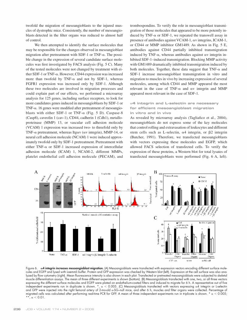
𝛂4 integrin and L-selectin are necessary for effi cient mesoangioblast migration in vitro and in vivoAs revealed by microarray analysis (Tagliafi co et al., 2004),
mesoangioblasts do not express some of the key molecules
that control rolling and extravasation of leukocytes and different
stem cells such as L-selectin, α4 integrin, or β2 integrin
(Butcher, 1991). Therefore, we transfected mesoangioblasts
with vectors expressing these molecules and EGFP, which
allowed FACS selection of transfected cells. To verify the
expression of these proteins, a Western blot for total lysates of
transfected mesoangioblasts were performed (Fig. 6 A, left).
Figure 6. 𝛂4 integrin increases mesoangioblast migration. (A) Mesoangioblasts were transfected with expression vectors encoding different surface mole-cules and EGFP and lysed with Laemmli buffer. Protein and GFP expression was checked by Western blot (left). Expression at the cell surface was also ana-lyzed by fl ow cytometry (right). Mean fl uorescence intensity is also shown in each plot. Transfected or pretreated mesoangioblasts were subjected to skeletal muscle differentiation assays. The mean of three different experiments is shown (bottom). (B) Mesoangioblasts transfected with one, two, or all three vectors expressing the different surface molecules and EGFP were plated on endothelium-coated fi lters and induced to migrate for 6 h. A representative out of fi ve independent experiments run in duplicate is shown. *, α < 0.005. (C) Mesoangioblasts transfected with vectors expressing α4 integrin or L-selectin and GFP were injected into the right femoral artery of 2-mo-old α-SG–null mice, and after 6 h, muscles and fi lter organs were collected. Percentage of migrated cells was calculated after performing real-time PCR for GFP. A mean of three independent experiments run in triplicate is shown. * α < 0.005; **, α < 0.01.
MESOANGIOBLAST MIGRATION • GALVEZ ET AL. 237
Correct surface expression of these proteins was also verifi ed by
fl ow cytometry analysis (Fig. 6 A, right). Finally, mesoangio-
blasts transfected with the different constructs or pretreated
with TNF-α or SDF-1 were subjected to differentiation assays.
Expression of L-selectin or α4 integrin did not interfere with
skeletal muscle differentiation of mesoangioblasts in vitro,
whereas expression of β2 integrin inhibited differentiation
(Fig. 6 A, bottom).
To assess whether the expression of these molecules could
improve mesoangioblast migration, cells transfected with one,
two, or all three constructs were subjected to transmigration in
vitro assay through activated endothelium. As shown in Fig. 6 B,
α4 (but nor β2) integrin or L-selectin expression increased
mesoangioblast transmigration six- to eightfold, whether or not
C2C12-derived myotubes were present in the lower chamber.
Thus, expression of the counterpart ligands for these molecules
at the surface of the activated endothelium was suffi cient to al-
low mesoangioblast transmigration independent of the presence
of myotubes, with a chemoattractant-independent mechanism.
Cotransfection of two or all constructs had only a slightly cu-
mulative effect, which was not statistically signifi cant. When
α4 integrin– or L-selectin–expressing mesoangioblasts were in-
jected into the femoral artery of ctx-treated WT, mdx, or α-SG–
null mice, the number of cells that reached downstream muscles
was increased approximately twofold with some variability in
different mice (Fig. 6 C). In general, α4 integrin appeared to be
more effi cient than L-selectin. Therefore, expression of α4 inte-
grins and L-selectin by mesoangioblasts improves their migra-
tion, possibly by favoring their interaction with the ligands
expressed on the endothelium surface.
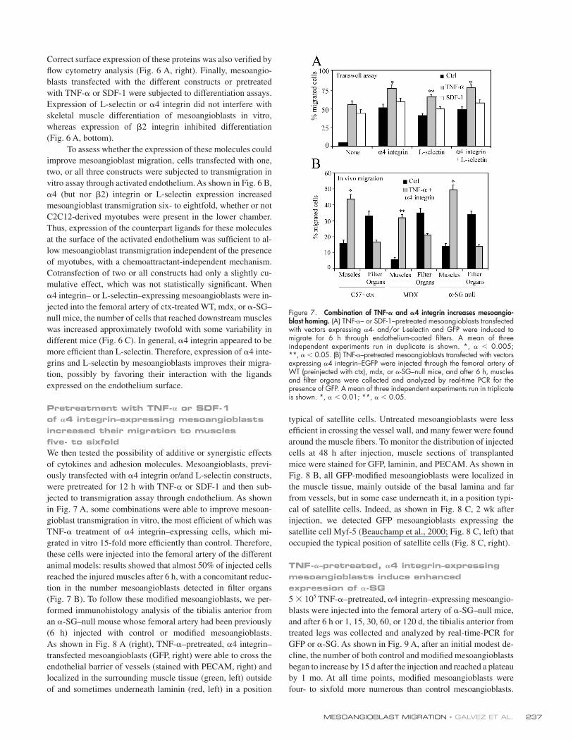
Pretreatment with TNF-𝛂 or SDF-1 of 𝛂4 integrin–expressing mesoangioblasts increased their migration to muscles fi ve- to sixfoldWe then tested the possibility of additive or synergistic effects
of cytokines and adhesion molecules. Mesoangioblasts, previ-
ously transfected with α4 integrin or/and L-selectin constructs,
were pretreated for 12 h with TNF-α or SDF-1 and then sub-
jected to transmigration assay through endothelium. As shown
in Fig. 7 A, some combinations were able to improve mesoan-
gioblast transmigration in vitro, the most effi cient of which was
TNF-α treatment of α4 integrin–expressing cells, which mi-
grated in vitro 15-fold more effi ciently than control. Therefore,
these cells were injected into the femoral artery of the different
animal models: results showed that almost 50% of injected cells
reached the injured muscles after 6 h, with a concomitant reduc-
tion in the number mesoangioblasts detected in fi lter organs
(Fig. 7 B). To follow these modifi ed mesoangioblasts, we per-
formed immunohistology analysis of the tibialis anterior from
an α-SG–null mouse whose femoral artery had been previously
(6 h) injected with control or modifi ed mesoangioblasts.
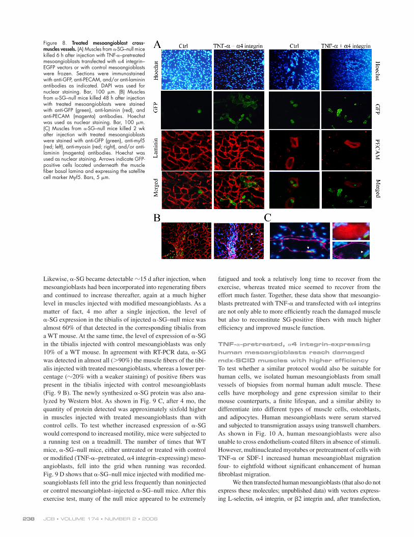
As shown in Fig. 8 A (right), TNF-α–pretreated, α4 integrin–
transfected mesoangioblasts (GFP, right) were able to cross the
endothelial barrier of vessels (stained with PECAM, right) and
localized in the surrounding muscle tissue (green, left) outside
of and sometimes underneath laminin (red, left) in a position
typical of satellite cells. Untreated mesoangioblasts were less
effi cient in crossing the vessel wall, and many fewer were found
around the muscle fi bers. To monitor the distribution of injected
cells at 48 h after injection, muscle sections of transplanted
mice were stained for GFP, laminin, and PECAM. As shown in
Fig. 8 B, all GFP-modifi ed mesoangioblasts were localized in
the muscle tissue, mainly outside of the basal lamina and far
from vessels, but in some case underneath it, in a position typi-
cal of satellite cells. Indeed, as shown in Fig. 8 C, 2 wk after
injection, we detected GFP mesoangioblasts expressing the
satellite cell Myf-5 (Beauchamp et al., 2000; Fig. 8 C, left) that
occupied the typical position of satellite cells (Fig. 8 C, right).
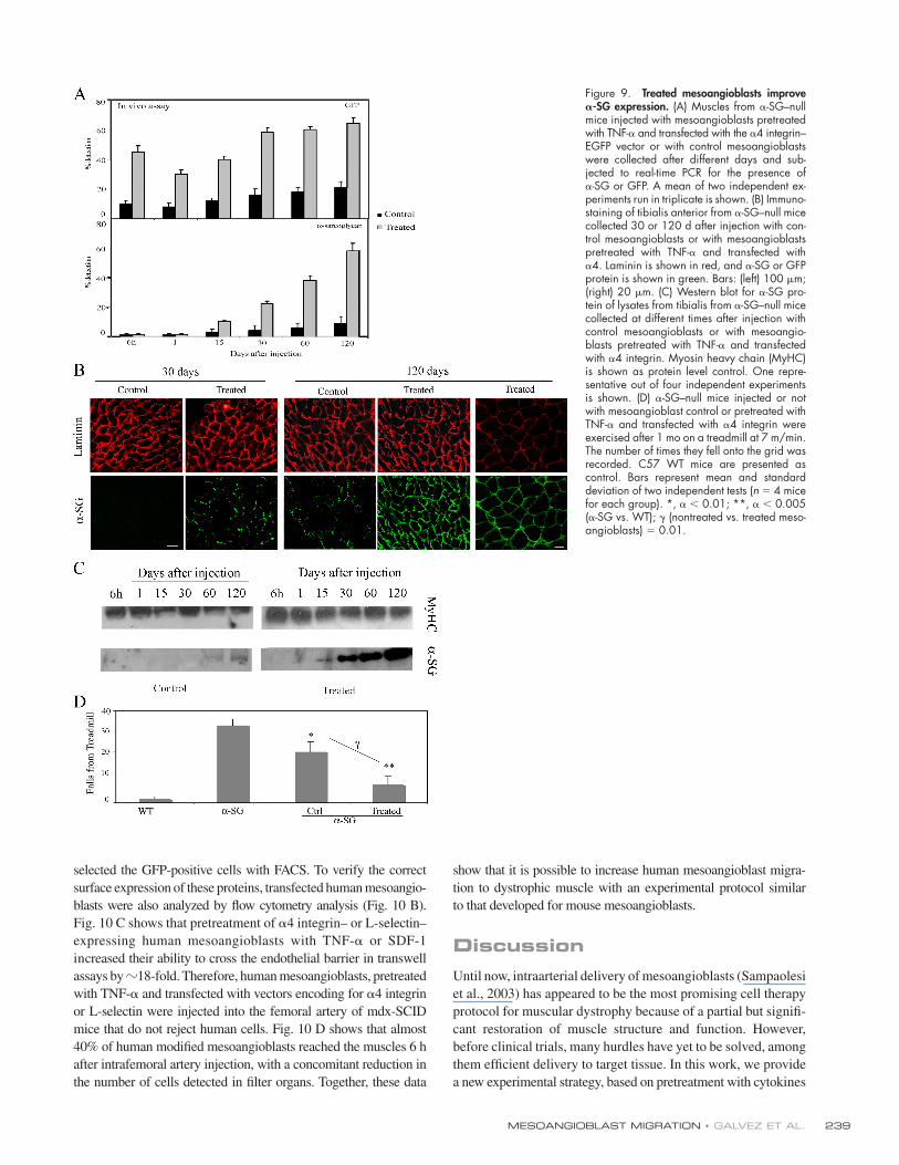
TNF-𝛂–pretreated, 𝛂4 integrin–expressing mesoangioblasts induce enhanced expression of 𝛂-SG5 × 105 TNF-α–pretreated, α4 integrin–expressing mesoangio-
blasts were injected into the femoral artery of α-SG–null mice,
and after 6 h or 1, 15, 30, 60, or 120 d, the tibialis anterior from
treated legs was collected and analyzed by real-time-PCR for
GFP or α-SG. As shown in Fig. 9 A, after an initial modest de-
cline, the number of both control and modifi ed mesoangioblasts
began to increase by 15 d after the injection and reached a plateau
by 1 mo. At all time points, modifi ed mesoangioblasts were
four- to sixfold more numerous than control mesoangioblasts.
Figure 7. Combination of TNF-𝛂 and 𝛂4 integrin increases mesoangio-blast homing. (A) TNF-α– or SDF-1–pretreated mesoangioblasts transfected with vectors expressing α4- and/or L-selectin and GFP were induced to migrate for 6 h through endothelium-coated fi lters. A mean of three independent experiments run in duplicate is shown. *, α < 0.005; **, α < 0.05. (B) TNF-α–pretreated mesoangioblasts transfected with vectors expressing α4 integrin–EGFP were injected through the femoral artery of WT (preinjected with ctx), mdx, or α-SG–null mice, and after 6 h, muscles and fi lter organs were collected and analyzed by real-time PCR for the presence of GFP. A mean of three independent experiments run in triplicate is shown. *, α < 0.01; **, α < 0.05.
JCB • VOLUME 174 • NUMBER 2 • 2006 238
Likewise, α-SG became detectable �15 d after injection, when
mesoangioblasts had been incorporated into regenerating fi bers
and continued to increase thereafter, again at a much higher
level in muscles injected with modifi ed mesoangioblasts. As a
matter of fact, 4 mo after a single injection, the level of
α-SG expression in the tibialis of injected α-SG–null mice was
almost 60% of that detected in the corresponding tibialis from
a WT mouse. At the same time, the level of expression of α-SG
in the tibialis injected with control mesoangioblasts was only
10% of a WT mouse. In agreement with RT-PCR data, α-SG
was detected in almost all (>90%) the muscle fi bers of the tibi-
alis injected with treated mesoangioblasts, whereas a lower per-
centage (�20% with a weaker staining) of positive fi bers was
present in the tibialis injected with control mesoangioblasts
(Fig. 9 B). The newly synthesized α-SG protein was also ana-
lyzed by Western blot. As shown in Fig. 9 C, after 4 mo, the
quantity of protein detected was approximately sixfold higher
in muscles injected with treated mesoangioblasts than with
control cells. To test whether increased expression of α-SG
would correspond to increased motility, mice were subjected to
a running test on a treadmill. The number of times that WT
mice, α-SG–null mice, either untreated or treated with control
or modifi ed (TNF-α–pretreated, α4 integrin–expressing) meso-
angioblasts, fell into the grid when running was recorded.
Fig. 9 D shows that α-SG–null mice injected with modifi ed me-
soangioblasts fell into the grid less frequently than noninjected
or control mesoangioblast–injected α-SG–null mice. After this
exercise test, many of the null mice appeared to be extremely
fatigued and took a relatively long time to recover from the
exercise, whereas treated mice seemed to recover from the
effort much faster. Together, these data show that mesoangio-
blasts pretreated with TNF-α and transfected with α4 integrins
are not only able to more effi ciently reach the damaged muscle
but also to reconstitute SG-positive fi bers with much higher
effi ciency and improved muscle function.
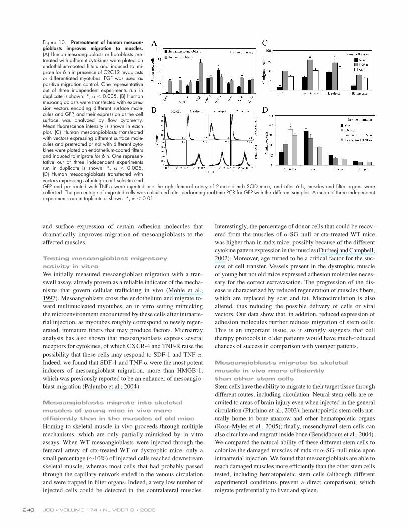
TNF-𝛂–pretreated, 𝛂4 integrin–expressing human mesoangioblasts reach damaged mdx-SCID muscles with higher effi ciencyTo test whether a similar protocol would also be suitable for
human cells, we isolated human mesoangioblasts from small
vessels of biopsies from normal human adult muscle. These
cells have morphology and gene expression similar to their
mouse counterparts, a fi nite lifespan, and a similar ability to
differentiate into different types of muscle cells, osteoblasts,
and adipocytes. Human mesoangioblasts were serum starved
and subjected to transmigration assays using transwell chambers.
As shown in Fig. 10 A, human mesoangioblasts were also
unable to cross endothelium-coated fi lters in absence of stimuli.
However, multinucleated myotubes or pretreatment of cells with
TNF-α or SDF-1 increased human mesoangioblast migration
four- to eightfold without signifi cant enhancement of human
fi broblast migration.
We then transfected human mesoangioblasts (that also do not
express these molecules; unpublished data) with vectors express-
ing L-selectin, α4 integrin, or β2 integrin and, after transfection,
Figure 8. Treated mesoangioblast cross- muscles vessels. (A) Muscles from α-SG–null mice killed 6 h after injection with TNF-α–pretreated mesoangioblasts transfected with α4 integrin–EGFP vectors or with control mesoangioblasts were frozen. Sections were immunostained with anti-GFP, anti-PECAM, and/or anti-laminin antibodies as indicated. DAPI was used for nuclear staining. Bar, 100 μm. (B) Muscles from α-SG–null mice killed 48 h after injection with treated mesoangioblasts were stained with anti-GFP (green), anti-laminin (red), and anti-PECAM (magenta) antibodies. Hoechst was used as nuclear staining. Bar, 100 μm. (C) Muscles from α-SG–null mice killed 2 wk after injection with treated mesoangioblasts were stained with anti-GFP (green), anti-myf5 (red; left), anti-myosin (red; right), and/or anti-laminin (magenta) antibodies. Hoechst was used as nuclear staining. Arrows indicate GFP-positive cells located underneath the muscle fi ber basal lamina and expressing the satellite cell marker Myf5. Bars, 5 μm.
MESOANGIOBLAST MIGRATION • GALVEZ ET AL. 239
selected the GFP-positive cells with FACS. To verify the correct
surface expression of these proteins, transfected human meso angio-
blasts were also analyzed by fl ow cytometry analysis (Fig. 10 B).
Fig. 10 C shows that pretreatment of α4 integrin– or L-selectin–
expressing human mesoangioblasts with TNF-α or SDF-1
increased their ability to cross the endothelial barrier in transwell
assays by �18-fold. Therefore, human mesoangioblasts, pretreated
with TNF-α and transfected with vectors encoding for α4 integrin
or L-selectin were injected into the femoral artery of mdx-SCID
mice that do not reject human cells. Fig. 10 D shows that almost
40% of human modifi ed mesoangioblasts reached the muscles 6 h
after intrafemoral artery injection, with a concomitant reduction in
the number of cells detected in fi lter organs. Together, these data
show that it is possible to increase human mesoangioblast migra-
tion to dystrophic muscle with an experimental protocol similar
to that developed for mouse mesoangioblasts.
DiscussionUntil now, intraarterial delivery of mesoangioblasts (Sampaolesi
et al., 2003) has appeared to be the most promising cell therapy
protocol for muscular dystrophy because of a partial but signifi -
cant restoration of muscle structure and function. However,
before clinical trials, many hurdles have yet to be solved, among
them effi cient delivery to target tissue. In this work, we provide
a new experimental strategy, based on pretreatment with cytokines
Figure 9. Treated mesoangioblasts improve 𝛂-SG expression. (A) Muscles from α-SG–null mice injected with mesoangioblasts pretreated with TNF-α and transfected with the α4 integrin–EGFP vector or with control mesoangioblasts were collected after different days and sub-jected to real-time PCR for the presence of α-SG or GFP. A mean of two independent ex-periments run in triplicate is shown. (B) Immuno-staining of tibialis anterior from α-SG–null mice collected 30 or 120 d after injection with con-trol mesoangioblasts or with mesoangioblasts pretreated with TNF-α and transfected with α4. Laminin is shown in red, and α-SG or GFP protein is shown in green. Bars: (left) 100 μm; (right) 20 μm. (C) Western blot for α-SG pro-tein of lysates from tibialis from α-SG–null mice collected at different times after injection with control mesoangioblasts or with mesoangio-blasts pretreated with TNF-α and transfected with α4 integrin. Myosin heavy chain (MyHC) is shown as protein level control. One repre-sentative out of four independent experiments is shown. (D) α-SG–null mice injected or not with mesoangioblast control or pretreated with TNF-α and transfected with α4 integrin were exercised after 1 mo on a treadmill at 7 m/min. The number of times they fell onto the grid was recorded. C57 WT mice are presented as control. Bars represent mean and standard deviation of two independent tests (n = 4 mice for each group). *, α < 0.01; **, α < 0.005 (α-SG vs. WT); γ (nontreated vs. treated meso-angioblasts) = 0.01.
JCB • VOLUME 174 • NUMBER 2 • 2006 240
and surface expression of certain adhesion molecules that
dramatically improves migration of mesoangioblasts to the
affected muscles.
Testing mesoangioblast migratory activity in vitroWe initially measured mesoangioblast migration with a tran-
swell assay, already proven as a reliable indicator of the mecha-
nisms that govern cellular traffi cking in vivo (Mohle et al.,
1997). Mesoangioblasts cross the endothelium and migrate to-
ward multinucleated myotubes, an in vitro setting mimicking
the microenvironment encountered by these cells after intraarte-
rial injection, as myotubes roughly correspond to newly regen-
erated, immature fi bers that may produce factors. Microarray
analysis has also shown that mesoangioblasts express several
receptors for cytokines, of which CXCR-4 and TNF-R raise the
possibility that these cells may respond to SDF-1 and TNF-α.
Indeed, we found that SDF-1 and TNF-α were the most potent
inducers of mesoangioblast migration, more than HMGB-1,
which was previously reported to be an enhancer of mesoangio-
blast migration (Palumbo et al., 2004).
Mesoangioblasts migrate into skeletal muscles of young mice in vivo more effi ciently than in the muscles of old miceHoming to skeletal muscle in vivo proceeds through multiple
mechanisms, which are only partially mimicked by in vitro
assays. When WT mesoangioblasts were injected through the
femoral artery of ctx-treated WT or dystrophic mice, only a
small percentage (�10%) of injected cells reached downstream
skeletal muscle, whereas most cells that had probably passed
through the capillary network ended in the venous circulation
and were trapped in fi lter organs. Indeed, a very low number of
injected cells could be detected in the contralateral muscles.
Interestingly, the percentage of donor cells that could be recov-
ered from the muscles of α-SG–null or ctx-treated WT mice
was higher than in mdx mice, possibly because of the different
cytokine pattern expression in the muscles (Durbeej and Campbell,
2002). Moreover, age turned to be a critical factor for the suc-
cess of cell transfer. Vessels present in the dystrophic muscle
of young but not old mice expressed adhesion molecules neces-
sary for the correct extravasation. The progression of the dis-
ease is characterized by reduced regeneration of muscles fi bers,
which are replaced by scar and fat. Microcirculation is also
altered, thus reducing the possible delivery of cells or viral
vectors. Our data show that, in addition, reduced expression of
adhesion molecules further reduces migration of stem cells.
This is an important issue, as it strongly suggests that cell
therapy protocols in older patients would have much-reduced
chances of success in comparison with younger patients.
Mesoangioblasts migrate to skeletal muscle in vivo more effi ciently than other stem cellsStem cells have the ability to migrate to their target tissue through
different routes, including circulation. Neural stem cells are re-
cruited to areas of brain injury even when injected in the general
circulation (Pluchino et al., 2003); hematopoietic stem cells nat-
urally home to bone marrow and other hematopoietic organs
(Rosu-Myles et al., 2005); fi nally, mesenchymal stem cells can
also circulate and engraft inside bone (Bensidhoum et al., 2004).
We compared the natural ability of these different stem cells to
colonize the damaged muscles of mdx or α-SG–null mice upon
intraarterial injection. We found that mesoangioblasts are able to
reach damaged muscles more effi ciently than the other stem cells
tested, including hematopoietic stem cells (although different
experimental conditions prevent a direct comparison), which
migrate preferentially to liver and spleen.
Figure 10. Pretreatment of human mesoan-gioblasts improves migration to muscles. (A) Human mesoangioblasts or fi broblasts pre-treated with different cytokines were plated on endothelium-coated fi lters and induced to mi-grate for 6 h in presence of C2C12 myoblasts or differentiated myotubes. FGF was used as positive migration control. One representative out of three independent experiments run in duplicate is shown. *, α < 0.005. (B) Human mesoangioblasts were transfected with expres-sion vectors encoding different surface mole-cules and GFP, and their expression at the cell surface was analyzed by fl ow cytometry. Mean fl uorescence intensity is shown in each plot. (C) Human mesoangioblasts transfected with vectors expressing different surface mole-cules and pretreated or not with different cyto-kines were plated on endothelium-coated fi lters and induced to migrate for 6 h. One represen-tative out of three independent experiments run in duplicate is shown. *, α < 0.005. (D) Human mesoangioblasts transfected with vectors expressing α4 integrin or L-selectin and GFP and pretreated with TNF-α were injected into the right femoral artery of 2-mo-old mdx-SCID mice, and after 6 h, muscles and fi lter organs were collected. The percentage of migrated cells was calculated after performing real-time PCR for GFP with the different samples. A mean of three independent experiments run in triplicate is shown. *, α < 0.01.
MESOANGIOBLAST MIGRATION • GALVEZ ET AL. 241
Enhancing mesoangioblast migratory activityPrevious work has shown that HMGB-1 stimulates proliferation
and migration of mesoangioblasts to dystrophic muscles that
expresses this molecule (Palumbo et al., 2004). We confi rm
those data; however, in a screen for factors and cytokines for
which mesoangioblasts are known to express receptors (Tagliafi co
et al., 2004), we observed that SDF-1 and TNF-α are more
potent as chemoattractants. Interestingly, our work shows that
pretreatment of donor cells with these cytokines is suffi cient to
activate or increase expression of certain surface proteins neces-
sary for the migration and extravasation process, immediately
suggesting a simple ex vivo treatment of cells that does not re-
quire expression in target muscles. We further investigated the
mechanisms involved in this migration increase through a low-
density array hybridization performed with mesoangioblasts
pretreated with TNF-α or SDF-1. The role of the different genes
modifi ed by the treatment was studied in the transmigration
assay using inhibitors or blocking antibodies. This experiment
revealed that TNF-α acts via an MMP- and CD44-dependent
mechanism, whereas the effect of SDF-1 was mediated by
MMPs and αv integrins.
Mesoangioblasts do not normally express many of the
leukocytes molecules needed for an effi cient rolling and arrest
at blood vessels, which may explain why most of the injected
cells do not bind effi ciently, fl ow through the muscle capillary
net, and are fi nally trapped inside fi lter organs. Therefore,
L-selectin, α4 integrin, and β2 integrin were expressed in me-
soangioblasts, forming dimers with other integrins (such as β1,
β7 or αl, αm) already expressed by these cells. Expression of
L-selectin and α4 integrin did not alter the myogenic differenti-
ation of mesoangioblasts, whereas expression of β2 integrin
did, a fact that precludes future use in vivo. The expression of
α4 integrin or L-selectin resulted in an eightfold increase of
mesoangioblast transmigration in vitro and a twofold increase
in the number of mesoangioblasts that reached the damaged
muscles, simultaneously reducing their trapping in fi lter organs.
The combined strategies (i.e., pretreatment with TNF-α of
mesoangioblasts expressing α4 integrin) resulted in a 15-fold
increase of transmigration through endothelium in vitro and
more than fi vefold enhanced migration in vivo, with >50%
of injected cells now present into the downstream muscles.
Immunohistology confi rmed these data, as many more modifi ed
mesoangioblasts could be detected in muscle interstitium and,
later, within muscle fi bers (as detected by expression of
α-SG) or as satellite cells.
Increased migration corresponds to increased muscle repair and motilityIncreasing migration to the target tissue has two positive out-
comes: (1) it delivers more cells to repair the target tissue and (2)
it reduces the number of cells that are trapped in fi lter organs.
This number is large, and in the case of lungs, it may cause respi-
ratory problems that are not evident in mice. In addition, human
mesoangioblasts have a fi nite number of cell divisions, and this
may in the end represent a limiting factor if most of them are
wasted outside the target tissue. However, increased migration
does not necessarily mean increased tissue repair. In principle, if
more cells reach the target tissue, they may undergo a reduced
number of divisions in vivo and thus give rise to a similar number
of differentiated cells. Our data show that this is not the case.
We previously showed that mesoangioblasts are able to
regenerate in part the damaged muscles after three injections
and long periods (Sampaolesi et al., 2003). Other types of stem
cells have also been shown to reconstitute the skeletal muscle of
dystrophic mice to various extents (Corbel et al., 2003; De Bari
et al., 2003), but a quantitative analysis was not reported.
Mesoangioblasts treated with TNF-α and expressing α4 integ-
rin were able to reach damaged muscles and to produce α-SG 30 d
after injection with dramatically increased effi ciency, allowing
treated α-SG–null mice to improve the performance of exercise.
4 mo after a single injection of modifi ed mesoangioblasts,
α-SG–null mice expressed almost 60% of the α-SG detected in a
WT mouse. Immunostaining of α-SG showed a partial recovery
after 30 d and >90% of well-structured fi bers expressing α-SG
after 4 mo. This corresponded to a signifi cant increase in the
ability to run on a treadmill, despite a unilateral treatment.
In the perspective of a cell therapy in patients, it is important
to test whether a similar protocol will also work with human cells.
Human mesoangioblasts have been recently isolated from frag-
ments of vessels inside biopsies of postnatal human skeletal
muscle. Regarding the use of these cells in the present study,
it is important to state that they have a morphology and marker
expression similar to their murine counterparts, a fi nite lifespan
(at variance with their murine counterparts), and a similar ability to
differentiate into different types of solid mesoderm (unpublished
data). Human mesoangioblasts were also exposed to TNF-α,
which in the case of human cells was more potent than SDF-1, and
transfected with either α4 integrin or L-selectin and, under these
conditions, reached skeletal muscles of mdx-SCID mice threefold
more effi ciently than their corresponding untreated cells. Although
these data suggest that further work may still be required to opti-
mize human mesoangioblast migration to skeletal muscle, they
clearly indicate that the basic migration mechanisms are similar in
mouse and human mesoangioblasts and equally susceptible to
enhancement by similar cytokines and adhesion molecules.
In conclusion, our results suggest that pretreatment of
mesoangioblasts with TNF-α and the presence of α4 integrin
are required for the effi cient delivery of stem cells to injured
muscles in the course of muscular dystrophy. Pretreatment of
stem cells with cytokines is a simple procedure, and inclusion
of a relatively small cDNA in a viral vector, designed for gene
correction, is also a feasible procedure in the context of autolo-
gous cell therapy with engineered stem cells. Together, these
strategies represent a novel and simple method for improving the
migration of stem cells to damaged tissue and could be tailored
in the future to different types of stem cells in different cell ther-
apy protocols requiring migration to other target tissues such as
the heart or the bone.
Materials and methodsReagentsAnti-GFP antibody and anti-isotype control antibodies were purchased from Chemicon. Anti–α4, anti–αv, and anti–β2 integrins; anti–L-selectin;
JCB • VOLUME 174 • NUMBER 2 • 2006 242
anti-CD44; anti-FGFR1; anti–VCAM-1; and anti–ICAM-1 antibodies were obtained from Serotec. Anti–L-selectin mAb Mel-14 was obtained from American Type Culture Collection. Anti–E- and anti–P-selectin mAbs (RME-1 and RMP-1) were obtained as previously described (Walter et al., 1997; Souza et al., 1999). Anti–MAdCAM-1 and anti-PECAM were provided by E. Butcher (Stanford University, Stanford, CA) and E. Dejana (FIRC Institute of Molecular Oncology, Milan, Italy). Anti–α-SG and anti-myf5 antibodies were purchased from NovoCastra.
Most reagents and some antibodies were purchased from Sigma- Aldrich, unless stated otherwise. All the cytokines used were obtained from PeproTech. GM1489, the MMP inhibitor, was obtained from Calbiochem. Alexa 488 and Alexa F 647 labeling kit were obtained from Invitrogen.
mdx and C57BL10 WT mice were purchased from Charles River Lab-oratories, and homo-EGFP mice were provided by A. Nagy (Mount Sinai Hospital, Toronto, Canada; Hadjantonakis et al., 1998). α-SG–null mice were a gift from K. Campbell (University of Iowa, Iowa City, IA; Sampaolesi et al., 2003). All mice were handled following institutional guidelines.
Cell culturesC2C12 mouse myoblasts or L6 rat myoblasts were cultured in Dulbecco’s minimal essential medium plus 20% FBS and induced to differentiate in Dulbecco’s minimal essential medium plus 2% FBS. D16 mice mesoangio-blasts were isolated from the dorsal aorta of mouse embryos (embryonic day 9.5), cloned, and expanded as described previously (Minasi et al., 2002). Mesoangioblasts transduced with a lentiviral vector encoding the GFP protein were used for some experiments. Murine microvascular endo-thelial H5V cells (a gift form E. Dejana, FIRC Institute of Molecular Oncol-ogy) and mouse embryonic fi broblasts 3T3 were grown in Dulbecco’s minimal essential medium plus 10% FBS and starved for 12 h before func-tional experiments. Mesenchymal and hematopoietic stem cells were iso-lated from homo-EGFP mice using the SpinSep TM kit (StemCell Technologies, Inc.) and checked for purity according to the manufacturer’s instructions. Neural stem cells were isolated from the forebrains of embryos (embryonic day 11.5) as previously described (Galli et al., 2000).
ConstructsMurine or human β2 integrin, L-selectin, and α4 integrin mRNA cloned in a pT7T3D-Pac1 vector were purchased from Research Genetics. Inserts were cloned into pIRES2-EGFP expression vector from CLONTECH Labora-tories, Inc., and transfected into mice or human mesoangioblasts with Lipo-fectamine (Invitrogen). GFP-positive mesoangioblasts were sorted and used for the different experiments.
Differentiation assaysTreated mesoangioblasts were cocultured with L6 cells in low serum (2%). After 3 d, dishes were fi xed with 4% PFA, and the number of mouse nuclei stained with DAPI inside myosin-positive cells was counted. The percentage of differentiation was calculated against the total number of cells.
Real-time PCRTotal RNA from different organs was isolated with TRIzol protocol (Invitrogen) and reverse transcribed by Taqman kit (Platinum Taq DNA polymerase; Invitrogen). RT-PCR was performed for E- and P-selectin (E-selectin primers: Fw, C C A T G T T C C A C G T C A A G G A C C ; Rw, G C C A T G T G A T A G G C C A C A G ; and P-selectin primers: Fw, C C A G T A T C T A G A C C C G A A G G ; Rw, G C C A T T-C T C T C T C C T C G A A A C A C ), and mRNA expression was analyzed by aga-rose gel. Alternatively, real-time quantitative PCR was done on a real-time PCR system (Mx3000P; Stratagene). Each cDNA sample was amplifi ed in duplicate by using the SYBR Green Supermix (Bio-Rad Laboratories) for GFP or ε chromosome detection (GFP primers: Fw, A A G T T C A T C T G C A C C-A C C G ; Rw, T C C T T G A A G A A G A T G G T G C G ; and ε chromosome primers: Fw, A A G C G A C C C A T G A A C G C A T T ; Rw, T T C G G G T A T T T C T C T C T G T C ) or the Taqman Universal PCR master mixture containing AmpliTaq Gold DNA with commercial primers (Applied Biosystems) for α-SG detection. Data are expressed as the percentage of migrated cells, which is calculated by comparing the concentration of target genes in our sample with the total input of injected cells. For the Taqman assay, the level of α-SG detected represents the specifi c signal detected in the sample (mesoangioblast-injected α-SG–null mouse tibialis anterior) compared with a positive control (WT mouse tibialis anterior; i.e., 100% of positive fi bers).
In vitro transwell migration assay8-μm transwell fi lters (Corning) were coated with 1% gelatin, and endothe-lial cells H5V (previously preactivated by 12 h of exposure to TNF-α) were plated to confl uence on them for 24 h. Confl uence of the endothelial mono-layer was assessed by measuring the diffusion of BSA from the upper to the
lower chamber. At the same time, C2C12 were grown on a p24w plate with or without differentiated medium for 0–4 d. 104 mice mesoangio-blasts, WT or pretreated or transfected, were plated in Dulbecco’s minimal essential medium containing 2% serum on the upper side of transwell chamber 24 h after plating H5V cells, and the chamber was then moved to the wells containing differentiated C2C12 or different cytokines. After 6 h of transmigration, migrated mesoangioblasts on the lower side of the fi lter were fi xed in 4% paraformaldehyde, stained with crystal violet, and counted using an inverted microscope (fi ve random fi elds of the lower face of the transwell membrane at 20× magnifi cation). The results show migrated cells as a percentage of the total number of input cells.
In vivo migration assay2- or 8-mo-old WT (injected slowly through all the anterior surface of the tibialis with a 27-gauge needle containing 100 μl of 5 μM ctx [Sigma- Aldrich] 12 h before treatment), mdx, or α-SG–null mice were injected through the right femoral artery with 5 × 10⁵ mouse mesoangioblasts. After 6, 12, or 24 h, animals were killed, and different muscles (quadri-ceps, gastrocnemius, and tibialis) or fi ltered organs (liver, lung, and spleen) were collected. RNA was extracted, and a real-time PCR for GFP was per-formed in all the samples as described (see Real-time PCR). Data are repre-sented as a percentage of migrated cells (percentage of GFP detected) to the different organs relative to the input value.
Intravital microscopy50 μg of Alexa 488–labeled mAbs anti–E- or anti–P-selectin or anti– MAdCAM-1 were injected through the tail vein of α-SG–null mice. 20 min later, the animal was anesthetized and perfused through the left ventricle with cold PBS (Homeister et al., 1998). Blood vessels of striated muscles were visualized with a silicon-intensifi ed target video camera (VE-1000 SIT; Dage-MTI) and monitor (SSM-125CE; Sony).
Microarray analysisFor mRNA analysis, total RNA was extracted from treated mesoangioblasts using TRIzol. cDNA was prepared by reverse-transcription reaction and hybridized to the GEArray gene expression array MM-010 (SuperArray) for extracellular matrix and adhesion molecules according to the manufac-turer’s instruction. Data were subjected to densitometric analysis using GEArray Expression Analysis Suite (SuperArray). RNA levels were expressed as the relative density after normalizing the hybridization signal to β-actin.
Western blotMesoangioblasts were transfected with the different constructs (see Constructs) and lysed directly in Laemmli buffer on ice. Lysates were resolved on 10% SDS-PAGE under reducing conditions, and proteins were transferred to nitrocellulose membrane (Hybond-ECL; GE Healthcare). Membranes were revealed with anti-GFP and anti–α4 or anti–β2 integrins or anti–L-selectin antibodies.
Flow cytometryMesoangioblasts pretreated with different cytokines or transfected with dis-tinct constructs were detached with PBS plus 5 mM EDTA on ice and incu-bated with antibodies against the different surface molecules for 30 min at 4°C. Cells were then incubated with a Alexa 488–conjugated anti–mouse Ig. Finally, fl uorescent samples were analyzed in a FACSCalibur fl ow cytometer (Becton Dickinson).
Cytokines measurementsSupernatants from fi broblasts, myoblasts, or myotube cultures were col-lected after 4 d of culture and analyzed with the Beadlyte mouse multicytokine-detection system (Upstate) for measuring cytokines according to the manufacturer’s instructions. Muscles from different animal models were homogenized and cytokines were measured using the same kit protocol.
InmunohistologyTibialis anterior muscles of α-SG–null mice were removed from mice previ-ously injected with mesoangioblasts and frozen in liquid N2 cooled isopentane. Serial muscle sections were fi xed with 4% PFA, permeabilized, satured, and immunostained as previously described (Sampaolesi et al., 2003) with anti-GFP, anti–α-SG, anti-laminin, anti-myosin, anti-PECAM, or anti-myf5 antibodies. Alexa 488 or 594 or Cyane (Invitrogen) were used as secondary staining Ig and DAPI for nuclear staining. Isotype control antibodies were used as negative staining. Images were taken with a microscope (S100 TV; Carl Zeiss MicroImaging, Inc.).
MESOANGIOBLAST MIGRATION • GALVEZ ET AL. 243
Treadmill testα-SG–null mice (n = 4 for each of the four groups) injected or not with control or treated mesoangioblasts were run on a treadmill (Columbus Instruments) set at 7 m/min for 2 min before and after 1 mo from injection. WT C57 mice are also presented as healthy control. The back of the tread-mill was equipped with a grid that discharged a mild current, a stimulus designed to motivate the animal to keep running on the treadmill. Performance was measured by the number of times a mouse failed to stay on the running belt and fell into the stimulus grid. Running time was limited to 2 min because the majority of the α-SG–null mice could not run any longer.
Statistical analysisStatistical signifi cance of the differences between the percentage values was assessed by using the Kruskal-Wallis one-way analysis of variance by ranks test. α represents the signifi cance in each independent case.
We thank E. Dejana, T. Lapidot, and R. Pardi for advice and help with the work; K. Campbell for the α-SG–null mice; and E. Butcher for the anti–MadCAM-1 mAb.
This work was supported by grants from Cassa di Risparmio Provincie Lombarde, Telethon, Association Francoise contra les Myopathies, Muscular Dystrophy Association, Duchenne Parent Project, Fondation Leducq, European Community, CariVerona, and Italian Ministries of Health and Research. Firb. B.G. Galvez was supported by a Programme 3+3 fellowship from the Centro Nacional de Investigaciones Cardiovasculares, Spain.
Submitted: 15 December 2005Accepted: 16 June 2006
ReferencesBeauchamp, J.R., L. Heslop, D.S.W. Yu, S. Tajbakhsh, R.G. Kelly, A. Wernig,
M.E. Buckingham, T.A. Partridge, and P.S. Zammit. 2000. Expression of CD34 and Myf5 defi nes the majority of quiescent adult skeletal muscle satellite cells. J. Cell Biol. 151:1221–1234.
Bensidhoum, M., A. Chapel, S. Francois, C. Demarquay, C. Mazurier, L. Fouillard, S. Bouchet, J. Bertho, P. Gourmelon, J. Aigueperse, et al. 2004. Homing of in vitro expanded Stro-1-or Stro-1+ human mesenchymal stem cells into the NOD/SCID mouse and their role in supporting human CD34 cell engraftment. Blood. 103:3313–3319.
Butcher, E. 1991. Leukocyte-endothelial cell recognition: three (or more) steps to specifi city and diversity. Cell. 67:1033–1036.
Corbel, S., A. Lee, L. Yi, J. Duenas, T. Brazelton, H. Blau, and F. Rossi. 2003. Contribution of hematopoietic stem cells to skeletal muscle. Nat. Med. 9:1528–1532.
De Bari, C., F. Dell’Accio, F. Vandenabeele, J. Vermeesch, J. Raymackers, and F. Luyten. 2003. Skeletal muscle repair by adult human mesenchymal stem cells from synovial membrane. J. Cell Biol. 160:909–918.
Durbeej, M., and K. Campbell. 2002. Muscular dystrophies involving the dystrophin-glycoprotein complex: an overview of current mouse models. Curr. Opin. Genet. Dev. 12:349–361.
Fu, S., and J. Liesveld. 2000. Mobilization of hematopoietic stem cells. Blood Rev. 14:205–218.
Galli, R., U. Borello, A. Gritti, M. Minasi, C. Bjornson, M. Coletta, M. Mora, M.D. Angelis, R. Fiocco, G. Cossu, and A. Vescovi. 2000. Skeletal myo-genic potential of human and mouse neural stem cells. Nat. Neurosci. 3:986–991.
Grabovsky, V., S. Feigelson, C. Chen, D. Bleijs, A. Peled, G. Cinamon, F. Baleux, F. Arenzana-Seisdedos, T. Lapidot, Y. van Kooyk, et al. 2000. Subsecond induction of α4 integrin clustering by immobilized chemokines stimu-lates leukocyte tethering and rolling on endothelial vascular cell adhesion molecule 1 under fl ow conditions. J. Exp. Med. 192:495–506.
Hadjantonakis, A., M. Gertsenstein, M. Ikawa, M. Okabe, and A. Nagy. 1998. Generating green fl uorescent mice by germline transmission of green fl uorescent ES cells. Mech. Dev. 76:79–90.
Homeister, J., M. Zhang, P. Frenette, R. Hynes, D. Wagner, J. Lowe, and R. Marks. 1998. Overlapping functions of E- and P-selectin in neutrophil recruitment during acute infl ammation. Blood. 92:2345–2352.
Laterveer, L., I. Lindley, D. Heemskerk, J. Camps, E. Pauwels, R. Willemze, and W. Fibbe. 1996. Rapid mobilization of hematopoietic progenitor cells in rhesus monkeys by a single intravenous injection of interleukin-8. Blood. 87:781–788.
Minasi, M., M. Riminucci, L.D. Angelis, U. Borello, B. Berarducci, A. Innocenzi, A. Caprioli, D. Sirabella, M. Baiocchi, R.D. Maria, et al. 2002. The meso-angioblast: a multipotent, self-renewing cell that originates from the dorsal
aorta and differentiates into most mesodermal tissues. Development. 129:2773–2783.
Mohle, R., M. Moore, R. Nachman, and S. Rafi i. 1997. Transendothelial migra-tion of CD34+ and mature hemopoietic cells: an in vitro study using a bone marrow endothelial cell line. Blood. 89:72–79.
Palumbo, R., M. Sampaolesi, F.D. Marchis, R. Tonlorenzi, S. Colombetti, A. Mondino, G. Cossu, and M. Bianchi. 2004. Extracellular HMGB1, a sig-nal of tissue damage, induces mesoangioblast migration and prolifera-tion. J. Cell Biol. 164:441–449.
Peled, A., V. Grabovsky, L. Habler, J.S. Arenzana-Seisdedos, I. Petit, H. Ben-Hur, T. Lapidot, and R. Alon. 1999. The chemokine SDF-1 stimulates integrin-mediated arrest of CD34+ cells on vascular endothelium under shear fl ow. J. Clin. Invest. 104:1199–1211.
Pluchino, S., A. Quattrini, E. Brambilla, A. Gritti, G. Salani, G. Dina, R. Galli, U.D. Carro, S. Amadio, A. Bergami, et al. 2003. Injection of adult neuro-spheres induces recovery in a chronic model of multiple sclerosis. Nature. 422:688–694.
Rosu-Myles, M., E. Stewart, J. Trowbridge, C. Ito, P. Zandstra, and M. Bhatia. 2005. A unique population of bone marrow cells migrates to skeletal mus-cle via hepatocyte growth factor/c-met axis. J. Cell Sci. 118:4343–4352.
Sampaolesi, M., Y. Torrente, A. Innocenzi, R. Tonlorenzi, G. D’Antona, M. Pellegrino, R. Barresi, N. Bresolin, M.D. Angelis, K. Campbell, et al. 2003. Cell therapy of alpha-sarcoglycan null dystrophic mice through intra-arterial delivery of mesoangioblasts. Science. 301:487–492.
Souza, H., C. Elia, J. Spencer, and T. MacDonald. 1999. Expression of lym-phocyte-endothelial receptor-ligand pairs, alpha4beta7/MAdCAM-1 and OX40/OX40 ligand in the colon and jejunum of patients with infl amma-tory bowel disease. Gut. 45:856–863.
Tagliafi co, E., S. Brunelli, A. Bergamaschi, L.D. Angelis, R. Scardigli, D. Galli, R. Battini, P. Bianco, S. Ferrari, G. Cossu, and S. Ferrari. 2004. TGFbeta/BMP activate the smooth muscle/bone differentiation programs in meso-angioblasts. J. Cell Sci. 117:4377–4388.
Walter, U., L. Ayer, A. Manning, P. Frenette, D. Wagner, R. Hynes, and B. Wolitzky. 1997. Generation and characterization of a novel adhesion function blocking monoclonal antibody recognizing both rat and mouse E-selectin. Hybridoma. 16:355–361.
Copyright © 2022 FDOKUMEN