When ‘slime’becomes ‘smile’: Developmental letter position dyslexia in English
Upload
khangminh22Category
view
0download
0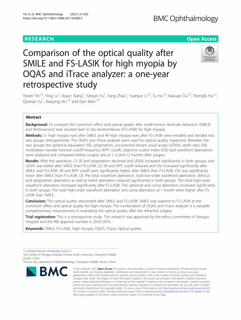
RESEARCH Open Access
Comparison of the optical quality afterSMILE and FS-LASIK for high myopia byOQAS and iTrace analyzer: a one-yearretrospective studyYewei Yin1,2, Ying Lu1, Aiqun Xiang1, Yanyan Fu1, Yang Zhao1, Yuanjun Li1,2, Tu Hu1,2, Kaixuan Du1,2, Shengfa Hu1,2,Qiuman Fu1, Xiaoying Wu1,2 and Dan Wen1,2*
Abstract
Background: To compare the correction effect and optical quality after small-incision lenticule extraction (SMILE)and femtosecond laser assisted laser in situ keratomileusis (FS-LASIK) for high myopia.
Methods: 51 high myopia eyes after SMILE and 49 high myopia eyes after FS-LASIK were enrolled and divided intotwo groups retrospectively. The OQAS and iTrace analyzer were used for optical quality inspection. Between thetwo groups the spherical equivalent (SE), astigmatism, uncorrected distant visual acuity (UDVA), strehl ratio (SR),modulation transfer function cutoff frequency (MTF cutoff), objective scatter index (OSI) and wavefront aberrationswere analyzed and compared before surgery and at 1, 6 and 12 months after surgery.
Results: After the operation: (1) SE and astigmatism declined and UDVA increased significantly in both groups, andUDVA was better after SMILE than FS-LASIK. (2) SR and MTF cutoff reduced and OSI increased significantly afterSMILE and FS-LASIK. SR and MTF cutoff were significantly higher after SMILE than FS-LASIK. OSI was significantlylower after SMILE than FS-LASIK. (3) The total wavefront aberration, total low-order wavefront aberration, defocusand astigmatism aberration as well as trefoil aberration reduced significantly in both groups. The total high-orderwavefront aberration increased significantly after FS-LASIK. The spherical and coma aberration increased significantlyin both groups. The total high-order wavefront aberration and coma aberration at 1 month were higher after FS-LASIK than SMILE.
Conclusion: The optical quality descended after SMILE and FS-LASIK. SMILE was superior to FS-LASIK at thecorrection effect and optical quality for high myopia. The combination of OQAS and iTrace analyzer is a valuablecomplementary measurement in evaluating the optical quality after the refractive surgery.
Trial registration: This is a retrospective study. This research was approved by the ethics committee of XiangyaHospital and the IRB approval number is 201612074.
Keywords: SMILE, FS-LASIK, High myopia, OQAS, iTrace, Optical quality
© The Author(s). 2021 Open Access This article is licensed under a Creative Commons Attribution 4.0 International License,which permits use, sharing, adaptation, distribution and reproduction in any medium or format, as long as you giveappropriate credit to the original author(s) and the source, provide a link to the Creative Commons licence, and indicate ifchanges were made. The images or other third party material in this article are included in the article's Creative Commonslicence, unless indicated otherwise in a credit line to the material. If material is not included in the article's Creative Commonslicence and your intended use is not permitted by statutory regulation or exceeds the permitted use, you will need to obtainpermission directly from the copyright holder. To view a copy of this licence, visit http://creativecommons.org/licenses/by/4.0/.The Creative Commons Public Domain Dedication waiver (http://creativecommons.org/publicdomain/zero/1.0/) applies to thedata made available in this article, unless otherwise stated in a credit line to the data.
* Correspondence: [email protected] Center of Xiangya Hospital, Central South University, Changsha 410008,Hunan, China2Hunan Key Laboratory of Ophthalmology, Changsha 410008, Hunan, China
Yin et al. BMC Ophthalmology (2021) 21:292 https://doi.org/10.1186/s12886-021-02048-5
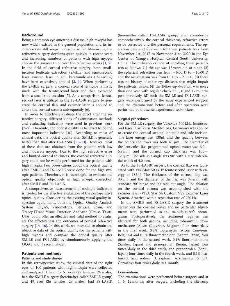
BackgroundBeing a common eye ametropia disease, high myopia hasnow widely existed in the general population and its in-cidence rate still keeps increasing so far. Meanwhile, therefractive surgery develops quite quickly in recent yearsand increasing numbers of patients with high myopiachoose the surgery to correct the refractive errors [1, 2].In the field of corneal refractive surgery, both small-incision lenticule extraction (SMILE) and femtosecondlaser assisted laser in situ keratomileusis (FS-LASIK)have been extensively applied [3, 4]. When performingthe SMILE surgery, a corneal stromal lenticule is firstlymade with the femtosecond laser and then extractedfrom a small side incision [5]. As a comparison, femto-second laser is utilized in the FS-LASIK surgery to gen-erate the corneal flap, and excimer laser is applied toablate the corneal stroma subsequently [6].In order to effectively evaluate the effect after the re-
fractive surgery, different kinds of examination methodsand evaluating indicators were used in previous work[7–9]. Thereinto, the optical quality is believed to be themost important indicator [10]. According to most ofclinical data, the optical quality after SMILE is generallybetter than that after FS-LASIK [11–13]. However, mostof these data are obtained from the patients with lowand moderate myopia. Due to the high refractive errorand limited corneal thickness, the corneal refractive sur-gery could not be widely performed for the patients withhigh myopia. Few observations about the optical qualityafter SMILE and FS-LASIK were done for the high my-opic patients. Therefore, it is meaningful to evaluate theoptical quality objectively in high myopia correctionafter SMILE and FS-LASIK.A comprehensive measurement of multiple indicators
is needed for the effective evaluation of the postoperativeoptical quality. Considering the existing visual quality in-spection equipments, both the Optical Quality AnalysisSystem (OQAS, Visiometrics, Terrassa, Spain) andTracey-iTrace Visual Function Analyzer (iTrace, Texas,USA) could offer an effective and valid method to evalu-ate the effectiveness and outcomes of corneal refractivesurgery [14–16]. In this work, we intended to obtain theobjective data of the optical quality for the patients withhigh myopia and compare the optical quality afterSMILE and FS-LASIK by simultaneously applying theOQAS and iTrace analyzer.
Patients and methodsPatients and study designIn this retrospective study, the clinical data of the righteyes of 100 patients with high myopia were collectedand analyzed. Thereinto, 51 eyes (27 females, 24 males)had the SMILE surgery (hereinafter called SMILE group)and 49 eyes (26 females, 23 males) had FS-LASIK
(hereinafter called FS-LASIK group) after consideringcomprehensively the corneal thickness, refractive errorsto be corrected and the personal requirements. The op-eration date and follow-up for these patients was fromNovember 1st, 2017 to November 31st, 2020 in the EyeCenter of Xiangya Hospital, Central South University,China. The inclusion criteria of enrolling these patientswas as follows: (1) the age was 18 years old or older, (2)the spherical refraction was from − 6.00 D to − 10.00 Dand the astigmatism was from 0 D to − 2.50 D, (3) therewas no history of other eye diseases that might impairthe patients’ vision, (4) the follow-up duration was morethan one year with regular check at 1, 6 and 12monthspostoperatively, (5) both the SMILE and FS-LASIK sur-gery were performed by the same experienced surgeonand the examinations before and after operation wereperformed by the same experienced technicians.
Surgical proceduresFor the SMILE surgery, the VisuMax 500 kHz femtosec-ond laser (Carl Zeiss Meditec AG, Germany) was appliedto create the corneal stromal lenticule and side incision.The laser energy was 130nJ, and the spacing betweenthe points and rows was both 4.5 μm. The diameter ofthe lenticules (i.e. programmed optical zone) was 6.0 ~6.8 mm, and the corneal cap thickness was 100 ~120 μm. The side-cut angle was 90° with a circumferen-tial width of 4.0 mm.As to the FS-LASIK surgery, the corneal flap was fabri-
cated with VisuMax 500 kHz femtosecond laser with en-ergy of 185nJ. The thickness of the corneal flap was90 μm, and the diameter of the flap was 8.0 mm withstandard 90° hinge and 90° side-cut angle. The ablationon the corneal stroma was accomplished with theexcimer laser (VISX Star S4 Custom VSS Excimer LaserSystem, America) with a repetition rate of 250 Hz.In the SMILE and FS-LASIK surgery the treatment
center was the corneal vertex and no particular adjust-ments were performed to the manufacturer’s nomo-grams. Postoperatively, the treatment regimen wasidentical for both groups, including tobramycin dexa-methasone (Alcon Couvreur, Belgium) four times dailyin the first week, 0.3% tobramycin (Alcon Couvreur,Belgium) and 0.1% fluorometholone (Santen, Japan) fourtimes daily in the second week, 0.1% fluorometholone(Santen, Japan) and pranoprofen (Senju, Japan) fourtimes daily in the third week, and pranoprofen (Senju,Japan) four times daily in the fourth week, and 0.1% hya-luronic acid sodium (Ursapharm Arzneimittel GmbH,Germany) four times daily in a month.
ExaminationsThe examinations were performed before surgery and at1, 6, 12 months after surgery, including the slit-lamp
Yin et al. BMC Ophthalmology (2021) 21:292 Page 2 of 10
examination, the eyesight test using the standard loga-rithmic visual acuity chart and snellen visual acuitychart, the manifest refraction, the OQAS examinationand iTrace analyzer examination. The OQAS was ap-plied with 4.0 mm pupil diameter to get the parametersincluding strehl ratio (SR), modulation transfer functioncutoff frequency (MTF cutoff, c/deg) and objective scat-ter index (OSI). The iTrace analyzer was used with 4.0mm pupil diameter to get the data of the total ocularwavefront aberrations, including the total wavefront ab-erration (TWA), total low-order wavefront aberration(TLOA), total high-order wavefront aberration (THOA),defocus aberration (DA), astigmatism aberration (AA),spherical aberration (SA), coma aberration (CA) and tre-foil aberration (TA). The examinations were completedby the same technicians respectively.
Statistical analysesThe SPSS statistical package (Version 25.0; IBM SPSSInc., Chicago, Illinois, USA) and the Microsoft ExcelSoftware were applied for the statistical analysis. Theone-way repetitive measurement and analysis of variancewas applied to analyze the difference of spherical equiva-lent (SE), astigmatism, uncorrected distant visual acuity(UDVA), SR, MTF cutoff, OSI and wavefront aberrationsbefore surgery and at 1, 6 and 12months after surgerywhen the data followed the law of normal distribution.Otherwise, the Friedman test was used. The paired t-testwas used to analyze the difference of age, SE,
astigmatism, UDVA, SR, MTF cutoff, OSI and wavefrontaberrations at each point of time between the twogroups. And the multiple-factor repetitive measurementand analysis of variance was used to compare the preand post-operative variation of SE, astigmatism, UDVA,SR, MTF cutoff, OSI and wavefront aberrations betweenthe SMILE and FS-LASIK group. p < 0.05 was taken asbeing statistically significant, and the adjusted alpha i.e.,alpha/no. of test, was adopted for the analysis with re-peated measurement.
ResultsDescriptive statistics of the patients, UDVA and the otherrefractive outcomesNo intraoperative or postoperative complications, suchas corneal infection, haze, corneal epithelial ingrowthand elevated intraocular pressure, occurred in both theSMILE and FS-LASIK group. As summarized in Table 1,there were no significant differences between the twogroups in terms of age, preoperative and postoperativeSE and astigmatism. Both SE and astigmatism signifi-cantly decreased after SMILE and FS-LASIK (p < 0.05).Between the two groups there were no significant differ-ences in SE and astigmatism at each point of time, andno significant differences in SE and astigmatism’s vari-ation before and after the operation were found (pSE =0.119, pastig = 0.782). The preoperative UDVA had nosignificant difference between the two groups and in-creased significantly after operation until the 12th
Table 1 The preoperative and postoperative demographics (mean ± SD) for the SMILE and FS-LASIK group
Parameters SMILE group FS-LASIK group p1-value
Age (years old) 23.9 ± 4.8 24.4 ± 4.9 0.605
SE(D) Preop −7.964 ± 0.943 −8.390 ± 1.372 0.078
Post 1mo −0.194 ± 0.549* −0.230 ± 0.487* 0.740
Post 6mo −0.156 ± 0.486* −0.194 ± 0.459* 0.677
Post 12mo −0.204 ± 0.461* −0.334 ± 0.441* 0.169
p0-value 0.119
Astigmatism (D) Preop −0.786 ± 0.508 −0.806 ± 0.736 0.846
Post 1mo 0.005 ± 0.377* −0.031 ± 0.453* 0.655
Post 6mo −0.163 ± 0.321* −0.102 ± 0.398* 0.326
Post 12mo −0.046 ± 0.356* −0.031 ± 0.352* 0.832
p0-value 0.782
UDVA (LogMAR) Preop −1.292 ± 0.274 −1.335 ± 0.289 0.427
Post 1mo 0.129 ± 0.167* 0.025 ± 0.111* <0.001
Post 6mo 0.104 ± 0.110* 0.045 ± 0.121* 0.023
Post 12mo 0.099 ± 0.106* 0.032 ± 0.176* 0.021
p0-value 0.481
SD: standard deviation. Preop: preoperative. Post 1mo: at 1 month postoperatively. Post 6mo: at 6 months postoperatively. Post 12mo: at 12 monthspostoperatively. p0 = p value of the difference for the pre and post-operative variation of SE, astigmatism and UDVA between the SMILE and FS-LASIK group. p1 =p value of the difference in age, SE, astigmatism and UDVA at each point of time between the SMILE and FS-LASIK group. When p < 0.05, the difference wasstatistically significant. *Compared with that before the operation, p < adjusted alpha
Yin et al. BMC Ophthalmology (2021) 21:292 Page 3 of 10
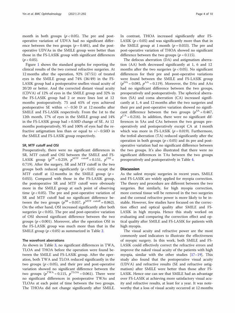
month in both groups (p < 0.05). The pre and post-operative variation of UDVA had no significant differ-ence between the two groups (p = 0.481), and the post-operative UDVAs in the SMILE group were better thanthose in the FS-LASIK group with significant differences(p < 0.05).Figure 1 shows the standard graphs for reporting the
clinical results of the two corneal refractive surgeries. At12 months after the operation, 92% (47/51) of treatedeyes in the SMILE group and 74% (36/49) in the FS-LASIK group had a postoperative snellen visual acuity of20/20 or better. And the corrected distant visual acuity(CDVA) of 12% of eyes in the SMILE group and 32% inthe FS-LASIK group had 2 or more lines lost at 12months postoperatively. 75 and 65% of eyes achievedpostoperative SE within +/− 0.50 D at 12 months afterSMILE and FS-LASIK respectively. From the 1st to the12th month, 17% of eyes in the SMILE group and 14%in the FS-LASIK group had > 0.50D change of SE. At 12months postoperatively, 95 and 100% of eyes had the re-fractive astigmatism less than or equal to +/− 0.50D inthe SMILE and FS-LASIK group respectively.
SR, MTF cutoff and OSIPreoperatively, there were no significant differences inSR, MTF cutoff and OSI between the SMILE and FS-LASIK group (pSR = 0.259, pMTF cutoff = 0.151, pOSI =0.719). After the surgery, SR and MTF cutoff in the twogroups both reduced significantly (p < 0.05) except theMTF cutoff at 12 months in the SMILE group (p =0.055). Compared with those in the FS-LASIK group,the postoperative SR and MTF cutoff were obviouslymore in the SMILE group at each point of observingtime (p < 0.05). The pre and post-operative variation ofSR and MTF cutoff had no significant difference be-tween the two groups (pSR = 0.057, pMTF cutoff = 0.062).On the other hand, OSI increased significantly after bothsurgeries (p < 0.05). The pre and post-operative variationof OSI showed significant difference between the twogroups (p < 0.001). Moreover, after the operation OSI inthe FS-LASIK group was much more than that in theSMILE group (p < 0.05) as summarized in Table 2.
The wavefront aberrationsAs shown in Table 3, no significant differences in TWA,TLOA and THOA before the operation were found be-tween the SMILE and FS-LASIK group. After the oper-ation, both TWA and TLOA reduced significantly in thetwo groups (p < 0.05), and their pre and post-operativevariation showed no significant difference between thetwo groups (pTWA = 0.115, pTLOA = 0.061). There wereno significant differences in postoperative TWAs andTLOAs at each point of time between the two groups.The THOAs did not change significantly after SMILE.
In contrast, THOA increased significantly after FS-LASIK (p < 0.05) and was significantly more than that inthe SMILE group at 1 month (p = 0.033). The pre andpost-operative variation of THOA showed no significantdifference between the two groups (p = 0.115).The defocus aberration (DA) and astigmatism aberra-
tion (AA) both decreased significantly at 1, 6 and 12months after the two surgeries (p < 0.05). No significantdifferences for their pre and post-operative variationswere found between the SMILE and FS-LASIK group(pDA = 0.085, pAA = 0.119). Moreover, the DAs and AAshad no significant difference between the two groups,preoperatively and postoperatively. The spherical aberra-tion (SA) and coma aberration (CA) increased signifi-cantly at 1, 6 and 12months after the two surgeries andtheir pre and post-operative variation showed no signifi-cant difference between the two groups (pSA = 0.467,pCA = 0.216). In addition, there were no significant dif-ferences in SAs and CAs between the two groups pre-operatively and postoperatively except CA at 1 monthwhich was more in FS-LASIK (p = 0.019). Furthermore,the trefoil aberration (TA) reduced significantly after theoperation in both groups (p < 0.05) and its pre and post-operative variation had no significant difference betweenthe two groups. It’s also illustrated that there were nosignificant differences in TAs between the two groupspreoperatively and postoperatively in Table 4.
DiscussionAs the safest myopic surgeries in recent years, SMILEand FS-LASIK are widely applied for myopia correction.The theory and procedure are different between the twosurgeries. But similarly, for high myopia correction,more corneal tissue will be removed in the two surgeriesand the corneal refractive power is more likely to be in-stable. However, few studies have focused on the correc-tion effect and optical quality after SMILE and FS-LASIK in high myopia. Hence this study worked onevaluating and comparing the correction effect and op-tical quality after SMILE and FS-LASIK for patients withhigh myopia.The visual acuity and refractive power are the most
commonly used indicators to illustrate the effectivenessof myopic surgery. In this work, both SMILE and FS-LASIK could effectively correct the refractive errors andimprove the naked visual acuity of the patients with highmyopia, similar with the other studies [17–19]. Thisstudy also found that the postoperative visual acuity(UDVA) and refractive results (SE and refractive astig-matism) after SMILE were better than those after FS-LASIK. Hence one can see that SMILE had an advantageover FS-LASIK at achieving more satisfactory visual acu-ity and refractive results, at least for a year. It was note-worthy that a loss of visual acuity occurred at 12 months
Yin et al. BMC Ophthalmology (2021) 21:292 Page 4 of 10
Fig. 1 The standard graphs for reporting the visual and refractive outcomes of 51 eyes in the SMILE group and 49 eyes in the FS-LASIK group at12months postoperatively
Yin et al. BMC Ophthalmology (2021) 21:292 Page 5 of 10

after SMILE and FS-LASIK, consistent with the changeof spherical equivalent refraction. We think the mainreason for the loss is that the subjects are all high my-opia patients, as it has been proved that the refractive re-gression is more likely to occur in patients with highmyopia [20, 21]. We also analyzed the individual eyeswith visual acuity loss and speculated that it might bethe result of abnormal accommodation function beforesurgery in these young high myopia patients [22]. A pre-vious research also concluded that a decrease in ampli-tude of accommodation and facility of accommodationmight result in some of the near-vision problems in
younger myopes in early postoperative days after refract-ive surgery [23]. Nevertheless, as we did not measure theaccommodation function of all these subjects preopera-tively, there is no hard proof for this assumption.Due to the postoperative corneal deformation, the in-
creased corneal scatter, wavefront aberrations and otherfactors might affect the optical quality [24–26]. Hencethis study measured and evaluated the postoperative ob-jective optical quality. Because of the limitation of a sin-gle detection equipment, this study used OQAS andiTrace analyzer simultaneously which were both widelyapplied for objective optical quality inspection and
Table 2 The variation of the SR, MTF cutoff and OSI with time in the SMILE and FS-LASIK group (mean ± SD)
Preop Post 1mo Post 6mo Post 12mo p0-value
SR
SMILE group 0.260 ± 0.064 0.221 ± 0.069* 0.212 ± 0.057* 0.233 ± 0.069* 0.057
FS-LASIK group 0.245 ± 0.077 0.185 ± 0.062* 0.175 ± 0.062* 0.178 ± 0.047*
p1-value 0.259 0.010 0.007 <0.001
MTF cutoff
SMILE group 43.684 ± 7.955 39.161 ± 10.711* 37.547 ± 10.297* 40.552 ± 10.005 0.062
FS-LASIK group 41.462 ± 9.809 32.520 ± 12.305* 32.361 ± 10.764* 31.489 ± 9.626*
p1-value 0.151 0.007 0.029 <0.001
OSI
SMILE group 0.49 ± 0.24 0.88 ± 0.53* 0.84 ± 0.44* 0.80 ± 0.53* <0.001
FS-LASIK group 0.59 ± 0.33 1.63 ± 1.01* 1.39 ± 0.92* 1.29 ± 0.72*
p1-value 0.719 <0.001 0.001 0.001
SD: standard deviation. Preop: preoperative. Post 1mo: at 1 month postoperatively. Post 6mo: at 6 months postoperatively. Post 12mo: at 12 monthspostoperatively. p0 = p value of the difference for the pre and post-operative variation of SR, MTF cutoff and OSI between the SMILE and FS-LASIK group. p1 = pvalue of the difference in SR, MTF cutoff and OSI at each point of time between the SMILE and FS-LASIK group. When p < 0.05, the difference was statisticallysignificant. * Compared with that before the surgery, p < adjusted alpha
Table 3 The variation of total wavefront aberrations with time in the SMILE and FS-LASIK group (mean ± SD)
Preop Post 1mo Post 6mo Post 12mo p0-value
TWA
SMILE group 7.896 ± 2.842 1.910 ± 1.830* 2.014 ± 1.665* 2.156 ± 1.791* 0.115
FS-LASIK group 8.797 ± 3.459 1.965 ± 1.582* 1.766 ± 1.509* 2.376 ± 1.761*
p1-value 0.122 0.841 0.440 0.539
TLOA
SMILE group 7.756 ± 2.967 1.812 ± 1.778* 1.900 ± 1.585* 2.052 ± 1.687* 0.061
FS-LASIK group 8.780 ± 3.446 1.717 ± 1.476* 1.572 ± 1.444* 2.226 ± 1.683*
p1-value 0.099 0.783 0.293 0.623
THOA
SMILE group 0.565 ± 0.775 0.543 ± 0.512 0.594 ± 0.597 0.598 ± 0.665 0.115
FS-LASIK group 0.485 ± 0.384 0.789 ± 0.727* 0.714 ± 0.575* 0.741 ± 0.642*
p1-value 0.514 0.033 0.307 0.251
SD: standard deviation. Preop: preoperative. Post 1mo: at 1 month postoperatively. Post 6mo: at 6 months postoperatively. Post 12mo: at 12 monthspostoperatively. TWA: total wavefront aberration. TLOA: total low-order wavefront aberration. THOA: total high-order wavefront aberration. p0 = p value of thedifference for the pre and post-operative variation of TWA, TLOA and THOA between the SMILE and FS-LASIK group. p1 = p value of the difference in TWA, TLOAand THOA at each point of time between the SMILE and FS-LASIK group. When p < 0.05, the difference was statistically significant. * Compared with that beforethe surgery, p < adjusted alpha
Yin et al. BMC Ophthalmology (2021) 21:292 Page 6 of 10
whose repeatability had been adequately verified in theprevious work [14–16]. Based on the double-pass tech-nique, OQAS is known as the device that can quantita-tively provide the data of ocular scatter [27, 28]. Themost dominant parameters detected by OQAS include:(1) SR that indicates the convergence ratio of light inten-sity in the image field of an optical system with aberra-tions, and the higher the value is, the better the opticalquality is; (2) MTF cutoff that characterizes the spatialfrequency corresponding to the minimum resolution ofhuman eyes in the modulation transfer function curve,and the higher the value is, the better the optical qualitybecomes; (3) OSI that objectively reflects the scatter ofthe refractive medium, and the higher the value is, themuddier the refractive media is. Another evaluation in-strument, i.e. iTrace analyzer, is composed of a cornealtopographer and an aberrometer whose workingprinciple is ray tracing. It is applicable for the measure-ment of corneal, internal and total ocular wavefront ab-errations [16]. Wavefront aberrations are distortions inthe phase of light entering the eye, which leads to thedefects in image-forming and thereby decreasing thequality of vision [29, 30]. They could be caused by thenon-optimal surface shapes, irregularities and
misalignments in the eye’s optical elements. It’s generallyaccepted that as the wavefront aberrations were cor-rected, the visual sensitivity, night vision, incidence ofglare and halo would significantly improve. They couldbe mathematically represented as the sum of a series ofpolynomial functions of different orders, and the higherthe wavefront aberration is, the more the visual qualityis affected. In particular, the high-order aberrations havea greater impact on the visual quality. This study appliediTrace analyzer to measure the total ocular wavefrontaberrations before and after the operation, includingTWA, TLOA, THOA, etc.As indicated by the variation of SR, MTF cutoff and
OSI in this study, the optical quality descended con-stantly for one year after both SMILE and FS-LASIK.This finding was different from some previous observa-tions about SMILE surgery [31–33]. Miao et al. used theOQAS to evaluate the optical quality after SMILE andfound that the optical quality was not significantly re-duced [31]. Niu et al. got a result that for high myopiacorrection, MTF cutoff declined slightly at 3 monthsafter SMILE but recovered to the preoperative value atthe one-year follow-up, and OSI increased at 20 daysafter surgery but gradually declined to the pre-operative
Table 4 The variation of the wavefront aberrations with time in the SMILE and FS-LASIK group (mean ± SD)
Preop Post 1mo Post 6mo Post 12mo p0-value
DA
SMILE group 7.814 ± 2.800 1.714 ± 1.797* 1.824 ± 1.598* 1.972 ± 1.670* 0.085
FS-LASIK group 8.671 ± 3.370 1.651 ± 1.458* 1.470 ± 1.462* 2.152 ± 1.704*
p1-value 0.133 0.856 0.250 0.613
AA
SMILE group 0.967 ± 0.557 0.381 ± 0.357* 0.391 ± 0.300* 0.441 ± 0.431* 0.119
FS-LASIK group 1.207 ± 0.988 0.420 ± 0.434* 0.380 ± 0.344* 0.427 ± 0.350*
p1-value 0.134 0.513 0.940 0.869
SA
SMILE group 0.081 ± 0.179 0.223 ± 0.374* 0.258 ± 0.452* 0.241 ± 0.487* 0.467
FS-LASIK group 0.053 ± 0.115 0.309 ± 0.536* 0.270 ± 0.400* 0.297 ± 0.388*
p1-value 0.338 0.316 0.887 0.503
CA
SMILE group 0.235 ± 0.167 0.367 ± 0.349* 0.396 ± 0.411* 0.407 ± 0.461* 0.216
FS-LASIK group 0.265 ± 0.298 0.571 ± 0.514* 0.537 ± 0.435* 0.574 ± 0.526*
p1-value 0.543 0.019 0.124 0.092
TA
SMILE group 0.213 ± 0.152 0.172 ± 0.130* 0.163 ± 0.119* 0.170 ± 0.172* 0.082
FS-LASIK group 0.246 ± 0.200 0.140 ± 0.090* 0.142 ± 0.097* 0.130 ± 0.085*
p1-value 0.363 0.162 0.215 0.142
SD: standard deviation. Preop: preoperative. Post 1mo: at 1 month postoperatively. Post 6mo: at 6 months postoperatively. Post 12mo: at 12 monthspostoperatively. DA: defocus aberration. AA: astigmatism aberration. SA: spherical aberration. CA: coma aberration. TA: trefoil aberration. p0 = p value of thedifference for the pre and post-operative variation of the wavefront aberrations between the SMILE and FS-LASIK group. p1 = p value of the difference in thewavefront aberrations at each point of time between the SMILE and FS-LASIK groups. When p < 0.05, the difference was statistically significant. * Compared withthat before the surgery, p < adjusted alpha
Yin et al. BMC Ophthalmology (2021) 21:292 Page 7 of 10
level at 3 months [32]. Qin et al. find that no significantdifference was found in SR or MTF cut-off in SMILE forhigh myopia correction before surgery or at any timepoint after surgery [33]. And few reports were foundabout the optical quality for high myopia after FS-LASIKevaluated by OQAS. The specific reasons for these dif-ferences are unknown, but it was worth the attention ofrefractive surgeons that there is a decline in the opticalquality after the operation.The results about SR, MTF cutoff and OSI also sug-
gested that SMILE achieved better optical quality thanFS-LASIK. In especial, the MTF cutoff at 12 months inSMILE returned to the preoperative level without anydifference, possibly indicating the slightly recovery of op-tical quality as time progressed. Similar research byOQAS was rare between SMILE and FS-LASIK. Weconsidered that the superiority of SMILE here was pos-sibly related to the size of optical zone and high pulsefrequency of femtosecond laser. The better optical qual-ity was often accompanied by the larger cutting area inthe corneal refractive surgery [34]. It’s indicated thatSMILE had a comparatively larger effective optical zonethan FS-LASIK though a similar optical zone was pro-grammed before the operation [35, 36]. Besides, the highpulse frequency of femtosecond laser in SMILE is ableto alleviate the inflammation reaction and corneal dam-age, and then induces limited potential loss of the cor-neal transparency and less scatter when compared withFS-LASIK [5, 37]. Hence, from the perspective of SR,MTF cutoff and OSI, this study suggested that it is morereasonable to have SMILE surgery for the populationwith high optical quality requirements.The wavefront aberration provides another objective
method to evaluate the postoperative optical quality. Inthis study, the TWA, TLOA, DA, AA, SA, CA and TAshowed a similar increasing or decreasing trends afterSMILE and FS-LASIK, except the CA at 1 month post-operatively. And the THOA did not change significantlyafter SMILE while showed an upward trend after FS-LASIK. These results indicated that except the THOAsand CA, SMILE and FS-LASIK had no obvious differ-ences in introducing wavefront aberrations, especiallythe lower order aberrations. Because the refractive errorswere effectively corrected after the two surgeries, the DAand AA decreased obviously. The DA and AA accountedfor the majority of the total aberrations, hence theTLOA and TWA also decreased after the operation. Asto the higher order aberrations, the increasing trend wassimilar to the previous studies [12, 38, 39]. Due to thesurgery incision and the postoperative wound healing,the asymmetry of eye plane such as irregularity, inclin-ation and decentration might occur, which could bereflected by CAs [24]. Due to the cutting of nerve end-ings when making the corneal flap in FS-LASIK, the
quality of tear film became poor and might also affectthe corneal aberrations [40–42]. And a minimal dis-placement of the corneal flap in FS-LASIK could obvi-ously affect the higher-order aberrations. Besides, SMILEbrings less damage to the anterior stromal layer, and theheat load generated by femtosecond laser is lower thanthat by excimer laser, which both would reduce the in-fluence on central asphericity of cornea [43]. In addition,through the aspheric scanning pattern without scanninginterval, SMILE can minimize the higher order aberra-tions introduced by surgery, while FS-LASIK adopts theexcimer laser with scanning interval and would intro-duce more surgically-induced higher order aberrations[44]. Therefore, there is a disadvantage of FS-LASIKagainst SMILE in terms of the postoperative higher-order wavefront aberrations. In sum, it proved the ad-vantage of SMILE on the optical quality over FS-LASIK.Additionally, in this study the change of higher-order
aberrations after SMILE or FS-LASIK was not exactlythe same with the other researches [24, 45, 46]. It mightbe related to the different refractive diopters, inspectionequipments and assessing parameters, detecting condi-tions, surgery equipments and procedures, surgeons, etc.For example, by using Sirius System, Jin et al. found thatSMILE showed better optical quality than FS-LASIK atlarger pupil diameter [13]. Yet no observations of opticalquality at different pupil diameters were made in ourstudy. Besides, the results reported in this work cannotrepresent the general myopic population as the pre-operative spherical refraction was from − 6.00 D to −10.00 D and the astigmatism was from 0 D to − 2.50 D.We expect more studies to focus on the optical qualityof patients with high myopia after different corneal re-fractive surgeries. And further work is needed to prolongthe observation time and increase the sample sizes inorder to detect the optical quality more deeply of pa-tients with high myopia after SMILE and FS-LASIK.
ConclusionsThe data reported in this work indicated that bothSMILE and FS-LASIK could improve the visual acuity ofthe patients with high myopia. And the optical qualitydescended after the operation. The better postoperativevisual acuity and refractive results, the higher SR andMTF cutoff, the lower OSI together with the differentchange of higher order aberrations illustrated thatSMILE was superior to FS-LASIK at the correction effectand optical quality for high myopia population. Inaddition, the combined application of OQAS and iTraceanalyzer is a valuable complementary measurement inevaluating the optical quality after the refractive surgery.
AbbreviationsSMILE: Small incision lenticule extraction; FS-LASIK: Femtosecond laser-assisted in situ keratomileusis; UDVA: Uncorrected distance visual acuity;
Yin et al. BMC Ophthalmology (2021) 21:292 Page 8 of 10
CDVA: Corrected distance visual acuity; SE: Spherical equivalent refraction;SR: Strehl ratio; MTF cutoff: Modulation transfer function cutoff frequency;OSI: Objective scatter index; WA: Wavefront aberration; TWA: Total wavefrontaberration; TLOA: Total low-order wavefront aberration; THOA: Total high-order wavefront aberration; DA: Defocus aberration; AA: Astigmatismaberration; SA: Spherical aberration; CA: Coma aberration; TA: Trefoilaberration
AcknowledgementsNot applicable.
Authors’ contributionsThe study concept and design were performed by YYW, WXY and WD. Theexaminations were completed by HT, DKX, HSF and FQM. Data collectionand interpretation of data was done by LY, XAQ, FYY and ZY. Writing themanuscript and critical revision of the manuscript were performed by YYW,LYJ and WD. All authors have reviewed the manuscript and approved thefinal manuscript.
FundingThis work was supported by the National Natural Science Foundation ofChina (No.81900890).
Availability of data and materialsThe datasets analysed during the current study are available from thecorresponding author on reasonable request.
Declarations
Ethics approval and consent to participateThis research was approved by the ethics committee of Xiangya hospital,which followed the tenets of the Declaration of Helsinki. The informedconsent was obtained from all the subjects for the operation and theapplication of data in clinical research. The subjects’ personal informationwas anonymized before the assessment of data.
Consent for publicationNot applicable.
Competing interestsWe have no any commercial or proprietary interest with the product orcompany. The authors declare to have no competing interests.
Received: 11 April 2021 Accepted: 9 July 2021
References1. Yan H, Gong LY, Huang W, Peng YL. Clinical outcomes of small incision
lenticule extraction versus femtosecond laser-assisted LASIK for myopia: ameta-analysis. Int J Ophthalmol. 2017;10(9):1436–45. https://doi.org/10.18240/ijo.2017.09.17.
2. Tian Y, Jiang HB, Jiang J, Wen D, Xia XB, Song WT. Comparison ofimplantable collamer lens visian ICL V4 and ICL V4c for high myopia: acohort study. Medicine. 2017;96(25):e7294. https://doi.org/10.1097/MD.0000000000007294.
3. Moshirfar M, McCaughey MV, Reinstein DZ, et al. Small-incision lenticuleextraction. J Cataract Refract Surg. 2015;41(3):652–65. https://doi.org/10.1016/j.jcrs.2015.02.006.
4. Shen ZR, Shi KD, Yu YH, Yu X, Lin Y, Yao K. Small incision Lenticuleextraction (SMILE) versus femtosecond laser-assisted in situ Keratomileusis(FS-LASIK) for myopia: a systematic review and meta-analysis. PLoS One.2016;11(7):e0158176. https://doi.org/10.1371/journal.pone.0158176.
5. Kunert KS, Blum M, Duncker GIW, Sietmann R, Heichel J. Surface quality ofhuman corneal lenticules after femtosecond laser surgery for myopiacomparing different laser parameters. Graefes Arch Clin Exp Ophthalmol.2011;249(9):1417–24. https://doi.org/10.1007/s00417-010-1578-4.
6. Chen SH, Feng YF, Stojanovic A, Jankov MR II, Wang Q. IntraLasefemtosecond laser vs mechanical microkeratomes in LASIK for myopia: asystematic review and meta-analysis. J Refract Surg. 2012;28(1):15–24.https://doi.org/10.3928/1081597X-20111228-02.
7. Fan L, Xiong L, Zhang B, Wang Z. Longitudinal and regional non-uniformremodeling of corneal epithelium after topography-guided FS-LASIK. JRefract Surg. 2019;35(2):88–95. https://doi.org/10.3928/1081597X-20190104-01.
8. Han T, Zheng K, Chen Y, Gao Y, He L, Zhou X. Four-year observation ofpredictability and stability of small incision lenticule extraction. BMCOphthalmol. 2016;16(1):149. https://doi.org/10.1186/s12886-016-0331-0.
9. Chen Y, Xia X. Comparison of the Orbscan II topographer and the iTraceaberrometer for the measurements of keratometry and corneal diameter inmyopic patients. BMC Ophthalmol. 2016;16(1):33. https://doi.org/10.1186/s12886-016-0210-8.
10. Zhou JQ, Xu Y, Li MY, et al. Preoperative refraction, age and optical zone aspredictors of optical and optical quality after advanced surface ablation inpatients with high myopia: a cross-sectional study. BMJ Open. 2018;8:e023877.
11. Gyldenkerne A, Ivarsen A, Hjortdal JO. Comparison of corneal shapechanges and aberrations induced by FS-LASIK and SMILE for myopia. JRefract Surg. 2015;31(4):223–9. https://doi.org/10.3928/1081597X-20150303-01.
12. Liu ML, Chen YL, Wang DY, Zhou Y, Zhang X, He J, et al. Clinical outcomesafter SMILE and femtosecond laser-assisted LASIK for myopia and myopicastigmatism: a prospective randomized comparative study. Cornea. 2016;35(2):210–6. https://doi.org/10.1097/ICO.0000000000000707.
13. Jin Y, Wang Y, Xu LL, Zuo T, Li H, Dou R, et al. Comparison of the opticalquality between small incision Lenticule extraction and femtosecond laserLASIK. J Ophthalmol. 2016;2016:1–9. https://doi.org/10.1155/2016/2507973.
14. Saad A, Saab M, Gatinel D. Repeatability of measurements with a double-pass system. J Cataract Refract Surg. 2010;36(1):28–33. https://doi.org/10.1016/j.jcrs.2009.07.033.
15. Xu Z, Hua YJ, Qiu W, Li G, Wu Q. Precision and agreement of higher orderaberrations measured with ray tracing and Hartmann-shack aberrometers.BMC Ophthalmol. 2018;18(1):18. https://doi.org/10.1186/s12886-018-0683-8.
16. Hao J, Li L, Tian F, Zhang H. Comparison of two types of optical qualityanalyzer for the measurement of high order aberrations. Int J Ophthalmol.2016;9(2):292–7. https://doi.org/10.18240/ijo.2016.02.22.
17. Xia LK, Ma J, Liu HN, Shi C, Huang Q. Three-year results of small incisionlenticule extraction and wavefront-guided femtosecond laser-assisted laserin situ keratomileusis for correction of high myopia and myopicastigmatism. Int J Ophthalmol. 2018;11(3):470–7. https://doi.org/10.18240/ijo.2018.03.18.
18. Yang W, Liu S, Li M, Shen Y, Zhou X. Visual outcomes after small incisionLenticule extraction and femtosecond laser-assisted LASIK for high myopia.Ophthalmic Res. 2020;63(4):427–33. https://doi.org/10.1159/000504304.
19. Han T, Xu Y, Han X, Zeng L, Shang J, Chen X, et al. Three-year outcomes ofsmall incision lenticule extraction (SMILE) and femtosecond laser-assistedlaser in situ keratomileusis (FS-LASIK) for myopia and myopic astigmatism.Br J Ophthalmol. 2019;103(4):565–8. https://doi.org/10.1136/bjophthalmol-2018-312140.
20. Wang Y, Ma JN. Future developments in SMILE: higher degree of myopiaand hyperopia. Asia Pac J Ophthalmol (Phila). 2019;8(5):412–6. https://doi.org/10.1097/01.APO.0000580128.27272.bb.
21. Blum M, Täubig K, Gruhn C, et al. Five-year results of Small Incision LenticuleExtraction (ReLEx SMILE). Br J Ophthalmol. 2016;100(9):1192.
22. Radhakrishnan H, Allen PM, Charman WN. Dynamics of accommodativefacility in myopes. Invest Ophthalmol Vis Sci. 2007;48(9):4375–82. https://doi.org/10.1167/iovs.07-0269.
23. Karimian F, Baradaran-Rafii A, Bagheria A, et al. Accommodative changesafter photorefractive keratectomy in myopic eyes. Optometry Vision Sci.2010;87(11):833–8. https://doi.org/10.1097/OPX.0b013e3181f6fccc.
24. Wang J, Ren YL, Liang K, Jiang Z, Tao L. Changes of corneal high-orderaberrations after femtosecond laser-assisted in situ keratomileusis. Medicine.2018;97(18):e0618. https://doi.org/10.1097/MD.0000000000010618.
25. Vilaseca M, Padilla A, Ondategui JC, Arjona M, Güell JL, Pujol J. Effect of laserin situ keratomileusis on vision analyzed using preoperative optical quality. JCataract Refract Surg. 2010;36(11):1945–53. https://doi.org/10.1016/j.jcrs.2010.05.029.
26. Lee K, Ahn JM, Kim EK, Kim TI. Comparison of optical quality parametersand ocular aberrations after wavefront-guided laser in-situ keratomileusisversus wavefront-guided laser epithelial keratomileusis for myopia. GraefesArch Clin Exp Ophthalmol. 2013;251(9):2163–9. https://doi.org/10.1007/s00417-013-2356-x.
Yin et al. BMC Ophthalmology (2021) 21:292 Page 9 of 10
27. Artal P, Benito A, Perez GM, et al. An objective scatter index based ondouble-pass retinal images of a point source to classify cataracts. PLoS One.2011;6(2):e16823. https://doi.org/10.1371/journal.pone.0016823.
28. Guell JL, Pujol J, Arjona M, et al. Optical quality analysis system: instrumentfor objective clinical evaluation of ocular optical quality. J Cataract RefractSurg. 2004;30(7):1598–9. https://doi.org/10.1016/j.jcrs.2004.04.031.
29. Mclellan JS, Prieto PM, Marcos S, et al. Effects of interactions among waveaberrations on optical image quality. Vis Res. 2006;46(18):3009–16. https://doi.org/10.1016/j.visres.2006.03.005.
30. Valentina BS, Ramona B, Speranta S, Calin T. The influence of opticalaberrations in refractive surgery. Rom J Ophthalmol. 2015;59(4):217–22.
31. Miao H, He L, Shen Y, Li M, Yu Y, Zhou X. Optical quality and intraocularscattering after femtosecond laser small incision lenticule extraction. JRefract Surg. 2014;30(5):296–302. https://doi.org/10.3928/1081597X-20140415-02.
32. Niu L, Miao H, Tian M, Fu D, Wang X, Zhou X. One year visual outcomesand optical quality of femtosecond laser small incision lenticule extractionand Visian implantable Collamer Lens (ICL V4c) implantation for highmyopia. Acta Ophthalmol. 2020;98(6):e662–7. https://doi.org/10.1111/aos.14344.
33. Qin Q, Bao L, Yang L, He Z, Huang Z. Comparison of visual quality afterEVO-ICL implantation and SMILE to select the appropriate surgical methodfor high myopia. BMC Ophthalmol. 2019;19(1):21. https://doi.org/10.1186/s12886-019-1029-x.
34. Alarcón A, Rubiño M, PérezOcón F, et al. Theoretical analysis of the effect ofpupil size, initial myopic level, and optical zone on quality of vision aftercorneal refractive surgery. J Refract Surg. 2012;28(12):901–6. https://doi.org/10.3928/1081597X-20121106-01.
35. Damgaard I, Ang M, Mahmoud A, et al. Functional optical zone andcentration following SMILE and LASIK: a prospective, randomized,contralateral eye study. J Refract Surg. 2019;35(4):230–7. https://doi.org/10.3928/1081597X-20190313-01.
36. Hou J, Wang Y, Lei Y, Zheng X. Comparison of effective optical zone aftersmall-incision lenticule extraction and femtosecond laser–assisted laser insitu keratomileusis for myopia. J Cataract Refract Surg. 2018;44(10):1179–85.https://doi.org/10.1016/j.jcrs.2018.06.046.
37. Denoyer A, Landman E, Trinh L, Faure JF, Auclin F, Baudouin C. Dry eyedisease after refractive surgery comparative outcomes of small incisionLenticule extraction versus LASIK. Ophthalmology. 2015;122(4):669–76.https://doi.org/10.1016/j.ophtha.2014.10.004.
38. Zhang H, Wang Y, Li H. Corneal spherical aberration and corneal Asphericityafter small incision Lenticule extraction and femtosecond laser-assisted LASIK. J Ophthalmol. 2017;2017:4921090.
39. Qian YS, Chen X, Naidu RK, Zhou X. Comparison of efficacy and visualoutcomes after SMILE and FS-LASIK for the correction of high myopia withthe sum of myopia and astigmatism from −10.00 to −14.00 dioptres. ActaOphthalmol. 2020;98(2):e161–72. https://doi.org/10.1111/aos.14078.
40. Montes-Mico R. Role of the tear film in the optical quality of the humaneye. J Cataract Refract Surg. 2007;33(9):1631–5. https://doi.org/10.1016/j.jcrs.2007.06.019.
41. Benito A, Perez GM, Mirabet S, et al. Objective optical assessment of tear-film quality dynamics in normal and mildly symptomatic dry eyes. J CataractRefract Surg. 2011;37(8):1481–7. https://doi.org/10.1016/j.jcrs.2011.03.036.
42. Serrao S, Buratto L, Lombardo G, de Santo MP, Ducoli P, Lombardo M.Optimal parameters to improve the interface quality of the flap bed infemtosecond laser-assisted laser in situ keratomileusis. J Cataract RefractSurg. 2012;38(8):1453–9. https://doi.org/10.1016/j.jcrs.2012.05.021.
43. George MR, Shah RA, Hood C, Krueger RR. Transitioning to optimizedcorrection with the WaveLight ALLEGRETTO WAVE: case distribution, visualoutcomes, and wavefront aberrations. J Refract Surg. 2010;26(10):S806–13.https://doi.org/10.3928/1081597X-20100921-07.
44. Lin F, Xu Y, Yang Y. Comparison of the visual results after SMILE andfemtosecond laser-assisted LASIK for myopia. J Refract Surg. 2014;30(4):248–54. https://doi.org/10.3928/1081597X-20140320-03.
45. Jin HY, Wan T, Wu F, et al. Comparison of visual results and higher-orderaberrations after small incision lenticule extraction (SMILE): high myopia vs.mild to moderate myopia. BMC Ophthalmol. 2017;17(1):118.
46. Jaurrieta-Hinojos JN, Fernandez O. Changes in visual acuity and high-orderaberrations after SMILE. Invest Ophthalmol Vis Sci. 2016;57(12):4884.
Publisher’s NoteSpringer Nature remains neutral with regard to jurisdictional claims inpublished maps and institutional affiliations.
Yin et al. BMC Ophthalmology (2021) 21:292 Page 10 of 10
Copyright © 2022 FDOKUMEN