Comparison of quantitative techniques including Xpert MTB/RIF to evaluate mycobacterial burden
10
Comparison of Quantitative Techniques including Xpert MTB/RIF to Evaluate Mycobacterial Burden Richard N. van Zyl-Smit 1 , Anke Binder 1 , Richard Meldau 1 , Hridesh Mishra 2 , Patricia L. Semple 1 , Grant Theron 1 , Jonathan Peter 1 , Andrew Whitelaw 3 , Suren K. Sharma 2 , Robin Warren 4 , Eric D. Bateman 1 , Keertan Dheda 1,5,6 * 1 Lung Infection and Immunity Unit, Division of Pulmonology and UCT Lung Institute, Department of Medicine, University of Cape Town, Cape Town, South Africa, 2 Department of Medicine, All India Institute of Medical Sciences, New Delhi, India, 3 Division of Medical Microbiology, University of Cape Town, Cape Town, South Africa, 4 DST/NRF Centre of Excellence for Biomedical TB Research/MRC Centre for Molecular and Cellular Biology, Stellenbosch University, Stellenbosch, South Africa, 5 Institute of Infectious Diseases and Molecular Medicine, University of Cape Town, Cape Town, South Africa, 6 Department of Infection, University College London Medical School, London, United Kingdom Abstract Introduction: Accurate quantification of mycobacterial load is important for the evaluation of patient infectiousness, disease severity and monitoring treatment response in human and in-vitro laboratory models of disease. We hypothesized that newer techniques would perform as well as solid media culture to quantify mycobacterial burden in laboratory specimens. Methods: We compared the turn-around-time, detection-threshold, dynamic range, reproducibility, relative discriminative ability, of 4 mycobacterial load determination techniques: automated liquid culture (BACTEC-MGIT-960), [ 3 H]-uracil incorporation assays, luciferase-reporter construct bioluminescence, and quantitative PCR(Xpert -MTB/RIF) using serial dilutions of Mycobacterium bovis and Mycobacterium tuberculosis H37RV. Mycobacterial colony-forming-units(CFU) using 7H10-Middlebrook solid media served as the reference standard. Results: All 4 assays correlated well with the reference standard, however, bioluminescence and uracil assays had a detection threshold $1 6 10 3 organisms. By contrast, BACTEC-MGIT-960 liquid culture, although only providing results in days, was user-friendly, had the lowest detection threshold (,10 organisms), the greatest discriminative ability (1 vs. 10 organisms; p = 0.02), and the best reproducibility (coefficient of variance of 2% vs. 38% compared to uracil incorporation; p = 0.02). Xpert-MTB/RIF correlated well with mycobacterial load, had a rapid turn-around-time (,2 hours), was user friendly, but had a detection limit of ,100 organisms. Conclusions: Choosing a technique to quantify mycobacterial burden for laboratory or clinical research depends on availability of resources and the question being addressed. Automated liquid culture has good discriminative ability and low detection threshold but results are only obtained in days. Xpert MTB/RIF provides rapid quantification of mycobacterial burden, but has a poorer discrimination and detection threshold. Citation: van Zyl-Smit RN, Binder A, Meldau R, Mishra H, Semple PL, et al. (2011) Comparison of Quantitative Techniques including Xpert MTB/RIF to Evaluate Mycobacterial Burden. PLoS ONE 6(12): e28815. doi:10.1371/journal.pone.0028815 Editor: Adithya Cattamanchi, San Francisco General Hospital, University of California San Francisco, United States of America Received August 12, 2011; Accepted November 15, 2011; Published December 22, 2011 Copyright: ß 2011 van Zyl-Smit et al. This is an open-access article distributed under the terms of the Creative Commons Attribution License, which permits unrestricted use, distribution, and reproduction in any medium, provided the original author and source are credited. Funding: RVZS is supported in part by a Fogarty International Clinical Research Scholars/Fellows Support Centre National Institutes of Health grant R24TW007988. The funder had no role in study design, data collection and analysis, decision to publish, or preparation of the manuscript. No additional external funding was received for this study. Competing Interests: The authors have declared that no competing interests exist. * E-mail: [email protected] Introduction Determining mycobacterial burden is a basic requirement for many laboratory and clinical studies, including those assessing disease severity and examining the efficacy of new therapies and interventions for tuberculosis (TB) control [1,2,3], all of which have now become urgent with the emerging public health threat of multidrug and extensively drug resistant TB [4,5]. In addition, mycobacterial burden, usually assessed as grades of smear positivity (scanty, 1+2+, and 3+), is used to evaluate the infectiousness of cases in the context of public health contact tracing and screening [6,7]. In translational research the need to accurately detect changes in M. tuberculosis burden is fundamental to the study of biologically meaningful immunological pathways, and drug and vaccine development in both human and murine models of disease. The latter include early bacteriocidal activity (EBA) studies related to drug development and evaluating vaccine efficacy in murine models [8,9,10,11]. Although several techniques for determining mycobacterial burden exist, each is associated with significant limitations such as inaccuracy, turn-around-time, limited reproducibility, cost, methodological complexity, relative discriminative ability and detection threshold. Culture on solid media using colony-forming units (CFU), is widely considered to be the gold standard for determining the PLoS ONE | www.plosone.org 1 December 2011 | Volume 6 | Issue 12 | e28815
-
Upload
independent -
Category
Documents
-
view
1 -
download
0
Transcript of Comparison of quantitative techniques including Xpert MTB/RIF to evaluate mycobacterial burden

Comparison of Quantitative Techniques including XpertMTB/RIF to Evaluate Mycobacterial BurdenRichard N. van Zyl-Smit1, Anke Binder1, Richard Meldau1, Hridesh Mishra2, Patricia L. Semple1, Grant
Theron1, Jonathan Peter1, Andrew Whitelaw3, Suren K. Sharma2, Robin Warren4, Eric D. Bateman1,
Keertan Dheda1,5,6*
1 Lung Infection and Immunity Unit, Division of Pulmonology and UCT Lung Institute, Department of Medicine, University of Cape Town, Cape Town, South Africa,
2 Department of Medicine, All India Institute of Medical Sciences, New Delhi, India, 3 Division of Medical Microbiology, University of Cape Town, Cape Town, South Africa,
4 DST/NRF Centre of Excellence for Biomedical TB Research/MRC Centre for Molecular and Cellular Biology, Stellenbosch University, Stellenbosch, South Africa, 5 Institute
of Infectious Diseases and Molecular Medicine, University of Cape Town, Cape Town, South Africa, 6 Department of Infection, University College London Medical School,
London, United Kingdom
Abstract
Introduction: Accurate quantification of mycobacterial load is important for the evaluation of patient infectiousness,disease severity and monitoring treatment response in human and in-vitro laboratory models of disease. We hypothesizedthat newer techniques would perform as well as solid media culture to quantify mycobacterial burden in laboratoryspecimens.
Methods: We compared the turn-around-time, detection-threshold, dynamic range, reproducibility, relative discriminativeability, of 4 mycobacterial load determination techniques: automated liquid culture (BACTEC-MGIT-960), [3H]-uracilincorporation assays, luciferase-reporter construct bioluminescence, and quantitative PCR(Xpert -MTB/RIF) using serialdilutions of Mycobacterium bovis and Mycobacterium tuberculosis H37RV. Mycobacterial colony-forming-units(CFU) using7H10-Middlebrook solid media served as the reference standard.
Results: All 4 assays correlated well with the reference standard, however, bioluminescence and uracil assays had adetection threshold $16103 organisms. By contrast, BACTEC-MGIT-960 liquid culture, although only providing results indays, was user-friendly, had the lowest detection threshold (,10 organisms), the greatest discriminative ability (1 vs. 10organisms; p = 0.02), and the best reproducibility (coefficient of variance of 2% vs. 38% compared to uracil incorporation;p = 0.02). Xpert-MTB/RIF correlated well with mycobacterial load, had a rapid turn-around-time (,2 hours), was userfriendly, but had a detection limit of ,100 organisms.
Conclusions: Choosing a technique to quantify mycobacterial burden for laboratory or clinical research depends onavailability of resources and the question being addressed. Automated liquid culture has good discriminative ability and lowdetection threshold but results are only obtained in days. Xpert MTB/RIF provides rapid quantification of mycobacterialburden, but has a poorer discrimination and detection threshold.
Citation: van Zyl-Smit RN, Binder A, Meldau R, Mishra H, Semple PL, et al. (2011) Comparison of Quantitative Techniques including Xpert MTB/RIF to EvaluateMycobacterial Burden. PLoS ONE 6(12): e28815. doi:10.1371/journal.pone.0028815
Editor: Adithya Cattamanchi, San Francisco General Hospital, University of California San Francisco, United States of America
Received August 12, 2011; Accepted November 15, 2011; Published December 22, 2011
Copyright: � 2011 van Zyl-Smit et al. This is an open-access article distributed under the terms of the Creative Commons Attribution License, which permitsunrestricted use, distribution, and reproduction in any medium, provided the original author and source are credited.
Funding: RVZS is supported in part by a Fogarty International Clinical Research Scholars/Fellows Support Centre National Institutes of Health grantR24TW007988. The funder had no role in study design, data collection and analysis, decision to publish, or preparation of the manuscript. No additional externalfunding was received for this study.
Competing Interests: The authors have declared that no competing interests exist.
* E-mail: [email protected]
Introduction
Determining mycobacterial burden is a basic requirement for
many laboratory and clinical studies, including those assessing
disease severity and examining the efficacy of new therapies and
interventions for tuberculosis (TB) control [1,2,3], all of which
have now become urgent with the emerging public health threat of
multidrug and extensively drug resistant TB [4,5]. In addition,
mycobacterial burden, usually assessed as grades of smear
positivity (scanty, 1+2+, and 3+), is used to evaluate the
infectiousness of cases in the context of public health contact
tracing and screening [6,7]. In translational research the need to
accurately detect changes in M. tuberculosis burden is fundamental
to the study of biologically meaningful immunological pathways,
and drug and vaccine development in both human and murine
models of disease. The latter include early bacteriocidal activity
(EBA) studies related to drug development and evaluating vaccine
efficacy in murine models [8,9,10,11]. Although several techniques
for determining mycobacterial burden exist, each is associated
with significant limitations such as inaccuracy, turn-around-time,
limited reproducibility, cost, methodological complexity, relative
discriminative ability and detection threshold.
Culture on solid media using colony-forming units (CFU), is
widely considered to be the gold standard for determining the
PLoS ONE | www.plosone.org 1 December 2011 | Volume 6 | Issue 12 | e28815

number of viable organisms in a specimen or experimental
condition, but is labor-intensive and has a long turn-around-time
[12,13]. Alternative techniques include the incorporation of tritiated
uracil into mycobacterial DNA, bioluminescence assays that use a
reporter construct, quantitative real-time polymerase chain reactions
(PCR), and time to positivity (TTP) in automated liquid culture
systems (BACTEC Mycobacterial Growth Indicator Tube (MGIT)
960] [14]. Each of the later has its own set of performance
characteristics that determine its suitability for different applications.
More recently, newer technologies such as the Xpert MTB/RIF
system (Cepheid, Sunnyvale, USA) have been developed for the
rapid detection of TB using clinical samples. However PCR
methods have been limited by their inability to distinguish viable
from degraded organisms. Whilst detecting TB-specific mRNA
from viable organisms is a potential solution, like PCR [15,16],
real time PCR it is technically demanding [17]. However, Xpert
MTB/RIF has the potential to circumvent this problem as
contaminating extracellular debris is removed in an intermediary
step by washing, DNA from intact organisms trapped in a mesh is
amplified by PCR [18,19]. However, its quantitative accuracy has
not yet been compared with that of automated culture, uracil
incorporation and bioluminescence techniques.
We hypothesized that newer automated mycobacterial load
determination techniques perform as well as traditional measures.
We therefore compared the performance characteristics of 4
quantitative techniques by evaluating turn-around-time, detection
threshold, dynamic range, reproducibility and quantitative discrimi-
native ability, with CFU on solid media as the reference standard.
Methods
Both BCG and H37RV luciferase reporter constructs (pSMT1
luciferase) [20] were used for all assays (gift of Muazaam Jacobs
from the Institute for Infectious Diseases and Molecular Medicine
University of Cape Town). Serial dilutions were prepared (aliquots
ranging from 1 to 16106 CFU per 500 ul) in sterile phosphate
buffer solution (PBS) from 3 ml of frozen stock for each strain.
Three vials of each dilution were provided for the five predeter-
mined assays. In addition, all dilutions were inoculated onto solid
media to confirm the number of CFUs at each dilution (Figure 1).
Solid Culture (reference standard)Aliquots of 1, 10 and 100 CFU were plated in 6 replicates of
10 ml on 7h10 Middlebrook agar enriched with OADC. Plates
were sealed in airtight bags and incubated at 37uC. Colonies were
counted daily between days 7 and 14 using an inverted light
microscope. The colony counts were over two days by two readers.
Liquid cultureA 500 ml aliquot of PBS containing the specified number of
organisms was injected into pre-prepared mycobacterial growth
indicator tubes (MGITs) (Becton Dickinson, Sparks, Maryland),
and the MGITs then incubated using the BACTEC-MGIT-960
automated culture system, which monitors the tubes for the
presence of growth on a constant basis. Each dilution was
prepared in triplicate and blinded to the MGIT operator. Time to
positivity was recorded by instrument as the time between start of
incubation and detection of growth.
Luminescence assayLuminescence was measured using a ModulusTM Microplate
Multimode Luminometer (Turner). 25 ml of 1% (v/v) n-decyl
aldehyde (Sigma), was injected into each well containing serial
dilutions (1:10 with 0.25% Tween/PBS) to a final volume of
125 ml. The results were read using a 0.5 s delay and a 0.5 s
integration time and were expressed as relative light units (RLU).
Figure 1. Preparation of Mycobacterial dilutions for load determination assays. A three-milliliter volume of frozen stock was diluted to aworking suspension of 26106 CFU/ml. From this stock 5 serial dilutions were prepared ranging from 2 to 26106 CFU/ml. From these dilutions, 15 aliquots oforganisms were prepared containing 1 to 16106 CFU in 500 ul. Three aliquots of each dilution were used in each of the 5-mycobacterial load determinationassays as described in the methods section. PCR-polymerase chain reaction, MGIT mycobacterial growth indicator tube, CFU-colony forming unit.doi:10.1371/journal.pone.0028815.g001
Comparison of Mycobacterial Load Techniques
PLoS ONE | www.plosone.org 2 December 2011 | Volume 6 | Issue 12 | e28815

Xpert MTB/RIF PCRA 500 ul aliquot of H37RV and BCG was treated with sample
buffer at a ratio of 1:3 supplied by the manufacturer as
recommended. The 2 ml mixture was later transferred into an
Xpert MTB/RIF cartridge. The cartridge was loaded into the
Gene Xpert IV instrument and the automated procedure started.
Results and CT values of probes were obtained using the software
(Xpert MTB/RIF version 2.0).
Uracil incorporation200 ml of each dilution was pipetted into 6 wells of 4 separate U-
bottomed 96 well plates and 50 ul of 3H uracil (final concentration
of 1 mCi/well) was added to each well. The plates were incubated
for 24 hours at 37uC in a 5% CO2 humidified chamber and
harvested onto fiberglass filter mats. The discs were placed into
scintillation bottles containing 1 ml of Quicksafe (Zinnser Analytic,
Frankfurt, Germany) and the amount of incorporated tritiated
uridine determined using a liquid scintillation counter and
reported as counts per minute (CPM).
Determination of performance characteristicsTurn-around-time. The turnaround time was defined as the
time taken from the start of each assay after preparation of the
aliquots until a bacterial load determination was possible (either
automated output or colony counting).
Detection threshold. The detection threshold was defined as
the lowest colony number detected by the assay: The result from 2
out of 3 MGIT bottles, 2 out of 3 Xpert MTB/RIF cartridges or
.50% of CFU/RLU/CPM replicates for a particular dilution were
as the reliable lower limit of detection for comparative purposes.
Discriminative ability. The ability of the assay to detect a
difference between serial dilutions was assessed using a 1-way
ANOVA with correction for multiple comparisons (Tukey).
GraphPad Prism software (version 5.00, GraphPad Software,
San Diego California USA, www.graphpad.com).
Reproducibility. Reproducibility of each assay was determined
by calculating the coefficient of variance (SD/mean) across all
dilutions. To compare reproducibility between assays the mean
coefficient of variance for BCG dilutions was calculated for each assay.
Results
A comparative overview of all the mycobacterial load assays is
summarised in Table 1. The performance characteristics of each
assay are detailed separately.
Solid media cultureAlthough technically simple to perform, plating for CFU was
time consuming and accuracy and reproducibility are affected by
pipetting skills as well visual counting of colonies. Counting of
CFUs and determination of mycobacterial load was possible
approximately 10–14 days after plating (Figure 2). Determination
of the CFU count was limited by the visual ability to accurately
count organisms and thus only dilutions of 1, 16101 and
16102 CFU were used for plating on 7H10 agar. The coefficient
of variance across the three dilutions ranges was 22%.
Automated liquid cultureThe turnaround time for the BACTEC-MGIT-960 system was
defined as time to positivity (TTP) and ranged from 117 hours (,5
days) to 467 hours (,19 days) for BCG and 123 hours (,5 days) to
528 hours (,22 days) for H37RV (dilution range 16106 to
16100 CFU; Figure 3) The lower limit of detection was less than
10 CFU (1 CFU detected in 3/3 BCG bottles and 2/3 H37RV
bottles). Reproducibility of the BACTEC-MGIT-960 system was
excellent as most replicates became positive within a few hours of
each other at all dilutions (coefficient of variance = 2%). The range of
detection was from 1 to 16106 CFU. BACTEC-MGIT-960 was able
to detect differences in mycobacterial load as small as 16102
organisms at low concentrations and 16105 at higher concentrations.
Luminescence assayThe lower limit of detection was 16102 CFU as no discrimi-
nation was possible below 100 organisms. Discrimination between
was possible at ranges of 16102 to 16105 CFU (Figure 4).
Reproducibility was comparable to CFU with a coefficient of
variance of 19%. Mycobacterial load determination was poten-
tially available within minutes of RLU determination using a pre-
prepared standard curve of RLU vs. CFU.
UracilUracil incorporation assays are complex and time consuming to
perform although the time to acquiring a result was approximately
24 hours. The effective lower limit of detection was 1000 CFU as
the tritiated uracil assay could detect but not discriminate between
loads at ranges below 16103 CFU. However, it was effective at
16103 to 16105 CFU (Figure 5).
PCR using the Xpert MTB/RIF systemThe automated real time MTB/RIF assay using the Gene
Xpert IV system was user friendly and minimal technical steps
Table 1. Performance characteristics of individual techniques used to quantify mycobacterial burden.
AssayTurn-aroundtime
Detection threshold (CFU;1 CFU = 1 organism) Dynamic range*
Reproducibility(Coefficient of Variance)+
Culture on 7H10solid media
Days to weeks 1 CFU Wide provided appropriate dilutions are made. Fair (22%)
Liquid culture usingMGIT 960 system
Days to weeks 1–10 CFU Excellent from 1 to 16106 CFU Good (2%)
Tritiated uracil assay 24 hours 1000 CFU Poor below 16103 CFU but able to detectdilutions of up to 16106 CFU
Poor (38%)
Luminescence assayusing reporter construct
,2 hours 100 CFU Poor below 16103 CFU but able to detectdilutions of up to 16106 CFU
Fair (19%)
PCR using Xpert MTB/RIF 2 hours 100 CFU Good at ranges between 16102 and 16106 CFU Good (3%)
*All experiments (apart from solid culture) require a standard curve to calculate the CFUs relative to the assay readout.+Reproducibility was determined by the mean coefficient of variance for the specific readout (CFU, RLU etc.) across all dilutions using the BCG-specific experiments.doi:10.1371/journal.pone.0028815.t001
Comparison of Mycobacterial Load Techniques
PLoS ONE | www.plosone.org 3 December 2011 | Volume 6 | Issue 12 | e28815

Figure 2. Mycobacterial load determination using solid media (Middlebrook 7H10). Serial dilutions of stock BCG (A) and H37RV (B) arerepresented on the X-axis with the calculated CFU on the Y-axis (log scale).doi:10.1371/journal.pone.0028815.g002
Comparison of Mycobacterial Load Techniques
PLoS ONE | www.plosone.org 4 December 2011 | Volume 6 | Issue 12 | e28815

Figure 3. Automated liquid culture (using BACTEC MGIT 960) time to positivity. Serial dilutions of stock BCG (A) and H37RV (B) arerepresented on the X-axis with automated positive detection time (time to positivity) on the Y-axis.doi:10.1371/journal.pone.0028815.g003
Comparison of Mycobacterial Load Techniques
PLoS ONE | www.plosone.org 5 December 2011 | Volume 6 | Issue 12 | e28815

were required prior to inserting the cartridge into the machine.
Xpert MTB/RIF results were available within 2 hours of the
experiment using the Gene Xpert IV machine with 4 cartridge
bays (only 4 samples could be run at once). BCG reproducibility
was excellent (coefficient of variance 3%) and the automated PCR
reliably detected 100 organisms (Figure 6). At the lowest dilution of
1 CFU, only 2 out of 3 cartridges were positive. The H37RV had
greater variability in cycle threshold (CT) values as compared to
Figure 4. Luminescence assay (using luminescent reporter construct) relative light units. Serial dilutions of stock BCG (A) and H37RV (B)are represented on the X-axis with the relative light unit output on the Y-axis.doi:10.1371/journal.pone.0028815.g004
Comparison of Mycobacterial Load Techniques
PLoS ONE | www.plosone.org 6 December 2011 | Volume 6 | Issue 12 | e28815

BCG (coefficient of variance 6%) and did not detect less than
100 CFU. For the BCG experiments mean CT values were
statistically significant across all ranges thus providing excellent
discriminative ability. Although the mean Ct values across the
range of loads tested were statistically different for the H37RV
experiments (1way ANOVA p,0.001), when correcting for
multiple comparisons (Tukey), statistical significance was only
present at a 2 log change in CFU i.e. 16104 vs. 16106.
Figure 5. Uracil incorporation assay counts per minute (CPM). Serial dilutions of stock BCG (A) and H37RV (B) are represented on the X-axiswith the calculated CPM on the Y-axis.doi:10.1371/journal.pone.0028815.g005
Comparison of Mycobacterial Load Techniques
PLoS ONE | www.plosone.org 7 December 2011 | Volume 6 | Issue 12 | e28815
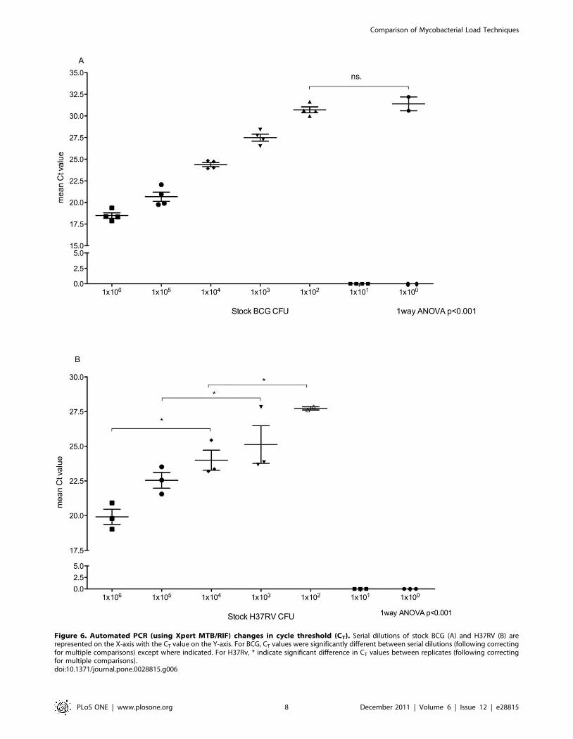
Figure 6. Automated PCR (using Xpert MTB/RIF) changes in cycle threshold (CT). Serial dilutions of stock BCG (A) and H37RV (B) arerepresented on the X-axis with the CT value on the Y-axis. For BCG, CT values were significantly different between serial dilutions (following correctingfor multiple comparisons) except where indicated. For H37Rv, * indicate significant difference in CT values between replicates (following correctingfor multiple comparisons).doi:10.1371/journal.pone.0028815.g006
Comparison of Mycobacterial Load Techniques
PLoS ONE | www.plosone.org 8 December 2011 | Volume 6 | Issue 12 | e28815

Discussion
Accurate determination of mycobacterial numbers is essential
for evaluating infectivity, the efficacy of treatments and for many
projects in translational research. However, there are limited
reports comparing the performance of different mycobacterial
load determination techniques. We evaluated several techniques
and to our knowledge head-to-head comparison of several
technologies has not been previously undertaken.
Our study has demonstrated that (i) Xpert MTB/RIF provides a
rapid measure of mycobacterial burden above a threshold of ,100
organisms/sample; (ii) TTP using MGIT 960, whilst accurate, with
better discriminative ability of mycobacterial load (than Xpert
MTB/RIF) and a detection threshold down to 1 CFU, has a
substantially longer turn-around-time; (iii) bioluminescence and
uracil incorporation assays are limited by lack of discrimination
below 1000 organisms. Thus, none of these assays has ‘ideal’
performance characteristics and the selection of an assay has to
based on the experimental question under study and details of the
study design including the likely range of bacterial burden which is
anticipated, need for rapid results, desired reproducibility, and
available laboratory and financial resources. In resource-limited
settings determination of CFU using solid media is likely to remain
the method of choice as it is likely to be associated with the least cost.
Although PCR for mycobacterial load is not a novel technique,
the Xpert MTB/RIF assay may overcome several of the known
drawbacks of real-time PCR. Most notably, apart from the semi
automation of the methods, DNA from degraded organisms is
thought to be removed in the wash step and only intact organisms
are retained in the cartridge mesh for the PCR step [19]. Our data
supports this view as Xpert-related results correlated with those
derived from solid culture CFU counts and thus intact organisms.
Although the Xpert MTB/RIF assay detects intact organisms, it
cannot distinguish viable from non-viable organisms and thus
suffers from similar drawbacks to conventional NAATs and smear
microscopy. All the other assays of mycobacterial burden
examined require the organisms to be viable and for detection
and hence are unable to detect the presence of intact but dead
organisms. Our work was specifically undertaken to establish
comparative utility of various techniques when performing in vitro
laboratory studies using human cells and not for clinical studies.
However, given its rapid turn-around-time and good correlation
with mycobacterial burden studies are now required to evaluate
the utility of Xpert MTB/RIF for monitoring treatment response,
disease prognosis, and evaluating risk of disease transmission. Like
with smear microscopy quantitative stratification is feasible. A
recent study has shown that Xpert MTB/RIF cycle threshold
correlates with organism load as defined by smear and MGIT time
to positivity [21]. In this study the discriminative ability of Xpert to
detect changes in organism load was suboptimal for H37RV
compared to BCG. Larger studies including clinical isolates are
required to verify this finding. Furthermore, the value of
mycobacterial load, as determined by Xpert has yet to be
demonstrated in the setting of a controlled clinical trial, particularly,
to what extent the presence of dead organisms in sputum may
confound the evaluation of treatment responses. Xpert has the
added advantage of sensitivity as it confirms infection in a significant
proportion of smear negative TB cases [21,22] and controls for
PCR inhibitors through an internal control.
Rapid turn-around-time is a further advantage of the Xpert
MTB/RIF assay, which might be of value in drug development.
For example, for providing ‘real time’ serial quantification of
bacterial burden during early bacteriocidal activity studies. Its
ability to detect quantitative changes in viable organisms will need
to be evaluated as all PCR techniques will detect DNA from both
live and dead organisms [23,24]. By contrast, liquid culture results,
although accurate, can take up to 6 weeks. However, the clinical
value of rapid turnaround time will needs to weighed up against
the potential greater cost and cost effectiveness studies will be
required at a national implementation level. Thus, in laboratory
studies while the rapid turnaround is highly attractive, it is likely to
come at a significantly higher cost compared to solid culture.
Another key drawback is the detection threshold of 100 organisms,
which may be inadequate for experimental models where a low
organism burden needs to be measured.
Liquid culture using the automated BACTEC 960 MGIT
system is an attractive quantification technology for both clinical
and laboratory studies. Already incorporated into EBA studies
[12,13] TTP has been well correlated with bacterial load
[7,13,14]. Its user-friendly format, automation, high discriminative
ability and low detection threshold (less than 10 organisms) makes
it well suited to laboratory studies. The key drawbacks of liquid
culture are the need to decontaminate clinical samples as MGIT
cultures are prone to bacterial overgrowth, in addition to the
slower turnaround time (days to weeks), compared to Xpert
MTB/RIF and solid culture, respectively [25].
Bioluminescence and uracil incorporation assays both provide
rapid turnaround time and are have been extensively used in
laboratory studies [10,26,27]. They have limited application to
clinical and public health studies as the findings are strain-specific
and there is a higher risk of bacterial contamination with uracil
incorporation assays. Both assays have limited discriminative
ability below 1000 organisms/sample. Uracil incorporation assays
offer little advantage over bioluminescence yet require significant
additional infrastructure to accommodate the storage and disposal
of radioactive waste. As with all the other ‘‘indirect’’ techniques of
quantitative load determination, a standard curve would be
required to calculate actual CFU counts if required.
There are several limitations to this study. The findings here
pertain to in vitro laboratory work done with a specific strain and
thus may not be generalizable to clinical strains or clinical samples
where sample quality and inhibitors etc. may impact on findings.
Smear microscopy was not used as a reference standard in this
study as the primary focus was on laboratory settings where
organism load below 100 000 organisms/ml are frequently used
and smear looses sensitivity below this threshold. Moreover, we
required a highly discriminative technique from a quantitation
point of view (smear only provides broad categorical data i.e. 1+ or
2+ etc. with poor discriminatory value). Therefore we chose not to
evaluate smear microscopy. Thus, because a single strain was used
for all experiments the MGIT findings may also not be
generalizable to clinical samples or different strains. Whilst Ct
correlated well with mycobacterial burden, as shown in recent
studies [21], to what extent it will correlate with risk of infection in
contacts of index cases remains to be determined.
In summary, no single mycobacterial quantitative technique has
ideal performance characteristics. Thus, the choice of assay will
largely depend on the research context, study question, and the
relative tradeoffs of cost versus turnaround time. Automated
systems like MGIT are sensitive and have high discriminatory
value but a long turnaround time. Xpert MTB/RIF is a good
quantitative tool with rapid turn-around-time but its detection
threshold was not as good as automated liquid culture. Although
solid culture is the most labor-intensive it likely remains the
cheapest option for highly discriminative quantification of
mycobacterial load over a wide dynamic range. Cost effectiveness
and clinical treatment follow up studies are still required for Xpert
and MGIT.
Comparison of Mycobacterial Load Techniques
PLoS ONE | www.plosone.org 9 December 2011 | Volume 6 | Issue 12 | e28815

Author Contributions
Conceived and designed the experiments: RvZS KD AB PLS RW GT.
Performed the experiments: RvZS AB RM HM PLS. Analyzed the data:
RvZS AB RM PLS. Contributed reagents/materials/analysis tools: RvZS
KD AW. Wrote the paper: RvZS KD EB GT JP SS HM AW RW PLS
RM AB.
References
1. Dheda K, van Zyl-Smit R, Badri M, Pai M (2009) T-cell interferon-gamma
release assays for the rapid immunodiagnosis of tuberculosis: clinical utility in
high-burden vs. low-burden settings. Curr Opin Pulm Med 15: 188–200.
2. Pai M, Kalantri S, Dheda K (2006) New tools and emerging technologies for the
diagnosis of tuberculosis: part II. Active tuberculosis and drug resistance. Expert
Rev Mol Diagn 6: 423–432.
3. Urdea M, Penny LA, Olmsted SS, Giovanni MY, Kaspar P, et al. (2006)
Requirements for high impact diagnostics in the developing world. Nature 444
Suppl 1: 73–79.
4. Dheda K, Shean K, Zumla A, Badri M, Streicher EM, et al. (2010) Early
treatment outcomes and HIV status of patients with extensively drug-resistant
tuberculosis in South Africa: a retrospective cohort study. Lancet 375:
1798–1807.
5. Dheda K, Warren RM, Zumla A, Grobusch MP (2010) Extensively Drug-
resistant Tuberculosis: Epidemiology and Management Challenges. Infectious
disease clinics of North America 24: 705–725.
6. Akhtar M, Bretzel G, Boulahbel D, Dawson DA, Fattorini L, et al. (2000)
Sputum Examination for Tuberculosis by Direct Microscopy in Low Income
Countries. .
7. Ritchie SR, Harrison AC, Vaughan RH, Calder L, Morris AJ (2007) New
recommendations for duration of respiratory isolation based on time to detect
Mycobacterium tuberculosis in liquid culture. European Respiratory Journal 30:
501–507.
8. Skinner MA, Ramsay AJ, Buchan GS, Keen DL, Ranasinghe C, et al. (2003) A
DNA prime-live vaccine boost strategy in mice can augment IFN-c responses to
mycobacterial antigens but does not increase the protective efficacy of two
attenuated strains of Mycobacterium bovis against bovine tuberculosis.
Immunology 108: 548–555.
9. Fremond CM, Togbe De, Doz E, Rose S, Vasseur V, et al. (2007) IL-1
Receptor-Mediated Signal Is an Essential Component of MyD88-Dependent
Innate Response to Mycobacterium tuberculosis Infection. The Journal of
Immunology 179: 1178–1189.
10. Snewin VA, Gares M-P, Gaora PO, Hasan Z, Brown IN, et al. (1999)
Assessment of Immunity to Mycobacterial Infection with Luciferase Reporter
Constructs. Infect Immun 67: 4586–4593.
11. Freeman S, Post FA, Bekker LG, Harbacheuski R, Steyn LM, et al. (2006)
Mycobacterium tuberculosis H37Ra and H37Rv differential growth and
cytokine/chemokine induction in murine macrophages in vitro. Journal of
interferon & cytokine research : the official journal of the International Society
for Interferon and Cytokine Research 26: 27–33.
12. Pheiffer C, Carroll NM, Beyers N, Donald P, Duncan K, et al. (2008) Time to
detection of Mycobacterium tuberculosis in BACTEC systems as a viable
alternative to colony counting. The international journal of tuberculosis and
lung disease : the official journal of the International Union against Tuberculosis
and Lung Disease 12: 792–798.
13. Diacon AH, Maritz JS, Venter A, van Helden PD, Andries K, et al. (2010) Time
to detection of the growth of Mycobacterium tuberculosis in MGIT 960 for
determining the early bactericidal activity of antituberculosis agents. European
journal of clinical microbiology & infectious diseases : official publication of the
European Society of Clinical Microbiology 29: 1561–1565.
14. Hesseling AC, Walzl G, Enarson DA, Carroll NM, Duncan K, et al. (2010)Baseline sputum time to detection predicts month two culture conversion and
relapse in non-HIV-infected patients. The international journal of tuberculosisand lung disease: the official journal of the International Union against
Tuberculosis and Lung Disease 14: 560–570.15. Dheda K, Huggett JF, Bustin SA, Johnson MA, Rook G, et al. (2004) Validation
of housekeeping genes for normalizing RNA expression in real-time PCR.
Biotechniques 37: 112–114, 116, 118–119.16. Dheda K, Huggett JF, Chang JS, Kim LU, Bustin SA, et al. (2005) The
implications of using an inappropriate reference gene for real-time reversetranscription PCR data normalization. Anal Biochem 344: 141–143.
17. Greco S, Rulli M, Girardi E, Piersimoni C, Saltini C (2009) Diagnostic accuracy
of in-house PCR for pulmonary tuberculosis in smear-positive patients: meta-analysis and metaregression. Journal of clinical microbiology 47: 569–576.
18. Blakemore R, Story E, Helb D, Kop J, Banada P, et al. (2010) Evaluation of theanalytical performance of the Xpert MTB/RIF assay. Journal of clinical
microbiology 48: 2495–2501.19. Helb D, Jones M, Story E, Boehme C, Wallace E, et al. (2010) Rapid detection
of Mycobacterium tuberculosis and rifampin resistance by use of on-demand,
near-patient technology. Journal of clinical microbiology 48: 229–237.20. Kampmann B, Gaora PO, Snewin VA, Gares MP, Young DB, et al. (2000)
Evaluation of human antimycobacterial immunity using recombinant reportermycobacteria. The Journal of Infectious Diseases 182: 895–901.
21. Theron G, Peter J, van Zyl-Smit R, Mishra H, Streicher E, et al. (2011)
Evaluation of the Xpert(R) MTB/RIF Assay for the Diagnosis of PulmonaryTuberculosis in a High HIV Prevalence Setting. American journal of respiratory
and critical care medicine.22. Boehme CC, Nabeta P, Hillemann D, Nicol MP, Shenai S, et al. (2010) Rapid
molecular detection of tuberculosis and rifampin resistance. The New Englandjournal of medicine 363: 1005–1015.
23. Hellyer TJ, Fletcher TW, Bates JH, Stead WW, Templeton GL, et al. (1996)
Strand displacement amplification and the polymerase chain reaction formonitoring response to treatment in patients with pulmonary tuberculosis. The
Journal of Infectious Diseases 173: 934–941.24. Thomsen VO, Kok-Jensen A, Buser M, Philippi-Schulz S, Burkardt HJ (1999)
Monitoring treatment of patients with pulmonary tuberculosis: can PCR be
applied? Journal of clinical microbiology 37: 3601–3607.25. O’Sullivan DM, Sander C, Shorten RJ, Gillespie SH, Hill AVS, et al. (2007)
Evaluation of liquid culture for quantitation of Mycobacterium tuberculosis inmurine models. Vaccine 25: 8203–8205.
26. Zhang T, Li SY, Converse PJ, Almeida DV, Grosset JH, et al. (2011) Usingbioluminescence to monitor treatment response in real time in mice with
Mycobacterium ulcerans infection. Antimicrobial agents and chemotherapy 55:
56–61.27. Andrew PW, Roberts IS (1993) Construction of a bioluminescent mycobacte-
rium and its use for assay of antimycobacterial agents. J Clin Microbiol 31:2251–2254.
Comparison of Mycobacterial Load Techniques
PLoS ONE | www.plosone.org 10 December 2011 | Volume 6 | Issue 12 | e28815