PaTTerns of small mammal sPecies richness in mediTerranean and TemPera Te chile
Original research or treatment paper
Comparison of oil and egg tempera paintsystems using time-of-flight secondary ionmass spectrometryZachary E. Voras1, Kristin deGhetaldi2, Brian Baade2, Eric Gordon3,Glenn Gates3, Thomas P. Beebe1
1Department of Chemistry and Biochemistry, University of Delaware, Newark, DE, USA, 2Department of ArtConservation, University of Delaware, Newark, DE, USA, 3The Walters Art Museum, Baltimore, MD, USA
Time-of-flight secondary ion mass spectrometry (ToF-SIMS) is quickly becoming a critical tool in the field ofart conservation. This technique provides high-resolution spatial maps of both inorganic and organiccomponents located within cross-sectional samples collected from works of art. With recent advances insurface analysis, ToF-SIMS can now be used to identify specific amino acids present in protein-containingmaterials as well as fatty acids in drying oils. For example, the detection of the ion fragment associatedwith the amino acid hydroxyproline can be used to confirm the use of animal glue in a paint sample. Asan analytical technique, ToF-SIMS avoids the need for derivatization/silylation reagents, with nointerference by the presence of pigments. Furthermore, the layered systems that are often encountered inhistorical paint samples remain intact throughout the analytical procedure. This allows for the co-localization of organic and inorganic species in specific layers (e.g. egg yolk paint atop a glue ground).Because of this ability to localize the analytical signal to approximately 6 μm or less, the mass spectralinformation can be used to produce mass-resolved and spatially-resolve images which can be correlatedto previous studies of the same samples. In this study, ToF-SIMS was used to analyze a paint crosssection obtained from a painting attributed to Raphael, and another from a painting by the Sienese artistMatteo di Giovanni.
Keywords: Raphael, Matteo di Giovanni, ToF-SIMS, Egg tempera, Drying oil, Fatty acids, Amino acids, Collagen
IntroductionIt has long been a goal of conservation scientists andhistorians of early Renaissance paintings to identifyand distinguish between the animal proteins in eggyolk and collagen, and to do so in a spatially resolvedmanner for the various layers. If such a determinationwere possible, researchers could identify the bound-aries of the layers that artists were known to use, orpossibly reveal previously unknown techniques byartists thought to adhere to certain practices.Furthermore, such information could greatly assist inestablishing a painting’s provenance. For example, ifanimal glue (i.e. collagen) is known historically to bethe binding agent in the ground layer of an artist’spaintings, and egg tempera (i.e. egg yolk) to be thebinding agent in the paint layers, then it would beimportant to confirm these practices with scientific
accuracy, and correlate the findings with historicalsources such as artists’ writings or treatises.
Analysis of complex multi-layered paint samplesThe use of analytical techniques on microsamples ‘inbulk’ has played a major role in the establishment ofour current knowledge base in art conservation andhistory (Mills & White, 1994). The ability to dis-solve/derivatize a complex, heterogeneous sample, fol-lowed by gas chromatography-mass spectrometry(GC-MS) analysis, has provided ample evidence forthe presence of certain proteins, oils, and pigments(Mills & White, 1976; Schilling & Khanjian, 1996a,1996b; Schilling et al., 1996; Colombini &Modugno, 2009). Nevertheless, the original location(and function) of those materials in the specimenmust be inferred, and the sample will have beendestroyed, making further analysis of the samesample from that location impossible. Complicationscan also arise when using chromatographic methods,
Correspondence to: Zachary E. Voras, Department of Chemistry andBiochemistry, 163 The Green, University of Delaware, Newark, DE19716, USA. Email: [email protected]
© The International Institute for Conservation of Historic and Artistic Works 2014DOI 10.1179/2047058414Y.0000000154 Studies in Conservation 2014 VOL. 0 NO. 0 1
such as insufficient sample size, the presence of non-original materials from previous restoration cam-paigns, and the interference of pigments (Schilling &Khanjian, 1996a; Schilling et al., 1996; Bonaduceet al., 2009; Colombini et al., 2010).Paint samples collected from complex and/or multi-
layered painted surfaces (e.g. easel paintings, murals,polychrome sculpture) can be especially challengingto characterize. Easel paintings executed before thetwentieth century typically contain binders consistingof animal glue, egg yolk, drying oils (e.g. linseed,walnut, or poppyseed), natural resins (e.g. dammar,mastic), among other organic materials (Mills &White, 1994; Bonaduce et al., 2009; Colombini &Modugno, 2009). Despite recent advances in analyti-cal procedures, it remains difficult to confirmwhether the various component materials, sometimesapplied in layers only a few micrometers thick, existas mixtures, emulsions, glazes, or surface coatingswithin these complex multi-layered paint systems.Attenuated total reflection Fourier transform infra-
red spectroscopy (ATR-FTIR) has been successfullyapplied to the analysis of cross sections, providinginformation about characteristic chemical groups inthe sample components (Rizzo, 2008; Spring et al.,2008). However, ATR-FTIR is limited in its abilityto distinguish between chemically similar materialssuch as proteins (e.g. collagen vs. egg yolk), and itdoes not allow the precise identification of markercompounds that is possible with mass spectrometrictechniques. Regarding inorganic materials, scanningelectron microscopy with energy dispersive X-ray spec-troscopy (SEM-EDX) is an invaluable and widelyused technique for imaging and elemental mappingof cross sections. However, as discussed below, time-of-flight secondary ion mass spectrometry (ToF-SIMS) is able to provide both inorganic and organicmapping simultaneously, and is also able to show theemission distribution of inorganic compounds (ascompared to single-element images) to aid inpigment identification.
ToF-SIMS and cultural heritage objectsOver the past decade, ToF-SIMS has shown greatpotential as a characterization tool in the field of cul-tural heritage (Keune & Boon, 2004; Boon et al., 2005;Adriaens & Dowsett, 2006; Mazel & Richardin, 2009).Recent developments in ToF-SIMS technology(specifically ion-beam technology and detectionsystems) have allowed for an improved total ion yieldand better spatial resolution for the analysis of biologi-cal, organic, and other ‘soft’ materials (Kötter &Benninghoven, 1998; Cliff et al., 2003; Kordys et al.,2008; Dubey et al., 2011). With respect to samplesfrom artworks or culturally significant objects, ToF-SIMS has four main advantages. First, the instrument
can be used to simultaneously collect chemical infor-mation (both elemental and molecular) from inorganicand organic materials (Keune & Boon, 2004;Richardin et al., 2011; Sanyova et al., 2011).Inorganic species originate from pigments and inertfillers or extenders, while the organic componentsstem from a much wider range of materials (Mills &White, 1994; Eastaugh et al., 2004; Colombini &Modugno, 2009). Secondly, ToF-SIMS is capable ofperforming both high mass-resolution and highspatial-resolution chemical imaging since theprimary ion beam can be focused to less than 1 μm.This enables the simultaneous, unambiguous identifi-cation of both organic and inorganic species presentin discrete areas within a sample, specifically thoselocated in individual layers or as distinct particles.Third, cross-sectional samples can be retained forfuture analysis. Finally, ToF-SIMS is one of the fewmass spectrometric techniques that allows the user toanalyze directly the surface of the unmodified solidsample, which is not subjected to an extraction proto-col prior to analysis, nor to the addition of a chemicalmatrix.
Many of these advantages can be exploited in par-ticular with the application of ToF-SIMS to thestudy of paint cross sections. ToF-SIMS imaging notonly assists with identification of the materials andtechniques used by the artist, but also with identifi-cation of compounds associated with pigment orbinder alteration, or non-original materials that maybe present from previous restoration campaigns.While some of the issues discussed above, such assample contamination, are also encountered usingSIMS, the technique has the benefit of providingspatial maps to help distinguish between unoriginal/original materials and to characterize discrete layers.Previous studies have published molecular ion mapscorresponding to compounds of interest found inpaint cross-section samples (Boon et al., 2001, 2005;Keune & Boon, 2005, 2007; Marino et al., 2006;Ferreira et al., 2008; Keune et al., 2008, 2009), buthave proven relatively unsuccessful at detectingmarkers for proteins, in particular, those associatedwith egg-based materials (Keune & Boon, 2005;Adriaens & Dowsett, 2006; Ferreira et al., 2008).However, more recent studies using ToF-SIMS havebeen used to identify blood on the surface of Africansculptures, and a protein-containing ground in asample collected from a painting by Rembrandt vanRijn (Mazel & Richardin, 2009; Sanyova et al., 2011).
Goals of the present studyThis study focuses on the simultaneous detection ofspecific markers associated with egg yolk, animalglue, and drying oil in two paint cross sections col-lected from Italian panel paintings in the collection
Voras et al. Comparison of oil and egg tempera paint systems
Studies in Conservation 2014 VOL. 0 NO. 02
of The Walters Art Museum, Baltimore. The crosssections were prepared more than 20 years prior tothe ToF-SIMS analysis, and no additional sampleswere collected from the paintings. The primary goalswere to obtain spatially resolved ToF-SIMS infor-mation relating to both inorganic and organic com-ponents of the historic samples, to assess whetherToF-SIMS could effectively differentiate betweentwo commonly used protein sources in the fifteenthand early sixteenth centuries: egg yolk and animalglue, and to evaluate whether ToF-SIMS could ident-ify and distinguish between these materials in layeredpaint systems. Previous studies using GC-MS haveshown that, over time, approximately seven aminoacids are most stable to oxidation and other degrada-tive processes (Halpine, 1992, 1995; Schilling &Khanjian, 1996a; Colombini et al., 2010). These rela-tively stable amino acids were thus chosen as theprimary markers for this study.
Materials and methodsReference materialsA set of historically accurate reference paint sampleswas used to generate relevant information regardingpositive and negative ion fragments observed in ToF-SIMS. These samples allowed the authors to optimizethe ToF-SIMS spectral and spatial performance as aresult of sample preparation (as discussed in detaillater). Table 1 summarizes the amino acid fragmentsthat were used for the identification of egg yolk andcollagen, as well as the fatty acid fragments associatedwith drying oils. Literature-accepted amino acidmarker fragments are compiled from previous studiesusing ToF-SIMS applied to protein-containingmaterials (Wagner & Castner, 2001; Xia et al., 2002;
Henry et al., 2003; Wagner et al., 2003), and boththe observed and calculated m/z values are reported.Paint reference samples used for the analysis and
identification of protein and oil markers were createdat the National Gallery of Art, Washington DC, in2008. Yellow ochre (Natural Sienna Monte Amiata),calcium sulfate (Terra Alba Natural Selenite), andSwedish cold-pressed linseed oil were purchased fromKremer Pigments, Inc. Azurite (fine grade), vermilion(dry process), and lead white (prepared using theDutch Stack Method) were obtained from Rublev/Natural Pigments. Egg yolks were collected fromUSDA grade organic brown eggs from free rangehens. Collagen was extracted by boiling parchmentscraps (obtained from skins of calf, sheep, and goat)purchased from Talas.All painted panel standards were prepared on glass
by first applying a gesso ground layer, followed bydrying, followed by brush application of the pigmen-ted paint in question. Painted panel standards weresubjected to short-term aging at the NationalGallery of Art using the Atlas Ci4000 Weather-Ometer, equipped with a Xenon light source(75 000 lux) using the ASTM Gamblin oil paint par-ameters (45% RH and 25°C for 400 hours). Small tri-angular cross sections of the reference paint sampleswere imbedded in Extec® polyester resin/hardener(approximately 10/0.5 ml).
Optical imaging of reference and historicalsamplesReference and historical samples were analyzed underhigh magnification using a Nikon Eclipse 80iBinocular Microscope (4× , 10× , and 20× objectives)with a Nikon X-cite® 120 Mercury Lamp for reflectedultraviolet light. Under ultraviolet light, the samples
Table 1 Positive and negative ion fragments observed during ToF-SIMS analysis, and the expectedmolar percentages of aminoacids as reported by Schilling et al. (1998) for aged egg yolk and animal glue (rabbit)
Expected molar percentages data fromSchilling et al. (1998)
Amino and fatty acid speciesobserved (ion polarity)
Empirical formula ofindicator ion
m/z of ioncalculated
(observed± 0.001)Egg yolk
(mole%± 9%)Animal glue
(mole%± 9%)
Glycine, GLY (+) CH4N+ 30.034 (30.034) 15.1% 46.0%
Alanine, ALA (+) C2H6N+ 44.050 (44.050) 23.1% 17.0%
Proline, PRO (+) C4H6N+ 68.050 (68.050) 10.5% 16.1%
Valine, VAL (+) C4H10N+ 72.081 (72.084) 16.6% 3.0%
Hydroxyproline, HYP (+) C4H8NO+ 86.061 (86.065) 0.0% 12.1%
Isoleucine, ISO (+) C5H12N+ 86.097 (86.100) 12.4% 1.9%
Leucine, LEU (+) C5H12N+ 86.097 (86.100) 21.9% 3.7%
Generic protein (−) CN− 26.003 (26.005) Not applicable Not applicable
Palmitic acid, PA (−) CH3(CH2)14COO− 255.232 (255.231) Not applicable Not applicable
Stearic acid, SA (−) CH3(CH2)16COO− 283.264 (283.261) Not applicable Not applicable
Percentages are given as mole percent of stable amino acids, determined by the remaining total protein before and after artificialaging at 50°C and 50% relative humidity for 500 hours. Minor differences from a total of 100% result from rounding errors.
Voras et al. Comparison of oil and egg tempera paint systems
Studies in Conservation 2014 VOL. 0 NO. 0 3
were viewed using a BV-2A cube (excitation wave-lengths between 400 and 420/470 nm barrier filter).Digital images were obtained using the DigitalEclipse DXM 1200f Nikon Camera in conjunctionwith the Automatic Camera Tamer control softwarefor PC systems.
Sample mounting and preparationThere are several challenges associated with the prep-aration of samples from cultural heritage objects,and especially historical paint cross-section samples,for surface-sensitive analyses such as ToF-SIMS.First, the samples are very small, making them diffi-cult to handle and manipulate in orientation.Second, the samples tend to be precious and limited,requiring a preparation method that does not precludefurther analyses. Third, particularly for the analysis ofpaint cross sections in this study, the method of creat-ing and exposing the cross section must not introduceartifacts resulting in contamination of the samplesurface. Since ToF-SIMS is a surface-sensitive tech-nique, artifacts caused by the effects of sample prep-aration will be immediately apparent during analysis.Additionally, any oils (resulting from unglovedsample handling) and polymeric residues (resultingfrom storage in improper containers) will be detectedin the ToF-SIMS spectra if the samples are contami-nated. To reduce these artifacts, room temperaturemicrotomy is the preferred method of sample prep-aration for the relatively hard embedding resinsencountered here. Cryomicrotomy, in which thesample is held at a low temperature during sectioning,is called for if the samples are soft and prone to smear-ing rather than cutting at room temperature. Whilesample preparation and microtomy have been dis-cussed previously in the literature (Mazel et al.,2006; Sanyova et al., 2011; Cersoy et al., 2012), adetailed account of our procedure is provided here.The goal of microtomy was to provide a flat surface
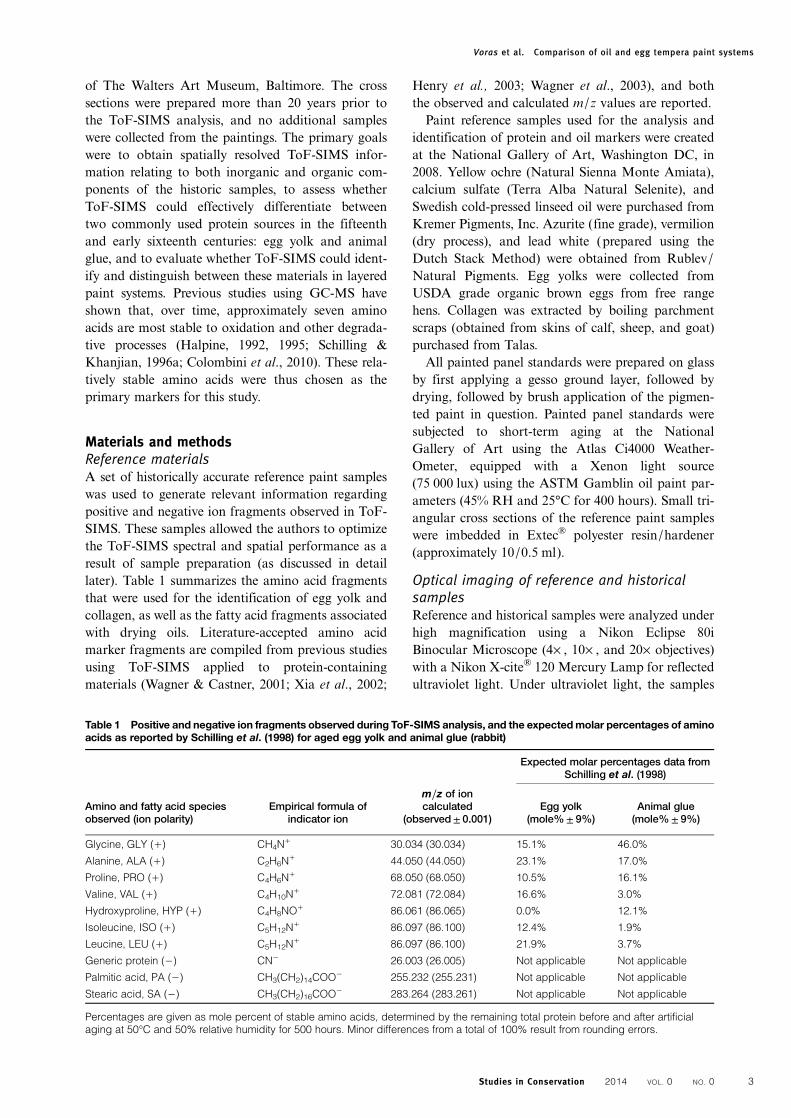
on the microtomed sample, rather than to remove andretain thin sections for analysis. Proper sample mount-ing proved to be a critical aspect to achieving this goal.Fig. 1 shows the sample preparation procedure for afreshly embedded sample (a similar procedure wasused for previously prepared samples). Starting froma roughly 1 cm3 resin cube (Fig. 1A), the embeddingresin was trimmed by hand using a Dremel® tooland cut-off wheel to remove most excess resin material,leaving a 4 mm3 resin cube containing the sample nearone face of the cube and oriented edge-on, such thatthe paint layer planes are perpendicular to thecutting plane (top of Fig. 1A). The sample was thenmounted on a Cryo Specimen Pin obtained fromElectron Microscopy Sciences (part number 70446),using cyanoacrylate-based glue obtained from PacerTechnology (part number 15187) (Fig. 1B). Next,
the embedding medium, now held firmly on the speci-men pin, was tapered by hand using a fresh standard0.009-inch-thick safety razor blade obtained fromFamous Smith Brand (part number 67-0238), tofurther reduce the resin area surrounding the futurecross section at the tip of the specimen (Fig. 1C).
It was observed during microtomy that thin sectionstended to fracture off (rather than cut cleanly) near theend of the cut when the tip of the specimen was square(i.e. when the microtome knife encountered a constantwidth of resin as it cut through the sample). Thistended to leave behind a rough specimen surfacewith an ill-defined sample cross section. It is surmisedthat mechanical stress during cutting could be lessenedif the tip of the specimen was shaped to reduce thewidth that the microtome knife encountered as it pro-gressed through each cut. Therefore, to lessen themechanical stress on the sample during the cutting ofthe flat-topped face of the resin and its embeddedpaint chip specimen, further hand shaping of theresin was used to form a trapezoid-shaped tip havinga wider end at the beginning of the cut, and a narrowerend at the completion of the cut (Fig. 1D; the directionof the arrow indicates the direction of microtome blademovement).
MicrotomyA series of cross-section cuts were then made on amicrotome (Leica Jung Biocut, model 2035), first bya stainless steel trimming knife obtained fromDelaware Diamond Knives (no part number), remov-ing approximately 10 μm per cut, until the first parts of
Figure 1 Preparation procedure for freshly embedded paintcross sections. (A) The sample is trimmed to a 1 cm3 cubefrom the original casting, (B) the cube is trimmed to a smallsize (∼4 mm) and attached to a specimen pin, (C) the sampleis then trimmed in preparation for microtomy. The cutdirection is indicated in (D).
Voras et al. Comparison of oil and egg tempera paint systems
Studies in Conservation 2014 VOL. 0 NO. 04
the paint sample became exposed. Finer cuts were thenmade using a diamond ultramicrotomy knife (3.5 mmwet cryo type) obtained from Delaware DiamondKnives, removing less than 1 μm per cut, while observ-ing the process under a monocular microscope on anarticulating arm (Specwell 10 × 30, 6°, with extrashort focus). When an appropriate depth into thesample had been reached, exposing a ‘fresh’ cross-sec-tional surface for analysis by ToF-SIMS, the sampleand stub assembly was transferred to a custom-designed, home-built sample holder to ensure properorientation of the exposed sample surface for Tof-SIMS analysis.
ToF-SIMS analysis conditionsThe ToF-SIMS instrument used for analysis wasa TOFSIMS IV, upgraded to the capabilities of aTOFSIMS V (ION-TOF, GmbH) equipped with abismuth/manganese primary ion beam. The instru-ment is housed in the Surface Analysis Facility at theUniversity of Delaware. Mass spectra and imageswere taken in the high-current ‘bunched’ mode, utiliz-ing 25 keV Bi3
+ ion clusters having a pre-bunched pulsewidth of 640 ps and an estimated spot size of less than5 μm in diameter, producing a sample current of∼0.27 pA. A low-energy (75 eV) electron flood gunwas used to stabilize the sample’s surface chargestate for the insulating samples analyzed here. TheToF mass analyzer used an extraction voltage of±2 kV, depending on ion polarity, and post-accelera-tion voltage of 10 kV.All spectra were acquired to the static SIMS limit of
1 × 10¹² ions/cm2. The mass scales of positive modespectra were calibrated with the following ions: H+,H2
+, H3+, C+, CH+, CH2
+, CH3+, C2H3
+, C3H5+,
C4H7+, C5H5
+, C6H5+, and C7H7
+; the mass scales ofnegative mode spectra were calibrated with the follow-ing ions: H−, H2
−, C−, CH−, CH2−, CH3
−, C2−, C2H
−,C3−, C4
−, C5−, C6
−, C7−, and C8
−. All data analysis wasperformed on ION-TOF software, version 6.2. Noquantification of signal intensities was used in thiswork, and thus no discussion of normalization isnecessary. A forthcoming publication will focus onthe quantitative analysis of these and similar culturalheritage objects (Voras et al., 2014). Lastly, it is impor-tant to note that all fragment peaks are identified byboth exact mass position and by the expected isotopicdistribution profile of the fragment.
ToF-SIMS mass resolution, mass accuracy, andmass precisionMass resolving power (also called mass resolution,m/Δm), mass precision, and mass accuracy affect theanalyst’s ability to interpret ToF-SIMS spectra with con-fidence. Over approximately 228 different ToF-SIMSpeaks, measured from 30 different samples, an analysis
of peak position accuracy and precision was made.Peak position precision averaged 46± 10 parts permillion (ppm), with a low of 36 ppm for some aminoacid fragments and a high of 62 ppm for others. Thatis, the ToF-SIMS instrument was able to reproduciblyobtain the same m/z values of all peaks in all samplesin repetitive measurements, to within 46 ppm onaverage. Peak position accuracy was calculated byexamining the difference between the observed m/zvalue in atomic mass units (AMU) of a particularmass fragment and that fragment’s theoretical m/zvalue. The absolute value of the peak position accu-racy averaged 1.8± 2.3 × 10⁻³ AMU, with a low devi-ation of −3.8 mAMU and a high deviation of+3.5 mAMU. That is, the ToF-SIMS instrumentwas able to reproducibly obtain the correct m/zvalue for a given mass fragment, making its identifi-cation unambiguous.Using the same dataset, an analysis of mass resol-
ving power (m/Δm) was also performed. The massresolving power averaged m/Δm= 6360± 1300, witha low of 3720 and a high of 9260. This relative widerange of m/Δm values highlights the importance of asample preparation technique that allows for reprodu-cible ToF-SIMS performance to resolve analyte peaks.Prior to analysis of samples, a clean silicon waferwas used to optimize conditions. Such an optimizationtypically resulted in a mass resolving power ofm/Δm= 8700 atm/z 29 (29Si+) for a clean siliconwafer.
Sample provenanceThe embedded paint sample from the Matteo diGiovanni painting was prepared in 1990, while thesample from the painting attributed to Raphael wasprepared in 1963. The embedding resin used for thesamples is not known, although based on its ToF-SIMS spectrum it was not cyanoacrylate based.
Results and discussionReference paint samplesCross sections from reference paint samples were ana-lyzed using ToF-SIMS in order to confirm the pres-ence of the ion fragments summarized in Table 1,and to assess whether certain pigments might havean effect on the detection of these fragments.Another purpose of the reference materials was toperfect the microtomy preparation technique priorto the handling of precious samples from paintings,to ensure optimum spectral and spatial instrumentalperformance. The characteristic markers for aminoacids associated with animal glue (see Table 1) weredetected in the gesso ground for all samples.Likewise, amino acid fragments (with the exceptionof hydroxyproline) associated with egg yolk weredetected in all egg-containing paints. Characteristic
Voras et al. Comparison of oil and egg tempera paint systems
Studies in Conservation 2014 VOL. 0 NO. 0 5
markers for fatty acids (see Table 1) were also observedin both egg and oil-bound paints.
Raphael’s ‘Madonna of the Candelabra’The Madonna of the Candelabra in the collection ofthe Walters Art Museum (accession number 37.484)dates to about 1513 and has been associated withRaphael’s Roman period (Zeri, 1976a; Henry &Joannides, 2012). From a stylistic perspective, it hasbeen suggested that Raphael’s workshop assistantsmay have played a role in the execution of the paint-ing, and questions remain about the materials andtechniques used by the artist or those working withinhis workshop. Several of Raphael’s works have beenextensively studied and it appears the artist adoptedthe use of egg tempera, drying oils, and even temperagrassa (emulsion of egg yolk and drying oil) asbinders in his paints (Mills & White, 1976;Pietrangeli et al., 1993; White & Pilc, 1995;Dunkerton, 1999; Roy et al., 2004).
A cross section from the proper right arm of theChrist child was prepared using a microtome equippedwith a diamond knife prior to analysis by ToF-SIMS,
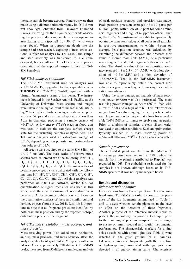
Figure 2 Cross section from proper right arm of the Christchild in theMadonna of the Candelabra attributed to Raphael,micrographs in visible (top) and ultraviolet illumination(bottom). A void is present (2) between the paint layer (1) andthe rest of the sample. An auto-fluorescent medium-rich layer(3) can be seen directly atop the ground layer (4).
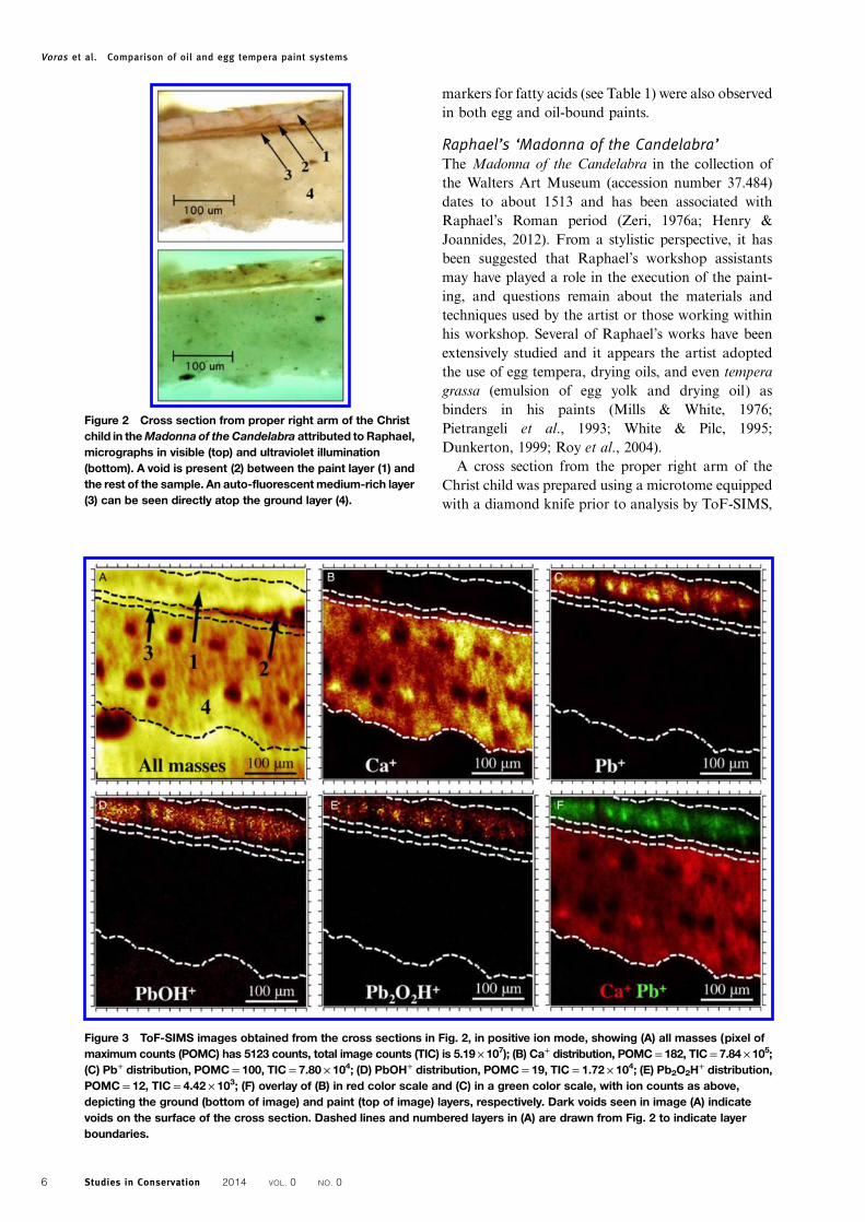
Figure 3 ToF-SIMS images obtained from the cross sections in Fig. 2, in positive ion mode, showing (A) all masses (pixel ofmaximum counts (POMC) has 5123 counts, total image counts (TIC) is 5.19× 10⁷); (B) Ca+ distribution, POMC= 182, TIC= 7.84× 10⁵;(C) Pb+ distribution, POMC= 100, TIC= 7.80 × 10⁴; (D) PbOH+ distribution, POMC= 19, TIC= 1.72 × 10⁴; (E) Pb2O2H
+ distribution,POMC= 12, TIC= 4.42 × 10³; (F) overlay of (B) in red color scale and (C) in a green color scale, with ion counts as above,depicting the ground (bottom of image) and paint (top of image) layers, respectively. Dark voids seen in image (A) indicatevoids on the surface of the cross section. Dashed lines and numbered layers in (A) are drawn from Fig. 2 to indicate layerboundaries.
Voras et al. Comparison of oil and egg tempera paint systems
Studies in Conservation 2014 VOL. 0 NO. 06
as discussed above and outlined schematically inFig. 1. Fig. 2 shows an optical microscope imageunder visible and ultraviolet illumination. In bothimages, the paint layer appears to be fractured, cleav-ing away from the layers beneath. A gap (labelled 2)was observed just beneath the paint layer (1), as wellas a medium-rich auto-fluorescent layer (3) immedi-ately atop the ground layer (4). As shown in Fig. 3A,these observations were confirmed in the ToF-SIMSall masses image (total ion image), revealing a lackof signal at the interface between the paint layer andthe layer immediately below. Lead white was locatedin the flesh paint layer, as indicated by several charac-teristic positive lead ions (Pb+, m/z 207.977, Fig. 3C;Pb2
+, m/z 415.953, not shown; PbOH+, m/z 224.979,Fig. 3D; Pb2O2H
+, m/z 448.951, Fig. 3E; Pb3O3H+,
m/z 672.923, not shown); their expected isotopicpeaks were observed as well. As SIMS did not detectiron oxides or mercuric sulfide, it was suspected thatthe red colorant in the paint layer is a red lake, anobservation that was supported by analysis usingSEM-EDX that did not detect iron or mercury. Nomarkers associated with possible mordants (e.g. Al,Ca), however, could be detected using either
technique. Further study is required to confirm theidentity of the red pigment, which may be present inconcentrations too low for either method to detect.The presence of a calcium-based ground layer suchas gesso was confirmed with ToF-SIMS by the pres-ence of calcium ions in positive ion mode (Ca+, m/z39.963, Fig. 3B; Ca2
+, m/z 79.923, not shown), andassociated characteristic fragments in negative ionmode (CaOH−, m/z 56.965, not shown; CaSO4
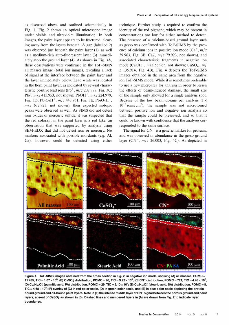
−, m/z 135.914, Fig. 4B). Fig. 4 depicts the ToF-SIMSimages obtained in the same area from the negativeion ToF-SIMS mode. While it is sometimes preferableto use a new microarea for analysis in order to lessenthe effects of beam-induced damage, the small sizeof the sample only allowed for a single analysis spot.Because of the low beam dosage per analysis (1 ×10¹² ions/cm2), the sample was not microtomedbetween positive ion and negative ion analysis sothat the sample could be preserved, and so that itcould be known with confidence that the analyses cor-responded to the same surface.The signal for CN− is a generic marker for proteins,
and was observed in abundance in the gesso groundlayer (CN−, m/z 26.003, Fig. 4C). As depicted in
Figure 4 ToF-SIMS images obtained from the cross section in Fig. 2, in negative ion mode, showing (A) all masses, POMC=11 435, TIC= 1.07 × 10⁸; (B) CaSO4
− distribution, POMC= 96, TIC= 3.22 × 10⁵; (C) CN− distribution, POMC= 721, TIC= 4.48 × 10⁶;(D) C16H31O2
− (palmitic acid, PA) distribution, POMC= 29, TIC= 2.10 × 10⁴; (E) C18H35O2− (stearic acid, SA) distribution, POMC= 8,
TIC= 4.68 × 10³; (F) overlay of (C) in red color scale, (D) in green color scale, and (E) in blue color scale depicting the protein-bound ground and oil-bound paint layers. Note in (F) the intensemiddle layer of CN− signal between the porous ground and paintlayers, absent of CaSO4
− as shown in (B). Dashed lines and numbered layers in (A) are drawn from Fig. 2 to indicate layerboundaries.
Voras et al. Comparison of oil and egg tempera paint systems
Studies in Conservation 2014 VOL. 0 NO. 0 7
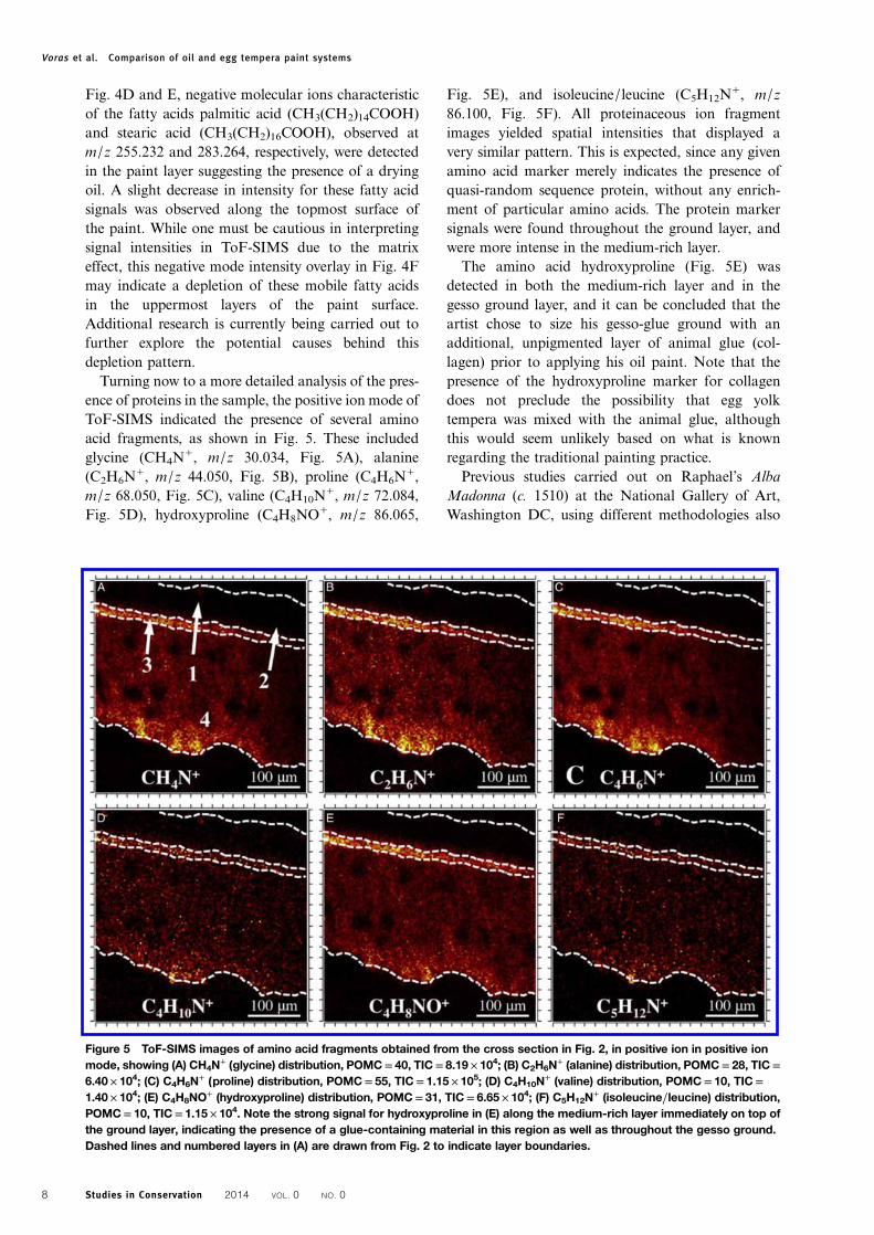
Fig. 4D and E, negative molecular ions characteristicof the fatty acids palmitic acid (CH3(CH2)14COOH)and stearic acid (CH3(CH2)16COOH), observed atm/z 255.232 and 283.264, respectively, were detectedin the paint layer suggesting the presence of a dryingoil. A slight decrease in intensity for these fatty acidsignals was observed along the topmost surface ofthe paint. While one must be cautious in interpretingsignal intensities in ToF-SIMS due to the matrixeffect, this negative mode intensity overlay in Fig. 4Fmay indicate a depletion of these mobile fatty acidsin the uppermost layers of the paint surface.Additional research is currently being carried out tofurther explore the potential causes behind thisdepletion pattern.Turning now to a more detailed analysis of the pres-
ence of proteins in the sample, the positive ion mode ofToF-SIMS indicated the presence of several aminoacid fragments, as shown in Fig. 5. These includedglycine (CH4N
+, m/z 30.034, Fig. 5A), alanine(C2H6N
+, m/z 44.050, Fig. 5B), proline (C4H6N+,
m/z 68.050, Fig. 5C), valine (C4H10N+, m/z 72.084,
Fig. 5D), hydroxyproline (C4H8NO+, m/z 86.065,
Fig. 5E), and isoleucine/leucine (C5H12N+, m/z
86.100, Fig. 5F). All proteinaceous ion fragmentimages yielded spatial intensities that displayed avery similar pattern. This is expected, since any givenamino acid marker merely indicates the presence ofquasi-random sequence protein, without any enrich-ment of particular amino acids. The protein markersignals were found throughout the ground layer, andwere more intense in the medium-rich layer.
The amino acid hydroxyproline (Fig. 5E) wasdetected in both the medium-rich layer and in thegesso ground layer, and it can be concluded that theartist chose to size his gesso-glue ground with anadditional, unpigmented layer of animal glue (col-lagen) prior to applying his oil paint. Note that thepresence of the hydroxyproline marker for collagendoes not preclude the possibility that egg yolktempera was mixed with the animal glue, althoughthis would seem unlikely based on what is knownregarding the traditional painting practice.
Previous studies carried out on Raphael’s AlbaMadonna (c. 1510) at the National Gallery of Art,Washington DC, using different methodologies also
Figure 5 ToF-SIMS images of amino acid fragments obtained from the cross section in Fig. 2, in positive ion in positive ionmode, showing (A) CH4N
+ (glycine) distribution, POMC= 40, TIC= 8.19 × 10⁴; (B) C2H6N+ (alanine) distribution, POMC= 28, TIC=
6.40 × 10⁴; (C) C4H6N+ (proline) distribution, POMC= 55, TIC= 1.15 × 10⁵; (D) C4H10N
+ (valine) distribution, POMC= 10, TIC=1.40 × 10⁴; (E) C4H8NO
+ (hydroxyproline) distribution, POMC= 31, TIC= 6.65 × 10⁴; (F) C5H12N+ (isoleucine/leucine) distribution,
POMC= 10, TIC= 1.15 × 10⁴. Note the strong signal for hydroxyproline in (E) along the medium-rich layer immediately on top ofthe ground layer, indicating the presence of a glue-containing material in this region as well as throughout the gesso ground.Dashed lines and numbered layers in (A) are drawn from Fig. 2 to indicate layer boundaries.
Voras et al. Comparison of oil and egg tempera paint systems
Studies in Conservation 2014 VOL. 0 NO. 08
revealed the use of an animal glue size layer atop thegesso ground layer (Berrie & Walmsley, 2007). Inaddition, a survey conducted at the Louvre in Parishas shown the presence of both proteins and oils inthe preparatory layer on a handful of works byRaphael, some of which were medium-rich andlacking pigments (Dunkerton, 1999; Mottin et al.,2012).
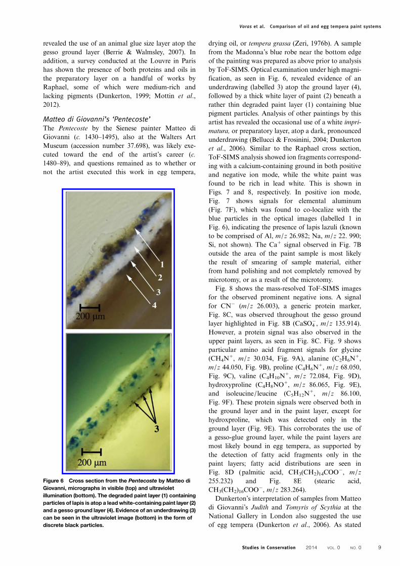
Matteo di Giovanni’s ‘Pentecoste’The Pentecoste by the Sienese painter Matteo diGiovanni (c. 1430–1495), also at the Walters ArtMuseum (accession number 37.698), was likely exe-cuted toward the end of the artist’s career (c.1480–89), and questions remained as to whether ornot the artist executed this work in egg tempera,
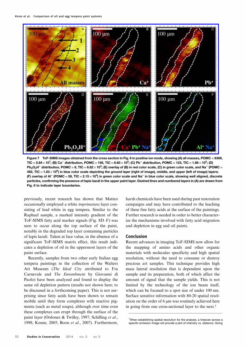
drying oil, or tempera grassa (Zeri, 1976b). A samplefrom the Madonna’s blue robe near the bottom edgeof the painting was prepared as above prior to analysisby ToF-SIMS. Optical examination under high magni-fication, as seen in Fig. 6, revealed evidence of anunderdrawing (labelled 3) atop the ground layer (4),followed by a thick white layer of paint (2) beneath arather thin degraded paint layer (1) containing bluepigment particles. Analysis of other paintings by thisartist has revealed the occasional use of a white impri-matura, or preparatory layer, atop a dark, pronouncedunderdrawing (Bellucci & Frosinini, 2004; Dunkertonet al., 2006). Similar to the Raphael cross section,ToF-SIMS analysis showed ion fragments correspond-ing with a calcium-containing ground in both positiveand negative ion mode, while the white paint wasfound to be rich in lead white. This is shown inFigs. 7 and 8, respectively. In positive ion mode,Fig. 7 shows signals for elemental aluminum(Fig. 7F), which was found to co-localize with theblue particles in the optical images (labelled 1 inFig. 6), indicating the presence of lapis lazuli (knownto be comprised of Al, m/z 26.982; Na, m/z 22. 990;Si, not shown). The Ca+ signal observed in Fig. 7Boutside the area of the paint sample is most likelythe result of smearing of sample material, eitherfrom hand polishing and not completely removed bymicrotomy, or as a result of the microtomy.Fig. 8 shows the mass-resolved ToF-SIMS images
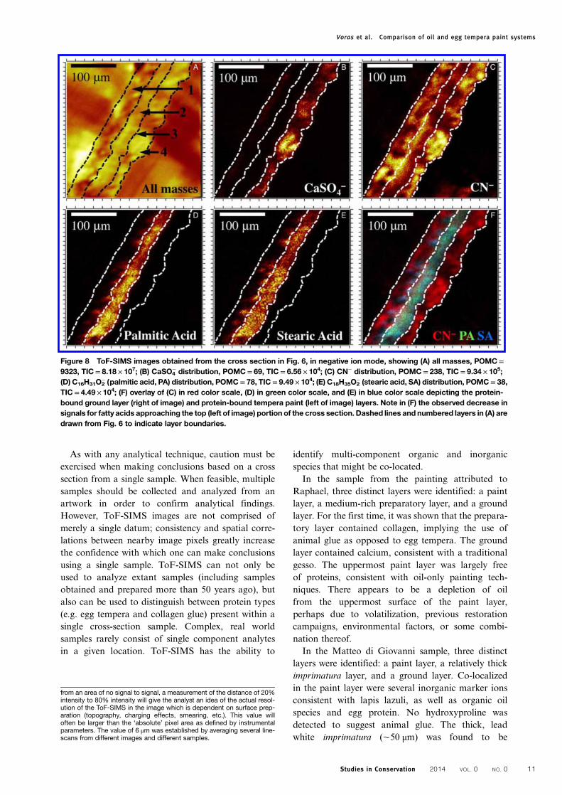
for the observed prominent negative ions. A signalfor CN− (m/z 26.003), a generic protein marker,Fig. 8C, was observed throughout the gesso groundlayer highlighted in Fig. 8B (CaSO4
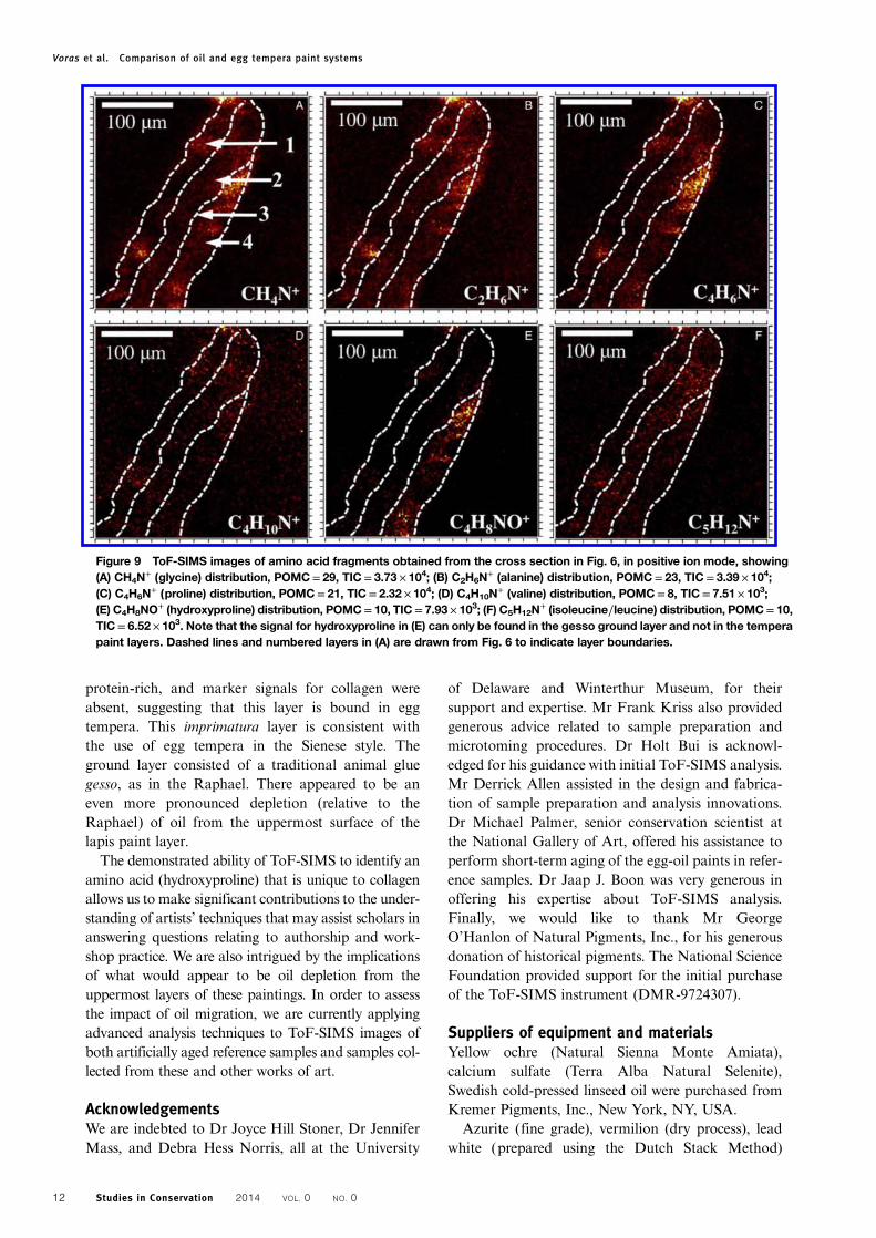
−, m/z 135.914).However, a protein signal was also observed in theupper paint layers, as seen in Fig. 8C. Fig. 9 showsparticular amino acid fragment signals for glycine(CH4N
+, m/z 30.034, Fig. 9A), alanine (C2H6N+,
m/z 44.050, Fig. 9B), proline (C4H6N+, m/z 68.050,
Fig. 9C), valine (C4H10N+, m/z 72.084, Fig. 9D),
hydroxyproline (C4H8NO+, m/z 86.065, Fig. 9E),and isoleucine/leucine (C5H12N
+, m/z 86.100,Fig. 9F). These protein signals were observed both inthe ground layer and in the paint layer, except forhydroxproline, which was detected only in theground layer (Fig. 9E). This corroborates the use ofa gesso-glue ground layer, while the paint layers aremost likely bound in egg tempera, as supported bythe detection of fatty acid fragments only in thepaint layers; fatty acid distributions are seen inFig. 8D (palmitic acid, CH3(CH2)14COO−, m/z255.232) and Fig. 8E (stearic acid,CH3(CH2)16COO−, m/z 283.264).Dunkerton’s interpretation of samples from Matteo
di Giovanni’s Judith and Tomyris of Scythia at theNational Gallery in London also suggested the useof egg tempera (Dunkerton et al., 2006). As stated
Figure 6 Cross section from the Pentecoste by Matteo diGiovanni, micrographs in visible (top) and ultravioletillumination (bottom). The degraded paint layer (1) containingparticles of lapis is atop a leadwhite-containing paint layer (2)and a gesso ground layer (4). Evidence of an underdrawing (3)can be seen in the ultraviolet image (bottom) in the form ofdiscrete black particles.
Voras et al. Comparison of oil and egg tempera paint systems
Studies in Conservation 2014 VOL. 0 NO. 0 9
previously, recent research has shown that Matteooccasionally employed a white imprimatura layer con-sisting of lead white in egg tempera. Similar to theRaphael sample, a marked intensity gradient of theToF-SIMS fatty acid marker signals (Fig. 8D–F) wasseen to occur along the top surface of the paint,notably in the degraded top layer containing particlesof lapis lazuli. Taken at face value, in the absence of asignificant ToF-SIMS matrix effect, this result indi-cates a depletion of oil in the uppermost layers of thepaint surface.Recently, samples from two other early Italian egg
tempera paintings in the collection of the WaltersArt Museum (The Ideal City attributed to FraCarnevale and The Entombment by Giovanni diPaolo) have been analyzed and found to display thesame oil depletion pattern (results not shown here; tobe discussed in a forthcoming paper). This is not sur-prising since fatty acids have been shown to remainmobile until they form complexes with reactive pig-ments (such as metal soaps), although over time eventhese complexes can erupt through the surface of thepaint layer (Ordonez & Twilley, 1997; Schilling et al.,1998; Keune, 2005; Boon et al., 2007). Furthermore,
harsh chemicals have been used during past restorationcampaigns and may have contributed to the leachingof these free fatty acids at the surface of the paintings.Further research is needed in order to better character-ize the mechanisms involved with fatty acid migrationand depletion in egg and oil paints.
ConclusionRecent advances in imaging ToF-SIMS now allow forthe mapping of amino acids and other organicmaterials with molecular specificity and high spatialresolution, without the need to consume or destroyprecious art samples. This technique provides highmass lateral resolution that is dependent upon thesample and its preparation, both of which affect theamount of signal that the sample yields. This is notlimited by the technology of the ion beam itself,which can be focused to a spot size of under 100 nm.Surface sensitive information with 80-20 spatial resol-ution on the order of 6 μm was routinely achieved herein going from one cross-sectional layer to the next.1
Figure 7 ToF-SIMS images obtained from the cross section in Fig. 6 in positive ionmode, showing (A) all masses, POMC= 6306,TIC= 5.84 × 10⁷; (B) Ca+ distribution, POMC= 156, TIC= 6.60 × 10⁵; (C) Pb+ distribution, POMC= 123, TIC= 1.86 × 10⁵; (D)Pb2O2H
+ distribution, POMC= 9, TIC= 6.82 × 10³; (E) overlay of (B) in red color scale, (C) in green color scale, and Na+ (POMC=492, TIC= 1.03 × 10⁶) in blue color scale depicting the ground layer (right of image), middle, and upper (left of image) layers;(F) overlay of Al+ (POMC= 59, TIC= 2.15 × 10⁴) in green color scale and Na+ in blue color scale, showing well aligned, discreteparticles, confirming the presence of lapis lazuli in the upper paint layer. Dashed lines and numbered layers in (A) are drawn fromFig. 6 to indicate layer boundaries.
1When establishing spatial resolution for the analysis, a linescan across aspecific emission image will provide a plot of intensity vs. distance. Going
Voras et al. Comparison of oil and egg tempera paint systems
Studies in Conservation 2014 VOL. 0 NO. 010
As with any analytical technique, caution must beexercised when making conclusions based on a crosssection from a single sample. When feasible, multiplesamples should be collected and analyzed from anartwork in order to confirm analytical findings.However, ToF-SIMS images are not comprised ofmerely a single datum; consistency and spatial corre-lations between nearby image pixels greatly increasethe confidence with which one can make conclusionsusing a single sample. ToF-SIMS can not only beused to analyze extant samples (including samplesobtained and prepared more than 50 years ago), butalso can be used to distinguish between protein types(e.g. egg tempera and collagen glue) present within asingle cross-section sample. Complex, real worldsamples rarely consist of single component analytesin a given location. ToF-SIMS has the ability to
identify multi-component organic and inorganicspecies that might be co-located.In the sample from the painting attributed to
Raphael, three distinct layers were identified: a paintlayer, a medium-rich preparatory layer, and a groundlayer. For the first time, it was shown that the prepara-tory layer contained collagen, implying the use ofanimal glue as opposed to egg tempera. The groundlayer contained calcium, consistent with a traditionalgesso. The uppermost paint layer was largely freeof proteins, consistent with oil-only painting tech-niques. There appears to be a depletion of oilfrom the uppermost surface of the paint layer,perhaps due to volatilization, previous restorationcampaigns, environmental factors, or some combi-nation thereof.In the Matteo di Giovanni sample, three distinct
layers were identified: a paint layer, a relatively thickimprimatura layer, and a ground layer. Co-localizedin the paint layer were several inorganic marker ionsconsistent with lapis lazuli, as well as organic oilspecies and egg protein. No hydroxyproline wasdetected to suggest animal glue. The thick, leadwhite imprimatura (∼50 μm) was found to be
Figure 8 ToF-SIMS images obtained from the cross section in Fig. 6, in negative ion mode, showing (A) all masses, POMC=9323, TIC= 8.18 × 10⁷; (B) CaSO4
− distribution, POMC= 69, TIC= 6.56 × 10⁴; (C) CN− distribution, POMC= 238, TIC= 9.34 × 10⁵;(D) C16H31O2
− (palmitic acid, PA) distribution, POMC= 78, TIC= 9.49 × 10⁴; (E) C18H35O2− (stearic acid, SA) distribution, POMC= 38,
TIC= 4.49 × 10⁴; (F) overlay of (C) in red color scale, (D) in green color scale, and (E) in blue color scale depicting the protein-bound ground layer (right of image) and protein-bound tempera paint (left of image) layers. Note in (F) the observed decrease insignals for fatty acids approaching the top (left of image) portion of the cross section. Dashed lines and numbered layers in (A) aredrawn from Fig. 6 to indicate layer boundaries.
from an area of no signal to signal, a measurement of the distance of 20%intensity to 80% intensity will give the analyst an idea of the actual resol-ution of the ToF-SIMS in the image which is dependent on surface prep-aration (topography, charging effects, smearing, etc.). This value willoften be larger than the ‘absolute’ pixel area as defined by instrumentalparameters. The value of 6 μm was established by averaging several line-scans from different images and different samples.
Voras et al. Comparison of oil and egg tempera paint systems
Studies in Conservation 2014 VOL. 0 NO. 0 11
protein-rich, and marker signals for collagen wereabsent, suggesting that this layer is bound in eggtempera. This imprimatura layer is consistent withthe use of egg tempera in the Sienese style. Theground layer consisted of a traditional animal gluegesso, as in the Raphael. There appeared to be aneven more pronounced depletion (relative to theRaphael) of oil from the uppermost surface of thelapis paint layer.The demonstrated ability of ToF-SIMS to identify an
amino acid (hydroxyproline) that is unique to collagenallows us to make significant contributions to the under-standing of artists’ techniques that may assist scholars inanswering questions relating to authorship and work-shop practice. We are also intrigued by the implicationsof what would appear to be oil depletion from theuppermost layers of these paintings. In order to assessthe impact of oil migration, we are currently applyingadvanced analysis techniques to ToF-SIMS images ofboth artificially aged reference samples and samples col-lected from these and other works of art.
AcknowledgementsWe are indebted to Dr Joyce Hill Stoner, Dr JenniferMass, and Debra Hess Norris, all at the University
of Delaware and Winterthur Museum, for theirsupport and expertise. Mr Frank Kriss also providedgenerous advice related to sample preparation andmicrotoming procedures. Dr Holt Bui is acknowl-edged for his guidance with initial ToF-SIMS analysis.Mr Derrick Allen assisted in the design and fabrica-tion of sample preparation and analysis innovations.Dr Michael Palmer, senior conservation scientist atthe National Gallery of Art, offered his assistance toperform short-term aging of the egg-oil paints in refer-ence samples. Dr Jaap J. Boon was very generous inoffering his expertise about ToF-SIMS analysis.Finally, we would like to thank Mr GeorgeO’Hanlon of Natural Pigments, Inc., for his generousdonation of historical pigments. The National ScienceFoundation provided support for the initial purchaseof the ToF-SIMS instrument (DMR-9724307).
Suppliers of equipment and materialsYellow ochre (Natural Sienna Monte Amiata),calcium sulfate (Terra Alba Natural Selenite),Swedish cold-pressed linseed oil were purchased fromKremer Pigments, Inc., New York, NY, USA.
Azurite (fine grade), vermilion (dry process), leadwhite (prepared using the Dutch Stack Method)
Figure 9 ToF-SIMS images of amino acid fragments obtained from the cross section in Fig. 6, in positive ion mode, showing(A) CH4N
+ (glycine) distribution, POMC= 29, TIC= 3.73 × 10⁴; (B) C2H6N+ (alanine) distribution, POMC= 23, TIC= 3.39 × 10⁴;
(C) C4H6N+ (proline) distribution, POMC= 21, TIC= 2.32 × 10⁴; (D) C4H10N
+ (valine) distribution, POMC= 8, TIC= 7.51 × 10³;(E) C4H8NO
+ (hydroxyproline) distribution, POMC= 10, TIC= 7.93 × 10³; (F) C5H12N+ (isoleucine/leucine) distribution, POMC= 10,
TIC= 6.52 × 10³. Note that the signal for hydroxyproline in (E) can only be found in the gesso ground layer and not in the temperapaint layers. Dashed lines and numbered layers in (A) are drawn from Fig. 6 to indicate layer boundaries.
Voras et al. Comparison of oil and egg tempera paint systems
Studies in Conservation 2014 VOL. 0 NO. 012
were obtained from Rublev/Natural Pigments, Willits,CA, USA.Egg yolks were collected from USDA organic
brown eggs from free range hens.Collagen was extracted by boiling parchment scraps
(from skins of calf, sheep, and goat) purchased fromTalas, Brooklyn, NY, USA.Atlas Ci4000 Weather-Ometer, Atlas Material
Testing Technology, Chicago, IL, USA.Extec® Polyester Resin, Extec Corporation, Enfield,
CT, USA.Leica Jung Biocut, model 2035, Leica
Microsystems, Inc., Buffalo Grove, IL, USA.Nikon Eclipse 80i Binocular Microscope, Nikon
X-cite® 120 Mercury Lamp Digital Eclipse DXM1200f Nikon Camera, Nikon Inc., Melville, NY, USA.Stainless steel trimming knife (no part number) and
Diamond ultramicrotomy knife (3.5 mm wet cryo),Delaware Diamond Knives, Wilmington, DE, USA.Dremel tool and cut-off wheel, Robert Bosch LLC,
Farmington Hills, MI, USA.Cryo specimen pin (part number 70446) obtained
from ElectronMicroscopy Sciences, Hatfield, PA, USA.Cyanoacrylate-based glue (part number 15187)
obtained from Pacer Technology, RanchoCucamonga, CA, USA.TOF-SIMS V (ION-TOF, GmbH), ION-TOF,
GmbH, Münster, Germany.Monocular microscope on an articulating arm
(Specwell), Specwell Corporation, Toshima-Ku,Tokyo, Japan.
ReferencesAdriaens, A. & Dowsett, M.G. 2006. Applications of SIMS to
Cultural Heritage Studies. Applied Surface Science, 252:7096–101.
Bellucci, R. & Frosinini, C. 2004. Un ‘Modello’ per la DiagnosticaIntegrata. Kermes, 53: 29–38.
Berrie, B. & Walmsley, E. 2007. Raphael’s ‘Alba Madonna’. In:M. Spring & A. Roy, eds. Raphael’s Painting Technique:Working Practices Before Rome. Florence: Nardini Editore,pp. 101–8.
Bonaduce, I., Marcello, C. & Colombini, M.P. 2009. TheDevelopment of a Gas Chromatographic–Mass SpectrometricAnalytical Procedure for the Determination of Lipids,Proteins and Resins in the Same Paint Micro-sampleAvoiding Interferences from Inorganic Media. Journal ofChromatography A, 1216: 5931–9.
Boon, J.J., Keune, K., van der Weerd, L., Geldof, M. & van Asperende Boer, J.R.J. 2001. Imaging Microspectroscopic, SecondaryIon Mass Spectrometric and Electron Microscopic Studies onDiscoloured and Partially Discoloured Smalt in Cross-sectionsof 16th Century Paintings. Chimia, 55: 952–60.
Boon, J.J., Keune, K. & Zucker, J. 2005. Imaging Analytical Studiesof Lead Soaps Aggregating in Preprimed Canvas Used by theHudson River School Painter F.E. Church. Microscopy andMicroanalysis, 11: 444–5.
Boon, J.J., Hoogland, F. & Keune, K. 2007. Chemical Process inAged Oil Paints Affecting Metal Soap Migration andAggregation. In: H.M. Parkin, ed. AIC Paintings SpecialtyGroup, 34th Annual Meeting, Providence, Rhode Island,Postprints. Washington, DC: American Institute forConservation, pp. 18–25.
Cersoy, S., Richardin, P., Walter, P. & Brunelle, A. 2012. ClusterTOF-SIMS Imaging of Human Skin Remains: Analysis of aSouth-Andean Mummy Sample. Journal of MassSpectrometry, 47: 338–6.
Cliff, B., Lockyer, N.P., Corlett, C. & Vickerman, J.C. 2003.Development of Instrumentation for Routine ToF-SIMSImaging Analysis of Biological Material. Applied SurfaceScience, 203: 730–3.
Colombini, M.P. &Modugno, F. 2009. OrganicMaterials in Art andArchaeology. In: M.P. Colombini & F. Modugno, eds. OrganicMass Spectrometry in Art and Archaeology. New York: Wiley,pp. 3–36.
Colombini, M.P., Andreotti, A., Bonaduce, I., Modugno, F. &Ribechini, E. 2010. Analytical Strategies for CharacterizingOrganic Paint Media using Gas Chromatography/MassSpectrometry. Accounts of Chemical Research, 43: 715–27.
Dubey, M., Brison, J., Grainger, D.W. & Castner, D.G. 2011.Comparison of Bi(1), Bi(3) and C(60) Primary Ion Sourcesfor ToF-SIMS Imaging of Patterned Protein Samples. SurfaceInterface Analysis, 43: 261–4.
Dunkerton, J. 1999. Osservazione sulla Tecnica della MadonnaLondinese di Giovanni Santi. In: R. Varese, ed. GiovanniSanti, Convegno internazionale di studi Urbino. Milan:Elemond, pp. 57–60.
Dunkerton, J., Christiansen, C. & Syson, L. 2006. The Master of theStory of Griselda and Paintings for Sienese Palaces. NationalGallery Technical Bulletin, 27: 1–70.
Eastaugh, N., Walsh, V., Chaplin, T. & Siddall, R. 2004. ThePigment Compendium. London: Butterworth-Heinemann.
Ferreira, E.S.B., Morrison, R. & Boon, J.J. 2008. Imaging ChemicalCharacterisation of Preparatory Layers in Fifteenth andSixteenth-century North European Panel Paintings. In:J. Townsend, T. Doherty, G. Heydenreich & J. Ridge, eds.Preparation for Painting: The Artist’s Choice and itsConsequences. London: Archetype Publications, pp. 50–9.
Halpine, S.M. 1992. Amino Acid Analysis of Proteinaceous Mediafrom Cosimo Tura’s ‘The Annunciation with Saint Francisand Saint Louis of Toulouse’. Studies in Conservation, 37:22–38.
Halpine, S.M. 1995. An Investigation of Artists’ Materials UsingAmino Acid Analysis: Introduction of the One-HourExtraction Method. Studies in the History of Art, 51: 29–70.
Henry, M., Dupone-Gillain, C. & Bertrand, P. 2003. ConformationChange of Albumin Adsorbed on PolycarbonateMembranes asRevealed by ToF-SIMS. Langmuir, 19: 6271–6.
Henry, T. & Joannides, P. 2012. The Madonna dei Candelabri. In:T. Henry & P. Joannides, eds. Late Raphael. London: Thames& Hudson, pp. 184–7.
Keune, K. 2005. Binding Medium, Pigments and Metal SoapsCharacterised and Localised in Paint Cross-Sections. PhDThesis, University of Amsterdam, Amsterdam, The Netherlands.
Keune, K. & Boon, J.J. 2004. Imaging Secondary Ion MassSpectrometry of a Paint Cross Section Taken from an EarlyNetherlandish Painting by Rogier van der Weyden. AnalyticalChemistry, 76: 1374–85.
Keune, K. & Boon, J.J. 2005. Analytical Imaging Studies Clarifyingthe Process of the Darkening of Vermilion in Paintings.Analytical Chemistry, 77: 4742–50.
Keune, K. & Boon, J.J. 2007. Analytical Imaging Studies of Cross-sections of Paintings Affected by Lead Soap AggregateFormation. Studies in Conservation, 52: 161–76.
Keune, K., Hoogland, F., Boon, J.J., Peggie, D. & Higgitt, C.2008. Comparative Study of the Effect of Traditional Pigmentson Artificially Aged Oil Paint Systems using ComplementaryAnalytical Techniques. In: J. Bridgland, ed. ICOM Committeefor Conservation, 15th Triennial Meeting, New Delhi, Preprints.New Delhi: Allied Publishers Pvt. Ltd., pp. 833–42.
Keune, K., Hoogland, F., Boon, J.J., Saunders, D. & Higgitt, C.2009. Evaluation of the ‘Added Value’ of SIMS: A MassSpectrometric and Spectroscopic Study of an Unusual NaplesYellow Oil Paint Reconstruction. International Journal ofMass Spectrometry, 284: 22–34.
Kordys, J., Fletcher, J.S., Lockyer, N.P. & Vickerman, J.C. 2008.Substrate Effects on the Analysis of Biomolecular Layersusing Au+, Au3
+ and C60+ Bombardments. Applied Surface
Science, 255: 890–2.Kötter, F. & Benninghoven, A. 1998. Secondary Ion Emission from
Polymer Surfaces under Ar+, Xe+ and SF5+ Ion Bombardment.
Applied Surface Science, 133: 47–57.
Voras et al. Comparison of oil and egg tempera paint systems
Studies in Conservation 2014 VOL. 0 NO. 0 13
Marino, B., Boon, J.J., Hendriks, E., Horreard, F. & Hillion, F.2006. Imaging TOF-SIMS and NANOSIMS Studies ofBarite-celestite Particles in Grounds from Paintings by VanGogh. e-Preservation Science, 3: 41–50.
Mazel, V. & Richardin, P. 2009. ToF SIMS Study of OrganicMaterials in Cultural Heritage. In: M.P. Colombini &F. Modugno, eds. Organic Mass Spectrometry in Art andArchaeology. New York: Wiley, pp. 433–57.
Mazel, V., Richardin, P., Touboul, D., Brunelle, A., Walter, P. &Laprévote, O. 2006. Chemical Imaging Techniques for theAnalysis of Complex Mixtures: New Application to theCharacterization of Ritual Matters on African WoodenStatuettes. Analytica Chimica Acta, 570: 34–40.
Mills, J.S. & White, R. 1976. The Gas ChromatographicExamination of Paint Media: Some Examples of MediumIdentification in Paintings by Fatty Acid Analysis. In:N. Brommelle & P. Smith, eds. Conservation and Restorationof Pictorial Arts. London: IIC Congress, p. 74.
Mills, J.S. & White, R. 1994. The Organic Chemistry of MuseumObjects, 2nd ed. London: Butterworth-Heinemann, pp. 31–159.
Mottin, B., Elisabeth, R., Bastian, G. & Myriam, E. 2012. Raphaeland his Entourage in Rome: Laboratory Study of the Works inthe Musee du Louvre. In: T. Henry & P. Joannides, eds. LateRaphael. London: Thames & Hudson, pp. 362–3.
Ordonez, E. & Twilley, J. 1997. Clarifying the Haze: Efflorescenceon Works of Art. Analytical Chemistry, 69: 416A–22A.
Pietrangeli, C., De Strobel, A.M. & Mancinelli, F., eds. 1993. LaPinacateca Vaticana. Rome: Catalogo Guida, p. 44.
Richardin, P., Mazel, V., Walter, P., Laprevote, O. & Brunelle, A. 2011.Identification of Different Copper Green Pigments in RenaissancePaintings by Cluster-TOF-SIMS Imaging Analysis. Journal of theAmerican Society for Mass Spectrometry, 22: 1729–36.
Rizzo, A. 2008. Progress in the Application of ATR-FTIRMicroscopyto the Study of Multilayered Cross-Sections from Works of Art.Analytical and Bioanalytical Chemistry, 392: 47–55.
Roy, A., Spring, M. & Plazzotta, C. 2004. Raphael’s Early Work inthe National Gallery: Paintings Before Rome. The NationalGallery Technical Bulletin, 25: 7–9.
Sanyova, J., Cersoy, S., Richardin, P., Laprevote, O., Walter, P. &Brunelle, A. 2011. Unexpected Materials in a RembrandtPainting Characterized by High Spatial Resolution Cluster-TOF-SIMS Imaging. Analytical Chemistry, 83: 753–60.
Schilling, M.R. & Khanjian, H.P. 1996a. Gas ChromatographicAnalysis of Amino Acids as Ethyl Chloroformate Derivatives.Part 2: The Effects of Pigments and Accelerated Aging on theIdentification of Proteinaceous Binding Media. Journal of theAmerican Institute for Conservation, 35: 123–44.
Schilling, M.R. & Khanjian, H.P. 1996b. Gas ChromatographicAnalysis of Amino Acids as Ethyl Chloroformate Derivatives.Part 3: The Identification of Proteinaceous Binding Media bythe Interpretation of Amino Acid Composition Data. In:J. Bridgland, ed. ICOM Committee for Conservation, 11thTriennial Meeting, Edinburgh, Preprints. London: James &James, pp. 220–7.
Schilling, M.R., Khanjian, H.P. & Souza, L.A.C. 1996. GasChromatographic Analysis of Amino Acids as EthylChloroformate Derivatives. Part 1: Composition of ProteinsAssociated with Art Objects and Monuments. Journal of theAmerican Institute for Conservation, 35: 45–9.
Schilling, M.R., Carson, D.M. & Khanjian, H.P. 1998. GasChromatographic Determination of the Fatty Acid andGlycerol Content of Lipids, IV: Evaporation of Fatty Acidsand the Formation of Ghost Images by Framed Oil Paintings.In: J. Bridgeland, ed. ICOM Committee for Conservation,12th Triennial Meeting, Lyon, Preprints. London: James &James, pp. 242–7.
Spring, M., Ricci, C., Peggie, D. & Kazarian, S.G. 2008. FTIRImaging for the Analysis of Organic Materials in Paint CrossSections: Case Studies on Samples from Paintings in theNational Gallery, London. Analytical and BioanalyticalChemistry, 392: 37–45.
Voras, Z., deGhetaldi, K., Baade, B., Gordon, E., Gates, G. &Beebe, T.P., Jr., in preparation, 2014.
Wagner, M.S. & Castner, D.G. 2001. Characterization of AdsorbedProtein Films by Time-of-Flight Secondary Ion MassSpectrometry with Principal Component Analysis. Langmuir,17: 4649–60.
Wagner, M.S., Shen, M., Horbett, T.A. & Castner, D.G. 2003.Quantitative Time-of-flight Secondary Ion MassSpectrometry for the Characterization of MulticomponentAdsorbed Protein Films. Applied Surface Science, 203–204:704–9.
White, R.S. & Pilc, J. 1995. Analyses of Paint Media. The NationalGallery Technical Bulletin, 16: 86–95.
Xia, N., May, C.J., McArthur, S.L. & Castner, D.G. 2002. Time-of-Flight Secondary Ion Mass Spectrometry Analysis ofConformational Changes in Adsorbed Protein Films.Langmuir, 18: 4090–7.
Zeri, F. 1976a. Raphael. In: Ursula E. McCracken, ed. ItalianPaintings in the Walters Art Gallery. Baltimore: Walters ArtGallery, vol. 2, pp. 348–54.
Zeri, F. 1976b. Matteo di Giovanni. In: Italian Paintings in theWalters Art Gallery. Baltimore: Walters Art Gallery, vol. 1,pp. 127–8.
Voras et al. Comparison of oil and egg tempera paint systems
Studies in Conservation 2014 VOL. 0 NO. 014
Copyright © 2022 FDOKUMEN


![Paint & Coating additives Broucher[1] - K-tech (india) Limited](https://static.fdokumen.com/doc/165x107/6334c8fdd2b7284203079d85/paint-coating-additives-broucher1-k-tech-india-limited.jpg)