
Cole_K.pdf - UCF College of Sciences
117
INTRINSIC FACTORS AFFECTING DECOMPOSITION CHANGES IN ARCHAEOLOGICAL HEAD HAIR FROM KELLIS 2 CEMETERY, DAKHLEH OASIS, EGYPT by KATHLEEN COLE B.A. University of Central Florida, 2012 A thesis submitted in partial fulfillment of the requirements for the degree of Master of Arts in the Department of Anthropology in the College of Sciences at the University of Central Florida Orlando, Florida Spring Term 2017
-
Upload
khangminh22 -
Category
Documents
-
view
3 -
download
0
Transcript of Cole_K.pdf - UCF College of Sciences

INTRINSIC FACTORS AFFECTING DECOMPOSITION CHANGES IN ARCHAEOLOGICAL HEAD HAIR
FROM KELLIS 2 CEMETERY, DAKHLEH OASIS, EGYPT
by
KATHLEEN COLE B.A. University of Central Florida, 2012
A thesis submitted in partial fulfillment of the requirements for the degree of Master of Arts
in the Department of Anthropology in the College of Sciences
at the University of Central Florida Orlando, Florida
Spring Term 2017

ii
©2017 Kathleen Cole

iii
ABSTRACT
Post-mortem hair root degradation, and associated characteristics such as post-
mortem root banding, brush-like ends, and hard keratin points, has remained a little
understood phenomenon in the forensics discipline since its discovery in the 1800’s. At
present, the underlying causes of these characteristics are still unknown. In addition,
there is no standardization for preparing samples for forensic or archaeological analysis.
In this study, 1200 hairs from a total of 51 individuals (males, n = 22; females, n = 29)
ranging in age from 16 to 60+ and interred at the Kellis 2 cemetery in the Dakhleh Oasis,
Egypt are examined microscopically for evidence of post-mortem hair root degradation.
These remains date from ~50AD to ~450AD, and all were naturally mummified. The
purposes of this thesis are two-fold; 1) to determine the efficacy of two preparation
methods, and 2) to examine the intrinsic and extrinsic variables in each hair in order to
ascertain the factors that affect the degradation of the human hair root after death.
The preparation methods include a dry sample, where the hair is directly removed
from the scalp tissue using tweezers, and a wet sample, where a 1cm portion of the scalp
was first rehydrated using dimethyl sulfoxide (DMSO) for 48 hours before the hairs were
extracted. Results indicate that when working with naturally mummified remains the wet
method provides for easier acquisition of the sample and less chance of breakage before
the hair is mounted for observation. This, therefore, provides a larger sample size for
analysis.
Each hair was observed using polarizing microscopy to determine whether
postmortem root degradation was present, the growth stage of the hair, color, pigment
density and distribution, pigment aggregate size and shape, medulla continuity and

iv
opacity, cuticle scale profile and thickness, inner cuticle margin, and the presence of
ovoid bodies. Significant correlations were found between the incidence of postmortem
root degradation and the growth stage, hair color, cuticle thickness, cuticle scale profile,
and individual’s age. The remaining variables did not show any significant correlations.

v
In Loving Memory of Granddad, Nano, Aunt Kathy, and Granddad Jack
You are deeply missed.

vi
ACKNOWLEDGMENTS
First and foremost, I want to thank God without which nothing is possible. I would
also like to thank my husband Jose for sticking through my good times and bad times all
along this long, hard road. I want to thank my children Aleyna and Juliana for being the
greatest kids, especially when mommy had extra work to do. I also want to thank my Aunt
Sharon who has supported me throughout this endeavor, and my best friend and sister
Kat for being there when I needed advice or when life was becoming particularly difficult
to handle. Finally, I would like to extend all my thanks to the members of my thesis
committee. Thank you to Dr. Williams for provided me with this wonderful opportunity and
guiding me along through the process; Thank you to Dr. Dupras for your multitude of
patience, especially when a couple of years-worth of work actually took three years; and
thank you Dr. Wheeler for offering advice whenever and wherever it was needed. Finally,
a special thank you to Dr. Groff who took over as one of my thesis committee members
in my final semester. I could not have done this without your help!
vii
TABLE OF CONTENTS
LIST OF FIGURES ..........................................................................................................ix
LIST OF TABLES ........................................................................................................... xii
LIST OF ACRONYMS/ABBREVIATIONS ...................................................................... xiv
CHAPTER 1: INTRODUCTION ....................................................................................... 1
Medico-legal Use of Postmortem Changes in the Hair Root as Evidence ................................. 3
Research Focus ........................................................................................................................... 7
The Dakhleh Oasis Project ........................................................................................................ 10
Research Questions ................................................................................................................... 12
Thesis Structure ........................................................................................................................ 13
References ................................................................................................................................. 15
CHAPTER 2: THE EXAMINATION OF TWO PREPARATION METHODS FOR THE STUDY OF POST-MORTEM HAIR ROOT MORPHOLOGY ......................................... 20
Dakhleh Oasis Project (DOP) ................................................................................................... 21
Materials and Methods .............................................................................................................. 24
Sample Preparation ................................................................................................................... 27
Results ....................................................................................................................................... 28
Discussion ................................................................................................................................. 31
Conclusion ................................................................................................................................ 32
References ................................................................................................................................. 34
CHAPTER 3: INTRINSIC FACTORS AFFECTING POSTMORTEM HAIR ROOT MORPHOLOGY ............................................................................................................ 37
Dakhleh Oasis Project (DOP) ................................................................................................... 39
Background and Significance ................................................................................................... 41
Morphological Changes Occurring to Human Hair during Decomposition ............................. 43
Hair Structure and Biology ....................................................................................................... 48
Intrinsic Factors ........................................................................................................................ 49
Growth Stages ........................................................................................................................... 51
Hair Color ................................................................................................................................. 55
Pigment Characteristics ............................................................................................................ 58
Pigment Density .................................................................................................................... 59
Pigment Distribution ............................................................................................................. 60
Pigment Aggregate Shape ..................................................................................................... 61
Pigment Aggregate Size ........................................................................................................ 62

viii
Medulla Characteristics ............................................................................................................ 63
Medulla Continuity ............................................................................................................... 65
Medulla Opacity.................................................................................................................... 66
Cuticle Characteristics .............................................................................................................. 67
Cuticle Thickness .................................................................................................................. 68
Inner Cuticle Margin ............................................................................................................. 69
Cuticle Scale Profile ............................................................................................................. 70
Ovoid Bodies ............................................................................................................................ 71
Sample Preparation ................................................................................................................... 72
Results ....................................................................................................................................... 72
Discussion ................................................................................................................................. 86
Benefits and Limitations of Research ....................................................................................... 88
Conclusion ................................................................................................................................ 89
References ................................................................................................................................. 92
CHAPTER 4 : CONCLUSION ....................................................................................... 97
Benefits and Limitations of Research ..................................................................................... 100
Future Considerations ............................................................................................................. 101
References ............................................................................................................................... 103

ix
LIST OF FIGURES
Figure 1.1: Map of the Kellis 2 cemetery. Burials of individuals included in this study are
highlighted in red. (Adapted from Williams 2008) ............................................................ 9
Figure 1.2: Map showing the approximate extent of the Dakhleh Oasis and its
geographical relationship in relationship to Cairo, Egypt. (Google Earth) ..................... 11
Figure 2.1: Map showing the approximate extent of the Dakhleh Oasis and its
geographical relationship in relationship to Cairo, Egypt. (Google Earth) ..................... 23
Figure 2.2: The Kellis 2 cemetery excavations conducted through 2007. Red graves are
those individuals whose remains were used in this study (Adapted from Williams 2008)
...................................................................................................................................... 26
Figure 3.1: Map of the Kellis 2 cemetery. Burials of individuals included in this study are
highlighted in red. (Adapted from Williams 2008) .......................................................... 38
Figure 3.2: Map showing the approximate extent of the Dakhleh Oasis and its
geographical relationship in relationship to Cairo, Egypt. (Google Earth) ..................... 40
Figure 3.3: (a) Postmortem Root Banding (PMRB) observable on the proximal end of
the human hair. PMRB is generally considered to be associated with the early stages of
decomposition. This hair was acquired from K2 B306. (b) This is typical of brush-like
ends, which are generally thought to result from the later stages of dry decomposition.
This hair was acquired from K2 B204. (c) Typically thought to result from the later
stages of moist decomposition, Hard Keratin Points were rare within this sample. This
example was acquired from K2 B309. ........................................................................... 44
Figure 3.4: Diagram of human hair follicle, hair shaft, and surrounding tissues. ........... 50
Figure 3.5: Diagram illustrating the inner structure of the human hair shaft. ................. 51

x
Figure 3.6 Human hair growth stages archetype reference chart for Kellis 2 sample
population. ..................................................................................................................... 54
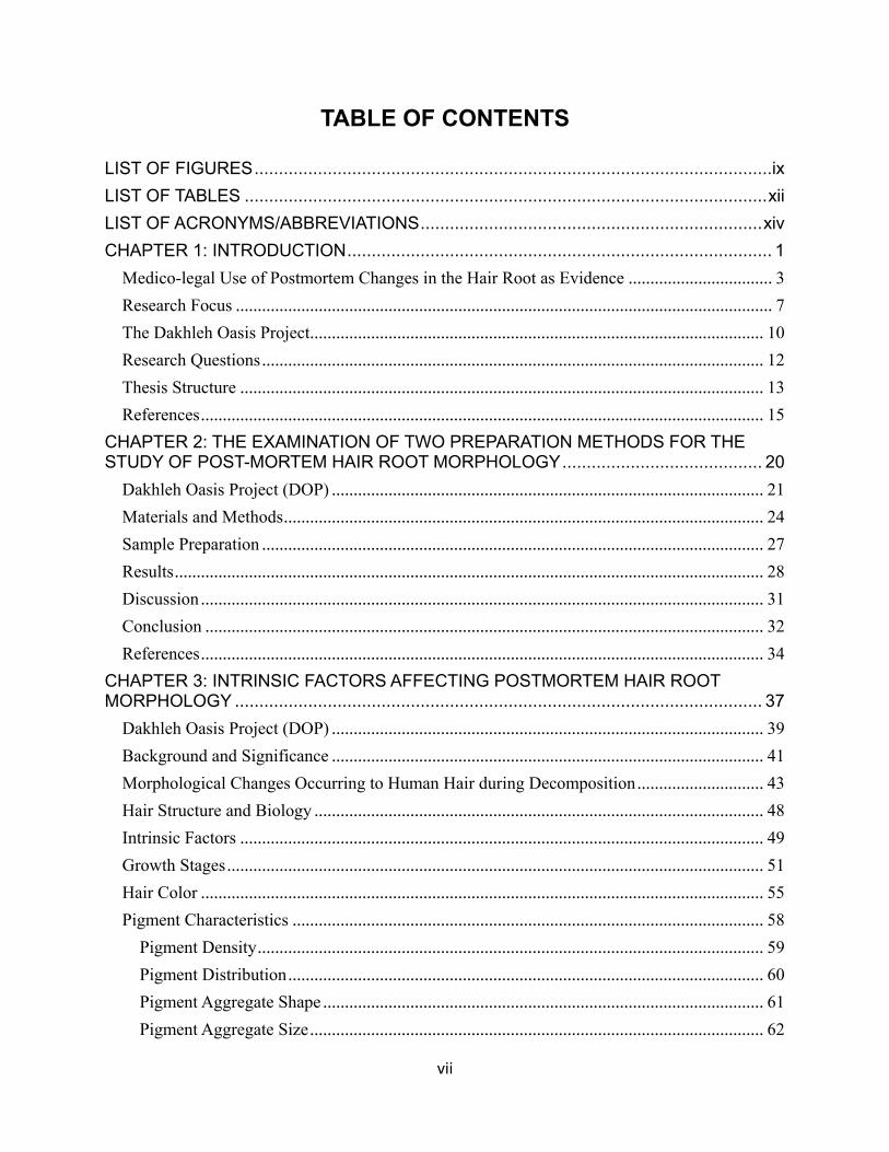
Figure 3.7: Hair comparison archetype reference chart for Kellis 2 sample population. 57
Figure 3.8: Pigment Density archetype reference chart for Kellis 2 sample population. 59
Figure 3.9: Pigment distribution archetype reference chart for Kellis 2 sample
population. ..................................................................................................................... 60
Figure 3.10: Pigment aggregate shape archetype reference chart for Kellis 2 sample
population. ..................................................................................................................... 61
Figure 3.11: Pigment aggregate size archetype reference chart for Kellis 2 sample
population. ..................................................................................................................... 62
Figure 3.12: Medulla continuity archetype reference chart for Kellis 2 sample population.
...................................................................................................................................... 65
Figure 3.13: Medulla opacity archetype reference chart for Kellis 2 sample population.66
Figure 3.14: Cuticle thickness archetype reference chart for Kellis 2 sample population.
...................................................................................................................................... 68
Figure 3.15: Inner cuticle margin archetype reference chart for Kellis 2 sample
population. ..................................................................................................................... 69
Figure 3.16: Cuticle scale profile archetype reference chart for Kellis 2 sample
population. ..................................................................................................................... 70
Figure 3.17: Ovoid bodies archetype reference chart for Kellis 2 sample population. ... 71
Figure 3.18: Bar graph showing the proportion of postmortem changes seen within the
sample population and how they are correlated with growth stages. ............................ 75

xi
Figure 3.19: Bar graph showing the proportion of postmortem changes within the
sample population and how it correlates with age. ........................................................ 77
Figure 3.20: A colorless hair from K2 B293 demonstrating an unknown cuticle thickness.
...................................................................................................................................... 79
Figure 3.21: Bar graph showing the proportion of postmortem changes documented
within the sample population and how they are correlated with cuticle thickness. ........ 80
Figure 3.22: Bar graph showing the proportion of postmortem changes documented
within the sample population and how they are correlated with pigment density. .......... 82
Figure 3.23: Bar graph showing the proportion of postmortem changes documented
within the sample population and how they are correlated with cuticle scale profile. .... 84

xii
LIST OF TABLES
Table 2.1: Sample IDs, age, biological sex, and number of hairs for each individual
included in the study population. ................................................................................... 24
Table 2.2: Age categories and distribution for individuals included in the study population
...................................................................................................................................... 25
Table 2.3: Distribution of biological sex for the study population ................................... 25
Table 2.4: Cross-tabulation analysis of presence and absence of roots using two dry
and wet preparation methods. ....................................................................................... 29
Table 2.5: Incidence of each growth stage observed using each preparation method .. 30
Table 2.6: Chi-Square analysis for preparation method and growth stage .................... 30
Table 2.7: Frequencies of postmortem changes in conjunction with preparation method
...................................................................................................................................... 31
Table 3.1: Hair Colors and Frequencies ........................................................................ 56
Table 3.2: Pearson Chi-Square results for intrinsic characteristics not demonstrating a
significant correlation with changes in postmortem hair root morphology. ..................... 73
Table 3.3: Pearson Chi-Square results for correlation between growth stages and root
decomposition. .............................................................................................................. 74
Table 3.4: Pearson Chi-Square results for correlation between age and root
decomposition. .............................................................................................................. 76
Table 3.5: Pearson Chi-Square results for correlation between cuticle thickness and root
decomposition. .............................................................................................................. 79
Table 3.6: Pearson Chi-Square results for correlation between pigment density and root
decomposition. .............................................................................................................. 81

xiii
Table 3.7: Pearson Chi-Square results for correlation between cuticle scale profile and
root decomposition. ....................................................................................................... 83
Table 3.8: Pearson Chi-Square results for correlation between hair color and root
decomposition. .............................................................................................................. 86
Table 3.9: Pearson Chi-Square results for correlation between hair colors represented
by 50+ hair samples and root decomposition. ............................................................... 86

xiv
LIST OF ACRONYMS/ABBREVIATIONS
AP- Arrector Pili Muscle
BLE- Brush-Like Ends
CSP- Cuticle Scale Profile
DMSO- Dimethyl Sulfoxide
DOP- Dakhleh Oasis Project
DP- Dermal Papilla
HKP- Hard Keratin Point
IRS- Inner Root Sheath
ORS- Outer Root Sheath
PHRM- Postmortem Hair Root Morphology
PMRB- Postmortem Root Banding

1
CHAPTER 1: INTRODUCTION
Hair analysis techniques were first entered into forensic evidence in the 1800’s,
and since then numerous case studies have examined the usefulness of hair trace
evidence in medico-legal cases (e.g., Gaudette et al. 1985; Petraco et al. 1988; Linch and
Prahlow 2001; Collier 2005; Koch et al. 2013). Detailed studies have been conducted to
determine the evidential significance of hair in DNA analysis, drug and toxicological
analysis, and elemental analysis (e.g., Gordus 1973; Ishiyama et al. 1982 Wilson et al.
1995; Uhl 1997; Tagliaro et al. 1998; Budowle et al. 2003; Kintz 2004; Goullé et al. 2004;
Pragst and Balikova 2006; Pillay and Kuis 2007). Additionally, comparative analyses have
been utilized to determine the possible sources of questioned hairs in cases where the
origin is unknown (e.g., Miller 1987; Smith and Goodman 1995-1996; Deedrick and Koch
2004; Bisbing 2007). In situations such as these, hairs and fibers are collected from
known sources and compared to the questioned hair with the hopes that a match can be
made and the source of the questioned hair fiber discovered. Despite the strides made
by previous researchers and investigators regarding forensic hair analysis, minimal
research has been conducted examining the morphological changes that occur to hair,
specifically to the proximal root-end of human hair, during the process of decomposition,
regardless of its potential use as evidence in forensic investigations (Linch and Prahlow
2001:15).
Currently, these hair fiber root-end changes are classified using the terms
postmortem root banding (PMRB), hard keratin points (HKP), and brush-like ends (BLE).
PMRB refers to “an opaque ellipsoidal band which appears to be composed of a collection
of parallel elongated air spaces [that] is approximately 0.5 mm above the root bulb and

2
about 2 mm below the skin surface” usually assumed to occur in the stages of early
decomposition (Petraco et al. 1988:73). BLE and HKP, on the other hand, are usually
thought to be associated with late term decomposition. In these cases, “the hair will break
off at the area of the dark band often leaving the proximal end discolored and with a
pointed or brush like appearance” (Koch et al. 2013: S52).
When these changes were first discovered by Japanese scientists Hajime Sato
and S. Seta in 1984, they referred to these phenomena collectively as 'putrid roots', a
term which is still in limited use today (Ort 2005; Kadane 2015). Following their discovery,
the Committee on Forensic Hair Comparison (1985), “in an effort to advance forensic hair
comparison as a science,” called for a standardization of the terminology to be used by
hair analysts, and the term “putrid root” was added to the list (Gaudette et al. 1985:1, 8).
These phenomena were not studied in depth until 1988 when Petraco et al. (1988) wrote
“The Morphology and Evidential Significance of Human Hair Roots”, the first in a very
short list of research conducted in an attempt to understand the causal factors associated
with the morphological changes observed on human hair roots after death. Unfortunately,
this study only discussed two types of putrid roots in depth, PHRB and BLE. The third
type, HKP, was only briefly mentioned. This is likely due to the probability that PMRB,
BLE, and HKP are related to different types and stages of decomposition, and the
research conducted by Petraco et al. (1988) was limited in both scope and scale. A study
examining all of the postmortem changes to human hair would have required a large and
diverse sample set, which was not available at the time. According to Koch et al. (2013)
the need for a large study sample might explain the paucity of research which has been
conducted on PHRM to date – as samples from cadavers are not easily acquired (S52).

3
It is without question that researchers face many difficulties pertaining to studies
involving deceased individuals, but it is imperative that these phenomena undergo
rigorous research. Since many investigations of PHRM (e.g., Petraco et al. 1988; Linch
and Prahlow 2001; Collier 2005; Koch et al. 2013) have found a link between changes of
the human hair root and decomposition of the body itself, its detection and association
with an active crime scene can potentially indicate that a deceased individual was present
even in cases where a body is missing. In short, hair trace evidence holds great potential
for medico-legal investigations, especially where postmortem changes in the hair root
may be the only indication that a crime has been committed.
Medico-legal Use of Postmortem Changes in the Hair Root as Evidence
In 2005, People v. Kogut, 10 Misc. 3d 245, 247, 805, N.Y.S.2d 789, 791 (N.Y. Sup.
Ct. 2005) set the precedent for admission of PMRB evidence in the 2011 case of the State
of Florida v. Casey Marie Anthony case number: 48-2008-CF-015606-O (2011a:3-5).
People v. Kogut, 10 Misc. 3d 245, 247, 805, N.Y.S.2d 789, 791 (N.Y. Sup. Ct. 2005),
however, was a retrial of a murder case which took place in 1986 with Kogut and two
others convicted of the murder of Theresa Fusco. In the original trial, Nicholas Petraco,
who is a leading forensic microscopist and trace evidence analyst, compared two
questioned hairs found in a van belonging to one of the defendants to known hairs that
were collected from the victim. He stated that the questioned hairs were found to be
consistent with the known hairs, thereby suggesting they came from the same individual
(Ort 2005:1). Despite having observed PMRB on the roots in 1986, this trait was not
discussed until the 2005 retrial of People v. Kogut, 10 Misc. 3d 245, 247, 805, N.Y.S.2d
789, 791 (N.Y. Sup. Ct. 2005).

4
Petraco, who testified for the prosecution in the 1986 trial, was now testifying for
the defense along with Peter De Forest, a forensic expert who was also a witness for the
defense in 1986. Both De Forest and Petraco testified that PMRB “occurs only when the
hair begins to decompose inside the hair follicle, while still on the head of the deceased”
and that “the victim's body would not have been in the van long enough for postmortem
hair banding to occur” (Ort 2005:1). Since the hair from the vehicle was eerily similar to
the hairs taken from the body during autopsy, and since those hairs were stored in
unsealed enveloped, their testimony led to the belief that that the evidence had been
contaminated and commingling had occurred between hair samples (Innocence Project
2015). After 19 years of incarceration, Kogut was finally found innocent, and in 2012
litigation began on a wrongful conviction suit where, again, PMRB is likely going to play
an important role (Seybert 2012: 1-36).
Another case where postmortem root banding was introduced as evidence was the
high-profile trial of the State of Florida v. Casey Marie Anthony case number: 48-2008-
CF-015606-O. This case began in July 2008 when Caylee Anthony, Casey Anthony’s only
child, was reported missing by her grandmother, Cindy, who stated to a 911 dispatcher
that, “I have a possible missing child. I have a three-year-old that's been missing for a
month” (State of Florida v. Casey Marie Anthony, 48-2008-CF-015606-O 2011b; Montaldo
2015). Later in the day, during a subsequent call to 911, she told the dispatcher, “I found
my daughter's car today and it smells like there's been a dead body in the damn car”
(Battaglia 2011/2012:1587; State of Florida v. Casey Marie Anthony, 48-2008-CF-015606-
O 2011b; Montaldo 2015). As it turned out, Caylee had last been seen alive on June 16th
of that year, but her mother had never reported her missing. She told her mother and

5
police that she had been searching for Caylee on her own, and that her daughter had
been abducted by her nanny. Changes in her original story, and the accompanying
circumstances, resulted in her being named as the number one suspect in the
disappearance of her daughter; she was arrested on July 16th, 2008. Unfortunately,
despite many searches of the property and surrounding woods, no body was found.
Casey Anthony was later released after posting bond, but searches of the car, residence,
and computer search history led police to the conclusion that Caylee was likely deceased.
On October 14th, 2008 Casey was rearrested and charged with murder, manslaughter,
and lying to police, all of which took place without the presence of Caylee’s remains.
On December 11th, 2008, the skeletonized remains of a child were discovered in
the woods adjacent to the Anthony’s neighborhood. The remains were confirmed as those
of Caylee Anthony on December 19th, 2008. Evidence of possible homicide was
immediately apparent as duct tape was adhered to the skull where the mouth would have
been located, and also due to the method and placement of body during disposal. The
real test for investigators was linking Casey Anthony to the death of her child. Hair
collected from the trunk of the car became part of the State’s evidence that Caylee’s body
had been placed into the trunk, and that Casey had indeed killed her own daughter.
Steven Shaw, an FBI hair examiner, testified at trial that the hair acquired from the
left side of the trunk liner was consistent with hairs obtained from the trash bag containing
Caylee’s remains. However, he noted that, “hairs are not a means of positive
identification” stating that he could not say “a hair did originate from a person to the
exclusion of all others” (State of Florida v. Casey Marie Anthony, 48-2008-CF-015606-O
2011b). Additionally, mtDNA testing was conducted on exhibit Q12.1 (a human head hair)

6
and compared to DNA obtained from a buccal sample of Casey, but since mtDNA is
passed directly from mother to child, Dr. Catherine Theisen, who provided the analysis,
stated that “neither Casey Anthony nor Caylee Marie Anthony can be excluded as the
source of the Q12.1 hair” (Theisen 2008:2). Since prosecutors could not use DNA or hair
comparison to exclude Casey as the source of the Q12.1 hair, they relied on evidence of
postmortem root banding in order to show that the hair belonged to an individual who was
deceased, and therefore, could not have belonged to Casey or Cindy Anthony.
Karen Korsberg Lowe, a trace evidence analyst for the FBI laboratories, examined
the Q12.1 hair and noted possible evidence of decomposition in her report. She stated
that “a Caucasian hair found in specimen Q12 exhibits characteristics of apparent
decomposition at the proximal (root) end”, but no other hairs were found in the evidence
collected that showed signs of putrid root formation (Theisen 2008:2). When called to the
stand to testify, Lowe stated that based upon her experience working with trace evidence
and on the work conducted by previous researchers, postmortem root banding was found
to be consistent with decomposition. Further, she did not know of any studies where
PMRB, like that observed on Q12.1, had been replicated in hairs acquired from a living
person (State of Florida v. Casey Marie Anthony, 48-2008-CF-015606-O 2011a). Despite
her expert testimony and previous experience, she admitted that there were only a
handful of studies conducted on PMRB. While this evidence may not have decided the
case for Casey Anthony, it is possible that a greater amount of scientific knowledge could
have buttressed the forensic hair analysis leading to a far different conclusion. On July
5th, 2011, the jury found Casey Anthony not guilty for the murder of her daughter. On July
17th, 2011, three years after Caylee went missing, Casey was released from jail.

7
Another case involved the murder of two adult males, a mother, and her son. In
this case, hair was analyzed from the crime scene and compared to known samples
acquired from the remains of each individual (Tafaro 2000). The hairs collected from the
two adult male victims were microscopically differentiated from the mother and her son,
but the hairs from the mother and her son could not be excluded from one another. When
microscopically examined it was found that the juvenile’s hair contained the only evidence
of PMRB, while his mother’s hair showed only BLE. In this way, investigators were able
to establish which hairs belonged to the mother and which belonged to her son. These
observations further helped to corroborate the suspect’s confession that he had returned
several days after the murder to slit the throats of all the victims. This also helped to
establish how hairs showing decomposition changes were found on knives discovered at
the crime scene (Tafaro 2000:497-498).
Research Focus
This thesis includes an examination of postmortem hair root morphology (PHRM)
utilizing hair samples obtained from the scalps of individuals buried in the Kellis 2
cemetery in the Dakhleh Oasis, Egypt. It has been suggested by previous researchers
that the presence of postmortem changes in the proximal root-end of the human hair, with
an emphasis being placed specifically on PHRB, could be a tell-tale sign that a deceased
individual was present at a location even if a body cannot be found. Therefore, the impact
this type of evidence can have on a medico-legal investigation is tremendous, and yet our
understanding of PHRM is suffering from a serious lack of scholarly research.
Fortunately, there are a few studies which discuss extrinsic factors that affect
PHRM, but literature discussing intrinsic factors- traits specific to the hair itself and the

8
individual- is nonexistent. Therefore, in an effort to fully understand the many factors
which may have an effect on PHRM, it is necessary to study the intrinsic variables as well.
This is mainly because individuals tend to die under various sets of circumstances and
experience different taphonomic environments. Given that no two hairs are exactly the
same, it is crucial to determine why postmortem changes occur in some hair roots, while
not occurring in others even within a single individual’s remains.
While most researchers suggest that PMRB, HKP, and BLE occur only after death,
the scarcity of studies related to these phenomena and the circumstances surrounding
their appearance leads to questions and complications when hair samples demonstrating
these traits are admitted into evidence. Therefore, at the focus of this thesis is the hair
root morphology of samples procured from the naturally mummified remains of individuals
interred at the Kellis 2 cemetery, Dakhleh Oasis, Egypt, in order to determine which
variables affect the appearance of postmortem changes in human hair roots. These
samples were archived by the Dakhleh Oasis Project (DOP) and housed at the University
of Central Florida Laboratory for Bioarchaeological Research. The hairs included in this
study were collected from individuals interred within the burials highlighted in red in Figure
1.1. Additionally, utilizing hair samples provided by the DOP will ensure a study sample
of known provenience and relative environmental stability. It is hoped that this research
will add to the scientific and forensic knowledge surrounding PHRM, aid and promote
future research within the field, and thereby provide a solid foundation for the use of
PHRM as evidence in future legal proceedings.

9
Figure 1.1: Map of the Kellis 2 cemetery. Burials of individuals included in this study are
highlighted in red. (Adapted from Williams 2008)

10
The Dakhleh Oasis Project
The Dakhleh Oasis is located approximately 660Km SSW of Cairo in Egypt's
Western Desert (Dupras 1999). It is one of five major depressions and is known for its
hyper-arid environment. Temperatures vary from -4°C to 25°C in winter and from 19°C to
50°C during the summer months (Blume et al. 1984, Giddy 1987, Dupras 1999, Wheeler
2010). Rainfall is also rare, with 0.3 mm/year being the average (Blume et al. 1984). High
atmospheric pressure, violent winds (Khamasins), and seasonal sandstorms further
contribute to the aridity in the oasis (Dupras 1999). It is believed that the climate has
remained relatively stable, with very little change, from the time that ancient Kellis was
inhabited, and the cemeteries were in use, until now. Because of this, the level of
preservation for many of the remains is remarkable. Additionally, since the remains at
Kellis 2 were naturally, rather than artificially, mummified, tissue samples, such as skin,
internal organs, muscle tissue, hair, and nails, were suitable for this study (Williams 2008).
Figure 1.2 shows the approximate extent of the Dakhleh Oasis and its geographical
location in relationship to Cairo, Egypt.
The excavations that have taken place at the Kellis 2 cemetery constitute a
significant portion of the research focus of the Dakhleh Oasis Project. This project
involves the excavation and subsequent analyses of archaeological sites within the
Dakhleh Oasis, Egypt (Bagnall 1997; Cook 1994). The Dakhleh Oasis Project (DOP)
began in 1978, under the direction of Anthony Mills, and has continued into the present
with the support of various universities and organizations; among them The Royal Ontario
Museum and the Society for the Study of Egyptian Antiquities (Dupras 1999). The
bioarchaeologists involved in the Dakhleh Oasis Project have excavated numerous

11
inhumations at the west and east cemeteries (Kellis 1 [K1] and Kellis 2 [K2] respectively)
associated with the ancient town of Kellis (Birrell 1999). Of importance for purposes of
this thesis, are the excavations at Kellis 2 which began in 1992 (Birrell 1999) and have
continued to the present day.
Figure 1.2: Map showing the approximate extent of the Dakhleh Oasis and its
geographical relationship in relationship to Cairo, Egypt. (Google Earth)

12
As of 2012, a total of 771 burials have been excavated from this site (701 have been
analyzed), but it is estimated that the Kellis 2 cemetery contains between 3,000-4,000
graves in total (Molto 2002; Wheeler 2010).
Kellis 2 dates from around 50 AD to about 450 AD, the Roman-Byzantine period in
Egypt, and is assumed to be an early Christian cemetery based on grave and body
orientation, and the mortuary practices observed at the site (Stewart et al. 2003; Molto
2002). The burials were generally single interments, where the body was wrapped in a
linen shroud, placed in an extended, supine position within a mud brick tomb, and situated
with the head to the west and the feet to the east; very few graves included a coffin of any
kind and many were lacking grave goods (Birrell 1999; Williams 2008; Wheeler 2009).
Additionally, the burials of infants and children were situated alongside those of adults,
demonstrating that this population believed in the humanness of all individuals within their
community. This is an idea that was lacking in Egypt during its earlier periods, but which
is consistent with later Christian ideals (Bowen 2003; Bowen et al. 2005). The period
during which this cemetery was in use also coincides with the appearance of the first
Christians in Egypt during the first two centuries AD (Bowen 2003; Wheeler 2010).
Additionally, the remains have been subjected to radiocarbon dating that has tentatively
dated Kellis 2 to the late third and early fourth centuries AD, suggesting that the cemetery
may have been in use for longer than was originally assumed (Birrell 1999; Molto 2001;
Stewart et al. 2003).
Research Questions
Three research questions will be addressed in this thesis; 1) What is the best
method of sample preparation for the observation of PHRM in a mummified sample?; 2)

13
Which intrinsic factors affect whether decomposition changes affect the hair root?; and,
3) Is postmortem root banding indicative of early stage decomposition? It is necessary to
understand which factors are related to the absence or presence of putrid roots. As such,
each hair in this study will be evaluated for extrinsic factors such as environmental
degradation, microbial action, and insect activity, as well as intrinsic attributes including:
growth stage, medulla characteristics, pigment characteristics, color, presence or
absence of ovoid bodies, and cuticle characteristics in order to analyze the factors related
to the appearance of PHRB, HKP, and BLE. Additionally, biological sex, age, and health
status (where available) of the individuals will also be noted to provide a more
comprehensive overview of the study sample. The overarching goal of this study is to
establish a standard methodology for other researchers wishing to study PHRM.
Thesis Structure
This thesis is structured in an integrated article format. Every chapter is set
up in such a way as to facilitate dissemination in peer-reviewed publications.
Chapter 2: The Examination of Two Preparation Methods for the Study of Post-
mortem Hair Root Morphology, introduces two methodologies employed in this study.
These two methods are utilized to determine which method is most beneficial when
working with dry decomposition. An overview of the processes for preparing wet and dry
samples, for mounting of the samples on to slides for observation, and an analysis of the
efficacy of each method in relation to the study sample are presented.
Chapter 3: Intrinsic Factors Affecting Postmortem Hair Root Morphology, focuses
on intrinsic factors and how they affect the occurrence of PHRM. The variables included
are age, biological sex, growth stage, hair color, pigment density, pigment distribution,

14
pigment aggregate size, pigment aggregate shape, medulla continuity, medulla opacity,
cuticle thickness, inner cuticle margin, cuticle scale profile, and ovoid bodies. This chapter
includes archetypes for each of these variables, as well as a discussion of the statistical
analyses performed and the variables’ relationships to PHRM.
Finally, Chapter 4 concludes this thesis with an overview of the results and offers
suggestions for applications and further research regarding PHRM.

15
References
Bagnall, Roger S. (editor) 1997. The Kellis Agricultural Account Book (P. Kell.IV Gr.96). Oxbow Books, Oxford and Oakville, Ct.
Battaglia, Nicholas A. 2011/2012. The Casey Anthony Trial and Wrongful Exonerations: How “Trial by Media” Cases Diminish Public Confidence in the Criminal Justice System. Albany Law Review 75 (3):1579-1611.
Birrell, Michael. 1999. Excavations in the Cemeteries of Ismant el-Kharab. In Dakhleh Oasis Project: Preliminary Reports on the 1992-1993 and 1993-1994 Field Seasons. Edited by C.A. Hope and A.J. Mills, pp. 29-41. Oxbow Books, Oxford [England]; Oakville, CT, USA.
Bisbing, Richard E. 2007. Forensic Hair Comparisons: Guidelines, Standards, Protocols, Quality Assurance and Enforcement. Presentation to The National Academies Committee on Identifying the Needs of the Forensic Sciences Community.
Blume, H.P, F. Alaily, U. Smettan, and J. Zielinski. 1984. Soil Types and Associations of Southwest Egypt. In Research in Egypt and Sudan. Berliner Geowissenschaftliche Abhandlungen. Edited by E. Klitzsch, R. Said and E. Schrank, pp. A50:293-302.
Bowen, Gillian E. 2003. Some observations on Christian burial practices at Kellis. In The Oasis Papers 3: Proceedings of the Third International Conference of the Dakhleh Oasis Project. Edited by Gillian E Bowen and Collin A. Hope, pp. 167-182. Oxbow Books, Oxford.
Bowen, Gillian E, Thomas Chandler, and Derrick Martin. 2005. Reconstructing Ancient Kellis. Buried History 41:51-64.
Budowle, Bruce, Marc W. Allard, Mark R. Wilson, Ranajit Chakraborty. 2003. Forensics and Mitochondrial DNA: Applications, Debates, and Foundations. Annual Review of Genomics and Human Genetics 4:119-141.
Collier, Jamie H. 2005. Estimating the Postmortem Interval in Forensic Cases through the Analysis of Postmortem Deterioration of Human Head Hair. M.A. Thesis, department of Geography and Anthropology, Louisiana State University.

16
Cook, M. 1994. The Mummies of Dakhleh. In Strength in Diversity: A Reader in Physical Anthropology. Edited by A Herring and L Chan, pp. 259-277. Canadian Scholars Press, Toronto.
Deedrick, Douglas W., Sandra L. Koch. 2004. Microscopy of hair part 1: a practical guide and manual for human hairs. Forensic Science Communications Electronic Document, https://archive.is/nnW3t, accessed April 10, 2017.
Dupras, Tosha L. 1999. Dining in the Dakhleh Oasis, Egypt: Determination of Diet Using Documents and Stable Isotope Analysis. Ph.D. dissertation, Department of Anthropology, McMaster University, Ontario.
Gaudette, B.D., J. Bailey, Richard Bisbing, Edward Burwitz, J. Cadman, C. Cwiklik, Harold Deadman, Peter DeForest, N. Erickson, John Kilbourn, Paul Krupenie, Thomas Kubic, D. Metger, T. Mozer, S. Palenik, L. Peterson, Nicholas Petraco, J. Robertson, S. Shaffer, R. Stockdale, M.A. Straus.
1985. Preliminary Report—Committee on Forensic Hair Comparison. Criminal Laboratory Digest 12:50-59.
Giddy, Lisa L. 1987. Egyptian oases. Bahariya, Dakhla, Farafra and Kharga during Pharaonic times. Aris & Phillips.
Gordus, A. J. 1973. Factors Affecting the Trace-Metal Content of Human Hair. Journal of Radioanalytical and Nuclear Chemistry 15(1) DOI:10.1007/BF02516574.
Goullé, Jean-Pierre, Loïc Mahieu, Julien Castermant, Nicolas Neveu, Laurent Bonneau, Gilbert Lainé, Daniel Bouige, Christian Lacroix.
2004. Metal and Metalloid Multi-Elementary ICP-MS Validation in Whole Blood, Plasma, Urine and Hair: Reference Values. Forensic Science International 153(1): 39-44.
Innocence Project. 2015. John Kogut. Electronic Document, http://www.innocenceproject.org/cases-falseimprisonment/john-kogut, accessed April 14, 2017.
Ishiyama, Ishu, T. Nagai, S. Toshida. 1982. Detection of Basic Drugs (Methamphetamine, Antidepressants, Nicotine) from Human Hair. Journal of Forensic Sciences 28(2):380-385.
Kadane, Joseph B. 2015. Post-Mortem Root Banding of Hairs: A Skeptical Review. Law Probability and Risk 14(3): 213-228.

17
Kintz, Pascal. 2004. Value of Hair Analysis in Postmortem Toxicology. Forensic Science International 142(2-3):137-134.
Koch, Sandra L., Amy L. Michaud, C.E. Mikell. 2013. Taphonomy of Hair-- A Study of Postmortem Root Banding. Journal of Forensic Sciences 58: S52-S59.
Linch, Charles A., Joseph A. Prahlow. 2001. Postmortem Microscopic Changes Observed at the Human Head Hair Proximal End. Journal of Forensic Sciences 46(1): 15-20.
Miller, Larry S. 1987. Procedural bias in forensic science examinations of human hair. Law and Human Behavior 11(2):157-163.
Molto, J.E. 2001. The Comparative Skeletal Biology and Paleoepidemiology of the People from Ein Tirghi and Kellis, Dakhleh Oasis, Egypt. In The Oasis Papers 1: The Proceedings of the First International Symposium of the Dakhleh Oasis Project. Edited by M. Marlow and A.J. Mills, pp. 81-100, Oxbow Books, Oxford.
Molto, J.E. 2002. Bio-Archaeological Research of Kellis 2: An Overview. In Dakhleh Oasis Project: Preliminary Reports on the 1994-1995 to 1998-1999 Field Seasons. Edited by Collin A. Hope, Gillian E. Bowen, and Richard S. Bagnall, pp. 239-255, Oxbow Books, Oxford [England], Oakville, CT, USA.
Montaldo, Charles. 2015. Missing Caylee Anthony: Transcripts of 9-1-1 calls from Cindy Anthony. Electronic Document http://crime.about.com/od/current/a/caylee_911calls.htm, accessed April 14, 2017.
Ort, Victor M. 2005. People v Kogut. http://law.justia.com/cases/new-york/other-courts/2005/
2005-25410.html. Petraco, Nicholas, C. Fraas, F.X. Callery, Peter De Forest.
1988. The Morphology and Evidential Significance of Human Hair Roots. Journal of Forensic Sciences 33(1): 68-75.
Pillay, K., R. Kuis. 2007. The Potentials and Limitations of Using Neutron Activation Analysis Data on Human Hair as Forensic Evidence. Journal of Radioanalytical and Nuclear Chemistry 43(2) DOI:10.1007/BF02519507.

18
Pragst, Fritz, Marie A. Balikova. 2006. State of the Art in Hair Analysis for Detection of Drug and Alcohol Abuse. Clinica Chimica Acta 70(1 and 2):17-49.
Seybert, Joanna.
2012. People v Kogut. Electronic Document http://docs.justia.com/cases/federal/districtcourts/newyork/nyedce/2:2006cv06695/264382/350/0.pdf?1345171557, accessed April 14, 2017.
Smith, Clive A., Patrick D. Goodman. 1995-1996. Forensic Hair Comparison Analysis: Nineteenth Century Science or Twentieth Century Snake Oil? Columbia Human Rights Law Review 27:227-291.
State of Florida v. Casey Marie Anthony.
2011a. Order Denying Motion to Exclude Unreliable Evidence (Post Mortem Banding) and Amended Motion in Limine for Hearing on the Unreliability of Scientific Testimony by Karen Lowe on Post-Mortem Hair Banding. In the Circuit Court of the Ninth Judicial Circuit in and for Orange County Florida. pp. 1-6.
State of Florida v. Casey Marie Anthony.
2011b. Casey Anthony trial. https://www.youtube.com/user/CrimeTimeVids, accessed April 14, 2017.
Stewart, J.D., J.E. Molto, P.J. Reimer. 2003. The Chronology of Kellis 2: The Interpretative Significance of Radiocarbon Dating of Human Remains. In Dakhleh Oasis Project Monograph. Edited by Gillian E. Bowen and C.A. Hope, pp 373-378, Oxbow Books, Oxford.
Tafaro, Joseph T. 2000. The Use of Microscopic Postmortem Changes in Anagen Hair Roots to
Associate Questioned Hairs with Known Hairs and Reconstruct Events in Two Murder Cases. Journal of Forensic Sciences 45(2): 495-499.
Tagliaro, Franco, Zeno De Battisti, Fredrick P. Smith, Mario Marigo. 1998. Death from Heroin Overdose: Findings from Hair Analysis. The Lancet 351(9120):1923-1925.
Theisen, Catherine E.
2008. Report of Examination: Caylee Marie Anthony- Victim Missing/ Abducted Minor. Orange County Florida. Electronic Document http://blogs.discovery.com/files/18530294.pdf, accessed April 14, 2017.
Uhl, M. 1997. Determination of Drugs in Hair Using GC/MS/MS. Forensic Science International 84(1-3):281-294.

19
Wheeler, Sandra M. 2009. Bioarchaeology of Infancy and Childhood at the Kellis 2 Cemetery, Dakhleh Oasis, Egypt. Ph.D. dissertation, Department of Anthropology, University of Western Ontario, Ontario.
Wheeler, Sandra M. 2010. Nutritional and Disease Stress of Juveniles from the Dakhleh Oasis, Egypt. International Journal of Osteoarchaeology 22(2):219-234.
Williams, Lana J. 2008. Investigating Seasonality of Death at Kellis 2 Cemetery Using Solar Alignment and Isotopic Analysis of Mummified Tissues. Ph.D. dissertation, Department of Anthropology, University of Western Ontario, Ontario.
Wilson, Mark R., Joseph A. DiZinno, Deborah Polanskey, Jeri Replogle, Bruce Budowle.
1995. Validation of Mitochondrial DNA Sequencing for Forensic Casework analysis. International Journal of Legal Medicine 108(2):68-74.

20
CHAPTER 2: THE EXAMINATION OF TWO PREPARATION METHODS FOR THE STUDY OF POST-MORTEM HAIR ROOT
MORPHOLOGY
Forensic hair examination is not new to the field of forensic science. An 1857 study
by John Glaster, Hairs of Mammalia from the Medico-Legal Aspect (1931; cf. Evans
2004:101-102), discusses the significance of hair from a medico-legal standpoint and is
still regarded as a reliable reference on the subject. Additionally, the first evidential use
of human hair in an American legal case occurred during June of 1882 in the state of
Wisconsin (Ellis 1882). Knoll v. State was a landmark murder case in that it was the first
to include the examination of human hair in witness testimony. This being a new field of
forensic science, however, the examination consisted of a single hair comparison
between the crime scene and victim. In addition to the minute amount of evidence, the
examination of the hair itself was conducted with the naked eye rather than
microscopically; a fact that had the judge questioning its validity. In truth, the most the
‘expert witness’, Dr. Piper, could offer was that “the hair was precisely the same in every
respect, in length, magnitude, color, and in every other respect” (Ellis 1882:381). Even
with this paltry evidence, he concluded, “as a result of that comparison, I can say that it
was from the head of the same person” (Ellis 1882:381).
Forensic hair analysis has come quite a long way from this starting point to include
toxicology, DNA, microscopic hair and fiber comparisons, and postmortem hair root
morphology (PHRM). Unlike many forensic hair applications, which have established
specific methodologies for analyses, PHRM is a relatively new area, and as such,
standards have yet to be established. Therefore, the purpose of this research is to

21
determine which preparation methodology will provide the most consistent and usable
results when examining PHRM for use as evidence in medico-legal cases. This study
specifically looks at whether preparation method affects the retention of a usable sample,
the decomposition changes observed, and the growth stages obtained. Growth stages
include anagen or active growth stage, catagen or transitional growth stage, and telogen
or terminal growth stage (Ogle and Fox 1999). In this case, growth stages are of particular
importance since it has already been established through previous research that growth
stages have an effect on whether decomposition changes are observed or not. A greater
proportion of anagen and catagen hairs within the sample, hairs that have been shown to
demonstrate postmortem changes in the hair root, should yield substantially different
results than a sample consisting mainly of telogen or terminal growth stage hairs. Since
telogen hairs have ceased growth and the root has become fully keratinized, postmortem
changes are no longer observed (Petraco et al. 1988; Linch and Prahlow 2001; Koch et
al. 2013). Therefore, a method that provides a greater proportion of anagen and catagen
hairs would be significantly more effective.
It is hoped this research will contribute to the establishment of methodological
protocols for future forensic analysts. The hair used in this particular study originated from
the remains of individuals associated with the Kellis 2 cemetery (~50AD- ~450AD) in the
Dakhleh Oasis, Egypt, and was acquired by bioarchaeologists involved in the Dakhleh
Oasis Project (DOP).
Dakhleh Oasis Project (DOP)
The Dakhleh Oasis is one of the seven main Oases of Egypt (Figure 2.1). It is
located approximately 660Km SSW of Cairo and 300Km west of Luxor in Egypt’s Western

22
Desert (Dupras 1999). Dakhleh Oasis boasts a hyper-arid environment, where
temperatures can range anywhere from -4°C in the winter months to 50°C during the
summer (Blume et al. 1984; Giddy 1987; Dupras 1999; Wheeler 2010). Annually, only
0.3mm of rainfall is accumulated (Blume et al. 1984). According to Dupras and Schwarcz
(2001), “humidity can range from 23% to 30% from March to September, rising to 33% to
50% from October to February” (1200). Khamasins, which are violent winds that ravage
this area, sandstorms, and high atmospheric pressure, serve to increase aridity in this
already arid environment (Dupras 1999).
Archaeological excavations of the Kellis 2 (K2) cemetery in the Dakhleh Oasis
have been ongoing since 1991 (Cook 1994:260). The K2 cemetery dates to circa AD 50
to about AD 450, which coincides with the Roman-Byzantine period and the appearance
of the first Christians in Egypt. This is also consistent with radiocarbon dating conducted
on the remains, which tentatively dates the cemetery to the late third to early fourth
centuries AD and suggests that the cemetery may have been in use for longer than
previously assumed (Birrell 1999; Molto 2001; Stewart et al. 2003). Additionally, the
mortuary practices observed at Kellis 2 are consistent with Christian mortuary practices
from the same era (Stewart et al. 2003; Molto 2002). These burials consisted mostly of
single interments with the deceased buried with their head facing towards the west and
their feet towards the east; the graves themselves contained little to no grave goods
(Birrell 1999; Williams 2008; Wheeler 2009). The inclusion of children and infants into the
cemetery populations lends further credence to the assumption that this is an early
Christian cemetery since children were not included in previous cemetery populations
(Bowen 2003; Bowen et al. 2005).

23
To date, 701 of the 770 individuals excavated have been examined and analyzed
by DOP bioarchaeology team members. Because the environment in Dakhleh Oasis has
remained consistent over the past 2000 years, the human remains from the Kellis 2
cemetery are very well preserved. Many of the individuals buried in the Kellis 2 cemetery
underwent natural, or spontaneous, mummification, allowing for the preservation of
tissues such as hair.
Figure 2.1: Map showing the approximate extent of the Dakhleh Oasis and its
geographical relationship in relationship to Cairo, Egypt. (Google Earth)

24
Materials and Methods
For this study, a total of 400 individual hairs from 10 individuals from the Kellis 2
cemetery were analyzed, and were used for the purpose of determining the best method
for sample preparation. The scalp samples are currently stored in the Laboratory for
Bioarchaeology Research at the University of Central Florida (UCF). Both males and
females are included in the study population and their ages range from between 16-21
up to 60+. Tables 2.1-2.3 provides a breakdown of the sample IDs included in the study
population, age categories for each individual, and the proportion of males to females.
Additionally, Figure 2.2 shows the extent of the excavations conducted at Kellis 2
cemetery as well as the distribution of those individuals that have been included in this
research.
Table 2.1: Sample IDs, age, biological sex, and number of hairs for each individual
included in the study population.
Age Biological Sex # of Individual
Hairs Acquired
Sample
ID
B261 60+ F 40
B265 36-50 M 40
B269 51-60 F 40
B279 22-35 F 40
B280 60+ F 40
B281 60+ M 40
B284 22-35 F 40
B291 36-50 F 40
B306 60+ F 40
B309 22-35 M 40
Total 400

25
Table 2.2: Age categories and distribution for individuals included in the study population
Table 2.3: Distribution of biological sex for the study population
Number % Total Sample
Sex F 280 70.0
M 120 30.0
Total 400 100.0
Age Range # of
Individuals in
Age Category
# of Hairs
Included in
Sample
Population
% of Sample
Age
Categories
22-35 3 120 30.0
36-50 2 80 20.0
51-60 1 40 10.0
60+ 4 160 40.0
Total 10 400 100.0

26
Figure 2.2: The Kellis 2 cemetery excavations conducted through 2007. Red graves are
those individuals whose remains were used in this study (Adapted from Williams 2008)

27
Sample Preparation
Forty hairs with roots attached were acquired from each individual using two
different methods. For the first preparation method, referred to as the dry method, ten
individuals were chosen based upon the significant amount of hair and scalp that had
been collected in the field. Age and sex data were provided by Dr. Lana Williams based
on previous research conducted by the DOP (Williams, 2008). Each hair was individually
extracted directly from the scalp, examined to confirm that the root was attached, and
placed in labeled bags. Without any further processing, the first 20 hairs were bagged
and labeled with the individual’s identification number.
The second preparation method, referred to as the wet method, utilized the same
ten individuals included in the first method to ensure that there was no prejudice in the
sample based on an individual’s characteristics or level of decomposition. A square
measuring 1cm x 1cm was cut from each scalp section and placed in a separate petri
dish where it was rehydrated using dimethyl sulfoxide (DMSO) for 24-48 hours following
the method outlined by Williams (2008). Twenty hairs were extracted from each of these
samples, allowed to dry overnight, and then placed in labeled bags. In this method, DMSO
was used to rehydrate the scalp to make it easier to pluck the individual hairs and lessen
any possible damage the root. This method was also used to determine whether the
preparation would affect the occurrence of decomposition changes in the hair root.
Five slides were prepared and labeled for each individual and the preparation
method was noted on each slide. Permount© was chosen as a mounting medium due to
its refractive index (RI) of 1.525 at 25°C being remarkably close to the RI of human hair
at ~1.55 (Linch and Prahlow 2001; Marschner et al. 2003; Committee on Identifying the

28
Needs of the Forensic Sciences Community 2009:156). Four hairs were mounted on each
slide using Permount©, creating five slides for the dry method and five slides for the wet
method for each individual. All slides were left to dry for 24-48 hours before being stored
to ensure that the mounting medium was fully set. The specimens were then examined
using an Amscope 40x-1500x Infinity Polarizing Microscope at 40x and 60x magnification.
Data was collected on the presence or absence of hair roots (i.e., were the roots lost after
they were bagged or during the sample preparation), growth stages (e.g., were more hairs
in an active growth stage present in one method versus the other), differences in the rate
of postmortem root banding (PMRB), brush-like ends (BLE), and hard keratin points
(HKP).
The data was input into SPSS® statistics software for statistical analysis to
determine which method provided the most definitive results. Chi-square analysis was
conducted to ascertain whether methodology affects the number of usable samples
obtained from the remains, the growth stages present within the study sample, the
incidence of postmortem changes seen, and the type of postmortem changes observed.
Results
The results show that in cases where the body and tissues succumb to dry
decomposition, it is beneficial to use the wet method for procurement of intact hair and
hair root samples. In fact, out of 200 hairs included in the dry method, 36% (72/200) of
the hair samples had no roots, whereas using the wet method only about 20% (39/200)
of the hairs sampled lost their roots during processing (Table 2.4).

29
Table 2.4: Cross-tabulation analysis of presence and absence of roots using two dry and
wet preparation methods.
Method No Roots Roots Total
Dry 72 128 200
Wet 39 161 200
Total 111 289 400
The growth stage for each sample was noted, except in cases where the root was
no longer present, to determine if the method of preparation had an effect on which growth
stages were observed in each sample. The results indicate that the preparation method
did affect the observed growth stages. In the wet method, more anagen and catagen roots
were present, while in the dry method the main roots that were intact were those in the
telogen growth stage (Table 2.5). Results from the Chi-square analysis indicate that the
correlation between the method of preparation and the growth stages observed is
significant at the p=0.01 level (Table 2.6). This is more than likely due to how each of the
hair samples were procured for each method. For example, anagen and catagen roots
were more easily obtained intact from the rehydrated scalp sample, while in the dry
samples the majority of intact roots were in the telogen growth stage. This is because
telogen roots tend to survive the process of pulling the hair out of the dry scalp better than
anagen and catagen roots, which are softer and more easily damaged during the
collection process and which retain anatomical structures affixing them to the dermal layer
of the scalp.

30
Table 2.5: Incidence of each growth stage observed using each preparation method
Method Total
Dry Wet
Growth Stage
Anagen 28 93 121
Catagen 7 4 11
Telogen 78 55 133
Total 113 152 265
Table 2.6: Chi-Square analysis for preparation method and growth stage
Value Asymp. Sig. (2-sided)
Pearson Chi-Square 51.024a .000
Likelihood Ratio 53.130 .000
N of Valid Cases 400
a. 0 cells (0.0%) have expected count less than 5. The minimum
expected count is 5.50.
Lastly, to determine which preparation method yielded the most definitive results,
the occurrence of PMRB, BLE, and HKP was documented. Since, as noted above, the
preparation method affected the growth stages procured, it is not surprising that the
decomposition changes observed also differed. The decomposition changes most
observed with the dry preparation method were both PMRB and BLE; together these
made up about 10% of the total sample (11/200 PMRB and 11/200 BLE). In the wet
samples, however, PMRB accounted for over 25% (53/200) of the total sample, and only
five BLEs were observed (Table 2.7).

31
Table 2.7: Frequencies of postmortem changes in conjunction with preparation method
Method Total Dry Wet
Decomposition Stage
PMRB 11 53 64
BLE 11 0 11
BLE & PMRB 0 5 5
HKP 3 0 3
N/A 103 103 206
Total 128 161 289
The analyses suggest that when dry decomposition is encountered, the most fitting
method to use, to acquire intact roots directly from the scalp of the deceased, is likely the
wet method. Not only did this method lessen the amount of damage observed on the
roots, but it can also be suggested that when BLE are encountered it could be the result
of hair that was forcibly removed from an already desiccated set of remains.
Discussion
The analyses suggest that when dry decomposition is encountered, the most fitting
method to use, to acquire intact roots directly from the scalp of the deceased, is likely the
wet method. Furthermore, it could be stated that the wet method would be beneficial
regardless of the state or type of decomposition observed in the remains. An individual’s
body tends to lose moisture as it decomposes making it more difficult to obtain intact and
usable hair samples if the scalp is not properly rehydrated. A drier scalp could lead to a
disproportionate number of hairs in the telogen growth stage or hairs showing brush-like
ends or hard keratin points.

32
Since telogen or terminal growth stage hairs are resistant to postmortem change,
acquiring a sample that includes a greater proportion of hairs in the anagen and catagen
growth stages would be preferable. Undamaged anagen and catagen roots are more
easily obtained from a rehydrated scalp, meaning the wet method is the method that
should be followed. Not only did the wet method provide a more usable sample for this
research (i.e. there was a greater proportion of anagen and catagen hairs in the wet
sample than were in the dry sample, 97:35 respectively), but it can also be suggested
that when BLE and HKP were encountered in the study sample it may be the result of the
hair being forcibly removed from an already desiccated set of remains. The fact that
PMRB was observed five times as much in the wet sample as the dry sample (53:11
respectively) and there were no BLE observed in the wet sample further supports this
assumption.
Conclusion
This study examined two sample preparation methods for use by forensic
investigators when obtaining intact hair root samples from mummified remains. The main
purpose of this research was to test two preparation methods to determine which is better
for preserving postmortem hair characteristics. The results of this research, examining
dry and wet methods, indicates that the wet method provided more hairs in the active
growth stage (anagen) and transitional growth stage (catagen). Additionally, the forceful
plucking of hair in the dry method caused the loss of many of the hair roots both during
the process of obtaining the hair and during the time when the hair was stored before
being mounted.

33
The majority of the hairs with lost roots were most likely in the anagen and catagen
growth stages since most of the roots that remained intact were in telogen stage, the final
hair stage where the hair has ceased growth. Since decomposition changes were not
observed in telogen roots, the dry method did not provide as much material as the wet
method in regards to studying postmortem human hair root morphological changes.
Additionally, since telogen hairs do not appear to show evidence of decomposition, it is
even more integral to a forensic investigation to obtain intact hairs and hair roots in the
anagen and catagen growth stages. As such, the wet method appears to be the most
useful and advantageous method of preparation for forensic investigators trying to obtain
hair samples from mummified remains.

34
References
Birrell, Michael. 1999. Excavations in the Cemeteries of Ismant el-Kharab. In Dakhleh Oasis Project: Preliminary Reports on the 1992-1993 and 1993-1994 Field Seasons. Edited by C.A. Hope and A.J. Mills, pp. 29-41. Oxbow Books, Oxford [England]; Oakville, CT, USA.
Blume, H.P, F. Alaily, U. Smettan, and J. Zielinski. 1984. Soil Types and Associations of Southwest Egypt. In Research in Egypt and Sudan. Berliner Geowissenschaftliche Abhandlungen. Edited by E. Klitzsch, R. Said and E. Schrank, pp. A50:293-302.
Bowen, Gillian E. 2003. Some observations on Christian burial practices at Kellis. In The Oasis Papers 3: Proceedings of the Third International Conference of the Dakhleh Oasis Project. Edited by Gillian E Bowen and C.A. Hope, pp. 167-182. Oxbow Books, Oxford.
Bowen, Gillian E, Thomas Chandler, and Derrick Martin. 2005. Reconstructing Ancient Kellis. Buried History 41:51-64.
Committee on identifying the needs of the forensic science community, National Research Council.
2009. Analysis of Hair Evidence. In Strengthening Forensic Science in the United States: A Path Forward, pp. 155-161, Washington, D.C: USA.
Cook, M. 1994. The Mummies of Dakhleh. In Strength in Diversity: A Reader in Physical Anthropology. Edited by A Herring and L Chan, pp. 259-277. Canadian Scholars Press, Toronto.
Dupras, Tosha L. 1999. Dining in the Dakhleh Oasis, Egypt: Determination of Diet using Documents and Stable Isotope Analysis. Ph.D. dissertation, Department of Anthropology, McMaster University, Ontario.
Dupras, Tosha L., Henry P. Schwarcz. 2001. Strangers in a strange land: Stable isotope evidence for human migration in the Dakhleh Oasis, Egypt. Journal of Archaeological Science 28(11):1199-1208.
Ellis, Howard. 1882. The Reporter: Containing Decisions of the Supreme and Circuit Courts of the United States, Courts of Last Resort in the Several States, and English and Irish Courts, pp 381-383. Houghton, Mifflin and Co: Boston, US.

35
Evans, Colin. 2004. Hair and Fibers. In Murder two: the second casebook of forensic detection, pp. 101-103 Hoboken, John Wiley & Sons, New Jersey.
Giddy, Lisa L. 1987. Egyptian oases. Bahariya, Dakhla, Farafra and Kharga during Pharaonic times. Aris & Phillips.
Koch, Sandra L., Amy L. Michaud, C.E. Mikell. 2013. Taphonomy of Hair-- A Study of Postmortem Root Banding. Journal of Forensic Sciences 58: S52-S59.
Linch, Charles A., Joseph A. Prahlow. 2001. Postmortem Microscopic Changes Observed at the Human Head Hair Proximal End. Journal of Forensic Sciences 46(1): 15-20.
Marschner, Steven R., Henrik W. Jensen, Mike Cammarano, Steve Worley, Pat Hanrahan. 2003. Light Scattering from Human Hair Fibers. ACM Transactions on Graphics 22(3):780-791.
Molto, J.E. 2001. The Comparative Skeletal Biology and Paleoepidemiology of the People from Ein Tirghi and Kellis, Dakhleh Oasis, Egypt. In The Oasis Papers 1: The Proceedings of the First International Symposium of the Dakhleh Oasis Project. Edited by M. Marlow and A.J. Mills, pp. 81-100, Oxbow Books, Oxford.
Molto, J.E. 2002. Bio-archaeological Research of Kellis 2: An Overview. In Dakhleh Oasis Project: Preliminary Reports on the 1994-1995 to 1998-1999 Field Seasons. Edited by C.A. Hope, G.E. Bowen, and R.S. Bagnall, pp. 239-255, Oxbow Books, Oxford [England], Oakville, CT, USA.
Ogle, Jr Robert R., Michelle J Fox. 1999. Atlas of Human Hair: Microscopic Characteristics. CRC Press LLC, Boca Raton, Florida.
Petraco, Nicholas, C. Fraas, F.X. Callery, Peter De Forest. 1988. The Morphology and Evidential Significance of Human Hair Roots. Journal of Forensic Sciences 33(1): 68-75.
Stewart, J.D., J.E. Molto, P.J. Reimer. 2003. The Chronology of Kellis 2: The Interpretative Significance of Radiocarbon Dating of Human Remains. In Dakhleh Oasis Project Monograph. Edited by Gillian E. Bowen and C.A. Hope, pp 373-378, Oxbow Books, Oxford.

36
Wheeler, Sandra M. 2009. Bioarchaeology of Infancy and Childhood at the Kellis 2 Cemetery, Dakhleh Oasis, Egypt. Ph.D. dissertation, Department of Anthropology, University of Western Ontario, Ontario.
Wheeler, Sandra M. 2010. Nutritional and Disease Stress of Juveniles from the Dakhleh Oasis, Egypt. International Journal of Osteoarchaeology 22(2):219-234.
Williams, Lana J. 2008. Investigating Seasonality of Death at Kellis 2 Cemetery Using Solar Alignment and Isotopic Analysis of Mummified Tissues. Ph.D. dissertation, Department of Anthropology, University of Western Ontario, Ontario.

37
CHAPTER 3: INTRINSIC FACTORS AFFECTING POSTMORTEM HAIR ROOT MORPHOLOGY
A number of studies have examined the effect that extrinsic factors have on the
appearance of postmortem hair root morphology (PHRM), but very few researchers have
sought to determine whether intrinsic factors affect these changes. Factors such as age,
biological sex, pigment, medulla, and cuticle characteristics can significantly affect the
type and/or extent of postmortem changes observed. Unfortunately, these have not been
analyzed by previous researchers. The only intrinsic growth factor that has been studied
in depth is hair growth stages.
Previous research (e.g. Petraco et al. 1988; Linch and Prahlow 2001; Koch et al.
2013) has shown that hairs in the anagen or active growth stage and those in the catagen
or transitional growth stage are the only hairs subject to postmortem hair root degradation.
Hairs in telogen or the terminal growth stage do not show decomposition changes. It is
believed that this is due to the full keratinization of the telogen hair root. Since anagen
and catagen stage hairs are not fully keratinized, they are more susceptible to
decomposition changes. Given that growth stages have been found to adversely affect
whether postmortem hair root degradation is observed, it is not a stretch to believe that
PHRM might be affected by other intrinsic factors such as those listed above. Therefore,
the purpose of this study is to examine the possible links between the occurrence of
postmortem root banding (PMRB), brush-like ends (BLE), and hard keratin points (HKP),
and the intrinsic characteristics that may affect PHRM. A total of 713 hair samples,
acquired from the scalp sections of 51 individuals interred at the Kellis 2 cemetery (Figure
3.1) in the Dakhleh Oasis, Egypt, composed the study sample.

38
Figure 3.1: Map of the Kellis 2 cemetery. Burials of individuals included in this study are
highlighted in red. (Adapted from Williams 2008)

39
Dakhleh Oasis Project (DOP)
The Dakhleh Oasis is located in Egypt's Western Desert and approximately 660Km
SSW of Cairo (Dupras 1999). The Oasis is known for its hyper-arid environment and
temperatures there can range from -4°C in the winter months to 50°C during the summer
(Blume et al. 1984, Giddy 1987, Dupras 1999, Wheeler 2010). Rainfall is rare, with an
average annual accumulation of only 0.3 mm (Blume et al. 1984). According to Dupras
and Schwarcz (2001), “humidity can range from 23% to 30% from March to September,
rising to 33% to 50% from October to February” (1200). Violent winds known as
Khamasins, sandstorms, and high atmospheric pressure also contribute to this hyper-arid
environment (Dupras 1999). Because the environment in Dakhleh Oasis has remained
relatively stable from the time Kellis was inhabited to the present day, the level of
preservation, despite the absence of artificial mummification procedures, is phenomenal.
Additionally, since the remains at Kellis 2 were naturally, rather than artificially,
mummified, samples from these individuals are more easily acquired (Williams 2008).
Figure 3.2 shows the approximate extent of the Dakhleh Oasis and its geographical
location in relationship to Cairo, Egypt.

40
Figure 3.2: Map showing the approximate extent of the Dakhleh Oasis and its
geographical relationship in relationship to Cairo, Egypt. (Google Earth)
Excavations have been ongoing at the Dakhleh Oasis since the project began in
1978, with those at Kellis 2 beginning in 1991 (Birrell 1999). The K2 cemetery itself dates
from around 50 AD to about 450 AD. This time frame is contemporaneous with the
Roman-Byzantine period and the appearance of the first Christians in Egypt. The Kellis 2
cemetery is an early Christian cemetery based on the mortuary practices observed at the

41
site as well as body positioning (Stewart et al. 2003; Molto 2002). The burials were
generally single interments, except in a few cases where two bodies (mother and child or
twins) were found buried together. Additionally, bodies were usually wrapped in linen
shrouds and placed in an extended, supine position. The body was situated with the head
facing to the west and the feet to the east; very few graves included any type of coffin or
grave goods (Birrell 1999; Williams 2008; Wheeler 2009). Additionally, the inclusion of
infants and children into the general cemetery population is strictly associated with
Christian ideology, which touts the humanness of all individuals (Bowen 2003; Bowen et
al. 2005). Some of these remains have also been radiocarbon dated, resulting in a
tentative date of the late third to early fourth centuries AD. This suggests that the cemetery
may have been in use for a longer period of time than was originally thought (Birrell 1999;
Molto 2001; Stewart et al. 2003).
Background and Significance
Very few studies have been conducted with the sole purpose of examining PHRM.
In fact, Linch and Prahlow (2001) state that “there are... no references for detailed
descriptions of hair root decomposition changes” (15). This, however, is not surprising.
Even though hair analysis has been part of the field of forensics for some time,
decomposition changes of the human hair root (also termed putrid roots) were not
discussed until 1984 by S. Seta and Hajime Sato (Kadane 2015). Later, Petraco et al.
(1988) added to the subject in the article Morphology and Evidential Significance of
Human Hair Roots. Theirs was the first known study conducted specifically with the
purpose of observing postmortem human hair root morphology and discussing what
factors affect its appearance.

42
Koch et al. (2013) suggest that the main reason for the dearth of research
conducted in this area is because of the need for a large number of cadavers that can be
placed under controlled circumstances and regularly monitored (S52). Controlling the
circumstances under which a body decomposes is indeed a problem since environmental
conditions have a direct impact on the decomposition changes observed; a very strict
protocol would need to be followed to ensure the accuracy of the study.
Fortunately, while studies regarding intrinsic hair characteristics are sorely lacking,
research has been conducted to determine whether environmental and other extrinsic
factors affect the occurrence and appearance of Postmortem Hair Root Morphology
(PHRM). For instance, Lynch and Prahlow (2001) state that according to their experience,
“the brush like hair proximal end appeared to more commonly arise from dry scalp area,
while the hair keratin point hair ends appeared to more commonly arise from moist scalp
areas” (16). The environment, as well as body positioning, can also affect the occurrence
of PHRM. For example, an exposed scalp can yield a drier mode of decomposition than
a scalp that is directly in contact with the ground. This is also dependent on the mode of
deposition. Bodies disposed of in water will undergo very different decomposition
changes than those that are placed in a hyper-arid environment, and these differences
extend to the postmortem changes observable in the human hair root.
Fortunately, even though the K2 cemetery contains individuals buried over many
centuries and who died almost two thousand years ago, this environment offers a
research advantage. The majority of the remains were buried in a similar fashion, and
with similar grave goods. While some minor environmental fluctuations may have
occurred, the climate has been relatively stable since the time of burial up until the

43
present. In addition, none of the bodies underwent artificial mummification. Not only does
this mean that the bodies decomposed naturally, it also means that the decomposition
changes should be mostly consistent with little variation throughout the study sample.
This being the case, it can be assumed that all of the hair samples used in this research
should yield similar results if only affected by extrinsic factors.
Morphological Changes Occurring to Human Hair during Decomposition
Specific terminology is currently used to describe changes that occur to the hair
proximal end and root after death and during the process of decomposition. These terms
are postmortem root banding (PMRB), hard keratin points (HKP), and brush-like ends
(BLE). PMRB is usually described as “an opaque ellipsoidal band which appears to be
composed of a collection of parallel elongated air spaces [that] is approximately 0.5 mm
above the root bulb and about 2 mm below the skin surface” (Petraco et al. 1988:73).
Figure 3.3 provides a good example of PMRB, which is generally thought by forensic hair
analysts to be associated with the earlier stages of decomposition. As decomposition
advances, “the hair will break off at the area of the dark band often leaving the proximal
end discolored and with a pointed or brush like appearance” (Koch et al. 2013: S52).
While this does not always occur, when HKP and BLE are present it suggests that the
body of the deceased is in an advanced stage of decomposition even if the body is not
present for examination. Examples of BLE and HKP are provided in Figure 3.3 for
comparison.

44
(a) Postmortem Root Banding
(b) Brush-like ends
(c) Hard keratin point
Figure 3.3: (a) Postmortem Root Banding (PMRB) observable on the proximal end of
the human hair. PMRB is generally considered to be associated with the early stages of decomposition. This hair was acquired from K2 B306. (b) This is typical of brush-like ends, which are generally thought to result from the later stages of dry decomposition. This hair was acquired from K2 B204. (c) Typically thought to result from the later stages of moist decomposition, Hard Keratin Points were rare within this sample. This example was acquired from K2 B309.

45
Although there is, without a doubt, a lack of material written on the subject of putrid
roots, there have been a few researchers who have studied PHRM. For instance, Linch
and Prahlow's (2001) discuss the decomposition criteria and extrinsic factors thought to
be associated with the occurrence of these morphological changes. They found that while
BLE were generally associated with dry decomposition, HKP resulted from moist
decomposition (Linch and Prahlow 2001:18). Additionally, PMRB seemed to occur early
on in the decomposition process, later being replaced by BLE or HKP as decomposition
advanced. Koch et al. (2013) further refined the categories of PMRB, HKP, and BLE.
Based on their observation of human hair roots, they presented a scoring system for any
of the observed morphological changes. A score of 0 meant that no changes were
observed, while a score of 1 indicated the beginning stages of root banding. A score of 2
was an indication that a full band was observed, and finally a score of 3 meant that the
root had broken off leaving a brush-like end; archetypes were provided for each of these
scores (Koch et al. 2013: S54-S55).
While more studies are needed on the subject of postmortem morphological
changes that may occur to human hair, several publications (Petraco et al. 1988:74; Linch
and Prahlow 2001:16; Jeffery et al. 2007:289; Koch et al. 2013: S58) agree that PMRB,
BLE, and HKP seem to appear exclusively in anagen or late anagen/early catagen roots.
Because of the keratinization process that occurs during the telogen growth stage,
telogen hairs appear to remain mostly unaffected by decomposition changes. The
keratinization process causes hairs in the telogen stage to be nearly impervious to
alteration (Domzalski 2004:2; Jeffery et al. 2007:290). Researchers (e.g., Petraco et al.
1988; Linch and Prahlow 2001; Koch et al. 2013) also seem to agree that PMRB occurs

46
during the earlier stages of decomposition, while HKP or BLE appear to occur as
decomposition progresses. According to most researchers, their appearance is also
conditional upon the type of decomposition (wet versus dry) associated with the remains
(Petraco et al. 1988:75; Linch and Prahlow 2001:19; Jeffery et al. 2007:290; Koch et al.
2013:S52). It should be noted, however, that it is not uncommon to see more than one
type of hair root decomposition change occurring within a single set of remains. This can
depend on body positioning, environmental circumstances surrounding the individual's
death, and the asynchronous pattern with which human hair grows during an individual’s
life (Koch et al. 2013:S58). Asynchronous hair growth means that human hair does not
grow in a predictable, seasonal pattern, unlike the hair growth cycles seen in other
mammals.
Inarguably, one of the most notable points that researchers seem to agree upon is
that putrid roots, in their many forms, are only observed after death. Though there have
been studies which have attempted to recreate these morphological characteristics in hair
acquired from living individuals (Domzalski 2004), none appear to have been completely
successful (though see Linch and Prahlow 2001:19). In fact, it seems that no researchers
up to the present have successfully pinpointed why these changes occur in some
situations, but fail to appear in others. Again, this is not because there is a lack of interest
in the field, nor because it is of little importance to the forensic science community, but
rather due to the need for a large number of cadavers that can be placed under controlled
circumstances and regularly monitored (Koch et al. 2013:S52). Due to the limitations
placed on this type of research, very few attempts have been made to study, en masse,
the various factors that might affect the appearance of putrid roots in any one individual.

47
Fortunately, even though there are still very few published articles on the subject
of putrid roots, some researchers have made it a point to study the effects that
environmental factors, climate, microbial action, time since death, and burial environment
have on human hair. For instance, in court records for People v. Kogut, 10 Misc. 3d 245,
247, 805, N.Y.S.2d 789, 791 (N.Y. Sup. Ct. 2005), Seybert (2012) cited independent
studies conducted by De Forest, Collins, and Houck who studied the effects of various
environmental factors on PHRM. According to their testimony, De Forest stated that he
believed “microbial action may be to blame for PMRB”, while Collins' research suggested
that “PMRB development is correlated with temperature” (as cited in Seybert 2012:13,
27). Other case studies have focused on time since death and its effect on PHRM;
although, notably, none can agree on how much time has to pass after death before putrid
roots appear. Researchers such as Houck (as cited in Seybert 2012), assert that the hair
must be “in the decomposing skin for a minimum of 7 days”, but this time frame is not
agreed upon by everyone in the forensic science community (Petraco et al. 1988:74;
Seybert 2012:9).
Unfortunately, unlike extrinsic factors that have received some interest in forensic
literature, intrinsic characteristics, those qualities relating to the individual involved and
the hair itself, have received little to no attention in these studies. The intrinsic factor that
has garnered the most attention, and the only one that appears to have been established
by previous researchers (e.g., Petraco et al. 1998; Linch and Prahlow 2001; Koch et al.
2013) as affecting hair after death, is the hair’s growth stage. The growth stage is a major
factor since PMRB and other degenerative changes only occur on anagen and early
catagen roots. Since these roots are not shed naturally, their presence at a crime scene

48
suggests that a struggle likely took place, with the anagen and catagen stage hairs being
forcibly removed from the scalp. In conjunction with the appearance of PMRB, HKP, or
BLE it further suggests that decomposition has possibly occurred, and that it is no longer
simply a case of assault. Despite the usefulness of such research, growth stage appears
to be the only intrinsic factor actively studied by previous researchers (e.g., Petraco et al.
1998; Linch and Prahlow 2001; Koch et al. 2013); other qualities, both macroscopic and
microscopic, have not accumulated as much interest.
Since human hair possesses a myriad of traits that make each hair unique unto
itself, it is just as imperative to study these in order to determine which of them might
affect PHRM. As outlined in several publications, these traits include: hair form (straight,
curved, wavy, loose curl, tight curl); color (light, light to medium, medium to dark, and
dark); pigment qualities (density, distribution, granule size, and aggregate shape and
size); and medulla opacity and continuity (Gaudette et al. 1985:52, 54-56; Bencze
1994:207-208; Ogle and Fox 1999:20-40; Deedrick and Koch 2004). Studying any one of
a combination of these traits might help to determine why some individuals show evidence
of PMRB, HKP, and BLE, while others do not.
Hair Structure and Biology
In order to discuss the intrinsic variables included in this study, it is important to
understand the structure and biology of hair itself. For most mammals, hair develops in
utero. This is because “hair is beneficial as insulation from the very moment of birth”
(Morioka 2005:4). For humans, hair development begins at the eighth week gestation
period and continues until birth. At approximately eight weeks after conception, a cluster
of cells forms a bulge under the epidermis. This is known as the pre-germ stage, and it is

49
soon followed by the hair-germ stage at the eleventh week gestation period. During this
stage, the dermal papilla, small protrusions of the dermis, begin to form and melanocytes,
pigment cells that provide the hair color, can be discerned in the epidermis. It is also at
this stage that the hair follicle will begin to take on the slanted position obtained once it
has been fully formed (Harding and Rogers 1999:2-3). The hair-germ stage is followed
by the hair-peg stage and then the bulbous-peg stage. Finally, at around the 19th to 21st
week gestation period the hair follicle is fully formed, and the fetus will be covered in a
fine, downy layer of lanugo (fetal hairs) or vellus hairs (non-medullated hairs) that will be
consistently shed and replaced until birth, although, this can continue into the post-natal
period (Wollina 1997:33; Harding and Rogers 1999:4; Sinclair et al. 1999:3).
Intrinsic Factors
The hair follicle can be divided into four zones; “(1) the cell proliferation and
differentiation zone at the base of the bulb, (2) the keratin gene expression zone in the
upper bulb, (3) the keratogenous zone in which the hardening of the fiber occurs, and (4)
the zone of IRS [inner root sheath] degradation” (Harding and Rogers 1999:7). These
zones are also known as the bulb, the suprabulbar area, the isthmus, and the
infundibulum. The bulb is comprised of the dermal papilla (DP) and epithelial matrix cells,
while the suprabulbar area and isthmus are areas in which the inner root sheath of the
hair begins and ends. Finally, the infandibulum includes the sebaceous gland and the
arrector pili muscle (AP) (Harding and Rogers 1999:5-6; Torkamani et al. 2014:88). The
sebaceous gland is responsible for producing a waxy material called sebum, which helps
to waterproof the hair and lubricate the skin of mammals. The AP, on the other hand,
“mediates thermoregulation by contracting to increase air-trapping, but was thought to be

50
vestigial in humans” (Torkamani et al. 2014:88). For most mammals, when the AP is
contracted it allows the hair to stand on end providing extra insulation or a useful
intimidation tactic by making the animal appear larger and more formidable. This is not
the case with humans given how unnoticeable human hair appears. An overview of the
hair follicle and surrounding tissues can be seen in Figure 3.4.
Figure 3.4: Diagram of human hair follicle, hair shaft, and surrounding tissues.
Figure 3.5 provides a closer look of the proximal end of the hair shaft itself, as well
as its various parts. From medial to lateral, the hair bulb or root consists of the dermal
papilla (DP), the basement membrane surrounding the DP, the medulla, cortex, and the
cuticle. The hair fiber is then encased in the inner root sheath (IRS) consisting of the IRS
cuticle layer, Huxley layer, and Henle layer, and this is surrounded by the outer root sheath
(ORS). The ORS is “the most external component, [which] encases the other cell layers

51
of the follicle and is contiguous with the epidermis” (Harding and Rogers 1999:5). The
following sections of this chapter provide an overview for each part of the hair shaft
examined in this thesis. Also provided are the archetypes for each variable included in
the study sample.
Figure 3.5: Diagram illustrating the inner structure of the human hair shaft.
Growth Stages
Humans undergo intermittent hair growth, where “each hair grows to a maximum
length, is retained for a period of time without further growth and is eventually shed and
replaced” (Sinclair et al. 1999:7-9); this is a constant process known as the hair growth
cycle. The term cycle, itself, is problematic because “it implies a periodic regularity and

52
this is not the case in human hair growth” (Harding and Rogers 1999:40). Given the
regular use of this terminology in scientific journals and publication, the term cycle is used
in this study to refer to the three growth stages which hair undergoes.
Hair begins this cycle in the anagen growth stage. During this hair is actively
growing. This length of time that this stage lasts depends mostly on where the hair is
found on the body. For vellus and arm hairs, the anagen stage lasts for a short period of
time (about 6-12 weeks). However, leg hair can remain in the anagen stage for 19-26
weeks, and scalp hair can stay in anagen stage for 2-5 years (Sinclair et al. 1999:9;
Jeffery et al. 2007:282). On average, 80-90% of human hair is in the anagen (active
growth) stage (Petraco et al. 1988:69; Robertson 1999:86; Koch et al. 2013:S53).
According to Robertson (1999) “the average human has in the order of 100,000 hairs”,
which means that about 80,000-90,000 of these are actively growing (86). A good
example of a hair root during anagen (active growth stage) can be seen below in example
(a) for Figure 3.6.
Following anagen stage is a period of regression or transition; this is known as the
catagen stage. Catagen hairs account for only ~1-2% of hair on the human scalp (Petraco
et al. 1988:69; Koch et al. 2013: S53). This is because the catagen stage of hair growth
lasts for only 2-3 weeks (Jeffery et al. 2007:282). During the catagen stage, the dermal
papilla and lower portion of the hair follicle retract upwards, and the hair root begins to
take on the club-like appearance it will obtain during the final stage (Whiting and Dy
2008:55). Given the low frequency of hairs in the catagen stage, there were very few
examples in the study sample. Additionally, the catagen stage is arguably the most difficult
growth stage to determine morphologically. Unlike the anagen and telogen stages, which

53
are characterized by their distinctive root morphology, the catagen stage lies somewhere
between anagen and telogen in appearance, making identification of this stage
challenging. Despite their rarity, however, there were a few examples found within the
study sample. A hair root in catagen is shown in Figure 3.6b below.
The hair cycle finishes when the hair root reaches telogen stage. This stage
accounts for the final ~10-20% of hair in the scalp (Petraco et al. 1988:69; Koch et al.
2013:S53). Hair generally spends a total of 2-4 months in telogen stage before finally
falling out (Harding and Rogers 1999:44; Jeffery et al. 2007:282). During telogen, the root
takes on a club-like appearance, and is visible to the naked eye due to its lack of pigment
(Harding and Roger 1999:45; Robbins 2002:9). Telogen hairs constitute the greatest
portion of hairs found on clothing and in forensic cases since they are usually naturally
shed, when a new hair pushes the telogen hair out, or brushed out. In fact, “compared
with hairs in anagen stage, they require only a small force to dislodge them” (Harding and
Roger 1999:44-45). A hair root in telogen can be seen below in Figure 3.6c.

54
(a) Typical appearance for the human hair root during the anagen growth stage. This hair was acquired from K2 B289.
(b) Typical appearance for the human hair root during the catagen growth stage. This hair was acquired from K2 B131.
(c) Typical appearance for the human hair root during the telogen growth stage. This hair was acquired from K2 B242.
Figure 3.6 Human hair growth stages archetype reference chart for Kellis 2 sample
population.

55
Hair Color
Follicular melanocytes, similar to epidermal melanocytes, are responsible for
giving each individual hair its unique color. The color itself is generally the product of
various amounts of melanin contained within the hair follicle, a process that is ultimately
determined through genetics. “Melanin granules are mainly in the cortex, their long axis
parallel to the hair surface”, and they are generally a mix of two types of melanin;
eumelanins and pheomelanins (Castanet and Ortonne 1997:213). These are not
exclusive, but rather both types can be present within a single hair follicle. Eumelanins
are associated with black to brown pigmentation and are usually associated with darker
hair, while lighter hair results from pheomelanins that are reddish yellow (Harding and
Rogers 1999:21; Robbins 2002:179; Joblonski 2004:590).
The Atlas of Human Hair (1999) provides a list of seven hair colors and the label
of “other” for colors that do not fit into these categories. For instance, hair that has been
dyed blue would be placed into the “other” category since it is not a color appearing
naturally in human hair. The colors provided by the Atlas of Human Hair (Ogle and Fox
1999) were used as archetypes for the hairs examined in this study. Following the
classification structure provided in the Atlas of Human Hair (Ogle and Fox 1999), Table
3.1 provides a list of the hair colors included in the study sample utilized in this thesis,
while Figure 3.7 shows some of the archetypes as they appeared within the study sample.
As can be seen from Table 3.1, the majority of the study sample included darker hair
colors such as dark golden brown. However, blonde and red hair were also present, as
were grey and colorless (white) hair.

56
Table 3.1: Hair Colors and Frequencies
Color Category Frequency in Sample
Colorless 130
Blond Light
Medium
Dark
66
24
8
Golden Brown Light
Dark
3
145
Brown Light
Medium
Dark
78
178
19
Gray-Brown Light
Medium
4
1
Red-Brown Light
Medium
Dark
8
27
18
Red Medium
Dark
1
3
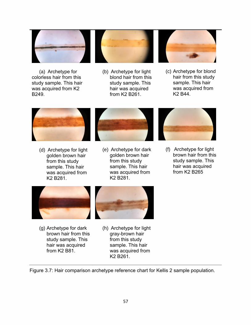
57
(a) Archetype for colorless hair from this study sample. This hair was acquired from K2 B249.
(b) Archetype for light blond hair from this study sample. This hair was acquired from K2 B261.
(c) Archetype for blond hair from this study sample. This hair was acquired from K2 B44.
(d) Archetype for light golden brown hair from this study sample. This hair was acquired from K2 B281.
(e) Archetype for dark golden brown hair from this study sample. This hair was acquired from K2 B281.
(f) Archetype for light brown hair from this study sample. This hair was acquired from K2 B265
(g) Archetype for dark brown hair from this study sample. This hair was acquired from K2 B81.
(h) Archetype for light gray-brown hair from this study sample. This hair was acquired from K2 B261.
Figure 3.7: Hair comparison archetype reference chart for Kellis 2 sample population.

58
Pigment Characteristics
Human beings are unique among primate species in that they are the only ones to
grow long, thick, and highly-pigmented scalp hair. Some propose that this may have been
an adaptation in response to a diet heavy in marine resources. Since these resources
regularly contain a large number of harmful toxins “the selective and avid binding of toxins
and metals to melanin within a rapidly growing and highly melanized tissue [such as hair]
would have been an important adaptation” (Tobin 2008:234). Other possibilities for the
excessive growth and pigmentation of human hair include sexual dimorphism and
protection from environmental factors (Tobin 2008:234).
The latter possibility is sometimes used to explain the darker pigmentation
generally attributed to individuals with African ancestry and those from tropical climates.
Darker skin and hair are thought to be adaptations offering “protection against sunstroke
afforded by deep skin and hair pigmentation as well as the contribution of melanin’s very
efficient and fast exchange of ion transport and efflux for adequate salt balance” (Tobin
2008:234). According to Tobin (2008) a full 90% of the human population has
“’environmentally friendly’ brown-black hair and tanning skin”, while the remaining 5-10%
has blonde or red hair and lighter skin pigmentation (234). In addition to melanin, when
discussing pigment in human hair several other variables also need to be considered.
These include pigment density, pigment distribution, pigment aggregate shape, and
pigment aggregate size. These are discussed in detail in the following sections.

59
Pigment Density
Pigment density is defined by the Atlas of Human Hair (1999) as “the abundance
of pigment granules observed using light microscopy” (28). Archetypes for absent, light,
medium, and heavy pigment density acquired from the study sample can be seen in
Figure 3.8 below.
(a) Archetype for absent pigment density. This hair was acquired from K2 B281.
(b) Archetype for light pigment density. This hair was acquired from K2 B281.
(c) Archetype for medium pigment density. This hair was acquired from K2 B131.
(d) Archetype for heavy pigment density. This is a beard hair acquired from K2 B81.
Figure 3.8: Pigment Density archetype reference chart for Kellis 2 sample population.

60
Pigment Distribution
Pigment distribution refers to how and where pigment is concentrated along the
hair shaft. Pigment can be distributed uniformly, to one side of the hair shaft, peripherally,
and randomly. In cases where the hair is colorless or pigment is unnoticeable, pigment
distribution is marked as absent. Archetypes for pigment distribution can be seen in Figure
3.9 below.
(a) Archetype for absent pigment distribution. This hair was acquired from K2 B309.
(b) Archetype for uniform pigment distribution. This hair was acquired from K2 B131.
(c) Archetype for one-sided pigment distribution. This hair was acquired from K2 B135.
(d) Archetype for peripheral pigment distribution. This hair was acquired from K2 B261.
Figure 3.9: Pigment distribution archetype reference chart for Kellis 2 sample
population.

61
Pigment Aggregate Shape
According to the Atlas of Human Hair (1999) “pigment aggregate shape refers to
the appearance of the pigment granules when they are concentrated in a mass that has
a recognizable form” (30). In other words, when individual melanin pigment granules
coalesce they tend to form specific shapes including streaks and clumps or a mixture of
both. Figure 3.10 represents archetypes for pigment aggregate shape.
(a) Archetype for clump pigment aggregate shape. This hair demonstrates large aggregate clumps. It was acquired from K2 B281.
(b) Archetype for streak pigment aggregate shape. This hair demonstrates small aggregate streaks. It was acquired from K2 265.
(c) Archetype for other (mixed) pigment aggregate shape. It was acquired from K2 B281.
Figure 3.10: Pigment aggregate shape archetype reference chart for Kellis 2 sample
population.

62
Pigment Aggregate Size
Pigment aggregate size, as its name suggests, concerns the size of the pigment
aggregates themselves. Figure 3.11 demonstrates small, medium, and large pigment
aggregate size archetypes found within the study sample. In cases where pigment
aggregates were not observed, pigment aggregate size was marked as absent.
(a) Archetype for small pigment aggregate size. This hair demonstrates small aggregate streaks. It was acquired from K2 B309.
(b) Archetype for medium pigment aggregate size. This hair demonstrates medium aggregate streaks. It was acquired from K2 B131.
(c) Archetype for large pigment aggregate size. This hair demonstrates large aggregate clumps. It was acquired from K2 B261.
Figure 3.11: Pigment aggregate size archetype reference chart for Kellis 2 sample
population.

63
Medulla Characteristics
Put into the very simplest of terms, hair can be described as being shaped like a
tube or a straw. Placed into the realm of biological anthropology, the structure of human
hair is comparable to human bone. Like bone there is a hard-outer shell, in this case it is
provided by the keratinized outer root sheath which is then followed by the inner root
sheath. Finally, the cuticle and cortex surround an air or fluid filled space in the middle of
the hair shaft, and like the marrow filled hollow of human bone this space is called the
medulla.
In thicker animal hair, like that seen in horse manes, cat whiskers, or porcupine
quills, the medulla can take up a large portion of the hair structure itself. However, in
thinner hair, as is found in most humans, “the medulla—if present—generally comprises
only a small percentage of [the fiber] mass” (Robbins 2002:50). The medulla in human
hair can take many forms. In some cases, the medulla is absent due to how thin the
human hair shaft can be, but where medullae are present they can be continuous,
interrupted, fragmented, streaked, and on rare occasions a double medulla may even be
present (Harding and Rogers 1999:17; Robertson 1999:80; Robbins 2002:50). In cases
where the medulla has been interrupted or fragmented, “the medulla may be broken
transversely at irregular intervals by cortical material (interrupted medulla) or it may only
be present irregularly in very small amounts in the cortex (fragmented medulla)” (Harding
and Rogers 1999:17). Additionally, when observed microscopically the medulla can
appear opaque, as happens when it is filled with air, or translucent if it is filled with fluid.
Some hair will even show a combination of both opaque and translucent medulla.

64
The Atlas of Human Hair (1999) defines a total of seven medulla continuity
archetypes and four medulla opacity archetypes. However, given the lengths of the hair
samples acquired for this particular study, some of the medulla continuity archetypes do
not apply. Since the hair samples utilized in this study were cut to approximately two
inches long, so that they could better fit onto the slides, having more than one medulla
continuity type within a single hair was generally not observable. Therefore, for purposes
of this paper the archetypes for medulla continuity were reduced to four archetypes, which
can be seen in Figure 3.12. These include absent, fragmented, interrupted, and
continuous. The archetypes provided by the Atlas of Human Hair (1999) for medulla
opacity, however, were all observed within the sample. Therefore, excluding absent, the
archetypes for medulla opacity within the study sample can be seen in Figures 3.13.
These archetypes include opaque, translucent, and opaque/translucent.

65
Medulla Continuity
As mentioned above, since the hair samples were cut to approximately two inches
the archetypes for medulla continuity were reduced from seven types to four (Figure
3.12).
(a) Archetype for absent medulla. This hair was acquired from K2 B131.
(b) Archetype for fragmented medulla continuity. This hair was acquired from K2 B204.
(c) Archetype for interrupted medulla continuity. This hair was acquired from K2 B249.
(d) Archetype for continuous medulla continuity. This hair was acquired from K2 B293.
Figure 3.12: Medulla continuity archetype reference chart for Kellis 2 sample population.

66
Medulla Opacity
This study utilized polarized light microscopy to observe individual hair
characteristics; this method is discussed in detail within the methodology section. Both
opaque and translucent medulla were observed in the study sample, and the archetypes
for translucent, opaque, and opaque/translucent medulla opacity can be seen in Figure
3.13.
(a) Archetype for transparent medulla opacity. This hair was acquired from K2 B44.
(b) Archetype for opaque medulla opacity. This hair was acquired from K2 B309.
(c) Archetype for other medulla opacity. The medulla for this hair is both opaque and transparent. This hair was acquired from K2 B293.
Figure 3.13: Medulla opacity archetype reference chart for Kellis 2 sample population.

67
Cuticle Characteristics
The cuticle is the hard, outer layer of the hair shaft. It is made up of scales, which
interlock and overlap both longitudinally and laterally. The cuticle’s main purpose is to
effectively hold the cortex together, while also protecting the cortex from chemical and
environmental damage (Harding and Rogers 1999: 22; Robbins 2002:26). In mature
human hairs, the cuticle is approximately six scales thick and the scale pattern, described
using Wildman’s nomenclature, is ‘close wave’; although, it should be noted that scale
patterns can vary slightly between individuals (Harding and Roger 1999:22; Robertson
1999:115; Morioka 2005:58). Scale pattern is especially important in the field of forensic
hair examination since it has been shown to differ between species. This being the case,
cuticle scale pattern can effectively distinguish human from non-human hairs.
However, since it has been demonstrated by previous researchers that cuticle
scale pattern is very similar among humans, and since all of the hairs in this study sample
were known to be of human origin, cuticle scale pattern was not included among the
cuticle characteristics examined in the study sample. Instead, the research in this thesis
is focused on cuticle characteristics that varied from one hair to the next. These
characteristics include cuticle thickness (Figure 3.14), inner cuticle margin (Figure 3.15),
and cuticle scale profile (Figure 3.16).

68
Cuticle Thickness
Cuticle thickness refers to how thick the cuticle appears, that is the thickness
between the inner cuticle margin and the outermost edge of the cuticle. This can be
subjective unless an accurate measure can be taken of the cuticle itself. However, there
is a notable difference between thick and thin cuticle, which is why they are broadly
defined as such for purposes of this thesis. Additionally, varied cuticle thickness was
observed in the study sample. This occurs when the thickness of the cuticle does not
remain constant along the hair shaft.
(a) Archetype for unknown cuticle thickness. This hair was acquired from K2 B281.
(b) Archetype for thin cuticle thickness. This hair was acquired from K2 B274.
(c) Archetype for thick cuticle thickness (space between black markers). This hair was acquired from K2 B22.
(d) Archetype for varied cuticle thickness. This hair was acquired from K2 B131.
Figure 3.14: Cuticle thickness archetype reference chart for Kellis 2 sample population.
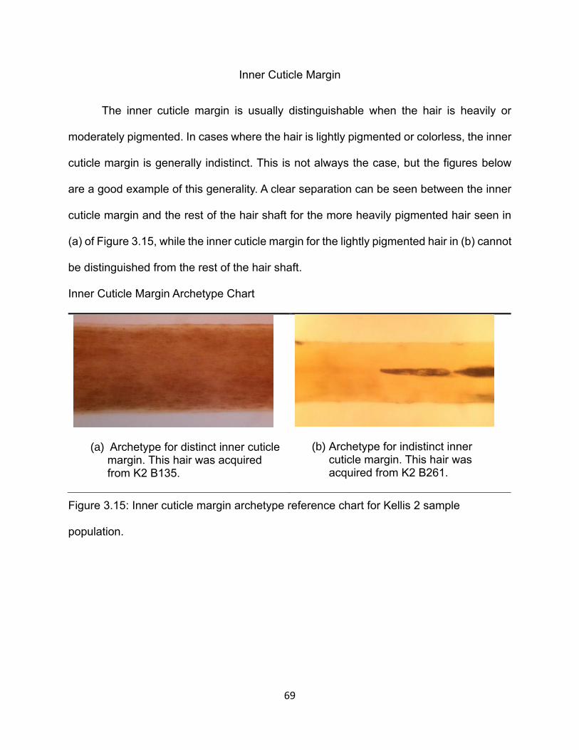
69
Inner Cuticle Margin
The inner cuticle margin is usually distinguishable when the hair is heavily or
moderately pigmented. In cases where the hair is lightly pigmented or colorless, the inner
cuticle margin is generally indistinct. This is not always the case, but the figures below
are a good example of this generality. A clear separation can be seen between the inner
cuticle margin and the rest of the hair shaft for the more heavily pigmented hair seen in
(a) of Figure 3.15, while the inner cuticle margin for the lightly pigmented hair in (b) cannot
be distinguished from the rest of the hair shaft.
Inner Cuticle Margin Archetype Chart
(a) Archetype for distinct inner cuticle margin. This hair was acquired from K2 B135.
(b) Archetype for indistinct inner cuticle margin. This hair was acquired from K2 B261.
Figure 3.15: Inner cuticle margin archetype reference chart for Kellis 2 sample
population.

70
Cuticle Scale Profile
Cuticle scale profile refers to the appearance of the cuticle on the outside of the
hair shaft. The most prolific archetype for cuticle scale profile seen in this study sample
was smooth, while the least observed archetype was looped. Figure 3.16 provides
archetypes for smooth, rough, and serrated cuticle scale profiles. In cases where the
cuticle was severely damaged, the cuticle scale profile was listed as unknown.
(a) Archetype for smooth cuticle scale profile. This hair was acquired from K2 B81.
(b) Archetype for ragged cuticle scale profile. This hair was acquired from K2 B131.
(c) Archetype for serrated cuticle scale profile (black line shows outermost cuticle edge). This hair was acquired from K2 B.
Figure 3.16: Cuticle scale profile archetype reference chart for Kellis 2 sample
population.

71
Ovoid Bodies
According to Robertson (1999) “ovoid bodies are well-defined, highly dense
clumps of undispersed pigment” (113). They can vary in size and distribution. Some hairs
can have many ovoid bodies, while others may have very few to none. Within the study
sample the majority of the hairs examined did not show the presence of ovoid bodies.
Some samples, however, did show many ovoid bodies and some showed few ovoid
bodies. The archetypes for ovoid bodies are shown below in Figures 3.17.
(a) Archetype for absent ovoid bodies. This hair was acquired from K2 B.
(b) Archetype for few ovoid bodies. This hair was acquired from K2 B.
(c) Archetype for many ovoid bodies. This hair was acquired from K2 B281.
Figure 3.17: Ovoid bodies archetype reference chart for Kellis 2 sample population.

72
Sample Preparation
There were two methods of preparation, dry and wet, utilized in this study. For the
dry method, hairs were extracted directly from the scalp sections of 51 individuals and
placed in labeled bags. Conversely, for the wet method 1cm2 scalp sections, acquired
from ten of the original 51 individuals, were soaked for 24 to 48 hours in dimethyl sulfoxide
(DMSO) to rehydrate the scalp following the method outlined in Williams (2008). Hairs
were then extracted individually from the scalp and left to dry overnight. These two
methods were used to help determine if sample preparation also affects PHRM and to
help initiate standardization procedures for analysts examining PHRM. Results for this
portion of the study can be seen in Chapter 2 of this thesis.
After all of the samples were procured, four hairs were mounted onto each labeled
glass slide using Permount©, for a total of 5-10 slides per individual depending on the
preparation method used. Permount© was chosen as a mounting medium because it has
a refractive index (RI) of 1.525 at 25°C, which is very similar to the RI of human hair at
1.55. This makes it an optimal mounting medium for this project (Linch and Prahlow 2001;
Marschner et al. 2003; Committee on identifying the needs of the forensic sciences
community 2009:156). After the hairs were mounted and the slides were fully dried, each
slide was observed using an Amscope 40x-1500x Infinity Polarizing Microscope at 40-
60x magnification.
Results
Using Chi-square analyses, each of the intrinsic factors listed above were analyzed
with SPSS® statistics software to determine their correlation to PHRM. Unsurprisingly, the
correlation between PHRM and growth stages, as demonstrated through previous

73
research (Petraco et al. 1988; Linch and Prahlow 2001; Jeffery et al. 2007; Koch et al.
2013), proved to be significant at p=.000. Again, this was not surprising; several studies
have reported a link between putrid roots and growth stages. There were several other
intrinsic factors, however, that demonstrated statistical significance in correlation with
PHRM including age, cuticle thickness, cuticle scale profile, and hair color. The remaining
variables did not show a statistically significant correlation (p≥ 0.05). Inner cuticle margin,
medulla opacity, ovoid bodies, medulla continuity, pigment aggregate shape, pigment
distribution, biological sex, and pigment aggregate size appear to be unrelated to the
incidence of putrid roots, though further studies are needed to rule out a relationship
entirely. Table 3.2 provides the statistical analysis for these variables in relation to PHRM.
Table 3.2: Pearson Chi-Square results for intrinsic characteristics not demonstrating a
significant correlation with changes in postmortem hair root morphology.
Asymp. Sig. (2-sided)
Inner Cuticle Margin
.070
Medulla Opacity
.109
Ovoid Bodies
.125
Medulla Continuity
.181
Pigment Aggregate Shape
.417
Pigment Distribution
.644
Biological Sex
.807
Pigment Aggregate Size
.864

74
Growth stages, as mentioned above, demonstrated a correlation with PHRM with
p=.000 (Table 3.3). The greatest proportion of postmortem changes were seen in hairs in
anagen stage, with very few changes occurring within the telogen stage. Nearly 43%
(118/277) of anagen roots showed postmortem changes in hair root morphology, with the
majority of these showing evidence of PMRB at over 70% (86/118) of the total changes
observed. Thirty hairs also had BLE and four were a mix of BLE and PMRB. The
remaining two hairs demonstrated changes that are consistent with HKP, a postmortem
change usually associated with wet decomposition and not something customarily
attributed to dry desert environments. Figure 3.18 is a bar graph detailing the distribution
of postmortem changes in association with hair growth stages.
Table 3.3: Pearson Chi-Square results for correlation between growth stages and root
decomposition.
Value Asymp. Sig. (2-sided)
Pearson Chi-Square
274.689a
.000
N of Valid Cases 713
a. 0 cells (0.0%) have expected count less than 5. The minimum expected count is 8.46.

75
Figure 3.18: Bar graph showing the proportion of postmortem changes seen within the
sample population and how they are correlated with growth stages.

76
Age also demonstrated statistical significance at p=.000 (Table 3.4), with those in
the youngest age group (16-21) showing the greatest correlation; 56% (19/34) of
individuals in this age range exhibited changes in PHRM. The lowest incidence of change
is in individuals between the ages of 36-50 (20%, 30/149). And, while it should be noted
that a large portion of the study population lies within this age range, the greatest
proportion of individuals were aged between 22-35 and they demonstrated the second
largest correlation at 31% (91/290). It was also determined that individuals in the older
age ranges (51-60 and 60+) showed evidence of putrid roots in only 25% (61/240) of the
population, suggesting the possibility that the occurrence of putrid roots decreases as
people age. It would be interesting to see if the same would hold true with a larger sample
population. Figure 3.19 shows a bar graph of postmortem changes in hair root
morphology and their relation to age.
Table 3.4: Pearson Chi-Square results for correlation between age and root
decomposition.
Value Asymp. Sig. (2-sided)
Pearson Chi-Square
20.061a
.000
N of Valid Cases 713
a. 0 cells (0.0%) have expected count less than 5. The minimum expected count is 9.58.

77
Figure 3.19: Bar graph showing the proportion of postmortem changes within the
sample population and how it correlates with age.

78
The next variable that appears to be associated with postmortem changes in hair
root morphology is cuticle thickness. At p=.002 (Table 3.5) cuticle thickness seems to be
nearly as significant as growth stages and age to the formation of putrid roots within this
study population. Hairs with thick cuticles comprise nearly 30% (206/713) of the sample,
but are ~18% more likely to exhibit postmortem changes. Additionally, 74% (52/70) of the
samples with thick cuticles that show evidence of postmortem changes has PMRB. This
is in opposition to the pattern exhibited by thin and varied cuticles. In thin and varied
cuticle thickness, the majority of postmortem changes observed included brush-like ends
(62%, 13/21, and 71%, 25/35, respectively). Unfortunately, one-third (237/713) of the
sample included hairs with unknown cuticle thickness. Generally, these were hairs with
light pigment density or those that were entirely colorless (see Figure 3.20). Despite this,
it is interesting to note that hair with varied cuticle thickness lies somewhere in the middle
when it comes to showing evidence of postmortem changes (25%, 35/142). This suggests
that there is likely to be a correlation between cuticle thickness and changes in PHRM.
Thin cuticles showed evidence of postmortem changes in only 16% (21/128) of the hairs
studied. Figure 3.21 is a bar graph detailing the relationship between cuticle thickness
and postmortem changes in the hair root.

79
Table 3.5: Pearson Chi-Square results for correlation between cuticle thickness and root
decomposition.
Value Asymp. Sig. (2-sided)
Pearson Chi-Square
14.470a
.002
N of Valid Cases 713
a. 0 cells (0.0%) have expected count less than 5. The minimum expected count is 36.08.
Figure 3.20: A colorless hair from K2 B293 demonstrating an unknown cuticle thickness.

80
Figure 3.21: Bar graph showing the proportion of postmortem changes documented
within the sample population and how they are correlated with cuticle thickness.

81
A link between changes in PHRM and pigment density was also statistically
significant at p=.006 (Table 3.6). PMRB had the greatest representation with 46% (55/119)
of these cases occurring in hairs with a medium pigment density. Additionally, the majority
of hair exhibiting postmortem changes occurred in this category (35%, 81/230).
Unfortunately, heavily pigmented hair was not well represented in the sample population;
only 6% (41/713) of hairs were heavily pigmented, while the great majority of hairs were
lightly pigmented (37%, 265/713), or medium pigmented (32%, 230/713). This could be
partially due to old age- 55% (389/713) of the study population was aged 36 or over- but
also because the sample was comprised of specimens from a single locale suggesting
probable physical and genetic similarities between individuals. As suggested earlier, a
greater diversity within the study sample would help to elucidate these results. The line
graph in Figure 3.22 provides a visual representation of the relationship between pigment
density and PHRM. It is clearly demonstrated that PMRB occurs the most in hairs with
medium pigment density.
Table 3.6: Pearson Chi-Square results for correlation between pigment density and root
decomposition.
Value Asymp. Sig. (2-sided)
Pearson Chi-Square
12.548a
.006
N of Valid Cases 713
a. 0 cells (0.0%) have expected count less than 5. The minimum expected count is 11.56.
82
Figure 3.22: Bar graph showing the proportion of postmortem changes documented
within the sample population and how they are correlated with pigment density.

83
Similarly, when regarding cuticle scale profile (CSP), a lack of diversity could be to
blame for the overwhelmingly smooth cuticles seen in this sample. Out of 694 individual
hairs where CSP could be discerned, a full 426 (59%) of them had smooth CSP, while
ragged accounted for ~10% (69/694), serrated 3% (22/694), and looped <.01% (4/694).
While those hairs with smooth CSP did demonstrate a closer association with changes in
PHRM, they were 16% more likely to show postmortem changes than those hairs with
ragged CSP-this could be due to the lack of diversity within the study sample. Therefore,
the statistical analysis regarding CSP is likely skewed. Further research should include
broadening the scope of the study population.
Table 3.7: Pearson Chi-Square results for correlation between cuticle scale profile and
root decomposition.
Value Asymp. Sig. (2-sided)
Pearson Chi-Square
18.116a
.003
N of Valid Cases 713
a. 4 cells (33.3%) have expected count less than 5. The minimum expected count is .28.

84
Figure 3.23: Bar graph showing the proportion of postmortem changes documented
within the sample population and how they are correlated with cuticle scale profile.

85
The last intrinsic characteristic that showed a statistically significant correlation with
putrid roots is hair color. Fortunately, unlike CSP, hair color was widely represented. The
most abundant color was medium brown, which accounted for 25% (178/713) hairs, but
dark golden brown (20%, 145/713) and colorless hair (18%, 130/713) were also present
in large quantities. The least represented colors were medium gray-brown (1), medium
red (1), dark red (3), and light gray-brown (4). Considering that red hair occurs in only
about 2% of the world population, it is not surprising that few exist in the study sample.
However, because there were a few hair colors represented by small numbers, analysis
was run using SPSS® statistics software on all hair colors as well as on hair colors with
50 or more representations in the sample; this latter analysis was comprised of 597 hairs.
Hair colors with a sample of 50 or more samples include colorless, dark golden brown,
light blonde, light brown, and medium brown.
After excluding colors that were not well represented, the analysis gave a p-value
of .006 (Table 3.9), showing a greater statistical significance than the p-value of .013 when
all hair colors were included into the analysis (Table 3.8). Each hair color demonstrated a
similar propensity for changes in PHRM (~30%-35%) except for light brown hair. The
incidence of putrid roots in light brown hair was reduced to a low 11% (9/78), a significant
reduction from the amount seen in other hair colors. Unfortunately, it is not understood
why light brown hair would show fewer changes, but perhaps this goes hand in hand with
cuticle thickness. More than half of the light brown hairs had thin or varied cuticles, which,
as mentioned earlier, accounts for fewer cases of putrid roots than those hairs with thick
cuticles. Contrary to this, dark golden brown hairs, which showed the greatest proclivity
for putrid roots, had a larger percentage of thick cuticles.

86
Table 3.8: Pearson Chi-Square results for correlation between hair color and root
decomposition.
Value Asymp. Sig. (2-sided)
Pearson Chi-Square
29.827a
.013
N of Valid Cases 713
a. 12 cells (37.5%) have expected count less than 5. The minimum expected count is .28.
Table 3.9: Pearson Chi-Square results for correlation between hair colors represented by
50+ hair samples and root decomposition.
Value Asymp. Sig. (2-sided)
Pearson Chi-Square
14.636a
.006
N of Valid Cases 597
a. 0 cells (0.0%) have expected count less than 5. The minimum expected count is 18.79.
Discussion
As mentioned earlier, it is not surprising to find an association between hair root
growth stages and PHRM. This characteristic has been studied by several researchers
(Petraco et al. 1988, Linch and Prahlow 2001, Koch et al. 2013) and it has already been
established that anagen and catagen stage hairs are more susceptible to postmortem
changes than telogen hairs. Again, the full keratinization of telogen hair roots likely causes
its resistance to postmortem root degradation.

87
Age related changes to hair keratin also explains why age affects PHRM. Younger
individuals have less cysteine disulphide in their hair, which means “children’s hair has
lower hardness… than adult hair” (Tang et al. 2015: 160-161). Cysteine disulphide “forms
cross-linkages in cuticle cells and contribute to the physical and mechanical properties of
hair” (Tang et al. 2015: 160). Therefore, younger hair, being deficient in cysteine
disulphide, would seem to be more vulnerable to postmortem degradation. This
assumption is bolstered by the analysis which shows that individuals in the age range of
16-21 are more prone to decomposition changes in their hair than older individuals.
The greater proportion of PMRB seen in thick cuticles can probably be explained
by looking at the greater percentage of BLE seen in thin cuticles. A thinner cuticle could
cause the hair to be more easily broken when forcibly removed from the scalp. Since
PMRB causes the root end to be more fragile, a thinner cuticle is more likely to break in
this area thereby resulting in a greater proportion of BLE. Thicker cuticles would be less
likely to break in the area of PMRB, meaning thick cuticles would be more inclined to
show evidence of PMRB than BLE.
The final three intrinsic factors and their affinity for PHRM can be explained by
limited sample size and limited sample diversity. Pigment density was similar throughout
the sample, with most samples showing a light or medium pigment density; this can
partially be due to the overall older age profile of the individuals included in the study
population. Unfortunately, heavy pigment density was not adequately represented. A
larger, more diverse sample population would help determine whether pigment density
affects PHRM. Cuticle scale profile and hair color would similarly benefit from a more
diverse sample population. 426/694 hairs, where CSP could be discerned, has smooth

88
CSP; that is equal to 59% of the total sample. It is fair to assume that this probably skewed
the results. The similarity of hair colors, likely due to similar ancestry, probably biased the
results in an identical manner.
Benefits and Limitations of Research
Now, more than ever, the admissibility of certain evidence is under scrutiny. Among
these is the utility and reliability of forensic analysis involving human hair. This comes in
the wake of an article by The Washington Post in which it was stated that, “the Justice
Department and FBI have formally acknowledged that nearly every examiner in an elite
FBI forensic unit gave flawed testimony in almost all trials in which they offered evidence
against criminal defendants over more than a two-decade period before 2000” (Hsu
2015). This included “overstat[ing] forensic matches in ways that favored prosecutors”; in
32 of those cases the defendant was sentenced to death (Hsu 2015:1). While it is unlikely
that this evidence alone resulted in a conviction, it does raise questions about the
authenticity of forensic analyses overall.
Fortunately, most types of admissible forensic evidence have proven their efficacy
over the course of history. The reliability of fingerprints and DNA are rarely questioned,
and blood spatter and ballistics have both shown to be dependable in criminal
investigations. With the introduction of television series such as CSI, Bones, and NCIS,
jurors are beginning to think of themselves as armchair detectives, and therefore are
requiring more evidence as a result. Furthermore, those who watch these crime
procedurals are led to believe that forensics is an exact science that can be accepted
without question, that evidence can speak for itself, and that the methods of crime scene
investigators are infallible (Kruse 2010:82-88). This means that the science behind

89
admitted evidence needs to be sound. Firmly put, if PHRM is better understood and if its
causes can be determined, the potential impact for medico-legal cases could be
enormous.
However, there are several limitations to studies such as this. The main limitation
is sample size. In order to acquire the large and diverse sample necessary to understand
the causes of putrid roots, cadavers from several different ancestries, both sexes, and
various ages need to be represented in the sample population. Additionally, bodies should
be placed in diverse, but controlled, environments and studied at various stages of
decomposition. Climate, predatory activity, insect populations, fungal growth, and other
extrinsic factors also need to be considered in determining the variables included within
the sample set. Unfortunately, the likelihood of a research sample with these criteria is
improbable. Therefore, it is necessary to undertake a great amount of research in order
to provide analysts with the best data regarding PHRM.
Conclusion
There are several hurdles to overcome before PHRM can be accepted as reliable
evidence. In addition to the issues stated above, there is also the problem of linking a
questioned hair to a known source. Without a way to identify the hair as belonging to a
specific deceased individual, PMRB, BLE, and HKP become moot points. Does the
appearance of changes in hair root morphology suggest decomposition? More than likely.
But without an understanding of the causes behind these changes, PMRB, BLE and HKP
bring minimal evidentiary value to criminal cases. Finding significant correlations between
PHRM and intrinsic variables, therefore, provides a better understanding of the
mechanisms involved in postmortem hair root degradation.

90
This study shows that there are more than extrinsic variables acting upon PHRM.
Much like decomposition of the body itself, decomposition in hair is influenced by intrinsic
factors. Age showed a greater association with hair root degradation, as did cuticle
thickness, pigment density, cuticle scale profile, and hair color. Both age and cuticle
thickness and their associations with PHRM make sense. Younger individuals have softer
and less structurally sound hair than older individuals due to the lack of cysteine
disulphide in their hair. This makes their hair and hair roots more susceptible to
postmortem changes. Additionally, in regards to cuticle thickness, it makes sense that
thinner cuticles are more likely to lead to BLE. A thinner cuticle means less support for
the hair shaft and a greater likelihood of breakage when the hair is forcibly removed from
the scalp. This statement is supported by the analysis which shows that thinner roots
showed more evidence of BLE, while thicker roots showed more evidence of PMRB.
Unfortunately, while growth stages, age, and cuticle thickness are well represented within
the sample, pigment density, cuticle scale profile, and hair color were skewed. These will
require further study in order to discover their association with PHRM.
The ultimate aim of this research is that future forensic analysts will use this study
as a stepping-stone, and broaden the scope of studies on PHRM. While hair analysis is
still an imperfect science, and it has proven to be more so since fabrication of such
evidence has come to light, hopefully more research will help to bolster its credibility.
Further research should look at those intrinsic factors that did show an association,
however weak, and attempt to discern whether there is in fact a connection. Additionally,
other researchers should look at those factors without strong connections, such as

91
medulla continuity and medulla opacity, to determine why these do not have an impact
on PHRM.
92
References
Bencze, Kolomon. 1994. Determination of metals in human hair. In Handbook on Metals in Clinical and Analytical Chemistry. Edited by Seiler HG, Sigel A, Sigel H, pp. 201-216, Marcel Dekker Inc, New York, NY.
Birrell, Michael. 1999. Excavations in the Cemeteries of Ismant el-Kharab. In Dakhleh Oasis Project: Preliminary Reports on the 1992-1993 and 1993-1994 Field Seasons. Edited by C.A. Hope and A.J. Mills, pp. 29-41. Oxbow Books, Oxford [England]; Oakville, CT, USA.
Blume, H.P, F. Alaily, U. Smettan, and J. Zielinski. 1984. Soil Types and Associations of Southwest Egypt. In Research in Egypt and Sudan. Berliner Geowissenschaftliche Abhandlungen. Edited by E. Klitzsch, R. Said and E. Schrank, pp. A50:293-302.
Bowen, Gillian E. 2003. Some observations on Christian burial practices at Kellis. In The Oasis Papers 3: Proceedings of the Third International Conference of the Dakhleh Oasis Project. Edited by Gillian E Bowen and C.A. Hope, pp. 167-182. Oxbow Books, Oxford.
Bowen, Gillian E, Thomas Chandler, and Derrick Martin. 2005. Reconstructing Ancient Kellis. Buried History 41:51-64.
Castanet, J., J.P. Ortonne. 1997. Hair melanin and hair color. In Formation and structure of human hair. Edited by Jollès P, Zahn H, Höcker H, pp. 209-225, Deutsche Bibliothek, Basel, Switzerland.
Committee on identifying the needs of the forensic science community, National Research Council.
2009. Analysis of Hair Evidence. In Strengthening Forensic Science in the United States: A Path Forward, pp. 155-161, Washington, D.C: USA.
Deedrick, Douglas W., Sandra L. Koch. 2004. Microscopy of hair part 1: a practical guide and manual for human hairs. Forensic Science Communications Electronic Document, https://archive.is/nnW3t, accessed April 10, 2017.
Domzalski, Alison C.
2004. The effects of environmental exposure on human hair root morphology. M.S. Thesis. John Jay College of Criminal Justice, New York.

93
Dupras, Tosha L. 1999. Dining in the Dakhleh Oasis, Egypt: Determination of Diet using Documents and Stable Isotope Analysis. Ph.D. dissertation, Department of Anthropology, McMaster University, Ontario.
Dupras, Tosha L., Henry P. Schwarcz. 2001. Strangers in a strange land: Stable isotope evidence for human migration in the Dakhleh Oasis, Egypt. Journal of Archaeological Science 28(11):1199-1208.
Gaudette, B.D., J. Bailey, Richard Bisbing, Edward Burwitz, J. Cadman, C. Cwiklik, Harold Deadman, Peter DeForest, N. Erickson, John Kilbourn, Paul Krupenie, Thomas Kubic, D. Metger, T. Mozer, S. Palenik, L. Peterson, Nicholas Petraco, J. Robertson, S. Shaffer, R. Stockdale, M.A. Straus.
1985. Preliminary Report—Committee on Forensic Hair Comparison. Criminal Laboratory Digest 12:50-59.
Giddy, Lisa L. 1987. Egyptian oases. Bahariya, Dakhla, Farafra and Kharga during Pharaonic times. Aris & Phillips.
Harding, Harry, George Rogers. 1999. Physiology and growth of human hair. In Forensic examination of hair. Edited by James Robertson, pp.1-77, Taylor & Francis, London, UK.
Hsu, Spencer S.
2015. FBI Admits Flaws in Hair Analysis Over Decades. The Washington Post. Electronic document https://www.washingtonpost.com/local/crime/fbi-overstated-forensic-hair- matches-in-nearly-all-criminal-trials-for-decades/2015/04/18 /39c8d8c6-e515-11e4-b510-962fcfabc310_story.html, accessed April 14, 2017.
Jeffery, Kathryn J., Kate A. Abernethy, Caroline E. G. Tutin, Michael W. Bruford.
2007. Biological and Environmental Degradation of Gorilla Hair and Microsatellite Amplification Success. Biological Journal of the Linnean Society 91: 281-294.
Joblonski, Nina G.
2004. The Evolution of Human Skin and Skin Color. Annual Review of Anthropology 33: 585-623.
Kadane, Joseph B.
2015. Post-Mortem Root Banding of Hairs: A Skeptical Review. Law Probability and Risk 14(3): 213-228.
Koch, Sandra L., Amy L. Michaud, C.E. Mikell. 2013. Taphonomy of Hair-- A Study of Postmortem Root Banding. Journal of Forensic Sciences 58: S52-S59.

94
Kruse, C. 2010. Producing absolute truth: CSI science as wishful thinking. American Anthropology 112(1): 79-91.
Linch, Charles A., Joseph A. Prahlow. 2001. Postmortem Microscopic Changes Observed at the Human Head Hair Proximal End. Journal of Forensic Sciences 46(1): 15-20.
Marschner, Steven R., Henrik W. Jensen, Mike Cammarano, Steve Worley, Pat Hanrahan. 2003. Light Scattering from Human Hair Fibers. ACM Transactions on Graphics 22(3):780-791.
Molto, J.E. 2001. The Comparative Skeletal Biology and Paleoepidemiology of the People from Ein Tirghi and Kellis, Dakhleh Oasis, Egypt. In The Oasis Papers 1: The Proceedings of the First International Symposium of the Dakhleh Oasis Project. Edited by M. Marlow and A.J. Mills, pp. 81-100, Oxbow Books, Oxford.
Molto, J.E. 2002. Bio-archaeological Research of Kellis 2: An Overview. In Dakhleh Oasis Project: Preliminary Reports on the 1994-1995 to 1998-1999 Field Seasons. Edited by C.A. Hope, G.E. Bowen, and R.S. Bagnall, pp. 239-255, Oxbow Books, Oxford [England], Oakville, CT, USA. [England]: Oxbow Books; Oakville, CT, USA: David Brown Book Co. p. 239-255.
Morioka, K. 2005. Hair follicle differentiation under the electron microscope: An atlas. Springer-Verlag Tokyo, Tokyo, Japan.
Ogle, Jr Robert, Michelle J. Fox. 1999. Atlas of Human Hair: Microscopic Characteristics. CRC Press LLC, Boca Raton, Florida.
Petraco, Nicholas, C. Fraas, F.X. Callery, Peter De Forest.
1988. The Morphology and Evidential Significance of Human Hair Roots. Journal of Forensic Sciences 33(1): 68-75.
Robbins, Clarence R.
2002. Chemical and Physical Behavior of Human Hair: Fourth Ed. Springer-Verlag New York Inc, New York.
Robertson, James. 1999. Forensic and Microscopic Examination of Human Hair. In Forensic examination of hair. Edited by Robertson, pp. 79-154, Taylor & Francis, London, UK.

95
Seybert, Joanna. 2012. People v Kogut. Electronic Document http://docs.justia.com/cases/federal/districtcourts/newyork/nyedce/2:2006cv06695/264382/350/0.pdf?1345171557, accessed April 14, 2017.
Sinclair, Rodney D., Cedric C. Banfield, Rodney P.R. Dawber.
1999. Handbook of Diseases of the Hair and Scalp. Blackwell Science Ltd, Oxford.
Stewart, J.D., J.E. Molto, P.J. Reimer. 2003. The Chronology of Kellis 2: The Interpretative Significance of Radiocarbon Dating of Human Remains. In Dakhleh Oasis Project Monograph. Edited by Gillian E. Bowen and C.A. Hope, pp 373-378, Oxbow Books, Oxford.
Tang, Wei, Shaogang Zhang, Jiankai Zhang, Si Chen, Hua Zhu, Shirong Ge. 2015. Aging Effects on the Diameter, Nanomechanical Properties and Tactile Perception of Human Hair. International Journal of Cosmetic Science 38:155-163.
Tobin D.J.
2008. Human Hair Pigmentation- Biological Aspects. International Journal of Cosmetic Science 30:233-257.
Torkamani, Niloufar, Nicholas Rufaut, Leslie Jones, Rodney D. Sinclair.
2014. Beyond Goosebumps: Does the Arrector Pili Muscle Have a Role in Hair Loss? International Journal of Trichology 6(3):88.
Wheeler, Sandra M. 2009. Bioarchaeology of Infancy and Childhood at the Kellis 2 Cemetery, Dakhleh Oasis, Egypt. Ph.D. dissertation, Department of Anthropology, University of Western Ontario, Ontario.
Wheeler, Sandra M. 2010. Nutritional and Disease Stress of Juveniles from the Dakhleh Oasis, Egypt. International Journal of Osteoarchaeology 22(2):219-234.
Whiting DA, Dy LC. 2008. Hair Follicle Anatomy in Human Scalp Biopsies. In Hair and Scalp Diseases: Medical, Surgical, and Cosmetic Treatments. Edited by A.J. McMichael, M.K. Hordinsky, pp. 41-58, Informa Healthcare USA Inc., New York.

96
Williams, Lana J. 2008. Investigating Seasonality of Death at Kellis 2 Cemetery Using Solar Alignment and Isotopic Analysis of Mummified Tissues. Ph.D. dissertation, Department of Anthropology, University of Western Ontario, Ontario.
Wollina Uwe. 1997. Histochemistry of the Human Hair Follicle. In Formation and Structure of Human Hair. Edited by P. Jollès, H. Zahn, H. Höcker, pp. 31-58, Deutsche Bibliothek, Basel, Switzerland.

97
CHAPTER 4: CONCLUSION
While other animals sport feathers and scales, mammals alone grow hair. It
enables them to keep warm, and for some serves as a useful scare tactic. For humans,
however, the necessity and overall usefulness of hair is still questioned. Unlike our
mammalian counterparts, for most of us hair is used more as an accessory. We style it,
color it, cut it, and in some cases, we shave it all off.
Fortunately, although we may not know the bodily or evolutionary function for the
retention of human hair, studies in hair analysis have taught us that human hair itself is
unique. It has a specific pattern, like a fingerprint, that sets it apart from hair in other
mammalian species. Because all mammals have hair that differs structurally from that of
other mammals, analysts are able to differentiate between species when examining a
single hair. This, along with other applications, have become useful in the field of
forensics, where the questioning of known and unknown hairs, DNA analysis, heavy metal
analysis, and toxicological analysis can mean the difference between catching the guilty
party or even ensuring an innocent person is not convicted for a crime they did not
commit. One such forensic application has been the subject of this thesis. First, two
preparation methods were assessed to determine their efficacy when investigating
mummified remains. Then the evidentiary significance and reliability of postmortem
human hair degradation in regards to medico-legal investigations were evaluated by
determining which intrinsic variables, if any, may have an effect on the occurrence of
degradation.

98
Hair samples acquired from fifty individuals from the Kellis 2 cemetery in the
Dakhleh Oasis, Egypt, were prepared using two preparation methods. These included
both a wet and dry method, and the resulting samples were evaluated to determine the
efficacy of each method. It was determined that the wet method, which involved
rehydrating the tissue using DMSO, provided the most definitive and advantageous
results. The hair samples acquired from the rehydrated scalp sections were more easily
obtained, and therefore resulted in less breakage and a better representation of anagen
and catagen hair roots. As postmortem hair degradation is generally relegated to these
earlier stages of hair growth, it is crucial that a large number of these are collected to
provide a more significant study sample for investigation.
Additionally, fewer roots were lost during the acquisition of hairs from the
rehydrated scalp sections. The breakage of the hair root from the shaft occurred nearly
half as often in the wet samples as it did in the dry samples (20% and 36%respectively).
Lastly, the occurrence of postmortem hair degradation and types of degradation
documented was significantly different between the two preparation methods. In the dry
samples, there were equal incidences of both BLE and PMRB, while in the wet sample
there was a predominance of PMRB (25%) within the sample and BLEs made up only
3%. There are two possible explanations for this disparity. The least likely explanation is
that there simply were more BLEs in the dry sample, but more than likely the cause of the
imbalance resulted from the forceful removal of the hair root from a dry scalp. This could
cause the root to break off, thereby producing a BLE on the proximal end of the shaft.
While there are some studies (e.g., Petraco et al. 1988; Tafaro 2000; Linch and
Prahlow 2001; Domzalski 2004; Jeffery et al. 2007; Koch et al. 2013) that examine how

99
extrinsic variables such as time since death, climate, method of decomposition, stage of
decomposition, and burial environment affect postmortem hair root morphology, none
have seriously considered intrinsic factors. It is already widely believed that postmortem
hair degradation proceeds in a predictable manner (e.g., Petraco et al. 1988; Tafaro 2000,
Linch and Prahlow 2001; Domzalski 2004; Jeffery et al. 2007; Koch et al. 2013), where
PMRB occurs in the earlier stages of decomposition, and BLEs and HKPs are thought to
be related to later stages of dry and wet decomposition respectively. This study calls this
into question. The individuals in this sample have been buried since AD50-AD450 in a
hyper arid environment, and PMRB was present in the sample as well as HKP, which are
thought to be related to moist decomposition. While the HKP may be able to be explained
by the fact that different portions of the body may be subjected to different types of
decomposition within a single burial environment, this does not explain why PMRB was
found on hairs from individuals who have been deceased for nearly 1600-2000 years.
Therefore, if time since death, climate, method of decomposition, stage of decomposition,
and burial environment do not fully explain why postmortem hair degradation occurs,
other factors need to be taken into account.
The second portion of this thesis concerns the effect that intrinsic variables might
have on the incidence of postmortem hair degradation. A total of 713 hairs were examined
for color, pigment density, pigment distribution, pigment aggregate shape, pigment
aggregate size, medulla continuity, medulla opacity, cuticle thickness, inner cuticle
margin, cuticle scale profile, and ovoid bodies. Additionally, age and biological sex were
recorded for each individual and included as part of the intrinsic variables.

100
While not all of these variables showed a significant correlation with postmortem
modification of the hair root, several did. The growth stage of the hair appeared to have
a significant impact on postmortem changes. Only roots in anagen and catagen growth
stages showed these changes. Hair roots in the telogen growth stage did not demonstrate
any of these modifications. This finding is in keeping with studies conducted by previous
researchers within the field of hair analysis (e.g., Petraco et al. 1988; Tafaro 2000; Linch
and Prahlow 2001; Domzalski 2004; Jeffery et al. 2007; Koch et al. 2013). In addition to
growth stage, however, age, cuticle thickness, pigment density, cuticle scale profile, and
hair color were shown to have a significant correlation with the appearance of postmortem
degradation in the human hair root. This suggests that there are other factors affecting
postmortem hair root degradation in addition to those that have been previously studied.
Benefits and Limitations of Research
The limitations to studies such as this are many and include: the need for a large
sample placed under controlled circumstances and regularly monitored; the need for a
large number of cadavers with diverse backgrounds and ancestry; and the need for
samples to be observed in numerous environmental conditions. This study was very
limited in scope since it was conducted on a single population of individuals who were
buried in similar, if not the same, burial environment, and their bodies all underwent
natural mummification. Additionally, the scale of this study was limited to 51 individuals
and the decomposition processes were not regularly monitored. This means that the
decomposition process may have been affected by factors which are not known. As such,
this study can be considered to be a pilot project.
101
Despite this, there are several benefits to this study. For one, it shows that PMRB
does not appear to be dependent on time since death. These individuals have been dead
for over 1500 years, yet some hairs still show evidence of PMRB. It also provides a good
representation of postmortem hair root changes in naturally mummified remains and
remains buried in arid environments. Furthermore, the methodology section provides a
good demonstration of preparation methods for hair samples from mummified remains.
This will hopefully lead to the standardization of hair analysis preparation methods in
future research.
Future Considerations
For postmortem hair degradation to be widely accepted as evidence in medico-
legal cases, more rigorous experimentation and research is needed. This thesis covers
merely a fraction of the scenarios that can affect human hair roots after death. Neither the
scope nor the scale of this investigation is large enough to encompass the entirety of
situations that affect human decomposition. In order to cover such a diverse set of
circumstances, a large-scale investigation would need to be conducted taking into
account climate and environmental variability, predation, burial environments, manner
and cause of death, underlying medical conditions, intrinsic variables such as those listed
above, and other factors that may affect postmortem hair root morphology.
Hair exhibiting PMRB is useless to investigators if it cannot be stated that it is
undoubtedly attributable to the presence of decomposition. While Lowe (State of Florida
v. Casey Marie Anthony, 48-2008-CF-015606-O 2011) did state that she had not come
across any instances of PMRB where the individual was not deceased, questions still

102
arise about the veracity of this testimony simply because of the scarcity of published
research.
Additionally, the question as to why PMRB occurs in some individuals while being
absent in others needs to be resolved. This can only be accomplished with more research
looking at extrinsic and intrinsic factors that affect PHRM. To lend credence to claims that
PMRB and other postmortem morphological changes are the result of decomposition, and
thereby are proof that a deceased individual was present, these studies need to be
undertaken by experts within the fields of trace evidence, forensics, and hair analysis.
Furthermore, sample sets need to become increasingly diverse, specimens need to be
regularly monitored, and dispositional environments need to be strictly controlled. Future
court cases will only benefit from this type of research.

103
References
Domzalski, Alison C. 2004. The effects of environmental exposure on human hair root morphology. M.S. Thesis. John Jay College of Criminal Justice, New York.
Jeffery, Kathryn J., Kate A. Abernethy, Caroline E. G. Tutin, Michael W. Bruford.
2007. Biological and Environmental Degradation of Gorilla Hair and Microsatellite Amplification Success. Biological Journal of the Linnean Society 91: 281-294.
Koch, Sandra L., Amy L. Michaud, C.E. Mikell.
2013. Taphonomy of Hair-- A Study of Postmortem Root Banding. Journal of Forensic Sciences 58: S52-S59.Kruse C. 2010. Producing absolute truth: CSI science as wishful thinking. American Anthropology 112(1): 79-91.
Linch, Charles A., Joseph A. Prahlow. 2001. Postmortem Microscopic Changes Observed at the Human Head Hair Proximal End. Journal of Forensic Sciences 46(1): 15-20.
Petraco, Nicholas, C. Fraas, F.X. Callery, Peter De Forest.
1988. The Morphology and Evidential Significance of Human Hair Roots. Journal of Forensic Sciences 33(1): 68-75.
State of Florida v. Casey Marie Anthony.
2011b. Casey Anthony trial. https://www.youtube.com/user/CrimeTimeVids, accessed April 14, 2017.
Tafaro, Joseph T.
2000. The Use of Microscopic Postmortem Changes in Anagen Hair Roots to Associate Questioned Hairs with Known Hairs and Reconstruct Events in Two Murder Cases. Journal of Forensic Sciences 45(2): 495-499.