Coal Depolymerising Activity and Haloperoxidase Activity of Mn Peroxidase from Fomes durissimus...
8
Hindawi Publishing Corporation Bioinorganic Chemistry and Applications Volume 2011, Article ID 260802, 8 pages doi:10.1155/2011/260802 Research Article Coal Depolymerising Activity and Haloperoxidase Activity of Mn Peroxidase from Fomes durissimus MTCC-1173 Sunil Kumar Singh, Meera Yadav, Sudha Yadava, and Kapil Deo Singh Yadav Department of Chemistry, DDU Gorakhpur University, Gorakhpur 273009, India Correspondence should be addressed to Meera Yadav, drmeerayadav@rediffmail.com Received 21 July 2011; Accepted 7 September 2011 Academic Editor: Spyros Perlepes Copyright © 2011 Sunil Kumar Singh et al. This is an open access article distributed under the Creative Commons Attribution License, which permits unrestricted use, distribution, and reproduction in any medium, provided the original work is properly cited. Mn peroxidase has been purified to homogeneity from the culture filtrate of a new fungal strain Fomes durissimus MTCC-1173 using concentration by ultrafiltration and anion exchange chromatography on diethylaminoethyl (DEAE) cellulose. The molecular mass of the purified enzyme has been found to be 42.0 kDa using SDS-PAGE analysis. The K m values using MnSO 4 and H 2 O 2 as the variable substrates in 50 mM lactic acid-sodium lactate buffer pH 4.5 at 30 ◦ C were 59 μM and 32 μM, respectively. The catalytic rate constants using MnSO 4 and H 2 O 2 were 22.4 s −1 and 14.0 s −1 , respectively, giving the values of k cat /K m 0.38 μM −1 s −1 and 0.44 μM −1 s −1 , respectively. The pH and temperature optima of the Mn peroxidase were 4 and 26 ◦ C, respectively. The purified MnP depolymerises humic acid in presence of H 2 O 2 . The purified Mn peroxidase exhibits haloperoxidase activity at low pH. 1. Introduction Manganese peroxidase, MnP [E.C.1.11.1.13], is a heme- containing enzyme [1]. It has been shown to be present in the culture filtrates of a number of fungal strains [2– 5]. The catalytic cycle of Mn peroxidase resembles those of other heme peroxidases such as horseradish peroxidase [6] and lignin peroxidase [7, 8] and includes the native ferric enzyme as well as the reactive intermediates compound I and compound II. The catalytic cycle can be shown as follows: MnP + H 2 O 2 −→ MnP(I) + H 2 O (I) MnP(I) + Mn II −→ MnP(II) + Mn III (II) MnP(II) + Mn II −→ MnP + Mn III +H 2 O (III) H 2 O 2 oxidizes the enzyme by two electrons to form MnP compound (I) which is oxyferryl porphyrin cation radical [Fe 4+ = O P] + . Mn(II) or phenolic compounds can serve as reductants for the MnP compound (I) and form MnP compound (II) which is an oxyferryl chemical species [Fe 4+ = O P], one electron oxidized form of the enzyme. For the reduction of MnP compound (II) to the enzyme, Mn(II) is absolutely essential [9, 10]. MnP is a biotechnological important enzyme having wide application in degradation of lignin [11], biopulping and biobleaching in paper industries [12], removal of recalci- trant organic pollutants [13], and enzymatic polymerization [14]. Keeping these points in view, we have purified Mn peroxidase from the culture filtrate of Fomes durissimus MTCC-1173 and its enzymatic characteristics like K m , pH, and temperature optima have been determined. Depolymeri- sation of coal by the purified enzyme has been demonstrated using humic acid as a model of coal. MnP from F. durissimus also possesses haloperoxidase activity at low pH. 2. Materials and Methods 2.1. Chemicals. DEAE Cellulose was from Sigma Chemical Company, St. Louis, USA MnSO 4 , NaCl, and sodium acetate were from Merck Ltd., Mumbai, India, and lactic acid, sodi- um lactate, malonic acid, sodium malonate, oxalic acid, sodi- um oxalate succinic acid, sodium succinate, and H 2 O 2 were from S.D. Fine Chem. Ltd., Mumbai, India, and were used without further purifications. The chemicals including the protein molecular weight markers used in SDS-PAGE anal- ysis of the purified enzyme were procured from Bangalore Genei Pvt. Ltd., Bangalore, India.
-
Upload
independent -
Category
Documents
-
view
1 -
download
0
Transcript of Coal Depolymerising Activity and Haloperoxidase Activity of Mn Peroxidase from Fomes durissimus...
Hindawi Publishing CorporationBioinorganic Chemistry and ApplicationsVolume 2011, Article ID 260802, 8 pagesdoi:10.1155/2011/260802
Research Article
Coal Depolymerising Activity and Haloperoxidase Activity ofMn Peroxidase from Fomes durissimus MTCC-1173
Sunil Kumar Singh, Meera Yadav, Sudha Yadava, and Kapil Deo Singh Yadav
Department of Chemistry, DDU Gorakhpur University, Gorakhpur 273009, India
Correspondence should be addressed to Meera Yadav, [email protected]
Received 21 July 2011; Accepted 7 September 2011
Academic Editor: Spyros Perlepes
Copyright © 2011 Sunil Kumar Singh et al. This is an open access article distributed under the Creative Commons AttributionLicense, which permits unrestricted use, distribution, and reproduction in any medium, provided the original work is properlycited.
Mn peroxidase has been purified to homogeneity from the culture filtrate of a new fungal strain Fomes durissimus MTCC-1173using concentration by ultrafiltration and anion exchange chromatography on diethylaminoethyl (DEAE) cellulose. The molecularmass of the purified enzyme has been found to be 42.0 kDa using SDS-PAGE analysis. The Km values using MnSO4 and H2O2 as thevariable substrates in 50 mM lactic acid-sodium lactate buffer pH 4.5 at 30◦C were 59 μM and 32 μM, respectively. The catalyticrate constants using MnSO4 and H2O2 were 22.4 s−1 and 14.0 s−1, respectively, giving the values of kcat/Km 0.38 μM−1s−1 and0.44 μM−1s−1, respectively. The pH and temperature optima of the Mn peroxidase were 4 and 26◦C, respectively. The purifiedMnP depolymerises humic acid in presence of H2O2. The purified Mn peroxidase exhibits haloperoxidase activity at low pH.
1. Introduction
Manganese peroxidase, MnP [E.C.1.11.1.13], is a heme-containing enzyme [1]. It has been shown to be presentin the culture filtrates of a number of fungal strains [2–5]. The catalytic cycle of Mn peroxidase resembles those ofother heme peroxidases such as horseradish peroxidase [6]and lignin peroxidase [7, 8] and includes the native ferricenzyme as well as the reactive intermediates compound I andcompound II. The catalytic cycle can be shown as follows:
MnP + H2O2 −→ MnP(I) + H2O (I)
MnP(I) + MnII −→ MnP(II) + MnIII (II)
MnP(II) + MnII −→ MnP + MnIII + H2O (III)
H2O2 oxidizes the enzyme by two electrons to form MnPcompound (I) which is oxyferryl porphyrin cation radical[Fe4+ = O P]+. Mn(II) or phenolic compounds can serveas reductants for the MnP compound (I) and form MnPcompound (II) which is an oxyferryl chemical species [Fe4+
= O P], one electron oxidized form of the enzyme. For thereduction of MnP compound (II) to the enzyme, Mn(II) isabsolutely essential [9, 10].
MnP is a biotechnological important enzyme havingwide application in degradation of lignin [11], biopulpingand biobleaching in paper industries [12], removal of recalci-trant organic pollutants [13], and enzymatic polymerization[14]. Keeping these points in view, we have purified Mnperoxidase from the culture filtrate of Fomes durissimusMTCC-1173 and its enzymatic characteristics like Km, pH,and temperature optima have been determined. Depolymeri-sation of coal by the purified enzyme has been demonstratedusing humic acid as a model of coal. MnP from F. durissimusalso possesses haloperoxidase activity at low pH.
2. Materials and Methods
2.1. Chemicals. DEAE Cellulose was from Sigma ChemicalCompany, St. Louis, USA MnSO4, NaCl, and sodium acetatewere from Merck Ltd., Mumbai, India, and lactic acid, sodi-um lactate, malonic acid, sodium malonate, oxalic acid, sodi-um oxalate succinic acid, sodium succinate, and H2O2 werefrom S.D. Fine Chem. Ltd., Mumbai, India, and were usedwithout further purifications. The chemicals including theprotein molecular weight markers used in SDS-PAGE anal-ysis of the purified enzyme were procured from BangaloreGenei Pvt. Ltd., Bangalore, India.
2 Bioinorganic Chemistry and Applications
2.2. Fungal Strain and Its Growth. The indigenous ligni-nolytic fungal strain F. durissimus MTCC-1173 was procuredfrom MTCC Centre and Gene Bank, Institute of MicrobialTechnology, Chandigarh, India. The fungal strain was main-tained on growth medium which consisted of “malt extract20.0 g and agar 20.0 g in 1.0 L double distilled water.” The pHof the medium was adjusted to 6.5 at temperature 25◦C.
For the production of Mn peroxidase, the fungal strainwas grown in a medium [15] containing per liter “glucose10.0 g, KH2PO4 0.2 g, CaCl2 0.11 g, (NH4)2HPO4 0.264 g,MgSO4·7H2O 0.05 g, ZnSO4·7H2O 0.0425 g, MnSO4·H2O0.175 g, CoCl2·6H2O 0.007 g, CuCl2·2H2O 0.007 g, FeCl3·6H2O 0.0009 g, NaCl 0.0009 g, yeast extract 0.2 g, veratrylalcohol 0.07 g, tartaric acid 3.0 g (the pH was adjusted to4.5 with 40% NaOH), and 1 g of Tween 80” was added.The sterilized 100 mL culture flask containing 20 mL of theliquid culture growth medium was inoculated with 1 mLof the spore suspension (spore density 5 × 10⁶ spores/mL)aseptically, and the fungal culture was grown under station-ary culture condition at 30◦C in a BOD (Biological OxygenDemand) incubator. One mL aliquots of the liquid culturefungal growth medium were withdrawn at regular inter-vals of 24 hrs, filtered through sartorius membrane filters(0.22 μm), and analysed for the activity of Mn peroxidaseusing the reported [16] method.
2.3. Enzyme Assay. The activity of Mn peroxidase wasdetermined spectrophotometrically [16] by monitoring theabsorbance change at λ = 240 nm due to the formation ofMn(III) lactate and using the molar extinction coefficientvalue of 65,00 M−1 cm−1. The reaction solution 1 mL con-sisted of “50 μM MnSO4, 50 μM H2O2, and a suitable aliquotof the enzyme solution in 50 mM sodium lactate/lactic acidbuffer pH 4.5 at 30◦C.” One enzyme unit transformed1 μmole of the substrate into the product under the specifiedassay condition. UV/VIS spectrophotometer Hitachi (Japan)Model U-2000 which was fitted with electronic temperaturecontrol unit was used for spectrophotometric measurements.The least count of the absorbance measurement was 0.001absorbance unit. Each data point is an average of triplicatemeasurements with standard deviation less than 4%.
2.4. Purification of the Enzyme. For the purification ofMn peroxidase, the fungal cultures were grown in sixty100 mL sterilized culture flasks each containing 20 mL ofthe growth medium as described above. On the fifth day ofinoculation of the fungal spores when Mn peroxidase activityreached maximum value, the cultures were pooled, myceliawere removed by filtration through four layers of cheesecloth, and culture filtrate 800 mL with 0.95 IU/mL activitywas concentrated with Amicon Concentration Cell Model8200 using PM10 ultrafiltration membrane with molecularweight. Cut-off value 10 kDa to 10 mL. The concentratedenzyme was dialysed against 1000 times excess of 10 mMsodium succinate buffer pH 4.5 overnight at 20◦C. Thedialysed enzyme was loaded on a DEAE cellulose columnsize 1 cm × 22 cm which was preequilibrated with the samebuffer. The adsorbed enzyme was washed with 50 mL of the
same buffer and was eluted by applying NaCl gradient (0–400 mM; 100 mL + 100 mL = 200 mL). The 5 mL fractionswere collected and analysed for Mn peroxidase activity usingthe method reported by Gold and Glenn [16] and forprotein concentration using Lowry method [17]. The activefractions were combined and concentrated with the AmiconConcentration Cell Model 8200 and thereafter with Model-3 using ultrafiltration membrane PM10. The concentratedenzyme was stored at 4◦C and was used for further studies.The enzyme did not loose activity for two months underthese conditions.
2.5. SDS-Polyacrylamide Gel Electrophoresis. The homogene-ity of the enzyme preparation was checked by SDS-PAGEanalysis [18], and molecular mass was determined usingthe method of Weber and Osborn [19]. The separating gelwas 12% acrylamide in 0.375 M Tris-HCl buffer pH 8.8 andstacking gel was 5% acrylamide in 0.063 M Tris-HCl buffer6.8. Proteins were visualized by staining with Coomassie BlueR-250. The molecular weight markers were phosphorylase(97.4 kDa), bovine serum albumin (66.0 kDa), ovalbumin(43.0 kDa), carbonic anhydrase (29.0 kDa), soyabean trypsininhibitor (20.1 kDa), and lysozyme (14.3 kDa). Gel was runat a constant current of 20 mA using Electragel 50 equipmentof Technosource, Mumbai, India.
2.6. Steady-State Kinetics. The Km value for Mn(II) wasdetermined by measuring the steady-state velocities of theenzyme catalysed reaction at different concentrations ofMn(II) ions at a fixed saturating concentration of H2O2 anddrawing double reciprocal plot [20]. The reaction solution1 mL consisted of, “100 μM H2O2 in 50 mM lactic acid-sodium lactate buffer pH 4.5 at 30◦C and MnSO4 was variedin the range 0.02 mM to 15 mM.” 5 μg of the enzyme withspecific activity 4 IU/mg was added. The same procedure wasadopted for determination of Km value for H2O2 except thatH2O2 concentration was varied in the range from 0.01 mMto 12 mM at the fixed 100 mM concentration of MnSO4. TheKm value was calculated by the linear regression analysis ofthe data points of double reciprocal plots. The pH optimumof the purified enzyme was determined by measuring thesteady-state velocity of the enzyme catalysed reaction insolutions of the above composition of varying pH in therange 2.0 to 5.0 using 50 mM lactic acid/sodium lactatebuffer and plotting a graph of the steady-state velocity againstpH of the reaction solutions. The temperature optimumwas determined by measuring the steady-state velocity ofthe enzyme catalysed reaction in solutions of the abovecomposition in the temperature range 15 to 35◦C andplotting the steady-state velocity versus temperature.
2.7. Effect of Chelators on MnP. The effect of differentMn(III) chelator molecules “oxalate, lactate and malonate”on the activity of the enzyme was determined by measuringthe activity of the enzyme at different concentrations ofMn(II) ions in presence of buffers of the chelating carboxylicacids with their sodium salts using the method reported inthe literature [20]. The initial velocity of Mn(III) formation

Bioinorganic Chemistry and Applications 3
Table 1: Purification chart for Mn peroxidase from the culture filtrate of the fungal strain Fomes durissimus MTCC-1173.
S. no. StepsTotal vol.
(mL)Protein
(mg/mL)activity
(IU/mL)
Specificactivity
(IU/mg)
Totalprotein(mg)
Totalactivity (IU)
PurificationFold
% Recovery
(1) Crude enzyme 800 0.50 0.95 1.90 400.00 760.00 1.00 100.00
(2)Concentratedenzyme
10 0.85 3.14 3.69 8.50 31.40 1.94 4.13
(3)Dialysedenzyme
12 0.80 2.90 3.62 9.60 34.80 1.91 4.57
(4) DEAE 35 0.15 1.40 9.33 5.25 49.00 4.91 6.44
was monitored by absorbance change at λ = 270 nm becausemolar extinction coefficient values for Mn(III) oxalate,Mn(III) lactate, and Mn(III) malonate, 5500, 3500, and8500 M−1 cm−1, respectively, were available in the literature[21]. The reaction solution 1 mL consisted of “100 μM H2O2
in the appropriate buffer pH 4.5 at 30◦C and MnSO4
concentration was varied in the range 5 μM to 140 μM.”In each case 10 μL of 2 IU/mL purified enzyme was added.The steady-state rate of Mn(III) production was calculatedusing the above molar coefficient values. The steady-staterate of Mn(III) chelate complex formation in μmole/min wasplotted against the concentration of MnSO4.
2.8. Coal Depolymerisation Activity. The coal depolymeriz-ing activity of the purified enzyme was assessed by recordingthe UV/VIS spectra at the intervals of 30 minutes of thesolution consisting of “200 μL of humic acid 1 mg/mL indistilled water, 400 μL of H2O2 freshly prepared (in distilledwater) 100 μM, 400 μL of 50 mM sodium lactate bufferpH 4.5 maintained at 30◦C and 10 μL of the enzyme of2 IU/mL” was added. The absorbance increased with timeat 360 nm while the absorbance decreased with time at450 nm. The kinetics of depolymerisation of humic acid wasstudied by monitoring the absorbance increase at 360 nmand absorbance decrease at 450 nm at the intervals of2 minutes of the above reaction solution. Graphs wereplotted in absorbances at 360 nm and 450 nm versus time.Humic acid depolymerization was studied in three buffers—succinic acid/sodium succinate, lactic acid/sodium lactate,and malonic acid/sodium malonate.
2.9. Haloperoxidase Activity. The haloperoxidase activity ofthe purified Mn peroxidase in presence of H2O2 at low pHwas assessed by recording the UV/VIS spectra of the reactionsolution 1 mL consisting of “20 mM of KBr or KI, 0.1 mMH2O2 in 20 mM succinic acid sodium succinate bufferpH 3.0 maintained at 30◦C.” 5 μL or 1 μL of the enzymesolution (2 IU/mL), respectively, was added in cases of KBror KI solutions. The characteristic spectra of tribromide(Br3
−) and (I3−) complexes were observed [22]. The pH
dependence of MnP catalysed oxidation of bromide andiodide was followed by measuring the increase in absorbanceat 266 nm and 353 nm, respectively, in reaction solutions ofthe above composition in which the pH of the buffer wasvaried in the range 2.0 to 4.5 pH unit. The steady-state
rates of oxidations were calculated using molar extinctioncoefficient of 3.6 × 10⁴ M−1 cm−1at 266 nm for tribromidecomplex and 2.5 × 10⁴ M−1 cm−1 at 353 nm for triiodidecomplex [23].
3. Results and Discussion
The maximum activity of Mn peroxidase in the liquid culturegrowth medium of F. durissimus MTCC-1173 appeared onthe 5th day after the incubation of fungal spores and the peakvalue of the activity was 0.95 IU/mL. The purification pro-cedure of the enzyme is summarized in Table 1. It involvedconcentration of the culture filtrate by ultrafiltration and col-umn chromatography on anion exchanger diethylaminoethyl(DEAE) cellulose. The enzyme bound to DEAE celluloseequilibrated with 50 mM succinic acid sodium succinatebuffer pH 4.5 at 20◦C and was eluted by the linear gradientof NaCl in the range 150 mM to 230 mM in the above buffer.The active eluted enzyme 35 mL was 30-fold concentratedand analysed by SDS-PAGE for purity. The results of SDS-PAGE analysis are shown in Figure 1. Lane 1 contains proteinmolecular weight markers and lane 2 contains the purifiedenzyme. The presence of a single protein band in lane 2clearly indicates that the purified enzyme is pure. The calcu-lated relative molecular mass of the enzyme from the SDS-PAGE analysis was 42.0 kDa. Though Mn peroxidase hasbeen purified from a number of fungal sources, namely, P.chrysosporium [19], Phanerochaete sordida [24], Nematolomafrowardii [25], Ceriporiopsis subvermispora [26], Aspergillusniger [27], Coriolus versicolor [28], Aspergillus terreus LD-1[29], Pleurotus ostreatus [30], and Lentinula edodes [4], inmost of the cases purification procedure is not so simple asin the case of the purification of the enzyme from the culturefiltrate of F. durissimus MTCC-1173.
Michaelis-Menten plots and double reciprocal plotsusing MnSO4 and H2O2 as the variable substrates are shownin Figures 2(a) and 2(b). The calculated Km values forMn(II) and H2O2 were 59 μM and 32 μM, respectively. Mnperoxidase of P. chrysosporium has been most extensivelystudied [21]. The reported Km values for the H4 isoenzymeof Mn peroxidase of P. chrysosporium using Mn(II) and H2O2
as the substrates are 41 μM and 39 μM, respectively. Thusthe Km values using Mn(II) and H2O2 as the substrates forthe Mn peroxidase of F. durissimus are in the same range asreported [21] for the Mn peroxidase of P. chrysosporium. The

4 Bioinorganic Chemistry and Applications
Table 2: Km, kcat, and kcat/Km values for the purified Mn peroxidase in presence of different chelating agents.
Chelating agentsMn peroxidase of F. durissimus Mn peroxidase of P. chrysosporium [21]
Km (μM) kcat (s−1) kcat/Km (M−1 s−1) Km (μM) kcat (s−1)kcat/Km
(M−1 s−1)
H2O2 32.0∗ 14.0∗ 0.44 × 106∗ 39.0 — 6.3× 10⁶
Oxalate 11.9 8.4 0.71× 10⁶ 13.0 308.0 2.4× 10⁷
Lactate 59.0 22.4 0.38× 10⁶ 41.0 211.0 5.1× 10⁶
Malonate 47.0 30.8 0.65× 10⁶ 18.0 220.0 1.2× 10⁷
∗The values for H2O2 were in lactate buffer.
97.4
66
43
29
20.1
MnP
1 2
(kD
a)
Figure 1: SDS-PAGE analysis of the purified enzyme. Lane 1:molecular weight. markers. Lane 2: purified Mn peroxidase (25 μg).
calculated kcat values for Mn(II) and H2O2 are 22.4 s−1 and14.0 s−1, respectively, giving kcat/Km values 0.38 μM−1 s−1 and0.44 μM−1 s−1, respectively. The reported [21] value of kcat
for Mn(II) in lactic acid-sodium lactate buffer pH 4.5 at 30◦Cin case of Mn peroxidase of P. chrysosporium is 211 s−1 givinga value of kcat/Km equal to 5.1 μM−1 s−1. Thus, the catalyticefficiency of the purified enzyme is lower than the catalyticefficiency of the Mn peroxidase isozyme H4 purified from P.chrysosporium.
The variation of the activity of the purified Mn per-oxidase with the pH of the reaction solutions is shown inFigure 3(a). The calculated pH optimum is 4.0 which is lowerthan the pH optimum reported [21] for the Mn peroxidase ofP. chrysosporium. The effect of temperature variation on theactivity of the purified Mn peroxidase is shown in Figure 3(b)from which it follows that the temperature optimum of theenzyme is 26◦C which is near to the temperature optimumvalue of 28◦C for the Mn peroxidase of P. chrysosporium [21].
0
0.04
0.08
0.12
0.16
0 5 10 15 20
0
5
10
15
20
25
30
0 10 20 30 40 50 60
[S] (mM)
1/[S] (mM)−1
v(µ
mol
e/m
in)
1/v
(µm
ole/
min
)−1
(a)
0
0.02
0.04
0.06
0.08
0.1
0.12
0.14
0 2 4 6 8 10 12 14
0
10
20
30
40
50
0 20 40 60 80 100 120
[S] (mM)
1/[S] (mM)−1
v(µ
mol
e/m
in)
1/v
(µm
ole/
min
)−1
(b)
Figure 2: Michaelis-Menten and double reciprocal plots using(a) MnSO4 and H2O2 as the variable substrates, respectively. Thereaction one mL contained “5 μg of the enzyme (specific activity4 IU/mg) in 50 mM lactic acid-sodium lactate buffer pH 4.5 at30◦C”. In (a) H2O2 was fixed at 100 μM and in (b) MnSO4 was fixedat 100 μM for purified Mn peroxidase using MnSO4 as the variablesubstrate at the fixed 50 μM concentration of H2O2. Details given inSection 2.
Bioinorganic Chemistry and Applications 5
0
0.04
0.08
0.12
0.16
0 1 2 3 4 5 6
pH
v(µ
mol
e/m
in)
(a)
0
0.04
0.08
0.12
0.16
Temperature (◦C)
0 10 20 30 40
v(µ
mol
e/m
in)
(b)
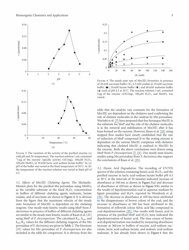
Figure 3: The variation of the activity of the purified enzyme (a)with pH and (b) temperature. The reaction solution 1 mL contained“5 μg of the enzyme (specific activity 4 IU/mg), 100 μM H2O2,100 μM MnSO4 in 50 mM lactic acid sodium lactate buffer.” In (a)pH of the buffer was varied at the fixed temperature of 30◦C. In (b)the temperature of the reaction solution was varied at fixed pH of4.5.
3.1. Effects of Mn(III) Chelating Agents. The Michaelis-Menten plots for the purified Mn peroxidase using MnSO4
as the variable substrate at the fixed H2O2 concentrationin buffers of different chelating agents malonate, lactateoxalate, and of succinate are shown in Figure 4. It is obviousfrom the figure that the maximum velocity of the steadystate formation of Mn(III) is dependent on the chelatingreagents. Our steady-state kinetic results using MnP from F.durissimus in presence of buffers of different chelating agentsare similar to the steady state kinetic results of Kuan et al. [21]using MnP of P. chrysosporium. The calculated Km, kcat, andkcat/Km values for the different chelating reagents of the Mnperoxidase of F. durissimus are given in Table 2. The literature[21] values for Mn peroxidase of P. chrysosporium are alsoincluded in the table for comparison. It is obvious from the
0.25
0.2
0.15
0.1
0.05
00 20 40 60 80 100 120 140 160
[S] (mM)
v(µ
mol
e/m
in)
Figure 4: The steady-state rate of Mn(III) formation in presenceof 20 mM succinate buffer (X), 0.5 mM oxalate in 10 mM succinatebuffer (�), 20 mM lactate buffer (�), and 40 mM malonate buffer(�) each of pH 4.5 at 30◦C. The reaction solution 1 mL contained5 μg of the enzyme (4 IU/mg), 100 μM H2O2, and MnSO4 wasvaried.
table that the catalytic rate constants for the formation ofMn(III) are dependent on the chelators used confirming therole of chelator molecules in the catalysis by Mn peroxidase.Wariishi et al. [7] have proposed that free hexaaqua Mn(II) isthe substrate for MnP and the role of the chelator moleculesis in the removal and stabilization of Mn(III) after it hasbeen formed on the enzyme. However, Kuan et al. [21] usingstopped flow studies have clearly established that the rateof reduction of MnP compound II to the resting enzyme isdependent on the various Mn(II) complexes with chelatorsindicating that chelated Mn(II) is oxidised to Mn(III) bythe enzyme. Both the above conclusions were drawn usingMnP from P. chrysosporium [7, 21]. Our steady-state kineticstudies using Mn peroxidase from F. durissimus also supportthe conclusions of Kuan et al. [21].
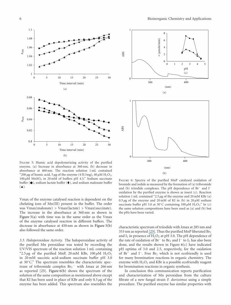
3.2. Humic Acid Degradation. The recording of UV/VISspectra of the solution containing humic acid, H2O2, and thepurified enzyme in lactic acid-sodium lactate buffer pH 4.5at 30◦C at the intervals of 30 minutes indicated increase ofabsorbance at 360 nm as shown in Figure 5(a) and decreaseof absorbance at 450 nm as shown in Figure 5(b) similar tothe results of depolymerisation coal in aqueous medium bylignin peroxidase and H2O2 reported by Wondrack et al.[31]. The decrease in absorbance at 450 has been attributedto the disappearance of brown colour of the coal, and theincrease in absorbance at 360 has been attributed to theformation of yellowish colour fulvic acid like compound incoal depolymerization [31]. Our results with humic acid inpresence of the purified MnP and H2O2 have indicated thedepolymerization of humic acid. The time course of humicacid depolymerization was studied by measuring absorbanceincrease at 360 nm in buffers of succinic acid-sodium suc-cinate, lactic acid-sodium lactate, and malonic acid-sodiummalonate. It has already been shown in Figure 4 that the
6 Bioinorganic Chemistry and Applications
Time interval (min)
1
1.02
1.04
1.06
1.08
1.1
5 10 15 20 25 30
A36
0
0
(a)
0.8
0.82
0.84
0.86
0.88
A45
0
Time interval (min)
0 5 10 15 20 25 30
(b)
Figure 5: Humic acid depolymerizing activity of the purifiedenzyme. (a) Increase in absorbance at 360 nm; (b) decrease inabsorbance at 460 nm. The reaction solution 1 mL contained“200 μg of humic acid, 5 μg of the enzyme (4 IU/mg), 40 μM H2O2,100 μM MnSO4 in 20 mM of buffers pH 4.5.” Sodium succinatebuffer (�), sodium lactate buffer (�), and sodium malonate buffer(�).
Vmax of the enzyme catalysed reaction is dependent on thechelating ions of Mn(III) present in the buffer. The orderwas Vmax(malonate) > Vmax(lactate) > Vmax(succinate).The increase in the absorbance at 360 nm as shown inFigure 5(a) with time was in the same order as the Vmaxof the enzyme catalysed reaction in different buffers. Thedecrease in absorbance at 450 nm as shown in Figure 5(b)also followed the same order.
3.3. Haloperoxidase Activity. The haloperoxidase activity ofthe purified Mn peroxidase was tested by recording theUV/VIS spectrum of the reaction solution 1 mL containing“2.5 μg of the purified MnP, 20 mM KBr, 100 μM H2O2
in 20 mM succinic acid-sodium succinate buffer pH 3.0at 30◦C.” The spectrum resembles the characteristic spec-trum of tribromide complex Br3
− with λmax at 266 nmas reported [23]. Figure 6(b) shows the spectrum of thesolution of the same composition as mentioned above exceptthat KI has been used in place of KBr and only 0.5 μg of theenzyme has been added. This spectrum also resembles the
1
0
AB
S
300 400
0 1 2 3 4 50
2
4
6
8
v(µ
mol
e/m
in)
pH
(c)
(nm)
(a)
1
0
AB
S
300 400
(nm)
(b)
Figure 6: Spectra of the purified MnP catalysed oxidation ofbromide and iodide as measured by the formation of (a) tribromideand (b) triiodide complexes. The pH dependence of Br− and I−
oxidation by the purified enzyme is shown as insert (c). Reactionsolution 1 mL contained “2.5 μg of the enzyme and 20 mM KBr (a)0.5 μg of the enzyme and 20 mM of KI in (b) in 20 μM sodiumsuccinate buffer pH 3.0 at 30◦C containing 100 μM H2O2.” In (c)the same solution compositions have been used as (a) and (b) butthe pHs have been varied.
characteristic spectrum of triiodide with λmax at 285 nm and353 nm as reported [23]. Thus the purified MnP liberated Br2
and I2 in presence of H2O2 at pH 3.0. The pH dependence ofthe rate of oxidation of Br− to Br2 and I− to I2 has also beendone, and the results shown in Figure 6(c) have indicatedpH optima of 3.0 and 2.5, respectively, for the oxidationof Br− and I−. Free Br2 which is not ecofriendly is usedfor many bromination reactions in organic chemistry. Theenzyme with H2O2 and KBr is a possible ecofriendly reagentfor bromination reactions in organic synthesis.
In conclusion this communication reports purificationand characterization of Mn peroxidase from the culturefiltrate of a new fungal strain F. durissimus using a simpleprocedure. The purified enzyme has similar properties with

Bioinorganic Chemistry and Applications 7
the MnP of P. chrysosporium, an extensively studied MnP.The enzyme depolymerises humic acid and shows haloper-oxidase activity for the oxidation of Br− and I− at low pH.
Acknowledgments
The authors are thankful to UGC, New Delhi for the financialsupport through it major research Project no. F. 31—94/2005(SR) dated March 30, 2006 under which this work has beendone. M. Yadav is thankful to UGC New Delhi for the awardof Dr. D. S. Kothari Fellowship. S. K. Singh is thankful tothe Department of Chemistry, DDU Gorakhpur Universityfor the award of a UGC-DSA Fellowship for meritoriousstudents.
References
[1] M. Kuwahara, J. K. Glenn, M. A. Morgan, and M. H. Gold,“Separation and characterization of two extracelluar H2O2-dependent oxidases from ligninolytic cultures of Phane-rochaete chrysosporium,” FEBS Letters, vol. 169, no. 2, pp.247–250, 1984.
[2] A. Hatakka, “Lignin-modifying enzymes from selected white-rot fungi: production and role in lignin degradation,” FEMSMicrobiology Reviews, vol. 13, no. 2-3, pp. 125–135, 1994.
[3] F. Pelaez, M. J. Martinez, and A. T. Martinez, “Screening of68 species of basidiomycetes for enzymes involved in lignindegradation,” Mycological Research, vol. 99, no. 1, pp. 37–42,1995.
[4] E. M. Silva, S. F. Martins, and A. M. F. Milagres, “Extractionof manganese peroxidase produced by Lentinula edodes,”Bioresource Technology, vol. 99, no. 7, pp. 2471–2475, 2008.
[5] H. Tanaka, K. Koike, S. Itakura, and A. Enoki, “Degradationof wood and enzyme production by Ceriporiopsis subvermis-pora,” Enzyme and Microbial Technology, vol. 45, no. 5, pp.384–390, 2009.
[6] H. B. Dunford, “Horseradish peroxidase: structure and kineticproperties,” in Peroxidases in Chemistry and Biology, J. Everse,K. E. Everse, and M. B. Grisham, Eds., pp. 1–24, CRC Press,Boca Raton, Fla, USA, 1991.
[7] H. Wariishi, J. Huang, H. B. Dunford, and M. H. Gold,“Reactions of lignin peroxidase compounds I and II with vera-tryl alcohol: transient-state kinetic charcterization,” Journal ofBiological Chemistry, vol. 266, no. 31, pp. 20694–20699, 1991.
[8] T. K. Kirk and D. Cullen, “Enzymology and molecular geneticsof wood degradation by white-rot fungi,” in EnvironmentallyFriendly Technologies for the Pulp and Paper Industry, R. A.Young and M. Akthar, Eds., pp. 273–307, John Wiley & Sons,New York, NY, USA, 1998.
[9] J. K. Glenn, L. Akileswaran, and M. H. Gold, “Mn(II)oxidation is the principal function of the extracellular Mn-peroxidase from Phanerochaete chrysosporium,” Archives ofBiochemistry and Biophysics, vol. 251, no. 2, pp. 688–696, 1986.
[10] H. Wariishi, K. Valli, and M. H. Gold, “Manganese(II)oxidation by manganese peroxidase from the basidiomycetePhanerochaete chrysosporium. Kinetic mechanism and role ofchelators,” Journal of Biological Chemistry, vol. 267, no. 33, pp.23688–23695, 1992.
[11] M. Hofrichter, “Review: lignin conversion by manganeseperoxidase (MnP),” Enzyme and Microbial Technology, vol. 30,no. 4, pp. 454–466, 2002.
[12] S. S. Marwaha, R. Grover, C. Prakash, and J. F. Kennedy,“Continuous biobleaching of black liquor from the pulp and
paper industry using an immobilized cell system,” Journal ofChemical Technology and Biotechnology, vol. 73, no. 3, pp. 292–296, 1998.
[13] J. A. Bumpus, M. Tien, D. Wright, and S. D. Aust, “Oxidationof persistent environmental pollutants by a white rot fungus,”Science, vol. 228, no. 4706, pp. 1434–1436, 1985.
[14] H. S. Kwon, E. Chung, D. I. Lee, C. H. Lee, I. S. Ahn, andJ. Y. Kim, “Comparison of chemical and enzymatic emulsionpolymerization of styrene,” Journal of Applied Polymer Science,vol. 112, no. 5, pp. 2935–2941, 2009.
[15] I. Grgic, H. Podgornik, M. Berovic, and A. Perdih, “Improve-ments in the determination of manganese peroxidase activity,”Biotechnology Letters, vol. 23, no. 13, pp. 1039–1042, 2001.
[16] M. H. Gold and J. K. Glenn, “Manganese peroxidase fromPhanerochaete chrysosporium,” Methods in Enzymology, vol.161, no. C, pp. 258–264, 1988.
[17] O. H. Lowry, N. J. Rosebrough, A. L. Farr, and R. J. Randall,“Protein measurement with the Folin phenol reagent,” TheJournal of Biological Chemistry, vol. 193, no. 1, pp. 265–275,1951.
[18] Polyacrylamide Gel Electrophoresis: Laboratory Techniques,Laboratory Separation Division, Pharmacia, Uppsala, Sweden,1983.
[19] K. Weber and M. Osborn, “The reliability of molecularweight determinations by dodecyl sulfate-polyacrylamide gelelectrophoresis,” Journal of Biological Chemistry, vol. 244, no.16, pp. 4406–4412, 1969.
[20] P. C. Engel, Enzyme Kinetics: The Steady State Approach,Chapman and Hall, London, UK, 1977.
[21] I. C. Kuan, K. A. Johnson, and M. Tien, “Kinetic analysis ofmanganese peroxidase. The reaction with manganese comple-xes,” Journal of Biological Chemistry, vol. 268, no. 27, pp.20064–20070, 1993.
[22] M. Hofrichter and W. Fritsche, “Depolymerization of low-rank coal by extracellular fungal enzyme systems. I. Screeningfor low-rank-coal-depolymerizing activities,” Applied Microbi-ology and Biotechnology, vol. 46, no. 3, pp. 220–225, 1996.
[23] R. D. Libby, J. A. Thomas, L. W. Kaiser, and L. P. Hager,“Chloroperoxidase halogenation reactions. Chemical versusenzymic halogenating intermediates,” Journal of BiologicalChemistry, vol. 257, no. 9, pp. 5030–5037, 1982.
[24] C. Ruttimann-Johnson, D. Cullen, and R. T. Lamar, “Man-ganese peroxidases of the white rot fungus Phanerochaetesordida,” Applied and Environmental Microbiology, vol. 60, no.2, pp. 599–605, 1994.
[25] M. Hofrichter, K. Scheibner, I. Schneegaß, and W. Fritsche,“Enzymatic combustion of aromatic and aliphatic compoundsby manganese peroxidase from Nematoloma frowardii,”Applied and Environmental Microbiology, vol. 64, no. 2, pp.399–404, 1998.
[26] L. F. Larrondo, S. Lobos, P. Stewart, D. Cullen, and R. Vicuna,“Isoenzyme multiplicity and characterization of recombinantmanganese peroxidases from Ceriporiopsis subvermispora andPhanerochaete chrysosporium,” Applied and EnvironmentalMicrobiology, vol. 67, no. 5, pp. 2070–2075, 2001.
[27] A. Conesa, D. Jeenes, D. B. Archer, C. A.M.J.J. Van den Hon-del, and P. J. Punt, “Calnexin overexpression increases man-ganese peroxidase prouction in Aspergillus niger,” Applied andEnvironmental Microbiology, vol. 68, no. 2, pp. 846–851, 2002.
[28] Y. Iimura, S. Ikeda, T. Sonoki et al., “Expression of a genefor Mn-peroxidase from Coriolus versicolor in transgenictobacco generates potential tools for phytoremediation,”Applied Microbiology and Biotechnology, vol. 59, no. 2-3, pp.246–251, 2002.

8 Bioinorganic Chemistry and Applications
[29] N. Kanayama, S. Tohru, and K. Keiichi, “Purification andcharacterization of an alkaline manganese peroxidase fromAspergillus terreus LD-1,” Journal of Bioscience and Bioengi-neering, vol. 93, no. 4, pp. 405–410, 2002.
[30] H. Kamitsuji, Y. Honda, T. Watanabe, and M. Kuwahara, “Pro-duction and induction of manganese peroxidase isozymes ina white-rot fungus Pleurotus ostreatus,” Applied Microbiologyand Biotechnology, vol. 65, no. 3, pp. 287–294, 2004.
[31] L. Wondrack, M. szanto, and W. A. wood, “Depolymerizationof water soluble coal polymer from subbituminous coaland lignite by lignin peroxidase,” Applied Biochemistry andBiotechnology, vol. 20-21, no. 1, pp. 765–780, 1989.