CMI
17
SEE-SAW NYSTAGMUS IN CHIARI MALFORMATION TYPE I OLEH: St. Suleha Umar PEMBIMBING: Dr. dr. Batari Todja Umar, Sp.M dr. Yunita, Sp.M, M.Kes Case Report
-
Upload
umimakassar -
Category
Documents
-
view
0 -
download
0
Transcript of CMI
SEE-SAW NYSTAGMUS IN CHIARI MALFORMATION TYPE I
OLEH:St. Suleha Umar
PEMBIMBING:Dr. dr. Batari Todja Umar, Sp.M (K)
dr. Yunita, Sp.M, M.Kes
Case Report
Purpose: To report a case of 25 years old woman with see saw nystagmus as a manifestation of Chiary’s Malformation Type I
Design & Methods: A case report of a 25 year-old woman referred with involuntary movements of both eyes. Visual acuity was assessed, anterior and posterior segments were examined. The patient then underwent magnetic resonance imaging of head and brainstem.
ABSTRACT
Result: See saw nystagmus was evident. Best corrected visual acuity were 6/6 in both eyes. Anterior and posterior segments of the eye were normal. Cranial nerve testing was unremarkable. Chiari malformation was confirmed by magnetic resonance imaging. The patient was diagnosed as Chiari Malformation Type I. The patients then reffered to neurologist but no treatment was given.
Conclussion: See saw nystagmus can be found in Chiary Malformation Type I. Treatment of Chiari Malformation Type I depends on the form, severity and associated symptoms. Regular monitoring, medications and surgery are treatment options. In some cases, no treatment is needed.
ABSTRACT
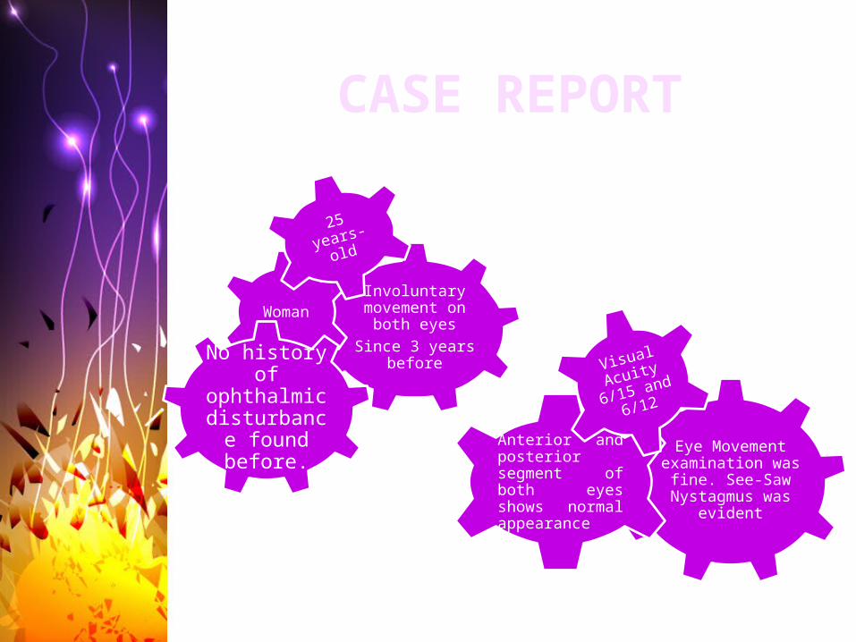
Involuntary movement on both eyes
Since 3 years before
Woman
25 years-old
Eye Movement examination was fine. See-Saw Nystagmus was
evident
Anterior and posterior segment of both eyes shows normal appearance
Visual
Acuity
6/15 and
6/12
No history of
ophthalmic disturbance found before.
CASE REPORT
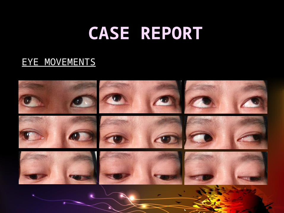
EYE MOVEMENTS
CASE REPORT

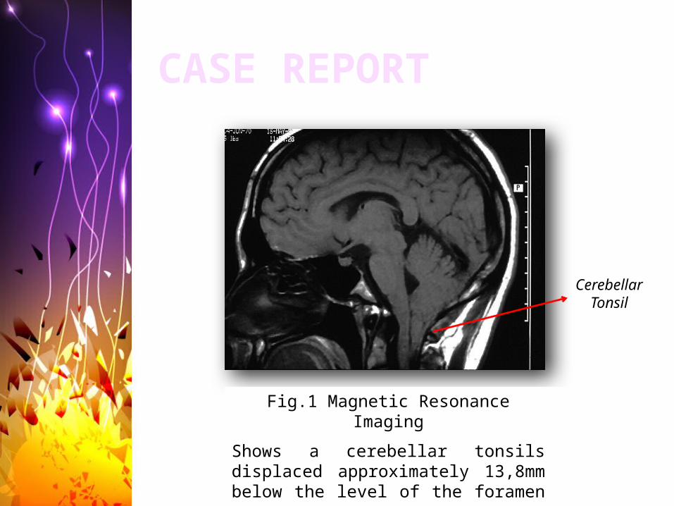
Fig.1 Magnetic Resonance Imaging
Shows a cerebellar tonsils displaced approximately 13,8mm below the level of the foramen magnum.
CASE REPORT
Cerebellar Tonsil
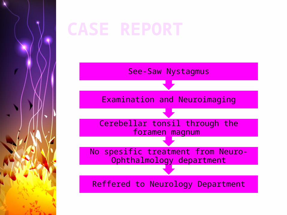
Reffered to Neurology Department
No spesific treatment from Neuro-Ophthalmology department
Cerebellar tonsil through the foramen magnum
Examination and Neuroimaging
See-Saw Nystagmus
CASE REPORT
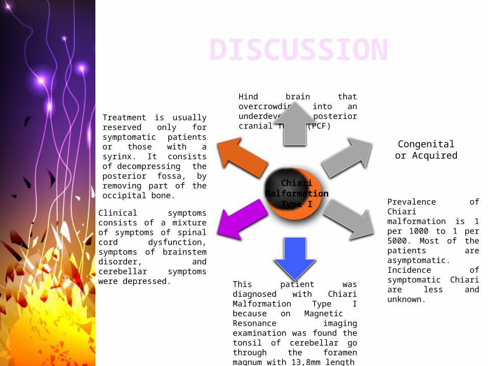
Clinical symptoms consists of a mixture of symptoms of spinal cord dysfunction, symptoms of brainstem disorder, and cerebellar symptoms were depressed.
Prevalence of Chiari malformation is 1 per 1000 to 1 per 5000. Most of the patients are asymptomatic. Incidence of symptomatic Chiari are less and unknown.
Hind brain that overcrowding into an underdeveloped posterior cranial fossa (PCF)
Congenital or Acquired
Chiari Malformation
Type I
This patient was diagnosed with Chiari Malformation Type I because on Magnetic Resonance imaging examination was found the tonsil of cerebellar go through the foramen magnum with 13,8mm length
Treatment is usually reserved only for symptomatic patients or those with a syrinx. It consists of decompressing the posterior fossa, by removing part of the occipital bone.
DISCUSSION
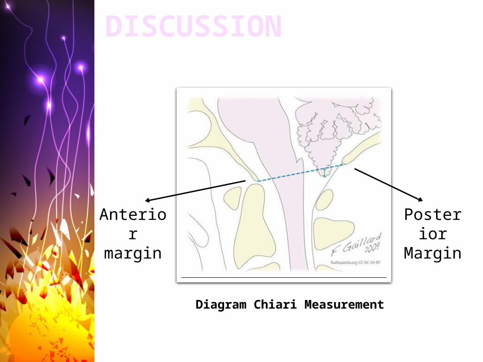
Diagram Chiari Measurement
Anterior
margin
Posterior
Margin
DISCUSSION
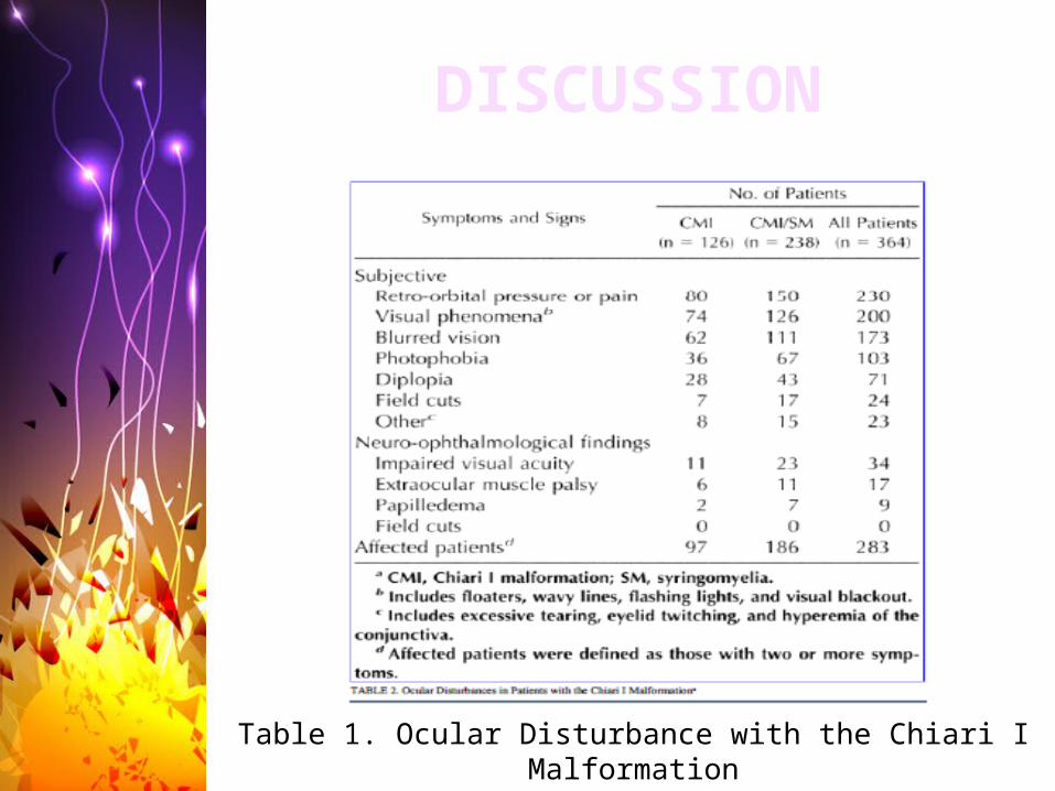
Table 1. Ocular Disturbance with the Chiari I Malformation
DISCUSSION
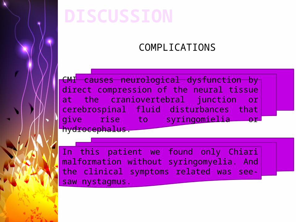
DISCUSSIONCOMPLICATIONS
CMI causes neurological dysfunction by direct compression of the neural tissue at the craniovertebral junction or cerebrospinal fluid disturbances that give rise to syringomielia or hydrocephalus.
In this patient we found only Chiari malformation without syringomyelia. And the clinical symptoms related was see-saw nystagmus.
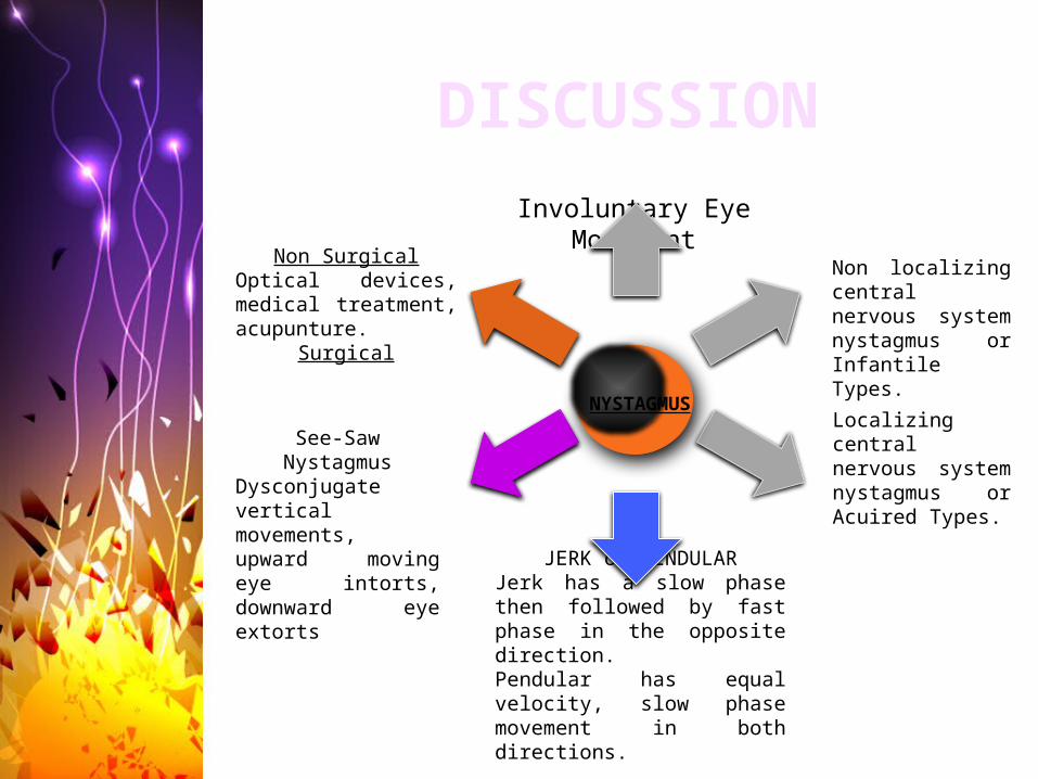
Localizing central nervous system nystagmus or Acuired Types.
Involuntary Eye Movement
Non localizing central nervous system nystagmus or Infantile Types.
JERK or PENDULARJerk has a slow phase then followed by fast phase in the opposite direction.Pendular has equal velocity, slow phase movement in both directions.
See-Saw Nystagmus
Dysconjugate vertical movements, upward moving eye intorts, downward eye extorts
Non SurgicalOptical devices, medical treatment, acupunture.
Surgical
DISCUSSION
NYSTAGMUS

See-saw nystagmus has been seen with tumors of the parasellar region and diencephalon, brain-stem vascular lesions, syringobulbia, and after trauma. And very rare found in CM1
DISCUSSION
The treatment for nystagmus in this patient was not conducted simply because the patient was not complaining of oscilopsia since the nystagmus was only appear on lateral extreme gaze
DISCUSSION
If the patient is clinically asymptomatic and no syringomyelia, should be observed first as we found in this patient.
Therapy of choice is continued clinical monitoring and surgical intervention.
Surgery is performed craniectomy of posterior fossa and cervical laminectomy for decompression.
The patient we reffered to neurology and no treatment was given.The patient was satisfied with prescribed glasses and no other complain.
Regular monitoring, medications and surgery are treatment options. In some cases, no treatment is needed.
2
3
See saw nystagmus can be found in Chiary Malformation Type I
1
Treatment of Chiari Malformation Type I depends on the form, severity and associated symptoms.
CONCLUSSION

THANK YOU