chest x ray equipment for use in TB prevalence surveys
57
WORKING DOCUMENT ON CHEST X RAY EQUIPMENT FOR USE IN TB PREVALENCE SURVEYS Prepared by KNCV Tuberculosis Foundation September 2008
-
Upload
khangminh22 -
Category
Documents
-
view
0 -
download
0
Transcript of chest x ray equipment for use in TB prevalence surveys

WORKING DOCUMENT ON CHEST X RAY EQUIPMENT
FOR USE IN TB PREVALENCE SURVEYS
Prepared by KNCV Tuberculosis Foundation September 2008

1
Foreword This document is a working document prepared to provide an overview of issues regarding chest X-ray equipment for use in TB prevalence surveys. The current information was collected by the research unit of KNCV Tuberculosis Foundation with input from external experts. A group of countries is preparing to carry out a TB prevalence survey in the coming years. When providing technical assistance for TB prevalence surveys we received questions from countries about chest X-ray equipment. Since there was no document with information available, we collected our own experience and experience form external experts. The information is intended to provide an overview to guide the choice of X-ray equipment for a TB prevalence survey. It assembles information on the type of X-ray equipment used in previous surveys and intends to give an overview of the type of X-ray machines available for use in TB prevalence surveys. It also lists some experiences that countries had during their surveys with different types of machines. It is a working document and by no means a fixed document so it will be updated when additional information on other surveys becomes available. The provided information in the annex on the X-ray equipment available from different companies is by no means meant to be an advice for a specific brand of equipment but is an overview of the information that is currently known to us. When more information is available from other companies with similar equipment this can be added to the document. Additional information can be send to KNCV Tuberculosis Foundation ([email protected]). KNCV Tuberculosis Foundation 9 October 2008

2
FROM: Assessing tuberculosis prevalence through population based surveys (pages 26-28) by WHO 2007 Type of chest radiograph. For prevalence surveys, the full-size posteroanterior film is recommended. With each examination, the film must be correctly labelled with the survey number and must be of good quality. X-ray reading. The film is ideally read by two independent readers. In case of discrepancy between the first two readers a third reading is obtained from an “umpire” reader. 5.2.3 Chest radiography equipment In prevalence surveys, the X-ray unit is usually mounted onto a mobile van (vehicle) for radiological examination. Difficult terrain without passable roads may pose many problems for these units. In such cases, it may be possible to establish a centre to which all persons will be directed for X-ray. If some participants do not wish to take the time to have the examination, special arrangements must be made to transport these patients. Bringing back the films to the main centre for processing, arranging for independent reading, and preserving the films for future reference may also be difficult. The use of chest X-rays is complicated by the high cost of the mobile chest X-ray equipment, logistical difficulties (accessibility of field sites by trucks, power supply, field robustness of the chest X-ray equipment, maintenance and repair), and the demands on human capacity, in particular the need for experienced clinicians in the field teams who can read chest X-rays. Issues to consider when procuring chest X-ray equipment are the speed of operation (patient throughput and film development time), image quality, weight and size of equipment, power requirements, and safety (radiation dose). Three different types of chest X-ray systems are available: (1) mass miniature radiography; (2) conventional X-ray (with conventional or automated developing); and (3) digital X-ray (computed radiography, direct digital systems, or slot-scan systems). Mass miniature radiography is no longer recommended since it has a large power requirement (three-phase, 30 kilowatt [kW] generator), gives significant radiation exposure to the patient, and produces low-quality images because of their small size. Fluoroscopy is not recommended for safety reasons. Furthermore, the lack of film complicates quality assurance. Conventional chest X-ray systems with a conventional developer are relatively inexpensive ($30 000–$50 000); can be transported or built in a small truck; are simple, robust, and easy to operate; and can often be maintained locally. Such systems use a condenser system running on a 3 kW generator. However, operation and the developing process are slow and the quality of film development is critical—a serious disadvantage in TB prevalence surveys. Furthermore, the developing process needs conditioned temperature (32°C). Conventional systems with an automated developer have advantages over conventional systems with a conventional developer. The developing process is faster and technical errors are less critical. Small developers are available for less

3
than $10 000. But these systems require calibration and maintenance, are still relatively slow, and have no instant quality check. Conventional systems with a conventional developer or automated developer require transport of films for centralized rereading. Digital X-ray systems are increasingly available for mobile use. They do away with the need for films, developing machines, and development solutions, and thus simplify the logistics. The X-ray images can be sent by Internet to a central place where they can be assessed by X-ray readers. In computed radiography, a phosphor plate is irradiated by a conventional chest X-ray system, and the phosphor image is converted into a digital image by a digital plate reader. Any conventional generator can be used, and the plates are reusable. There is no film developing and a quick quality check of the image is possible. Furthermore, the images are stored electronically and can be easily sent by Internet to a central place for rereading. Digital plate readers are relatively expensive ($30 000–$50 000), generally sensitive to dust and (probably) humidity, and large and must be transported by air-conditioned truck. But highly field-robust tabletop plate readers developed for military use have recently become available at reasonable cost. In completely digital systems the X-ray image is directly converted into a digital image by a transducer that takes the place of the image plate. There is no need for film developing or plate readers, and the image quality can be checked in real time. The transducers used are, however, expensive and highly sensitive to jarring. Mobile systems based on full-size transducers are therefore highly expensive (around $400 000) and vulnerable if transported over bumpy roads. Among other things, they require air suspension. The feasibility of using digital systems in prevalence surveys should still be assessed. Slot-scan systems present an alternative that is increasingly being used in mobile X-ray. A small transducer moves in slots across the patient’s chest along with a narrow X-ray beam and the image is computed from the digital images for each slot. These systems have the same advantages as those with full-size transducers but are less vulnerable. Image quality depends on speed of scanning. The fastest scanners produce the best images but are also expensive (up to $500 000). Slow scanners are less expensive. Slot-scan systems, particularly the moving parts of the X-ray generator and the transducer, are still relatively vulnerable and repair often requires specific expertise. Most mobile digital systems use battery-operated X-ray generators and have relatively low power requirements (two-phase, 10 kW generator for the system and air conditioning). An important factor in deciding which chest X-ray system should be procured is the availability of service contracts in the country.

4
Chest x-rays used in TB prevalence surveys
Table 1 gives an overview of the type of machines used or planned to be used in different countries that are planning or have conducted a TB
prevalence survey. More detailed documentation for each type of
equipment is provided in the annexes.
The X-ray equipment consists of different parts: 1) Car/Truck: To make the equipment mobile in most cases X-ray
equipment is installed in a truck with shielding that fulfils the
requirements of the radiation board. The equipment can also be installed in a 20 feet container that can be transported on any truck.
A third option is portable equipment transported in the back of a
pick up that will be set up in locality. 2) X-ray generator: produces the X-ray beam, this can be digital or
conventional
3) X-ray imager: makes an image out of the beam
4) X-ray reader; visualizes the image for reading by the radiologist. This can be digital or conventional. Digital systems can be linked to
hard disks to store all the images. Images can be send elsewhere
for reading or cross reading.
The following combinations are mostly distinguished
a) conventional: both X-ray generator and X-ray imager are
conventional
b) direct digital: fully digital, both X-ray generator and X-ray imager
are conventional c) indirect digital: conventional X-ray generator and digital X-ray
imager (computed radiography)
Most countries decided on X-ray equipment that was mounted onto trucks.
In Vietnam they used a combination of 3 fully digital systems in a truck
and 2 MMR systems. In the prevalence survey in Cambodia they used a combination of and X-ray car and portable equipment that can be
mounted on the back of a pick up truck. Portable X-ray equipment is
cheaper than a mobile X-ray truck but can not be used in all countries due
to the radiation regulations in the countries overseen by the radiation board. Before a decision on which type of X-ray to use is made the radiation board needs to be consulted. They will also have to approve the
truck to certify it fulfils the countries radiation requirements.
Apart from the X-ray equipment listed in table 1, there is a range of other
companies that make equipment. Information of equipment of other companies has been added in the annex were available. The list is not
complete and there will be other companies that produce comparable
equipment but the presented information is intended to provide an
overview of the type of equipment that is available.
.

5
Table 1 Type of machines used in countries carrying out TB prevalence surveys
country Survey
status
Type Equipment Platform Number
units
Vietnam Finished* X-ray system built
into truck
Pro-Scan 7000 integrated system
built into Kamaz truck
Direct digital, slot scan,
fully digital
3
Kenya–
Nyanza
Finished* X-ray system built
into truck X-ray: Practix 400, Philips with Bucky Diagnost VE - wall stand And Agfa automatic processor Curix 60 with Aga ID printer Camera type 8400/300 Truck: Isuzu NPR 66, 4 ton truck
Conventional generator
(practix400 with bucky)
with conventional
developer (AGFA)
1
Tanzania Planned X-ray system built
into truck
Philips Eleva S
plus practix 400 plus view forum
assembled into truck
Indirect digital:
Conventional X-ray
generator (practix400)
with computed
radiography system for
imaging (Eleva-S) and
viewing forum
2
Cambodia/
Myanmar
Finished Portable equipment
carried on back of a
pick up
Portable X-ray Unit (PX-20HF
with stationary stand (PS-1-III)
both from Adore Medical
Corporation with automatic X-ray
film processor (EKOMAT 21 ELK
Corp. Japan) and portable
generator (EX28 HONDA Japan)
Conventional generator
with conventional
developer
Bangladesh Ongoing – no X-ray used
Eritrea Finished – no X-ray used * field data collection finished, official report of study being awaited

6
Table 1 notes: 1. Prices estimates per truck in the above ranged from 185,000-250,000 us dollars. For current budgeting an estimate
of 250,000 usd per truck is recommended. 2. Prices can vary locally due to presence of company representatives and possibilities of providing in-country service.
The best option should be assessed for each country.
3. Prices also depend largely of what is all included, i.e. plates, transport boxes, laptops for viewing etc. This should be well negotiated.
4. An extended viewer for definite diagnosis (higher resolution than workstation viewer in the truck) is definitely not standard included in the price and should be considered in addition.
5. A large part of the price consists of the price of the truck and prices of trucks vary. It is important the truck can
access all the roads in the country and quality of the roads varies per country. Truck price depends on the need for 4- wheel or all-wheel drive, air suspension and of course availability of local made trucks.
6. In the negotiations with the companies the conditions of the service contract are very important, how fast can spare
part be delivered, when is on the ground technical assistance provide, survey staff should be trained in use and maintenance of the equipment and basic repairs, if they can not repair it how fast will it be repaired or an alternative
machines delivered if not possible to repair (a max time should be set for this) etc.
7
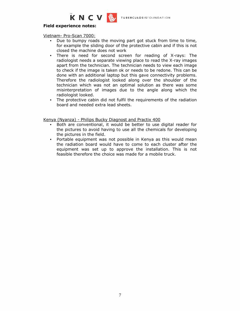
Field experience notes:
Vietnam- Pro-Scan 7000: • Due to bumpy roads the moving part got stuck from time to time,
for example the sliding door of the protective cabin and if this is not
closed the machine does not work
• There is need for second screen for reading of X-rays: The radiologist needs a separate viewing place to read the X-ray images
apart from the technician. The technician needs to view each image
to check if the image is taken ok or needs to be redone. This can be done with an additional laptop but this gave connectivity problems.
Therefore the radiologist looked along over the shoulder of the
technician which was not an optimal solution as there was some misinterpretation of images due to the angle along which the
radiologist looked.
• The protective cabin did not fulfil the requirements of the radiation
board and needed extra lead sheets.
Kenya (Nyanza) - Philips Bucky Diagnost and Practix 400 • Both are conventional, it would be better to use digital reader for
the pictures to avoid having to use all the chemicals for developing the pictures in the field.
• Portable equipment was not possible in Kenya as this would mean
the radiation board would have to come to each cluster after the equipment was set up to approve the installation. This is not
feasible therefore the choice was made for a mobile truck.

8
How to choose?
The specifications that are important to take into account when choosing X-ray equipment are the following:
X-ray generator:
• speed of recharge of condenser as this determines the throughput • robustness
• digital or conventional, both is possible
• size of generator needed • size & weight (needs to fit in a truck)
X-ray imaging system • output, should be sufficient to reach your targeted sample size per
day � at least 40 pictures per hour
• robustness
• preferably digital to avoid need for chemicals
Truck/transport system: • usable for all areas of the survey, all road types � 4 wheel/all
wheel drive
• availability of spare parts • weight should be suitable for weight of equipment
• size of body of truck to fit all equipment
• Depending on the type of equipment and the road condition in the country there might be a need for a truck with air suspension to
reduce shocks for the equipment. Air suspended trucks are
expensive and will considerably increase the cost.
Power issues:
• not all field sites will have reliable electricity therefore a generator
is needed, the choice depends on the power requirements of the X-ray equipment but the generator needs to be movable and not too
large otherwise a second truck will be needed to transport it.
• Some systems require a high KW and therefore a 3-phase current
when using the power network and a large generator (up to 30 KW) if there is no reliable power supply. This is an important
disadvantage since 3-phase may not be available in some clusters
and large generators are expensive (buy or rent), bulky and consume lots of fuel. An indication of size & weight: a generator of
up to 10 KW can be lifted relatively easily and has to size of a big suitcase, whereas a 30 KW generator is heavy and needs a separate trailer.
Overall:
• service contract
One of the most important specifications is the service contract provided with the equipment, what kind of training is offered
on operating and maintaining the equipment, what assistance is given when a breakdown occurs, are spare parts available in
the country, who pays when someone has to be flown in, what is the maximum time for repairs to be carried out and will a replacement machine be provided if repairs take longer etc.

9
Appendix 1 Philips Bucky Diagnost

10

11
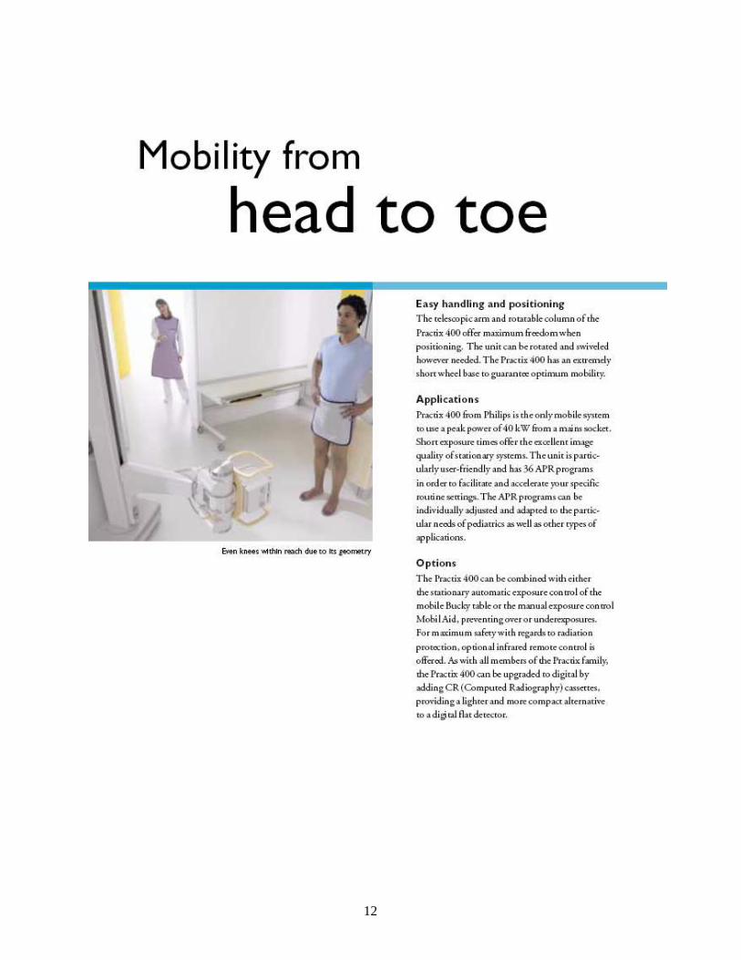
Appendix 2 Practice 400
12

13

14

15

16

17
Appendix 3 AGFA CP1000 processor

18

19
Appendix 4 Philips Elvira

20

21

22

23

24

25

26

27

28

29

30

31

32

33

34

35
Appendix 5 Kodak Orex ACL4
LowLowLowLow----dose digital scanning fluoroscope dose digital scanning fluoroscope dose digital scanning fluoroscope dose digital scanning fluoroscope ProScanProScanProScanProScan----7000 7000 7000 7000 ®®®® (DRAU (DRAU (DRAU (DRAU----01010101---- "AMICO" ) "AMICO" ) "AMICO" ) "AMICO" )
To record X-rays that have been sent through the patient's body, fluoroscope ProScan-7000® applies a silicon line detector. The detector is about 550 mm long. For X-raying of lungs, the detector moves horizontally along the thorax, simultaneously with the fan-shaped beam that is formed by the slot diaphragm. . To this end, both the detector and the slot diaphragm are fastened onto the rod rotating around the focal radiation spot on the anode. The rod is set in motion by a microstep motor. The company has developed a new type of protective cabin made of up-to-date composite materials. The protective cabin reduces X-ray exposure of the personnel and so can safely be placed close to the doctor's workplace, even in closed areas such as mobile fluoroscopic vans. The X-ray tube with the focus 0.3 mm is energized by a medium frequency X-ray
generator. The fluorograph includes two workbenches – the radiologist's WB and the assistant's WB. The radiologist runs the fluoroscope from the 17" console on the basis of a PC with a touch screen. Easy-to-use ''floating'' menus allow to enter patient's data, adjust fluoroscope settings and control resulting X-ray images. The doctor's WB is supplied with a professional medical 20.3" TFT-monitor EIZO (Japan).
X-raying can go in two modes: prophylactic mode (that one is recommended for examining normal patients) and diagnostic mode. The prophylactic mode uses a considerably lower dose (about 200µR), smaller image dimensions (4-fold, which allows to save space on DVD) and the maximum possible image contrast. Spatial resolution in this case is reduced up to 2 pairs of lines per mm. In case of lesion detection during prophylaxis, the doctor is recommended to switch over to the diagnostic mode and take the required number of images. The software used at both WB's allows to control the fluoroscope and process the images received from ProScan. For archiving X-ray images, digital video disks

36
(DVD's) are used with the capacity 3,500 images each. . If necessary, a hard copy can be printed on a professional medical printer - both on paper or a film. For printing out periodic reports, an office laser printer is also supplied. The performance of the fluoroscope is demonstrated in the Video clip showing performance of the digital fluoroscope ProScan-7000® (11,6 Mb) .
Advantages of ProScan-7000®
• The silicon linear detector The silicon linear detector The silicon linear detector The silicon linear detector does not require any maintenance. The spatial resolution in the
patient's plane is 3.2 pairs of lines per mm and contrast sensitivity not less than 1%. High detective efficiency allows to obtain a direct image at the low dose of 400 µR.
• The protective cabin The protective cabin The protective cabin The protective cabin with the absorption coefficient equivalent to 1.2 mm lead shield reduces X-ray exposure of the personnel practically to the natural background level.
• The medium frequency XThe medium frequency XThe medium frequency XThe medium frequency X----ray generator ''AMICO'' ray generator ''AMICO'' ray generator ''AMICO'' ray generator ''AMICO'' can be connected to one-phase mains at 220 V ± 10% and resistance up to 1 Ohm. Most available fluoroscopes require three-phase mains at 380 V ± 10% with resistance no more than 0.3 Ohm.
• Complete equipment of the radiologist's and assistant's WB's. Complete equipment of the radiologist's and assistant's WB's. Complete equipment of the radiologist's and assistant's WB's. Complete equipment of the radiologist's and assistant's WB's. This is a unique professional computer set that is characterized by efficient production and guarantees patients' examinations in maximally comfortable conditions.
• The ''ProScan''The ''ProScan''The ''ProScan''The ''ProScan'' software software software software is in compliance with the international protocol DICOM-3.0, and so can, if necessary, be integrated into any medical information system. . The program has been developed in close cooperation with radiologists; therefore apart from standard formalized protocols it also contains templates of periodic reports. The resources of the program as for image processing with special filters are practically unlimited. Images are archived onto the digital video discs with the capacity 4,700 Mb (about 1,000 images). For the time being, this is the most reliable way of data storage. The Demo version of ProScan is available to registered users of our website.

37
Mobile Digital Fluoroscopic Room onMobile Digital Fluoroscopic Room onMobile Digital Fluoroscopic Room onMobile Digital Fluoroscopic Room on Motor Vehicles ZIL Motor Vehicles ZIL Motor Vehicles ZIL Motor Vehicles ZIL----5301 ЕО ("Bychok") and 5301 ЕО ("Bychok") and 5301 ЕО ("Bychok") and 5301 ЕО ("Bychok") and Landrover КАМАZLandrover КАМАZLandrover КАМАZLandrover КАМАZ----43114431144311443114
Here we present the most recent product of RentgenProm, a mobile digital fluoroscopic room that is mounted on chassis of motor vehicles ZIL-5301 ЕО "Bychok" and KAMAZ-43114. The chassis were not chosen at random ZIL-5301 ЕО is an up-to-date motor vehicle with high characteristics and due comfort. "Bychok" had originally been developed for driving both in urban conditions and on earth roads in the countryside. It has a diesel engine, gasoline consumption being 12 litres / 100 km.
If the fluoroscopic room is to be used on earth roads, the best way out is to drive the landrover КАMАZ-43114. A modern van, it is characterized by excellent heat insulation and muffling properties. The rapid heating system allows to warm the van and the equipment thoroughly and start to work fast (about 30 min.) even when the atmospheric temperature outside the van is far below zero (up to -40°C).
High ceilings (2,16 m) and a convenient placement of the equipment make work comfortable throughout the day. Fluoroscope ProScan-2000® is placed at the front of the van. The entrance and the exit to the room are through 2 doors, which increases the capacity of the X-ray considerably. Up to three patients can simultaneously dress or undress in the medical room (1). For cold season, there is a heat curtain over each door. Every time when a patient is in the medical room, the bactericidal oscillator is working. The room for the personnel is partitioned off the medical room with a transparent polycarbonate shield. The personnel room is supplied with:
• Radiologist’s workbench (2); • Wardrobe; • Drawer (4); • Sofa; • Air conditioning (3); • Mini-fridge (over the AC).

38
Appendix 6 Kodak Orex ACL4

39
40
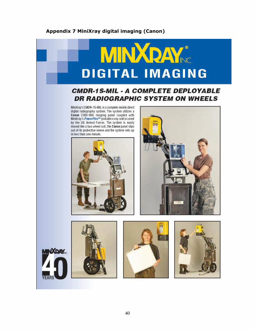
Appendix 7 MiniXray digital imaging (Canon)
41

42
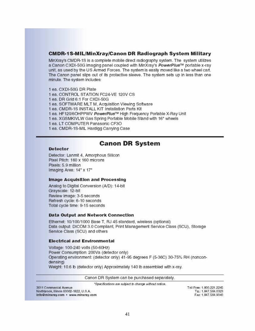
Annex 8 Odelca-DR unit The concept offered by Delft allows “anyone to make a chest X-ray (CXR) image almost anywhere” and “(re)read the image wherever you have a trained reader”. Additionally dedicated TB viewer with Computer Aided Diagnose (CAD) (Work in Progress) will allow non formally trained radiographers/ radiologists to make a (first) diagnosis. In practice, a self supporting unit in the field has the ability to make, store, and diagnose CXR’s. With rapidly expanding GSM networks around the world it is possible to sent these CXR’s from virtually any venue to a diagnostic centre, where these images are interpreted and from which the diagnosis can be relayed immediately to the unit. The unit consists in a shielded standard 20 feet container that includes the following amenities: • X-Ray unit . As successor to the famous analogue Odelca (of which more than 25,000
were sold), the present Odelca-DR has unique features. Slot-scan technology first developed by Delft on the Amber system provides unsurpassed image quality with ultra low patient dose. Specially designed for more remote and rural areas, the unit is composed of very few components. This makes it easy to maintain. Remote diagnosis indicates any component to be replaced or serviced.
• ROX, VPX/TB viewer with CRRS and CAD. The technology to store, diagnose and
sent images over a low band with -made by sister company ROGAN-Delft- makes it a complete package.
• Transport Because of its limited size, the unit can be transported by locally available
trucks. Considering the cost of a truck is more than half the price of a mobile unit and is only used for transporting the unit this (semi) mobile concept is far more economical. With a sophisticated - yet simple to operate - lifting device, the unit can be installed and be up and running in less then 2 hours. In this way, any unit can serve both as a stationary or mobile entity.
• Energy supply. With the latest technology in battery and inverters, the unit is capable of
making - storing and sending more then 100 images without the need of external power. The on board battery can be charged with ‘whatever electric power source is available’, be it of 220 VAC or other, a generator, solar, wind or otherwise generated energy.

43
As already discussed one should not compare our envisaged solution with stand-alone modalities as offered by several companies. We offer a complete solution in X-Ray screening where we provide the possibility to send your images even over a low bandwidth (GPRS). It certainly is interesting for us to participate in prevalence studies, since this would be of a great assistance to further develop our CAD system. Obviously the CAD system will in time be integrated into the systems in and will be done free of charge. We would ask for the data to become available to increase our database to be used for further development of the CAD. General The digital thorax system “Odelca DR” is specially developed to meet the growing demand of Tuberculosis and Labour screening. Tuberculosis is a rapidly growing worldwide decease. The newest technology is used in the development of the Odelca DR. For examples, the detector makes use of the CMOS technology and the generator has a frequency of 100 kHz. The Odelca DR draws out by the very low dose for the client with a very high image quality. This makes the system very suitable for screening purposes. The acquisition software is specially made for the Odelca DR and is guaranteed very user-friendly. A diagnosis can immediately be done on the diagnostic monitor on the spot. Sending these images to a central location requires no user intervention and an image is typically available anywhere in the world just 20 seconds after acquisition.
44
All components of the DR Odelca are engineered in such a way that the highest possible quality is ensured. The materials used are of high quality and solid material, which makes it possible to place the Odelca DR in a mobile unit. The user-friendliness of the system makes it possible to reach a high throughput (300 /day) Operation The operation of the Odelca DR is very user friendly. Because the DR Odelca has a U-shaped frame, the positioning of the patient is very easy. The motor supported high / low movement is positioned on the main unit and can be easily operated.

45
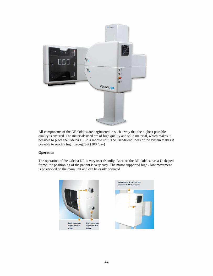
The main unit has also the possibility to collimate. In the vertical direction both on the left and on the right side you can collimate. The collimation in a horizontal direction is top centered. This has the advantage that you can collimate the x-ray system on the top of the lungs and collimate from the bottom to the diaphragm cupola. Exposure The acquisition station is a workstation and a storage station. From this station images can be created and stored, forwarded to the archive and viewed with the special VPX-TB viewer.
To take a picture you first select a client . After the client is selected the user will be guided through a short menu. When the user chooses to use the pre programmed exposure technique the operator can easily make the exposure. The simple user interface and ease of operation enables a high throughput and excellent workflow.
Delft Imaging Systems developed a digitization platform, which can be entirely adapted in workflow and user interface to your specific wishes and demands. Furthermore, the acquired data can be forwarded to any location brought on line and thus be available to any authorized person. The belief of Delft Imaging Systems is that a system should comply with the current and future standards and should be supportive to your way of current and wanted workflow. This means in daily practice that a system will be tailored to the demanded workflow and way of working within the department. The Odelca DR makes this already available and is also extreme scalable for future developments. Open & standard based solutions Delft Imaging Systems stimulates and complies with the open standards and systems. Due to these open standards, implementation of future applications can be guaranteed. In this way you can grow and connect without borders or impossibilities. What is included? The acquisition station of the Odelca DR will be set up as a diagnostic workstation including Rogan View-Pro X Diagnostic and Review software. All systems include a DICOM archive (Rogan Online XS Archive) where the acquired images will be stored. These images are on line and can be easily accessed anywhere in the world. Any additional viewers can be set up in a matter of minutes at a very moderate cost.
46
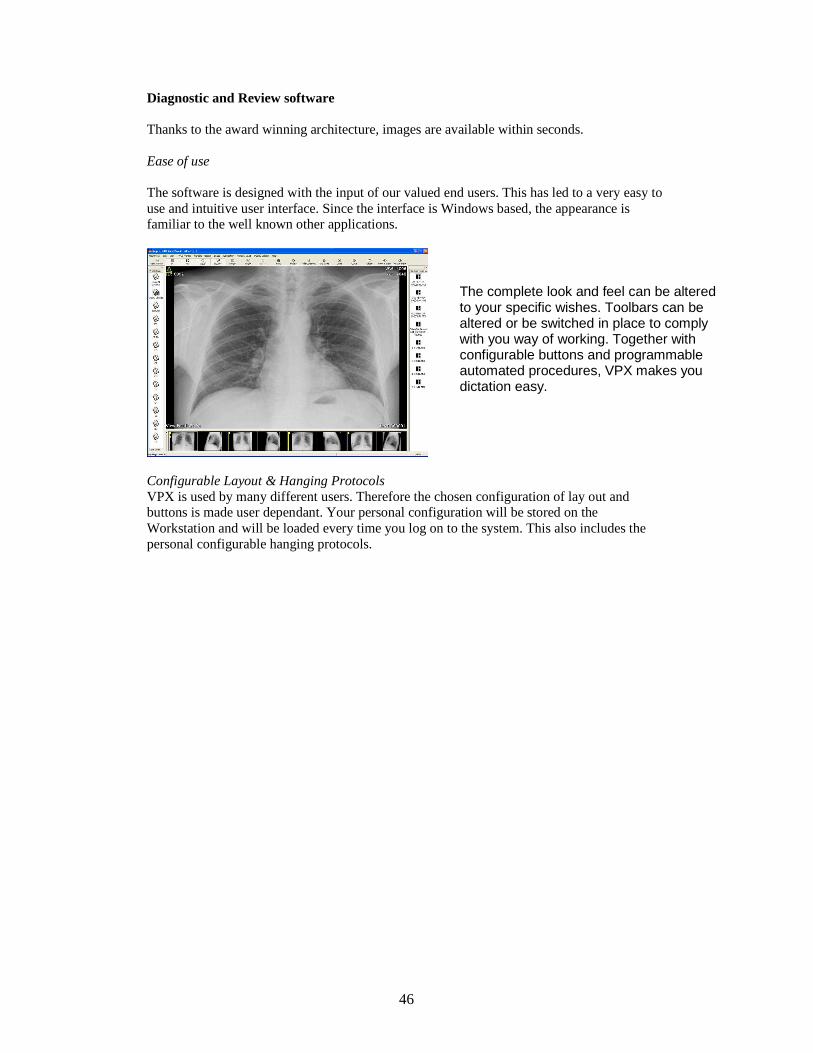
Diagnostic and Review software Thanks to the award winning architecture, images are available within seconds. Ease of use The software is designed with the input of our valued end users. This has led to a very easy to use and intuitive user interface. Since the interface is Windows based, the appearance is familiar to the well known other applications.
Configurable Layout & Hanging Protocols VPX is used by many different users. Therefore the chosen configuration of lay out and buttons is made user dependant. Your personal configuration will be stored on the Workstation and will be loaded every time you log on to the system. This also includes the personal configurable hanging protocols.
The complete look and feel can be altered to your specific wishes. Toolbars can be altered or be switched in place to comply with you way of working. Together with configurable buttons and programmable automated procedures, VPX makes you dictation easy.

47
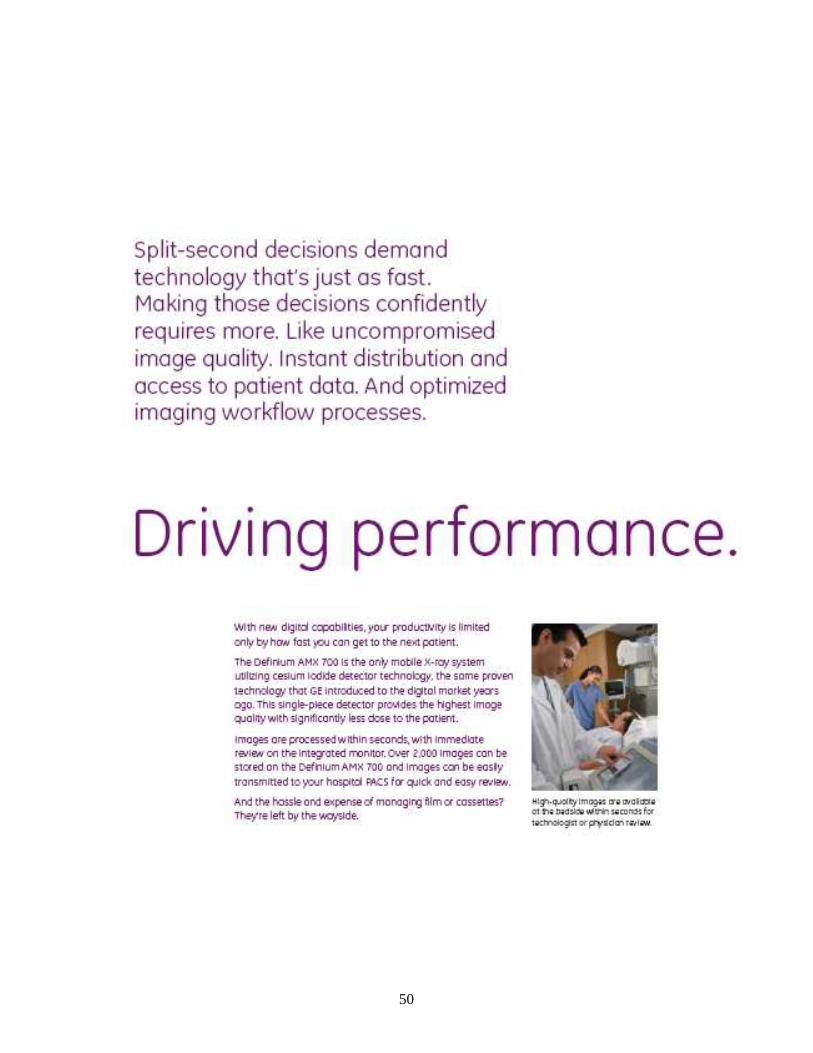
Appendix 9 General Electric
Note: can be delivered mounted into truck

48

49
50

51

52

53

54
55
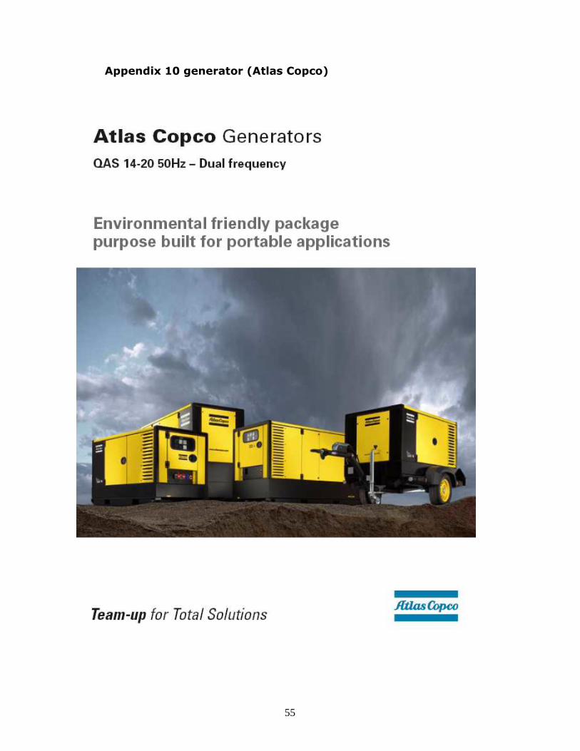
Appendix 10 generator (Atlas Copco)

56