Chemical attribution of fentanyl - TNO Publications
13
Forensic Chemistry 24 (2021) 100330 Available online 10 March 2021 2468-1709/© 2021 Elsevier B.V. This is an open access article under the CC BY license (http://creativecommons.org/licenses/by/4.0/). Chemical attribution of fentanyl: The effect of human metabolism Mirjam de Bruin-Hoeg´ ee a, b, * , Djarah Kleiweg a , Daan Noort b , Arian C. van Asten a, c a van ‘t Hoff Institute for Molecular Sciences, Faculty of Science, University of Amsterdam, P.O. Box 94157, 1090GD Amsterdam, The Netherlands b TNO Defence, Safety and Security, Dep. CBRN Protection, Lange Kleiweg 137, 2288GJ Rijswijk, The Netherlands c CLHC, Amsterdam Center for Forensic Science and Medicine, University of Amsterdam, P.O. Box 94157, 1090GD Amsterdam, The Netherlands A R T I C L E INFO Keywords: Forensic chemistry Fentanyl Forensic toxicology Impurity profiling Chemical attribution signatures Multivariate data analysis ABSTRACT Chemical attribution typically aims to establish a link between material found at a crime scene and a person, location or other evidence. In the field of illicit drugs, chemical attribution signatures are usually impurity profiles. Extending these to metabolized samples would create new possibilities in forensic investigations. The present study explores the effect of human metabolism on the impurity profile of fentanyl, as representative of synthetic opioids. Two different methods (Gupta and Siegfried) were used to synthesize fentanyl, after which the samples were incubated with liver microsomes to mimic human metabolism. The impurity profiles have been characterized with gas chromatography-mass spectrometry (GC-MS), gas chromatography with flame ionization detector (GC-FID), liquid chromatography quadrupole-time of flight mass spectrometry (LC-Q-TOF-MS) and liquid chromatography orbitrap mass spectrometry (LC-Orbitrap-MS). It was found that GC-FID and LC-Orbitrap- MS can both be used to discriminate between the Gupta and Siegfried synthesis method. This holds both for the analyses performed before and after metabolism. In addition, principal component analysis (PCA) identified acetyl fentanyl as the most important marker compound. Associated detection limits are in the range of con- centrations expected in case work. While acetyl fentanyl is not stable during metabolism, its discriminating potential is transferred to its metabolic product acetyl norfentanyl. In addition, the stable impurities phenyl- acetamide and 1-phenylethylpiperidin-4-ol were found to be significant classifiers. To implement the results in a forensic framework, linear discriminant analysis (LDA) was applied and used to establish likelihood ratios. To our knowledge, the present work demonstrates for the first time the possibility of chemical attribution of drugs through the analysis of metabolic trace levels in biological samples. 1. Introduction An emerging category of chemical threat agents consists of the so- called pharmaceutical based agents (PBAs) [1]. This includes the fam- ily of synthetic opioids like fentanyl, a compound which is estimated to be 50–100 times more potent than morphine [2]. Fentanyl has a lethal dose of only a few milligrams [3]. Its analogues such as sufentanil and carfentanil are even more potent [4]. Carfentanil was one of the com- ponents of the mixture of chemicals used to end the Moscow theater hostage crisis [5]. Fentanyl is intended for analgesic use, but it is also illicitly used in pills or mixed with heroin or cocaine. The illicit use of fentanyl and its analogues has seen a rapid increase in the last decades, with a large outbreak starting in 2013 in the United States of America (USA) and Canada [6]. In 2018, fentanyl and its analogues were the most common cause of overdose deaths in the USA [7]. Its potency and wide availability make fentanyl attractive for misuse by criminals and terrorist groups. In this respect, chemical attribution typically aims to establish a link between material found at a crime scene and a person (usually a suspect), location or other evidence. Chemical attribution signatures, such as impurities, isotope ratios and other chemical or physical characteristics, are used to assess whether two different sam- ples share a common origin. In the field of illicit drugs, chemical profiles are usually based on impurities related to production, processing and storage and are typically analyzed by analytical methods such as gas chromatography-mass spectrometry (GC-MS) and liquid chromatography-mass spectrometry (LC-MS) [8]. The potential of im- purity profiling has been demonstrated for cannabis, heroin, cocaine, 3,4-methylenedioxymethamphetamine (MDMA) and amphetamine [9–13]. It can be expected that the identity of the impurities heavily relies on * Corresponding author at: van ‘t Hoff Institute for Molecular Sciences, Faculty of Science, University of Amsterdam, P.O. Box 94157, 1090GD Amsterdam, The Netherlands. E-mail address: [email protected] (M. de Bruin-Hoeg´ ee). Contents lists available at ScienceDirect Forensic Chemistry journal homepage: www.sciencedirect.com/journal/forensic-chemistry https://doi.org/10.1016/j.forc.2021.100330 Received 5 January 2021; Received in revised form 23 February 2021; Accepted 2 March 2021
-
Upload
khangminh22 -
Category
Documents
-
view
1 -
download
0
Transcript of Chemical attribution of fentanyl - TNO Publications

Forensic Chemistry 24 (2021) 100330
Available online 10 March 20212468-1709/© 2021 Elsevier B.V. This is an open access article under the CC BY license (http://creativecommons.org/licenses/by/4.0/).
Chemical attribution of fentanyl: The effect of human metabolism
Mirjam de Bruin-Hoegee a,b,*, Djarah Kleiweg a, Daan Noort b, Arian C. van Asten a,c
a van ‘t Hoff Institute for Molecular Sciences, Faculty of Science, University of Amsterdam, P.O. Box 94157, 1090GD Amsterdam, The Netherlands b TNO Defence, Safety and Security, Dep. CBRN Protection, Lange Kleiweg 137, 2288GJ Rijswijk, The Netherlands c CLHC, Amsterdam Center for Forensic Science and Medicine, University of Amsterdam, P.O. Box 94157, 1090GD Amsterdam, The Netherlands
A R T I C L E I N F O
Keywords: Forensic chemistry Fentanyl Forensic toxicology Impurity profiling Chemical attribution signatures Multivariate data analysis
A B S T R A C T
Chemical attribution typically aims to establish a link between material found at a crime scene and a person, location or other evidence. In the field of illicit drugs, chemical attribution signatures are usually impurity profiles. Extending these to metabolized samples would create new possibilities in forensic investigations. The present study explores the effect of human metabolism on the impurity profile of fentanyl, as representative of synthetic opioids. Two different methods (Gupta and Siegfried) were used to synthesize fentanyl, after which the samples were incubated with liver microsomes to mimic human metabolism. The impurity profiles have been characterized with gas chromatography-mass spectrometry (GC-MS), gas chromatography with flame ionization detector (GC-FID), liquid chromatography quadrupole-time of flight mass spectrometry (LC-Q-TOF-MS) and liquid chromatography orbitrap mass spectrometry (LC-Orbitrap-MS). It was found that GC-FID and LC-Orbitrap- MS can both be used to discriminate between the Gupta and Siegfried synthesis method. This holds both for the analyses performed before and after metabolism. In addition, principal component analysis (PCA) identified acetyl fentanyl as the most important marker compound. Associated detection limits are in the range of con-centrations expected in case work. While acetyl fentanyl is not stable during metabolism, its discriminating potential is transferred to its metabolic product acetyl norfentanyl. In addition, the stable impurities phenyl-acetamide and 1-phenylethylpiperidin-4-ol were found to be significant classifiers. To implement the results in a forensic framework, linear discriminant analysis (LDA) was applied and used to establish likelihood ratios. To our knowledge, the present work demonstrates for the first time the possibility of chemical attribution of drugs through the analysis of metabolic trace levels in biological samples.
1. Introduction
An emerging category of chemical threat agents consists of the so- called pharmaceutical based agents (PBAs) [1]. This includes the fam-ily of synthetic opioids like fentanyl, a compound which is estimated to be 50–100 times more potent than morphine [2]. Fentanyl has a lethal dose of only a few milligrams [3]. Its analogues such as sufentanil and carfentanil are even more potent [4]. Carfentanil was one of the com-ponents of the mixture of chemicals used to end the Moscow theater hostage crisis [5]. Fentanyl is intended for analgesic use, but it is also illicitly used in pills or mixed with heroin or cocaine. The illicit use of fentanyl and its analogues has seen a rapid increase in the last decades, with a large outbreak starting in 2013 in the United States of America (USA) and Canada [6]. In 2018, fentanyl and its analogues were the most common cause of overdose deaths in the USA [7]. Its potency and
wide availability make fentanyl attractive for misuse by criminals and terrorist groups. In this respect, chemical attribution typically aims to establish a link between material found at a crime scene and a person (usually a suspect), location or other evidence. Chemical attribution signatures, such as impurities, isotope ratios and other chemical or physical characteristics, are used to assess whether two different sam-ples share a common origin. In the field of illicit drugs, chemical profiles are usually based on impurities related to production, processing and storage and are typically analyzed by analytical methods such as gas chromatography-mass spectrometry (GC-MS) and liquid chromatography-mass spectrometry (LC-MS) [8]. The potential of im-purity profiling has been demonstrated for cannabis, heroin, cocaine, 3,4-methylenedioxymethamphetamine (MDMA) and amphetamine [9–13].
It can be expected that the identity of the impurities heavily relies on
* Corresponding author at: van ‘t Hoff Institute for Molecular Sciences, Faculty of Science, University of Amsterdam, P.O. Box 94157, 1090GD Amsterdam, The Netherlands.
E-mail address: [email protected] (M. de Bruin-Hoegee).
Contents lists available at ScienceDirect
Forensic Chemistry
journal homepage: www.sciencedirect.com/journal/forensic-chemistry
https://doi.org/10.1016/j.forc.2021.100330 Received 5 January 2021; Received in revised form 23 February 2021; Accepted 2 March 2021

Forensic Chemistry 24 (2021) 100330
2
the synthesis method applied. The first synthesis method of fentanyl was patented by Janssen and Gardocki in 1964 [14]. This method requires advanced skills in organic chemistry. Other methods, as developed by Siegfried [15] and Gupta [16] are more commonly used in clandestine laboratories [17,18]. The Siegfried method is based on an internet recipe [15]. The Gupta method is also referred to as ‘One-Pot method’, because the synthesis is carried out in a single reaction vessel [16]. Compared to other illicit drugs, there is not much literature available on the chemical profiling of fentanyl. It is typically dosed at relatively low levels as ad-ditive to other psychoactive substances such as heroin. This makes chemical impurity profiling of fentanyl in case work samples very challenging. Lurie et al. [19] developed an ultra-high pressure liquid chromatography-tandem mass spectrometry (UHPLC-MS/MS) method to identify 40 preselected potential impurities of the Janssen or Siegfried synthesis method. Mayer et al. [17] investigated chemical attribution signatures for the Siegfried, Valdez and Gupta method and hybrid ver-sions thereof. Multivariate statistical analysis was applied on the data obtained by GC-MS, liquid chromatography-tandem mass spectrometry- time of flight (LC-MS/MS-TOF) and inductively coupled plasma-mass spectrometry (ICP-MS). Another study demonstrated the potential of direct-infusion electrospray ionization-mass spectrometry (ESI-MS) for profiling of fentanyl synthesized by the Siegfried method [20]. Most recently, Casale et al. used GC-MS and nuclear magnetic resonance (NMR) spectroscopy for the identification of three impurities charac-teristic to the Gupta synthesis method [21].
After the (deliberate) release of a chemical threat agent, it is often difficult to find traces of the chemical that was used [22,23], either because of a lack of persistency, or due to the inability to enter the scene because of safety and security reasons. Under such circumstances the exposed people themselves might form a valuable source of information: biochemical indicators (biomarkers) of exposure can be found in human tissue for longer periods of time, depending on the nature of the chemical agent. Based on this concept a variety of methods have been developed over the last decades that allow retrospective analysis of signatures of chemical threat agents in biomedical samples, up to weeks after the actual exposure [24]. The general aim of the current study was to explore whether these two concepts, i.e. chemical profiling and retrospective biomarker analysis, could be combined into a novel concept of “chemical profiling in biomedical samples”. To this date, such studies have not yet been performed.
In the present study the effect of human metabolism on the impurity profile of fentanyl was investigated. Two different synthesis methods (Gupta and Siegfried) were used to synthesize small batches of fentanyl, which were subsequently analyzed in detail to assess their chemical profiles. The fentanyl samples were then incubated with human liver microsomes to mimic human metabolism. Oxidative N-dealkylation of fentanyl to norfentanyl is the predominant pathway in fentanyl meta-bolism [25,26], as illustrated in Fig. 1. The other metabolic product phenylacetaldehyde can either be reduced to phenethyl alcohol or oxidized to phenylacetic acid. The impurity profiles (pre- and post- metabolism) were constructed with GC-MS, gas chromatography with flame ionization detection (GC-FID), liquid chromatography-
quadrupole-time of flight mass spectrometry (LC-Q-TOF-MS) and LC- Orbitrap-MS. Multivariate data analysis was applied to the analytical data to identify potential marker compounds that indicate a specific fentanyl synthesis method. The current study demonstrates that analysis of biomedical samples for chemical provenance purposes might in principle form a valuable addition to the more classical chemical profiling approach based on analysis of bulk samples.
2. Materials and methods
2.1. Safety
Due to the extremely potent nature of fentanyl, samples and dilutions should be handled with care. Precautions were taken to prevent acci-dental exposure, including wearing gloves and eye protection. In case of exposure to opioids, the antidote naloxone could directly be adminis-tered to mitigate respiratory depression.
2.2. Chemicals
Fentanyl, benzylfentanyl and methyl 4-anilino-1-benzyl-4-piperidi-necarboxylate were synthesized at TNO, Rijswijk. The chemicals used for the syntheses were obtained from Sigma Aldrich, except aniline which was obtained from Janssen Chimica. Norfentanyl (Sigma-Aldrich, ≥97%) and d5-norfentanyl (Supelco, certified reference material) were used for the method optimization. The incubation protocol was opti-mized using fentanyl citrate (Spruyt Hillen). Pooled human liver mi-crosomes (Sigma-Aldrich, CAS M0317, lot #SLCC0060 and lot #SLCC7022 and Xenotech, CAS H0610, lot #1610016) were used for the incubation. The NADPH-regenerating system consisted of NADP+(Sigma-Aldrich, anhydrous), glucose-6-phosphate (Sigma-Aldrich, anhydrous), glucose-6-phosphate dehydrogenase (Sigma-Aldrich, lyophilized powder) and uridine 5′-diphosphoglucuronic acid (UDPGA, Sigma-Aldrich, trisodium salt, >98%). For LC analyses formic acid was obtained from Fluka (#06440) and acetonitrile was Optima LC-MS grade (Fisher #A955-1). Millipore water was used as a solvent (Sim-Pak® 1). For GC analyses, stock solutions were made in dichloro-methane (Biosolve, >99.9%).
2.3. Synthesis
The synthesis of fentanyl was performed in triplicate according to the open literature methods of Gupta [16] and Siegfried [15]. Fig. 2 gives the overall synthetic strategy for the current study, similar to the scheme published by Mayer et al. [17]. Because of the nature of the compounds of interest, not all synthesis details are provided in the current work. So, briefly, in case of the Siegfried method the precursor N-Phenethyl- Piperidone (NPP) was synthesized from piperidone and phenethyl- bromide. The obtained NPP further reacted with aniline giving the imine derivative which was reduced to the 4-Anilino-N-Phenethyl-Piper-idine (4-ANPP). The latter was converted with propionyl chloride giving fentanyl hydrochloride. For the Siegfried method, each intermediate
ON
HN
O
oxidative N-dealkylation
+
norfentanyl phenylacetaldehydeOH
phenethyl alcohol
oxida
tion
reduction
OOH
phenylacetic acid
N
N O
fentanyl
Fig. 1. Reaction scheme of fentanyl metabolized to norfentanyl by oxidative N-dealkylation. Phenylacetaldehyde can either be reduced to phenethyl alcohol or oxidized to phenylacetic acid.
M. de Bruin-Hoegee et al.

Forensic Chemistry 24 (2021) 100330
3
was purified before continuing with the next step, and the final product was obtained by crystallization. For the Gupta One-Pot method, no purification of intermediates was applied. Only the final product was obtained by crystallization, like the Siegfried method. Both syntheses were performed in triplicate and the products were stored as a powder at − 20 ◦C. For two of the three samples of each synthesis method an aliquot of the reaction mixture was stored at room temperature (RT). All sam-ples were analyzed in the current study.
2.4. Microsomal incubations and sample preparation
The liver is primarily responsible for the fentanyl metabolism [27–29]. Liver microsomes can therefore be used to mimic human metabolism. This system is one of the most common in vitro models to study phase I metabolism and glucuronidation [30]. In this study, fen-tanyl was incubated according to earlier described methods [25–29]. The conditions were optimized using fentanyl citrate with benzylfen-tanyl as internal standard to correct for varying instrument response. The following conditions were varied: concentration fentanyl, concen-tration microsomes and incubation time.
The fentanyl samples synthesized by the Gupta and Siegfried method were incubated in triplicate. The incubation method and sample prep-aration are described in detail in Fig. 1 of the Supplementary informa-tion. First, 100 µL of 1 mg/mL fentanyl and 200 µL of 2.5 mg/mL human liver microsomes in 500 µL buffer were pre-incubated in a Grant-Bio PHMT Thermoshaker at 37 ◦C and were shaken at 300 rpm for 3 min. An 0.1 M potassium phosphate buffer with 2.5 mM MgCl2 was used (pH = 7.4). Subsequently, the reaction was initiated by adding 200 µL of the NADPH-regenerating system. Concentrations were prepared of 1 mM NADP+, 5 mM glucose-6-phosphate, 1 U/mL glucose-6-phosphate de-hydrogenase and 2 mM UDPGA. The samples were incubated for 72 h (37 ◦C, 300 rpm). For each series of experiments a negative control was included, i.e. a blank experiment including all components except fen-tanyl. In addition, experiments were conducted without the addition of microsomes or the NADPH-regenerating system. A final control experi-ment assessed the stability of the fentanyl metabolites during
incubation, by monitoring deuterated norfentanyl. After incubation, the samples were divided into two fractions of 500
µL used for GC and LC analysis. For the sample work-up, 500 µL aceto-nitrile was added to the LC fraction to induce precipitation of the pro-teins. For the GC fraction, no acetonitrile was added before centrifuging, to avoid the presence of water in the sample afterwards. Both fractions were centrifuged for 10 min at 14,000 rpm (Eppendorf, 5417R). For the LC fraction, the supernatant was transferred to a glass vial and diluted five times in MilliQ, after which it was analyzed with LC-MS. For the GC fraction, the supernatant was transferred to a glass vial and 500 µL dichloromethane was added for liquid–liquid extraction. The dichloro-methane fraction was then analyzed with GC-MS and GC-FID.
The efficiency of the sample preparation was determined using a known concentration of fentanyl and norfentanyl with benzylfentanyl as internal standard for LC-MS analyses and d5-norfentanyl as internal standard for GC-MS analyses. The incubation procedure was followed as described, but without incubation time to prevent conversion of fentanyl due to the metabolism.
2.5. Chemical analysis
2.5.1. GC-MS The analyses were performed on an Agilent 7890B GC equipped with
an Agilent VF-5 ms column (5% phenylmethyl polysiloxane, 30 m ×0.25 mm × 0.25 µm). One microliter of sample was injected using an autosampler (Combi Pal, Ctc analytics). Helium was used as the carrier gas at a constant flow of 1 mL/min. The GC injector was operated in splitless mode at 275 ◦C. The oven temperature was maintained at 40 ◦C for 1 min., then ramped at 10 ◦C/min. to 280 ◦C and held for 15 min. Detection was performed with an Agilent 5977A MS, which operated in electron ionization (EI) mode with an ionization potential of 70 eV and a scan range of 25–550 mass units. Compounds were identified using Agilent ChemStation by spectral comparison to the National Institute of Standards and Technology (NIST) Mass Spectral Library. The results of the in vitro experiments were compared to a negative control, as described in Section 2.4.
Fig. 2. Reaction scheme of fentanyl synthesized by the One-Pot Gupta method (top, red) and the Siegfried method (bottom, blue). (For interpretation of the ref-erences to colour in this figure legend, the reader is referred to the web version of this article.)
M. de Bruin-Hoegee et al.

Forensic Chemistry 24 (2021) 100330
4
2.5.2. GC-FID The analyses were performed with an Agilent 7890A GC equipped
with an Agilent VF-5 ms column (5% phenylmethyl polysiloxane, 50 m × 0.32 mm × 0.40 µm). One microliter of sample was injected by an autosampler (Agilent 7683B injector) to the injector, which was oper-ated in splitless injection mode at 275 ◦C. Helium was used as the carrier gas at a constant flow of 1 mL/min. The oven temperature was held at 40 ◦C for 1 min., then ramped at 10 ◦C/min. to 210 ◦C and held for 5 min., and finally ramped at 10 ◦C/min. to 280 ◦C and held for 30 min. Detection was performed with an FID at 250 ◦C, with a hydrogen flow of 40 mL/min. and an air flow of 450 mL/min. Data analysis was done by manual integration of peaks previously identified with GC-MS using Agilent ChemStation. The results of the in vitro experiments were compared to a negative control, as described in Section 2.4.
2.5.3. LC-Q-TOF-MS The first exploratory sample analyses were performed with a Thermo
Ultimate 3000 UHPLC equipped with a Waters Acquity HSS T3 C18 column (1.8 µm, 1.0 × 150 mm). The column temperature was main-tained at 35 ◦C and the flow rate was 100 µL/min. Eluent A was 0.2 v% formic acid in MilliQ water. Eluent B was 0.2 v% formic acid in aceto-nitrile. Gradient elution started at 100% eluent A, ramping to 80% eluent B in 30 min and holding for 5 min. Then equilibrating at 100% eluent A for 1 min. The injection volume was 10 µL. The UHPLC was coupled to a Bruker Maxis Impact QTOF MS, which was set to a mass range of m/z 50–700 and operated in the positive electrospray ionization (ESI) mode. Data were acquired with full scan MS mode. The capillary voltage was 4500 V and the collision energy was 6 eV. The spectral acquisition rate was 1 Hz. The data were analyzed with Metabo-liteDetect to search for compounds with DataAnalysis after subtraction of a of a negative control baseline signal. Peak areas of compounds were calculated by automatic integration of the extracted ion chromatogram of the identified compounds. After analysis, a target table was con-structed and searched against all samples.
2.5.4. LC-Orbitrap-MS After LC-Q-TOF-MS analyses, the samples were analyzed with a
Thermo Ultimate 3000 UHPLC equipped with a Waters Acquity HSS T3 C18 column (1.8 µm, 2.1 × 100 mm). The column temperature was maintained at 30 ◦C and the flow rate was 100 µL/min. Eluent A was 0.2 v% formic acid in MilliQ water. Eluent B was 0.2 v% formic acid in acetonitrile. Gradient elution started at 100% eluent A, ramping to 80% eluent B in 10 min and holding for 5 min. Then equilibrating at 100% eluent A for 1 min. The injection volume was 10 µL. The UHPLC was coupled to a Thermo Scientific Q Exactive Plus Orbitrap MS, which was set to a mass range of m/z 50–750 and operated in positive ESI mode. The capillary voltage was set to 3.5 kV, and the source temperature was maintained at 320 ◦C, the relative sheath gas (nitrogen) flow was 35. The sensitivity of the method was assessed for fentanyl and its impu-rities. Data were first acquired with full scan MS mode. Based on the results obtained and the previously constructed target table with LC-Q- TOF-MS, an inclusion list was established using targeted MS/MS in parallel reaction monitoring (PRM) mode. The collision energy was 25 eV for all compounds. The data were analyzed with Xcalibur and Compound Discoverer. Peak areas were calculated by automatic inte-gration of the extracted ion chromatogram of the identified compounds after subtraction of a negative control baseline signal. Compounds with a peak area lower than 10,000 were excluded. If peaks were only detected post metabolism, compounds with a peak area lower than 1,000,000 were excluded. Compounds that were present in the inclusion list and that were characteristic to a synthesis method were included.
2.5.5. LC-MS/MS The optimization experiments were performed with a Waters Acq-
uity ultra-high pressure liquid chromatography (UPLC) equipped with a Waters Acquity HSS T3 C18 column (1.8 µm, 2.1 × 100 mm). The mobile
phase consisted of MilliQ water and acetonitrile both with 0.2% formic acid, using a gradient at a flow rate of 100 μL/min. Gradient elution started at 100% eluent A, ramping to 80% eluent B in 10 min and holding for 2 min. Then equilibrating at 100% eluent A for 3 min. The injection volume was 10 µL. The UPLC was coupled to a ThermoFisher TSQ Triple Quadrupole MS, which was operated in the positive ESI mode. The capillary voltage was set to 3.5 kV and the cone gas flow was 150 L/h. The cone voltage was 40 V and the collision energy was 30 eV for all compounds, with argon as collision gas set at a flow of 0.19 mL/ min. Data was acquired with selected reaction monitoring (SRM) mode. The transitions monitored were m/z 337 → 188 and 337 → 105 for fentanyl, m/z 233 → 177 and 233 → 84 for norfentanyl and m/z 323 → 174 and 323 → 91 for the internal standard benzylfentanyl. A solvent delay of 3 min was used. Data analysis was performed with Xcalibur software.
2.6. Data analysis
Statistical analysis was performed with Python 3.8.2 using scikit- learn 0.22.1 [31]. Since these packages are open source it is freely us-able and distributable. The code written for this research is published under a GNU General Public License [32]. The peak areas of the GC-FID were normalized to the sum of all peaks. The peak areas of the LC- Orbitrap-MS were normalized to the internal standard methyl 4-ani-lino-1-benzyl-4-piperidinecarboxylate. The responses of the GC-FID for fentanyl and the found impurities were assumed to be uniform [33]. Two types of statistical analysis were performed. Principal component analysis (PCA) was used to reduce the dimensionality of the data and to confirm the discriminatory power of potential markers that were iden-tified by visual inspection. The robustness of PCA was checked by leave- one-out validation. Supervised linear discriminant analysis (LDA) was used to maximize discrimination between the two synthesis methods. Kernel density estimations (KDEs) were constructed and used to express likelihood ratio (LR) values for the assignment of an unknown sample, in a similar way as Brust et al. [34]. A match criterion approach offers a simple alternative to multivariate statistical analysis. This approach results in absolute statements accompanied by error rates. The statistical tool Student’s t was applied to assess which impurities were character-istic for a specific synthesis route. An impurity was considered charac-teristic if there was less than 1% probability that the observation arises from random variation (beyond 99% confidence interval). Given the aim of the research the following hypothesis pair was considered:
H1: The victim has been exposed to fentanyl produced with the Gupta method
H2: The victim has been exposed to fentanyl produced with the Siegfried method
3. Results
3.1. Method optimization and validation
The GC-MS method was optimized for fentanyl citrate (tr: 25.4 min.) and norfentanyl (tr: 18.9 min.) with internal standard d5-norfentanyl (tr: 18.9 min.), in order to determine the conversion of fentanyl to norfen-tanyl. Linear calibration curves were obtained for these standards in the range of 5–50 µg/mL with R2 = 0.9969–0.9984 (Fig. 2, Supplementary information). The mean values of the quality controls were within 23% at 5 µg/mL and within 10% relative standard deviation at 50 µg/mL (n = 3). The sample preparation efficiencies, determined by spiking a known concentration of fentanyl and norfentanyl into the microsomal incubation mixture, were respectively 129% ± 12% and 22.3% ± 1.1% (std., n = 8). Evaporation of the volatile organic phase is the most likely cause for obtaining recoveries exceeding 100%. The low recovery of norfentanyl is probably due to limited transfer from the polar phase to the organic phase. The most relevant impurities in the GC-FID chro-matogram as presented in Fig. 3 were identified with GC-MS as fentanyl
M. de Bruin-Hoegee et al.

Forensic Chemistry 24 (2021) 100330
5
(X, tr: 45.7 min.), acetyl fentanyl (W, tr: 43.9 min.), 4-ANPP (V, tr: 38.1 min.) and N-phenylpropanamide (L, tr: 20.4 min.). These impurities were clearly visible in the pre-incubation chromatogram. In the post- incubation chromatogram, other compounds were observed, including the metabolism product phenethyl alcohol (H, tr: 15.5 min.).
The LC-MS/MS method was optimized for fentanyl citrate (tr: 7.49 min.) with internal standard benzylfentanyl (tr: 7.32 min.) and for norfentanyl (tr: 6.37 min.). Linear calibration curves were obtained in the range of 5–200 ng/mL with R2 = 0.9986–0.9996. The mean values of the quality controls were within 25% at 5 ng/mL and within − 7% relative standard deviation at 200 ng/mL (n = 3). The sample prepa-ration efficiencies, determined by spiking a known concentration of fentanyl and norfentanyl into the microsomal incubation mixture, were respectively 105% ± 6% and 107% ± 3% (std., n = 5). Surprisingly, no
losses due to protein precipitation were observed. This is probably due the simplified microsomal model system compared to sample prepara-tion of more complex biological samples such as human whole blood. The following most relevant impurities were identified in the LC- Orbitrap-MS total ion chromatograms as presented in Fig. 4: fentanyl (X, tr: 10.9 min.), acetyl fentanyl (W, tr: 10.5 min.) and N-phenyl-acetamide (J, tr: 11.1 min.). In the post-incubation chromatogram, more peaks were visible, including the metabolic product norfentanyl (T, tr: 9.4 min.).
3.2. Pre-metabolism impurity profiling
In the pre-metabolism fentanyl samples, a total of 24 impurities were identified. An overview of all the impurities found in this study is given
L
V
XW
A
L W
H
X
B
Fig. 3. Representative GC-FID chromatograms of the Gupta (G) and Siegfried (S) samples. A) Pre-metabolism B) Post-metabolism. The following most relevant impurities have been highlighted: phenylethyl alcohol (H), N-phenylpropanamide (L), 4-ANPP (V), acetyl fentanyl (W) and fentanyl (X).
WXJ
A
B
WXJT
Fig. 4. Representative LC-Orbitrap-MS total ion chromatograms of the Gupta (G) and Siegfried (S) samples. A) Pre-metabolism B) Post-metabolism. The following most relevant impurities have been highlighted: norfentanyl (T), acetyl fentanyl (W), fentanyl (X) and N-phenylacetamide (J).
M. de Bruin-Hoegee et al.

Forensic Chemistry 24 (2021) 100330
6
in Table 1. The suggested tentative structures were based on the use of various methods and reference sources, as indicated in the table foot-note. An example of a tentative identification of a compound by targeted MS/MS is given in Fig. 3 of the Supplementary information. All the listed impurities were present in the samples prepared according to the Gupta synthesis method, whereas only 17 impurities were detected in the samples made by the Siegfried method. This difference can be explained by the purification steps performed after each intermediate synthesis step in the Siegfried method.
3.2.1. Match criterion using confidence intervals A simple rule-of-thumb guideline was established in order to obtain
information on the synthesis route involved. A match criterion approach was applied using confidence intervals. Consequently, three impurities were found to be significant classifiers for the synthesis method for the pre-metabolism samples. The impurities 1-phenylethylpiperidin-4-ol (R) and acetyl fentanyl (W) were characteristic for the Gupta synthesis route. The impurity 4-ANPP (V) was found to be indicative for the Siegfried synthesis route. Table 2 shows the characteristic relative levels of fentanyl impurities prior to metabolism. The responses were measured by GC-FID and LC-Orbitrap-MS for both the Gupta and Sieg-fried method. For example, if the relative response of impurity W is within 23–47% of the fentanyl peak area, as measured by LC-Orbitrap- MS, this indicates that fentanyl is produced with the Gupta method. The same holds when either acetyl fentanyl (W) or 4-ANPP (V) is detected with GC-FID. Not applicable (n.a.) is shown when the impurity is not detected by the analysis method. Another approach is to use a ratio of compounds as indicator for the synthesis method used. This ratio is often constant in various kinds of matrices. For LC-Orbitrap-MS the ratio of V/R was 1.7 ± 0.9 and 110 ± 84 (±95% confidence interval), for respectively the Gupta and Siegfried method. Additionally, the ratio of W/V was 103 ± 60 for the Gupta and 0.4 ± 0.6 for the Siegfried method. Because no overlapping values were found, these ratios are interesting factors for a simple and straight forward assessment of the synthesis route involved.
3.2.2. PCA In addition to the simple rule-of-thumb guideline, a chemometric
analysis was applied to retrieve robust information on the synthesis route. To illustrate the chemical attribution signatures of the Gupta and Siegfried synthesis method, a PCA model was built for the impurities detected by LC-Orbitrap-MS. Since a limited number of impurities was detected by GC, chemometric analysis of the GC data provided little added value.
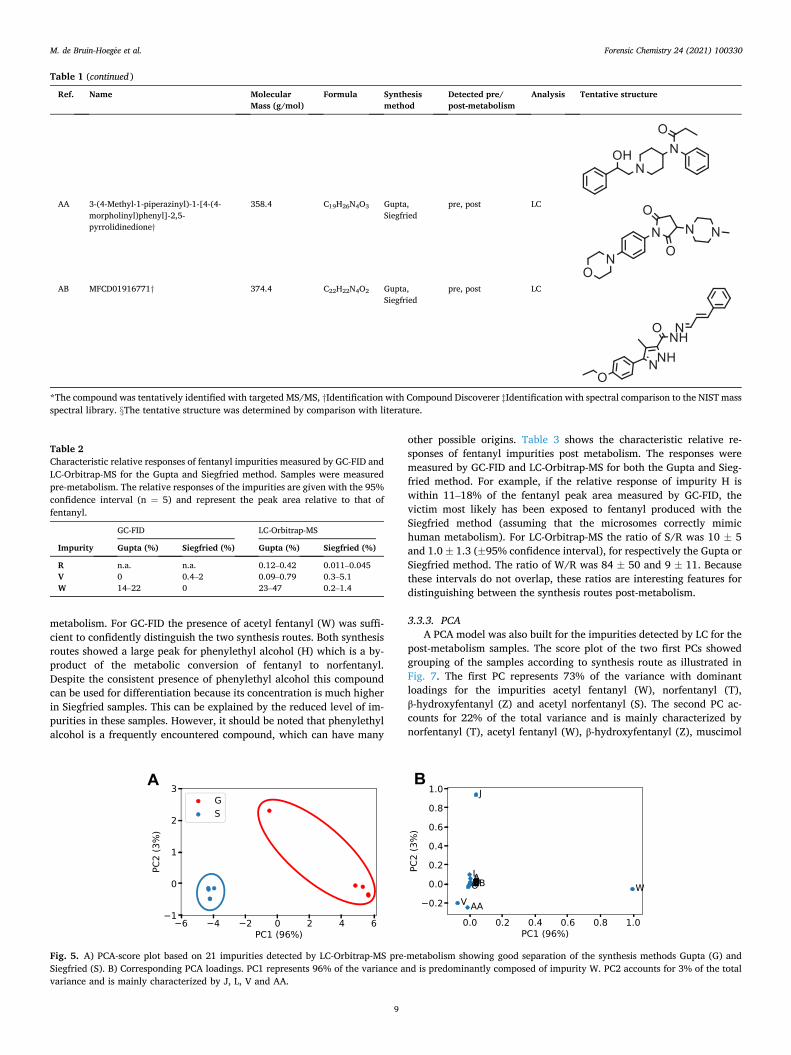
The score plot of the two first principal components (PCs) based on impurities detected by LC pre-metabolism, showed grouping of the samples according to their synthesis as is illustrated in Fig. 5. The first PC accounts for 96% of the variance and is predominantly composed of impurity acetyl fentanyl (W). The second PC accounts for 3% of the total variance and is mainly characterized by N-phenylacetamide (J), N- phenylpropanamide (L), 4-ANPP (V) and 3-(4-Methyl-1-piperazinyl)-1- [4-(4-morpholinyl)phenyl]-2,5-pyrrolidinedione (AA). N-Butylaniline (M) was only detected by GC and AA was only detected by LC. The performance of the unsupervised PCA models was tested with leave-one- out validation (Fig. 4, Supplementary information). The PCA model showed good robustness, since leaving out one sample resulted in similar explained variance. In this study other impurities were found than described in previous studies [19,21]. The first study focused on the Siegfried and Janssen synthesis routes and reported among others, N- phenylpropanamide (L), 1-phenylethylpiperidin-4-ol (R), 4-ANPP (V) and acetyl fentanyl (W) as markers common for both routes. In the current study 4-ANPP (V) was also found to be a Siegfried-specific marker, although the other compounds were identified as Gupta markers. The second study also characterized acetyl fentanyl (W) as a Gupta marker. Additional characteristic impurities in the current research included N-phenylacetamide (J), N-Butylaniline (M) and 3-(4-
Methyl-1-piperazinyl)-1-[4-(4-morpholinyl)phenyl]-2,5-pyrrolidine-dione (AA). Most of these impurities were also found by Mayer et al. [17], except for N-Butylaniline (M) which to our knowledge has not been previously reported.
3.2.3. LDA To put the results in a forensic framework, LDA was used to find
maximum discrimination between two groups. The LDA value is a linear combination of the original variables, i.e. normalized peak area. An advantage of this supervised pattern recognition method is that only one dimension (first canonical variate) is defined, when LDA is applied to two classes. In this study two classes were used, requiring only the first canonical variate for the separation of the classes. A disadvantage of the supervised LDA technique is a greater risk of overfitting, which results in too optimistic model performance. To avoid this problem PCA can be applied in combination with LDA [36]. This is often performed when the number of features exceed the number of samples per category. PCA reduces the dimensionality as described in Section 3.2.2. Fig. 6 shows the KDE distributions for both synthesis methods obtained using LDA in combination with PCA. The distributions based on LC-Orbitrap-MS pre- metabolism data were highly discriminating. Because the GC results showed very good discrimination only based on a few impurities, no LDA was applied.
3.3. Post-metabolism impurity profiling
3.3.1. Metabolism of fentanyl Fentanyl was incubated using human liver microsomes. The con-
version of fentanyl to norfentanyl was 31.8 ± 1.5% (std., n = 4), using a fentanyl concentration of 100 µg/mL, 0.5 mg/mL microsomes and 72 h incubation time. The reduction in fentanyl concentration statistically corresponded to the formation of norfentanyl. A decrease in the incu-bation time, the concentration of microsomes or an increase in the concentration of fentanyl resulted in a relatively lower conversion.
Several blank runs were conducted to ensure that the effects observed could be related to the metabolic processes occurring during the incubation process. A blank experiment including all components except fentanyl indicated that no disturbing matrix effects occurred for the analysis of fentanyl and the related metabolites. This blank run was also used to correct for the baseline signal in establishing the peak areas of the compounds of interest. Additionally, experiments were conducted without the addition of the microsomes or the NADPH-regenerating system. This resulted in no detectable formation of norfentanyl indi-cating that this compound is not a-priori present in the fentanyl samples and is only formed as the result of the metabolism. A final control experiment demonstrated the stability of the fentanyl metabolites dur-ing the incubation by monitoring deuterated norfentanyl. These series of blank runs, in combination with the results obtained from the pre- metabolism experiments, provided convincing evidence that the for-mation of fentanyl metabolites can be exclusively attributed to liver microsome action.
3.3.2. Match criterion using confidence intervals In the post-metabolism fentanyl samples, a total of 23 impurities
were found. These chemicals are included in the overview given in Table 1. Five new compounds were found in the post-metabolism sam-ples, including the two fentanyl metabolic products. All impurities were present in the samples made by the Gupta synthesis method and all except two were identified in the Siegfried samples.
The match criterion approach was also applied to the post- metabolism samples. Five impurities were found to be synthesis method specific on the basis of significant differences with respect to the confidence intervals. The impurities 1-phenylethylpiperidin-4-ol (R), acetyl norfentanyl (S), N-(1-phenylethylpiperidin-4-yl) propanamide (U) and acetyl fentanyl (W) were found to be characteristic for the Gupta synthesis route. Acetyl norfentanyl (S) was only detected post-
M. de Bruin-Hoegee et al.
Forensic Chemistry 24 (2021) 100330
7
Table 1 Impurities detected by GC-FID and LC-Orbitrap-MS in samples synthesized by the Gupta or Siegfried method, obtained before and after metabolism. Chemicals are sorted by their mass.
Ref. Name Molecular Mass (g/mol)
Formula Synthesis method
Detected pre/ post-metabolism
Analysis Tentative structure
A Aniline* 93.1 C6H7N Gupta, Siegfried
pre, post LC
B 4-piperidone* 99.1 C5H9NO Gupta,
Siegfried pre, post LC
C γ-Aminobutyric acid† 103.1 C4H9NO2 Gupta, Siegfried
pre, post LC
D Muscimol† 114.1 C4H6N2O2 Gupta, Siegfried
post LC
E Tetramethylurea* 116.2 C5H12N2O Gupta, Siegfried
pre, post LC
F Tris(hydroxymethyl)-aminomethane* 121.1 C4H11NO3 Gupta, Siegfried
pre, post LC
G N-Ethylaniline‡ 121.2 C8H11N Gupta, Siegfried
pre, post GC
H phenylethyl alcohol‡ 122.2 C8H10O Gupta, Siegfried
post GC
I N-BOC-hydroxylamine† 133.2 C5H11NO3 Gupta,
Siegfried pre LC
J N-phenylacetamide*‡ 135.2 C8H9NO Gupta, Siegfried
pre, post LC, GC
K Trimethadione* 143.1 C6H9NO3 Gupta, Siegfried
post LC
L N-phenyl-propanamide [17,19] ठ149.2 C9H11NO Gupta,
Siegfried pre, post LC, GC
M N-Butylaniline‡ 149.2 C10H15N Gupta pre GC
N phenylethyl acetate‡ 164.2 C10H12O2 Gupta pre GC
O 1-benzylpiperazine† 176.3 C11H16N2 Gupta, Siegfried
pre, post LC
P Phenethylpropanamide [19]§ 177.2 C11H15NO pre, post LC
(continued on next page)
M. de Bruin-Hoegee et al.

Forensic Chemistry 24 (2021) 100330
8
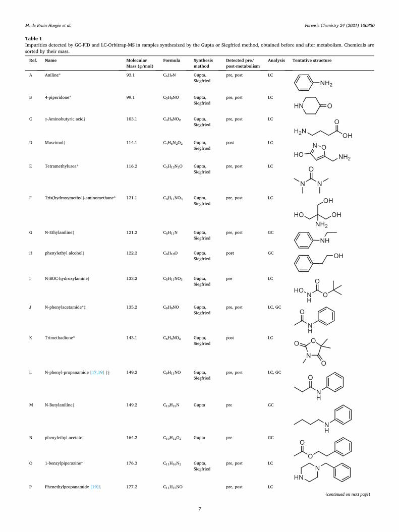
Table 1 (continued )
Ref. Name Molecular Mass (g/mol)
Formula Synthesis method
Detected pre/ post-metabolism
Analysis Tentative structure
Gupta, Siegfried
Q 2,6-Diisopropylaniline† 177.3 C12H19N Gupta pre, post LC
R 1-phenylethylpiperidin-4-ol [17,19]§ 205.3 C13H19NO Gupta, Siegfried
pre, post LC
S Acetyl norfentanyl [35]§ 218.3 C13H18N2O Gupta, Siegfried
post LC
T Norfentanyl* 232.3 C14H20N2O Gupta, Siegfried
post LC
U N-(1-phenylethylpiperidin-4-yl) propenamide*
260.4 C16H24N2O Gupta pre, post LC
V 4-anilino-N-phenethylpiperidine (4- ANPP)*‡
280.4 C19H24N2 Gupta, Siegfried
pre, post LC, GC
W Acetyl fentanyl*‡ 322.4 C21H26N2O Gupta, Siegfried
pre, post LC, GC
X Fentanyl*‡ 336.5 C22H28N2O Gupta, Siegfried
pre, post LC, GC
Y Methoxyacetyl fentanyl* 352.5 C22H28N2O2 Gupta, Siegfried
pre, post LC
Z β-Hydroxyfentanyl* 352.5 C22H28N2O2 Gupta, Siegfried
pre, post LC
(continued on next page)
M. de Bruin-Hoegee et al.
Forensic Chemistry 24 (2021) 100330
9
metabolism. For GC-FID the presence of acetyl fentanyl (W) was suffi-cient to confidently distinguish the two synthesis routes. Both synthesis routes showed a large peak for phenylethyl alcohol (H) which is a by- product of the metabolic conversion of fentanyl to norfentanyl. Despite the consistent presence of phenylethyl alcohol this compound can be used for differentiation because its concentration is much higher in Siegfried samples. This can be explained by the reduced level of im-purities in these samples. However, it should be noted that phenylethyl alcohol is a frequently encountered compound, which can have many
other possible origins. Table 3 shows the characteristic relative re-sponses of fentanyl impurities post metabolism. The responses were measured by GC-FID and LC-Orbitrap-MS for both the Gupta and Sieg-fried method. For example, if the relative response of impurity H is within 11–18% of the fentanyl peak area measured by GC-FID, the victim most likely has been exposed to fentanyl produced with the Siegfried method (assuming that the microsomes correctly mimic human metabolism). For LC-Orbitrap-MS the ratio of S/R was 10 ± 5 and 1.0 ± 1.3 (±95% confidence interval), for respectively the Gupta or Siegfried method. The ratio of W/R was 84 ± 50 and 9 ± 11. Because these intervals do not overlap, these ratios are interesting features for distinguishing between the synthesis routes post-metabolism.
3.3.3. PCA A PCA model was also built for the impurities detected by LC for the
post-metabolism samples. The score plot of the two first PCs showed grouping of the samples according to synthesis route as illustrated in Fig. 7. The first PC represents 73% of the variance with dominant loadings for the impurities acetyl fentanyl (W), norfentanyl (T), β-hydroxyfentanyl (Z) and acetyl norfentanyl (S). The second PC ac-counts for 22% of the total variance and is mainly characterized by norfentanyl (T), acetyl fentanyl (W), β-hydroxyfentanyl (Z), muscimol
Table 1 (continued )
Ref. Name Molecular Mass (g/mol)
Formula Synthesis method
Detected pre/ post-metabolism
Analysis Tentative structure
AA 3-(4-Methyl-1-piperazinyl)-1-[4-(4- morpholinyl)phenyl]-2,5- pyrrolidinedione†
358.4 C19H26N4O3 Gupta, Siegfried
pre, post LC
AB MFCD01916771† 374.4 C22H22N4O2 Gupta, Siegfried
pre, post LC
*The compound was tentatively identified with targeted MS/MS, †Identification with Compound Discoverer ‡Identification with spectral comparison to the NIST mass spectral library. §The tentative structure was determined by comparison with literature.
Table 2 Characteristic relative responses of fentanyl impurities measured by GC-FID and LC-Orbitrap-MS for the Gupta and Siegfried method. Samples were measured pre-metabolism. The relative responses of the impurities are given with the 95% confidence interval (n = 5) and represent the peak area relative to that of fentanyl.
GC-FID LC-Orbitrap-MS
Impurity Gupta (%) Siegfried (%) Gupta (%) Siegfried (%)
R n.a. n.a. 0.12–0.42 0.011–0.045 V 0 0.4–2 0.09–0.79 0.3–5.1 W 14–22 0 23–47 0.2–1.4
A B
Fig. 5. A) PCA-score plot based on 21 impurities detected by LC-Orbitrap-MS pre-metabolism showing good separation of the synthesis methods Gupta (G) and Siegfried (S). B) Corresponding PCA loadings. PC1 represents 96% of the variance and is predominantly composed of impurity W. PC2 accounts for 3% of the total variance and is mainly characterized by J, L, V and AA.
M. de Bruin-Hoegee et al.
Forensic Chemistry 24 (2021) 100330
10
(D) and N-phenylacetamide (J). The LC chromatogram showed a large response of norfentanyl which is the main product of the metabolism of fentanyl and hence is present in all post-metabolism samples. N-phe-nylacetamide (J) is mainly detected in the Gupta samples and is most likely the product of the reaction between aniline and acetic acid. The latter is only used in the Gupta synthesis method.
3.3.4. LDA Like the pre-metabolism samples, PCA was used for dimensionality
reduction before the application of LDA. Subsequently, LDA was applied to maximize the discrimination between the post-metabolism profiles for the two synthesis methods. Fig. 8 shows the distributions of the LDA values for both synthesis methods. The distributions for the post- metabolism profiles as measured with LC-Orbitrap-MS show only a
small overlap, indicating that the synthesis method information can still confidently be retrieved after metabolic processes have taken place. The discussion in the next section elaborates on the transformation to LRs.
4. Discussion
Tippett plots were used to evaluate the performance of the LDA model for LR calculations [37]. In addition to LR calculations, Tippett plots can be used to provide information on the number of mis-classifications [34]. Fig. 9 shows the cumulative LR distributions for samples measured by LC-Orbitrap-MS. The Tippett plot shows the fraction of fentanyl samples that have an LR greater than the given value for samples produced with the Gupta (H1) and Siegfried (H2) method. The LRs were calculated for LDA values between respectively − 20 to 20 and − 8 to 8 for pre- and post-metabolism samples. This corresponds to the visual range of LDA values shown in Fig. 6 and Fig. 8. For pre- metabolism samples, LDA values above 3 were not used for the calcu-lation, due to machine underflow. The LDA model for both pre- and post- metabolism samples yields well separated likelihood distributions and high evidential strength. If for an unknown sample a large positive LR is found the obtained profile is more probable when the victim has been exposed to fentanyl produced with the Gupta method (H1) than when the victim has been exposed to fentanyl produced with the Siegfried method (H2). Vice versa, for a large negative LR the evidence is more probable when H2 is true than when H1 is true. In over 50% of the pre- metabolism fentanyl samples, the LR exceeds very high minimum and maximum values of 10-300 and 1090. It is expected that after metabolism the impurity profile will contain less synthesis method specific infor-mation due to marker loss, dilution and reactivity. The aim of this study was to investigate whether sufficient information would still be
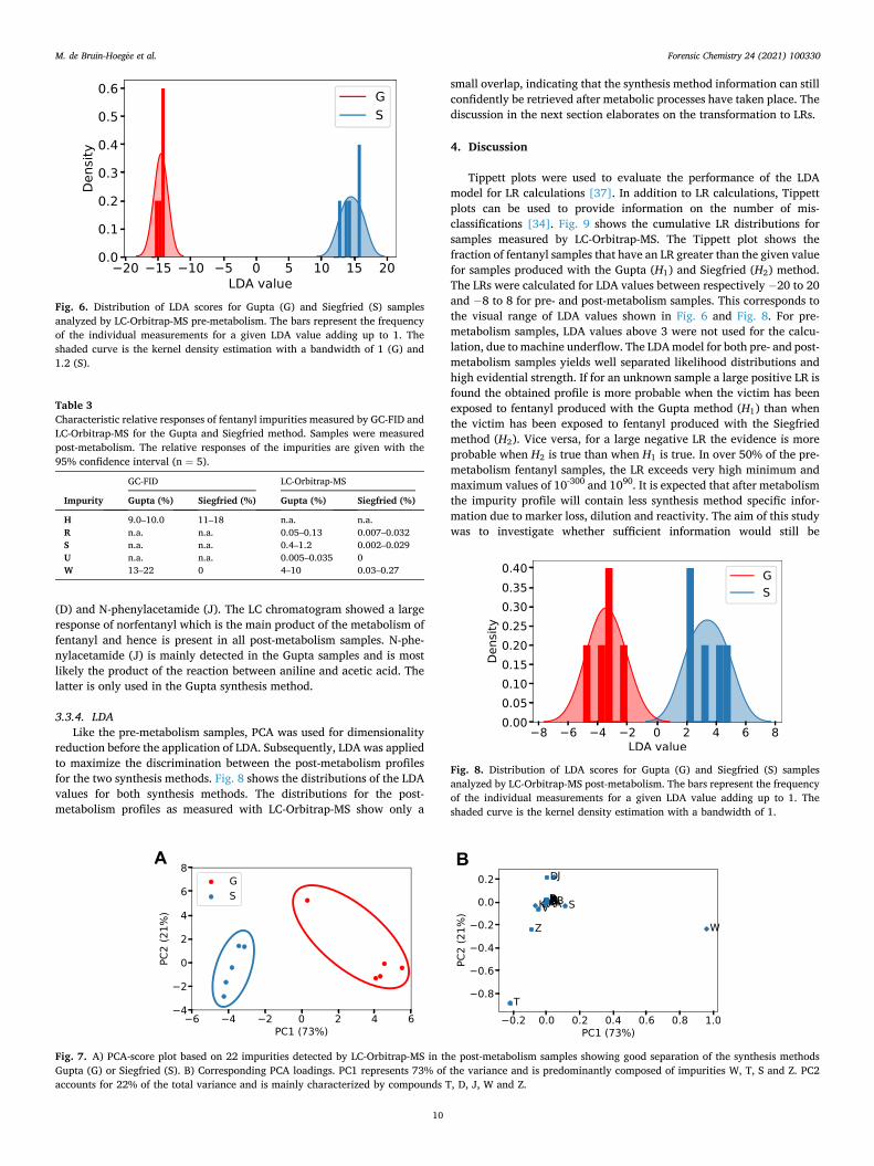
Fig. 6. Distribution of LDA scores for Gupta (G) and Siegfried (S) samples analyzed by LC-Orbitrap-MS pre-metabolism. The bars represent the frequency of the individual measurements for a given LDA value adding up to 1. The shaded curve is the kernel density estimation with a bandwidth of 1 (G) and 1.2 (S).
Table 3 Characteristic relative responses of fentanyl impurities measured by GC-FID and LC-Orbitrap-MS for the Gupta and Siegfried method. Samples were measured post-metabolism. The relative responses of the impurities are given with the 95% confidence interval (n = 5).
GC-FID LC-Orbitrap-MS
Impurity Gupta (%) Siegfried (%) Gupta (%) Siegfried (%)
H 9.0–10.0 11–18 n.a. n.a. R n.a. n.a. 0.05–0.13 0.007–0.032 S n.a. n.a. 0.4–1.2 0.002–0.029 U n.a. n.a. 0.005–0.035 0 W 13–22 0 4–10 0.03–0.27
A B
Fig. 7. A) PCA-score plot based on 22 impurities detected by LC-Orbitrap-MS in the post-metabolism samples showing good separation of the synthesis methods Gupta (G) or Siegfried (S). B) Corresponding PCA loadings. PC1 represents 73% of the variance and is predominantly composed of impurities W, T, S and Z. PC2 accounts for 22% of the total variance and is mainly characterized by compounds T, D, J, W and Z.
Fig. 8. Distribution of LDA scores for Gupta (G) and Siegfried (S) samples analyzed by LC-Orbitrap-MS post-metabolism. The bars represent the frequency of the individual measurements for a given LDA value adding up to 1. The shaded curve is the kernel density estimation with a bandwidth of 1.
M. de Bruin-Hoegee et al.

Forensic Chemistry 24 (2021) 100330
11
available to discern the fentanyl production route. Fig. 9 shows that after liver microsome metabolism such information is indeed still available which is a very promising result. In over 50% of the fentanyl samples detected post-metabolism the LR will exceed minimum and maximum values of 10-8 and 107. These plots also allow the study of mis-classifications. For a method without misclassification, the pre- and post-metabolism graphs in Fig. 9 would be perfectly separated at LR = 1 (dashed line). A perfect separation is visible for the pre-metabolism samples in the left plot. The post-metabolism distributions in the right plot show a small fraction of Siegfried samples with an LR > 1 and a small fraction of Gupta samples with an LR < 1. Consequently, on the basis of the fitted KDEs, a false positive error rate of 0.4% and 0.3% is expected for the Siegfried and Gupta samples respectively. The reason for this is the overlap in the KDEs shown in Fig. 6 and Fig. 8, which can be the result of the limited dataset. To improve the accuracy of the LR values, more data is required and a post-hoc calibration step is necessary [38]. Hence the LDA results and associated LR values presented in this study are of an indicative nature only.
The findings of the current study may be limited, because we had no access to real casework samples to verify the results. Having access to such samples is difficult as in many countries criminal law forbids the use of human biological case work samples for forensic research. Furthermore, fentanyl induced overdose is currently still extremely rare in the Netherlands. However, in future investigations it is especially important to demonstrate the presence of the markers in real forensic toxicological samples, i.e. to demonstrate that the impurity profile can also be retrieved from whole blood samples from overdose victims. In addition, it might be valuable to use a separate training and test set, to see whether unknown samples are correctly classified. Due to the limited number of syntheses that were performed, such a robust validation could not be conducted. Furthermore, samples synthesized by other labora-tories or with chemicals from other suppliers could give different results. Surprisingly, Mayer et al. identified 4-ANPP (V) as a potential marker for the Gupta synthesis method, whereas it was identified as a Siegfried marker in the current study [17]. A possible explanation for this con-tradictory result could be that Mayer et al. made some modifications to the Siegfried method, whereas in this study the synthesis was kept as close as possible to the original method as published by Siegfried. This suggests that small differences in synthesis method could have large consequences on the impurities present in fentanyl samples. An advan-tage however is that this would open possibilities to link a seized fen-tanyl sample to a specific laboratory. The prospect of linking a biomedical profile obtained from a victim to a given batch of material found at the home of a suspect could be very valuable in a forensic setting.
A concentration of 15 µg fentanyl per kg body mass is associated with health effects like chest wall rigidity, apnea and loss of consciousness [39]. Lethal doses are estimated to be a few milligrams, based on an lethal dose (LD50) of 30 µg/kg for non-human primates [3]. Patches designed for analgesic use, deliver fentanyl in a rate of 12–100 µg/h. A
specific case study describes a homicide attempt involving 120 μg/kg fentanyl (around 10 times the effective dose (ED50)), with a total dose of 6 mg. On arrival at the emergency department the young female victim was apneic with pinpoint pupils and had a decreased level of con-sciousness. She responded well to naloxone and was eventually dis-charged after nine days [40]. In the current study 100 µg was used for the microsomal incubations. This is a lower concentration than in the described examples, which might indicate that the sensitivity of the analysis methods applied in this work is sufficient for the levels relevant in forensic casework.
Because a model was used to mimic human metabolism, it is relevant to compare our post-metabolism results with fentanyl concentration in biomedical samples. It has been reported that fentanyl serum levels range from 0.3 to 2.5 ng/mL in patients that use fentanyl as a trans-dermal patch, and from 3.0 to 383 ng/mL in overdose victims [41]. Similar concentrations have been reported in blood and urine of over-dose victims [42]. In this study, a higher fentanyl concentration was found after metabolism. A sensitivity analysis was applied in order to determine the lowest fentanyl level for which characteristic impurities can still be detected by the LC-Orbitrap-MS. These data must be inter-preted with caution, because the lower concentration samples were dissolved in water thus not mimicking a complex biological matrix. To support these findings, the sensitivity analysis could be repeated with lower pre-metabolism concentrations and fentanyl samples spiked in biological matrices. For the impurities described in Table 3, acetyl norfentanyl (S) and N-(1-phenylethylpiperidin-4-yl) propanamide (U) were detected up to a fentanyl level of 90 ng/mL. This is in the range of levels found in victims after an overdose. The impurities 1-phenylethyl-piperidin-4-ol (R) and acetyl fentanyl (W) could even be detected up to a lower concentration of 0.9 ng/mL. This indicates that these impurities can also be detected in persons that receive a medical dose or experience mild poisoning symptoms. Qin et al. reported that acetyl norfentanyl (S) and acetyl fentanyl (W) could be detected up to a concentration of 2 pg/ mg by UPLC-MS/MS [43].
Based on the match criterion method and PCA analysis, it can be concluded that N-phenylacetamide (J), 1-phenylethylpiperidin-4-ol (R) and acetyl fentanyl (W) are the most important markers for discrimi-nation. However, acetyl fentanyl is not stable during metabolism, as it is metabolized to acetyl norfentanyl (S). In the present study 10% of acetyl norfentanyl was metabolized. Fortunately, enough acetyl fentanyl was left to discriminate between the synthesis methods. In the literature, contradictory results are reported regarding the extent of fentanyl metabolism. Some studies suggest that (almost) no intact fentanyl is left after exposure [44,45], while other authors state that norfentanyl often remains undetected in overdose cases [46]. In either case, impurities can be detected, so acetyl norfentanyl could be used as a potential marker if no acetyl fentanyl is left. It must be noted that acetyl (nor)fentanyl needs to be found in combination with (nor)fentanyl to be indicative of the Gupta synthesis method as acetyl fentanyl itself has also been classified as an illicit drug [6,47]. There are two other impurities that show
A B
Fig. 9. Tippett plots showing the cumulative likelihood ratio (LR) distributions calculated with LDA for fentanyl samples produced with the Gupta (G) and Siegfried (S) method, measured by LC-Orbitrap-MS. A) Pre-metabolism. B) Post-metabolism. The dashed lines show LR = 1.
M. de Bruin-Hoegee et al.

Forensic Chemistry 24 (2021) 100330
12
discriminative features pre- as well as post-metabolism. The first, phe-nylacetamide (J), can be formed from the starting materials aniline and acetic acid. The latter compound is only used for the Gupta synthesis, which is a probable explanation for the increased presence in these samples. The second impurity, 1-phenylethylpiperidin-4-ol (R), is a reduction product of the synthesis intermediate NPP. This compound is present at a slightly higher concentration in the Gupta samples, which is likely due to the presence of reducing agents and the lack of purification of intermediate reaction products. Therefore, phenylacetamide, 1-phe-nylethylpiperidin-4-ol, acetyl fentanyl and acetyl norfentanyl are the most important markers to discriminate between the Gupta and Sieg-fried synthesis method.
5. Conclusions
In the present study, the effect of human metabolism on the impurity profile of fentanyl was investigated with the use of human liver micro-somes. The aim of the study was to identify marker compounds for a specific fentanyl synthesis method that could be detected post- metabolism, in order to evaluate the feasibility of using biological samples for chemical provenance purposes. In cases where only bio-logical samples are available, the potential added value of the meta-bolism attribution method is obvious. In case where both biomedical samples and samples not subjected to metabolism are available, com-bination of both methods would provide two complementary datasets. A total of 24 impurities were detected pre-metabolism and 23 impurities were identified post-metabolism. On the basis of a simple match crite-rion the fentanyl synthesis route could be established from the post- metabolism profile. Phenylethyl alcohol was found to be indicative of the Siegfried method, while acetyl norfentanyl, N-(1-phenyl-ethylpiperidin-4-yl) propanamide, 1-phenylethylpiperidin-4-ol and acetyl fentanyl were indicative of the Gupta method. Classification of the synthesis method was also possible through the application of the PCA model. Eight relevant impurities were identified in the post- metabolism profiles using PCA, including three impurities which were also characteristic for the pre-metabolism profiles. Acetyl fentanyl and 1-phenylethylpiperidin-4-ol were characteristic both pre- and post- metabolism and could be detected up to levels that are realistic for forensic casework. GC-FID and LC-Orbitrap-MS can be used separately to discriminate between the Gupta and Siegfried synthesis method. Although it is valuable to use two different analysis methods, it is not necessary for obtaining appropriate discrimination. As one of the two methods is sufficient for the synthesis route attribution, almost every forensic (toxicological) laboratory will be able to implement post- metabolic fentanyl profiling. However, additional research is needed with respect to synthesis variations in a criminal setting and the pres-ervation of the fentanyl marker profiles in whole blood samples of vic-tims in forensic case work. Nonetheless, this work for the first time introduces a post-metabolic profiling concept that can be applied to human biological samples for forensic casework and intelligence purposes.
CRediT authorship contribution statement
Mirjam Bruin-Hoegee: Conceptualization, Software, Visualization, Writing - original draft. Djarah Kleiweg: Methodology, Validation, Investigation. Daan Noort: Conceptualization, Writing - review & editing. Arian C. Van Asten: Writing - review & editing.
Declaration of Competing Interest
The authors declare that they have no known competing financial interests or personal relationships that could have appeared to influence the work reported in this paper.
Acknowledgments
This work is part of the Forensic Attribution for CWA INtelliGence (FACING) project, a collaboration between the Van ’t Hoff Institute for Molecular Sciences (HIMS) of the University of Amsterdam and TNO Defence, Safety & Security. The FACING project is financed by the DO- AIO fund of the Dutch Ministry of Defence; part of the research was performed under grant V1802.
We would like to thank Debora van der Riet-Van Oeveren and Ad L. de Jong (TNO Rijswijk) for their assistance with LC-Q-TOF-MS and LC- Orbitrap-MS analyses. We are also grateful to Gerrit-Jan de Bruin of Leiden University for fruitful discussions on multivariate statistics and critically reviewing the manuscript.
Appendix A. Supplementary data
Supplementary data to this article can be found online at https://doi. org/10.1016/j.forc.2021.100330.
References
[1] D.J. Heslop, P.G. Blain, Threat potential of pharmaceutical based agents, Intell. Natl. Secur. 35 (4) (2020) 539–555, https://doi.org/10.1080/ 02684527.2020.1750158.
[2] T.H. Stanley, The fentanyl story, J. Pain. 15 (12) (2014) 1215–1226, https://doi. org/10.1016/j.jpain.2014.08.010.
[3] R. Vardanyan, V. Hruby, Future prospects for pharmaceutical applications, Future Med. Chem. 6 (2014) 385–412, https://doi.org/10.4155/fmc.13.215.Fentanyl- related.
[4] P. Frisoni, E. Bacchio, S. Bilel, A. Talarico, R.M. Gaudio, M. Barbieri, M. Neri, M. Marti, Novel synthetic opioids: the pathologist’s point of view, Brain Sci. 8 (2018) 1–17, https://doi.org/10.3390/brainsci8090170.
[5] J.R. Riches, R.W. Read, R.M. Black, N.J. Cooper, C.M. Timperley, Analysis of clothing and urine from Moscow theatre siege casualties reveals carfentanil and remifentanil use, J. Anal. Toxicol. 36 (2012) 647–656, https://doi.org/10.1093/ jat/bks078.
[6] P. Armenian, K.T. Vo, J. Barr-Walker, K.L. Lynch, Fentanyl, fentanyl analogs and novel synthetic opioids: a comprehensive review, Neuropharmacology 134 (2018) 121–132, https://doi.org/10.1016/j.neuropharm.2017.10.016.
[7] National Institute on Drug Abuse, Overdose Death Rates, (2019). https://www. drugabuse.gov/drug-topics/trends-statistics/overdose-death-rates (accessed September 3, 2020).
[8] N. Nicdaeid, R. Waddell, The analytical and chemometric procedures used to profile illicit drug seizures, Talanta 67 (2) (2005) 280–285, https://doi.org/ 10.1016/j.talanta.2005.05.018.
[9] A. Slosse, F. Van Durme, N. Samyn, D. Mangelings, Y. Vander Heyden, Evaluation of data preprocessings for the comparison of GC–MS chemical profiles of seized cannabis samples, Forensic Sci. Int. 310 (2020) 110228, https://doi.org/10.1016/j. forsciint.2020.110228.
[10] R. Dams, T. Benijts, W.E. Lambert, D.L. Massart, A.P. De Leenheer, Heroin impurity profiling: trends throughout a decade of experimenting, Forensic Sci. Int. 123 (2-3) (2001) 81–88, https://doi.org/10.1016/S0379-0738(01)00541-2.
[11] S. Lociciro, P. Hayoz, P. Esseiva, L. Dujourdy, F. Besacier, P. Margot, Cocaine profiling for strategic intelligence purposes, a cross-border project between France and Switzerland. Part I. Optimisation and harmonisation of the profiling method, Forensic Sci. Int. 167 (2-3) (2007) 220–228, https://doi.org/10.1016/j. forsciint.2006.06.052.
[12] M. Morelato, A. Beavis, M. Tahtouh, O. Ribaux, P. Kirkbride, C. Roux, The use of organic and inorganic impurities found in MDMA police seizures in a drug intelligence perspective, Sci. Justice. 54 (1) (2014) 32–41, https://doi.org/ 10.1016/j.scijus.2013.08.006.
[13] K. Andersson, E. Lock, K. Jalava, H. Huizer, S. Jonson, E. Kaa, A. Lopes, A. Poortman-van der Meer, E. Sippola, L. Dujourdy, J. Dahlen, Development of a harmonised method for the profiling of amphetamines VI. Evaluation of methods for comparison of amphetamine, Forensic Sci. Int. 169 (1) (2007) 86–99, https:// doi.org/10.1016/j.forsciint.2006.10.020.
[14] P.A. Janssen, J.F. Gardocki, Method for producing analgesia, U.S. Patent 3141823, 1964.
[15] Siegfried, Synthesis of Fentanyl, (2004). https://www.erowid.org/archive/ rhodium/chemistry/fentanyl.html (accessed October 28, 2020).
[16] P.K. Gupta, K. Ganesan, A. Pande, R.C. Malhotra, A convenient one pot synthesis of fentanyl, J. Chem. Res. 2005 (7) (2005) 452–453.
[17] B.P. Mayer, A.J. DeHope, D.A. Mew, P.E. Spackman, A.M. Williams, Chemical attribution of fentanyl using multivariate statistical analysis of orthogonal mass spectral data, Anal. Chem. 88 (8) (2016) 4303–4310, https://doi.org/10.1021/acs. analchem.5b04434.
[18] L. Moren, J. Qvarnstrom, M. Engqvist, R. Afshin-Sander, X. Wu, J. Dahlen, C. Lofberg, A. Larsson, A. Ostin, Attribution of fentanyl analogue synthesis routes by multivariate data analysis of orthogonal mass spectral data, Talanta 203 (2019) 122–130, https://doi.org/10.1016/j.talanta.2019.05.025.
M. de Bruin-Hoegee et al.
Forensic Chemistry 24 (2021) 100330
13
[19] I.S. Lurie, A.L. Berrier, J.F. Casale, R. Iio, J.S. Bozenko, Profiling of illicit fentanyl using UHPLC-MS/MS, Forensic Sci. Int. 220 (1-3) (2012) 191–196, https://doi. org/10.1016/j.forsciint.2012.02.024.
[20] E.M. McBride, R.E. Keller, G.F. Verbeck, Direct-infusion electrospray ionization- mass spectrometry profiling of fentanyl and acetylfentanyl reaction mixtures, Int. J. Mass Spectrom. 428 (2018) 55–61, https://doi.org/10.1016/j.ijms.2018.03.004.
[21] J.F. Casale, P.A. Hays, S.G. Toske, J.R. Mallette, Unique bipiperidinyl impurities produced from the “One-Pot” synthesis of fentanyl, Forensic Chem. 17 (2020) 100203, https://doi.org/10.1016/j.forc.2019.100203.
[22] J.A. van der Meer, H.C. Trap, D. Noort, M.J. van der Schans, Comprehensive gas chromatography with Time of Flight MS and large volume introduction for the detection of fluoride-induced regenerated nerve agent in biological samples, J. Chromatogr. B. 878 (17-18) (2010) 1320–1325, https://doi.org/10.1016/j. jchromb.2010.02.019.
[23] OPCW, United Nations Mission to Investigate Allegations of the Use of Chemical Weapons in the Syrian Arab Republic. Final report, (2013). https://unoda-web.s3. amazonaws.com/wp-content/uploads/%0A2013/12/report.pdf (accessed October 6, 2020).
[24] H. John, M.J. Van Der Schans, M. Koller, H.E.T. Spruit, F. Worek, H. Thiermann, D. Noort, H. John, Fatal sarin poisoning in Syria 2013: forensic verification within an international laboratory network, Forensic Toxicol. 36 (2018) 61–71, https:// doi.org/10.1007/s11419-017-0376-7.
[25] Tomonori Tateishi, Alastair J.J. Wood, F.Peter Guengerich, Margaret Wood, Biotransformation of tritiated fentanyl in human liver microsomes. Monitoring metabolism using phenylacetic acid and 2-phenylethanol, Biochem. Pharmacol. 50 (11) (1995) 1921–1924, https://doi.org/10.1016/0006-2952(95)02088-8.
[26] E. Feierman, M. Lasker, Metabolism of fentanyl, a synthetic opioid analgesic, by human liver microsomes, Drug Metab. Dispos. 24 (1996) 932–939.
[27] T. Tateishi, Y. Krivoruk, Y-F. Ueng, A.J.J. Wood, F.P. Guengerich, M. Wood, Identification of human liver cytochrome P-450 3A4 as the enzyme responsible for fentanyl and sufentanil N-dealkylation, Anesth. Analg. 82 (1) (1996) 167–172, https://doi.org/10.1097/00000539-199601000-00031.
[28] R.B. Labroo, M.F. Paine, K.E. Thummel, E.D. Kharasch, Fentanyl metabolism by human hepatic and intestinal cytochrome P450 3A4: Implications for interindividual variability in disposition, efficacy, and drug interactions, Drug Metab. Dispos. 25 (1997) 1072–1080.
[29] Jerome Guitton, Thierry Buronfosse, Michel Desage, Alain Lepape, Jean- Louis Brazier, Philippe Beaune, Possible involvement of multiple cytochrome P450S in fentanyl and sufentanil metabolism as opposed to alfentanil, Biochem. Pharmacol. 53 (11) (1997) 1613–1619, https://doi.org/10.1016/S0006-2952(96) 00893-3.
[30] Sepuri Asha, Maravajhala Vidyavathi, Role of human liver microsomes in in vitro metabolism of drugs-A review, Appl. Biochem. Biotechnol. 160 (6) (2010) 1699–1722, https://doi.org/10.1007/s12010-009-8689-6.
[31] F. Pedregosa, G. Varoquaux, A. Gramfort, V. Michel, B. Thirion, O. Grisel, M. Blondel, P. Prettenhofer, R. Weiss, V. Dubourg, J. Vanderplas, A. Passos, D. Cournapeau, M. Brucher, M. Perrot, E. Duchesnay, Scikit-learn: Machine Learning in Python, J. Mach. Learn. Res. 12 (2011) 2825–2830.
[32] M. de Bruin-Hoegee, G.J. de Bruin, Python code for Chemical Attribution of Fentanyl, (2020). https://doi.org/10.5281/zenodo.4276396.
[33] J.T. Scanlon, D.E. Willis, Calculation of flame ionization detector relative response factors using the effective carbon number concept, J. Chromatogr. Sci. 23 (8) (1985) 333–340.
[34] H. Brust, M. Koeberg, A. Van Der Heijden, W. Wiarda, I. Mügler, M. Schrader, G. Vivo-Truyols, P. Schoenmakers, A. Van Asten, Isotopic and elemental profiling of ammonium nitrate in forensic explosives investigations, Forensic Sci. Int. 248 (2015) 101–112, https://doi.org/10.1016/j.forsciint.2014.11.024.
[35] Shimpei Watanabe, Svante Vikingsson, Markus Roman, Henrik Green, Robert Kronstrand, Ariane Wohlfarth, In vitro and in vivo metabolite identification studies for the new synthetic opioids acetylfentanyl, acrylfentanyl, furanylfentanyl, and 4-fluoro-isobutyrylfentanyl, AAPS J. 19 (4) (2017) 1102–1122, https://doi. org/10.1208/s12248-017-0070-z.
[36] Jian Yang, Jing-yu Yang, Why can LDA be performed in PCA transformed space? Pattern Recognit. 36 (2) (2003) 563–566, https://doi.org/10.1016/S0031-3203 (02)00048-1.
[37] I.W. Evett, J.S. Buckleton, Statistical analysis of STR data, in: A. Carracedo, B. Brinkmann, W. Bar (Eds.), Adv. Forensic Haemogenet., Springer, Heidelberg, 1996: pp. 79–86. https://doi.org/10.2169/naika.96.385.
[38] Andrew van Es, Wim Wiarda, Maarten Hordijk, Ivo Alberink, Peter Vergeer, Implementation and assessment of a likelihood ratio approach for the evaluation of LA-ICP-MS evidence in forensic glass analysis, Sci. Justice. 57 (3) (2017) 181–192, https://doi.org/10.1016/j.scijus.2017.03.002.
[39] J.B. Streisand, P.L. Bailey, L. LeMaire, M.A. Ashburn, S.D. Tarver, J. Varvel, T. H. Stanley, Fentanyl-induced rigidity and unconsciousness in human volunteers, Clin. Investig. 78 (1993) 629–634.
[40] Mark D. Lyttle, Sapna Verma, Rhian Isaac, Transdermal fentanyl in deliberate overdose in pediatrics, Pediatr. Emerg. Care. 28 (5) (2012) 463–464, https://doi. org/10.1097/PEC.0b013e31825358b4.
[41] J. Suzuki, S. El-Haddad, A review: fentanyl and non-pharmaceutical fentanyls, Drug Alcohol Depend. 171 (2017) 107–116, https://doi.org/10.1016/j. drugalcdep.2016.11.033.
[42] G.L. Henderson, Fentanyl-related deaths: demographics, circumstances, and toxicology of 112 cases, J. Forensic Sci. 36 (1991) 422–433, https://doi.org/ 10.1520/jfs13045j.
[43] N. Qin, M. Shen, P. Xiang, D. Wen, B. Shen, H. Deng, H. Qiang, F. Song, Y. Shi, Determination of 37 fentanyl analogues and novel synthetic opioids in hair by UHPLC-MS/MS and its application to authentic cases, Sci. Rep. 10 (2020) 1–13, https://doi.org/10.1038/s41598-020-68348-w.
[44] Deborah A. McClain, Carl C. Hug, Intravenous fentanyl kinetics, Clin. Pharmacol. Ther. 28 (1) (1980) 106–114, https://doi.org/10.1038/clpt.1980.138.
[45] Alphonse Poklis, Fentanyl: a review for clinical and analytical toxicologists, Clin. Toxicol. 33 (5) (1995) 439–447, https://doi.org/10.3109/15563659509013752.
[46] Glenn Burns, Rebecca T. DeRienz, Daniel D. Baker, Marcel Casavant, Henry A. Spiller, Could chest wall rigidity be a factor in rapid death from illicit fentanyl abuse? Clin. Toxicol. 54 (5) (2016) 420–423, https://doi.org/10.3109/ 15563650.2016.1157722.
[47] M. Wilde, S. Pichini, R. Pacifici, A. Tagliabracci, F.P. Busardo, V. Auwarter, R. Solimini, Metabolic pathways and potencies of new fentanyl analogs, Front. Pharmacol. 10 (2019) 1–16, https://doi.org/10.3389/fphar.2019.00238.
M. de Bruin-Hoegee et al.