MicroRNA Regulation of Cbx7 Mediates a Switch of Polycomb Orthologs during ESC Differentiation
Upload
u-bordeaux1Category
view
2download
0
Characterization of the nm23-M2, nm23-M3 and nm23-M4 mouse genes:comparison with their human orthologs
K. Massea,1, S. Dabernata, P.-M. Bourbona, M. Laroua, L. Amreina, P. Barrauda,2, Y. Perela,M. Camaraa,3, M. Landrya,4, M.-L. Lacombeb, J.-Y. Daniela,*
aBiologie de la Differenciation et du Developpement, Universite Victor Segalen-Bordeaux2, 146 rue Leo Saignat, 33076 Bordeaux, FrancebINSERM U402, Faculte de Medecine Saint Antoine, 75012 Paris, France
Received 3 March 2002; received in revised form 24 June 2002; accepted 8 July 2002
Received by E. Boncinelli
Abstract
The nm23 gene family is thought to be involved in physiopathological processes such as growth, differentiation and cancer promotion,
progression or metastasis. We report here the mouse nm23-M3 and nm23-M4 complementary DNA sequences and the genomic cloning,
characterization and tissue expression pattern of the nm23-M2, nm23-M3 and nm23-M4 genes, in comparison with their human and rat
orthologs and with the human nm23-H1 and mouse nm23-M1 genes. The organization and structure of the members of this gene family are
remarkably similar in human and rodents. Accordingly, the striking similarities between the human and mouse nm23 genes enable the use of
mouse transgenic and knock-out models for studying the role of nucleoside diphosphate kinase isoforms in human physiopathology. q 2002
Elsevier Science B.V. All rights reserved.
Keywords: Nucleoside diphosphate kinase; Gene organization; Expression
1. Introduction
Nucleoside diphosphate (NDP) kinase was initially
described as a housekeeping enzyme which catalyzes the
transfer of a g-phosphate between nucleoside tri- and di-
phosphates, via a phosphohistidine intermediate (Lascu
and Gonin, 2000, for review). Over the past 10 years,
however, new data have rekindled interest in this protein
which exists as different isoforms encoded by a family of
highly related genes. Currently, eight human nm23 genes
have been characterized. The first four human members of
this gene family, nm23-H1, nm23-H2, nm23-H3 (or DR-
nm23) and nm23-H4, express protein products which
possess NDP kinase activity and are named respectively
NDP kinase A–D (Lacombe et al., 2000, for review).
A growing set of evidence suggests that in addition to
their basic enzymatic activity, NDP kinase isoforms might
display other functions more or less related to signal trans-
duction pathways, cell growth and differentiation, embryo-
nic development, tumor progression, metastasis and
apoptosis (see for reviews, de S. Otero, 2000; Hartsough
and Steeg, 2000; Kimura et al., 2000; Lacombe et al.,
2000). Additionally, the human NDP kinase B has been
shown to be identical to the c-myc transcription factor
PuF (Postel et al., 2000b, for review) and was recently
shown to be involved in DNA repair processes (Postel et
al., 2000a). However, the precise role and mechanism of
action of these additional functions of the NDP kinases
are not yet well understood. Moreover, it was established
by in vitro experiments that NDP kinase is a heterohexa-
meric enzyme which contains randomly associated subunits
consisting of A and B isoforms (Gilles et al., 1991).
Although the four recombinant NDP kinases can be recon-
stituted into heterohexamers in vitro (Lascu et al., 2000), no
information is presently available either on the physiologi-
Gene 296 (2002) 87–97
0378-1119/02/$ - see front matter q 2002 Elsevier Science B.V. All rights reserved.
PII: S0378-1119(02)00836-3
www.elsevier.com/locate/gene
Abbreviations: dpc, days post-coitum; ES cells, embryonic stem cells;
EST, expressed sequence tag; K-pn mutation, killer of prune mutation;
NDP kinase, nucleoside diphosphate kinase; PCR, polymerase chain reac-
tion; RT, reverse transcription
* Corresponding author. Tel.: 133-5-5757-1284; fax: 133-5-5624-0643.
E-mail address: [email protected]
(J.-Y. Daniel).1 Present address: Department of Biological Sciences, University of
Warwick, Coventry, UK.2 Present address: Wallenberg Neuroscience Center, Division of Neuro-
biology, BMC A11, 221 84 Lund, Sweden.3 Present adress: Laboratoire de Biochimie Medicale, CHU Cocody,
Abidjan, Cote d’Ivoire, France.4 Present address: INSERM EPI 9914, Institut Francois Magendie, rue
Camille Saint-Saens, 33077 Bordeaux, France.

cal effects of the relative concentration of each subunit in
the complex formation, or on a possible involvement in vivo
of the C and D isoforms in the hexameric conformation of
the protein in a living organism. Therefore, it is difficult to
know whether each of the many suspected biological roles
of the different NDP kinase isoforms is performed in a
particular cell compartment either through their interactions
with cell-specific factors or through the precise composition
of the NDP kinase heterohexamers in this cell compartment.
A unique animal model for studying the relationships that
exist between the different NDP kinase isoforms and their
biological function ex vivo or in vivo could be used to
address these questions. As a first step towards developing
a mouse model, we have previously described the organiza-
tion and expression pattern of the mouse gene encoding
NDP kinase A (Dabernat et al., 1999a). We describe here
the cloning, characterization and tissue distribution pattern
of the murine nm23-M2, nm23-M3 and nm23-M4 genes. The
properties of these genes are compared to those of their
orthologs previously published in the rat and to the corre-
sponding human genes, either published or identified from
genome sequences registered in database.
2. Material and methods
2.1. Cloning of the mouse nm23-M3 and nm23-M4
complementary DNAs (cDNAs)
A search with the FASTA program (Pearson and Lipman,
1988) in the EMBL sequence data base, for mouse
expressed sequence tags sharing more than 80% nucleotide
identity with human cDNA for nm23-H3 and nm23-H4
(Venturelli et al., 1995; Milon et al., 1997) led to the iden-
tification of several clones corresponding to the putative
mouse isoforms nm23-M3 (13 clones) and nm23-M4 (10
clones). The consensus sequence corresponding to the
coding domain and to the 3 0 untranslated region of the
mouse nm23-M3 and nm23-M4 cDNAs was deduced from
the sequence alignment of these clones by using the CLUS-
TALW program (Thompson et al., 1991). Reverse transcrip-
tion-polymerase chain reaction (RT-PCR) reactions were
then carried out on mouse liver and heart total RNA by
using gene-specific primer pairs designed from each
cDNA consensus sequence, namely 5 0TGA CCA TCT
TTG CTA ACC/5 0CAA CCT CCA CGC AGA AAT for
nm23-M3 and 5 0GAG GAA GCC ATT CTA CCC/5 0TGA
CAG AGG TAG TAG GTC for nm23-M4. Finally, the
precise sequences of the mouse transcripts were established
by direct sequencing of the amplification products. Two
cDNA clones displaying the closest similarity with the
sequence of each RT-PCR product (ID: MMAA8932; AC:
g1660383 for nm23-M3 and ID:MMA59517; AC:
AA059517 for nm23-M4) were finally obtained from
cDNA clone I.M.A.G.E. the United Kingdom Human
Genome Mapping Project (Cambridge), verified by
sequence analysis and corrected for their nucleotide
mistakes. Site-directed mutagenesis was used to introduce
a G in position 228 (GenBank AF288689) in nm23-M3
sequence and a G and a C in position 59 and 149, respec-
tively in nm23-M4 sequence (GenBank AF288690). Three
rounds of PCR were necessary for each insertion. During the
first two rounds, the fragments upstream and downstream of
the insertion carried by the primers were amplified. Then
these partially overlapping PCR products were used
together as templates to amplify the entire coding sequence,
resulting in the production of a corrected sequence. Taq
DNA polymerase from Boehringer was used according to
the manufacturer’s recommendations (Boehringer
Mannheim, Germany). The corrected cDNAs were then
inserted into the cloning vector pBluescript II SK2 (Strata-
gene, La Jolla, CA, USA) for the production of specific
probes. The nm23-M3 and nm23-M4 cDNA sequences
were submitted to GenBank databases.
2.2. Cloning of the mouse nm23-M2, nm23-M3 and nm23-
M4 genes
2.2.1. Screening of the genomic library
The genes were isolated by screening an embryonic stem
cell (ES cell) genomic library constructed in the lEMBL3
vector, with 32P labeled probes corresponding to the 3 0
untranslated regions of mouse nm23-M2, nm23-M3 and
nm23-M4 cDNA. The ES library was a generous gift from
Prof. P. Chambon, (IGBMC, Ilkirch, France). Positive
clones were purified by three other subsequent screening
rounds, the first two by using the same probes and the last
one with a specific probe located in the 5 0 region of each
gene: Since in human and in rat, nm23-1 and nm23-2 genes
are located on the same chromosome and are distant by less
than 5000 bp (Shimada et al., 1993; Seifert et al., 1995), the
3 0 untranslated domain sequence of the mouse nm23-M1
gene was used as a probe for purifying nm23-M2 gene
clones containing the full length 5 0 upstream regulatory
domain. The genes nm23-M3 and nm23-M4 were character-
ized by using specific cDNA probes chosen in their 5 0 trans-
lated presequence region and were registered in a library
data base (GenBank AF288691 and GenBank AF288692,
respectively).
2.2.2. Identification of exon-intron boundaries of nm23-2,
nm23-3 and nm23-4 genes
The putative intron boundaries of the mouse genes were
positioned by reference to the organization of the mouse
nm23-M1 gene (Dabernat et al., 1999a). Primers were
designed from exon sequences adjacent to each putative
junction. By using these primers, the introns of each purified
phage were amplified by PCR, and then the regions neigh-
boring the exon-intron borders were sequenced. The follow-
ing PCR primer pairs were used to characterize the exon/
intron boundaries of the different genes. For the nm23-2
gene: 5 0GAC CCA CCG GCT TTC GGT/5 0GAG GTT
K. Masse et al. / Gene 296 (2002) 87–9788
GGC CAT GGT CCT (intron 1); 5 0CTC GAG CGT ACC
TTC ATT/5 0TCA TGT ACT TCA CCA GCC (intron 2); 5 0
CTC TGA AGA ACA CCT GAA/5 0GCC AAC TTG AAT
GCA GAA (intron 3) and 5 0TGG GAG GGG CTC AAT
GTG/5 0CAT CCT GTC AGT GGG ATG (intron 4). For the
nm23-3 gene: 5 0ATG ATC TGT CTG GTG CTG/5 0CTA
GCT TCA GTG CCA CCA (intron 1); 5 0CGT GAA CGA
GCG CAC GTT/5 0CAC GGG GCC AGA ACT CAT (intron
2); 5 0CGA AGA GCT ACT GCG GGA/5 0CCT ATG AGG
GCT CGC GAA (intron 3); and 5 0GGG ACG CCA TGC
CCG GTA/5 0TAT GAA AGG CAA CAA GGG (intron 4) –
and finally for the nm23-4 gene: 5 0ATG GGC AGC CTT
TTC GGG/5 0GCC TCT CAA AGC GTT GTA (intron 1);
5 0CAA GAG CGG ACG CTG GTT/5 0CAC CAC AGG
CCC AGA GCT (intron 2); 5 0ACC AGA AAG CAT CCT
TGC/5 0CTA TCA TGG CCC TTG AGA (intron 3) and
5 0ACT CAA CAG AGG CAG CCC/5 0GTC TCT CCT
TTG AGG CAG (intron 4).
2.3. DNA Sequencing and PCR amplifications
DNA sequence analysis was performed on double-
stranded plasmids, phages or PCR products by the Sanger
dideoxy chain termination method using a commercially
available kit (BigDye Terminator from Perkin Elmer
Corporation, Norwalk, CT, USA) and primers designed to
obtain overlapping sequences on both DNA strands. PCR
amplifications were carried out using the Goldstarw taq
DNA polymerase (Eurogentec Seraing, Belgium) according
to the manufacturer’s recommendations.
2.4. Northern blot analysis
Northern blot analysis was performed using a commercial
adult mouse RNA poly(A)1 blot (MTN Blot, Clontech, Palo
Alto, CA, USA). The nm23-M1, nm23-M2, nm23-M3 and
nm23-M4 specific probes were produced by PCR amplifica-
tion from the 3 0 untranslated region of each cDNA. The
primers used were 5 0TAG-GAC-GGT-GCC-GGT-TTT/
5 0TCT-AAT-GAA-TTC-TCT-GTT for nm23-M1, 5 0TAG-
ACA-TGA-AGA-AAC-CAG/5 0CAG-TTC-CAA-AGT-
CTT-TAT for nm23-M2, 5 0GAT GTT ATT GCA GTC
AGC/5 0TAT GAA AGG CAA CAA GGG for nm23-M3
and 5 0CTT GCA CTG CCT TCT GCA/5 0GTC TCT CCT
TTG AGG CAG for nm23-M4. A cDNA encoding the
ubiquitous ribosomal protein (36B4) was used as a control
probe to normalize RNA loading (Laborda, 1991). The
36B4 clone was kindly provided by Prof. P. Chambon.
The blots were analyzed using a phosphorimager (Molecu-
lar Dynamics, Sunnyvale, CA, USA) and quantitative analy-
sis was carried out by the ImageQuant software (Molecular
Dynamics). The relative messenger RNA (mRNA) abun-
dance of each nm23 isoform in each tissue extract was
normalized by reference to the amount of the control
36B4 mRNA and standardized to heart nm23 mRNA
contents. Probe labeling was performed as already described
(Dabernat et al., 1999b), and hybridization was done
according to the recommendation of the membrane manu-
facturer.
2.5. RNase mapping
Total RNAs from kidney tissue of adult mice were
extracted by using Trizol (GIBCO BRL) according to the
manufacturer’s protocol. Three recombinant pBS-SK2 plas-
mids were used in this study, namely pM2, pM3 and pM4.
Each of them contains the first exon of each mouse gene, as
defined from its 3 0 splicing site, flanked by its 5 0 adjacent
genomic DNA domains. The different inserts were 394, 430
and 440 bp long respectively. The radioactive sense and
antisense probes were synthesized according to the plasmid
supplier’s recommendations (Stratagene) using the T3 and
T7 RNA polymerases. Hybridizations and RNase digestions
were performed as previously described (Rio et al., 1988).
2.6. In situ hybridization
In situ hybridizations were carried out on C57BL/6 15.5
days post-coitum mouse embryos. The oligonucleotide
probes sequences were complementary to nucleotides
551–596 and 660–704 of the mouse nm23-M1 cDNA
(accession number U85511 GenBank), to nucleotides
477–522 and 524–569 of the mouse nm23-M2 cDNA
(Urano et al., 1992), to nucleotides 486–528 and 589–633
of the mouse nm23-M3 cDNA (numbering for initiation
ATG codon) and to nucleotides 599–643 and 771–815 of
the mouse nm23-M4 cDNA (numbering from initiation
ATG codon). The oligonucleotides were labeled by tailing
and were purified as previously described (Dagerlind et al.,
1992). In situ hybridization, autoradiography and control
experiments, using an excess of cold probes, were
performed as described previously (Dabernat et al.,
1999b). Tissue distribution was deduced from autoradio-
graphic films.
3. Results
3.1. Cloning of the mouse nm23-M3 and nm23-M4 cDNAs
and deduced amino-acid primary structure
The nm23-M3 cDNA (GenBank, accession number
AF288689) displays a 507-nucleotide-long translated region
encoding a putative protein of 169 amino acids (Fig. 1). The
first ATG possessed a Kozak-like sequence (cgccATGa)
and a single AATAAA polyadenylation consensus signal
was present 152 nucleotides downstream from the TAG
stop codon. The deduced primary structure of the NDP
kinase C mouse protein displayed 88% amino acid identity
with that of its human counterpart. By comparison with
human NDP kinase A, a NH2 17 amino acid hydrophobic
polypeptide extension was present in the mouse NDP kinase
C. This extension is identical to the corresponding domain
of the rat putative protein (GenBank, accession number
K. Masse et al. / Gene 296 (2002) 87–97 89
AY017337) and shares the same first 14 amino acids with
the human NDP kinase C. A Lys, which is equivalent to the
Arg 114 found in mammalian NDP kinase A and B was
present in the human and mouse NDP kinase C.
The nm23-M4 cDNA (GenBank, accession number
AF288690) contained an open reading frame of 558 nucleo-
tides for a predicted protein 186 amino acids long (Fig. 1).
The nucleotide sequence around the first ATG conforms to
the consensus Kozak sequence (caacATGg). The TGA stop
codon was followed by an untranslated domain of 292
nucleotides and the poly(A)1 tail was located 18 nucleotides
downstream from the ATTAAA polyadenylation signal. The
mouse protein sequence is 82% identical to its human coun-
terpart and displays several characteristics already described
for human NDP kinase D (Milon et al., 1997). The NH2
terminus extension of the mouse NDP kinase D was 32
amino acids long (33 amino acids in human) and shared
only 50% identical amino acids with the corresponding
human protein. However, this extension displayed a high
level of Arg, Leu and Ser (13 residues), Gly or Pro (10 resi-
dues) but only two Val, and no Lys, Asp, Glu or Ile. This
amino acid composition is characteristic of a mitochondrial
targeting sequence (von Heijne, 1986). By computer analysis
(von Heijne, 1986) and according to the numbering conven-
tion used in Fig. 1, a cleavage site may be postulated between
Pro(26) and Ser(25), of the protein primary sequence. This
site corresponds to the putative proteolytic cleavage site
between residues His(26) and Gly(25) already described
for the human protein (Milon et al., 2000). As already
found in human (Milon et al., 1997), the mouse NDP kinase
D presents a Ser(96) instead of the Pro(96) which is
constantly found in isoforms A, B and C. This substitution
is equivalent to the killer of prune (K-pn) mutation of the
Awd/NDP kinase of Drosophila (Timmons et al., 1995).
Moreover, the carboxy terminus of NDP kinase D is two
amino acids (PA) longer than that of the other mammalian
NDP kinases, which normally end with a tyrosine-glutamate
(YE) motif. Finally, neither NDP kinase C nor D showed the
concomitant presence of the three amino acids previously
described as being essential for the DNA binding of NDP
kinase B, namely Arg 34, Asn 69 and Lys 135 (Postel et
al., 2000b, for review). By contrast, all the mammalian
NDP kinases have the Lys 12, associated with the predicted
DNA repair functions of the protein (Postel et al., 2000a).
K. Masse et al. / Gene 296 (2002) 87–9790
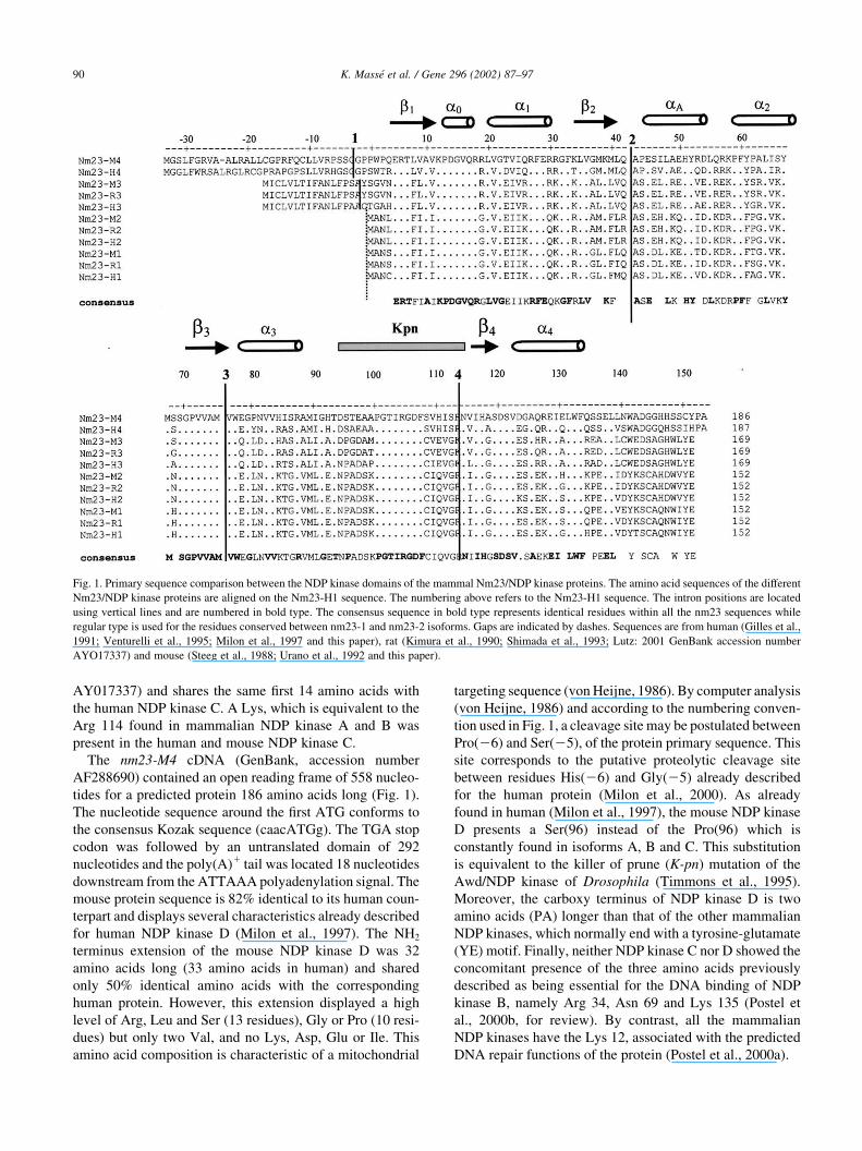
Fig. 1. Primary sequence comparison between the NDP kinase domains of the mammal Nm23/NDP kinase proteins. The amino acid sequences of the different
Nm23/NDP kinase proteins are aligned on the Nm23-H1 sequence. The numbering above refers to the Nm23-H1 sequence. The intron positions are located
using vertical lines and are numbered in bold type. The consensus sequence in bold type represents identical residues within all the nm23 sequences while
regular type is used for the residues conserved between nm23-1 and nm23-2 isoforms. Gaps are indicated by dashes. Sequences are from human (Gilles et al.,
1991; Venturelli et al., 1995; Milon et al., 1997 and this paper), rat (Kimura et al., 1990; Shimada et al., 1993; Lutz: 2001 GenBank accession number
AYO17337) and mouse (Steeg et al., 1988; Urano et al., 1992 and this paper).

3.2. Organization of the human and mouse nm23-2, nm23-3
and nm23-4 genes
As shown in Figs. 1 and 2, the first four members of the
nm23 gene family characterized so far in human, rat and
mouse exhibit the same pattern of organization. All of them
have four introns flanked by five exons. The position of the
fourth intron of the nm23-H3 gene, as reported on the NCBI
site, is not convincingly located. It was therefore corrected
by reference to the other genes (Fig. 2) and according to the
Breathnach and Chambon (1981) law. On a multiple nucleo-
tide sequence alignment, the exon-intron borders between
introns two, three and four are strictly conserved (Figs. 1
and 2) and the position of the first intron is always the same
for one particular gene of this family, whatever the species.
Finally, the overall gene sizes are about 9000 nucleotides for
nm23-1 genes, 6000 nucleotides for nm23-2 genes, 1000
nucleotides for nm23-3 genes and 3500 nucleotides for
nm23-4 genes.
3.3. Promoter organization of human and mouse nm23-1,
nm23-2, nm23-3 and nm23-4 genes
The 1800 bp 5 0 region upstream the first exon of the four
human and mouse orthologous genes were compared for
their nucleotide distribution and were studied by computer
analysis with the Prestridge (1991) software for the occur-
rence of putative transcription factor binding sites (Fig. 3).
The promoters of the human and mouse genes do not display
consensus TATA or CAAT boxes or Inr sequences. A large
number of putative transcription activator binding sites are
shared by the four promoters, especially AP2, NF1, SP1,
TCF/LEF1 and response elements to glucocorticosteroid
receptors. These sites are clustered in overlapping domains
and are distributed throughout the analyzed gene 5 0-
upstream regions. In addition, other putative binding sites
such as AP1, C/EBP, CREB, LBP-1, MyoD, Pit1 or PuF are
found as a few units. Some of them are present in every
promoter analyzed, (LBP-1, MyoD or Pit-1) and the others
not. Surprisingly, when considering the same orthologous
genes, some putative binding sites are present in a species
but are absent in others (PuF in nm-23H2 versus nm23-M2
for instance).
3.4. Identification of the transcription initiation sites of
nm23-M2 and nm23-M3 genes
RNase protection assays were performed to determine the
transcription start sites of the mouse nm23-M2 and nm23-
M3 genes (Fig. 4): both gene transcripts were initiated from
a wide range of sites. The nm23-M2 gene displayed a major
start site giving rise to a first exon 68 nucleotides long.
However, the gene transcription was also initiated from
other minor sites. These sites were organized into two clus-
ters, giving a first exon ranging from 48 to 52 nucleotides
long on one hand, and 68–106 nucleotides long on the other.
The dominant start site of the nm23-M3 gene defined a first
exon 145 nucleotides long. Minor transcription initiation
sites were also observed leading to a first exon 121 or 167
nucleotides long. On repeated experiments, no signal was
found using the antisense M4 riboprobe so the transcription
initiation sites of the nm23-M4 gene could not be deter-
mined.
K. Masse et al. / Gene 296 (2002) 87–97 91
Fig. 2. Exon intron organization of human, rat and mouse nm23 genes. The capitals H, R and M refer to human, rat and mouse respectively. The orthologous
genes of the different species studied were assigned the same final number. Accordingly, nm23-R1 and nm23-R2 refer to the rat NDP kinase b and NDP kinase
a, respectively. The exons are represented by boxes and the introns are represented by a horizontal line. The total size, in base pairs, of the different exons and
introns is indicated in the corresponding columns. The size in base pairs of the coding sequence of each exon is bracketed.

K. Masse et al. / Gene 296 (2002) 87–9792
Fig. 3. Promoter organization of nm23-2, nm23-3 and nm23-4 genes. The 1.8 kb sequence containing the 5 0-flanking region of each gene was analysed with the
Prestige software method (Prestridge, 1991). DNA consensus binding sites for known transcription factors are localized within the promoter region by
reference to the major transcription initiation site numbered as nucleotide 1.

3.5. Tissue expression of nm23-M3 and nm23-M4 genes
The human and rat nm23 genes are known to be ubiqui-
tously expressed although the level of expression of the
different gene products is tissue-dependent (Lakso et al.,
1992). The differential expression of the nm23-M1 and
nm23-M2 transcripts has already been published (Dabernat
et al., 1999a). In the present paper, the expression pattern of
the nm23-M3 and nm23-M4 genes was studied by reference
to that of nm23-M1 and -M2 genes, using either commer-
cially available northern blots of adult tissue or in situ hybri-
dization of 15 days post coitum old embryos.
By northern blot analysis, a single signal was identified
for both nm23-M3 (0.9 kb) and nm23-M4 (1 kb) in the
different mouse tissue extracts analyzed (Fig. 5). By using
different independent northern hybridization assays, the
nm23-M1 and nm23-M2 genes appeared to be more highly
expressed than nm23-M3 and nm23-M4, and the differences
in transcript concentrations in the tissues were higher for
nm23-M1 than for nm23-M2. The four gene transcripts were
found in liver and kidney at a relatively high level compared
with the other tissues tested. As expected, the two nm23-M1
transcripts resulting from the use of different polyadenyla-
tion signals and already discussed in our previous paper
(Dabernat et al., 1999b) were found again in the commercial
adult mouse RNA poly(A)1 blot. Moreover, we confirm that
nm23-M1 mRNA is very highly expressed in the central
nervous system and that the concentration of nm23-M2
mRNA is high in heart and intermediate in skeletal muscles.
The level of nm23-M3 transcripts is moderate in heart,
brain, spleen and lung. Finally, nm23-M4 mRNA was
detected only in heart, kidney and liver tissues and at very
low levels.
In situ hybridization experiments performed in 15-day
post coitum embryos (Fig. 6) showed comparable results
in terms of mRNAs distribution. Nm23-M2 seemed to be
ubiquitously expressed while nm23-M1 was found espe-
cially in the central and peripheral nervous system, sensory
organs and thymus. The nm23-M3 expression pattern is very
close to that of nm23-M1 in the sense that the corresponding
transcripts were found in the nervous tissue and thymus. By
contrast, the expression level of nm23-M4 transcripts was
under the sensitivity threshold of the technique used.
4. Discussion
On their discovery, NDP kinases were ascribed a single
physiological function, namely the synthesis of nucleoside
triphosphate other than ATP (Parks and Agarwal, 1973, for
review). However, an increasing set of observations
suggests that these proteins are multifunctional and regulate
a fascinating variety of cellular activities including prolif-
eration, development and differentiation (see for review,
Lacombe et al., 2000, and the other papers of the same
issue). Most of these effects are not related to the protein
phosphotranspherase activity and the results obtained by the
different groups do not follow an obviously discernible
pattern. Consequently, to date there is no unifying hypoth-
esis for the physiological role of NDP kinases. Our aim was
to compare the first four NDP kinase isoforms in human rat
K. Masse et al. / Gene 296 (2002) 87–97 93
Fig. 4. RNase protection analysis of the nm23-M2 and nm23-M3 transcrip-
tion initiation sites. Total RNA (50 mg) from adult mouse kidney tissue was
incubated with riboprobes spanning the first exon and the 5 0 adjacent region
of the nm23-M2, nm23-M3 or nm23-M4 genes. These riboprobes were
retrotranscribed from the plasmids pM2, pM3 and pM4 and were 394,
430 and 440 nucleotides long, respectively. The annealed products were
digested with RNase and the protected fragments were analyzed on a dena-
turing 8% polyacrylamide gel as described previously (Rio et al., 1988).
The size of the protected fragments was assessed from the 3 0 end of the first
exon of the gene in comparison with a Sanger sequencing reaction. Lane 1:
sense pM2 riboprobe; lane 2: antisense pM2 riboprobe; pM2 sequencing
reaction; lane 3: sense pM3 riboprobe; and lane 4: antisense pM3 riboprobe.

and mouse in order to propose the mouse as a convenient
animal model for a thorough investigation of the NDP
kinase functions.
Our study clearly demonstrates that the first four human,
rat and mouse nm23 genes, share a quite similar overall
genomic organization: they all display five exons and the
splicing sites of the last four exons are rigorously conserved.
This conclusive evidence has never been reported so far, so
the DR-nm23 (Martinez et al., 1997) and the recently
described nm23-M3 (Kargul et al., 2000) genes, which
were described not to strictly follow the organization pattern
here described, need to be corrected. In addition, it may be
anticipated that the genomic organization of the nm23-R3
gene, whose cDNA sequence was recently reported
(GenBank accession number AYO17337), would conform
to our consensus structure model once characterized.
All the nm23 genes studied in this paper present a very
high identity ratio in their nucleic and amino acid
sequences: the position of the different a-helix and b-
sheet structural domains are conserved (Janin et al., 2000)
K. Masse et al. / Gene 296 (2002) 87–9794
Fig. 5. Tissue distribution of mouse nm23-M1, nm23-M2, nm23-M3 and nm23-M4 mRNAs. (A) A commercial blot containing 2 mg poly(A)1 RNAs of the
indicated mouse tissues was hybridized with labeled probes specific to either nm23-M1, nm23-M2, nm23-M3, nm23-M4 or 36B4 prepared as described in the
material and methods section. 36B4 is a housekeeping gene cDNA coding for a ubiquitous ribosomal protein (Laborda, 1991) used in this paper as a control for
the amount of loaded mRNA. The autoradiographs were analyzed using a phosphorimager (Molecular Dynamic, Sunnyvale, CA, USA). The relative levels of
the different mRNAs present in each line were quantified by ImageQuant software (Molecular Dynamics) and expressed as a percentage of the control 36B4
mRNA level. All the results were normalized to heart mRNA content. (B) nm23-M1 mRNA, longer transcript: white bar, shorter transcript: black bar; (C)
nm23-M2 mRNA; (D) nm23-M3 mRNA; and (E) nm23-M4 mRNA. H, heart; B, brain; S, spleen; Lu, lungs; Li, liver; SM, skeletal muscle; K, kidney; and T,
testis.

as well as the functional residues associated with either the
NDP kinase function (Lascu and Gonin, 2000) or with the
recently described DNA repair function (Postel et al.,
2000a). In addition, the four genes may be distributed into
two subgroups, the first containing the nm23-1 and nm23-2
genes and the second containing nm23-3 and nm23-4 genes.
The genes belonging to the same subgroup show a more
closely related sequence and share common features such
as the position of the translation start site. In each subgroup,
however, some individual properties such as the overall size
of the genes, the size of their introns or the presence of the
K-pn mutation can be considered as distinctive gene marks.
The prominent similarities found between the mammal
nm23 genes studied here strengthen the concept that the four
genes are generated by a series of duplications from a puta-
tive ancestor gene, which occurred before the divergence of
the different mammal species (Milon et al., 1997). The more
recently discovered nm23-H5, nm23-H6 and nm23-H7
genes which show an unequivocally different organization
pattern (Lacombe et al., 2000, for review) are clearly diver-
gent. They should not be considered as strictly belonging to
the same gene series and their products are likely to fulfill
quite distinct cellular functions.
The existence of four closely related but different NDP
kinase isoforms might result in a functional diversity. It is
tempting to postulate that the polypeptide extension found
in NDP kinase C and D could be associated with a protein
addressing property towards an assigned cell compartment.
Even though no known consensus polypeptide addressing
motives have been identified in NDP kinase C, the striking
sequence conservation of the protein NH2 terminus found in
human (Venturelli et al., 1995), rat (Lutz, GenBank acces-
sion number AY017337) and mouse (this paper) suggests
that the protein domain may carry an essential cellular func-
tion more or less related to cell membrane (Martinez et al.,
1997; Barraud et al., 2002) or mitochondrial targeting
(Negroni et al., 2000). In human, the mature NDP kinase
D has been found to be associated with the outer and inner
mitochondrial membranes, and the cleavage of its prese-
quence between the His 28 and the Gly 29 seems to be a
prerequisite for that protein localization (Milon et al., 2000).
Even if the 32-amino-acid NH2 terminal extension of the
mouse NDP kinase D shows only a 50% amino acid
sequence identity with its human counterpart, this protein
also presents the characteristics of mitochondrial targeting
in terms of amino acid sequence composition, (von Heijne,
1986) and shows a putative cleavage site located at the same
position. Therefore, the mouse NDP kinase D is also likely
to be a mitochondrial addressed protein. Remarkably, both
the human and mouse NDP kinase D naturally possess the
Pro96Ser substitution equivalent to the K-pn mutation of the
Drosophila Awd/NDP kinase. This mutation has been
shown to affect the folding/assembly pathway of the Droso-
phila (Lascu et al., 1992) and human (Milon et al., 2000)
NDP kinases without modifying their enzymatic activity
once folded and assembled into the mature hexameric
complex. Although not demonstrated to date, the presence
of the K-pn mutation in NDP kinase D might play a role in
its mitochondrial targeting by destabilizing the protein fold-
ing and thus making the trans-membrane transfer easier.
The occurrence of a differential expression pattern of the
nm23 genes has already been reported for the human and rat
NDP kinases (see for reviews: Hartsough and Steeg, 2000;
Kimura et al., 2000; Lacombe et al., 2000). This is clearly
documented at the mouse RNA level (Dabernat et al., 1999a
and this paper) and calls for two comments: Firstly, NDP
kinase B seems to be more ubiquitously expressed than the
isoforms A, C and D. Considering that all the four recom-
binant NDP kinases can be involved in the heterohexamer
assembly in vitro (Lascu et al., 2000), an exciting hypothesis
to be tested might be that NDP kinase B is a common
subunit present in every cell and that the three other
isoforms could locally interfere with this subunit to generate
an heterohexameric protein and perform a specific function
at a cellular or sub-cellular level, in relation to the relative
composition of the complex. Secondly, gene transcription
initiation depends on the binding of general transcription
factors to proximal DNA-binding elements that lead to an
increase in the affinity of the basal transcriptional machinery
for a particular promoter in a continuum having several
K. Masse et al. / Gene 296 (2002) 87–97 95
Fig. 6. Film autoradiograms of sagittal sections of mouse embryo at E15.5
after in situ hybridization with 35S-labeled probes complementary to nm23-
M1 (a); nm23-M2 (b); nm23-M3 (c); and nm23-M4 (d) mRNA. (a) A strong
signal corresponding to nm23-M1 mRNA is seen in nervous system and
also in the thymus and intestine. (b) nm23-M2 mRNA displays a wide
distribution throughout the whole embryo. (c) Expression pattern of
nm23-M3 is similar to that of nm23-M1 with a preferential expression in
nervous system, thymus and intestine. (d) nm23-M4 mRNA is hardly
detectable at this stage. However, a weak signal is seen in the intestine.
br: brain; th: thymus; he: heart; in: intestine; and sc: spinal cord. Bar: 5 mm,
same magnification for all.

levels of regulation. In addition, regulatory proteins,
referred to as activators, bind to specific sequences in the
vicinity of the promoter and influence the basal transcrip-
tional machinery through intermediary factors (cofactors)
that convey signals from activators to transcription proteins
(Veenstra and Wolffe, 2001, for review). Thus, it was tempt-
ing to look for the occurrence of a differential combinatorial
organization in transcription factor binding elements of the
proximal and distal domains of the four nm23 gene promo-
ters to tentatively explain their tissue specific regulation.
Disappointingly, the promoters of all the human and
mouse genes appeared to be similarly organized and no
clear-cut feature in the distribution of their putative tran-
scription factor binding sequences was found to clearly
explain the differences observed in the gene expression
patterns. Subtle dissimilarity in the promoter organization,
which cannot be revealed by our approach, may account for
the specific transcription patterns. Moreover, the actual
functional significance of those putative binding sites
remains to be established by appropriate methods (gel mobi-
lity shift, footprint and reporter gene analysis). Thus, to
unravel the mechanisms by which the spatial and temporal
expression of nm23 genes is controlled, additional studies
are required using suitable and informative models. Our
data constitute a good starting point.
Mouse nm23 genes express several transcripts which
diverge in their 5 0 or 3 0 untranslated domains and whose
relative levels may vary depending on the cells or tissues
considered. The isoforms A–C are known to have different
transcription start sites but the use of an alternative poly-
adenylation signal seems to be restricted to NDP kinase A
(Dabernat et al., 1999a and this paper; Dabernat et al.,
1999b). In a previous paper (Dabernat et al., 1999b), the
nm23-M1 longer transcript expression was found to be
prominent in mouse central nervous system but the func-
tional significance of the nm23-1 mRNA 3 0untranslated
region length is unclear to date. By contrast, the use of
different transcription start sites for the rat nm23-R2 subunit
is associated with differences in transcript translation effi-
ciencies leading to a post-transcriptional regulation of the
protein concentration (Kimura et al., 2000, for review).
Accordingly, it would be interesting to establish whether
those last results can be extended to the other NDP kinase
isoforms in the different mammal species.
Deciphering nm23-related functions in human health and
disease will require understanding of the tissue-specific
regulation of the genes, as well as the physiological proper-
ties and mechanism of action of their products. A mouse
model would be suitable for these purposes. The data
presented herein indicate that the rodent nm23 genes and
their products share very high similarities with their human
counterparts in terms of gene organization, tissue expression
and protein primary structure. Therefore, the generation of
nm23 knockout mouse models that we are developing
should not only provide insight into the evolution of the
function of these genes, but also help in understanding the
function of the composition of the different sub-units
present in a specific tissue at a given step of development
or during the onset of a cellular pathological process.
Acknowledgements
Karine Masse was supported by a grant from the Comite
Departemental des Pyrenees Atlantiques of the French
Ligue Nationale Contre le Cancer. This work was supported
by the same institution. We are grateful to Prof. Ioan Lascu
(IBGC, Bordeaux, France) for fruitful discussion. We would
also like to thank Prof. Pierre Chambon and Dr Andree
Dierich (IGBMC, Illkirch, France) for providing us with
the ES H1 cell line, Prof. Michel Darmon (Universite Victor
Segalen, Bordeaux, France) for the gift of cDNA
I.M.A.G.E. clones, Catherine Robinet and Helene Rober-
teau for their technical assistance, and Prof. Elisabeth
Oliver-Jones (Warwick, UK) for stylistic advice. The
Mouse Facility of the University of Bordeaux2 was funded
by the Conseil Regional d’Aquitaine, the French Ligue
Nationale Contre le Cancer and the French Fondation
pour la Recherche Medicale.
References
Barraud, P., Amrein, L., Dobremez, E., Dabernat, S., Masse, K., Larou, M.,
Daniel, J.Y., Landry, M., 2002. Differential expression of nm23 genes
in adult mouse dorsal root ganglia. J. Comp. Neurol. 444, 306–323.
Breathnach, R., Chambon, P., 1981. Organization and expression of eucar-
yotic split genes coding for proteins. Annu. Rev. Biochem. 50, 349–
383.
Dabernat, S., Larou, M., Masse, K., Dobremez, E., Landry, R., Mathieu, C.,
Daniel, J.Y., 1999a. Organization and expression of mouse nm23-M1
gene. Comparison with nm23-M2 expression. Gene 236, 221–230.
Dabernat, S., Larou, M., Masse, K., Hokfelt, T., Mayer, G., Daniel, J.-Y.,
Landry, M., 1999b. Cloning of a second nm23-M1 cDNA: expression in
the central nervous system of adult mouse and comparison with nm23-
M2 mRNA distribution. Mol. Brain Res. 63, 351–365.
Dagerlind, A., Friberg, K., Bean, A., Hokfelt, T., 1992. Sensitive mRNA
detection using unfixed tissue: combined radioactive and non-radioac-
tive in situ hybridization histochemistry. Histochemistry 98, 39–49.
de S. Otero, A., 2000. Nm23/nucleoside diphosphate kinase and signal
transduction. J. Bioenerg. Biomembr. 32, 269–275.
Gilles, A.M., Presecan, E., Vonica, A., Lascu, I., 1991. Nucleoside dipho-
sphate kinase from human erythrocytes. Structural characterization of
the two polypeptide chains responsible for heterogeneity of the hexame-
ric enzyme. J. Biol. Chem. 266, 8784–8789.
Hartsough, M.T., Steeg, P.S., 2000. Nm23/nucleoside diphosphate kinase
in human cancers. J. Bioenerg. Biomembr. 32, 301–308.
Janin, J., Dumas, C., Morera, S., Xu, Y., Meyer, P., Chiadmi, M., Cherfils,
J., 2000. The tree-dimensional structure of nucleoside diphosphate
kinases. J. Bioenerg. Biomembr. 32, 215–225.
Kargul, G.J., Nagaraja, R., Shimada, T., Grahovac, M.J., Lim, M.K., Naka-
shima, H., Waeltz, P., Ma, P., Chen, E., Schlessinger, D., Ko, M.S.,
2000. Eleven densely clustered genes, six of them novel, in 176 kb of
mouse t-complex DNA. Genome Res. 10, 916–923.
Kimura, N., Shimada, N., Nomura, K., Watanabe, K., 1990. Isolation and
characterization of a cDNA clone encoding rat nucleoside diphosphate
kinase. J. Biol. Chem. 265, 15744–15749.
Kimura, N., Shimada, N., Fukuda, M., Ishijima, Y., Miyazaki, H., Ishii, A.,
Takagi, Y., Ishikawa, N., 2000. Regulation of cellular functions by of
K. Masse et al. / Gene 296 (2002) 87–9796

nucleoside diphosphate kinases in mammals. J. Bioenerg. Biomembr.
32, 309–315.
Laborda, J., 1991. 36B4 cDNA used as an estradiol-independent mRNA
control is the cDNA for human acidic ribosomal phosphoprotein PO.
Nucleic Acids Res. 19, 3998.
Lacombe, M.L., Milon, L., Munier, A., Mehus, J.G., Lambeth, O.D., 2000.
The human Nm23/nucleoside diphosphate kinases. J. Bioenerg.
Biomembr. 32, 247–258.
Lakso, M., Steeg, P.S., Westphal, H., 1992. Embryonic expression of nm23
during mouse organogenesis. Cell Growth Differ. 3, 873–879.
Lascu, I., Gonin, P., 2000. The catalytic mechanism of nucleoside dipho-
sphate kinases. J. Bioenerg. Biomembr. 32, 237–246.
Lascu, I., Chaffotte, A., Limbourg-Bouchon, B., Veron, M., 1992. A Pro/
Ser substitution in nucleoside diphosphate kinase of Drosophila mela-
nogaster (mutation killer of prune) affects stability but not catalytic
efficiency of the enzyme. J. Biol. Chem. 267, 12775–12781.
Lascu, I., Giartosio, A., Ransac, S., Erent, M., 2000. Quaternary structure of
nucleoside diphosphate kinases. J. Bioenerg. Biomembr. 32, 227–236.
Martinez, R., Venturelli, D., Perrotti, D., Veronese, M.L., Kastury, K.,
Druck, T., Huebner, K., Calabretta, B., 1997. Gene structure, promoter
activity, and chromosomal location of the DR-nm23 gene, a related
member of the nm23 gene family. Cancer Res. 57, 1180–1187.
Milon, L., RousseauMerck, M.F., Munier, A., Erent, M., Lascu, I., Capeau,
J., Lacombe, M.L., 1997. nm23-H4, a new member of the family of
human nm23 nucleoside diphosphate kinase genes localized on chro-
mosome 16p13. Hum. Genet. 99, 550–557.
Milon, L., Meyer, P., Chiadmi, M., Munier, A., Johansson, M., Karlsson,
A., Lascu, I., Capeau, J., Janin, J., Lacombe, M.L., 2000. The human
nm23-H4 gene product is a mitochondrial nucleoside diphosphate
kinase. J. Biol. Chem. 275, 14264–14272.
Negroni, A., Venturelli, D., Tanno, B., Amendola, R., Ransac, S., Cesi, V.,
Calabretta, B., Raschella, G., 2000. Neuroblastoma specific effects of
DR-nm23 and its mutant forms on differentiation and apoptosis. Cell
Death Differ. 7, 843–850.
Parks Jr, R.E., Agarwal, R.P., 1973. Nucleoside diphosphokinases. The
Enzymes, Academic Press, New York, pp. 307–334.
Pearson, W.R., Lipman, D.J., 1988. Improved tools for biological sequence
comparison. Proc. Natl. Acad. Sci. USA 85, 2444–2448.
Postel, E.H., Abramczyk, B.M., Levit, M.N., Kyin, S., 2000a. Catalysis of
DNA cleavage and nucleoside triphosphate synthesis by NM23-H2/
NDP kinase share an active site that implies a DNA repair function.
Proc. Natl. Acad. Sci. USA 97, 14194–14199.
Postel, E.H., Berberich, S.J., Rooney, J.W., Kaetzel, D.K., 2000b. Human
NM23/nucleoside diphosphate kinase regulates gene expression
through DNA binding to nuclease-hypersensitive transcriptional
elements. J. Bioenerg. Biomembr. 32, 277–284.
Prestridge, D.S., 1991. A computer program that scans DNA sequences for
eukaryotic transcriptional elements. Comput. Appl. Biosci. 7, 203–206.
Rio, M.C., Bellocq, J.P., Daniel, J.Y., Tomasetto, C., Lathe, R., Chenard,
M.P., Batzenschlager, A., Chambon, P., 1988. Breast cancer-associated
pS2 protein: synthesis and secretion by normal stomach mucosa.
Science 241, 705–708.
Seifert, M., Seib, T., Engel, M., Dooley, S., Welter, C., 1995. Characteriza-
tion of the human NM23-H2 promoter region and localization of the
microsatellite D17S396. Biochem. Biophys. Res. Commun. 215, 910–
914.
Shimada, N., Ishikawa, N., Munakata, Y., Toda, T., Watanabe, K., Kimura,
N., 1993. A 2nd form (beta-isoform) of nucleoside diphosphate kinase
from rat – isolation and characterization of complementary and geno-
mic DNA and expression. J. Biol. Chem. 268, 2583–2589.
Steeg, P.S., Bevilacqua, G., Kopper, L., Thorgeirsson, U.P., Talmadge, J.E.,
Liotta, L.A., Sobel, M.E., 1988. Evidence for a novel gene associated
with low tumor metastatic potential. J. Natl. Cancer Inst. 80, 200–204.
Thompson, J.D., Higgins, D.G., Gibson, T.J., 1991. Clustal W: improving
the sensitivity of progressive multiple sequence alignment through
sequence weighting, position specific gap penalties and weight matrix
choice. Nucleic Acids Res. 22, 4673–4680.
Timmons, L., Xu, J., Hersperger, G., Deng, X.F., Shearn, A., 1995. Point
mutations in awd(Kpn) which revert the prune/killer of prune lethal
interaction affect conserved residues that are involved in nucleoside
diphosphate kinase substrate binding and catalysis. J. Biol. Chem.
270, 23021–23030.
Urano, T., Takamiya, K., Furukawa, K., Shiku, H., 1992. Molecular cloning
and functional expression of the second mouse nm23/NDP kinase gene,
nm23-M2. FEBS Lett. 309, 358–362.
Veenstra, G.J., Wolffe, A.P., 2001. Gene-selective developmental roles of
general transcription factors. Trends Biochem. Sci. 26, 665–671.
Venturelli, D., Martinez, R., Melotti, P., Casella, I., Peschle, C., Cucco, C.,
Spampinato, G., Darzynkiewicz, Z., Calabretta, B., 1995. Overexpres-
sion of DR-nm23, a protein encoded by a member of the nm23 gene
family, inhibits granulocyte differentiation and induces apoptosis in
32Dc13 myeloid cells. Proc. Natl. Acad. Sci. USA 92, 7435–7439.
von Heijne, G., 1986. Mitochondrial targeting sequences may form amphi-
philic helices. EMBO J. 5, 1335–1342.
K. Masse et al. / Gene 296 (2002) 87–97 97
Copyright © 2022 FDOKUMEN