Mathematics 8 Unit 3: Operations with Fractions - Nova Scotia ...
Upload
khangminh22Category
view
1download
0
CHAPTER - 8 CELL : THE UNIT OF LIFE.
(1) Cytology : (G.k. kyios = cell ; logas = study) is the branch of biology. Which
comprises the study of cell structure and function. “Cell is the structure and functional unit of all living beings”.
All living organisms are composed of repeated structural units called cells. Each cell is independent in performing all necessary processes of life and is the least complex unit of matter which can be called living. Robert Hooke (1665) discovered hollow cavities (empty boxes) like compartments in a very thin slice of cork (cell wall) under his microscope. He wrote a book “Micrographia” and coined the term cellula, which was later changed into cell. Grew and Malpighi also observed small structures in slice of plants and animals. Leeuwenhoek was the first to see free cells. He observed bacteria, protozoa, RBCs, sperms, etc. under his microscope. (i) Cell theory : H.J. Dutrochet (1924) a French worker gave the idea of cell theory.
The actual credit for cell theory goes to two German scientists, a Botanist M.J. Schleiden (1838) and a Zoologist T. Schwann(1839).They gave the concept “all living organisms are composed of cell”. Schleiden and Schwann both supported the theory of “spontaneous generation”. They also mentioned that “the new cell arises from nucleus by budding”. Main postulates of cell theory are :
(a) Living beings are made of cells. They may be unicellular, colonial or multicellular.
(b) Cell is a mass of protoplasm having nucleus. (c) Cells are similar in structure and metabolisms. (d) The functions of an organism are due to activities and interactions of cells.
(ii) Exceptions to the cell theory : Viruses, viroids and prions are an exception to the cell theory as they are obligate parasites (sub–cellular in nature). Paramecium, Rhizopus, Vaucheria are some examples, which may or may not be exceptions to the cell theory. (iii) Modification of cell theory : Modification of cell theory was done by Rudolf Virchow (1885). He proposed the “law of cell lineage” which states that cell originates from pre-existing cells. i.e. (omnis cellula-e-cellula). It is also called “cell principle” or “cell doctrine”. It states : –
(a) Life exists only in cells. (b) Membrane bound cell organelles of the protoplasm do not survive alone or outside the protoplasm. (c) Cells never arise de novo. The new cells are like the parent cell in all respect. (d) All cells have similar fundamental structure and metabolic reactions. (e) Cells display homeostasis and remain alive. (f) Functions of an organism as a whole are the sums of the activities and interactions of its constituent cell units. An organism can not show functions which is absent in its cells.

(g) Genetic information is stored in DNA and expressed within the cells. (h) DNA controls structure and working of a cell.
(iv) The cell as a self contained unit : Autonomy of a cell is believed due to presence of DNA and its expressibility, otherwise, cell components have different shape and function. It has two positions. (a) Autonomy in unicellular organisms : Unicellular organisms lead to a totally independent life due to different shape, size and role of different organelles shows division of labour. All these display homeostasis. Unicellular organisms are more active due to large surface volume ratio. (b) Autonomy in multicellular organisms : In multicellular organisms life activities are displayed by each of the cells independently. Multicellular organisms have one thing advantage over unicellular organisms is division of labour. (v) Cellular totipotency : Totipotency was suggested by Haberlandt (1902). When cells have tendency or ability to divide and redivide the condition of the cell is called totipotent and this phenomenon is called totipotency. (vi) Steward’s experiment : Steward et.al. showed the phenomenon of cellular totipotency in carrot culture. Small fragments (phloem) of mature carrot roots were placed in liquid medium in special containers and growth factors like “coconut milk” was added. The culture developed into clumps or embryoids. When these were shifted to semisolid media, full plants were formed. The plants flowered normally and even bore the seeds. (vii) Surface volume ratio : Metabolically active cells are small, as small cells have higher nucleocytoplasmic ratio for better control and higher surface volume ratio for quicker exchange of materials between the cell and its outside environment. Larger cells have lower surface volume ratio as well as lower nucleocytoplasmic ratio. Surface volume ratio decreases by one half if cell size doubles. Differences between plant cell and animal cell
Plant cell Animal cell Cell wall present. Cell wall absent. Nucleus usually lies near periphery due to vacuole.
Nucleus present near the centre.
Centrosome is usually absent from higher plant cells, except lower motile cells.
Usually centrosome is present that helps in formation of spindle fibres.
Plastids are present, except fungi. Plastids are absent. Mitochondria is generally spherical or oval in shape.
Generally tubular in shape.
Single large central vacuole is present. Many vacuoles occurs, which are smaller in size. Number of mitochondria from 200 – 2000. Number of mitochondria is approximately 1600 –

16000 in liver cells. Cytoplasm during cell division usually divides by cell plate method.
Cytoplasm divides by furrowing or cleavage method.
Plant cells are capable of forming all the amino acids coenzymes and vitamins.
Animal cells cannot form all the amino acids, coenzymes and vitamins.
There is no contractile vacuole. Contractile vacuole may occur to pump excess water.
Sodium chloride is toxic to plant cells. Tissue fluid containing sodium chloride bathes the animal cells.
Plant cells are generally well over 100 m long. Generally much smaller than 100 .m
Spindle formed during cell division is anastral. Spindle formed during cell division are amphiastral.
Lysosomes present in less number. Lysosomes present in more number. Chromosomes are larger in size. Chromosomes are smaller in size.
Structure of the cell . (1) Introduction (i) Study of cell is called cytology. (ii) Study of metabolic aspects of cell component is called cell biology. (iii) Leeuwenhoek : First to see free cells called them “wild animalcules” and published a book “The secret of nature”. (iv) Robert Hooke is known as father of cytology. (v) C.P. Swanson is known as father of modern cytology/ cell doctrine. (vi) A.K. Sharma is known as father of cytology in India. (vii) Dougherty classified cells based on plan as prokaryotic and eukaryotic. (2) Mesokaryon : Dodge gave the term ‘Mesokaryon’ for dinoflagellates. These are intermediate type of cell organisation in dinophyceae of algae. In mesokaryotic there is present a true or eukaryotic nucleus with definite nuclear membrane and chromosomes. Chromosomes are not well organised and basic proteins or histones are absent. Nuclear membrane is persistent during cell division. Chromosomes are permanently attached to nuclear membrane. They show dinomitosis e.g.- Dinophysis Heterocapsa, Dinothrix etc. (3) Types of cell : Chatton gave the term prokaryote and eukaryote. Depending upon the nature of nucleus cells are classified. A primitive ill defined or incipient nucleus is present in prokaryotes, where as in eukaryotes. Well organised nucleus is present. Differences between Prokaryotic and Eukaryotic cell
Prokaryotic cell Eukaryotic cell
It is a single membrane system. It is a double membrane system.
Cell wall surrounds the plasma membrane. Cell wall surrounds the plasma membrane in

some protists, most fungi and all plant cell. Animal cell lack it.
Cell wall composed of peptidoglycans. Strengthening material is mureir.
It is composed of polysaccharide. Strengthening material is chitin in fungi & cellulose in others plants.
Cell membrane bears respiratory enzymes. It lacks respiratory enzymes.
Cytoplasm lacks cell organelles e.g., Mitochondria, ER, Golgi body etc.
Cytoplasm contains various cell organelles.
Ribosomes are 70 S type. Ribosomes are 80 S type.
There are no streaming movements of cytoplasm. Cytoplasm show streaming movements.
Endocytosis and exocytosis do not occur. Endocytosis and exocytosis occur in animal cells.
Mitotic spindle is not formed in cell division. Mitotic spindle is formed in cell division.
The mRNA does not need processing. The mRNA needs processing.
Nuclear material is not enclosed by nuclear envelope and lies directly in cytoplasm. It is called nucleoid.
It is enveloped by nuclear envelope. Nucleus is distinct from cytoplasm.
DNA is circular and not associated with histone proteins.
Nuclear DNA is linear and associated with histone proteins extranuclear DNA is circular and protein free.
Replication of DNA occurs continuously through out cell cycle.
Replication of DNA occurs during S- Phase of cell cycle only.
These have small size (0.5 to 10 m ) and have much less DNA.
These are relatively large (10 – 15 m ) and have much more DNA.
Sexual reproduction absent but parasexuality present.
Sexual reproduction is present.
Plasmids and pili occur in many prokaryotes Example – E. coli
There are no plasmids and pili in eukaryotic cells Example – Spirogyra, Chlorella
Cell division mostly amitotic. Cell division is typically mitotic.
Plasma invaginates and from finger like process. Mesosome which take part in respiration
Absent

(4) Cell compartmentation map
Cell components
Cell wall Protoplasm
Middle lamellae
Primary wall
Secondary wall Tertiary wall
Cytoplasm Nucleus
Nuclear Nucleoplasm membrane
Nucleolus
Chromatin material
Ectoplasm (Plasma membrane)
Endoplasm
Cell organelles
Hyloplasm (Cytosole)
Without unit
membrane
With single unit membrane
With double unit
membrane
Organic contents
Inorganic contents
Ribosome E.R. Mitochondria Reserve food material Nucleolus Golgi body Plastid Excretory products
Metals Non metals
Centriole Lysosome Nucleus Secretory products Kinetosome etc. Glyoxysome Sphaerosome Peroxisome Vacuole Microtubule etc.
Cell wall. (1) Discovery : It was first discovered by Robert Hooke in 1665.
Cell wall is the outer most, rigid, protective, non living and supportive layer found in all the plant cells, bacteria, cyanobacteria and some protists. It is not found in animal cells. (2) Chemical composition : Mainly cell wall consists of two parts, matrix and cellulosic fibres (microfibriles). Matrix consists of hemicellulose, pectin, glycoproteins, lipids and water. A cellulose molecule is long unbranched chain of glucose molecules. There are about 6,000 glucose units in each chain. In most of the

plants cell wall is made up of cellulose ,)( 5106 nOHC a polymer made-up of unbranched chain of glucose molecule linked by 41, glycosidic bond. About 100 molecules of cellulose form a micelle, about 20 micelle form a microfibril and approx 200 microfibril form a fibril. The cell wall of bacteria and the inner layer of blue green algae is made-up of mucopeptide and not of cellulose. The mucopeptide is a polymer of two amino sugars namely N-acetyl glucosamine (NAG) and N-acetyl muramic acid (NAM) held alternately in –1,4- linkage. In higher fungi, the cell wall is made up of chitin, polymer of glucosamine.
Pectin is a mixture of polymerised and methylated galacturans, galacturonic acid and neutral sugars. Hemicellulose is a mixture of polymerised xylans, mannans, glucomannans, galactans, xyloglucans and arabinogalactans. Glycoproteins are known to influence metabolic activities of the wall. A glycoprotein called extensin or expansin takes part in loosening and expansion of cell was through incorporation of cellulose molecules to cellulose microfibrils.
Plant cell wall may have lignin for strength (e.g., woody tissue), silica for stiffness and protection (e.g., epidermal cells of grasses, Equisetum), cutin for preventing loss of water (e.g., epidermal cells), wax as component of cuticle and surface bloom as water repellent (floating leaves) and checking transpiration, suberin for impermeability (e.g., cork cells, endodermal cells), etc. (3) Structure : Cell wall consists of middle lamella, primary wall, secondary wall, tertiary wall. (i) Middle lamella : Middle lamella is the outermost region which functions as a cementing layer between two cells. It is absent on the outer free surface. It ruptures to create intercellular spaces. Middle lamella is formed of calcium and magnecium pectate. Fruit softening is due to gelatinisation of pectic compounds of middle lamella. Pectin is used as commercial jellying agent. Which is present outside the primary wall. (ii) Primary wall : A young plant cell forms a single layer of wall material. This layer is known as the primary cell wall. The primary wall is thin, elastic and capable of expansion in a growing cell. It grows by intussusception. Meristematic and parenchymatous cells have primary cell wall only. The cells of leaves and fruits too have only primary wall. (iii) Secondary wall : In mature cell, more layers of wall material are added internal to the primary wall. These are called the secondary cell wall. Growth by addition of
Middle Lamella Primary Wall
Lumen
Secondary wall Layers
T. S. of A Plant cell L.S. cell walls of two adjacent cells
Middle Lamella Primary Wall
Secondary wall Layers
S1 S1
S2 S2
S3 S3
Fig : Layers of cell wall in T.S. and L.S. of a cell

new wall material on the primary wall is called accretion. The secondary wall is thick and rigid. It usually consists of three layers, which are often named .S and, 321 SS It is found in collenchyma and sclerenchyma cells, xylem vesseles. (iv) Tertiary wall : Sometimes tertiary wall is laid down on secondary wall, e.g., tracheids of gymnosperms. It is composed of cellulose and xylan, another ploysaccharides. (4) Origin : A cell wall is organised at telophase stage of cell division. The plane and place of cell wall is determined by the microtubules. Fragments of ER and vesicles of golgi body alligned at the equator, called as phragmoplast, later which forms the cell plate. The synthesis of cellulose takes place by the help of enzyme cellulose synthase present in the plasma membrane. The cell plate forms the cell wall. A cell posses three phases of growth namely cell formation, cell elongation and cell maturation. The formation of new cells occurs by mitotic activity. The cell elongation is initiated by an increase in cell turgor. It is brought about by special proteins called expansion. They are of two types expansion and expansion. As a result, lacunae or gaps appear in between the cellulose micelle. There are two possibilities for the deposition of new wall material. (i) By intussuception : As the cell wall stretches in one or more directions, new cell wall material secreted by protoplasm gets embedded within the original wall. (ii) By apposition : In this method new cell wall material secreted by protoplasm is deposited by definite thin plates one after the other.
Differences between primary and secondary cell wall Primary cell wall Secondary cell wall
Primary wall is laid inner to middle lamella Secondary wall is laid inner to primary wall. It is formed in a growing cell. It is formed when the cells have stopped
growing. It is capable of extension. Extensibility is absent except in collenchyma
cells. It is single layered. It is three or more layered. Cellulose content is comparatively low (5 – 20%).
Cellulose content is comparatively high (20 – 90%).
Cellulose microfibrils are shorter, wavy and loosely arranged.
They are longer, closely arranged straight and parallel.
Protein content up to 5%. Protein content up to 1%. Hemicellulose content is high up to 50%. It is 25% of the total. Lipid content up to 5 – 10%. Lipid is absent. Primary wall is comparatively thin 1 – 5 .m It is comparatively thick 5 – 10 m
(5) Thickenings of cell wall : In many secondary walls specially those of xylem the cell wall becomes hard and thick due to the deposition of lignin. With the increasing amount of lignin, deposition protoplasm is lost. First the lignin is deposited in middle
A B C D
E F
Fig : Different types of secondary wall thickenings – (a) annular (b) spiral (c) scalariform (d) reticulate (e) pitted-simple
pits (f) pitted-bordered pit

lamella and primary wall and later on in secondary wall. Like cellulose lignin is permeable to water and substances dissolved in it. Lignin is deposited at specific places of the cell walls due to which xylem tracheids and trachea take up following forms:
(i) Annular thickenings : Deposition of lignin takes place in the form of rings on the inner surface of protoxylem cells. These rings are placed one above the other leaving some space in between each other.
(ii) Spiral thickenings : In these thickenings deposition of lignin takes place in the form of complete spiral bands and are formed in tracheids and trachea of protoxylem. (iii) Scalariform (Ladder like) thickenings : In these thickenings lignin is deposited in the form of transverse rods of the ladder. The unthickened areas between the successive thickenings appear as elongated transverse pits. This type of thickening is common in protoxylem. (iv) Reticulate (Net like) thickenings : The lignin is deposited in the form of a net or reticulum. The unthickened areas are irregular in shape. These are found in metaxylem. (v) Pitted thickenings : These are found in metaxylem. In such thickening the whole inner wall is more or less uniformly thickened leaving here and there some unthickened areas called pits. (6) Pits : Secondary walls may have irregular thickenings at some places and these places are called pits. Pits are of two types :– (i) Simple pit : In which pit chamber is uniform in diameter. (ii) Bordered pit : In which pit chamber is flask shaped in tracheids of gymnosperm and vessels of angiosperms.
(7) Plasmodesmata : Tangle (1879) first of all discovered them and were studied elaborately by Strasburger (1901). A number of plasmodesmata or cytoplasmic strands are present in pit through which the cytoplasm of one cell is in contact with another. Endoplasmic reticulum plays a role in origin of plasmodesmata. (8) Intercellular spaces : In mature cells certain spaces or cavities are produced which are of 3 types.
Simple pit
Pit chamber
Pit cavity
Pit aperture
Border
Margo
Torus
A. Simple pit
Bordered pit
B. Bordered pit C. Bordered pit pair D. Half bordered pit

(i) Schizogenous cavities : In mature cells, the cell walls separate from each other and form a cavity. e.g., resin canals in Pinus. (ii) Lysogenous cavities : It is formed by the break down of cell walls e.g., Citrus oil cavities. (iii) Schizo-lysogenous cavities : Both the above processes are involved in this cavity formtion e.g., protoxylem of maize. (9) Function of cell wall : The cell wall serves many functions – (i) It maintain shape of the cells. (ii) It protect the cells from mechanical injury. (iii) It wards off the attacks of pathogens (viruses, bacteria, fungi, protozoans). (iv) It provides mechanical support against gravity. It is due to the rigid cell walls that the aerial parts of the plants are able to keep erect and expose their leaves to sunlight. (v) The cell wall prevents undue expansion of the cell when water enters by osmosis to compensate for the lack of contractile vacuole. This prevents bursting of cells. (vi) It allows the materials to pass in and out of the cell. (vii) Though permeable, the cell wall plays some regulatory role on the passage of materials into and out of the cell. (viii) Many enzymic activities associated with metabolism are known to occur in the cell wall. (ix) Cutin and suberin deposits check loss of water form the cell surface by evaporation. (x) The cell wall helps in the maintenance of balance of intracellular osmotic pressure with that of its surroundings. (xi) Pores in the cell walls permit plasmodesmata to link up all the protoplasts into a system called symplast (symplasm). (xii) The walls of xylem vessels, tracheids and sieve tubes allow movement of materials. (xiii) The wall in some cases has a role in defence and offence by means of spines. (xiv) Growth of the cell wall enables the cells to enlarge in size. (xv) Cell wall and intercellular spaces constitute a nonliving component of plant body known as apoplasm. Plasma membrane. (1) Definition : Every living cell is externally covered by a thin transparent electron microscopic, elastic regenerative and selective permeable membrane called plasma membrane. It is quasi fluid in nature. According to Singer and Nicolson it is “protein iceberg in a sea of lipid”. A cell wall lies external to plasmalemma in plant cells, many monerans, some protists and fungal cells. Membranes also occur inside the cells. They are collectively called biomembranes. The term cell membrane was given by C. Nageli and C. Cramer (1855) for outer membrane covering of the portoplast. It was replaced by the term plasmalemma or plasma membrane by Plowe (1931). (2) Chemical composition : Proteins lipoprotein (Lipid +Protein) are the major component forming 60% of the plasma membrane. Proteins provide mechanical
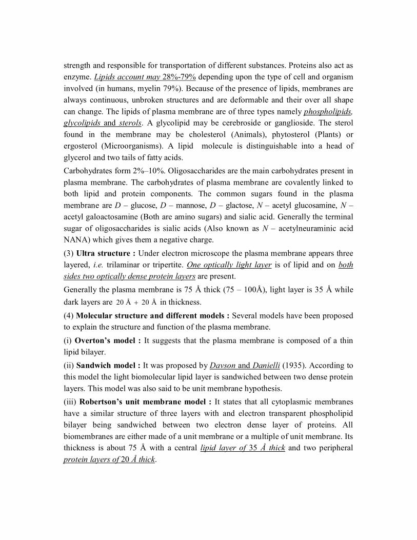
strength and responsible for transportation of different substances. Proteins also act as enzyme. Lipids account may 28%-79% depending upon the type of cell and organism involved (in humans, myelin 79%). Because of the presence of lipids, membranes are always continuous, unbroken structures and are deformable and their over all shape can change. The lipids of plasma membrane are of three types namely phospholipids, glycolipids and sterols. A glycolipid may be cerebroside or ganglioside. The sterol found in the membrane may be cholesterol (Animals), phytosterol (Plants) or ergosterol (Microorganisms). A lipid molecule is distinguishable into a head of glycerol and two tails of fatty acids. Carbohydrates form 2%–10%. Oligosaccharides are the main carbohydrates present in plasma membrane. The carbohydrates of plasma membrane are covalently linked to both lipid and protein components. The common sugars found in the plasma membrane are D – glucose, D – mannose, D – glactose, N – acetyl glucosamine, N – acetyl galoactosamine (Both are amino sugars) and sialic acid. Generally the terminal sugar of oligosaccharides is sialic acids (Also known as N – acetylneuraminic acid NANA) which gives them a negative charge. (3) Ultra structure : Under electron microscope the plasma membrane appears three layered, i.e. trilaminar or tripertite. One optically light layer is of lipid and on both sides two optically dense protein layers are present. Generally the plasma membrane is 75 Å thick (75 – 100Å), light layer is 35 Å while dark layers are Å 20Å 20 in thickness. (4) Molecular structure and different models : Several models have been proposed to explain the structure and function of the plasma membrane. (i) Overton’s model : It suggests that the plasma membrane is composed of a thin lipid bilayer. (ii) Sandwich model : It was proposed by Davson and Danielli (1935). According to this model the light biomolecular lipid layer is sandwiched between two dense protein layers. This model was also said to be unit membrane hypothesis. (iii) Robertson’s unit membrane model : It states that all cytoplasmic membranes have a similar structure of three layers with and electron transparent phospholipid bilayer being sandwiched between two electron dense layer of proteins. All biomembranes are either made of a unit membrane or a multiple of unit membrane. Its thickness is about 75 Å with a central lipid layer of 35 Å thick and two peripheral protein layers of 20 Å thick.

(iv) Fluid mosaic model : The most important and widely accepted latest model for plasma membrane was given by Singer and Nicolson in 1972. According to them it is “protein iceberg in a sea of lipids.”
According to this model, the cell membrane consists of a highly viscous fluid matrix of two layers of phospholipid molecules. These serve as relatively impermeable barrier to the passage of most water soluble molecules. Protein molecules occur in the membrane, but not in continuous layer; Instead, these occur as separate particles asymmetrical arranged in a mosaic pattern.
Some of these are loosely bound at the polar surfaces of lipid layers, called peripheral or extrinsic proteins. Others penetrate deeply into the lipid layer called integral or intrinsic proteins. Some of the integral proteins penetrate through the phospholipid layers and project on both the surface. These are called trans membrane or tunnel proteins (glycophorins). Singly or in groups, they function as channels for passage of water ions and other solutes. The channels may have gate mechanism for opening in response to specific condition. The carbohydrates occur only at the outer surface of the membrane. Their molecules are covalently linked to the polar heads of some lipid molecules (forming glycolipids) and most of the proteins exposed at outer surface (forming glycoproteins).
The sugar protions of glycolipids and glycoproteins are involved in recognition mechanisms :– (a) Sugar recognition sites of two neighbouring cells may bind each other causing cell to cell
adhesion. This enables cells to orientate themselves and to form tissues. (b) Through glycoproteins, bacteria recognise each other. e.g., female bacteria are recognised by
male bacteria. (c) These provide the basis of immune response and various control system, where
glycoproteins act as antigens. Lipid and integral proteins are amphipathic in nature i.e., they have hydrophilic and hydrophobic groups with in the same molecules. The NMR (Nuclear magnetic resonance) and ESR (Electron spin resonance) studies showed that the membrane is dynamic. The lipid tails show flexibility. The molecule can rotate or show flip flop motion.
Difference between protein types Extrinsic Protein Intrinsic Protein
These are associated with surface only. These lie throughout phospholipid matrix and project on both surfaces, also called transmembrane or tunnel protein.
They form about 30% of the total membrane protein. They form about 70% of total membrane proteins. Example – Spectrin in red blood cells & ATPase in mitochondria.
Example – Rhodopsin in retinal rod cells.
(5) Membrane protein can be of following types with different functions (i) Carrier molecules : These bind with the specific molecules into or out of the cell. This provides selective exchange of materials. The carrier protein molecules are called “permeases” e.g., Na+ – K+ pump, Na+– sugar transport. (ii) Receptor molecules : The glycoproteins on the cell surface act as receptors that recognize and bind with specific molecules.
Boundary lipid
Intrinsic protein
Hydrophobic tailHydrophilic head
Intrinsic protein
Extrinsic proteins
Lipid bilayer
Lipid
Polar
Non-polar end
Fig : Fluid-mosaic model of the plasma membrane. Proteins floating in a sea of lipid. Some proteins span the lipid bilayer, others are exposed only to one surface or the other (Modified

(iii) Enzyme molecules : The inner mitochondrial membrane carrier enzyme comprising the electron transport chain for cellular respiration. (6) Cell membranes are fluid and dynamic due to
(i) The constituent molecules can move freely in the membrane. (ii) The cell membranes are constantly renewed during the cells life. (iii) They can repair minor injuries. (iv) They expand and contract during cell movement and during change in shape. (v) They allow interactions of cells such as recognition of self and fusion of cells.
(7) Membrane permeability : According to permeability, membranes are classified as – (i) Permeable membrane : They allow both solvent and solute molecules or ions through them. e.g., cellulose wall, lignified cell walls. (ii) Impermeable membrane : They do not allow solute and solvent molecules. e.g., heavily cutinised or suberinised cell walls in plants. (iii) Semi-permeable membrane : They allow solvent molecules only. e.g., membranes of colloidion, parchment paper and copper ferrocyanide membranes. (iv) Differentially permeable membrane : All membranes found in plants allow some solutes to pass through them along with the solvent molecules. e.g., plasma membrane, tonoplast (vacuolar membrane) etc. (8) Intercellular communications/modification of plasma membrane/following structures are derived from plasma membrane (i) Microvilli : They are fingers like evaginations of 0.1 m diameter, engaged in absorption. e.g., intestinal cells, hepatic cell, mesothelial cells. The surface having microvilli is called striated border or brush border. (ii) Lomasomes : They are plasmalemma foldings found in fungal cells. (iii) Mesosomes : It serves as site for cellular respiration in prokaryotes. (iv) Tight junctions : Plasma membrane of two adjacent cells are fused at a series of points with a network of ridges or sealing strands. e.g., capillaries, brain cells collecting tubules etc. (v) Plasmodesmata : They are protoplasmic bridges amongst plant cells, which occur in area of cell wall pits. It was discovered and reported by Tangle and Strasburger respectively. (vi) Desmosomes : concerned with cell adherence. (9) Functions
(i) They control the flow of material through them and provides passage for different substances. (ii) It is differentially permeable, solute particles (1-15 Å) can pass through it. (iii) It is not only provides mechenical strength but also acts as a protective layer. (iv) Plasma membrane is responsible for the transportation of materials, molecules, ions etc. (v) It helps in osmoregulation. (vi) Diffusion of gases take place through plasma membrane by simple and facilitated diffusion. (vii) Transport of ions, small polar molecules through active (energy used) and passive
transport (energy not used). (viii) Gases like 2O and 2CO diffuse rapidly in solutions through membranes. (ix) Ions and small polar molecules diffuse slowly through the membranes. (x) Some solute molecules or ions first bind with certain specific carrier or transport proteins
called permeases. (xi) Water as well as some solute molecules and ion pass through membranes pores; pores are always bordered by channel proteins. (xii) When diffusion takes place through channel, called simple diffusion and through carrier proteins, called facilitated diffusion. (10) Membrane transport : It is passage of metabolites, by-products and biochemicals across biomembrane. Membrane transport occurs through four methods–passive, facilitated, active and bulk. Size of the particles passing through plasmalemma is generally 1 – 15 Å.

(i) Passive transport : No energy spent. Passive transport occurs through diffusion and osmosis. (a) Diffusion : It is movement of particles from the region of their higher concentration or electrochemical potential to the region of their lower concentration or electrochemical potential. Electrochemical potential operates in case of charged particles like ions. Diffusion can be observed by opening a bottle of scent or ammonia in one corner, placing a crystal of copper sulphate in a beaker of water or a crystal of 4KMnO on a piece of gelatin. Simple diffusion does not require carrier molecules. Independent Diffusion : In a system having two or more diffusion substances, each individual substance will diffuse independent of others as per gradient of its own concentration, diffusion pressure or partial pressure form region of higher one to region of lower one. Rate of diffusion is proportional to difference in concentration and inversely to distance between the two ends of the system, inversely to square root of relative density of substance and density of medium, directly to temperature and pressure. (b) Osmosis is diffusion of water across a semipermeable membrane that occurs under the influence of an osmotically active solution. (c) Mechanism of passive transport : Passive transport can continue to occur if the absorbed solute is immobilised. Cations have a tendency to passively pass from electropositive to electronegative side. While anions can pass from electronegative to electropositive side. There are two modes of passive transports. Lipid matrix permeability : Lipid soluble substances pass through the cell membrane according to their solubility and concentration gradient, e.g., triethyl citrate, ethyl alcohol, methane. Hydrophillic membrane channels : They are narrow channels formed in the membrane by tunnel proteins. The channels make the membrane semipermeable. Water passes inwardly or outwardly from a cell through these channels according to osmotic gradients. 2CO and 2O also diffuse through these channels as per their concentration gradients. Certain small ions and other small water soluble solutes may also do so. (d) Ultrafiltration is fine filtration that occurs under pressure as from blood capillaries, epithelia and endothelia. It is of two types : –
Paracellular through leaky junctions or gaps in between cells. Transcellular through fenestrations in the cells. ‘Dialysis’ is removal of waste products and
toxins from blood by means of diffusion between blood and an isotonic dialysing solution. (e) Facilitated transport or Facilitated diffusion : It is passage of substances along the
concentration gradient without expenditure of energy that occurs with the help of special permeating substances called permeases. Permeases form pathways for movement of certain substances without involving any expenditure of energy. At times certain substances are transported alongwith the ones requiring active transport. The latter phenomenon called cotransport. Facilitated transport occurs in case of some sugars, amino acids and nucleotides. (ii) Active transport : It occurs with the help of energy, usually against concentration gradient. For this, cell membranes possess carriers and gated channels. (a) Carrier particles or Proteins : They are integral protein particles which have affinity for specific solutes. A solute particles combines with a carrier to form carrier solute complex. The latter undergoes conformational change in such a way as to transport the solute to the inner side where it is released into cytoplasm. (b) Gated channels : The channels are opened by either change in electrical potential or specific substances, e.g., Calcium channels.
Active transport systems are also called pumps, e.g., H pump, K pump, Cl pump, KNapump. The pumps operate with the help of ATP. HK exchange pump occurs in guard cells.
KNa exchange pump operates across many animal membranes. For every ATP hydrolysed, three

Na ions are passed out while two K ions are pumped in. Sea Gulls and Penguins operate KNa pump for excreting NaCl through their nasal glands.
Active transport of one substance is often accompanied by permeation of other substances. The phenomenon is called secondary active transport. It is of two main types, cotransport (e.g., glucose and some amino acids alongwith inward pushing of excess Na ) and counter-transport ( 2Ca and
H movement outwardly as excess Na passes inwardly). (iii) Bulk transport : It is transport of large quantities of micromolecules, macromolecules and
food particles through the membrane. It is accompanied by formation of transport or carrier vesicles. The latter are endocytotic and perform bulk transport inwardly. The phenomenon is called endocytosis. Endocytosis is of two types, pinocytosis and phagocytosis. Exocytic vesicle perform bulk transport outwardly. It is called exocytosis. Exocytosis performs secretion, excretion and ephagy.
(a) Pinocytosis : (Lewis, 1931). It is bulk intake of fluid, ions and molecules through development of small endocytotic vesicles of 100 – 200 nm in diameter. ATP, ,2Ca fibrillar protein clathrin and contractile protein actin are required. Fluid-phase pinocytosis is also called cell drinking. It is generally nonselective. For ions and molecules the membrane has special receptor or adsorptive sites located in small pits. They perform adsorptive pinocytosis. After coming in contact with specific substance, the area of plasma membrane having adsorptive sites, invaginates and forms vesicle. The vesicle separates. It is called pinosome. Pinosome may burst in cytosol, come in contact with tonoplast and pass its contents into vacuole, form digestive vacuole with lysosome or deliver its contents to Golgi apparatus when it is called receptosome.
(b) Phagocytosis : (Metchnikoff, 1883). It is cell eating or ingestion of large particles by living cells, e.g., white blood corpuscles (neutrophils, monocytes), Kupffer’s cells of liver, reticular cells of spleen, histiocytes of connective tissues, macrophages, Amoeba and some other protists, feeding cells of sponges and coelentrates. Plasma membrane has receptors. As soon as the food particle comes in contact with the receptor site, the edges of the latter evaginate, form a vesicle which pinches off as phagosome.
One or more lysosomes fuse with a phagosome, form digestive vacuole or food vacuole. Digestion occurs inside the vacuole. The digested substances diffuse out, while the residual vacuole passes out, comes in contact with plasma membrane for throwing out its contents through exocytosis or ephagy.
Protoplasm. 1) Definition : Protoplasm is a complex, granular, elastic, viscous and colourless substance. It is selectively or differentially permeable. It is considered as “Polyphasic colloidal system”. (2) Discoveries
(i) J. Huxley defined it as “physical basis of life”. (ii) Dujardin (1835) discovered it and called them “sarcode”. (iii) Purkinje (1837) renamed it as “Protoplasm”. (iv) Hugo Von Mohl (1844) gave the significance of it. (v) Max Schultz (1861) gave the protoplasmic theory for plants. (vi) Fischer (1894) and Hardy (1899) showed its colloidal nature. (vii) Altman (1893) suggested protoplasm as granular. (3) Composition : Chemically it is composed of
Water 75 – 85% Carbon 20% Proteins 10 – 25% Oxygen 62% Lipids 2 – 3% Hydrogen 10% Inorganic Materials 1% Nitrogen 3%
Trace elements – 5% ( ,,,,,,,,, FeIMgNaKSClPCa etc.)

Maximum water content in protoplasm is found in hydrophytes, i.e. 95% where as minimum in seeds, spores (dormant organs) i.e. 10 – 15%. In animals water is less (about 65%) and proteins are more (about 15%).
(4) Physical properties of protoplasm : Cyclosis movement are shown by protoplasm. These are of two types.
(i) Rotation : In one direction, either clockwise or anticlockwise e.g., Hydrilla, Vallisneria. Found only in eukaryotes.
(ii) Circulation : Multidirectional movements around vacuole e.g. Tradescantia. (a) It shows stimulation or irritability. (b) Protoplasm is polyphasic. Colloidal substance or true solution because true solution act as dispersion medium and different colloidal particles constitute dispersed phase. (c) It shows increased surface area and adsorption. (d) It shows sol – gel transformation. (e) It is highly viscous. (f) It coagulates at 60o C or above or if treated with concentrated acids or bases. (g) It shows Brownian movements. (h) It’s specific gravity is slightly more than 1. (i) It’s pH is on acidic side, but different vital activities occur at neutral pH which is considered as 7, injury decreases the pH of the cell (i.e. 5.2 – 5.5) and if it remains for a long time, the cell dies. (j) Scattering and dispersion of light is shown by protoplasm i.e. Tyndall effect.
Cytoplasm. The substance occur around the nucleus and inside the plasma membrane containing various organelles and inclusions is called cytoplasm. (1) The cytoplasm is a semisolid, jelly – like material. It consists of an aqueous, structureless ground substance called cytoplasmic matrix or hyaloplasm or cytosol. (2) It forms about half of the cell’s volume and about 90% of it is water. (3) It contains ions, biomolecules, such as sugar, amino acid, nucleotide, tRNA, enzyme,
vitamins, etc. (4) The cytosol also contains storage products such as glycogen/starch, fats and proteins in
colloidal state. (5) It also forms crystallo – colloidal system. (6) Cytomatrix is differentiated into ectoplasm or plasmagel and endoplasm or plasmasol. (7) Cytomatrix is three dimensional structure appear like a network of fine threads and these threads are called microfilaments (now called actin filaments or microtrabecular lattice) and it is believed to be a part of cytoskeleton. It also contains microtubules and inter mediate cytoplasmic filaments. (8) Hyaloplasm contains metabolically inactive products or cell inclusions called deutoplast or
metaplasts. (9) Cytoplasmic organelles are plastid, lysosome, sphaerosome, peroxisome, glyoxysomes, mitochondria, ribosome, centrosome, flagellum or cilia etc. (10) The movement of cytoplasm is termed as cyclosis (absent in plant cells).

Mitochondria. (1) Definition : (Gk – mito = thread ; chondrion = granule) Mitochondria are semi autonomous having hollow sac like structures present in all eukaryotes except mature RBCs of mammals and sieve tubes of phloem. These are absent in all prokaryotes like bacteria and cyanobacteria. Mitochondria are also called chondriosome, chondrioplast, plasmosomes, plastosomes and plastochondriane. (2) Discoveries (i) These were first observed in striated muscles (Voluntary) of insects as granules by Kolliker (1850), he called them “sarcosomes”.
(ii) Flemming (1882) called them “fila” for thread like structure.
(iii) Altman (1890) called them “bioplast”.
(iv) C. Benda (1897) gave the term mitochondria.
(v) F. Meves (1904) observed mitochondria in plant (Nymphaea).
(vi) Michaelis (1898) demonstrated that mitochondria play a significant role in respiration.
(vii) Bensley and Hoerr (1934) isolated mitochondria from liver cells.
(viii) Seekevitz called them “Power house of the cell”.
(ix) Nass and Afzelius (1965) observed first DNA in mitochondria.
(3) Number of mitochondria : Presence of mitochondria depends upon the metabolic activity of the cell. Higher is the metabolic activity, higher is the number e.g., in germinating seeds.
(i) Minimum number of mitochondria is one in Microasterias, Trypanosoma, Chlorella, Chlamydomonas (green alga) and Micromonas. Maximum numbers are found (up to 50,000) in giant Amoeba called Chaos – Chaos. These are 25 in human sperm, 300 - 400 in kidney cells and 1000 – 1600 in liver cells.
(ii) Mitochondria of a cell are collectively called chondriome. (4) Size of mitochondria : Average size is 0.5–1.00 m and length up to 1 – 10 m.
(i) Smallest sized mitochondria in yeast cells ).1( 3m (ii) Largest sized are found in oocytes of Rana pipiens and are 20 – 40 .m (iii) A dye for staining mitochondria is Janus B – green.
F1 Particles Outer membrane
Inner membrane Mitochondrial crest
Respiratory chain and enzymess
Outer chamber Protein layer
Lipid layer
Intracristael space
F1 Particles
Perimitochondrial space
Fig : Molecular organization of inner membrane of mitochondria
Intermembranous space Outer membrane
Inner membrane
Cristae
Matrix
Inclusions Intercristaeal space
Tubuli Ribosomes
F1 Particles or Oxysomes
DNA
Inner membrane
Crista
A F1 Particles or Oxysomes
Intermembranous space
B
Matrix Inclusions
Outer membrane
Matrix
Ribosomes
Outer chamber DNA
Intratubuli space
Intermembranous space Inner
chember Inner membrane Outer membrane
C
D
Fig : Three dimentional structure of mitochondrion. A. From an animal cell. B. From plant cell, C. T.S.
mitechondrion, D. One tubule
F1 Particles or Oxysomes

(5) Ultrastructure of mitochondria : Mitochondrion is bounded by two unit membranes separated by perimitochondrial space (60 – 80 Å). The outer membrane is specially permeable because of presence of integral proteins called porins. The inner membrane is selective permeable. The inner membrane is folded or convoluted to form mitochondrial crests. In animals these are called cristae and in plants these folding are called tubuli or microvili.
The matrix facing face is called ‘M’ face and face towards perimitochondrial space is called ‘C’ face. The ‘M’ face have some small stalked particles called oxysomes or F1 particle or elementory particle or Fernandez – Moran Particles. Each particle is made up of base, stalk and head and is about 10nm in length. Number of oxysomes varies to 104 to 105 per mitochondrion and chemically they are made of phospholipid core and protein cortex. Oxysomes have ATPase enzyme molecule (Packer, 1967) and therefore, responsible for ATP synthesis. These elementary particles are also called F0 – F1 particles.
In the matrix 2–6 copies of naked, double stranded DNA (circular) and ribosome of 70 S type are present. It is rich in G-C ratio. Basic histone proteins are absent in mitochondrial DNA. The synthesis of ATP in mitochondria is called oxidative phosphorylation, which is O2 dependent and light independent. Cristae control dark respiration. F0 particles synthesize all the enzymes required to operate Kreb’s cycle. Inner membrane contains cytochrome.
(6) Semi-autonomous nature of mitochondrion : Mitochondria contain all requirements of protein synthesis :
(i) 70 S ribosomes. (ii) DNA molecules to form mRNA and also replicate. (iii) ATP molecules to provide energy. The mitochondria can form some of the required proteins but for most of proteins, these are
dependent upon nuclear DNA and cytoplasmic ribosomes, so the mitochondria are called semi-autonomous organelles.
(7) Two states of mitochondria : When ATP synthesis is low or the respiratory chain of mitochondrion is inhibited, it is called inactive or orthodox state, and has large amount of matrix and only a few cristae. But when mitochondria are active or condensed state, and have small amount of matrix and highly developed cristae. This shows that the development of mitochondria depends upon the physiological activity of the cell.
(8) Chemical composition : Cohn gave the chemical composition of mitochondrion: Proteins = 65 – 70% Lipids = 25 – 30% (90% phospholipids and 10% cholesterol, Vit. E., etc) 2 – 5% RNA Some amount of DNA The mitochondrial matrix has many catabolic enzymes like cytochrome oxidase and reductases, fatty acid oxidase, transaminase, etc. (9) Enzymes of Mitochondria (i) Outer membrane : Monoamine oxidase, glycerophosphatase, acyltransferase, phospholipase
A. (ii) Inner membrane : Cytochrome b,c1,c,a, (cyt.b, cyt.c1, cyt.c, cyt.a, cyt.a3) NADH, dehydrogenase, succinate dehydrogenase, ubiquinone, flavoprotein, ATPase. (iii) Perimitochondrial space : Adenylate kinase, nucleoside diphosphokinase. (iv) Inner matrix : Pyruvate dehydrogenase, citrate synthase, aconitase, isocitrate dehydrogenase, fumarase, Ketoglutarate dehydrogenase, malate dehydrogenase. (10) Origin : Mitochondria are self-duplicating organelles due to presence of DNA molecules so new mitochondria are always formed by growth and division of pre-existing mitochondria by binary fission.
Difference between outer and inner membrane of mitrochondria Outer membrane Inner membrane
It is smooth having less area. It is infolded to form cristae hence large surface area. It is freely permeable. Semipermeable, impermeable to coenzyme A and
NAD. It consist 50% lipid and 50% protein. It consist 80% protein and 20% lipid. Sialic acid is more (4 – 5 time). Sialic acid is less. Near about 14% enzymes are present. Near about 60 enzymes are present.
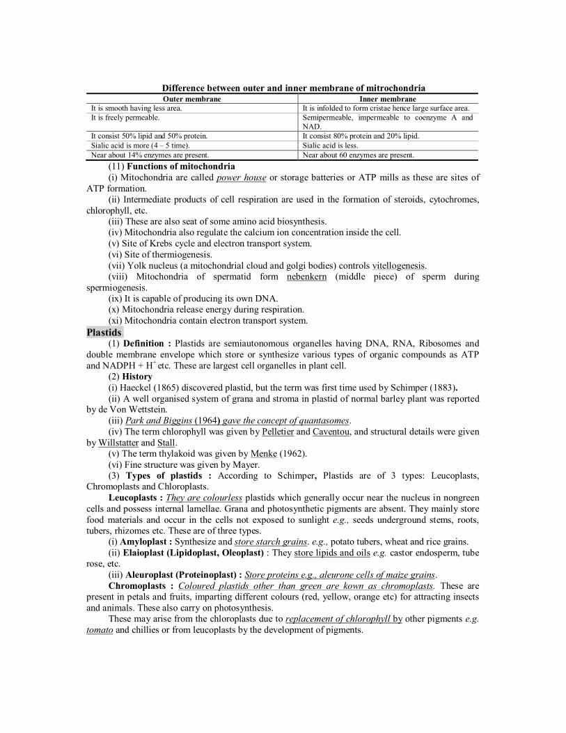
(11) Functions of mitochondria (i) Mitochondria are called power house or storage batteries or ATP mills as these are sites of
ATP formation. (ii) Intermediate products of cell respiration are used in the formation of steroids, cytochromes,
chlorophyll, etc. (iii) These are also seat of some amino acid biosynthesis. (iv) Mitochondria also regulate the calcium ion concentration inside the cell. (v) Site of Krebs cycle and electron transport system. (vi) Site of thermiogenesis. (vii) Yolk nucleus (a mitochondrial cloud and golgi bodies) controls vitellogenesis. (viii) Mitochondria of spermatid form nebenkern (middle piece) of sperm during
spermiogenesis. (ix) It is capable of producing its own DNA. (x) Mitochondria release energy during respiration. (xi) Mitochondria contain electron transport system.
Plastids. (1) Definition : Plastids are semiautonomous organelles having DNA, RNA, Ribosomes and
double membrane envelope which store or synthesize various types of organic compounds as ATP and NADPH + H+ etc. These are largest cell organelles in plant cell.
(2) History (i) Haeckel (1865) discovered plastid, but the term was first time used by Schimper (1883). (ii) A well organised system of grana and stroma in plastid of normal barley plant was reported
by de Von Wettstein. (iii) Park and Biggins (1964) gave the concept of quantasomes. (iv) The term chlorophyll was given by Pelletier and Caventou, and structural details were given
by Willstatter and Stall. (v) The term thylakoid was given by Menke (1962). (vi) Fine structure was given by Mayer. (3) Types of plastids : According to Schimper, Plastids are of 3 types: Leucoplasts,
Chromoplasts and Chloroplasts. Leucoplasts : They are colourless plastids which generally occur near the nucleus in nongreen
cells and possess internal lamellae. Grana and photosynthetic pigments are absent. They mainly store food materials and occur in the cells not exposed to sunlight e.g., seeds underground stems, roots, tubers, rhizomes etc. These are of three types.
(i) Amyloplast : Synthesize and store starch grains. e.g., potato tubers, wheat and rice grains. (ii) Elaioplast (Lipidoplast, Oleoplast) : They store lipids and oils e.g. castor endosperm, tube
rose, etc. (iii) Aleuroplast (Proteinoplast) : Store proteins e.g., aleurone cells of maize grains. Chromoplasts : Coloured plastids other than green are kown as chromoplasts. These are
present in petals and fruits, imparting different colours (red, yellow, orange etc) for attracting insects and animals. These also carry on photosynthesis.
These may arise from the chloroplasts due to replacement of chlorophyll by other pigments e.g. tomato and chillies or from leucoplasts by the development of pigments.

All colours (except green) are produced by flavins, flavenoids and cyanin. Cyanin pigment is of two types one is anthocyanin (blue) and another is erythrocyanin (red). Anthocyanin express different colours on different pH value. These are variously coloured e.g. in flowers. They give colour to petals and help in pollination. They are water soluble. They are found in cell sap.
Green tomatoes and chillies turn red on ripening because of replacement of chlorophyll molecule in chloroplasts by the red pigment lycopene in tomato and capsanthin in chillies. Thus, chloroplasts are changed into chromatophores.
Chloroplast : Discovered by Sachs and named by Schimper. They are greenish plastids which possess photosynthetic pigments.
(i) Number : It is variable. Number of chloroplast is 1 in Spirogyra indica, 2 in Zygnema, 16 in S.rectospora, up to 100 in mesophyll cells. The minimum number of one chloroplast per cell is found in Ulothrix and species of Chlamydomonas.
(ii) Shape : They have various shapes
Shape Example Cup shaped Chlamydomonas sp. Stellate shaped Zygnema. Collar or girdle shaped Ulothrix Spiral or ribbon shaped Spirogyra Reticulate Oedogonium Discoid Voucheria
(iii) Size : It ranges from 3 – 10 m (average 5 )m in diameter. The discoid chloroplast of higher plants are 4 – 10 m in length and 2– 4 m in breadth. Chloroplast of spirogyra may reach a
length of 1 mm. Sciophytes (Shade plant) have larger chloroplast. (iv) Chemical composition : (a) Proteins 50 – 60%, (b) Lipids 25 – 30% , (c) Chlorophyll – 5- 10 %, (d) Carotenoids (carotenes and xanthophylls) 1 –2%, (e) DNA – 0.5%, RNA 2 – 3%, (f) Vitamins K and E, (g) Quinines, Mg, Fe, Co, Mn, P, etc. in traces. (v) Ultrastructure : It is double membrane structure. Both membranes are smooth. The inner
membrane is less permeable than outer but rich in proteins especially carrier proteins. Each membrane is 90 – 100 Å thick. The inter-membrane space is called the periplastidial space. Inner to membranes, matrix is present, which is divided into two parts.
(a) Grana : Inner plastidial membrane of the chloroplast is invaginated to form a series of parallel membranous sheets, called lamellae, which form a number of oval – shaped closed sacs, called thylakoids. Thylakoids are structural and functional elements of chloroplasts. These thylakoids contain all the requirements of light reactions e.g., pigments like chlorophyll, carotenoids, plastoquinone, plastocyanin, etc. that are involved in photosynthesis. Each thylakoid has an intrathylakoid space, called loculus (size 10-30Å) bounded by a unit membrane. Along the inner side of thylakoid membrane, there are number of small rounded para-crystalline bodies, called quantasomes (a quantasome is the photosynthetic unit) which can trap a mole of quantum of light and can bring about photosynthetic act. Each quantasome contains about 230 chlorophyll molecules and 50 carotenoid molecules.
Fig : A chloroplast in section
Inner membrane Granum in Thylakoi
Stroma Granum
Frets or
Outer membrane

In eukaryotic plant cells, a number of thylakoids are superimposed like a pile of coins to form a granum. The number of thylakoids in a granum ranges from 10-100 (average number is 20-50). The number of grana per chloroplast also varies widely e.g., one granum per chloroplast in Euglena while there are 40-60 grana per chloroplast in spinach. The size of each granum varies from 0.2 – 0.6 m in diameter. But in blue-green algae, the thylakoids are not organised to form granum.
Adjacent grana are interconnected by branched tubules, called stromal lamellae or Fret-channel or Fret membrane's.
(b) Stroma : It is transparent, proteinaceous and watery substance. Dark reaction of photosynthesis occurs in this portion. Stroma is almost filled with “Rubisco” (about 15% of total enzyme, protein) enzyme 2CO is accepted by this enzyme. CO2 assimilation results in carbohydrate formation. It has 20 – 60 copies of naked circular double stranded DNA. Each DNA copy is 40 in length, which can code for 125 amino acids. All plastids of a cell called as “Plastidome” (Dangeared 1920) in stroma. Amount of DNA per chloroplast is 10–15 g. Chloroplast genome has 145 kilobase pairs. It shows semiautonomous nature and ribosomes are of 70 S type.
(vi) Pigments of chloroplast : Willsttater and Stall observed the following pigments: (a) Chlorophyll a : MgNOHC 457255 (with methyl group) (b) Chlorophyll b : MgNOHC 467055 (with aldehyde group) (c) Chlorophyll c : MgNOHC 453235 (d) Chlorophyll d : MgNOHC 467054 (e) Carotenes, Xanthophylls : Carotenoids.
Difference between Chl. a and Chl. b
Chl. a Chl. b Absorption peak at 430, 662. It is 453, 642. Bluish green in colour. Yellowish green. Soluble in petroleum, ether. Soluble in methyl alcohol. Functional group at C3 position is CH3 Functional group attached to pyrrol ring is
CHO. Present in all green plants excepts autotrophic bacteria. Present in all green plants except blue green,
brown and red algae. In chloroplast it is 75%. It is 25% In reflected light Chl. a shows blood red colour while in transmitted light, it shows blue green colour.
In reflected light it show dull brown colour while in transmitted light, it shows yellowish green colour.
(vii) Chlorophylls and their presence : Term by Cavantou (1818). It’s molecule has
tetrapyrollic or porphyrin head ( Å15Å 15 ) and phytol tail (20 Å long). Mg++ is present in the centre of porphyrin head. If chlorophyll is burnt only Mg is left.
(a) Chlorophyll b : It is found in members of chlorophyceae. (b) Chlorophyll c : It is found in members of phaeophyceae, bacillariophyceae. (c) Chlorophyll d : It is found in members of rhodophyceae. (d) Chlorophyll e : It is found in members of xanthophyceae. (e) Phycoerythrin and phycocyanin (phycobilins) are the red and blue green pigments in
rhodophyceae and cyanophyceae respectively. (f) Fucoxanthin (brown pigment) in phaeophyceae.

(g) Bacteriochlorophyll )( 467455 MgNOHC or chlorobium chlorophyll present in photosynthetic bacteria. These pigment are red in acidic and blue in alkaline medium.
(viii) Carotenoids : These are hydrocarbons, soluble in organic solvents. These are of 2 types: (a) Carotenes : 5640 HC derivatives of vitamin A. Carrot coloured ,, carotene, lycopene,
etc. (b) Xanthophyll : ,25640 OHC yellowish in colour, fucoxanthin, violaxanthin. Molar ratio of
carotene and xanthophyll in young leaves is 2 : 1. (ix) Plastids are interchangeable Leucoplast Chloroplast Chromoplast (degenerate chloroplast) The leucoplast and chloroplast are interconvertible but once they have converted into
chromoplast, the reverse can not take place. Because, chromoplasts are aged or degenerated form of chloroplast e.g. in tomato.
Young ovary (colourless) – Leucoplast Young fruit (green) – Chloroplast Mature fruits (red) (due to Lycopene) – Chromoplast. In carrot leucoplast – Chromoplast (carotene) etc. (x) Origin of chloroplast : Plastids, like the mitochondria, are self duplicating organelles.
These develop from colourless precursors, called proplastids. They are believed to be evolved from endosymbiont origination.
(4) Function of plastids (i) It is the site of photosynthesis, (light and dark reaction). (ii) Photolysis of water, reduction of NADP to NADPH2 take place in granum. (iii) Photophosphorylation through cytochrome b6 f, plastocyanine and plastoguinone etc. (iv) They store starch or factory of synthesis of sugars. (v) Chloroplast store fat in the form of plastoglobuli. (vi) They can be changed into chromoplasts to provide colour to many flowers and fruits for
attracting animals. (vii) They maintain the percentage of CO2 and O2 in atmosphere.
Endoplasmic reticulum (ER). (1) Definition : It is well developed electron microscopic network of interconnected cisternae, tubules and vesicles present throghout the cytoplasm, especially in the endoplasm. (2) Discovery : Garnier (1897) was first to observe the ergastoplasm in a cell. The ER was first noted by Porter, Claude, and Fullman in 1945 as a network. It was named by Porter in 1953. (3) Occurrence : The ER is present in almost all eukaryotic cells. A few cells such as ova, embryonic cells, and mature RBCs, however, lack ER. It is also absent in prokaryotic cell.
In muscle cells, it is called sarcoplasmic reticulum, myeloid bodies and nissel granules are believed to be formed from ER. ER is little develop in meristematic cells. (4) Chemical composition : All the components of ER are lipoperoteins and trilaminar like the plasma membrane but differ in following
(i) Thinner )6050( Å than plasma membrane. (ii) With less cholesterol. (iii) With more lipids. (iv) The lumen is filled with fluid containing 70% phospholipids lecithin and cephalin etc.
(5) Ultrastructure : The ER is made up of three components :

(i) Cisternae : These are flattened, unbranched, sac like structures. They lie in stacks (piles) parallel to one another. They bear ribosomes. They contain glycoproteins named ribophorin-I and ribophorin-II that bind the ribosomes. Found in protein forming cells. (ii) Vesicles : These are oval or rounded, vacuole like elements, scattered in cytoplasm. These are also studded with ribosomes. (iii) Tubules : Wider, tubular, branched elements mainly present near the cell membrane. They are free from ribosomes. These are more in lipid forming cells.
All the three structures are bound by a single unit membrane. (6) Types of ER : Depending upon the presence of ribosomes, the ER has been categorised into two types: (i) A smooth or Agranular endoplasmic reticulum (SER) : It consists mainly of tubules and vesicles. It has no ribosomes associated to it. It is well developed in the muscle cells, adipose tissue cells, interstitial cells, glycogen storing liver cells, etc. and the cells that synthesize and secrete steroids. SER also takes part in synthesis of vitamins, carbohydrates and detoxification. Detoxification of pollutants carcinogens and drugs is carried out SER of liver cells and mitochondria, SER is associated with storage and release of 2Ca ions. It gives rise to spherosomes. (ii) Rough or Granular endoplasmic reticulum (RER) : It mainly consists of cisternae. It has ribosomes attached on its cytoplasmic surface. It is abundant in cells engaged in production and excertion of proteins, e.g., plasma cells, goblets cells, pancreatic acinus cells and certain liver cells. The RER is more stable than SER. The RER is basophilic due to the presence of ribosomes. Ribosomes are attached to ER through hydrophobic interaction.
The proteins synthesised by the ER membrane bound ribosomes pass into the ER lumen, where most of the proteins are glycosylated. For this, an oligosaccharide is always linked to the 2NHgroup on side chain of an asparagine residue. The ER lumen serves as a compartment to contain substances which must be kept separate from cytosol. In the ER lumen, the enzymes modify the proteins. Differences between SER and RER
SER RER SER or smooth endoplasmic reticulum does not possesses ribosomes over the surface of its membrane.
RER possesses ribosomes attached to its membrane.
It is mainly formed of vesicles and tubules. It is mainly formed of cisternae and few tubules. It is engaged in the synthesis of glycogen lipids and steroids. The reticulum takes part in the synthesis of proteins. Pores are absent so that materials synthesised by SER do not pass into its channels.
RER possesses narrow pores below its ribosomes for the passage of synthesised polypeptides into ER channels.
SER is often peripheral. It may be connected with plasmalemma.
It is often internal and connected with nuclear envelope.
Ribophorins are absent. RER contains Ribophorins I and II for providing attachment to ribosomes.
SER gives rise to sphaerosomes. It helps in the formation of lysosome through the agency of golgi apparatus.
Fig : Elements of Endoplasmic Reticulum
Ribosomes
Ribosomes
Vesicles
Lamellae
Cisternae Tubules
(7) Origin : RER is formed from nuclear membrane while SER is formed from RER by loss of ribosomes. Rough vesicles originate only from RER after homogenisation of cell. RER breaks in small fragments (Vesicles) and it is called microsome (This is not a cell organelle). ER constitute cytoskeleton and also help as intracellular transport system. And it is sensitive to irritation. (8) Functions
(i) Synthesis and secretion of specific proteins via – golgi bodies. (ii) Formation of protein ribophorin. Which helps in attachment of ribosome. (iii) Give rise to SER. (iv) Provides surface for synthesis of cholesterol, steroid, ascorbic acid and visual pigments. (v) It helps in synthesis of harmones e.g., testosterone and estrogen. (vi) It helps in glycogenolysis in the liver cells and brings about detoxification (SER). (vii) Gastric cells secreting zymogen have well developed SER. (viii) ER is a component of cytoskeleton (Spread as a net) of cell and provides mechanical
support and shape to the cell. (ix) ER acts as segregation apparatus and divides the cytoplasm into chambers. Compartmentalisation is most necessary for cellular life. (x) It participates in the formation of cell-plate during cytokinesis in the plant cells by the
formation of phragmoplasts. (xi) ER has many types of enzymes e.g. ATPase, reductases, dehydrogenases and phosphatases.
(9) Sacroplasmic reticulum : It is a modified SER striated muscle fibres which forms a network of interconnected tubules in the sarcoplasm. It helps in conduction of motor nerve impulses throughout the muscle fibre and in the removal of lactic acid so prevents muscle fatigue. It is called “ergastoplasm” in muscle and “nisslegranules” in nerve cells.
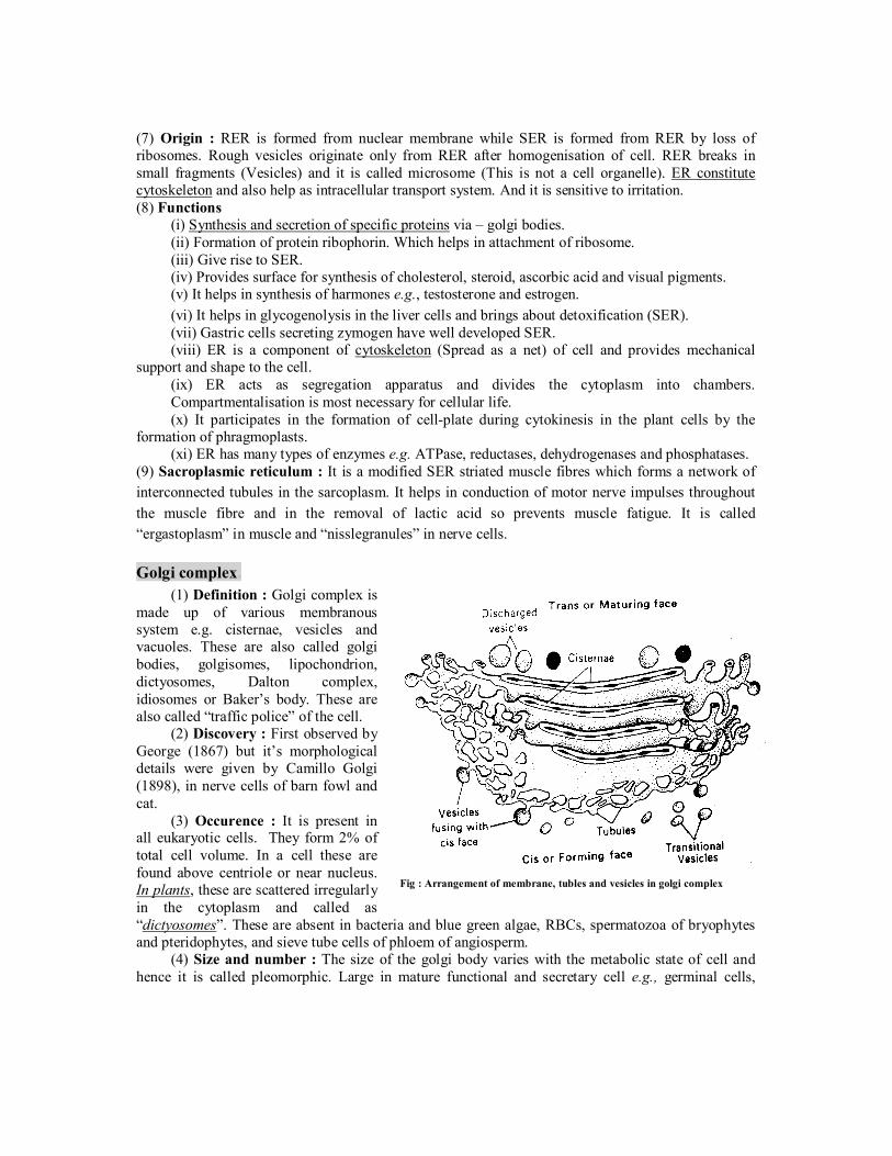
Golgi complex. (1) Definition : Golgi complex is
made up of various membranous system e.g. cisternae, vesicles and vacuoles. These are also called golgi bodies, golgisomes, lipochondrion, dictyosomes, Dalton complex, idiosomes or Baker’s body. These are also called “traffic police” of the cell.
(2) Discovery : First observed by George (1867) but it’s morphological details were given by Camillo Golgi (1898), in nerve cells of barn fowl and cat.
(3) Occurence : It is present in all eukaryotic cells. They form 2% of total cell volume. In a cell these are found above centriole or near nucleus. In plants, these are scattered irregularly in the cytoplasm and called as “dictyosomes”. These are absent in bacteria and blue green algae, RBCs, spermatozoa of bryophytes and pteridophytes, and sieve tube cells of phloem of angiosperm.
(4) Size and number : The size of the golgi body varies with the metabolic state of cell and hence it is called pleomorphic. Large in mature functional and secretary cell e.g., germinal cells,
Fig : Arrangement of membrane, tubles and vesicles in golgi complex

goblet cells, but small size in non-secretary cells. There may be 25,000 dictysomes present in rhizoidal cells of Chara. Average number 10 – 20 per cell. Number increases during cell division.
(5) Structure : Under transmission electron microscope the st. of golgibodies was study by Dalton and Felix (1954), golgi body is made of 4 parts.
(i) Cisternae : Golgi apparatus is made up of stack of flat. Sac like structure called cisternae. The margins of each cisterna are gently curved so that the entire golgi body takes on a cup like appearance. The golgi body has a definite polarity. The cisternae at the convex end of the dictyosome comprises forming face (F. face) or cis face. While the cisternae at the concave end comprises the maturing face (M. face) or trans face. The forming face is located next to either the nucleus or endoplasmic reticulum. The maturing face is usually directed towards the plasma membranes. It is the functional unit of golgi body.
(ii) Tubules : These arise due to fenestration of cisternae and it forms a complex of network. (iii) Secretory vesicles : These are small sized components each about 40 Å in diameter
presents along convex surface of edges of cisternae. These are smooth and coated type of vesicles. Smooth or secretory vesicles, which have a smooth surface and contain secretions of the cell and coated vesicles, that have rough surface. They carry materials to or from the cisternae.
(iv) Golgian vacuoles : These are spherical components each about 600 Å in diameter. These are produced by vesiculation of saccules of cisternae. Scattered cisternae are called dictyosomes and condition is called diffused.
(6) Function (i) The main function of golgi body is secretion, so it is large sized among the secretory cells.
Secretion are released either by exocytosis or reverse pinocytosis. (ii) Glycosidation of lipids i.e. addition of oligosaccharides to produce glycolipids. (iii) Glycosylation of proteins i.e. addition of carbohydrate to produce glycoproteins. (iv) Formation of lysosomes. (v) Golgi body forms the cell plate. During cell division by secreting hemicellulose formation of
enzyme and hormones (Thyroxine) etc. (vi) Matrix of connective tissue is formed by golgi complex. (vii) In oocytes of animal, golgi apparatus functions as the centre around which yolk is deposited i.e.
vitellogenesis. (viii) Membrane of the vesicles produced by golgi apparatus join in the region of cytokinesis to produce new
plasmalemma. (ix) It is also called export house of cell. (x) Golgi body contains phospholipids, proteins, enzymes and vitamin-c. (xi) The golgi complex gives rise to the acrosome in an animal sperm. (7) Origin : Most accepted view is that golgi body originates from RER-that has lost its
ribosomes from this RER arise transport vesicles that contain Golgi membrane and fuse with the saccule on the forming face of Golgi apparatus. This is why this face is called the forming face. Lysosomes.
(1) Definition : Lysosomes are electron microscopic, vesicular structures of the cytoplasm, bounded by a single membrane which are involved in intracellular digestive activities, contains hydrolytic enzymes, so called lysosomes. (2) Discovery : These were first discovered by a Belgian biochemist, Christian de Duve (1995) in the liver cells and were earlier named pericanalicular dense bodies. Terms Lysosome was given by Novikoff under the study of electron microscope. Maltile (1964) was first to demonstrate their presence in plants, particularly in the fungus neurospora.
(3) Occurrence : These are absent from the prokaryotes but are present in all eukaryotic animal cells except mammalian RBCs. They have been recorded in fungi, euglena, cotton and pea seeds. (4) Shape : These are generally spherical in shape but are irregular in plant root tip cells. (5) Size : Size range is 0.2-0.8 m while size is 0.5 m (500 nm). (6) Number : Lysosomes are more in those cells which are involved in intracellular digestive activities e.g., WBCs of blood, histiocytes of connective tissue; phagocytes of liver and spleen; osteoclasts; cells of degenerating tissue like tail of tadpole larva etc. (7) Ultrastructure : Under electron microscope, a lysosome is formed of two parts : (i) Limiting membrane : It is outer, single layered, lipoproteinous and trilaminar unit membrane. It keeps a limit on glycoproteinous digestive enzymes. (ii) Matrix : It is inner, finely granular and highly heterogeneous group substance inside the
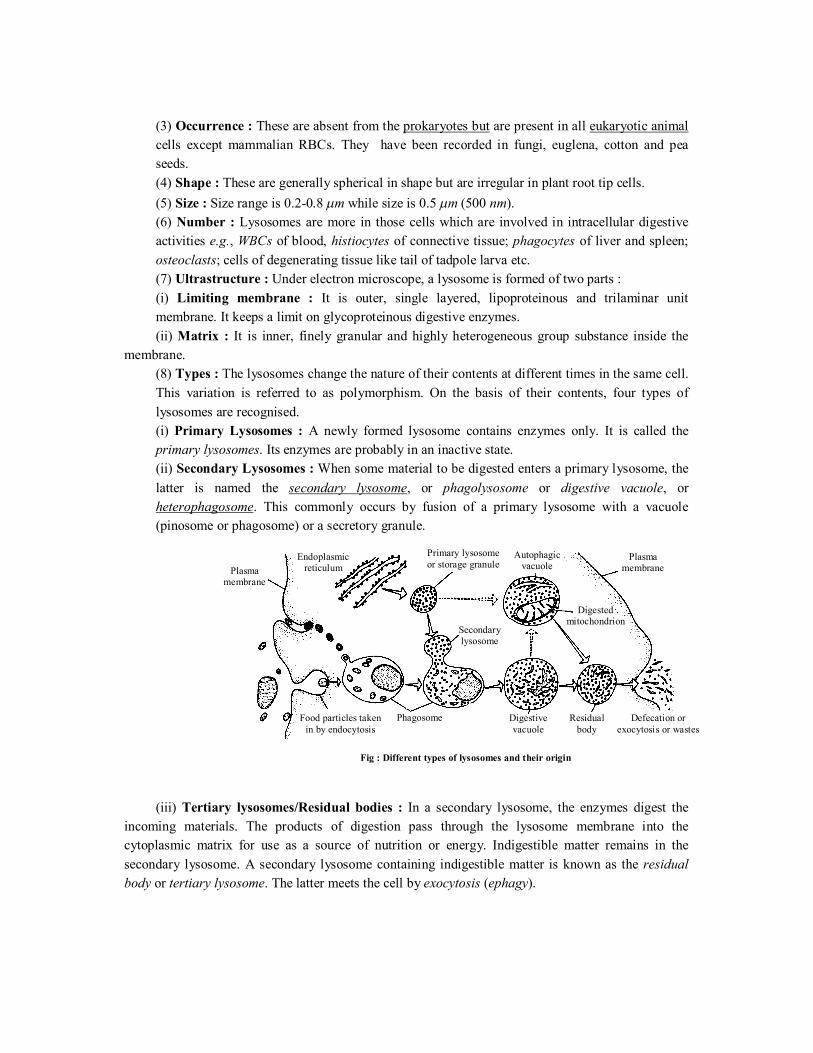
membrane. (8) Types : The lysosomes change the nature of their contents at different times in the same cell. This variation is referred to as polymorphism. On the basis of their contents, four types of lysosomes are recognised. (i) Primary Lysosomes : A newly formed lysosome contains enzymes only. It is called the primary lysosomes. Its enzymes are probably in an inactive state. (ii) Secondary Lysosomes : When some material to be digested enters a primary lysosome, the latter is named the secondary lysosome, or phagolysosome or digestive vacuole, or heterophagosome. This commonly occurs by fusion of a primary lysosome with a vacuole (pinosome or phagosome) or a secretory granule.
(iii) Tertiary lysosomes/Residual bodies : In a secondary lysosome, the enzymes digest the incoming materials. The products of digestion pass through the lysosome membrane into the cytoplasmic matrix for use as a source of nutrition or energy. Indigestible matter remains in the secondary lysosome. A secondary lysosome containing indigestible matter is known as the residual body or tertiary lysosome. The latter meets the cell by exocytosis (ephagy).
Plasma membrane
Primary lysosome or storage granule
Endoplasmic reticulum
Plasma membrane
Autophagic vacuole
Digested mitochondrion
Secondary lysosome
Phagosome Digestive vacuole
Residual body
Defecation or exocytosis or wastes
Food particles taken in by endocytosis
Fig : Different types of lysosomes and their origin

(iv) Autophagosomes /Autolysosomes /Autophagic vaculoes : A cell may digest its own organelles, such as mitochondria, ER. This process is called autophagy or autolysis. These are formed of primary lysosomes. The enzymes (hydrolytic) of lysosomes digest the organelles thus enclosed. Therefore, the lysosome are sometimes called disposal units/suicidal bags.
(9) Chemical composition : Matrix of primary lysosome is formed of hydrolases, which is involved in hydrolysis or polymeric compounds, that operate in acidic medium at pH 5, so called acid hydrolases. Upto now 50 types of enzyme have been reported to be present in latent form in different types of lysosomes. These enzymes are synthesized on RER, transported to cisternae of golgi body where these are packed into the lysosomes. These are as (i) Proteases e.g., cathepsin and collagenase. (ii) Nucleases e.g., DNAse and RNAse. (iii) Glycosidases e.g., -galactosidase, -glucoronidase. (iv) Phosphatases e.g., ATPase, acid phosphatase (marker enzyme). (v) Sulphatases e.g., for sulphate-linked organic compounds. (vi) Esterases e.g., phospholipase, acid lipase. (10) Origin : Lysosomes arise from the golgi complex their membrane and hydrolytic enzymes are synthesized on the RER and are transported invesicles to the golgi complex for modification and packaging. (11) Functions (i) Lysosomes take part in digestion of food through phagosomes, known as intracellular digestion. (ii) In metamorphosis of many animals certain embryonic parts are digested by it. (iii) Obstructing structures are destroyed by lysosome. (iv) Lysosomes perform the function of exocytosis and endocytosis. (v) Lysosomes of sperms provide enzyme for breaking limiting membrane of egg e.g., hyaluronidase
enzyme. (vi) They cause breakdown of ageing and dead cells. (vii) Lysosomes functions as trigger of cell division or initiate cell division by digesting
repressor molecules. (viii) Nucleases (DNAse) of lysosomes may cause gene mutations which may cause disease like
leukemia or blood cancer (partial deletion of 21st chromosome). (ix) Sometimes residual bodies accumulate inside the cells leading to storage diseases e.g. a
glycogen storage disease called Pompe’s disease, polynephritis Hurler’s disease (deformed bones due to accumulation of mucopolysaccharides).
(x) Lysosomes also engulf the carcinogens. Ribosomes.
(1) Definition : The ribosomes are smallest known electron microscopic without membrane, ribonucleo–protein particles attached either on RER or floating freely in the cytoplasm and are the sites of protein synthesis.
(2) Discovery : In 1943 Claude observed some basophilic bodies and named them as microsome. Palade (1955) coined the term ribosome (form animal cell). Ribosomes in nucleoplasm were observed by Tsao and Sato (1959). First isolated by Tissieres and Watson (1958) from E. coli. Ribosomes found in groups are termed as polyribosomes or ergosomes (Rich and Warner 1963 observed first time polyribosomes).
(3) Occurrence : These are found in both prokaryotes as well as eukaryotes these are present only in free form in the cytoplasm. While in the eukaryotes the ribosomes are found in two forms in

the cytoplasm, free form and bind form (bound on RER and outer nuclear membrane). These are also reported inside some cell organelles like mitochondria and plastids respectively called mitoribosomes and plastidoribosomes.
(4) Number : The number of ribosomes depends upon the RNA contents of the cell. These are more in plasma cells, liver cells, Nissl’s granules of nerve cells, meristematic cells and cancerous cells.
(5) Types of ribosomes : It is determined on the basis of sedimentation coefficient measured in Svedberg unit or ‘S’ unit and their size. Velocity of sedimentation is 13101 /cmsec/ dyne/gm.
(i) 70S ribosomes : Found in prokaryotes, mitochondria and plastid of eukaryotes. Each is about 200 – 290Å × 170 – 210Å in size and 2.7 ×10⁶ dalton in molecular weight.
(ii) 80S ribosomes : Found in cytoplasm of eukaryotes. Each is about 300 – 340 Å × 200 – 240 Å in size and 4.5 – 5.0 ×10⁶ daltons in molecular weight.
(iii) 77S, 60S and 55S ribosomes : Levine and Goodenough (1874) observed 77S ribosomes in fungal mitochondria 60S ribosomes in animal mitochondria and 55S in mammalian mitochondria.
(6) Structure : Each ribosome is formed of two unequal subunits, which join only at the time of protein synthesis. In 70S and 80S ribosomes, 50S and 30S, 60S and 40S are larger and smaller subunits respectively. Larger subunits is dome shaped and attached to ER by glycoproteins called “ribophorins”. It has a depression on the flate side which leads into a channel having elongating polypeptide chain. It has a protuberance, a ridge and a stalk. It also has 2 binding sites. Peptidyl or P or Donor site and Amino actyl or A or Acceptor site. These sites are for the attachment of charged tRNA molecules. Smaller subunit is oval shaped and fits as a cap on flat side of larger subunit. It has a platform, cleft head and base. It has binding site for mRNA. Delimiting membrane is not found in it. Ribosomes are attached to ER through hydrophobic interactions. (7) Chemical composition : Ribosomes are chemically composed of rRNA and proteins Ribonucleo-Protein (RNP). Lipids are altogether absent in ribosomes. Ribosomes are strongly negative binding cations and basic dyes. 70S ribosomes has 60-65% rRNA and 35-40% proteins (ratio is 1.5 : 1). rRNAs are of three types : 23S type and 5S type rRNAs in 50S and 16S type rRNA in 30S sub-units. There are about 55 types of proteins in 70S ribosome out of which 21 proteins are found in 30S while 34 proteins are found in 50S ribosomal sub-unit and are called core-proteins.
80S ribosome has 45% rRNA and 55% proteins (ratio is about 1 : 1). r-RNA are of four types : 28S, 5S and 5.8S types of rRNAs in 60S and 18S type rRNA in 40S sub-units. There are about 70 types of proteins in 80S ribosome out of which 30 proteins are found in 40S while 40 proteins are found in 60S ribosomal sub-units. The ribosomal proteins are basic and almost surround the rRNA. Some proteins act as structural proteins while other proteins act as enzymes e.g., peptidyl transferase of 50S (controls the interlinking of amino acids by peptide bonds).
A 3101 (0.001 M) molar concentration of Mg is needed for the structural cohesion of ribosomes i.e., for holding the two subunits together. If this concentration is increased by ten folds, two ribosomes unite to form a dimer. The sedimentation coefficient of dimer of 70S ribosmes is 100S and that of 80S is 120S. By decreasing the Mg conc. to normal, the dimer breaks into monomers (single ribosomes).
290Å
Len
gth
300-
340Å
Len
gth
30S Subunit
210Å Width
70S Ribosome
50S Subunit
80S Ribosome
200-240Å Width
60S Subunit
Fig : 70S and 80S ribosome
40S Subunit

Monomers7070 SS
Dimer100 S , SS 8080 120S
If the Mg concentration is decreased to 4101 molar, the ribosomes break up into its sub-
units. The 70S ribosome breaks up into 50S and 30S sub-units. These 50S and 30S sub-units further dissociates into RNA and protein components. Similarly, the 80S ribosomes dissociates into 60S and 40S sub-units which further breakup into RNA and protein components. (8) Biogenesis of ribosome :
(i) In eukaryotes the ribosomal RNAs like 18S, 5.8S and 28S are synthesized by nucleolus and 5S RNA out of the nucleus.
(ii) In prokaryotes both rRNA and its protein are synthesized as well as assembled by cytoplasm. (9) Polyribosomes or Polysomes : When many ribosomes (generally 6 – 8) are attached at some mRNA strand. It is called polysome. The distance between adjacent ribosomes is of 90 nucleotides. These are functional unit of protein synthesis. (10) Function : (i) Ribosomes are also called protein factory of the cell or work branch of proteins. (ii) Free ribosomes synthesize structural proteins and bounded ribosomes synthesize proteins for transport. (iii) Ribosomes are essential for protein synthesis. (iv) Help in the process of photosynthesis. (v) They are found numerously in actively synthesizing cells like liver cells, pancreas, endocrine, yeast cells and meristematic cells. (vi) Ribosomes also store the proteins temporarily. (vii) These also store rRNAs, which helps in protein synthesis. (viii) Enzyme peptidyl transferase occurs in large subunit of ribosome which helps in protein synthesis. (ix) Newly formed polypeptide is protected from degradation by cytoplasmic enzymes in large sub-unit of ribosomes before releasing it into RER lumen. Microbodies.
Microbodies are single membrane bounded small spherical or oval organelles, which take part in oxidation reactions other than those of respiration. They can only be seen by electron microscope. Microbodies posses a crystalline core and granules matrix. They are following types :– (1) Sphaerosomes (i) Discovery : These were first observed by Hanstein (1880) but discovered by Perner (1953). Term sphaerosomes was given by Dangeard. (ii) Occurrence : These are found in all the plant cells which involves in the synthesis and storage of lipids i.e. endosperm and cotyledon. (iii) Shape, size and structure : These are spherical or oval in shape about 0.5-2.5 m in diameter. They contain hydrolytic enzymes like protease, ribonuclease, phosphatase, esterase etc. They are bounded by a single unit membrane. (iv) Function : The main function of sphaerosomes is to help in lipid metabolism. These are also known as plant lysosomes. (2) Peroxisomes (Uricosomes) (i) Discovery : These were first discovered by J. Rhodin (1954) in the cells of mouse kidney with the help of electron microscope, and were called microbodies. De Duve (1965) isolated certain sac like organelles from various types of animals and plants. These were called peroxisomes because these contain peroxide producing enzymes (oxidases) and peroxide destroying enzymes (catalases). (ii) Occurrence : These are found in photosynthetic cells of plants. In animals peroxisomes are abundant in the liver and kidney cells of vertebrates. They are also found in other organs like brain,
+Mg++
–Mg++
+Mg++
–Mg++

small intestine, testis and adrenal cortex. They also occur in invertebrates and protozoans e.g., Paramecium. (iii) Shape, size and structure : These are spherical in shape, about 1.5 m in size. They are bounded by a single unit membrane. They contains granular consents condensing in the centre. Their membrane is permeable to amino acids, uric acids, etc. They contain four enzymes of H2O2 metabolism. The enzymes urate oxidase, d-amino oxidase, -hydroxy acid oxidase produce 22OH whereas the catalases plays a significant protective role because 22 OH is toxic for cells.
22oxidase amino2
oxidase Urate2
acid AminoacidUric OH
OO
D
OHOH
OH2Catalase
22
Catalase22
acidFormic alcoholMethyl
(iv) Function : These are involved in the formation and degrading of 22OH . Plant peroxisomes are also involved in photorespiration. In which glycolic acid oxidase enzyme oxidises the glycolic acid to glyoxylic acid. In case of plants peroxisomes is also known as glyoxisomes. (3) Glyoxysomes
(i) Discovery : These were discovered by Beevers in 1961 and Briedenbach in 1967. (ii) Occurrence : These are found in fungi, some protists and germinating seeds especially in
germinating fatty seeds where insoluble lipid food reserves must be turned into soluble sugars. Animals cannot execute this conversion because they do not posses glyoxylate enzymes. (iii) Shape, size and structure : These are spherical in shape, about 0.5-1 m in size, they
contain enzymes of metabolism of glycolic acid via glyoxylate cycle and bounded by a unit membrane. These are also contain enzymes for -oxidation of fatty acids.

(iv) Functions : The main function of glyoxysomes is conversion of fats into carbohydrates. (4) Lomasomes : These are sac like structures found between cell wall and plasmalemma in the
haustoria of fungal hyphae. These were first discovered by Bowen and Berlin. Webster called them border bodies. Centrosome. (1) Discovery : Centrosome was first discovered by Van Benden (1887) and structure was given by T. Boveri. (2) Occurrence : It is found in all the animal cell except mature mammalian RBC’s. It is also found in most of protists and motile plant cells like antherozoids of ferns, zoospores of algae and motile algal forms e.g., Chlamydomonas but is absent in prokaryotes, fungi, gymnosperms and angiosperms. (3) Structure : Centrosome is without unit membrane structure. It is formed of two darkly stained granules called centrioles, which are collectively called diplosome. These centrioles are surrounded by a transparent cytoplasmic area called centrosphere or Kinetoplasm. Centriole and centrosphere are collectively called centrosome. Before the cell division the centrioles at each pole of the spindle. The two centrioles are situated at o90 to each other. Each centriole is a microtubular structure and is formed of microtubules arranged in 9 + 0 manner (all the 9 microtubules are peripheral in position).
Each microtubule is a triplet and is formed of three subtubules which are called A, B and C. A subtubule is about 45Å thick and is formed of 13 parallel protofilaments while each of B and C subtubule is formed of 10 parallel protofilaments. Each protofilament is formed of a row of , -tubulin dimers. C sub-tubule of each microtubule is linked to A sub-tubule of adjacent microtubule by a dense material (DM) strand called A-C linker, so all the microtubules are tilted at o40 . Each microtubule is about 250Å in diameter.
Inside the microtubules, there is an intra-centriolar or cart-wheel structure which is formed of a central hub (about 25Å in diameter) and 9 radial spokes or radial fibres. Each radial spoke ends into a dense material (DM) thickening, called X-body or foot which is further linked to A-subtubule. Between two adjacent X-bodies there is another DM-thickening, called Y-body, which is linked to X-body on either side and to A-C linker on outer side.
Centriole is rich in tubulin and ATPase. Centriole can replicate but has no DNA. Centrioles replicate in 2G phase of interphase of cell cycle but do not initiate cell division.
(4) Chemical composition : Centrosome is lipoproteinaceous structure. The microtubules of centriole are composed of protein tubulin and some lipids. They are rich in ATPase enzyme. (5) Origin : The daughter centriole is formed from the pre-existing centriole in 2G of interphase so called self-replicating organelle. (6) Functions
Cart-wheel structure central rod (HUB)
9-Spokes
13 Globular subunits 250Å
3 Subtubules (Subfibres)
A-C Connective (DM) 3 Subtubules (Subfibres)
Globular subunits (45Å)
250Å
Fig : (A) T.S. Centriole (B) Three subtubules (C) A subtubule
C A
B

(i) The centrioles help organising the spindle fibres and astral rays during cell division. Therefore, they are called microtubules organising centres. The cells of higher plants lack centrioles and still form a spindle. (ii) They provide basal bodies which give rise to cilia and flagella. (iii) The distal centriole of a spermatozoan give rise to the axial filament of the tail. Cilia and flagella.
(1) Discovery : Flagellum presence was first reported by Englemann (1868). Jansen (1887) was first scientist to report the structure of sperm flagellum.
(2) Definition : Cilia and flagella are microscopic, hair or thread-like motile structures present extra-cellularly but originate intra-cellularly from the basal body and help in movements, locomotion, feeding, circulation etc.
(3) Occurrence : Cilia are found in all the ciliate protozoans e.g., Paramecium, Vorticella etc. flame cells of flat worms; in some larval forms e.g., Trochophore larva of Nereis, Bipinnaria larva of starfish etc.; in some body structures e.g. wind-pipe, fallopian tubes, kidney-nephrons etc. Flagella are found in all the flagellate protozoans e.g., Euglena, Trichonympha etc., collar cells of sponges; gastrodermal cells of coelenterates; spermatozoa of animals and lower plants; zoospores of algae etc. These are absent in red algae, blue-green algae, angiosperms, nematodes, arthropodes etc.
(4) Flagella are 1 – 4 per cell where as cilia are infinity in number. (5) Cilia are smaller and flagella are longer in size, 5 – 10 m and 150 m respectively.
(6) Structure : Both cilia flagella are structurally similar and possess similar parts-basal body, rootlets, basal plate and shaft (i) Basal body : These are also termed as blepharoplast (kinetosome) or basal granule. It is present below the plasma membrane in cytoplasm. The structure is similar to centriole made of 9 triplets of microtubules. Out of the 3 fibrils of a triplet first is A which is round and other two B and C are semi-circular. 9 triplets are connected to the centre by spokes. ‘C’ fibrils disappears as it enters into shaft. (ii) Rootlets : Made of microfilament and providing support to the basal body. These are striated fibrillar outgrowths. (iii) Basal plate : Central fibril develop in this area. It is highly dense and lie above plasma-membrane. (iv) Shaft : It is the hair like projecting part of cilia and flagella which remains outside the cytoplasm. It has 9 duplets of microtubules in radial symmetry. These are called axonema. Each axonema has 11 fibrils, 9 in the periphery and 2 in the centre. The arrangement is called 9 + 2 pattern. Central fibrils are singlet fibrils and covered by a central sheath. 9 pheripheral fibrils are duplet and are present at 10o difference from each other. Inner fibril of duplet is known as subfibre A with two bent arms and the outer one is subfibre-B. Peripheral fibrils are linked with each other by peripheral linkage and with the central fibril by radial linkage. (7) Chemical composition : Chemically, the central tubules are formed of dynein protein while the peripheral microtubules are formed of tubulin protein. Dynein is the ATPase enzyme which hydrolyses the ATP to provide free energy for ciliary /flagellar beating. The interdoublet linkers are formed of nexin protein. Quantitatively, it is formed of Proteins = 74 – 84% Lipids = 13 – 23% Carbohydrates = 1 – 6% Nucleotides = 0.2 – 0.4%
Fig : A diagram of T.S. Cilium or flagellum
Membrane Link
Central sheath
Bridge
Radial spoke
Subtubule B
Subtubule A
Interdoublet link
Spoke Head
Outer arm
Inner arm
Outer microtubules
Central microtubule
Whiplash Tinsel
Fig : Types of flagella

(8) Type of flagella : There are two types of flagella. (i) Tinsel – type : In this, flagellum has lateral hair-like processes, called flimmers or mastigonemes. (ii) Whiplash – type : In this, flagellum has no flimmers. (9) Motion : Cilia beat in coordinated rhythm either simultaneously (synchronus) or one after the other (metachronic rhythm). The cilia produce a sweeping or pendular stroke. The flagella beat independently, hence produce undulatory motion. (10) Function
(i) They help in locomotion, respiration, cleaning, circulation, feeding, etc. (ii) Being protoplasmic structure they can function as sensory organs. (iii) They show sensitivity to changes in light, temperature and contact. (iv) Ciliated larvae take part in dispersal of the species. (v) The cilia of respiratory tract remove solid particles from it. Long term smoking damages the
ciliated epithelium, allowing dust and smoke particles to enter the long alveoli. (vi) The cilia of urinary and genital tracts drive out urine and gametes.
Difference between cilia and flagella Characters Cilia Flagella
Number More in number (may be upto 14,000 per cell). Less in number (1-8). Size Small sized (5-10 m ). Large sized (upto 100-200
m ).
Distribution Generally distributed on whole body. Generally located at anterior end of body.
Beating Beat in either metachronous or synchronous coordination.
Beat independently.
Type of motion Sweeping or rowing motion. Undulatory motion. Function Locomotion, feeding, circulation, etc. Only locomotion.
1.17 Cytoskeleton. In eukaryotic cell, a framework of fibrous protein elements became necessary to support the extensive system of membranes. These elements collectively form cytoskeleton of the cell. There are of three types. (1) Microtubules : (i) Discovery : These were first discovered by De Robertis and Franchi (1953) in the axons of medullated nerve fibres and were named neurotubules. (ii) Position : The microtubules are electron-microscopic structures found only in the eukaryotic cellular structures like cilia, flagella, centriole, basal-body, astral fibres, spindle fibres, sperms tail, neuraxis of nerve fibres etc. These are absent from amoebae, slime-moulds and prokaryotes. (iii) Structure : A microtubule is a hollow cylindrical structure of about 250 Å in diameter with about 150 Å luman. Its wall is about 50Å thick. Its walls is formed of 13 parallel, proto-tubules, each being formed of a liner series of globular dimeric protein molecules. (iv) Chemical composition : These are mainly formed of tubulin protein. A tubulin protein is formed of 2 sub-units : tubulin molecule and tubulin molecule which are alternatively in a helical manner. (v) Function
(a) These form a part of cytoskeleton and help in cell-shape and mechanical support. (b) The microtubules of cilia and flagella help in locomotion and feeding.

(c) The microtubules of asters and spindle fibres of the mitotic apparatus help in the movement of chromosomes towards the opposite poles in cell-division. (d) These help in distribution of pigment in the chromatophores, so help in skin colouration. (e) These also form micro-circulatory system of the cell which helps in intracellular transport. (f) These control the orientation of cellulose microfibrils of the cell wall of plants.
(2) Microfilament (i) Position : These are electron-microscopic, long, narrow, cylindrical, non-contractile and proteins structures found only in the eukaryotic cytoplasm. These are present in the microvilli, muscle fibres (called myofilaments) etc. But these are absent from the prokaryotes. These are also associated with the pseudopodia, plasma membrane of fibroblats, etc. These are either scattered or organized into network or parallel arrays in the cytoplasmic matrix. (ii) Discovery : These were discovered by Paleviz et. al. (1974). (iii) Structure : Each microfilament is a solid filament of 50-60 Å diameter and is formed of a helical series of globular protein molecules. These are generally grouped to form bundles. (iv) Chemical composition : These are mainly formed of actin-protein. (v) Functions
(a) The microfilaments forms a part of cytoskeleton to support the relatively fluid matrix. (b) The microfilaments bring about directed movements of particles and organelles along them
in the cell. (c) The microfilaments also produce streaming movements of cytoplasm. (d) The microfilaments also cause cleavage of animal cells which is brought about by
contraction of a ring of microfilaments. (e) The microfilaments also participate in gliding amoeboid motion shown by amoebae,
leucocytes and macrophages. (f) The microfilaments are also resoponsible for the change in cell shape curing development,
motility and division. (g) Myofilaments bring about muscle contraction. (h) The microfilaments cause movements of villi to quicken absorption of food. (i) The microfilaments are responsible for the movement of cell membrane during endocytosis
and exocytosis. (j) The microfilaments cause plasma membrane undulations that enable the firoblasts to move.
(3) Intermediate filaments (i) Location : They are supportive elements in the cytoplasm of the eukaryotic cells, except the plant cells. They are missing in mammalian RBCes and in the prokaryotes. (ii) Structure : The IFs are somewhat larger than the microfilaments and are about 10 nm thick. They are solid, unbranched and composed of nonmotile structural proteins, such as keratin, desmine, vimentin. (iii) Functions
(a) They form a part of cytoskeleton that supports the fuild cytosol and maintains the shape of the cell.
(b) They stabilize the epithelia by binding to the spot desmosomes. (c) They form major structural proteins of skin and hair. (d) They integrate the muscle cell components into a functional unit. (e) They provided strength to the axons. (f) They keep nucleus and other organelles in place.
Differences between microtubules and microfilaments Microtubules Microfilaments
Are hollow cylinders. Are solid rods. About 200 to 270 Å thick. About 50 to 60 Å thick. Composed of 13 longitudinal protofilaments each. Not composed of protofilaments.

Formed of protein tubulin. Formed of proteins actin and myosin. Subunits are dimers that have bound GTP and GDP. Subunits are monomers that have bound ATP and
ADP. Are noncontractile. Are contractile. Have no role in cytoplasmic streaming, endocytosis and exocytosis.
Play a role in cytoplasmic streaming, endocytosis and exocytosis.
1.18 Metabolically inactive cell inclusions/Deutoplasmic substances/Ergastic material . Within the cytoplasm of a cell there occur many different kinds of non-living structures which are called inclusions or ergastic substances. They are formed as a result of metabolic activities. They are of following types: (1) Vacuoles : It is a non-living reservoir, bounded by a differentially or selectively permeable membrane, the tonoplast. The structure of tonoplast is similar to that of single unit membrane i.e. tripartite structure. The vacuole is filled with cell sap or tonoplasm. The thin layer of protoplasm, pushed towards the wall of the cell is called as primordial utricle. They contain water and minerals. The vacuole in plants was discovered by Spallanzani. The vacuole is not air filled cavity, rather it is filled with a highly concentrated solution the vacuolar sap. It is generally neutral, but at maturity it becomes acidic. The cell sap contains following.
(i) Gases : 22 , OCO and 2N . (ii) Inorganic salts : Nitrates, chlorides, sulphates, phosphates of CaNaK ,, and Mg. (iii) Organic acids : Malic acid, formic acid, acetic acid, oxalic acid or their salts. (iv) Sugars : Cane sugar, glucose and maltose. (v) Soluble proteins : Enzymes. (vi) Glycosides : Like anthocyanins (water soluble pigment)
Some protozoans have contractile vacuoles which enlarge by accumulation of fluid or collapse by expelling them from the cell. The vacuoles may be sap vacuoles, contractile vacuoles or gas vacuoles (pseudo vacuoles). (vii) Function of vacuoles : Vacuole maintains osmotic relation of cell which is helpful in absorption of water. They also act as reservoir of cells. Turgidity and flaccid stages of a cell are due to the concentrations of sap in the vacuole. In animal cell, it is phagocytic, food vacuole, autophagic or contractile in nature. (2) Reserve food material

The reserve food material may be classified as follows :–
(i) Carbohydrates : Non-nitrogenous, soluble or non- soluble important reserve food material. Starch cellulose and glycogen are all insoluble. (a) Starch : Found in plants in the form of minute solid grains. Starch grains are of two types: Assimilation starch : It is formed as a result of photosynthesis of chloroplasts. Diastase enzyme converts it into soluble sugar at night time. The conversion of sugar into reserve or storage starch is brought about by leucoplast as amyloplast. Reserve starch : Thick layers are deposited around an organic centre called hilum. When hilum is situated just at the centre of starch grain, it is said to be concentric e.g. pea, bean, wheat etc. While it is situated not at the centre, but nearer the margin it is said to eccentric e.g. potato. (b) Glycogen : Glycogen or animal starch occurs only in colourless plants like fungi. It occurs in the cytoplasm as an amorphous body. (c) Inulin : It is a complex type of polysaccharide, soluble found dissolved in cell sap of roots of Dahlia, Jaruslem, Artichoke, Dandelion and members of compositae. When these roots are preserved in alcohol it precipitates in the form of “ Sphaerites” or fan shaped crystals. (d) Sugars : A number of sugars are found in solution of cell sap. These include glucose, fructose, sucrose, etc. Glucose and fructose are monosaccharides while can sugar is disaccharide and occurs in beet root and sugar- cane. (e) Cellulose : Chemical formula is nOHC )( 5106 . The cell wall is made up of cellulose. It is insoluble in water. (ii) Fats and Oils : These are important reserve food material. These are always decomposed into glycerol and fatty acids by enzymatic action. Fat is usually abundant in cotyledons than in the endosperm. e.g. flax seed produce linseed oil, castor produce castor oil, cotton seeds produce cottonseed oil, etc. (iii) Proteins and Amides (Aleurone grains) : Storage organ usually contain protein in the form of crystalline bodies known as crystalloids (potato). Proteins may be in the form of aleurone grains as in pea, maize, castor, wheat, etc. Each aleurone grain consists of a large crystalline grain of protein known as crystalloid associated with it there is a smaller body globoid. It is not a protein but double phosphate of calcium and magnesium. (3) Excretory Products : The organic waste products of plants are by-product of metabolism. They are stored as inclusions. Depending upon chemical composition they are classified as: (i) Resins : They are believed to be aromatic compounds consisting of carbon, hydrogen and oxygen and are acidic in nature. Sometimes they are found in combination with gums and are called gum resin. e.g. Asafoetida (heeng). These are used in making varnishes and gums.
Food
Non-nitrogenous Nitrogenous
Proteins Amides
Fats and oils Carbohydrates
Inulin Glycogen Sugar Cellulose Starch

(ii) Tannins : They are complex nitrogenous compounds of acid nature having an astringent taste. They are used in conversion of hide into leather. With ferric salt they are largely used manufacture of ink. Presence of tannin in plants makes its wood hard durable and germ proof. (iii) Alkaloids : These are organic, basic, nitrogenous substance. They occur in combination with organic acids and most of them are poisonous. From plants, cocaine, hyoscine, morphine, nicotine, quinine, atropine, strychnine and daturine etc. are extracted. (iv) Glucosides : Some glucosides or glycosides function as storage substance e.g. amygdaline of the bitter almond. Erythrocyanins and Anthocyanins are responsible red and blue colour and flavines for cream colour. Carotene is an unsaturated fatty acid and not a glycoside, gives red and orange colour to roots. (v) Etherial and Essential oils : These consist mixture of various hydrocarbons known as tarpenes and their oxygen derivatives. They are responsible for flavor of many fruits and scent of many flowers etc. They are volatile and are soluble in water, ether, petroleum etc. e.g. lavender, mint, clove oil, eucalyptus oil, theme oil etc. (vi) Mineral matter : Many minerals are waste products in plants. (a) Calcium oxalate : It occurs in the form of crystals of various shapes. Raphides : Needle shaped crystals are known as raphides. They are found single or in bundles. e.g. in plants like jamikand, Colocasia, water hyacinth (Jal kumbhi), amorphophallus and aroids. Rosette or Sphaeraphides : Star shaped crystals. They occur in special mucilaginous parenchyma cells of the petiole of arum, water hyacinth, etc. Crystals in the form of cubes are found in tunic of onion bulb. In the leaf of belladona, these crystals are in the form of sand and also called as sand crystals. Calcium oxalate crystals : In members of family solanaceae. They are found as cubics, rods and prisms. (b) Calcium carbonate : It is deposited in the form of crystalline masses hanging from a cellulose stalk in enlarged epidermal cells of leaves of Ficus elastica (Indian rubber plant) and is called as cystolith. (vii) Latex : It is an emulsion in water having many substances either in suspension or in true solution. It may contain sugars, alkaloids and oils. It is watery in banana, milky white in Euphorbia, yellow or orange red in opium (poppy) is dried latex. (viii) Organic acids : Tartaric acid in tamarind, and grapes, citric acid in lemon, orange etc. malic acid in apple and Bryophyllum. Oxalic acid in the form of crystals. (ix) Gums : It is formed by decomposition of cellulose cell wall. Gum arabic of commerce is obtained from Acacia senegal. (4) Secretory products : The chief secretion of plants are enzymes nectar, colouring matter, water etc. These secretion are helpful to plants. Nucleus . (1) Definition : (Karyon = Nucleus) The nucleus also called director of the cell. It is the most important part of the cell which directs and controls all the cellular function. (2) Discovery : The nucleus was first observed by Robert Brown (1831). Nucleus plays determinative (in heredity) role in cell and organism, that was experimentally demonstrated by Hammerling (1934) by conducting surgical experiments with green marine unicelled algae Acetabularia. (3) Occurence : A true nucleus with definite nuclear membrane and linear chromosome, is present in all the eukaryotes except mature mammalian RBCs, sieve tube cell of phloem, tracheids and vessels of xylem. The prokaryotes have an incipient nucleus, called nucleoid or prokaryon or genophore or false nucleus or bacterial chromosome. (4) Number : Usually there is a single nucleus per cell i.e. mononucleate condition, e.g. Acetabularia. (i) Anucleate (without nucleus) : RBCs of mammals, phloem sieve tube, trachids and vessels of xylam. (ii) Binucleate : e.g. Ciliate, Protozoans like Paramoecium.

(iii) Polynucleate : e.g. fungal hyphae of Rhizopus, Vaucheria. Polynucleate condition may be because of fusion of a number of cells. i.e. syncytium, coconut endosperm or by free nuclear divisions without cytokinesis i.e. coenocyte. (5) Shape : It varies widely, generally spherical e.g. cuboidal germ cells, oval e.g. columnar cells of intestine, bean shaped in paramoecium, horse-shoe shaped in vorticella, bilobed, e.g. WBCs (acidophils), 3 lobed e.g. basophil, multilobed e.g. neutrophils, long and beaded form (moniliform) e.g. stentor and branched in silk spinning cells of platy phalyx insect larva. (6) Size : The size of nucleus is variable i.e. 5 – 30. In metabolically active cells size of the nucleus is larger than metabolically inactive cells. The size depends upon metabolic activity of the cells. It is directly proportional to number of chromosomes. (7) Chemical composition of nucleus
Proteins = 80% (65% acidic, neutral and enzymatic proteins; 15% basic proteins-histones) DNA = 12% RNA = 5% Lipids = 3%
Enzymes like polymerases are abundantly present and help in synthesis of DNA and RNA. Minerals like ,,, 22 NaMgCa and K are present in traces. (8) Ultrastructure : The nucleus is composed of following structure
(i) The nuclear membrane (ii) The nucleous. (iii) The nuclear sap or nucleoplasm. (iv) The chromatin fibres.
The nuclear membrane or karyotheca (i) Definition : It is defined as a regulatory envelope which controls the nucleo-cytoplasmic interacitons and exchange of materials. (ii) Discovery : Nuclear membrane, also called nuclear envelope or nucleolemma or karyotheca, was first discovered by Erclab (1845). (iii) Structure : It is a bilayered envelope. Each membrane is about 90 Å thick lipoproteinous and trilaminar. Outer membrane, called ectokaryotheca, is studded with ribosomes on its cytoplasmic surface and is continuos with RER at some points. Inner membrane, called endokaryotheca, is without ribosomes and is internally lined by electron-dense material of protein fibres called fibrous or nuclear lamina nuclear cortex or hoeny comb layer (about 300 Å thick). Two membranes are separated by a fluid-filled intermembranous perinuclear space (about 100-300Å). Nuclear membrane contains following structure. (a) Nuclear pore : Nuclear membrane is porous and has 1,000-10,000 octagonal nuclear pores. Each nuclear pore is about 400-1,000 Å in diameter (average size is 800 Å). The number and size of the nuclear pores depend upon the needs of the cell. Nuclear pores are interspaced at about 1000-1500 Å. Each nuclear pore is fitted with a cylindrical structure, called annulus (with a lumen of 500 Å) and both collectively form the pore
Fig : Electron microscopic structure of nucleus
Endoplasmic reticulum Nucleolus
Ribosomes
Euchromatin Nuclear pore
Parinuclear space
Heterochromatin
Perinucleolar chromatin
Intranucleolar chromatin
Nuclear envelope
Inner membrane
Outer membrane
Outer nuclear membrane Pore complex Ribosomes Perinuclear space
Fibrous lamina Annulus Inner nuclear membrane
Fig : V.S. of nuclear envelope showing nuclear pore, Ribosomes and fibrous lamina

complex or pore basket. Annulus has 8 micro-cylinders (each about 200 Å in diameter and with a lumen of 50 Å) in its wall. It also encloses a channel having nucleoplasmin for the movement of substances. Annulus acts as a diphragm and regulates the size of the nuclear pore. (b) Nuclear blebbing : The nuclear envelope shows evagination. As a result, blebs are formed which are pinched off. This phenomenon is called blebbing. The nuclear vesicles so formed are thought to give rise to mitochondria, plastids, etc. Blebbing may also occur from the outer unit membrane only. A row of these blebs move towards the periphery. As a result of deposition of matrix material in between these blebs, and annulate lamella is formed. The annulate lamellae is thought to give rise to ER cisternae. (iv) Origin : It is formed by the fusion of ER elements during the telophase of cell division. (v) Functions (a) It regulates the nucleo-cytoplasmic interactions. (b) It allows the passage of inorganic ions and small organic molecules. (c) It helps in pinocytosis and phagocytosis of large sized molecules . (d) It allows passage of ribosomal subunits, RNAs and proteins through nuclear pores. (e) It maintains the shape of the nucleus. (f) Fibrous lamina strengthens the nuclear envelope. It also helps in dissolution and reformation of nuclear membrane during cell division. The nucleolus (Little nucleus) (i) Discovery : Nucleolus was first observed by Fontana (1781) in the skin cells of an eel. Term ‘nucleous’ was coined by Bowman (1840). Its light microscopic structure was given by Wagner (1840). (ii) Position : It is generally associated with nucleolar organizer region (NOR) of the nucleolar chromosomes. It is absent in muscle fibres, RBC, yeast, sperm and prokaryotes. (iii) Number : Generally, a diploid cell is with two nucleoli but there are five nucleoli in somatic cell of man and about 1000 nucleoli in the oocytes of Xenopus. (iv) Structure : (De Robertis et. al 1971). A nucleolus is distinguishable into following regions :- (a) Chromatin : The nucleolus is surrounded by perinucleolar chromatin. Heterochromatic intrunsions are also seen in the nucleolus which constitutes the intranucleolar chromatin. (b) Pars fibrosa : Fibrils of 80 – 100 Å size form a part of the nucleolus. (c) Pars granulosa : Granules of 150 – 200 Å diameter constitute the granular part of the nucleolus. They appear like vesicle with a light central core. The granules may be joined by filament forming a beaded primary nucleolonema. The fibrils may also be associated to it. The primary nucleolonema may further coil to form the secondary nucleolonema. (d) Pars amorpha : The granules and the fibrils lie dispered in an amorphous proteinaceous matrix. Nucleolus contains large amount of proteins mainly phosphoproteins. There are no histones proteins. RNA methylase, an enzyme that transfers methyl groups to the RNA bases has been localized in nucleolus. Nucleolus is stained by “pyronine”. It is not bounded by any limiting membrane. Fibrillar region of nucleolus is called secondary constriction or nucleolar organising region (NOR) and this region directs the synthesis of rRNA. Ribosomes are assembled here as such it is also called ribosome producing machine or factory. Ribosomal units so formed are joined together by thin filament (rRNA) forming a structure like string of beads and it is called “nucleonema”. (v) Chemical composition : Nucleolus is mainly formed of RNA and non histone acidic proteins. It is a storehouse of RNA. (vi) Origin : A nucleolus is formed at specific sites, called the nucleolar organizers, present on certain chromosomes region (NOR).
Perinucleolar chromatin
Intranucleolar chromatin
Matrix Fibrils
Granules
Fig : Ultrastructure of a Nucleolus

Chromonemata Telameresa Chromonema
Telomeres
Chromonemata
Satellite
Centromere
Nucleolus
Nucleolar organizers
Chromomeres
Centromers
A B C D E
Fig : Chromosomes A. Diagrammatic B, C, D, E-Different parts of chromosome
(vii) Functions (a) It is seat of biogenesis of rRNA and also stores rRNA. (b) It plays important role in spindle formation during cell division. (c) It receives the ribosomal proteins from the cytoplasm, combines the rRNAs and ribosomal proteins to form ribosomal subunits. Nucleoplasm : It is also called karyolymph. It is transparent, homogenous, semifluid, colloidal, ground substance present inside the nuclear membrane in which nuclear chromatin and nucleoli are embedded. Chemically it contains. Nucleoplasm is also known as protoplasm of nucleus. (i) Nucleic acid : Monomer nucleotides of DNA and RNA (ii) Proteins : Basic proteins (nuclear protamines and nucleohistones and acidic proteins (non-histone) (iii) Enzymes : DNA polymerase, RNA polymerase, NAD synthetase, nucleoside triphosphatase, and pyruvic acid kinase, etc. (iv) Minerals : Phosphorus, potassium, sodium, calcium, magnesium, etc. (v) Ribonucleoproteins : Contain perichromatin granules and interchromatin granules. Histone proteins are basic because they contain arginine in much amount e.g. 43221 and,,, HHBHAHH . The nucleoplasm helps in maintaining the shape of nucleus formation of spindle protein of NAD, ATP, DNA, RNAs and ribosomal subunits. Plasmosome and karyosome combindly called “amphinucleoli”. Chromatin fibres /Nuclear chromatin : The nucleoplasm contains many thread like, coiled and much elongated structures which take readily the basic stains such as “basic fuschin”. These thread like structures are known as chromatin fibre. They are uniformly distributed in the nucleoplasm. They are observed only in the “interphase stage”. Chromatin fibres are made of chromosomes. In resting nondividing eukaryotic cells the genome is nucleoprotein complex and it is called chromatin. Chromosome . (1) Definition : During interphase, chromatin threads are present in the form of a network called chromatin reticulum. At the time of cell division, these thread like structures of chromatin become visible as independent structures, called chromosomes. (2) Structure of chromosome : Each chromosome consists of two coiled filaments throughout its length called chromonemata by Vejdovsky. These have bead like structures called chromomeres which bear genes. Chromatid is a half chromosome or daughter chromosome. The two chromatids are connected at the centromere or primary constriction. Primary constriction (centromere) and secondary constriction gives rise to satellite. The secondary constriction consists of genes which code for ribosomal RNA and nucleolus hence it is called as “nucleolar organizer region”. Chromosomes having satellite are called SAT chromosomes.
The ends of chromosomes are called “telomeres” (which do not unite with any other structure). A tertiary constriction is also present in chromosomes, which perhaps helps in recognition of chromosomes.
In 1928 Emile Heitz developed a technique for stainning of chromosomes. Chromosomes can be stainned with acetocarmine or fuelgen

(basic fuschin) there are two types of regions are seen :– (i) Heterochromatin : It is formed of thick regions which are more darkly stained than others areas. It is with condensed DNA which is transcriptionally inactive and late replicating. It generally lies near the nuclear lamina. Heterochromatin are of two types : – (a) Facultative heterochromatin : Temporarily inactivated chromatin and forms 2.5% of the genome. (b) Constitutive hetrochromatin : Permanently inactivated chromatin and generally ground near centromeres. (ii) Euchromatin : It is true chromatin and is formed of thin, less darkly stained areas. It is with loose DNA which is transcriptionally active and early replicating. (3) Chemical chomposition : DNA - 40%. Histone – 50%. Other (acid) Proteins – 8.5%. RNA – 1.5%. Traces of lipids, Ca, Mg and Fe. Histone are low molecular weight basic proteins which occur alongwith DNA in 1:1 ratio. Nonhistone chromosomal or NHC proteins are of three types– structural, enzymatic and regulatory. Structural NHC proteins form the core or axis of the chromosome. They are also called scaffold proteins. Enzymatic proteins form enzymes for chemical transformation, e.g., phosphates, RNA polymerase, DNA polymerase. Regulatory proteins control gene expression. HMG (high mobility group) proteins get linked to histones for releasing DNA to express itself.
Copyright © 2022 FDOKUMEN