Cell Migration Analysis After In Vitro Wounding Injury with a Multi-Agent Approach
26
Artificial Intelligence Review 12: 137–162, 1998. 137 c 1998 Kluwer Academic Publishers. Printed in the Netherlands. Cell Migration Analysis After In Vitro Wounding Injury with a Multi-Agent Approach ALAIN BOUCHER 1 , ANNE DOISY 2 , XAVIER RONOT 2 and CATHERINE GARBAY 1 1 Laboratoire TIMC/IMAG, Grenoble, France; 2 DyOGen (UPRES n EA 2021), INSERM U309, Grenoble, France and Laboratoire de Neurobiologie du D´ eveloppement, E.P.H.E., Montpellier, France (Authors can be contacted at: Institut Albert Bonniot, Domaine de la Merci, 38706 La Tronche Cedex, France; E-mail: [email protected]) Abstract. This paper presents a multiagent system for studying in vitro cell motion. A typical application on the wound closure process is presented to illustrate the possibilities of the system, where different image sequences will be treated. The motion issue involves three aspects: image segmentation, object tracking and motion analysis. The current system version focuses mainly on the image segmentation aspect. A general agent model has been designed, which will be further expanded to include tracking and motion analysis behaviors as well. The agents integrate three basic behaviors: perception, interaction and reproduction. The perception evaluates pixels upon static and motion-based criteria. The interaction behavior allows two agents to merge or to negotiate parts of regions. The negotiation can be seen as a segmentation refinement process done by the agents. Finally, the reproduction behavior defines an exploration strategy of the images. Agents can start other agents around them, or they can duplicate themselves in the next frame. The frames are processed in pipeline, where previous information is used to treat the current frame. One unique agent model exist. Agents are specialized on execution time according to their goals. The results, coming from an existing prototype, show different types of cell behavior during cell migration, based on cell nuclei analysis. Key words: cell migration, computer vision, multi-agent, wound healing 1. Introduction The study of cell motion and behavior is important to the understanding of the mechanisms involved in areas such as wound healing, host defense behavior and propagation of diseases. Quantitative analysis of the cellular deformation and motion appears to be an essential tool to improve knowledge on the cell’s migratory properties. In this paper, we focused our attention on a particular issue of the migration problem, the wound process. To study this phenomena, we designed a multi-agent system. The study of motion includes three aspects: image segmentation, object tracking and motion analysis. The main aspect developed in this system is the image
Transcript of Cell Migration Analysis After In Vitro Wounding Injury with a Multi-Agent Approach
Artificial Intelligence Review12: 137–162, 1998. 137c 1998Kluwer Academic Publishers. Printed in the Netherlands.
Cell Migration Analysis After In Vitro Wounding Injury with aMulti-Agent Approach
ALAIN BOUCHER1, ANNE DOISY2, XAVIER RONOT2 andCATHERINE GARBAY11Laboratoire TIMC/IMAG, Grenoble, France;2DyOGen (UPRES n� EA 2021), INSERMU309, Grenoble, France and Laboratoire de Neurobiologie du Developpement, E.P.H.E.,Montpellier, France (Authors can be contacted at: Institut Albert Bonniot, Domaine de laMerci, 38706 La Tronche Cedex, France; E-mail: [email protected])
Abstract. This paper presents a multiagent system for studyingin vitro cell motion. A typicalapplication on the wound closure process is presented to illustrate the possibilities of thesystem, where different image sequences will be treated. The motion issue involves threeaspects: image segmentation, object tracking and motion analysis. The current system versionfocuses mainly on the image segmentation aspect. A general agent model has been designed,which will be further expanded to include tracking and motion analysis behaviors as well.The agents integrate three basic behaviors: perception, interaction and reproduction. Theperception evaluates pixels upon static and motion-based criteria. The interaction behaviorallows two agents to merge or to negotiate parts of regions. The negotiation can be seenas a segmentation refinement process done by the agents. Finally, the reproduction behaviordefines an exploration strategy of the images. Agents can start other agents around them, orthey can duplicate themselves in the next frame. The frames are processed in pipeline, whereprevious information is used to treat the current frame. One unique agent model exist. Agentsare specialized on execution time according to their goals. The results, coming from an existingprototype, show different types of cell behavior during cell migration, based on cell nucleianalysis.
Key words: cell migration, computer vision, multi-agent, wound healing
1. Introduction
The study of cell motion and behavior is important to the understanding of themechanisms involved in areas such as wound healing, host defense behaviorand propagation of diseases. Quantitative analysis of the cellular deformationand motion appears to be an essential tool to improve knowledge on the cell’smigratory properties. In this paper, we focused our attention on a particularissue of the migration problem, the wound process.
To study this phenomena, we designed a multi-agent system. The studyof motion includes three aspects: image segmentation, object tracking andmotion analysis. The main aspect developed in this system is the image
138 ALAIN BOUCHER ET AL.
segmentation. The other two are linked with the segmentation process. Thisprocess uses motion information to perform its task and can help in thefurther tasks of motion tracking and analysis. A general agent model has beendesigned, which will be further expanded to include specific tracking andmotion analysis behaviors as well.
1.1 The cell wound process
Cell migration has been described as a major event of tissue formation andremodelling which takes place during several physiological processes, suchas embryonic morphogenesis and wound healing. Cell migration is also animportant component of some pathological situations such as invasion andmetastasis of tumor cells (Mareel et al. 1990). Cell motility can be explainedby cell deformations and cytoplasmic forces. These constraints can be trans-mitted to the extracellular substrate by means of transmembrane receptorslike integrins (Sims et al. 1992). This constraint field varies with intracel-lular and extracellular stimulations. Thus, cell migration has been describedas an increasing concentration of a soluble factor (chemotaxy) or along aconcentration gradient of a substratum component (haptotaxy) leading to themodification of the cell response (Lester and McCarthy 1992; Bernstein et al.1993). There are various methods to study cell migration such as the Boydenchamber technique (Dunlevy and Couchman 1993),under agaroseassays(Stokes et al. 1990; Muller et al. 1993) andin vitro wound assays (Matthay etal. 1993; Watanabe et al. 1994). This approach is of interest for investigatingin vivowound healing mechanisms: for instance the presence of macrophageshas been shown to be required for the regeneration of many cell types dur-ing wound healing(Rappolee et al.1988) and microvascular endothelial cellsseem to play a central role in wound healing (Karasek 1989). Cell migra-tion during wound healing could also be influenced by extracellular matrixfactors (Person et al. 1988). This method can be ideally combined withinsitu hybridization procedures thus allowing a direct comparison of the geneexpression pattern of the migrating cells with that of the stationary ones(Scharffetter et al. 1989). Drug effects on this process can also be assessedby studying rate of wound closure (Wong et al. 1994; van Luyn et al. 1992;Bodor et al. 1991; Smoot et al. 1991). Moreover, wounding has been demon-strated to be critically involved in tumor formation, leading to the notionthat tumors can be regarded as wounds that never heal (Schuh et al. 1990).Thus, it is important to understand whether the induction and regulation ofcell motility during wound repair differ from those involved in tumor cellmotility (Matthay et al. 1993).
However, tools available for analysing cell migration results are not yetvery powerful. On the one hand, using macroscopic methods such as the
CELL MIGRATION ANALYSIS 139
Boyden chamber assay, it is possible to evaluate cell mean density. But thisapproach is rather long and poorly reproductible. Furthermore it precludesspatial and temporal analysis of cell dynamics. On the other hand, microscopictechniques usually use fixed cells.
In this context, computer-assisted image analysis is very useful for follow-ing up cell motion. Without perturbing its functions, this method makes itpossible to investigate the chromatin organization, which has been shown toplay a central role in tumor evolution (Palcic 1994; Beil et al. 1995; Franciscoet al. 1995). However, microscopic techniques usually require fixed cells. Onthe other hand, DNA quantification is possible using the fluorescent nonperturbating staining with the vital bisbenzimidazole probe, Hoechst 33342(Paillasson et al. 1995). Moreover, in order to minimize Hoechst effects on thecell cycle, verapamil, a calcium entry blocker, has also been added (Leitneret al. 1995).
1.2 Computing cell motion
Three main aspects are known to be associated with the motion problem:image segmentation, object tracking and motion analysis. These aspects willbe considered at different levels.
Most research on visual motion aims at either one or another of theseaspects. But few papers exist on combining these techniques. Some work hasbeen done on combining detection and object tracking (Parvin et al. 1995). Inour case, we simplify the object tracking issue (Naudet et al. 1996). We do notconsider occlusions nor excessive displacements of objects. This is justifiedby the cell motion observed in the images. Cells move relatively slowly andon a planar slide. The system follows objects by keeping links between theimages during the segmentation process. Migration can be easily representedin this way.
Different approaches have already been suggested for motion analysis.Motion is often retrieved from pixel correspondence (Schweitzer 1995), oroptical flow (Siegert et al. 1994). The image motion is computed from con-straints derived from pixels and from models. We use motion as part of theobject detection and tracking to guide the image segmentation. It can befurther analysed through some statistics collected during the segmentationprocess.
The hardest problem is the cell segmentation, the quality of which stronglyconditions the tracking and analysis steps. For a long time, this problemhas been seen as applying edge or region detectors to an image, especiallyin cytology (Fu and Mui 1981). We now assist at two successive progresslines. First, the use ofa priori knowledge (Liedtke et al. 1987) and modelsallows us to cope with the difficulty of moving image segmentation. Secondly,
140 ALAIN BOUCHER ET AL.
most of the research concluded that there was a necessity of a cooperationbetween methods, and to their adaptation to the local situations in the image(Cocquerez and Philipp 1995).
These conclusions lead to the consideration of a multi-agent system.Behavior-based systems have received much attention for the past few years,and have given rise to different integration models (Maes 1989; Steels 1990;Brooks 1991) which integrate the abilities of the separate behaviors. In thesegmentation domain, knowledge-basedsystems have been developed, whereagents use problem-related knowledge (Baujard and Garbay 1993; Boissieret al. 1994), to adapt to application needs. Some reactive approaches, likeBellet’s cooperative system (Bellet et al. 1995), were also studied, whereedge and region growing methods co-operate in a multi-process data-drivensystem for low level image segmentation.
The segmentation of cell images has been considered by many researchersand has been a difficult problem (Liedtke et al. 1987; Wu et al. 1995; Cloppet-Oliva and Stamon 1996), due to the contrast quality and to the deformablestructure of the cell. With respect to the first point, the contrast intensitybetween the cells and the background is low, as is shown by the unimodalhistogram of the images. The intensity of the background is nonuniform acrossthe image, due to light source in microscopy. High local variation of intensitiesis observed both within the cell and the background. To attempt to solve thesedifficulties, several methods have been tried, like active contours methods(Leymarie and Levine 1993; Gwydir et al. 1994; Leitner et al. 1995). Evennucleus detection is difficult (Lockett and Herman 1994). In our application,cell segmentation and tracking is aided by the introduction of a second imageshowing the nuclei acquired in fluorescence.
With respect to the second point, cell motion introduces some other prob-lems, because cells are non-rigid and deformable. Over-constrained globaldescriptions of shape and motion are a kind of approach used mostly forhuman motion (Leung and Yang 1995), but for cells there is no existingmodel. We preferred differential techniques, because of their adaptability andlocalibility. Motion information is provided using a differential operator (Jain1981) used to estimate the motion between frames. Motion is integrated in thesegmentation process, and this simplifies further motion analysis and objecttracking.
CELL MIGRATION ANALYSIS 141
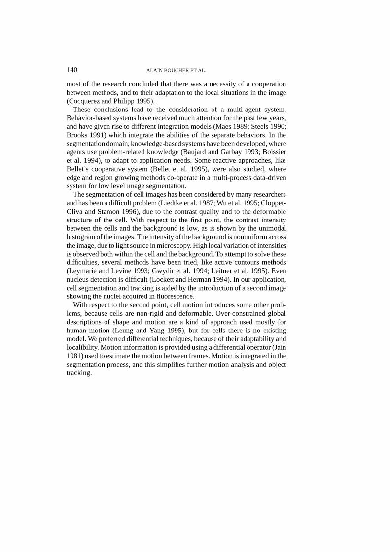
Figure 1. The wound process. (a) Example of a typical injury in a fibroblast monolayer, (b)(c) (d) Time course migration of cells during wound closure. (b) t = 0. (c) t = 13h. (d) t = 24h.
2. Materials and Methods
2.1 Biological experimental procedures
2.1.1 Cell cultureAnchorage dependent L929 murine fibroblasts (ATCC, CCl 1) were grown toplateau phase in antibiotic free Minimum Essential Medium (MEM, Techgen)supplemented with 5% fetal bovine serum (FBS) and L-glutamine. Culturedcells were verified to be free from mycoplasma contamination.
142 ALAIN BOUCHER ET AL.
2.1.2 DNA stainingVerapamil (VER) and Hoechst 33342 (Ho 342) were purchased from Sigma.Stock solutions of VER and Ho 342 were prepared in water, respectively ata concentration of 10�2M and 10�3M. Cultured cells were then washed inPhosphate Buffered Saline (PBS) and incubated with serum-deprived MEMmedium for 30 min, VER and Ho 342 were added in MEM medium (finalconcentrations 40 and 4�M respectively) and cells incubated for 30 min. Theywere washed three times with fresh serum-supplemented MEM medium.
2.1.3 Artificial woundingThree days after cell culture in glass chamber (Lab-Tech), L929 cells form aconfluent monolayer in culture. This confluent cell monolayer is then gentlyscratched with a Gilson pipette blue tip without damaging the glass surfaceand then throughly rinsed with medium to remove all cellular debris (Fig-ure 1a). Cell migration during the artificial wound closure was computedusing microscopic imaging.
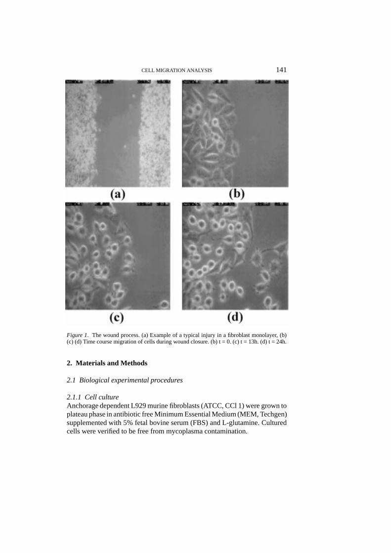
2.1.4 Microscopic imagingThe image sequences were obtained using an imaging workstation, composedof an inverted microscope (Axiovert 135M, Zeiss) equipped with a tempera-ture and CO2 controlled chamber and a SIT camera (C2400-08, Hamamatsu)connected to an analyser (SAMBA TM 2640, Unilog). The modificationof cells position was observed at regular intervals for several hours (Fig-ures 1b, c and d). Both phase contrast (cells) and fluorescent Ho 342 labeled(nuclei) images were registered (Figure 2). The images of fluorescent nucleiwere acquired using a combination of an excitation-filter passing wavelength365 nm, a dichroic filter passing 395 nm and an emission high-pass filter forwavelength 450 nm (when bound to� emission and excitation of Ho 342where 340 and 450 nm respectively).
2.2 The multi-agent system description
A multi-agent system has been designed to assist the analysis and inter-pretation of cell migration as previously presented. The present design ismainly oriented toward image segmentation. Cell tracking and motion analy-sis are currently integrated as part of the segmentation process. A generalagent model has been designed, which will be further expanded to includecomplete tracking and motion analysis capabilities. Our segmentation of cellimages distinguishes four different zones (Figure 2):
1. Nuclei are the cell’s hearts. They are textured and therefore hard todelineate, but they are easier to find with the fluorescence image.
CELL MIGRATION ANALYSIS 143
Figure 2. Images of fibroblasts L929. (a) Image of cells observed in phase contrast. (b) Imageof nuclei observed by fluorescence.
2. White haloscircle the cell. They result from light diffraction around thecells. They look like halos which blur the cell’s exact boundary.
3. Pseudopodsare cytoplasmic extensions of the cell. They represent thehardest part of the segmentation problem. They are poorly contrastedfrom the background, but their motion is important. We segment thecytoplasm around the nucleus along with the pseudopods.
4. Background is segmented to define the cell’s boundary. Its intensity isnonuniform across the image, because of the light source used in themicroscope.
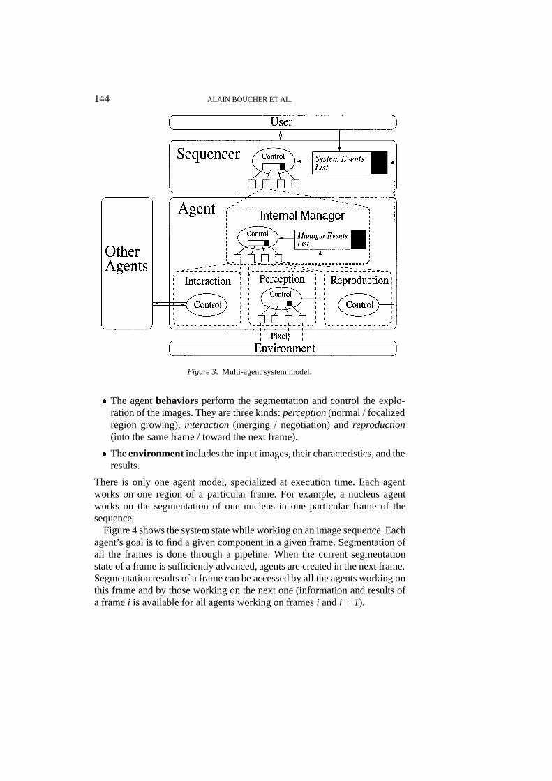
Segmentation is modelled as a complex task combining perception, merging,negotiation and reproduction. Each subtask is modelled as a specific behaviorof the agent. The behaviors are controlled by an internal manager. The agentsociety is supervised by a scheduler, according to the specification of theexternal user. The system is divided into several levels (Figure 3):� Theuser interacts with the system through the sequencer. He can create
or delete agents, or modify their priorities on execution time. He can alsoconfigure each agent before execution through a dedicated panel.
� Thesequencermanages the agent’s execution. It is similar to an operatingsystem. Its goal is to manage all the agents into the same UNIX process.
� The internal manager controls the selection and the execution of theagent’s behaviors. A mechanism of priorities driven by events allows thechoice of a behavior for execution.
144 ALAIN BOUCHER ET AL.
Figure 3. Multi-agent system model.
� The agentbehaviors perform the segmentation and control the explo-ration of the images. They are three kinds:perception(normal / focalizedregion growing),interaction (merging / negotiation) andreproduction(into the same frame / toward the next frame).
� Theenvironment includes the input images, their characteristics, and theresults.
There is only one agent model, specialized at execution time. Each agentworks on one region of a particular frame. For example, a nucleus agentworks on the segmentation of one nucleus in one particular frame of thesequence.
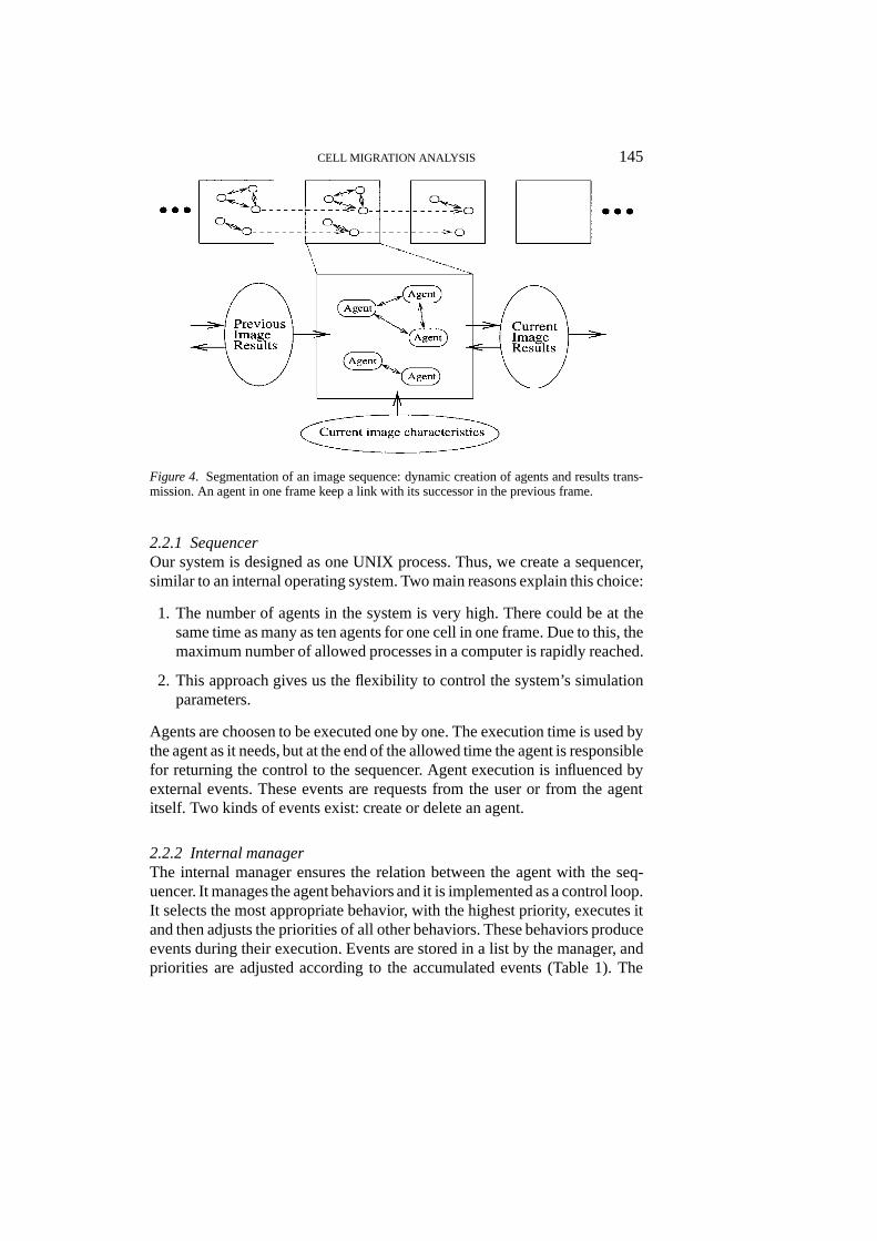
Figure 4 shows the system state while working on an image sequence. Eachagent’s goal is to find a given component in a given frame. Segmentation ofall the frames is done through a pipeline. When the current segmentationstate of a frame is sufficiently advanced, agents are created in the next frame.Segmentation results of a frame can be accessed by all the agents working onthis frame and by those working on the next one (information and results ofa framei is available for all agents working on framesi andi + 1).
CELL MIGRATION ANALYSIS 145
Figure 4. Segmentation of an image sequence: dynamic creation of agents and results trans-mission. An agent in one frame keep a link with its successor in the previous frame.
2.2.1 SequencerOur system is designed as one UNIX process. Thus, we create a sequencer,similar to an internal operating system. Two main reasons explain this choice:
1. The number of agents in the system is very high. There could be at thesame time as many as ten agents for one cell in one frame. Due to this, themaximum number of allowed processes in a computer is rapidly reached.
2. This approach gives us the flexibility to control the system’s simulationparameters.
Agents are choosen to be executed one by one. The execution time is used bythe agent as it needs, but at the end of the allowed time the agent is responsiblefor returning the control to the sequencer. Agent execution is influenced byexternal events. These events are requests from the user or from the agentitself. Two kinds of events exist: create or delete an agent.
2.2.2 Internal managerThe internal manager ensures the relation between the agent with the seq-uencer. It manages the agent behaviors and it is implemented as a control loop.It selects the most appropriate behavior, with the highest priority, executes itand then adjusts the priorities of all other behaviors. These behaviors produceevents during their execution. Events are stored in a list by the manager, andpriorities are adjusted according to the accumulated events (Table 1). The
146 ALAIN BOUCHER ET AL.
Table 1. Internal manager events and involved behaviors. An event (in first column) is sentto the internal manager by a behavior (in second column) during its execution and will affect(increasing or decreasing) the execution priority of another behavior (in third column).
Events Origin Modified priority behavior
Pixel with same region type added Normal growing Fusion (request)Pixel with different region type added Normal growing Focalized growingFocalized growing finished Focalized growing Negotiation (request)Merging / negotiation requested External agent Fusion/negotiation (answer)Merging / negotiation answered External agent Fusion/negotiation (action)Size of region increased Normal growing Reproduction (next frame)Normal growing finished Normal growing Reproduction (same frame)
way priorities are defined and computed has been adjusted experimentally.Further work will provide a more accurate handling of the parameters.
At each loop, the behavior with the highest priority is executed. Its executiontime depends on the time conceded by the sequencer to the internal manager.This time can be managed between the behaviors as needed, as long as themanager gives back the control to the sequencer when it is elapsed.
As a consequence, the agent scheduling is not fixed in advance. It is updatedduring execution according to incoming events. The agent basic behavior isnormal region growing and is always present. Its termination means the endof the agent’s life, since its primary goal is segmentation. Other behaviors areactivated depending on the events produced by this first behavior, and willproduce other events themselves. They can also react to events from otheragents.
2.2.3 Behaviors of agentThree different kinds of behavior exist:perception(normal / focalized regiongrowing),interaction(merging / negotiation) andreproduction(into the sameframe / toward the next frame). The perception and reproduction behaviorscan be configured at execution time depending on the segmentation type theagent has to perform (nucleus, pseudopod, white halo or background).
Perception: Normal growing.Normal growing is the basic behavior. It is responsible for analyzing the localenvironment, evaluating it, and elaborating a list of candidate pixels. Theregion growing method is based from the work of Bellet (Bellet et al. 1995)and depends on candidate pixels. At each loop, the pixel with the highestevaluation is chosen from the list and added to the region. Its neighbors arethen evaluated (2 adjacent neighbors in the horizontal direction and 2 adjacentneighbors in the vertical direction). An evaluation function is used to order
CELL MIGRATION ANALYSIS 147
the candidate pixels. This function weighs various static and motion-basedcriteria. The general expression of this function is:
" = (1� )"static + "motion
" = (1� )
6X
i=1
�siesi +
3X
i=1
�miemi
where is the motion evaluation weight in the global evaluation,�siand�mi
are the weights of the static and motion- based criteria respectively,esi andemi
are the evaluations of the respective criteria. All these values can varyfrom 0 to 1.
Six static criteria are used: variance similarity, compactness, gray levelsimilarity, gradient direction similarity, cell and nucleus image thresholding.The nucleus image thresholding method is evolutive and adaptative. It iscomputed on the gray level (according to the nucleus image) of the regionand updated at each newly aggregated pixel (threshold= mean gray level�factor). This mean gray level changes along with the segmentation process,as the threshold does. Therefore, it is computed dynamically as the situationevolved. All other criteria are defined in a similar way. An example with thevariance level criterion is:
evariance =
8>><>>:
1:0 if 0 � variance � v1
0:6 if v1 < variance � v2
0:4 if v2 < variance � v3
0: else
v1, v2 and v3 are computed automatically from the variance histogram.From the usual static criteria, we developed other criteria based on motioninformation. We use two motion information sources: the difference image,computed between two following frames, and the segmentation of the previ-ous frame. Four different motion-based criteria have been developed. Figure 5shows two examples.
The criteria weights, the static / motion weight ( ), and the thresholdingfactor for aggregation are specialized for the type of object to be segmented(nucleus, pseudopod, white halo, background). Pixel evaluations are then dif-ferent and adapted to the particular objectives of the agent. These parametersare given fixed values.
All the neighboring pixels are evaluated and added to the candidate list.If one chosen pixel is already labelled by another region, then it is added tothe manager’s event list. In so doing, we take note that a segmentation errormight have occured, or that two regions might describe the same entity. These
148 ALAIN BOUCHER ET AL.
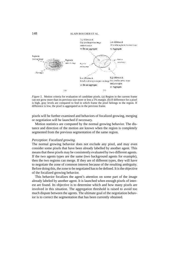
Figure 5. Motion criteria for evaluation of candidate pixels. (a) Region in the current framecan not grow more than its previous size more or less a 5% margin. (b) If difference for a pixelis high, gray levels are compared to find in which frame the pixel belongs to the region. Ifdifference is low, the pixel is aggregated as in the previous frame.
pixels will be further examined and behaviors of focalized growing, mergingor negotiation will be launched if necessary.
Motion statistics are computed by the normal growing behavior. The dis-tance and direction of the motion are known when the region is completelysegmented from the previous segmentation of the same region.
Perception: Focalized growing.The normal growing behavior does not exclude any pixel, and may evenconsider some pixels that have been already labelled by another agent. Thismeans that these pixels may be consistently evaluated by two different agents.If the two agents types are the same (two background agents for example),then the two regions can merge. If they are of different types, they will haveto negotiate the zone of common interest because of the resulting ambiguity.Before doing this, the zone to be negotiated has to be defined. It is the objectiveof the focalized growing behavior.
This behavior focalizes the agent’s attention on some part of the imagealready labeled by another agent. It is launched when enough pixels of inter-est are found. Its objective is to determine which and how many pixels areinvolved in this situation. The aggregation threshold is raised to avoid toomuch dispute between the agents. The ultimate goal of the negotiation behav-ior is to correct the segmentation that has been currently obtained.
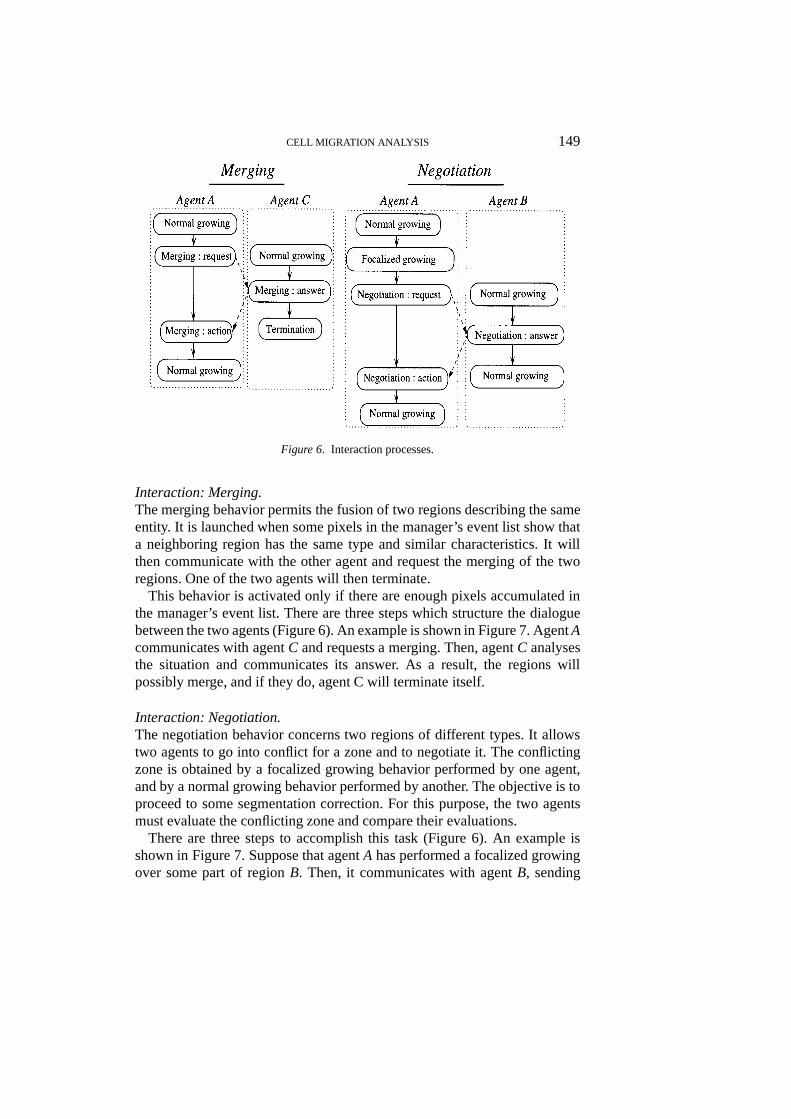
CELL MIGRATION ANALYSIS 149
Figure 6. Interaction processes.
Interaction: Merging.The merging behavior permits the fusion of two regions describing the sameentity. It is launched when some pixels in the manager’s event list show thata neighboring region has the same type and similar characteristics. It willthen communicate with the other agent and request the merging of the tworegions. One of the two agents will then terminate.
This behavior is activated only if there are enough pixels accumulated inthe manager’s event list. There are three steps which structure the dialoguebetween the two agents (Figure 6). An example is shown in Figure 7. AgentAcommunicates with agentC and requests a merging. Then, agentC analysesthe situation and communicates its answer. As a result, the regions willpossibly merge, and if they do, agent C will terminate itself.
Interaction: Negotiation.The negotiation behavior concerns two regions of different types. It allowstwo agents to go into conflict for a zone and to negotiate it. The conflictingzone is obtained by a focalized growing behavior performed by one agent,and by a normal growing behavior performed by another. The objective is toproceed to some segmentation correction. For this purpose, the two agentsmust evaluate the conflicting zone and compare their evaluations.
There are three steps to accomplish this task (Figure 6). An example isshown in Figure 7. Suppose that agentA has performed a focalized growingover some part of regionB. Then, it communicates with agentB, sending
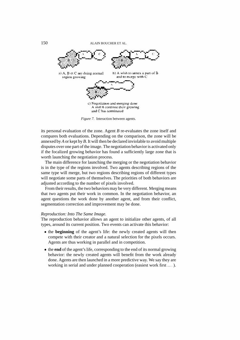
150 ALAIN BOUCHER ET AL.
Figure 7. Interaction between agents.
its personal evaluation of the zone. AgentB re-evaluates the zone itself andcompares both evaluations. Depending on the comparison, the zone will beannexed byAor kept byB. It will then be declared inviolable to avoid multipledisputes over one part of the image. The negotiation behavior is activated onlyif the focalized growing behavior has found a sufficiently large zone that isworth launching the negotiation process.
The main difference for launching the merging or the negotiation behavioris in the type of the regions involved. Two agents describing regions of thesame type will merge, but two regions describing regions of different typeswill negotiate some parts of themselves. The priorities of both behaviors areadjusted according to the number of pixels involved.
From their results, the two behaviors may be very different. Merging meansthat two agents put their work in common. In the negotiation behavior, anagent questions the work done by another agent, and from their conflict,segmentation correction and improvement may be done.
Reproduction: Into The Same Image.The reproduction behavior allows an agent to initialize other agents, of alltypes, around its current position. Two events can activate this behavior:
� the beginning of the agent’s life: the newly created agents will thencompete with their creator and a natural selection for the pixels occurs.Agents are thus working in parallel and in competition.
� theendof the agent’s life, corresponding to the end of its normal growingbehavior: the newly created agents will benefit from the work alreadydone. Agents are then launched in a more predictive way. We say they areworking in serial and under planned cooperation (easiest work first: : : ).
CELL MIGRATION ANALYSIS 151
This behavior tries to maximize co-operation and result-sharing to ensurethe achievement of the global goal, the segmentation of the whole image.Examples of reproduction are shown with the results (Section 3). Reproduc-tion is specialized, like the perception behavior. The specialization includesthe type, number, time, and place where new agents will be created.
Reproduction: Toward The Next Image.The duplication behavior allows agents to initiate the segmentation of otherimages. When its segmentation is judged sufficiently mature (bigger than afixed size), an agent can initiate the segmentation of that region in the nextimage by duplicating itself. In this way, the segmentation of all the frames isdone through a pipeline. An agent working on one frame can, furthermore,access the previous image information.
The maturity of a region is computed from its size (depending on the regiontype). The more a region grows, the more its behavior’s priority increases.When sufficiently high, the behavior launches one identical agent at the sameplace in the next frame. A specific link is created between the newly createdagent and its initiator. Any agent knows which one launched it and can thusretrace its motion from this knowledge. The motion segmentation is based onlinks kept between agents on different frames.
By the two different mechanisms of reproduction, we can define a seg-mentation strategy adapted to the application constraints and dynamicallyelaborated, based on local rules. Indeed, the sequence exploration strategy isnot predefined. It emerges from the agent activation and findings.
3. Results
The results shown in this section are generated by a prototype which canapply the normal growing, merging and reproduction behaviors. Each agentin this prototype is conceived as a separate UNIX process, so that there is nosequencer nor internal manager. The specialization of behaviors comes froma configuration file.
This section illustrates 4 different kinds of results: (i) the user interfacefor configuration and visualization, (ii) discussion of segmentation results onsome frames, (iii) cell tracking results for random motion and for the woundprocess are compared, and (iv) the different cell motions are analyzed.
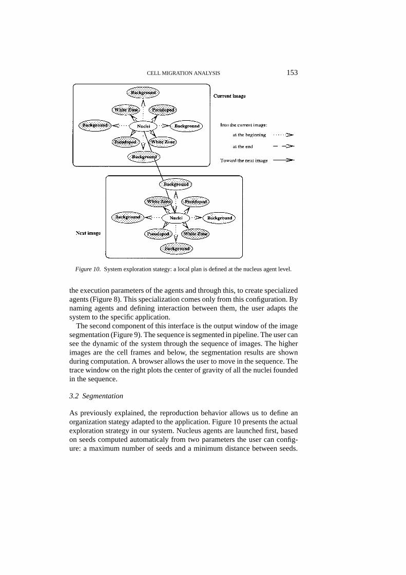
3.1 User Interface
A user interface has been developed for better use of the system. This interfacerelies on two components. First, a configuration file allows the user to specify
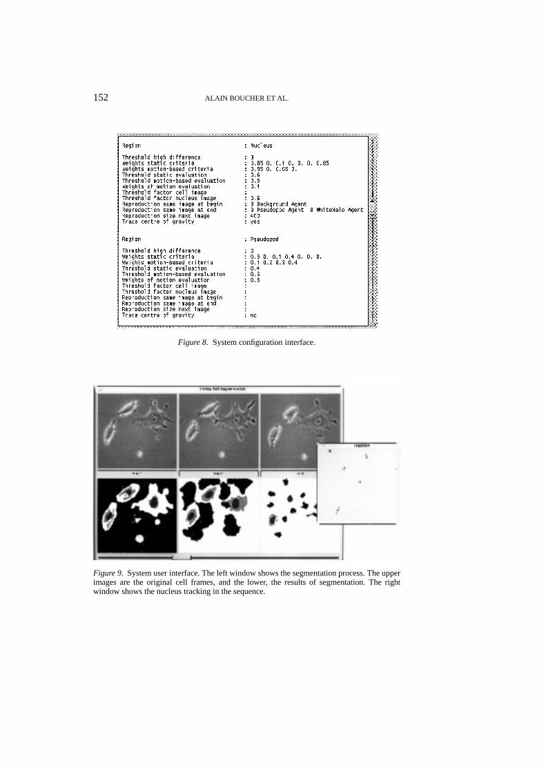
152 ALAIN BOUCHER ET AL.
Figure 8. System configuration interface.
Figure 9. System user interface. The left window shows the segmentation process. The upperimages are the original cell frames, and the lower, the results of segmentation. The rightwindow shows the nucleus tracking in the sequence.
CELL MIGRATION ANALYSIS 153
Figure 10. System exploration stategy: a local plan is defined at the nucleus agent level.
the execution parameters of the agents and through this, to create specializedagents (Figure 8). This specialization comes only from this configuration. Bynaming agents and defining interaction between them, the user adapts thesystem to the specific application.
The second component of this interface is the output window of the imagesegmentation (Figure 9). The sequence is segmented in pipeline. The user cansee the dynamic of the system through the sequence of images. The higherimages are the cell frames and below, the segmentation results are shownduring computation. A browser allows the user to move in the sequence. Thetrace window on the right plots the center of gravity of all the nuclei foundedin the sequence.
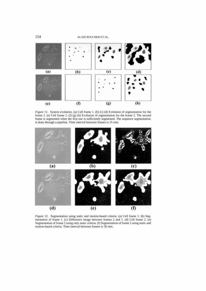
3.2 Segmentation
As previously explained, the reproduction behavior allows us to define anorganization stategy adapted to the application. Figure 10 presents the actualexploration strategy in our system. Nucleus agents are launched first, basedon seeds computed automaticaly from two parameters the user can config-ure: a maximum number of seeds and a minimum distance between seeds.
154 ALAIN BOUCHER ET AL.
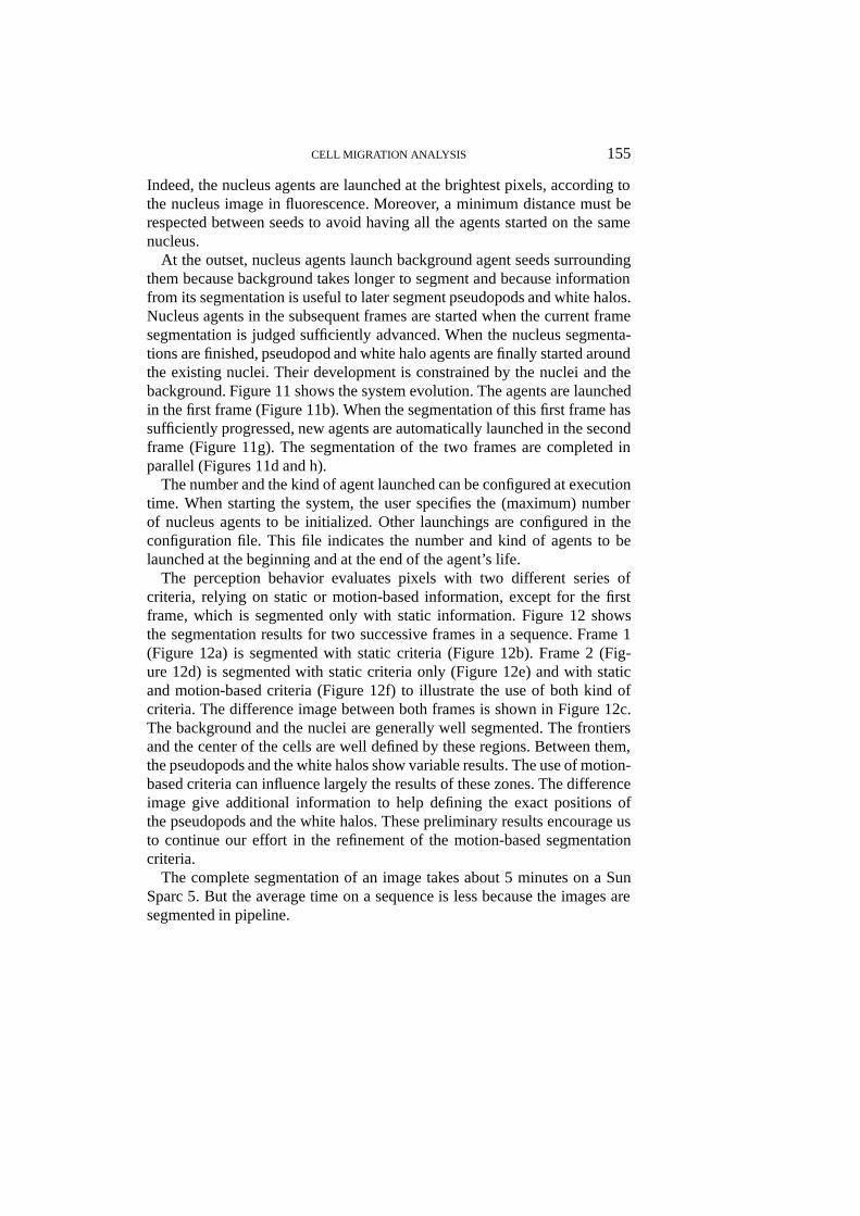
Figure 11. System evolution. (a) Cell frame 1. (b) (c) (d) Evolution of segmentation for theframe 1. (e) Cell frame 2. (f) (g) (h) Evolution of segmentation for the frame 2. The secondframe is segmented when the first one is sufficiently segmented. The sequence segmentationis done through a pipeline. Time interval between frames is 15 min.
Figure 12. Segmentation using static and motion-based criteria. (a) Cell frame 1. (b) Seg-mentation of frame 1. (c) Difference image between frames 2 and 1. (d) Cell frame 2. (e)Segmentation of frame 2 using only static criteria. (f) Segmentation of frame 2 using static andmotion-based criteria. Time interval between frames is 30 min.
CELL MIGRATION ANALYSIS 155
Indeed, the nucleus agents are launched at the brightest pixels, according tothe nucleus image in fluorescence. Moreover, a minimum distance must berespected between seeds to avoid having all the agents started on the samenucleus.
At the outset, nucleus agents launch background agent seeds surroundingthem because background takes longer to segment and because informationfrom its segmentation is useful to later segment pseudopods and white halos.Nucleus agents in the subsequent frames are started when the current framesegmentation is judged sufficiently advanced. When the nucleus segmenta-tions are finished, pseudopod and white halo agents are finally started aroundthe existing nuclei. Their development is constrained by the nuclei and thebackground. Figure 11 shows the system evolution. The agents are launchedin the first frame (Figure 11b). When the segmentation of this first frame hassufficiently progressed, new agents are automatically launched in the secondframe (Figure 11g). The segmentation of the two frames are completed inparallel (Figures 11d and h).
The number and the kind of agent launched can be configured at executiontime. When starting the system, the user specifies the (maximum) numberof nucleus agents to be initialized. Other launchings are configured in theconfiguration file. This file indicates the number and kind of agents to belaunched at the beginning and at the end of the agent’s life.
The perception behavior evaluates pixels with two different series ofcriteria, relying on static or motion-based information, except for the firstframe, which is segmented only with static information. Figure 12 showsthe segmentation results for two successive frames in a sequence. Frame 1(Figure 12a) is segmented with static criteria (Figure 12b). Frame 2 (Fig-ure 12d) is segmented with static criteria only (Figure 12e) and with staticand motion-based criteria (Figure 12f) to illustrate the use of both kind ofcriteria. The difference image between both frames is shown in Figure 12c.The background and the nuclei are generally well segmented. The frontiersand the center of the cells are well defined by these regions. Between them,the pseudopods and the white halos show variable results. The use of motion-based criteria can influence largely the results of these zones. The differenceimage give additional information to help defining the exact positions ofthe pseudopods and the white halos. These preliminary results encourage usto continue our effort in the refinement of the motion-based segmentationcriteria.
The complete segmentation of an image takes about 5 minutes on a SunSparc 5. But the average time on a sequence is less because the images aresegmented in pipeline.
156 ALAIN BOUCHER ET AL.
3.3 Object tracking
At the end of an agent’s life, a trace of its center of gravity is plotted in thetrace window. Lines are also plotted to link one component in two successiveframes, and therefore to show the displacement of the component betweenthese frames. In the current version of the system, only nucleus agent tracesare usable, due to the difficulty of segmentation of the other components. Asan extension of this work, we want to link the different cell zones and studythe cell behavior when moving.
Four sequences were studied, and will be referenced with the followingnames:
� Natural motion: two different image sequences� 19 frames every 30 min (NM30);� 20 frames every 3 min (NM3).
� Wound closure: two image sequences representing two phases of thesame process� 27 frames every 30 min (first phase: t = 0 to t = 13h) (WC1);� 15 frames every 30 min (second phase: t = 17h to t = 24h) (WC2).
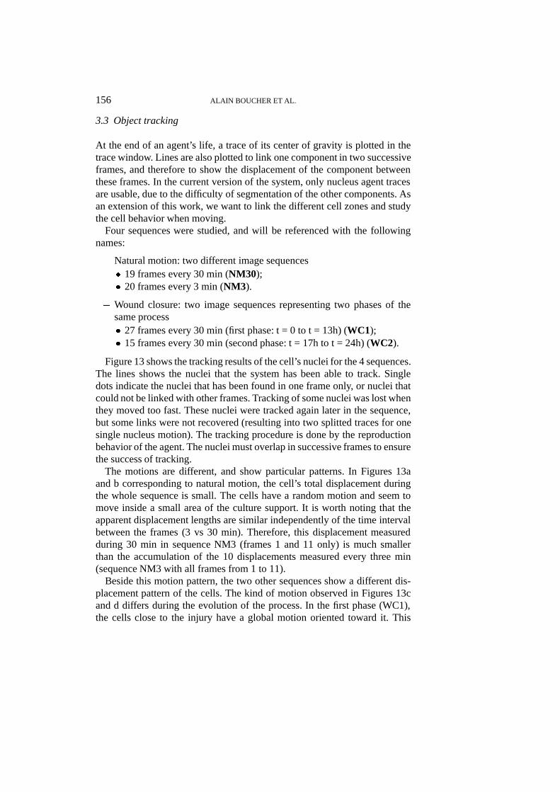
Figure 13 shows the tracking results of the cell’s nuclei for the 4 sequences.The lines shows the nuclei that the system has been able to track. Singledots indicate the nuclei that has been found in one frame only, or nuclei thatcould not be linked with other frames. Tracking of some nuclei was lost whenthey moved too fast. These nuclei were tracked again later in the sequence,but some links were not recovered (resulting into two splitted traces for onesingle nucleus motion). The tracking procedure is done by the reproductionbehavior of the agent. The nuclei must overlap in successive frames to ensurethe success of tracking.
The motions are different, and show particular patterns. In Figures 13aand b corresponding to natural motion, the cell’s total displacement duringthe whole sequence is small. The cells have a random motion and seem tomove inside a small area of the culture support. It is worth noting that theapparent displacement lengths are similar independently of the time intervalbetween the frames (3 vs 30 min). Therefore, this displacement measuredduring 30 min in sequence NM3 (frames 1 and 11 only) is much smallerthan the accumulation of the 10 displacements measured every three min(sequence NM3 with all frames from 1 to 11).
Beside this motion pattern, the two other sequences show a different dis-placement pattern of the cells. The kind of motion observed in Figures 13cand d differs during the evolution of the process. In the first phase (WC1),the cells close to the injury have a global motion oriented toward it. This
CELL MIGRATION ANALYSIS 157
Figure 13. Cell tracking by their nuclei. (a) Natural motion (sequence NM30). (b) Naturalmotion (sequence NM3) (c) First phase of the wound process (sequence WC1). (d) Secondphase of the wound process (sequence WC2).
suggests two different behaviors, depending on the position of the cells rela-tive to the injury. In the second phase (WC2), the cells are spaced and somecells still have a directionnal motion. In the two phases, some cells do notfollow the main observed motion but have a random displacement. It shouldbe noted that random motion can have a directional component and that direc-tional motion can have a random component. During the wound closure, asequence of shorter time interval (3 min) would be of interest to determinemore accurately the nature of the motion at higher frequency.
158 ALAIN BOUCHER ET AL.
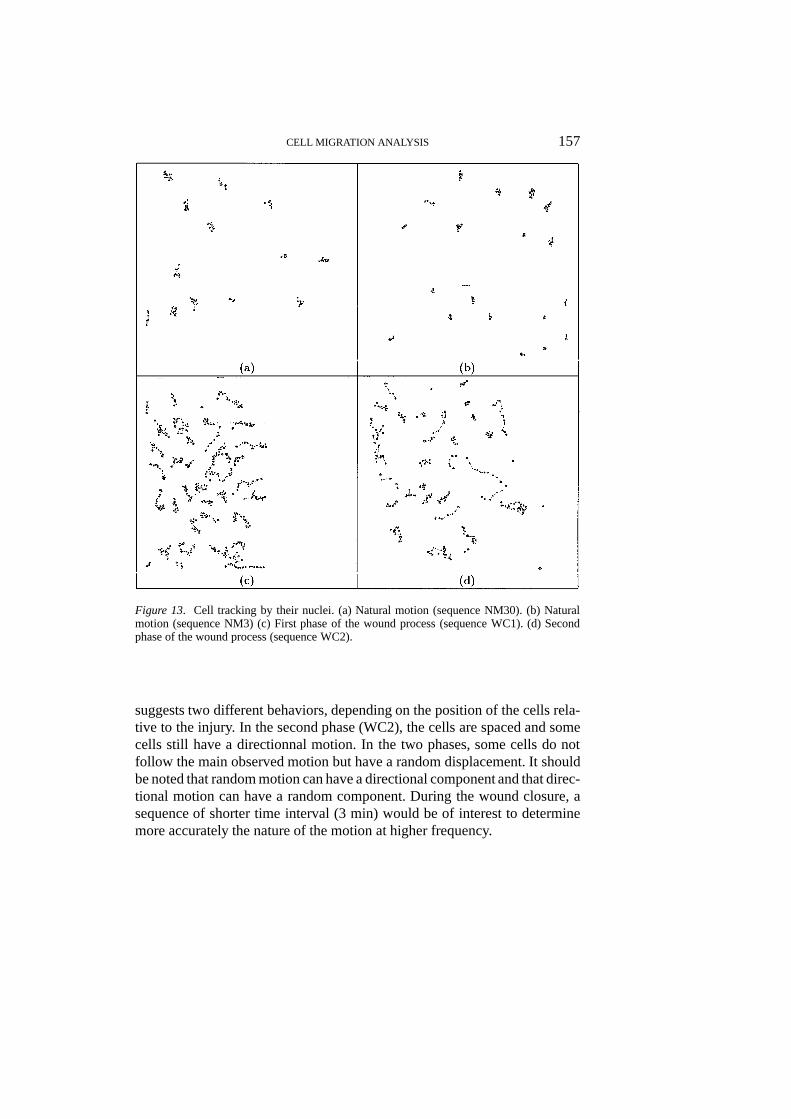
Figure 14. Histograms of distances and direction. (a) Histogram of distance covered for thenatural motion (sequence NM30). (b) Histogram of distance covered for the first phase ofthe wound process (sequence WC1). (c) Histogram of motion direction for a natural motion(sequence NM30). (d) Histogram of distance covered for the first phase of the wound process(sequence WC1). (e) Histogram of motion direction for the first phase of the wound process(cells far from the injury) (sequence WC1). (f) Histogram of motion direction for the firstphase of the wound process (cells close to the injury) (sequence WC1). All data (distance anddirection) are computed between two successive frames. Distances are in pixels and directionsare coded upon the Freeman code.
3.4 Analysis of the migration process
3.4.1 DistanceMigration rates of fibroblasts were determined by calculating the nucleardisplacement for individual cells. From Figures 14a and b, one can see thatthe distances covered during natural motion or during the wound processare almost the same. After calibration, the mean speed of the cells duringnatural motion (sequence NM30) and wound closure (sequence WC1) arerespectively 4.6�m/hour and 4.3�m/hour (Figure 14a and b). Those valuescan be compared to the value of 1.5�m/hour obtained by Dunlevy andCouchman (1993), with rat embryo fibroblasts treated by heart-conditionedmedium.
3.4.2 DirectionDirections in histograms were computed using the Freeman code. In thiscode, distances are rounded to 45� and labeled from 0 to 7, 0 being a leftdisplacement, 2 an upward displacement, etc.: : :From the comparison of thedirection histograms of natural motion (NM30) and wound closure (WC1)sequences (Figures 14c and d), a preferential direction can be observed forthe second one.
CELL MIGRATION ANALYSIS 159
Figures 14e and 14f show that nearly all the cells near the injury’s sideclearly move toward 0, 1, 2, 6 and 7 directions i.e. toward the denuded areaof the cell monolayer. This tendency is not so evident for the cells inside thecell sheet. In the first case, cell migration can be the result of a directionalvoluntarycell displacement. It could also be explained by a random movementinto a denuded area. However, from Figure 13c we can see that some cellsnear the injury’s side still have a nearly straight displacement; cells beingmore widely spaced, random motion would be doubtful.
Here, only primary motion analysis methods have been presented. Furtherextension to this analysis is under study to show potential correlations betweenmotion and some shape factors of the cells. Only nucleus components havebeen studied to date. The integration of all the cell’s components will improvethe characterization of cell motion.
4. Discussion
The present system would be of interest for studying the influence of varioussubstances such as growth factors on the wound closure process. This modelcould also be modified in order to more accurately reflect thein vivosystem.For instance, cells could be cultured on a fibronectin coated support to specifythe importance of extracellular matrix components during cell migration. Cellmigration could also be studied using chemo-attractant substances or orientedgels leading to a contact guidance of cells (Dickinson et al. 1994). It wouldthus be possible to analyse whether factors promoting autonomous migrationof cells into a denuded area of a cell monolayer differ from those responsiblefor their chemotactic or biased migration (Kondo et al. 1993). These differentmodels could provide a better knowledge ofin vivocell migration mechanismsinvolved in processes such as wound healing and tumor cell invasion.
The proposed agent model is similar to the one presented by Guessoum andDojat (Guessoum and Dojat 1996) and applied to breath monitoring and eco-nomic simulation. Both models are multi-level and integrate various behaviorsof different types, reactive (perception, reproduction) and cognitive (interac-tion). Two levels of competition are present in the system. Agents competewith themselves in the segmentation of an image. They can label, mergeor negotiate zones. The behaviors interact asynchronously and concurrentlyunder the control of a internal manager, which supervises the global timescheduling. Behaviors into one agent also work under a competition scheme.They are driven by external and internal events. These events modify theirpriorities and allow one of them to execute at a given time.
Our system integrates various levels of analysis, from the segmentationto cell tracking. The segmentation is specialized for the different compo-
160 ALAIN BOUCHER ET AL.
nents of the images. It relies on sets of criteria depending on both static andmotion-based information. We are attempting to make these criteria moreadaptative to the local situation in an image. For exemple, an adaptative andevolutive thresholding is used for the nuclei, computed dynamically basedon the nuclear mean gray level and a threshold factor. Another improvementwould be to analyse the gray level profiles around the nuclei, according toa few predefined directions, and to adapt the threshold factor based on thisprofile. The reproduction strategy can be improved in the same way. In thecurrent system, an agent launches a seed around itself at a prefixed distance.This launching strategy could be easily improved by an exploration phase ofthe environment, based on the previously mentionned nucleus surroundingprofiles to find the best location for the new agent. This would imply anadaptative reproduction depending on the type of agent to be initiated. Forthe reproduction toward the next frame, the process could estimate the futureposition of the agent using the motion information computed so far.
The possibilities of motion analysis are reduced to tracking the nucleithrough the sequence and computing some statistics on their motion. Forfurther work, a cellular entity will be created to organize the agents workingon the same cell. This higher abstraction level will give the opportunity tobuild a more complete analysis of cell behavior. One cell component motioncan be decomposed into two motions: the global cell motion and the specificmotion of the component. By studying these two motions separately, a morecomplete interpretation of the complete cell motion can be made.
In this paper, the wounding injury process was presented as an example ofa typical application in which an intelligent system can be used.
References
Baujard, O. & Garbay, C. (1993). KISS: a multiagent segmentation system.Optical Engineer-ing 32(6): 1235–1249.
Beil, M., Irinopoulou, T., Vassy, J. & Rigault, J. P. (1995). Chromatin texture analysis inthree-dimensional images from confocal scanning laser microscopy.Analyt. Quant. Cytol.Histol. 17(5): 323–331.
Bellet, F., Salotti, J. M. & Garbay, C. (1995). Une approche opportunists et cooperative pourla vision de bas niveau.Traitement du Signal12(5): 479–494.
Bernstein, J. J., Goldberg, W. J., & Law Jr., E. R. (1993). Migration of fresh human malignantastrocytoma cells into hydrated gel wafers in vitro.J. Neurooncol.18: 151–161.
Bodor, N. S., Kiss-Buris, S. T. & Buris, L. (1991). Novel soft steroids: effects on cell growthin vitro and on wound healing in the mouse.Steroids56: 434–439.
Boissier, O., Demazeau, Y., Masini, G. & Skaf, H. (1994). Une architecture multi-agents pourl’impl ementation du bas niveau d’un systeme de comprehension de scenes. In2iemesjournees francophones IAD et SMA. Voiron (France).
Brooks, R. A. (1993). Intelligence without representation.Artificial Intelligence47: 139–159.
CELL MIGRATION ANALYSIS 161
Cloppet-Oliva, F. & Stamon, G. (1996). Segmentation cooperative region/contour pour uneanalyse automatique d’images de cellules en culture. InProceedings of 10e congres Recon-naissances des Formes et Intelligence Artificielle, volume 2, 1063–1072.
Cocquerez, J. P. & Philipp, S. (1995). Analyse d’images: filtrage et segmentation. Masson.Dickinson, R. B., Guido, S. & Tranquillo, R. T. (1994). Biased cell migration of fibroblasts
exhibiting contact guidance in oriented collagen gels.Ann. Biomed. Eng.22: 342–356.Dunlevy, J. R. & Couchman, J. R. (1993). Controlled induction of focal adhesion disassembly
and migration in primary fibroblasts.J. Cell Sci.105: 489–500.Francisco, J., Pauwels, O., Simon, S., Gasperin, P., van Houtte, P., Pasteels, J. L. & Kiss,
R. (1995). Computer-assissted morphonuclear characterization of radiotherapy-inducedeffects in MXT mouse mammary adenocarcinomas surviving earlier radiotherapy.Int. J.Radiation Oncology Biol. Phys.32(2): 409–419.
Fu, K. S. & Mui, J. K. (1987). A survey on image segmentation.Pattern Recognition13(l):3–16.
Guessoum, Z. & Dojat, M. A real-time agent model in a asynchronous-object environment. InProceedings of MAAMAW, 190–20.
Gwydir, S. H., Buettner, H. M. & Dunn, S. M. (1994). Non-rigid motion analysis of the growthcone using continuity splines.ITBM 15(3): 309–321.
Jain, R. (1981). Extraction of motion information from peripheral processes.IEEE Trans. onPAMI 3(5): 489–503.
Karasek, M. A. (1989). Microvascular endothelial cell culture.J. Invest. Dermatol.93: 33S–38S.
Kondo, H., Matsuda, R. & Yonezawa, Y. (1993). Autonomous migration of human fetal skinfibroblasts into a denuded are in a cell monolayer is mediated by basic fibroblast growthfactor and collagen,In vitro Cell Dev. Biol.29A: 929–935.
Leitner, F., Paillasson, S., Ronot, X. & Demongeot, J. (1995). Dynamic functional and structuralanalysis of living cells: new tools for vital staining of nuclear DNA and for characterizationof cell motion.Acta Biotheoritica(43): 299–317.
Lester, B. R. & McCarthy, J. B.. Tumor cell adhesion to the extracellular matrix and signaltransduction mechanisms implicated in tumor cell motility, invasion and metastasis.CancerMetast. Rev.11: 31–44.
Leung, M. K. &Yang, Y. H. (1995). First Sight: A human body outline labeling system.IEEETrans. on PAMI17(4): 359–377.
Leymarie, F. & Levine, M. D. (1993). Tracking deformable objects in the plane using an activecontour model.IEEE Trans. on PAMI15(6): 617–634.
Liedtke, C. E., Gahm, T., Kappei, F. & Aeikens, B. (1987). Segmentation of microscopic cellscenes.Analyt. Quant. Cytol. Histol.9: 197–211.
Lockett, S. J. & Herman, B. (1994). Automatic detection of clustered, fluorescent-stainednuclei by digital image-based cytometry.Cytometry17: 1–12.
Maes, P. (1989). How to do the right thing.Connection Science Journal1(3): 291–323.Mareel, M. M., van Roy, F. M. & de Baetselier, P. (1990). The invasive phenotypes.Cancer
Metast. Rev.9: 45–60.Matthay, M. A., Thiery, J. P., Lafont, F., Stampfer, M. F. & Boyer, B. (1993). Transient effect
of epidermal growth factor on the motility of an immortalized mammary epithelial cellline. J. Cell Sci.106: 869–878.
Muller, T., Geimer, P. & Bade, E. G. (1993). The growth factor-induced migration of epithelialcells is associated with a transient inhibiton of DNA synthesis and a downregulation ofPCNA RNA.Eur. J. Cell. Biol.60: 123.
Naudet, S., Nicolas, L., Faye, C. & Viala, M. (1996). Suivi temporel 3D d’objets en presenced’occultations dans une sequence d’images monoculaires. In Proceedings of 10e congresReconnaissances des Formes et Intelligence Artificielle, volume 2, 849–858.
Paillasson, S., Robert-Nicoud, M. & Ronot, X. (1995). Optimizing DNA staining by Hoechst33342 for assessment of chromatin Organisation in living cells. InProceedings of Opticaland Imaging Techniques in Biomedecine, SPIE Proceedings Series, volume 2329, 225–235.
162 ALAIN BOUCHER ET AL.
Palcic, B. (1994). Nuclear texture: it can be used as a surrogate endpoint biomarker.J. CellBiochem.19: 40–46.
Parvin, B. A., Peng, C., Johnston, W. & Maestre, F. M. (1995). Tracking of tubular moleculesfor scientific applications.IEEE Trans. on PAMI17(8): 800–804.
Person, J. M., Lovett, D. H. & Raugi, G. J. (1988). Modulation of mesangial cell migrationby extracellular matrix components. inhbition by heparinlike glycosaminoglycans.Am. J.Pathol.133: 609–614.
Rappolee, D. A., Mark, D., Banda, M. G. & Werb, Z. (1988). Wound macrophages expressTGF-alpha and other growth factors in vivo: analysis by MRNA phenotyping.Science241: 708–712.
Scharffetter, K., Stolz W., Lankat-Burttgereit, B., Mauch, C., Kulozik, M. & Krieg, T. (1989).In situ hybridization: a useful tool for studies on collagen gene expression in cell cultureas well as in normal and altered tissue.Virchows Arch. B. Cell Pathol.56: 299–306.
Schuh, A. C., Keating, S. J., Monteclaro, F. S., Vogt, P. K. & Breitman, M. L. (1990). Obligatorywounding requirement for tumorigenesis in v-jun transgenic mice.Nature346: 756–760.
Schweitzer, H. (1995). Occam algorithms for computing visual motion.IEEE Trans. on PAMI17(11): 1033–1042.
Siegert, F., Weijer, C. J., Nomura, A. & Miike, H. (1994). A gradient method for the quantitativeanalysis of cell movement and tissue flow and its application to the analysis of multicellularDictyostelium development.J. Cell Sci.(107): 97–104.
Sims, J. R., Karp, S. & Ingber, D. (1992). Altering the cellular mechanical force balance resultsin integrated changes in cell, cytosqueletal and nuclear shape.J. Cell Sci.103: 1215–1222.
Smoot, E. C., Kucan, J. O., Roth, A., Mody, N. & Debs, N. (1991). In vitro toxixity testing forantibacterials against human keratinocytes.Plast. Reconstr. Surg.87: 917–924.
Steels, L. (1990). Cooperation between distributed agents through self-organization. InDAI,volume 1, 175–196. Elsevier Science.
Stokes, C. L., Rupnick, M. A., Williams, S. K. & Lauffenburger, D. A. (1990). Chemotaxisof human microvessel endothelial cells in response to acidic fibroblast growth factor.Lab.Invest.63: 657–668.
van Luyn, M. J., van Wachem, P. B., Nieuwenhuis, P. & Jonjman, M. F. (1992). Cytotoxicitytesting of wound dressings using methylcellulose cell culture.Biomaterials13: 267–275.
Watanabe, S., Hirose, M., Yasuda, T., Miyazaki, A. & Sato, N. (1994). Role of actin andcalmodulin in migration and proliferation of rabbit gastric mucosal cells in culture.J.Gastroenterol. Hepatol.9: 325–333.
Wong, J., Wang, N., Miller, J. W. & Schuman, J. S. (1994). Modulation of human fibroblastactivity by selected angiogenesis inhibitors.Exp. Eye Res.58: 439–451.
Wu, K., Gauthier, D. & Levine, M. D. (1995). Live cell image segmentation.IEEE Trans. onBio. Eng.42(l): 1–12.