Case 24-2007: A 20-Year-Old Pregnant Woman with Altered ...
12
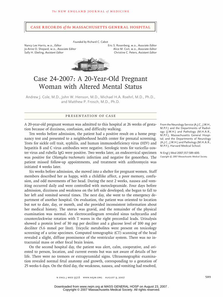
case records of the massachusetts general hospital The new england journal of medicine n engl j med 357;6 www.nejm.org august 9, 2007 589 Founded by Richard C. Cabot Nancy Lee Harris, m.d., Editor Eric S. Rosenberg, m.d., Associate Editor Jo-Anne O. Shepard, m.d., Associate Editor Alice M. Cort, m.d., Associate Editor Sally H. Ebeling, Assistant Editor Christine C. Peters, Assistant Editor From the Neurology Service (A.J.C., J.W.H., M.P.F.) and the Departments of Radiol- ogy (J.W.H.) and Pathology (M.H.A.R., M.P.F.), Massachusetts General Hospi- tal; and the Departments of Neurology (A.J.C., J.W.H.) and Pathology (M.H.A.R., M.P.F.), Harvard Medical School. N Engl J Med 2007;357:589-600. Copyright © 2007 Massachusetts Medical Society. Presentation of Case A 20-year-old pregnant woman was admitted to this hospital at 26 weeks of gesta- tion because of dizziness, confusion, and difficulty walking. Ten weeks before admission, the patient had a positive result on a home preg- nancy test and presented to a neighborhood health center for prenatal screening. Tests for sickle cell trait, syphilis, and human immunodeficiency virus (HIV) and hepatitis B and C virus antibodies were negative. Serologic tests for varicella–zos- ter virus and rubella IgG were positive. Two weeks later, an endocervical specimen was positive for Chlamydia trachomatis infection and negative for gonorrhea. The patient missed follow-up appointments, and treatment with azithromycin was initiated 4 weeks later. Six weeks before admission, she moved into a shelter for pregnant women. Staff members described her as happy, with a childlike affect, a poor memory, confu- sion, and odd movements of her head. During the next 2 weeks, nausea and vom- iting occurred daily and were controlled with metoclopramide. Four days before admission, dizziness and weakness on the left side developed; she began to fall to her left and vomited several times. The next day, she went to the emergency de- partment of another hospital. On evaluation, the patient was oriented to location but not to date, day, or month, and she provided inconsistent information about her medical history. The uterus was gravid, and the remainder of the physical examination was normal. An electrocardiogram revealed sinus tachycardia and counterclockwise rotation with T waves in the right precordial leads. Urinalysis showed a protein level of 30 mg per deciliter and a glucose level of 100 mg per deciliter (5.6 mmol per liter). Tricyclic metabolites were present on toxicology screening of a urine specimen. Computed tomographic (CT) scanning of the head revealed a slight, diffuse prominence of the ventricular system. There was no in- tracranial mass or other focal brain lesion. On the second hospital day, the patient was alert, calm, cooperative, and ori- ented to person, location, and current events but was not aware of details of her life. There were no tremors or extrapyramidal signs. Ultrasonographic examina- tion revealed normal fetal anatomy and growth, corresponding to a gestation of 25 weeks 6 days. On the third day, the weakness, nausea, and vomiting had resolved, Case 24-2007: A 20-Year-Old Pregnant Woman with Altered Mental Status Andrew J. Cole, M.D., John W. Henson, M.D., Michael H.A. Roehrl, M.D., Ph.D., and Matthew P. Frosch, M.D., Ph.D. Copyright © 2007 Massachusetts Medical Society. All rights reserved. Downloaded from www.nejm.org at MASS GENERAL HOSP on August 23, 2007 .
-
Upload
khangminh22 -
Category
Documents
-
view
1 -
download
0
Transcript of Case 24-2007: A 20-Year-Old Pregnant Woman with Altered ...
case records of the massachusetts general hospital
T h e n e w e ng l a nd j o u r na l o f m e dic i n e
n engl j med 357;6 www.nejm.org august 9, 2007 589
Founded by Richard C. Cabot Nancy Lee Harris, m.d., Editor Eric S. Rosenberg, m.d., Associate EditorJo-Anne O. Shepard, m.d., Associate Editor Alice M. Cort, m.d., Associate EditorSally H. Ebeling, Assistant Editor Christine C. Peters, Assistant Editor
From the Neurology Service (A.J.C., J.W.H., M.P.F.) and the Departments of Radiol-ogy ( J.W.H.) and Pathology (M.H.A.R., M.P.F.), Massachusetts General Hospi-tal; and the Departments of Neurology (A.J.C., J.W.H.) and Pathology (M.H.A.R., M.P.F.), Harvard Medical School.
N Engl J Med 2007;357:589-600.Copyright © 2007 Massachusetts Medical Society.
Pr esen tation of C a se
A 20-year-old pregnant woman was admitted to this hospital at 26 weeks of gesta-tion because of dizziness, confusion, and difficulty walking.
Ten weeks before admission, the patient had a positive result on a home preg-nancy test and presented to a neighborhood health center for prenatal screening. Tests for sickle cell trait, syphilis, and human immunodeficiency virus (HIV) and hepatitis B and C virus antibodies were negative. Serologic tests for varicella–zos-ter virus and rubella IgG were positive. Two weeks later, an endocervical specimen was positive for Chlamydia trachomatis infection and negative for gonorrhea. The patient missed follow-up appointments, and treatment with azithromycin was initiated 4 weeks later.
Six weeks before admission, she moved into a shelter for pregnant women. Staff members described her as happy, with a childlike affect, a poor memory, confu-sion, and odd movements of her head. During the next 2 weeks, nausea and vom-iting occurred daily and were controlled with metoclopramide. Four days before admission, dizziness and weakness on the left side developed; she began to fall to her left and vomited several times. The next day, she went to the emergency de-partment of another hospital. On evaluation, the patient was oriented to location but not to date, day, or month, and she provided inconsistent information about her medical history. The uterus was gravid, and the remainder of the physical examination was normal. An electrocardiogram revealed sinus tachycardia and counterclockwise rotation with T waves in the right precordial leads. Urinalysis showed a protein level of 30 mg per deciliter and a glucose level of 100 mg per deciliter (5.6 mmol per liter). Tricyclic metabolites were present on toxicology screening of a urine specimen. Computed tomographic (CT) scanning of the head revealed a slight, diffuse prominence of the ventricular system. There was no in-tracranial mass or other focal brain lesion.
On the second hospital day, the patient was alert, calm, cooperative, and ori-ented to person, location, and current events but was not aware of details of her life. There were no tremors or extrapyramidal signs. Ultrasonographic examina-tion revealed normal fetal anatomy and growth, corresponding to a gestation of 25 weeks 6 days. On the third day, the weakness, nausea, and vomiting had resolved,
Case 24-2007: A 20-Year-Old Pregnant Woman with Altered Mental Status
Andrew J. Cole, M.D., John W. Henson, M.D., Michael H.A. Roehrl, M.D., Ph.D., and Matthew P. Frosch, M.D., Ph.D.
Copyright © 2007 Massachusetts Medical Society. All rights reserved. Downloaded from www.nejm.org at MASS GENERAL HOSP on August 23, 2007 .

T h e n e w e ng l a nd j o u r na l o f m e dic i n e
n engl j med 357;6 www.nejm.org august 9, 2007590
and the patient was thought to have returned to her baseline mental status. She was discharged to the shelter with a recommendation to sched-ule a follow-up neurologic evaluation. At the shelter, she was dizzy, had difficulty walking, and fell into a chair. That evening, she was brought to the emergency department of this hospital.
In the emergency department, the patient re-ported feeling “woozy” and nauseated. She noted a mild headache of gradual onset, extending band-like across the brow. The history as given by the patient was inconsistent; the history was then provided by staff members of the shelter, and many details were lacking. The patient was a native of Cape Verde who had immigrated to this country 3 years previously. She had had measles at 4 months of age and varicella infection in child-hood. At 7 years of age, she injured her head in a fall but was said to have recovered fully. Immu-nizations included polio vaccine and diphtheria, pertussis, and tetanus vaccine series; measles vac-cine (at 11 months of age); measles, mumps, and rubella vaccine combination; and hepatitis B vac-cine (between 2 and 3 years before admission, on enrollment in high school).
The patient had attended school through the 10th grade and was unemployed. During the 3 years before admission, she had lived with rela-tives, friends, and a boyfriend, as well as in shel-ters. She was single, and she no longer main-tained a social relationship with the father of the fetus. Her parents and seven siblings were alive but not in contact with her at the time of admis-sion. No family medical history was available. She had no known allergies and did not use alcohol, illicit drugs, or tobacco.
On examination in the emergency department, the patient was alert but somewhat uncoopera-tive, with involuntary head movements. Her men-tal status was not formally assessed, but her level of cognitive function was said by a friend to be at baseline. The blood pressure was 107/81 mm Hg, the pulse 84 beats per minute, and the tempera-ture 36.3°C; the respirations were 18 per minute, and the oxygen saturation was 100% while the pa-tient was breathing ambient air. Acneiform lesions were present on her face. The abdomen was soft, gravid, and not tender; the fetus appeared to be healthy. The 1st cranial nerve was not tested, and the 2nd through 12th nerves were intact. Strength was intact, and the gait was unsteady. The remain-der of the examination was normal.
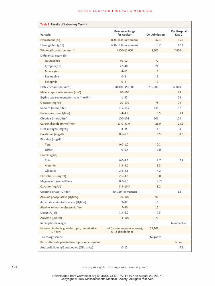
Results of laboratory tests are shown in Table 1. After premedication with lorazepam at a dose of 1 mg to control involuntary movements, magnet-ic resonance imaging (MRI) of the brain was performed without administration of contrast material. On T2-weighted, fluid-attenuated inver-sion recovery (FLAIR) images, hyperintense signal was seen in the left hippocampus and parahippo-campal gyrus as well as in the posterior limb of the left internal capsule. There was no evidence of restricted diffusion.
Examination by a neurology consultant showed that the patient was oriented to person and place, with a childlike affect. Her speech was fluent, and naming was intact. She could read a short sentence and do simple addition. She was left-handed, could write her name but not a sentence, and followed simple and complex commands. Her attention was variable, and testing of her memory showed recollection of zero of three items at 5 minutes on repeated examination. There was mild asymmetry of the face with flat-tening of the right nasolabial fold. Smell and taste were not tested. The function of the other cranial nerves was intact. There were choreiform movements of the head and neck, poor perfor-mance of rapid alternating movements, and aprax-ia. Hypertonia and hyperreflexia with clonus were noted in the right leg. The gait was wide-based, with postural instability and leaning toward the left. She was unable to stand on one foot. She was admitted to the neurology service.
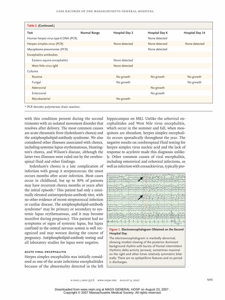
On the second hospital day, a lumbar punc-ture was performed. Results of cerebrospinal f luid analysis are shown in Table 2; other test results are listed in Table 1. An enzyme-linked im-munosorbent assay for serum antibodies against HIV was negative. An electroencephalogram showed diffuse theta slowing and frontal inter-mittent rhythmic delta activity, which was more prominent in the right hemisphere than in the left. There was no epileptiform activity (Fig. 1). Repeated MRI of the brain after the administra-tion of gadolinium showed no changes and no evidence of abnormal enhancement. Acyclovir was administered intravenously.
The next day, a serum Lyme antibody test, a test of a throat swab for Mycoplasma pneumoniae nucle-ic acid, and cultures of blood and urine were negative; results of other tests are listed in Table 1. On the fifth day, the patient’s condition appeared to be improved. She was oriented and remem-bered details of her past; dysmetria and truncal
Copyright © 2007 Massachusetts Medical Society. All rights reserved. Downloaded from www.nejm.org at MASS GENERAL HOSP on August 23, 2007 .

case records of the massachusetts gener al hospital
n engl j med 357;6 www.nejm.org august 9, 2007 591
ataxia were reduced. Results on a repeated elec-troencephalogram were unchanged. The next day, a repeated lumbar puncture was performed (Table 2).
Between the 7th and 18th hospital days, the patient’s motor function gradually worsened, right-sided neglect developed, she became unable to feed herself, her responsiveness and ability to follow commands decreased, and she became incontinent. She began lying in a fetal position, moaning and crying out unintelligible sounds. A skin test for tuberculosis, a test of a nasopha-ryngeal specimen for respiratory viral antigens, and a viral culture of a stool specimen were nega-tive. Nucleic acid testing for HIV RNA and tests for antinuclear antibodies were negative. Levels of free and total thyroxine were normal, and the thyroglobulin level was elevated (54.7 ng per milli-liter; normal range, 4 to 40). On the 12th day, the acyclovir was discontinued, and ceftriaxone, at a dose of 2 g, was administered intravenously. A repeated electroencephalographic study showed increased attenuation of background activity and less abundant frontal intermittent rhythmic delta activity. MRI on the 13th day showed new hyper-intense signal in the pons and middle cerebellar peduncles with associated restricted diffusion of water on T2-weighted FLAIR images. Restricted diffusion was also noted in the posterior limb of the left internal capsule. There was atrophy in the left medial temporal lobe, with resolution of the abnormal hyperintense signal on FLAIR images. On the 14th day, a third lumbar puncture was performed.
On the 18th hospital day, a test result was received.
Differ en ti a l Di agnosis
Dr. Andrew J. Cole: I was involved in this patient’s care from the time of her admission and am there-fore aware of the diagnosis. I will discuss the case as it unfolded in order to illustrate the diag-nostic process and therapeutic decision making that took place. The patient lived semi-indepen-dently until she became pregnant 27 weeks before admission. Her level of function at that time was unknown, and because of the lack of informa-tion, it was not possible to determine either her level of function before her illness or the tempo of her disease.
Neurologic differential diagnosis relies primar-ily on the physical examination for localization
of lesions and on the history, especially the nature of onset and pace of progression, to identify the disease process. This patient’s neurologic exami-nation showed abnormal cognitive function indi-cating dysfunction of the cortical and subcortical gray matter, abnormal motor function indicat-ing dysfunction of the pyramidal motor system, and choreiform movements indicating dysfunc-tion of the extrapyramidal motor systems. This examination also showed a clumsy gait and dif-ficulty performing rapid alternating movements, indicating dysfunction of the cerebellum or its connections. With the limited information about the pace of her disease, we needed to consider inherited, congenital, and acquired diseases that could be acute, subacute, or chronic, with static, episodic, or progressive tempos. We had to base our differential diagnosis on the neurologic ex-amination, initial laboratory testing, and electro-encephalographic and MRI studies.
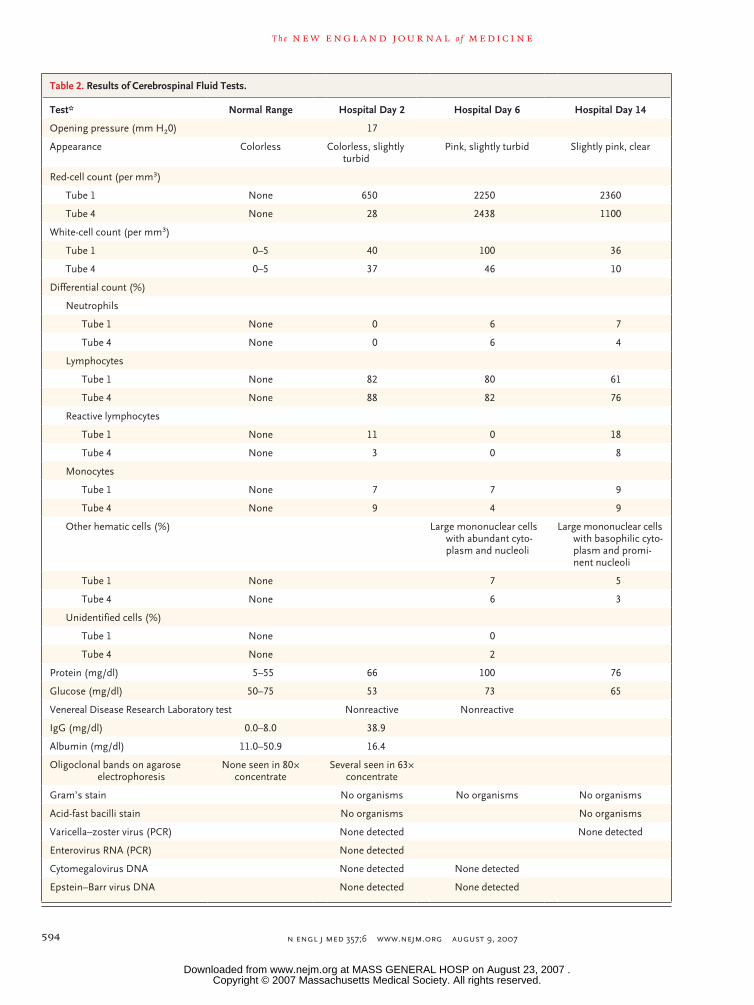
Cerebrospinal Fluid Examination
The results of the cerebrospinal fluid analysis in this patient showed a lymphocytic pleocytosis with few red cells, a mildly elevated protein level, and a normal glucose level. These findings are characteristic of aseptic meningitis. We were thus concerned about viruses, rickettsia, spirochetes, partially treated bacterial infection, a paramenin-geal focus of infection, certain autoimmune ill-nesses such as systemic lupus erythematosus or Behçet’s disease, vasculitides, carcinoma, a reac-tion to the toxic effects of certain medications such as nonsteroidal antiinflammatory drugs, and chemical meningitis related to the rupture of a cyst. Although they were nonspecific, the cerebrospinal fluid findings provided support for the possibility of acute or subacute infection or inflammatory illness. The presence of an inflam-matory response made chronic degenerative diseas-es such as Huntington’s disease, Wilson’s disease, and systems abiotrophies such as multisystem atrophy unlikely.
Electroencephalographic Studies
The initial electroencephalogram was markedly abnormal, but the findings were nonspecific (Fig. 1). The slow and attenuated posterior domi-nant rhythm suggests cortical gray-matter dis-ease, whereas the intermittent frontal rhythmic delta activity suggests subcortical gray-matter disease. The monomorphic slow waves also sug-gest that initially the subcortical white matter
Copyright © 2007 Massachusetts Medical Society. All rights reserved. Downloaded from www.nejm.org at MASS GENERAL HOSP on August 23, 2007 .
T h e n e w e ng l a nd j o u r na l o f m e dic i n e
n engl j med 357;6 www.nejm.org august 9, 2007592
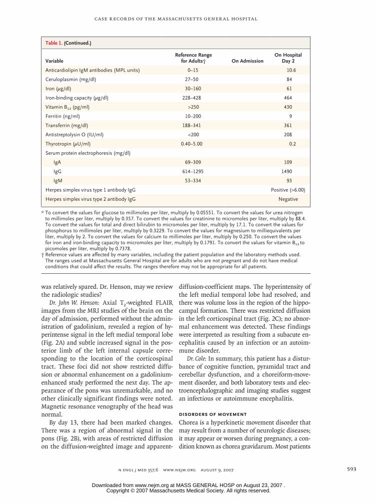
Table 1. Results of Laboratory Tests.*
VariableReference Range
for Adults† On AdmissionOn Hospital
Day 2
Hematocrit (%) 36.0–46.0 (in women) 37.4 35.2
Hemoglobin (g/dl) 12.0–16.0 (in women) 13.2 12.1
White-cell count (per mm3) 4500–13,000 8,100 7,600
Differential count (%)
Neutrophils 40–62 72
Lymphocytes 27–40 21
Monocytes 4–11 6
Eosinophils 0–8 1
Basophils 0–3 0
Platelet count (per mm3) 150,000–350,000 236,000 192,000
Mean corpuscular volume (μm3) 80–100 89
Erythrocyte sedimentation rate (mm/hr) 1–25 26
Glucose (mg/dl) 70–110 78 73
Sodium (mmol/liter) 135–145 135 137
Potassium (mmol/liter) 3.4–4.8 3.5 3.4
Chloride (mmol/liter) 100–108 106 104
Carbon dioxide (mmol/liter) 23.0–31.9 26.0 25.2
Urea nitrogen (mg/dl) 8–25 8 4
Creatinine (mg/dl) 0.6–1.5 0.5 0.6
Bilirubin (mg/dl)
Total 0.0–1.0 0.1
Direct 0–0.4 0.0
Protein (g/dl)
Total 6.0–8.3 7.7 7.4
Albumin 3.3–5.0 3.5
Globulin 2.6–4.1 4.2
Phosphorus (mg/dl) 2.6–4.5 3.0
Magnesium (mmol/liter) 0.7–1.0 0.75
Calcium (mg/dl) 8.5–10.5 9.2
Creatine kinase (U/liter) 40–150 (in women) 62
Alkaline phosphatase (U/liter) 30–100 96
Aspartate aminotransferase (U/liter) 9–32 18
Alanine aminotransferase (U/liter) 7–30 15
Lipase (U/dl) 1.3–6.0 7.5
Amylase (U/liter) 3–100 79
Rapid plasma reagin Nonreactive
Human chorionic gonadotropin, quantitative (IU/liter)
<6 (in nonpregnant women); 6–15 (borderline)
32,907
Toxicology screen Negative
Partial-thromboplastin time lupus anticoagulant None
Anticardiolipin IgG antibodies (GPL units) 0–15 7.9
Copyright © 2007 Massachusetts Medical Society. All rights reserved. Downloaded from www.nejm.org at MASS GENERAL HOSP on August 23, 2007 .
case records of the massachusetts gener al hospital
n engl j med 357;6 www.nejm.org august 9, 2007 593
was relatively spared. Dr. Henson, may we review the radiologic studies?
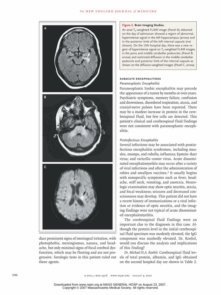
Dr. John W. Henson: Axial T2-weighted FLAIR images from the MRI studies of the brain on the day of admission, performed without the admin-istration of gadolinium, revealed a region of hy-perintense signal in the left medial temporal lobe (Fig. 2A) and subtle increased signal in the pos-terior limb of the left internal capsule corre-sponding to the location of the corticospinal tract. These foci did not show restricted diffu-sion or abnormal enhancement on a gadolinium-enhanced study performed the next day. The ap-pearance of the pons was unremarkable, and no other clinically significant findings were noted. Magnetic resonance venography of the head was normal.
By day 13, there had been marked changes. There was a region of abnormal signal in the pons (Fig. 2B), with areas of restricted diffusion on the diffusion-weighted image and apparent-
diffusion-coefficient maps. The hyperintensity of the left medial temporal lobe had resolved, and there was volume loss in the region of the hippo-campal formation. There was restricted diffusion in the left corticospinal tract (Fig. 2C); no abnor-mal enhancement was detected. These findings were interpreted as resulting from a subacute en-cephalitis caused by an infection or an autoim-mune disorder.
Dr. Cole: In summary, this patient has a distur-bance of cognitive function, pyramidal tract and cerebellar dysfunction, and a choreiform-move-ment disorder, and both laboratory tests and elec-troencephalographic and imaging studies suggest an infectious or autoimmune encephalitis.
Disorders of Movement
Chorea is a hyperkinetic movement disorder that may result from a number of neurologic diseases; it may appear or worsen during pregnancy, a con-dition known as chorea gravidarum. Most patients
Table 1. (Continued.)
VariableReference Range
for Adults† On AdmissionOn Hospital
Day 2
Anticardiolipin IgM antibodies (MPL units) 0–15 10.6
Ceruloplasmin (mg/dl) 27–50 84
Iron (μg/dl) 30–160 61
Iron-binding capacity (μg/dl) 228–428 464
Vitamin B12 (pg/ml) >250 430
Ferritin (ng/ml) 10–200 9
Transferrin (mg/dl) 188–341 361
Antistreptolysin O (IU/ml) <200 208
Thyrotropin (μU/ml) 0.40–5.00 0.2
Serum protein electrophoresis (mg/dl)
IgA 69–309 109
IgG 614–1295 1490
IgM 53–334 93
Herpes simplex virus type 1 antibody IgG Positive (>6.00)
Herpes simplex virus type 2 antibody IgG Negative
* To convert the values for glucose to millimoles per liter, multiply by 0.05551. To convert the values for urea nitrogen to millimoles per liter, multiply by 0.357. To convert the values for creatinine to micromoles per liter, multiply by 88.4. To convert the values for total and direct bilirubin to micromoles per liter, multiply by 17.1. To convert the values for phosphorus to millimoles per liter, multiply by 0.3229. To convert the values for magnesium to milliequivalents per liter, multiply by 2. To convert the values for calcium to millimoles per liter, multiply by 0.250. To convert the values for iron and iron-binding capacity to micromoles per liter, multiply by 0.1791. To convert the values for vitamin B12
to picomoles per liter, multiply by 0.7378.
† Reference values are affected by many variables, including the patient population and the laboratory methods used. The ranges used at Massachusetts General Hospital are for adults who are not pregnant and do not have medical conditions that could affect the results. The ranges therefore may not be appropriate for all patients.
Copyright © 2007 Massachusetts Medical Society. All rights reserved. Downloaded from www.nejm.org at MASS GENERAL HOSP on August 23, 2007 .
T h e n e w e ng l a nd j o u r na l o f m e dic i n e
n engl j med 357;6 www.nejm.org august 9, 2007594
Table 2. Results of Cerebrospinal Fluid Tests.
Test* Normal Range Hospital Day 2 Hospital Day 6 Hospital Day 14
Opening pressure (mm H20) 17
Appearance Colorless Colorless, slightly turbid
Pink, slightly turbid Slightly pink, clear
Red-cell count (per mm3)
Tube 1 None 650 2250 2360
Tube 4 None 28 2438 1100
White-cell count (per mm3)
Tube 1 0–5 40 100 36
Tube 4 0–5 37 46 10
Differential count (%)
Neutrophils
Tube 1 None 0 6 7
Tube 4 None 0 6 4
Lymphocytes
Tube 1 None 82 80 61
Tube 4 None 88 82 76
Reactive lymphocytes
Tube 1 None 11 0 18
Tube 4 None 3 0 8
Monocytes
Tube 1 None 7 7 9
Tube 4 None 9 4 9
Other hematic cells (%) Large mononuclear cells with abundant cyto-plasm and nucleoli
Large mononuclear cells with basophilic cyto-plasm and promi-nent nucleoli
Tube 1 None 7 5
Tube 4 None 6 3
Unidentified cells (%)
Tube 1 None 0
Tube 4 None 2
Protein (mg/dl) 5–55 66 100 76
Glucose (mg/dl) 50–75 53 73 65
Venereal Disease Research Laboratory test Nonreactive Nonreactive
IgG (mg/dl) 0.0–8.0 38.9
Albumin (mg/dl) 11.0–50.9 16.4
Oligoclonal bands on agarose electrophoresis
None seen in 80 × concentrate
Several seen in 63× concentrate
Gram’s stain No organisms No organisms No organisms
Acid-fast bacilli stain No organisms No organisms
Varicella–zoster virus (PCR) None detected None detected
Enterovirus RNA (PCR) None detected
Cytomegalovirus DNA None detected None detected
Epstein–Barr virus DNA None detected None detected
Copyright © 2007 Massachusetts Medical Society. All rights reserved. Downloaded from www.nejm.org at MASS GENERAL HOSP on August 23, 2007 .
case records of the massachusetts gener al hospital
n engl j med 357;6 www.nejm.org august 9, 2007 595
with this condition present during the second trimester with an isolated movement disorder that resolves after delivery. The most common causes are acute rheumatic fever (Sydenham’s chorea) and the antiphospholipid-antibody syndrome. We also considered other illnesses associated with chorea, including systemic lupus erythematosus, Hunting-ton’s chorea, and Wilson’s disease, although the latter two illnesses were ruled out by the cerebro-spinal fluid and other findings.
Sydenham’s chorea is a late complication of infection with group A streptococcus; the onset occurs months after acute infection. Most cases occur in childhood, but up to 30% of patients may have recurrent chorea months or years after the initial episode.1 This patient had only a mini-mally elevated antistreptolysin-antibody titer, with no other evidence of recent streptococcal infection or cardiac disease. The antiphospholipid-antibody syndrome2 may be primary or secondary to sys-temic lupus erythematosus, and it may become manifest during pregnancy. This patient had no symptoms or signs of systemic lupus, but lupus confined to the central nervous system is well rec-ognized and may worsen during the course of pregnancy. Antiphospholipid-antibody testing and all laboratory studies for lupus were negative.
Acute Viral Encephalitis
Herpes simplex encephalitis was initially consid-ered as one of the acute infectious encephalitides because of the abnormality detected in the left
hippocampus on MRI. Unlike the arboviral en-cephalitides and West Nile virus encephalitis, which occur in the summer and fall, when mos-quitoes are abundant, herpes simplex encephali-tis occurs sporadically throughout the year. The negative results on cerebrospinal fluid testing for herpes simplex virus nucleic acid and the lack of response to acyclovir made this diagnosis unlike-ly. Other common causes of viral encephalitis, including enteroviral and echoviral infections, as well as infection with coxsackievirus, typically pro-
16p6
AUTHOR Cole
FIGURE 1 of 4
JOB: ISSUE:
4-CH/T
RETAKE 1st2nd
SIZE
ICM
CASE
EMail LineH/TCombo
Revised
AUTHOR, PLEASE NOTE: Figure has been redrawn and type has been reset.
Please check carefully.
REG F
FILL
TITLE3rd
Enon ARTIST:
7-9-07
mst
35706
Figure 1. Electroencephalogram Obtained on the Second Hospital Day.
The electroencephalogram is markedly abnormal, showing modest slowing of the posterior dominant background rhythm with bursts of frontal intermittent rhythmic delta activity (arrows), sometimes maximal on the right and other times relatively symmetric bilat-erally. There are no epileptiform features and no period-ic discharges.
Table 2. (Continued.)
Test Normal Range Hospital Day 2 Hospital Day 6 Hospital Day 14
Human herpes virus type 6 DNA (PCR) None detected
Herpes simplex virus (PCR) None detected None detected None detected
Mycoplasma pneumoniae (PCR) None detected
Encephalitis antibodies
Eastern equine encephalitis None detected
West Nile virus IgM None detected
Cultures
Routine No growth No growth No growth
Fungal No growth No growth
Adenoviral No growth
Enteroviral No growth
Mycobacterial No growth
* PCR denotes polymerase chain reaction.
Copyright © 2007 Massachusetts Medical Society. All rights reserved. Downloaded from www.nejm.org at MASS GENERAL HOSP on August 23, 2007 .
T h e n e w e ng l a nd j o u r na l o f m e dic i n e
n engl j med 357;6 www.nejm.org august 9, 2007596
duce prominent signs of meningeal irritation, with photophobia, meningismus, nausea, and head-ache, but only minimal signs of focal cerebral dys-function, which may be fleeting and are not pro-gressive. Serologic tests in this patient ruled out these agents.
Subacute EncephalitidesParaneoplastic EncephalitisParaneoplastic limbic encephalitis may precede the appearance of a tumor by months or even years. Psychiatric symptoms, memory failure, confusion and drowsiness, disordered respiration, ataxia, and cranial-nerve palsies have been reported. There may be a modest increase in protein in the cere-brospinal fluid, but few cells are detected. This patient’s clinical and cerebrospinal fluid findings were not consistent with paraneoplastic enceph-alitis.
Postinfectious EncephalitisSeveral infections may be associated with postin-fectious encephalitis syndromes, including mea-sles, mumps, and rubella; influenza; Epstein–Barr virus; and varicella–zoster virus. Acute dissemi-nated encephalomyelitis may occur after a variety of viral infections and after the administration of rabies and smallpox vaccines.3 It usually begins with nonspecific symptoms such as fever, head-ache, stiff neck, vomiting, and anorexia. Neuro-logic examination may show optic neuritis, ataxia, and focal weakness; seizures and decreased con-sciousness may develop. This patient did not have a recent history of immunizations or a viral infec-tion or evidence of optic neuritis, and the imag-ing findings were not typical of acute disseminat-ed encephalomyelitis.
The cerebrospinal f luid findings were an important clue to the diagnosis in this case. Al-though the protein level in the initial cerebrospi-nal fluid specimen was modestly elevated, the IgG component was markedly elevated. Dr. Roehrl, would you discuss the analysis and implications of this finding?
Dr. Michael H.A. Roehrl: Cerebrospinal fluid lev-els of total protein, albumin, and IgG obtained on the second hospital day are shown in Table 2.
A
B
C
16p6
AUTHOR Cole
FIGURE 2a-c of 4
JOB: ISSUE:
4-CH/T
RETAKE 1st2nd
SIZE
ICM
CASE
EMail LineH/TCombo
Revised
AUTHOR, PLEASE NOTE: Figure has been redrawn and type has been reset.
Please check carefully.
REG F
FILL
TITLE3rd
Enon ARTIST:
8-9-07
mst
35706
Figure 2. Brain Imaging Studies.
An axial T2-weighted FLAIR image (Panel A) obtained on the day of admission showed a region of abnormal, hyperintense signal in the left hippocampus (arrow) and in the posterior limb of the left internal capsule (not shown). On the 13th hospital day, there was a new re-gion of hyperintense signal on T2-weighted FLAIR images in the pons and middle cerebellar peduncles (Panel B, arrow) and restricted diffusion in the middle cerebellar peduncle and posterior limb of the internal capsule as shown on the diffusion-weighted images (Panel C, arrow).
Copyright © 2007 Massachusetts Medical Society. All rights reserved. Downloaded from www.nejm.org at MASS GENERAL HOSP on August 23, 2007 .

case records of the massachusetts gener al hospital
n engl j med 357;6 www.nejm.org august 9, 2007 597
Agarose-gel electrophoresis of cerebrospinal fluid revealed oligoclonal bands in the gamma region (Fig. 3A). The cerebrospinal fluid to serum mass concentration quotients for albumin and IgG concentrations were QAlb = 4.7×10⁻³ and QIgG = 26.1×10⁻³, corresponding to an IgG index of 5.6 (normal value, <0.85).4 On the basis of an analy-sis developed by Reiber,5‑7 this patient’s results (Fig. 3B) indicated markedly increased intrathecal IgG synthesis (intrathecal production fraction, 87.7%) without evidence of clinically significant blood–brain barrier dysfunction. The mass con-centration ratio of IgG to total protein in the ce-rebrospinal fluid was 58.9%.
Dr. Cole: The diseases that can elicit an intra-thecal response of this magnitude are syphilis, chronic rubella panencephalitis, and subacute sclerosing panencephalitis.8‑11 This patient did not have syphilis, as shown by negative results on serologic testing and a symptom complex that was inconsistent with the disease. Postrubella en-cephalitis12,13 may affect patients with history of remote or congenital rubella infection and pres-
ents with progressive dementia, ataxia, chorea, retinal degeneration, and seizures. Examination of the cerebrospinal fluid shows pleocytosis and a moderately elevated protein level, with up to 50% of the cerebrospinal fluid protein composed of immunoglobulins. The diagnosis is confirmed by a high antirubella-antibody titer in the cere-brospinal fluid.
Subacute Sclerosing Panencephalitis
Measles causes three distinct diseases of the cen-tral nervous system: postinfectious encephalomy-elitis, subacute measles encephalitis, and subacute sclerosing panencephalitis.14 Subacute scleros-ing panencephalitis is typically seen 7 to 10 years after infection with measles, and patients present most commonly with declining performance in school, behavioral changes, headache, adventitious movements, and sometimes seizures.15 Charac-teristic findings include myoclonic jerks that are often periodic and are associated with periodic lateralized or bilateral epileptiform discharges on an electroencephalogram. Although most cases occur in childhood or adolescence, cases begin-ning as late as the fifth decade of life have been described.16,17 The incidence of subacute scleros-
2
3
4
1
B
A
100
QIg
G
50
10
2
5
0.5
20
1
52 10 20 50 100
Q High
Q Mea
n
Q Low
QAIb
AUTHOR:
FIGURE:
JOB:
4-CH/T
RETAKE
SIZE
ICM
CASE
EMail LineH/TCombo
Revised
AUTHOR, PLEASE NOTE: Figure has been redrawn and type has been reset.
Please check carefully.
REG F
Enon
1st
2nd3rd
Cole
3 of 4
08-09-07
ARTIST: ts
35706 ISSUE:
16p6
Normal cerebrospinalfluid
Patient’s cerebrospinalfluid
P A α1 α2 β γ
(×10−3)
(×10−3)80
%60
%40
%20
%
Figure 3. Results of Cerebrospinal Fluid Electrophoresis.
Panel A shows the results of agarose-gel electrophoresis of a specimen of the patient’s cerebrospinal fluid collect-ed on the second hospital day (concentrated to 1/63 of the original volume) and cerebrospinal fluid from a nor-mal control (concentrated to 1/80 of the original volume). P, A, α1, α2, β, and γ denote the electrophoretic pre-albumin, albumin, alpha-1, alpha-2, beta, and gamma regions, respectively. The arrow shows the position of several strong bands in the gamma region, indicating the presence of multiple oligoclonal immunoglobulins. There is also a relative decrease of the level of albumin in the patient’s cerebrospinal fluid. Panel B shows a double-logarithmic graph designed according to the method proposed by Reiber (also called a Reibergram), in which cerebrospinal fluid to serum mass concentra-tion quotients for albumin (Q
Alb) and IgG (QIgG) are
plotted along the abscissa and ordinate, respectively. Upper limits (Q
High) and lower limits (Q
Low) of normal
values are shown as solid lines, with the dotted line in-dicating mean normal values (Q
Mean). The isopercentiles
(dashed lines) correspond to various relative amounts of intrathecal IgG production (20 to 80%). The vertical dashed line denotes the age-adjusted upper limit of Q
Alb, separating normal function (left) and abnormal
function (right) of the blood–brain barrier. The patient (red dot) had markedly increased intrathecal IgG synthesis (intrathecal production fraction, 87.7%) without evidence of significant dysfunction of the blood–brain barrier (zone 4). Zone 1 denotes normal function, zone 2 de-notes pure blood–brain barrier dysfunction, and zone 3 denotes a combination of increased intrathecal IgG synthesis and blood–brain barrier dysfunction.
Copyright © 2007 Massachusetts Medical Society. All rights reserved. Downloaded from www.nejm.org at MASS GENERAL HOSP on August 23, 2007 .

T h e n e w e ng l a nd j o u r na l o f m e dic i n e
n engl j med 357;6 www.nejm.org august 9, 2007598
ing panencephalitis has decreased with wide-spread vaccination against measles; however, it persists in places where measles vaccination is uncommon.18 The incidence of this condition is increased as much as 10 times in patients in whom measles develops before the age of 2 years; this patient had measles at 4 months of age. The illness may present during pregnancy, possibly as a result of altered immune status.8,19
In summary, this patient presented with a subacute progressive neurologic disease charac-terized by widespread dysfunction of the central nervous system, inflammatory features in the cerebrospinal fluid, and an extremely high level of cerebrospinal fluid IgG. My colleagues and I favored the diagnosis of subacute sclerosing pan-encephalitis, a delayed consequence of her infec-tion with measles at 4 months of age. This condi-tion may have been exacerbated by her pregnancy. Specimens of serum and cerebrospinal fluid from the 14th hospital day were sent for testing of levels of antibodies against measles.
Dr. Roehrl: The measles-specific cerebrospinal fluid to serum IgG antibody index was elevated at 31.8 (normal value, <1.4)7 (Table 3). Measles-spe-cific IgM antibodies were not detected in the se-rum. This profile indicates a chronic immune re-sponse to measles infection in the cerebrospinal f luid compartment. A comparison with mumps titers shows the specificity of the immunologic process. The results confirm the diagnosis of sub-acute sclerosing panencephalitis in the patient. The disease was a late consequence of persistent infection with measles virus.
Discussion of M a nagemen t
Dr. Cole: Cell-mediated immunity stimulated by T-helper cell type 1 (Th1) inducing cytokines is crucial for the clearance of measles virus in the weeks after infection, whereas cytokines that in-duce type 2 helper T (Th2) cells are implicated in antibody production. Several reports have sug-gested that treatment with intrathecal interferon alfa-2 (a cytokine that promotes Th1 activity), with or without treatment with the antiviral im-munomodulatory agent inosine pranobex, may slow or even arrest the progression of subacute sclerosing panencephalitis.20‑25 However, no de-finitive data are available to show the efficacy of this treatment approach.
When we made the diagnosis in this patient,
she was at 28 weeks of gestation, and her condi-tion was deteriorating rapidly. We believed that an attempt at treatment was in the best interest of the patient and her fetus. We treated her with interferon and inosine pranobex for 8 weeks, without any clear clinical benefit. After the de-livery of a healthy baby by elective cesarean sec-tion at 34 weeks’ gestation, a decision was made to discontinue treatment, with the consent of the patient’s mother. The patient died 6 weeks later.
DR . A NDR E W J. COLE’S DI AGNOSIS
Subacute sclerosing panencephalitis.
Pathol o gic a l Discussion
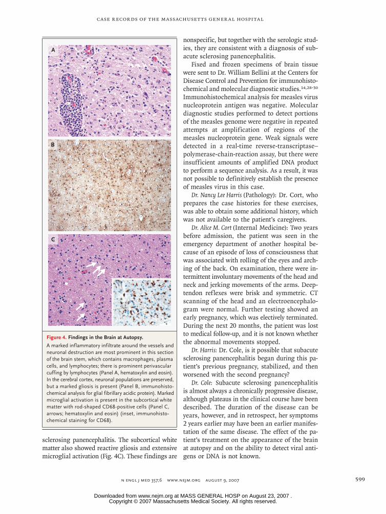
Dr. Matthew P. Frosch: The autopsy revealed inflam-matory infiltrates containing macrophages, plas-ma cells, and lymphocytes around vessels, with neuronal destruction and reactive gliosis that were most prominent in the brain stem (Fig. 4A). No viral inclusions of the type usually seen in acute measles encephalitis (and occasionally present in subacute sclerosing panencephalitis) were seen in the nuclei or cytoplasm of any cell types (neurons, glia, or vascular endothelial cells). Unlike other measles-associated diseases, subacute sclerosing panencephalitis is caused by the persistence of defective viruses that do not form complete viral particles but may infect adjacent cells by direct contact.26,27 In the cerebral cortex, neuronal pop-ulations were preserved, but there was marked gliosis with reactive astrocytes (Fig. 4B). The brain stem was softened by the pathologic process, but the hemispheric white matter was firmer than normal; this was most evident when the brain was cut in the fresh state. This firmness corre-sponds to the sclerosis in the name, subacute
Table 3. Results of Blood and Cerebrospinal Fluid Tests for Measles and Mumps Antibodies on Hospital Day 14.
Test Result
Blood Cerebrospinal Fluid
Measles
IgM Negative 1:512
IgG >1:800,000 1:81,920
Mumps
IgM Negative Negative
IgG 1:512 1:32
Copyright © 2007 Massachusetts Medical Society. All rights reserved. Downloaded from www.nejm.org at MASS GENERAL HOSP on August 23, 2007 .
case records of the massachusetts gener al hospital
n engl j med 357;6 www.nejm.org august 9, 2007 599
sclerosing panencephalitis. The subcortical white matter also showed reactive gliosis and extensive microglial activation (Fig. 4C). These findings are
nonspecific, but together with the serologic stud-ies, they are consistent with a diagnosis of sub-acute sclerosing panencephalitis.
Fixed and frozen specimens of brain tissue were sent to Dr. William Bellini at the Centers for Disease Control and Prevention for immunohisto-chemical and molecular diagnostic studies.14,28‑30 Immunohistochemical analysis for measles virus nucleoprotein antigen was negative. Molecular diagnostic studies performed to detect portions of the measles genome were negative in repeated attempts at amplification of regions of the measles nucleoprotein gene. Weak signals were detected in a real-time reverse-transcriptase–polymerase-chain-reaction assay, but there were insufficient amounts of amplified DNA product to perform a sequence analysis. As a result, it was not possible to definitively establish the presence of measles virus in this case.
Dr. Nancy Lee Harris (Pathology): Dr. Cort, who prepares the case histories for these exercises, was able to obtain some additional history, which was not available to the patient’s caregivers.
Dr. Alice M. Cort (Internal Medicine): Two years before admission, the patient was seen in the emergency department of another hospital be-cause of an episode of loss of consciousness that was associated with rolling of the eyes and arch-ing of the back. On examination, there were in-termittent involuntary movements of the head and neck and jerking movements of the arms. Deep-tendon reflexes were brisk and symmetric. CT scanning of the head and an electroencephalo-gram were normal. Further testing showed an early pregnancy, which was electively terminated. During the next 20 months, the patient was lost to medical follow-up, and it is not known whether the abnormal movements stopped.
Dr. Harris: Dr. Cole, is it possible that subacute sclerosing panencephalitis began during this pa-tient’s previous pregnancy, stabilized, and then worsened with the second pregnancy?
Dr. Cole: Subacute sclerosing panencephalitis is almost always a chronically progressive disease, although plateaus in the clinical course have been described. The duration of the disease can be years, however, and in retrospect, her symptoms 2 years earlier may have been an earlier manifes-tation of the same disease. The effect of the pa-tient’s treatment on the appearance of the brain at autopsy and on the ability to detect viral anti-gens or DNA is not known.
A
B
C
16p6
AUTHOR Cole
FIGURE 4a-c of 4
JOB: ISSUE:
4-CH/T
RETAKE 1st
2nd
SIZE
ICM
CASE
EMail LineH/TCombo
Revised
AUTHOR, PLEASE NOTE: Figure has been redrawn and type has been reset.
Please check carefully.
REG F
FILL
TITLE3rd
Enon ARTIST:
8-9-07
mst
35706
Figure 4. Findings in the Brain at Autopsy.
A marked inflammatory infiltrate around the vessels and neuronal destruction are most prominent in this section of the brain stem, which contains macrophages, plasma cells, and lymphocytes; there is prominent perivascular cuffing by lymphocytes (Panel A, hematoxylin and eosin). In the cerebral cortex, neuronal populations are preserved, but a marked gliosis is present (Panel B, immunohisto-chemical analysis for glial fibrillary acidic protein). Marked microglial activation is present in the subcortical white matter with rod-shaped CD68-positive cells (Panel C, arrows; hematoxylin and eosin) (inset, immunohisto-chemical staining for CD68).
Copyright © 2007 Massachusetts Medical Society. All rights reserved. Downloaded from www.nejm.org at MASS GENERAL HOSP on August 23, 2007 .
n engl j med 357;6 www.nejm.org august 9, 2007600
case records of the massachusetts gener al hospital
A nat omic a l Di agnosis
Subacute sclerosing panencephalitis secondary to measles virus infection.
Dr. Cole reports receiving consulting fees from GlaxoSmithKline, Abbott Laboratories, and Supernus Pharmaceuticals and lecture fees from GlaxoSmithKline, Abbott Laboratories, and Ortho-McNeil;
Dr. Henson, consulting fees from GlaxoSmithKline; and Dr. Frosch, consulting fees from Biogen Idec and Bristol-Myers Squibb. No other potential conflict of interest relevant to this article was re-ported.
We thank Dr. Mandakolathur R. Murali, director of the Clinical Immunology Laboratory, Department of Pathology, Massachusetts General Hospital, for the analysis of the immunologic findings in the cerebrospinal f luid.
Lantern Slides Updated: Complete PowerPoint Slide Sets from the Clinicopathological Conferences
Any reader of the Journal who uses the Case Records of the Massachusetts General Hospital as a teaching exercise or reference material is now eligible to receive a complete set of PowerPoint slides, including digital images, with identifying legends, shown at the live Clinicopathological Conference (CPC) that is the basis of the Case Record. This slide set contains all of the images from the CPC, not only those published in the Journal. Radiographic, neuro-logic, and cardiac studies, gross specimens, and photomicrographs, as well as unpublished text slides, tables, and diagrams, are included. Every year 40 sets are produced, averaging 50-60 slides per set. Each set is supplied on a compact disc and is mailed to coincide with the publication of the Case Record.
The cost of an annual subscription is $600, or individual sets may be purchased for $50 each. Application forms for the current subscription year, which began in January, may be obtained from the Lantern Slides Service, Department of Pathology, Massachusetts General Hospital, Boston, MA 02114 (telephone 617-726-2974) or e-mail [email protected].
References
al-Eissa A. Sydenham’s chorea: a new look at an old disease. Br J Clin Pract 1993; 47:14-6.
Levine JS, Branch DW, Rauch J. The antiphospholipid syndrome. N Engl J Med 2002;346:752-63.
Menge T, Hemmer B, Nessler S, et al. Acute disseminated encephalomyelitis: an update. Arch Neurol 2005;62:1673-80.
Thompson EJ, Riches PG, Kohn J. Anti-body synthesis within the central nervous system: comparisons of CSF IgG indices and electrophoresis. J Clin Pathol 1983;36: 312-5.
Reiber H. The discrimination between different blood-CSF barrier dysfunctions and inflammatory reactions of the CNS by a recent evaluation graph for the pro-tein profile of cerebrospinal fluid. J Neu-rol 1980;224:89-99.
Idem. Flow rate of cerebrospinal fluid (CSF) — a concept common to normal blood-CSF barrier function and to dysfunc-tion in neurological diseases. J Neurol Sci 1994;122:189-203.
Reiber H, Peter JB. Cerebrospinal flu-id analysis: disease-related data patterns and evaluation programs. J Neurol Sci 2001;184:101-22.
Case Records of the Massachusetts General Hospital (Case 15-1998). N Engl J Med 1998;338:1448-56.
Fishman RA. Cerebrospinal f luid in diseases of the nervous system. 2nd ed. Philadelphia: W.B. Saunders, 1992.
Norrby E, Vandvik B. Relationship be-tween measles virus-specific antibody ac-tivities and oligoclonal IgG in the central nervous system of patients with subacute sclerosing panencephalitis and multiple sclerosis. Med Microbiol Immunol 1975; 162:63-72.
Wolinsky JS. Progressive rubella pan-encephalitis. In: Vinken PJ, Bruyn GW, Kla-wans HL, McKendall RR, eds. Viral dis-ease. Amsterdam: Elsevier, 1989:405-16.
1.
2.
3.
4.
5.
6.
7.
8.
9.
10.
11.
Townsend JJ, Baringer JR, Wolinsky JS, et al. Progressive rubella panencephalitis: late onset after congenital rubella. N Engl J Med 1975;292:990-3.
Weil ML, Itabashi H, Cremer NE, Oshi-ro L, Lennette EH, Carnay L. Chronic pro-gressive panencephalitis due to rubella virus simulating subacute sclerosing pan-encephalitis. N Engl J Med 1975;292:994-8.
Honarmand S, Glaser CA, Chow E, et al. Subacute sclerosing panencephalitis in the differential diagnosis of encephalitis. Neurology 2004;63:1489-93.
Swoveland PT, Johnson KP. Subacute sclerosing panencephalitis and other para-myxovirus infections. In: Vinken PJ, Bruyn GW, Klawans HL, McKendall RR, eds. Viral disease. Amsterdam: Elsevier, 1989:417.
Gagnon A, Bouchard RW. Fulminating adult-onset subacute sclerosing panen-cephalitis in a 49-year-old man. Arch Neu-rol 2003;60:1160-1.
Prashanth LK, Taly AB, Ravi V, Sinha S, Arunodaya GR. Adult onset subacute scle-rosing panencephalitis: clinical profile of 39 patients from a tertiary care centre. J Neu-rol Neurosurg Psychiatry 2006;77:630-3.
Bellini WJ, Rota JS, Lowe LE, et al. Subacute sclerosing panencephalitis: more cases of this fatal disease are prevented by measles immunization than was previously recognized. J Infect Dis 2005;192:1686-93.
Wirguin I, Steiner I, Kidron D, et al. Fulminant subacute sclerosing panenceph-alitis in association with pregnancy. Arch Neurol 1988;45:1324-5.
Gokcil Z, Odabasi Z, Demirkaya S, Eroglu E, Vural O. Alpha-interferon and isoprinosine in adult-onset subacute scle-rosing panencephalitis. J Neurol Sci 1999; 162:62-4.
Anlar B, Yalaz K, Oktem F, Köse G. Long-term follow-up of patients with sub-acute sclerosing panencephalitis treated with intraventricular alpha-interferon. Neurology 1997;48:526-8.
12.
13.
14.
15.
16.
17.
18.
19.
20.
21.
Gascon G, Yamani S, Crowell J, et al. Combined oral isoprinosine-intraventric-ular alpha-interferon therapy for subacute sclerosing panencephalitis. Brain Dev 1993; 15:346-55.
Yalaz K, Anlar B, Oktem F, et al. Intra-ventricular interferon and oral inosiplex in the treatment of subacute sclerosing pan-encephalitis. Neurology 1992;42:488-91.
Steiner I, Wirguin I, Morag A, Abram-sky O. Intraventricular interferon treat-ment for subacute sclerosing panenceph-alitis. J Child Neurol 1989;4:20-4.
Panitch HS, Gomez-Plascencia J, Nor-ris FH, Cantell K, Smith RA. Subacute sclerosing panencephalitis: remission after treatment with intraventricular interferon. Neurology 1986;36:562-6.
Ueda S, Okuno Y, Hamamoto Y, Oya H. Subacute sclerosing panencephalitis (SSPE): isolation of a defective variant of measles virus from brain obtained at autopsy. Biken J 1975;18:113-22.
Hirano A, Ayata M, Wang AH, Wong TC. Functional analysis of matrix proteins expressed from cloned genes of measles virus variants that cause subacute scleros-ing panencephalitis reveals a common de-fect in nucleocapsid binding. J Virol 1993; 67:1848-53.
Zaki SR, Bellini WJ. Measles. In: Con-nor DH, Chandler FW, Schwartz DA, Manz HJ, Lack EE, eds. Pathology of infectious diseases. Stamford, CT: Appleton & Lange, 1997:233-44.
Bellini WJ, Rota JS, Lowe LE, et al. Subacute sclerosing panencephalitis: more cases of this fatal disease are prevented by measles immunization than previously rec-ognized. J Infect Dis 2005;192:1686-93.
Hummel KB, Lowe L, Bellini WJ, Rota PA. Development of quantitative gene-specific real-time RT-PCR assays for the de-tection of measles virus in clinical speci-mens. J Virol Methods 2006;132:166-73.Copyright © 2007 Massachusetts Medical Society.
22.
23.
24.
25.
26.
27.
28.
29.
30.
Copyright © 2007 Massachusetts Medical Society. All rights reserved. Downloaded from www.nejm.org at MASS GENERAL HOSP on August 23, 2007 .